Molecular Brain Research 87 (2001) 145–159 www.elsevier.com / locate / bres Research report Evidence for an endocannabinoid system in the central nervous system of the leech Hirudo medicinalis a b b a Isabel Matias , Tiziana Bisogno , Dominique Melck , Franck Vandenbulcke , a c d Martine Verger-Bocquet , Luciano De Petrocellis , Christian Sergheraert , a b ,1 a, * Christophe Breton , Vincenzo Di Marzo , Michel Salzet a ´ ´ Laboratoire d’ Endocrinologie et immunite des Annelides UPRES- A CNRS 8017, SN3-USTL, 59655 Villeneuve d’ Ascq, France b Endocannabinoid Research Group, Istituto per la Chimica di Molecole di Interesse Biologico, CNR, Via Toiano 6, 80072 Arco Felice, Napoli, Italy c Istituto di Cibernetica, CNR, Via Toiano 6, 80072 Arco Felice, Napoli, Italy d ` ´ Laboratoire de synthese, structure et fonctions des biomolecules, UMR 8525 CNRS, Institut de Biologie de Lille,1 rue du Professeur calmette, 59021 Lille, France Accepted 28 November 2000 Abstract In invertebrates, like Hydra and sea urchins, evidence for a functional cannabinoid system was described. The partial characterization of a putative CB1 cannabinoid receptor in the leech Hirudo medicinalis led us to investigate the presence of a complete endogenous cannabinoid system in this organism. By using gas chromatography–mass spectrometry, we demonstrate the presence of the endocannabinoids anandamide (N-arachidonoylethanolamine, 21.560.7 pmol / g) and 2-arachidonoyl-glycerol (147.4642.7 pmol/g), and of the biosynthetic precursor of anandamide, N-arachidonylphosphatidyl-ethanolamine (16.563.3 pmol / g), in the leech central nervous system (CNS). Anandamide-related molecules such as N-palmitoylethanolamine (32.461.6 pmol/g) and N-linolenoylethanolamine (5.8 pmol / g) were also detected. We also found an anandamide amidase activity in the leech CNS cytosolic fraction with a maximal activity at pH 7 and little sensitivity to typical fatty acid amide hydrolase (FAAH) inhibitors. Using an antiserum directed against the amidase signature sequence, we focused on the identification and the localization of the leech amidase. Firstly, leech nervous system protein extract was subjected to Western blot analysis, which showed three immunoreactive bands at ca. |42, |46 and |66 kDa. The former and latter bands were very faint and were also detected in whole homogenates from the coelenterate Hydra vulgaris, where the presence of CB1-like receptors, endocannabinoids and a FAAH-like activity was reported previously. Secondly, amidase immunocytochemical detection revealed numerous immunoreactive neurons in the CNS of three species of leeches. In addition, we observed that leech amidase-like immunoreactivity matches to a certain extent with CB1-like immunoreactivity. Finally, we also found that stimulation by anandamide of this receptor leads, as in mammals, to inhibition of cAMP formation, although this effect appeared to be occurring through the previously described anandamide-induced and CB1-mediated activation of nitric oxide release. Taken together, these results suggest the existence of a complete and functional cannabinoid system in leeches. 2001 Elsevier Science B.V. All rights reserved. Theme: Neuromodulators, modulators, transporters, and receptors Topic: Transmitters in invertebrate Keywords: Leech; Central nervous system; Endocannabinoid; Anandamide amidase; Cannabinoid receptor; Localization Abbreviations: 2-AG, 2-arachidonoylglycerol; CNS, Central nervous system; FAAH, Fatty acid amide hydrolase; GC-EIMS, Gas chromatography– electron impact mass spectrometry; L-NAME, N-omega-nitro-L-arginine methyl ester; NArPE, N-arachidonoyl-phosphatidyl-ethanolamine; NO, Nitric oxide; NP-HPLC, Normal phase high pressure liquid chromatography; PLD, Phospholipase D; SNAP, S-nitroso-N-acetyl-penicillamine. *Corresponding author. Tel.: 133-3-2043-6839; fax: 133-3-2004-1130. E-mail address: [email protected] (M. Salzet). 1 Corresponding author. Tel.: 139-81-8534256; fax: 139-81-8041770. E-mail address: [email protected] (V. Di Marzo). 0169-328X / 01 / $ – see front matter 2001 Elsevier Science B.V. All rights reserved. PII: S0169-328X(00)00290-4

Welcome message from author
This document is posted to help you gain knowledge. Please leave a comment to let me know what you think about it! Share it to your friends and learn new things together.
Transcript

Molecular Brain Research 87 (2001) 145–159www.elsevier.com/ locate /bres
Research report
Evidence for an endocannabinoid system in the central nervous systemof the leech Hirudo medicinalis
a b b aIsabel Matias , Tiziana Bisogno , Dominique Melck , Franck Vandenbulcke ,a c dMartine Verger-Bocquet , Luciano De Petrocellis , Christian Sergheraert ,
a b ,1 a ,*Christophe Breton , Vincenzo Di Marzo , Michel Salzeta ´ ´Laboratoire d’Endocrinologie et immunite des Annelides UPRES-A CNRS 8017, SN3-USTL, 59655 Villeneuve d’Ascq, France
bEndocannabinoid Research Group, Istituto per la Chimica di Molecole di Interesse Biologico, CNR, Via Toiano 6, 80072 Arco Felice, Napoli, ItalycIstituto di Cibernetica, CNR, Via Toiano 6, 80072 Arco Felice, Napoli, Italy
d ` ´Laboratoire de synthese, structure et fonctions des biomolecules, UMR 8525 CNRS, Institut de Biologie de Lille, 1 rue du Professeur calmette,59021 Lille, France
Accepted 28 November 2000
Abstract
In invertebrates, like Hydra and sea urchins, evidence for a functional cannabinoid system was described. The partial characterization ofa putative CB1 cannabinoid receptor in the leech Hirudo medicinalis led us to investigate the presence of a complete endogenouscannabinoid system in this organism. By using gas chromatography–mass spectrometry, we demonstrate the presence of theendocannabinoids anandamide (N-arachidonoylethanolamine, 21.560.7 pmol /g) and 2-arachidonoyl-glycerol (147.4642.7 pmol /g), andof the biosynthetic precursor of anandamide, N-arachidonylphosphatidyl-ethanolamine (16.563.3 pmol /g), in the leech central nervoussystem (CNS). Anandamide-related molecules such as N-palmitoylethanolamine (32.461.6 pmol /g) and N-linolenoylethanolamine (5.8pmol /g) were also detected. We also found an anandamide amidase activity in the leech CNS cytosolic fraction with a maximal activity atpH 7 and little sensitivity to typical fatty acid amide hydrolase (FAAH) inhibitors. Using an antiserum directed against the amidasesignature sequence, we focused on the identification and the localization of the leech amidase. Firstly, leech nervous system proteinextract was subjected to Western blot analysis, which showed three immunoreactive bands at ca. |42, |46 and |66 kDa. The former andlatter bands were very faint and were also detected in whole homogenates from the coelenterate Hydra vulgaris, where the presence ofCB1-like receptors, endocannabinoids and a FAAH-like activity was reported previously. Secondly, amidase immunocytochemicaldetection revealed numerous immunoreactive neurons in the CNS of three species of leeches. In addition, we observed that leechamidase-like immunoreactivity matches to a certain extent with CB1-like immunoreactivity. Finally, we also found that stimulation byanandamide of this receptor leads, as in mammals, to inhibition of cAMP formation, although this effect appeared to be occurring throughthe previously described anandamide-induced and CB1-mediated activation of nitric oxide release. Taken together, these results suggestthe existence of a complete and functional cannabinoid system in leeches. 2001 Elsevier Science B.V. All rights reserved.
Theme: Neuromodulators, modulators, transporters, and receptors
Topic: Transmitters in invertebrate
Keywords: Leech; Central nervous system; Endocannabinoid; Anandamide amidase; Cannabinoid receptor; Localization
Abbreviations: 2-AG, 2-arachidonoylglycerol; CNS, Central nervous system; FAAH, Fatty acid amide hydrolase; GC-EIMS, Gas chromatography–electron impact mass spectrometry; L-NAME, N-omega-nitro-L-arginine methyl ester; NArPE, N-arachidonoyl-phosphatidyl-ethanolamine; NO, Nitricoxide; NP-HPLC, Normal phase high pressure liquid chromatography; PLD, Phospholipase D; SNAP, S-nitroso-N-acetyl-penicillamine.
*Corresponding author. Tel.: 133-3-2043-6839; fax: 133-3-2004-1130.E-mail address: [email protected] (M. Salzet).1Corresponding author. Tel.: 139-81-8534256; fax: 139-81-8041770.E-mail address: [email protected] (V. Di Marzo).
0169-328X/01/$ – see front matter 2001 Elsevier Science B.V. All rights reserved.PI I : S0169-328X( 00 )00290-4

146 I. Matias et al. / Molecular Brain Research 87 (2001) 145 –159
1. Introduction The endocannabinoid system comprises the ligands(anandamide, 2-AG), the anandamide precursor (N-arach-
For centuries marijuana, derived from the Indian hemp idonylphosphatidyl-ethanolamine, NArPE), the can-Cannabis sativa, has been used for its medicinal as well as nabinoid receptors (CB1, CB1a, CB2), the anandamide
9its psychotropic effects [28]. Trans-D -tetrahydrocan- membrane transporter and FAAH. Recently, the questionnabinol (THC) is the major active psychotropic component of the conservation in the course of evolution of suchof cannabis [19]. Central nervous system responses to system has been examined. In invertebrates, there isTHC and synthetic cannabimetic drugs include the thera- evidence for the existence of an endocannabinoid system,peutically beneficial analgesia, attenuation of nausea and in particular, in sea urchins [4,46], in the coelenteratevomiting in cancer chemotherapy, appetite stimulation in Hydravulgaris [8] and in the mollusk Mytilus eduliswasting syndromes, and reduction of intestinal motility [2]. [47,51]. Together with cannabinoid binding sites andThese effects are mediated by receptors, two of which have endogenous agonists, enzymatic activities capable ofbeen identified: the CB1 and CB2 receptors. The CB1 catalyzing the hydrolysis of anandamide were detected inreceptor was cloned in 1990 from the rat [35], and later these animals. Finally, we have partially characterized afrom human [20] and mouse [6]. Additionally, a splice putative CB1-like receptor in the CNS of the leech Hirudovariant of CB1, named CB1a, has been discovered in the medicinalis and have provided preliminary evidence of arat [48], and this latter appears to be a relatively minor hydrolytic enzyme for anandamide in leech brain [49,50].component of the total CB1 message. CB1 is distributed Here, we demonstrate the presence in leech CNS of theprimarily in the CNS [18], in peripheral neurons and in endogenous agonists of the CB1-like receptor and of theirgonads [27,43]. The CB2 receptor was cloned by Munro et biosynthetic precursors, as well as of an amidase enzymeal. from a human promyelocytic leukemia cell HL60 distinct from FAAH. Moreover, by using an antiserumcDNA library [39]. It exhibits only 44% homology with raised against the amidase signature sequence we describeCB1. Both receptors belong to the G-coupled seven trans- a strong amidase immunoreactivity in the leech H.membrane domain family. It was demonstrated that activa- medicinalis and in two others species, which partiallytion of these receptors is coupled to inhibition of adenylate matches with CB1-like immunoreactivity. Finally, wecyclase [29] and to nitric oxide release [51]. The first demonstrate that anandamide stimulation cause NO re-endogenous cannabinoid was isolated from porcine brain lease, thereby leading to the inhibition of adenylateby Mechoulam and co-workers [11] and was found to be cyclase.an unsaturated fatty acid ethanolamide, N-arach-idonoylethanolamine (AEA or anandamide). A second‘endocannabinoid’, 2-arachidonoylglycerol (2-AG), was
2. Experimental proceduresfound in rat brain [52] and in canine gut [37]. Thebiosynthesis of anandamide and 2-AG in the brain is
2.1. Animalsaccompanied by anandamide-related molecules such asN-palmitoylethanolamine, N-oleoylethanolamine, N-
Leeches of three species were used in this study: (i)linolenoyl-ethanolamine [12,25], and by non-can-
Hirudo medicinalis, purchased from Ricardimpex (Bor-nabimimetic monoacylglycerols [14,31].
deaux); (ii) Erpobdella octoculata, collected at HarchiesThe endocannabinoids have been proposed to act as
(Belgium); and (iii) Theromyzon tessulatum reared in ourneuromodulators [16]. Anandamide has a short duration of
laboratory and whose life cycle is subdivided in stagesaction in vivo [56] because of its facilitated diffusion
defined by taking as indicators the three blood meals andthrough the cell membranes of neurons and astrocytes [17]
sex maturation [34]. Hydra vulgaris polyps were equili-via a selective transporter [26]. It is not clear yet whether
brated at room temperature in physiological solution (12-AG also uses the same transporter [13,44]. Furthermore,
mM CaCl , 0.1 mM NaHCO , buffered with 1 mM Tris–2 3anandamide is inactivated by enzymatic breakdown [10].HCl, pH 7.35) before the test. Adult male Wistar rats
The enzyme responsible for anandamide hydrolysis was(animal use accreditation by the French Ministry of the
first named anandamide amidase and later fatty acid amideAgriculture No. 04860) were used in this study and
hydrolase (FAAH). This enzyme was cloned from rat [7],maintained under standard care.
mouse and human [21] liver. FAAH, a serine hydrolase[40] and a member of the amidase family [30,42], is anintegral membrane protein that hydrolyzes anandamide 2.2. Dissections and surgical procedures[1,32] and 2-arachidonoylglycerol [15,22,55]. FAAH wasdetected in large neurons of brain [53], preferentially in the Leeches were anesthetized with chloretone (0.01%) andhippocampus [54], but also in the liver [53], retina [57], pinned flat ventral side up in leech saline solution [38].vascular tissues [3] and uterus [41]. This distribution CNS (brain and complete nervous cord) were dissectedoverlaps to a large extent with the localization of can- and placed in liquid nitrogen. Rats were anesthetizednabinoid receptors [54]. before their liver was removed and placed in nitrogen.
I. Matias et al. / Molecular Brain Research 87 (2001) 145 –159 147
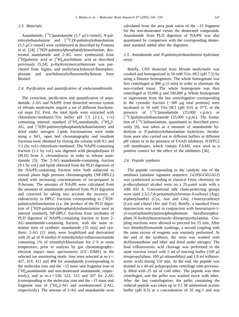
2.3. Materials calculated from the area peak ratios of the –15 fragmentfor the non-deuterated versus the deuterated compounds.
14Anandamide, [ C]anandamide (5.7 mCi/mmol), N-pal- Anandamide from PLD digestion of NArPE was also14mitoylethanolamine and [ C]N-palmitoylethanolamine quantitated by comparison with the corresponding deuter-
(5.5 mCi/mmol) were synthesized as described by Fontana ated standard added after the digestion.3et al. [24]. [ H]N-palmitoylphosphatidyletanolamine, deu-
terated anandamide and 2-AG were synthesized from 2.5. Anandamide and N-palmitoylethanolamine hydrolase3 2[ H]palmitic acid or [ H] arachidonic acid as described assay8
previously [5,24]. p-Hydroxymercuribenzoate was pur-chased from Sigma, and methylarachidonoyl-fluorophos- Briefly, CNS dissected from Hirudo medicinalis wasphonate and arachidonoyltrifluoromethylketone from crushed and homogenized in 50 mM Tris–HCl (pH 7.5) byBiomol. using a Dounce homogenizer. The whole homogenate was
first centrifuged at 800 g (5 min) in order to eliminate the2.4. Purification and quantification of endocannabinoids non-crushed tissue. The whole homogenate was then
centrifuged at 10,000 g and 100,000 g. Whole homogenateThe extraction, purification and quantification of anan- or supernatant from the last centrifugation corresponding
damide, 2-AG and NArPE from dissected nervous system to the cytosolic fraction (|300 mg total proteins) wereof Hirudo medicinalis require a set of different biochemi- incubated in 50 mM Tris–HCl (pH 9.0) at 378C in the
14cal steps [5]. First, the total lipids were extracted with presence of [ C]anandamide (25,000 c.p.m.) or14chloroform/methanol /Tris buffer pH 7.5 (2:1:1, v /v) [ C]palmitoylethanolamide (25,000 c.p.m.). The forma-
2 2 14containing internal standard ([ H] anandamide, [ H] 2- tion of [ C]ethanolamine, quantitated as described previ-8 83AG, and [ H]N-palmitoylphosphatidylethanolamine) and ously [9], was taken as a measure of anandamide hy-
dried under nitrogen. Lipids fractionations were made drolysis or N-palmitoylethanolamine hydrolysis. Incuba-using a SiO open bed chromatography and resultant tions were also carried out in different buffers at different2
fractions were obtained by eluting the column with 9:1 and pH values or in the presence of FAAH inhibitors. N18TG21:1 (by vol.) chloroform/methanol. The NArPE-containing cell membranes, which contain FAAH, were used as afraction (1:1 by vol.) was digested with phospholipase D positive control for the effect of the inhibitors [36].(PLD) from S. chromofuscus in order to release anan-damide [5]. The 2-AG/anandamide-containing fraction 2.6. Peptide synthesis(9:1 by vol.) and lipids obtained from the PLD digestion ofthe NArPE-containing fraction were both subjected to The peptide corresponding to the catalytic site of thenormal phase high pressure chromatography (NP-HPLC) amidases (amidase signature sequence: GGSSGGEGALI)eluted with increasing concentrations of iso-propanol in was synthesized according to classical Fmoc chemistry onN-hexane. The amounts of NArPE were calculated from p-alkoxybenzyl alcohol resin on a 25-mmol scale with athe amounts of anandamide produced from PLD digestion ABI 432 A. Conventional side chain-protecting groupsand corrected by taking into account the recovery of were used 2,3,5,7,8-pentamethylchroman 6-sulfonyl (Arg),
3radioactivity in HPLC fractions corresponding to [ H]N- triphenylmethyl (Cys, Asn and Glu), t-butoxycarbonylpalmitoylethanolamine (i.e. the product of the PLD diges- (Lys) and t-butyl (Ser and Tye). Briefly, a standard Fmoc
3tion of [ H]N-palmitoylphosphatidylethanolamine used as deprotection was used in conjunction with benzotriazol-1-internal standard). NP-HPLC fractions from incubates of yl-oxytris(dimethylamino)phosphonium hexafluorophos-PLD digestion of NArPE-containing fraction or from 2- phate /N-hydroybenzotriazole /diisopropyethylamine. Cou-AG/anandamide-containing fraction, with the same re- pling reactions were allowed to proceed for 15 min. Aftertention time of synthetic anandamide (35 min) and syn- two dimethylformamide washings, a second coupling withthetic 2-AG (21 min), were lyophilized and derivatized the same excess of reagents was routinely performed. Atwith 20 ml of N-methyl-N-trimethylsilyl-trifluoroacetamide the end of the synthesis, the resin was washed withcontaining 1% of trimethylchlorosilane for 2 h at room dichloromethane and ether and dried under nitrogen. Thetemperature, prior to analysis by gas chromatography– final trifluoroacetic acid cleavage was performed in theelectron impact mass spectrometry (GC–EIMS) in the same reaction vessel with 5 ml of reacting buffer (100 mlselected ion monitoring mode. Ions were selected at m /z5 trisopropylsilane, 100 ml ethanedithiol and 1.8 ml trifluoro-427, 419, 412 and 404 for anandamide (corresponding to acetic acid) during 150 min. At the end, the peptide wasthe molecular ions and the –15 mass unit fragment ions of drained in a 40-ml polypropylene centrifuge tube previous-
2[ H] anandamide and non-deuterated anandamide, respec- ly filled with 25 ml of cold ether. The peptide was then8
tively), and at m /z5530, 522, 515 and 507 for 2-AG centrifuged, and the pellet was washed twice with ether.(corresponding to the molecular ions and the –15 mass unit After the last centrifugation, the pellet containing the
2fragment ions of [ H] 2-AG and nondeuterated 2-AG, reduced peptide was taken up in 0.1 M ammonium acetate8
respectively). The amount of 2-AG and anandamide were buffer (pH 8.5) at a concentration of 35 mg/ l and was

148 I. Matias et al. / Molecular Brain Research 87 (2001) 145 –159
allowed to refold via air oxidation for 17 h at room centrifuged at 8000 g for 10 min at 48C. The supernatanttemperature with constant stirring. The refolded peptide was cleaned on a 0.22 mm filter and successively filteredwas purified by semi-preparative reversed-phase chroma- on a cut-off (Pall Filtron) of 100 kDa and 30 kDa. Thetography (Aquapore RP300 column, 25037.0 mm) with a yielded supernatant was used as enzyme source.linear gradient of 1%/min in acidified water (0.1%) at aflow rate of 1 ml /min. 2.9. Western immunoblotting
2.7. Antibodies Analytical SDS–polyacrylamide gel electrophoresis(SDS–PAGE 10%) was performed as described previously
Polyclonal amidase antiserum raised against a consensus [33] on lysates from H. medicinalis dissected nervoussignature sequence of vertebrate and invertebrate FAAH, system, whole Hydra vulgaris, rat liver and on recombi-corresponding to the catalytic site of the amidases (Amid- nant FAAH enzyme source. Western blot analysis was thenase Signature Family) [30], was obtained in our laboratory carried out with the amidase antiserum. Briefly, rat liver orby immunizing one rabbit with the synthetic peptide H. medicinalis dissected nervous system and whole HydraGGSSGGEGALI coupled with glutaraldehyde to thyro- vulgaris were homogenized using a Dounce homogenizerglobulin according to procedure described elsewhere [45]. in lysis buffer (1 mM EDTA, 50 mM Tris–HCl pH 7.4,Control of specificities was performed by preadsorbing the 150 mM NaCl, 1 mM Na-ortovanadate, 1 mM Na-fluoro-antibody by the homologous antigen at a concentration of nate, 1% NP-40, 0.1% SDS, 1% Triton, 0.25% Na-desox-350 mg/ml of pure serum. Rabbit anti-human CB1 poly- ycholate, 1 mM PMSF, 1 mg/ml serine proteases in-clonal antibody was purchased from Chemicon (Temecula, hibitors), incubated at 48C for 30 min and finally cen-USA). The antibody was raised against the N-terminal trifuged at 10,000 g for 20 min. The amount of proteins in
23 38region of human CB1 ( NKSLSSFKENEENIQC ). This each resulting supernatant and in recombinant FAAHsequence is upstream of the leech-CB1 like partially enzyme source was titrated by Biorad assay. Supernatantscloned by Stefano et al. [50]. The specificity of this were mixed 4:1 (v /v) with sample buffer (300 mM Tris–
¨antibody was described in Dove Petit et al. [18]. HCl pH 6.8, 50% glycerol, 500 mM dithiotreıtol, 0.05%Bromophenol Blue, 10% SDS) and boiled for 5 min prior
2.8. Production of recombinant rat FAAH to loading on a 0.75-mm-thick gel. Samples were subjectedto electrophoresis (150 V) for 2 h under reducing con-
To confirm that the polyclonal antiserum against the ditions, and separated proteins on the gel were transferredcatalytic site of vertebrate and invertebrate FAAH recog- onto Immobilon Protein Transfer at 65 mA overnight atnizes effectively the fatty acid amide hydrolase, an eukary- 48C. The membrane was preincubated with 5% non-fat dryotic expression vector containing rat FAAH (pcDNA3- milk in Tris-buffered saline (TBS: 10 mM Tris–HCl pH 8,FAAH, kindly provided by Dr Dale G. Deutsch, Depart- 150 mM NaCl) for 30 min to block non-specific binding.ment of Biochemistry and Cell Biology, State University The membrane was incubated for 1 h in amidase antiserumof New York at Stony Brook), was expressed transiently in at a dilution of 1:3000 and a control was made in the sameCOS-7 cells. COS-7 cells were grown in Dulbecco’s conditions using the amidase antiserum preabsorbed withmodified Eagle’s medium plus 10% fetal bovine serum, the homologous antigen (45 mg/ml of pure antiserum).and L-glutamine (complete DMEM) at 378C and 5% CO Then, the membrane was washed 3310 min in TBS2
atmosphere to 70% confluency, and then washed with containing 0.05% Tween-20 (TBST) and incubated with5serum free medium. Cells (2.10 cells /100-mn dish) were goat anti-rabbit IgG conjugated with horseradish peroxi-
incubated in a DNA/DEAE–dextran–chloroquine mixture dase (dilution 1:3000) for 1 h. The membrane was again(1.6 mg/ml pcDNA3-FAAH, 10 mg/ml DEAE–dextran, washed 3310 min in TBST and rinsed in TBS. Signals2.5 mM chloroquine in 5 ml of serum-free medium) for 4 were detected with an ECL kit (Biorad). Control ofh. The cells were then washed with 5 ml of a phosphate- specificities was performed by preabsorbing the antibodybuffered saline solution (PBS: 137 mM NaCl, 2.7 mM by the homologous antigen at a concentration of 140KCl, 4.3 mM Na HPO .7H O, 1.4 mM KH PO ) con- mg/ml of pure serum2 4 2 2 4
taining 10% of dimethyl sulfoxide (DMSO) to increaseDNA uptake. After 2 min of incubation, complete medium 2.10. Immunocytochemical procedurewas added and cells were incubated for 72 h with onechange of medium at 24 h. The COS-7 cells were washed Anterior parts of mature leeches (H. medicinalis,two times with PBS and with PBS plus 0.5 mM EDTA, Theromyzon tessulatum, and Erpobdella octoculata) wereharvested with the aid of trypsin–EDTA and suspended in fixed overnight at 48C in Bouin Hollande fixative sup-0.1 ml of 250 mM Tris–HCl buffer (pH 7.4) after plemented with 10% of mercuric chloride, embedded incentrifugation at 2000 g for 10 min. The cells were Paraplast and serially sectioned at 7 mm. After removal ofsubmitted to heat shock (2708C/ 1378C for 5 min each Paraplast with toluene, sections were subjected to anthree times) and sonicated for 3 s. The lysate was indirect immunoperoxidase method as described elsewhere

I. Matias et al. / Molecular Brain Research 87 (2001) 145 –159 149
[34,45]. In short, sections of anterior parts were incubated 147.4642.7 pmol /g of wet weight of tissue, for anan-overnight at room temperature with either anti-amidase damide and 2-AG, respectively (means6S.D., n52).(1:200) or anti-CB1 (16 mg/ml) sera. They were then Anandamide precursor NArPE was also quantified as thetreated for 1 h at room temperature with goat anti-rabbit anandamide released from PLD digestion of the 1:1IgG conjugated to horseradish peroxidase (1:50). Immuno- fraction from SiO chromatography and estimated at2
staining was revealed with a solution of 4-chloro-1-naphtol 16.463.2 pmol /g of wet weight of tissue (means6S.D.,(0.4 mg/ml in 0.1 M Tris buffer, pH 7.6) containing n52). We also demonstrate that leech anandamide and0.01% H O . Controls of specificity were performed by 2-AG are accompanied by anandamide-related molecules2 2
incubation of the primary antiserum saturated with the like N-palmitoylethanolamine, at an amount of 32.361.5homologous antigen (1 mg synthetic peptide /ml of pure pmol /g of wet weight of tissue (means6S.D., n52), andantiserum) prior to application. Sections were examined its precursor, N-palmitoylphosphatidylethanolaminewith a Zeiss Axioskop microscope. (348.9699.3 pmol /g of wet weight of tissue means6S.D.,
n52), and N-linolenoylethanolamine (5.8 pmol /g of wetweight of tissue, n51).
2.11. Cyclic AMP assay
Nervous system of H. medicinalis (10 ganglia) was 3.2. Evidence for anandamide amidase enzyme(s) inplaced in 2 ml Tris /malate buffer (2 mM, pH 7.4)
leech CNScontaining EGTA (0.8 mM) and homogenized using aground glass homogenizer. Following homogenization, the
Whole homogenate and subcellular fractions obtainedsample was maintained at 48C. The material was de-
from H. medicinalis CNS were analyzed for the presenceproteinized in acidic ethanol (1 ml, 1 M HCl/100 ml 14of [ C]anandamide-hydrolyzing activity (Table 1). Theethanol), and centrifuged. The supernatant was washed 14enzymatic formation of [ C]ethanolamine by wholethree times with four volumes of water-saturated ether, and
homogenate was essentially due to the cytosolic fraction.the residue was then evaluated for cAMP using the
As a matter of fact the enzymatic formation of3Amersham/cAmp-[ H] kit RIA TrK432 instructions (Ar- 14[ C]ethanolamide in the membrane and ‘microsomal’lington Heights, IL, USA). Triplicate incubations for each
(100,000 g pellet) fraction was very weak (Table 1). Thetest condition were run for each experiment. The results 14anandamide congener [ C]palmitoylethanolamide wasare corrected for recoveries and expressed as mean
also hydrolyzed by the whole homogenate (Table 1). Likevalues6S.D. (means6S.D., n53). Protein concentration 14 14for the [ C]anandamide amidase activity, the [ C]N-determinations were made according to the Biorad method.
palmitoylethanolamine amidase activity was most abun-dant in the cytosolic fraction .
The anandamide amidase activity from whole homoge-3. Results nate was further characterized and found to exhibit a pH
dependency profile different from that of mammalian3.1. Endocannabinoid characterization and quantification FAAH, with a maximal activity at pH 7 (Fig. 2). The
typical FAAH inhibitors, p-hydroxymercuribenzoate,After a lipid extraction in chloroform/methanol, a methylarachidonoylfluorophosphonate and arach-
separation was conducted using SiO open bed chromatog- idonoyltrifluoromethylketone, at concentrations causing2
raphy. The separated lipids (9:1 fraction) were then almost 100% inhibition of N18TG2 cell FAAH (control),14submitted to normal phase high-pressure chromatography. were not effective on the leech [ C]anandamide-hydrolyz-
Elution was performed by a linear gradient of iso-propanol ing activity (Fig. 3). Furthermore, most of this activity wasin N-hexane. In these conditions, synthetic 2-AG and not selective for anandamide or 2-AG because theseanandamide elute from the column at a retention time of, compounds exhibited only a small inhibition of
14respectively, 21 min and 35 min. Leech material collected [ C]anandamide hydrolysis.at these retention times were then derivatized and subject- The predicted size of rat FAAH protein, based on itsed to GC–MS analysis (Fig. 1). As seen in Fig. 1, GC–MS amino acid sequence following extrapolation from itschromatograms obtained in ion selected monitoring mode, corresponding cDNA, is 63.4 kDa. Western immunoblotingshow peaks at m /z 427, 419, 412 and 404 for the material is performed with the anti-amidase serum that is able tocollected at 35 min (Fig. 1A) and peaks at m /z 530, 522, recognize proteins containing the amidase consensus se-515 and 507 for the material collected at 21 min (Fig. 1B). quence, including FAAH or other amidases. WesternThis corresponds to the molecular ions and the –15 mass immunobloting of recombinant rat FAAH reveals an
2ions of [ H] anandamide and non-deuterated anandamide, immunoreactive band at |64 kDa (Fig. 4, lane c). The8
respectively, and similarly to the molecular ions and the same analysis performed on rat liver homogenate confirms2–15 mass ions of [ H] 2-AG and non-deuterated 2-AG, the specificity of the anti-amidase serum for the native8
respectively. The amounts were 21.560.7 pmol /g and enzyme (data not shown). In leech CNS, the immunoblot

150 I. Matias et al. / Molecular Brain Research 87 (2001) 145 –159
Fig. 1. GC–MS chromatograms of bio-active lipids in the leech Hirudo medicinalis CNS. (A) Selected ion monitoring GC–MS chromatogram ofanandamide (middle panel, selected ion m /z5404) in the presence of deuterated internal standard (upper panel, selected ion m /z5412), and selected ionmonitoring fragmentogram of the peak at 17.40–17.50 min (lower panel). (B) Selected ion monitoring GC–MS chromatogram of 2-AG (upper panel,selected ion m /z5522; middle panel, selected ion m /z5507) in the presence of deuterated internal standard (data not shown) and total ion monitoringfragmentogram of the peak at 17.78 min (lower panel).
analysis revealed three specific immunoreactive bands at anti-amidase serum raised against the consensus sequenceca. |42, |46 and |66 kDa (Fig. 4, lane a). The most of amidases (Signature Amidase Family) and anti-CB1abundant band was the one at |46 kDa, which was present serum in different species of leeches. The anti-amidasein both total homogenate and cytosolic fractions, and was serum is able to localize in principle proteins containingabsent in Hydra homogenate (data not shown). The band at the amidase consensus sequence, including FAAH or other|42 kDa was less intense and was present also in Hydra. amidases.The |66 kDa band was very faint in leech homogenate but As seen in Figs. 5, 6 and 7, immunoreactivity to anti-quite intense in Hydra and could correspond to a FAAH- amidase was observed in three different species of leeches.like enzyme. Control of specificity was carried out by In H. medicinalis numerous perikaria exhibit an intensepre-adsorbing the anti-amidase serum with its antigen (Fig. immunolabeling throughout the supra-esophageal ganglia4, lanes b and d). (Figs. 5A, B). The immunostained cells correspond to
neurons and the labeling is found on nerve cell bodies. In3.3. Localization of amidase in leech CNS E. octoculata, amidase-like immunoreactivity was mainly
detected in the brain (Fig. 6A) but immunoreactive cellsImmunocytochemical studies were performed with both were also seen in the segmental ganglia (Fig. 6C). In

I. Matias et al. / Molecular Brain Research 87 (2001) 145 –159 151
Fig. 1. (continued)
Theromyzon tessulatum, a much larger distribution of 3.4. Evidence for a functional CB1-like receptoramidase-like signal was observed. Indeed, intense im-munostaining was seen in different compartments of the Ganglia of H. medicinalis were exposed to either
26supra-esophageal ganglia (compartments 4 and 5, Fig. 7A) anandamide (10 M), the NO donor S-nitroso-N-acetyl-and in ganglia of the nerve cord (Fig. 7D). Coelomocytes penicillamine (SNAP) or the NO synthase inhibitor N-(Fig. 7E) and parts of genital tracts (Fig. 7G) exhibited omega-nitro-L-arginine methyl ester (L-NAME) (Table 2).also a low but specific immunostaining. Pre-absorption of Anandamide alone was not able to induce the cyclic AMPamidase antiserum with the homologous antigen abolished release even though forskolin was able to initiate thisthe positive staining, reflecting the specificity of the release. By contrast, anandamide is able to inhibit theimmunolabelling in the three species (Figs. 5C; 6B, D; 7B, forskolin-induced formation of cyclic AMP and this effectC, F, H). Labeling of glial cells was never observed. was abolished by the NO synthase inhibitor L-NAME. The
Table 1Distribution of the amidase activity in leech CNS
Whole homogenate Cytosol fraction Membrane fraction14[ C]AEA 14.3461.45 12.48 1.5560.55
(pmol /min /mg of protein)14[ C]PEA 4.9660.56 3.160.33 0.38560.003
(pmol /min /mg of protein)

152 I. Matias et al. / Molecular Brain Research 87 (2001) 145 –159
14Fig. 2. pH dependency of the [ C]anandamide-hydrolyzing activity from Hirudo medicinalis CNS whole homogenate expressed as a percentage of theactivity at pH 7.0 (maximal activity) (means S.D., n52).
14Fig. 3. Effect of inhibitors of mammalian FAAH on the [ C]anandamide-hydrolyzing activity from Hirudo medicinalis CNS whole homogenate. Theeffect is expressed as a percentage of the activity in the absence of inhibitors (and with vehicle) and compared with the effect on N18TG2 cell membraneactivity. AEA, anandamide; 2-AG, 2-arachidonoylglycerol; p-HMB, p-hydroxymercuribenzoate; MAFP, methylarachidonoyl-fluorophosphonate; AACF3,arachidonoyltrifluoromethylketone (mean6S.D., n52).

I. Matias et al. / Molecular Brain Research 87 (2001) 145 –159 153
NO donor, SNAP, is also capable to inhibit the forskolin-induced formation of cyclic AMP, suggesting that theaction of anandamide occurs through NO release.
As regards the localization of the CB1-like cannabinoidreceptor in H. medicinalis, specific immunostaining wasdetected in neurons of the supra-esophageal ganglion (Fig.8A). T. tessulatum also reveals a few immunoreactiveneurons in median compartments of the sub-esophagealganglia (Fig. 9A, C) and in the supra-esophageal ganglion(data not show). Pre-adsorption of the primary antibodywith the synthetic peptide completely abolished the signal(Figs. 8B, D; 9B, D).
4. Discussion
By using a combination of techniques including GC–MS, we have demonstrated, for the first time, the presenceof anandamide, 2-AG, NArPE, N-palmitoylethanolamine
Fig. 4. Western blot analysis of the putative leech amidase. Nervousand N-linolenoylethanolamine in the CNS of the leech H.system of Hiudo medicinalis (50 mg of proteins, lane a) and recombinantmedicinalis. These results are in line with previous ob-FAAH (50 mg of proteins, lane c) were submitted to SDS–poly-servations reported for Hydra vulgaris [8], Mytilus edulisacrylamide gel electrophoresis and Western immunobloting using rabbit
antiserum against a peptide corresponding to amino acids 215–226 of the [47] and sea urchins [4].rat FAAH enzyme, pre-treated with the homologous antigen (lanes b and The leech anandamide amount was comparable to thed) or not (lanes a and c). The three arrows on the left indicate the position
one detected in the rat, where the compound was proposedof ca. |42, |46 and |66 kDa leech FAAH. The arrow on the rightto act as a neuromodulator [12]. Leech 2-AG amount wasindicates the migration of the recombinant FAAH (64 kDa).lower than that found in vertebrates and other invertebrates
Fig. 5. Immunohistochemical detection (indirect peroxidase) of an amidase-like protein in frontal sections of H. medicinalis. (A) Immunoreactive neuronsare detected in the supra-esophageal ganglia. (B, C) Adjacent sections of different compartments of the supra-esophageal ganglion treated either withanti-amidase serum (B) or with anti-amidase serum preadsorbed with the homologous antigen (C). Pre-adsorption of the serum with the correspondingpeptide abolished the immunostaining.

154 I. Matias et al. / Molecular Brain Research 87 (2001) 145 –159
Fig. 6. Immunohistochemical detection (indirect peroxidase) of an amidase-like protein in frontal sections of E. octoculata. (A) Immunoreactive neuronsare detected in the supra-esophageal ganglia. (A, B) Adjacent sections of different compartments of the supra-esophageal ganglion treated either withanti-amidase serum (A) or with anti-amidase serum pre-adsorbed with the homologous antigen (B). The amidase-staining capacity is abolished afteradsorption with the synthetic peptide. (C) Immunoreactive neurons are also detected in ganglia of the nerve cord. Pre-absorption of the antiserum with thecorresponding peptide abolished the immunostaining (D).

I. Matias et al. / Molecular Brain Research 87 (2001) 145 –159 155
Fig. 7. Immunohistochemical detection (indirect peroxidase) of an amidase-like protein in frontal sections of T. tessulatum. (A) Immunoreactive neuronsare detected in compartments 4 and 5 of the supra-esophageal ganglia. (D) Numerous neurons are detected in segmental ganglia. A moderateimmunoreactivity is detected in coelomocytes (E) and in genital tract (G). Pre-absorption of the anti-amidase serum with the corresponding peptideabolished the immunostaining (B, C, F, H).

156 I. Matias et al. / Molecular Brain Research 87 (2001) 145 –159
Table 2aInhibition of forskolin-induced cAMP formation by anandamide through NO production
Substances cAMP levelspmol /mg protein /min
26Anandamide (10 M) 1.660.826Forskolin (10 M) 52.2615.4
26 26SNAP (10 M)1Forskolin (10 M) 2.660.9526 26Anandamide (10 M)1Forskolin (10 M) 5.361.326 26 26Anandamide (10 M)1L-NAME (10 M)1Forskolin (10 M) 33.8618.4
a (mean6S.D., n53).
[12], but still seemingly more abundant than anandamide, the phospholipase D pathway suggested for mammalsanalogous to others animals. In view of the finding of the [12,16]. As for N-palmitoylethanolamine, this anandamideputative anandamide biosynthetic precursor (NArPE), we congener is more abundant than anandamide in leech as incan suggest that the formation of anandamide occurs via mammals.
Fig. 8. Immunohistochemical detection (indirect peroxidase) of a CB1-like protein in frontal sections of H. medicinalis. (A, B) Numerous immunoreactiveneurons are observed in the supra-esophageal ganglia. The amidase-staining capacity is abolished after adsorption with the synthetic peptide (B). (C, D)Adjacent sections of different compartments of the supra-esophageal ganglia treated either with anti-CB1 antibody (C) or with anti-CB1 antibodypre-absorbed with the homologous antigen (D). Pre-absorption of the antibody with the corresponding peptide abolished the immunostaining.

I. Matias et al. / Molecular Brain Research 87 (2001) 145 –159 157
Fig. 9. Immunohistochemical detection (indirect peroxidase) of CB1-like protein in frontal sections of T. tessulatum. (A, C) Numerous immunoreactiveneurons are observed in median compartments 14, 15, 16 (A) and 21 (C) of the sub-esophageal ganglia. Pre-adsorption of CB1 antiserum with thehomologous antigen abolished the positive staining (B, D).
The discovery of these endocannabinoids in leech CNS, expected for mammalian FAAH enzymes. However, thetogether with the previously described finding of CB1-like major band at ca. 46 kDa was absent in Hydra and wasreceptors in the same tissues [50], suggests that a complete very abundant in leech cytosolic fractions. These data,endocannabinoid system exists in leeches. Therefore, the taken together with the enzyme activity data obtained herenext step in our study was to assess the presence of an for the leech and previously for Hydra [8], suggest that,anandamide amidase in leech CNS, as indirect evidence for while in Hydra one of the amidase-immunoreactive bandsthe existence of such enzyme had been provided in could be due to a FAAH-like enzyme, in leech CNS most
14previous studies [49]. We found that [ C]anandamide was of the anandamide amidase activity is due to (an) en-enzymatically hydrolyzed by both total leech CNS zyme(s) distinct from mammalian FAAH.homogenates and cytosolic fractions, and that very little In the present study we utilized the anti-amidase an-activity was present in membrane fractions. This shows tiserum for immunohistochemical localization of the en-that this enzymatic activity has a distribution different zyme in leech CNS, as compared to CB1 localization. Wefrom that reported for either mammalian or invertebrate previously cloned a fragment of leech cannabinoid receptorFAAH [1,8,32]. In support of this hypothesis, we found cDNA [50]. The fragment presents, within its amino acidthat leech anandamide amidase activity was (i) optimal at sequence, two highly conserved motifs — between aminopH57, instead of 9–10 for FAAH, (ii) not sensitive to acids 1–97 and 128–153 — which show 80% and 58%typical FAAH inhibitors and (iii) inhibited only to a little homology to human CB receptors, respectively, while a1
14extent by either AEA or 2-AG. We also found that [ C]N- third region is 98% identical to part of the bovinepalmitoylethanolamine is hydrolyzed, to a lesser extent, by adrenocorticotropic hormone receptor. According to El-both homogenates and cytosolic fractions. These observa- phick [23] this protein may therefore represent the putativetions suggest that the anandamide amidase activity found ancestor of mammalian cannabinoid and melanocortinhere is, in fact, mostly not selective for anandamide, and receptors. This hypothesis is supported by the high homol-that additional inactivating mechanisms more specific for ogy between the sequence of mammalian CB1 andendocannabinoids may exist in leech CNS. melanocortin receptors, which contain domains, located in
Based on these data we decided to raise a non-selective the central part of the two proteins, with more than 80%antiserum against the amidase signature sequence [30]. sequence identity. Here we found that CB1 immuno-This would have allowed us to identify in leech CNS also reactivity in both H. medicinalis and T. tessulatum ispossible amidase enzymes different from FAAH, including located in neurons of the CNS, more precisely in thethe one responsible for the anandamide amidase activity supra-esophageal ganglia. More importantly, we observeddetected in leech homogenates. By using this antiserum we that leech CB1 immunoreactivity matches to a certainfound three immunoreactivity bands in Western immuno- extent with amidase immunostaining. This is in agreementblot analyses. The two least abundant of these proteins with data obtained in the rat and showing that FAAH andwere also present in Hydra, and the very minor of them CB1 distributions in the brain overlap to a great extentexhibited an apparent molecular weight similar to that [53,54,57]. These data suggest that, even though the leech

158 I. Matias et al. / Molecular Brain Research 87 (2001) 145 –159
[7] B.F. Cravatt, D.K. Giang, S. Mayfield, D.L. Boger, R.A. Lerner,amidase may not be selective for the endocannabinoidsN.B. Gilula, Molecular characterization of an enzyme that degradesanandamide and 2-AG, this enzyme may still be deputed toneuromodulatory fatty-acid amides, Nature 384 (1996) 83–87.
the degradation of these metabolites since it is localized [8] L. De Petrocellis, D. Melck, T. Bisogno, A. Milone, V. Di Marzo,near their putative molecular target, i.e. the CB1 receptor. Finding of the endocannabinoid signalling system in Hydra, a veryMoreover, in the leech, immunostaining with anti-CB2 was primitive organism: possible role in the feeding response, Neuro-
science 92 (1999) 377–387.found in neurons and glial cells in the central neuropil of[9] L. De Petrocellis, D. Melck, N. Ueda, S. Maurelli, Y. Kurahashi, S.the ventral chain ganglia (Salzet et al., unpublished
Yamamoto, G. Marino, V. Di Marzo, Novel inhibitors of brain,observations). This suggests the presence of different typesneuronal and basophilic anandamide amidohydrolase, Biochem.
of cannabinoid receptors in these simple animals, as Biophys. Res. Commun. 231 (1997) 82–88.previously described in mammals [6,20,35,39]. Finally, [10] D. Deutsch, A. Chin, Enzymatic synthesis and degradation of
anandamide, a cannabinoid receptor agonist, Biochem. Pharmacol.based on the information that in vertebrates as in mammals46 (1993) 791–796.cannabinoid receptor stimulation by anandamide leads to
[11] W.A. Devane, L. Hanus, A. Breuer, R.G. Pertwee, L.A. Sevenson,the release of NO [51] and inhibition of cAMP formationG. Griffin, D. Gibson, A. Mandelbaum, A. Etinger, R. Mechoulam,
[29], we wanted to assess whether the same phenomenon Isolation and structure of a brain constituent that bind to theoccurred in our model. Our data allow us to conclude that cannabinoid receptor, Science 258 (1992) 1946–1949.
[12] V. Di Marzo, Biosynthesis and inactivation of endocannabinoids:two of the secondary messengers mediating cannabinoidrelevance to their proposed role as neuromodulators, Life Sci. 65receptor signaling, e.g., NO and cAMP, are the same in(1999) 645–655.leech as in mammals.
[13] V. Di Marzo, T. Bisogno, L. De Petrocellis, D. Melck, P. Orlando,Taken together, our results demonstrate the existence of J.A. Wagner, G. Kunos, Biosynthesis and inactivation of the
a complete endocannabinoid system in leech brain, and endocannabinoid 2-arachidonoylglycerol in circulating and tumoralmacrophages, Eur. J. Biochem. 264 (1999) 258–267.support the concept that this signaling system has been
[14] V. Di Marzo, D.G. Deutsch, Biochemistry of the endogenous ligandsconserved throughout animal evolution.of cannabinoid receptors, Neurobiol. Dis. 5 (1998) 386–404.
[15] V. Di Marzo, T. Bisogno, T. Sugiura, D. Melck, L. De Petrocellis,The novel endogenous cannabinoid 2-arachidonoylglycerol is inacti-vated by neuronal- and basophil-like cells: connections with anan-Acknowledgementsdamide, Biochem. J. 331 (1998) 15–19.
[16] V. Di Marzo, D. Melck, T. Bisogno, L. De Petrocellis, Endo-The authors are grateful to A. Desmons for her technicalcannabinoids: endogenous cannabinoid receptor ligands with neuro-
assistance. This work was partially supported by the modulatory action, Trends Neurosci. 21 (1998) 521–528.FEDER, the Conseil Regional Nord-Pas de Calais, the [17] V. Di Marzo, A. Fontana, H. Cadas, S. Schinelli, G. Cimino, J.C.
Schwartz, D. Piomelli, Formation and inactivation of endogenousCentre National de la Recherche Scientifique and thecannabinoid anandamide in central neurons, Nature 372 (1994)NIH-Fogarty INT 00045 (to MS) grant and INTAS (grant686–691.97-1297 to VDM). Professor Stefano G.B. is acknowl-
[18] D.A. Dove Pettit, M.P. Harrison, J.M. Olson, R.F. Spencer, G.A.edged for his support and help in this project. Cabral, Immunohistochemical localization of the neural cannabinoid
receptor in rat brain, J. Neurosci. Res. 51 (1998) 391–402.[19] Y. Gaoni, R. Mechoulam, Isolation, structure, and partial synthesis
of an active constituent of hashish, J. Am. Chem. Soc. 86 (1964)References 1646–1647.
[20] C.M. Gerard, C. Mollereau, G. Vassart, M. Parmentier, Molecular[1] G. Arreaza, W.A. Devane, R.L. Omeir, G. Sajnani, J. Kunz, B.F. cloning of human cannabinoid receptor which is also expressed in
Cravatt, D.G. Deutsch, The cloned rat hydrolytic enzyme respon- testis, Biochem. J. 279 (1991) 129–134.sible for the breakdown of anandamide also catalyzes its formation [21] D.K. Giang, B.F. Cravatt, Molecular characterization of human andvia the condensation of arachidonic acid and ethanolamine, Neuro- mouse fatty acid amide hydrolases, Proc. Natl. Acad. Sci. USA 94sci. Lett. 234 (1997) 59–62. (1997) 2238–2242.
[2] C.H. Ashton, Biomedical benefits of cannabinoids?, Addict. Biol. 4 [22] S.K. Goparaju, N. Ueda, H. Yamaguchi, S. Yamamoto, Anandamide(1999) 111–126. amidohydrolase reacting with 2-aracidonoylglycerol, another can-
[3] T.V. Bilfinger, M. Salzet, C. Fimiani, D.G. Deutsch, G. Tramu, G.B. nabinoid receptor ligand, FEBS Lett. 422 (1998) 69–73.Stefano, Pharmacological evidence for anandamide amidase in [23] M.R. Elphick, An invertebrate G-protein coupled receptor is ahuman cardiac and vascular tissues, Int. J. Cardiol. 64 (1998) chimeric cannabinoid /melanocortin receptor, Brain Res. 780 (1997)S15–S22. 170–173.
[4] T. Bisogno, M. Ventriglia, A. Milone, M. Mosca, G. Cimino, V. Di [24] A. Fontana, V. Di Marzo, H. Cadas, D. Piomelli, Analysis ofMarzo, Occurrence and metabolism of anandamide and related anandamide, an endogenous cannabinoid substance, and of otheracyl-ethanolamides in ovaries of the sea urchin Paracentrotus natural N-acylethanolamines, Prostaglandins Leukot. Essent. Fattylividus, Biochim. Biophys. Acta 1345 (1997) 338–348. Acids 53 (1995) 301–308.
[5] T. Bisogno, S. Maurelli, D. Melck, L. De Pretrocellis, V. Di Marzo, [25] H.S. Hansen, L. Lauritzen, A.M. Strand, A.M. Vinggaard, A.Biosynthesis, uptake and degradation of anandamide and pal- Frandsen, A. Schousboe, Characterization of glutamate-inducedmitoylethanolamide in leukocytes, J. Biol. Chem. 272 (1997) 3315– formation of N-acylphosphatidylethanolamine and N-3323. acylethanolamine in cultured neocortical neurons, J. Neurochem. 69
[6] A. Chakrabarti, E.S. Onaivi, G. Chaudhuri, Cloning and sequencing (1997) 753–761.of a cDNA encoding the mouse brain-type cannabinoid receptor [26] C.J. Hillard, W.S. Edgemond, A. Jarrahian, W.B. Campbell, Ac-protein, DNA Seq. 5 (1995) 385–388. cumulation of N-arachidonoylethanolamine (anandamide) into cere-

I. Matias et al. / Molecular Brain Research 87 (2001) 145 –159 159
bellar granule cells occurs via facilitated diffusion, J. Neurochem. 69 [43] R.G. Pertwee, Evidence for the presence of CB1 cannabinoid(1997) 631–638. receptors on peripheral neurons and for the existence of neural
[27] A.G. Hohmann, M.K. Herkenham, Localization of central can- non-CB1 cannabinoid receptor, Life Sci. 65 (1999) 597–605.nabinoid CB1 receptor messenger RNA in neuronal subpopulations [44] D. Piomelli, M. Beltramo, S. Glasnapp, S.Y. Lin, A. Goutopoulos,of rat dorsal root ganglia: a double-label in situ hybridization study, X.-Q. Xie, A. Makriyannis, Structural determinants for recognitionNeuroscience 90 (1999) 923–931. and translocation by the anandamide transporter, Proc. Natl. Acad.
[28] A.C. Howlett, Pharmacology of cannabis, in: Tarter et al. (Eds.), Sci. USA 96 (1999) 5802–5807.Handbook of Substance Abuse. Neurobehavioral Pharmacology, [45] M. Salzet, C. Wattez, M.C. Slomianny, B. Leu, K.J. Siegert, ELISAPlenum, New York, 1998, pp. 113–129. for oxytocin. Highly sensitive tests and application to the titration of
[29] A.C. Howlett, J.M. Qualy, L.L. Khachatrian, Involvement of Gi in an oxytocin-like substance in the leech Erpobdella octoculata,the inhibition of adenylate cyclase by cannabimimetic drugs, Mol. Comp. Biochem. Physiol. 102C (1992) 483–487.Pharmacol. 29 (1986) 307–317. [46] H. Schuel, E. Goldstein, R. Mechoulam, A.M. Zimmerman, S.
[30] M. Kobayashi, Y. Fujiwara, M. Goda, H. Komeda, S. Shimizu, Zimmerman, Anandamide (arachidonoylethanolamide), a brain can-Identification of active sites in amidase: evolutionary relationship nabinoid receptor agonist, reduces sperm fertilizing capacity in seabetween amide bond- and peptide bond-cleaving enzymes, Proc. urchins by inhibiting the acrosome reaction, Proc. Natl. Acad. Sci.Natl. Acad. Sci. USA 94 (1997) 11986–11991. USA 91 (1994) 7678–7682.
[31] S. Kondo, H. Kondo, S. Nakane, T. Kodaka, A. Tokumara, K. [47] N. Sepe, L. De Petrocellis, F. Montanaro, G. Cimino, V. Di Marzo,Waku, T. Sugiura, 2-Arachidonoylglycerol, an endogenous can- Bioactive long chain N-acylethanolamines in five species of ediblenabinoid receptor agonist: identification as one of the major species bivalve mollusk. Possible implications for mollusk physiology andof monoacylglycerols in various rat tissues, and evidence for its sea food industry, Biochem. Biophys. Acta 1389 (1998) 101–111.
11generation through Ca -dependent and -independent mechanisms, [48] D. Shire, C. Carillon, M. Kaghad, B. Calandra, M. Rinaldi-Carmona,FEBS Lett. 429 (1998) 152–156. G. Le Fur, D. Caput, P. Ferrara, An amino-terminal variant of the
[32] Y. Kurahashi, N. Ueda, H. Suzuki, M. Suzuki, S. Yamamoto, central cannabinoid receptor resulting from alternative splicing, J.Reversible hydrolysis and synthesis of anandamide demonstrated by Biol. Chem. 270 (1995) 3726–3731.recombinant rat fatty-acid amide hydrolase, Biochem. Biophys. Res. [49] G.B. Stefano, C.M. Rialas, D.G. Deutsch, M. Salzet, AnandamideCommun. 237 (1997) 512–515. amidase inhibition enhances anandamide-stimulated nitric oxide
[33] U.K. Laemmli, Cleavage of structural proteins during assembly of release in invertebrate neural tissues, Brain Res. 793 (1998) 341–the head of bacteriophage T4, Nature 227 (1970) 680–685. 345.
´[34] J. Malecha, M. Verger-Bocquet, G. Tramu, Mise en evidence et [50] G.B. Stefano, B. Salzet, M. Salzet, Identification and characteriza-evolution, au cours du cycle biologique, de neurones producteurs tion of the leech CNS cannabinoid receptor: coupling to nitric oxide
´ `d’une substance apparentee a la motiline porcine dans le ganglion release, Brain Res. 753 (1997) 219–224.supra œsophagien de la sangsue Theromyzon tessulatum, Can. J. [51] G.B. Stefano, Y. Liu, M.S. Goligorsky, Cannabinoid receptors areZool. 67 (1989) 636–640. coupled to nitric oxide release in invertebrates immunocytes,
[35] L.A. Matsuda, S.J. Lolait, M.J. Brownstein, A.C. Young, T.I. microglia and human monocytes, J. Biol. Chem. 271 (1996) 19238–Bonner, Structure of cannabinoid receptor and functional expression 19242.of the cloned cDNA, Nature 346 (1990) 561–564. [52] T. Sugiura, S. Kondo, A. Sukagaza, S. Nakane, A. Shinoda, K. Itoh,
[36] S. Maurelli, T. Bisogno, L. De Petrocellis, A. Di Luccia, G. Marino, A. Yamashita, K. Waku, 2-arachidonoylglycerol: a possible endogen-V. Di Marzo, Two novel classes of neuroactive fatty acid amides are ous cannabinoid receptor ligand in brain, Biochem. Biophys. Res.substrates for mouse neuroblastoma anandamide amidohydrolase, Commun. 215 (1995) 89–97.FEBS Lett. 377 (1995) 82–86. [53] E.A. Thomas, B.F. Cravatt, P.E. Danielson, N.B. Gillula, J.G.
[37] R. Mechoulam, S. Ben-Shabat, L. Hanus, M. Ligunsky, N.E. Sutcliffe, Fatty acid amide hydrolase, the degradative enzyme forKaninski, A.R. Schatz, A. Gopher, S. Almog, B.R. Martin, D.R. anandamide and oleamide, has selective distribution in neuronsCompton, R.G. Pertwee, G. Griffin, M. Bayewitch, J. Barg, Z.Vogel, within the rat central nervous system, J. Neurosci. Res. 50 (1997)Identification of an endogenous 2-monoglyceride, present in canine 1047–1052.gut, that binds to cannabinnoid receptors, Biochem. Pharmacol. 50 [54] K. Tsou, M.I. Nogueron, S. Muthian, M.C. Sanudo-Pena, C.J.(1995) 83–90. Hillard, D.G. Deutsch, J.M. Walker, Fatty acid amide hydrolase is
[38] K.J. Muller, J.G. Nicholls, G.S. Stent, Neurobiology of the Leech, located preferentially in large neurons in the rat central nervousCold Spring Harbor Laboratory, Cold Sring Harbor, New York, system as revealed by immunohistochemistry, Neurosci. Lett. 2541981. (1998) 137–140.
[39] M. Munro, K.L. Thomas, M. Abu-Shaar, Molecular characterization [55] N. Ueda, S.K. Goparaju, K. Katayama, Y. Kurahashi, H. Suzuki, S.of a peripheral receptor for cannabinoids, Nature 365 (1993) 61–65. Yamamoto, A hydrolase enzyme inactivating endogenous ligands
[40] R.L. Omeir, G. Arreaza, D.G. Deutsch, Identification of two serine for cannabinoid receptors, J. Med. Invest. 45 (1998) 27–36.residues involved in catalysis by fatty acid amide hydrolase, [56] K.A. Willoughby, S.F. Moore, B.R. Martin, E. Ellis, The biodisposi-Biochem. Biophys. Res. Commun. 264 (1999) 316–320. tion and metabolism of anandamide in mice, J. Pharmacol. Exp.
[41] B.C. Paria, J. Zhao, S.K. Das, S.K. Dey, Fatty-acid amide hydrolase Ther. 282 (1997) 243–247.is expressed in the mouse uterus and embryo during the periim- [57] S. Yazulla, K.M. Studholme, H.H. McIntosh, D.G. Deutsch, Im-plantation period, Biol. Reprod. 60 (1999) 1151–1157. munocytochemical localization of cannabinoid CB1 receptor and
[42] M.P. Patricelli, M.A. Lovato, B.F. Cravatt, Chemical and mutagenic fatty acid amide hydrolase in rat retina, J. Comp. Neurol. 415 (1999)investigations of fatty acid amide hydrolase: evidence for a family 80–90.of serine hydrolases with distinct catalytic properties, Biochemistry38 (1999) 9804–9812.
Related Documents