Oncology Imaging Guidelines Version 2.0 Effective September 1, 2021 eviCore healthcare Clinical Decision Support Tool Diagnostic Strategies: This tool addresses common symptoms and symptom complexes. Imaging requests for individuals with atypical symptoms or clinical presentations that are not specifically addressed will require physician review. Consultation with the referring physician, specialist and/or individual’s Primary Care Physician (PCP) may provide additional insight. CPT ® (Current Procedural Terminology) is a registered trademark of the American Medical Association (AMA). CPT ® five digit codes, nomenclature and other data are copyright 2017 American Medical Association. All Rights Reserved. No fee schedules, basic units, relative values or related listings are included in the CPT ® book. AMA does not directly or indirectly practice medicine or dispense medical services. AMA assumes no liability for the data contained herein or not contained herein. © 2021 eviCore healthcare. All rights reserved. CLINICAL GUIDELINES

Welcome message from author
This document is posted to help you gain knowledge. Please leave a comment to let me know what you think about it! Share it to your friends and learn new things together.
Transcript
Oncology Imaging Guidelines
Version 2.0 Effective September 1, 2021
eviCore healthcare Clinical Decision Support Tool Diagnostic Strategies: This tool addresses common symptoms and symptom complexes. Imaging requests for individuals with atypical symptoms or clinical presentations that are not specifically addressed will require physician review. Consultation with the referring physician, specialist and/or individual’s Primary Care Physician (PCP) may provide additional insight.
CPT® (Current Procedural Terminology) is a registered trademark of the American Medical Association (AMA). CPT® five digit codes, nomenclature and other data are copyright 2017 American Medical Association. All Rights Reserved. No fee schedules, basic units, relative values or related listings are included in the CPT® book. AMA does not directly or indirectly practice medicine or dispense medical services. AMA assumes no liability for the data contained herein or not contained herein.
© 2021 eviCore healthcare. All rights reserved.
CLINICAL GUIDELINES
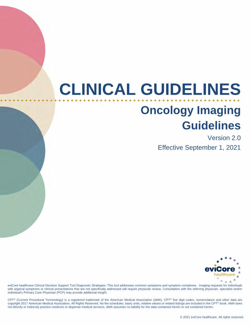
Oncology Imaging Guidelines Abbreviations for Oncology Guidelines ONC-1: General Guidelines ONC-2: Primary Central Nervous System Tumors ONC-3: Squamous Cell Carcinomas of the Head and Neck ONC-4: Salivary Gland Cancers ONC-5: Melanomas and Other Skin Cancers ONC-6: Thyroid Cancer ONC-7: Small Cell Lung Cancer ONC-8: Non-Small Cell Lung Cancer ONC-9: Esophageal and GE Junction Cancer ONC-10: Other Thoracic Tumors ONC-11: Breast Cancer ONC-12: Sarcomas – Bone, Soft Tissue and GIST ONC-13: Pancreatic Cancer ONC-14: Upper GI Cancers ONC-15: Neuroendocrine Cancers and Adrenal Tumors ONC-16: Colorectal and Small Bowel Cancer ONC-17: Renal Cell Cancer (RCC) ONC-18: Transitional Cell Cancer ONC-19: Prostate Cancer ONC-20: Testicular, Ovarian and Extragonadal Germ Cell Tumors ONC-21: Ovarian Cancer ONC-22: Uterine Cancer ONC-23: Cervical Cancer ONC-24: Anal Cancer & Cancers of the External Genitalia ONC-25: Multiple Myeloma and Plasmacytomas ONC-26: Leukemias, Myelodysplasia and Myeloproliferative Neoplasms ONC-27: Non-Hodgkin Lymphomas ONC-28: Hodgkin Lymphoma ONC-29: Hematopoietic Stem Cell Transplantation ONC-30: Medical Conditions with Cancer in the Differential Diagnosis ONC-31: Metastatic Cancer, Carcinoma of Unknown Primary Site, and Other Types of Cancer ONC-32: Medicare Coverage Policies for PET
Oncology Imaging Guidelines V2.0
______________________________________________________________________________________________________ ©2021 eviCore healthcare. All Rights Reserved. 400 Buckwalter Place Boulevard, Bluffton, SC 29910 (800) 918-8924 www.eviCore.com
Page 2 of 302
Onc
olog
y Im
agin
g
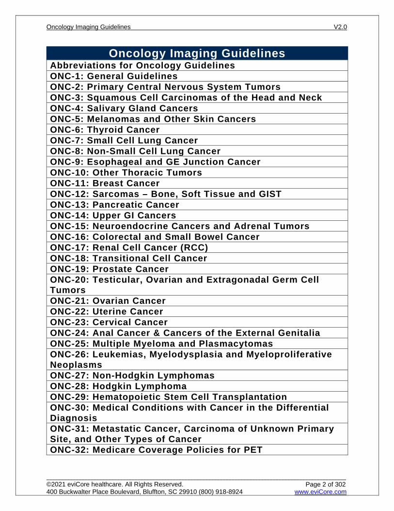
Abbreviations for Oncology Guidelines ACTH adrenocorticotropic hormone
AFP alpha-fetoprotein AP anteroposterior
betaHCG beta human chorionic gonadotropin
CA 125 cancer antigen 125 test CA 19-9 cancer antigen 19-9
CA 15-3 cancer antigen 15-3 CA 27-29 cancer antigen 27-29
CBC complete blood count
CEA carcinoembryonic antigen CNS central nervous system
CR complete response
CTA computed tomography angiography DCIS ductal carcinoma in situ
DLBCL diffuse large B cell lymphomas
DRE digital rectal exam EGD esophagogastroduodenoscopy
ENT ear, nose, throat EOT end of therapy
ERCP endoscopic retrograde cholangiopancreatography
ESR erythrocyte sedimentation rate EUA exam under anesthesia
EUS endoscopic ultrasound
FDG fluorodeoxyglucose FNA fine needle aspiration
FUO fever of unknown origin
GE gastroesophageal GI gastrointestinal
GU genitourinary
GTR gross total resection HG high grade
HIV human immunodeficiency disease
HRPC hormone refractory prostate cancer hypermet hypermetabolic
IFRT Involved field radiation therapy inv invasive
LAR low anterior resection LCIS lobular carcinoma in situ
LDH lactate dehydrogenase LFT liver function tests
LND Lymph node dissection
MALT mucosa associated lymphoid tissue maint maintenance
Oncology Imaging Guidelines V2.0
______________________________________________________________________________________________________ ©2021 eviCore healthcare. All Rights Reserved. 400 Buckwalter Place Boulevard, Bluffton, SC 29910 (800) 918-8924 www.eviCore.com
Page 3 of 302
Onc
olog
y Im
agin
g
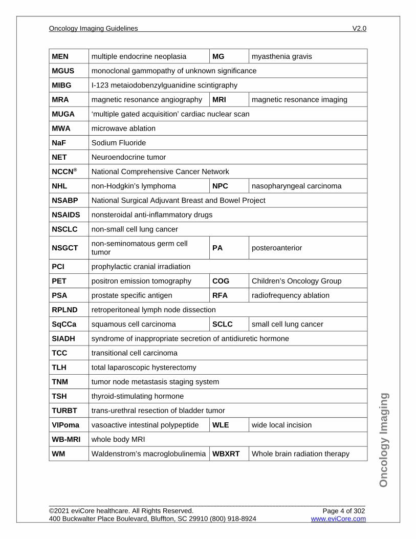
MEN multiple endocrine neoplasia MG myasthenia gravis
MGUS monoclonal gammopathy of unknown significance
MIBG I-123 metaiodobenzylguanidine scintigraphy
MRA magnetic resonance angiography MRI magnetic resonance imaging
MUGA ‘multiple gated acquisition’ cardiac nuclear scan
MWA microwave ablation
NaF Sodium Fluoride
NET Neuroendocrine tumor
NCCN® National Comprehensive Cancer Network
NHL non-Hodgkin’s lymphoma NPC nasopharyngeal carcinoma
NSABP National Surgical Adjuvant Breast and Bowel Project
NSAIDS nonsteroidal anti-inflammatory drugs
NSCLC non-small cell lung cancer
NSGCT non-seminomatous germ cell tumor PA posteroanterior
PCI prophylactic cranial irradiation
PET positron emission tomography COG Children’s Oncology Group
PSA prostate specific antigen RFA radiofrequency ablation
RPLND retroperitoneal lymph node dissection
SqCCa squamous cell carcinoma SCLC small cell lung cancer
SIADH syndrome of inappropriate secretion of antidiuretic hormone
TCC transitional cell carcinoma
TLH total laparoscopic hysterectomy
TNM tumor node metastasis staging system
TSH thyroid-stimulating hormone
TURBT trans-urethral resection of bladder tumor
VIPoma vasoactive intestinal polypeptide WLE wide local incision
WB-MRI whole body MRI
WM Waldenstrom’s macroglobulinemia WBXRT Whole brain radiation therapy
Oncology Imaging Guidelines V2.0
______________________________________________________________________________________________________ ©2021 eviCore healthcare. All Rights Reserved. 400 Buckwalter Place Boulevard, Bluffton, SC 29910 (800) 918-8924 www.eviCore.com
Page 4 of 302
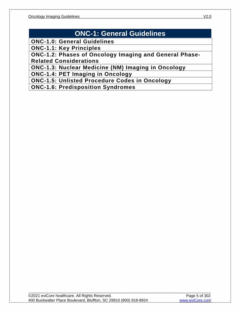
ONC-1: General Guidelines ONC-1.0: General GuidelinesONC-1.1: Key PrinciplesONC-1.2: Phases of Oncology Imaging and General Phase-Related ConsiderationsONC-1.3: Nuclear Medicine (NM) Imaging in OncologyONC-1.4: PET Imaging in OncologyONC-1.5: Unlisted Procedure Codes in OncologyONC-1.6: Predisposition Syndromes
Oncology Imaging Guidelines V2.0
______________________________________________________________________________________________________ ©2021 eviCore healthcare. All Rights Reserved. 400 Buckwalter Place Boulevard, Bluffton, SC 29910 (800) 918-8924 www.eviCore.com
Page 5 of 302
Onc
olog
y Im
agin
g
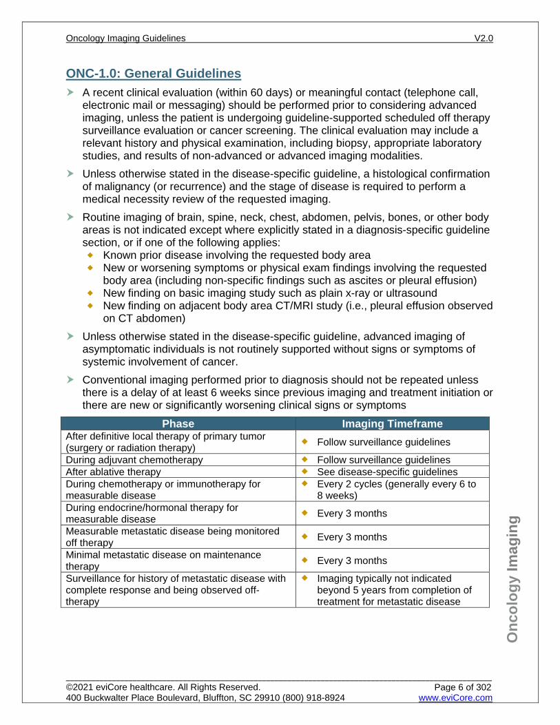
ONC-1.0: General Guidelines A recent clinical evaluation (within 60 days) or meaningful contact (telephone call,
electronic mail or messaging) should be performed prior to considering advanced imaging, unless the patient is undergoing guideline-supported scheduled off therapy surveillance evaluation or cancer screening. The clinical evaluation may include a relevant history and physical examination, including biopsy, appropriate laboratory studies, and results of non-advanced or advanced imaging modalities.
Unless otherwise stated in the disease-specific guideline, a histological confirmation of malignancy (or recurrence) and the stage of disease is required to perform a medical necessity review of the requested imaging.
Routine imaging of brain, spine, neck, chest, abdomen, pelvis, bones, or other body areas is not indicated except where explicitly stated in a diagnosis-specific guideline section, or if one of the following applies: Known prior disease involving the requested body area New or worsening symptoms or physical exam findings involving the requested
body area (including non-specific findings such as ascites or pleural effusion) New finding on basic imaging study such as plain x-ray or ultrasound New finding on adjacent body area CT/MRI study (i.e., pleural effusion observed
on CT abdomen) Unless otherwise stated in the disease-specific guideline, advanced imaging of
asymptomatic individuals is not routinely supported without signs or symptoms of systemic involvement of cancer.
Conventional imaging performed prior to diagnosis should not be repeated unless there is a delay of at least 6 weeks since previous imaging and treatment initiation or there are new or significantly worsening clinical signs or symptoms
Phase Imaging Timeframe After definitive local therapy of primary tumor (surgery or radiation therapy) Follow surveillance guidelines
During adjuvant chemotherapy Follow surveillance guidelines After ablative therapy See disease-specific guidelines During chemotherapy or immunotherapy for measurable disease
Every 2 cycles (generally every 6 to 8 weeks)
During endocrine/hormonal therapy for measurable disease Every 3 months
Measurable metastatic disease being monitored off therapy Every 3 months
Minimal metastatic disease on maintenance therapy Every 3 months
Surveillance for history of metastatic disease with complete response and being observed off-therapy
Imaging typically not indicated beyond 5 years from completion of treatment for metastatic disease
Oncology Imaging Guidelines V2.0
______________________________________________________________________________________________________ ©2021 eviCore healthcare. All Rights Reserved. 400 Buckwalter Place Boulevard, Bluffton, SC 29910 (800) 918-8924 www.eviCore.com
Page 6 of 302
Onc
olog
y Im
agin
g
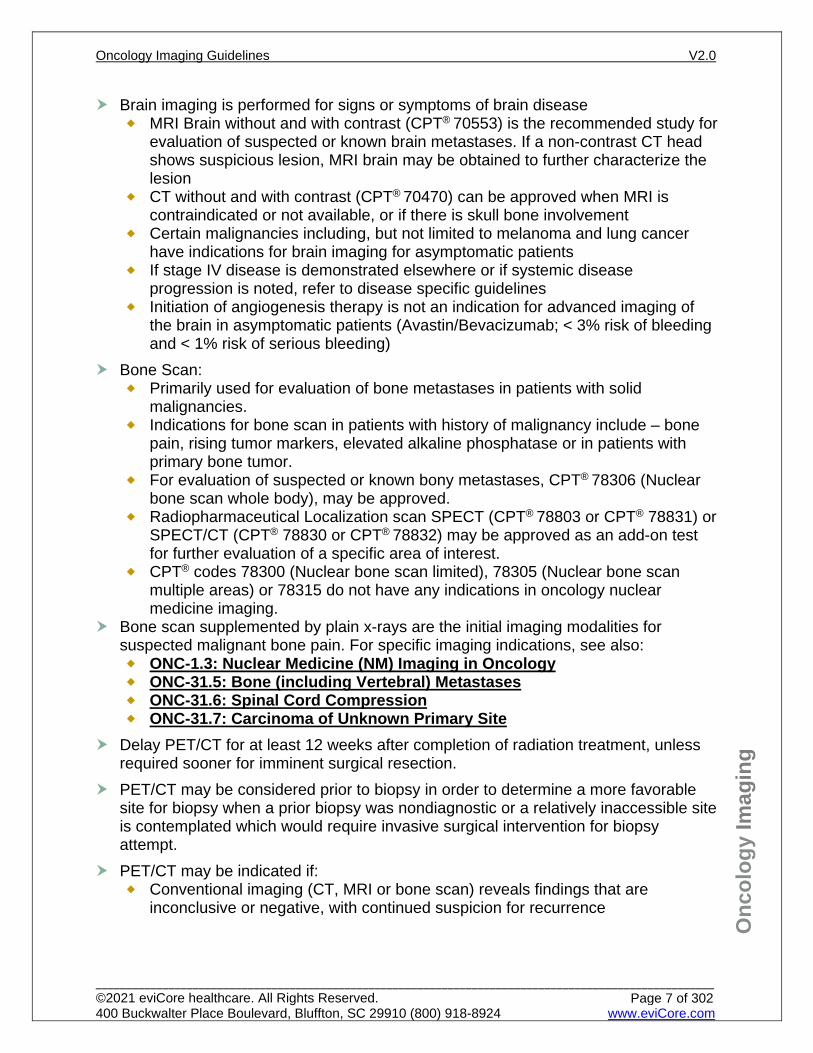
Brain imaging is performed for signs or symptoms of brain disease MRI Brain without and with contrast (CPT® 70553) is the recommended study for
evaluation of suspected or known brain metastases. If a non-contrast CT head shows suspicious lesion, MRI brain may be obtained to further characterize the lesion
CT without and with contrast (CPT® 70470) can be approved when MRI is contraindicated or not available, or if there is skull bone involvement
Certain malignancies including, but not limited to melanoma and lung cancer have indications for brain imaging for asymptomatic patients
If stage IV disease is demonstrated elsewhere or if systemic disease progression is noted, refer to disease specific guidelines
Initiation of angiogenesis therapy is not an indication for advanced imaging of the brain in asymptomatic patients (Avastin/Bevacizumab; < 3% risk of bleeding and < 1% risk of serious bleeding)
Bone Scan: Primarily used for evaluation of bone metastases in patients with solid
malignancies. Indications for bone scan in patients with history of malignancy include – bone
pain, rising tumor markers, elevated alkaline phosphatase or in patients with primary bone tumor.
For evaluation of suspected or known bony metastases, CPT® 78306 (Nuclear bone scan whole body), may be approved.
Radiopharmaceutical Localization scan SPECT (CPT® 78803 or CPT® 78831) or SPECT/CT (CPT® 78830 or CPT® 78832) may be approved as an add-on test for further evaluation of a specific area of interest.
CPT® codes 78300 (Nuclear bone scan limited), 78305 (Nuclear bone scan multiple areas) or 78315 do not have any indications in oncology nuclear medicine imaging.
Bone scan supplemented by plain x-rays are the initial imaging modalities for suspected malignant bone pain. For specific imaging indications, see also: ONC-1.3: Nuclear Medicine (NM) Imaging in Oncology ONC-31.5: Bone (including Vertebral) Metastases ONC-31.6: Spinal Cord Compression ONC-31.7: Carcinoma of Unknown Primary Site
Delay PET/CT for at least 12 weeks after completion of radiation treatment, unless required sooner for imminent surgical resection.
PET/CT may be considered prior to biopsy in order to determine a more favorable site for biopsy when a prior biopsy was nondiagnostic or a relatively inaccessible site is contemplated which would require invasive surgical intervention for biopsy attempt.
PET/CT may be indicated if: Conventional imaging (CT, MRI or bone scan) reveals findings that are
inconclusive or negative, with continued suspicion for recurrence
Oncology Imaging Guidelines V2.0
______________________________________________________________________________________________________ ©2021 eviCore healthcare. All Rights Reserved. 400 Buckwalter Place Boulevard, Bluffton, SC 29910 (800) 918-8924 www.eviCore.com
Page 7 of 302
Onc
olog
y Im
agin
g
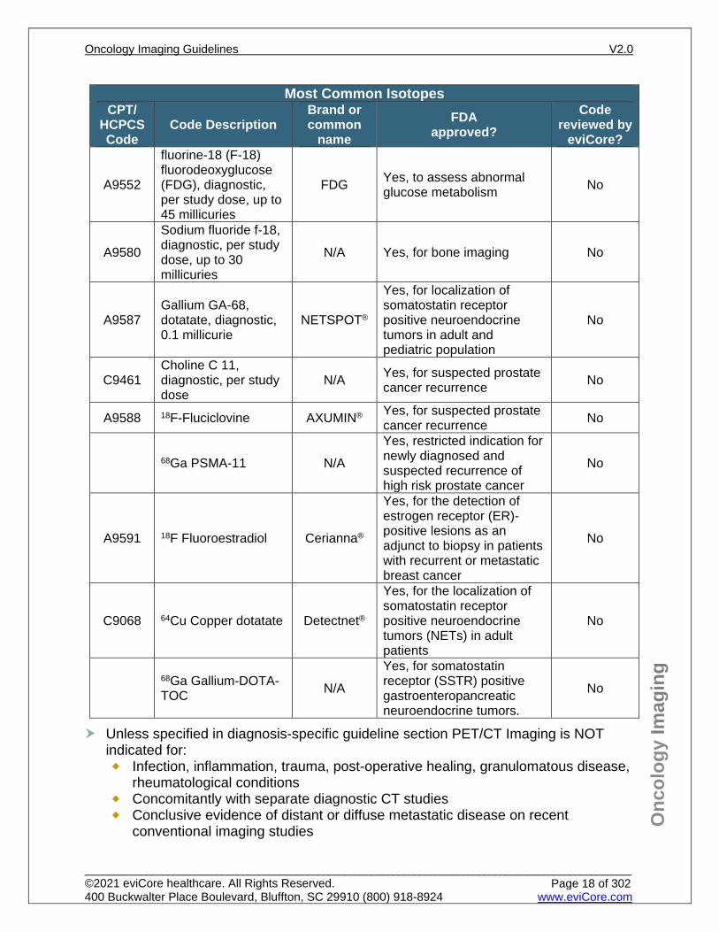
Unless specified in diagnosis-specific guideline section PET/CT Imaging is NOT indicated for: Infection, inflammation, trauma, post-operative healing, granulomatous disease,
rheumatological conditions Concomitantly with separate diagnostic CT studies Conclusive evidence of distant or diffuse metastatic disease on recent
conventional imaging studies Metastatic disease in the central nervous system (CNS) Lesions less than 8 mm in size Follow up after localized therapy (i.e. radiofrequency ablation, embolization,
stereotactic radiation, etc.) Rare malignancies, due to lack of available evidence regarding the diagnostic
accuracy of PET in rare cancers Surveillance Serial monitoring of individuals who are not currently receiving anti-tumor
treatment or are receiving maintenance treatment Serial monitoring of FDG avidity until resolution. PET/CT avidity in a residual mass at the end of planned therapy is not an
indication for PET/CT imaging during surveillance. Residual mass that has not changed in size since the last conventional
imaging does not justify PET imaging Unless otherwise specified for a specific cancer type, once PET has been
documented to be negative for a given patient’s cancer or all PET-avid disease has been surgically resected, PET should not be used for continued disease monitoring or surveillance.
Advanced imaging is not indicated for evaluation of in situ or non-invasive cancers or cancer surveillance after complete surgical removal of primary disease unless otherwise stated in the cancer-specific guidelines.
Advanced imaging is not indicated for monitoring disease in individuals who choose to not receive standard oncologic therapy, but may be receiving alternative therapies or palliative care and/or hospice. All advanced imaging indicated for initial staging of the specific cancer type can be approved once when the patient is considering initiation of a standard therapeutic approach (surgery, chemotherapy, or radiation therapy).
Oncology Imaging Guidelines V2.0
______________________________________________________________________________________________________ ©2021 eviCore healthcare. All Rights Reserved. 400 Buckwalter Place Boulevard, Bluffton, SC 29910 (800) 918-8924 www.eviCore.com
Page 8 of 302
Onc
olog
y Im
agin
g
The specific radiotracer planned to be used with PET/CT imaging is required to perform a medical necessity review. Indications for PET/CT imaging using non-FDG radiotracers are listed in diagnosis-specific guidelines. Covered radiotracers: 18F-FDG 68Gallium DOTATATE (NETSPOT®) for low grade neuroendocrine tumors and
medullary thyroid cancer 11C Choline for prostate cancer 18F-Fluciclovine (AXUMIN®) for prostate cancer
Not covered radiotracers: 18F-Na Fluoride PET bone scan 68Ga PSMA-11 18F Fluoroestradiol 64Cu Copper dotatate 68Ga Gallium-DOTA-TOC PET/CT imaging using isotopes other than those specified above
Octreotide scan: Specific for low and intermediate grade neuroendocrine tumors which express
specific cell surface somatostatin receptors. See cancer specific guidelines for recommended use.
One of the following codes may be approved when Octreotide scan is requested:
CPT® 78802 (Radiopharmaceutical localization of tumor whole body single day study)
CPT® 78804 (Radiopharmaceutical localization of tumor whole body two or more days)
In addition to one of the above CPT codes, CPT® 78803 (Radiopharmaceutical localization of tumor SPECT), SPECT CPT® 78831, or hybrid SPECT/CT (CPT® 78830 or 78832) may be approved as an add-on test for further evaluation of a specific area of interest.
Clinical Trials Similar to investigational and experimental studies, clinical trial imaging requests will
be considered to determine whether they meet Health Plan coverage and eviCore’s evidence-based guidelines.
Imaging studies which are inconsistent with established clinical standards, or are requested for data collection and not used in direct clinical management are not supported.
Oncology Imaging Guidelines V2.0
______________________________________________________________________________________________________ ©2021 eviCore healthcare. All Rights Reserved. 400 Buckwalter Place Boulevard, Bluffton, SC 29910 (800) 918-8924 www.eviCore.com
Page 9 of 302
Onc
olog
y Im
agin
g
ONC-1.1: Key Principles
AGE APPROPRIATE GUIDELINES Age of Individual Appropriate Imaging Guidelines
≥ 18 years old at initial diagnosis
Adult Oncology Imaging Guidelines, except where directed otherwise by a specific guideline section
< 18 years old at initial diagnosis
Pediatric Oncology Imaging Guidelines, except where directed otherwise by a specific guideline section
15 to 39 years old at initial diagnosis (defined as Adolescent and Young Adult (AYA) oncology individuals)
When unique guidelines for a specific cancer type exist only in either Oncology or Pediatric Oncology, AYA individuals should be imaged according to the guideline section for their specific cancer type, regardless of the individual’s age
When unique guidelines for a specific cancer type exist in both Oncology and Pediatric Oncology, AYA individuals should be imaged according to the age rule in the previous bullet
Conventional Imaging (mostly CT, sometimes MRI or bone scan) of the affectedarea(s) drives much of initial and re-staging and surveillance. PET is not indicatedfor surveillance imaging unless specifically stated in the diagnosis-specific guidelinesections
Brain imaging is performed for signs or symptoms of brain disease MRI Brain without and with contrast (CPT® 70553) is the recommended study for
evaluation of suspected or known brain metastases. If a non-contrast CT head shows suspicious lesion, MRI brain may be obtained to further characterize the lesion
CT without and with contrast (CPT® 70470) can be approved when MRI is contraindicated or not available, or if there is skull bone involvement
Certain malignancies including, but not limited to melanoma and lung cancer have indications for brain imaging for asymptomatic patients
If stage IV disease is demonstrated elsewhere or if systemic disease progression is noted, refer to disease specific guidelines
Initiation of angiogenesis therapy is not an indication for advanced imaging of the brain in asymptomatic patients (Avastin/Bevacizumab; < 3% risk of bleeding and < 1% risk of serious bleeding)
Patients receiving cardiotoxic chemotherapy (such as doxorubicin, trastuzumab,pertuzumab, mitoxantrone, etc.) may undergo cardiac evaluation – at baseline andfor monitoring while on active therapy. eviCore guidelines support using Echocardiography (CPT® 93306, CPT® 93307,
or CPT® 93308) rather than MUGA scan for determination of LVEF and/or wall motion EXCEPT in one of the circumstances described previously in CD-3.4: MUGA Study – Cardiac Indications.
The timeframe should be determined by the provider, but no more often than baseline and at every 6 weeks.
May repeat every 4 weeks if cardiotoxic chemotherapeutic drug is withheld for significant left ventricular cardiac dysfunction.
If the LVEF is < 50% on echocardiogram than follow up can be done with MUGA at appropriate intervals.
Oncology Imaging Guidelines V2.0
______________________________________________________________________________________________________ ©2021 eviCore healthcare. All Rights Reserved. 400 Buckwalter Place Boulevard, Bluffton, SC 29910 (800) 918-8924 www.eviCore.com
Page 10 of 302
Onc
olog
y Im
agin
g
See also: CD-12.1: Oncologic Indications for Cancer Therapeutics-Related Cardiac Dysfunction (CTRCD)
Adults (≥18 years) with a diagnosis of Li-Fraumeni Syndrome (LFS) may be screened for malignancy with a Whole Body MRI (CPT® 76498) on an annual basis. Annual Brain MRI (CPT® 70553) may be performed as part of Whole Body MRI or as a separate exam. Due to lack of standardization of technique, interpretation, and availability of Whole Body MRI, individuals with LFS are encouraged to participate in clinical trials.
CTA or MRA of a specific anatomic region is indicated when requested for surgical planning when there is suspected vascular proximity to proposed resection margin.
Use of Contrast CT imaging should be performed with contrast for known or suspected body regions,
unless contraindicated. Shellfish allergy is not a contraindication to contrast. Patients with known
shellfish allergy do not have contrast reaction any more often than other atopic individuals or patients with other food allergies.
For iodinated contrast dye allergy, either CT scans without contrast or MRI scans without and with contrast are indicated.
If CT scanning is considered strongly indicated in a patient with known contrast allergy, CT with contrast may be considered to be safely performed following prednisone premedication over a 24-hour period prior to the study.
For patients with renal insufficiency which precludes contrast use, CT without contrast appropriate disease-specific areas should be offered. Further imaging (such as MRI) may be indicated if noncontrast CT results are inconclusive.
Severe renal insufficiency, i.e. an eGFR less than 30, is a contraindication for an MRI using a gadolinium-based contrast agent (GBCA) as well. In patients with eGFR greater than 40, GBCA administration can be safely performed. GBCA administered to patients with acute kidney injury or severe chronic kidney disease can result in a syndrome of nephrogenic systemic fibrosis (NSF), but GBCAs are not considered nephrotoxic at dosages approved for MRI.
Gadolinium deposition has been found in patients with normal renal function following the use of gadolinium based contrast agents (GBCAs). The U.S. Food and Drug Administration (FDA) is investigating the risk of brain
deposits following repeated use of GBCAs. The FDA has noted that, “It is unknown whether these gadolinium deposits are
harmful or can lead to adverse health effects.” and have recommended: To reduce the potential for gadolinium accumulation, health care
professionals should consider limiting GBCA use to clinical circumstances in which the additional information provided by the contrast is necessary.
Health care professionals are also urged to reassess the necessity of repetitive GBCA MRIs in established treatment protocols.
Oncology Imaging Guidelines V2.0
______________________________________________________________________________________________________ ©2021 eviCore healthcare. All Rights Reserved. 400 Buckwalter Place Boulevard, Bluffton, SC 29910 (800) 918-8924 www.eviCore.com
Page 11 of 302
Onc
olog
y Im
agin
g
Radiation Exposure The use of MRI in place of CT scans to reduce risk of secondary malignancy is not
supported by the peer-reviewed literature. Unless otherwise specified in the Guidelines, MRI in place of CT scans for this purpose alone is not indicated. In some instances (i.e., testicular cancer surveillance), MRI may be considered inferior to CT scans.
Oncology Imaging Guidelines V2.0
______________________________________________________________________________________________________ ©2021 eviCore healthcare. All Rights Reserved. 400 Buckwalter Place Boulevard, Bluffton, SC 29910 (800) 918-8924 www.eviCore.com
Page 12 of 302
Onc
olog
y Im
agin
g
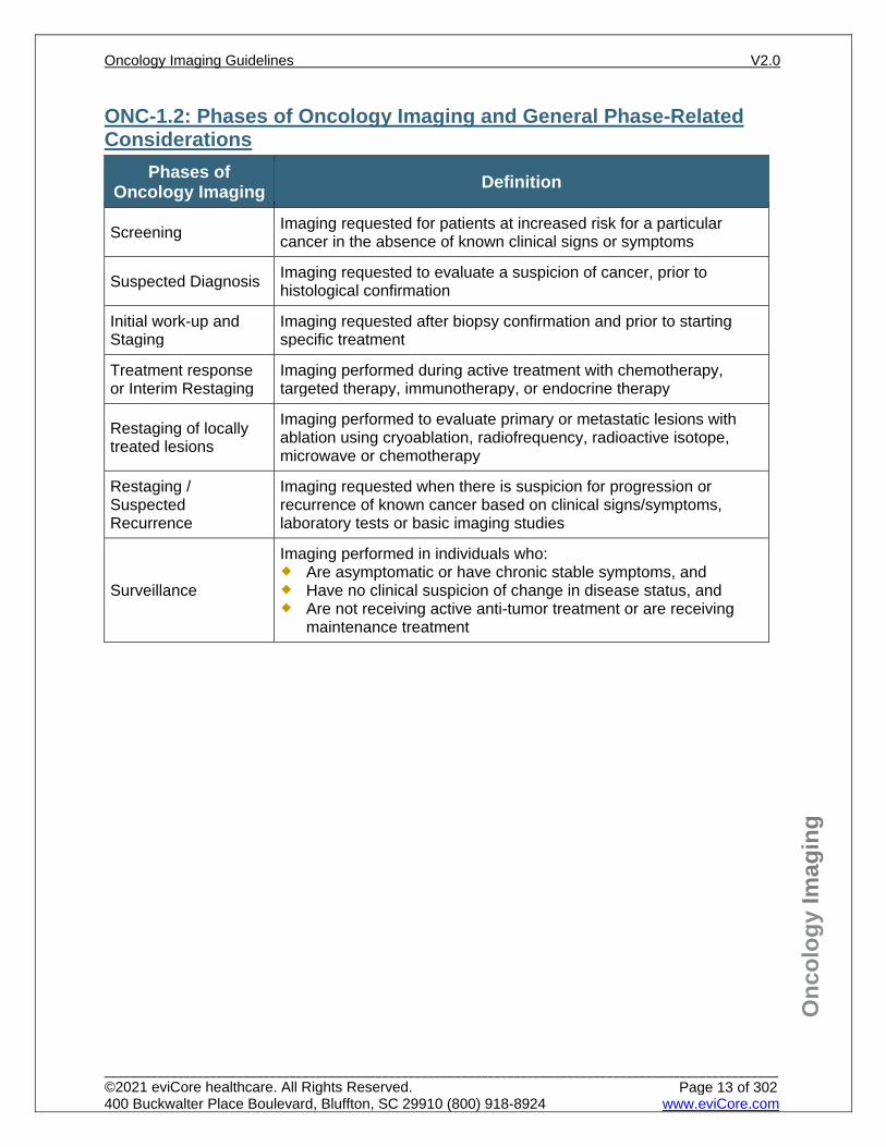
ONC-1.2: Phases of Oncology Imaging and General Phase-Related Considerations
Phases of Oncology Imaging Definition
Screening Imaging requested for patients at increased risk for a particular cancer in the absence of known clinical signs or symptoms
Suspected Diagnosis Imaging requested to evaluate a suspicion of cancer, prior to histological confirmation
Initial work-up and Staging
Imaging requested after biopsy confirmation and prior to starting specific treatment
Treatment response or Interim Restaging
Imaging performed during active treatment with chemotherapy, targeted therapy, immunotherapy, or endocrine therapy
Restaging of locally treated lesions
Imaging performed to evaluate primary or metastatic lesions with ablation using cryoablation, radiofrequency, radioactive isotope, microwave or chemotherapy
Restaging / Suspected Recurrence
Imaging requested when there is suspicion for progression or recurrence of known cancer based on clinical signs/symptoms, laboratory tests or basic imaging studies
Surveillance
Imaging performed in individuals who: Are asymptomatic or have chronic stable symptoms, and Have no clinical suspicion of change in disease status, and Are not receiving active anti-tumor treatment or are receiving
maintenance treatment
Oncology Imaging Guidelines V2.0
______________________________________________________________________________________________________ ©2021 eviCore healthcare. All Rights Reserved. 400 Buckwalter Place Boulevard, Bluffton, SC 29910 (800) 918-8924 www.eviCore.com
Page 13 of 302
Onc
olog
y Im
agin
g
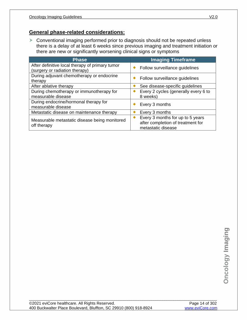
General phase-related considerations: Conventional imaging performed prior to diagnosis should not be repeated unless
there is a delay of at least 6 weeks since previous imaging and treatment initiation or there are new or significantly worsening clinical signs or symptoms
Phase Imaging Timeframe After definitive local therapy of primary tumor (surgery or radiation therapy) Follow surveillance guidelines
During adjuvant chemotherapy or endocrine therapy Follow surveillance guidelines
After ablative therapy See disease-specific guidelines During chemotherapy or immunotherapy for measurable disease
Every 2 cycles (generally every 6 to 8 weeks)
During endocrine/hormonal therapy for measurable disease Every 3 months
Metastatic disease on maintenance therapy Every 3 months
Measurable metastatic disease being monitored off therapy
Every 3 months for up to 5 years after completion of treatment for metastatic disease
Oncology Imaging Guidelines V2.0
______________________________________________________________________________________________________ ©2021 eviCore healthcare. All Rights Reserved. 400 Buckwalter Place Boulevard, Bluffton, SC 29910 (800) 918-8924 www.eviCore.com
Page 14 of 302
Onc
olog
y Im
agin
g
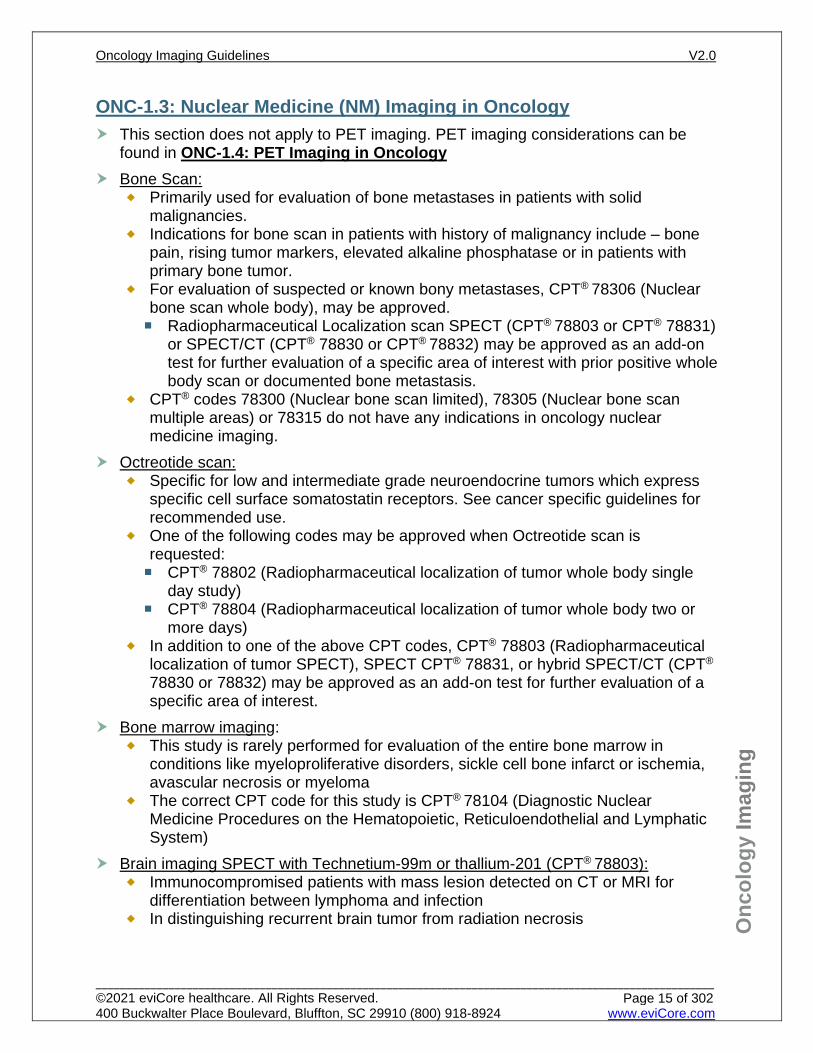
ONC-1.3: Nuclear Medicine (NM) Imaging in Oncology This section does not apply to PET imaging. PET imaging considerations can be
found in ONC-1.4: PET Imaging in Oncology Bone Scan:
Primarily used for evaluation of bone metastases in patients with solid malignancies.
Indications for bone scan in patients with history of malignancy include – bone pain, rising tumor markers, elevated alkaline phosphatase or in patients with primary bone tumor.
For evaluation of suspected or known bony metastases, CPT® 78306 (Nuclear bone scan whole body), may be approved. Radiopharmaceutical Localization scan SPECT (CPT® 78803 or CPT® 78831)
or SPECT/CT (CPT® 78830 or CPT® 78832) may be approved as an add-on test for further evaluation of a specific area of interest with prior positive whole body scan or documented bone metastasis.
CPT® codes 78300 (Nuclear bone scan limited), 78305 (Nuclear bone scan multiple areas) or 78315 do not have any indications in oncology nuclear medicine imaging.
Octreotide scan: Specific for low and intermediate grade neuroendocrine tumors which express
specific cell surface somatostatin receptors. See cancer specific guidelines for recommended use.
One of the following codes may be approved when Octreotide scan is requested: CPT® 78802 (Radiopharmaceutical localization of tumor whole body single
day study) CPT® 78804 (Radiopharmaceutical localization of tumor whole body two or
more days) In addition to one of the above CPT codes, CPT® 78803 (Radiopharmaceutical
localization of tumor SPECT), SPECT CPT® 78831, or hybrid SPECT/CT (CPT® 78830 or 78832) may be approved as an add-on test for further evaluation of a specific area of interest.
Bone marrow imaging: This study is rarely performed for evaluation of the entire bone marrow in
conditions like myeloproliferative disorders, sickle cell bone infarct or ischemia, avascular necrosis or myeloma
The correct CPT code for this study is CPT® 78104 (Diagnostic Nuclear Medicine Procedures on the Hematopoietic, Reticuloendothelial and Lymphatic System)
Brain imaging SPECT with Technetium-99m or thallium-201 (CPT® 78803): Immunocompromised patients with mass lesion detected on CT or MRI for
differentiation between lymphoma and infection In distinguishing recurrent brain tumor from radiation necrosis
Oncology Imaging Guidelines V2.0
______________________________________________________________________________________________________ ©2021 eviCore healthcare. All Rights Reserved. 400 Buckwalter Place Boulevard, Bluffton, SC 29910 (800) 918-8924 www.eviCore.com
Page 15 of 302
Onc
olog
y Im
agin
g
Radiopharmaceutical localization of tumor or distribution of radiopharmaceutical agent(s): CPT® 78800, CPT® 78801, CPT® 78802, CPT® 78804, CPT® 78803, CPT® 78831
(SPECT), or CPT® 78830 or CPT® 78832 (SPECT/CT) For evaluation of fever of unknown origin and osteomyelitis For suspected infections such as infected central lines, grafts or shunts
Gallium Isotope Scan: Radiopharmaceutical Localization of tumor (CPT® 78800, CPT® 78801, CPT®
78802, CPT® 78803, or CPT® 78804), SPECT CPT® 78831, or hybrid SPECT/CT CPT® 78830 or 78832
This may be rarely used in place of PET/CT scan when PET/CT scan not available and PET/CT is indicated by guidelines for lymphoma, sarcoma, melanoma or myeloma
Oncology Imaging Guidelines V2.0
______________________________________________________________________________________________________ ©2021 eviCore healthcare. All Rights Reserved. 400 Buckwalter Place Boulevard, Bluffton, SC 29910 (800) 918-8924 www.eviCore.com
Page 16 of 302
Onc
olog
y Im
agin
g
ONC-1.4: PET Imaging in Oncology NOTE: Some payors have specific restrictions on PET imaging, and those coverage policies may supersede the recommendations for PET imaging in these guidelines.
CPT codes: PET imaging in oncology should use PET/CT fusion imaging (CPT® 78815 or
CPT® 78816) Unbundling PET/CT imaging into separate PET and diagnostic CT codes is otherwise not supported.
The decision whether to use skull base to mid-femur (“eyes to thighs”) procedure code for PET (CPT® 78812 or CPT® 78815) or whole body PET (CPT® 78813 or CPT® 78816) is addressed in the diagnosis-specific guideline sections.
‘Limited area’ protocol is done infrequently, but may be considered, and is reported with PET (CPT® 78811) or for PET/CT, (CPT® 78814).
Radiotracers: Unless specified otherwise, the term “PET” refers to 18F-FDG-PET and PET/CT
fusion studies Indications for PET/CT imaging using non-FDG radiotracers are listed in
diagnosis-specific guidelines. The indications may be as follows: Covered: 18F-FDG 68Gallium DOTATATE (NETSPOT®) for low grade neuroendocrine tumors
for localization of somatostatin receptor positive neuroendocrine tumors in adult and pediatric population
11C Choline for prostate cancer 18F-Fluciclovine (AXUMIN®) for prostate cancer
Not covered: 18F-Na Fluoride PET bone scan 68Ga PSMA-11 18F Fluoroestradiol 64Cu Copper dotatate 68GaGallium-DOTA-TOC PET/CT imaging using isotopes other than those specified above
Oncology Imaging Guidelines V2.0
______________________________________________________________________________________________________ ©2021 eviCore healthcare. All Rights Reserved. 400 Buckwalter Place Boulevard, Bluffton, SC 29910 (800) 918-8924 www.eviCore.com
Page 17 of 302
Onc
olog
y Im
agin
g
Unless specified in diagnosis-specific guideline section PET/CT Imaging is NOT indicated for: Infection, inflammation, trauma, post-operative healing, granulomatous disease,
rheumatological conditions Concomitantly with separate diagnostic CT studies Conclusive evidence of distant or diffuse metastatic disease on recent
conventional imaging studies
Most Common Isotopes CPT/
HCPCS Code
Code Description Brand or common
name FDA
approved? Code
reviewed by eviCore?
A9552
fluorine-18 (F-18) fluorodeoxyglucose (FDG), diagnostic, per study dose, up to 45 millicuries
FDG Yes, to assess abnormal glucose metabolism No
A9580
Sodium fluoride f-18, diagnostic, per study dose, up to 30 millicuries
N/A Yes, for bone imaging No
A9587 Gallium GA-68, dotatate, diagnostic, 0.1 millicurie
NETSPOT®
Yes, for localization of somatostatin receptor positive neuroendocrine tumors in adult and pediatric population
No
C9461 Choline C 11, diagnostic, per study dose
N/A Yes, for suspected prostate cancer recurrence No
A9588 18F-Fluciclovine AXUMIN® Yes, for suspected prostate cancer recurrence No
68Ga PSMA-11 N/A
Yes, restricted indication for newly diagnosed and suspected recurrence of high risk prostate cancer
No
A9591 18F Fluoroestradiol Cerianna®
Yes, for the detection of estrogen receptor (ER)-positive lesions as an adjunct to biopsy in patients with recurrent or metastatic breast cancer
No
C9068 64Cu Copper dotatate Detectnet®
Yes, for the localization of somatostatin receptor positive neuroendocrine tumors (NETs) in adult patients
No
68Ga Gallium-DOTA-TOC N/A
Yes, for somatostatin receptor (SSTR) positive gastroenteropancreatic neuroendocrine tumors.
No
Oncology Imaging Guidelines V2.0
______________________________________________________________________________________________________ ©2021 eviCore healthcare. All Rights Reserved. 400 Buckwalter Place Boulevard, Bluffton, SC 29910 (800) 918-8924 www.eviCore.com
Page 18 of 302
Onc
olog
y Im
agin
g
Metastatic disease in the central nervous system (CNS) Lesions less than 8 mm in size Follow up after localized therapy (i.e. radiofrequency ablation, embolization,
stereotactic radiation, etc.) Rare malignancies, due to lack of available evidence regarding the diagnostic
accuracy of PET in rare cancers Surveillance Serial monitoring of individuals who are not currently receiving anti-tumor
treatment or are receiving maintenance treatment Serial monitoring of FDG avidity until resolution. PET/CT avidity in a residual mass at the end of planned therapy is not an
indication for PET/CT imaging during surveillance. Residual mass that has not changed in size since the last conventional
imaging does not justify PET imaging Unless otherwise specified for a specific cancer type, once PET has been
documented to be negative for a given patient’s cancer or all PET-avid disease has been surgically resected, PET should not be used for continued disease monitoring or surveillance.
PET/CT may be indicated if: Conventional imaging (CT, MRI or bone scan) reveals findings that are
inconclusive or negative, with continued suspicion for recurrence The patient is undergoing salvage treatment for a recurrent solid tumor with
residual measurable disease on conventional imaging and confirmed repeat negative PET imaging will allow the patient to transition from active treatment to surveillance.
PET/CT may be considered prior to biopsy in order to determine a more favorable site for biopsy when a prior biopsy was nondiagnostic or a relatively inaccessible site is contemplated which would require invasive surgical intervention for biopsy attempt.
PET/CT for rare malignancies is not covered by eviCore guidelines due to lack of available evidence regarding diagnostic accuracy of PET/CT in the majority of rare cancers. Conventional imaging studies should be used for initial staging and treatment response for these diagnoses. PET/CT can be approved if all of the following apply: Conventional imaging (CT, MRI or bone scan) reveals equivocal or suspicious
findings No other specific metabolic imaging (MIBG, octreotide, technetium, etc.) is
appropriate for the disease type The submitted clinical information describes a specific decision regarding the
patient’s care that will be made based on the PET/CT results Delay PET/CT for at least 12 weeks after completion of radiation treatment, unless
required sooner for imminent surgical resection. PET mammography (PEM, generally reported with CPT® 78811) is considered
experimental and investigational at this time.
Oncology Imaging Guidelines V2.0
______________________________________________________________________________________________________ ©2021 eviCore healthcare. All Rights Reserved. 400 Buckwalter Place Boulevard, Bluffton, SC 29910 (800) 918-8924 www.eviCore.com
Page 19 of 302
Onc
olog
y Im
agin
g
ONC-1.5: Unlisted Procedure Codes in Oncology eviCore authorizes requests for CT or MRI associated with image-directed biopsy or
radiation therapy treatment planning for some payors. eviCore does not routinely authorize requests for PET associated with image-
directed biopsy or radiation therapy treatment planning. There is often no unique procedure code for a service performed solely for treatment
planning purposes. AMA instructions in the CPT state that if no specific code exists for a particular service, the service is reported with an unlisted code.
Advanced imaging being used for radiation therapy treatment planning should not be authorized using any of the diagnostic imaging codes for CT, MRI or PET. In the absence of written payor guidelines, advanced imaging performed in support of radiation therapy treatment planning should be reported with: CPT® 76498 for Unlisted MRI – when MRI will be used for treatment planning
of radiation therapy to be delivered ONLY to the brain, prostate and cervix. The use of this code for radiation treatment planning of any other cancers/body parts not listed above may be reviewed on a case-by-case basis.
CPT ® 76497 for Unlisted CT – may NOT be used for radiation treatment planning. CT imaging performed in support of radiation therapy treatment planning is bundled in with the concurrent radiation treatment authorization codes and a separate authorization for treatment planning is not required.
CPT® 78999 for Unlisted procedure, nuclear medicine (PET) – eviCore does not perform prior authorization for this CPT code for any payor. This code may not be reviewed or offered as an alternative recommendation to the provider.
Imaging associated with image-directed biopsy should be reported with the corresponding interventional codes. See also: Preface-4.2: CT-, MR-, or Ultrasound-Guided Procedures.
For advanced imaging used solely for the purpose of Surgical planning, see: Preface-4.3: Unlisted Procedures/Therapy treatment planning
Oncology Imaging Guidelines V2.0
______________________________________________________________________________________________________ ©2021 eviCore healthcare. All Rights Reserved. 400 Buckwalter Place Boulevard, Bluffton, SC 29910 (800) 918-8924 www.eviCore.com
Page 20 of 302
Onc
olog
y Im
agin
g
ONC-1.6: Predisposition Syndromes For predisposition syndrome screening in adult patients, see: PEDONC-2: Screening Imaging in Cancer Predisposition Syndromes
References 1. ACR Committee on Drugs and Contrast Media. ACR Manual on Contrast Media, version 10.3.
Reston, VA: American College of Radiology; 2018. 2. The American College of Radiology. Practice parameter for the performance of skeletal scintigraphy
(bone scan). Rev. 2017. 3. The American College of Radiology. Practice parameter for performing FDG-PET/CT in oncology.
Rev. 2016. 4. The American College of Radiology. Practice parameter for the performance of tumor scintigraphy
with gamma cameras). Rev. 2015. 5. Erdi YE. Limits of tumor detectability in nuclear medicine and PET. Mol Imaging Radionucl Ther.
2012;21(1):23-28. doi:10.4274/Mirt.128. 6. Hapani S, Sher A, Chu D, Wu S. Increased risk of serious hemorrhage with bevacizumab in cancer
patients: a meta-analysis. Oncology. 2010;79(1):27-38. doi:10.1159/000314980. 7. ACR Appropriateness Criteria. Pretreatment planning of Invasive cancer of Cervix. Rev. 2015. 8. ACR Appropriateness Criteria. External Beam Radiation therapy treatment planning for clinically
localized prostate cancer. Rev. 2016. 9. Metcalfe P, Liney GP, Holloway L, et al. The potential for an enhanced role for MRI in radiation-
therapy treatment planning. Technol Cancer Res Treat. 2013;12(5):429-46. doi:10.7785/tcrt.2012.500342.
10. National Comprehensive Cancer Network (NCCN) Guidelines Version 2.2021 – November 20, 2020, Genetic/Familial High Risk Assessment: Breast and Ovarian, available at: https://www.nccn.org/professionals/physician_gls/pdf/genetics_screening.pdf. Referenced with permission from the NCCN Clinical Practice Guidelines in Oncology (NCCN Guidelines™) for Genetic/Familial High Risk Assessment: Breast and Ovarian V2.2021 – November 20, 2020 ©2020 National Comprehensive Cancer Network, Inc. All rights reserved. The NCCN Guidelines™ and illustrations herein may not be reproduced in any form for any purpose without the express written permission of the NCCN. To view the most recent and complete version of the NCCN Guidelines™, go online to NCCN.org.
11. Coverage of Clinical Trials under the Patient Protection and Affordable Care Act; 42 U.S.C.A. § 300gg-8.
Oncology Imaging Guidelines V2.0
______________________________________________________________________________________________________ ©2021 eviCore healthcare. All Rights Reserved. 400 Buckwalter Place Boulevard, Bluffton, SC 29910 (800) 918-8924 www.eviCore.com
Page 21 of 302
ONC-2: Primary Central Nervous System Tumors ONC-2.1: Primary Central Nervous System Tumors – General Considerations ONC-2.2: Low Grade Gliomas ONC-2.3: High Grade Gliomas ONC-2.4: Medulloblastoma and Supratentorial Primitive Neuroectodermal Tumors (sPNET) ONC-2.5: Ependymoma ONC-2.6: Central Nervous System Germ Cell Tumors ONC-2.7: CNS Lymphoma (also known as Microglioma)
ONC-2.8: Meningiomas (Intracranial and Intraspinal) ONC-2.9: Spinal Cord Tumors (Benign and Malignant) ONC-2.10: Choroid Plexus Tumors
Oncology Imaging Guidelines V2.0
______________________________________________________________________________________________________ ©2021 eviCore healthcare. All Rights Reserved. 400 Buckwalter Place Boulevard, Bluffton, SC 29910 (800) 918-8924 www.eviCore.com
Page 22 of 302
Onc
olog
y Im
agin
g
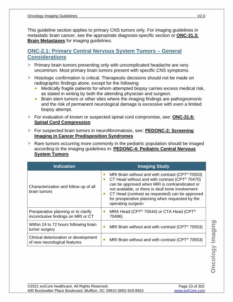
This guideline section applies to primary CNS tumors only. For imaging guidelines in metastatic brain cancer, see the appropriate diagnosis-specific section or ONC-31.3: Brain Metastases for imaging guidelines.
ONC-2.1: Primary Central Nervous System Tumors – General Considerations Primary brain tumors presenting only with uncomplicated headache are very
uncommon. Most primary brain tumors present with specific CNS symptoms. Histologic confirmation is critical. Therapeutic decisions should not be made on
radiographic findings alone, except for the following: Medically fragile patients for whom attempted biopsy carries excess medical risk,
as stated in writing by both the attending physician and surgeon. Brain stem tumors or other sites where the imaging findings are pathognomonic
and the risk of permanent neurological damage is excessive with even a limited biopsy attempt.
For evaluation of known or suspected spinal cord compromise, see: ONC-31.6: Spinal Cord Compression
For suspected brain tumors in neurofibromatosis, see: PEDONC-2: Screening Imaging in Cancer Predisposition Syndromes
Rare tumors occurring more commonly in the pediatric population should be imaged according to the imaging guidelines in: PEDONC-4: Pediatric Central Nervous System Tumors
Indication Imaging Study
Characterization and follow up of all brain tumors
MRI Brain without and with contrast (CPT® 70553) CT Head without and with contrast (CPT® 70470)
can be approved when MRI is contraindicated or not available, or there is skull bone involvement
CT Head (contrast as requested) can be approved for preoperative planning when requested by the operating surgeon
Preoperative planning or to clarify inconclusive findings on MRI or CT
MRA Head (CPT® 70544) or CTA Head (CPT® 70496)
Within 24 to 72 hours following brain tumor surgery MRI Brain without and with contrast (CPT® 70553)
Clinical deterioration or development of new neurological features MRI Brain without and with contrast (CPT® 70553)
Oncology Imaging Guidelines V2.0
______________________________________________________________________________________________________ ©2021 eviCore healthcare. All Rights Reserved. 400 Buckwalter Place Boulevard, Bluffton, SC 29910 (800) 918-8924 www.eviCore.com
Page 23 of 302

Onc
olog
y Im
agin
g
MR Spectroscopy in Brain Tumors (MRS, CPT® 76390) NOTE: Some payors have specific restrictions on MR Spectroscopy, and those coverage policies may supersede the recommendations for MRS in these guidelines
MRS is only supported for use in brain tumors of specified histologies where diagnostic accuracy has been established in peer-reviewed literature See diagnosis-specific guidelines for MRS indications
MRS is considered investigational/experimental for all other histologies and indications not listed in a diagnosis-specific guideline section.
PET Brain Imaging (CPT® 78608 and CPT® 78609) NOTE: Some payors have specific restrictions on PET Brain Metabolic Imaging, and those coverage policies may supersede the recommendations for this study in these guidelines.
PET Brain Metabolic Imaging (CPT® 78608) is only supported for use in brain tumors of specified histologies where diagnostic accuracy has been established in peer-reviewed literature See diagnosis-specific guidelines for PET indications below. According to Medicare NCD 220.6.17, FDG-PET may be approved once for initial
treatment strategy and three times for subsequent treatment strategy for brain tumors.
PET Brain metabolic imaging (CPT® 78608) is considered investigational/experimental for all other histologies and indications not listed in a diagnosis-specific guideline section.
PET Brain perfusion imaging (CPT® 78609) is not indicated in the evaluation or management of primary CNS tumors, and is nationally non-covered by Medicare per NCD 220.6.17.
Body PET studies (CPT® 78811, CPT® 78812, and CPT® 78813) and fusion PET/CT studies (CPT® 78814, CPT® 78815, or CPT® 78816) are not indicated in the evaluation or management of primary CNS tumors
See: HD-24: Other Imaging Studies for details on other advanced neuro-imaging studies
Oncology Imaging Guidelines V2.0
______________________________________________________________________________________________________ ©2021 eviCore healthcare. All Rights Reserved. 400 Buckwalter Place Boulevard, Bluffton, SC 29910 (800) 918-8924 www.eviCore.com
Page 24 of 302
Onc
olog
y Im
agin
g
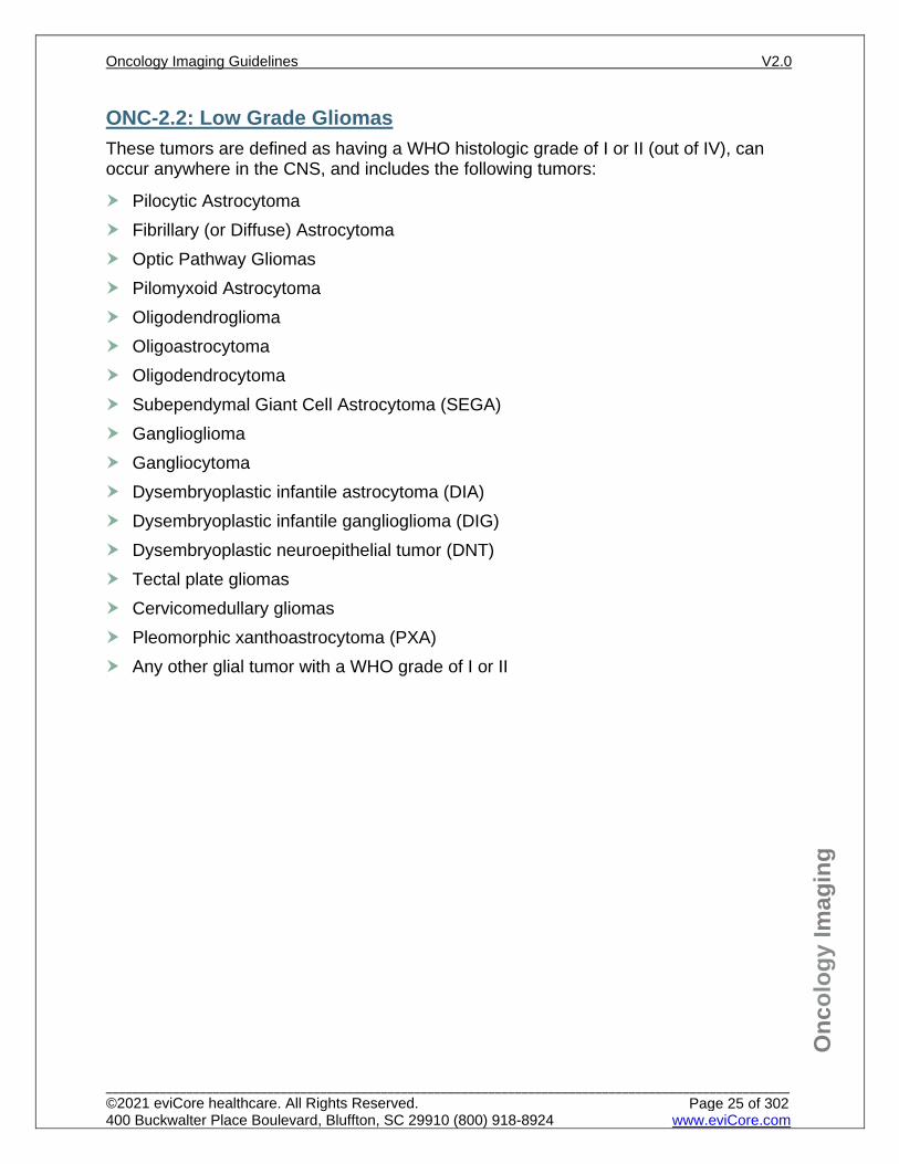
ONC-2.2: Low Grade Gliomas These tumors are defined as having a WHO histologic grade of I or II (out of IV), can occur anywhere in the CNS, and includes the following tumors:
Pilocytic Astrocytoma Fibrillary (or Diffuse) Astrocytoma Optic Pathway Gliomas Pilomyxoid Astrocytoma Oligodendroglioma Oligoastrocytoma Oligodendrocytoma Subependymal Giant Cell Astrocytoma (SEGA) Ganglioglioma Gangliocytoma Dysembryoplastic infantile astrocytoma (DIA) Dysembryoplastic infantile ganglioglioma (DIG) Dysembryoplastic neuroepithelial tumor (DNT) Tectal plate gliomas Cervicomedullary gliomas Pleomorphic xanthoastrocytoma (PXA) Any other glial tumor with a WHO grade of I or II
Oncology Imaging Guidelines V2.0
______________________________________________________________________________________________________ ©2021 eviCore healthcare. All Rights Reserved. 400 Buckwalter Place Boulevard, Bluffton, SC 29910 (800) 918-8924 www.eviCore.com
Page 25 of 302
Onc
olog
y Im
agin
g
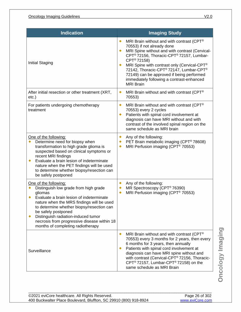
Indication Imaging Study
Initial Staging
MRI Brain without and with contrast (CPT®
70553) if not already done MRI Spine without and with contrast (Cervical-
CPT® 72156, Thoracic-CPT® 72157, Lumbar-CPT® 72158)
MRI Spine with contrast only (Cervical-CPT®
72142, Thoracic-CPT® 72147, Lumbar-CPT®
72149) can be approved if being performed immediately following a contrast-enhanced MRI Brain
After initial resection or other treatment (XRT, etc.)
MRI Brain without and with contrast (CPT®
70553)
For patients undergoing chemotherapy treatment
MRI Brain without and with contrast (CPT®
70553) every 2 cycles Patients with spinal cord involvement at
diagnosis can have MRI without and with contrast of the involved spinal region on the same schedule as MRI brain
One of the following: Determine need for biopsy when
transformation to high grade glioma is suspected based on clinical symptoms or recent MRI findings
Evaluate a brain lesion of indeterminate nature when the PET findings will be used to determine whether biopsy/resection can be safely postponed
Any of the following: PET Brain metabolic imaging (CPT® 78608) MRI Perfusion imaging (CPT® 70553)
One of the following: Distinguish low grade from high grade
gliomas Evaluate a brain lesion of indeterminate
nature when the MRS findings will be used to determine whether biopsy/resection can be safely postponed
Distinguish radiation-induced tumor necrosis from progressive disease within 18 months of completing radiotherapy
Any of the following: MR Spectroscopy (CPT® 76390) MRI Perfusion imaging (CPT® 70553)
Surveillance
MRI Brain without and with contrast (CPT®
70553) every 3 months for 2 years, then every 6 months for 3 years, then annually
Patients with spinal cord involvement at diagnosis can have MRI spine without and with contrast (Cervical-CPT® 72156, Thoracic-CPT® 72157, Lumbar-CPT® 72158) on the same schedule as MRI Brain
Oncology Imaging Guidelines V2.0
______________________________________________________________________________________________________ ©2021 eviCore healthcare. All Rights Reserved. 400 Buckwalter Place Boulevard, Bluffton, SC 29910 (800) 918-8924 www.eviCore.com
Page 26 of 302
Onc
olog
y Im
agin
g
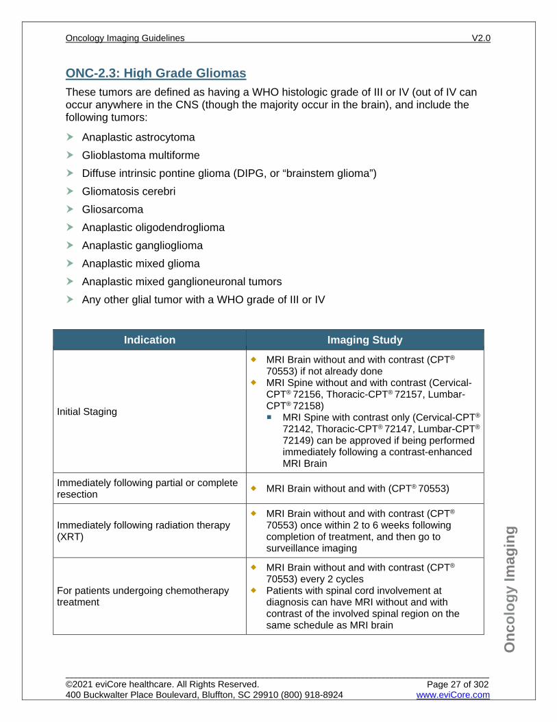
ONC-2.3: High Grade Gliomas These tumors are defined as having a WHO histologic grade of III or IV (out of IV can occur anywhere in the CNS (though the majority occur in the brain), and include the following tumors:
Anaplastic astrocytoma Glioblastoma multiforme Diffuse intrinsic pontine glioma (DIPG, or “brainstem glioma”) Gliomatosis cerebri Gliosarcoma Anaplastic oligodendroglioma Anaplastic ganglioglioma Anaplastic mixed glioma Anaplastic mixed ganglioneuronal tumors Any other glial tumor with a WHO grade of III or IV
Indication Imaging Study
Initial Staging
MRI Brain without and with contrast (CPT®
70553) if not already done MRI Spine without and with contrast (Cervical-
CPT® 72156, Thoracic-CPT® 72157, Lumbar-CPT® 72158) MRI Spine with contrast only (Cervical-CPT®
72142, Thoracic-CPT® 72147, Lumbar-CPT®
72149) can be approved if being performed immediately following a contrast-enhanced MRI Brain
Immediately following partial or complete resection MRI Brain without and with (CPT® 70553)
Immediately following radiation therapy (XRT)
MRI Brain without and with contrast (CPT®
70553) once within 2 to 6 weeks following completion of treatment, and then go to surveillance imaging
For patients undergoing chemotherapy treatment
MRI Brain without and with contrast (CPT®
70553) every 2 cycles Patients with spinal cord involvement at
diagnosis can have MRI without and with contrast of the involved spinal region on the same schedule as MRI brain
Oncology Imaging Guidelines V2.0
______________________________________________________________________________________________________ ©2021 eviCore healthcare. All Rights Reserved. 400 Buckwalter Place Boulevard, Bluffton, SC 29910 (800) 918-8924 www.eviCore.com
Page 27 of 302
Onc
olog
y Im
agin
g
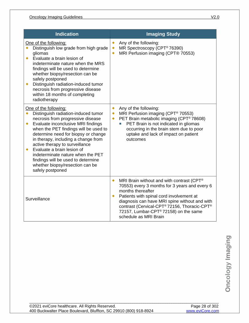
Indication Imaging Study One of the following: Distinguish low grade from high grade
gliomas Evaluate a brain lesion of
indeterminate nature when the MRS findings will be used to determine whether biopsy/resection can be safely postponed
Distinguish radiation-induced tumor necrosis from progressive disease within 18 months of completing radiotherapy
Any of the following: MR Spectroscopy (CPT® 76390) MRI Perfusion imaging (CPT® 70553)
One of the following: Distinguish radiation-induced tumor
necrosis from progressive disease Evaluate inconclusive MRI findings
when the PET findings will be used to determine need for biopsy or change in therapy, including a change from active therapy to surveillance
Evaluate a brain lesion of indeterminate nature when the PET findings will be used to determine whether biopsy/resection can be safely postponed
Any of the following: MRI Perfusion imaging (CPT® 70553) PET Brain metabolic imaging (CPT® 78608)
PET Brain is not indicated in gliomas occurring in the brain stem due to poor uptake and lack of impact on patient outcomes
Surveillance
MRI Brain without and with contrast (CPT®
70553) every 3 months for 3 years and every 6 months thereafter
Patients with spinal cord involvement at diagnosis can have MRI spine without and with contrast (Cervical-CPT® 72156, Thoracic-CPT®
72157, Lumbar-CPT® 72158) on the same schedule as MRI Brain
Oncology Imaging Guidelines V2.0
______________________________________________________________________________________________________ ©2021 eviCore healthcare. All Rights Reserved. 400 Buckwalter Place Boulevard, Bluffton, SC 29910 (800) 918-8924 www.eviCore.com
Page 28 of 302
Onc
olog
y Im
agin
g
ONC-2.4: Medulloblastoma and Supratentorial Primitive Neuroectodermal Tumors (sPNET) Medulloblastoma and sPNET imaging indications in adult patients are identical to those for pediatric patients. See: PEDONC-4.4: Medulloblastoma (MDB), Supratentorial Primitive Neuroectodermal Tumors (sPNET), and Pineoblastoma for imaging guidelines.
Oncology Imaging Guidelines V2.0
______________________________________________________________________________________________________ ©2021 eviCore healthcare. All Rights Reserved. 400 Buckwalter Place Boulevard, Bluffton, SC 29910 (800) 918-8924 www.eviCore.com
Page 29 of 302
Onc
olog
y Im
agin
g
ONC-2.5: Ependymoma Ependymoma imaging indications in adult patients are identical to those for pediatric patients. See: PEDONC-4.8: Ependymoma for imaging guidelines.
Oncology Imaging Guidelines V2.0
______________________________________________________________________________________________________ ©2021 eviCore healthcare. All Rights Reserved. 400 Buckwalter Place Boulevard, Bluffton, SC 29910 (800) 918-8924 www.eviCore.com
Page 30 of 302
Onc
olog
y Im
agin
g
ONC-2.6: Central Nervous System Germ Cell Tumors Central nervous system germ cell tumor imaging indications in adult patients are identical to those for pediatric patients. See: PEDONC-4.7: CNS Germinomas and Non-Germinomatous Germ Cell Tumors (NGGCT) for imaging guidelines.
Oncology Imaging Guidelines V2.0
______________________________________________________________________________________________________ ©2021 eviCore healthcare. All Rights Reserved. 400 Buckwalter Place Boulevard, Bluffton, SC 29910 (800) 918-8924 www.eviCore.com
Page 31 of 302
Onc
olog
y Im
agin
g
ONC-2.7: CNS Lymphoma (also known as Microglioma) Indication Imaging Study
Initial Staging
All of the following are indicated: MRI Brain without and with contrast (CPT® 70553) MRI Cervical spine without and with contrast (CPT®
72156) MRI Thoracic spine without and with contrast (CPT®
72157) MRI Lumbar spine without and with contrast (CPT®
72158)
Extra-neural evaluation to confirm CNS primary
*Patients with CNS Lymphoma that is metastatic should be imaged according to: ONC-27: Non-Hodgkin
Lymphomas for patients age ≥ 18 years
PEDONC-5.3: Pediatric Aggressive Mature B-Cell Non-Hodgkin Lymphomas (NHL) for patients age ≤ 17 years
Any or all of the following are indicated: CT Chest with contrast (CPT® 71260) CT Abdomen/Pelvis with contrast (CPT® 74177) PET/CT (CPT® 78815) can be approved for
evaluation of inconclusive findings on CT imaging
Treatment Response MRI without and with contrast of all positive disease sites every 2 cycles
Surveillance MRI without and with contrast of all positive disease
sites every 3 months for 2 years, then every 6 months for 3 years, then annually thereafter
Oncology Imaging Guidelines V2.0
______________________________________________________________________________________________________ ©2021 eviCore healthcare. All Rights Reserved. 400 Buckwalter Place Boulevard, Bluffton, SC 29910 (800) 918-8924 www.eviCore.com
Page 32 of 302
Onc
olog
y Im
agin
g
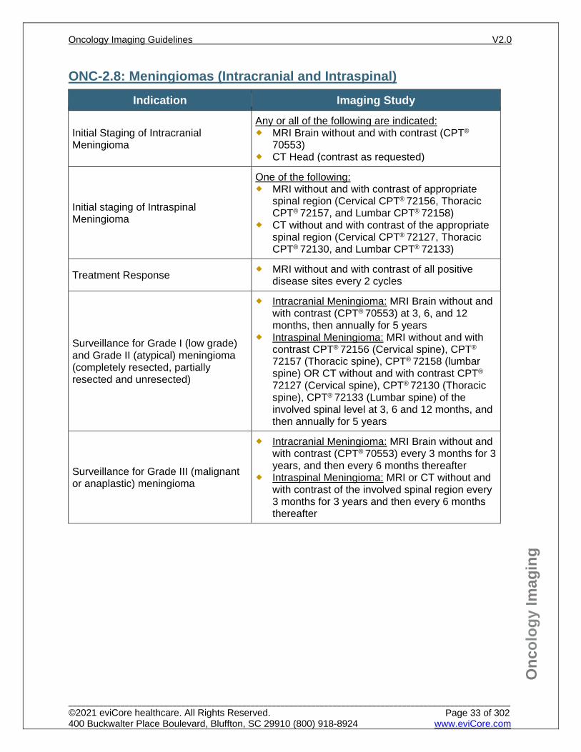
ONC-2.8: Meningiomas (Intracranial and Intraspinal) Indication Imaging Study
Initial Staging of Intracranial Meningioma
Any or all of the following are indicated: MRI Brain without and with contrast (CPT®
70553) CT Head (contrast as requested)
Initial staging of Intraspinal Meningioma
One of the following: MRI without and with contrast of appropriate
spinal region (Cervical CPT® 72156, Thoracic CPT® 72157, and Lumbar CPT® 72158)
CT without and with contrast of the appropriate spinal region (Cervical CPT® 72127, Thoracic CPT® 72130, and Lumbar CPT® 72133)
Treatment Response MRI without and with contrast of all positive disease sites every 2 cycles
Surveillance for Grade I (low grade) and Grade II (atypical) meningioma (completely resected, partially resected and unresected)
Intracranial Meningioma: MRI Brain without and with contrast (CPT® 70553) at 3, 6, and 12 months, then annually for 5 years
Intraspinal Meningioma: MRI without and with contrast CPT® 72156 (Cervical spine), CPT®
72157 (Thoracic spine), CPT® 72158 (lumbar spine) OR CT without and with contrast CPT®
72127 (Cervical spine), CPT® 72130 (Thoracic spine), CPT® 72133 (Lumbar spine) of the involved spinal level at 3, 6 and 12 months, and then annually for 5 years
Surveillance for Grade III (malignant or anaplastic) meningioma
Intracranial Meningioma: MRI Brain without and with contrast (CPT® 70553) every 3 months for 3 years, and then every 6 months thereafter
Intraspinal Meningioma: MRI or CT without and with contrast of the involved spinal region every 3 months for 3 years and then every 6 months thereafter
Oncology Imaging Guidelines V2.0
______________________________________________________________________________________________________ ©2021 eviCore healthcare. All Rights Reserved. 400 Buckwalter Place Boulevard, Bluffton, SC 29910 (800) 918-8924 www.eviCore.com
Page 33 of 302
Onc
olog
y Im
agin
g
ONC-2.9: Spinal Cord Tumors (Benign and Malignant) See also: ONC-2.2: Low Grade Gliomas and ONC-2.3: High Grade Gliomas for
imaging guidelines of low grade and high grade gliomas of the spinal cord See also: PEDONC-4.9: Malignant Tumors of the Spinal Cord for imaging
guidelines for other malignant spinal cord tumors See also: PEDPN-2.1: Neurofibromatosis 1 and PEDPN-2.2: Neurofibromatosis
2 for spinal tumors in patients with Neurofibromatosis 1 or 2 See also: ONC-31.6: Spinal Cord Compression for known secondary malignancy
involving the spine/spinal canal/spinal cord
Oncology Imaging Guidelines V2.0
______________________________________________________________________________________________________ ©2021 eviCore healthcare. All Rights Reserved. 400 Buckwalter Place Boulevard, Bluffton, SC 29910 (800) 918-8924 www.eviCore.com
Page 34 of 302
Onc
olog
y Im
agin
g
ONC-2.10: Choroid Plexus Tumors Choroid Plexus Tumor imaging indications in adult patients are identical to those for pediatric patients. See: PEDONC-4.13: Choroid Plexus Tumors for imaging guidelines.
Oncology Imaging Guidelines V2.0
______________________________________________________________________________________________________ ©2021 eviCore healthcare. All Rights Reserved. 400 Buckwalter Place Boulevard, Bluffton, SC 29910 (800) 918-8924 www.eviCore.com
Page 35 of 302
Onc
olog
y Im
agin
g
References 1. Nabors LB, Portnow J, Ahluwalia M, et. al. National Comprehensive Cancer Network (NCCN)
Guidelines Version 3.2020 – September 11, 2020 Central Nervous System Cancers, available at: https://www.nccn.org/professionals/physician_gls/pdf/cns.pdf. Referenced with permission from the NCCN Clinical Practice Guidelines in Oncology (NCCN Guidelines™) for Central Nervous System Tumors Cancer V3.2020. – September 11, 2020 ©2020 National Comprehensive Cancer Network, Inc. All rights reserved. The NCCN Guidelines™ and illustrations herein may not be reproduced in any form for any purpose without the express written permission of the NCCN. To view the most recent and complete version of the NCCN Guidelines™, go online to NCCN.org.
2. Brandão LA, Castillo M. Adult brain tumors: clinical applications of magnetic resonance spectroscopy. Magn Reson Imaging Clin N Am. 2016;24(4):781-809. doi:10.1016/j.mric.2016.07.005.
3. Pasquier D, Bijmolt S, Veninga T, et al. Atypical and malignant meningioma: outcome and prognostic factors in 119 irradiated patients. A multicenter, retrospective study of the Rare Cancer Network. Int J Radiat Oncol Biol Phys. 2008;71(5):1388. doi:10.1016.j.ijrobp.2007.12.020.
4. Modha A, Gutin PH. Diagnosis and treatment of atypical and anaplastic meningiomas: a review. Neurosurgery. 2005;57(3):538-550.
5. Horská A, Barker PB. Imaging of brain tumors: MR spectroscopy and metabolic imaging. Neuroimaging Clin N Am. 2010;20(3):293-310. doi:10.1016/j.nic.2010.04.003.
6. Sundgren PC. MR Spectroscopy in radiation Injury. Am J Neuroradiol. 2009;30(8):1469-1476. doi:10.3174/ajnr.A1580.
7. American College of Radiology. ACR–ASNR-SPR practice parameter for the performance of intracranial magnetic resonance perfusion imaging. 2017; Available at: https://www.acr.org/-/media/ACR/Files/Practice-Parameters/MR-Perfusion.pdf?la=en.
Oncology Imaging Guidelines V2.0
______________________________________________________________________________________________________ ©2021 eviCore healthcare. All Rights Reserved. 400 Buckwalter Place Boulevard, Bluffton, SC 29910 (800) 918-8924 www.eviCore.com
Page 36 of 302
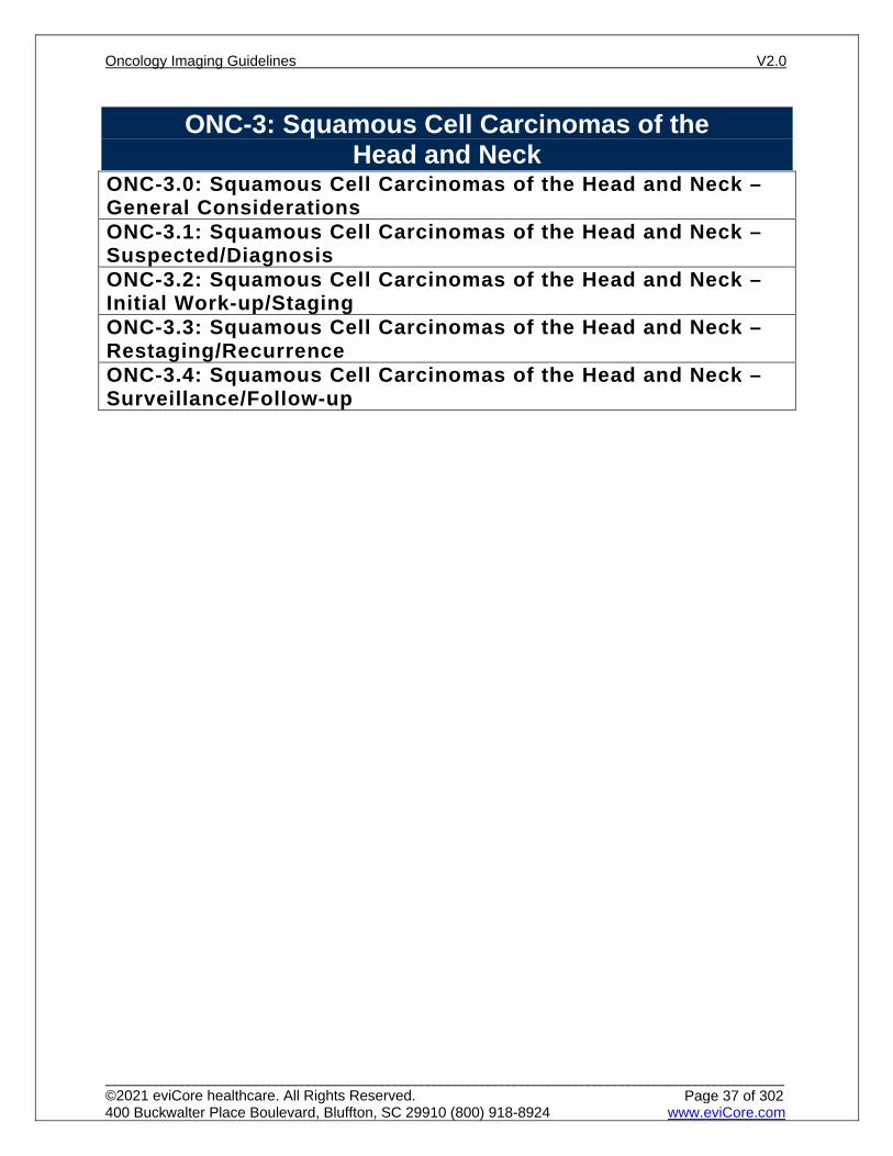
ONC-3: Squamous Cell Carcinomas of the Head and Neck
ONC-3.0: Squamous Cell Carcinomas of the Head and Neck – General ConsiderationsONC-3.1: Squamous Cell Carcinomas of the Head and Neck – Suspected/DiagnosisONC-3.2: Squamous Cell Carcinomas of the Head and Neck – Initial Work-up/StagingONC-3.3: Squamous Cell Carcinomas of the Head and Neck – Restaging/RecurrenceONC-3.4: Squamous Cell Carcinomas of the Head and Neck – Surveillance/Follow-up
Oncology Imaging Guidelines V2.0
______________________________________________________________________________________________________ ©2021 eviCore healthcare. All Rights Reserved. 400 Buckwalter Place Boulevard, Bluffton, SC 29910 (800) 918-8924 www.eviCore.com
Page 37 of 302
Onc
olog
y Im
agin
g
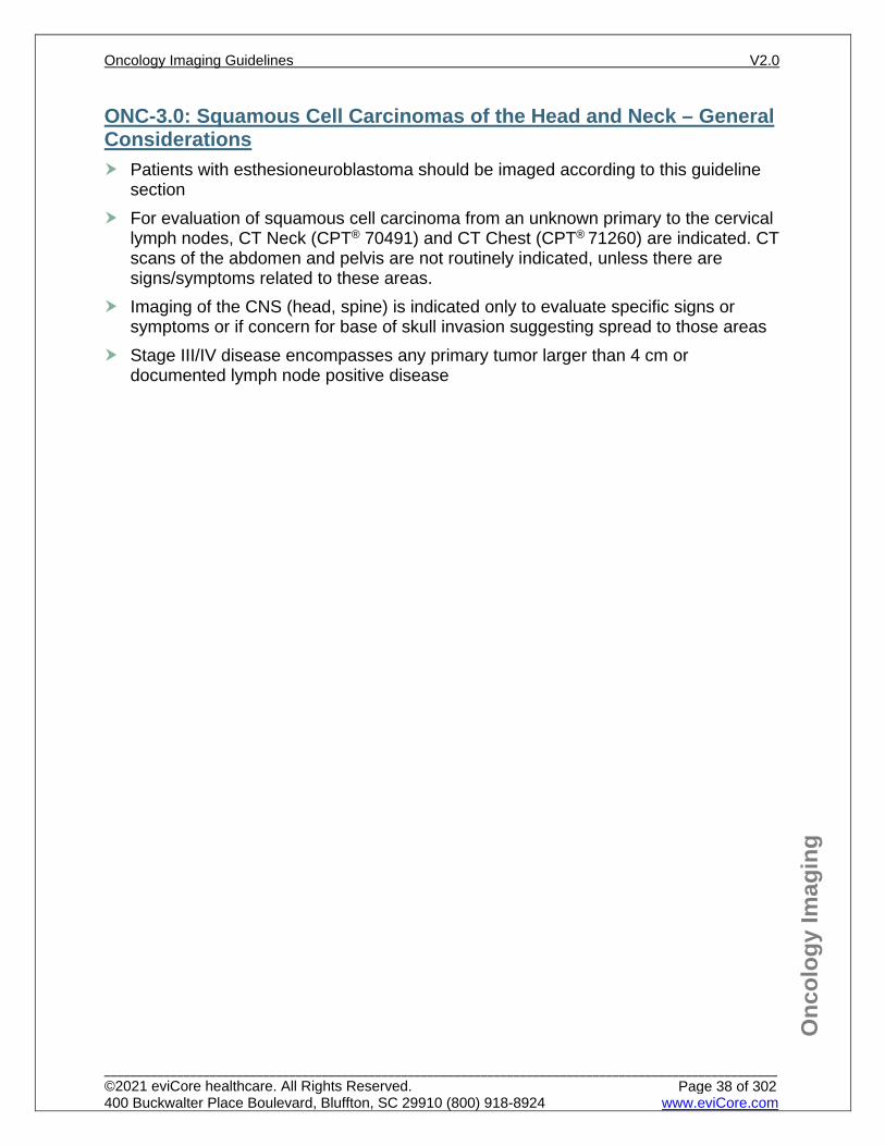
ONC-3.0: Squamous Cell Carcinomas of the Head and Neck – General Considerations Patients with esthesioneuroblastoma should be imaged according to this guideline
section For evaluation of squamous cell carcinoma from an unknown primary to the cervical
lymph nodes, CT Neck (CPT® 70491) and CT Chest (CPT® 71260) are indicated. CT scans of the abdomen and pelvis are not routinely indicated, unless there are signs/symptoms related to these areas.
Imaging of the CNS (head, spine) is indicated only to evaluate specific signs or symptoms or if concern for base of skull invasion suggesting spread to those areas
Stage III/IV disease encompasses any primary tumor larger than 4 cm or documented lymph node positive disease
Oncology Imaging Guidelines V2.0
______________________________________________________________________________________________________ ©2021 eviCore healthcare. All Rights Reserved. 400 Buckwalter Place Boulevard, Bluffton, SC 29910 (800) 918-8924 www.eviCore.com
Page 38 of 302
Onc
olog
y Im
agin
g
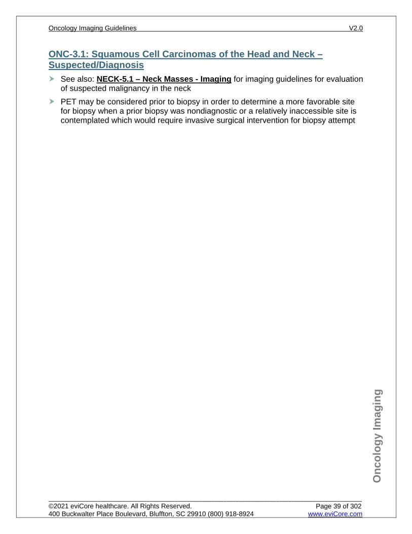
ONC-3.1: Squamous Cell Carcinomas of the Head and Neck – Suspected/Diagnosis See also: NECK-5.1 – Neck Masses - Imaging for imaging guidelines for evaluation
of suspected malignancy in the neck PET may be considered prior to biopsy in order to determine a more favorable site
for biopsy when a prior biopsy was nondiagnostic or a relatively inaccessible site is contemplated which would require invasive surgical intervention for biopsy attempt
Oncology Imaging Guidelines V2.0
______________________________________________________________________________________________________ ©2021 eviCore healthcare. All Rights Reserved. 400 Buckwalter Place Boulevard, Bluffton, SC 29910 (800) 918-8924 www.eviCore.com
Page 39 of 302
Onc
olog
y Im
agin
g
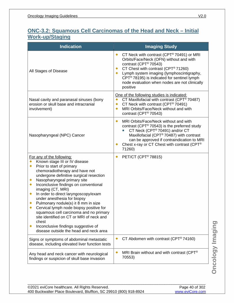
ONC-3.2: Squamous Cell Carcinomas of the Head and Neck – Initial Work-up/Staging
Indication Imaging Study
All Stages of Disease
CT Neck with contrast (CPT® 70491) or MRI Orbits/Face/Neck (OFN) without and with contrast (CPT® 70543)
CT Chest with contrast (CPT® 71260) Lymph system imaging (lymphoscintigraphy,
CPT® 78195) is indicated for sentinel lymph node evaluation when nodes are not clinically positive
Nasal cavity and paranasal sinuses (bony erosion or skull base and intracranial involvement)
One of the following studies is indicated: CT Maxillofacial with contrast (CPT® 70487) CT Neck with contrast (CPT® 70491) MRI Orbits/Face/Neck without and with
contrast (CPT® 70543)
Nasopharyngeal (NPC) Cancer
MRI Orbits/Face/Neck without and with contrast (CPT® 70543) is the preferred study CT Neck (CPT® 70491) and/or CT
Maxillofacial (CPT® 70487) with contrast can be approved if contraindication to MRI
Chest x-ray or CT Chest with contrast (CPT®
71260)
For any of the following: Known stage III or IV disease Prior to start of primary
chemoradiotherapy and have not undergone definitive surgical resection
Nasopharyngeal primary site Inconclusive findings on conventional
imaging (CT, MRI) In order to direct laryngoscopy/exam
under anesthesia for biopsy Pulmonary nodule(s) ≥ 8 mm in size Cervical lymph node biopsy positive for
squamous cell carcinoma and no primary site identified on CT or MRI of neck and chest
Inconclusive findings suggestive of disease outside the head and neck area
PET/CT (CPT® 78815)
Signs or symptoms of abdominal metastatic disease, including elevated liver function tests
CT Abdomen with contrast (CPT® 74160)
Any head and neck cancer with neurological findings or suspicion of skull base invasion
MRI Brain without and with contrast (CPT®
70553)
Oncology Imaging Guidelines V2.0
______________________________________________________________________________________________________ ©2021 eviCore healthcare. All Rights Reserved. 400 Buckwalter Place Boulevard, Bluffton, SC 29910 (800) 918-8924 www.eviCore.com
Page 40 of 302
Onc
olog
y Im
agin
g
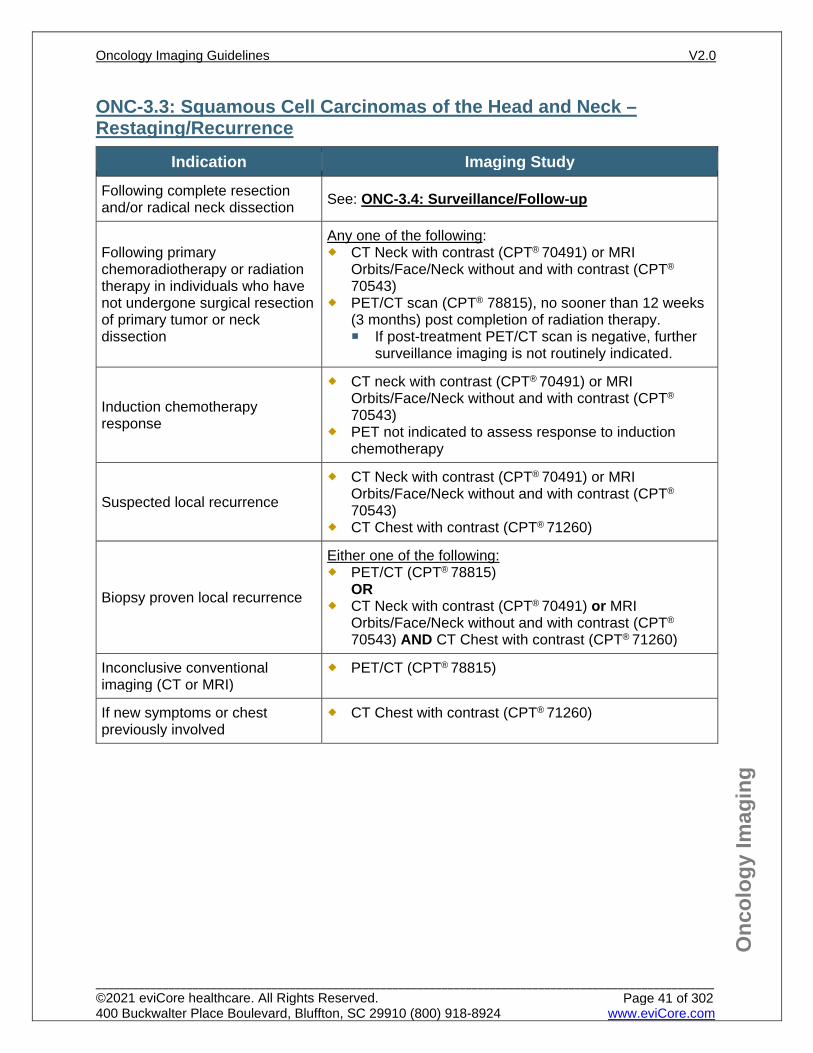
ONC-3.3: Squamous Cell Carcinomas of the Head and Neck – Restaging/Recurrence
Indication Imaging Study Following complete resection and/or radical neck dissection See: ONC-3.4: Surveillance/Follow-up
Following primary chemoradiotherapy or radiation therapy in individuals who have not undergone surgical resection of primary tumor or neck dissection
Any one of the following: CT Neck with contrast (CPT® 70491) or MRI
Orbits/Face/Neck without and with contrast (CPT®
70543) PET/CT scan (CPT® 78815), no sooner than 12 weeks
(3 months) post completion of radiation therapy. If post-treatment PET/CT scan is negative, further
surveillance imaging is not routinely indicated.
Induction chemotherapy response
CT neck with contrast (CPT® 70491) or MRI Orbits/Face/Neck without and with contrast (CPT®
70543) PET not indicated to assess response to induction
chemotherapy
Suspected local recurrence
CT Neck with contrast (CPT® 70491) or MRI Orbits/Face/Neck without and with contrast (CPT®
70543) CT Chest with contrast (CPT® 71260)
Biopsy proven local recurrence
Either one of the following: PET/CT (CPT® 78815)
OR CT Neck with contrast (CPT® 70491) or MRI
Orbits/Face/Neck without and with contrast (CPT®
70543) AND CT Chest with contrast (CPT® 71260)
Inconclusive conventional imaging (CT or MRI)
PET/CT (CPT® 78815)
If new symptoms or chest previously involved
CT Chest with contrast (CPT® 71260)
Oncology Imaging Guidelines V2.0
______________________________________________________________________________________________________ ©2021 eviCore healthcare. All Rights Reserved. 400 Buckwalter Place Boulevard, Bluffton, SC 29910 (800) 918-8924 www.eviCore.com
Page 41 of 302
Onc
olog
y Im
agin
g
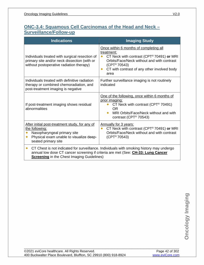
ONC-3.4: Squamous Cell Carcinomas of the Head and Neck – Surveillance/Follow-up
Indications Imaging Study
Individuals treated with surgical resection of primary site and/or neck dissection (with or without postoperative radiation therapy)
Once within 6 months of completing all treatment: CT Neck with contrast (CPT® 70491) or MRI
Orbits/Face/Neck without and with contrast (CPT® 70543)
CT with contrast of any other involved body area
Individuals treated with definitive radiation therapy or combined chemoradiation, and post-treatment imaging is negative
Further surveillance imaging is not routinely indicated
If post-treatment imaging shows residual abnormalities
One of the following, once within 6 months of prior imaging:
CT Neck with contrast (CPT® 70491) OR
MRI Orbits/Face/Neck without and with contrast (CPT® 70543)
After initial post-treatment study, for any of the following: Nasopharyngeal primary site Physical exam unable to visualize deep-
seated primary site
Annually for 3 years: CT Neck with contrast (CPT® 70491) or MRI
Orbits/Face/Neck without and with contrast (CPT® 70543)
CT Chest is not indicated for surveillance. Individuals with smoking history may undergo annual low dose CT cancer screening if criteria are met (See: CH-33: Lung Cancer Screening in the Chest Imaging Guidelines)
Oncology Imaging Guidelines V2.0
______________________________________________________________________________________________________ ©2021 eviCore healthcare. All Rights Reserved. 400 Buckwalter Place Boulevard, Bluffton, SC 29910 (800) 918-8924 www.eviCore.com
Page 42 of 302
Onc
olog
y Im
agin
g
References 1. Pfister DG, Spencer S, Adelstein D et al. National Comprehensive Cancer Network (NCCN)
Guidelines Version 1.2021 – November 9, 2020 Head and Neck Cancers, available at: https://www.nccn.org/professionals/physician_gls/pdf/head-and-neck.pdf. Referenced with permission from the NCCN Clinical Practice Guidelines in Oncology (NCCN Guidelines™) for Head and Neck Cancer V1.2021 – November 9, 2020. ©2020 National Comprehensive Cancer Network, Inc. All rights reserved. The NCCN Guidelines™ and illustrations herein may not be reproduced in any form for any purpose without the express written permission of the NCCN. To view the most recent and complete version of the NCCN Guidelines™, go online to NCCN.org.
2. Goel R, Moore W, Sumer B, Khan S, Sher D, Subramaniam RM. Clinical practice in PET/CT for the management of head and neck squamous cell cancer. AJR Am J Roentgenol. 2017;209(2):289-303. doi:10.2214/AJR.17.18301.
Oncology Imaging Guidelines V2.0
______________________________________________________________________________________________________ ©2021 eviCore healthcare. All Rights Reserved. 400 Buckwalter Place Boulevard, Bluffton, SC 29910 (800) 918-8924 www.eviCore.com
Page 43 of 302
ONC-4: Salivary Gland Cancers ONC-4.0: Salivary Gland Cancers – General ConsiderationsONC-4.1: Salivary Gland Cancers – Suspected/DiagnosisONC-4.2: Salivary Gland Cancers – Initial Work-up/StagingONC-4.3: Salivary Gland Cancers – Restaging/RecurrenceONC-4.4: Salivary Gland Cancers – Surveillance/Follow-up
Oncology Imaging Guidelines V2.0
______________________________________________________________________________________________________ ©2021 eviCore healthcare. All Rights Reserved. 400 Buckwalter Place Boulevard, Bluffton, SC 29910 (800) 918-8924 www.eviCore.com
Page 44 of 302
Onc
olog
y Im
agin
g
ONC-4.0: Salivary Gland Cancers – General Considerations Salivary gland tumors may originate within the parotid, submandibular, sublingual or
minor salivary glands in the mouth. Histological subtypes include mucoepidermoid, acinic, adenocarcinoma, adenoid
cystic carcinoma, malignant myoepithelial tumors and squamous cell carcinoma. Lymphoma and metastatic squamous carcinoma can also occur in the parotid gland.
Over 80% of parotid gland tumors are benign. A bilateral parotid tumor is most likely Warthin’s tumor.
The role of PET in salivary gland tumors has yet to be established.
Oncology Imaging Guidelines V2.0
______________________________________________________________________________________________________ ©2021 eviCore healthcare. All Rights Reserved. 400 Buckwalter Place Boulevard, Bluffton, SC 29910 (800) 918-8924 www.eviCore.com
Page 45 of 302
Onc
olog
y Im
agin
g
ONC-4.1: Salivary Gland Cancers – Suspected/Diagnosis See: NECK-11 and NECK-5.1 for evaluation of salivary gland masses, salivary gland stones and neck masses.
Oncology Imaging Guidelines V2.0
______________________________________________________________________________________________________ ©2021 eviCore healthcare. All Rights Reserved. 400 Buckwalter Place Boulevard, Bluffton, SC 29910 (800) 918-8924 www.eviCore.com
Page 46 of 302
Onc
olog
y Im
agin
g
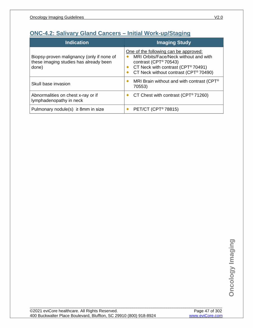
ONC-4.2: Salivary Gland Cancers – Initial Work-up/Staging Indication Imaging Study
Biopsy-proven malignancy (only if none of these imaging studies has already been done)
One of the following can be approved: MRI Orbits/Face/Neck without and with
contrast (CPT® 70543) CT Neck with contrast (CPT® 70491) CT Neck without contrast (CPT® 70490)
Skull base invasion MRI Brain without and with contrast (CPT®
70553)
Abnormalities on chest x-ray or if lymphadenopathy in neck
CT Chest with contrast (CPT® 71260)
Pulmonary nodule(s) ≥ 8mm in size PET/CT (CPT® 78815)
Oncology Imaging Guidelines V2.0
______________________________________________________________________________________________________ ©2021 eviCore healthcare. All Rights Reserved. 400 Buckwalter Place Boulevard, Bluffton, SC 29910 (800) 918-8924 www.eviCore.com
Page 47 of 302
Onc
olog
y Im
agin
g
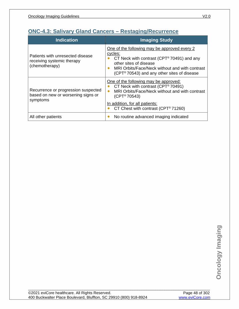
ONC-4.3: Salivary Gland Cancers – Restaging/Recurrence Indication Imaging Study
Patients with unresected disease receiving systemic therapy (chemotherapy)
One of the following may be approved every 2 cycles: CT Neck with contrast (CPT® 70491) and any
other sites of disease MRI Orbits/Face/Neck without and with contrast
(CPT® 70543) and any other sites of disease
Recurrence or progression suspected based on new or worsening signs or symptoms
One of the following may be approved: CT Neck with contrast (CPT® 70491) MRI Orbits/Face/Neck without and with contrast
(CPT® 70543) In addition, for all patients: CT Chest with contrast (CPT® 71260)
All other patients No routine advanced imaging indicated
Oncology Imaging Guidelines V2.0
______________________________________________________________________________________________________ ©2021 eviCore healthcare. All Rights Reserved. 400 Buckwalter Place Boulevard, Bluffton, SC 29910 (800) 918-8924 www.eviCore.com
Page 48 of 302
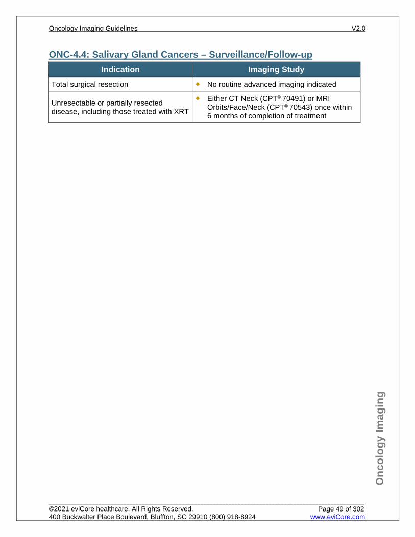
Onc
olog
y Im
agin
g
ONC-4.4: Salivary Gland Cancers – Surveillance/Follow-up Indication Imaging Study
Total surgical resection No routine advanced imaging indicated
Unresectable or partially resected disease, including those treated with XRT
Either CT Neck (CPT® 70491) or MRI Orbits/Face/Neck (CPT® 70543) once within 6 months of completion of treatment
Oncology Imaging Guidelines V2.0
______________________________________________________________________________________________________ ©2021 eviCore healthcare. All Rights Reserved. 400 Buckwalter Place Boulevard, Bluffton, SC 29910 (800) 918-8924 www.eviCore.com
Page 49 of 302
Onc
olog
y Im
agin
g
References 1. Pfister DG, Spencer S, Adelstein D et al. National Comprehensive Cancer Network (NCCN)
Guidelines Version 1.2021 – November 9, 2020 Head and Neck Cancers, available at: https://www.nccn.org/professionals/physician_gls/pdf/head-and-neck.pdf. Referenced with permission from the NCCN Clinical Practice Guidelines in Oncology (NCCN Guidelines™) for Head and Neck Cancer V1.2021 – November 9, 2020. ©2020 National Comprehensive Cancer Network, Inc. All rights reserved. The NCCN Guidelines™ and illustrations herein may not be reproduced in any form for any purpose without the express written permission of the NCCN. To view the most recent and complete version of the NCCN Guidelines™, go online to NCCN.org.
2. Palacios E, Ellis M, Lam EC, Neitzschman H, Haile M. Pitfalls in imaging the submandibular glands with PET/CT. Ear Nose Throat J. 2015;94(10-11):E37-E39.
3. Seo YL, Yoon DY, Baek S, et al. Incidental focal FDG uptake in the parotid glands on PET/CT in patients with head and neck malignancy. Eur Radiol. 2015;25(1):171-177. doi:10.1007/s00330-0140339701.
4. Park HL, Yoo le R, Lee N, et al. The value of F-18 FDG PET for planning treatment and detecting recurrence in malignant salivary gland tumors: comparison with conventional imaging studies. Nucl Med Mol Imaging. 2013;47(4):242-248. doi:10.1007/s13139-013-0222-8.
5. Bertagna F, Nicolai P, Maroldi R. Diagnostic role of 18F-FDG-PET or PET/CT in salivary gland tumors: a systematic review. Rev Esp Med Nucl Imagen Mol. 2015;34(5):295-302.
Oncology Imaging Guidelines V2.0
______________________________________________________________________________________________________ ©2021 eviCore healthcare. All Rights Reserved. 400 Buckwalter Place Boulevard, Bluffton, SC 29910 (800) 918-8924 www.eviCore.com
Page 50 of 302
ONC-5: Melanomas and Other Skin Cancers ONC-5.0: Melanoma – General ConsiderationsONC-5.1: Melanoma – Suspected/DiagnosisONC-5.2: Melanoma – Initial Work-up/StagingONC-5.3: Melanoma – Restaging/RecurrenceONC-5.4: Melanoma – Surveillance/Follow-upONC-5.5: Non-Melanoma Skin Cancers – General ConsiderationsONC-5.6: Non-Melanoma Skin Cancers – Initial Work-up/StagingONC-5.7: Non-Melanoma Skin Cancers – Restaging/RecurrenceONC-5.8: Non-Melanoma Skin Cancers – Surveillance/Follow-upONC-5.9: Ocular Melanoma
Oncology Imaging Guidelines V2.0
______________________________________________________________________________________________________ ©2021 eviCore healthcare. All Rights Reserved. 400 Buckwalter Place Boulevard, Bluffton, SC 29910 (800) 918-8924 www.eviCore.com
Page 51 of 302
Onc
olog
y Im
agin
g
ONC-5.0: Melanoma – General Considerations Melanomas can metastasize in an unpredictable fashion.
Oncology Imaging Guidelines V2.0
______________________________________________________________________________________________________ ©2021 eviCore healthcare. All Rights Reserved. 400 Buckwalter Place Boulevard, Bluffton, SC 29910 (800) 918-8924 www.eviCore.com
Page 52 of 302
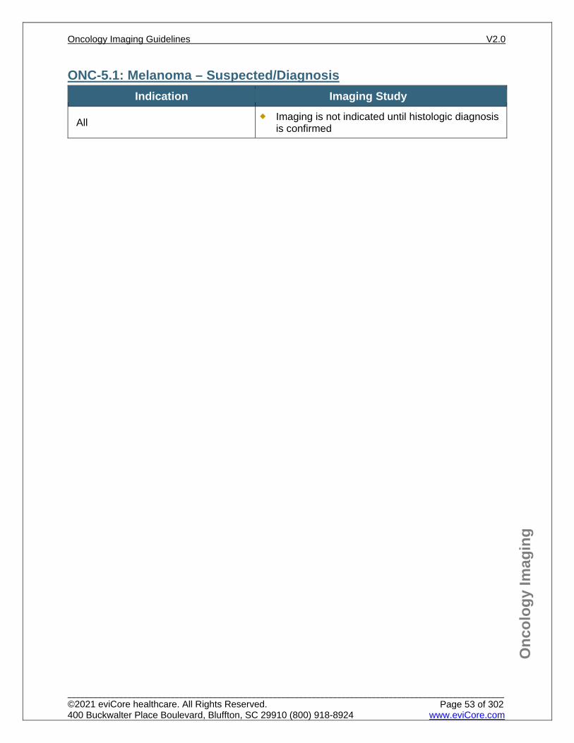
Onc
olog
y Im
agin
g
ONC-5.1: Melanoma – Suspected/Diagnosis Indication Imaging Study
All Imaging is not indicated until histologic diagnosis is confirmed
Oncology Imaging Guidelines V2.0
______________________________________________________________________________________________________ ©2021 eviCore healthcare. All Rights Reserved. 400 Buckwalter Place Boulevard, Bluffton, SC 29910 (800) 918-8924 www.eviCore.com
Page 53 of 302
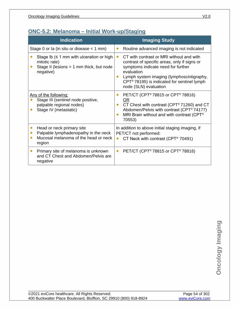
Onc
olog
y Im
agin
g
ONC-5.2: Melanoma – Initial Work-up/Staging Indication Imaging Study
Stage 0 or Ia (in situ or disease < 1 mm) Routine advanced imaging is not indicated
Stage Ib (≤ 1 mm with ulceration or high mitotic rate)
Stage II (lesions > 1 mm thick, but node negative)
CT with contrast or MRI without and with contrast of specific areas, only if signs or symptoms indicate need for further evaluation
Lymph system imaging (lymphoscintigraphy, CPT® 78195) is indicated for sentinel lymph node (SLN) evaluation
Any of the following: Stage III (sentinel node positive,
palpable regional nodes) Stage IV (metastatic)
PET/CT (CPT® 78815 or CPT® 78816) OR
CT Chest with contrast (CPT® 71260) and CT Abdomen/Pelvis with contrast (CPT® 74177)
MRI Brain without and with contrast (CPT®
70553)
Head or neck primary site Palpable lymphadenopathy in the neck Mucosal melanoma of the head or neck
region
In addition to above initial staging imaging, if PET/CT not performed: CT Neck with contrast (CPT® 70491)
Primary site of melanoma is unknown and CT Chest and Abdomen/Pelvis are negative
PET/CT (CPT® 78815 or CPT® 78816)
Oncology Imaging Guidelines V2.0
______________________________________________________________________________________________________ ©2021 eviCore healthcare. All Rights Reserved. 400 Buckwalter Place Boulevard, Bluffton, SC 29910 (800) 918-8924 www.eviCore.com
Page 54 of 302
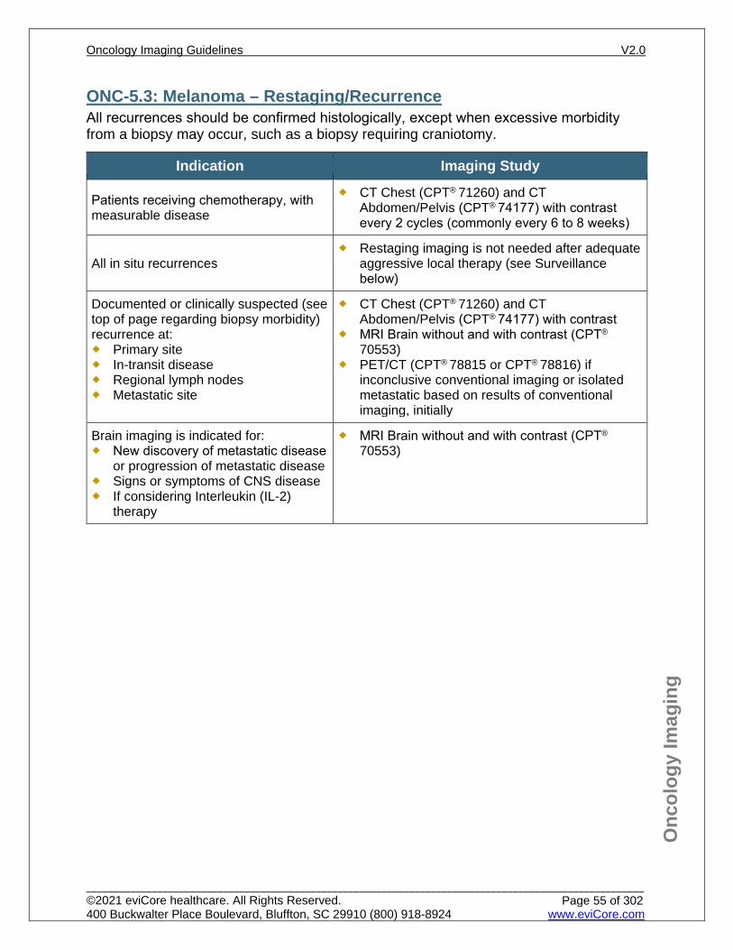
Onc
olog
y Im
agin
g
ONC-5.3: Melanoma – Restaging/Recurrence All recurrences should be confirmed histologically, except when excessive morbidity from a biopsy may occur, such as a biopsy requiring craniotomy.
Indication Imaging Study
Patients receiving chemotherapy, with measurable disease
CT Chest (CPT® 71260) and CT Abdomen/Pelvis (CPT® 74177) with contrast every 2 cycles (commonly every 6 to 8 weeks)
All in situ recurrences Restaging imaging is not needed after adequate
aggressive local therapy (see Surveillance below)
Documented or clinically suspected (see top of page regarding biopsy morbidity) recurrence at: Primary site In-transit disease Regional lymph nodes Metastatic site
CT Chest (CPT® 71260) and CT Abdomen/Pelvis (CPT® 74177) with contrast
MRI Brain without and with contrast (CPT®
70553) PET/CT (CPT® 78815 or CPT® 78816) if
inconclusive conventional imaging or isolated metastatic based on results of conventional imaging, initially
Brain imaging is indicated for: New discovery of metastatic disease
or progression of metastatic disease Signs or symptoms of CNS disease If considering Interleukin (IL-2)
therapy
MRI Brain without and with contrast (CPT®
70553)
Oncology Imaging Guidelines V2.0
______________________________________________________________________________________________________ ©2021 eviCore healthcare. All Rights Reserved. 400 Buckwalter Place Boulevard, Bluffton, SC 29910 (800) 918-8924 www.eviCore.com
Page 55 of 302
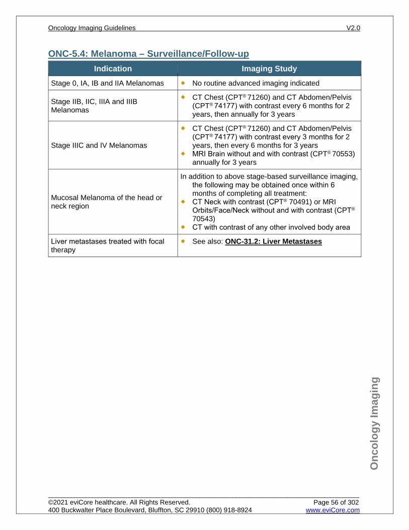
Onc
olog
y Im
agin
g
ONC-5.4: Melanoma – Surveillance/Follow-up Indication Imaging Study
Stage 0, IA, IB and IIA Melanomas No routine advanced imaging indicated
Stage IIB, IIC, IIIA and IIIB Melanomas
CT Chest (CPT® 71260) and CT Abdomen/Pelvis (CPT® 74177) with contrast every 6 months for 2 years, then annually for 3 years
Stage IIIC and IV Melanomas
CT Chest (CPT® 71260) and CT Abdomen/Pelvis (CPT® 74177) with contrast every 3 months for 2 years, then every 6 months for 3 years
MRI Brain without and with contrast (CPT® 70553) annually for 3 years
Mucosal Melanoma of the head or neck region
In addition to above stage-based surveillance imaging, the following may be obtained once within 6 months of completing all treatment:
CT Neck with contrast (CPT® 70491) or MRI Orbits/Face/Neck without and with contrast (CPT® 70543)
CT with contrast of any other involved body area
Liver metastases treated with focal therapy
See also: ONC-31.2: Liver Metastases
Oncology Imaging Guidelines V2.0
______________________________________________________________________________________________________ ©2021 eviCore healthcare. All Rights Reserved. 400 Buckwalter Place Boulevard, Bluffton, SC 29910 (800) 918-8924 www.eviCore.com
Page 56 of 302
Onc
olog
y Im
agin
g
ONC-5.5: Non-Melanoma Skin Cancers – General Considerations Advanced Imaging is generally not indicated for basal cell and squamous cell skin
cancers PET/CT scan is not indicated for evaluation of non-melanoma skin cancers unless
specified within the guidelines below (e.g. Merkel cell carcinoma) Merkel cell carcinoma is an unusual skin cancer with neuroendocrine-like histologic
features, which has a high propensity (25% to 33%) for regional lymph node spread and occasionally, metastatic spread to lungs.
Merkel cell carcinoma may present as a primary cancer or as a skin metastasis from a noncutaneous primary neuroendocrine carcinoma (i.e., small cell lung cancer), therefore conventional imaging is indicated initially to confirm the absence of metastasis prior to considering PET scan.
Oncology Imaging Guidelines V2.0
______________________________________________________________________________________________________ ©2021 eviCore healthcare. All Rights Reserved. 400 Buckwalter Place Boulevard, Bluffton, SC 29910 (800) 918-8924 www.eviCore.com
Page 57 of 302
Onc
olog
y Im
agin
g
ONC-5.6: Non-Melanoma Skin Cancers – Initial Work-up/Staging Indication Imaging Study
Body area with unexplained signs or symptoms
CT with contrast of that body area
Perineural invasion or local regional extension (i.e. bone; deep soft tissue) involvement
One of the following may be approved of the primary site: MRI without contrast or without and with
contrast CT (contrast as requested)
Skin lesion may be a dermal metastasis from distant primary
CT Chest (CPT® 71260) and Abdomen/Pelvis (CPT® 74177) with contrast
PET/CT (CPT® 78815 or 78816) is indicated if conventional imaging (CT or MRI) is unable to identify a primary site
Squamous cell carcinoma head or neck skin with regional lymphadenopathy
CT Neck (CPT® 70491) and CT Chest (CPT®
71260) with contrast
Merkel Cell carcinoma
CT Chest with contrast (CPT® 71260) and CT Abdomen/Pelvis with contrast (CPT® 74177)
CT with contrast of other involved body area(s) PET/CT (CPT® 78815 or 78816) if inconclusive
conventional imaging Lymph system imaging (lymphoscintigraphy,
CPT® 78195) for sentinel lymph node evaluation MRI Brain with and without contrast (CPT®
70553)
Oncology Imaging Guidelines V2.0
______________________________________________________________________________________________________ ©2021 eviCore healthcare. All Rights Reserved. 400 Buckwalter Place Boulevard, Bluffton, SC 29910 (800) 918-8924 www.eviCore.com
Page 58 of 302
Onc
olog
y Im
agin
g
ONC-5.7: Non-Melanoma Skin Cancers – Restaging/Recurrence All recurrences should be confirmed histologically, except when excessive morbidity from a biopsy may occur, such as a biopsy requiring craniotomy.
Indication Imaging Study
Recurrence where planned therapy is more extensive than simple wide local excision
CT with contrast of the primary and recurrent site(s)
Recurrence of Merkel cell carcinoma
CT Chest (CPT® 71260) and CT Abdomen/Pelvis (CPT®
74177) with contrast MRI Brain without and with contrast (CPT® 70553) PET/CT (CPT® 78815 or 78816) if no metastatic disease on
any of the previous imaging studies
Oncology Imaging Guidelines V2.0
______________________________________________________________________________________________________ ©2021 eviCore healthcare. All Rights Reserved. 400 Buckwalter Place Boulevard, Bluffton, SC 29910 (800) 918-8924 www.eviCore.com
Page 59 of 302
Onc
olog
y Im
agin
g
ONC-5.8: Non-Melanoma Skin Cancers – Surveillance/Follow-up Indication Imaging Study
Merkel cell cancer – only if node positive
CT Chest (CPT® 71260) and CT Abdomen/Pelvis (CPT®
74177) with contrast every 6 months for 5 years Add CT Neck with contrast (CPT® 70491) if known prior neck
disease or scalp/facial/neck disease
All others Routine advanced imaging for surveillance is not indicated Imaging indicated only for signs and symptoms of recurrent
disease
Oncology Imaging Guidelines V2.0
______________________________________________________________________________________________________ ©2021 eviCore healthcare. All Rights Reserved. 400 Buckwalter Place Boulevard, Bluffton, SC 29910 (800) 918-8924 www.eviCore.com
Page 60 of 302
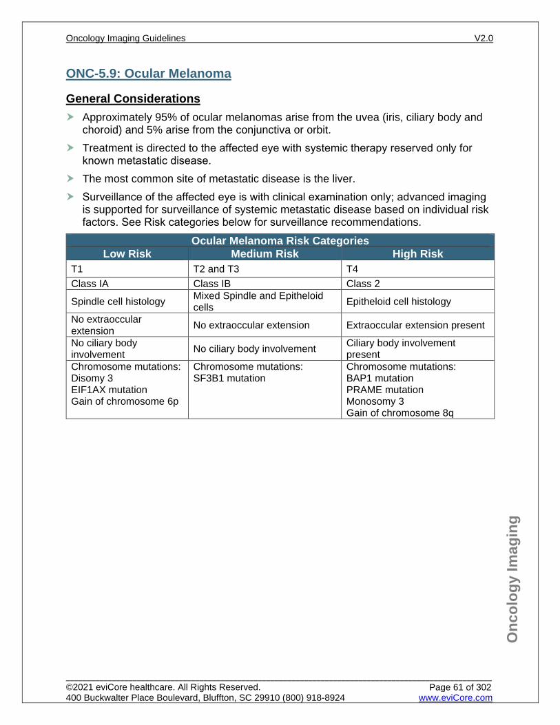
Onc
olog
y Im
agin
g
ONC-5.9: Ocular Melanoma
General Considerations Approximately 95% of ocular melanomas arise from the uvea (iris, ciliary body and
choroid) and 5% arise from the conjunctiva or orbit. Treatment is directed to the affected eye with systemic therapy reserved only for
known metastatic disease. The most common site of metastatic disease is the liver. Surveillance of the affected eye is with clinical examination only; advanced imaging
is supported for surveillance of systemic metastatic disease based on individual risk factors. See Risk categories below for surveillance recommendations.
Ocular Melanoma Risk Categories Low Risk Medium Risk High Risk
T1 T2 and T3 T4 Class IA Class IB Class 2
Spindle cell histology Mixed Spindle and Epitheloid cells Epitheloid cell histology
No extraoccular extension No extraoccular extension Extraoccular extension present
No ciliary body involvement No ciliary body involvement Ciliary body involvement
present Chromosome mutations: Disomy 3 EIF1AX mutation Gain of chromosome 6p
Chromosome mutations: SF3B1 mutation
Chromosome mutations: BAP1 mutation PRAME mutation Monosomy 3 Gain of chromosome 8q
Oncology Imaging Guidelines V2.0
______________________________________________________________________________________________________ ©2021 eviCore healthcare. All Rights Reserved. 400 Buckwalter Place Boulevard, Bluffton, SC 29910 (800) 918-8924 www.eviCore.com
Page 61 of 302
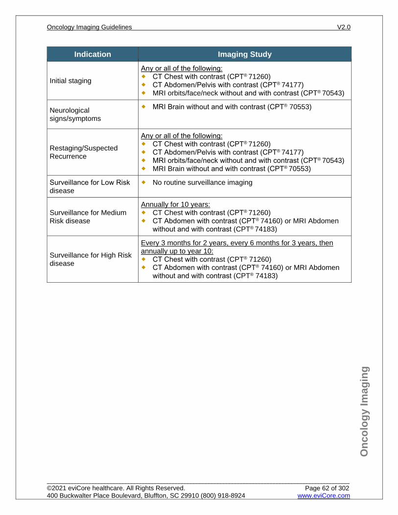
Onc
olog
y Im
agin
g
Indication Imaging Study
Initial staging
Any or all of the following: CT Chest with contrast (CPT® 71260) CT Abdomen/Pelvis with contrast (CPT® 74177) MRI orbits/face/neck without and with contrast (CPT® 70543)
Neurological signs/symptoms
MRI Brain without and with contrast (CPT® 70553)
Restaging/Suspected Recurrence
Any or all of the following: CT Chest with contrast (CPT® 71260) CT Abdomen/Pelvis with contrast (CPT® 74177) MRI orbits/face/neck without and with contrast (CPT® 70543) MRI Brain without and with contrast (CPT® 70553)
Surveillance for Low Risk disease
No routine surveillance imaging
Surveillance for Medium Risk disease
Annually for 10 years: CT Chest with contrast (CPT® 71260) CT Abdomen with contrast (CPT® 74160) or MRI Abdomen
without and with contrast (CPT® 74183)
Surveillance for High Risk disease
Every 3 months for 2 years, every 6 months for 3 years, then annually up to year 10: CT Chest with contrast (CPT® 71260) CT Abdomen with contrast (CPT® 74160) or MRI Abdomen
without and with contrast (CPT® 74183)
Oncology Imaging Guidelines V2.0
______________________________________________________________________________________________________ ©2021 eviCore healthcare. All Rights Reserved. 400 Buckwalter Place Boulevard, Bluffton, SC 29910 (800) 918-8924 www.eviCore.com
Page 62 of 302
Onc
olog
y Im
agin
g
References 1. Swetter SM, Thompson JA, Coit DGAlbertini MR, et al, National Comprehensive Cancer Network
(NCCN) Guidelines Version 3.20202.2021 – May 18, 2020February 19, 2021 Cutaneous Melanoma, available at: https://www.nccn.org/professionals/physician_gls/pdf/cutaneous_melanoma.pdf Referenced with permission from the NCCN Clinical Practice Guidelines in Oncology (NCCN Guidelines™) for Cutaneous Melanoma VV3.20202.2021 – February 19, 2021May 18, 2020 ©2020 2021 National Comprehensive Cancer Network, Inc. All rights reserved. The NCCN Guidelines™ and illustrations herein may not be reproduced in any form for any purpose without the express written permission of the NCCN. To view the most recent and complete version of the NCCN Guidelines™, go online to NCCN.org.
2. Swetter S, Thompson JA, Albertini MR, et al, National Comprehensive Cancer Network (NCCN) Guidelines Version 1.20203.2020 – May 21, 2020January 15, 2021 Uveal Melanoma, available at: https://www.nccn.org/professionals/physician_gls/pdf/uveal.pdf Referenced with permission from the NCCN Clinical Practice Guidelines in Oncology (NCCN Guidelines™) for Uveal Melanoma V1.2020V3.2020 – May 21, 2020January 15, 2021 ©2020 2021 National Comprehensive Cancer Network, Inc. All rights reserved. The NCCN Guidelines™ and illustrations herein may not be reproduced in any form for any purpose without the express written permission of the NCCN. To view the most recent and complete version of the NCCN Guidelines™, go online to NCCN.org .NCCN.org.
3. Schmults CD, Blitzblau R, Aasi SZ, et al. National Comprehensive Cancer Network (NCCN) Guidelines Version 1.20201.2021 – October 2, 2019 February 18, 2021 Merkel Cell Carcinoma, available at: https://www.nccn.org/professionals/physician_gls/pdf/mcc.pdf Referenced with permission from the NCCN Clinical Practice Guidelines in Oncology (NCCN Guidelines™) for Merkel Cell Carcinoma V1.20201.2021 – February 18, 2021October 2, 2019 ©2019 2021 National Comprehensive Cancer Network, Inc. All rights reserved. The NCCN Guidelines™ and illustrations herein may not be reproduced in any form for any purpose without the express written permission of the NCCN. To view the most recent and complete version of the NCCN Guidelines™, go online to NCCN.org .
4. Schmults CD, Blitzblau R, Aasi SZ, et al. National Comprehensive Cancer Network (NCCN) Guidelines Version 1.20202.2021 – October 24, 2019February 25, 2021 Basal Cell Skin Cancer, available at: https://www.nccn.org/professionals/physician_gls/pdf/nmsc.pdf Referenced with permission from the NCCN Clinical Practice Guidelines in Oncology (NCCN Guidelines™) for Basal Cell Skin Cancer V1.20202.2021 – October 24, 2019February 25, 2021 ©2019 2021 National Comprehensive Cancer Network, Inc. All rights reserved. The NCCN Guidelines™ and illustrations herein may not be reproduced in any form for any purpose without the express written permission of the NCCN. To view the most recent and complete version of the NCCN Guidelines™, go online to NCCN.org .
5. Schmults CD, Blitzblau R, Aasi SZ, et al, National Comprehensive Cancer Network (NCCN) Guidelines Version 2.20201.2021 – July 14, 2020February 5, 2021 Squamous Cell Skin Cancer, available at: https://www.nccn.org/professionals/physician_gls/pdf/squamous.pdf Referenced with permission from the NCCN Clinical Practice Guidelines in Oncology (NCCN Guidelines™) for Squamous Cell Skin Cancer V2.2020V1.2021 – July 14, 2020February 5, 2021 ©2020 2021 National Comprehensive Cancer Network, Inc. All rights reserved. The NCCN Guidelines™ and illustrations herein may not be reproduced in any form for any purpose without the express written permission of the NCCN. To view the most recent and complete version of the NCCN Guidelines™, go online to NCCN.org .
6. Schröer-Günther MA, Wolff RF, Westwood ME, et al. F-18-fluoro-2-deoxyglucose positron emission tomography (PET) and PET/computed tomography imaging in primary staging of patients with malignant melanoma: a systematic review. Syst Rev. 2012;1:62. doi:10.1186/2046-4053-1-62.
7. Xing Y, Bronstein Y, Ross MI, et al. Contemporary diagnostic imaging modalities for the staging and surveillance of melanoma patients: a meta-analysis. J Natl Cancer Inst. 2011;103(2):129-142. doi:10.1093/jnci/djq455.
8. Rodriguez Rivera AM, Alabbas H, Ramjuan A, Meguerditchian AN. Value of positron emission tomography scan in stage III cutaneous melanoma: a systematic review and meta-analysis. Surg Oncol. 2014;23(1):11-16. doi: 10.1016/j.suronc.2014.01.002.
9. Nathan P, Cohen V, Coupland S, et al. Uveal melanoma UK national guidelines. European Journal of Cancer. 2015;51(16):2404-2412. doi:10.1016/j.ejca/2015.07.013.
Oncology Imaging Guidelines V2.0
______________________________________________________________________________________________________ ©2021 eviCore healthcare. All Rights Reserved. 400 Buckwalter Place Boulevard, Bluffton, SC 29910 (800) 918-8924 www.eviCore.com
Page 63 of 302
ONC-6: Thyroid Cancer ONC-6.0: Thyroid Cancer – General ConsiderationsONC-6.1: Thyroid Cancer – Suspected/DiagnosisONC-6.2: Thyroid Cancer – Initial Work-up/StagingONC-6.3: Thyroid Cancer – Restaging/RecurrenceONC-6.4: Thyroid Cancer – Surveillance/Follow-up
Oncology Imaging Guidelines V2.0
______________________________________________________________________________________________________ ©2021 eviCore healthcare. All Rights Reserved. 400 Buckwalter Place Boulevard, Bluffton, SC 29910 (800) 918-8924 www.eviCore.com
Page 64 of 302
Onc
olog
y Im
agin
g
ONC-6.0: Thyroid Cancer – General Considerations PET for initial staging for anaplastic thyroid cancer is currently not recommended
before conventional imaging since recommendations for PET are derived from observational studies and clinical trials with other methodological limitations.
Patients with measurable metastatic disease that are RAI refractory may be followed with conventional imaging, PET-CT scan is reserved for inconclusive findings.
Whole body thyroid nuclear scan is coded with CPT® 78018. If CPT® 78018 is obtained and found to be positive, CPT® 78020 may be approved as an add-on test to evaluate the degree of iodine uptake.
Single photon emission computed tomography (SPECT) imaging – Radiopharmaceutical Localization of Tumor SPECT (CPT® 78803 or 78831) or SPECT/CT Hybrid study (CPT® 78830, or 78832) may complement planar and pinhole imaging and can be approved as an add-on wherever radioiodine scans are indicated.
Oncology Imaging Guidelines V2.0
______________________________________________________________________________________________________ ©2021 eviCore healthcare. All Rights Reserved. 400 Buckwalter Place Boulevard, Bluffton, SC 29910 (800) 918-8924 www.eviCore.com
Page 65 of 302
Onc
olog
y Im
agin
g
ONC-6.1: Thyroid Cancer – Suspected/Diagnosis See: NECK-8.1: Thyroid Nodule for imaging guidelines for suspected thyroid malignancies
Oncology Imaging Guidelines V2.0
______________________________________________________________________________________________________ ©2021 eviCore healthcare. All Rights Reserved. 400 Buckwalter Place Boulevard, Bluffton, SC 29910 (800) 918-8924 www.eviCore.com
Page 66 of 302
Onc
olog
y Im
agin
g
ONC-6.2: Thyroid Cancer – Initial Work-up/Staging Follicular, Papillary and Hürthle Cell Carcinomas Imaging Study
One of the following: Fixation suggested by clinical exam
and/or ultrasound Substernal or bulky disease Disease precluding full ultrasound
examination
One of the following: MRI Neck without contrast (CPT® 70540) MRI Neck without and with contrast (CPT®
70543) CT Neck without contrast (CPT® 70490) CT Neck with contrast (CPT® 70491) can be
approved if contrast study is necessary for complete pre-operative assessment and use of IV contrast will not delay post-operative use of RAI therapy.
Post-thyroidectomy to assess thyroid remnant and to look for iodine-avid metastases for one of the following: Extent of thyroid remnant cannot be
accurately ascertained from the surgical report or neck ultrasound
When the results may alter the decision to treat
Prior to administration of RAI therapy
Whole body thyroid nuclear scan (CPT®
78018) The following may be approved as an add-on
test: CPT® 78020 to evaluate the degree of
iodine uptake SPECT (CPT® 78803 or 78831), or
SPECT/CT Hybrid study (CPT® 78830, or 78832)
Skeletal pain
Bone scan See also: ONC-1.3: Nuclear Medicine (NM) Imaging in Oncology
Whole body nuclear thyroid scan (CPT® 78018)
The following may be approved as an add-on test: CPT® 78020 to evaluate the degree of
iodine uptake SPECT (CPT® 78803 or 78831), or
SPECT/CT Hybrid study (CPT® 78830, or 78832)
Suspicious findings on CXR, US, or substernal extension of mass
CT Chest without contrast (CPT® 71250)
All other patients Routine preoperative advanced imaging is not indicated
Oncology Imaging Guidelines V2.0
______________________________________________________________________________________________________ ©2021 eviCore healthcare. All Rights Reserved. 400 Buckwalter Place Boulevard, Bluffton, SC 29910 (800) 918-8924 www.eviCore.com
Page 67 of 302
Onc
olog
y Im
agin
g
Medullary Thyroid Carcinomas Imaging Study
All patients with positive lymph nodes or calcitonin level > 500 pg/mL
Any or all of the following: CT Neck with contrast (CPT® 70491) CT Chest with contrast (CPT® 71260) CT Abdomen either with (CPT® 74160) or CT
Abdomen without and with contrast (CPT®
74170) Bone scan see also: ONC-1.3: Nuclear
Medicine (NM) Imaging in Oncology
Skeletal pain Bone scan see also: ONC-1.3: Nuclear Medicine (NM) Imaging in Oncology
Inconclusive finding on conventional imaging PET/CT (CPT® 78815)
Anaplastic Thyroid Carcinomas Imaging Study
All
Any or all of the following: CT Neck with contrast (CPT® 70491) CT Chest with contrast (CPT® 71260) CT Abdomen/Pelvis with contrast (CPT®
74177) MRI Brain without and with contrast (CPT®
70553) Bone scan see also: ONC-1.3: Nuclear
Medicine (NM) Imaging in Oncology
Skeletal pain Bone scan see also: ONC-1.3: Nuclear Medicine (NM) Imaging in Oncology
Inconclusive finding on conventional imaging PET/CT (CPT® 78815 or CPT® 78816)
Oncology Imaging Guidelines V2.0
______________________________________________________________________________________________________ ©2021 eviCore healthcare. All Rights Reserved. 400 Buckwalter Place Boulevard, Bluffton, SC 29910 (800) 918-8924 www.eviCore.com
Page 68 of 302
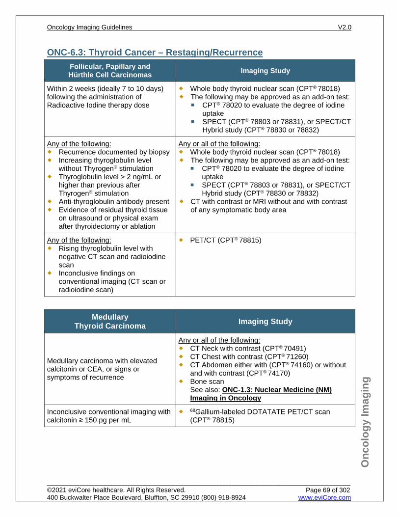
Onc
olog
y Im
agin
g
ONC-6.3: Thyroid Cancer – Restaging/Recurrence Follicular, Papillary and Hürthle Cell Carcinomas Imaging Study
Within 2 weeks (ideally 7 to 10 days) following the administration of Radioactive Iodine therapy dose
Whole body thyroid nuclear scan (CPT® 78018) The following may be approved as an add-on test:
CPT® 78020 to evaluate the degree of iodine uptake
SPECT (CPT® 78803 or 78831), or SPECT/CT Hybrid study (CPT® 78830 or 78832)
Any of the following: Recurrence documented by biopsy Increasing thyroglobulin level
without Thyrogen® stimulation Thyroglobulin level > 2 ng/mL or
higher than previous after Thyrogen® stimulation
Anti-thyroglobulin antibody present Evidence of residual thyroid tissue
on ultrasound or physical exam after thyroidectomy or ablation
Any or all of the following: Whole body thyroid nuclear scan (CPT® 78018) The following may be approved as an add-on test:
CPT® 78020 to evaluate the degree of iodine uptake
SPECT (CPT® 78803 or 78831), or SPECT/CT Hybrid study (CPT® 78830 or 78832)
CT with contrast or MRI without and with contrast of any symptomatic body area
Any of the following: Rising thyroglobulin level with
negative CT scan and radioiodine scan
Inconclusive findings on conventional imaging (CT scan or radioiodine scan)
PET/CT (CPT® 78815)
Medullary Thyroid Carcinoma Imaging Study
Medullary carcinoma with elevated calcitonin or CEA, or signs or symptoms of recurrence
Any or all of the following: CT Neck with contrast (CPT® 70491) CT Chest with contrast (CPT® 71260) CT Abdomen either with (CPT® 74160) or without
and with contrast (CPT® 74170) Bone scan
See also: ONC-1.3: Nuclear Medicine (NM) Imaging in Oncology
Inconclusive conventional imaging with calcitonin ≥ 150 pg per mL
68Gallium-labeled DOTATATE PET/CT scan (CPT® 78815)
Oncology Imaging Guidelines V2.0
______________________________________________________________________________________________________ ©2021 eviCore healthcare. All Rights Reserved. 400 Buckwalter Place Boulevard, Bluffton, SC 29910 (800) 918-8924 www.eviCore.com
Page 69 of 302
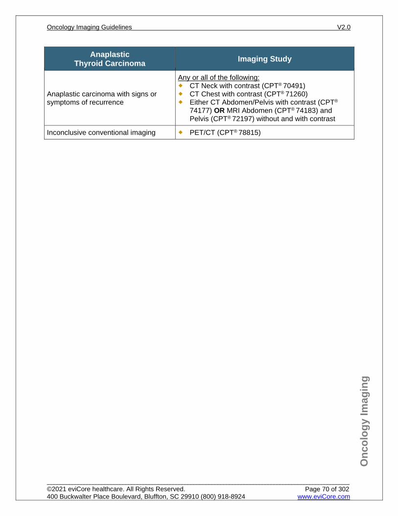
Onc
olog
y Im
agin
g
Anaplastic Thyroid Carcinoma Imaging Study
Anaplastic carcinoma with signs or symptoms of recurrence
Any or all of the following: CT Neck with contrast (CPT® 70491) CT Chest with contrast (CPT® 71260) Either CT Abdomen/Pelvis with contrast (CPT®
74177) OR MRI Abdomen (CPT® 74183) and Pelvis (CPT® 72197) without and with contrast
Inconclusive conventional imaging PET/CT (CPT® 78815)
Oncology Imaging Guidelines V2.0
______________________________________________________________________________________________________ ©2021 eviCore healthcare. All Rights Reserved. 400 Buckwalter Place Boulevard, Bluffton, SC 29910 (800) 918-8924 www.eviCore.com
Page 70 of 302

Onc
olog
y Im
agin
g
ONC-6.4: Thyroid Cancer – Surveillance/Follow-up Follicular, Papillary and Hürthle Cell Carcinomas Imaging/Diagnostic Study
All patients Neck ultrasound (CPT® 76536), chest x-ray, and
laboratory studies every 6 months for the first year, then annually
For patients with any of the following: Node positive disease RAI-avid metastases
Whole body thyroid nuclear scan annually (CPT®
78018) The following may be approved as an add-on test:
CPT® 78020 to evaluate the degree of iodine uptake
SPECT (CPT® 78803 or 78831), or SPECT/CT Hybrid study (CPT® 78830 or 78832)
Medullary Carcinomas Imaging/Diagnostic Study
All patients CEA and calcitonin are required for monitoring
medullary carcinomas Routine surveillance imaging is not indicated
Anaplastic Thyroid Carcinomas Imaging Study
All patients
Every 3 months for 2 years: CT Neck with contrast (CPT® 70491) CT Chest with contrast (CPT® 71260) CT Abdomen/Pelvis with contrast (CPT® 74177)
Oncology Imaging Guidelines V2.0
______________________________________________________________________________________________________ ©2021 eviCore healthcare. All Rights Reserved. 400 Buckwalter Place Boulevard, Bluffton, SC 29910 (800) 918-8924 www.eviCore.com
Page 71 of 302
Onc
olog
y Im
agin
g
References 1. Haddad RH, Bischoff L, Bernet V, et al. National Comprehensive Cancer Network (NCCN) Guidelines
Version 3.2020 – February 2, 2021 Thyroid carcinoma, available at: https://www.nccn.org/professionals/physician_gls/pdf/thyroid.pdf Referenced with permission from the NCCN Clinical Practice Guidelines in Oncology (NCCN Guidelines™) for Thyroid carcinoma V3.2020 – February 2, 2021 ©2021 National Comprehensive Cancer Network, Inc. All rights reserved. The NCCN Guidelines™ and illustrations herein may not be reproduced in any form for any purpose without the express written permission of the NCCN. To view the most recent and complete version of the NCCN Guidelines™, go online to NCCN.org.
2. Slough CM, Randolph GW. Workup of well-differentiated thyroid carcinoma. Cancer Control. 2006;13(2):99-105. doi:10.1177/107327480601300203.
3. Smallridge RC, Ain KB, Asa SL, et al. American Thyroid Association guidelines for management of patients with anaplastic thyroid cancer. Thyroid. 2012;22(11):1104-1139. doi:10.1089/thy.2012.0302.
4. Wells SA Jr, Asa SL, Dralle H, et al. American Thyroid Association guidelines for the management of medullary thyroid carcinoma. Thyroid. 2015;25(6):567-610. doi:10.1089/thy.2014.0335.
5. Yeh MW, Bauer AJ, Bernet VA, et al. American Thyroid Association statement on preoperative imaging for thyroid cancer surgery. Thyroid. 2015;25:3-14. doi:10.1089/thy.2014.0096.
6. Haugen BR, Alexander EK, Bible KB, et al. 2015 American Thyroid Association management guidelines for adult patients with thyroid nodules and differentiated thyroid cancer: the American Thyroid Association guidelines task force on thyroid nodules and differentiated thyroid cancer. Thyroid. 2016;26(1):1-133. doi:10.1089/thy.2015.0020.
7. Silberstein EB, Alavi A, Balon HR, et al. The SNMMI Practice Guideline for therapy of thyroid disease with 131I3.0. J Nucl Med. 2012;53(10):1633-1651. doi:10.2967/jnumed.112.105148.
8. Avram AM, Fig LM, Frey KA, Gross MD, Wong KK. Preablation 131-I scans with SPECT/CT in postoperative thyroid cancer patients: what is the impact on staging? J Clin Endocrinol Metab. February 21, 2013 [Epub ahead of print].
Oncology Imaging Guidelines V2.0
______________________________________________________________________________________________________ ©2021 eviCore healthcare. All Rights Reserved. 400 Buckwalter Place Boulevard, Bluffton, SC 29910 (800) 918-8924 www.eviCore.com
Page 72 of 302
ONC-7: Small Cell Lung Cancer ONC-7.0: Small Cell Lung Cancer – General ConsiderationsONC-7.1: Small Cell Lung Cancer – Suspected/DiagnosisONC-7.2: Small Cell Lung Cancer – Initial Work-up/StagingONC-7.3: Small Cell Lung Cancer – Restaging/RecurrenceONC-7.4: Small Cell Lung Cancer – Surveillance/Follow-up
Oncology Imaging Guidelines V2.0
______________________________________________________________________________________________________ ©2021 eviCore healthcare. All Rights Reserved. 400 Buckwalter Place Boulevard, Bluffton, SC 29910 (800) 918-8924 www.eviCore.com
Page 73 of 302
Onc
olog
y Im
agin
g
ONC-7.0: Small Cell Lung Cancer – General Considerations Combined histologies of Small and Non-Small cell are considered Small cell lung
cancer. Use this guideline for imaging recommendations for small and large cell high-grade (poorly differentiated) neuroendocrine tumors of the lung.
Imaging is presently guided by traditional staging of limited or extensive disease. Extensive stage is either metastatic disease or an extent which cannot be
encompassed by a single radiotherapy portal. Limited staging is confined to one side of the chest.
Patients treated curatively for SCLC are at increased risk for developing a second lung cancer. If new lung nodule is seen on imaging without any evidence of other systemic disease, follow ONC-31.1: Lung Metastases for work-up of nodule.
For carcinoid (low grade neuroendocrine tumors) of the lung, see: ONC-15: Neuroendocrine Cancers and Adrenal Tumors
Oncology Imaging Guidelines V2.0
______________________________________________________________________________________________________ ©2021 eviCore healthcare. All Rights Reserved. 400 Buckwalter Place Boulevard, Bluffton, SC 29910 (800) 918-8924 www.eviCore.com
Page 74 of 302
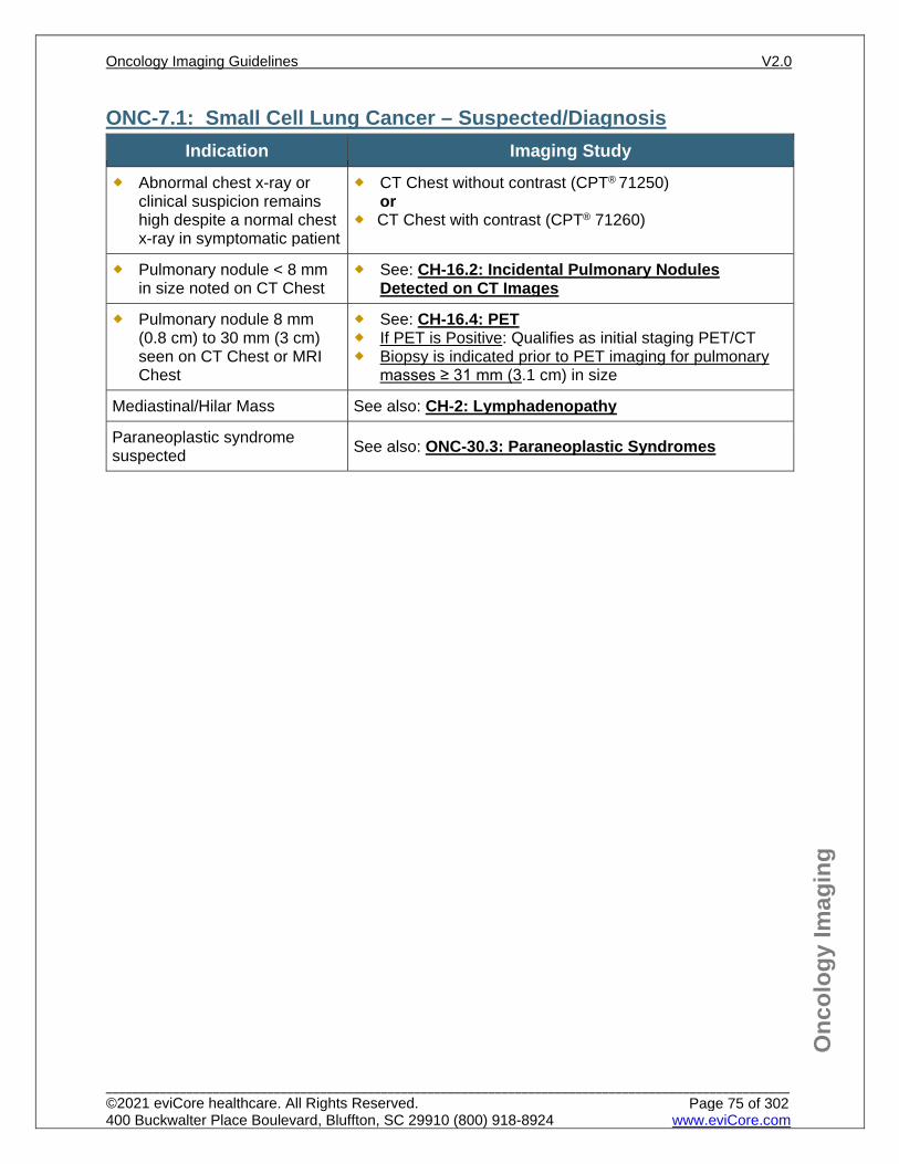
Onc
olog
y Im
agin
g
ONC-7.1: Small Cell Lung Cancer – Suspected/Diagnosis Indication Imaging Study
Abnormal chest x-ray or clinical suspicion remains high despite a normal chest x-ray in symptomatic patient
CT Chest without contrast (CPT® 71250) or
CT Chest with contrast (CPT® 71260)
Pulmonary nodule < 8 mm in size noted on CT Chest
See: CH-16.2: Incidental Pulmonary Nodules Detected on CT Images
Pulmonary nodule 8 mm (0.8 cm) to 30 mm (3 cm) seen on CT Chest or MRI Chest
See: CH-16.4: PET If PET is Positive: Qualifies as initial staging PET/CT Biopsy is indicated prior to PET imaging for pulmonary
masses ≥ 31 mm (3.1 cm) in size
Mediastinal/Hilar Mass See also: CH-2: Lymphadenopathy
Paraneoplastic syndrome suspected See also: ONC-30.3: Paraneoplastic Syndromes
Oncology Imaging Guidelines V2.0
______________________________________________________________________________________________________ ©2021 eviCore healthcare. All Rights Reserved. 400 Buckwalter Place Boulevard, Bluffton, SC 29910 (800) 918-8924 www.eviCore.com
Page 75 of 302
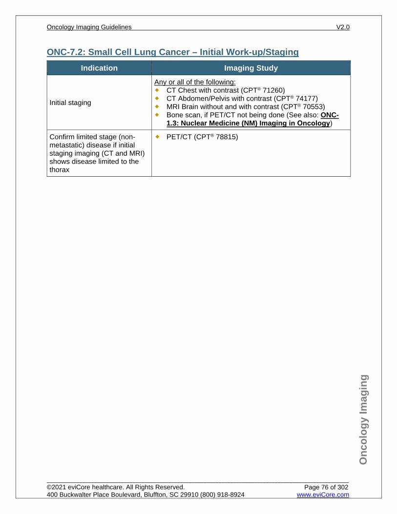
Onc
olog
y Im
agin
g
ONC-7.2: Small Cell Lung Cancer – Initial Work-up/Staging Indication Imaging Study
Initial staging
Any or all of the following: CT Chest with contrast (CPT® 71260) CT Abdomen/Pelvis with contrast (CPT® 74177) MRI Brain without and with contrast (CPT® 70553) Bone scan, if PET/CT not being done (See also: ONC-
1.3: Nuclear Medicine (NM) Imaging in Oncology)
Confirm limited stage (non-metastatic) disease if initial staging imaging (CT and MRI) shows disease limited to the thorax
PET/CT (CPT® 78815)
Oncology Imaging Guidelines V2.0
______________________________________________________________________________________________________ ©2021 eviCore healthcare. All Rights Reserved. 400 Buckwalter Place Boulevard, Bluffton, SC 29910 (800) 918-8924 www.eviCore.com
Page 76 of 302
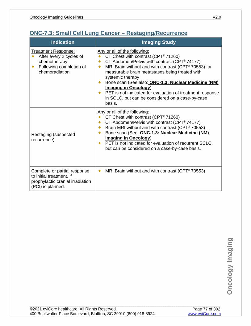
Onc
olog
y Im
agin
g
ONC-7.3: Small Cell Lung Cancer – Restaging/Recurrence Indication Imaging Study
Treatment Response: After every 2 cycles of
chemotherapy Following completion of
chemoradiation
Any or all of the following: CT Chest with contrast (CPT® 71260) CT Abdomen/Pelvis with contrast (CPT® 74177) MRI Brain without and with contrast (CPT® 70553) for
measurable brain metastases being treated with systemic therapy
Bone scan (See also: ONC-1.3: Nuclear Medicine (NM) Imaging in Oncology)
PET is not indicated for evaluation of treatment response in SCLC, but can be considered on a case-by-case basis.
Restaging (suspected recurrence)
Any or all of the following: CT Chest with contrast (CPT® 71260) CT Abdomen/Pelvis with contrast (CPT® 74177) Brain MRI without and with contrast (CPT® 70553) Bone scan (See: ONC-1.3: Nuclear Medicine (NM)
Imaging in Oncology) PET is not indicated for evaluation of recurrent SCLC,
but can be considered on a case-by-case basis.
Complete or partial response to initial treatment, if prophylactic cranial irradiation (PCI) is planned.
MRI Brain without and with contrast (CPT® 70553)
Oncology Imaging Guidelines V2.0
______________________________________________________________________________________________________ ©2021 eviCore healthcare. All Rights Reserved. 400 Buckwalter Place Boulevard, Bluffton, SC 29910 (800) 918-8924 www.eviCore.com
Page 77 of 302
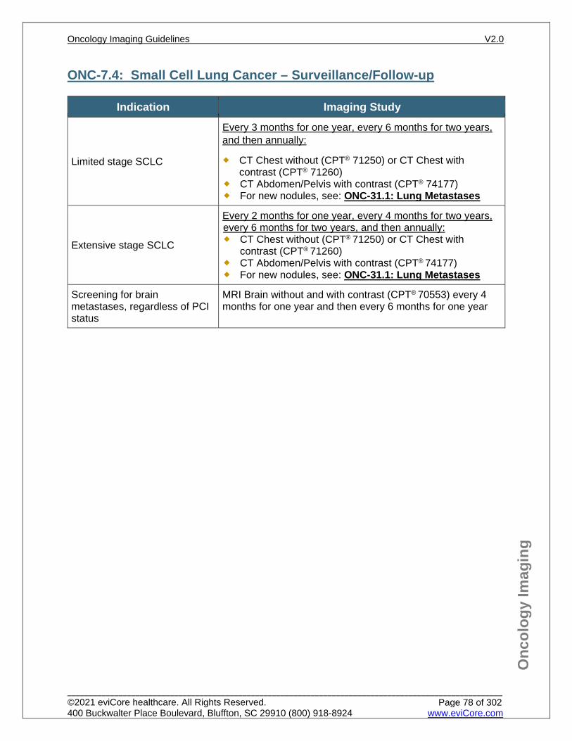
Onc
olog
y Im
agin
g
ONC-7.4: Small Cell Lung Cancer – Surveillance/Follow-up
Indication Imaging Study
Limited stage SCLC
Every 3 months for one year, every 6 months for two years, and then annually:
CT Chest without (CPT® 71250) or CT Chest with contrast (CPT® 71260)
CT Abdomen/Pelvis with contrast (CPT® 74177) For new nodules, see: ONC-31.1: Lung Metastases
Extensive stage SCLC
Every 2 months for one year, every 4 months for two years, every 6 months for two years, and then annually: CT Chest without (CPT® 71250) or CT Chest with
contrast (CPT® 71260) CT Abdomen/Pelvis with contrast (CPT® 74177) For new nodules, see: ONC-31.1: Lung Metastases
Screening for brain metastases, regardless of PCI status
MRI Brain without and with contrast (CPT® 70553) every 4 months for one year and then every 6 months for one year
Oncology Imaging Guidelines V2.0
______________________________________________________________________________________________________ ©2021 eviCore healthcare. All Rights Reserved. 400 Buckwalter Place Boulevard, Bluffton, SC 29910 (800) 918-8924 www.eviCore.com
Page 78 of 302
Onc
olog
y Im
agin
g
References 1. Ganti AKP, Loo Jr. BW, Bassetti M, et al. National Comprehensive Cancer Network (NCCN)
Guidelines Version 2.2021 – January 11, 2021 Small Cell Lung Cancer, available at: https://www.nccn.org/professionals/physician_gls/pdf/sclc.pdf Referenced with permission from the NCCN Clinical Practice Guidelines in Oncology (NCCN Guidelines™) for Small Cell Lung Cancer V2.2021 – January 11, 2021 ©2021 National Comprehensive Cancer Network, Inc. All rights reserved. The NCCN Guidelines™ and illustrations herein may not be reproduced in any form for any purpose without the express written permission of the NCCN. To view the most recent and complete version of the NCCN Guidelines™, go online to NCCN.org.
2. Lu YY, Chen JH, Liang JA, Chu S, Lin WY, Kao CH. 18F-FDG PET or PET/CT for detecting extensive disease in small-cell lung cancer: a systematic review and meta-analysis. Nucl Med Commun. 2014;35(7):697-703. doi:10.1097/MNM.0000000000000122.
3. Carter BW, Glisson BS, Truong MT, Erasmus JJ. Small cell lung carcinoma: staging, imaging, and treatment considerations. Radiographics. 2014;34(6):1707-1721. doi:10.1148/rg.346140178.
4. Kalemkerian G. Staging and imaging of small cell lung cancer. Cancer Imag. 2011;11(1):253-258. doi:10.1102/1470-7330.2011.0036.
Oncology Imaging Guidelines V2.0
______________________________________________________________________________________________________ ©2021 eviCore healthcare. All Rights Reserved. 400 Buckwalter Place Boulevard, Bluffton, SC 29910 (800) 918-8924 www.eviCore.com
Page 79 of 302
ONC-8: Non-Small Cell Lung Cancer ONC-8.0: Non-Small Cell Lung Cancer – General ConsiderationsONC-8.1: Non-Small Cell Lung Cancer – Asymptomatic ScreeningONC-8.2: Non-Small Cell Lung Cancer – Suspected/DiagnosisONC-8.3: Non-Small Cell Lung Cancer – Initial Work-up/StagingONC-8.4: Non-Small Cell Lung Cancer – Restaging/RecurrenceONC-8.5: Non-Small Cell Lung Cancer – Surveillance/Follow-up
Oncology Imaging Guidelines V2.0
______________________________________________________________________________________________________ ©2021 eviCore healthcare. All Rights Reserved. 400 Buckwalter Place Boulevard, Bluffton, SC 29910 (800) 918-8924 www.eviCore.com
Page 80 of 302
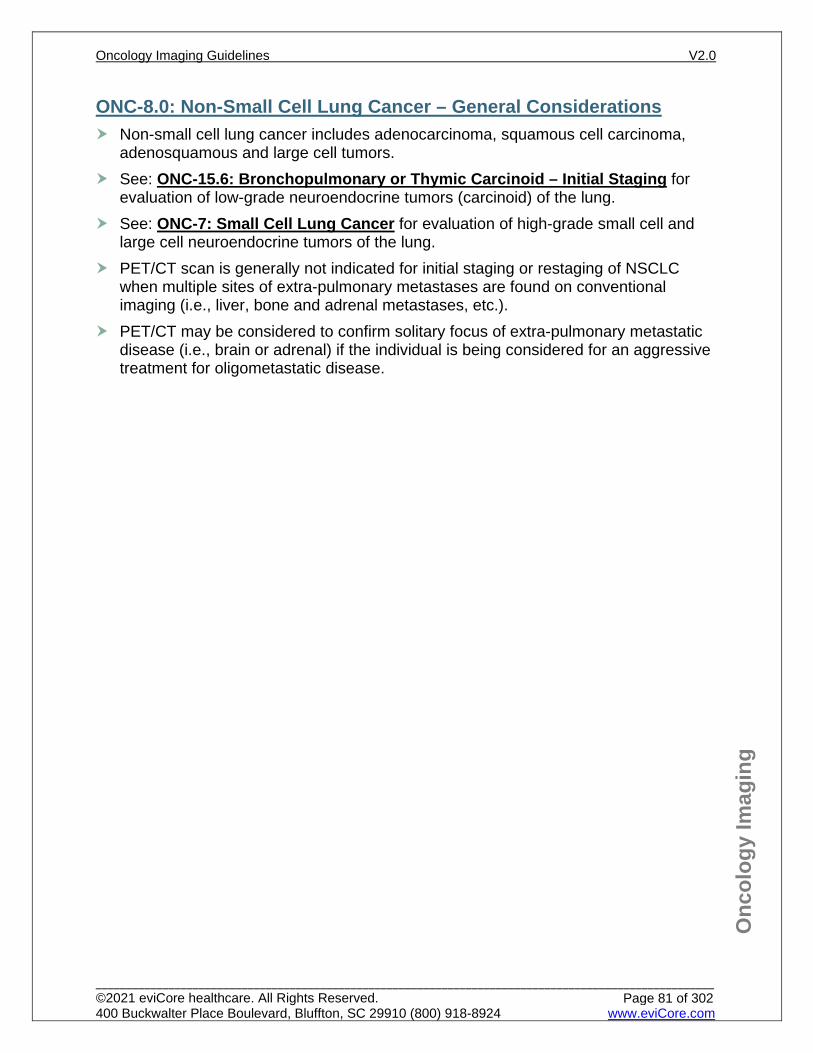
Onc
olog
y Im
agin
g
ONC-8.0: Non-Small Cell Lung Cancer – General Considerations Non-small cell lung cancer includes adenocarcinoma, squamous cell carcinoma,
adenosquamous and large cell tumors. See: ONC-15.6: Bronchopulmonary or Thymic Carcinoid – Initial Staging for
evaluation of low-grade neuroendocrine tumors (carcinoid) of the lung. See: ONC-7: Small Cell Lung Cancer for evaluation of high-grade small cell and
large cell neuroendocrine tumors of the lung. PET/CT scan is generally not indicated for initial staging or restaging of NSCLC
when multiple sites of extra-pulmonary metastases are found on conventional imaging (i.e., liver, bone and adrenal metastases, etc.).
PET/CT may be considered to confirm solitary focus of extra-pulmonary metastatic disease (i.e., brain or adrenal) if the individual is being considered for an aggressive treatment for oligometastatic disease.
Oncology Imaging Guidelines V2.0
______________________________________________________________________________________________________ ©2021 eviCore healthcare. All Rights Reserved. 400 Buckwalter Place Boulevard, Bluffton, SC 29910 (800) 918-8924 www.eviCore.com
Page 81 of 302

Onc
olog
y Im
agin
g
ONC-8.1: Non-Small Cell Lung Cancer – Asymptomatic Screening See: CH-33: Lung Cancer Screening for criteria for low-dose CT scan chest for lung cancer screening.
Oncology Imaging Guidelines V2.0
______________________________________________________________________________________________________ ©2021 eviCore healthcare. All Rights Reserved. 400 Buckwalter Place Boulevard, Bluffton, SC 29910 (800) 918-8924 www.eviCore.com
Page 82 of 302
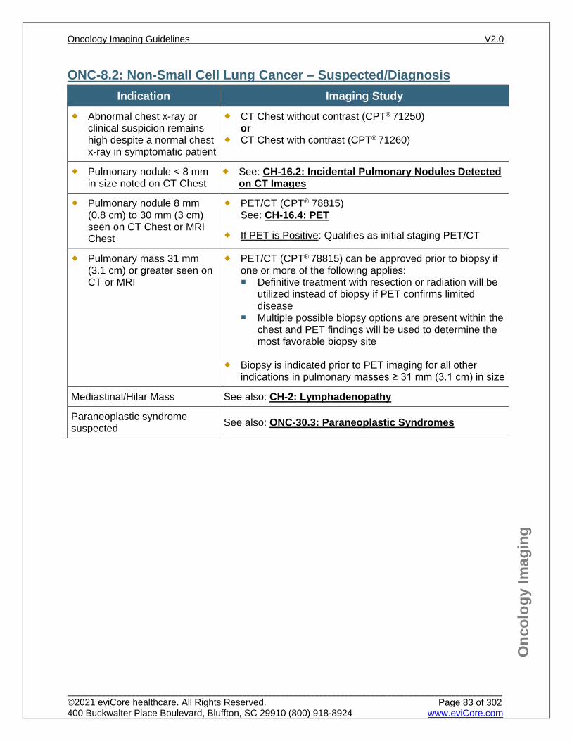
Onc
olog
y Im
agin
g
ONC-8.2: Non-Small Cell Lung Cancer – Suspected/Diagnosis Indication Imaging Study
Abnormal chest x-ray or clinical suspicion remains high despite a normal chest x-ray in symptomatic patient
CT Chest without contrast (CPT® 71250) or
CT Chest with contrast (CPT® 71260)
Pulmonary nodule < 8 mm in size noted on CT Chest
See: CH-16.2: Incidental Pulmonary Nodules Detected on CT Images
Pulmonary nodule 8 mm (0.8 cm) to 30 mm (3 cm) seen on CT Chest or MRI Chest
PET/CT (CPT® 78815) See: CH-16.4: PET
If PET is Positive: Qualifies as initial staging PET/CT
Pulmonary mass 31 mm (3.1 cm) or greater seen on CT or MRI
PET/CT (CPT® 78815) can be approved prior to biopsy if one or more of the following applies: Definitive treatment with resection or radiation will be
utilized instead of biopsy if PET confirms limited disease
Multiple possible biopsy options are present within the chest and PET findings will be used to determine the most favorable biopsy site
Biopsy is indicated prior to PET imaging for all other indications in pulmonary masses ≥ 31 mm (3.1 cm) in size
Mediastinal/Hilar Mass See also: CH-2: Lymphadenopathy
Paraneoplastic syndrome suspected See also: ONC-30.3: Paraneoplastic Syndromes
Oncology Imaging Guidelines V2.0
______________________________________________________________________________________________________ ©2021 eviCore healthcare. All Rights Reserved. 400 Buckwalter Place Boulevard, Bluffton, SC 29910 (800) 918-8924 www.eviCore.com
Page 83 of 302
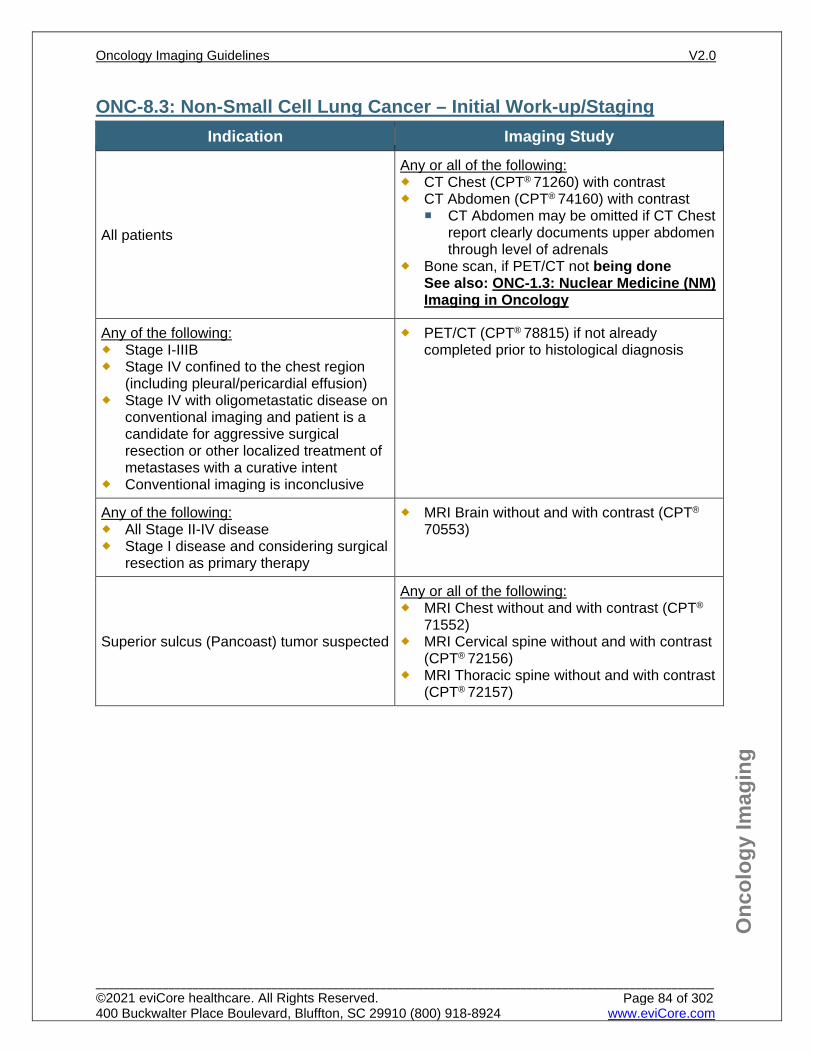
Onc
olog
y Im
agin
g
ONC-8.3: Non-Small Cell Lung Cancer – Initial Work-up/Staging Indication Imaging Study
All patients
Any or all of the following: CT Chest (CPT® 71260) with contrast CT Abdomen (CPT® 74160) with contrast
CT Abdomen may be omitted if CT Chest report clearly documents upper abdomen through level of adrenals
Bone scan, if PET/CT not being done See also: ONC-1.3: Nuclear Medicine (NM) Imaging in Oncology
Any of the following: Stage I-IIIB Stage IV confined to the chest region
(including pleural/pericardial effusion) Stage IV with oligometastatic disease on
conventional imaging and patient is a candidate for aggressive surgical resection or other localized treatment of metastases with a curative intent
Conventional imaging is inconclusive
PET/CT (CPT® 78815) if not already completed prior to histological diagnosis
Any of the following: All Stage II-IV disease Stage I disease and considering surgical
resection as primary therapy
MRI Brain without and with contrast (CPT®
70553)
Superior sulcus (Pancoast) tumor suspected
Any or all of the following: MRI Chest without and with contrast (CPT®
71552) MRI Cervical spine without and with contrast
(CPT® 72156) MRI Thoracic spine without and with contrast
(CPT® 72157)
Oncology Imaging Guidelines V2.0
______________________________________________________________________________________________________ ©2021 eviCore healthcare. All Rights Reserved. 400 Buckwalter Place Boulevard, Bluffton, SC 29910 (800) 918-8924 www.eviCore.com
Page 84 of 302
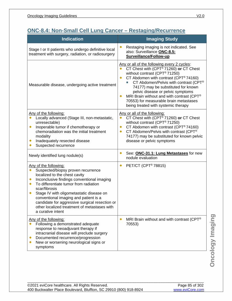
Onc
olog
y Im
agin
g
ONC-8.4: Non-Small Cell Lung Cancer – Restaging/Recurrence Indication Imaging Study
Stage I or II patients who undergo definitive local treatment with surgery, radiation, or radiosurgery
Restaging imaging is not indicated. See also: Surveillance ONC-8.5: Surveillance/Follow-up
Measurable disease, undergoing active treatment
Any or all of the following every 2 cycles: CT Chest with (CPT® 71260) or CT Chest
without contrast (CPT® 71250) CT Abdomen with contrast (CPT® 74160)
CT Abdomen/Pelvis with contrast (CPT®
74177) may be substituted for known pelvic disease or pelvic symptoms
MRI Brain without and with contrast (CPT®
70553) for measurable brain metastases being treated with systemic therapy
Any of the following: Locally advanced (Stage III, non-metastatic,
unresectable) Inoperable tumor if chemotherapy or
chemoradiation was the initial treatment modality
Inadequately resected disease Suspected recurrence
Any or all of the following: CT Chest with (CPT® 71260) or CT Chest
without contrast (CPT® 71250) CT Abdomen with contrast (CPT® 74160) CT Abdomen/Pelvis with contrast (CPT®
74177) may be substituted for known pelvic disease or pelvic symptoms
Newly identified lung nodule(s) See: ONC-31.1: Lung Metastases for new nodule evaluation
Any of the following: Suspected/biopsy proven recurrence
localized to the chest cavity Inconclusive findings conventional imaging To differentiate tumor from radiation
scar/fibrosis Stage IV with oligometastatic disease on
conventional imaging and patient is a candidate for aggressive surgical resection or other localized treatment of metastases with a curative intent
PET/CT (CPT® 78815)
Any of the following: Following a demonstrated adequate
response to neoadjuvant therapy if intracranial disease will preclude surgery
Documented recurrence/progression New or worsening neurological signs or
symptoms
MRI Brain without and with contrast (CPT®
70553)
Oncology Imaging Guidelines V2.0
______________________________________________________________________________________________________ ©2021 eviCore healthcare. All Rights Reserved. 400 Buckwalter Place Boulevard, Bluffton, SC 29910 (800) 918-8924 www.eviCore.com
Page 85 of 302
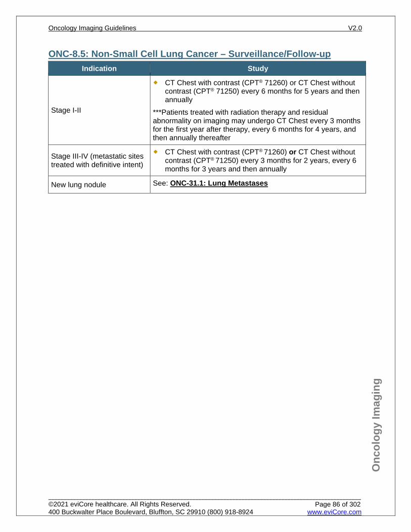
Onc
olog
y Im
agin
g
ONC-8.5: Non-Small Cell Lung Cancer – Surveillance/Follow-up Indication Study
Stage I-II
CT Chest with contrast (CPT® 71260) or CT Chest without contrast (CPT® 71250) every 6 months for 5 years and then annually
***Patients treated with radiation therapy and residual abnormality on imaging may undergo CT Chest every 3 months for the first year after therapy, every 6 months for 4 years, and then annually thereafter
Stage III-IV (metastatic sites treated with definitive intent)
CT Chest with contrast (CPT® 71260) or CT Chest without contrast (CPT® 71250) every 3 months for 2 years, every 6 months for 3 years and then annually
New lung nodule See: ONC-31.1: Lung Metastases
Oncology Imaging Guidelines V2.0
______________________________________________________________________________________________________ ©2021 eviCore healthcare. All Rights Reserved. 400 Buckwalter Place Boulevard, Bluffton, SC 29910 (800) 918-8924 www.eviCore.com
Page 86 of 302
Onc
olog
y Im
agin
g
References 1. Ettinger DS, Wood DE, Aisner DL, et al, National Comprehensive Cancer Network (NCCN)
Guidelines Version 3.2021 – February 16, 2021. Non small cell lung cancer, available at: https://www.nccn.org/professionals/physician_gls/pdf/nscl.pdf Referenced with permission from the NCCN Clinical Practice Guidelines in Oncology (NCCN Guidelines™) for Non-small cell lung cancer V3.2021 – February 16, 2021. ©2021 National Comprehensive Cancer Network, Inc. All rights reserved. The NCCN Guidelines™ and illustrations herein may not be reproduced in any form for any purpose without the express written permission of the NCCN. To view the most recent and complete version of the NCCN Guidelines™, go online to NCCN.org.
2. Scheider BJ, Ismaila N, Aerts J, et al. Lung cancer surveillance after definitive curative-intent therapy: ASCO guideline. J Clin Oncol. 2020;38(7):753-766. doi:10.1200/JCO.19.02748.
3. MacMahon H, Naidich DP, Goo JM, et al. Guidelines for management of incidental pulmonary nodules detected on CT images: from the Fleischner Society 2017. Radiology. 2017;284(1):228-243. doi:10.1148/radiol.2017161659.
4. Calman L, Beaver K, Hind D, Lorigan P, Roberts C, Lloyd-Jones M. Survival benefits from follow-up of patients with lung cancer: a systematic review and meta-analysis. J Thorac Oncol. 2011;6(12):1993-2004. doi:10.1097/JTO.0b013e31822b01a1.
5. Lou F, Huang J, Sima CS et al. Patterns of recurrence and second primary lung cancer in early-stage lung cancer survivors followed with routine computed tomography surveillance. J Thorac Cardiovasc Surg. 2013;145:75-81. https://www.ncbi.nlm.nih.gov/pubmed/23127371 .
6. Colt HG, Murgu SD, Korst RJ, et al. Follow-up and surveillance of the patient with lung cancer after curative-intent therapy: diagnosis and management of lung cancer, 3rd ed: American College of Chest Physicians evidence-based clinical practice guidelines. Chest. 2013;143:e437S-454S. https://www.ncbi.nlm.nih.gov/pubmed/23649451 .
7. Dane B, Grechushkin V, Plank A, et al. PET/CT vs. non-contrast CT alone for surveillance 1-year post lobectomy for stage I non-small cell lung cancer. Am J Nucl Med Mol Imaging. 2013; 3:408-416. https://www.ncbi.nlm.nih.gov/pmc/articles/PMC3784804/ .
8. Zhao L, He ZY, Zhong XN, et al. (18)FDG-PET/CT for detection of mediastinal nodal metastasis in non-small cell lung cancer: a meta-analysis. Surg Oncol. 2012;21(3):230-236. https://www.ncbi.nlm.nih.gov/pubmedhealth/PMH0049561/ .
9. Li J, Xu W, Kong F, et al. Meta-analysis: accuracy of 18FDG PET-CT for distant metastasis in lung cancer patients. Surg Oncol. 2013;22(3):151-155. https://www.ncbi.nlm.nih.gov/pubmed/23664848 .
10. Ravenel JG. Evidence-based imaging in lung cancer: a systematic review. J Thorac Imaging. 2012; 27(5):315-324. http://journals.lww.com/thoracicimaging/Abstract/2012/09000/Evidence_based_Imaging_in_Lung_Cancer__A.8.aspx .
11. Bille A, Pelosi E, Skanjeti A, et al. Preoperative intrathoracic lymph node staging in patients with non-small-cell lung cancer: accuracy of integrated positron emission tomography and computed tomography. Eur J Cardiothorac Surg. 2009;36(3):440-445. https://academic.oup.com/ejcts/article-lookup/doi/10.1016/j.ejcts.2009.04.003 .
Oncology Imaging Guidelines V2.0
______________________________________________________________________________________________________ ©2021 eviCore healthcare. All Rights Reserved. 400 Buckwalter Place Boulevard, Bluffton, SC 29910 (800) 918-8924 www.eviCore.com
Page 87 of 302
ONC-9: Esophageal and GE Junction Cancer ONC-9.0: Esophageal and GE Junction Cancer – General ConsiderationsONC-9.1: Esophageal and GE Junction Cancer – Suspected/DiagnosisONC-9.2: Esophageal and GE Junction Cancer – Initial Work-up/StagingONC-9.3: Esophageal and GE Junction Cancer – Restaging/RecurrenceONC-9.4: Esophageal and GE Junction Cancer – Surveillance/Follow-up
Oncology Imaging Guidelines V2.0
______________________________________________________________________________________________________ ©2021 eviCore healthcare. All Rights Reserved. 400 Buckwalter Place Boulevard, Bluffton, SC 29910 (800) 918-8924 www.eviCore.com
Page 88 of 302
Onc
olog
y Im
agin
g
ONC-9.0: Esophageal and GE Junction Cancer – General Considerations Clinicians must describe esophageal cancer by cell type and in which third of the
esophagus it occurs. Cancers of the upper and middle third are usually squamous cell and are highly
associated with tobacco and alcohol abuse. Cancers of the gastroesophageal (GE) junction are treated as lower third cancers.
Lower third cancers are usually adenocarcinomas; 62% of these arise in the setting of Barrett’s esophagus, a condition associated with high body mass index (BMI).
Oncology Imaging Guidelines V2.0
______________________________________________________________________________________________________ ©2021 eviCore healthcare. All Rights Reserved. 400 Buckwalter Place Boulevard, Bluffton, SC 29910 (800) 918-8924 www.eviCore.com
Page 89 of 302
Onc
olog
y Im
agin
g
ONC-9.1: Esophageal and GE Junction Cancer – Suspected/Diagnosis See also: NECK-3.1: Dysphagia for imaging guidelines for evaluation of suspected
esophageal malignancy
Oncology Imaging Guidelines V2.0
______________________________________________________________________________________________________ ©2021 eviCore healthcare. All Rights Reserved. 400 Buckwalter Place Boulevard, Bluffton, SC 29910 (800) 918-8924 www.eviCore.com
Page 90 of 302
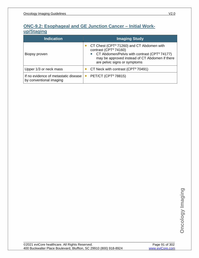
Onc
olog
y Im
agin
g
ONC-9.2: Esophageal and GE Junction Cancer – Initial Work-up/Staging
Indication Imaging Study
Biopsy proven
CT Chest (CPT® 71260) and CT Abdomen with contrast (CPT® 74160) CT Abdomen/Pelvis with contrast (CPT® 74177)
may be approved instead of CT Abdomen if there are pelvic signs or symptoms
Upper 1/3 or neck mass CT Neck with contrast (CPT® 70491)
If no evidence of metastatic disease by conventional imaging
PET/CT (CPT® 78815)
Oncology Imaging Guidelines V2.0
______________________________________________________________________________________________________ ©2021 eviCore healthcare. All Rights Reserved. 400 Buckwalter Place Boulevard, Bluffton, SC 29910 (800) 918-8924 www.eviCore.com
Page 91 of 302
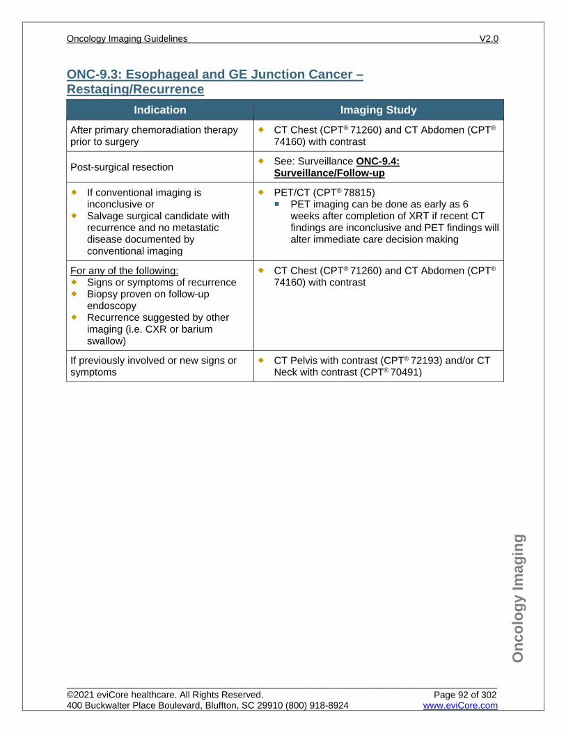
Onc
olog
y Im
agin
g
ONC-9.3: Esophageal and GE Junction Cancer – Restaging/Recurrence
Indication Imaging Study After primary chemoradiation therapy prior to surgery
CT Chest (CPT® 71260) and CT Abdomen (CPT®
74160) with contrast
Post-surgical resection See: Surveillance ONC-9.4: Surveillance/Follow-up
If conventional imaging is inconclusive or
Salvage surgical candidate with recurrence and no metastatic disease documented by conventional imaging
PET/CT (CPT® 78815) PET imaging can be done as early as 6
weeks after completion of XRT if recent CT findings are inconclusive and PET findings will alter immediate care decision making
For any of the following: Signs or symptoms of recurrence Biopsy proven on follow-up
endoscopy Recurrence suggested by other
imaging (i.e. CXR or barium swallow)
CT Chest (CPT® 71260) and CT Abdomen (CPT®
74160) with contrast
If previously involved or new signs or symptoms
CT Pelvis with contrast (CPT® 72193) and/or CT Neck with contrast (CPT® 70491)
Oncology Imaging Guidelines V2.0
______________________________________________________________________________________________________ ©2021 eviCore healthcare. All Rights Reserved. 400 Buckwalter Place Boulevard, Bluffton, SC 29910 (800) 918-8924 www.eviCore.com
Page 92 of 302
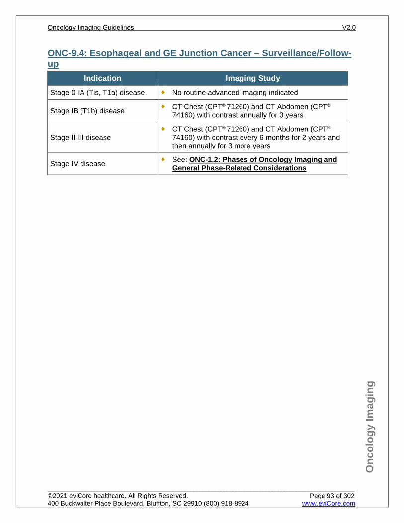
Onc
olog
y Im
agin
g
ONC-9.4: Esophageal and GE Junction Cancer – Surveillance/Follow-up
Indication Imaging Study Stage 0-IA (Tis, T1a) disease No routine advanced imaging indicated
Stage IB (T1b) disease CT Chest (CPT® 71260) and CT Abdomen (CPT®
74160) with contrast annually for 3 years
Stage II-III disease CT Chest (CPT® 71260) and CT Abdomen (CPT®
74160) with contrast every 6 months for 2 years and then annually for 3 more years
Stage IV disease See: ONC-1.2: Phases of Oncology Imaging and General Phase-Related Considerations
Oncology Imaging Guidelines V2.0
______________________________________________________________________________________________________ ©2021 eviCore healthcare. All Rights Reserved. 400 Buckwalter Place Boulevard, Bluffton, SC 29910 (800) 918-8924 www.eviCore.com
Page 93 of 302
Onc
olog
y Im
agin
g
References 1. Ajani JA, D’Amico TA, Bentrem DJ, et al. National Comprehensive Cancer Network (NCCN)
Guidelines Version 1.2021 – February 9, 2021. Esophageal and esophagogastric junction cancers, available at: https://www.nccn.org/professionals/physician_gls/pdf/esophageal.pdf Referenced with permission from the NCCN Clinical Practice Guidelines in Oncology (NCCN Guidelines™) for Esophageal and esophagogastric junction cancers V1.2021 – February 9, 2021. ©2021 National Comprehensive Cancer Network, Inc. All rights reserved. The NCCN Guidelines™ and illustrations herein may not be reproduced in any form for any purpose without the express written permission of the NCCN. To view the most recent and complete version of the NCCN Guidelines™, go online to NCCN.org.
2. Klaeser B, Nitzsche E, Schuller JC, et al. Limited predictive value of FDG-PET for response assessment in the preoperative treatment of esophageal cancer: results of a prospective multi-center trial (SAKK 75/02). Onkologie. 2009;32(12):724-730. doi:10.1159/000251842.
3. Malik V, Lucey JA, Duffy GJ, et al. Early repeated 18F-FDG PET scans during neoadjuvant chemoradiation fail to predict histopathologic response or survival benefit in adenocarcinoma of the esophagus. J Nucl Med. 2010;51(12):1863-1869. doi:10.2967/jnumed.110.079566.
4. Stiekema J, Vermeulen D, Vegt E, et al. Detecting interval metastases and response assessment using 18F-FDG PET/CT after neoadjuvant chemoradiotherapy for esophageal cancer. Clin Nucl Med. 2014;39(10):862-867. doi:10.1097/RLU.0000000000000517.
5. Sudo K, Xiao L, Wadhwa R, et al. Importance of surveillance and success of salvage strategies after definitive chemoradiation in patients with esophageal cancer. J Clin Oncol. 2014;32(30):3400-3405. doi:10.1200/JCO.2014.56.7156.
6. Lou F, Sima CS, Adusumilli PS, et al. Esophageal cancer recurrence patterns and implications for surveillance. J Thorac Oncol. 2013;8(12):1558–1562. doi:10.1097/01.JTO.0000437420.38972.fb.
7. Goense L, van Rossum PS, Reitsma JB, et al. Diagnostic performance of 18F-FDG PET and PET/CT for the detection of recurrent esophageal cancer after treatment with curative intent: a systematic review and meta-analysis. J Nucl Med. 2015;56(7):995-1002. doi:10.2967/jnumed.115.155580.
Oncology Imaging Guidelines V2.0
______________________________________________________________________________________________________ ©2021 eviCore healthcare. All Rights Reserved. 400 Buckwalter Place Boulevard, Bluffton, SC 29910 (800) 918-8924 www.eviCore.com
Page 94 of 302
ONC-10: Other Thoracic Tumors ONC-10.1: Malignant Pleural Mesothelioma – Suspected/DiagnosisONC-10.2: Malignant Pleural Mesothelioma – Initial Work-up/StagingONC-10.3: Malignant Pleural Mesothelioma – RestagingONC-10.4: Malignant Pleural Mesothelioma – SurveillanceONC-10.5: Thymoma and Thymic Carcinoma – Suspected/DiagnosisONC-10.6: Thymoma and Thymic Carcinoma – Initial Work-up/StagingONC-10.7: Thymoma and Thymic Carcinoma – RestagingONC-10.8: Thymoma and Thymic Carcinoma – Surveillance
Oncology Imaging Guidelines V2.0
______________________________________________________________________________________________________ ©2021 eviCore healthcare. All Rights Reserved. 400 Buckwalter Place Boulevard, Bluffton, SC 29910 (800) 918-8924 www.eviCore.com
Page 95 of 302
Onc
olog
y Im
agin
g
ONC-10.1: Malignant Pleural Mesothelioma – Suspected/Diagnosis See: CH-9.1: Asbestos Exposure for imaging guidelines for evaluation of
suspected mesothelioma
Oncology Imaging Guidelines V2.0
______________________________________________________________________________________________________ ©2021 eviCore healthcare. All Rights Reserved. 400 Buckwalter Place Boulevard, Bluffton, SC 29910 (800) 918-8924 www.eviCore.com
Page 96 of 302
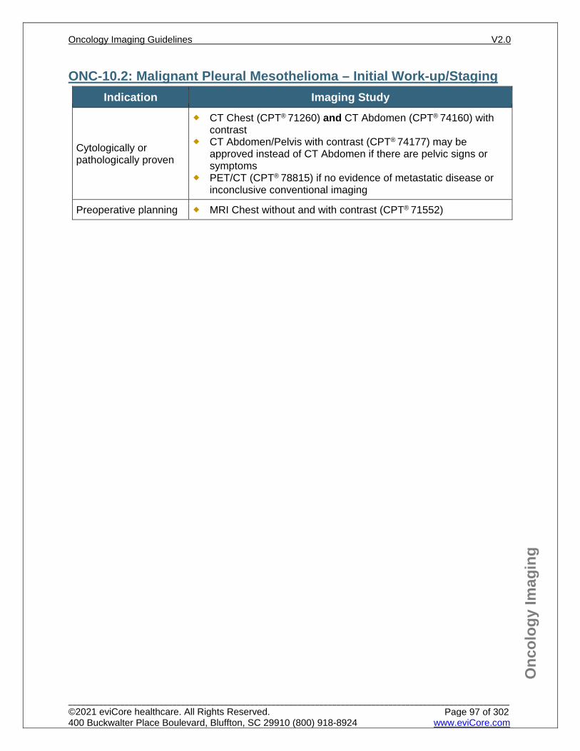
Onc
olog
y Im
agin
g
ONC-10.2: Malignant Pleural Mesothelioma – Initial Work-up/Staging Indication Imaging Study
Cytologically or pathologically proven
CT Chest (CPT® 71260) and CT Abdomen (CPT® 74160) with contrast
CT Abdomen/Pelvis with contrast (CPT® 74177) may be approved instead of CT Abdomen if there are pelvic signs or symptoms
PET/CT (CPT® 78815) if no evidence of metastatic disease or inconclusive conventional imaging
Preoperative planning MRI Chest without and with contrast (CPT® 71552)
Oncology Imaging Guidelines V2.0
______________________________________________________________________________________________________ ©2021 eviCore healthcare. All Rights Reserved. 400 Buckwalter Place Boulevard, Bluffton, SC 29910 (800) 918-8924 www.eviCore.com
Page 97 of 302
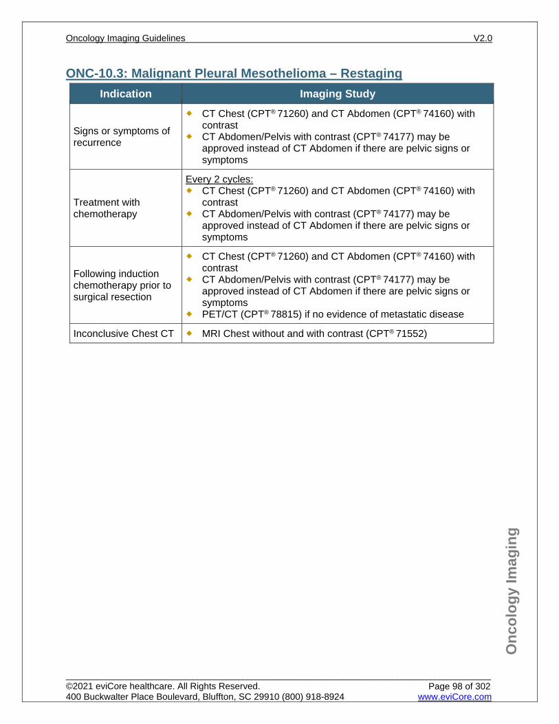
Onc
olog
y Im
agin
g
ONC-10.3: Malignant Pleural Mesothelioma – Restaging Indication Imaging Study
Signs or symptoms of recurrence
CT Chest (CPT® 71260) and CT Abdomen (CPT® 74160) with contrast
CT Abdomen/Pelvis with contrast (CPT® 74177) may be approved instead of CT Abdomen if there are pelvic signs or symptoms
Treatment with chemotherapy
Every 2 cycles: CT Chest (CPT® 71260) and CT Abdomen (CPT® 74160) with
contrast CT Abdomen/Pelvis with contrast (CPT® 74177) may be
approved instead of CT Abdomen if there are pelvic signs or symptoms
Following induction chemotherapy prior to surgical resection
CT Chest (CPT® 71260) and CT Abdomen (CPT® 74160) with contrast
CT Abdomen/Pelvis with contrast (CPT® 74177) may be approved instead of CT Abdomen if there are pelvic signs or symptoms
PET/CT (CPT® 78815) if no evidence of metastatic disease
Inconclusive Chest CT MRI Chest without and with contrast (CPT® 71552)
Oncology Imaging Guidelines V2.0
______________________________________________________________________________________________________ ©2021 eviCore healthcare. All Rights Reserved. 400 Buckwalter Place Boulevard, Bluffton, SC 29910 (800) 918-8924 www.eviCore.com
Page 98 of 302
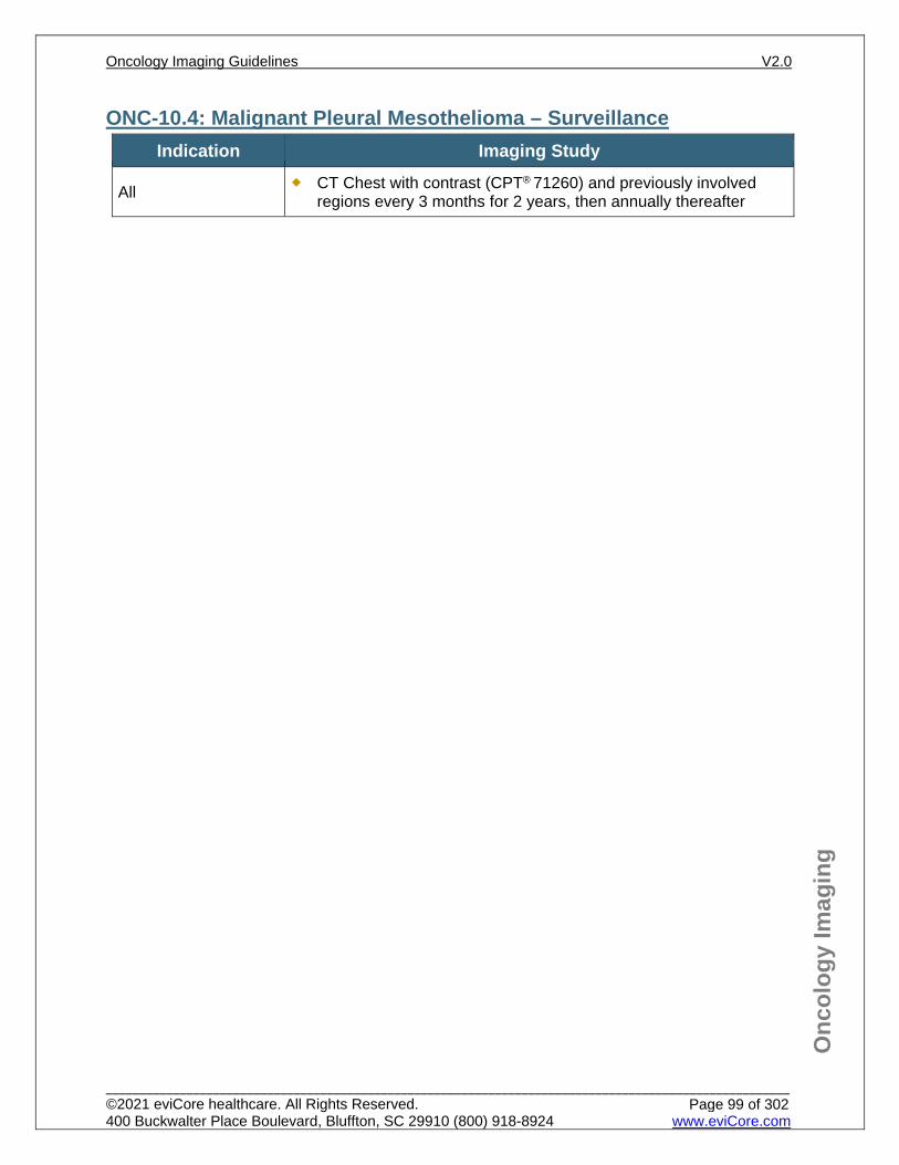
Onc
olog
y Im
agin
g
ONC-10.4: Malignant Pleural Mesothelioma – Surveillance Indication Imaging Study
All CT Chest with contrast (CPT® 71260) and previously involved regions every 3 months for 2 years, then annually thereafter
Oncology Imaging Guidelines V2.0
______________________________________________________________________________________________________ ©2021 eviCore healthcare. All Rights Reserved. 400 Buckwalter Place Boulevard, Bluffton, SC 29910 (800) 918-8924 www.eviCore.com
Page 99 of 302
Onc
olog
y Im
agin
g
ONC-10.5: Thymoma and Thymic Carcinoma – Suspected/Diagnosis See: CH-20.1: Mediastinal Mass for imaging guidelines for evaluation of suspected
thymic malignancies See: ONC-15.6: Bronchopulmonary or Thymic Carcinoid – Initial Staging for
imaging guidelines for thymic carcinoid
Oncology Imaging Guidelines V2.0
______________________________________________________________________________________________________ ©2021 eviCore healthcare. All Rights Reserved. 400 Buckwalter Place Boulevard, Bluffton, SC 29910 (800) 918-8924 www.eviCore.com
Page 100 of 302
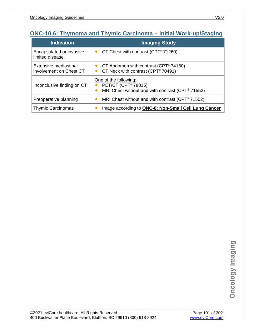
Onc
olog
y Im
agin
g
ONC-10.6: Thymoma and Thymic Carcinoma – Initial Work-up/Staging Indication Imaging Study
Encapsulated or invasive limited disease
CT Chest with contrast (CPT® 71260)
Extensive mediastinal involvement on Chest CT
CT Abdomen with contrast (CPT® 74160) CT Neck with contrast (CPT® 70491)
Inconclusive finding on CT One of the following: PET/CT (CPT® 78815) MRI Chest without and with contrast (CPT® 71552)
Preoperative planning MRI Chest without and with contrast (CPT® 71552)
Thymic Carcinomas Image according to ONC-8: Non-Small Cell Lung Cancer
Oncology Imaging Guidelines V2.0
______________________________________________________________________________________________________ ©2021 eviCore healthcare. All Rights Reserved. 400 Buckwalter Place Boulevard, Bluffton, SC 29910 (800) 918-8924 www.eviCore.com
Page 101 of 302
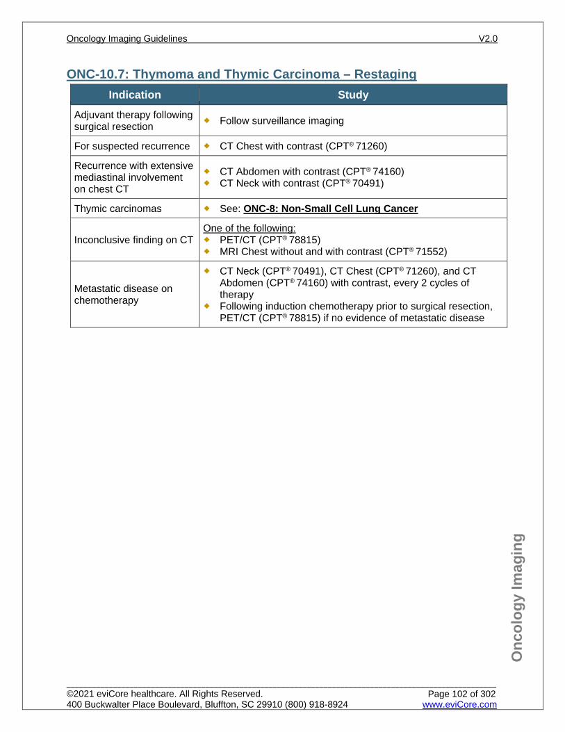
Onc
olog
y Im
agin
g
ONC-10.7: Thymoma and Thymic Carcinoma – Restaging Indication Study
Adjuvant therapy following surgical resection Follow surveillance imaging
For suspected recurrence CT Chest with contrast (CPT® 71260)
Recurrence with extensive mediastinal involvement on chest CT
CT Abdomen with contrast (CPT® 74160) CT Neck with contrast (CPT® 70491)
Thymic carcinomas See: ONC-8: Non-Small Cell Lung Cancer
Inconclusive finding on CT One of the following: PET/CT (CPT® 78815) MRI Chest without and with contrast (CPT® 71552)
Metastatic disease on chemotherapy
CT Neck (CPT® 70491), CT Chest (CPT® 71260), and CT Abdomen (CPT® 74160) with contrast, every 2 cycles of therapy
Following induction chemotherapy prior to surgical resection, PET/CT (CPT® 78815) if no evidence of metastatic disease
Oncology Imaging Guidelines V2.0
______________________________________________________________________________________________________ ©2021 eviCore healthcare. All Rights Reserved. 400 Buckwalter Place Boulevard, Bluffton, SC 29910 (800) 918-8924 www.eviCore.com
Page 102 of 302
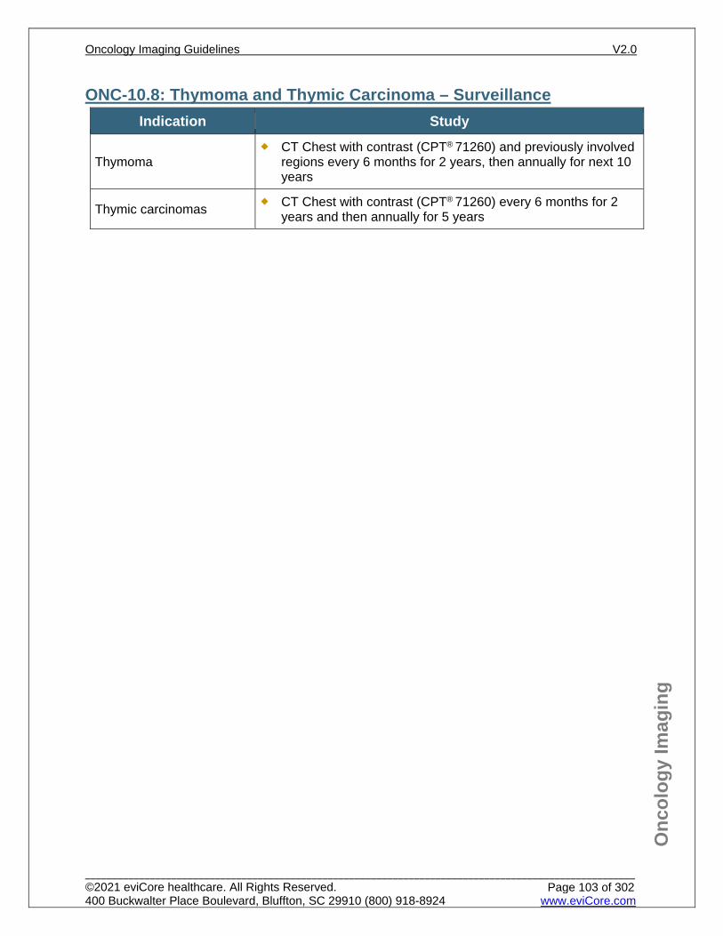
Onc
olog
y Im
agin
g
ONC-10.8: Thymoma and Thymic Carcinoma – Surveillance Indication Study
Thymoma CT Chest with contrast (CPT® 71260) and previously involved
regions every 6 months for 2 years, then annually for next 10 years
Thymic carcinomas CT Chest with contrast (CPT® 71260) every 6 months for 2 years and then annually for 5 years
Oncology Imaging Guidelines V2.0
______________________________________________________________________________________________________ ©2021 eviCore healthcare. All Rights Reserved. 400 Buckwalter Place Boulevard, Bluffton, SC 29910 (800) 918-8924 www.eviCore.com
Page 103 of 302
Onc
olog
y Im
agin
g
References 1. Ettinger DS, Wood DE, Aisner DL, et al. National Comprehensive Cancer Network (NCCN)
Guidelines Version 2.2021– February 16, 2021. Malignant Pleural Mesothelioma, available at: https://www.nccn.org/professionals/physician_gls/pdf/mpm.pdf Referenced with permission from the NCCN Clinical Practice Guidelines in Oncology (NCCN Guidelines™) for Malignant Pleural Mesothelioma V2.2021 – February 16, 2021. ©2021 National Comprehensive Cancer Network, Inc. All rights reserved. The NCCN Guidelines™ and illustrations herein may not be reproduced in any form for any purpose without the express written permission of the NCCN. To view the most recent and complete version of the NCCN Guidelines™, go online to NCCN.org.
2. Ettinger DS, Wood DE, Aisner DL, et al. National Comprehensive Cancer Network (NCCN) Guidelines Version 1.2021 – December 4, 2020. Thymoma and Thymic carcinoma, available at: https://www.nccn.org/professionals/physician_gls/pdf/thymic.pdf Referenced with permission from the NCCN Clinical Practice Guidelines in Oncology (NCCN Guidelines™) for Thymoma and Thymic carcinoma, V1.2021 – December 4, 2020. ©2020 National Comprehensive Cancer Network, Inc. All rights reserved. The NCCN Guidelines™ and illustrations herein may not be reproduced in any form for any purpose without the express written permission of the NCCN. To view the most recent and complete version of the NCCN Guidelines™, go online to NCCN.org.
3. Sørensen JB, Ravn J, Loft A, Brenøe J, Berthelsen AK, Nordic Mesothelioma Group. Preoperative staging of mesothelioma by 18F-fluoro-2-deoxy-D-glucose positron emission tomography/computer tomography fused imaging and mediastinoscopy compared to pathological findings after extrapleural pneumonectomy. Eur J Cardiothorac Surg. 2008;34:1090-1096. doi:10.1016/j.ejcts.2008.07.050.
4. Wilcox BE, Subramaniam RM, Peller PJ, et al. Utility of computed tomography-positron emission tomography for selection of operable malignant pleural mesothelioma. Clin lung cancer. 2009;10:244-248. doi: 10.3816/CLC.2009.n.033.
5. Marom EM. Imaging thymoma. J Thorac Oncol. 2010;5(10 Suppl 4):S296-S303. doi:10.1097/JTO.0b013e3181f209ca.
6. Marom EM. Advances in thymoma imaging. J Thorac Imaging. 2013;28(2):69-80. doi:10.1097/RTI.0b013e31828609a0.
7. Hayes SA, Huang J, Plodkowski AJ, et al. Preoperative computed tomography findings predict surgical resectability of thymoma. J Thorac Oncol. 2014;9(7):1023-1030. doi:10.1097/JTO.0000000000000204.
8. Mineo TC, Ambrogi V. Malignant pleural mesothelioma: factors influencing the prognosis. Oncology. 2012;26(12):1164-75.
Oncology Imaging Guidelines V2.0
______________________________________________________________________________________________________ ©2021 eviCore healthcare. All Rights Reserved. 400 Buckwalter Place Boulevard, Bluffton, SC 29910 (800) 918-8924 www.eviCore.com
Page 104 of 302

ONC-11: Breast Cancer ONC-11.0: Breast Cancer – General ConsiderationsONC-11.1: Breast Cancer – Suspected/DiagnosisONC-11.2: Breast Cancer – Initial Work-up/StagingONC-11.3: Breast Cancer – Restaging/RecurrenceONC-11.4: Breast Cancer – Surveillance/Follow-up
Oncology Imaging Guidelines V2.0
______________________________________________________________________________________________________ ©2021 eviCore healthcare. All Rights Reserved. 400 Buckwalter Place Boulevard, Bluffton, SC 29910 (800) 918-8924 www.eviCore.com
Page 105 of 302
Onc
olog
y Im
agin
g
ONC-11.0: Breast Cancer – General Considerations Advanced imaging to evaluate for distant metastases is not indicated for pre-
invasive or in-situ breast cancer (histologies such as DCIS and LCIS). Bone scan has a high concordance rate with PET for detecting bone metastases.
Scintimammography and Breast Specific Gamma Imaging (BSGI) are considered experimental and investigational.
PET is not indicated for the following: Non-invasive breast cancers Prior to lymph node sampling in a patient with clinical stage I, II, or operable IIIA
disease Obvious multi-organ metastatic disease is present on CT or MRI
Oncology Imaging Guidelines V2.0
______________________________________________________________________________________________________ ©2021 eviCore healthcare. All Rights Reserved. 400 Buckwalter Place Boulevard, Bluffton, SC 29910 (800) 918-8924 www.eviCore.com
Page 106 of 302

Onc
olog
y Im
agin
g
ONC-11.1: Breast Cancer – Suspected/Diagnosis See: BR-5: Breast MRI Indications for imaging guidelines for evaluation of suspected breast cancer
Oncology Imaging Guidelines V2.0
______________________________________________________________________________________________________ ©2021 eviCore healthcare. All Rights Reserved. 400 Buckwalter Place Boulevard, Bluffton, SC 29910 (800) 918-8924 www.eviCore.com
Page 107 of 302
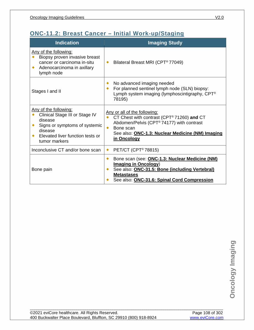
Onc
olog
y Im
agin
g
ONC-11.2: Breast Cancer – Initial Work-up/Staging Indication Imaging Study
Any of the following: Biopsy proven invasive breast
cancer or carcinoma in-situ Adenocarcinoma in axillary
lymph node
Bilateral Breast MRI (CPT® 77049)
Stages I and II
No advanced imaging needed For planned sentinel lymph node (SLN) biopsy:
Lymph system imaging (lymphoscintigraphy, CPT®
78195)
Any of the following: Clinical Stage III or Stage IV
disease Signs or symptoms of systemic
disease Elevated liver function tests or
tumor markers
Any or all of the following: CT Chest with contrast (CPT® 71260) and CT
Abdomen/Pelvis (CPT® 74177) with contrast Bone scan
See also: ONC-1.3: Nuclear Medicine (NM) Imaging in Oncology
Inconclusive CT and/or bone scan PET/CT (CPT® 78815)
Bone pain
Bone scan (see: ONC-1.3: Nuclear Medicine (NM) Imaging in Oncology)
See also: ONC-31.5: Bone (including Vertebral) Metastases
See also: ONC-31.6: Spinal Cord Compression
Oncology Imaging Guidelines V2.0
______________________________________________________________________________________________________ ©2021 eviCore healthcare. All Rights Reserved. 400 Buckwalter Place Boulevard, Bluffton, SC 29910 (800) 918-8924 www.eviCore.com
Page 108 of 302
Onc
olog
y Im
agin
g
ONC-11.3: Breast Cancer – Restaging/Recurrence Indication Imaging Study
Any of the following: Biopsy proven local recurrence Suspicion of recurrence with inconclusive
mammogram and/or ultrasound (BIRADS 0) Mammogram and ultrasound conflicts with
physical exam End of planned neoadjuvant chemotherapy
to determine resectability
Bilateral MRI Breast without and with contrast (CPT® 77049)
Any of the following: Elevated LFTs Elevated tumor markers Signs or symptoms of recurrence Biopsy proven recurrence
Any or all of the following: CT Chest (CPT® 71260) and CT
Abdomen/Pelvis (CPT® 74177) with contrast Bone scan (See also: ONC-1.3: Nuclear
Medicine (NM) Imaging in Oncology)
Treatment response in patients with metastatic disease and measurable disease on imaging
Any or all of the following for patients being treated with chemotherapy, every 2 cycles: CT Chest (CPT® 71260) and CT
Abdomen/Pelvis (CPT® 74177) with contrast Bone scan (see also: ONC-1.3: Nuclear
Medicine (NM) Imaging in Oncology) MRI Brain without and with contrast (CPT®
70553) for patients receiving systemic treatment for brain metastases
Any or all of the following for patients being treated with endocrine/hormonal therapy, every 3 months: CT Chest (CPT® 71260) and CT
Abdomen/Pelvis (CPT® 74177) with contrast Bone scan (See: ONC-1.3: Nuclear Medicine
(NM) Imaging in Oncology)
Inconclusive CT, MRI, and/or bone scan for suspected recurrence, and further characterization is needed to make treatment decisions
PET/CT (CPT® 78815)
Any of the following: Assessing for residual disease after surgery Assessing response to neoadjuvant
chemotherapy After lumpectomy or mastectomy, prior to
adjuvant therapy
Neither PET nor CT are indicated for systemic restaging after neoadjuvant chemotherapy or after surgery
Bone metastasis as the only site of stage IV disease (excluding brain metastases) and a prior bone scan has not been performed for serial comparison
PET/CT (CPT® 78815)
Oncology Imaging Guidelines V2.0
______________________________________________________________________________________________________ ©2021 eviCore healthcare. All Rights Reserved. 400 Buckwalter Place Boulevard, Bluffton, SC 29910 (800) 918-8924 www.eviCore.com
Page 109 of 302
Onc
olog
y Im
agin
g
ONC-11.4: Breast Cancer – Surveillance/Follow-up Indication Imaging Study
Measurable metastatic disease on maintenance therapy or being monitored off therapy
Any or all of the following, every 3 months for up to 5 years after completion of active treatment: CT Chest (CPT® 71260) and CT Abdomen/Pelvis
(CPT® 74177) with contrast Bone scan (see also: ONC-1.3: Nuclear Medicine
(NM) Imaging in Oncology)
Asymptomatic non-metastatic disease
Individuals receiving post-operative adjuvant therapy
No advanced imaging indicated
Breast imaging surveillance See: BR-5: Breast MRI Indications for imaging guidelines
Oncology Imaging Guidelines V2.0
______________________________________________________________________________________________________ ©2021 eviCore healthcare. All Rights Reserved. 400 Buckwalter Place Boulevard, Bluffton, SC 29910 (800) 918-8924 www.eviCore.com
Page 110 of 302
Onc
olog
y Im
agin
g
References 1. Gradishar WJ, Moran MS, Abraham J, et al. National Comprehensive Cancer Network (NCCN)
Guidelines Version 1.2021 – January 15, 2021. Breast cancer, available at: https://www.nccn.org/professionals/physician_gls/pdf/breast.pdf Referenced with permission from the NCCN Clinical Practice Guidelines in Oncology (NCCN Guidelines™) for Breast Cancer V1.2021 – January 15, 2021. ©2021 National Comprehensive Cancer Network, Inc. All rights reserved. The NCCN Guidelines™ and illustrations herein may not be reproduced in any form for any purpose without the express written permission of the NCCN. To view the most recent and complete version of the NCCN Guidelines™, go online to NCCN.org.
2. Cardoso F, Costa A, Norton L, et al. ESO-ESMO 2nd international consensus guidelines for advanced breast cancer (ABC2). Ann Oncol. 2014;25(10):1871-1888. doi:10.1093/annonc/mdu385.
3. Khatcheressian JL, Hurley P, Bantug E, et al. Breast cancer follow-up and management after primary treatment: American Society of Clinical Oncology clinical practice guideline update. J Clin Oncol. 2013;31:961-965. doi:10.1200/JCO.2012.45.9859.
4. Puglisi F, Follador A, Minisini AM, et al. Baseline staging tests after a new diagnosis of breast cancer: further evidence of their limited indications. Ann Oncol. 2005;16(2):263-266. doi:10.1093/annonc/mdi063.
5. Rong J, Wang S, Ding Q, Yun M, Zheng Z, Ye S. Comparison of 18 FDG PET-CT and bone scintigraphy for detection of bone metastases in breast cancer patients. A meta-analysis. Surg Oncol. 2013;22(2):86-91. doi:10.1016/j.suronc.2013.01.002.
6. Hong S, Li J, Wang S. 18FDG PET-CT for diagnosis of distant metastases in breast cancer patients. A meta-analysis. Surg Oncol. 2013;22(2):139-143. doi:10.1016/j.suronc.2013.03.001.
7. Cheng X, Li Y, Liu B, Xu Z, Bao L, Wang J. 18F-FDG PET/CT and PET for evaluation of pathological response to neoadjuvant chemotherapy in breast cancer: a meta-analysis. Acta Radiol. 2012;53(6):615-627. doi:10.1258/ar.2012.110603.
8. Simos D, Catley C, van Walraven C, et al. Imaging for distant metastases in women with early-stage breast cancer: a population-based cohort study. CMAJ. 2015;187(12):E387-E397. doi:10.1503/cmaj.150003.
9. Crivello ML, Ruth K, Sigurdson ER, et al. Advanced imaging modalities in early stage breast cancer: preoperative use in the United States Medicare population. Ann Surg Oncol. 2013;20(1):102-110. doi:10.1245/s10434-012-2571-4.
Oncology Imaging Guidelines V2.0
______________________________________________________________________________________________________ ©2021 eviCore healthcare. All Rights Reserved. 400 Buckwalter Place Boulevard, Bluffton, SC 29910 (800) 918-8924 www.eviCore.com
Page 111 of 302
ONC-12: Sarcomas – Bone, Soft Tissue and GIST ONC-12.1: Bone and Soft Tissue Sarcomas – General ConsiderationsONC-12.2: Soft Tissue Sarcomas – Initial Work-up/StagingONC-12.3: Soft Tissue Sarcomas – Restaging/RecurrenceONC-12.4: Soft Tissue Sarcomas – Surveillance/Follow-upONC-12.5: Gastrointestinal Stromal Tumor (GIST)ONC-12.6: Bone Sarcomas – Initial Work-up/StagingONC-12.7: Bone Sarcomas – Restaging/RecurrenceONC-12.8: Bone Sarcomas – Surveillance/Follow-upONC-12.9: Benign Bone Tumors – General ConsiderationsONC-12.10: Benign Bone Tumors – Initial Work-up/StagingONC-12.11: Benign Bone Tumors – Restaging/RecurrenceONC-12.12: Benign Bone Tumors – Surveillance/Follow-up
Oncology Imaging Guidelines V2.0
______________________________________________________________________________________________________ ©2021 eviCore healthcare. All Rights Reserved. 400 Buckwalter Place Boulevard, Bluffton, SC 29910 (800) 918-8924 www.eviCore.com
Page 112 of 302
Onc
olog
y Im
agin
g
ONC-12.1: Bone and Soft Tissue Sarcomas – General Considerations Sarcomas are tumors of mesenchymal origin, classified as high-, intermediate-, and low-grade (G) tumors (sometimes described as “spindle cell” cancers). They can arise in any bony, cartilaginous, smooth muscle, skeletal muscle, or cardiac muscle tissue.
Sarcomas occur in both adult and pediatric patients, but some are more common in one age group than the other. Unless specified below, patients age ≥ 18 years old should be imaged according to this guideline section.
Exceptions include:
Rhabdomyosarcoma patients of all ages should be imaged according to guidelines in PEDONC-8.2: Rhabdomyosarcoma
Osteogenic sarcoma (Osteosarcoma) patients of all ages should be imaged according to guidelines in PEDONC-9.3: Osteogenic Sarcoma (OS)
Ewing sarcoma and Primitive Neuroectodermal Tumor patients of all ages should be imaged according to guidelines in PEDONC-9.4: Ewing Sarcoma and Primitive Neuroectodermal Tumors (ESFT)
Kaposi’s sarcoma patients of all ages should be imaged according to guidelines in ONC-31.10: Kaposi’s Sarcoma
Oncology Imaging Guidelines V2.0
______________________________________________________________________________________________________ ©2021 eviCore healthcare. All Rights Reserved. 400 Buckwalter Place Boulevard, Bluffton, SC 29910 (800) 918-8924 www.eviCore.com
Page 113 of 302
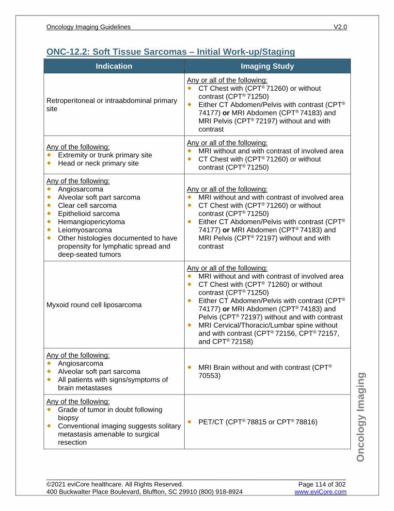
Onc
olog
y Im
agin
g
ONC-12.2: Soft Tissue Sarcomas – Initial Work-up/Staging Indication Imaging Study
Retroperitoneal or intraabdominal primary site
Any or all of the following: CT Chest with (CPT® 71260) or without
contrast (CPT® 71250) Either CT Abdomen/Pelvis with contrast (CPT®
74177) or MRI Abdomen (CPT® 74183) and MRI Pelvis (CPT® 72197) without and with contrast
Any of the following: Extremity or trunk primary site Head or neck primary site
Any or all of the following: MRI without and with contrast of involved area CT Chest with (CPT® 71260) or without
contrast (CPT® 71250)
Any of the following: Angiosarcoma Alveolar soft part sarcoma Clear cell sarcoma Epithelioid sarcoma Hemangiopericytoma Leiomyosarcoma Other histologies documented to have
propensity for lymphatic spread and deep-seated tumors
Any or all of the following: MRI without and with contrast of involved area CT Chest with (CPT® 71260) or without
contrast (CPT® 71250) Either CT Abdomen/Pelvis with contrast (CPT®
74177) or MRI Abdomen (CPT® 74183) and MRI Pelvis (CPT® 72197) without and with contrast
Myxoid round cell liposarcoma
Any or all of the following: MRI without and with contrast of involved area CT Chest with (CPT® 71260) or without
contrast (CPT® 71250) Either CT Abdomen/Pelvis with contrast (CPT®
74177) or MRI Abdomen (CPT® 74183) and Pelvis (CPT® 72197) without and with contrast
MRI Cervical/Thoracic/Lumbar spine without and with contrast (CPT® 72156, CPT® 72157, and CPT® 72158)
Any of the following: Angiosarcoma Alveolar soft part sarcoma All patients with signs/symptoms of
brain metastases
MRI Brain without and with contrast (CPT®
70553)
Any of the following: Grade of tumor in doubt following
biopsy Conventional imaging suggests solitary
metastasis amenable to surgical resection
PET/CT (CPT® 78815 or CPT® 78816)
Oncology Imaging Guidelines V2.0
______________________________________________________________________________________________________ ©2021 eviCore healthcare. All Rights Reserved. 400 Buckwalter Place Boulevard, Bluffton, SC 29910 (800) 918-8924 www.eviCore.com
Page 114 of 302
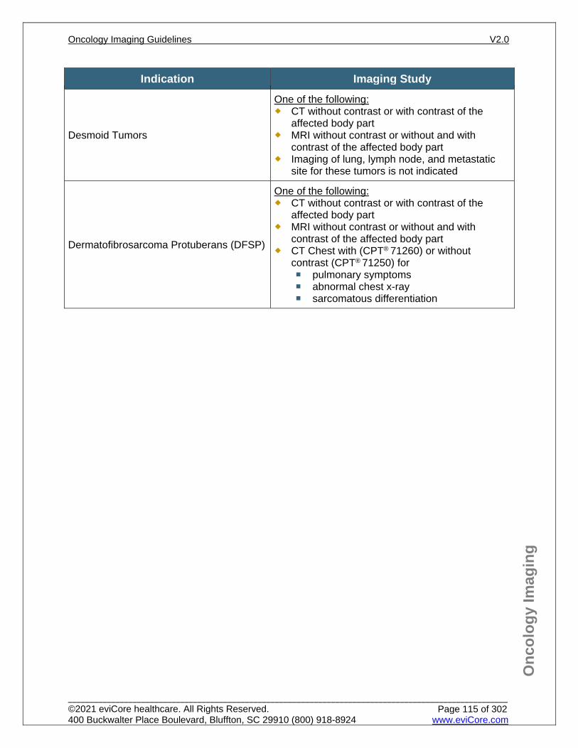
Onc
olog
y Im
agin
g
Indication Imaging Study
Desmoid Tumors
One of the following: CT without contrast or with contrast of the
affected body part MRI without contrast or without and with
contrast of the affected body part Imaging of lung, lymph node, and metastatic
site for these tumors is not indicated
Dermatofibrosarcoma Protuberans (DFSP)
One of the following: CT without contrast or with contrast of the
affected body part MRI without contrast or without and with
contrast of the affected body part CT Chest with (CPT® 71260) or without
contrast (CPT® 71250) for pulmonary symptoms abnormal chest x-ray sarcomatous differentiation
Oncology Imaging Guidelines V2.0
______________________________________________________________________________________________________ ©2021 eviCore healthcare. All Rights Reserved. 400 Buckwalter Place Boulevard, Bluffton, SC 29910 (800) 918-8924 www.eviCore.com
Page 115 of 302
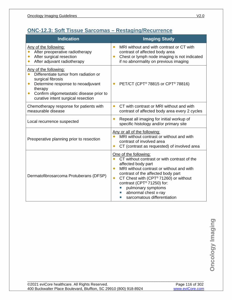
Onc
olog
y Im
agin
g
ONC-12.3: Soft Tissue Sarcomas – Restaging/Recurrence Indication Imaging Study
Any of the following: After preoperative radiotherapy After surgical resection After adjuvant radiotherapy
MRI without and with contrast or CT with contrast of affected body area
Chest or lymph node imaging is not indicated if no abnormality on previous imaging
Any of the following: Differentiate tumor from radiation or
surgical fibrosis Determine response to neoadjuvant
therapy Confirm oligometastatic disease prior to
curative intent surgical resection
PET/CT (CPT® 78815 or CPT® 78816)
Chemotherapy response for patients with measurable disease
CT with contrast or MRI without and with contrast of affected body area every 2 cycles
Local recurrence suspected Repeat all imaging for initial workup of specific histology and/or primary site
Preoperative planning prior to resection
Any or all of the following: MRI without contrast or without and with
contrast of involved area CT (contrast as requested) of involved area
Dermatofibrosarcoma Protuberans (DFSP)
One of the following: CT without contrast or with contrast of the
affected body part MRI without contrast or without and with
contrast of the affected body part CT Chest with (CPT® 71260) or without
contrast (CPT® 71250) for: pulmonary symptoms abnormal chest x-ray sarcomatous differentiation
Oncology Imaging Guidelines V2.0
______________________________________________________________________________________________________ ©2021 eviCore healthcare. All Rights Reserved. 400 Buckwalter Place Boulevard, Bluffton, SC 29910 (800) 918-8924 www.eviCore.com
Page 116 of 302
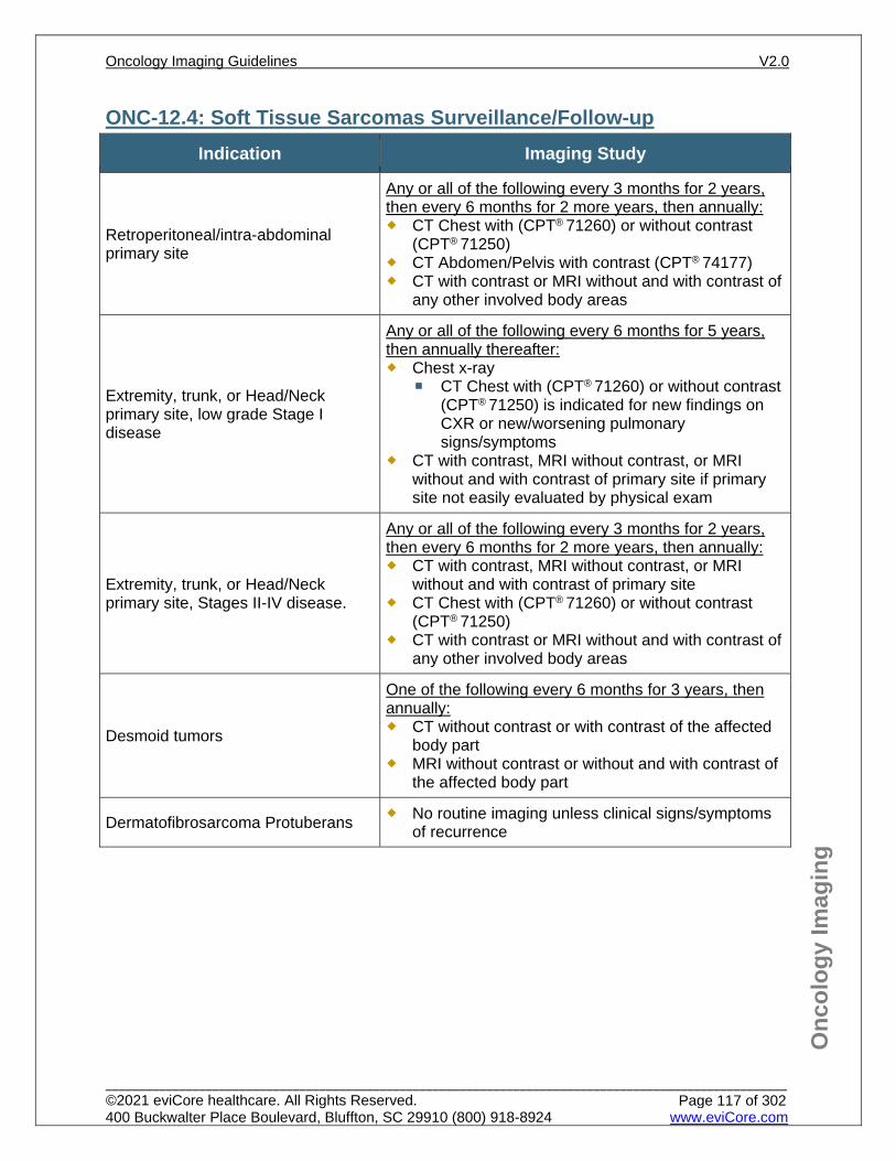
Onc
olog
y Im
agin
g
ONC-12.4: Soft Tissue Sarcomas Surveillance/Follow-up Indication Imaging Study
Retroperitoneal/intra-abdominal primary site
Any or all of the following every 3 months for 2 years, then every 6 months for 2 more years, then annually: CT Chest with (CPT® 71260) or without contrast
(CPT® 71250) CT Abdomen/Pelvis with contrast (CPT® 74177) CT with contrast or MRI without and with contrast of
any other involved body areas
Extremity, trunk, or Head/Neck primary site, low grade Stage I disease
Any or all of the following every 6 months for 5 years, then annually thereafter: Chest x-ray
CT Chest with (CPT® 71260) or without contrast (CPT® 71250) is indicated for new findings on CXR or new/worsening pulmonary signs/symptoms
CT with contrast, MRI without contrast, or MRI without and with contrast of primary site if primary site not easily evaluated by physical exam
Extremity, trunk, or Head/Neck primary site, Stages II-IV disease.
Any or all of the following every 3 months for 2 years, then every 6 months for 2 more years, then annually: CT with contrast, MRI without contrast, or MRI
without and with contrast of primary site CT Chest with (CPT® 71260) or without contrast
(CPT® 71250) CT with contrast or MRI without and with contrast of
any other involved body areas
Desmoid tumors
One of the following every 6 months for 3 years, then annually: CT without contrast or with contrast of the affected
body part MRI without contrast or without and with contrast of
the affected body part
Dermatofibrosarcoma Protuberans No routine imaging unless clinical signs/symptoms of recurrence
Oncology Imaging Guidelines V2.0
______________________________________________________________________________________________________ ©2021 eviCore healthcare. All Rights Reserved. 400 Buckwalter Place Boulevard, Bluffton, SC 29910 (800) 918-8924 www.eviCore.com
Page 117 of 302
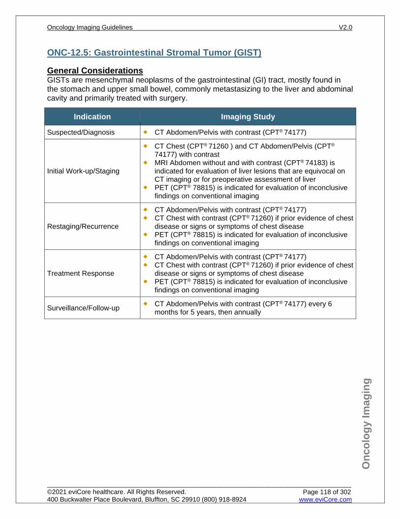
Onc
olog
y Im
agin
g
ONC-12.5: Gastrointestinal Stromal Tumor (GIST)
General Considerations GISTs are mesenchymal neoplasms of the gastrointestinal (GI) tract, mostly found in the stomach and upper small bowel, commonly metastasizing to the liver and abdominal cavity and primarily treated with surgery.
Indication Imaging Study
Suspected/Diagnosis CT Abdomen/Pelvis with contrast (CPT® 74177)
Initial Work-up/Staging
CT Chest (CPT® 71260 ) and CT Abdomen/Pelvis (CPT®
74177) with contrast MRI Abdomen without and with contrast (CPT® 74183) is
indicated for evaluation of liver lesions that are equivocal on CT imaging or for preoperative assessment of liver
PET (CPT® 78815) is indicated for evaluation of inconclusive findings on conventional imaging
Restaging/Recurrence
CT Abdomen/Pelvis with contrast (CPT® 74177) CT Chest with contrast (CPT® 71260) if prior evidence of chest
disease or signs or symptoms of chest disease PET (CPT® 78815) is indicated for evaluation of inconclusive
findings on conventional imaging
Treatment Response
CT Abdomen/Pelvis with contrast (CPT® 74177) CT Chest with contrast (CPT® 71260) if prior evidence of chest
disease or signs or symptoms of chest disease PET (CPT® 78815) is indicated for evaluation of inconclusive
findings on conventional imaging
Surveillance/Follow-up CT Abdomen/Pelvis with contrast (CPT® 74177) every 6 months for 5 years, then annually
Oncology Imaging Guidelines V2.0
______________________________________________________________________________________________________ ©2021 eviCore healthcare. All Rights Reserved. 400 Buckwalter Place Boulevard, Bluffton, SC 29910 (800) 918-8924 www.eviCore.com
Page 118 of 302
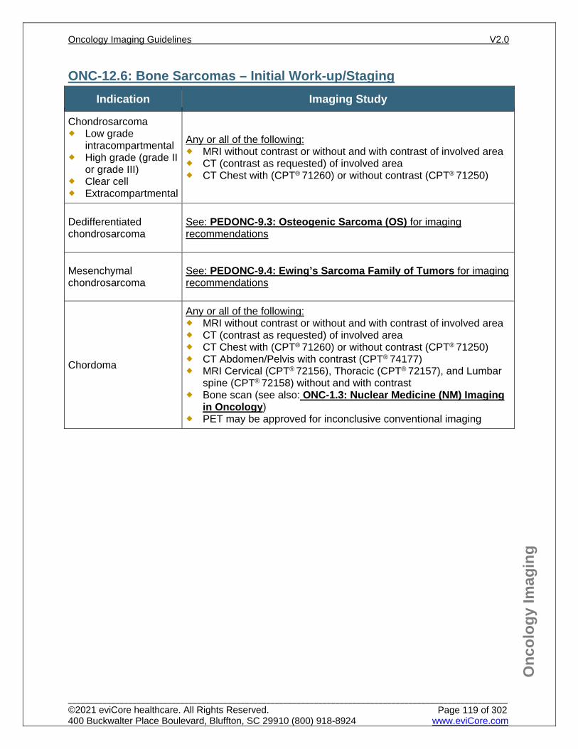
Onc
olog
y Im
agin
g
ONC-12.6: Bone Sarcomas – Initial Work-up/Staging Indication Imaging Study
Chondrosarcoma Low grade
intracompartmental High grade (grade II
or grade III) Clear cell Extracompartmental
Any or all of the following: MRI without contrast or without and with contrast of involved area CT (contrast as requested) of involved area CT Chest with (CPT® 71260) or without contrast (CPT® 71250)
Dedifferentiated chondrosarcoma
See: PEDONC-9.3: Osteogenic Sarcoma (OS) for imaging recommendations
Mesenchymal chondrosarcoma
See: PEDONC-9.4: Ewing’s Sarcoma Family of Tumors for imaging recommendations
Chordoma
Any or all of the following: MRI without contrast or without and with contrast of involved area CT (contrast as requested) of involved area CT Chest with (CPT® 71260) or without contrast (CPT® 71250) CT Abdomen/Pelvis with contrast (CPT® 74177) MRI Cervical (CPT® 72156), Thoracic (CPT® 72157), and Lumbar
spine (CPT® 72158) without and with contrast Bone scan (see also: ONC-1.3: Nuclear Medicine (NM) Imaging
in Oncology) PET may be approved for inconclusive conventional imaging
Oncology Imaging Guidelines V2.0
______________________________________________________________________________________________________ ©2021 eviCore healthcare. All Rights Reserved. 400 Buckwalter Place Boulevard, Bluffton, SC 29910 (800) 918-8924 www.eviCore.com
Page 119 of 302
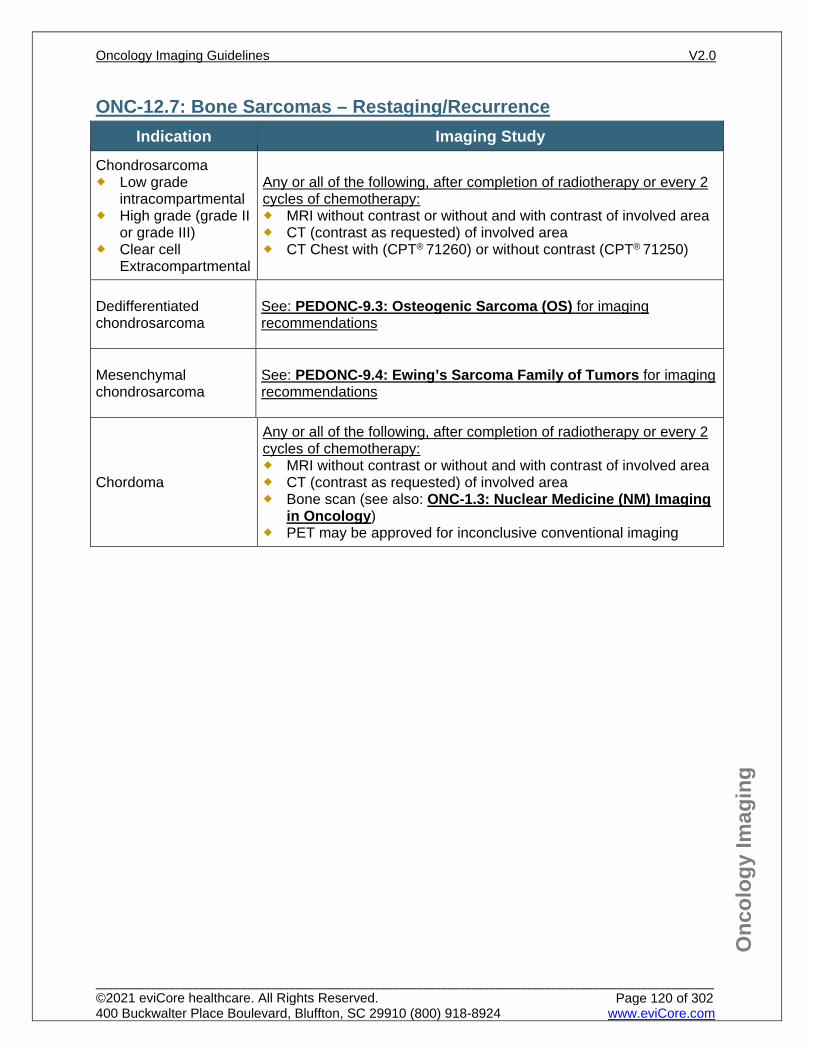
Onc
olog
y Im
agin
g
ONC-12.7: Bone Sarcomas – Restaging/Recurrence Indication Imaging Study
Chondrosarcoma Low grade
intracompartmental High grade (grade II
or grade III) Clear cell
Extracompartmental
Any or all of the following, after completion of radiotherapy or every 2 cycles of chemotherapy: MRI without contrast or without and with contrast of involved area CT (contrast as requested) of involved area CT Chest with (CPT® 71260) or without contrast (CPT® 71250)
Dedifferentiated chondrosarcoma
See: PEDONC-9.3: Osteogenic Sarcoma (OS) for imaging recommendations
Mesenchymal chondrosarcoma
See: PEDONC-9.4: Ewing’s Sarcoma Family of Tumors for imaging recommendations
Chordoma
Any or all of the following, after completion of radiotherapy or every 2 cycles of chemotherapy: MRI without contrast or without and with contrast of involved area CT (contrast as requested) of involved area Bone scan (see also: ONC-1.3: Nuclear Medicine (NM) Imaging
in Oncology) PET may be approved for inconclusive conventional imaging
Oncology Imaging Guidelines V2.0
______________________________________________________________________________________________________ ©2021 eviCore healthcare. All Rights Reserved. 400 Buckwalter Place Boulevard, Bluffton, SC 29910 (800) 918-8924 www.eviCore.com
Page 120 of 302
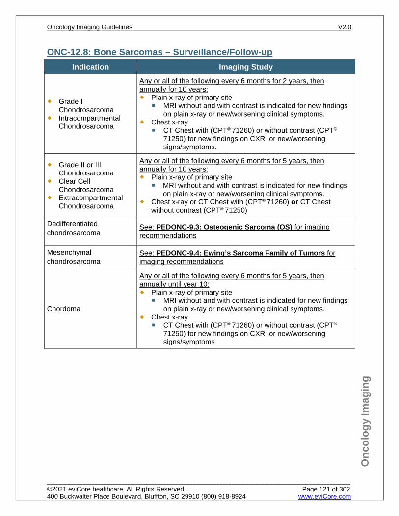
Onc
olog
y Im
agin
g
ONC-12.8: Bone Sarcomas – Surveillance/Follow-up Indication Imaging Study
Grade I Chondrosarcoma
Intracompartmental Chondrosarcoma
Any or all of the following every 6 months for 2 years, then annually for 10 years: Plain x-ray of primary site
MRI without and with contrast is indicated for new findings on plain x-ray or new/worsening clinical symptoms.
Chest x-ray CT Chest with (CPT® 71260) or without contrast (CPT®
71250) for new findings on CXR, or new/worsening signs/symptoms.
Grade II or III Chondrosarcoma
Clear Cell Chondrosarcoma
Extracompartmental Chondrosarcoma
Any or all of the following every 6 months for 5 years, then annually for 10 years: Plain x-ray of primary site
MRI without and with contrast is indicated for new findings on plain x-ray or new/worsening clinical symptoms.
Chest x-ray or CT Chest with (CPT® 71260) or CT Chest without contrast (CPT® 71250)
Dedifferentiated chondrosarcoma
See: PEDONC-9.3: Osteogenic Sarcoma (OS) for imaging recommendations
Mesenchymal chondrosarcoma
See: PEDONC-9.4: Ewing’s Sarcoma Family of Tumors for imaging recommendations
Chordoma
Any or all of the following every 6 months for 5 years, then annually until year 10: Plain x-ray of primary site
MRI without and with contrast is indicated for new findings on plain x-ray or new/worsening clinical symptoms.
Chest x-ray CT Chest with (CPT® 71260) or without contrast (CPT®
71250) for new findings on CXR, or new/worsening signs/symptoms
Oncology Imaging Guidelines V2.0
______________________________________________________________________________________________________ ©2021 eviCore healthcare. All Rights Reserved. 400 Buckwalter Place Boulevard, Bluffton, SC 29910 (800) 918-8924 www.eviCore.com
Page 121 of 302
Onc
olog
y Im
agin
g
ONC-12.9: Benign Bone Tumors – General Considerations Variety of diagnoses, including osteoid osteochondroma, chondroblastoma,
desmoplastic fibroma, Paget’s disease, osteoid osteoma and others Plain x-ray appearance is diagnostic for many benign bone tumors and advanced
imaging is generally unnecessary except for preoperative planning MRI without and with contrast is the primary modality for advanced imaging of bone
tumors, and can be approved to help narrow differential diagnoses and determine whether biopsy is indicated
Some benign bone tumor types carry a risk of malignant degeneration over time, but routine advanced imaging surveillance has not been shown to improve outcomes for these patients
MRI without and with contrast can be approved to evaluate new findings on plain x-ray new/worsening clinical symptoms not explained by a recent plain x-ray
There are no data to support the use of PET/CT in the evaluation of benign bone tumors, and PET requests should not be approved without biopsy confirmation of a malignancy
Other benign bone tumor patients of all ages should be imaged according to guidelines in PEDONC-9.2: Benign Bone Tumors
Oncology Imaging Guidelines V2.0
______________________________________________________________________________________________________ ©2021 eviCore healthcare. All Rights Reserved. 400 Buckwalter Place Boulevard, Bluffton, SC 29910 (800) 918-8924 www.eviCore.com
Page 122 of 302
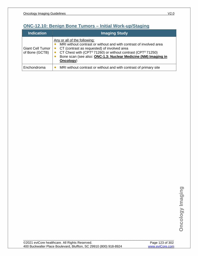
Onc
olog
y Im
agin
g
ONC-12.10: Benign Bone Tumors – Initial Work-up/Staging Indication Imaging Study
Giant Cell Tumor of Bone (GCTB)
Any or all of the following: MRI without contrast or without and with contrast of involved area CT (contrast as requested) of involved area CT Chest with (CPT® 71260) or without contrast (CPT® 71250) Bone scan (see also: ONC-1.3: Nuclear Medicine (NM) Imaging in
Oncology)
Enchondroma MRI without contrast or without and with contrast of primary site
Oncology Imaging Guidelines V2.0
______________________________________________________________________________________________________ ©2021 eviCore healthcare. All Rights Reserved. 400 Buckwalter Place Boulevard, Bluffton, SC 29910 (800) 918-8924 www.eviCore.com
Page 123 of 302
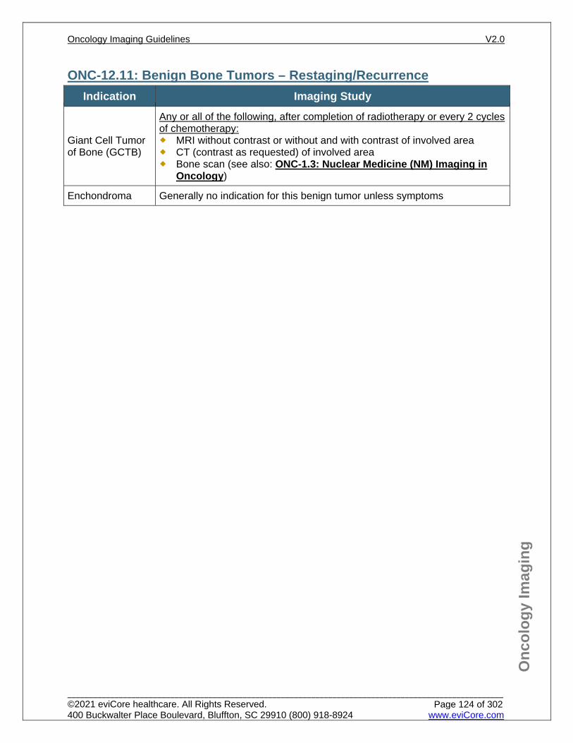
Onc
olog
y Im
agin
g
ONC-12.11: Benign Bone Tumors – Restaging/Recurrence Indication Imaging Study
Giant Cell Tumor of Bone (GCTB)
Any or all of the following, after completion of radiotherapy or every 2 cycles of chemotherapy: MRI without contrast or without and with contrast of involved area CT (contrast as requested) of involved area Bone scan (see also: ONC-1.3: Nuclear Medicine (NM) Imaging in
Oncology)
Enchondroma Generally no indication for this benign tumor unless symptoms
Oncology Imaging Guidelines V2.0
______________________________________________________________________________________________________ ©2021 eviCore healthcare. All Rights Reserved. 400 Buckwalter Place Boulevard, Bluffton, SC 29910 (800) 918-8924 www.eviCore.com
Page 124 of 302
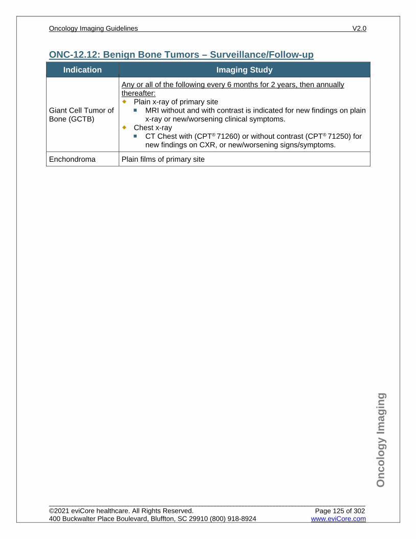
Onc
olog
y Im
agin
g
ONC-12.12: Benign Bone Tumors – Surveillance/Follow-up Indication Imaging Study
Giant Cell Tumor of Bone (GCTB)
Any or all of the following every 6 months for 2 years, then annually thereafter: Plain x-ray of primary site
MRI without and with contrast is indicated for new findings on plain x-ray or new/worsening clinical symptoms.
Chest x-ray CT Chest with (CPT® 71260) or without contrast (CPT® 71250) for
new findings on CXR, or new/worsening signs/symptoms.
Enchondroma Plain films of primary site
Oncology Imaging Guidelines V2.0
______________________________________________________________________________________________________ ©2021 eviCore healthcare. All Rights Reserved. 400 Buckwalter Place Boulevard, Bluffton, SC 29910 (800) 918-8924 www.eviCore.com
Page 125 of 302
Onc
olog
y Im
agin
g
References 1. Mehren MV, Kane III JM, Bui MM, et al. National Comprehensive Cancer Network (NCCN) Guidelines
Version 1.2021 – October 30, 2020. Soft Tissue Sarcoma, available at: https://www.nccn.org/professionals/physician_gls/pdf/sarcoma.pdf Referenced with permission from the NCCN Clinical Practice Guidelines in Oncology (NCCN Guidelines™) for Soft Tissue Sarcoma V1.2021 – October 30, 2020. ©2020 National Comprehensive Cancer Network, Inc. All rights reserved. The NCCN Guidelines™ and illustrations herein may not be reproduced in any form for any purpose without the express written permission of the NCCN. To view the most recent and complete version of the NCCN Guidelines™, go online to NCCN.org.
2. Biermann JS, Chow W, Boles S, et al. National Comprehensive Cancer Network (NCCN) Guidelines Version 1.2021 – November 20, 2020. Bone cancer, available at: https://www.nccn.org/professionals/physician_gls/pdf/bone.pdf Referenced with permission from the NCCN Clinical Practice Guidelines in Oncology (NCCN Guidelines™) for Bone cancer V1.2021 – November 20, 2020. ©2020 National Comprehensive Cancer Network, Inc. All rights reserved. The NCCN Guidelines™ and illustrations herein may not be reproduced in any form for any purpose without the express written permission of the NCCN. To view the most recent and complete version of the NCCN Guidelines™, go online to NCCN.org.
3. Nishiguchi T, Mochizuki K, Ohsawa M, et al, Differentiating benign notochordal cell tumors from chordomas: radiographic features on MRI, CT, and tomography. Am Jour Roentgenol. 2011;196(3):644-650. doi:10.2214/AJR.10.4460.
4. Van den Abbeele AD. The lessons of GIST-PET and PET/CT: a new paradigm for imaging. Oncologist. 2008;13:8-13. doi:10.1634/theoncologist.13-S2-8.
5. Demetri GD, von Mehren M, Antonescu CR, et al. NCCN Task Force report: update on the management of patients with gastrointestinal stromal tumors. J Natl Compr Canc Netw. 2010;8(Suppl 2):S42-44.
6. Peng PD, Hyder O, Mavros MN, et al. Management and recurrence patterns of desmoids tumors: a multi-institutional analysis of 211 patients. Ann Surg Oncol. 2012;19(13):4036-4042. doi:10.1245/s10434-012-2634-6.
7. Tseng WW, Amini B, Madewell JE. Follow-up of the soft tissue sarcoma patient. J Surg Oncol. 2015;111(5):641-645. doi:10.1002/jso.23814.
8. Grotz TE, Donohue JH. Surveillance strategies for gastrointestinal stromal tumors. J Surg Oncol. 2011;104(8):921-927. doi:10.1002/jso.21862.
9. Akram J, Wooler G, Lock-Andersen J. Dermatofibrosarcoma protuberans: clinical series, national Danish incidence data and suggested guidelines. J Plast Surg Hand Surg. 2014;48(1):67-73. doi:10.3109/2000656X.2013.812969.
10. Puri A, Gulia A, Hawaldar R, Ranganathan P, Badwe RA. Does intensity of surveillance affect survival after surgery for sarcomas? Results of a randomized noninferiority trial. Clin Orthop Relat Res. 2014;472(5):1568-1575. doi:10.1007/s11999-013-3385-9.
11. Biermann JS, Adkins DR, Aqulnik M, et al. Bone cancer. J Natl Compr Canc Netw. 2013;11(6):688-723.
Oncology Imaging Guidelines V2.0
______________________________________________________________________________________________________ ©2021 eviCore healthcare. All Rights Reserved. 400 Buckwalter Place Boulevard, Bluffton, SC 29910 (800) 918-8924 www.eviCore.com
Page 126 of 302
ONC-13: Pancreatic Cancer ONC-13.0: Pancreatic Cancer – General ConsiderationsONC-13.1: Pancreatic Cancer – Screening Studies for Pancreatic CancerONC-13.2: Pancreatic Cancer – Suspected/DiagnosisONC-13.3: Pancreatic Cancer – Initial Work-up/StagingONC-13.4: Pancreatic Cancer – Restaging/RecurrenceONC-13.5: Pancreatic Cancer – Surveillance/Follow-up
Oncology Imaging Guidelines V2.0
______________________________________________________________________________________________________ ©2021 eviCore healthcare. All Rights Reserved. 400 Buckwalter Place Boulevard, Bluffton, SC 29910 (800) 918-8924 www.eviCore.com
Page 127 of 302
Onc
olog
y Im
agin
g
ONC-13.0: Pancreatic Cancer – General Considerations This guideline refers only to adenocarcinoma of the exocrine pancreas, which
accounts for over 90% of pancreatic malignancies. This guideline may also be used for cancer of the Ampulla of Vater.
Neuroendocrine and carcinoid tumors of the pancreas are not included in this guideline, see: ONC-15: Neuroendocrine Cancers and Adrenal Tumors
Oncology Imaging Guidelines V2.0
______________________________________________________________________________________________________ ©2021 eviCore healthcare. All Rights Reserved. 400 Buckwalter Place Boulevard, Bluffton, SC 29910 (800) 918-8924 www.eviCore.com
Page 128 of 302
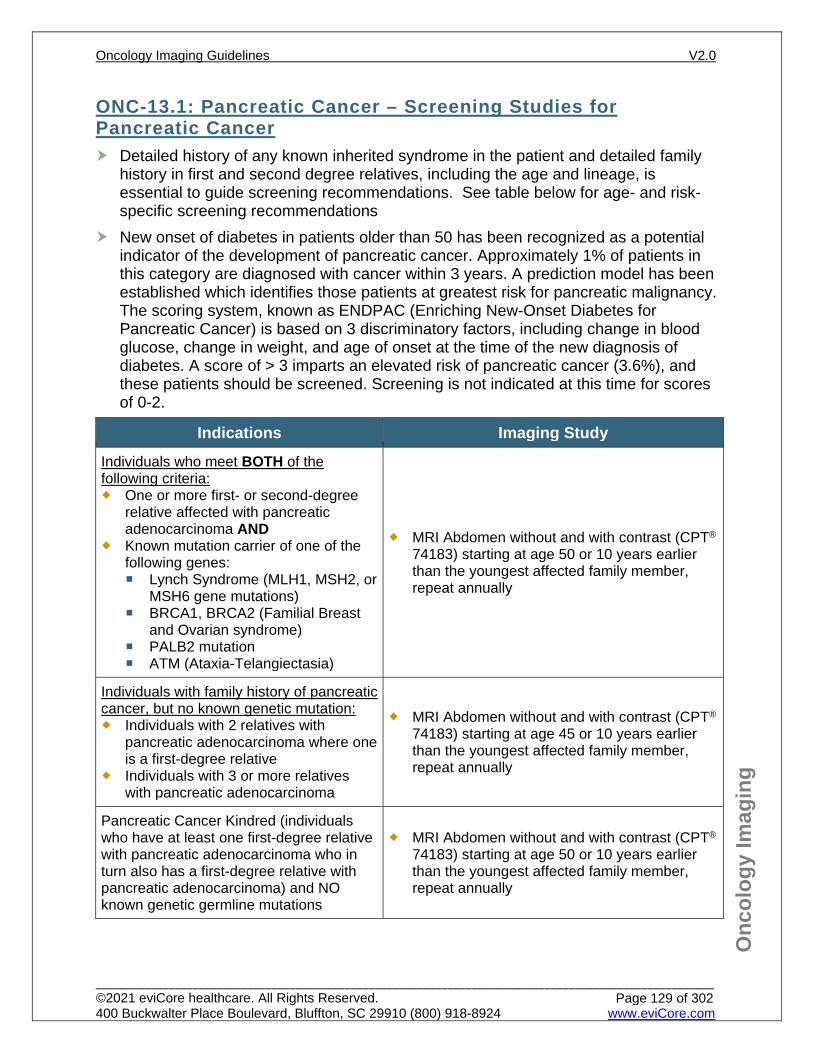
Onc
olog
y Im
agin
g
ONC-13.1: Pancreatic Cancer – Screening Studies for Pancreatic Cancer Detailed history of any known inherited syndrome in the patient and detailed family
history in first and second degree relatives, including the age and lineage, is essential to guide screening recommendations. See table below for age- and risk-specific screening recommendations
New onset of diabetes in patients older than 50 has been recognized as a potential indicator of the development of pancreatic cancer. Approximately 1% of patients in this category are diagnosed with cancer within 3 years. A prediction model has been established which identifies those patients at greatest risk for pancreatic malignancy. The scoring system, known as ENDPAC (Enriching New-Onset Diabetes for Pancreatic Cancer) is based on 3 discriminatory factors, including change in blood glucose, change in weight, and age of onset at the time of the new diagnosis of diabetes. A score of > 3 imparts an elevated risk of pancreatic cancer (3.6%), and these patients should be screened. Screening is not indicated at this time for scores of 0-2.
Indications Imaging Study Individuals who meet BOTH of the following criteria: One or more first- or second-degree
relative affected with pancreatic adenocarcinoma AND
Known mutation carrier of one of the following genes: Lynch Syndrome (MLH1, MSH2, or
MSH6 gene mutations) BRCA1, BRCA2 (Familial Breast
and Ovarian syndrome) PALB2 mutation ATM (Ataxia-Telangiectasia)
MRI Abdomen without and with contrast (CPT®
74183) starting at age 50 or 10 years earlier than the youngest affected family member, repeat annually
Individuals with family history of pancreatic cancer, but no known genetic mutation: Individuals with 2 relatives with
pancreatic adenocarcinoma where one is a first-degree relative
Individuals with 3 or more relatives with pancreatic adenocarcinoma
MRI Abdomen without and with contrast (CPT®
74183) starting at age 45 or 10 years earlier than the youngest affected family member, repeat annually
Pancreatic Cancer Kindred (individuals who have at least one first-degree relative with pancreatic adenocarcinoma who in turn also has a first-degree relative with pancreatic adenocarcinoma) and NO known genetic germline mutations
MRI Abdomen without and with contrast (CPT® 74183) starting at age 50 or 10 years earlier than the youngest affected family member, repeat annually
Oncology Imaging Guidelines V2.0
______________________________________________________________________________________________________ ©2021 eviCore healthcare. All Rights Reserved. 400 Buckwalter Place Boulevard, Bluffton, SC 29910 (800) 918-8924 www.eviCore.com
Page 129 of 302
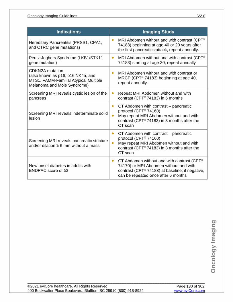
Onc
olog
y Im
agin
g
Indications Imaging Study
Hereditary Pancreatitis (PRSS1, CPA1, and CTRC gene mutations)
MRI Abdomen without and with contrast (CPT® 74183) beginning at age 40 or 20 years after the first pancreatitis attack, repeat annually.
Peutz-Jeghers Syndrome (LKB1/STK11 gene mutation)
MRI Abdomen without and with contrast (CPT® 74183) starting at age 30, repeat annually
CDKN2A mutation (also known as p16, p16INK4a, and MTS1, FAMM-Familial Atypical Multiple Melanoma and Mole Syndrome)
MRI Abdomen without and with contrast or MRCP (CPT® 74183) beginning at age 40, repeat annually.
Screening MRI reveals cystic lesion of the pancreas
Repeat MRI Abdomen without and with contrast (CPT® 74183) in 6 months
Screening MRI reveals indeterminate solid lesion
CT Abdomen with contrast – pancreatic protocol (CPT® 74160)
May repeat MRI Abdomen without and with contrast (CPT® 74183) in 3 months after the CT scan
Screening MRI reveals pancreatic stricture and/or dilation ≥ 6 mm without a mass
CT Abdomen with contrast – pancreatic protocol (CPT® 74160)
May repeat MRI Abdomen without and with contrast (CPT® 74183) in 3 months after the CT scan
New onset diabetes in adults with ENDPAC score of ≥3
CT Abdomen without and with contrast (CPT® 74170) or MRI Abdomen without and with contrast (CPT® 74183) at baseline; if negative, can be repeated once after 6 months
Oncology Imaging Guidelines V2.0
______________________________________________________________________________________________________ ©2021 eviCore healthcare. All Rights Reserved. 400 Buckwalter Place Boulevard, Bluffton, SC 29910 (800) 918-8924 www.eviCore.com
Page 130 of 302
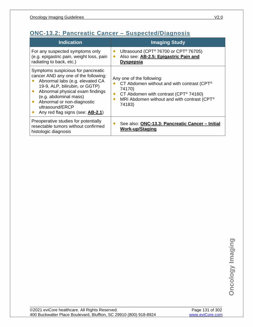
Onc
olog
y Im
agin
g
ONC-13.2: Pancreatic Cancer – Suspected/Diagnosis Indication Imaging Study
For any suspected symptoms only (e.g. epigastric pain, weight loss, pain radiating to back, etc.)
Ultrasound (CPT® 76700 or CPT® 76705) Also see: AB-2.5: Epigastric Pain and
Dyspepsia
Symptoms suspicious for pancreatic cancer AND any one of the following: Abnormal labs (e.g. elevated CA
19-9, ALP, bilirubin, or GGTP) Abnormal physical exam findings
(e.g. abdominal mass) Abnormal or non-diagnostic
ultrasound/ERCP Any red flag signs (see: AB-2.1)
Any one of the following: CT Abdomen without and with contrast (CPT®
74170) CT Abdomen with contrast (CPT® 74160) MRI Abdomen without and with contrast (CPT®
74183)
Preoperative studies for potentially resectable tumors without confirmed histologic diagnosis
See also: ONC-13.3: Pancreatic Cancer – Initial Work-up/Staging
Oncology Imaging Guidelines V2.0
______________________________________________________________________________________________________ ©2021 eviCore healthcare. All Rights Reserved. 400 Buckwalter Place Boulevard, Bluffton, SC 29910 (800) 918-8924 www.eviCore.com
Page 131 of 302
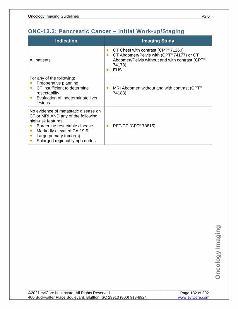
Onc
olog
y Im
agin
g
ONC-13.3: Pancreatic Cancer – Initial Work-up/Staging Indication Imaging Study
All patients
CT Chest with contrast (CPT® 71260) CT Abdomen/Pelvis with (CPT® 74177) or CT
Abdomen/Pelvis without and with contrast (CPT®
74178) EUS
For any of the following: Preoperative planning CT insufficient to determine
resectability Evaluation of indeterminate liver
lesions
MRI Abdomen without and with contrast (CPT®
74183)
No evidence of metastatic disease on CT or MRI AND any of the following high-risk features: Borderline resectable disease Markedly elevated CA 19-9 Large primary tumor(s) Enlarged regional lymph nodes
PET/CT (CPT® 78815)
Oncology Imaging Guidelines V2.0
______________________________________________________________________________________________________ ©2021 eviCore healthcare. All Rights Reserved. 400 Buckwalter Place Boulevard, Bluffton, SC 29910 (800) 918-8924 www.eviCore.com
Page 132 of 302
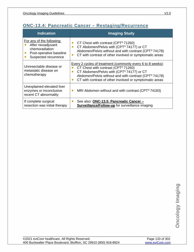
Onc
olog
y Im
agin
g
ONC-13.4: Pancreatic Cancer – Restaging/Recurrence Indication Imaging Study
For any of the following: After neoadjuvant
chemoradiation Post-operative baseline Suspected recurrence
CT Chest with contrast (CPT® 71260) CT Abdomen/Pelvis with (CPT® 74177) or CT
Abdomen/Pelvis without and with contrast (CPT® 74178) CT with contrast of other involved or symptomatic areas
Unresectable disease or metastatic disease on chemotherapy
Every 2 cycles of treatment (commonly every 6 to 8 weeks): CT Chest with contrast (CPT® 71260) CT Abdomen/Pelvis with (CPT® 74177) or CT
Abdomen/Pelvis without and with contrast (CPT® 74178) CT with contrast of other involved or symptomatic areas
Unexplained elevated liver enzymes or inconclusive recent CT abnormality
MRI Abdomen without and with contrast (CPT® 74183)
If complete surgical resection was initial therapy
See also: ONC-13.5: Pancreatic Cancer – Surveillance/Follow-up for surveillance imaging
Oncology Imaging Guidelines V2.0
______________________________________________________________________________________________________ ©2021 eviCore healthcare. All Rights Reserved. 400 Buckwalter Place Boulevard, Bluffton, SC 29910 (800) 918-8924 www.eviCore.com
Page 133 of 302
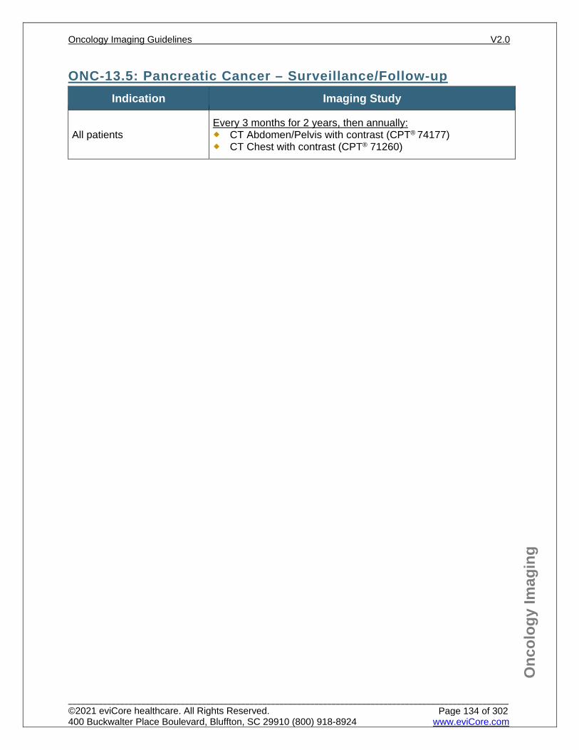
Onc
olog
y Im
agin
g
ONC-13.5: Pancreatic Cancer – Surveillance/Follow-up Indication Imaging Study
All patients Every 3 months for 2 years, then annually: CT Abdomen/Pelvis with contrast (CPT® 74177) CT Chest with contrast (CPT® 71260)
Oncology Imaging Guidelines V2.0
______________________________________________________________________________________________________ ©2021 eviCore healthcare. All Rights Reserved. 400 Buckwalter Place Boulevard, Bluffton, SC 29910 (800) 918-8924 www.eviCore.com
Page 134 of 302
Onc
olog
y Im
agin
g
References 1. Tempero MA, Malafa MP, Al-Hawary M, et al. National Comprehensive Cancer Network (NCCN)
Guidelines Version 2.2021 – February 25, 2021. Pancreatic Adenocarcinoma, available at: https://www.nccn.org/professionals/physician_gls/pdf/pancreatic.pdf Referenced with permission from the NCCN Clinical Practice Guidelines in Oncology (NCCN Guidelines™) for Pancreatic Adenocarcinoma V2.2021 – February 25, 2021. ©2021 National Comprehensive Cancer Network, Inc. All rights reserved. The NCCN Guidelines™ and illustrations herein may not be reproduced in any form for any purpose without the express written permission of the NCCN. To view the most recent and complete version of the NCCN Guidelines™, go online to NCCN.org.
2. Syngal S, Brand RE, Church JM, et al. ACG clinical guideline: genetic testing and management of hereditary gastrointestinal cancer syndromes. Am. J. Gastroenterol. 2015;110(2):223-262. doi:10.1038/ajg.2014.435.
3. Canto MI, Harinck F, Hruban RH, et al. International Cancer of the Pancreas Screening (CAPS) consortium summit on the management of patients with increased risk for familial pancreatic cancer. Gut. 2013;62(3):339-347. doi:10.1136/gutjnl-2012-303108.
4. U.S. Preventive Services Task Force. Screening for pancreatic cancer: recommendation statement. Rockville, Maryland: Agency for Healthcare Research and Quality (AHRQ); 2004.
5. Heinrich S, Goerres GW, Schafer M, et al. Positron emission tomography/computed tomography influences on the management of resectable pancreatic cancer and its cost-effectiveness. Ann Surg. 2005;242(2):235-243.
6. Gemmel C, Eickhoff A, Helmstädter L, Riemann JF. Pancreatic cancer screening: state of the art. Expert Rev Gastroenterol Hepatol. 2009;3(1):89-96. doi:10.1586/17474124.3.1.89.
7. Al-Hawary MM, Francis IR, Chari ST, et al. Pancreatic ductal adenocarcinoma radiology reporting template: consensus statement of the Society of Abdominal Radiology and the American Pancreatic Association. Gastroenterology. 2014;146(1):291-304. doi:10.1053/j.gastro.2013.11.004.
8. Tersmette AC, Petersen GM, Offerhaus GJ. Increased risk of incident pancreatic cancer among first-degree relatives of patients with familial pancreatic cancer. Clin Cancer Res. 2001;7(3):738-44.
9. Tzeng CW, Abbott DE, Cantor SB et al. Frequency and intensity of postoperative surveillance after curative treatment of pancreatic cancer: a cost-effectiveness analysis. Ann Surg Oncol. 2013;20(7):2197-2203. doi:10.1245/s10434-013-2889-6.
10. Furman MJ, Lambert LA, Sullivan ME, Whalen GF. Rational follow-up after curative cancer resection. Journal of Clinical Oncology. 2013;31(9):1130-1133. doi:10.1200/JCO.2012.46.4438.
11. Tzeng C, Fleming J, Lee J, et al. Yield of clinical and radiographic surveillance in patients with resected pancreatic adenocarcinoma following multimodal therapy. HPB. 2012;14(6):365-372. doi:10.1111/j.1477-2574.2012.00445.x.
12. Sharma, A, Kandlakunta H, Nagpal SJS, et.al. Model to determine risk of pancreatic cancer in patients with new-onset diabetes. Gastroenterology. 2018;155(3):730-739.
13. Goggins M, Overbeek KA, Brand R, et. al. Management of patients with increased risk for familial pancreatic cancer: updated recommendations from the International Cancer of the Pancreas Screening Consortium. Gut. 2020;69(1):7-17. doi:10.1136/gutjnl-2019-319352.
Oncology Imaging Guidelines V2.0
______________________________________________________________________________________________________ ©2021 eviCore healthcare. All Rights Reserved. 400 Buckwalter Place Boulevard, Bluffton, SC 29910 (800) 918-8924 www.eviCore.com
Page 135 of 302
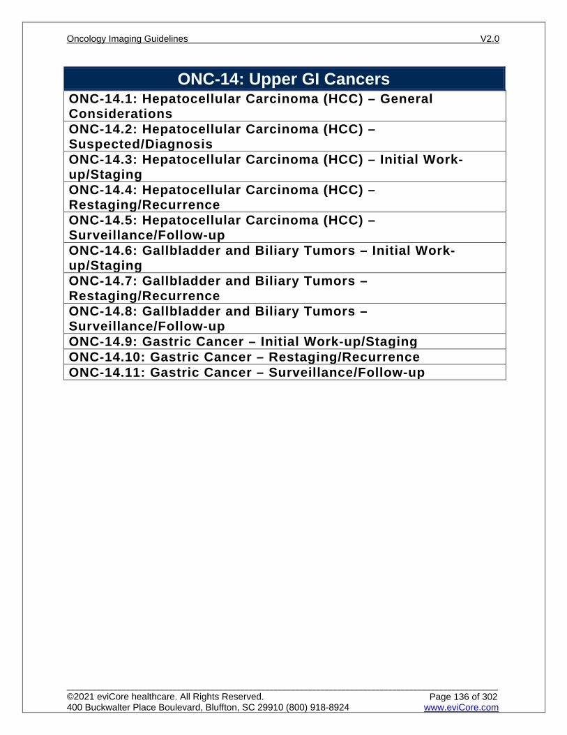
ONC-14: Upper GI Cancers ONC-14.1: Hepatocellular Carcinoma (HCC) – General ConsiderationsONC-14.2: Hepatocellular Carcinoma (HCC) – Suspected/DiagnosisONC-14.3: Hepatocellular Carcinoma (HCC) – Initial Work-up/StagingONC-14.4: Hepatocellular Carcinoma (HCC) – Restaging/RecurrenceONC-14.5: Hepatocellular Carcinoma (HCC) – Surveillance/Follow-upONC-14.6: Gallbladder and Biliary Tumors – Initial Work-up/StagingONC-14.7: Gallbladder and Biliary Tumors – Restaging/RecurrenceONC-14.8: Gallbladder and Biliary Tumors – Surveillance/Follow-upONC-14.9: Gastric Cancer – Initial Work-up/StagingONC-14.10: Gastric Cancer – Restaging/RecurrenceONC-14.11: Gastric Cancer – Surveillance/Follow-up
Oncology Imaging Guidelines V2.0
______________________________________________________________________________________________________ ©2021 eviCore healthcare. All Rights Reserved. 400 Buckwalter Place Boulevard, Bluffton, SC 29910 (800) 918-8924 www.eviCore.com
Page 136 of 302
Onc
olog
y Im
agin
g
ONC-14.1: Hepatocellular Carcinoma (HCC) – General Considerations Diagnosis: A biopsy is not always required for the diagnosis of Hepatocellular
carcinoma (HCC). A dedicated triple-phase CT or MRI may be obtained. MRI with contrast is the test of choice for the evaluation of liver masses and offers soft tissue contrast resolution superior to CT as well as the possibility of using two different contrast agents, one of which if more blood flow based and the other which also is blood flow based and demonstrates hepatobiliary function (Eovist).Classical imaging findings include: Arterial phase hyperenhancement Venous phase washout appearance Capsule appearance Threshold growth
For patients who are high risk for developing HCC (cirrhosis, chronic Hepatitis B or current or prior HCC), if the liver lesion is > 1 cm with 2 classic enhancements on triple-phase CT or MRI, the diagnosis is confirmatory and biopsy is not needed.
For lesions less than 1 cm or with less than 2 classical enhancements or for any liver lesions in patients who are not high risk, a biopsy is needed for histological confirmation.
PET/CT scan is not indicated for diagnosis or staging of Hepatocellular carcinoma
Oncology Imaging Guidelines V2.0
______________________________________________________________________________________________________ ©2021 eviCore healthcare. All Rights Reserved. 400 Buckwalter Place Boulevard, Bluffton, SC 29910 (800) 918-8924 www.eviCore.com
Page 137 of 302
Onc
olog
y Im
agin
g
ONC-14.2: Hepatocellular Carcinoma (HCC) – Suspected/Diagnosis See: AB-26.1: Chronic Liver Disease, Cirrhosis and Screening for HCC See: AB-29.1: Liver Lesion Characterization
Oncology Imaging Guidelines V2.0
______________________________________________________________________________________________________ ©2021 eviCore healthcare. All Rights Reserved. 400 Buckwalter Place Boulevard, Bluffton, SC 29910 (800) 918-8924 www.eviCore.com
Page 138 of 302
Onc
olog
y Im
agin
g
ONC-14.3: Hepatocellular Carcinoma (HCC) – Initial Work-up/Staging Indication Imaging Study
All patients
CT Chest with contrast (CPT® 71260) or CT Chest without contrast (CPT® 71250)
One of the following: CT Abdomen with contrast (CPT® 74160) CT Abdomen without and with contrast (CPT® 74170) CT Abdomen and Pelvis with contrast (CPT® 74177) or without and with
contrast (CPT® 74178) MRI Abdomen (CPT® 74183) and MRI Pelvis (CPT® 72197) without and
with contrast
Oncology Imaging Guidelines V2.0
______________________________________________________________________________________________________ ©2021 eviCore healthcare. All Rights Reserved. 400 Buckwalter Place Boulevard, Bluffton, SC 29910 (800) 918-8924 www.eviCore.com
Page 139 of 302
Onc
olog
y Im
agin
g
ONC-14.4: Hepatocellular Carcinoma (HCC) – Restaging/Recurrence Indication Imaging Study
One of the following: After initial therapy For suspected
recurrence or new liver lesions
Individuals receiving systemic therapy (every 2 cycles)
CT Chest with contrast (CPT® 71260) or CT Chest without contrast (CPT® 71250)
One of the following: CT Abdomen with contrast (CPT® 74160) CT Abdomen without and with contrast (CPT® 74170) CT Abdomen/Pelvis with contrast (CPT® 74177) or CT
Abdomen/Pelvis without and with contrast (CPT® 74178) MRI Abdomen (CPT® 74183) and MRI Pelvis (CPT® 72197)
without and with contrast
Hepatocellular Carcinoma treated with embolization
CTA Abdomen (CPT® 74175) can be approved immediately prior to embolization
One of the following, immediately prior to and 1 month post-ablation: MRI Abdomen without and with contrast (CPT® 74183) CT Abdomen without and with contrast (CPT® 74170)
See also: ONC-31.2 for imaging studies indicated prior to and post-embolization
Hepatocellular Carcinoma awaiting liver transplant
See: AB-42.1: Liver Transplant, Pre-Transplant for imaging guidelines
Oncology Imaging Guidelines V2.0
______________________________________________________________________________________________________ ©2021 eviCore healthcare. All Rights Reserved. 400 Buckwalter Place Boulevard, Bluffton, SC 29910 (800) 918-8924 www.eviCore.com
Page 140 of 302
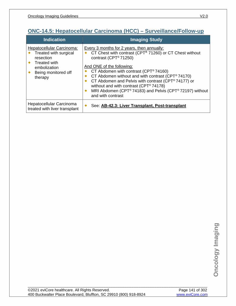
Onc
olog
y Im
agin
g
ONC-14.5: Hepatocellular Carcinoma (HCC) – Surveillance/Follow-up Indication Imaging Study
Hepatocellular Carcinoma: Treated with surgical
resection Treated with
embolization Being monitored off
therapy
Every 3 months for 2 years, then annually: CT Chest with contrast (CPT® 71260) or CT Chest without
contrast (CPT® 71250)
And ONE of the following: CT Abdomen with contrast (CPT® 74160) CT Abdomen without and with contrast (CPT® 74170) CT Abdomen and Pelvis with contrast (CPT® 74177) or
without and with contrast (CPT® 74178) MRI Abdomen (CPT® 74183) and Pelvis (CPT® 72197) without
and with contrast
Hepatocellular Carcinoma treated with liver transplant
See: AB-42.3: Liver Transplant, Post-transplant
Oncology Imaging Guidelines V2.0
______________________________________________________________________________________________________ ©2021 eviCore healthcare. All Rights Reserved. 400 Buckwalter Place Boulevard, Bluffton, SC 29910 (800) 918-8924 www.eviCore.com
Page 141 of 302
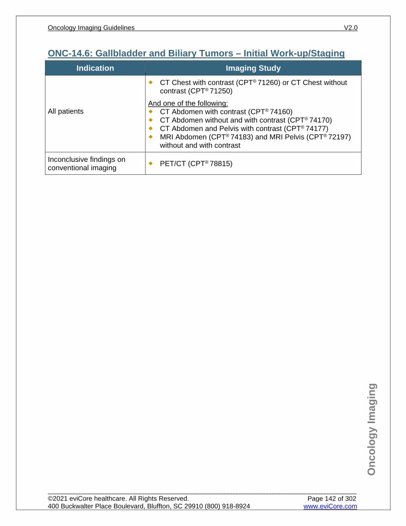
Onc
olog
y Im
agin
g
ONC-14.6: Gallbladder and Biliary Tumors – Initial Work-up/Staging Indication Imaging Study
All patients
CT Chest with contrast (CPT® 71260) or CT Chest without contrast (CPT® 71250)
And one of the following: CT Abdomen with contrast (CPT® 74160) CT Abdomen without and with contrast (CPT® 74170) CT Abdomen and Pelvis with contrast (CPT® 74177) MRI Abdomen (CPT® 74183) and MRI Pelvis (CPT® 72197)
without and with contrast
Inconclusive findings on conventional imaging PET/CT (CPT® 78815)
Oncology Imaging Guidelines V2.0
______________________________________________________________________________________________________ ©2021 eviCore healthcare. All Rights Reserved. 400 Buckwalter Place Boulevard, Bluffton, SC 29910 (800) 918-8924 www.eviCore.com
Page 142 of 302
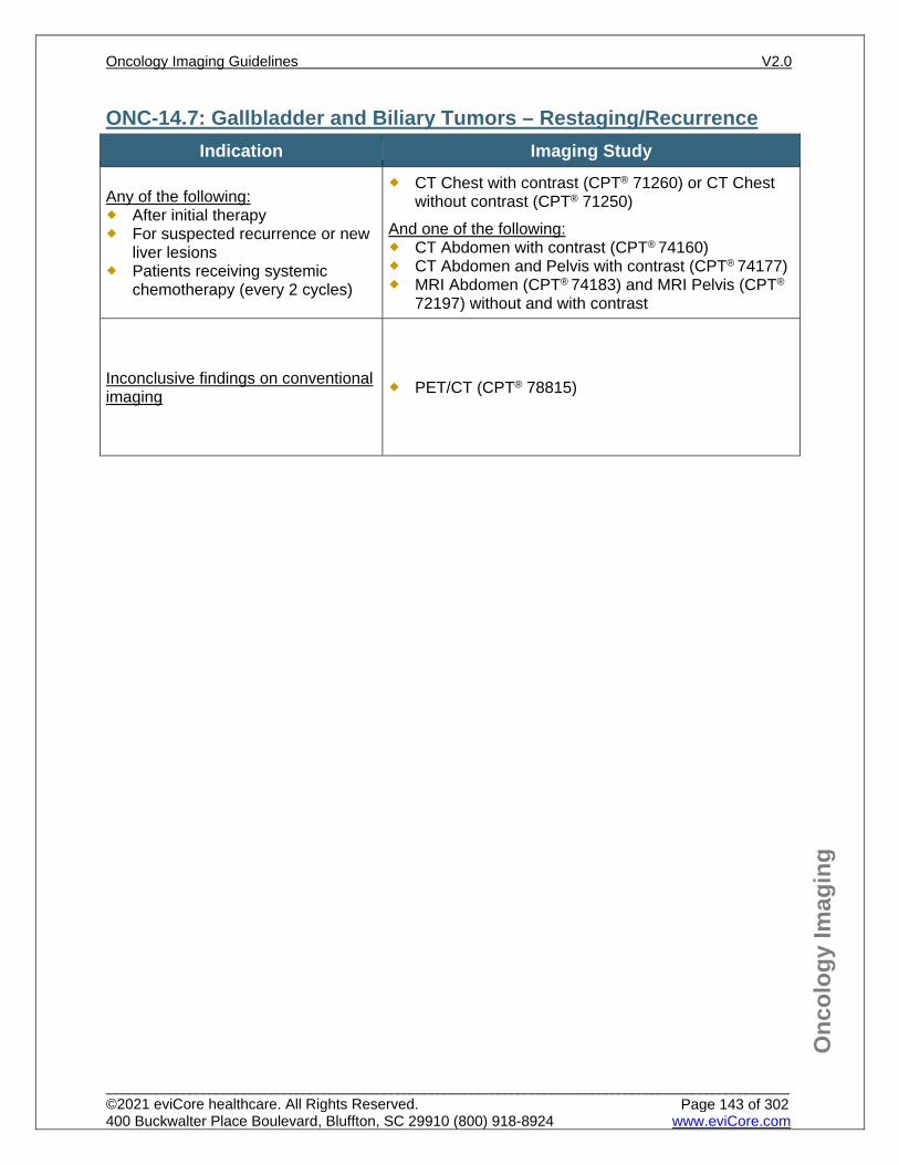
Onc
olog
y Im
agin
g
ONC-14.7: Gallbladder and Biliary Tumors – Restaging/Recurrence Indication Imaging Study
Any of the following: After initial therapy For suspected recurrence or new
liver lesions Patients receiving systemic
chemotherapy (every 2 cycles)
CT Chest with contrast (CPT® 71260) or CT Chest without contrast (CPT® 71250)
And one of the following: CT Abdomen with contrast (CPT® 74160) CT Abdomen and Pelvis with contrast (CPT® 74177) MRI Abdomen (CPT® 74183) and MRI Pelvis (CPT®
72197) without and with contrast
Inconclusive findings on conventional imaging PET/CT (CPT® 78815)
Oncology Imaging Guidelines V2.0
______________________________________________________________________________________________________ ©2021 eviCore healthcare. All Rights Reserved. 400 Buckwalter Place Boulevard, Bluffton, SC 29910 (800) 918-8924 www.eviCore.com
Page 143 of 302
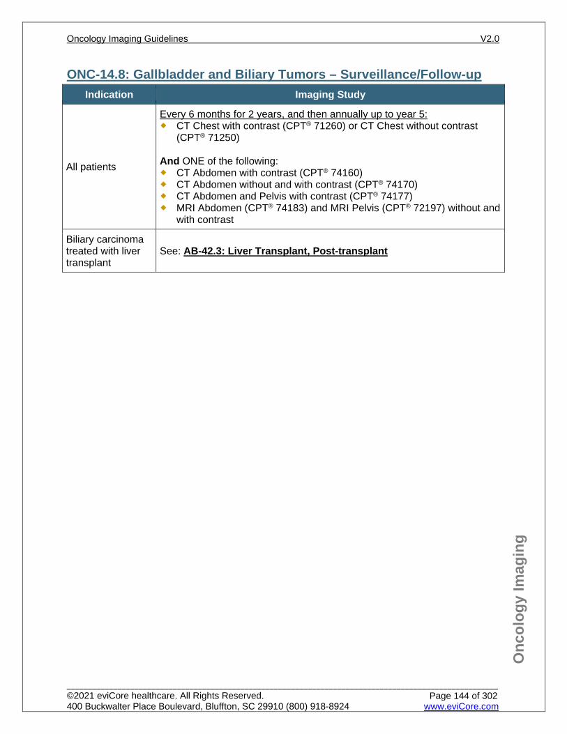
Onc
olog
y Im
agin
g
ONC-14.8: Gallbladder and Biliary Tumors – Surveillance/Follow-up Indication Imaging Study
All patients
Every 6 months for 2 years, and then annually up to year 5: CT Chest with contrast (CPT® 71260) or CT Chest without contrast
(CPT® 71250)
And ONE of the following: CT Abdomen with contrast (CPT® 74160) CT Abdomen without and with contrast (CPT® 74170) CT Abdomen and Pelvis with contrast (CPT® 74177) MRI Abdomen (CPT® 74183) and MRI Pelvis (CPT® 72197) without and
with contrast
Biliary carcinoma treated with liver transplant
See: AB-42.3: Liver Transplant, Post-transplant
Oncology Imaging Guidelines V2.0
______________________________________________________________________________________________________ ©2021 eviCore healthcare. All Rights Reserved. 400 Buckwalter Place Boulevard, Bluffton, SC 29910 (800) 918-8924 www.eviCore.com
Page 144 of 302
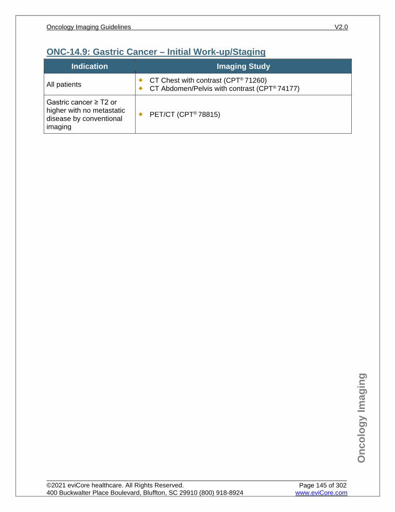
Onc
olog
y Im
agin
g
ONC-14.9: Gastric Cancer – Initial Work-up/Staging Indication Imaging Study
All patients CT Chest with contrast (CPT® 71260) CT Abdomen/Pelvis with contrast (CPT® 74177)
Gastric cancer ≥ T2 or higher with no metastatic disease by conventional imaging
PET/CT (CPT® 78815)
Oncology Imaging Guidelines V2.0
______________________________________________________________________________________________________ ©2021 eviCore healthcare. All Rights Reserved. 400 Buckwalter Place Boulevard, Bluffton, SC 29910 (800) 918-8924 www.eviCore.com
Page 145 of 302
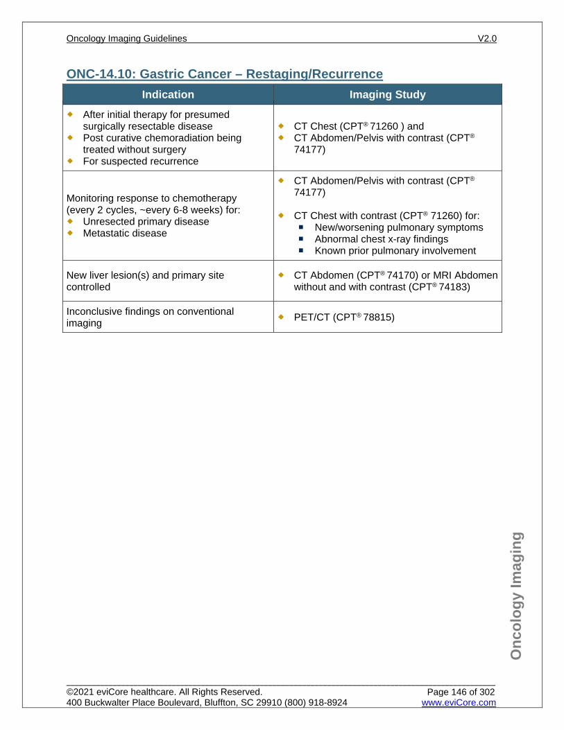
Onc
olog
y Im
agin
g
ONC-14.10: Gastric Cancer – Restaging/Recurrence Indication Imaging Study
After initial therapy for presumed surgically resectable disease
Post curative chemoradiation being treated without surgery
For suspected recurrence
CT Chest (CPT® 71260 ) and CT Abdomen/Pelvis with contrast (CPT®
74177)
Monitoring response to chemotherapy (every 2 cycles, ~every 6-8 weeks) for: Unresected primary disease Metastatic disease
CT Abdomen/Pelvis with contrast (CPT® 74177)
CT Chest with contrast (CPT® 71260) for:
New/worsening pulmonary symptoms Abnormal chest x-ray findings Known prior pulmonary involvement
New liver lesion(s) and primary site controlled
CT Abdomen (CPT® 74170) or MRI Abdomen without and with contrast (CPT® 74183)
Inconclusive findings on conventional imaging PET/CT (CPT® 78815)
Oncology Imaging Guidelines V2.0
______________________________________________________________________________________________________ ©2021 eviCore healthcare. All Rights Reserved. 400 Buckwalter Place Boulevard, Bluffton, SC 29910 (800) 918-8924 www.eviCore.com
Page 146 of 302
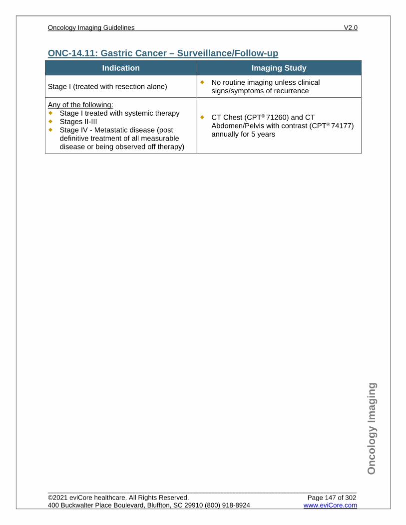
Onc
olog
y Im
agin
g
ONC-14.11: Gastric Cancer – Surveillance/Follow-up Indication Imaging Study
Stage I (treated with resection alone) No routine imaging unless clinical signs/symptoms of recurrence
Any of the following: Stage I treated with systemic therapy Stages II-III Stage IV - Metastatic disease (post
definitive treatment of all measurable disease or being observed off therapy)
CT Chest (CPT® 71260) and CT Abdomen/Pelvis with contrast (CPT® 74177) annually for 5 years
Oncology Imaging Guidelines V2.0
______________________________________________________________________________________________________ ©2021 eviCore healthcare. All Rights Reserved. 400 Buckwalter Place Boulevard, Bluffton, SC 29910 (800) 918-8924 www.eviCore.com
Page 147 of 302
Onc
olog
y Im
agin
g
References 1. Ajani JA, D’Amico TA, Bentrem DJ et al. National Comprehensive Cancer Network (NCCN)
Guidelines Version 1.2021 – February 9, 2021. Gastric cancer, available at: https://www.nccn.org/professionals/physician_gls/pdf/gastric.pdf Referenced with permission from the NCCN Clinical Practice Guidelines in Oncology (NCCN Guidelines™) for Gastric cancer V1.2021– February 9, 2021. ©2021 National Comprehensive Cancer Network, Inc. All rights reserved. The NCCN Guidelines™ and illustrations herein may not be reproduced in any form for any purpose without the express written permission of the NCCN. To view the most recent and complete version of the NCCN Guidelines™, go online to NCCN.org.
2. Benson AB, D’Angelica MI, Abbot D et al. National Comprehensive Cancer Network (NCCN) Guidelines Version 5.2020 – August 4, 2020. Hepatobiliary cancers, available at: https://www.nccn.org/professionals/physician_gls/pdf/hepatobiliary.pdf Referenced with permission from the NCCN Clinical Practice Guidelines in Oncology (NCCN Guidelines™) for Hepatobiliary cancers, V5.2020 – August 4, 2020. ©2020 National Comprehensive Cancer Network, Inc. All rights reserved. The NCCN Guidelines™ and illustrations herein may not be reproduced in any form for any purpose without the express written permission of the NCCN. To view the most recent and complete version of the NCCN Guidelines™, go online to NCCN.org.
3. Vallböhmer D, Hölscher AH, Schnieder PM, et al. [18F]-fluorodeoxyglucose-positron emission tomography for the assessment of histopathologic response and prognosis after completion of neoadjuvant chemotherapy in gastric cancer. J Surg Oncol. 2010;102(2):135-140. doi:10.1002/jso.21592.
4. Zou H, Zhao Y. 18FDG PET-CT for detecting gastric cancer recurrence after surgical resection: a meta-analysis. Surg Oncol. 2013;22(3):162-166. doi:10.1016/j.suronc.2013.05.001.
5. Bridgewater J, Galle PR, Khan SA, et al. Guidelines for the diagnosis and management of intrahepatic cholangiocarcinoma. J Hepatol. 2014;60(6):1268-1289. doi:10.1016/j.jhep.2014.01.021.
6. Khan SA, Davidson BR, Goldin RD, et al. Guidelines for the diagnosis and treatment of cholangiocarcinoma: an update. Gut. 2012;61(12):1657-1669. doi:10.1136/gutjnl-2011-301748.
7. Benson AB 3rd, D’Angelica MI, Abrams TA, et al. Hepatobiliary cancers, version 2.2014. J Natl Compr Canc Netw. 2014;12(8):1152-1182.
Oncology Imaging Guidelines V2.0
______________________________________________________________________________________________________ ©2021 eviCore healthcare. All Rights Reserved. 400 Buckwalter Place Boulevard, Bluffton, SC 29910 (800) 918-8924 www.eviCore.com
Page 148 of 302
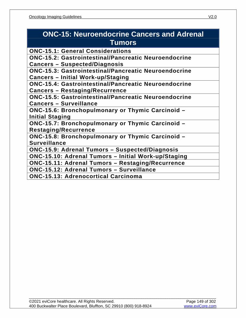
ONC-15: Neuroendocrine Cancers and Adrenal Tumors
ONC-15.1: General ConsiderationsONC-15.2: Gastrointestinal/Pancreatic Neuroendocrine Cancers – Suspected/DiagnosisONC-15.3: Gastrointestinal/Pancreatic Neuroendocrine Cancers – Initial Work-up/StagingONC-15.4: Gastrointestinal/Pancreatic Neuroendocrine Cancers – Restaging/RecurrenceONC-15.5: Gastrointestinal/Pancreatic Neuroendocrine Cancers – SurveillanceONC-15.6: Bronchopulmonary or Thymic Carcinoid – Initial StagingONC-15.7: Bronchopulmonary or Thymic Carcinoid –Restaging/RecurrenceONC-15.8: Bronchopulmonary or Thymic Carcinoid – SurveillanceONC-15.9: Adrenal Tumors – Suspected/DiagnosisONC-15.10: Adrenal Tumors – Initial Work-up/StagingONC-15.11: Adrenal Tumors – Restaging/RecurrenceONC-15.12: Adrenal Tumors – SurveillanceONC-15.13: Adrenocortical Carcinoma
Oncology Imaging Guidelines V2.0
______________________________________________________________________________________________________ ©2021 eviCore healthcare. All Rights Reserved. 400 Buckwalter Place Boulevard, Bluffton, SC 29910 (800) 918-8924 www.eviCore.com
Page 149 of 302
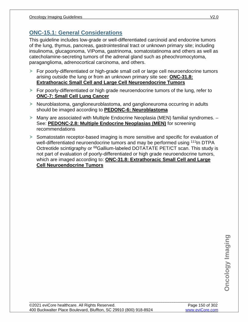
Onc
olog
y Im
agin
g
ONC-15.1: General Considerations This guideline includes low-grade or well-differentiated carcinoid and endocrine tumors of the lung, thymus, pancreas, gastrointestinal tract or unknown primary site; including insulinoma, glucagonoma, VIPoma, gastrinoma, somatostatinoma and others as well as catecholamine-secreting tumors of the adrenal gland such as pheochromocytoma, paraganglioma, adrenocortical carcinoma, and others.
For poorly-differentiated or high-grade small cell or large cell neuroendocrine tumors arising outside the lung or from an unknown primary site see: ONC-31.8: Extrathoracic Small Cell and Large Cell Neuroendocrine Tumors
For poorly-differentiated or high grade neuroendocrine tumors of the lung, refer to ONC-7: Small Cell Lung Cancer
Neuroblastoma, ganglioneuroblastoma, and ganglioneuroma occurring in adults should be imaged according to PEDONC-6: Neuroblastoma
Many are associated with Multiple Endocrine Neoplasia (MEN) familial syndromes. – See: PEDONC-2.8: Multiple Endocrine Neoplasias (MEN) for screening recommendations
Somatostatin receptor-based imaging is more sensitive and specific for evaluation of well-differentiated neuroendocrine tumors and may be performed using 111In DTPA Octreotide scintigraphy or 68Gallium-labeled DOTATATE PET/CT scan. This study is not part of evaluation of poorly-differentiated or high grade neuroendocrine tumors, which are imaged according to: ONC-31.8: Extrathoracic Small Cell and Large Cell Neuroendocrine Tumors
Oncology Imaging Guidelines V2.0
______________________________________________________________________________________________________ ©2021 eviCore healthcare. All Rights Reserved. 400 Buckwalter Place Boulevard, Bluffton, SC 29910 (800) 918-8924 www.eviCore.com
Page 150 of 302
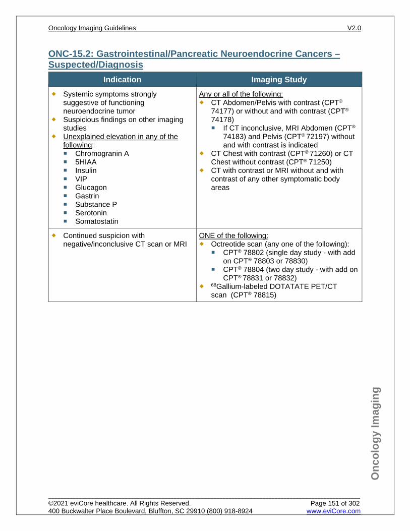
Onc
olog
y Im
agin
g
ONC-15.2: Gastrointestinal/Pancreatic Neuroendocrine Cancers – Suspected/Diagnosis
Indication Imaging Study Systemic symptoms strongly
suggestive of functioning neuroendocrine tumor
Suspicious findings on other imaging studies
Unexplained elevation in any of the following: Chromogranin A 5HIAA Insulin VIP Glucagon Gastrin Substance P Serotonin Somatostatin
Any or all of the following: CT Abdomen/Pelvis with contrast (CPT®
74177) or without and with contrast (CPT®
74178) If CT inconclusive, MRI Abdomen (CPT®
74183) and Pelvis (CPT® 72197) without and with contrast is indicated
CT Chest with contrast (CPT® 71260) or CT Chest without contrast (CPT® 71250)
CT with contrast or MRI without and with contrast of any other symptomatic body areas
Continued suspicion with negative/inconclusive CT scan or MRI
ONE of the following: Octreotide scan (any one of the following):
CPT® 78802 (single day study - with add on CPT® 78803 or 78830)
CPT® 78804 (two day study - with add on CPT® 78831 or 78832)
68Gallium-labeled DOTATATE PET/CT scan (CPT® 78815)
Oncology Imaging Guidelines V2.0
______________________________________________________________________________________________________ ©2021 eviCore healthcare. All Rights Reserved. 400 Buckwalter Place Boulevard, Bluffton, SC 29910 (800) 918-8924 www.eviCore.com
Page 151 of 302
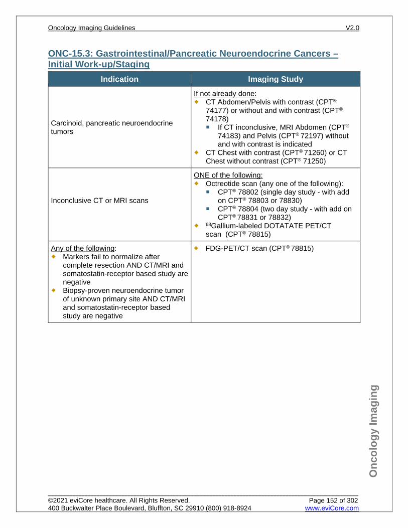
Onc
olog
y Im
agin
g
ONC-15.3: Gastrointestinal/Pancreatic Neuroendocrine Cancers – Initial Work-up/Staging
Indication Imaging Study
Carcinoid, pancreatic neuroendocrine tumors
If not already done: CT Abdomen/Pelvis with contrast (CPT®
74177) or without and with contrast (CPT®
74178) If CT inconclusive, MRI Abdomen (CPT®
74183) and Pelvis (CPT® 72197) without and with contrast is indicated
CT Chest with contrast (CPT® 71260) or CT Chest without contrast (CPT® 71250)
Inconclusive CT or MRI scans
ONE of the following: Octreotide scan (any one of the following):
CPT® 78802 (single day study - with add on CPT® 78803 or 78830)
CPT® 78804 (two day study - with add on CPT® 78831 or 78832)
68Gallium-labeled DOTATATE PET/CT scan (CPT® 78815)
Any of the following: Markers fail to normalize after
complete resection AND CT/MRI and somatostatin-receptor based study are negative
Biopsy-proven neuroendocrine tumor of unknown primary site AND CT/MRI and somatostatin-receptor based study are negative
FDG-PET/CT scan (CPT® 78815)
Oncology Imaging Guidelines V2.0
______________________________________________________________________________________________________ ©2021 eviCore healthcare. All Rights Reserved. 400 Buckwalter Place Boulevard, Bluffton, SC 29910 (800) 918-8924 www.eviCore.com
Page 152 of 302
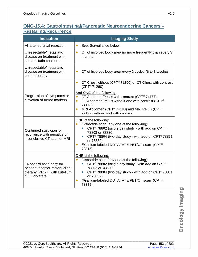
Onc
olog
y Im
agin
g
ONC-15.4: Gastrointestinal/Pancreatic Neuroendocrine Cancers – Restaging/Recurrence
Indication Imaging Study All after surgical resection See: Surveillance below
Unresectable/metastatic disease on treatment with somatostatin analogues
CT of involved body area no more frequently than every 3 months
Unresectable/metastatic disease on treatment with chemotherapy
CT of involved body area every 2 cycles (6 to 8 weeks)
Progression of symptoms or elevation of tumor markers
CT Chest without (CPT® 71250) or CT Chest with contrast (CPT® 71260)
And ONE of the following: CT Abdomen/Pelvis with contrast (CPT® 74177) CT Abdomen/Pelvis without and with contrast (CPT®
74178) MRI Abdomen (CPT® 74183) and MRI Pelvis (CPT®
72197) without and with contrast
Continued suspicion for recurrence with negative or inconclusive CT scan or MRI
ONE of the following: Octreotide scan (any one of the following):
CPT® 78802 (single day study - with add on CPT® 78803 or 78830)
CPT® 78804 (two day study - with add on CPT® 78831 or 78832)
68Gallium-labeled DOTATATE PET/CT scan (CPT® 78815)
To assess candidacy for peptide receptor radionuclide therapy (PRRT) with Lutetium 177Lu-dotatate
ONE of the following: Octreotide scan (any one of the following):
CPT® 78802 (single day study - with add on CPT® 78803 or 78830)
CPT® 78804 (two day study - with add on CPT® 78831 or 78832)
68Gallium-labeled DOTATATE PET/CT scan (CPT® 78815)
Oncology Imaging Guidelines V2.0
______________________________________________________________________________________________________ ©2021 eviCore healthcare. All Rights Reserved. 400 Buckwalter Place Boulevard, Bluffton, SC 29910 (800) 918-8924 www.eviCore.com
Page 153 of 302
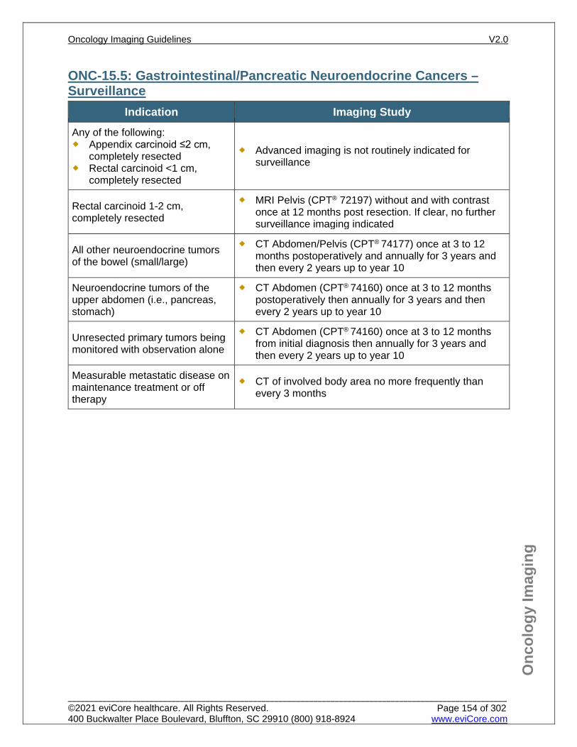
Onc
olog
y Im
agin
g
ONC-15.5: Gastrointestinal/Pancreatic Neuroendocrine Cancers – Surveillance
Indication Imaging Study Any of the following: Appendix carcinoid ≤2 cm,
completely resected Rectal carcinoid <1 cm,
completely resected
Advanced imaging is not routinely indicated for surveillance
Rectal carcinoid 1-2 cm, completely resected
MRI Pelvis (CPT® 72197) without and with contrast once at 12 months post resection. If clear, no further surveillance imaging indicated
All other neuroendocrine tumors of the bowel (small/large)
CT Abdomen/Pelvis (CPT® 74177) once at 3 to 12 months postoperatively and annually for 3 years and then every 2 years up to year 10
Neuroendocrine tumors of the upper abdomen (i.e., pancreas, stomach)
CT Abdomen (CPT® 74160) once at 3 to 12 months postoperatively then annually for 3 years and then every 2 years up to year 10
Unresected primary tumors being monitored with observation alone
CT Abdomen (CPT® 74160) once at 3 to 12 months from initial diagnosis then annually for 3 years and then every 2 years up to year 10
Measurable metastatic disease on maintenance treatment or off therapy
CT of involved body area no more frequently than every 3 months
Oncology Imaging Guidelines V2.0
______________________________________________________________________________________________________ ©2021 eviCore healthcare. All Rights Reserved. 400 Buckwalter Place Boulevard, Bluffton, SC 29910 (800) 918-8924 www.eviCore.com
Page 154 of 302
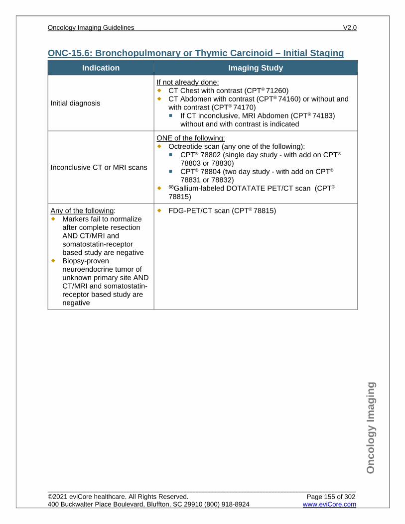
Onc
olog
y Im
agin
g
ONC-15.6: Bronchopulmonary or Thymic Carcinoid – Initial Staging Indication Imaging Study
Initial diagnosis
If not already done: CT Chest with contrast (CPT® 71260) CT Abdomen with contrast (CPT® 74160) or without and
with contrast (CPT® 74170) If CT inconclusive, MRI Abdomen (CPT® 74183)
without and with contrast is indicated
Inconclusive CT or MRI scans
ONE of the following: Octreotide scan (any one of the following):
CPT® 78802 (single day study - with add on CPT® 78803 or 78830)
CPT® 78804 (two day study - with add on CPT®
78831 or 78832) 68Gallium-labeled DOTATATE PET/CT scan (CPT®
78815)
Any of the following: Markers fail to normalize
after complete resection AND CT/MRI and somatostatin-receptor based study are negative
Biopsy-proven neuroendocrine tumor of unknown primary site AND CT/MRI and somatostatin-receptor based study are negative
FDG-PET/CT scan (CPT® 78815)
Oncology Imaging Guidelines V2.0
______________________________________________________________________________________________________ ©2021 eviCore healthcare. All Rights Reserved. 400 Buckwalter Place Boulevard, Bluffton, SC 29910 (800) 918-8924 www.eviCore.com
Page 155 of 302
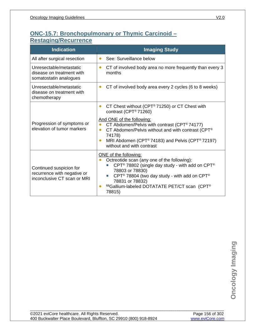
Onc
olog
y Im
agin
g
ONC-15.7: Bronchopulmonary or Thymic Carcinoid – Restaging/Recurrence
Indication Imaging Study All after surgical resection See: Surveillance below
Unresectable/metastatic disease on treatment with somatostatin analogues
CT of involved body area no more frequently than every 3 months
Unresectable/metastatic disease on treatment with chemotherapy
CT of involved body area every 2 cycles (6 to 8 weeks)
Progression of symptoms or elevation of tumor markers
CT Chest without (CPT® 71250) or CT Chest with contrast (CPT® 71260)
And ONE of the following: CT Abdomen/Pelvis with contrast (CPT® 74177) CT Abdomen/Pelvis without and with contrast (CPT®
74178) MRI Abdomen (CPT® 74183) and Pelvis (CPT® 72197)
without and with contrast
Continued suspicion for recurrence with negative or inconclusive CT scan or MRI
ONE of the following: Octreotide scan (any one of the following):
CPT® 78802 (single day study - with add on CPT® 78803 or 78830)
CPT® 78804 (two day study - with add on CPT®
78831 or 78832) 68Gallium-labeled DOTATATE PET/CT scan (CPT®
78815)
Oncology Imaging Guidelines V2.0
______________________________________________________________________________________________________ ©2021 eviCore healthcare. All Rights Reserved. 400 Buckwalter Place Boulevard, Bluffton, SC 29910 (800) 918-8924 www.eviCore.com
Page 156 of 302
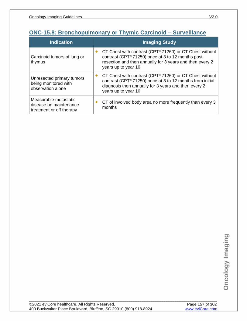
Onc
olog
y Im
agin
g
ONC-15.8: Bronchopulmonary or Thymic Carcinoid – Surveillance Indication Imaging Study
Carcinoid tumors of lung or thymus
CT Chest with contrast (CPT® 71260) or CT Chest without contrast (CPT® 71250) once at 3 to 12 months post resection and then annually for 3 years and then every 2 years up to year 10
Unresected primary tumors being monitored with observation alone
CT Chest with contrast (CPT® 71260) or CT Chest without contrast (CPT® 71250) once at 3 to 12 months from initial diagnosis then annually for 3 years and then every 2 years up to year 10
Measurable metastatic disease on maintenance treatment or off therapy
CT of involved body area no more frequently than every 3 months
Oncology Imaging Guidelines V2.0
______________________________________________________________________________________________________ ©2021 eviCore healthcare. All Rights Reserved. 400 Buckwalter Place Boulevard, Bluffton, SC 29910 (800) 918-8924 www.eviCore.com
Page 157 of 302
Onc
olog
y Im
agin
g
ONC-15.9: Adrenal Tumors – Suspected/Diagnosis See: AB-16.1: Adrenal Cortical Lesions for imaging guidelines for evaluation of suspected adrenal malignancies
If concern for genetic predisposition syndrome such as MEN, neurofibromatosis, or Von Hippel-Lindau disease, see screening recommendations in PEDONC-2: Screening Imaging and Cancer Predisposition Syndromes.
Oncology Imaging Guidelines V2.0
______________________________________________________________________________________________________ ©2021 eviCore healthcare. All Rights Reserved. 400 Buckwalter Place Boulevard, Bluffton, SC 29910 (800) 918-8924 www.eviCore.com
Page 158 of 302
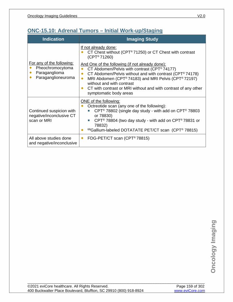
Onc
olog
y Im
agin
g
ONC-15.10: Adrenal Tumors – Initial Work-up/Staging Indication Imaging Study
For any of the following: Pheochromocytoma Paraganglioma Paraganglioneuroma
If not already done: CT Chest without (CPT® 71250) or CT Chest with contrast
(CPT® 71260) And One of the following (if not already done): CT Abdomen/Pelvis with contrast (CPT® 74177) CT Abdomen/Pelvis without and with contrast (CPT® 74178) MRI Abdomen (CPT® 74183) and MRI Pelvis (CPT® 72197)
without and with contrast CT with contrast or MRI without and with contrast of any other
symptomatic body areas
Continued suspicion with negative/inconclusive CT scan or MRI
ONE of the following: Octreotide scan (any one of the following):
CPT® 78802 (single day study - with add on CPT® 78803 or 78830)
CPT® 78804 (two day study - with add on CPT® 78831 or 78832)
68Gallium-labeled DOTATATE PET/CT scan (CPT® 78815)
All above studies done and negative/inconclusive
FDG-PET/CT scan (CPT® 78815)
Oncology Imaging Guidelines V2.0
______________________________________________________________________________________________________ ©2021 eviCore healthcare. All Rights Reserved. 400 Buckwalter Place Boulevard, Bluffton, SC 29910 (800) 918-8924 www.eviCore.com
Page 159 of 302
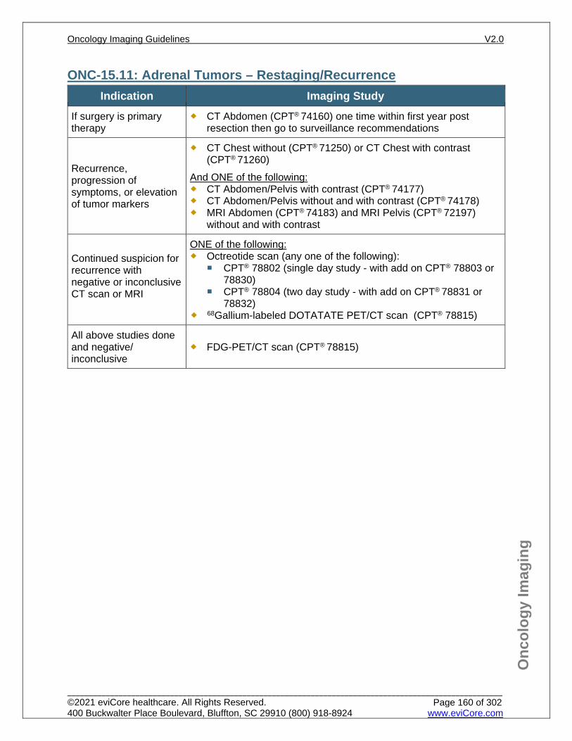
Onc
olog
y Im
agin
g
ONC-15.11: Adrenal Tumors – Restaging/Recurrence Indication Imaging Study
If surgery is primary therapy
CT Abdomen (CPT® 74160) one time within first year post resection then go to surveillance recommendations
Recurrence, progression of symptoms, or elevation of tumor markers
CT Chest without (CPT® 71250) or CT Chest with contrast (CPT® 71260)
And ONE of the following: CT Abdomen/Pelvis with contrast (CPT® 74177) CT Abdomen/Pelvis without and with contrast (CPT® 74178) MRI Abdomen (CPT® 74183) and MRI Pelvis (CPT® 72197)
without and with contrast
Continued suspicion for recurrence with negative or inconclusive CT scan or MRI
ONE of the following: Octreotide scan (any one of the following):
CPT® 78802 (single day study - with add on CPT® 78803 or 78830)
CPT® 78804 (two day study - with add on CPT® 78831 or 78832)
68Gallium-labeled DOTATATE PET/CT scan (CPT® 78815)
All above studies done and negative/ inconclusive
FDG-PET/CT scan (CPT® 78815)
Oncology Imaging Guidelines V2.0
______________________________________________________________________________________________________ ©2021 eviCore healthcare. All Rights Reserved. 400 Buckwalter Place Boulevard, Bluffton, SC 29910 (800) 918-8924 www.eviCore.com
Page 160 of 302
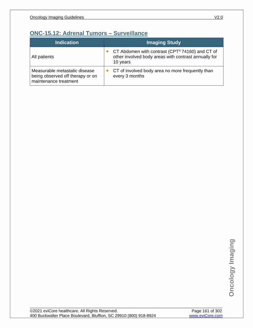
Onc
olog
y Im
agin
g
ONC-15.12: Adrenal Tumors – Surveillance Indication Imaging Study
All patients CT Abdomen with contrast (CPT® 74160) and CT of
other involved body areas with contrast annually for 10 years
Measurable metastatic disease being observed off therapy or on maintenance treatment
CT of involved body area no more frequently than every 3 months
Oncology Imaging Guidelines V2.0
______________________________________________________________________________________________________ ©2021 eviCore healthcare. All Rights Reserved. 400 Buckwalter Place Boulevard, Bluffton, SC 29910 (800) 918-8924 www.eviCore.com
Page 161 of 302
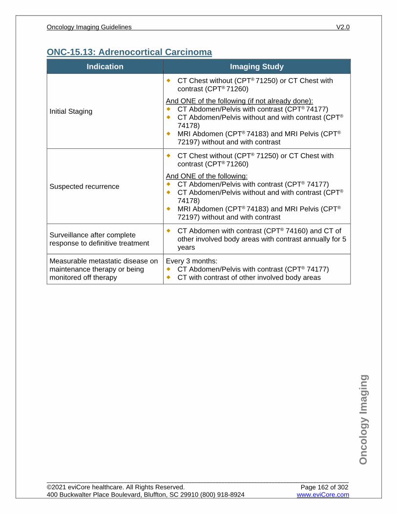
Onc
olog
y Im
agin
g
ONC-15.13: Adrenocortical Carcinoma Indication Imaging Study
Initial Staging
CT Chest without (CPT® 71250) or CT Chest with contrast (CPT® 71260)
And ONE of the following (if not already done): CT Abdomen/Pelvis with contrast (CPT® 74177) CT Abdomen/Pelvis without and with contrast (CPT®
74178) MRI Abdomen (CPT® 74183) and MRI Pelvis (CPT®
72197) without and with contrast
Suspected recurrence
CT Chest without (CPT® 71250) or CT Chest with contrast (CPT® 71260)
And ONE of the following: CT Abdomen/Pelvis with contrast (CPT® 74177) CT Abdomen/Pelvis without and with contrast (CPT®
74178) MRI Abdomen (CPT® 74183) and MRI Pelvis (CPT®
72197) without and with contrast
Surveillance after complete response to definitive treatment
CT Abdomen with contrast (CPT® 74160) and CT of other involved body areas with contrast annually for 5 years
Measurable metastatic disease on maintenance therapy or being monitored off therapy
Every 3 months: CT Abdomen/Pelvis with contrast (CPT® 74177) CT with contrast of other involved body areas
Oncology Imaging Guidelines V2.0
______________________________________________________________________________________________________ ©2021 eviCore healthcare. All Rights Reserved. 400 Buckwalter Place Boulevard, Bluffton, SC 29910 (800) 918-8924 www.eviCore.com
Page 162 of 302
Onc
olog
y Im
agin
g
References 1. Benson III AB, D ’Angelica MI, Abbott DE, et al. National Comprehensive Cancer Network (NCCN)
Guidelines Version 2.2020 – July 24, 2020. Neuroendocrine tumors, available at: https://www.nccn.org/professionals/physician_gls/pdf/neuroendocrine.pdf Referenced with permission from the NCCN Clinical Practice Guidelines in Oncology (NCCN Guidelines™) for Neuroendocrine tumors V2.2020 – July 24, 2020. ©2020 National Comprehensive Cancer Network, Inc. All rights reserved. The NCCN Guidelines™ and illustrations herein may not be reproduced in any form for any purpose without the express written permission of the NCCN. To view the most recent and complete version of the NCCN Guidelines™, go online to NCCN.org.
2. Qadan M, Ma Y, Visser BC, et al. Reassessment of the current American Join Committee on Cancer staging system for pancreatic neuroendocrine tumors. J Am Coll Surg. 2014;218(2):188-195. doi:10.1016/j.jamcollsurg.2013.11.001.
3. Lenders JWM, Duh Q-Y, Eisenhofer G, et al. Pheochromocytoma and paraganglioma: an Endocrine Society clinical practice guideline. J Clin Endocrinol Metab. 2014;99(6):1915-1942. doi:10.1210/jc.2014-1498.
4. Ruys AT, Bennink RJ, van Westreenen HL, et al. FDG-positron emission tomography/computed tomography and standardized uptake value in the primary diagnosis and staging of hilar cholangiocarcinoma. HPB (Oxford). 2011;13(4):256-262. doi: 10.1111/j.1477-2574.2010.00280.x.
5. Ter-Minassian M, Chan JA, Hooshmand SM, et al. Clinical presentation, recurrence, and survival in patients with neuroendocrine tumors: results from a prospective institutional database. Endocr Relat Can. 2013;20(2):187-196. doi:10.1530/ERC-12-0340.
6. Murray SE, Lloyd RV, Sippel RS, Chen H, Oltmann SC. Postoperative surveillance of small appendiceal carcinoid tumors. Am J Surg. 2014;207(3):342-345. doi:10.1016/j.amjsurg.2013.08.038.
7. Thakker RV, Newey PJ, Walls GV, et al. Clinical practice guidelines for multiple endocrine neoplasia type 1 (MEN1). J Clin Endocrinol Metab. 2012;97(9):2990-3011. doi:10.1210/jc.2012-1230.
8. Singh S, Moody L, Chan DL, et al. Follow-up recommendations for completely resected gastroenteropancreatic neuroendocrine tumors. JAMA Oncol. 2018;4(11):1597-1604. doi:10.1001/jamaoncol.2018.2428.
Oncology Imaging Guidelines V2.0
______________________________________________________________________________________________________ ©2021 eviCore healthcare. All Rights Reserved. 400 Buckwalter Place Boulevard, Bluffton, SC 29910 (800) 918-8924 www.eviCore.com
Page 163 of 302
ONC-16: Colorectal and Small Bowel Cancer ONC-16.0: Colorectal Cancer – General ConsiderationsONC-16.1: Colorectal Cancer – Suspected/DiagnosisONC-16.2: Colorectal Cancer – Initial Work-up/StagingONC-16.3: Colorectal Cancer – Restaging/RecurrenceONC-16.4: Colorectal Cancer – Surveillance/Follow-upONC-16.5: Small Bowel Cancer – Initial Work-up/StagingONC-16.6: Small Bowel Cancer – Restaging/RecurrenceONC-16.7: Small Bowel Cancer – Surveillance/Follow-up
Oncology Imaging Guidelines V2.0
______________________________________________________________________________________________________ ©2021 eviCore healthcare. All Rights Reserved. 400 Buckwalter Place Boulevard, Bluffton, SC 29910 (800) 918-8924 www.eviCore.com
Page 164 of 302
Onc
olog
y Im
agin
g
ONC-16.0: Colorectal Cancer – General Considerations Neuroendocrine tumors of the bowel are covered in: ONC-15: Neuroendocrine
Cancers and Adrenal Tumors Appendiceal adenocarcinoma (including pseudomyxoma peritonei) follows imaging
guidelines for colorectal cancer For squamous cell carcinoma of the rectum, see: ONC-24: Anal Carcinoma
Oncology Imaging Guidelines V2.0
______________________________________________________________________________________________________ ©2021 eviCore healthcare. All Rights Reserved. 400 Buckwalter Place Boulevard, Bluffton, SC 29910 (800) 918-8924 www.eviCore.com
Page 165 of 302
Onc
olog
y Im
agin
g
ONC-16.1: Colorectal Cancer – Suspected/Diagnosis See: AB-22: GI Bleeding or AB-25.1: CT Colonography (CTC) for imaging
guidelines for evaluation of suspected colorectal malignancies See: AB-13.3 for advanced imaging to evaluate Abnormal Findings on
Endoscopy/Colonoscopy
Oncology Imaging Guidelines V2.0
______________________________________________________________________________________________________ ©2021 eviCore healthcare. All Rights Reserved. 400 Buckwalter Place Boulevard, Bluffton, SC 29910 (800) 918-8924 www.eviCore.com
Page 166 of 302
Onc
olog
y Im
agin
g
ONC-16.2: Colorectal Cancer – Initial Work-up/Staging Indication Imaging Study
Carcinoma within a polyp that is completely removed
No advanced imaging needed
Invasive adenocarcinoma CT Chest (CPT® 71260) and CT Abdomen/Pelvis (CPT® 74177) with contrast
Further evaluation of an inconclusive liver lesion seen on CT
Potentially resectable liver metastases
MRI Abdomen without and with contrast (CPT® 74183)
One of the following: Isolated metastatic lesion(s) on other
imaging and patient is a candidate for aggressive surgical resection or other localized treatment to metastasis for curative intent
Inconclusive conventional imaging
PET/CT (CPT® 78815)
Rectal adenocarcinoma
In addition to above, for evaluation of localized rectal cancer appropriate for resection: Endorectal ultrasound (CPT® 76872) MRI Pelvis without and with contrast (CPT®
72197) or MRI Pelvis without contrast (CPT®
72195)
Oncology Imaging Guidelines V2.0
______________________________________________________________________________________________________ ©2021 eviCore healthcare. All Rights Reserved. 400 Buckwalter Place Boulevard, Bluffton, SC 29910 (800) 918-8924 www.eviCore.com
Page 167 of 302
Onc
olog
y Im
agin
g
ONC-16.3: Colorectal Cancer – Restaging/Recurrence Indication Imaging Study
Complete resection Individuals receiving post-operative
adjuvant chemotherapy
See Surveillance below
Recurrence suspected CT Chest (CPT® 71260) and CT Abdomen/Pelvis (CPT® 74177) with contrast
After completion of planned neoadjuvant therapy
Prior to surgical resection in patients with non-metastatic rectal cancer: CT Chest (CPT® 71260) and
Any one of the following: CT Abdomen/Pelvis with contrast (CPT®
74177) CT Abdomen with contrast (CPT® 74160) and
MRI Pelvis without and with contrast (CPT® 72197)
Unresected primary disease or metastatic disease on chemotherapy
Every 2 cycles of chemotherapy treatment and at the completion of chemoradiotherapy: CT Chest with contrast (CPT® 71260) CT Abdomen/Pelvis with contrast (CPT®
74177) CT with contrast of other involved or
symptomatic areas
Further evaluation of an inconclusive liver lesion seen on CT
Potentially resectable liver metastases
MRI Abdomen without and with contrast (CPT® 74183)
One of the following: Postoperative elevated or rising CEA
or LFTs with negative recent conventional imaging
Isolated metastatic lesion(s) on other imaging and patient is a candidate for aggressive surgical resection or other localized treatment to metastasis for curative intent
Differentiate local tumor recurrence from postoperative and/or post-radiation scarring
PET/CT (CPT® 78815)
New or worsening pelvic pain and recent CT imaging negative or inconclusive
MRI Pelvis without and with contrast (CPT®
72197)
Oncology Imaging Guidelines V2.0
______________________________________________________________________________________________________ ©2021 eviCore healthcare. All Rights Reserved. 400 Buckwalter Place Boulevard, Bluffton, SC 29910 (800) 918-8924 www.eviCore.com
Page 168 of 302
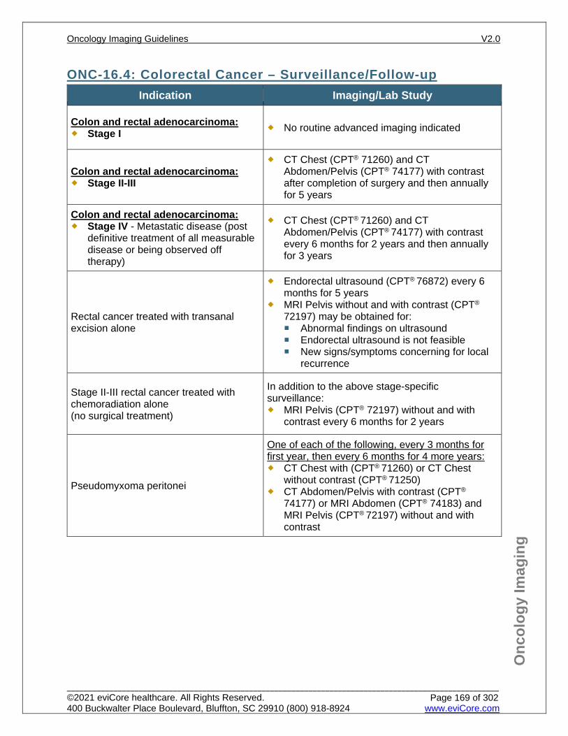
Onc
olog
y Im
agin
g
ONC-16.4: Colorectal Cancer – Surveillance/Follow-up Indication Imaging/Lab Study
Colon and rectal adenocarcinoma: Stage I No routine advanced imaging indicated
Colon and rectal adenocarcinoma: Stage II-III
CT Chest (CPT® 71260) and CT Abdomen/Pelvis (CPT® 74177) with contrast after completion of surgery and then annually for 5 years
Colon and rectal adenocarcinoma: Stage IV - Metastatic disease (post
definitive treatment of all measurable disease or being observed off therapy)
CT Chest (CPT® 71260) and CT Abdomen/Pelvis (CPT® 74177) with contrast every 6 months for 2 years and then annually for 3 years
Rectal cancer treated with transanal excision alone
Endorectal ultrasound (CPT® 76872) every 6 months for 5 years
MRI Pelvis without and with contrast (CPT®
72197) may be obtained for: Abnormal findings on ultrasound Endorectal ultrasound is not feasible New signs/symptoms concerning for local
recurrence
Stage II-III rectal cancer treated with chemoradiation alone (no surgical treatment)
In addition to the above stage-specific surveillance: MRI Pelvis (CPT® 72197) without and with
contrast every 6 months for 2 years
Pseudomyxoma peritonei
One of each of the following, every 3 months for first year, then every 6 months for 4 more years: CT Chest with (CPT® 71260) or CT Chest
without contrast (CPT® 71250) CT Abdomen/Pelvis with contrast (CPT®
74177) or MRI Abdomen (CPT® 74183) and MRI Pelvis (CPT® 72197) without and with contrast
Oncology Imaging Guidelines V2.0
______________________________________________________________________________________________________ ©2021 eviCore healthcare. All Rights Reserved. 400 Buckwalter Place Boulevard, Bluffton, SC 29910 (800) 918-8924 www.eviCore.com
Page 169 of 302
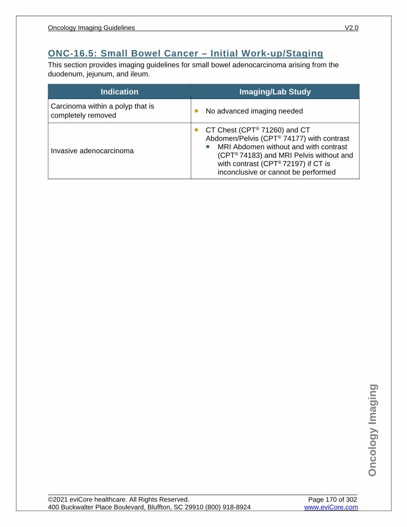
Onc
olog
y Im
agin
g
ONC-16.5: Small Bowel Cancer – Initial Work-up/Staging This section provides imaging guidelines for small bowel adenocarcinoma arising from the duodenum, jejunum, and ileum.
Indication Imaging/Lab Study Carcinoma within a polyp that is completely removed No advanced imaging needed
Invasive adenocarcinoma
CT Chest (CPT® 71260) and CT Abdomen/Pelvis (CPT® 74177) with contrast MRI Abdomen without and with contrast
(CPT® 74183) and MRI Pelvis without and with contrast (CPT® 72197) if CT is inconclusive or cannot be performed
Oncology Imaging Guidelines V2.0
______________________________________________________________________________________________________ ©2021 eviCore healthcare. All Rights Reserved. 400 Buckwalter Place Boulevard, Bluffton, SC 29910 (800) 918-8924 www.eviCore.com
Page 170 of 302
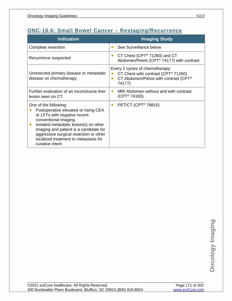
Onc
olog
y Im
agin
g
ONC-16.6: Small Bowel Cancer – Restaging/Recurrence Indication Imaging Study
Complete resection See Surveillance below
Recurrence suspected CT Chest (CPT® 71260) and CT Abdomen/Pelvis (CPT® 74177) with contrast
Unresected primary disease or metastatic disease on chemotherapy
Every 2 cycles of chemotherapy: CT Chest with contrast (CPT® 71260) CT Abdomen/Pelvis with contrast (CPT®
74177)
Further evaluation of an inconclusive liver lesion seen on CT
MRI Abdomen without and with contrast (CPT® 74183)
One of the following: Postoperative elevated or rising CEA
or LFTs with negative recent conventional imaging
Isolated metastatic lesion(s) on other imaging and patient is a candidate for aggressive surgical resection or other localized treatment to metastasis for curative intent
PET/CT (CPT® 78815)
Oncology Imaging Guidelines V2.0
______________________________________________________________________________________________________ ©2021 eviCore healthcare. All Rights Reserved. 400 Buckwalter Place Boulevard, Bluffton, SC 29910 (800) 918-8924 www.eviCore.com
Page 171 of 302
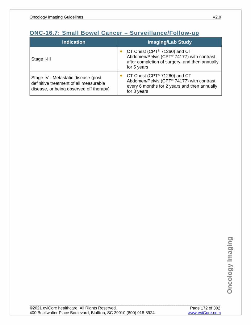
Onc
olog
y Im
agin
g
ONC-16.7: Small Bowel Cancer – Surveillance/Follow-up Indication Imaging/Lab Study
Stage I-III CT Chest (CPT® 71260) and CT
Abdomen/Pelvis (CPT® 74177) with contrast after completion of surgery, and then annually for 5 years
Stage IV - Metastatic disease (post definitive treatment of all measurable disease, or being observed off therapy)
CT Chest (CPT® 71260) and CT Abdomen/Pelvis (CPT® 74177) with contrast every 6 months for 2 years and then annually for 3 years
Oncology Imaging Guidelines V2.0
______________________________________________________________________________________________________ ©2021 eviCore healthcare. All Rights Reserved. 400 Buckwalter Place Boulevard, Bluffton, SC 29910 (800) 918-8924 www.eviCore.com
Page 172 of 302
Onc
olog
y Im
agin
g
References 1. Benson AB, Venook AP, Al-Hawary MM, et al. National Comprehensive Cancer Network (NCCN)
Guidelines Version 2.2021 – January 21, 2021. Colon cancer, available at: https://www.nccn.org/professionals/physician_gls/pdf/colon.pdf Referenced with permission from the NCCN Clinical Practice Guidelines in Oncology (NCCN Guidelines™) for Colon cancer V2.2021 – January 21, 2021. ©2021 National Comprehensive Cancer Network, Inc. All rights reserved. The NCCN Guidelines™ and illustrations herein may not be reproduced in any form for any purpose without the express written permission of the NCCN. To view the most recent and complete version of the NCCN Guidelines™, go online to NCCN.org.
2. Benson AB, Venook AP, Al-Hawary MM, et al. National Comprehensive Cancer Network (NCCN) Guidelines Version 1.2021 – December 22, 2020. Rectal cancer, available at: https://www.nccn.org/professionals/physician_gls/pdf/rectal.pdf Referenced with permission from the NCCN Clinical Practice Guidelines in Oncology (NCCN Guidelines™) for Rectal cancer V1.2021 – December 22, 2020. ©2020 National Comprehensive Cancer Network, Inc. All rights reserved. The NCCN Guidelines™ and illustrations herein may not be reproduced in any form for any purpose without the express written permission of the NCCN. To view the most recent and complete version of the NCCN Guidelines™, go online to NCCN.org.
3. Benson AB, Venook AP, Al-Hawary MM, et al. National Comprehensive Cancer Network (NCCN) Guidelines Version 1.2021 – February 16, 2021. Small Bowel Adenocarcinoma, available at: https://www.nccn.org/professionals/physician_gls/pdf/small_bowel.pdf. Referenced with permission from the NCCN Clinical Practice Guidelines in Oncology (NCCN Guidelines™) for Small Bowel Adenocarcinoma V1.2021 – February 16, 2021. ©2021 National Comprehensive Cancer Network, Inc. All rights reserved. The NCCN Guidelines™ and illustrations herein may not be reproduced in any form for any purpose without the express written permission of the NCCN. To view the most recent and complete version of the NCCN Guidelines™, go online to NCCN.org.
4. ACR Appropriateness Criteria. Pretreatment Staging of Colorectal Cancer. Rev. 2011. 5. Bailey CE, Hu C-Y, You YN et al. Variation in positron emission tomography use after colon cancer
resection. J Oncol Pract. 2015;11(3):e363-e372. doi:10.1200/JOP.2014.001933. 6. Lu YY, Chen JH, Ding HJ, Chien CR, Lin WY, Kao CH. A systematic review and meta-analysis of
pretherapeutic lymph node staging of colorectal cancer by 18F-FDG PET or PET/CT. Nucl Med commun. 2012;33(11):1127-1133. doi:10.1097/MNM0b013e328357b2d9.
7. Moulton CA, Gu CS, Law CH, et al. Effect of PET before liver resection on surgical management for colorectal adenocarcinoma metastases: a randomized clinical trial. JAMA. 2014;311(18):1863-1869. doi:10.1001/jama.2014.3740.
8. Steele SR, Chang GJ, Hendren S, et al. Practice guideline for the surveillance of patients after curative treatment of colon and rectal cancer. Dis Colon Rectum. 2015;58(8):713-725. doi:10.1097/DCR.0000000000000410.
9. van de Velde CJ, Boelens PG, Borras JM, et al. EURECCA colorectal: multidisciplinary management: European concensus conference colon & rectum. Eur J Cancer. 2014;50(1):e1-e34. doi:10.1016/j.ejca.2013.06.048.
10. Akce M, El-Rayes BF. Nonsurgical management of rectal cancer. Journal of Oncology Practice. 2019;15(3):123-131. doi:10.1200/JOP.18.00769.
Oncology Imaging Guidelines V2.0
______________________________________________________________________________________________________ ©2021 eviCore healthcare. All Rights Reserved. 400 Buckwalter Place Boulevard, Bluffton, SC 29910 (800) 918-8924 www.eviCore.com
Page 173 of 302
ONC-17: Renal Cell Cancer (RCC) ONC-17.0: Renal Cell Cancer (RCC) – General ConsiderationsONC-17.1: Renal Cell Cancer (RCC) – Suspected/DiagnosisONC-17.2: Renal Cell Cancer (RCC) – Initial Work-up/StagingONC-17.3: Renal Cell Cancer (RCC) – Restaging/RecurrenceONC-17.4: Renal Cell Cancer (RCC) – Surveillance
Oncology Imaging Guidelines V2.0
______________________________________________________________________________________________________ ©2021 eviCore healthcare. All Rights Reserved. 400 Buckwalter Place Boulevard, Bluffton, SC 29910 (800) 918-8924 www.eviCore.com
Page 174 of 302
Onc
olog
y Im
agin
g
ONC-17.0: Renal Cell Cancer (RCC) – General Considerations PET is not routinely indicated for initial diagnosis, staging or restaging of renal cell
cancer. A minority of adult patients with renal cell cancer (RCC) will have translocations in
TFE3 or TFEB, which have a different natural history than “adult type” RCC. Patients of any age with TFE3 or TFEB translocated RCC should be imaged according to guidelines in PEDONC-7.4: Pediatric Renal Cell Carcinoma (RCC).
Patients of any age with Wilms tumor should be imaged according to guidelines in section PEDONC-7.2: Unilateral Wilms Tumor (UWT) or PEDONC-7.3 Bilateral Wilms Tumor (BWT).
Oncology Imaging Guidelines V2.0
______________________________________________________________________________________________________ ©2021 eviCore healthcare. All Rights Reserved. 400 Buckwalter Place Boulevard, Bluffton, SC 29910 (800) 918-8924 www.eviCore.com
Page 175 of 302
Onc
olog
y Im
agin
g
ONC-17.1: Renal Cell Cancer (RCC) – Suspected/Diagnosis Indication Imaging Study
Solitary renal mass suspicious for renal cell cancer
See: AB-35.1: Indeterminate Renal Lesion for imaging guidelines for evaluation of suspected renal malignancies
Chest x-ray CT chest with contrast with (CPT® 71260) or without
contrast (CPT® 71250) may be obtained for one of the following: New chest x-ray abnormalities Pulmonary signs/symptoms Histologically confirmed renal cell cancer
Oncology Imaging Guidelines V2.0
______________________________________________________________________________________________________ ©2021 eviCore healthcare. All Rights Reserved. 400 Buckwalter Place Boulevard, Bluffton, SC 29910 (800) 918-8924 www.eviCore.com
Page 176 of 302
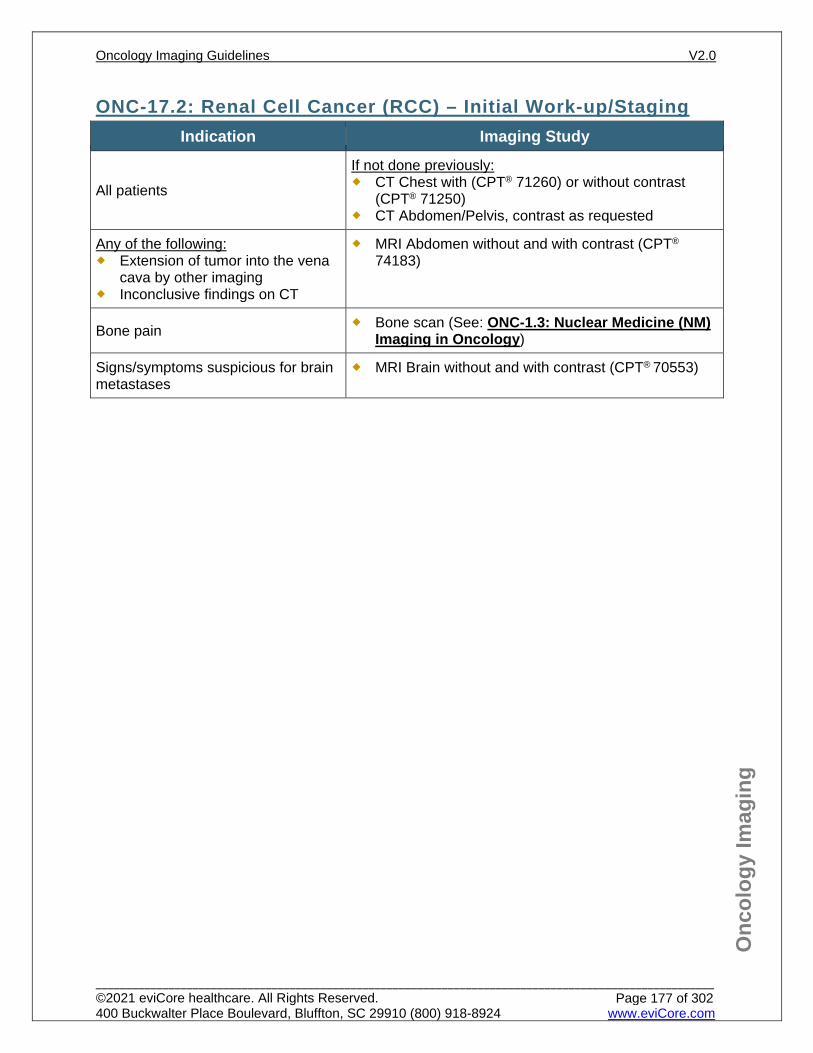
Onc
olog
y Im
agin
g
ONC-17.2: Renal Cell Cancer (RCC) – Initial Work-up/Staging Indication Imaging Study
All patients
If not done previously: CT Chest with (CPT® 71260) or without contrast
(CPT® 71250) CT Abdomen/Pelvis, contrast as requested
Any of the following: Extension of tumor into the vena
cava by other imaging Inconclusive findings on CT
MRI Abdomen without and with contrast (CPT®
74183)
Bone pain Bone scan (See: ONC-1.3: Nuclear Medicine (NM) Imaging in Oncology)
Signs/symptoms suspicious for brain metastases
MRI Brain without and with contrast (CPT® 70553)
Oncology Imaging Guidelines V2.0
______________________________________________________________________________________________________ ©2021 eviCore healthcare. All Rights Reserved. 400 Buckwalter Place Boulevard, Bluffton, SC 29910 (800) 918-8924 www.eviCore.com
Page 177 of 302
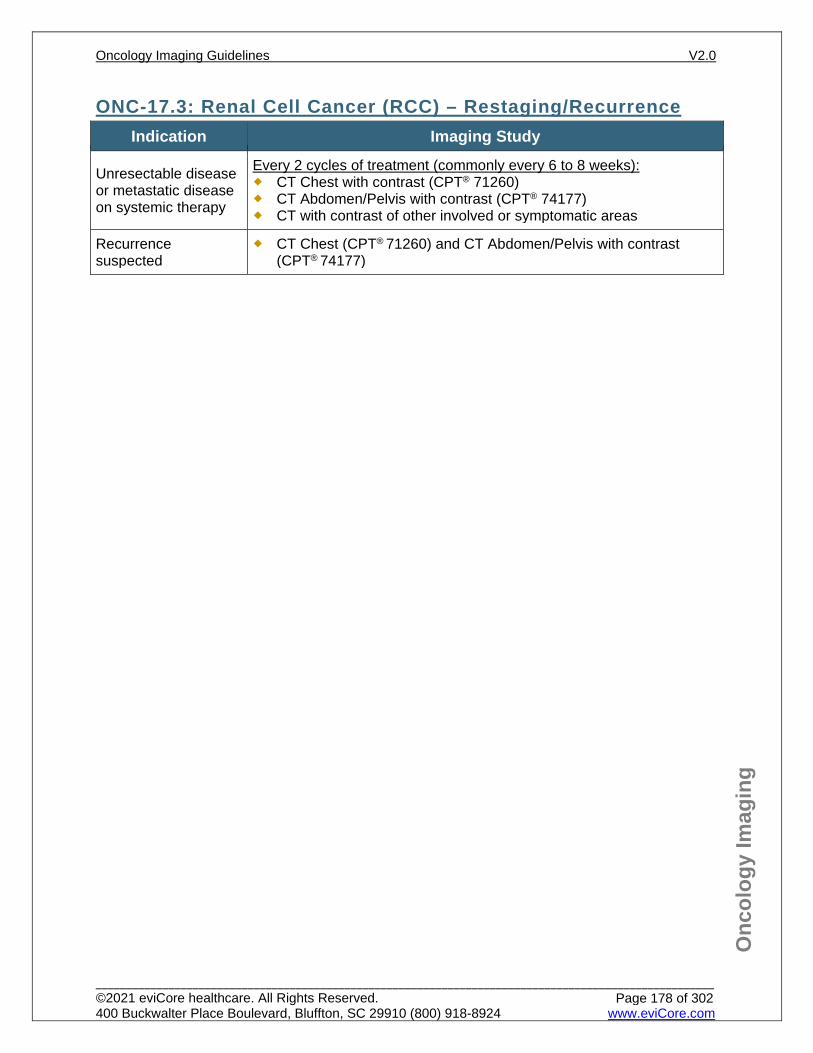
Onc
olog
y Im
agin
g
ONC-17.3: Renal Cell Cancer (RCC) – Restaging/Recurrence Indication Imaging Study
Unresectable disease or metastatic disease on systemic therapy
Every 2 cycles of treatment (commonly every 6 to 8 weeks): CT Chest with contrast (CPT® 71260) CT Abdomen/Pelvis with contrast (CPT® 74177) CT with contrast of other involved or symptomatic areas
Recurrence suspected
CT Chest (CPT® 71260) and CT Abdomen/Pelvis with contrast (CPT® 74177)
Oncology Imaging Guidelines V2.0
______________________________________________________________________________________________________ ©2021 eviCore healthcare. All Rights Reserved. 400 Buckwalter Place Boulevard, Bluffton, SC 29910 (800) 918-8924 www.eviCore.com
Page 178 of 302
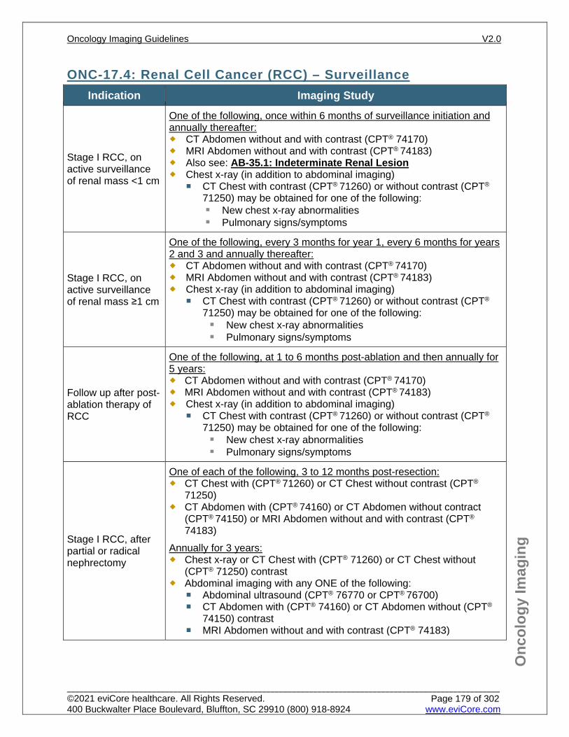
Onc
olog
y Im
agin
g
ONC-17.4: Renal Cell Cancer (RCC) – Surveillance Indication Imaging Study
Stage I RCC, on active surveillance of renal mass <1 cm
One of the following, once within 6 months of surveillance initiation and annually thereafter: CT Abdomen without and with contrast (CPT® 74170) MRI Abdomen without and with contrast (CPT® 74183) Also see: AB-35.1: Indeterminate Renal Lesion Chest x-ray (in addition to abdominal imaging)
CT Chest with contrast (CPT® 71260) or without contrast (CPT®
71250) may be obtained for one of the following: New chest x-ray abnormalities Pulmonary signs/symptoms
Stage I RCC, on active surveillance of renal mass ≥1 cm
One of the following, every 3 months for year 1, every 6 months for years 2 and 3 and annually thereafter: CT Abdomen without and with contrast (CPT® 74170) MRI Abdomen without and with contrast (CPT® 74183) Chest x-ray (in addition to abdominal imaging)
CT Chest with contrast (CPT® 71260) or without contrast (CPT®
71250) may be obtained for one of the following: New chest x-ray abnormalities Pulmonary signs/symptoms
Follow up after post-ablation therapy of RCC
One of the following, at 1 to 6 months post-ablation and then annually for 5 years: CT Abdomen without and with contrast (CPT® 74170) MRI Abdomen without and with contrast (CPT® 74183) Chest x-ray (in addition to abdominal imaging)
CT Chest with contrast (CPT® 71260) or without contrast (CPT®
71250) may be obtained for one of the following: New chest x-ray abnormalities Pulmonary signs/symptoms
Stage I RCC, after partial or radical nephrectomy
One of each of the following, 3 to 12 months post-resection: CT Chest with (CPT® 71260) or CT Chest without contrast (CPT®
71250) CT Abdomen with (CPT® 74160) or CT Abdomen without contract
(CPT® 74150) or MRI Abdomen without and with contrast (CPT®
74183) Annually for 3 years: Chest x-ray or CT Chest with (CPT® 71260) or CT Chest without
(CPT® 71250) contrast Abdominal imaging with any ONE of the following:
Abdominal ultrasound (CPT® 76770 or CPT® 76700) CT Abdomen with (CPT® 74160) or CT Abdomen without (CPT®
74150) contrast MRI Abdomen without and with contrast (CPT® 74183)
Oncology Imaging Guidelines V2.0
______________________________________________________________________________________________________ ©2021 eviCore healthcare. All Rights Reserved. 400 Buckwalter Place Boulevard, Bluffton, SC 29910 (800) 918-8924 www.eviCore.com
Page 179 of 302
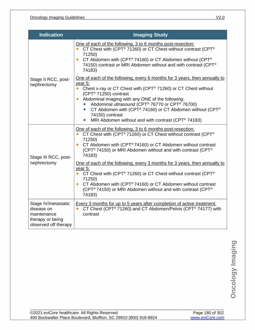
Onc
olog
y Im
agin
g
Indication Imaging Study
Stage II RCC, post-nephrectomy
One of each of the following, 3 to 6 months post-resection: CT Chest with (CPT® 71260) or CT Chest without contrast (CPT®
71250) CT Abdomen with (CPT® 74160) or CT Abdomen without (CPT®
74150) contrast or MRI Abdomen without and with contrast (CPT® 74183)
One of each of the following, every 6 months for 3 years, then annually to year 5: Chest x-ray or CT Chest with (CPT® 71260) or CT Chest without
(CPT® 71250) contrast Abdominal imaging with any ONE of the following:
Abdominal ultrasound (CPT® 76770 or CPT® 76700) CT Abdomen with (CPT® 74160) or CT Abdomen without (CPT®
74150) contrast MRI Abdomen without and with contrast (CPT® 74183)
Stage III RCC, post-nephrectomy
One of each of the following, 3 to 6 months post-resection: CT Chest with (CPT® 71260) or CT Chest without contrast (CPT®
71250) CT Abdomen with (CPT® 74160) or CT Abdomen without contrast
(CPT® 74150) or MRI Abdomen without and with contrast (CPT® 74183)
One of each of the following, every 3 months for 3 years, then annually to year 5: CT Chest with (CPT® 71260) or CT Chest without contrast (CPT®
71250) CT Abdomen with (CPT® 74160) or CT Abdomen without contrast
(CPT® 74150) or MRI Abdomen without and with contrast (CPT® 74183)
Stage IV/metastatic disease on maintenance therapy or being observed off therapy
Every 3 months for up to 5 years after completion of active treatment: CT Chest (CPT® 71260) and CT Abdomen/Pelvis (CPT® 74177) with
contrast
Oncology Imaging Guidelines V2.0
______________________________________________________________________________________________________ ©2021 eviCore healthcare. All Rights Reserved. 400 Buckwalter Place Boulevard, Bluffton, SC 29910 (800) 918-8924 www.eviCore.com
Page 180 of 302
Onc
olog
y Im
agin
g
References 1. Motzer RJ, Jonasch E, Agarwal N, et al. National Comprehensive Cancer Network (NCCN)
Guidelines Version 2.2021 – February 3, 2021. Kidney cancer, available at: https://www.nccn.org/professionals/physician_gls/pdf/kidney.pdf Referenced with permission from the NCCN Clinical Practice Guidelines in Oncology (NCCN Guidelines™) for Kidney cancer V2.2021 – February 3, 2021. ©2021 National Comprehensive Cancer Network, Inc. All rights reserved. The NCCN Guidelines™ and illustrations herein may not be reproduced in any form for any purpose without the express written permission of the NCCN. To view the most recent and complete version of the NCCN Guidelines™, go online to NCCN.org.
2. ACR Appropriateness Criteria. Post-treatment follow up of renal cell carcinoma. Rev. 2013. 3. Herts BR, Silverman SG, Hindman NM, et al. Management of the incidental renal mass on CT: a
white paper of the ACR incidental findings committee. J Am Coll Radiol. 2018;15(2):264-273. doi:10.1016/j.jacr.2017.04.028.
4. Finelli A, Ismaila N, Bro B, et al. Management of small renal masses. American Society of Clinical Oncology clinical practice guideline. Journal of Clinical Oncology. 2017;35(6):668-680. doi:10.1200/JCO.2016.69.9645.
5. Davenport MS, Caoili EM, Cohan RH, et al. MRI and CT characteristics of successfully ablated renal masses: imaging surveillance after radiofrequency ablation. AJR Am J Roentgenol. 2009;192:1571-1578. doi:10.2214/AJR.08.1303.
6. Clark TW, Millward SF, Gervais DA, et al. Reporting standards for percutaneous thermal ablation of renal cell carcinoma. J Vasc Interv Radiol. 2009;20(7 Suppl):S409-S416. doi:10.1016/j.jvir.2009.04.013.
7. Rais-Bahrami S, Guzzo TJ, Jarrett TW, Kavoussi LR, Allaf ME. Incidentally discovered renal masses: oncological and perioperative outcomes in patients with delayed surgical intervention. BJU Int. 2009;103(10):1355-1358. doi:10.1111/j.1464-410X.2008.08242.x.
8. Wang HY, Ding HJ, Chen JH, Chao CH, Lu YY, Lin WY, Kao CH. Meta-analysis of the diagnostic performance of [18F]FDG-PET and PET/CT in renal cell carcinoma. Cancer Imaging. 2012 October;12:464-474. doi:10.1102/1470-7330.2012.0042.
9. Kim EH, Strope SA. Postoperative surveillance imaging for patients undergoing nephrectomy for renal cell carcinoma. Urol Oncol. 2015;33(12):499-502. doi:10.1016/j.urolonc.2015.08.008.
10. Sankineni S, Brown A, Cieciera M, Choyke PL, Turkbey B. Imaging of renal cell carcinoma. Urol Oncol. 2016;34(3):147-155. doi:10.1016/j.urolonc.2015.05.020.
11. ACR Appropriateness Criteria. Renal cell carcinoma staging. Rev. 2015. 12. Campbell S, Uzzo R, Allaf M, et al. Renal mass and localized renal cancer: AUA guideline. J Urol.
2017:198(3):520-529. doi:10.1016/j.juro.2017.04.100.
Oncology Imaging Guidelines V2.0
______________________________________________________________________________________________________ ©2021 eviCore healthcare. All Rights Reserved. 400 Buckwalter Place Boulevard, Bluffton, SC 29910 (800) 918-8924 www.eviCore.com
Page 181 of 302
ONC-18: Transitional Cell Cancer ONC-18.0: Transitional Cell Cancer – General ConsiderationsONC-18.1: Transitional Cell Cancer – Suspected/DiagnosisONC-18.2: Transitional Cell Cancer – Initial Work-up/StagingONC-18.3: Transitional Cell Cancer – Restaging/RecurrenceONC-18.4: Transitional Cell Cancer – Surveillance/Follow-up
Oncology Imaging Guidelines V2.0
______________________________________________________________________________________________________ ©2021 eviCore healthcare. All Rights Reserved. 400 Buckwalter Place Boulevard, Bluffton, SC 29910 (800) 918-8924 www.eviCore.com
Page 182 of 302
Onc
olog
y Im
agin
g
ONC-18.0: Transitional Cell Cancer – General Considerations Transitional cell cancers can include: tumors of the bladder, ureters, prostate,
urethra, or renal pelvis. For primary cancer of the kidney, see: ONC-17: Renal Cell Cancer (RCC).
Most common histology of bladder cancer is transitional cell (TCC) or urothelial carcinoma (UCC). Rare histologies include squamous cell (imaged according to ONC-18: Transitional Cell Cancer) or small cell (imaged according to ONC-31.8: Extrathoracic Small Cell and Large Cell)
Urachal cancer is rare type of bladder cancer; the most common histology is adenocarcinoma. These are imaged according to muscle invasive bladder cancer.
PET not routinely indicated in transitional cell cancer with exception noted below in ONC-18.2: Transitional Cell Cancer – Initial Work-up/Staging
Oncology Imaging Guidelines V2.0
______________________________________________________________________________________________________ ©2021 eviCore healthcare. All Rights Reserved. 400 Buckwalter Place Boulevard, Bluffton, SC 29910 (800) 918-8924 www.eviCore.com
Page 183 of 302
Onc
olog
y Im
agin
g
ONC-18.1: Transitional Cell Cancer – Suspected/Diagnosis See: AB-39: Hematuria and Hydronephrosis for imaging guidelines for evaluation of suspected transitional cell malignancies
Oncology Imaging Guidelines V2.0
______________________________________________________________________________________________________ ©2021 eviCore healthcare. All Rights Reserved. 400 Buckwalter Place Boulevard, Bluffton, SC 29910 (800) 918-8924 www.eviCore.com
Page 184 of 302
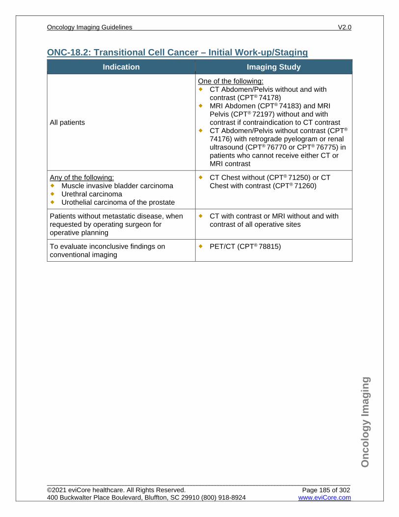
Onc
olog
y Im
agin
g
ONC-18.2: Transitional Cell Cancer – Initial Work-up/Staging Indication Imaging Study
All patients
One of the following: CT Abdomen/Pelvis without and with
contrast (CPT® 74178) MRI Abdomen (CPT® 74183) and MRI
Pelvis (CPT® 72197) without and with contrast if contraindication to CT contrast
CT Abdomen/Pelvis without contrast (CPT®
74176) with retrograde pyelogram or renal ultrasound (CPT® 76770 or CPT® 76775) in patients who cannot receive either CT or MRI contrast
Any of the following: Muscle invasive bladder carcinoma Urethral carcinoma Urothelial carcinoma of the prostate
CT Chest without (CPT® 71250) or CT Chest with contrast (CPT® 71260)
Patients without metastatic disease, when requested by operating surgeon for operative planning
CT with contrast or MRI without and with contrast of all operative sites
To evaluate inconclusive findings on conventional imaging
PET/CT (CPT® 78815)
Oncology Imaging Guidelines V2.0
______________________________________________________________________________________________________ ©2021 eviCore healthcare. All Rights Reserved. 400 Buckwalter Place Boulevard, Bluffton, SC 29910 (800) 918-8924 www.eviCore.com
Page 185 of 302
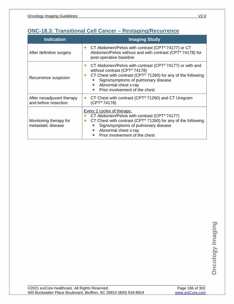
Onc
olog
y Im
agin
g
ONC-18.3: Transitional Cell Cancer – Restaging/Recurrence Indication Imaging Study
After definitive surgery CT Abdomen/Pelvis with contrast (CPT® 74177) or CT
Abdomen/Pelvis without and with contrast (CPT® 74178) for post-operative baseline
Recurrence suspicion
CT Abdomen/Pelvis with contrast (CPT® 74177) or with and without contrast (CPT® 74178)
CT Chest with contrast (CPT® 71260) for any of the following: Signs/symptoms of pulmonary disease Abnormal chest x-ray Prior involvement of the chest
After neoadjuvant therapy and before resection
CT Chest with contrast (CPT® 71260) and CT Urogram (CPT® 74178)
Monitoring therapy for metastatic disease
Every 2 cycles of therapy: CT Abdomen/Pelvis with contrast (CPT® 74177) CT Chest with contrast (CPT® 71260) for any of the following:
Signs/symptoms of pulmonary disease Abnormal chest x-ray Prior involvement of the chest
Oncology Imaging Guidelines V2.0
______________________________________________________________________________________________________ ©2021 eviCore healthcare. All Rights Reserved. 400 Buckwalter Place Boulevard, Bluffton, SC 29910 (800) 918-8924 www.eviCore.com
Page 186 of 302
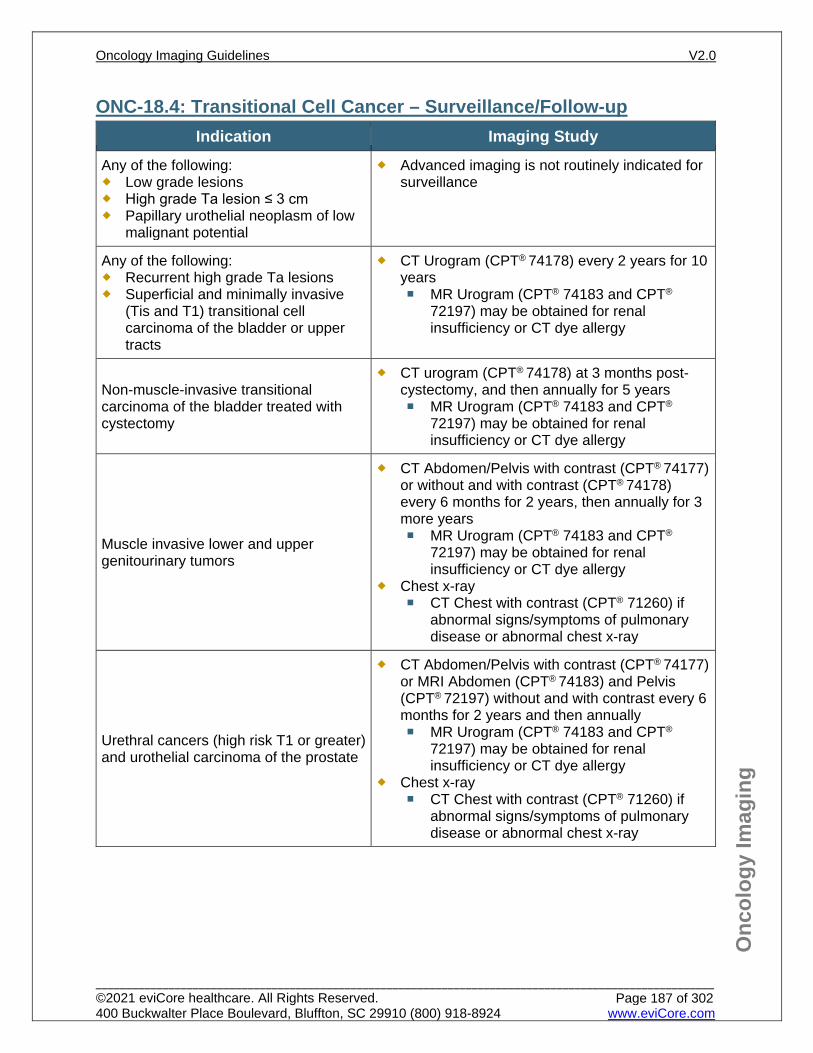
Onc
olog
y Im
agin
g
ONC-18.4: Transitional Cell Cancer – Surveillance/Follow-up Indication Imaging Study
Any of the following: Low grade lesions High grade Ta lesion ≤ 3 cm Papillary urothelial neoplasm of low
malignant potential
Advanced imaging is not routinely indicated for surveillance
Any of the following: Recurrent high grade Ta lesions Superficial and minimally invasive
(Tis and T1) transitional cell carcinoma of the bladder or upper tracts
CT Urogram (CPT® 74178) every 2 years for 10 years MR Urogram (CPT® 74183 and CPT®
72197) may be obtained for renal insufficiency or CT dye allergy
Non-muscle-invasive transitional carcinoma of the bladder treated with cystectomy
CT urogram (CPT® 74178) at 3 months post-cystectomy, and then annually for 5 years MR Urogram (CPT® 74183 and CPT®
72197) may be obtained for renal insufficiency or CT dye allergy
Muscle invasive lower and upper genitourinary tumors
CT Abdomen/Pelvis with contrast (CPT® 74177) or without and with contrast (CPT® 74178) every 6 months for 2 years, then annually for 3 more years MR Urogram (CPT® 74183 and CPT®
72197) may be obtained for renal insufficiency or CT dye allergy
Chest x-ray CT Chest with contrast (CPT® 71260) if
abnormal signs/symptoms of pulmonary disease or abnormal chest x-ray
Urethral cancers (high risk T1 or greater) and urothelial carcinoma of the prostate
CT Abdomen/Pelvis with contrast (CPT® 74177) or MRI Abdomen (CPT® 74183) and Pelvis (CPT® 72197) without and with contrast every 6 months for 2 years and then annually MR Urogram (CPT® 74183 and CPT®
72197) may be obtained for renal insufficiency or CT dye allergy
Chest x-ray CT Chest with contrast (CPT® 71260) if
abnormal signs/symptoms of pulmonary disease or abnormal chest x-ray
Oncology Imaging Guidelines V2.0
______________________________________________________________________________________________________ ©2021 eviCore healthcare. All Rights Reserved. 400 Buckwalter Place Boulevard, Bluffton, SC 29910 (800) 918-8924 www.eviCore.com
Page 187 of 302
Onc
olog
y Im
agin
g
References 1. Flaig TW, Spiess PE, Agarwal N, et al. National Comprehensive Cancer Network (NCCN) Guidelines
Version 6.2020 – July 16, 2020. Bladder cancer, available at: https://www.nccn.org/professionals/physician_gls/pdf/bladder.pdf Referenced with permission from the NCCN Clinical Practice Guidelines in Oncology (NCCN Guidelines™) for Bladder cancer V6.2020 – July 16, 2020. ©2020 National Comprehensive Cancer Network, Inc. All rights reserved. The NCCN Guidelines™ and illustrations herein may not be reproduced in any form for any purpose without the express written permission of the NCCN. To view the most recent and complete version of the NCCN Guidelines™, go online to NCCN.org.
2. Verma S, Rajesh A, Prasad SR et al, Urinary bladder cancer: role of MR imaging. Radiographics. 2012;32(2):371-387. doi:10.1148/rg.322115125.
3. Lu YY, Chen JH, Liang JA. Clinical value of FDG PET or PET/CT in urinary bladder cancer: a systematic review and meta-analysis. Eur J Radiol. 2012;81(9):2411-2416. doi:10.1016/j.ejrad.2011.07.018.
4. Witjes JA, Comperat E, Cowan NC, et al. EAU guidelines on muscle-invasive and metastatic bladder cancer: summary of the 2013 guidelines. Eur Urol. 2014;65(4):778-792. doi:10.1016/j.eururo.2013.11.046.
5. Gakis G, Witjes JA, Comperat E, et al. EAU guidelines on primary urethral carcinoma. Eur Urol. 2013;64(5):823-830. doi:10.1016/j.eururo.2013.03.044.
6. Rouprêt M, Babjuk M, Compérat E, et al. European guidelines on upper tract urothelial carcinomas: 2013 update. Eur Urol. 2013;63(6):1059-1071. doi:10.1016/j.eururo.2013.03.032.
Oncology Imaging Guidelines V2.0
______________________________________________________________________________________________________ ©2021 eviCore healthcare. All Rights Reserved. 400 Buckwalter Place Boulevard, Bluffton, SC 29910 (800) 918-8924 www.eviCore.com
Page 188 of 302
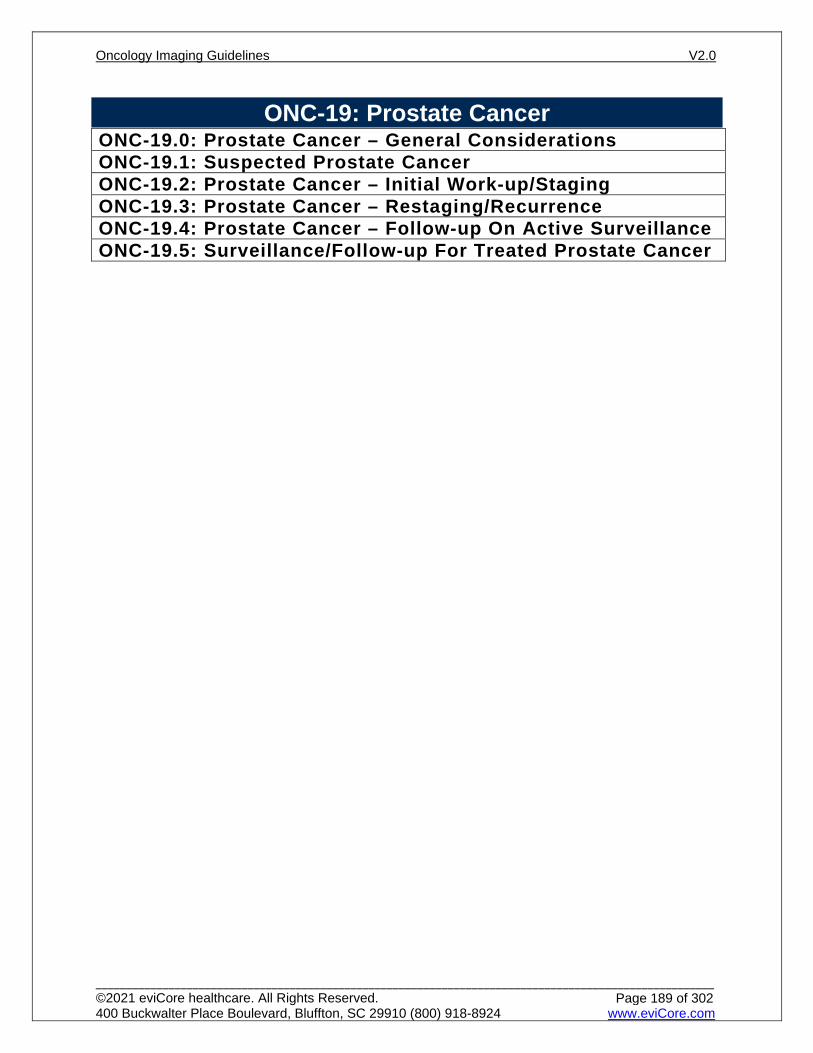
ONC-19: Prostate Cancer ONC-19.0: Prostate Cancer – General ConsiderationsONC-19.1: Suspected Prostate CancerONC-19.2: Prostate Cancer – Initial Work-up/StagingONC-19.3: Prostate Cancer – Restaging/RecurrenceONC-19.4: Prostate Cancer – Follow-up On Active SurveillanceONC-19.5: Surveillance/Follow-up For Treated Prostate Cancer
Oncology Imaging Guidelines V2.0
______________________________________________________________________________________________________ ©2021 eviCore healthcare. All Rights Reserved. 400 Buckwalter Place Boulevard, Bluffton, SC 29910 (800) 918-8924 www.eviCore.com
Page 189 of 302
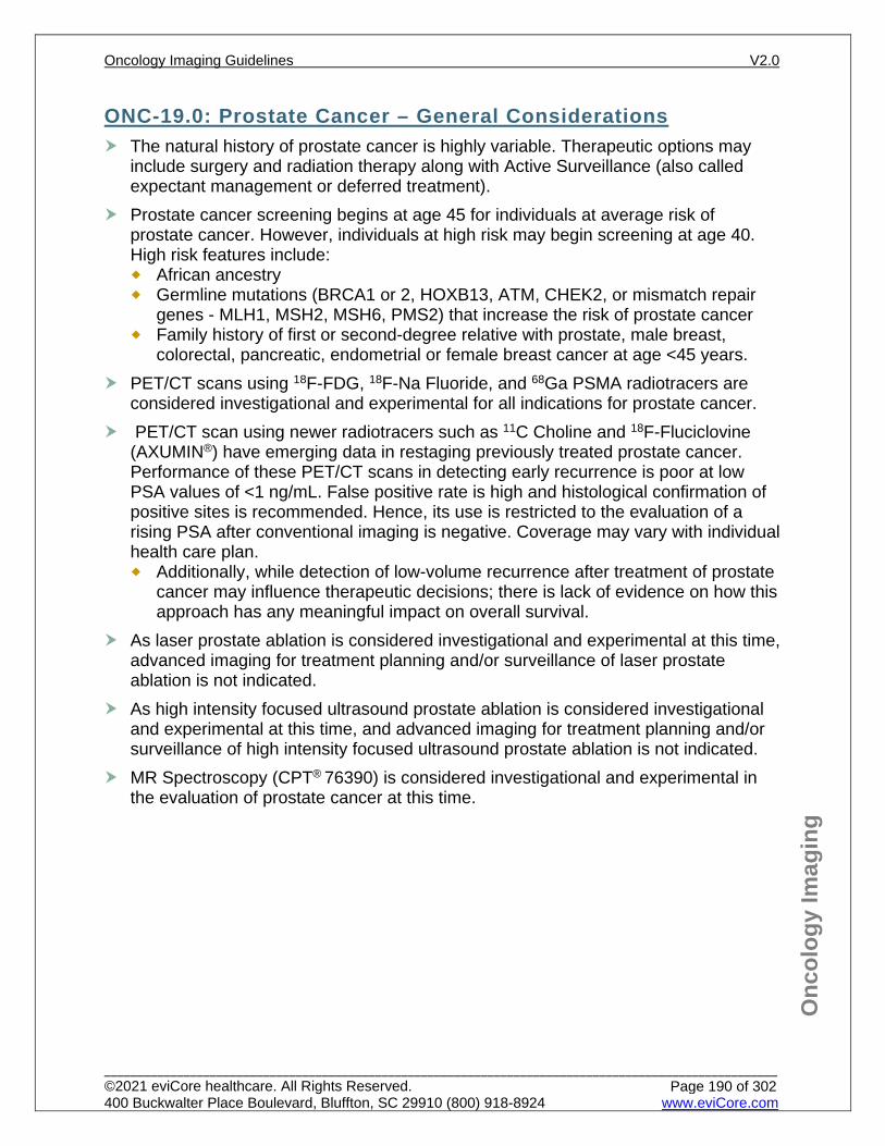
Onc
olog
y Im
agin
g
ONC-19.0: Prostate Cancer – General Considerations The natural history of prostate cancer is highly variable. Therapeutic options may
include surgery and radiation therapy along with Active Surveillance (also called expectant management or deferred treatment).
Prostate cancer screening begins at age 45 for individuals at average risk of prostate cancer. However, individuals at high risk may begin screening at age 40. High risk features include: African ancestry Germline mutations (BRCA1 or 2, HOXB13, ATM, CHEK2, or mismatch repair
genes - MLH1, MSH2, MSH6, PMS2) that increase the risk of prostate cancer Family history of first or second-degree relative with prostate, male breast,
colorectal, pancreatic, endometrial or female breast cancer at age <45 years. PET/CT scans using 18F-FDG, 18F-Na Fluoride, and 68Ga PSMA radiotracers are
considered investigational and experimental for all indications for prostate cancer. PET/CT scan using newer radiotracers such as 11C Choline and 18F-Fluciclovine
(AXUMIN®) have emerging data in restaging previously treated prostate cancer. Performance of these PET/CT scans in detecting early recurrence is poor at low PSA values of <1 ng/mL. False positive rate is high and histological confirmation of positive sites is recommended. Hence, its use is restricted to the evaluation of a rising PSA after conventional imaging is negative. Coverage may vary with individual health care plan. Additionally, while detection of low-volume recurrence after treatment of prostate
cancer may influence therapeutic decisions; there is lack of evidence on how this approach has any meaningful impact on overall survival.
As laser prostate ablation is considered investigational and experimental at this time, advanced imaging for treatment planning and/or surveillance of laser prostate ablation is not indicated.
As high intensity focused ultrasound prostate ablation is considered investigational and experimental at this time, and advanced imaging for treatment planning and/or surveillance of high intensity focused ultrasound prostate ablation is not indicated.
MR Spectroscopy (CPT® 76390) is considered investigational and experimental in the evaluation of prostate cancer at this time.
Oncology Imaging Guidelines V2.0
______________________________________________________________________________________________________ ©2021 eviCore healthcare. All Rights Reserved. 400 Buckwalter Place Boulevard, Bluffton, SC 29910 (800) 918-8924 www.eviCore.com
Page 190 of 302

Onc
olog
y Im
agin
g
Based on the local extent of tumor, PSA level and Gleason score, prostate cancer patients can be classified into risk groups as below:
Prostate Cancer – Risk Categories
Risk Category T stage Gleason score PSA (ng/ml) Very low T1c ≤ 6 < 10
Low T1-T2a ≤ 6 < 10
Intermediate T2b-T2c 7 10-20
High T3a 8 to 10 > 20
Very High T3b-T4 8 to 10 > 20
3D Rendering of MRI for MRI / Ultrasound Fusion Biopsy: When specific target lesion(s) is (are) detected on mpMRI prostate and classified as
PIRADS 4 or 5, then 3D Rendering at independent workstation (CPT® 76377, 3D rendering requiring image post-processing on an independent workstation) for the radiologist to generate prostate segmentation data image set for target identification on MRI/TRUS fusion biopsy is approvable either as subsequent separate standalone request or as retrospective request for medical necessity.
If there is no target lesion identified on MRI then 3D rendering and MRI/TRUS fusion biopsy is not generally indicated. The urologist may request MRI/TRUS fusion biopsy of a PIRADS 1-3 lesion. Then approval of 3D rendering at independent workstation (CPT® 76376 or CPT® 76377) can be considered on a case-by-case basis.
The 3D rendering for the TRUS component of the fusion is a part of the UroNav Fusion Equipment Software and an additional 3D code CPT® 76376 or CPT® 76377 should not be approved.
Oncology Imaging Guidelines V2.0
______________________________________________________________________________________________________ ©2021 eviCore healthcare. All Rights Reserved. 400 Buckwalter Place Boulevard, Bluffton, SC 29910 (800) 918-8924 www.eviCore.com
Page 191 of 302
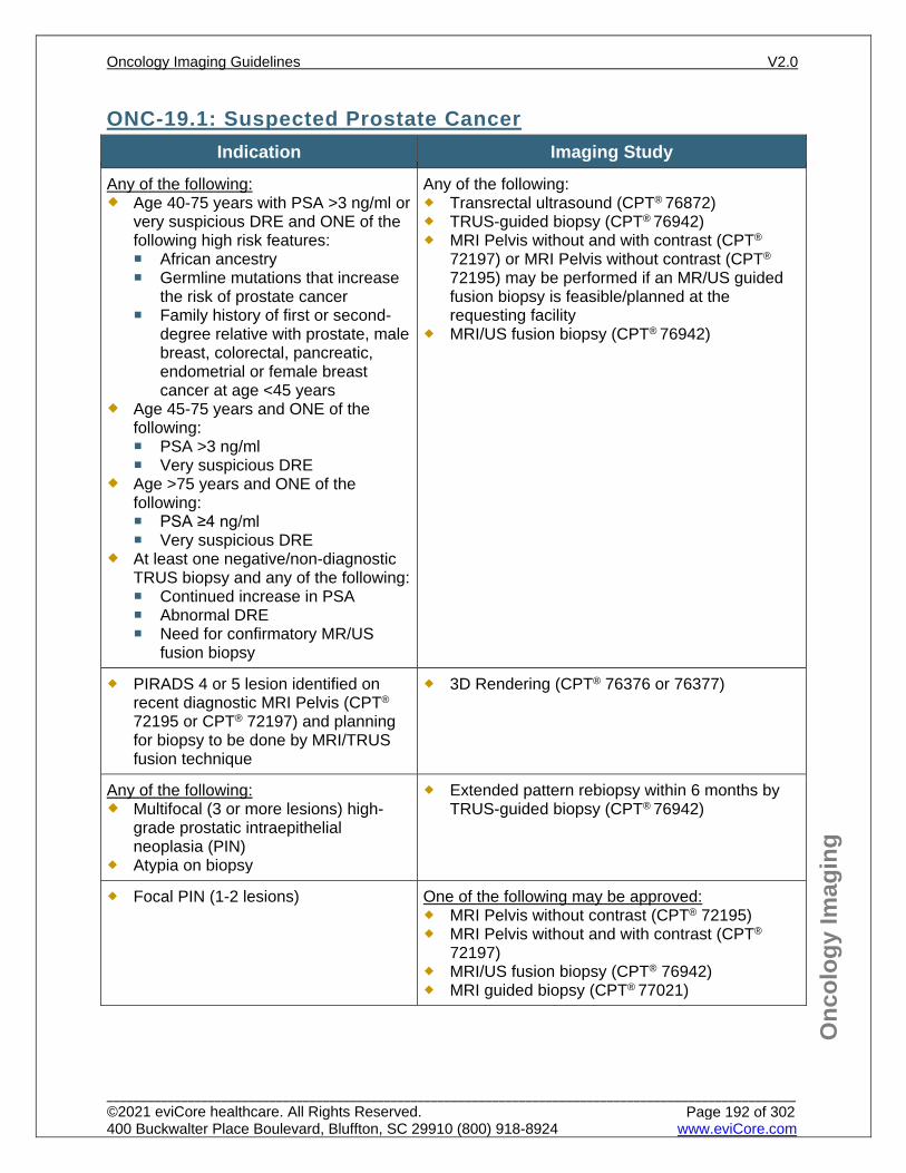
Onc
olog
y Im
agin
g
ONC-19.1: Suspected Prostate Cancer Indication Imaging Study
Any of the following: Age 40-75 years with PSA >3 ng/ml or
very suspicious DRE and ONE of the following high risk features: African ancestry Germline mutations that increase
the risk of prostate cancer Family history of first or second-
degree relative with prostate, male breast, colorectal, pancreatic, endometrial or female breast cancer at age <45 years
Age 45-75 years and ONE of the following: PSA >3 ng/ml Very suspicious DRE
Age >75 years and ONE of the following: PSA ≥4 ng/ml Very suspicious DRE
At least one negative/non-diagnostic TRUS biopsy and any of the following: Continued increase in PSA Abnormal DRE Need for confirmatory MR/US
fusion biopsy
Any of the following: Transrectal ultrasound (CPT® 76872) TRUS-guided biopsy (CPT® 76942) MRI Pelvis without and with contrast (CPT®
72197) or MRI Pelvis without contrast (CPT®
72195) may be performed if an MR/US guided fusion biopsy is feasible/planned at the requesting facility
MRI/US fusion biopsy (CPT® 76942)
PIRADS 4 or 5 lesion identified on recent diagnostic MRI Pelvis (CPT® 72195 or CPT® 72197) and planning for biopsy to be done by MRI/TRUS fusion technique
3D Rendering (CPT® 76376 or 76377)
Any of the following: Multifocal (3 or more lesions) high-
grade prostatic intraepithelial neoplasia (PIN)
Atypia on biopsy
Extended pattern rebiopsy within 6 months by TRUS-guided biopsy (CPT® 76942)
Focal PIN (1-2 lesions) One of the following may be approved: MRI Pelvis without contrast (CPT® 72195) MRI Pelvis without and with contrast (CPT®
72197) MRI/US fusion biopsy (CPT® 76942) MRI guided biopsy (CPT® 77021)
Oncology Imaging Guidelines V2.0
______________________________________________________________________________________________________ ©2021 eviCore healthcare. All Rights Reserved. 400 Buckwalter Place Boulevard, Bluffton, SC 29910 (800) 918-8924 www.eviCore.com
Page 192 of 302
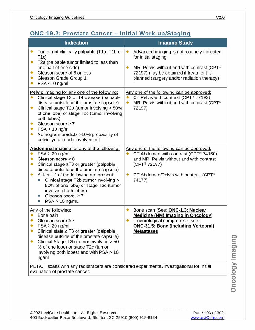
Onc
olog
y Im
agin
g
ONC-19.2: Prostate Cancer – Initial Work-up/Staging Indication Imaging Study
Tumor not clinically palpable (T1a, T1b or T1c)
T2a (palpable tumor limited to less than one half of one side)
Gleason score of 6 or less Gleason Grade Group 1 PSA <10 ng/ml
Advanced imaging is not routinely indicated for initial staging
MRI Pelvis without and with contrast (CPT®
72197) may be obtained if treatment is planned (surgery and/or radiation therapy)
Pelvic imaging for any one of the following: Clinical stage T3 or T4 disease (palpable
disease outside of the prostate capsule) Clinical stage T2b (tumor involving > 50%
of one lobe) or stage T2c (tumor involving both lobes)
Gleason score ≥ 7 PSA > 10 ng/ml Nomogram predicts >10% probability of
pelvic lymph node involvement
Any one of the following can be approved: CT Pelvis with contrast (CPT® 72193) MRI Pelvis without and with contrast (CPT®
72197)
Abdominal imaging for any of the following: PSA ≥ 20 ng/mL Gleason score ≥ 8 Clinical stage ≥T3 or greater (palpable
disease outside of the prostate capsule) At least 2 of the following are present:
Clinical stage T2b (tumor involving > 50% of one lobe) or stage T2c (tumor involving both lobes)
Gleason score ≥ 7 PSA > 10 ng/mL
Any one of the following can be approved: CT Abdomen with contrast (CPT® 74160)
and MRI Pelvis without and with contrast (CPT® 72197)
CT Abdomen/Pelvis with contrast (CPT® 74177)
Any of the following: Bone pain Gleason score ≥ 7 PSA ≥ 20 ng/ml Clinical state ≥ T3 or greater (palpable
disease outside of the prostate capsule) Clinical Stage T2b (tumor involving > 50
% of one lobe) or stage T2c (tumor involving both lobes) and with PSA > 10 ng/ml
Bone scan (See: ONC-1.3: Nuclear Medicine (NM) Imaging in Oncology)
If neurological compromise, see: ONC-31.5: Bone (Including Vertebral) Metastases
PET/CT scans with any radiotracers are considered experimental/investigational for initial evaluation of prostate cancer.
Oncology Imaging Guidelines V2.0
______________________________________________________________________________________________________ ©2021 eviCore healthcare. All Rights Reserved. 400 Buckwalter Place Boulevard, Bluffton, SC 29910 (800) 918-8924 www.eviCore.com
Page 193 of 302
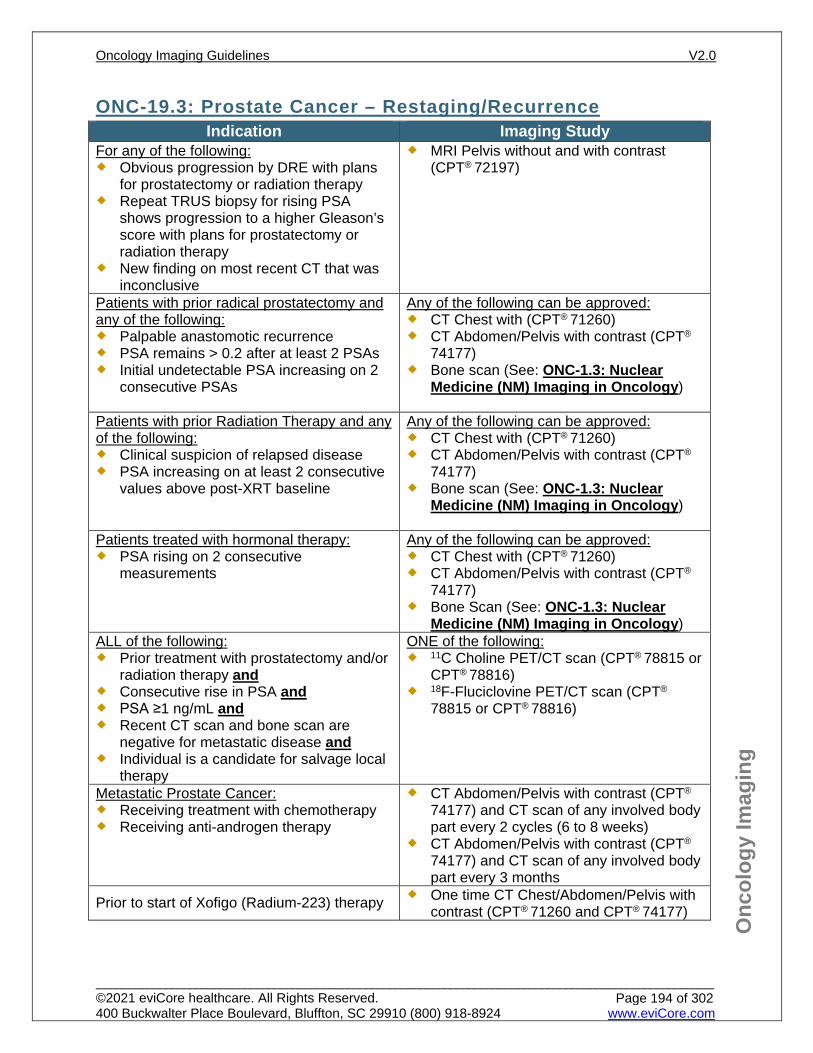
Onc
olog
y Im
agin
g
ONC-19.3: Prostate Cancer – Restaging/Recurrence Indication Imaging Study
For any of the following: Obvious progression by DRE with plans
for prostatectomy or radiation therapy Repeat TRUS biopsy for rising PSA
shows progression to a higher Gleason’s score with plans for prostatectomy or radiation therapy
New finding on most recent CT that was inconclusive
MRI Pelvis without and with contrast (CPT® 72197)
Patients with prior radical prostatectomy and any of the following: Palpable anastomotic recurrence PSA remains > 0.2 after at least 2 PSAs Initial undetectable PSA increasing on 2
consecutive PSAs
Any of the following can be approved: CT Chest with (CPT® 71260) CT Abdomen/Pelvis with contrast (CPT®
74177) Bone scan (See: ONC-1.3: Nuclear
Medicine (NM) Imaging in Oncology)
Patients with prior Radiation Therapy and any of the following: Clinical suspicion of relapsed disease PSA increasing on at least 2 consecutive
values above post-XRT baseline
Any of the following can be approved: CT Chest with (CPT® 71260) CT Abdomen/Pelvis with contrast (CPT®
74177) Bone scan (See: ONC-1.3: Nuclear
Medicine (NM) Imaging in Oncology)
Patients treated with hormonal therapy: PSA rising on 2 consecutive
measurements
Any of the following can be approved: CT Chest with (CPT® 71260) CT Abdomen/Pelvis with contrast (CPT®
74177) Bone Scan (See: ONC-1.3: Nuclear
Medicine (NM) Imaging in Oncology) ALL of the following: Prior treatment with prostatectomy and/or
radiation therapy and Consecutive rise in PSA and PSA ≥1 ng/mL and Recent CT scan and bone scan are
negative for metastatic disease and Individual is a candidate for salvage local
therapy
ONE of the following: 11C Choline PET/CT scan (CPT® 78815 or
CPT® 78816) 18F-Fluciclovine PET/CT scan (CPT®
78815 or CPT® 78816)
Metastatic Prostate Cancer: Receiving treatment with chemotherapy Receiving anti-androgen therapy
CT Abdomen/Pelvis with contrast (CPT®
74177) and CT scan of any involved body part every 2 cycles (6 to 8 weeks)
CT Abdomen/Pelvis with contrast (CPT®
74177) and CT scan of any involved body part every 3 months
Prior to start of Xofigo (Radium-223) therapy One time CT Chest/Abdomen/Pelvis with contrast (CPT® 71260 and CPT® 74177)
Oncology Imaging Guidelines V2.0
______________________________________________________________________________________________________ ©2021 eviCore healthcare. All Rights Reserved. 400 Buckwalter Place Boulevard, Bluffton, SC 29910 (800) 918-8924 www.eviCore.com
Page 194 of 302
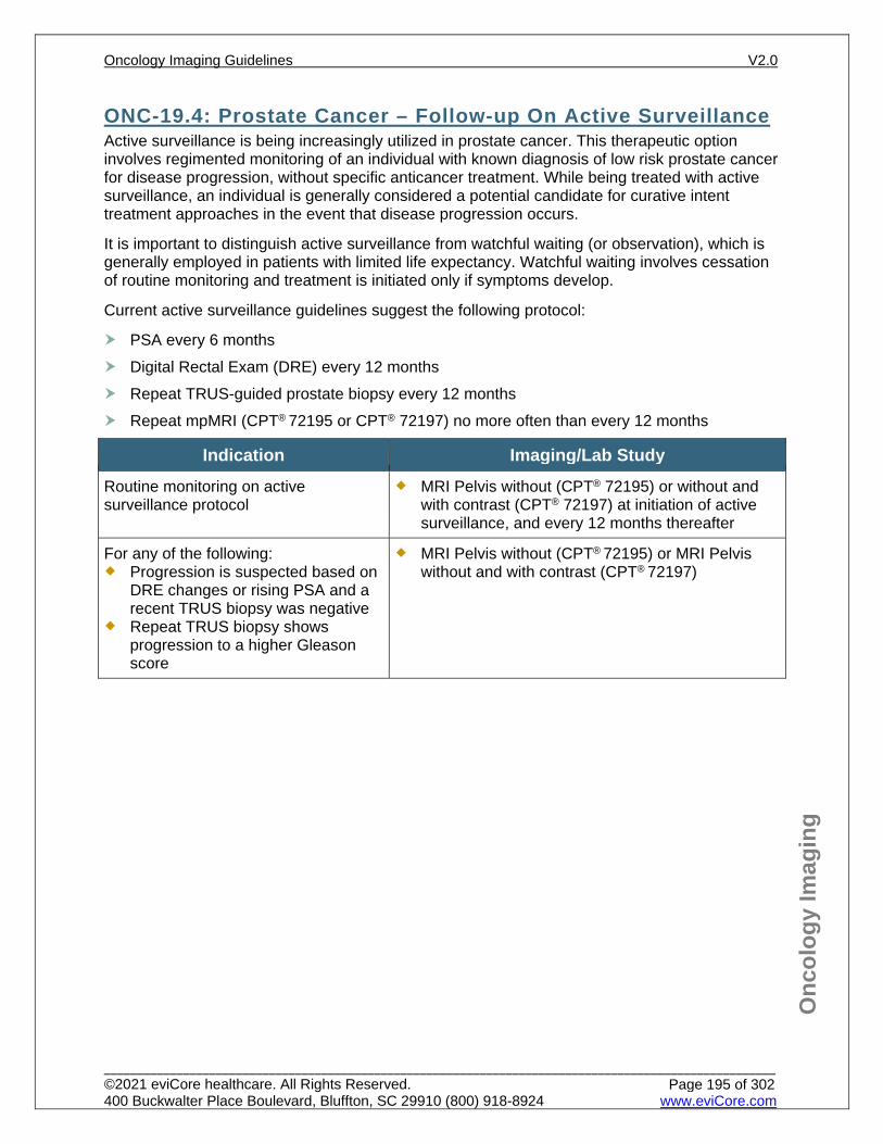
Onc
olog
y Im
agin
g
ONC-19.4: Prostate Cancer – Follow-up On Active Surveillance Active surveillance is being increasingly utilized in prostate cancer. This therapeutic option involves regimented monitoring of an individual with known diagnosis of low risk prostate cancer for disease progression, without specific anticancer treatment. While being treated with active surveillance, an individual is generally considered a potential candidate for curative intent treatment approaches in the event that disease progression occurs.
It is important to distinguish active surveillance from watchful waiting (or observation), which is generally employed in patients with limited life expectancy. Watchful waiting involves cessation of routine monitoring and treatment is initiated only if symptoms develop.
Current active surveillance guidelines suggest the following protocol:
PSA every 6 months Digital Rectal Exam (DRE) every 12 months Repeat TRUS-guided prostate biopsy every 12 months Repeat mpMRI (CPT® 72195 or CPT® 72197) no more often than every 12 months
Indication Imaging/Lab Study Routine monitoring on active surveillance protocol
MRI Pelvis without (CPT® 72195) or without and with contrast (CPT® 72197) at initiation of active surveillance, and every 12 months thereafter
For any of the following: Progression is suspected based on
DRE changes or rising PSA and a recent TRUS biopsy was negative
Repeat TRUS biopsy shows progression to a higher Gleason score
MRI Pelvis without (CPT® 72195) or MRI Pelvis without and with contrast (CPT® 72197)
Oncology Imaging Guidelines V2.0
______________________________________________________________________________________________________ ©2021 eviCore healthcare. All Rights Reserved. 400 Buckwalter Place Boulevard, Bluffton, SC 29910 (800) 918-8924 www.eviCore.com
Page 195 of 302
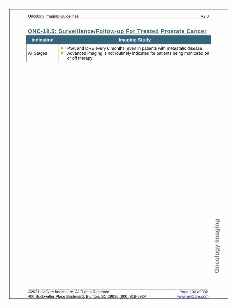
Onc
olog
y Im
agin
g
ONC-19.5: Surveillance/Follow-up For Treated Prostate Cancer Indication Imaging Study
All Stages PSA and DRE every 6 months, even in patients with metastatic disease. Advanced imaging is not routinely indicated for patients being monitored on
or off therapy.
Oncology Imaging Guidelines V2.0
______________________________________________________________________________________________________ ©2021 eviCore healthcare. All Rights Reserved. 400 Buckwalter Place Boulevard, Bluffton, SC 29910 (800) 918-8924 www.eviCore.com
Page 196 of 302
Onc
olog
y Im
agin
g
References 1. Schaeffer E, Srinivas S, Antonarakis ES, et al. National Comprehensive Cancer Network (NCCN)
Guidelines Version 2.2021 – February 17, 2021. Prostate cancer, available at: https://www.nccn.org/professionals/physician_gls/pdf/prostate.pdf Referenced with permission from the NCCN Clinical Practice Guidelines in Oncology (NCCN Guidelines™) for Prostate cancer V2.2021 – February 17, 2021. ©2021 National Comprehensive Cancer Network, Inc. All rights reserved. The NCCN Guidelines™ and illustrations herein may not be reproduced in any form for any purpose without the express written permission of the NCCN. To view the most recent and complete version of the NCCN Guidelines™, go online to NCCN.org.
2. Carroll PR, Parsons JK, Carlsson S, et al. National Comprehensive Cancer Network (NCCN) Guidelines V1.2021 – January 5, 2021 Prostate Cancer Early Detection available at: https://www.nccn.org/professionals/physician_gls/pdf/prostate_detection.pdf. Referenced with permission from the NCCN Clinical Practice Guidelines in Oncology (NCCN Guidelines™) for Prostate Cancer Early Detection V1.2021 – January 5, 2021 ©2021 National Comprehensive Cancer Network, Inc. All rights reserved. The NCCN Guidelines™ and illustrations herein may not be reproduced in any form for any purpose without the express written permission of the NCCN. To view the most recent and complete version of the NCCN Guidelines™, go online to NCCN.org.
3. Trabulsi EJ, Rumble RB, Jadvar H, et al. Optimum imaging strategies for advanced prostate cancer: ASCO guideline. J Clin Oncol. 2020 Jan 15. doi:10.1200/JCO/19.02757 (Epub ahead of print).
4. Andriole G, Siegel B, LOCATE Study Group. PD60-12 Sites of prostate cancer recurrence delineated with 18F-Flucicloved positron emission tomography in patients with negative or equivocal conventional imaging. Journal of Urology. 2019;201(4):e1100-e1101. doi:10.1097/01.JU.0000557289.21741.20.
5. ACR Appropriateness Criteria. Prostate cancer – pretreatment detection, surveillance, and staging. Rev. 2016.
6. Schoots IG, Nieboer D, Giganti F, Moore CM, Bangma CH, Roobol MJ. Is magnetic resonance imaging-targeted biopsy a useful addition to systematic confirmatory biopsy in men on active surveillance for low risk prostate cancer? A systematic review and meta-analysis. BJU Int. 2018;122(6):946-958. doi:10.1111/bju.14358.
7. Mullins J, Bodenkamp D, Landis P, et al. Multiparametric magnetic resonance imaging findings in men with low-risk prostate cancer followed by active surveillance. BJU Int. 2013;111(7):1037-1045. doi:10.1111/j.1464-410X.2012.11641.x.
8. Sanda MG, Chen RC, Crispino T, et al. AUA/ASTRO/SUO guidelines for clinically localized prostate cancer. Linthicum, MD: American Urological Association; 2017.
9. Lu-Yao GL, Albertsen PC, Moore DF, et al. Outcomes of localized prostate cancer following conservative management. JAMA. 2009;302(11):1202-1209. doi:10.1001/jama.2009.1348.
10. Chen RC, Rumble RB, Loblaw DA, et al. Active surveillance for the management of localized prostate cancer (Cancer Care Ontario guideline): American Society of Clinical Oncology clinical practice guideline endorsement. J Clin Oncol. 2016;34(18):2182-2190. doi:10.1200/JCO.2015.65.7759.
11. Liu D, Lehmann HP, Frick KD, Carter HB. Active surveillance versus surgery for low risk prostate cancer: a clinical decision analysis. J Urol. 2012;187(4):1241-1246. doi:10.1016/j.juro/2011.12.015.
12. Klotz L, Zhang L, Lam A, Nam R, Mamedov A, Loblaw A. Clinical results of long-term follow-up of a large, active surveillance cohort with localized prostate cancer. J Clin Oncol. 2010;28(1):126-131. doi:10.1200/JCO.2009.24.2180.
13. Blomqvist L, Carlsson S, Gjertsson P, et al. Limited evidence for the use of imaging to detect prostate cancer: a systematic review. Eur J Radiol. 2014;83(9):1601–1606. doi:10.1016.j.ejrad.2014.06.028.
14. Schoots IG, Petrides N, Giganti F, et al. Magnetic resonance imaging in active surveillance of prostate cancer: a systematic review. Eur Urol. 2015;67(4):627-636. doi:10.1016/j.eururo.2014.10.050.
15. Quentin M, Blondin D, Arsov C, et al. Prospective evaluation of magnetic resonance imaging guided in-bore prostate biopsy versus systematic transrectal ultrasound guided prostate biopsy in biopsy naïve men with elevated prostate specific antigen. J Urol. 2014;192(5):1374-1379. doi:10.1016/j.juro.2014.05.090.
16. Klotz L, Vesprini D, Sethukavalan P, et al. Long-term follow-up of a large active surveillance cohort of patients with prostate cancer. J Clin Oncol. 2015;33(3):272-277. doi:10.1200/JCO.2014.55.1192.
Oncology Imaging Guidelines V2.0
______________________________________________________________________________________________________ ©2021 eviCore healthcare. All Rights Reserved. 400 Buckwalter Place Boulevard, Bluffton, SC 29910 (800) 918-8924 www.eviCore.com
Page 197 of 302
Onc
olog
y Im
agin
g
17. Cooperberg MR. Long-term active surveillance for prostate cancer: answers and questions. J Clin Oncol. 2015;33(3):238-240. doi:10.1200/JCO.2014.59.2329.
18. Risko R, Merdan S, Womble PR, et al. Clinical predictors and recommendations for staging CT scan among men with prostate cancer. Urology. 2014;84(6):1329-1334. doi:10.1016.j.urology.2014.07.051.
19. Heck MM, Souvatzoglou M, Retz M, et al. Prospective comparison of computed tomography, diffusion-weighted magnetic resonance imaging and [11C]choline positron emission tomography/computed tomography for preoperative lymph node staging in prostate cancer patients. Eur J Nucl Med Mol Imaging. 2014;41(4):694-701. doi:10.1007/s00259-013-2634-1.
20. Armstrong JM, Martin CR, Dechet C, et al. 18F-fluciclovine PET CT detection of biochemical recurrent prostate cancer at specific PSA thresholds after definitive treatment. J Urol Onc. 2020;38(7):636.e1-636.e6. doi:10.1016/j.urolonc.2020.03.021.
21. Baruch B, Lovrec P, Solanki A, et al. Fluorine 18 labeled fluciclovine PET/CT in clinical practice: factors affecting the rate of detection of recurrent prostate cancer. AJR. 2019;213(4):851-858. doi:10.2214/AJ.19.21153.
22. Marcus C, Butler P, Bagrodia A, et al. Fluorine-18-labeled fluciclovine PET/CT in primary and biochemical recurrent prostate cancer management. AJR. 2020:1-10. doi:10.2214/AJR.19.22404.
23. Trabulsi EJ, Rumble BR, Jadvar H, et. al. Optimum imaging strategies for advanced prostate cancer: ASCO guideline. J Clin Oncol. 2020;38:1963-1996. doi:10.1200/JCO.19.02757.
24. Lowrance WT, Breau RH, Chou R, et. al. Advanced Prostate Cancer: AUA/ASTRO/SUO Guideline PART I. J Urol. 2021;205:14.
Oncology Imaging Guidelines V2.0
______________________________________________________________________________________________________ ©2021 eviCore healthcare. All Rights Reserved. 400 Buckwalter Place Boulevard, Bluffton, SC 29910 (800) 918-8924 www.eviCore.com
Page 198 of 302
ONC-20: Testicular, Ovarian and Extragonadal Germ Cell Tumors
ONC-20.0: Testicular, Ovarian and Extragonadal Germ Cell Tumors – General ConsiderationsONC-20.1: Testicular, Ovarian and Extragonadal Germ Cell Tumors – Initial Work-up/StagingONC-20.2: Testicular, Ovarian and Extragonadal Germ Cell Tumors – Restaging/RecurrenceONC-20.3: Testicular, Ovarian and Extragonadal Germ Cell Tumors – Surveillance
Oncology Imaging Guidelines V2.0
______________________________________________________________________________________________________ ©2021 eviCore healthcare. All Rights Reserved. 400 Buckwalter Place Boulevard, Bluffton, SC 29910 (800) 918-8924 www.eviCore.com
Page 199 of 302
Onc
olog
y Im
agin
g
ONC-20.0: Testicular, Ovarian and Extragonadal Germ Cell Tumors – General Considerations This section applies to primary germ cell tumors occurring outside the central
nervous system in patients age > 15 years at the time of initial diagnosis. Patients age ≤ 15 years at diagnosis should be imaged according to pediatric guidelines in: PEDONC-10: Pediatric Germ Cell Tumors
These guidelines are for germ cell tumors of the testicle, ovary and extragonadal sites as well as malignant sex cord stromal tumors (granulosa cell and Sertoli-Leydig cell tumors).
Requests for imaging must state the histologic type of the cancer being evaluated. Classified as pure seminomas (dysgerminomas, 40%) or Non-seminomatous germ
cell tumors (NSGCT, 60%). Pure seminomas are defined as pure seminoma histology with a normal serum
concentration of alpha fetoprotein (AFP). Seminomas with elevated AFP are by definition Mixed.
Required for TNM staging are the tumor marker levels indicated by “S” (TNMS) Mixed tumors are treated as NSGCTs, as they tend to be more aggressive. The NSGCT histologies include:
Yolk-Sac tumors Immature (malignant) teratomas Choriocarcinomas (< 1%) Embryonal cell carcinomas (15% to 20%) Endodermal Sinus Tumors (ovarian) Combinations of all of the above (Mixed)
MRI in place of CT scans to reduce risk of secondary malignancy is not supported by the peer-reviewed literature. CT scans are indicated for surveillance and are the preferred modality of imaging to assess for recurrence.
PET/CT Scan is not indicated for evaluation of non-seminomatous germ cell tumors Active surveillance in testicular cancer refers to treatment with surgery (orchiectomy)
alone without any additional post-operative treatment such as chemotherapy or radiotherapy
Oncology Imaging Guidelines V2.0
______________________________________________________________________________________________________ ©2021 eviCore healthcare. All Rights Reserved. 400 Buckwalter Place Boulevard, Bluffton, SC 29910 (800) 918-8924 www.eviCore.com
Page 200 of 302
Onc
olog
y Im
agin
g
ONC-20.1: Testicular, Ovarian and Extragonadal Germ Cell Tumors – Initial Work-up/Staging
Indication Imaging Study Orchiectomy/oophorectomy is both diagnostic and therapeutic
All patients, following orchiectomy or oophorectomy: CT Abdomen/Pelvis with contrast (CPT® 74177)
For any of the following: Non-seminoma histology Ovarian germ cell tumor Abdominal lymphadenopathy noted
on CT scan Abnormal CXR or signs/symptoms
suggestive of chest involvement
CT Chest with contrast (CPT® 71260)
Extragonadal Germ Cell Tumor CT Chest with contrast (CPT® 71260) and CT Abdomen/Pelvis with contrast (CPT® 74177)
Oncology Imaging Guidelines V2.0
______________________________________________________________________________________________________ ©2021 eviCore healthcare. All Rights Reserved. 400 Buckwalter Place Boulevard, Bluffton, SC 29910 (800) 918-8924 www.eviCore.com
Page 201 of 302
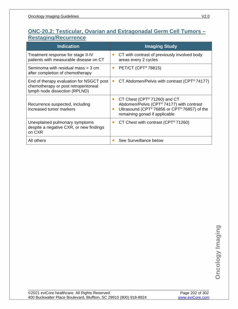
Onc
olog
y Im
agin
g
ONC-20.2: Testicular, Ovarian and Extragonadal Germ Cell Tumors – Restaging/Recurrence
Indication Imaging Study Treatment response for stage II-IV patients with measurable disease on CT
CT with contrast of previously involved body areas every 2 cycles
Seminoma with residual mass > 3 cm after completion of chemotherapy
PET/CT (CPT® 78815)
End of therapy evaluation for NSGCT post chemotherapy or post retroperitoneal lymph node dissection (RPLND)
CT Abdomen/Pelvis with contrast (CPT® 74177)
Recurrence suspected, including increased tumor markers
CT Chest (CPT® 71260) and CT Abdomen/Pelvis (CPT® 74177) with contrast
Ultrasound (CPT® 76856 or CPT® 76857) of the remaining gonad if applicable
Unexplained pulmonary symptoms despite a negative CXR, or new findings on CXR
CT Chest with contrast (CPT® 71260)
All others See Surveillance below
Oncology Imaging Guidelines V2.0
______________________________________________________________________________________________________ ©2021 eviCore healthcare. All Rights Reserved. 400 Buckwalter Place Boulevard, Bluffton, SC 29910 (800) 918-8924 www.eviCore.com
Page 202 of 302
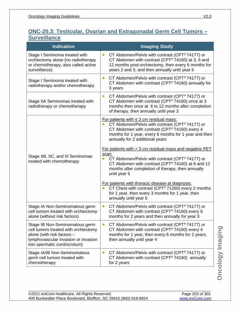
Onc
olog
y Im
agin
g
ONC-20.3: Testicular, Ovarian and Extragonadal Germ Cell Tumors – Surveillance
Indication Imaging Study Stage I Seminoma treated with orchiectomy alone (no radiotherapy or chemotherapy, also called active surveillance)
CT Abdomen/Pelvis with contrast (CPT® 74177) or CT Abdomen with contrast (CPT® 74160) at 3, 6 and 12 months post-orchiectomy, then every 6 months for years 2 and 3, and then annually until year 5
Stage I Seminoma treated with radiotherapy and/or chemotherapy
CT Abdomen/Pelvis with contrast (CPT® 74177) or CT Abdomen with contrast (CPT® 74160) annually for 3 years
Stage IIA Seminomas treated with radiotherapy or chemotherapy
CT Abdomen/Pelvis with contrast (CPT® 74177) or CT Abdomen with contrast (CPT® 74160) once at 3 months then once at 6 to 12 months after completion of therapy, then annually until year 3
Stage IIB, IIC, and III Seminomas treated with chemotherapy
For patients with ≤ 3 cm residual mass: CT Abdomen/Pelvis with contrast (CPT® 74177) or
CT Abdomen with contrast (CPT® 74160) every 4 months for 1 year, every 6 months for 1 year and then annually for 2 additional years
For patients with > 3 cm residual mass and negative PET scan: CT Abdomen/Pelvis with contrast (CPT® 74177) or
CT Abdomen with contrast (CPT® 74160) at 6 and 12 months after completion of therapy, then annually until year 5
For patients with thoracic disease at diagnosis: CT Chest with contrast (CPT® 71260) every 2 months
for 1 year, then every 3 months for 1 year, then annually until year 5
Stage IA Non-Seminomatous germ cell tumors treated with orchiectomy alone (without risk factors)
CT Abdomen/Pelvis with contrast (CPT® 74177) or CT Abdomen with contrast (CPT® 74160) every 6 months for 2 years and then annually for year 3
Stage IB Non-Seminomatous germ cell tumors treated with orchiectomy alone (with risk factors – lymphovascular invasion or invasion into spermatic cord/scrotum)
CT Abdomen/Pelvis with contrast (CPT® 74177) or CT Abdomen with contrast (CPT® 74160) every 4 months for 1 year, then every 6 months for 2 years, then annually until year 4
Stage IA/IB Non-Seminomatous germ cell tumors treated with chemotherapy
CT Abdomen/Pelvis with contrast (CPT® 74177) or CT Abdomen with contrast (CPT® 74160) annually for 2 years
Oncology Imaging Guidelines V2.0
______________________________________________________________________________________________________ ©2021 eviCore healthcare. All Rights Reserved. 400 Buckwalter Place Boulevard, Bluffton, SC 29910 (800) 918-8924 www.eviCore.com
Page 203 of 302
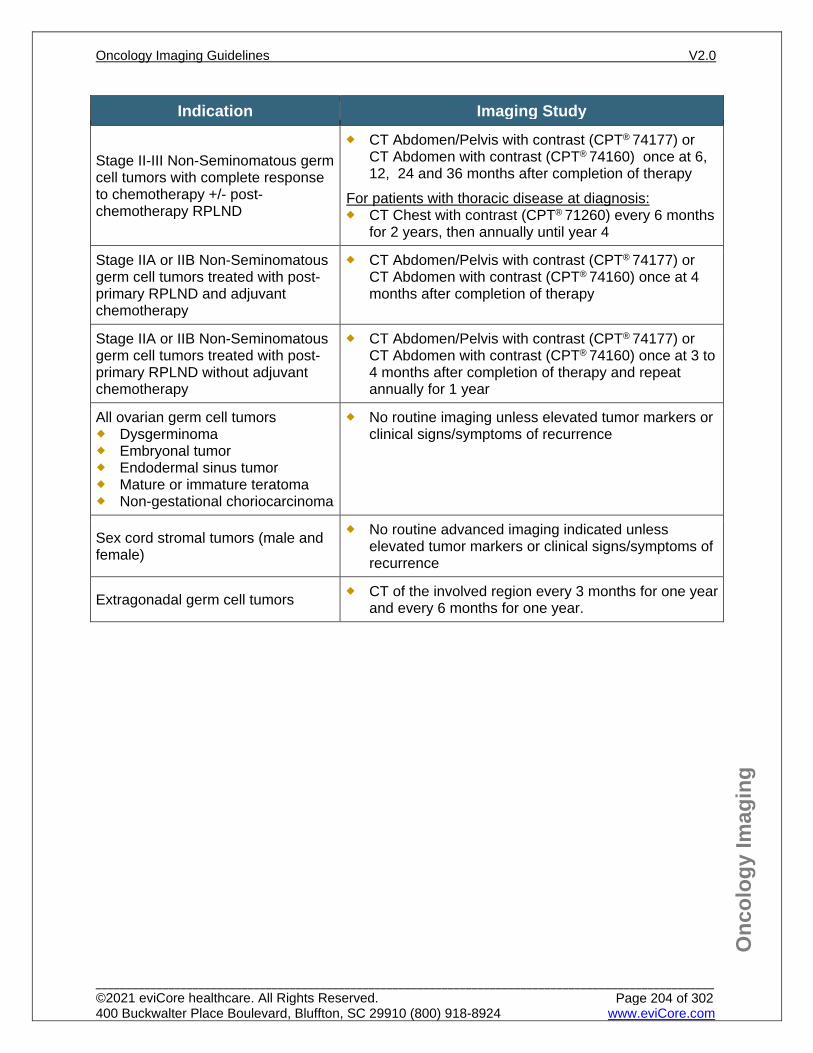
Onc
olog
y Im
agin
g
Indication Imaging Study
Stage II-III Non-Seminomatous germ cell tumors with complete response to chemotherapy +/- post-chemotherapy RPLND
CT Abdomen/Pelvis with contrast (CPT® 74177) or CT Abdomen with contrast (CPT® 74160) once at 6, 12, 24 and 36 months after completion of therapy
For patients with thoracic disease at diagnosis: CT Chest with contrast (CPT® 71260) every 6 months
for 2 years, then annually until year 4
Stage IIA or IIB Non-Seminomatous germ cell tumors treated with post-primary RPLND and adjuvant chemotherapy
CT Abdomen/Pelvis with contrast (CPT® 74177) or CT Abdomen with contrast (CPT® 74160) once at 4 months after completion of therapy
Stage IIA or IIB Non-Seminomatous germ cell tumors treated with post-primary RPLND without adjuvant chemotherapy
CT Abdomen/Pelvis with contrast (CPT® 74177) or CT Abdomen with contrast (CPT® 74160) once at 3 to 4 months after completion of therapy and repeat annually for 1 year
All ovarian germ cell tumors Dysgerminoma Embryonal tumor Endodermal sinus tumor Mature or immature teratoma Non-gestational choriocarcinoma
No routine imaging unless elevated tumor markers or clinical signs/symptoms of recurrence
Sex cord stromal tumors (male and female)
No routine advanced imaging indicated unless elevated tumor markers or clinical signs/symptoms of recurrence
Extragonadal germ cell tumors CT of the involved region every 3 months for one year and every 6 months for one year.
Oncology Imaging Guidelines V2.0
______________________________________________________________________________________________________ ©2021 eviCore healthcare. All Rights Reserved. 400 Buckwalter Place Boulevard, Bluffton, SC 29910 (800) 918-8924 www.eviCore.com
Page 204 of 302
Onc
olog
y Im
agin
g
References 1. Gilligan T, Lin DW, Aggarwal R, et al. National Comprehensive Cancer Network (NCCN) Guidelines
Version 1.2021 – November 5, 2020. Testicular cancer, available at: https://www.nccn.org/professionals/physician_gls/pdf/testicular.pdf Referenced with permission from the NCCN Clinical Practice Guidelines in Oncology (NCCN Guidelines™) for Testicular cancer V1.2021 – November 5, 2020. ©2020 National Comprehensive Cancer Network, Inc. All rights reserved. The NCCN Guidelines™ and illustrations herein may not be reproduced in any form for any purpose without the express written permission of the NCCN. To view the most recent and complete version of the NCCN Guidelines™, go online to NCCN.org.
2. Salani R, Backes FJ, Fung MF, et al. Post treatment surveillance and diagnosis of recurrence in women with gynecologic malignancies: Society of Gynecologic Oncologists recommendations. Am J Obstet Gynecol. 2011;204(6):466-478. doi:10.1016/j.ajog.2011.03.008.
3. Gershenson DM. Management of ovarian germ cell tumors. J Clin Oncol. 2007;25(20):2938-2943. doi:10.1200/JCO.2007.10.8738.
4. Colombo N, Parma G, Zanagnolo V, Insinga A. Management of ovarian stromal cell tumors. J Clin Oncol. 2007;25(20):2944-2951. doi:10.1200/JCO.2007.11.1005.
5. Cadron I, Leunen K, Van Gorp T, Amant F, Neven P, Vergote I. Management of Borderline Ovarian Neoplasms. J Clin Oncol. 2007;25(20):2928-2937. doi:10.1200/JCO/2007.10.8076.
6. del Carmen MG, Birrer M, Schorge JO. Carcinosarcoma of the ovary: a review of the literature. Gynecol Oncol. 2012;125(1):271-277. Doi:10.1016/j.ygyno.2011.12.418.
7. Kollmannsberger C, Tandstad T, Bedard PL, et al. Patterns of relapse in patients with clinical stage I testicular cancer managed with active surveillance. J Clin Oncol. 2015;33(1):51-57. doi:10.1200/JCO.2014.56.2116.
8. Oechsle K, Hartmann M, Brenner W, et al. [18F]Fluorodeoxyglucose positron emission tomography in nonseminomatous germ cell tumors after chemotherapy: the German multicenter positron emission tomography study group. J Clin Oncol. 2008;26(36):5930-5935. doi:10.1200/JCO.2008.17.1157.
9. Daugard G, Gundgaard MG, Mortensen MS, et al. Surveillance for stage I non seminoma testicular cancer: outcomes and long term follow-up in a population based cohort. J. Clin Oncol. 2014;32(34):3817-3823. doi:10.1200/JCO.2013.53.5831.
10. Zuniga A, Kakiashvilli D, Jewett MA. Surveillance in stage I nonseminomatous germ cell tumours of the testis. BJU Int. 2009;104:1351-1356. doi:10.1111/j.1464-410X.2009.08858.x.
Oncology Imaging Guidelines V2.0
______________________________________________________________________________________________________ ©2021 eviCore healthcare. All Rights Reserved. 400 Buckwalter Place Boulevard, Bluffton, SC 29910 (800) 918-8924 www.eviCore.com
Page 205 of 302
ONC-21: Ovarian Cancer ONC-21.0: Ovarian Cancer – General ConsiderationsONC-21.1: Screening for Ovarian CancerONC-21.2: Ovarian Cancer – Suspected/DiagnosisONC-21.3: Ovarian Cancer – Initial Work-up/StagingONC-21.4: Ovarian Cancer – Restaging/RecurrenceONC-21.5: Ovarian Cancer – Surveillance
Oncology Imaging Guidelines V2.0
______________________________________________________________________________________________________ ©2021 eviCore healthcare. All Rights Reserved. 400 Buckwalter Place Boulevard, Bluffton, SC 29910 (800) 918-8924 www.eviCore.com
Page 206 of 302
Onc
olog
y Im
agin
g
ONC-21.0: Ovarian Cancer – General Considerations Ovarian cancers include: epithelial ovarian cancers, ovarian cancers of low
malignant potential and mixed Müllerian tumors, primary peritoneal and fallopian tube cancers.
Germ cell tumors and sex cord stromal tumors (granulosa cell tumors), are imaged according to ONC-20: Testicular, Ovarian and Extragonadal Germ Cell Cancer.
Oncology Imaging Guidelines V2.0
______________________________________________________________________________________________________ ©2021 eviCore healthcare. All Rights Reserved. 400 Buckwalter Place Boulevard, Bluffton, SC 29910 (800) 918-8924 www.eviCore.com
Page 207 of 302
Onc
olog
y Im
agin
g
ONC-21.1: Screening for Ovarian Cancer Indication Imaging/Lab Study
High Risk Factors: Family history of BRCA 1 or
BRCA 2 mutations Family history of ovarian cancer Hereditary ovarian cancer
syndrome that includes ovarian, breast, and/or endometrial and gastrointestinal cancers [Lynch II syndrome] in multiple members of two to four generations
Low parity Decreased fertility Delayed childbearing
Ovarian cancer screening is considered experimental & investigational and is not recommended.
Genetic counseling is recommended for women with an increased-risk family history (USPSTF, 2015)
Known BRCA-1 or BRCA-2 mutation
Transvaginal ultrasound (CPT® 76830), combined with CA-125 for ovarian cancer screening may be considered annually starting at age 30, until risk-reducing salpingo-oophorectomy is performed
Oncology Imaging Guidelines V2.0
______________________________________________________________________________________________________ ©2021 eviCore healthcare. All Rights Reserved. 400 Buckwalter Place Boulevard, Bluffton, SC 29910 (800) 918-8924 www.eviCore.com
Page 208 of 302
Onc
olog
y Im
agin
g
ONC-21.2: Ovarian Cancer – Suspected/Diagnosis See: PV-5.3: Complex Adnexal Masses for imaging guidelines for evaluation of
suspected ovarian malignancies Staging of ovarian cancer is primarily surgical and routine imaging is not indicated
pre-operatively, unless it is obtained to evaluate specific signs/symptoms. To differentiate the origin of pelvic masses that are not clearly of ovarian origin, see:
PV-5.1: Suspected Adnexal Mass.
Indication Imaging/Lab Study Pelvic symptoms (pelvic pain,
abdominal bloating) Palpable pelvic mass
Transvaginal (TV) ultrasound imaging (CPT® 76830) and/or Pelvic ultrasound (CPT® 76856 or CPT® 76857) is the initial study of choice
Ultrasound shows a complex and/or solid adnexal mass
See: PV-5.3: Complex Adnexal Masses
Ultrasound shows complex and/or solid adnexal mass suspicious for ovarian malignancy AND any of the following signs/symptoms concerning for metastatic disease: Ascites Abdominal symptoms (distension,
tenderness) Elevated CA-125 Elevated LFTs Obstructive uropathy**
CT Abdomen and Pelvis with contrast (CPT® 74177)
**CT Abdomen/Pelvis without and with contrast (CT Urogram – CPT® 74178) may be approved only for symptoms of obstructive uropathy
Oncology Imaging Guidelines V2.0
______________________________________________________________________________________________________ ©2021 eviCore healthcare. All Rights Reserved. 400 Buckwalter Place Boulevard, Bluffton, SC 29910 (800) 918-8924 www.eviCore.com
Page 209 of 302
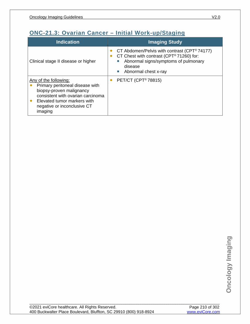
Onc
olog
y Im
agin
g
ONC-21.3: Ovarian Cancer – Initial Work-up/Staging Indication Imaging Study
Clinical stage II disease or higher
CT Abdomen/Pelvis with contrast (CPT® 74177) CT Chest with contrast (CPT® 71260) for:
Abnormal signs/symptoms of pulmonary disease
Abnormal chest x-ray
Any of the following: Primary peritoneal disease with
biopsy-proven malignancy consistent with ovarian carcinoma
Elevated tumor markers with negative or inconclusive CT imaging
PET/CT (CPT® 78815)
Oncology Imaging Guidelines V2.0
______________________________________________________________________________________________________ ©2021 eviCore healthcare. All Rights Reserved. 400 Buckwalter Place Boulevard, Bluffton, SC 29910 (800) 918-8924 www.eviCore.com
Page 210 of 302
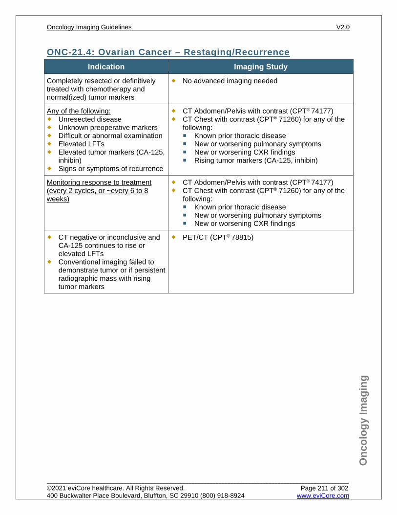
Onc
olog
y Im
agin
g
ONC-21.4: Ovarian Cancer – Restaging/Recurrence Indication Imaging Study
Completely resected or definitively treated with chemotherapy and normal(ized) tumor markers
No advanced imaging needed
Any of the following: Unresected disease Unknown preoperative markers Difficult or abnormal examination Elevated LFTs Elevated tumor markers (CA-125,
inhibin) Signs or symptoms of recurrence
CT Abdomen/Pelvis with contrast (CPT® 74177) CT Chest with contrast (CPT® 71260) for any of the
following: Known prior thoracic disease New or worsening pulmonary symptoms New or worsening CXR findings Rising tumor markers (CA-125, inhibin)
Monitoring response to treatment (every 2 cycles, or ~every 6 to 8 weeks)
CT Abdomen/Pelvis with contrast (CPT® 74177) CT Chest with contrast (CPT® 71260) for any of the
following: Known prior thoracic disease New or worsening pulmonary symptoms New or worsening CXR findings
CT negative or inconclusive and CA-125 continues to rise or elevated LFTs
Conventional imaging failed to demonstrate tumor or if persistent radiographic mass with rising tumor markers
PET/CT (CPT® 78815)
Oncology Imaging Guidelines V2.0
______________________________________________________________________________________________________ ©2021 eviCore healthcare. All Rights Reserved. 400 Buckwalter Place Boulevard, Bluffton, SC 29910 (800) 918-8924 www.eviCore.com
Page 211 of 302
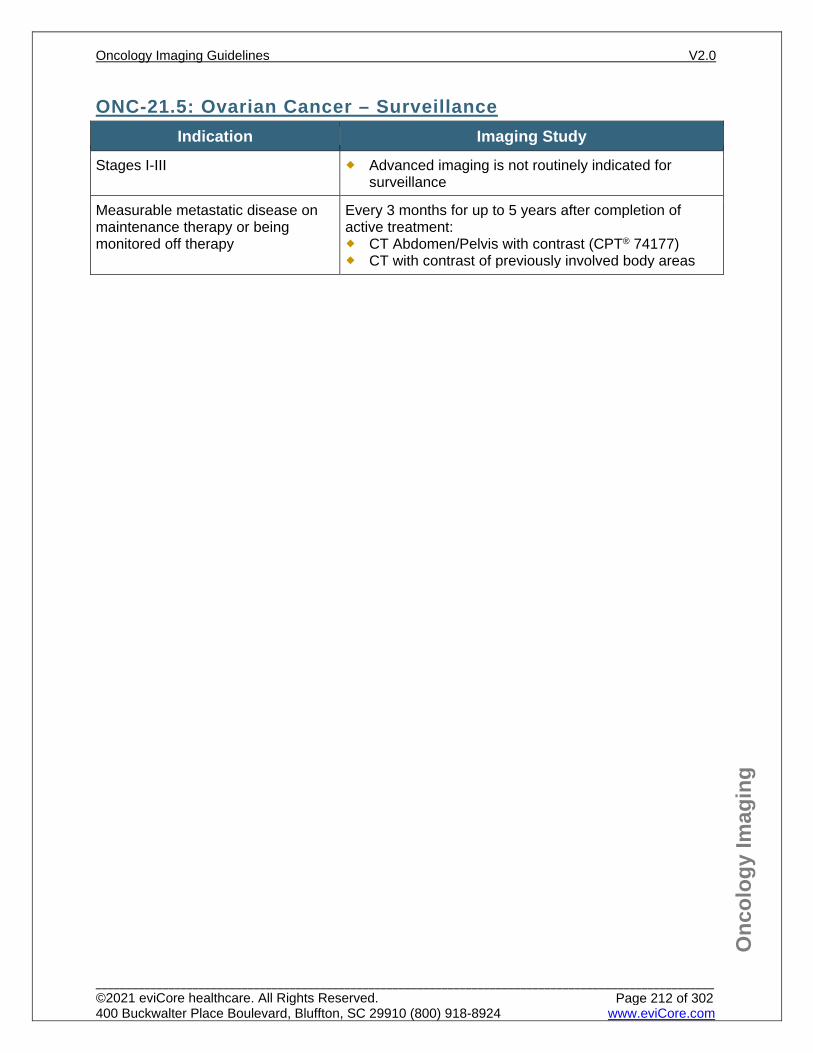
Onc
olog
y Im
agin
g
ONC-21.5: Ovarian Cancer – Surveillance Indication Imaging Study
Stages I-III Advanced imaging is not routinely indicated for surveillance
Measurable metastatic disease on maintenance therapy or being monitored off therapy
Every 3 months for up to 5 years after completion of active treatment: CT Abdomen/Pelvis with contrast (CPT® 74177) CT with contrast of previously involved body areas
Oncology Imaging Guidelines V2.0
______________________________________________________________________________________________________ ©2021 eviCore healthcare. All Rights Reserved. 400 Buckwalter Place Boulevard, Bluffton, SC 29910 (800) 918-8924 www.eviCore.com
Page 212 of 302
Onc
olog
y Im
agin
g
References 1. Armstrong DK, Alvarex RD, Bakkum-Gamez JN, et al. National Comprehensive Cancer Network
(NCCN) Guidelines Version 1.2021 – February 26, 2021. Ovarian Cancer, available at: https://www.nccn.org/professionals/physician_gls/pdf/ovarian.pdf Referenced with permission from the NCCN Clinical Practice Guidelines in Oncology (NCCN Guidelines™) for Ovarian Cancer V1.2021 – February 26, 2021. ©2021 National Comprehensive Cancer Network, Inc. All rights reserved. The NCCN Guidelines™ and illustrations herein may not be reproduced in any form for any purpose without the express written permission of the NCCN. To view the most recent and complete version of the NCCN Guidelines™, go online to NCCN.org.
2. Daly MB, Pal T, Berry MP, et al. National Comprehensive Cancer Network (NCCN) Guidelines Version V2.2021 – November 20, 2020. Genetic/Familial High-Risk Assessment: Breast and Ovarian, available at: https://www.nccn.org/professionals/physician_gls/pdf/genetics_screening.pdf Referenced with permission from the NCCN Clinical Practice Guidelines in Oncology (NCCN Guidelines™) for Ovarian cancer V2.2021 – November 20, 2020. ©2020 National Comprehensive Cancer Network, Inc. All rights reserved. The NCCN Guidelines™ and illustrations herein may not be reproduced in any form for any purpose without the express written permission of the NCCN. To view the most recent and complete version of the NCCN Guidelines™, go online to NCCN.org.
3. Moyer VA, U.S. Preventive Services Task Force. Screening for ovarian cancer: U.S. Preventive Services Task Force reaffirmation recommendation statement. Ann Intern Med. 2012;157(12):900-904. doi:10.7326/0003-4819-157-11-201212040-00539.
4. Cadron I, Leunen K, Van Gorp T, Amant F, Neven P, Vergote I. Management of borderline ovarian neoplasms. J Clin Oncol. 2007;25(20):2928-2937. doi:10.1200/JCO.2007.10.8076.
5. ACR Appropriateness Criteria. Ovarian cancer screening. Rev. 2017. 6. Rosenthal AN, Fraser LSM, Phipott S. Evidence of stage shift in women diagnosed with ovarian
cancer during phase II of the United Kingdom familial ovarian cancer screening study. J Clin Oncol. 2017;35(13):13:1411-1420. doi:10.1200/JCO.2016.69.9330.
7. Shinagare AB, O’Neill AC, Cheng S, et al. Advanced high-grade serous ovarian cancer: frequency and timing of thoracic metastases and the implications for chest imaging follow-up. Radiology. 2015;277(3):733-740. doi:10.1148/radiol.2015142467.
8. Musto A, Grassetto G, Marzola MC, et al. Management of epithelial ovarian cancer from diagnosis to restaging: an overview of the role of imaging techniques with particular regard to the contribution of 18F-FDG PET/CT. Nucl Med Commun. 2014;35(6):588-597. doi:10.1097/MNM.0000000000000091.
9. Fischerova D, Burgetova A. Imaging techniques for the evaluation of ovarian cancer. Best Pract Res Clin Obstet Gynaecol. 2014;28(5):697-720. doi:10.1016/j.bpobgyn.2014.04.006.
Oncology Imaging Guidelines V2.0
______________________________________________________________________________________________________ ©2021 eviCore healthcare. All Rights Reserved. 400 Buckwalter Place Boulevard, Bluffton, SC 29910 (800) 918-8924 www.eviCore.com
Page 213 of 302

ONC-22: Uterine Cancer ONC-22.0: Uterine Cancer – General ConsiderationsONC-22.1: Uterine Cancer – Suspected/DiagnosisONC-22.2: Uterine Cancer – Initial Work-up/StagingONC-22.3: Uterine Cancer – Restaging/RecurrenceONC-22.4: Uterine Cancer – Surveillance
Oncology Imaging Guidelines V2.0
______________________________________________________________________________________________________ ©2021 eviCore healthcare. All Rights Reserved. 400 Buckwalter Place Boulevard, Bluffton, SC 29910 (800) 918-8924 www.eviCore.com
Page 214 of 302
Onc
olog
y Im
agin
g
ONC-22.0: Uterine Cancer – General Considerations Gestational trophoblastic neoplasia (GTN) – see: PV-16.1: Molar Pregnancy and
Gestational Trophoblastic Neoplasia (GTN) Most common cell type is adenocarcinoma. Uterine sarcomas are also imaged
according to this guideline. Staging of uterine cancer is primarily surgical. Advanced imaging is not routinely
indicated pre-operatively for laparoscopic/minimally invasive surgery unless initial staging criteria are met. Pelvic and para-aortic lymphadenectomy can still be performed.
Oncology Imaging Guidelines V2.0
______________________________________________________________________________________________________ ©2021 eviCore healthcare. All Rights Reserved. 400 Buckwalter Place Boulevard, Bluffton, SC 29910 (800) 918-8924 www.eviCore.com
Page 215 of 302
Onc
olog
y Im
agin
g
ONC-22.1: Uterine Cancer – Suspected/Diagnosis See: PV-2.1: Abnormal Uterine Bleeding for imaging guidelines for evaluation of
suspected uterine malignancies
Oncology Imaging Guidelines V2.0
______________________________________________________________________________________________________ ©2021 eviCore healthcare. All Rights Reserved. 400 Buckwalter Place Boulevard, Bluffton, SC 29910 (800) 918-8924 www.eviCore.com
Page 216 of 302
Onc
olog
y Im
agin
g
ONC-22.2: Uterine Cancer – Initial Work-up/Staging Indication Imaging Study
Extra-uterine disease suspected Bulky uterine tumor High grade (grade 3) tumor
MRI Pelvis without and with contrast (CPT®
72197) or CT Pelvis with contrast (CPT®
72193)
Any of the following: Abdominal symptoms or abnormal
examination findings Elevated LFTS Other imaging studies suggest liver
involvement
One of the following may be approved: CT Abdomen with contrast (CPT® 74160)
CT Abdomen/Pelvis with contrast (CPT®
74177) if being completed in the same imaging session as CT Pelvis
Any of the following histologies: Papillary serous Clear cell Carcinosarcoma Soft tissue sarcoma of the uterus Leiomyosarcoma Undifferentiated sarcoma Endometrial stromal sarcoma Poorly differentiated endometroid
CT Chest (CPT® 71260) and CT Abdomen/Pelvis with contrast (CPT® 74177)
Tumors detected incidentally or incompletely staged surgically AND any of the following high risk features: Myoinvasion > 50% Cervical stromal involvement Lymphovascular invasion Tumor > 2 cm
CT Chest (CPT® 71260) and CT Abdomen/Pelvis with contrast (CPT® 74177)
Considering fertility sparing surgery for well-differentiated Stage IA (grade 1) uterine cancer
Poor surgical candidate (due to medical comorbidities) considering medical therapy
MRI Pelvis without and with contrast (CPT® 72197) or CT Pelvis with contrast (CPT® 72193)
Transvaginal ultrasound (CPT® 76830) if MRI is contraindicated
Chest x-ray CT Chest with contrast (CPT® 71260) if
chest x-ray is abnormal
Inconclusive findings on conventional imaging
PET/CT scan (CPT® 78815)
Oncology Imaging Guidelines V2.0
______________________________________________________________________________________________________ ©2021 eviCore healthcare. All Rights Reserved. 400 Buckwalter Place Boulevard, Bluffton, SC 29910 (800) 918-8924 www.eviCore.com
Page 217 of 302
Onc
olog
y Im
agin
g
ONC-22.3: Uterine Cancer – Restaging/Recurrence Indication Imaging Study
Unresected disease Medically inoperable disease Incomplete surgical staging Difficult or abnormal examination Elevated LFTs or rising tumor markers Signs or symptoms of recurrence
CT Chest (CPT® 71260) and CT Abdomen/Pelvis with contrast (CPT®
74177) MRI Pelvis (CPT® 72197) without and with
contrast if CT is inconclusive
Monitoring response to chemotherapy (every 2 cycles, ~every 6-8 weeks) for: Unresected primary disease Metastatic disease
CT Abdomen/Pelvis with contrast (CPT®
74177) CT Chest with contrast (CPT® 71260) for:
New/worsening pulmonary symptoms Abnormal chest x-ray findings Known prior pulmonary involvement
Inconclusive findings on conventional imaging
PET/CT scan (CPT® 78815)
Oncology Imaging Guidelines V2.0
______________________________________________________________________________________________________ ©2021 eviCore healthcare. All Rights Reserved. 400 Buckwalter Place Boulevard, Bluffton, SC 29910 (800) 918-8924 www.eviCore.com
Page 218 of 302
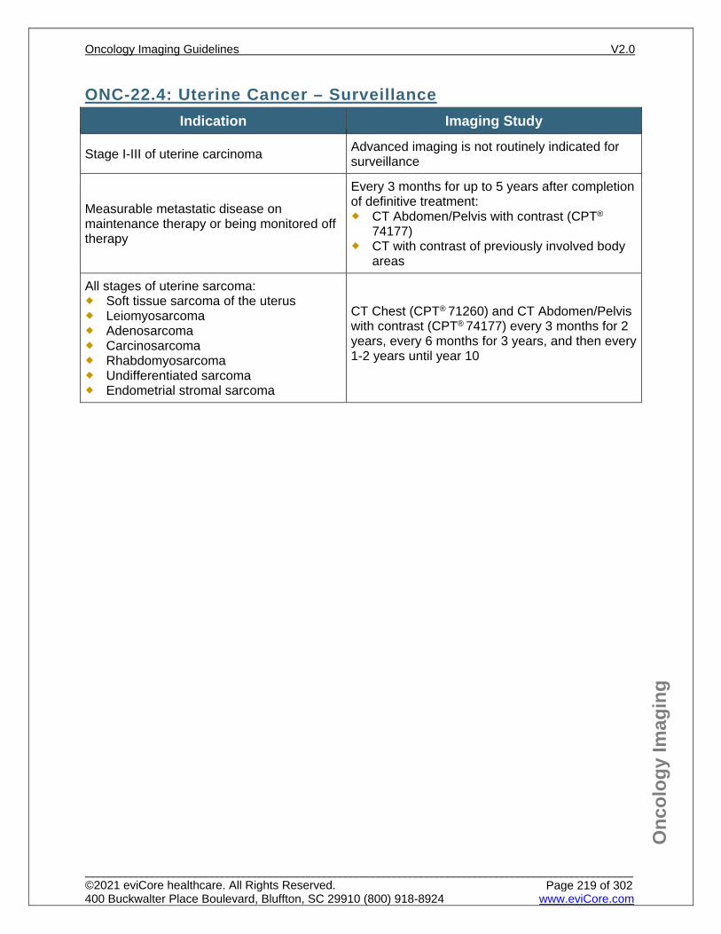
Onc
olog
y Im
agin
g
ONC-22.4: Uterine Cancer – Surveillance Indication Imaging Study
Stage I-III of uterine carcinoma Advanced imaging is not routinely indicated for surveillance
Measurable metastatic disease on maintenance therapy or being monitored off therapy
Every 3 months for up to 5 years after completion of definitive treatment: CT Abdomen/Pelvis with contrast (CPT®
74177) CT with contrast of previously involved body
areas
All stages of uterine sarcoma: Soft tissue sarcoma of the uterus Leiomyosarcoma Adenosarcoma Carcinosarcoma Rhabdomyosarcoma Undifferentiated sarcoma Endometrial stromal sarcoma
CT Chest (CPT® 71260) and CT Abdomen/Pelvis with contrast (CPT® 74177) every 3 months for 2 years, every 6 months for 3 years, and then every 1-2 years until year 10
Oncology Imaging Guidelines V2.0
______________________________________________________________________________________________________ ©2021 eviCore healthcare. All Rights Reserved. 400 Buckwalter Place Boulevard, Bluffton, SC 29910 (800) 918-8924 www.eviCore.com
Page 219 of 302
Onc
olog
y Im
agin
g
References 1. Abu-Rustum NR, Yashar CM, Bradley K, et al. National Comprehensive Cancer Network (NCCN)
Guidelines Version 1.2021 – October 20, 2020. Uterine Neoplasms, available at: https://www.nccn.org/professionals/physician_gls/pdf/uterine.pdf Referenced with permission from the NCCN Clinical Practice Guidelines in Oncology (NCCN Guidelines™) for Uterine Neoplasms V1.2021 – October 20, 2020. ©2020 National Comprehensive Cancer Network, Inc. All rights reserved. The NCCN Guidelines™ and illustrations herein may not be reproduced in any form for any purpose without the express written permission of the NCCN. To view the most recent and complete version of the NCCN Guidelines™, go online to NCCN.org.
2. Fader AN, Boruta D, Olawaiye AB, Gehrig PA. Updates on uterine papillary serous carcinoma. Expert Rev Obstet Gynecol. 2009;4(6):647-657. doi:10.1586/eog.09.49.
3. Boruta DM 2nd, Gehrig PA, Fader AN, Olawaiye AB. Management of women with uterine papillary serous cancer: A Society of Gynecologic Oncology (SGO) review. Gynecol Oncol. 2009;115(1):142-153. doi:10.1016/j.ygyno.2009.06.011.
4. Olawaiye AB, Boruta DM 2nd. Management of women with clear cell endometrial cancer: a Society of Gynecologic Oncology (SGO) review. Gynecol Oncol. 2009;113(2):277-283. doi:10.1016/j.ygyno.2009.02.003.
5. Salani R, Backes FJ, Fung MF et al. Post treatment surveillance and diagnosis of recurrence in women with gynecologic malignancies: Society of Gynecologic Oncologists recommendations. Am J Obstet Gynecol. 2011;204(6):466-478. doi:10.1016/j.ajog.2011.03.008.
6. Reinhold C, Ueno Y, Akin EA, et. al. ACR Appropriateness Criteria® - Evaluation and follow-up of endometrical cancer. Available at https://acsearch.acr.org/docs/69459/Narrative/. American College of Radiology. Accessed 7/29/2020.
Oncology Imaging Guidelines V2.0
______________________________________________________________________________________________________ ©2021 eviCore healthcare. All Rights Reserved. 400 Buckwalter Place Boulevard, Bluffton, SC 29910 (800) 918-8924 www.eviCore.com
Page 220 of 302
ONC-23: Cervical Cancer ONC-23.0: Cervical Cancer – General ConsiderationsONC-23.1: Cervical Cancer – Suspected/DiagnosisONC-23.2: Cervical Cancer – Initial Work-up/StagingONC-23.3: Cervical Cancer – Restaging/RecurrenceONC-23.4: Cervical Cancer – Surveillance
Oncology Imaging Guidelines V2.0
______________________________________________________________________________________________________ ©2021 eviCore healthcare. All Rights Reserved. 400 Buckwalter Place Boulevard, Bluffton, SC 29910 (800) 918-8924 www.eviCore.com
Page 221 of 302
Onc
olog
y Im
agin
g
ONC-23.0: Cervical Cancer – General Considerations Primary histology for cervical cancer is squamous cell. Other, less common
histologies are adenosquamous and adenocarcinoma. If biopsy is consistent with one of these less common histologies, it is necessary to clarify that tumor is not of primary uterine origin.
If the primary histology is uterine in origin, follow imaging recommendations for uterine cancer, see: ONC-22: Uterine Cancer.
Oncology Imaging Guidelines V2.0
______________________________________________________________________________________________________ ©2021 eviCore healthcare. All Rights Reserved. 400 Buckwalter Place Boulevard, Bluffton, SC 29910 (800) 918-8924 www.eviCore.com
Page 222 of 302
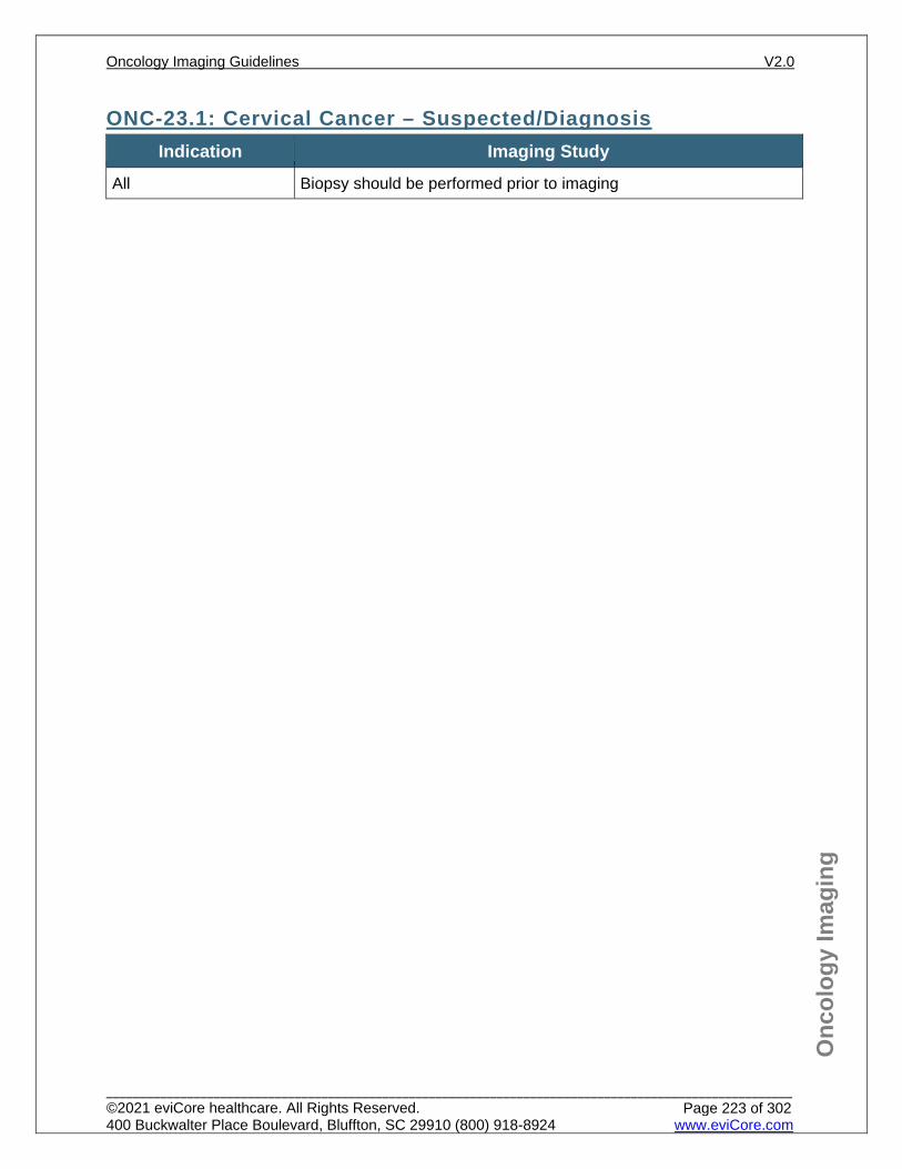
Onc
olog
y Im
agin
g
ONC-23.1: Cervical Cancer – Suspected/Diagnosis Indication Imaging Study
All Biopsy should be performed prior to imaging
Oncology Imaging Guidelines V2.0
______________________________________________________________________________________________________ ©2021 eviCore healthcare. All Rights Reserved. 400 Buckwalter Place Boulevard, Bluffton, SC 29910 (800) 918-8924 www.eviCore.com
Page 223 of 302
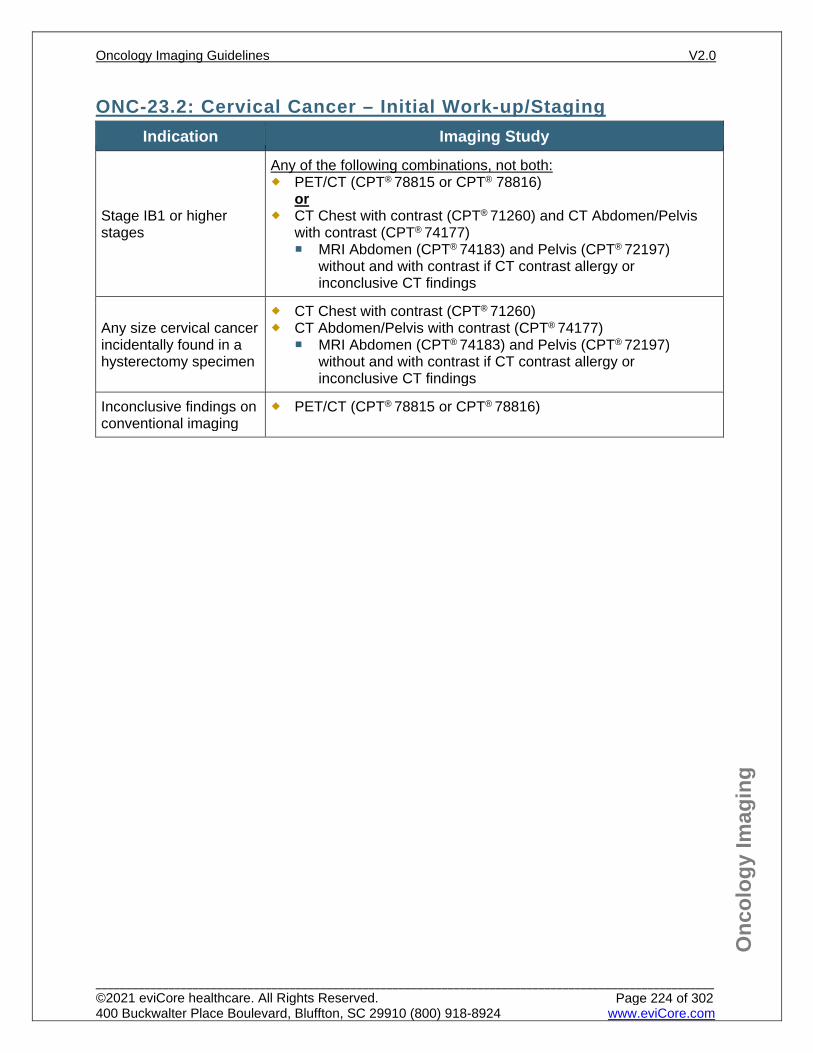
Onc
olog
y Im
agin
g
ONC-23.2: Cervical Cancer – Initial Work-up/Staging Indication Imaging Study
Stage IB1 or higher stages
Any of the following combinations, not both: PET/CT (CPT® 78815 or CPT® 78816)
or CT Chest with contrast (CPT® 71260) and CT Abdomen/Pelvis
with contrast (CPT® 74177) MRI Abdomen (CPT® 74183) and Pelvis (CPT® 72197)
without and with contrast if CT contrast allergy or inconclusive CT findings
Any size cervical cancer incidentally found in a hysterectomy specimen
CT Chest with contrast (CPT® 71260) CT Abdomen/Pelvis with contrast (CPT® 74177)
MRI Abdomen (CPT® 74183) and Pelvis (CPT® 72197) without and with contrast if CT contrast allergy or inconclusive CT findings
Inconclusive findings on conventional imaging
PET/CT (CPT® 78815 or CPT® 78816)
Oncology Imaging Guidelines V2.0
______________________________________________________________________________________________________ ©2021 eviCore healthcare. All Rights Reserved. 400 Buckwalter Place Boulevard, Bluffton, SC 29910 (800) 918-8924 www.eviCore.com
Page 224 of 302
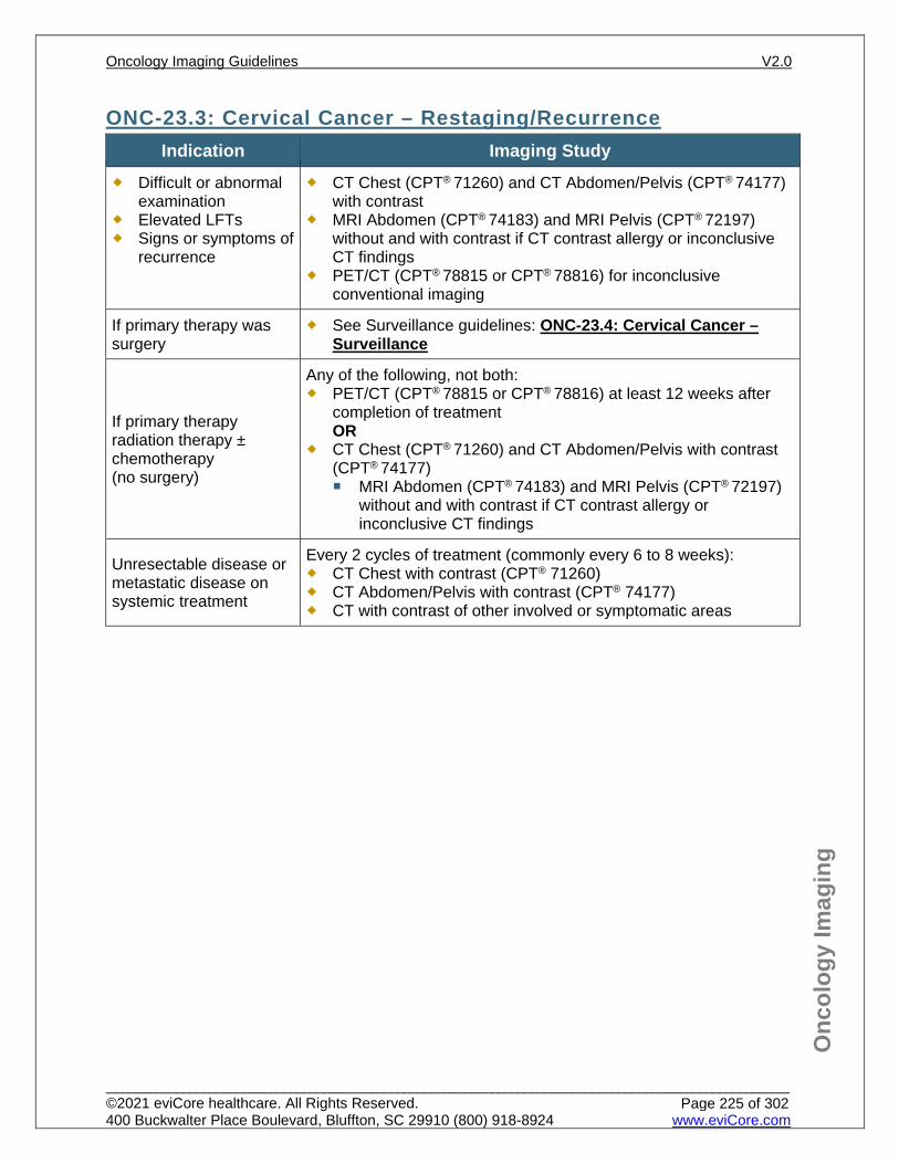
Onc
olog
y Im
agin
g
ONC-23.3: Cervical Cancer – Restaging/Recurrence Indication Imaging Study
Difficult or abnormal examination
Elevated LFTs Signs or symptoms of
recurrence
CT Chest (CPT® 71260) and CT Abdomen/Pelvis (CPT® 74177) with contrast
MRI Abdomen (CPT® 74183) and MRI Pelvis (CPT® 72197) without and with contrast if CT contrast allergy or inconclusive CT findings
PET/CT (CPT® 78815 or CPT® 78816) for inconclusive conventional imaging
If primary therapy was surgery
See Surveillance guidelines: ONC-23.4: Cervical Cancer – Surveillance
If primary therapy radiation therapy ± chemotherapy (no surgery)
Any of the following, not both: PET/CT (CPT® 78815 or CPT® 78816) at least 12 weeks after
completion of treatment OR
CT Chest (CPT® 71260) and CT Abdomen/Pelvis with contrast (CPT® 74177) MRI Abdomen (CPT® 74183) and MRI Pelvis (CPT® 72197)
without and with contrast if CT contrast allergy or inconclusive CT findings
Unresectable disease or metastatic disease on systemic treatment
Every 2 cycles of treatment (commonly every 6 to 8 weeks): CT Chest with contrast (CPT® 71260) CT Abdomen/Pelvis with contrast (CPT® 74177) CT with contrast of other involved or symptomatic areas
Oncology Imaging Guidelines V2.0
______________________________________________________________________________________________________ ©2021 eviCore healthcare. All Rights Reserved. 400 Buckwalter Place Boulevard, Bluffton, SC 29910 (800) 918-8924 www.eviCore.com
Page 225 of 302
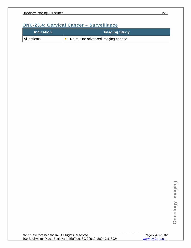
Onc
olog
y Im
agin
g
ONC-23.4: Cervical Cancer – Surveillance Indication Imaging Study
All patients No routine advanced imaging needed.
Oncology Imaging Guidelines V2.0
______________________________________________________________________________________________________ ©2021 eviCore healthcare. All Rights Reserved. 400 Buckwalter Place Boulevard, Bluffton, SC 29910 (800) 918-8924 www.eviCore.com
Page 226 of 302
Onc
olog
y Im
agin
g
References 1. Abu-Rustum NR, Yashar CM, Bradley K, et al. National Comprehensive Cancer Network (NCCN)
Guidelines Version 1.2021 – October 2, 2020. Cervical Cancer, available at: https://www.nccn.org/professionals/physician_gls/pdf/cervical.pdf Referenced with permission from the NCCN Clinical Practice Guidelines in Oncology (NCCN Guidelines™) for Cervical Cancer V1.2021 – October 2, 2020. ©2020 National Comprehensive Cancer Network, Inc. All rights reserved. The NCCN Guidelines™ and illustrations herein may not be reproduced in any form for any purpose without the express written permission of the NCCN. To view the most recent and complete version of the NCCN Guidelines™, go online to NCCN.org.
2. Salani R, Backes FJ, Fung MF et al. Post treatment surveillance and diagnosis of recurrence in women with gynecologic malignancies: Society of Gynecologic Oncologists recommendations. Am J Obstet Gynecol. 2011;204(6):466-478. doi:10.1016/j.ajog.2011.03.008.
3. Zanagnolo V, Ming L, Gadducci A, et al. Surveillance procedures for patients with cervical carcinoma: a review of the literature. Int J Gynecol Cancer. 2009;19(3):194-201. doi:10.1111/IGC.0b013e3181a130f3.
4. Elit L, Fyles AW, Devries MC, et al. Follow-up for women after treatment for cervical cancer: A systematic review. Gynecol Oncol. 2009;114(3):528-535. doi:10.1016/j.ygyno.2009.06.001.
5. Schwarz JK, Siegel BA, Dehdashti F, Grigsby PW. Association of posttherapy positron emission tomography with tumor response and survival in cervical carcinoma. JAMA. 2007;298(19):2289-2295. doi:10.1001/jama.298.19.2289.
6. Meads C, Davenport C, Malysiak S, et al. Evaluating PET-CT in the detection and management of recurrent cervical cancer: systematic reviews of diagnostic accuracy and subjective elicitation. BJOG. 2014;121(4):398-407. doi:10.1111/1471-0528.12488.
7. Chu Y, Zheng A, Wang F, et al. Diagnostic value of 18F-FDG-PET or PET-CT in recurrent cervical cancer: a systematic review and meta-analysis. Nucl Med Commun. 2014; 35(2):144-150. doi:10.1097/MNM. 0000000000000026.
Oncology Imaging Guidelines V2.0
______________________________________________________________________________________________________ ©2021 eviCore healthcare. All Rights Reserved. 400 Buckwalter Place Boulevard, Bluffton, SC 29910 (800) 918-8924 www.eviCore.com
Page 227 of 302
ONC-24: Anal Cancer & Cancers of the External Genitalia
ONC-24.0: Anal Carcinoma – General ConsiderationsONC-24.1: Anal Carcinoma – Suspected/DiagnosisONC-24.2: Anal Carcinoma – Initial Work-up/StagingONC-24.3: Anal Carcinoma – Restaging/RecurrenceONC-24.4: Anal Carcinoma – SurveillanceONC-24.5: Cancers of External Genitalia – General ConsiderationsONC-24.6: Cancers of External Genitalia – Initial Work-up/StagingONC-24.7: Cancers of External Genitalia – Restaging/RecurrenceONC-24.8: Cancers of External Genitalia – Surveillance
Oncology Imaging Guidelines V2.0
______________________________________________________________________________________________________ ©2021 eviCore healthcare. All Rights Reserved. 400 Buckwalter Place Boulevard, Bluffton, SC 29910 (800) 918-8924 www.eviCore.com
Page 228 of 302
Onc
olog
y Im
agin
g
ONC-24.0: Anal Carcinoma – General Considerations Most are squamous cell carcinomas, although some transitional and cloacogenic
carcinomas are seen. Adenocarcinoma of the anal canal is managed as rectal cancer according to ONC-
16: Colorectal and Small Bowel Cancer Squamous cell carcinoma of the perianal region (up to 5 cm radius from the anal
verge) are imaged according to anal carcinoma guidelines. Bowen’s disease and Paget’s disease of the perianal and perigenital skin are
considered non-invasive/in-situ conditions and do not routinely require advanced imaging. See: ONC-5.6: Non-Melanoma Skin Cancers – Initial Work-up/Staging.
Oncology Imaging Guidelines V2.0
______________________________________________________________________________________________________ ©2021 eviCore healthcare. All Rights Reserved. 400 Buckwalter Place Boulevard, Bluffton, SC 29910 (800) 918-8924 www.eviCore.com
Page 229 of 302
Onc
olog
y Im
agin
g
ONC-24.1: Anal Carcinoma – Suspected/Diagnosis Indication Imaging Study
All Advanced imaging prior to biopsy is not needed
Oncology Imaging Guidelines V2.0
______________________________________________________________________________________________________ ©2021 eviCore healthcare. All Rights Reserved. 400 Buckwalter Place Boulevard, Bluffton, SC 29910 (800) 918-8924 www.eviCore.com
Page 230 of 302
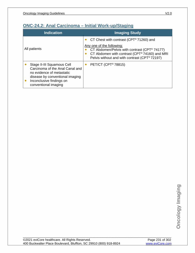
Onc
olog
y Im
agin
g
ONC-24.2: Anal Carcinoma – Initial Work-up/Staging Indication Imaging Study
All patients
CT Chest with contrast (CPT® 71260) and Any one of the following: CT Abdomen/Pelvis with contrast (CPT® 74177) CT Abdomen with contrast (CPT® 74160) and MRI
Pelvis without and with contrast (CPT® 72197)
Stage II-III Squamous Cell Carcinoma of the Anal Canal and no evidence of metastatic disease by conventional imaging
Inconclusive findings on conventional imaging
PET/CT (CPT® 78815)
Oncology Imaging Guidelines V2.0
______________________________________________________________________________________________________ ©2021 eviCore healthcare. All Rights Reserved. 400 Buckwalter Place Boulevard, Bluffton, SC 29910 (800) 918-8924 www.eviCore.com
Page 231 of 302
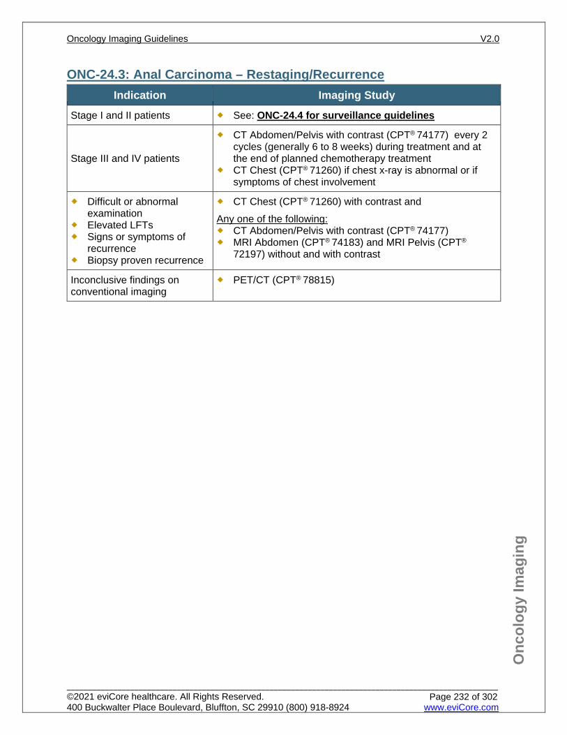
Onc
olog
y Im
agin
g
ONC-24.3: Anal Carcinoma – Restaging/Recurrence Indication Imaging Study
Stage I and II patients See: ONC-24.4 for surveillance guidelines
Stage III and IV patients
CT Abdomen/Pelvis with contrast (CPT® 74177) every 2 cycles (generally 6 to 8 weeks) during treatment and at the end of planned chemotherapy treatment
CT Chest (CPT® 71260) if chest x-ray is abnormal or if symptoms of chest involvement
Difficult or abnormal examination
Elevated LFTs Signs or symptoms of
recurrence Biopsy proven recurrence
CT Chest (CPT® 71260) with contrast and Any one of the following: CT Abdomen/Pelvis with contrast (CPT® 74177) MRI Abdomen (CPT® 74183) and MRI Pelvis (CPT®
72197) without and with contrast
Inconclusive findings on conventional imaging
PET/CT (CPT® 78815)
Oncology Imaging Guidelines V2.0
______________________________________________________________________________________________________ ©2021 eviCore healthcare. All Rights Reserved. 400 Buckwalter Place Boulevard, Bluffton, SC 29910 (800) 918-8924 www.eviCore.com
Page 232 of 302
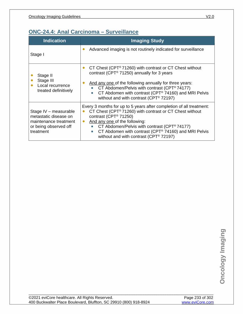
Onc
olog
y Im
agin
g
ONC-24.4: Anal Carcinoma – Surveillance Indication Imaging Study
Stage I Advanced imaging is not routinely indicated for surveillance
Stage II Stage III Local recurrence
treated definitively
CT Chest (CPT® 71260) with contrast or CT Chest without contrast (CPT® 71250) annually for 3 years
And any one of the following annually for three years:
CT Abdomen/Pelvis with contrast (CPT® 74177) CT Abdomen with contrast (CPT® 74160) and MRI Pelvis
without and with contrast (CPT® 72197)
Stage IV – measurable metastatic disease on maintenance treatment or being observed off treatment
Every 3 months for up to 5 years after completion of all treatment: CT Chest (CPT® 71260) with contrast or CT Chest without
contrast (CPT® 71250) And any one of the following:
CT Abdomen/Pelvis with contrast (CPT® 74177) CT Abdomen with contrast (CPT® 74160) and MRI Pelvis
without and with contrast (CPT® 72197)
Oncology Imaging Guidelines V2.0
______________________________________________________________________________________________________ ©2021 eviCore healthcare. All Rights Reserved. 400 Buckwalter Place Boulevard, Bluffton, SC 29910 (800) 918-8924 www.eviCore.com
Page 233 of 302
Onc
olog
y Im
agin
g
ONC-24.5: Cancers of External Genitalia – General Considerations These imaging guidelines are applicable for squamous cell carcinomas arising from
the vulva, vagina, penis and scrotum
Oncology Imaging Guidelines V2.0
______________________________________________________________________________________________________ ©2021 eviCore healthcare. All Rights Reserved. 400 Buckwalter Place Boulevard, Bluffton, SC 29910 (800) 918-8924 www.eviCore.com
Page 234 of 302
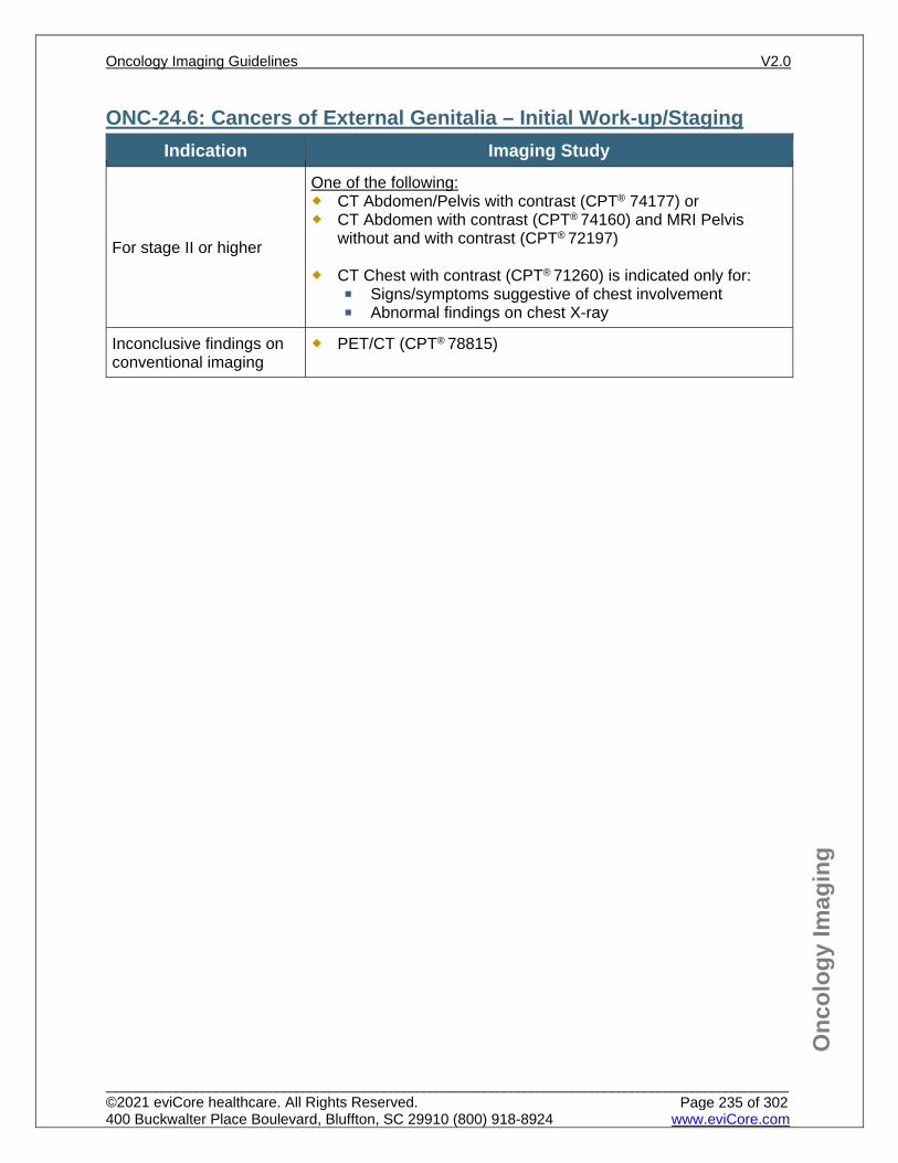
Onc
olog
y Im
agin
g
ONC-24.6: Cancers of External Genitalia – Initial Work-up/Staging Indication Imaging Study
For stage II or higher
One of the following: CT Abdomen/Pelvis with contrast (CPT® 74177) or CT Abdomen with contrast (CPT® 74160) and MRI Pelvis
without and with contrast (CPT® 72197)
CT Chest with contrast (CPT® 71260) is indicated only for: Signs/symptoms suggestive of chest involvement Abnormal findings on chest X-ray
Inconclusive findings on conventional imaging
PET/CT (CPT® 78815)
Oncology Imaging Guidelines V2.0
______________________________________________________________________________________________________ ©2021 eviCore healthcare. All Rights Reserved. 400 Buckwalter Place Boulevard, Bluffton, SC 29910 (800) 918-8924 www.eviCore.com
Page 235 of 302
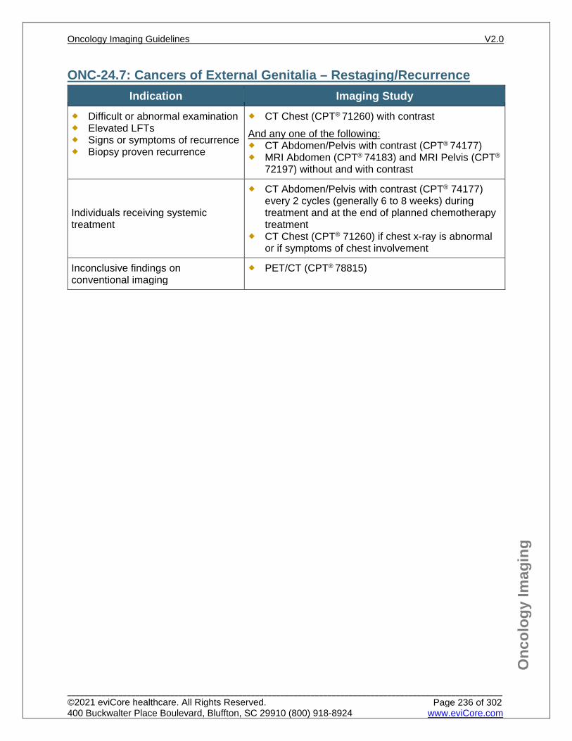
Onc
olog
y Im
agin
g
ONC-24.7: Cancers of External Genitalia – Restaging/Recurrence Indication Imaging Study
Difficult or abnormal examination Elevated LFTs Signs or symptoms of recurrence Biopsy proven recurrence
CT Chest (CPT® 71260) with contrast And any one of the following: CT Abdomen/Pelvis with contrast (CPT® 74177) MRI Abdomen (CPT® 74183) and MRI Pelvis (CPT®
72197) without and with contrast
Individuals receiving systemic treatment
CT Abdomen/Pelvis with contrast (CPT® 74177) every 2 cycles (generally 6 to 8 weeks) during treatment and at the end of planned chemotherapy treatment
CT Chest (CPT® 71260) if chest x-ray is abnormal or if symptoms of chest involvement
Inconclusive findings on conventional imaging
PET/CT (CPT® 78815)
Oncology Imaging Guidelines V2.0
______________________________________________________________________________________________________ ©2021 eviCore healthcare. All Rights Reserved. 400 Buckwalter Place Boulevard, Bluffton, SC 29910 (800) 918-8924 www.eviCore.com
Page 236 of 302
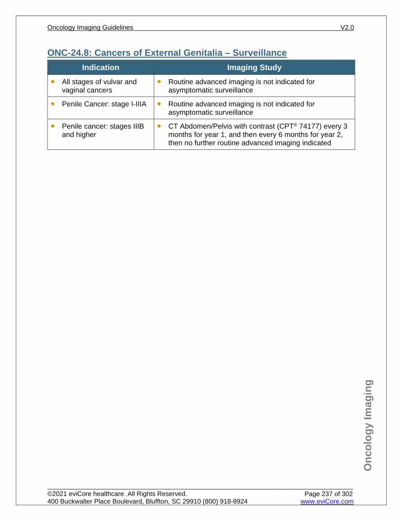
Onc
olog
y Im
agin
g
ONC-24.8: Cancers of External Genitalia – Surveillance Indication Imaging Study
All stages of vulvar and vaginal cancers
Routine advanced imaging is not indicated for asymptomatic surveillance
Penile Cancer: stage I-IIIA Routine advanced imaging is not indicated for asymptomatic surveillance
Penile cancer: stages IIIB and higher
CT Abdomen/Pelvis with contrast (CPT® 74177) every 3 months for year 1, and then every 6 months for year 2, then no further routine advanced imaging indicated
Oncology Imaging Guidelines V2.0
______________________________________________________________________________________________________ ©2021 eviCore healthcare. All Rights Reserved. 400 Buckwalter Place Boulevard, Bluffton, SC 29910 (800) 918-8924 www.eviCore.com
Page 237 of 302
Onc
olog
y Im
agin
g
References 1. Benson AB, Venook AP, Al-Hawary MM, et al. National Comprehensive Cancer Network (NCCN)
Guidelines Version 1.2021 – February 16, 2021. Anal Carcinoma, available at: https://www.nccn.org/professionals/physician_gls/pdf/anal.pdf Referenced with permission from the NCCN Clinical Practice Guidelines in Oncology (NCCN Guidelines™) for anal carcinoma V1.2021– February 16, 2021. ©2021 National Comprehensive Cancer Network, Inc. All rights reserved. The NCCN Guidelines™ and illustrations herein may not be reproduced in any form for any purpose without the express written permission of the NCCN. To view the most recent and complete version of the NCCN Guidelines™, go online to NCCN.org.
2. Flaig TW, Spiess PE, Agarwal N. National Comprehensive Cancer Network (NCCN) Guidelines Version 1.2021 – January 13, 2020. Penile Cancer, available at: https://www.nccn.org/professionals/physician_gls/pdf/penile.pdf Referenced with permission from the NCCN Clinical Practice Guidelines in Oncology (NCCN Guidelines™) for Penile Cancer V1.2021 – January 13, 2021. ©2021 National Comprehensive Cancer Network, Inc. All rights reserved. The NCCN Guidelines™ and illustrations herein may not be reproduced in any form for any purpose without the express written permission of the NCCN. To view the most recent and complete version of the NCCN Guidelines™, go online to NCCN.org.
3. Abu-Rustum NR, Yashar CM, Bean S. et al. National Comprehensive Cancer Network (NCCN) Guidelines Version 2.2021 – October 19, 2020. Vulvar Cancer (Squamous Cell Carcinoma), available at: https://www.nccn.org/professionals/physician_gls/pdf/vulvar.pdf Referenced with permission from the NCCN Clinical Practice Guidelines in Oncology (NCCN Guidelines™) for Vulvar Cancer (Squamous Cell Carcinoma) V2.2021 – October 19, 2020. ©2020 National Comprehensive Cancer Network, Inc. All rights reserved. The NCCN Guidelines™ and illustrations herein may not be reproduced in any form for any purpose without the express written permission of the NCCN. To view the most recent and complete version of the NCCN Guidelines™, go online to NCCN.org.
4. Bhuva NJ, Glynne-Jones R, Sonoda L, Wong WL, Harrison MK. To PET or not to PET? That is the question. Staging in anal cancer. Ann Oncol. 2012;23(8):2078-2082. doi:10.1093/annonc/mdr599.
5. Mistrangelo M, Pelosi E, Bellò M, et al. Role of positron emission tomography-computed tomography in the management of anal cancer. Int J Radiat Oncol Biol Phys. 2012;84(1):66-72. doi:10.1016/j.ijrobp.2011.10.048.
6. Jones M, Hruby G, Solomon M, Rutherford N, Martin J. The role of FDG-PET in the initial staging and response assessment of anal cancer: a systematic review and meta-analysis. Ann Surg Oncol. 2015;22(11):3574-3581. doi:10.1245/s10434-015-4391-9.
Oncology Imaging Guidelines V2.0
______________________________________________________________________________________________________ ©2021 eviCore healthcare. All Rights Reserved. 400 Buckwalter Place Boulevard, Bluffton, SC 29910 (800) 918-8924 www.eviCore.com
Page 238 of 302
ONC-25: Multiple Myeloma and Plasmacytomas ONC-25.0: Multiple Myeloma and Plasmacytomas – General ConsiderationsONC-25.1: Multiple Myeloma and Plasmacytomas – Suspected/DiagnosisONC-25.2: Multiple Myeloma and Plasmacytomas – Initial Work-up/StagingONC-25.3: Multiple Myeloma and Plasmacytomas – Restaging/RecurrenceONC-25.4: Multiple Myeloma and Plasmacytomas – Surveillance
Oncology Imaging Guidelines V2.0
______________________________________________________________________________________________________ ©2021 eviCore healthcare. All Rights Reserved. 400 Buckwalter Place Boulevard, Bluffton, SC 29910 (800) 918-8924 www.eviCore.com
Page 239 of 302
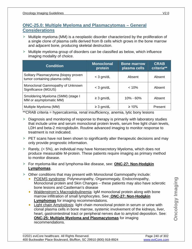
Onc
olog
y Im
agin
g
ONC-25.0: Multiple Myeloma and Plasmacytomas – General Considerations Multiple myeloma (MM) is a neoplastic disorder characterized by the proliferation of
a single clone of plasma cells derived from B cells which grows in the bone marrow and adjacent bone, producing skeletal destruction.
Multiple myeloma group of disorders can be classified as below, which influence imaging modality of choice.
Condition Monoclonal protein
Bone marrow plasma cells
CRAB criteria**
Solitary Plasmacytoma (biopsy proven tumor containing plasma cells) < 3 gm/dL Absent Absent
Monoclonal Gammopathy of Unknown Significance (MGUS) < 3 gm/dL < 10% Absent
Smoldering Myeloma (SMM) (stage I MM or asymptomatic MM) ≥ 3 gm/dL 10% - 60% Absent
Multiple Myeloma (MM) ≥ 3 gm/dL ≥ 10% Present **CRAB criteria = hypercalcemia, renal insufficiency, anemia, lytic bony lesions
Diagnosis and monitoring of response to therapy is primarily with laboratory studies that include urine and serum monoclonal protein levels, serum free light chain levels, LDH and beta-2 microglobulin. Routine advanced imaging to monitor response to treatment is not indicated.
PET scans have not been shown to significantly alter therapeutic decisions and may only provide prognostic information.
Rarely, (< 5%), an individual may have Nonsecretory Myeloma, which does not produce measurable M-protein. These patients require imaging as primary method to monitor disease.
For myeloma-like and lymphoma-like disease, see: ONC-27: Non-Hodgkin Lymphomas.
Other conditions that may present with Monoclonal Gammopathy include: POEMS syndrome: Polyneuropathy, Organomegaly, Endocrinopathy,
Monoclonal protein and Skin Changes – these patients may also have sclerotic bone lesions and Castleman’s disease
Waldenstrom’s Macroglobulinemia: IgM monoclonal protein along with bone marrow infiltration of small lymphocytes. See: ONC-27: Non-Hodgkin Lymphomas for imaging recommendations.
Light chain Amyloidosis: light chain monoclonal protein in serum or urine with clonal plasma cells in bone marrow, systemic involvement of the kidneys, liver, heart, gastrointestinal tract or peripheral nerves due to amyloid deposition. See: ONC-25: Multiple Myeloma and Plasmacytomas for imaging recommendations.
Oncology Imaging Guidelines V2.0
______________________________________________________________________________________________________ ©2021 eviCore healthcare. All Rights Reserved. 400 Buckwalter Place Boulevard, Bluffton, SC 29910 (800) 918-8924 www.eviCore.com
Page 240 of 302
Onc
olog
y Im
agin
g
ONC-25.1: Multiple Myeloma and Plasmacytomas – Suspected/Diagnosis
Indication Imaging Study All X-ray skeletal series
Oncology Imaging Guidelines V2.0
______________________________________________________________________________________________________ ©2021 eviCore healthcare. All Rights Reserved. 400 Buckwalter Place Boulevard, Bluffton, SC 29910 (800) 918-8924 www.eviCore.com
Page 241 of 302
Onc
olog
y Im
agin
g
ONC-25.2: Multiple Myeloma and Plasmacytomas – Initial Work-up/Staging
Indication Imaging Study Any of the following: Abnormal skeletal survey Abnormal myeloma labs Signs/symptoms of
multiple myeloma
Whole body low-dose skeletal CT scan (CPT® 76497)
If skeletal CT is negative, inconclusive, or not feasible
Any one of the following: MRI Bone Marrow Blood Supply (CPT® 77084) MRI Cervical (CPT® 72141), Thoracic (CPT® 72146),
Lumbar spine (CPT® 72148), and Pelvis (CPT® 72195) without contrast
MRI Cervical (CPT® 72156), Thoracic (CPT® 72157), Lumbar spine (CPT® 72158), and Pelvis (CPT® 72197) without and with contrast
CT contrast as requested of a specific area to determine radiotherapy or surgical candidacy, or for suspected extraosseous plasmacytoma
For any of the following (after the tests listed above are completed): Determining if a
plasmacytoma is truly solitary
Suspected extraosseous plasmacytomas
Suspected progression of MGUS or SMM to a more malignant form and CT/MRI imaging are negative
Inconclusive or negative conventional imaging
PET/CT (CPT® 78815 or CPT® 78816)
Oncology Imaging Guidelines V2.0
______________________________________________________________________________________________________ ©2021 eviCore healthcare. All Rights Reserved. 400 Buckwalter Place Boulevard, Bluffton, SC 29910 (800) 918-8924 www.eviCore.com
Page 242 of 302
Onc
olog
y Im
agin
g
ONC-25.3: Multiple Myeloma and Plasmacytomas – Restaging/Recurrence
Indication Imaging Study
Extra-osseous plasmacytoma response to initial therapy
CT contrast as requested or MRI without contrast, or MRI without and with contrast of any previously involved area
Laboratory tests fail to normalize with treatment
CT contrast as requested or MRI without contrast or MRI without and with contrast of symptomatic areas
Known spine involvement with new neurological signs/symptoms or worsening pain
MRI Cervical (CPT® 72156), Thoracic (CPT®
72157), Lumbar spine (CPT® 72158) without and with contrast
Any of the following: Suspected relapse/recurrence Suspected progression of MGUS
or SMM to a more malignant form To determine therapy response
with inconclusive labs
One of the following: Whole body low-dose skeletal CT scan (CPT®
76497) MRI Bone Marrow Blood Supply (CPT® 77084) MRI Cervical (CPT® 72141), Thoracic (CPT®
72146), Lumbar spine (CPT® 72148), and Pelvis (CPT® 72195) without contrast
MRI Cervical (CPT® 72156), Thoracic (CPT®
72157), Lumbar spine (CPT® 72158), and Pelvis (CPT® 72197) without and with contrast
MRI without contrast, or MRI without and with contrast for any previously involved bony area or symptomatic area
Any of the following: Negative PET will allow change in
management from active treatment to maintenance or surveillance.
Determine additional therapies in refractory disease or non-secretory disease.
PET/CT (CPT® 78815 or CPT® 78816)
Stem cell transplant recipients
One of the following, once before transplant and once after transplant: Whole body low-dose skeletal CT scan (CPT®
76497) MRI Bone Marrow Blood Supply (CPT® 77084) MRI Cervical (CPT® 72141), Thoracic (CPT®
72146), Lumbar spine (CPT® 72148), and Pelvis (CPT® 72195) without Contrast
MRI Cervical (CPT® 72156), Thoracic (CPT®
72157), Lumbar spine (CPT® 72158), and Pelvis (CPT® 72197) without and with contrast
Oncology Imaging Guidelines V2.0
______________________________________________________________________________________________________ ©2021 eviCore healthcare. All Rights Reserved. 400 Buckwalter Place Boulevard, Bluffton, SC 29910 (800) 918-8924 www.eviCore.com
Page 243 of 302
Onc
olog
y Im
agin
g
ONC-25.4: Multiple Myeloma and Plasmacytomas – Surveillance Indication Study
Plasmacytomas Smoldering myeloma
Any one of the following annually for 5 years: Whole body low-dose skeletal CT scan (CPT® 76497) Skeletal survey annually
Multiple myeloma after treatment and/or after stem cell transplant
Advanced imaging is not routinely indicated for surveillance in asymptomatic individuals
Oncology Imaging Guidelines V2.0
______________________________________________________________________________________________________ ©2021 eviCore healthcare. All Rights Reserved. 400 Buckwalter Place Boulevard, Bluffton, SC 29910 (800) 918-8924 www.eviCore.com
Page 244 of 302
Onc
olog
y Im
agin
g
References 1. Kumar SK, Callander NS, Alsina M, et al. National Comprehensive Cancer Network (NCCN)
Guidelines Version 4.2021 – December 10, 2020. Myeloma, available at: https://www.nccn.org/professionals/physician_gls/pdf/myeloma.pdf Referenced with permission from the NCCN Clinical Practice Guidelines in Oncology (NCCN Guidelines™) for Myeloma V4.2021 – December 10, 2020. ©2020 National Comprehensive Cancer Network, Inc. All rights reserved. The NCCN Guidelines™ and illustrations herein may not be reproduced in any form for any purpose without the express written permission of the NCCN. To view the most recent and complete version of the NCCN Guidelines™, go online to NCCN.org.
2. Hillengass J, Usmani S, Rajkumar SV, Durie BGM, Mateos M, Lonial S. International myeloma working group consensus recommendations on imaging in monoclonal plasma cell disorders. The Lancet. 2019:20(6):PE302-E312. doi:10.1016/S1470-2045(19)30309-2.
3. Kyle RA, Remstein ED, Therneau TM, et al. Clinical course and prognosis of smoldering (asymptomatic) multiple myeloma. N Engl J Med. 2007;356:2582-2590. doi:10.1056/NEJMoa070389.
4. Dimopoulos M, Terpos E, Comenzo RL, et al. International myeloma working group consensus statement and guidelines regarding the current role of imaging techniques in the diagnosis and monitoring of multiple myeloma. Leukemia. 2009;23(9):1545-1556. doi:10.1038/leu.2009.89.
5. ACR Committee on Drugs and Contrast Media. ACR Manual on Contrast Media, version 10.3. Reston, VA: American College of Radiology; 2018.
6. Mulligan ME, Badros AZ. PET/CR and MR imaging in myeloma. Skeletal Radiol. 2007;36(1):5-16. doi:10.1007/s00256-006-0184-3.
7. Dimopoulos MA, Hillengrass J, Usmani S, et al. Role of magnetic resonance imaging in the management of patients with multiple myeloma: a consensus statement. J Clin Oncol. 2015;33(6):657-664. doi:10.1200/JCO.2014.57.9961.
8. Dimopoulos M, Terpos E, Comenzo RL, et al. International myeloma working group consensus statement and guidelines regarding the current role of imaging techniques in the diagnosis and monitoring of multiple myeloma. Leukemia. 2009;23(9):1545-1556. doi:10.1038.leu.2008.89.
9. Dammacco F, Rubini G, Ferrari C, Vacca A, Racanelli V. 18F-FDG PET/CT: a review of diagnostic and prognostic features in multiple myeloma and related disorders. Clin Exp Med. 2015;15(1):1-18. doi:10.1007/s10238-014-0308-3.
10. Ferraro R, Agarwal A, Martin-Macintosh EL, Peller PJ, Subramaniam RM. MR imaging and PET/CT in diagnosis and management of multiple myeloma. Radiographics. 2015;35(2):438-454. doi:10.1148/rg.352140112.
11. Rajkumar SV, Kumar S. Multiple myeloma: diagnosis and treatment. Mayo Clin Proc. 2016;91(1):101-119. doi:10.1016/j.mayocp.2015.11.007.
12. Westerland O, Amlani A, Kelly-Morland C, et. al. Comparison of the diagnostic performance and impact on management of 18F-FDG PET/CT and whole-body MRI in multiple myeloma. Eur J Nucl ed Mol Imaging. 2021. doi:10.1007/s00259-020-05182-2
Oncology Imaging Guidelines V2.0
______________________________________________________________________________________________________ ©2021 eviCore healthcare. All Rights Reserved. 400 Buckwalter Place Boulevard, Bluffton, SC 29910 (800) 918-8924 www.eviCore.com
Page 245 of 302

ONC-26: Leukemias, Myelodysplasia and Myeloproliferative Neoplasms
ONC-26.1: Leukemias, Myelodysplasia and Myeloproliferative Neoplasms – General ConsiderationsONC-26.2: Acute LeukemiasONC-26.3: Chronic Myeloid Leukemias, Myelodysplastic Syndrome and Myeloproliferative DisordersONC-26.4: Chronic Lymphocytic Leukemia (CLL)/Small Lymphocytic Lymphoma (SLL)
Oncology Imaging Guidelines V2.0
______________________________________________________________________________________________________ ©2021 eviCore healthcare. All Rights Reserved. 400 Buckwalter Place Boulevard, Bluffton, SC 29910 (800) 918-8924 www.eviCore.com
Page 246 of 302
Onc
olog
y Im
agin
g
ONC-26.1: Leukemias, Myelodysplasia and Myeloproliferative Neoplasms – General Considerations PET imaging is considered investigational and experimental for all indications in
acute lymphoblastic leukemia, acute myeloid leukemia, and chronic myeloid leukemia.
Routine advanced imaging is not indicated in the evaluation and management of Hairy cell leukemia in the absence of specific localizing clinical symptoms.
Oncology Imaging Guidelines V2.0
______________________________________________________________________________________________________ ©2021 eviCore healthcare. All Rights Reserved. 400 Buckwalter Place Boulevard, Bluffton, SC 29910 (800) 918-8924 www.eviCore.com
Page 247 of 302
Onc
olog
y Im
agin
g
ONC-26.2: Acute Leukemias Imaging indications for acute lymphoblastic leukemia in adult patients are identical to
those for pediatric patients. See: PEDONC-3.2: Acute Lymphoblastic Leukemia (ALL) for imaging guidelines.
Imaging indications for acute myeloid leukemia in adult patients are identical to those for pediatric patients. See: PEDONC-3.3: Acute Myeloid Leukemia (AML) for imaging guidelines.
Oncology Imaging Guidelines V2.0
______________________________________________________________________________________________________ ©2021 eviCore healthcare. All Rights Reserved. 400 Buckwalter Place Boulevard, Bluffton, SC 29910 (800) 918-8924 www.eviCore.com
Page 248 of 302
Onc
olog
y Im
agin
g
ONC-26.3: Chronic Myeloid Leukemias, Myelodysplastic Syndrome and Myeloproliferative Disorders Routine advanced imaging is not indicated in the evaluation and management of
chronic myeloid leukemias, myelodysplastic syndromes or myeloproliferative disorders in the absence of specific localizing clinical symptoms or clearance for hematopoietic stem cell transplantation.
See: ONC-29: Hematopoietic Stem Cell Transplantation for imaging guidelines related to transplant.
For work-up of elevated blood counts, see: ONC-30.3: Paraneoplastic Syndromes – General Considerations
Oncology Imaging Guidelines V2.0
______________________________________________________________________________________________________ ©2021 eviCore healthcare. All Rights Reserved. 400 Buckwalter Place Boulevard, Bluffton, SC 29910 (800) 918-8924 www.eviCore.com
Page 249 of 302
Onc
olog
y Im
agin
g
ONC-26.4: Chronic Lymphocytic Leukemia (CLL)/Small Lymphocytic Lymphoma (SLL) PET imaging is not indicated in the evaluation of CLL/SLL with the exception of
suspected Richter’s transformation (see Suspected transformation, below) CLL/SLL is monitored with serial laboratory studies. Routine advanced imaging is
not indicated for monitoring treatment response or surveillance, except when initial studies reveal bulky disease involvement.
Bulky disease is defined as lymph node mass > 5 cm or spleen > 6 cm below costal margin
Indication Imaging Study
Initial Staging/Diagnosis Any or all of the following may be approved: CT Chest with contrast (CPT® 71260) CT Abdomen/Pelvis with contrast (CPT® 74177)
Treatment Response
For patients with bulky nodal disease at diagnosis, CT with contrast of previously involved area(s) every 2 cycles of therapy
Routine imaging is not indicated for patients without bulky nodal disease at diagnosis
End of Therapy Evaluation For patients with bulky nodal disease at diagnosis, CT with contrast of previously involved area(s)
Suspected progression
Any or all of the following may be approved: CT Chest with contrast (CPT® 71260) CT Abdomen/Pelvis with contrast (CPT® 74177) CT with contrast of previously involved area(s)
Suspected transformation (Richter’s) from a low grade lymphoma to a more aggressive type based on one or more of the following: New B symptoms Rapidly growing lymph nodes Extranodal disease develops Significant recent rise in LDH
above normal range
PET/CT (CPT® 78815)
Surveillance
For patients with bulky nodal disease at diagnosis, every 6 months for two years, then annually: CT Chest with contrast (CPT® 71260) CT Abdomen/Pelvis with contrast (CPT® 74177) CT with contrast of previously involved area(s)
Routine imaging is not indicated for patients without bulky nodal disease at diagnosis
Oncology Imaging Guidelines V2.0
______________________________________________________________________________________________________ ©2021 eviCore healthcare. All Rights Reserved. 400 Buckwalter Place Boulevard, Bluffton, SC 29910 (800) 918-8924 www.eviCore.com
Page 250 of 302
Onc
olog
y Im
agin
g
References 1. Zelenetz AD, Gordon LI, Wierda WG, et al. National Comprehensive Cancer Network (NCCN)
Guidelines Version 2.2021 – December 3, 2020. CLL/SLL, available at: https://www.nccn.org/professionals/physician_gls/pdf/cll.pdf Referenced with permission from the NCCN Clinical Practice Guidelines in Oncology (NCCN Guidelines™) for CLL/SLL V2.2021 – December 3, 2020. ©2020 National Comprehensive Cancer Network, Inc. All rights reserved. The NCCN Guidelines™ and illustrations herein may not be reproduced in any form for any purpose without the express written permission of the NCCN. To view the most recent and complete version of the NCCN Guidelines™, go online to NCCN.org.
2. Conte MJ, Bowen DA, Wiseman GA, et al. Use of positron emission tomography-computed tomography in the management of patients with chronic lymphocytic leukemia/small lymphocytic lymphoma. Leuk Lymphoma. 2014;55(9):2079-2084. doi:10.3109/10428194.2013.869801.
3. Mauro FR, Chauvie S, Paoloni F, et al. Diagnostic and prognostic role of PET/CT in patients with chronic lymphocytic leukemia and progressive disease. Leukemia. 2015;29(6):1360-1365. doi:10.1038/leu.2015.21.
4. Nabhan C, Rosen ST. Chronic lymphocytic leukemia: a clinical review. JAMA. 2014;312(21):2265-2276. doi:10.1001/jama.2014.14553.
5. Patnaik MM, Tefferi A. Chronic myelomonocytic leukemia: focus on clinical practice. Mayo Clin Proc. 2016;91(2):259-272. doi:10.1016/j.mayocp.2015.11.011.
Oncology Imaging Guidelines V2.0
______________________________________________________________________________________________________ ©2021 eviCore healthcare. All Rights Reserved. 400 Buckwalter Place Boulevard, Bluffton, SC 29910 (800) 918-8924 www.eviCore.com
Page 251 of 302
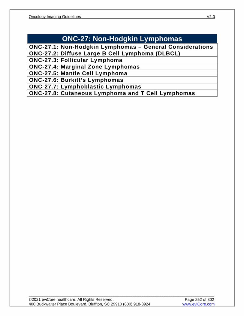
ONC-27: Non-Hodgkin Lymphomas ONC-27.1: Non-Hodgkin Lymphomas – General ConsiderationsONC-27.2: Diffuse Large B Cell Lymphoma (DLBCL)ONC-27.3: Follicular LymphomaONC-27.4: Marginal Zone LymphomasONC-27.5: Mantle Cell LymphomaONC-27.6: Burkitt’s LymphomasONC-27.7: Lymphoblastic LymphomasONC-27.8: Cutaneous Lymphoma and T Cell Lymphomas
Oncology Imaging Guidelines V2.0
______________________________________________________________________________________________________ ©2021 eviCore healthcare. All Rights Reserved. 400 Buckwalter Place Boulevard, Bluffton, SC 29910 (800) 918-8924 www.eviCore.com
Page 252 of 302
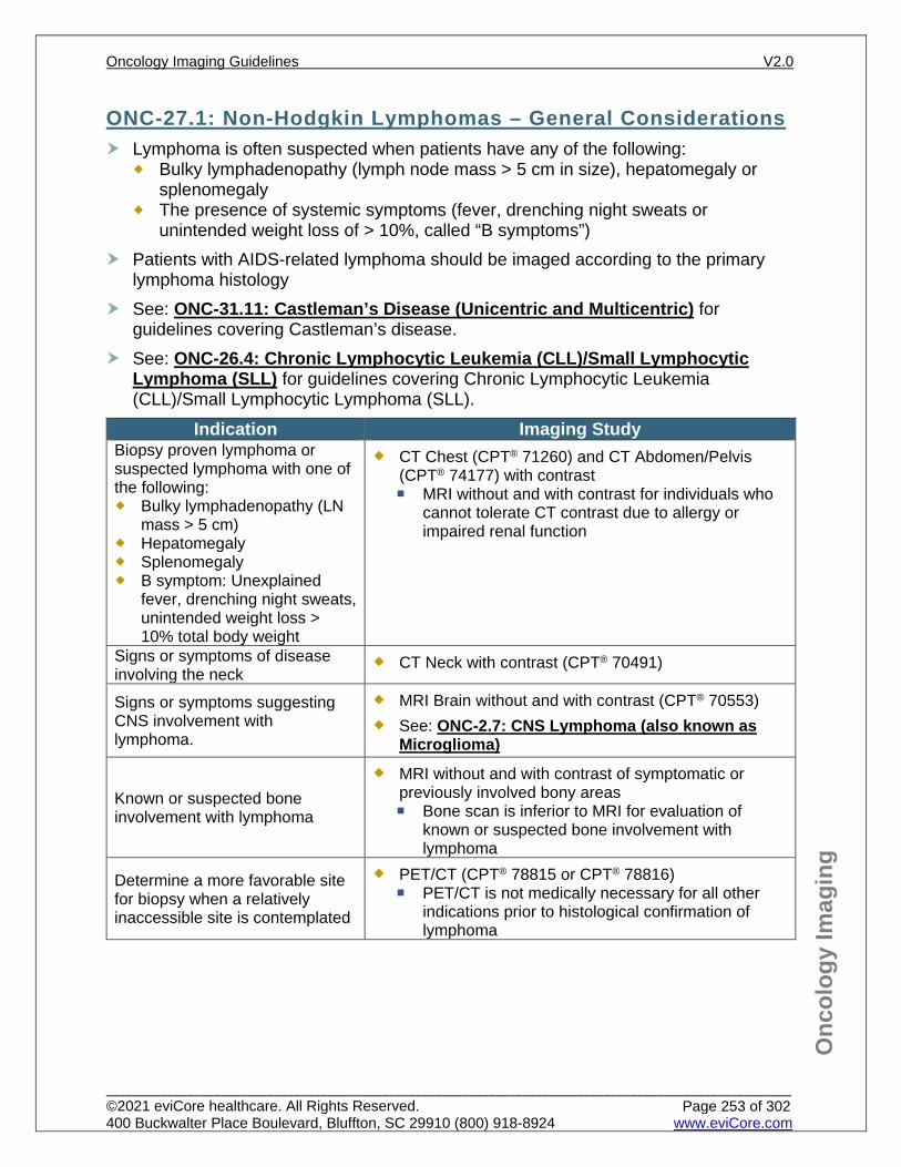
Onc
olog
y Im
agin
g
ONC-27.1: Non-Hodgkin Lymphomas – General Considerations Lymphoma is often suspected when patients have any of the following:
Bulky lymphadenopathy (lymph node mass > 5 cm in size), hepatomegaly or splenomegaly
The presence of systemic symptoms (fever, drenching night sweats or unintended weight loss of > 10%, called “B symptoms”)
Patients with AIDS-related lymphoma should be imaged according to the primary lymphoma histology
See: ONC-31.11: Castleman’s Disease (Unicentric and Multicentric) for guidelines covering Castleman’s disease.
See: ONC-26.4: Chronic Lymphocytic Leukemia (CLL)/Small Lymphocytic Lymphoma (SLL) for guidelines covering Chronic Lymphocytic Leukemia (CLL)/Small Lymphocytic Lymphoma (SLL).
Indication Imaging Study Biopsy proven lymphoma or suspected lymphoma with one of the following: Bulky lymphadenopathy (LN
mass > 5 cm) Hepatomegaly Splenomegaly B symptom: Unexplained
fever, drenching night sweats, unintended weight loss > 10% total body weight
CT Chest (CPT® 71260) and CT Abdomen/Pelvis (CPT® 74177) with contrast MRI without and with contrast for individuals who
cannot tolerate CT contrast due to allergy or impaired renal function
Signs or symptoms of disease involving the neck
CT Neck with contrast (CPT® 70491)
Signs or symptoms suggesting CNS involvement with lymphoma.
MRI Brain without and with contrast (CPT® 70553) See: ONC-2.7: CNS Lymphoma (also known as
Microglioma)
Known or suspected bone involvement with lymphoma
MRI without and with contrast of symptomatic or previously involved bony areas Bone scan is inferior to MRI for evaluation of
known or suspected bone involvement with lymphoma
Determine a more favorable site for biopsy when a relatively inaccessible site is contemplated
PET/CT (CPT® 78815 or CPT® 78816) PET/CT is not medically necessary for all other
indications prior to histological confirmation of lymphoma
Oncology Imaging Guidelines V2.0
______________________________________________________________________________________________________ ©2021 eviCore healthcare. All Rights Reserved. 400 Buckwalter Place Boulevard, Bluffton, SC 29910 (800) 918-8924 www.eviCore.com
Page 253 of 302
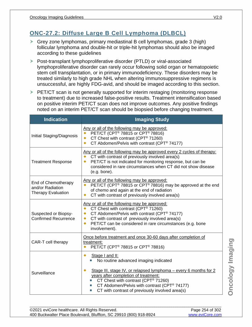
Onc
olog
y Im
agin
g
ONC-27.2: Diffuse Large B Cell Lymphoma (DLBCL) Grey zone lymphomas, primary mediastinal B cell lymphomas, grade 3 (high)
follicular lymphoma and double-hit or triple-hit lymphomas should also be imaged according to these guidelines
Post-transplant lymphoproliferative disorder (PTLD) or viral-associated lymphoproliferative disorder can rarely occur following solid organ or hematopoietic stem cell transplantation, or in primary immunodeficiency. These disorders may be treated similarly to high grade NHL when altering immunosuppressive regimens is unsuccessful, are highly FDG-avid, and should be imaged according to this section.
PET/CT scan is not generally supported for interim restaging (monitoring response to treatment) due to increased false-positive results. Treatment intensification based on positive interim PET/CT scan does not improve outcomes. Any positive findings noted on an interim PET/CT scan should be biopsied before changing treatment.
Indication Imaging Study
Initial Staging/Diagnosis
Any or all of the following may be approved: PET/CT (CPT® 78815 or CPT® 78816) CT Chest with contrast (CPT® 71260) CT Abdomen/Pelvis with contrast (CPT® 74177)
Treatment Response
Any or all of the following may be approved every 2 cycles of therapy: CT with contrast of previously involved area(s) PET/CT is not indicated for monitoring response, but can be
considered in rare circumstances when CT did not show disease (e.g. bone).
End of Chemotherapy and/or Radiation Therapy Evaluation
Any or all of the following may be approved: PET/CT (CPT® 78815 or CPT® 78816) may be approved at the end
of chemo and again at the end of radiation CT with contrast of previously involved area(s)
Suspected or Biopsy-Confirmed Recurrence
Any or all of the following may be approved: CT Chest with contrast (CPT® 71260) CT Abdomen/Pelvis with contrast (CPT® 74177) CT with contrast of previously involved area(s) PET/CT can be considered in rare circumstances (e.g. bone
involvement).
CAR-T cell therapy Once before treatment and once 30-60 days after completion of treatment: PET/CT (CPT® 78815 or CPT® 78816)
Surveillance
Stage I and II: No routine advanced imaging indicated
Stage III, stage IV, or relapsed lymphoma – every 6 months for 2
years after completion of treatment: CT Chest with contrast (CPT® 71260) CT Abdomen/Pelvis with contrast (CPT® 74177) CT with contrast of previously involved area(s)
Oncology Imaging Guidelines V2.0
______________________________________________________________________________________________________ ©2021 eviCore healthcare. All Rights Reserved. 400 Buckwalter Place Boulevard, Bluffton, SC 29910 (800) 918-8924 www.eviCore.com
Page 254 of 302
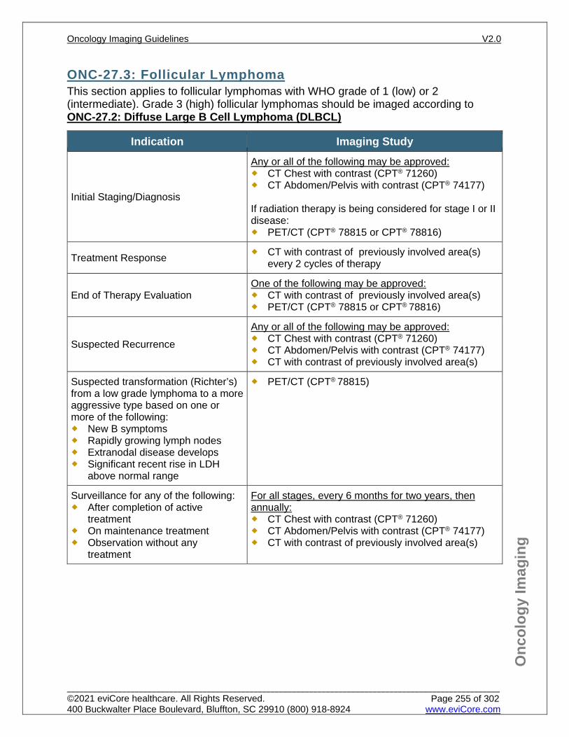
Onc
olog
y Im
agin
g
ONC-27.3: Follicular Lymphoma This section applies to follicular lymphomas with WHO grade of 1 (low) or 2 (intermediate). Grade 3 (high) follicular lymphomas should be imaged according to ONC-27.2: Diffuse Large B Cell Lymphoma (DLBCL)
Indication Imaging Study
Initial Staging/Diagnosis
Any or all of the following may be approved: CT Chest with contrast (CPT® 71260) CT Abdomen/Pelvis with contrast (CPT® 74177) If radiation therapy is being considered for stage I or II disease: PET/CT (CPT® 78815 or CPT® 78816)
Treatment Response CT with contrast of previously involved area(s) every 2 cycles of therapy
End of Therapy Evaluation One of the following may be approved: CT with contrast of previously involved area(s) PET/CT (CPT® 78815 or CPT® 78816)
Suspected Recurrence
Any or all of the following may be approved: CT Chest with contrast (CPT® 71260) CT Abdomen/Pelvis with contrast (CPT® 74177) CT with contrast of previously involved area(s)
Suspected transformation (Richter’s) from a low grade lymphoma to a more aggressive type based on one or more of the following: New B symptoms Rapidly growing lymph nodes Extranodal disease develops Significant recent rise in LDH
above normal range
PET/CT (CPT® 78815)
Surveillance for any of the following: After completion of active
treatment On maintenance treatment Observation without any
treatment
For all stages, every 6 months for two years, then annually: CT Chest with contrast (CPT® 71260) CT Abdomen/Pelvis with contrast (CPT® 74177) CT with contrast of previously involved area(s)
Oncology Imaging Guidelines V2.0
______________________________________________________________________________________________________ ©2021 eviCore healthcare. All Rights Reserved. 400 Buckwalter Place Boulevard, Bluffton, SC 29910 (800) 918-8924 www.eviCore.com
Page 255 of 302
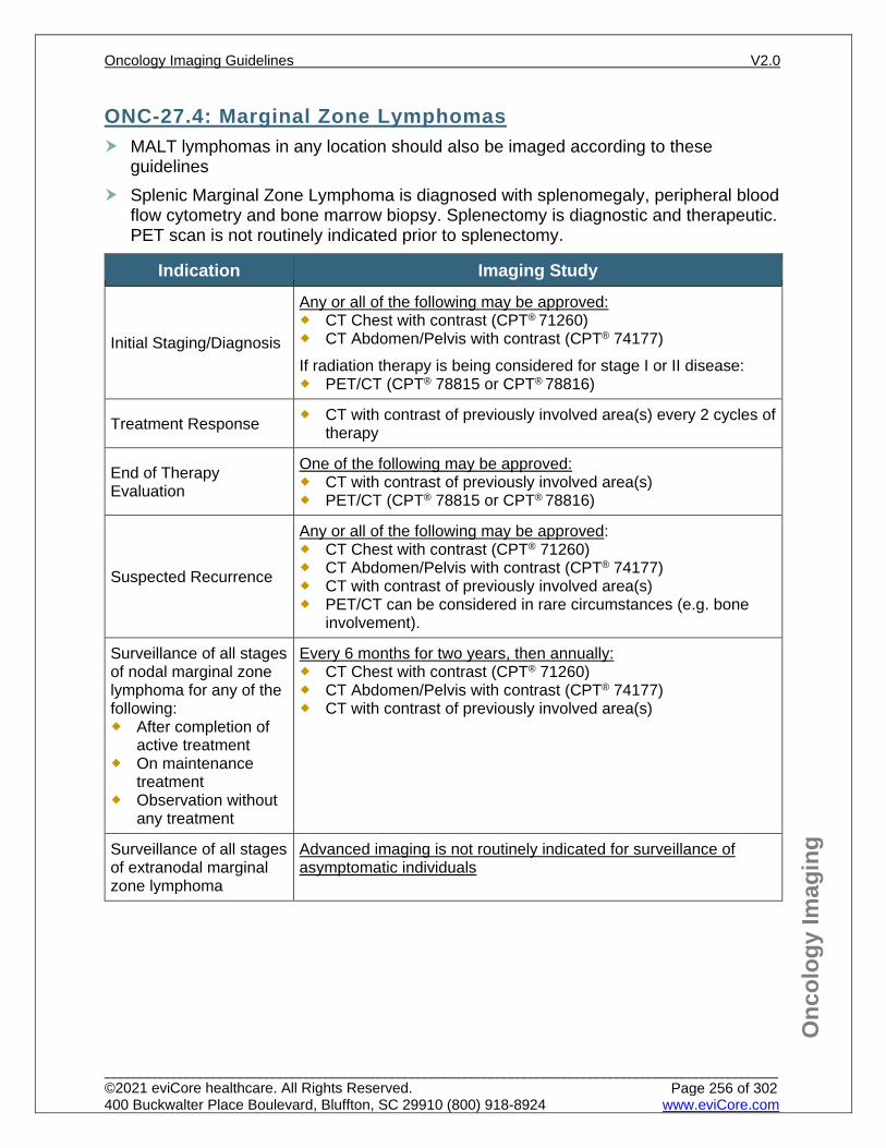
Onc
olog
y Im
agin
g
ONC-27.4: Marginal Zone Lymphomas MALT lymphomas in any location should also be imaged according to these
guidelines Splenic Marginal Zone Lymphoma is diagnosed with splenomegaly, peripheral blood
flow cytometry and bone marrow biopsy. Splenectomy is diagnostic and therapeutic. PET scan is not routinely indicated prior to splenectomy.
Indication Imaging Study
Initial Staging/Diagnosis
Any or all of the following may be approved: CT Chest with contrast (CPT® 71260) CT Abdomen/Pelvis with contrast (CPT® 74177) If radiation therapy is being considered for stage I or II disease: PET/CT (CPT® 78815 or CPT® 78816)
Treatment Response CT with contrast of previously involved area(s) every 2 cycles of therapy
End of Therapy Evaluation
One of the following may be approved: CT with contrast of previously involved area(s) PET/CT (CPT® 78815 or CPT® 78816)
Suspected Recurrence
Any or all of the following may be approved: CT Chest with contrast (CPT® 71260) CT Abdomen/Pelvis with contrast (CPT® 74177) CT with contrast of previously involved area(s) PET/CT can be considered in rare circumstances (e.g. bone
involvement).
Surveillance of all stages of nodal marginal zone lymphoma for any of the following: After completion of
active treatment On maintenance
treatment Observation without
any treatment
Every 6 months for two years, then annually: CT Chest with contrast (CPT® 71260) CT Abdomen/Pelvis with contrast (CPT® 74177) CT with contrast of previously involved area(s)
Surveillance of all stages of extranodal marginal zone lymphoma
Advanced imaging is not routinely indicated for surveillance of asymptomatic individuals
Oncology Imaging Guidelines V2.0
______________________________________________________________________________________________________ ©2021 eviCore healthcare. All Rights Reserved. 400 Buckwalter Place Boulevard, Bluffton, SC 29910 (800) 918-8924 www.eviCore.com
Page 256 of 302
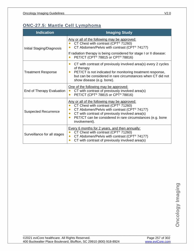
Onc
olog
y Im
agin
g
ONC-27.5: Mantle Cell Lymphoma Indication Imaging Study
Initial Staging/Diagnosis
Any or all of the following may be approved: CT Chest with contrast (CPT® 71260) CT Abdomen/Pelvis with contrast (CPT® 74177) If radiation therapy is being considered for stage I or II disease: PET/CT (CPT® 78815 or CPT® 78816)
Treatment Response
CT with contrast of previously involved area(s) every 2 cycles of therapy
PET/CT is not indicated for monitoring treatment response, but can be considered in rare circumstances when CT did not show disease (e.g. bone).
End of Therapy Evaluation One of the following may be approved: CT with contrast of previously involved area(s) PET/CT (CPT® 78815 or CPT® 78816)
Suspected Recurrence
Any or all of the following may be approved: CT Chest with contrast (CPT® 71260) CT Abdomen/Pelvis with contrast (CPT® 74177) CT with contrast of previously involved area(s) PET/CT can be considered in rare circumstances (e.g. bone
involvement).
Surveillance for all stages
Every 6 months for 2 years, and then annually: CT Chest with contrast (CPT® 71260) CT Abdomen/Pelvis with contrast (CPT® 74177) CT with contrast of previously involved area(s)
Oncology Imaging Guidelines V2.0
______________________________________________________________________________________________________ ©2021 eviCore healthcare. All Rights Reserved. 400 Buckwalter Place Boulevard, Bluffton, SC 29910 (800) 918-8924 www.eviCore.com
Page 257 of 302
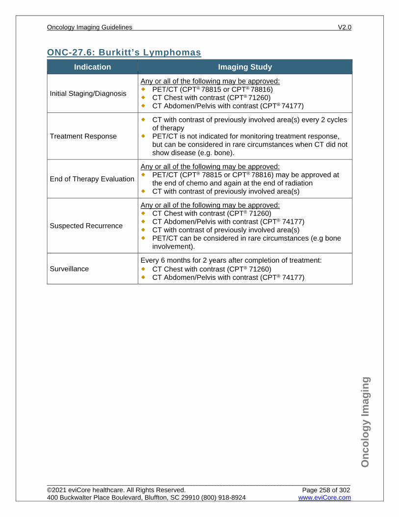
Onc
olog
y Im
agin
g
ONC-27.6: Burkitt’s Lymphomas Indication Imaging Study
Initial Staging/Diagnosis
Any or all of the following may be approved: PET/CT (CPT® 78815 or CPT® 78816) CT Chest with contrast (CPT® 71260) CT Abdomen/Pelvis with contrast (CPT® 74177)
Treatment Response
CT with contrast of previously involved area(s) every 2 cycles of therapy
PET/CT is not indicated for monitoring treatment response, but can be considered in rare circumstances when CT did not show disease (e.g. bone).
End of Therapy Evaluation
Any or all of the following may be approved: PET/CT (CPT® 78815 or CPT® 78816) may be approved at
the end of chemo and again at the end of radiation CT with contrast of previously involved area(s)
Suspected Recurrence
Any or all of the following may be approved: CT Chest with contrast (CPT® 71260) CT Abdomen/Pelvis with contrast (CPT® 74177) CT with contrast of previously involved area(s) PET/CT can be considered in rare circumstances (e.g bone
involvement).
Surveillance Every 6 months for 2 years after completion of treatment: CT Chest with contrast (CPT® 71260) CT Abdomen/Pelvis with contrast (CPT® 74177)
Oncology Imaging Guidelines V2.0
______________________________________________________________________________________________________ ©2021 eviCore healthcare. All Rights Reserved. 400 Buckwalter Place Boulevard, Bluffton, SC 29910 (800) 918-8924 www.eviCore.com
Page 258 of 302
Onc
olog
y Im
agin
g
ONC-27.7: Lymphoblastic Lymphomas Patients with lymphoblastic lymphoma (even those with bulky nodal disease) are
treated using the leukemia treatment plan appropriate to the cell type (B or T cell). Imaging indications in adult patients are identical to those for pediatric patients. See: PEDONC-3.2: Acute Lymphoblastic Leukemia (ALL) for imaging guidelines.
Oncology Imaging Guidelines V2.0
______________________________________________________________________________________________________ ©2021 eviCore healthcare. All Rights Reserved. 400 Buckwalter Place Boulevard, Bluffton, SC 29910 (800) 918-8924 www.eviCore.com
Page 259 of 302
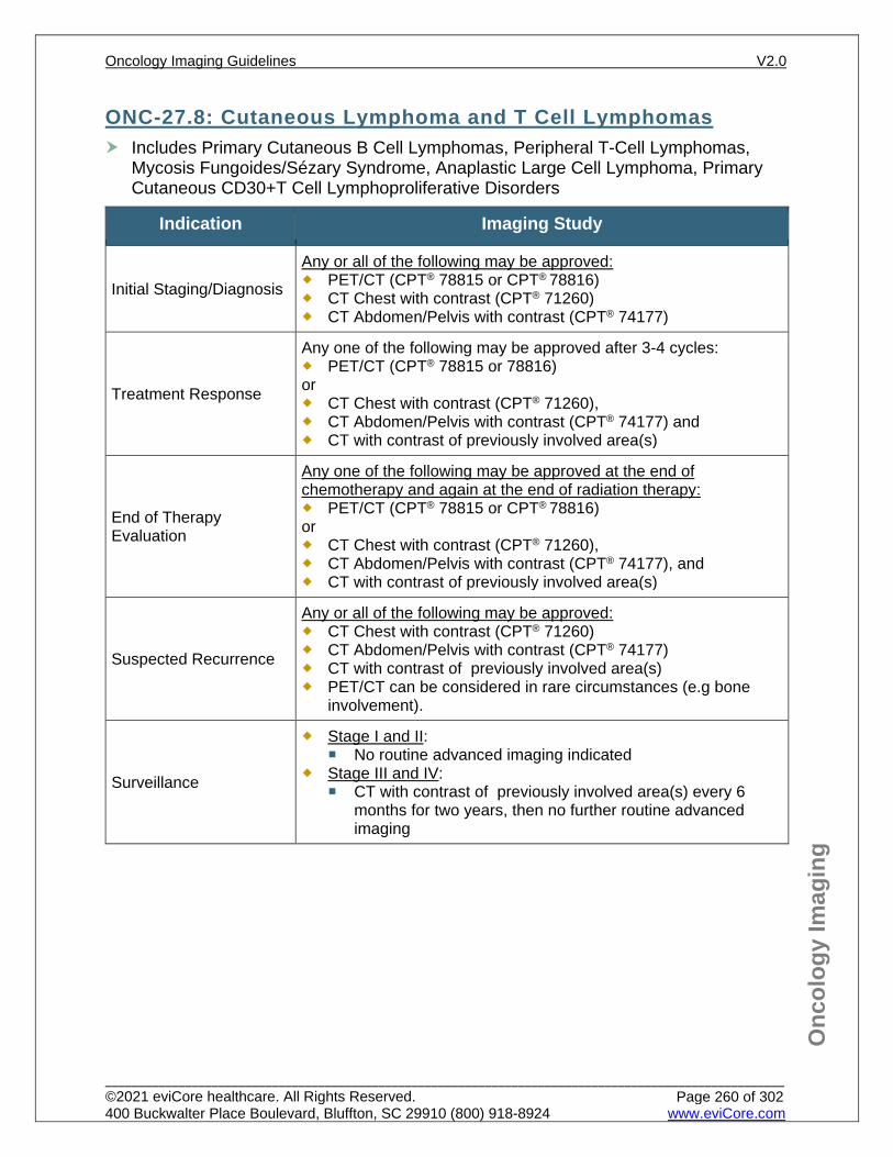
Onc
olog
y Im
agin
g
ONC-27.8: Cutaneous Lymphoma and T Cell Lymphomas Includes Primary Cutaneous B Cell Lymphomas, Peripheral T-Cell Lymphomas,
Mycosis Fungoides/Sézary Syndrome, Anaplastic Large Cell Lymphoma, Primary Cutaneous CD30+T Cell Lymphoproliferative Disorders
Indication Imaging Study
Initial Staging/Diagnosis
Any or all of the following may be approved: PET/CT (CPT® 78815 or CPT® 78816) CT Chest with contrast (CPT® 71260) CT Abdomen/Pelvis with contrast (CPT® 74177)
Treatment Response
Any one of the following may be approved after 3-4 cycles: PET/CT (CPT® 78815 or 78816) or CT Chest with contrast (CPT® 71260), CT Abdomen/Pelvis with contrast (CPT® 74177) and CT with contrast of previously involved area(s)
End of Therapy Evaluation
Any one of the following may be approved at the end of chemotherapy and again at the end of radiation therapy: PET/CT (CPT® 78815 or CPT® 78816) or CT Chest with contrast (CPT® 71260), CT Abdomen/Pelvis with contrast (CPT® 74177), and CT with contrast of previously involved area(s)
Suspected Recurrence
Any or all of the following may be approved: CT Chest with contrast (CPT® 71260) CT Abdomen/Pelvis with contrast (CPT® 74177) CT with contrast of previously involved area(s) PET/CT can be considered in rare circumstances (e.g bone
involvement).
Surveillance
Stage I and II: No routine advanced imaging indicated
Stage III and IV: CT with contrast of previously involved area(s) every 6
months for two years, then no further routine advanced imaging
Oncology Imaging Guidelines V2.0
______________________________________________________________________________________________________ ©2021 eviCore healthcare. All Rights Reserved. 400 Buckwalter Place Boulevard, Bluffton, SC 29910 (800) 918-8924 www.eviCore.com
Page 260 of 302
Onc
olog
y Im
agin
g
References 1. Zelenetz AD, Gordon LI, Abramson JS, et al. National Comprehensive Cancer Network (NCCN)
Guidelines Version 2.2021 – February 16, 2021. B-cell lymphomas, available at: https://www.nccn.org/professionals/physician_gls/pdf/B-CELL.pdf Referenced with permission from the NCCN Clinical Practice Guidelines in Oncology (NCCN Guidelines™) for B-cell lymphomas V2.2021 – February 16, 2021. ©2021 National Comprehensive Cancer Network, Inc. All rights reserved. The NCCN Guidelines™ and illustrations herein may not be reproduced in any form for any purpose without the express written permission of the NCCN. To view the most recent and complete version of the NCCN Guidelines™, go online to NCCN.org.
2. Zelenetz AD, Gordon LI, Wierda WG, et al. National Comprehensive Cancer Network (NCCN) Guidelines Version 1.2021 – October 5, 2020. T-cell lymphomas, available at: https://www.nccn.org/professionals/physician_gls/pdf/T-CELL.pdf Referenced with permission from the NCCN Clinical Practice Guidelines in Oncology (NCCN Guidelines™) for T-cell lymphomas V1.2021 – October 5, 2020. ©2020 National Comprehensive Cancer Network, Inc. All rights reserved. The NCCN Guidelines™ and illustrations herein may not be reproduced in any form for any purpose without the express written permission of the NCCN. To view the most recent and complete version of the NCCN Guidelines™, go online to NCCN.org.
3. Cheson BD, Fisher RI, Barrington SF, et al. Recommendations for initial evaluation, staging, and response assessment for Hodgkin and Non-Hodgkin lymphoma: the Lugano classification. J Clin Oncol. 2014;32(27):3059-3067. doi:10.1200/JCO.2013.54.8800.
4. Barrington SF, Mikhaeel NG, Kostakoglu L, et al. Role of imaging in the staging and response assessment of lymphoma: consensus of the International Conference on Malignant Lymphomas Imaging Working Group. J Clin Oncol. 2014;32(27):3048-3058. doi:10.1200/JCO.2013.53.5229.
5. Thompson CA, Ghesquieres H, Maurer MJ, et al. Utility of routine post-therapy surveillance imaging in diffuse Large B-Cell Lymphoma. J Clin Oncol. 2014;32(31):3506-3512. doi:10.1200/JCO.2014.55.7561.
6. El-Galaly TC, Jakobsen LH, Hutchings M, et al. Routine imaging for diffuse Large B-Cell Lymphoma in first complete remission does not improve post-treatment survival: a Danish-Swedish population-based study. J Clin Oncol. 2015;33(34):3993-3998. doi:10.1200/JCO.2015.62.0229.
7. Huntington SF, Svoboda J, Doshi JA. Cost-effectiveness analysis of routine surveillance imaging of patients with diffuse Large B-Cell Lymphoma in first remission. J Clin Oncol. 2015;33(13):1467-1474. doi:10.1200/JCO.2014.58.5729.
8. Mamot C, Klingbiel D, Hitz F, et al. Final results of a prospective evaluation of the predictive value of interim positron emission tomography in patients with diffuse large B-cell lymphoma treated with R-CHOP-14 (SAKK 38/07). J Clin Oncol. 2015;33(23):2523-2529. doi:10.1200/JCO.2014.58.9846.
9. Mylam KJ, Nielsen AL, Pedersen LM, Hutchings M. Fluorine-18-fluorodeoxyglucose positron emission tomography in diffuse large B-cell lymphoma. PET Clin. 2014;9(4):443-455. doi:10.1016/j.cpet.2014.06.001.
10. Avivi I, Zilberlicht A, Dann EJ, et al. Strikingly high false positivity of surveillance FDG-PET/CT scanning among patients with diffuse large cell lymphoma in the rituximab era. Am J Hematol. 2013;88(5):400-405. doi:10.1002/ajh.23423.
11. Ulrich Dührsen, Stefan Müller, Bernd Hertenstein, et al. Positron emission tomography-guided therapy of aggressive non-Hodgkin lymphomas (PETAL): a multicenter, randomized phase III trial. J Clin Oncol. 2018;36(20):2024-2034. doi:10.1200/JCO.2017.76.8093.
Oncology Imaging Guidelines V2.0
______________________________________________________________________________________________________ ©2021 eviCore healthcare. All Rights Reserved. 400 Buckwalter Place Boulevard, Bluffton, SC 29910 (800) 918-8924 www.eviCore.com
Page 261 of 302
ONC-28: Hodgkin Lymphoma ONC-28.1: Hodgkin Lymphoma – General ConsiderationsONC-28.2: Classical Hodgkin LymphomaONC-28.3: Nodular Lymphocyte – Predominant Hodgkin Lymphoma
Oncology Imaging Guidelines V2.0
______________________________________________________________________________________________________ ©2021 eviCore healthcare. All Rights Reserved. 400 Buckwalter Place Boulevard, Bluffton, SC 29910 (800) 918-8924 www.eviCore.com
Page 262 of 302
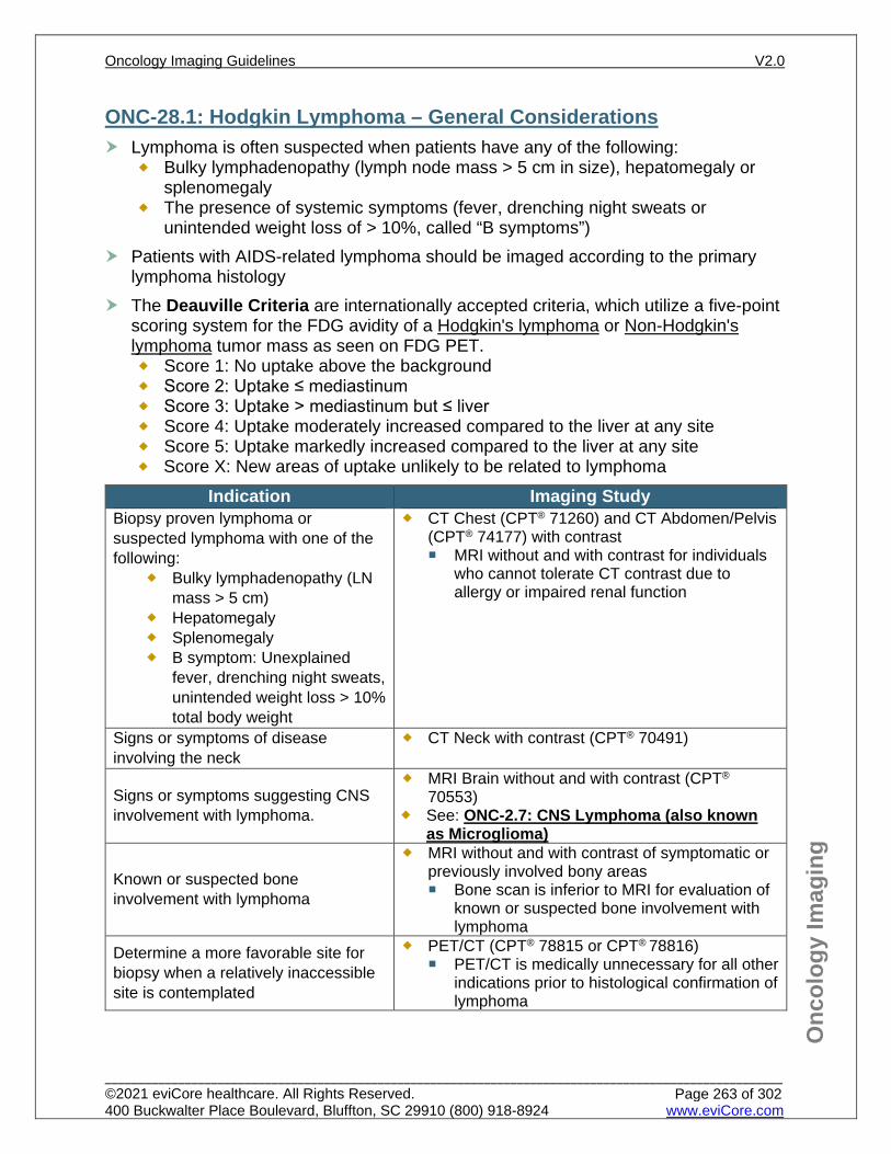
Onc
olog
y Im
agin
g
ONC-28.1: Hodgkin Lymphoma – General Considerations Lymphoma is often suspected when patients have any of the following:
Bulky lymphadenopathy (lymph node mass > 5 cm in size), hepatomegaly or splenomegaly
The presence of systemic symptoms (fever, drenching night sweats or unintended weight loss of > 10%, called “B symptoms”)
Patients with AIDS-related lymphoma should be imaged according to the primary lymphoma histology
The Deauville Criteria are internationally accepted criteria, which utilize a five-point scoring system for the FDG avidity of a Hodgkin's lymphoma or Non-Hodgkin's lymphoma tumor mass as seen on FDG PET. Score 1: No uptake above the background Score 2: Uptake ≤ mediastinum Score 3: Uptake > mediastinum but ≤ liver Score 4: Uptake moderately increased compared to the liver at any site Score 5: Uptake markedly increased compared to the liver at any site Score X: New areas of uptake unlikely to be related to lymphoma
Indication Imaging Study Biopsy proven lymphoma or suspected lymphoma with one of the following:
Bulky lymphadenopathy (LN mass > 5 cm)
Hepatomegaly Splenomegaly B symptom: Unexplained
fever, drenching night sweats, unintended weight loss > 10% total body weight
CT Chest (CPT® 71260) and CT Abdomen/Pelvis (CPT® 74177) with contrast MRI without and with contrast for individuals
who cannot tolerate CT contrast due to allergy or impaired renal function
Signs or symptoms of disease involving the neck
CT Neck with contrast (CPT® 70491)
Signs or symptoms suggesting CNS involvement with lymphoma.
MRI Brain without and with contrast (CPT® 70553)
See: ONC-2.7: CNS Lymphoma (also known as Microglioma)
Known or suspected bone involvement with lymphoma
MRI without and with contrast of symptomatic or previously involved bony areas Bone scan is inferior to MRI for evaluation of
known or suspected bone involvement with lymphoma
Determine a more favorable site for biopsy when a relatively inaccessible site is contemplated
PET/CT (CPT® 78815 or CPT® 78816) PET/CT is medically unnecessary for all other
indications prior to histological confirmation of lymphoma
Oncology Imaging Guidelines V2.0
______________________________________________________________________________________________________ ©2021 eviCore healthcare. All Rights Reserved. 400 Buckwalter Place Boulevard, Bluffton, SC 29910 (800) 918-8924 www.eviCore.com
Page 263 of 302
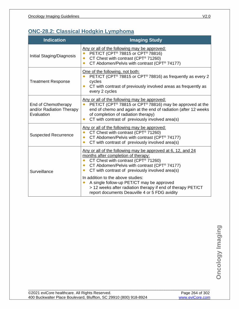
Onc
olog
y Im
agin
g
ONC-28.2: Classical Hodgkin Lymphoma Indication Imaging Study
Initial Staging/Diagnosis
Any or all of the following may be approved: PET/CT (CPT® 78815 or CPT® 78816) CT Chest with contrast (CPT® 71260) CT Abdomen/Pelvis with contrast (CPT® 74177)
Treatment Response
One of the following, not both: PET/CT (CPT® 78815 or CPT® 78816) as frequently as every 2
cycles CT with contrast of previously involved areas as frequently as
every 2 cycles
End of Chemotherapy and/or Radiation Therapy Evaluation
Any or all of the following may be approved: PET/CT (CPT® 78815 or CPT® 78816) may be approved at the
end of chemo and again at the end of radiation (after 12 weeks of completion of radiation therapy)
CT with contrast of previously involved area(s)
Suspected Recurrence
Any or all of the following may be approved: CT Chest with contrast (CPT® 71260) CT Abdomen/Pelvis with contrast (CPT® 74177) CT with contrast of previously involved area(s)
Surveillance
Any or all of the following may be approved at 6, 12, and 24 months after completion of therapy: CT Chest with contrast (CPT® 71260) CT Abdomen/Pelvis with contrast (CPT® 74177) CT with contrast of previously involved area(s) In addition to the above studies: A single follow-up PET/CT may be approved
> 12 weeks after radiation therapy if end of therapy PET/CT report documents Deauville 4 or 5 FDG avidity
Oncology Imaging Guidelines V2.0
______________________________________________________________________________________________________ ©2021 eviCore healthcare. All Rights Reserved. 400 Buckwalter Place Boulevard, Bluffton, SC 29910 (800) 918-8924 www.eviCore.com
Page 264 of 302
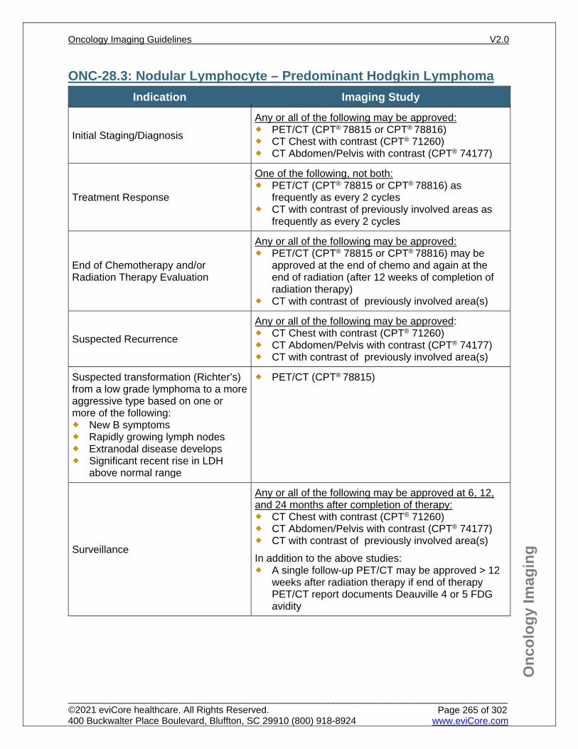
Onc
olog
y Im
agin
g
ONC-28.3: Nodular Lymphocyte – Predominant Hodgkin Lymphoma Indication Imaging Study
Initial Staging/Diagnosis
Any or all of the following may be approved: PET/CT (CPT® 78815 or CPT® 78816) CT Chest with contrast (CPT® 71260) CT Abdomen/Pelvis with contrast (CPT® 74177)
Treatment Response
One of the following, not both: PET/CT (CPT® 78815 or CPT® 78816) as
frequently as every 2 cycles CT with contrast of previously involved areas as
frequently as every 2 cycles
End of Chemotherapy and/or Radiation Therapy Evaluation
Any or all of the following may be approved: PET/CT (CPT® 78815 or CPT® 78816) may be
approved at the end of chemo and again at the end of radiation (after 12 weeks of completion of radiation therapy)
CT with contrast of previously involved area(s)
Suspected Recurrence
Any or all of the following may be approved: CT Chest with contrast (CPT® 71260) CT Abdomen/Pelvis with contrast (CPT® 74177) CT with contrast of previously involved area(s)
Suspected transformation (Richter’s) from a low grade lymphoma to a more aggressive type based on one or more of the following: New B symptoms Rapidly growing lymph nodes Extranodal disease develops Significant recent rise in LDH
above normal range
PET/CT (CPT® 78815)
Surveillance
Any or all of the following may be approved at 6, 12, and 24 months after completion of therapy: CT Chest with contrast (CPT® 71260) CT Abdomen/Pelvis with contrast (CPT® 74177) CT with contrast of previously involved area(s) In addition to the above studies: A single follow-up PET/CT may be approved > 12
weeks after radiation therapy if end of therapy PET/CT report documents Deauville 4 or 5 FDG avidity
Oncology Imaging Guidelines V2.0
______________________________________________________________________________________________________ ©2021 eviCore healthcare. All Rights Reserved. 400 Buckwalter Place Boulevard, Bluffton, SC 29910 (800) 918-8924 www.eviCore.com
Page 265 of 302
Onc
olog
y Im
agin
g
References 1. Hoppe RT, Advani RH, Ai WZ, et al. National Comprehensive Cancer Network (NCCN) Guidelines
Version 2.2021 – January 19, 2021. Hodgkin lymphoma, available at: https://www.nccn.org/professionals/physician_gls/pdf/hodgkins.pdf Referenced with permission from the NCCN Clinical Practice Guidelines in Oncology (NCCN Guidelines™) for Hodgkins Lymphoma V2.2021 – January 19, 2021. ©2021 National Comprehensive Cancer Network, Inc. All rights reserved. The NCCN Guidelines™ and illustrations herein may not be reproduced in any form for any purpose without the express written permission of the NCCN. To view the most recent and complete version of the NCCN Guidelines™, go online to NCCN.org.
2. Cheson BD, Fisher RI, Barrington SF, et al. Recommendations for initial evaluation, staging, and response assessment for Hodgkin and Non-Hodgkin lymphoma: the Lugano classification. J Clin Oncol. 2014;32(27):3059-3067. doi:10.1200/JCO.2013.54.8800.
3. Barrington SF, Mikhaeel NG, Kostakoglu L, et al. Role of imaging in the staging and response assessment of lymphoma: consensus of the International Conference on Malignant Lymphomas Imaging Working Group. J Clin Oncol. 2014;32(27):3048-3058. doi:10.1200/JCO.2013.53.5229.
4. Pingali SR, Jewell SW, Havlat L, et al. Limited utility of routine surveillance imaging for classical Hodgkin lymphoma patients in first complete remission. Cancer. 2014;120:2122-2129.
5. Ha CS, Hodgson DC, Advani R, et al. Follow-up of Hodgkin lymphoma. ACR Appropriateness Criteria® 2014;1-16.
6. Picardi M, Pugliese N, Cirillo, M et al. Advanced-stage Hodgkin lymphoma: US/Chest radiography for detection of relapse in patients in first complete remission—a randomized trial of routine surveillance imaging procedures. Radiology. 2014;272:262-274.
7. Gallamini A, and Kostakoglu L. Interim FDG-PET in Hodgkin lymphoma: a compass of a safe navigation in clinical trials? Blood. 2012;120(25):4913-4920.
8. Biggi A, Gallamini A, Chauvie S, et al. International validation study for interim PET in ABVD-treated, advanced-stage Hodgkin lymphoma: interpretation criteria and concordance rate among reviewers. J Nucl Med. 2013; 54(5):683-690.
9. Gallamini A, Barrington SF, Biggi, et al. The predictive role of interim positron emission tomography for Hodgkin lymphoma treatment outcome is confirmed using the interpretation criteria of the Deauville five-point scale. Haematologica. 2014; 99(6):1107-1113.
10. El-Galaly TC, Mylam KJ, Brown P, et al. Positron emission tomography/computed tomography surveillance in patients with Hodgkin lymphoma in first remission has a low positive predictive value and high costs. Haematologica. 2012;97(6):931-936.
Oncology Imaging Guidelines V2.0
______________________________________________________________________________________________________ ©2021 eviCore healthcare. All Rights Reserved. 400 Buckwalter Place Boulevard, Bluffton, SC 29910 (800) 918-8924 www.eviCore.com
Page 266 of 302
ONC-29: Hematopoietic Stem Cell Transplantation
ONC-29.1: General Considerations for Stem Cell Transplant
Oncology Imaging Guidelines V2.0
______________________________________________________________________________________________________ ©2021 eviCore healthcare. All Rights Reserved. 400 Buckwalter Place Boulevard, Bluffton, SC 29910 (800) 918-8924 www.eviCore.com
Page 267 of 302
Onc
olog
y Im
agin
g
ONC-29.1: General Considerations for Stem Cell Transplant
Transplant types: Allogeneic (“allo”): The donor and recipient are different people, and there are multiple types depending on the source of the stem cells and degree of match between donor and recipient. This is most commonly used in diseases originating in the hematopoietic system, such as leukemias and lymphomas, and bone marrow failure syndromes or metabolic disorders. Common types are:
Matched sibling donor (MSD or MRD): Donor and recipient are full siblings and HLA-matched
Matched unrelated donor (MUD): Donor and recipient are HLA matched but not related to each other
Cord blood: Donor stem cells come from frozen umbilical cord blood not related to the recipient, sometimes from multiple different donors at once
Haploidentical transplant (haplo): Donor is a half-HLA match to the recipient, usually a parent
Autologous (“auto”): The donor and recipient are the same person. The process involves delivery of high dose chemotherapy that is ablative to the bone marrow, followed by an infusion of one’s own harvested stem cells. Allogeneic HSCT results in a much greater degree of immunosuppression than autologous HSCT because of the need to allow the new immune system to chimerize with the recipient’s body. Immune reconstitution commonly takes more than a year for individuals who receive allogeneic HSCT, and individuals remain at high risk for invasive infections until that has occurred.
Oncology Imaging Guidelines V2.0
______________________________________________________________________________________________________ ©2021 eviCore healthcare. All Rights Reserved. 400 Buckwalter Place Boulevard, Bluffton, SC 29910 (800) 918-8924 www.eviCore.com
Page 268 of 302
Onc
olog
y Im
agin
g
Pre-Transplant Imaging in HSCT: Pre-transplant imaging in HSCT generally takes place within 30 days prior to
transplant and involves a reassessment of the individual’s disease status as well as infectious disease clearance
Indication Imaging
Immediate pre-transplant period
Chest x-ray CT Chest with contrast (CPT® 71260) or CT Chest
without contrast (CPT® 71250) for new findings on CXR, or new/worsening signs/symptoms.
CT Sinus (CPT® 70486) for any clinical signs or symptoms
Assess cardiac function
Echocardiogram (CPT® 93306, CPT® 93307 or CPT®
93308) MUGA scan (CPT® 78472) may be indicated in
specific circumstances, see: CD-12.1: Oncologic Indications for Cancer Therapeutics-Related Cardiac Dysfunction (CTRCD)
Assess pulmonary function Pulmonary function tests Assess primary disease status
See disease-specific guideline
Oncology Imaging Guidelines V2.0
______________________________________________________________________________________________________ ©2021 eviCore healthcare. All Rights Reserved. 400 Buckwalter Place Boulevard, Bluffton, SC 29910 (800) 918-8924 www.eviCore.com
Page 269 of 302
Onc
olog
y Im
agin
g
Post-Transplant Imaging in HSCT: There are many common complications from HSCT, including infection, graft versus
host disease, hepatic sinusoidal obstruction syndrome, restrictive lung disease, among others.
Disease response generally takes place at ~Day +30 (autos and some allos) or ~Day +100 (allos) post-transplant.
Indication Imaging Assess known or suspected HSCT complications
Site-specific imaging should generally be approved
Assess primary disease status post-transplant
See disease-specific guidelines for end of therapy evaluation and surveillance
Individuals receiving tandem auto transplants (2-4 autos back-to-back, spaced 6 to 8 weeks apart)
Guideline recommended imaging can be repeated after each transplant
Suspected Bronchiolitis obliterans with organizing pneumonia (BOOP)
CT Chest without contrast (CPT® 71250)
Oncology Imaging Guidelines V2.0
______________________________________________________________________________________________________ ©2021 eviCore healthcare. All Rights Reserved. 400 Buckwalter Place Boulevard, Bluffton, SC 29910 (800) 918-8924 www.eviCore.com
Page 270 of 302
Onc
olog
y Im
agin
g
References 1. National Comprehensive Cancer Network (NCCN) Guidelines Version 1.2021 – January 28, 2021.
Hematopoietic Cell Transplantation, available at: https://www.nccn.org/professionals/physician_gls/pdf/hct.pdf. Referenced with permission from the NCCN Clinical Practice Guidelines in Oncology (NCCN Guidelines®) for Hematopoietic Cell Transplantation V1.2021 – January 28, 2021. ©2021 National Comprehensive Cancer Network, Inc. All rights reserved. The NCCN Guidelines® and illustrations herein may not be reproduced in any form for any purpose without the express written permission of the NCCN. To view the most recent and complete version of the NCCN Guidelines®, go online to NCCN.org.
Oncology Imaging Guidelines V2.0
______________________________________________________________________________________________________ ©2021 eviCore healthcare. All Rights Reserved. 400 Buckwalter Place Boulevard, Bluffton, SC 29910 (800) 918-8924 www.eviCore.com
Page 271 of 302
ONC-30: Medical Conditions with Cancer in the Differential Diagnosis
ONC-30.1: Fever of Unknown Origin (FUO)ONC-30.2: Unexplained Weight LossONC-30.3: Paraneoplastic Syndromes
Oncology Imaging Guidelines V2.0
______________________________________________________________________________________________________ ©2021 eviCore healthcare. All Rights Reserved. 400 Buckwalter Place Boulevard, Bluffton, SC 29910 (800) 918-8924 www.eviCore.com
Page 272 of 302
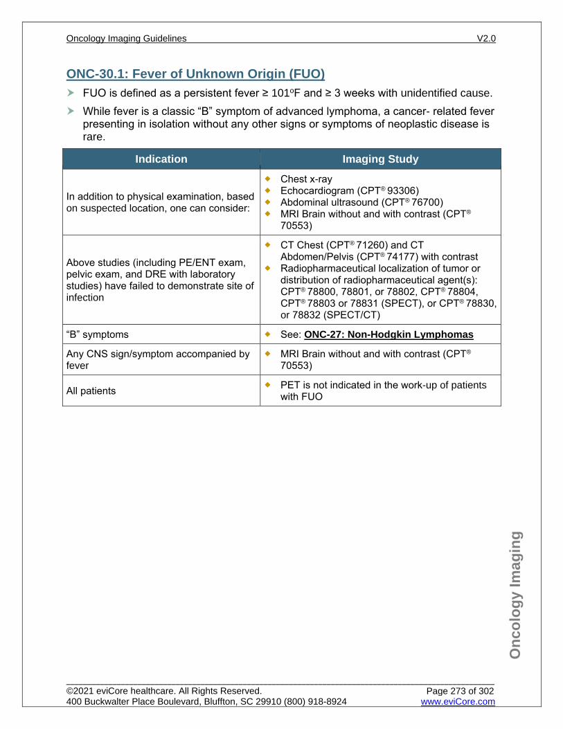
Onc
olog
y Im
agin
g
ONC-30.1: Fever of Unknown Origin (FUO) FUO is defined as a persistent fever ≥ 101oF and ≥ 3 weeks with unidentified cause. While fever is a classic “B” symptom of advanced lymphoma, a cancer- related fever
presenting in isolation without any other signs or symptoms of neoplastic disease is rare.
Indication Imaging Study
In addition to physical examination, based on suspected location, one can consider:
Chest x-ray Echocardiogram (CPT® 93306) Abdominal ultrasound (CPT® 76700) MRI Brain without and with contrast (CPT®
70553)
Above studies (including PE/ENT exam, pelvic exam, and DRE with laboratory studies) have failed to demonstrate site of infection
CT Chest (CPT® 71260) and CT Abdomen/Pelvis (CPT® 74177) with contrast
Radiopharmaceutical localization of tumor or distribution of radiopharmaceutical agent(s): CPT® 78800, 78801, or 78802, CPT® 78804, CPT® 78803 or 78831 (SPECT), or CPT® 78830, or 78832 (SPECT/CT)
“B” symptoms See: ONC-27: Non-Hodgkin Lymphomas
Any CNS sign/symptom accompanied by fever
MRI Brain without and with contrast (CPT®
70553)
All patients PET is not indicated in the work-up of patients with FUO
Oncology Imaging Guidelines V2.0
______________________________________________________________________________________________________ ©2021 eviCore healthcare. All Rights Reserved. 400 Buckwalter Place Boulevard, Bluffton, SC 29910 (800) 918-8924 www.eviCore.com
Page 273 of 302
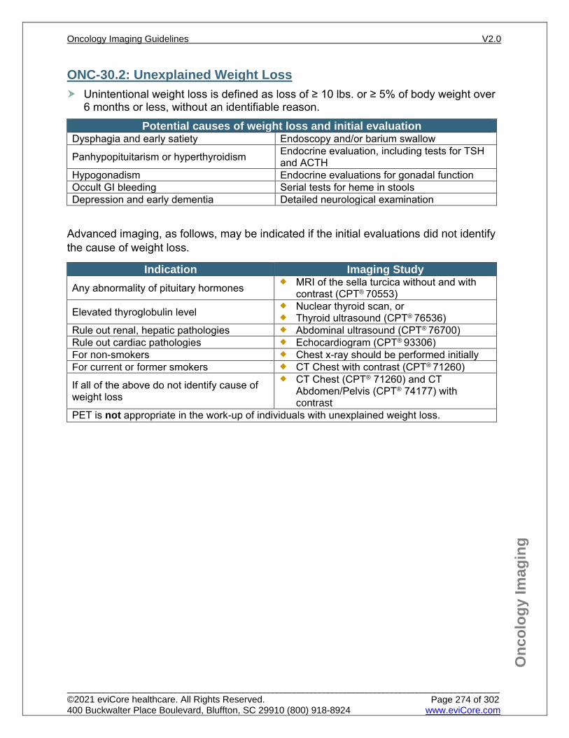
Onc
olog
y Im
agin
g
ONC-30.2: Unexplained Weight Loss Unintentional weight loss is defined as loss of ≥ 10 lbs. or ≥ 5% of body weight over
6 months or less, without an identifiable reason. Potential causes of weight loss and initial evaluation
Dysphagia and early satiety Endoscopy and/or barium swallow
Panhypopituitarism or hyperthyroidism Endocrine evaluation, including tests for TSH and ACTH
Hypogonadism Endocrine evaluations for gonadal function Occult GI bleeding Serial tests for heme in stools Depression and early dementia Detailed neurological examination
Advanced imaging, as follows, may be indicated if the initial evaluations did not identify the cause of weight loss.
Indication Imaging Study Any abnormality of pituitary hormones MRI of the sella turcica without and with
contrast (CPT® 70553)
Elevated thyroglobulin level Nuclear thyroid scan, or Thyroid ultrasound (CPT® 76536)
Rule out renal, hepatic pathologies Abdominal ultrasound (CPT® 76700) Rule out cardiac pathologies Echocardiogram (CPT® 93306) For non-smokers Chest x-ray should be performed initially For current or former smokers CT Chest with contrast (CPT® 71260)
If all of the above do not identify cause of weight loss
CT Chest (CPT® 71260) and CT Abdomen/Pelvis (CPT® 74177) with contrast
PET is not appropriate in the work-up of individuals with unexplained weight loss.
Oncology Imaging Guidelines V2.0
______________________________________________________________________________________________________ ©2021 eviCore healthcare. All Rights Reserved. 400 Buckwalter Place Boulevard, Bluffton, SC 29910 (800) 918-8924 www.eviCore.com
Page 274 of 302
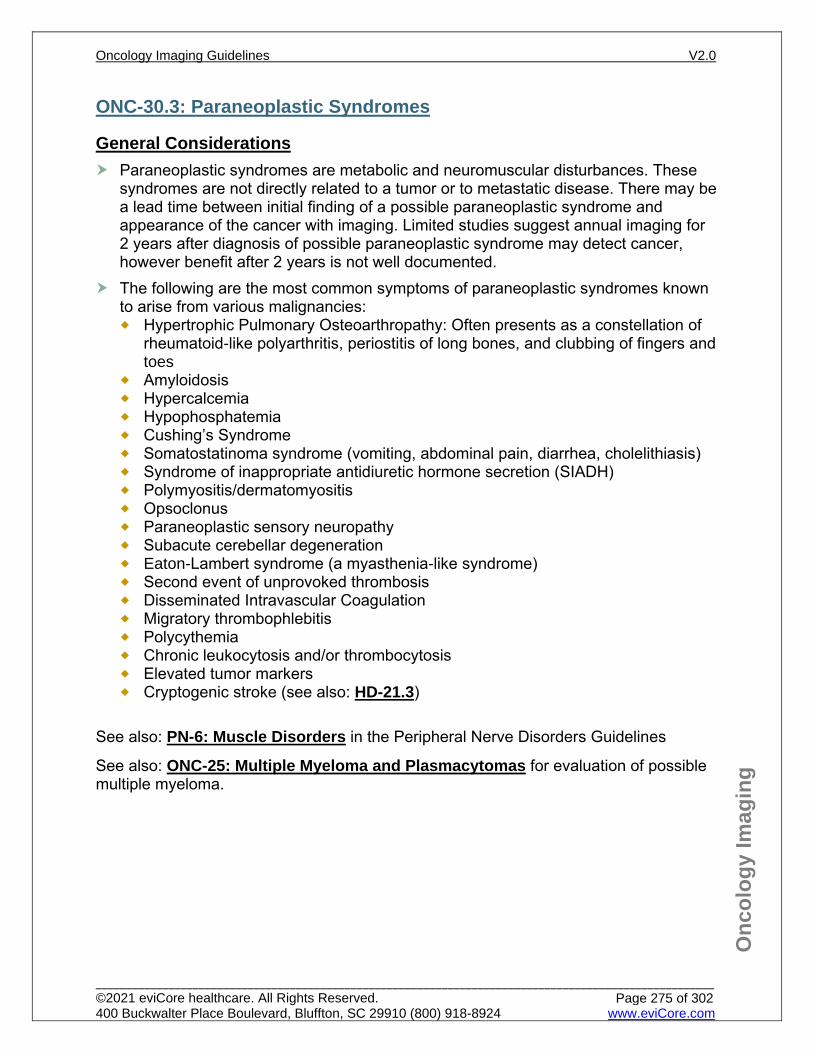
Onc
olog
y Im
agin
g
ONC-30.3: Paraneoplastic Syndromes
General Considerations Paraneoplastic syndromes are metabolic and neuromuscular disturbances. These
syndromes are not directly related to a tumor or to metastatic disease. There may be a lead time between initial finding of a possible paraneoplastic syndrome and appearance of the cancer with imaging. Limited studies suggest annual imaging for 2 years after diagnosis of possible paraneoplastic syndrome may detect cancer, however benefit after 2 years is not well documented.
The following are the most common symptoms of paraneoplastic syndromes known to arise from various malignancies: Hypertrophic Pulmonary Osteoarthropathy: Often presents as a constellation of
rheumatoid-like polyarthritis, periostitis of long bones, and clubbing of fingers and toes
Amyloidosis Hypercalcemia Hypophosphatemia Cushing’s Syndrome Somatostatinoma syndrome (vomiting, abdominal pain, diarrhea, cholelithiasis) Syndrome of inappropriate antidiuretic hormone secretion (SIADH) Polymyositis/dermatomyositis Opsoclonus Paraneoplastic sensory neuropathy Subacute cerebellar degeneration Eaton-Lambert syndrome (a myasthenia-like syndrome) Second event of unprovoked thrombosis Disseminated Intravascular Coagulation Migratory thrombophlebitis Polycythemia Chronic leukocytosis and/or thrombocytosis Elevated tumor markers Cryptogenic stroke (see also: HD-21.3)
See also: PN-6: Muscle Disorders in the Peripheral Nerve Disorders Guidelines
See also: ONC-25: Multiple Myeloma and Plasmacytomas for evaluation of possible multiple myeloma.
Oncology Imaging Guidelines V2.0
______________________________________________________________________________________________________ ©2021 eviCore healthcare. All Rights Reserved. 400 Buckwalter Place Boulevard, Bluffton, SC 29910 (800) 918-8924 www.eviCore.com
Page 275 of 302
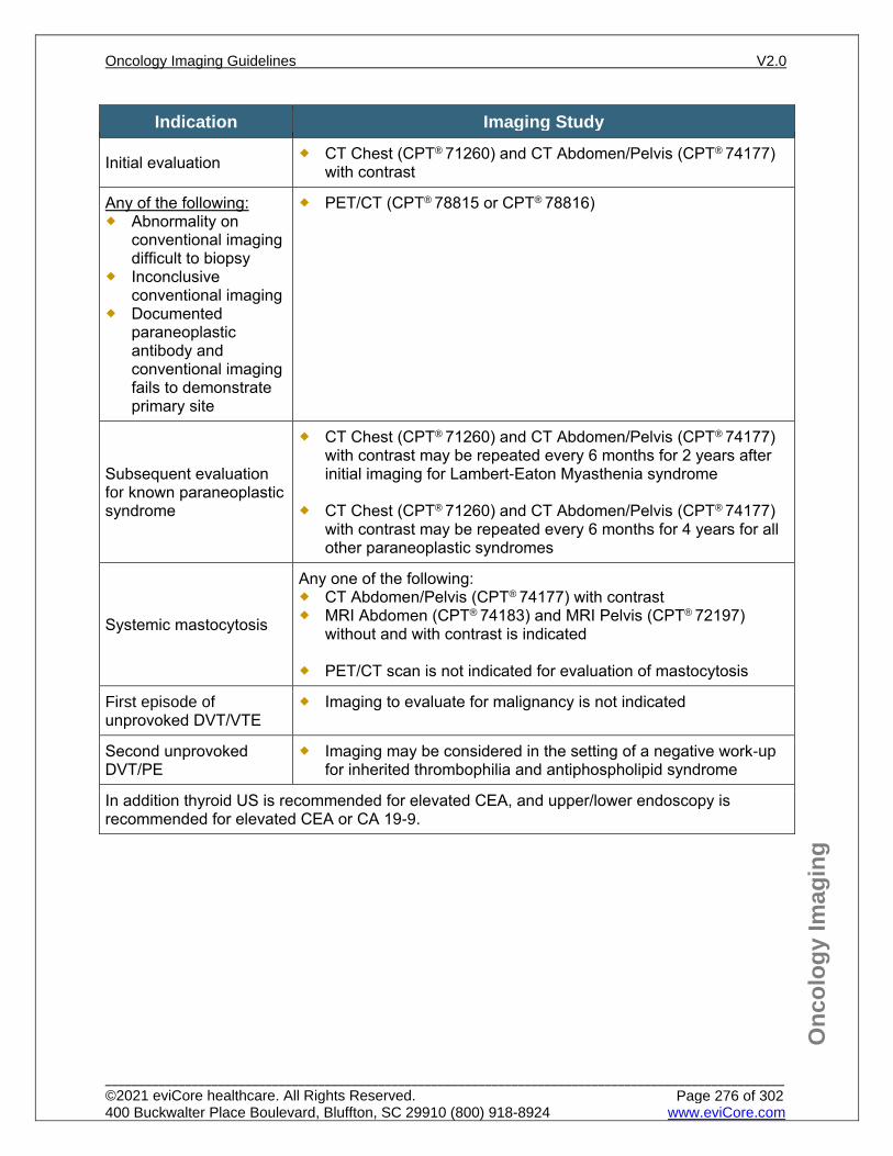
Onc
olog
y Im
agin
g
Indication Imaging Study
Initial evaluation CT Chest (CPT® 71260) and CT Abdomen/Pelvis (CPT® 74177) with contrast
Any of the following: Abnormality on
conventional imaging difficult to biopsy
Inconclusive conventional imaging
Documented paraneoplastic antibody and conventional imaging fails to demonstrate primary site
PET/CT (CPT® 78815 or CPT® 78816)
Subsequent evaluation for known paraneoplastic syndrome
CT Chest (CPT® 71260) and CT Abdomen/Pelvis (CPT® 74177) with contrast may be repeated every 6 months for 2 years after initial imaging for Lambert-Eaton Myasthenia syndrome
CT Chest (CPT® 71260) and CT Abdomen/Pelvis (CPT® 74177) with contrast may be repeated every 6 months for 4 years for all other paraneoplastic syndromes
Systemic mastocytosis
Any one of the following: CT Abdomen/Pelvis (CPT® 74177) with contrast MRI Abdomen (CPT® 74183) and MRI Pelvis (CPT® 72197)
without and with contrast is indicated
PET/CT scan is not indicated for evaluation of mastocytosis
First episode of unprovoked DVT/VTE
Imaging to evaluate for malignancy is not indicated
Second unprovoked DVT/PE
Imaging may be considered in the setting of a negative work-up for inherited thrombophilia and antiphospholipid syndrome
In addition thyroid US is recommended for elevated CEA, and upper/lower endoscopy is recommended for elevated CEA or CA 19-9.
Oncology Imaging Guidelines V2.0
______________________________________________________________________________________________________ ©2021 eviCore healthcare. All Rights Reserved. 400 Buckwalter Place Boulevard, Bluffton, SC 29910 (800) 918-8924 www.eviCore.com
Page 276 of 302
Onc
olog
y Im
agin
g
References 1. Carrier M, Lazo-Langner A, Shivakumar S, et al. Screening for occult cancer in unprovoked venous
thromboembolism. N Engl J Med. 2015 June;373:697-704. doi:10.1056/NEJMoa1506623. 2. Sioka C, Fotopoulos A, Kyritsis AP. Paraneoplastic neurological syndromes and the role of PET
imaging. Oncology. 2010;78(2):150–156. doi:10.1159/000312657. 3. Schramm N, Rominger A, Schmidt C, et al. Detection of underlying malignancy in patients with
paraneoplastic neurological syndromes: comparison of 18F-FDG PET/CT and contrast-enhanced CT. Eur J Nucl Med Mol Imaging. 2013;40(7):1014-1024. doi:10.1007/s00259-013-2372-4.
4. Qiu L, Chen Y. The role of 18F-FDG PET or PET/CT in the detection of fever of unknown origin. Eur J Radiol. 2012;81(11):3524-3529. doi:10.1016/j.ejrad.2012.05.025.
5. Pelosof LC, Gerber DE. Paraneoplastic syndromes: an approach to diagnosis and treatment. Mayo Clin Proc. 2010;85(9):838-854. doi:10.4065/mcp.2010.0099.
6. Wong CJ. Involuntary weight loss. Med Clin North Am. 2014;98(3):625-43. doi:10.1016/j.mcna.2014.01.012.
7. Titulaer MJ, Soffieti R, Dalmau J, et al. Screening of tumours in paraneoplastic syndromes: report of an EFNS task force. Eur J Neurol. 2011;18(1):19–e3. doi:10.1111/j.1468-1331.2010.03220.x.
8. Lancaster E. Paraneoplastic disorders. Continuum (Minneap Minn). 2017;23(6, Neuro-oncology):1653-1679. doi:10.1212/CON.0000000000000542.
9. Gerds AT, Gotlib J et al, National Comprehensive Cancer Network (NCCN) Guidelines Version 1.2020 – May 21, 2020. Systemic Mastocytosis, available at: https://www.nccn.org/professionals/physician_gls/pdf/mastocytosis.pdf Referenced with permission from the NCCN Clinical Practice Guidelines in Oncology (NCCN Guidelines™) for Systemic Mastocytosis V1.2020 – May 21, 2020. ©2020 National Comprehensive Cancer Network, Inc. All rights reserved. The NCCN Guidelines™ and illustrations herein may not be reproduced in any form for any purpose without the express written permission of the NCCN. To view the most recent and complete version of the NCCN Guidelines™, go online to NCCN.org.
10. Saver JL. Cryptogenic stroke. N Engl J Med. 2016;374:2065-2074. doi:10.1056/NEJMcp1503946. 11. Schwarzbach CJ, Schaefer A, Ebert A, et. al. Stroke and cancer: the importance of cancer-assocaited
hypercoagulation as a possible stroke etiology. Stroke. 2012;43(11):3029-3034. doi:10.1161/STROKEAHA.112.658625.
12. Kamel H, Merkler AE, Iadecola C, Gupta A, Navi B. Tailoring the approach to embolic stroke of undetermined source: a review. JAMA Neurol. 2019;76(7):855-861. doi:10.1001/jamaneurol.2019.0591.
Oncology Imaging Guidelines V2.0
______________________________________________________________________________________________________ ©2021 eviCore healthcare. All Rights Reserved. 400 Buckwalter Place Boulevard, Bluffton, SC 29910 (800) 918-8924 www.eviCore.com
Page 277 of 302
ONC-31: Metastatic Cancer, Carcinoma of Unknown Primary Site, and Other Types of Cancer
ONC-31.1: Lung MetastasesONC-31.2: Liver MetastasesONC-31.3: Brain MetastasesONC-31.4: Adrenal Gland MetastasesONC-31.5: Bone (including Vertebral) MetastasesONC-31.6: Spinal Cord CompressionONC-31.7: Carcinoma of Unknown Primary SiteONC-31.8: Extrathoracic Small Cell and Large Cell Neuroendocrine TumorsONC-31.9: Primary Peritoneal MesotheliomaONC-31.10: Kaposi’s SarcomaONC-31.11: Castleman’s Disease (Unicentric and Multicentric)
Oncology Imaging Guidelines V2.0
______________________________________________________________________________________________________ ©2021 eviCore healthcare. All Rights Reserved. 400 Buckwalter Place Boulevard, Bluffton, SC 29910 (800) 918-8924 www.eviCore.com
Page 278 of 302
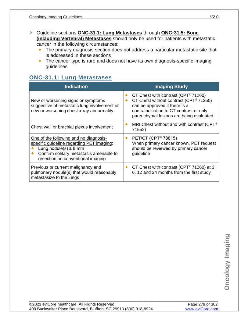
Onc
olog
y Im
agin
g
Guideline sections ONC-31.1: Lung Metastases through ONC-31.5: Bone (including Vertebral) Metastases should only be used for patients with metastatic cancer in the following circumstances: The primary diagnosis section does not address a particular metastatic site that
is addressed in these sections The cancer type is rare and does not have its own diagnosis-specific imaging
guidelines
ONC-31.1: Lung Metastases Indication Imaging Study
New or worsening signs or symptoms suggestive of metastatic lung involvement or new or worsening chest x-ray abnormality
CT Chest with contrast (CPT® 71260) CT Chest without contrast (CPT® 71250)
can be approved if there is a contraindication to CT contrast or only parenchymal lesions are being evaluated
Chest wall or brachial plexus involvement MRI Chest without and with contrast (CPT®
71552)
One of the following and no diagnosis-specific guideline regarding PET imaging: Lung nodule(s) ≥ 8 mm Confirm solitary metastasis amenable to
resection on conventional imaging
PET/CT (CPT® 78815) When primary cancer known, PET request should be reviewed by primary cancer guideline
Previous or current malignancy and pulmonary nodule(s) that would reasonably metastasize to the lungs
CT Chest with contrast (CPT® 71260) at 3, 6, 12 and 24 months from the first study
Oncology Imaging Guidelines V2.0
______________________________________________________________________________________________________ ©2021 eviCore healthcare. All Rights Reserved. 400 Buckwalter Place Boulevard, Bluffton, SC 29910 (800) 918-8924 www.eviCore.com
Page 279 of 302
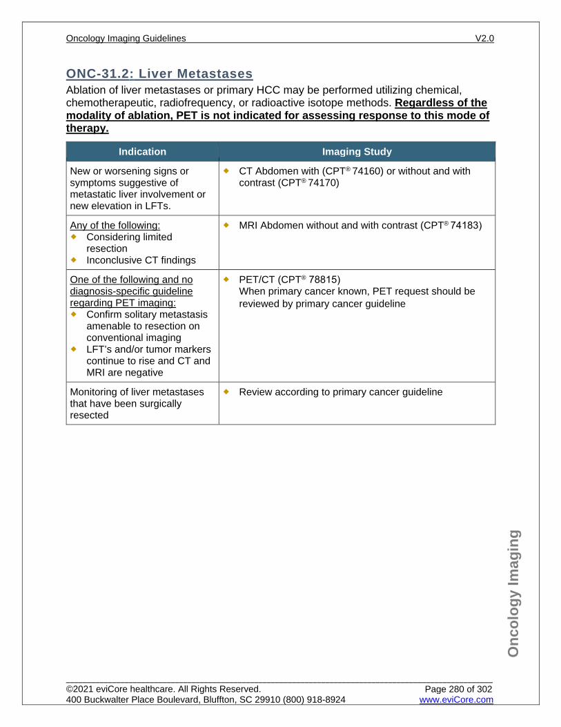
Onc
olog
y Im
agin
g
ONC-31.2: Liver Metastases Ablation of liver metastases or primary HCC may be performed utilizing chemical, chemotherapeutic, radiofrequency, or radioactive isotope methods. Regardless of the modality of ablation, PET is not indicated for assessing response to this mode of therapy.
Indication Imaging Study
New or worsening signs or symptoms suggestive of metastatic liver involvement or new elevation in LFTs.
CT Abdomen with (CPT® 74160) or without and with contrast (CPT® 74170)
Any of the following: Considering limited
resection Inconclusive CT findings
MRI Abdomen without and with contrast (CPT® 74183)
One of the following and no diagnosis-specific guideline regarding PET imaging: Confirm solitary metastasis
amenable to resection on conventional imaging
LFT’s and/or tumor markers continue to rise and CT and MRI are negative
PET/CT (CPT® 78815) When primary cancer known, PET request should be reviewed by primary cancer guideline
Monitoring of liver metastases that have been surgically resected
Review according to primary cancer guideline
Oncology Imaging Guidelines V2.0
______________________________________________________________________________________________________ ©2021 eviCore healthcare. All Rights Reserved. 400 Buckwalter Place Boulevard, Bluffton, SC 29910 (800) 918-8924 www.eviCore.com
Page 280 of 302
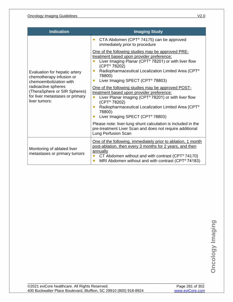
Onc
olog
y Im
agin
g
Indication Imaging Study
Evaluation for hepatic artery chemotherapy infusion or chemoembolization with radioactive spheres (TheraSphere or SIR Spheres) for liver metastases or primary liver tumors:
CTA Abdomen (CPT® 74175) can be approved immediately prior to procedure
One of the following studies may be approved PRE-treatment based upon provider preference: Liver Imaging Planar (CPT® 78201) or with liver flow
(CPT® 78202) Radiopharmaceutical Localization Limited Area (CPT®
78800) Liver Imaging SPECT (CPT® 78803) One of the following studies may be approved POST-treatment based upon provider preference: Liver Planar Imaging (CPT® 78201) or with liver flow
(CPT® 78202) Radiopharmaceutical Localization Limited Area (CPT®
78800) Liver Imaging SPECT (CPT® 78803) Please note: liver-lung shunt calculation is included in the pre-treatment Liver Scan and does not require additional Lung Perfusion Scan
Monitoring of ablated liver metastases or primary tumors
One of the following, immediately prior to ablation, 1 month post-ablation, then every 3 months for 2 years, and then annually CT Abdomen without and with contrast (CPT® 74170) MRI Abdomen without and with contrast (CPT® 74183)
Oncology Imaging Guidelines V2.0
______________________________________________________________________________________________________ ©2021 eviCore healthcare. All Rights Reserved. 400 Buckwalter Place Boulevard, Bluffton, SC 29910 (800) 918-8924 www.eviCore.com
Page 281 of 302
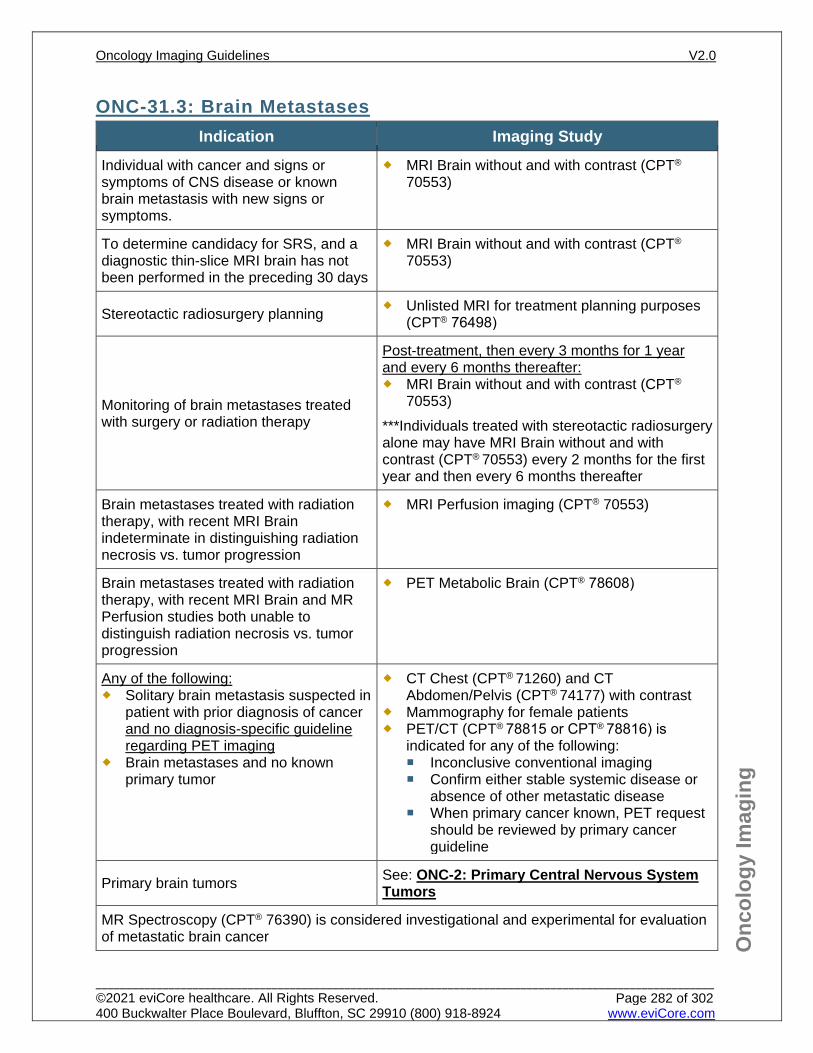
Onc
olog
y Im
agin
g
ONC-31.3: Brain Metastases Indication Imaging Study
Individual with cancer and signs or symptoms of CNS disease or known brain metastasis with new signs or symptoms.
MRI Brain without and with contrast (CPT®
70553)
To determine candidacy for SRS, and a diagnostic thin-slice MRI brain has not been performed in the preceding 30 days
MRI Brain without and with contrast (CPT®
70553)
Stereotactic radiosurgery planning Unlisted MRI for treatment planning purposes (CPT® 76498)
Monitoring of brain metastases treated with surgery or radiation therapy
Post-treatment, then every 3 months for 1 year and every 6 months thereafter: MRI Brain without and with contrast (CPT®
70553) ***Individuals treated with stereotactic radiosurgery alone may have MRI Brain without and with contrast (CPT® 70553) every 2 months for the first year and then every 6 months thereafter
Brain metastases treated with radiation therapy, with recent MRI Brain indeterminate in distinguishing radiation necrosis vs. tumor progression
MRI Perfusion imaging (CPT® 70553)
Brain metastases treated with radiation therapy, with recent MRI Brain and MR Perfusion studies both unable to distinguish radiation necrosis vs. tumor progression
PET Metabolic Brain (CPT® 78608)
Any of the following: Solitary brain metastasis suspected in
patient with prior diagnosis of cancer and no diagnosis-specific guideline regarding PET imaging
Brain metastases and no known primary tumor
CT Chest (CPT® 71260) and CT Abdomen/Pelvis (CPT® 74177) with contrast
Mammography for female patients PET/CT (CPT® 78815 or CPT® 78816) is
indicated for any of the following: Inconclusive conventional imaging Confirm either stable systemic disease or
absence of other metastatic disease When primary cancer known, PET request
should be reviewed by primary cancer guideline
Primary brain tumors See: ONC-2: Primary Central Nervous System Tumors
MR Spectroscopy (CPT® 76390) is considered investigational and experimental for evaluation of metastatic brain cancer
Oncology Imaging Guidelines V2.0
______________________________________________________________________________________________________ ©2021 eviCore healthcare. All Rights Reserved. 400 Buckwalter Place Boulevard, Bluffton, SC 29910 (800) 918-8924 www.eviCore.com
Page 282 of 302
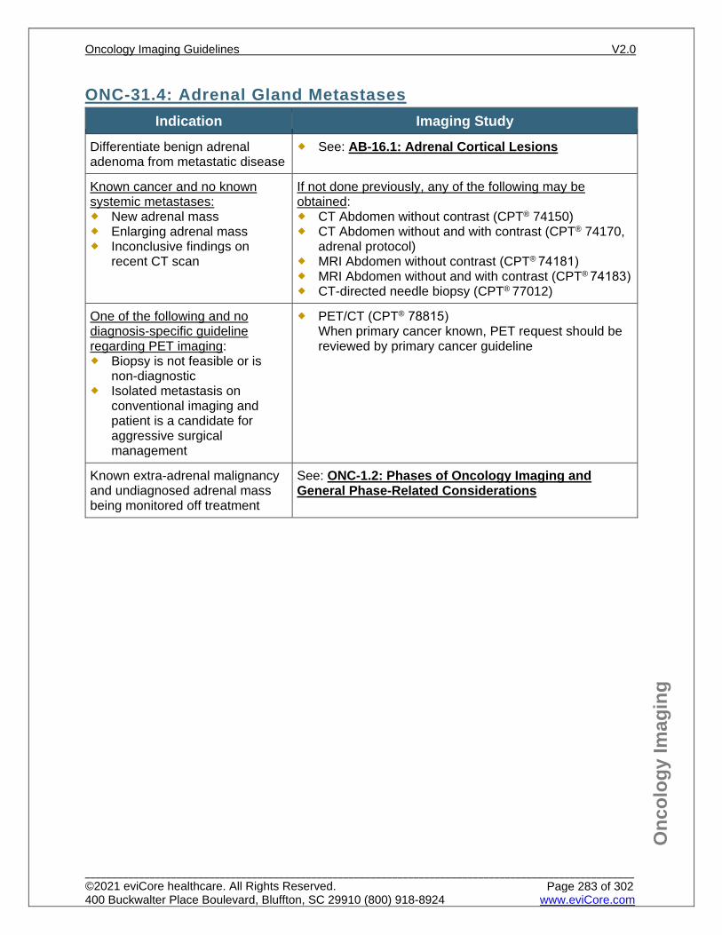
Onc
olog
y Im
agin
g
ONC-31.4: Adrenal Gland Metastases Indication Imaging Study
Differentiate benign adrenal adenoma from metastatic disease
See: AB-16.1: Adrenal Cortical Lesions
Known cancer and no known systemic metastases: New adrenal mass Enlarging adrenal mass Inconclusive findings on
recent CT scan
If not done previously, any of the following may be obtained: CT Abdomen without contrast (CPT® 74150) CT Abdomen without and with contrast (CPT® 74170,
adrenal protocol) MRI Abdomen without contrast (CPT® 74181) MRI Abdomen without and with contrast (CPT® 74183) CT-directed needle biopsy (CPT® 77012)
One of the following and no diagnosis-specific guideline regarding PET imaging: Biopsy is not feasible or is
non-diagnostic Isolated metastasis on
conventional imaging and patient is a candidate for aggressive surgical management
PET/CT (CPT® 78815) When primary cancer known, PET request should be reviewed by primary cancer guideline
Known extra-adrenal malignancy and undiagnosed adrenal mass being monitored off treatment
See: ONC-1.2: Phases of Oncology Imaging and General Phase-Related Considerations
Oncology Imaging Guidelines V2.0
______________________________________________________________________________________________________ ©2021 eviCore healthcare. All Rights Reserved. 400 Buckwalter Place Boulevard, Bluffton, SC 29910 (800) 918-8924 www.eviCore.com
Page 283 of 302
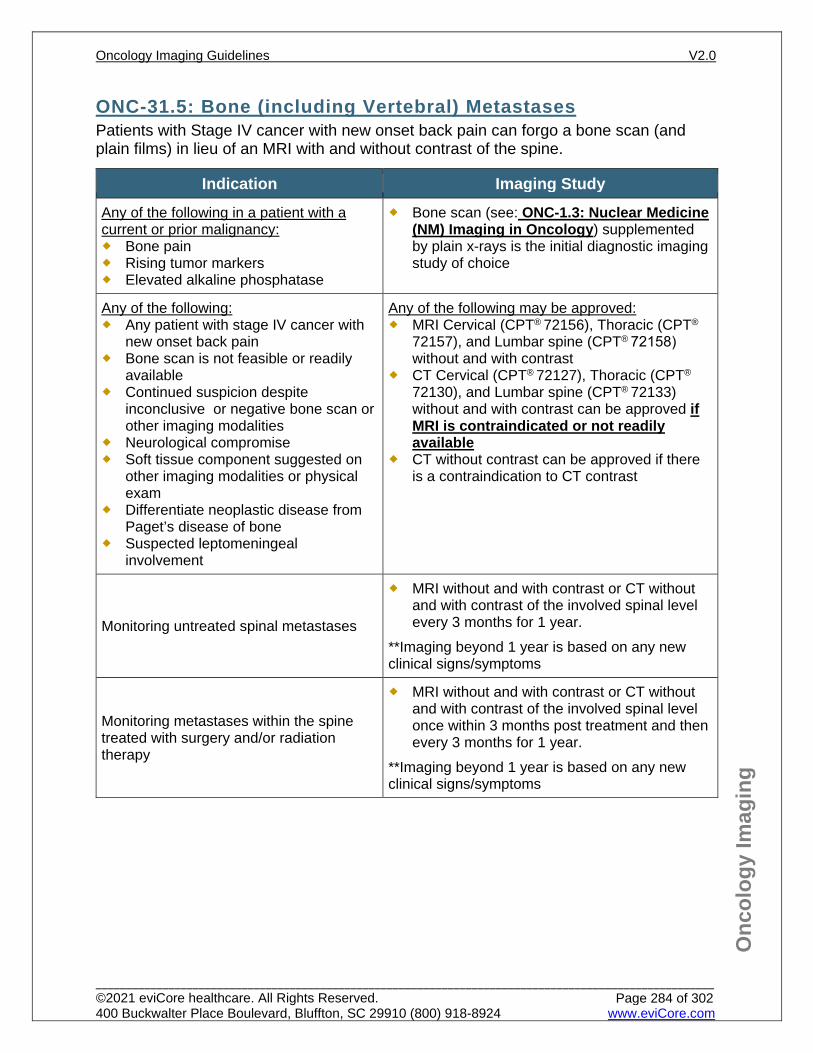
Onc
olog
y Im
agin
g
ONC-31.5: Bone (including Vertebral) Metastases Patients with Stage IV cancer with new onset back pain can forgo a bone scan (and plain films) in lieu of an MRI with and without contrast of the spine.
Indication Imaging Study Any of the following in a patient with a current or prior malignancy: Bone pain Rising tumor markers Elevated alkaline phosphatase
Bone scan (see: ONC-1.3: Nuclear Medicine (NM) Imaging in Oncology) supplemented by plain x-rays is the initial diagnostic imaging study of choice
Any of the following: Any patient with stage IV cancer with
new onset back pain Bone scan is not feasible or readily
available Continued suspicion despite
inconclusive or negative bone scan or other imaging modalities
Neurological compromise Soft tissue component suggested on
other imaging modalities or physical exam
Differentiate neoplastic disease from Paget’s disease of bone
Suspected leptomeningeal involvement
Any of the following may be approved: MRI Cervical (CPT® 72156), Thoracic (CPT®
72157), and Lumbar spine (CPT® 72158) without and with contrast
CT Cervical (CPT® 72127), Thoracic (CPT®
72130), and Lumbar spine (CPT® 72133) without and with contrast can be approved if MRI is contraindicated or not readily available
CT without contrast can be approved if there is a contraindication to CT contrast
Monitoring untreated spinal metastases
MRI without and with contrast or CT without and with contrast of the involved spinal level every 3 months for 1 year.
**Imaging beyond 1 year is based on any new clinical signs/symptoms
Monitoring metastases within the spine treated with surgery and/or radiation therapy
MRI without and with contrast or CT without and with contrast of the involved spinal level once within 3 months post treatment and then every 3 months for 1 year.
**Imaging beyond 1 year is based on any new clinical signs/symptoms
Oncology Imaging Guidelines V2.0
______________________________________________________________________________________________________ ©2021 eviCore healthcare. All Rights Reserved. 400 Buckwalter Place Boulevard, Bluffton, SC 29910 (800) 918-8924 www.eviCore.com
Page 284 of 302
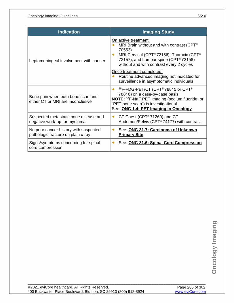
Onc
olog
y Im
agin
g
Indication Imaging Study
Leptomeningeal involvement with cancer
On active treatment: MRI Brain without and with contrast (CPT®
70553) MRI Cervical (CPT® 72156), Thoracic (CPT®
72157), and Lumbar spine (CPT® 72158) without and with contrast every 2 cycles
Once treatment completed: Routine advanced imaging not indicated for
surveillance in asymptomatic individuals
Bone pain when both bone scan and either CT or MRI are inconclusive
18F-FDG-PET/CT (CPT® 78815 or CPT®
78816) on a case-by-case basis NOTE: 18F-NaF PET imaging (sodium fluoride, or “PET bone scan”) is investigational. See: ONC-1.4: PET Imaging in Oncology
Suspected metastatic bone disease and negative work-up for myeloma
CT Chest (CPT® 71260) and CT Abdomen/Pelvis (CPT® 74177) with contrast
No prior cancer history with suspected pathologic fracture on plain x-ray
See: ONC-31.7: Carcinoma of Unknown Primary Site
Signs/symptoms concerning for spinal cord compression
See: ONC-31.6: Spinal Cord Compression
Oncology Imaging Guidelines V2.0
______________________________________________________________________________________________________ ©2021 eviCore healthcare. All Rights Reserved. 400 Buckwalter Place Boulevard, Bluffton, SC 29910 (800) 918-8924 www.eviCore.com
Page 285 of 302
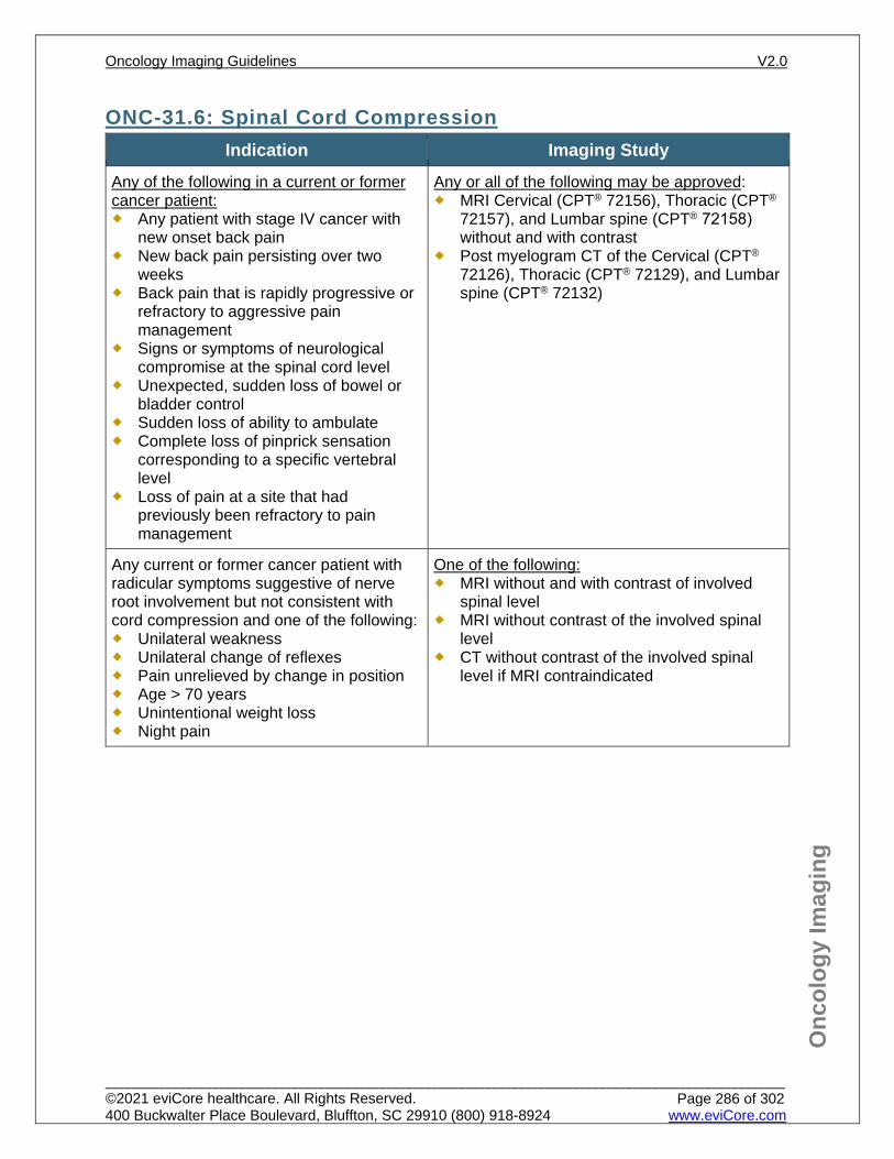
Onc
olog
y Im
agin
g
ONC-31.6: Spinal Cord Compression Indication Imaging Study
Any of the following in a current or former cancer patient: Any patient with stage IV cancer with
new onset back pain New back pain persisting over two
weeks Back pain that is rapidly progressive or
refractory to aggressive pain management
Signs or symptoms of neurological compromise at the spinal cord level
Unexpected, sudden loss of bowel or bladder control
Sudden loss of ability to ambulate Complete loss of pinprick sensation
corresponding to a specific vertebral level
Loss of pain at a site that had previously been refractory to pain management
Any or all of the following may be approved: MRI Cervical (CPT® 72156), Thoracic (CPT®
72157), and Lumbar spine (CPT® 72158) without and with contrast
Post myelogram CT of the Cervical (CPT®
72126), Thoracic (CPT® 72129), and Lumbar spine (CPT® 72132)
Any current or former cancer patient with radicular symptoms suggestive of nerve root involvement but not consistent with cord compression and one of the following: Unilateral weakness Unilateral change of reflexes Pain unrelieved by change in position Age > 70 years Unintentional weight loss Night pain
One of the following: MRI without and with contrast of involved
spinal level MRI without contrast of the involved spinal
level CT without contrast of the involved spinal
level if MRI contraindicated
Oncology Imaging Guidelines V2.0
______________________________________________________________________________________________________ ©2021 eviCore healthcare. All Rights Reserved. 400 Buckwalter Place Boulevard, Bluffton, SC 29910 (800) 918-8924 www.eviCore.com
Page 286 of 302
Onc
olog
y Im
agin
g
ONC-31.7: Carcinoma of Unknown Primary Site
General Considerations Defined as carcinoma found in a lymph node or in an organ known not to be the
primary for that cell type (e.g., adenocarcinoma arising in the brain or in a neck lymph node).
This guideline also applies to a pathologic fracture that is clearly due to metastatic neoplastic disease in a patient without a previous cancer history.
Detailed history and physical examination including pelvic and rectal exams and laboratory tests to be performed before advanced imaging.
Patients presenting with a thoracic squamous cell carcinoma described as metastatic appearing on chest imaging, or in lymph nodes above the clavicle, should undergo a detailed head and neck examination by a clinician skilled in laryngeal and pharyngeal examinations, especially in smokers.
Patients with suspected unknown primary carcinomas based on only suspicious lytic bone lesions should be considered for serum protein electrophoresis (SPEP); urine protein electrophoresis (UPEP) and serum free light chains prior to consideration of extensive imaging
Oncology Imaging Guidelines V2.0
______________________________________________________________________________________________________ ©2021 eviCore healthcare. All Rights Reserved. 400 Buckwalter Place Boulevard, Bluffton, SC 29910 (800) 918-8924 www.eviCore.com
Page 287 of 302
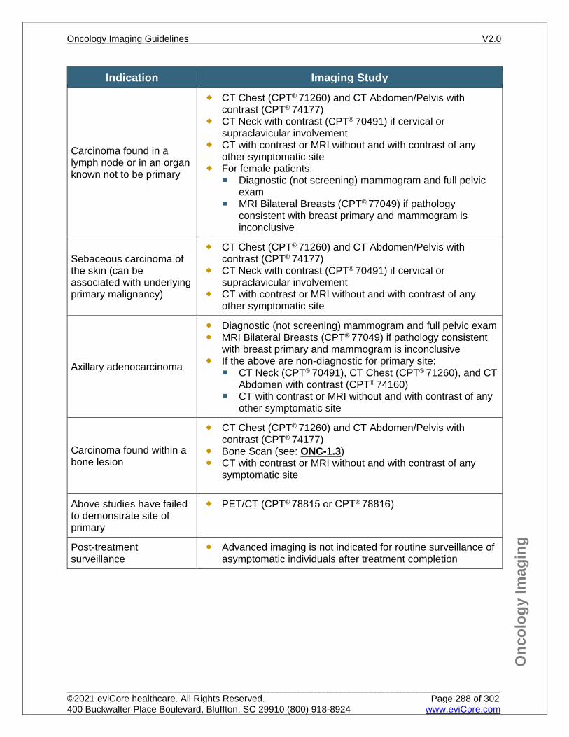
Onc
olog
y Im
agin
g
Indication Imaging Study
Carcinoma found in a lymph node or in an organ known not to be primary
CT Chest (CPT® 71260) and CT Abdomen/Pelvis with contrast (CPT® 74177)
CT Neck with contrast (CPT® 70491) if cervical or supraclavicular involvement
CT with contrast or MRI without and with contrast of any other symptomatic site
For female patients: Diagnostic (not screening) mammogram and full pelvic
exam MRI Bilateral Breasts (CPT® 77049) if pathology
consistent with breast primary and mammogram is inconclusive
Sebaceous carcinoma of the skin (can be associated with underlying primary malignancy)
CT Chest (CPT® 71260) and CT Abdomen/Pelvis with contrast (CPT® 74177)
CT Neck with contrast (CPT® 70491) if cervical or supraclavicular involvement
CT with contrast or MRI without and with contrast of any other symptomatic site
Axillary adenocarcinoma
Diagnostic (not screening) mammogram and full pelvic exam MRI Bilateral Breasts (CPT® 77049) if pathology consistent
with breast primary and mammogram is inconclusive If the above are non-diagnostic for primary site:
CT Neck (CPT® 70491), CT Chest (CPT® 71260), and CT Abdomen with contrast (CPT® 74160)
CT with contrast or MRI without and with contrast of any other symptomatic site
Carcinoma found within a bone lesion
CT Chest (CPT® 71260) and CT Abdomen/Pelvis with contrast (CPT® 74177)
Bone Scan (see: ONC-1.3) CT with contrast or MRI without and with contrast of any
symptomatic site
Above studies have failed to demonstrate site of primary
PET/CT (CPT® 78815 or CPT® 78816)
Post-treatment surveillance
Advanced imaging is not indicated for routine surveillance of asymptomatic individuals after treatment completion
Oncology Imaging Guidelines V2.0
______________________________________________________________________________________________________ ©2021 eviCore healthcare. All Rights Reserved. 400 Buckwalter Place Boulevard, Bluffton, SC 29910 (800) 918-8924 www.eviCore.com
Page 288 of 302
Onc
olog
y Im
agin
g
ONC-31.8: Extrathoracic Small Cell and Large Cell Neuroendocrine Tumors All poorly-differentiated or high-grade, small cell and large cell neuroendocrine tumors arising outside the lungs or of unknown primary origin are imaged according to these guidelines.
Indication Imaging Study
Initial staging
Any or all of the following are indicated: CT Chest (CPT® 71260) and CT Abdomen/Pelvis with contrast
(CPT® 74177) MRI Brain without and with contrast (CPT® 70553) should be
performed for symptoms of CNS involvement and for poorly differentiated neuroendocrine cancers of the neck or extrapulmonary thorax.
PET/CT (CPT® 78815) if no evidence of metastatic disease or conventional imaging is inconclusive for determining localized vs. distant metastatic disease
Restaging during treatment
CT Chest (CPT® 71260) and CT Abdomen/Pelvis (CPT®
74177) and any known sites of disease with contrast every 2 cycles
Suspected Recurrence
Any or all of the following are indicated: CT Chest (CPT® 71260) and CT Abdomen/Pelvis with contrast
(CPT® 74177) MRI brain without and with contrast (CPT® 70553) Bone scan (See: ONC-1.3: Nuclear Medicine (NM) Imaging
in Oncology) PET imaging is generally not indicated but can be considered
for rare circumstances.
Surveillance CT Chest (CPT® 71260) and Abdomen/Pelvis with contrast
(CPT® 74177) every 3 months for 1 year, then every 6 months for 4 additional years and then annually
Oncology Imaging Guidelines V2.0
______________________________________________________________________________________________________ ©2021 eviCore healthcare. All Rights Reserved. 400 Buckwalter Place Boulevard, Bluffton, SC 29910 (800) 918-8924 www.eviCore.com
Page 289 of 302
Onc
olog
y Im
agin
g
ONC-31.9: Primary Peritoneal Mesothelioma Indication Imaging Study
Initial staging
CT Chest (CPT® 71260) and CT Abdomen/Pelvis with contrast (CPT®
74177) PET/CT (CPT® 78815) if there is no evidence of metastatic disease or
conventional imaging is inconclusive
Recurrence/ Restaging
If there is known prior disease, CT Chest (CPT® 71260) and Abdomen/Pelvis with contrast (CPT® 74177)
PET for inconclusive finding on conventional imaging
Surveillance CT Abdomen/Pelvis with contrast (CPT® 74177) every 3 months for 2 years, then every year of life
Oncology Imaging Guidelines V2.0
______________________________________________________________________________________________________ ©2021 eviCore healthcare. All Rights Reserved. 400 Buckwalter Place Boulevard, Bluffton, SC 29910 (800) 918-8924 www.eviCore.com
Page 290 of 302
Onc
olog
y Im
agin
g
ONC-31.10: Kaposi’s Sarcoma Indication Imaging Study
Kaposi’s Sarcoma
Advanced imaging is not generally indicated since disease is generally localized to skin.
CT Chest (CPT® 71260) and CT Abdomen/Pelvis with contrast (CPT®
74177) can be approved at initial diagnosis. If initial scans are negative then future imaging would be based on signs or symptoms.
Oncology Imaging Guidelines V2.0
______________________________________________________________________________________________________ ©2021 eviCore healthcare. All Rights Reserved. 400 Buckwalter Place Boulevard, Bluffton, SC 29910 (800) 918-8924 www.eviCore.com
Page 291 of 302
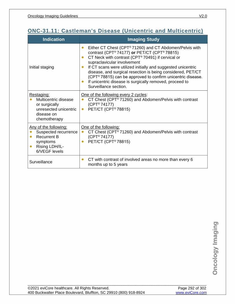
Onc
olog
y Im
agin
g
ONC-31.11: Castleman’s Disease (Unicentric and Multicentric) Indication Imaging Study
Initial staging
Either CT Chest (CPT® 71260) and CT Abdomen/Pelvis with contrast (CPT® 74177) or PET/CT (CPT® 78815)
CT Neck with contrast (CPT® 70491) if cervical or supraclavicular involvement
If CT scans were utilized initially and suggested unicentric disease, and surgical resection is being considered, PET/CT (CPT® 78815) can be approved to confirm unicentric disease.
If unicentric disease is surgically removed, proceed to Surveillance section.
Restaging: Multicentric disease
or surgically unresected unicentric disease on chemotherapy
One of the following every 2 cycles: CT Chest (CPT® 71260) and Abdomen/Pelvis with contrast
(CPT® 74177) PET/CT (CPT® 78815)
Any of the following: Suspected recurrence Recurrent B
symptoms Rising LDH/IL-
6/VEGF levels
One of the following: CT Chest (CPT® 71260) and Abdomen/Pelvis with contrast
(CPT® 74177) PET/CT (CPT® 78815)
Surveillance CT with contrast of involved areas no more than every 6 months up to 5 years
Oncology Imaging Guidelines V2.0
______________________________________________________________________________________________________ ©2021 eviCore healthcare. All Rights Reserved. 400 Buckwalter Place Boulevard, Bluffton, SC 29910 (800) 918-8924 www.eviCore.com
Page 292 of 302
Onc
olog
y Im
agin
g
References 1. Ettinger DS, Varadhachary GR, Bajor D, et al. National Comprehensive Cancer Network (NCCN)
Guidelines Version 2.2021 – February 8, 2021. Occult primary, available at: https://www.nccn.org/professionals/physician_gls/pdf/occult.pdf Referenced with permission from the NCCN Clinical Practice Guidelines in Oncology (NCCN Guidelines™) for Occult Primary V2.2021 – February 8, 2021. ©2021 National Comprehensive Cancer Network, Inc. All rights reserved. The NCCN Guidelines™ and illustrations herein may not be reproduced in any form for any purpose without the express written permission of the NCCN. To view the most recent and complete version of the NCCN Guidelines™, go online to NCCN.org.
2. Nabors BL, Portnow J, Ahluwalia M, et al. National Comprehensive Cancer Network (NCCN) Guidelines Version 3.2020 – September 11, 2020. Central Nervous System Cancers, available at: https://www.nccn.org/professionals/physician_gls/pdf/cns.pdf Referenced with permission from the NCCN Clinical Practice Guidelines in Oncology (NCCN Guidelines™) for CNS Cancer V3.2020 – September 11, 2020. ©2020 National Comprehensive Cancer Network, Inc. All rights reserved. The NCCN Guidelines™ and illustrations herein may not be reproduced in any form for any purpose without the express written permission of the NCCN. To view the most recent and complete version of the NCCN Guidelines™, go online to NCCN.org.
3. Zelenetz AD, Gordon LI, Abramson JS, et al, National Comprehensive Cancer Network (NCCN) Guidelines Version 2.2021 – February 16, 2021. B-cell lymphomas, available at: https://www.nccn.org/professionals/physician_gls/pdf/B-CELL.pdf Referenced with permission from the NCCN Clinical Practice Guidelines in Oncology (NCCN Guidelines™) for B-cell lymphomas V2.2021 – February 16, 2021. ©2021 National Comprehensive Cancer Network, Inc. All rights reserved. The NCCN Guidelines™ and illustrations herein may not be reproduced in any form for any purpose without the express written permission of the NCCN. To view the most recent and complete version of the NCCN Guidelines™, go online to NCCN.org.
4. Mayo-Smith WM, Song JH, Boland GL, et al. Management of incidental adrenal masses: a white paper of the ACR Incidental Findings Committee. J Am Coll Radiol. 2017;14(8):1038-1044. doi:10.1016/j.jacr.2017.05.001.
5. ACR Appropriateness Criteria®. Incidentally discovered adrenal mass. Rev. 2012. 6. Braat AJ, Smits ML, Braat MN, et al. 90Y hepatic radioembolization: an update on current practice
and recent developments. J Nucl Med. 2015;56(7):1079–1087. doi:10.2967/jnumed.115.157446. 7. Pawaskar AS, Basu S. Role of 2-fluoro-2-deoxyglucose PET/computed tomography in carcinoma of
unknown primary. PET Clin. 2015;10(3):297-310. doi:10.1016/j.cpet.2015.03.004. 8. Avram AM. Radioiodine scintigraphy with SPECT/CT: an important diagnostic tool for thyroid cancer
staging and risk stratification. J Nucl Med. 2012;53(5): 754-764. doi:10.2967/jnumed.111.104133. 9. Mayo-Smith WW, Song JH, Boland GL, et al. Management of incidental adrenal masses: a white
paper of the ACR Incidental Findings Committee. J Am Coll Radiol. 2017;14(8):1038-1044. doi:10.1016/j.jacr.2017.05.001.
10. Vaidya A, Hamrahian A, Bancos I, Fleseriu M, Ghayee HK. The evaluation of incidentally discovered adrenal masses. Endocrine Practice. 2019:25(2);178-192. doi: 10.4158/DSCR-2018-0565.
11. Shah MH, Goldner WS, Benson III AB, et al. National Comprehensive Cancer Network (NCCN) Guidelines Version 2.2020 – July 24, 2020. Neuroendocrine and Adrenal Tumors, available at: https://www.nccn.org/professionals/physician_gls/pdf/neuroendocrine.pdf Referenced with permission from the NCCN Clinical Practice Guidelines in Oncology (NCCN Guidelines™) for Neuroendocrine and Adrenal Tumors V2.2020 – July 24, 2020. ©2020 National Comprehensive Cancer Network, Inc. All rights reserved. The NCCN Guidelines™ and illustrations herein may not be reproduced in any form for any purpose without the express written permission of the NCCN. To view the most recent and complete version of the NCCN Guidelines™, go online to NCCN.org.
12. Furuse M, Nonoguchi N, Yamada K, et. al. Radiological diagnosis of brain radiation necrosis after cranial irradiation for brain tumor: a systematic review. Radiat Oncol. 2019;14(28). doi:10.1186/s13014-019-1228-x.
13. American College of Radiology. ACR practice parameter for the performance of stereotactic radiosurgery. 2016; https://www.acr.org/-/media/ACR/Files/Practice-Parameters/stereobrain.pdf
14. Soffietti R, Abacioglu U, Baumert B, et. al. Diagnosis and treatment of brain metastases from solid tumors: guidelines from the European Assocation of Neuro-Oncology (EANO). Neuro-Oncology. 2017;19(2):162-174. doi:10.1093/neuonc/now241.
Oncology Imaging Guidelines V2.0
______________________________________________________________________________________________________ ©2021 eviCore healthcare. All Rights Reserved. 400 Buckwalter Place Boulevard, Bluffton, SC 29910 (800) 918-8924 www.eviCore.com
Page 293 of 302
Onc
olog
y Im
agin
g
15. Mehrabian H, Detsky J, Soliman H, Sahgal A, Stanisz GJ. Advanced magnetic resonance imaging techniques in management of brain metastases. Front Oncol. 2019;9(440). doi:10.3389/fonc.2019.00440.
Oncology Imaging Guidelines V2.0
______________________________________________________________________________________________________ ©2021 eviCore healthcare. All Rights Reserved. 400 Buckwalter Place Boulevard, Bluffton, SC 29910 (800) 918-8924 www.eviCore.com
Page 294 of 302
ONC-32: Medicare Coverage Policies for PET ONC-32.1: Oncologic FDG PETONC-32.2: Oncologic Non-FDG PETONC-32.3: This section left intentionally blankONC-32.4: This section left intentionally blankONC-32.5: This section left intentionally blank
Oncology Imaging Guidelines V2.0
______________________________________________________________________________________________________ ©2021 eviCore healthcare. All Rights Reserved. 400 Buckwalter Place Boulevard, Bluffton, SC 29910 (800) 918-8924 www.eviCore.com
Page 295 of 302
Onc
olog
y Im
agin
g
ONC-32.1: Oncologic FDG PET The complete coverage policy is found in the Medicare National Coverage Determinations (NCD) Manual, Section 220.6.17: (see: http://www.cms.gov/Regulations-and-Guidance/Guidance/Manuals/downloads/ncd103c1_Part4.pdf )
220.6.17 – Positron Emission Tomography (FDG PET) for Oncologic Conditions
General FDG (2-[F18] fluoro-2-deoxy-D-glucose) PET is a minimally invasive diagnostic imaging procedure used to evaluate glucose metabolism in normal tissue, as well as in diseased tissues, in conditions such as cancer, ischemic heart disease, and some neurologic disorders. FDG is an injected radionuclide (or radiopharmaceutical that emits sub-atomic particles, known as positrons, as it decays. FDG PET uses a positron camera (tomograph) to measure the decay of FDG. The rate of FDG decay provides biochemical information on glucose metabolism in the tissue being studied. As malignancies can cause abnormalities of metabolism and blood flow, FDG PET evaluation may indicate the probable presence or absence of a majority of cancer types based upon observed differences in biologic activity compared to adjacent tissues.
The Centers for Medicare and Medicaid Services (CMS) was asked by the National Oncologic PET Registry (NOPR) to reconsider section 220.6 of the National Coverage Determination (NCD) Manual to end the prospective data collection requirements under Coverage with Evidence Development (CED) across all oncologic indications of FDG PET imaging. The CMS received public input indicating that the current framework of prospective data collection under CED be ended for all oncologic uses of FDG PET imaging
1. Framework Effective for claims with dates of service on and after June 11, 2013, CMS is adopting a coverage framework that ends the prospective data collection requirements by NOPR under CED for all oncologic uses of FDG PET imaging. CMS is making this change for all NCDs that address coverage of FDG PET for oncologic uses addressed in this decision. This decision does not change coverage for any use of PET imaging using radiopharmaceuticals ammonia N-13, or rubidium-82 (Rb-82).
2. Initial Anti-Tumor Treatment Strategy CMS continues to believe that the evidence is adequate to determine that the results of FDG PET imaging are useful in determining the appropriate initial anti-tumor treatment strategy for beneficiaries with suspected cancer and improve health outcomes and thus are reasonable and necessary under §1862(a)(1)(A) of the Social Security Act (the “Act”).
Therefore, CMS continues to nationally cover ONE FDG PET study for beneficiaries who have cancers that are biopsy proven or strongly suspected based on other diagnostic testing when the beneficiary’s treating physician determines that the FDG
Oncology Imaging Guidelines V2.0
______________________________________________________________________________________________________ ©2021 eviCore healthcare. All Rights Reserved. 400 Buckwalter Place Boulevard, Bluffton, SC 29910 (800) 918-8924 www.eviCore.com
Page 296 of 302
Onc
olog
y Im
agin
g
PET study is needed to determine the location and/or extent of the tumor for the following therapeutic purposes related to the initial anti-tumor treatment strategy:
To determine whether or not the beneficiary is an appropriate candidate for an invasive diagnostic or therapeutic procedure; or
To determine the optimal anatomic location for an invasive procedure; or To determine the anatomic extent of tumor when the recommended anti-tumor
treatment reasonably depends on the extent of the tumor.
See the table at the end of this section for a synopsis of all nationally covered and non-covered oncologic uses of FDG PET imaging.
Initial Anti-Tumor Treatment Strategy Nationally Covered Indication Effective: June 11, 2013 CMS continues to nationally cover FDG PET imaging for the initial anti-tumor
treatment strategy for male and female breast cancer only when used in staging distant metastasis.
CMS continues to nationally cover FDG PET to determine initial anti-tumor treatment strategy for melanoma other than for the evaluation of regional lymph nodes.
CMS continues to nationally cover FDG PET imaging for the detection of pre-treatment metastasis (i.e., staging) in newly diagnosed cervical cancers following conventional imaging.
Initial Anti-Tumor Treatment Strategy Nationally Non-Covered Indication Effective: June 11, 2013 CMS continues to nationally non-cover initial anti-tumor treatment strategy in
Medicare beneficiaries who have adenocarcinoma of the prostate. CMS continues to nationally non-cover FDG PET imaging for diagnosis of breast
cancer and initial staging of axillary nodes. CMS continues to nationally non-cover FDG PET imaging for initial anti-tumor
treatment strategy for the evaluation of regional lymph nodes in melanoma. CMS continues to nationally non-cover FDG PET imaging for the diagnosis (no
biopsy result) of cervical cancer related to initial anti-tumor treatment strategy.
3. Subsequent Anti-Tumor Treatment Strategy Subsequent Anti-Tumor Treatment Strategy Nationally Covered Indication, Effective: June 11, 2013 THREE FDG PET scans are nationally covered when used to guide subsequent management of anti-tumor treatment strategy after completion of initial anti-tumor therapy. Coverage of more than three FDG PET scans to guide subsequent management of anti-tumor treatment strategy after completion of initial anti-tumor therapy shall be determined by the local Medicare Administrative Contractors.
Oncology Imaging Guidelines V2.0
______________________________________________________________________________________________________ ©2021 eviCore healthcare. All Rights Reserved. 400 Buckwalter Place Boulevard, Bluffton, SC 29910 (800) 918-8924 www.eviCore.com
Page 297 of 302
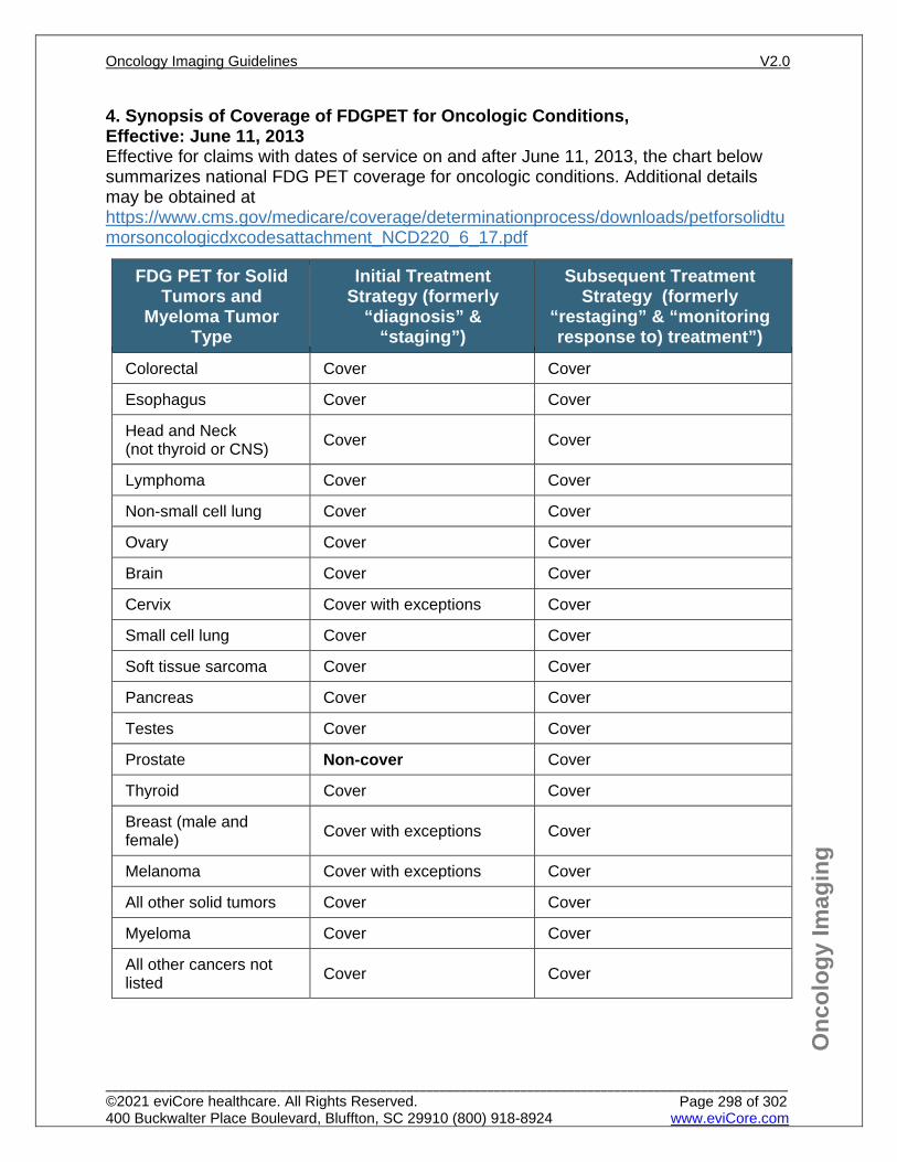
Onc
olog
y Im
agin
g
4. Synopsis of Coverage of FDGPET for Oncologic Conditions, Effective: June 11, 2013 Effective for claims with dates of service on and after June 11, 2013, the chart below summarizes national FDG PET coverage for oncologic conditions. Additional details may be obtained at https://www.cms.gov/medicare/coverage/determinationprocess/downloads/petforsolidtumorsoncologicdxcodesattachment_NCD220_6_17.pdf
FDG PET for Solid Tumors and
Myeloma Tumor Type
Initial Treatment Strategy (formerly
“diagnosis” & “staging”)
Subsequent Treatment Strategy (formerly
“restaging” & “monitoring response to) treatment”)
Colorectal Cover Cover
Esophagus Cover Cover
Head and Neck (not thyroid or CNS) Cover Cover
Lymphoma Cover Cover
Non-small cell lung Cover Cover
Ovary Cover Cover
Brain Cover Cover
Cervix Cover with exceptions Cover
Small cell lung Cover Cover
Soft tissue sarcoma Cover Cover
Pancreas Cover Cover
Testes Cover Cover
Prostate Non-cover Cover
Thyroid Cover Cover
Breast (male and female) Cover with exceptions Cover
Melanoma Cover with exceptions Cover
All other solid tumors Cover Cover
Myeloma Cover Cover
All other cancers not listed Cover Cover
Oncology Imaging Guidelines V2.0
______________________________________________________________________________________________________ ©2021 eviCore healthcare. All Rights Reserved. 400 Buckwalter Place Boulevard, Bluffton, SC 29910 (800) 918-8924 www.eviCore.com
Page 298 of 302
Onc
olog
y Im
agin
g
Invasive Breast Cancer: Nationally non-covered for initial diagnosis and/or staging of axillary lymph nodes. Nationally covered for initial staging of known or suspected metastatic disease. All other indications for initial anti-tumor strategy for breast cancer are nationally covered. Prior to surgical lymph node sampling: NOT indicated Metastatic disease or suspicious lesions seen on CT and/or bone scan: Indicated After completion of surgical lymph node sampling: Indicated
Melanoma: Nationally non-covered for initial staging of regional lymph nodes. All other indications for initial anti-tumor treatment strategy for melanoma are nationally covered. Prior to surgical lymph node sampling: NOT indicated Metastatic disease or suspicious lesions seen on CT and/or bone scan: Indicated After completion of surgical lymph node sampling: Indicated
5. CPT codes for FDG-PET scan for Oncologic Conditions The decision whether to use skull base to mid-femur (“eyes to thighs”: procedure
code for PET (CPT® 78812 or CPT® 78815) or whole body PET (CPT® 78813 or CPT® 78816) is addressed in the diagnosis-specific guideline sections.
Tumor imaging by Positron Emission Tomography (PET) may be reported with CPT® codes 78811-78816. CPT® codes 78811-78813 refer to PET imaging alone without localization CT scan and by itself, may not be the most appropriate study to evaluate oncologic conditions.
A PET/CT fusion study should be reported with CPT® codes 78814-78816. CT obtained for localization should not be reported separately with PET codes.
If a cancer-specific guideline considers a diagnostic CT scan to be medically necessary in addition to a PET/CT scan, and if this study is obtained concurrently with PET imaging on the same piece of equipment, the diagnostic CT codes may be reported separately along with CPT® codes 78811–78813. In this instance, CPT® codes 78814-78816 should not be reported along with diagnostic CT codes.
However, if a cancer-specific guideline considers a diagnostic CT scan to be medically necessary in addition to a PET/CT scan, and these studies are obtained on separate pieces of equipment, the diagnostic CT codes may be reported separately along with CPT® codes 78814–78816.
Additional details are available here: Medicare National Coverage Determinations Manual (https://www.cms.gov/Regulations-and-Guidance/Guidance/Manuals/Downloads/ncd103c1_Part4.pdf)
CPT® 78608 (FDG Metabolic PET of the Brain) is still covered and appropriate for evaluation of CNS malignancies, and is covered under the NCD
Oncology Imaging Guidelines V2.0
______________________________________________________________________________________________________ ©2021 eviCore healthcare. All Rights Reserved. 400 Buckwalter Place Boulevard, Bluffton, SC 29910 (800) 918-8924 www.eviCore.com
Page 299 of 302
Onc
olog
y Im
agin
g
ONC-32.2: Oncologic Non-FDG PET
PET/CT Scan using non-FDG Radiotracers: Medicare National Coverage Determination for PET (NCD 220.6.17) has recently
included coverage of PET-CT scans with three new non-FDG radiotracers. Local Medicare contractors have the authority to make coverage decisions about oncologic studies performed with other agents. Additional details are available at: https://www.cms.gov/Regulations-and-Guidance/Guidance/Transmittals/Downloads/R1755OTN.pdf https://www.cms.gov/Regulations-and-Guidance/Guidance/Transmittals/Downloads/R1753OTN.pdf
PET/CT scan using non-FDG radiotracers is reported with the same CPT codes (CPT® 78815 and CPT® 78816)
Either FDG or non-FDG PET/CT scan may be approved to assess the disease status, both may not be obtained simultaneously.
Per NCD 220.6.17, one PET scan for initial treatment strategy and up to three additional PET scans for subsequent treatment strategy may be obtained regardless of the radiotracer used. Coverage of more than three PET scans to guide subsequent management of anti-tumor treatment strategy after completion of initial anti-tumor therapy shall be determined by the local Medicare Administrative Contractors.
11C Choline for Prostate cancer COVERED FOR:
Subsequent treatment strategy for patients with prostate cancer who have a rising PSA after prior treatment
NOT COVERED FOR: Initial treatment strategy for newly diagnosed prostate cancer Surveillance of patients with localized/advanced prostate cancer, who have
completed definitive therapy or are receiving maintenance therapy
18F-Fluciclovine (AXUMIN®) for Prostate cancer COVERED FOR:
Subsequent treatment strategy for patients with prostate cancer who have a rising PSA after prior treatment
NOT COVERED FOR: Initial treatment strategy for newly diagnosed prostate cancer Surveillance of patients with localized/advanced prostate cancer, who have
completed definitive therapy or are receiving maintenance therapy
Oncology Imaging Guidelines V2.0
______________________________________________________________________________________________________ ©2021 eviCore healthcare. All Rights Reserved. 400 Buckwalter Place Boulevard, Bluffton, SC 29910 (800) 918-8924 www.eviCore.com
Page 300 of 302
Onc
olog
y Im
agin
g
68Gallium DOTATATE (NETSPOT®) COVERED FOR:
Initial treatment strategy for newly diagnosed low-grade neuroendocrine tumors and medullary carcinoma of the thyroid
Subsequent treatment strategy for low-grade neuroendocrine tumors and medullary carcinoma of the thyroid
NOT COVERED FOR: Surveillance of patients with localized/advanced low-grade neuroendocrine
tumors or medullary carcinoma of the thyroid, who have completed definitive therapy or are receiving maintenance therapy
18F Na Fluoride PET/CT Scan for Bone Metastases PET/CT using F-18 sodium fluoride (NaF-18) has been studied to identify bone
metastases. At this time, Medicare NCD excludes coverage for PET/CT scan using Na fluoride radiotracer.
Coverage with Evidence Development (CED) CED is a program designed to make PET/CT available to Medicare beneficiaries
while at the same time gathering data regarding PET’s effectiveness. Under CED, Medicare will reimburse the claim if the beneficiary is enrolled in, and
the PET provider is participating in, a qualifying prospective clinical trial or registry. Full details regarding qualifying clinical trials, including the list of required scientific
integrity standards and relevance to the Medicare population are available in the Medicare NCD Manual, Section 220.6.17.
Qualifying research trials must be registered on the www.ClinicalTrials.gov website by the principal sponsor/investigator, prior to the enrollment of the first study subject.
Oncology Imaging Guidelines V2.0
______________________________________________________________________________________________________ ©2021 eviCore healthcare. All Rights Reserved. 400 Buckwalter Place Boulevard, Bluffton, SC 29910 (800) 918-8924 www.eviCore.com
Page 301 of 302
Onc
olog
y Im
agin
g
ONC-32.3
This section left intentionally blank
ONC-32.4
This section left intentionally blank
ONC-32.5
This section left intentionally blank
Oncology Imaging Guidelines V2.0
______________________________________________________________________________________________________ ©2021 eviCore healthcare. All Rights Reserved. 400 Buckwalter Place Boulevard, Bluffton, SC 29910 (800) 918-8924 www.eviCore.com
Page 302 of 302
Related Documents