This article was downloaded by: [Cristina Saavedra] On: 11 March 2012, At: 11:45 Publisher: Routledge Informa Ltd Registered in England and Wales Registered Number: 1072954 Registered office: Mortimer House, 37-41 Mortimer Street, London W1T 3JH, UK Experimental Aging Research: An International Journal Devoted to the Scientific Study of the Aging Process Publication details, including instructions for authors and subscription information: http://www.tandfonline.com/loi/uear20 Event-Related Potentials Elicited By Face Identity Processing In Elderly Adults With Cognitive Impairment Cristina Saavedra a b , Jaime Iglesias a & Ela I. Olivares a a Departamento de Psicología Biológica y de la Salud, Facultad de Psicología, Universidad Autónoma de Madrid, Madrid, Spain b División de Psicología, CES Colegio Universitario Cardenal Cisneros Adscrito a la Universidad Complutense de Madrid, Madrid, Spain Available online: 09 Mar 2012 To cite this article: Cristina Saavedra, Jaime Iglesias & Ela I. Olivares (2012): Event- Related Potentials Elicited By Face Identity Processing In Elderly Adults With Cognitive Impairment, Experimental Aging Research: An International Journal Devoted to the Scientific Study of the Aging Process, 38:2, 220-245 To link to this article: http://dx.doi.org/10.1080/0361073X.2012.660057 PLEASE SCROLL DOWN FOR ARTICLE Full terms and conditions of use: http://www.tandfonline.com/page/terms- and-conditions

Welcome message from author
This document is posted to help you gain knowledge. Please leave a comment to let me know what you think about it! Share it to your friends and learn new things together.
Transcript
This article was downloaded by: [Cristina Saavedra]On: 11 March 2012, At: 11:45Publisher: RoutledgeInforma Ltd Registered in England and Wales Registered Number: 1072954Registered office: Mortimer House, 37-41 Mortimer Street, London W1T 3JH,UK
Experimental Aging Research:An International JournalDevoted to the Scientific Studyof the Aging ProcessPublication details, including instructions forauthors and subscription information:http://www.tandfonline.com/loi/uear20
Event-Related PotentialsElicited By Face IdentityProcessing In Elderly AdultsWith Cognitive ImpairmentCristina Saavedra a b , Jaime Iglesias a & Ela I.Olivares aa Departamento de Psicología Biológica y de laSalud, Facultad de Psicología, Universidad Autónomade Madrid, Madrid, Spainb División de Psicología, CES Colegio UniversitarioCardenal Cisneros Adscrito a la UniversidadComplutense de Madrid, Madrid, Spain
Available online: 09 Mar 2012
To cite this article: Cristina Saavedra, Jaime Iglesias & Ela I. Olivares (2012): Event-Related Potentials Elicited By Face Identity Processing In Elderly Adults With CognitiveImpairment, Experimental Aging Research: An International Journal Devoted to theScientific Study of the Aging Process, 38:2, 220-245
To link to this article: http://dx.doi.org/10.1080/0361073X.2012.660057
PLEASE SCROLL DOWN FOR ARTICLE
Full terms and conditions of use: http://www.tandfonline.com/page/terms-and-conditions

This article may be used for research, teaching, and private study purposes.Any substantial or systematic reproduction, redistribution, reselling, loan,sub-licensing, systematic supply, or distribution in any form to anyone isexpressly forbidden.
The publisher does not give any warranty express or implied or make anyrepresentation that the contents will be complete or accurate or up todate. The accuracy of any instructions, formulae, and drug doses should beindependently verified with primary sources. The publisher shall not be liablefor any loss, actions, claims, proceedings, demand, or costs or damageswhatsoever or howsoever caused arising directly or indirectly in connectionwith or arising out of the use of this material.
Dow
nloa
ded
by [
Cri
stin
a Sa
aved
ra]
at 1
1:45
11
Mar
ch 2
012
EVENT-RELATED POTENTIALS ELICITED BY FACEIDENTITY PROCESSING IN ELDERLY ADULTS WITHCOGNITIVE IMPAIRMENT
Cristina Saavedra
Departamento de Psicologıa Biologica y de la Salud, Facultad de Psicologıa,Universidad Autonoma de Madrid, Madrid, Spain and Division de
Psicologıa, CES Colegio Universitario Cardenal Cisneros Adscrito a laUniversidad Complutense de Madrid, Madrid, Spain
Jaime IglesiasEla I. Olivares
Departamento de Psicologıa Biologica y de la Salud, Facultad de Psicologıa,Universidad Autonoma de Madrid, Madrid, Spain
Background=Study Context: Patients with mild Alzheimer’s disease andmild cognitive impairment show selective loss of knowledge regardingfacial identification.
Methods: The authors focus on decline effects on event-related poten-tials (ERPs) P100, N170, N250, and N400, associated with the processingof facial identity. Different famous and unknown faces were presented inexplicit and implicit familiarity tasks.
Results: Patients with cognitive impairment showed modulations onP100 and N170 and greater activity in prefrontal areas in the earliercomponent. In healthy elderly individuals, but not in patients, famous
Received 21 August 2010; accepted 10 February 2011.
This work was financed by grants from the Spanish Ministry for Education and Culture
(AP2001-0664) and the Spanish Directorate-General for Research (SEJ2007-67858 and
SEJ2006-07089), as well as partially supported by UAM-Grupo Santander (CEAL=2006).
Address correspondence to Cristina Saavedra, Departamento de Psicologıa Biologica y de
la Salud, Facultad de Psicologıa, Campus de Cantoblanco, Universidad Autonoma de Madrid,
C= Ivan P. Pavlov 6, 28049 Madrid, Spain. E-mail: [email protected]
Experimental Aging Research, 38: 220–245, 2012
Copyright # Taylor & Francis Group, LLC
ISSN: 0361-073X print=1096-4657 online
DOI: 10.1080/0361073X.2012.660057
Dow
nloa
ded
by [
Cri
stin
a Sa
aved
ra]
at 1
1:45
11
Mar
ch 2
012
faces modulated the long-latency ERPs N250 and N400, related to theaccess and retrieval of stored facial-related information, respectively.
Conclusion: ERPs have potential as markers of neurodegenerativedisease such as dementia. The neural systems supporting facial identifi-cation may differ in normal and cognitively impaired older adults.
The ability to recognize faces is an important skill for social interac-tions that is impaired from the initial stages of Alzheimer’s disease(AD) (Della Sala, Muggia, Spinnler, & Zuffi, 1995; Dudas, Clague,Thompson, Graham, & Hodges, 2005; Greene & Hodges, 1996a;Hodges, Salmon, & Butters, 1993; Roudier et al., 1998) and in theclinical disorder of mild cognitive impairment (MCI) (Thompson,Graham, Patterson, Sahakian, & Hodges, 2002). In the present study,we focus on neurocognitive decline effects on the neurophysiologicalresponses associated with the perception and recognition of facialidentity, following influential cognitive and neurobiological modelsof face processing (Bruce & Young, 1986; Gobbini & Haxby, 2007).
Behaviorally, both AD and MCI are traditionally characterizedand diagnosed in relation to abnormalities in higher-level brainfunctions. AD is characterized by the early emergence of deficits inboth episodic and semantic memory (Salmon & Bondi, 1999), whichare accounted for in part by the Braak staging of neurofibrillarypathology (Braak & Braak, 1991). In this staging of Alzheimer’sdisease-related neurofibrillary changes, the entorhinal cortex and sur-rounding medial temporal structures are the earliest predilection sites,with progressive neocortical involvement as cognition deteriorates.MCI, which is usually accompanied by AD neurofibrillary pathology(Morris et al., 2001; Petersen et al., 2006), is characterized mainly byexplicit memory deficits, and these patients have a higher risk of con-version to Alzheimer’s disease than elderly without cognitive impair-ment (Palmer, Wang, Backman, Winblad, & Fratiglioni, 2002).Remarkably, in spite of neurodegenerative processes, both mild ADand MCI patients can reach, in some tasks, a behavioral performancesimilar to that of healthy elderly adults by recruiting alternativeneural networks and a broader range of brain areas, which are relatedto compensatory mechanisms (Bokde et al., 2006; Dickerson et al.,2004, 2005; Han, Bangen, & Bondi, 2009; Rinne, Laine, Hiltunen,& Erkinjuntti, 2003; Stern et al., 2000). This differential recruitmentcould be prior to the dedifferentiation associated with a reduced abil-ity to achieve task-relevant focal activation and a decline in cognitiveperformance (Grady, Furney, Pietrini, Horwitz, & Rapoport, 2001;Li, Lindenberger, & Sikstrom, 2001; Cabeza, 2002; Sperling et al.,
ERPs to Face Identity in Mild Dementia 221
Dow
nloa
ded
by [
Cri
stin
a Sa
aved
ra]
at 1
1:45
11
Mar
ch 2
012
2003). The functional changes reflecting the transition from normalto compensatory neural recruitment may be relevant markers fordisease presence and its early detection may have importantimplications for timely therapeutic intervention (Bokde et al., 2006;Stern et al., 2000).
Neurophysiological measures such as event-related potentials(ERPs), considered reliable real-time markers of cognitive operations,have proved sensitive to the functional changes occurring in mild ADandMCI. ERP studies on attention to incoming stimuli and represen-tations updating have found both increased latencies and decreasedamplitudes of P300 in individuals affected by these disorders(Chapman et al., 2007; Golob, Johnson, & Starr, 2001; Green &Levey, 1999; Polich & Corey-Bloom, 2005; Reinvang, Espeseth, &Gjerstad, 2005; Van Deursen, Vuurman, Smits, Verhey, & Riedel,2009). ERP studies on semantic processing have found increasedlatencies and decreased amplitudes of semantic-related N400(Olichney et al., 2006; Olichney et al., 2002; Ostrosky-Solıs,Castaneda, Perez, Castillo, & Bobes, 1998; Schwartz, Federmeier, VanPetten, Salmon, &Kutas, 2003). Of special interest for the present study,it is unknown how ERPs elicited by highly familiar faces are modulatedby neurodegenerative processes, despite the fact that face-specificmodulations of ERPs have been extensively studied in adults and pro-vide an excellent tool for measuring the precise timing of neurocognitiveprocesses evoked by both familiar and unfamiliar faces.
According to the cognitive model proposed by Bruce and Young(1986), the processing of facial identity requires four successivestages: structural encoding (related to the construction of a visualpercept of the face), comparison with face recognition units (whichcontain the structural face representations in long-term memory),access to personal identity nodes (which contain the biographicalinformation about that person), and access to the person’s name.Diverse electrophysiological studies have allowed establishing corre-spondences among some stages of face processing and ERPs. Theposterior-temporal–located N170 has larger amplitude when elicitedby faces rather than objects (Bentin, Allison, Puce, Perez, &McCarthy, 1996; Bentin, Golland, Flevaris, Robertson, & Moscov-itch, 2006; Botzel, Schulze, & Stodieck, 1995; Campanella et al.,2000) and may be modulated by facial emotional expression (Batty& Taylor, 2003). The N170 is likely generated in basal occipitotem-poral areas (Allison, Puce, Spencer, & McCarthy, 1999; Schweinber-ger, Pickering, Jentzsch, Burton, & Kaufmann, 2002; Shibata et al.,2002) and it is considered an ERP that reflects a ‘‘face detector’’mechanism that triggers the face-encoding process (Bentin et al.,
222 C. Saavedra et al.
Dow
nloa
ded
by [
Cri
stin
a Sa
aved
ra]
at 1
1:45
11
Mar
ch 2
012
2006, Zion-Golumbic & Bentin, 2007). Additionally, the earlier P100(peaking around 120 ms) has also been associated with processing offace structure (Linkenkaer-Hansen et al., 1998; Olivares & Iglesias,2008), although showing less robust effects than N170 (Rossion &Jacques, 2008), and it is possibly generated in the striate and peristri-ate cortices (Allison et al., 1999; Katada, Sato, Ojika, & Ueda, 2004).Although, these early effects have been consistently found in diversestudies, it should be pointed out that not all authors agree with theirinterpretation in terms of face-specific effects (see, for example,McKeeff, McGugin, Tong, & Gauthier, 2010; Thierry, Martin,Downing, & Pegna, 2007). Regarding facial recognition, ERP modu-lations have been observed mainly at latencies beyond 170 ms. Thus,N250 is modulated by repetition of facial stimuli in short intervals(even using different pictures), especially when they are familiar(the ‘‘N250r’’) (Schweinberger, Pfutze, & Sommer, 1995; Schweinber-ger et al., 2002), and shows larger amplitude when elicited by facesversus objects (Schweinberger, Huddy, & Burton, 2004; Nasr &Esteky, 2009). N250 can be associated with access to face recognitionunits in the Bruce and Young’s model (1986), and it has been relatedto activity in basal temporal regions (e.g., fusiform gyrus), althoughin a more rostrally located region than that generating the N170(Schweinberger & Burton, 2003; Schweinberger et al., 2002).Additionally, N400 is larger when elicited by famous faces in com-parison with unknown faces (Bentin & Deouell, 2000; Eimer, 2000).In face-feature matching tasks, faces without associated verbal=sem-antic information have elicited responses analogous to N400 at 360ms, whereas faces with associated semantic information or famousfaces elicited effects at 380 ms (Jemel, George, Olivares, Fiori, &Renault, 1999; Olivares, Iglesias, & Rodrıguez-Holguın, 2003). Theselate ERP modulations have been thought to reflect the neural subrou-tines involved in the access to long-term face representations andassociated verbal=semantic information (personal identity nodes),which in the case of famous faces has been linked with greater acti-vation of left brain regions, including medial-temporal structuresand frontotemporal areas (Gorno-Tempini et al., 1998; Leveroniet al., 2000; Pourtois, Schwartz, Seghier, Lazeyras, & Vuilleumier,2005; Olivares & Iglesias, 2010). Finally, it is worth pointing out thatN250 and N400 have shown to be modulated by the task at hand(Saavedra, Iglesias, & Olivares, 2010), being enhanced by the explicit(overt) processing compared to the implicit (covert) processing offamiliarity in the context of an expression task. It suggests that brainresources devoted to familiarity processing become highly taskdependent in long latencies.
ERPs to Face Identity in Mild Dementia 223
Dow
nloa
ded
by [
Cri
stin
a Sa
aved
ra]
at 1
1:45
11
Mar
ch 2
012
In this study, we examined the modulations by neurodegenerativeprocesses related to mild cognitive impairment and Alzheimer’sdementia on the aforementioned ERPs during both the explicit andimplicit processing of face identity. Thus, we presented famous andunknown faces with happy and neutral expressions in face familiarity(explicit processing) and expression (implicit processing) judgmenttasks. Behavioral studies have pointed out that AD and MCI patientsshow impaired person recognition (related to semantic knowledge)(Dudas et al., 2005; Thompson et al., 2002). Considering the face-processing model by Bruce and Young (1986), the major damageappears to fall on the semantic system underlying person identifi-cation (personal identity nodes) (Greene & Hodges, 1996a; Hodgeset al., 1993), which would be related to late ERP modulations onN400. Additionally, there is also a deficit in the recognition of fam-iliar faces (Della Salla et al., 1995; Dudas et al., 2005; Thompsonet al., 2002; Roudier et al., 1998), which would be more related toaccess to face recognition units and thus with ERP modulations inthe N250 time window. The visual discrimination of unknown faces(by face matching or age estimation) appears less frequently as a dif-ficulty in the face-processing performance in these patients (DellaSalla et al., 1995) as reported by a neuropsychological approach.These findings are congruent with the well-known disproportionateimpairment of mnestic versus perceptual performance in early ADand MCI, explained by the aforementioned Braak staging of neurofi-brillary pathology (Braak & Braak, 1991). Nonetheless, in the contextof the present study, it should be noted that neurofibrillary pathologyin the primary and secondary visual structures and pathways is moreprevalent than previously observed (Leuba & Kraftsik, 2004; Leuba& Saini, 1995; McKee et al., 2006). Moreover, although impaired per-ceptual performance is not a distinguishing feature of MCI and AD,neuroimaging studies suggest that metabolism in posterior brainregions, including the posterior temporoparietal and occipital cortex,is correlated with deficits in neuropsychological function (Newberget al., 2003; Van Rhijn et al., 2004). In sum, we expected that anyimpairment concerning the encoding and structural processing ofthe stimuli related to perceptual processes would be reflected onmodulations (in latency or amplitude) of P100 and N170 ERPs.According to the aforementioned studies regarding the recognitionand identification of faces, we expected that patients would show adiminished effect not only on N400 but also on N250, whereas inhealthy elderly adults the modulations in these responses differentiat-ing famous and unknown faces would be more enhanced, particularlyin the familiarity explicit processing task. Furthermore, in the
224 C. Saavedra et al.
Dow
nloa
ded
by [
Cri
stin
a Sa
aved
ra]
at 1
1:45
11
Mar
ch 2
012

absence of behavioral differences in performance between patientsand healthy elderly adults, differentiation in ERPs could reflect braincompensatory mechanisms in the patient group.
METHODS
Participants
Twenty-seven right-handed elderly adults (23 women) volunteered asparticipants, 6 of whom were from the day center ‘‘Asociacion deFamiliares de Enfermos de Alzheimer de Tres Cantos’’ and 21 fromthe retirement home ‘‘Residencia Asistida San Camilo de TresCantos.’’ As shown in Table 1, age (t(25)¼�0.491, p¼ .628) andeducation (t(25)¼�0.64, p¼ .525) differences between the healthyelderly adults and patients with cognitive impairment were not sig-nificant, but Mini-Mental State Examination (Folstein, Folstein &McHugh, 1975) score differences were significant (t(25)¼�4.51,p< .001). Participants in both groups used similar medication includ-ing diuretics, antihypertensives and nonsteroidal anti-inflammatorydrugs. Although none of the medication was considered to havemajor central nervous system (CNS) side effects, any possible CNSside effects cannot be excluded. All participants had corrected-to-normal vision.
ERP data are presented from 12 patients with cognitive impair-ment (all women, 4 with probable AD, 3 with possible AD, 5 withMCI) and 15 healthy elderly adults (11 women). The inclusion cri-teria for the group of patients with cognitive impairment were diag-nosis of probable or possible AD according to the criteriadeveloped by the National Institute of Neurological CommunicativeDisorders and Stroke and the Alzheimer’s Disease and RelatedDisorders Association (McKhann et al., 1984), or diagnosis of MCI(Petersen et al., 1999). Diagnoses were made by medical personnel
Table 1. Mean (and SD) demographic and Mini-Mental Status
Examination (MMSE) score data
Patients with cognitive impairment Healthy elderly adults
N 12 15
Age (years) 81.8 (9.2) 83.5 (8.1)
Education (years) 4.9 (2.6) 5.8 (4.1)
MMSE 20.8 (4.3) 27.2 (3.1)
ERPs to Face Identity in Mild Dementia 225
Dow
nloa
ded
by [
Cri
stin
a Sa
aved
ra]
at 1
1:45
11
Mar
ch 2
012
who were external to this study, either neurologists from the publichealth system or geriatricians from the ‘‘Residencia Asistida SanCamilo de Tres Cantos.’’ The inclusion criteria for the group of healthyelderly adults were over 65 years old and with absence of cognitiveimpairment, both self-reported and assessed by the aforementionedgeriatricians. Caregivers of the patients were informed and gave theirconsent, but all participants, including the AD andMCI patients, wereable to give their own consent to participate in the study.
Stimuli and Procedure (Experimental Tasks)
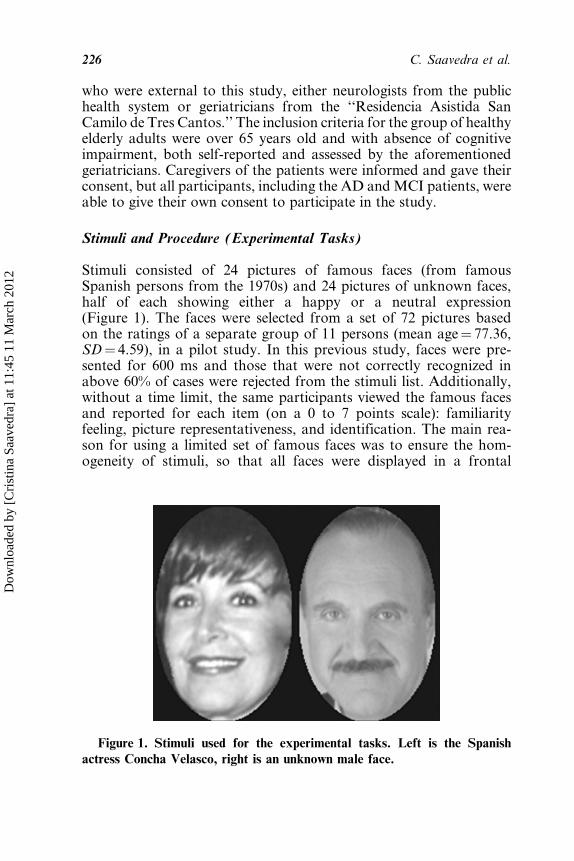
Stimuli consisted of 24 pictures of famous faces (from famousSpanish persons from the 1970s) and 24 pictures of unknown faces,half of each showing either a happy or a neutral expression(Figure 1). The faces were selected from a set of 72 pictures basedon the ratings of a separate group of 11 persons (mean age¼ 77.36,SD¼ 4.59), in a pilot study. In this previous study, faces were pre-sented for 600 ms and those that were not correctly recognized inabove 60% of cases were rejected from the stimuli list. Additionally,without a time limit, the same participants viewed the famous facesand reported for each item (on a 0 to 7 points scale): familiarityfeeling, picture representativeness, and identification. The main rea-son for using a limited set of famous faces was to ensure the hom-ogeneity of stimuli, so that all faces were displayed in a frontal
Figure 1. Stimuli used for the experimental tasks. Left is the Spanish
actress Concha Velasco, right is an unknown male face.
226 C. Saavedra et al.
Dow
nloa
ded
by [
Cri
stin
a Sa
aved
ra]
at 1
1:45
11
Mar
ch 2
012
view and had no glasses or similar on them. On the other hand, andmainly to ensure that the faces used were appropriately known by theparticipants, we chose famous persons from the 1970s given that inAD, public memory has been shown to deteriorate longitudinally(Greene & Hodges, 1996b), which was challenging taking intoaccount that television was not widespread in Spain 40 years ago.All stimuli were digitized grayscale photographs in which the faceswere framed with a black circle in order to standardize the presen-tation. The size of stimuli on the computer screen was 18 cm highand 10 cm wide, occupying a visual angle of about 12.8� � 7.2�.
These stimuli were separated into two sets (with both sets matchedfor familiarity, gender, age, and facial expression of the faces) in orderto form the two experimental tasks with different facial identities. Ineach set, 12 famous and 12 unknown faces were presented, inter-spersed following a quasi-random order in seven consecutive blocks(24 faces per block). The order of stimuli in each block was createdensuring that no more than three consecutive stimuli showed the sameidentity, expression, or gender. A total of 168 stimuli were displayed ineach of the two tasks. The photos were presented, for 600 ms, by theexperimenter pressing a key. After the image disappeared from thescreen, the experimenter asked the participant the question relevantto the task (‘‘Was this face known or unknown?’’ or ‘‘Was this facesmiling or not?’’) and participants’ verbal responses were recordedby the experimenter. Participants were instructed not to respond untilthe question was asked. Thus, each participant carried out the twojudgment tasks during the electroencephalographic (EEG) recording.The judgment tasks, for both face familiarity and face expression,were performed with different sets of faces. The order of the two tasksand the assignment of a face set to each task were counterbalancedamong the participants. Participants were instructed not to blink, inorder to minimize the artifacts. If participants felt tired, no photoswere presented until they decided to continue the task.
After the recording, participants saw all 48 faces used in the experi-mental tasks. For each face, participants were asked if they con-sidered it a known or unknown face. The faces remained on thescreen until the subject responded to it. The purpose of this taskwas to verify the participants’ familiarity with the faces used as stim-uli in the preceding recording tasks and to average the EEG segmentsconsidering only such information. This implies that averaging wasnot performed on the actual responses of the subjects during therecording in any of the tasks. By this additional control, we furtherensured that we used only faces that were recognized by the subjects,which was initially accomplished through the pilot study (selecting
ERPs to Face Identity in Mild Dementia 227
Dow
nloa
ded
by [
Cri
stin
a Sa
aved
ra]
at 1
1:45
11
Mar
ch 2
012
only famous faces that were identified by a high percentage of part-icipants), and we can focus on the effects of the explicit and implicitprocessing of familiarity. This procedure does not exclude the possi-bility that some images were only correctly identified after repeatedpresentation in the main experiment, but this would equally affectboth tasks and both stimuli, otherwise it would be indicated in beha-vioral performance. We consider this additional control better thanjust relying on ratings from the pilot study to assess the ERP effectsof explicit and implicit processing of familiarity in our participants.On the other hand, it should be noted that the repetition of facestimuli can have effects on markers of neural processing (Grill-Spector, Henson, & Martin, 2006; Henson & Rugg, 2003). For faces,repetition effects have been demonstrated beyond 250 ms, andaffecting both famous and unknown faces, which in our study couldaffect mainly N250 and N400 and to a lesser extent P100 and N170 inboth tasks.
Data Acquisition
EEG was recorded with Ag=AgCl disk electrodes from 20 recordingsites: Fp1–2, F3–4, C3–4, P3–4, O1–2, F7–8, T3–4, T5–6, Fz, Cz, Pz(10=20 International System), and Oz. The tip of the nose was used asthe reference site. Impedance was below 5 kX. Electrooculogram(EOG) was recorded from electrodes placed just above the rightsupraorbital ridge (vertical EOG) and on the right outer canthus(horizontal EOG). EEG and EOG signals were filtered onlinebetween 0.05 and 30Hz. A notch filter for 50Hz was also used.
ERPs were calculated offline, averaging segments of 850 ms(including a prestimulus interval of 148 ms for baseline correction)of digitized EEG (12-bit A=D converter, sampling period of 4 ms).Before averaging, EEG was visually inspected and those segmentswith excessive EOG and larger than 50mV artifacts eliminated.
Data Analysis
Behavioral data were analyzed using the discrimination sensitivity(d’) and the response bias (C) of the ‘‘signal detection theory’’(Macmillan & Creelman, 2005). The former index allowed theevaluation of participants’ sensitivity to discriminate famous fromunknown faces, and the latter indicated any possible response bias,which is especially relevant when examining memorial performancein a memory-impaired population.
228 C. Saavedra et al.
Dow
nloa
ded
by [
Cri
stin
a Sa
aved
ra]
at 1
1:45
11
Mar
ch 2
012
Regarding electrophysiological data, in accordance with the designdescribed above, the averaging of EEG segments was performed fortwo main conditions: familiarity (F) and reaveraged expression con-sidering the postrecording familiarity judgment (rE-F) (only trialsrelated to recognized faces). In each of these main conditions, theprocessing of famous and unknown faces was compared, resultinga total of four experimental conditions. Only trials obtaining correctresponses for familiarity judgments in the postrecording behavioraltask were considered for averaging. For each subject, the numberof segments included in the average was superior to the square rootof the number of trials in the corresponding experimental condition.After rejecting incorrect responses and artifacts, the average numberof segments included in the analyses for the group of patients withcognitive impairment were F famous: 38.67 (46.03%), F unknown:35.83 (42.66%), rE-F famous: 39.83 (47.42%), rE-F unknown: 35(41.67%); and for the group of healthy elderly adults: F famous:40.4 (48.1%), F unknown: 41.2 (49.05%), rE-F famous: 47.6(56.67%), rE-F unknown: 39.6 (47.14%).
To assess experimental effects, linear mixed analyses of variance(ANOVAs) were performed on ERP mean amplitude measurementsin four time windows, corresponding to the main peaks (P100: from80 to 120 ms; N170: from 150 to 200 ms; N250: from 330 to 400 ms;N400: from 600 to 680 ms). Linear mixed ANOVAs are useful foranalyzing complex data in a more flexible way than traditional lineargeneral ANOVAs, especially regarding the assumptions of indepen-dence and homoscedasticity, and allow the comparison of differentsized groups (Brown & Prescott, 1999). ANOVAs were performedwith Group (impaired vs. healthy) as the intersubject factor, andTask (F vs. rE-F), Face (famous vs. unknown), and Electrode asintrasubject factors (repeated measures). The Electrode factorincluded the site showing the most positive or negative amplitudevalue in the grand averages in the corresponding window (O1 forP100 and T6 for N170, N250 and N400), and its contralateralhomologue. When appropriate, significant effects were exploredusing pairwise t tests.
In the aforementioned time windows, neural activity sources wereexamined using the sLORETA inverse solution method (Pascual-Marqui, 2002), which computes the three-dimensional distributionof electrical brain activity as a current density value. The cortex ismodeled (Fuchs, Kastner, Wagner, Hawes, & Ebersole, 2002) as6430 voxels in the MNI152 digitized structural magnetic resonanceimaging (MRI) template (Mazziotta et al., 2001), and anatomicallabels are reported using an appropriate correction from Montreal
ERPs to Face Identity in Mild Dementia 229
Dow
nloa
ded
by [
Cri
stin
a Sa
aved
ra]
at 1
1:45
11
Mar
ch 2
012
Neurological Institute to Talairach space (Brett, Johnsrude, & Owen,2002). Statistical analyses of condition differences were performedusing statistical nonparametric mapping (SnPM) (Nichols & Holmes,2001) implemented in the sLORETA software. The sLORETAmethod is an improved version of LORETA (Pascual-Marqui,Michel, & Lehmann, 1994), which has been compared with other dis-tributed inverse solutions obtaining an accurate localization and hasbeen validated with experimental data (including those derived fromface-processing tasks) under conditions where the sources wereknown a priori.
RESULTS
Behavioral Data
Table 2 displays the accuracy, sensitivity (d0) and response bias (C)data for the postrecording task. These behavioral data were submit-ted to linear mixed ANOVAs with Group (impaired vs. healthy) asthe intersubject factor, and Task (F vs. rE-F) as the intrasubject fac-tor (repeated measures). For the analyses of accuracy, Face (famousvs. unknown) was also included as an intrasubject factor. Effects andinteraction are reported below, although they were nonsignificant forthe each of the measures. Regarding accuracy, the results were thefollowing: Group (F(1, 25)¼ 0.178, p¼ .677), Task (F(1, 25)¼0.906, p¼ .350), and Face (F(1, 25)¼ 2.52, p¼ .125). Regarding sen-sitivity, analysis showed the following: Group (F(1, 25)¼ 0.135,p¼ .717) and Task (F(1, 25)¼ 0.0, p¼ .987). Regarding response bias,
Table 2. Mean accuracy of response as a percent of correct responses
(Accuracy), sensitivity (d0), and response bias (C) for the experimental con-ditions familiarity (F), reaveraged expression considering familiarity (rE-F),
famous faces (Famous), and unknown faces (Unknown)
Patients with cognitive impairment Healthy elderly adults
F rE-F F rE-F
Famous Unknown Fam. Unk. Fam. Unk. Fam. Unk.
Accuracy 73.64 66 73.65 68.78 73.92 68.37 81.14 66.69
d0 1.54 1.55 1.73 1.71
C �0.04 0.06 �0.15 �0.38
230 C. Saavedra et al.
Dow
nloa
ded
by [
Cri
stin
a Sa
aved
ra]
at 1
1:45
11
Mar
ch 2
012
results were the following: Group (F(1, 25)¼ 1.342, p¼ .258) andTask (F(1, 25)¼ 0.212, p¼ .650).
Analyses of Variance
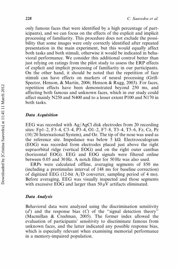
Figure 2 shows the ERP waveforms for the group of elderly adultswith cognitive impairment, recorded over temporal (T5, T6) andoccipital (O1, O2) sites for all conditions (see also Table 3).Figure 3 shows the same ERP waveforms for the group of healthyelderly adults (see also Table 4).
P100The amplitude analysis revealed a Group�Face interaction (F(1,25)¼ 3.97, p¼ .057), explained by the more positive amplitude ofP100 for the elderly with cognitive impairment when elicited byunknown faces (t(11)¼�2.406, p¼ .035).
Figure 2. ERP waveforms elicited in response to famous and unknown
faces for the experimental conditions: F (familiarity) and rE-F (reaveraged
expression considering familiarity) in the patients with cognitive impairment.
The peaks of the effects described in the text are indicated.
ERPs to Face Identity in Mild Dementia 231
Dow
nloa
ded
by [
Cri
stin
a Sa
aved
ra]
at 1
1:45
11
Mar
ch 2
012
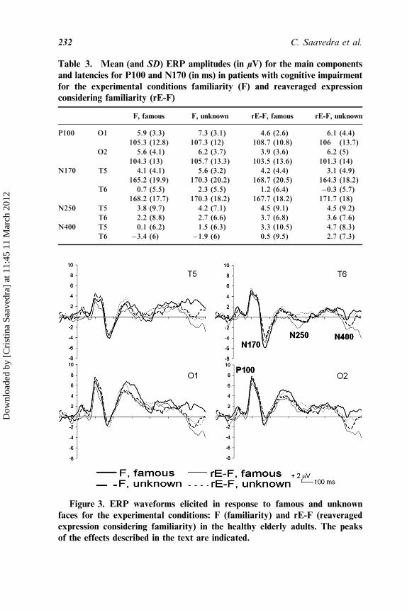
Table 3. Mean (and SD) ERP amplitudes (in lV) for the main components
and latencies for P100 and N170 (in ms) in patients with cognitive impairment
for the experimental conditions familiarity (F) and reaveraged expression
considering familiarity (rE-F)
F, famous F, unknown rE-F, famous rE-F, unknown
P100 O1 5.9 (3.3) 7.3 (3.1) 4.6 (2.6) 6.1 (4.4)
105.3 (12.8) 107.3 (12) 108.7 (10.8) 106 (13.7)
O2 5.6 (4.1) 6.2 (3.7) 3.9 (3.6) 6.2 (5)
104.3 (13) 105.7 (13.3) 103.5 (13.6) 101.3 (14)
N170 T5 4.1 (4.1) 5.6 (3.2) 4.2 (4.4) 3.1 (4.9)
165.2 (19.9) 170.3 (20.2) 168.7 (20.5) 164.3 (18.2)
T6 0.7 (5.5) 2.3 (5.5) 1.2 (6.4) �0.3 (5.7)
168.2 (17.7) 170.3 (18.2) 167.7 (18.2) 171.7 (18)
N250 T5 3.8 (9.7) 4.2 (7.1) 4.5 (9.1) 4.5 (9.2)
T6 2.2 (8.8) 2.7 (6.6) 3.7 (6.8) 3.6 (7.6)
N400 T5 0.1 (6.2) 1.5 (6.3) 3.3 (10.5) 4.7 (8.3)
T6 �3.4 (6) �1.9 (6) 0.5 (9.5) 2.7 (7.3)
Figure 3. ERP waveforms elicited in response to famous and unknown
faces for the experimental conditions: F (familiarity) and rE-F (reaveraged
expression considering familiarity) in the healthy elderly adults. The peaks
of the effects described in the text are indicated.
232 C. Saavedra et al.
Dow
nloa
ded
by [
Cri
stin
a Sa
aved
ra]
at 1
1:45
11
Mar
ch 2
012
N170The amplitude analysis revealed a main effect for group (F(1,25)¼ 6.16, p¼ .020), explained by the more negative amplitude ofN170 for healthy elderly adults, and a Group�Task�Face interac-tion (F(1, 25)¼ 4.29, p¼ .049), explained by the more negative ampli-tude of N170 for healthy elderly adults, compared to patients, whenelicited by F famous (t(24.932)¼ 2.042, p¼ .052), F unknown(t(20.698)¼ 2.957, p¼ .008), and rE-F famous (t(24.680)¼ 2.419,p¼ .023). There was also a main effect for electrode (F(1,25)¼ 4.37, p¼ .047), explained by the more negative amplitude ofN170 at T6.
N250The amplitude analysis revealed a Group�Task�Face� Site inter-action (F(1, 25)¼ 3.84, p¼ .061), explained by the more negativeamplitude of N250 for healthy elderly adults when elicited by rE-Ffamous at T6 (t(25)¼�2.119, p¼ .044).
N400The amplitude analysis revealed a main effect for electrode (F(1,25)¼ 10.3, p¼ .004), explained by the more negative amplitude ofN400 at T6, and a Task� Site interaction (F(1, 25)¼ 7.9, p¼ .009),explained by the more negative amplitude of N400 at T6, comparedto T5, when elicited by the F condition (t(26)¼ 3.72, p¼ .001).
Table 4. Mean (and SD) ERP amplitudes (in lV) for the main components
and latencies for P100 and N170 (in ms) in healthy elderly adults for the
experimental conditions familiarity (F) and reaveraged expression considering
familiarity (rE-F)
F, famous F, unknown rE-F, famous rE-F, unknown
P100 O1 6.3 (4.3) 4.7 (6.1) 3.3 (4.5) 5 (2.3)
105.3 (11.2) 107.1 (9.4) 106.3 (11.6) 109.3 (8.5)
O2 6.1 (3.5) 4.2 (5.7) 4 (5.7) 5.7 (3)
106.4 (11.9) 101.9 (10.5) 106.5 (9.9) 108 (7.3)
N170 T5 �1.3 (5.9) �2.2 (7.8) �1.9 (5.2) �0.8 (3.7)
176 (14.3) 176 (14.9) 175.3 (17.3) 179.3 (12.9)
T6 �1.6 (5.6) �2.5 (7.5) �2 (5.6) �0.9 (4.5)
180.9 (11.9) 176.8 (17.4) 183.5 (13.7) 180.3 (13.6)
N250 T5 �1 (6.7) �1.2 (10.6) 0.1 (4.7) 0.9 (4.1)
T6 �1.3 (6) �1.7 (10.2) �0.9 (4.4) 1.2 (2.3)
N400 T5 1.9 (13) �1.4 (14.3) �1.8 (11.6) 2 (12)
T6 0.6 (12.8) �2.2 (14.2) �2 (11.5) 2.8 (12.3)
ERPs to Face Identity in Mild Dementia 233
Dow
nloa
ded
by [
Cri
stin
a Sa
aved
ra]
at 1
1:45
11
Mar
ch 2
012
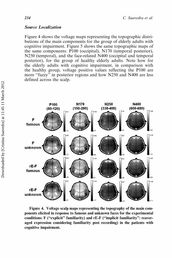
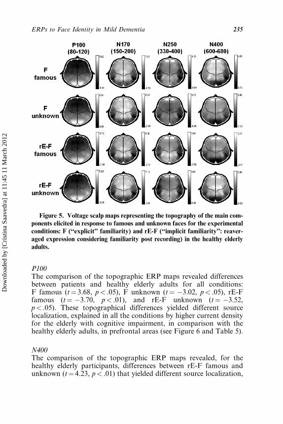
Source Localization
Figure 4 shows the voltage maps representing the topographic distri-butions of the main components for the group of elderly adults withcognitive impairment. Figure 5 shows the same topographic maps ofthe same components: P100 (occipital), N170 (temporal posterior),N250 (temporal), and the face-related N400 (occipital and temporalposterior), for the group of healthy elderly adults. Note how forthe elderly adults with cognitive impairment, in comparison withthe healthy group, voltage positive values reflecting the P100 aremore ‘‘fuzzy’’ in posterior regions and how N250 and N400 are lessdefined across the scalp.
Figure 4. Voltage scalp maps representing the topography of the main com-
ponents elicited in response to famous and unknown faces for the experimental
conditions: F (‘‘explicit’’ familiarity) and rE-F (‘‘implicit familiarity’’: reaver-
aged expression considering familiarity post recording) in the patients with
cognitive impairment.
234 C. Saavedra et al.
Dow
nloa
ded
by [
Cri
stin
a Sa
aved
ra]
at 1
1:45
11
Mar
ch 2
012
P100The comparison of the topographic ERP maps revealed differencesbetween patients and healthy elderly adults for all conditions:F famous (t¼ 3.68, p< .05), F unknown (t¼ �3.02, p< .05), rE-Ffamous (t¼ �3.70, p< .01), and rE-F unknown (t¼ �3.52,p< .05). These topographical differences yielded different sourcelocalization, explained in all the conditions by higher current densityfor the elderly with cognitive impairment, in comparison with thehealthy elderly adults, in prefrontal areas (see Figure 6 and Table 5).
N400The comparison of the topographic ERP maps revealed, for thehealthy elderly participants, differences between rE-F famous andunknown (t¼ 4.23, p< .01) that yielded different source localization,
Figure 5. Voltage scalp maps representing the topography of the main com-ponents elicited in response to famous and unknown faces for the experimental
conditions: F (‘‘explicit’’ familiarity) and rE-F (‘‘implicit familiarity’’: reaver-
aged expression considering familiarity post recording) in the healthy elderly
adults.
ERPs to Face Identity in Mild Dementia 235
Dow
nloa
ded
by [
Cri
stin
a Sa
aved
ra]
at 1
1:45
11
Mar
ch 2
012
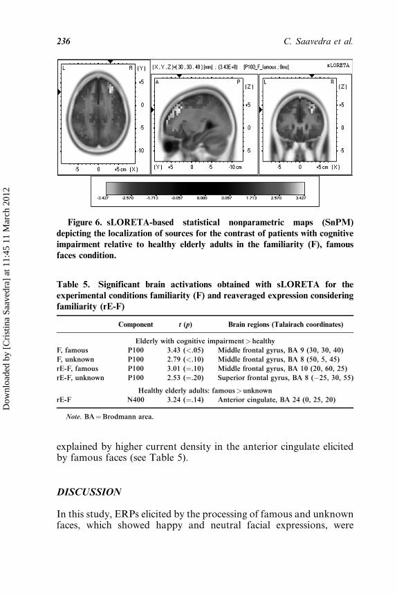
explained by higher current density in the anterior cingulate elicitedby famous faces (see Table 5).
DISCUSSION
In this study, ERPs elicited by the processing of famous and unknownfaces, which showed happy and neutral facial expressions, were
Figure 6. sLORETA-based statistical nonparametric maps (SnPM)
depicting the localization of sources for the contrast of patients with cognitive
impairment relative to healthy elderly adults in the familiarity (F), famousfaces condition.
Table 5. Significant brain activations obtained with sLORETA for the
experimental conditions familiarity (F) and reaveraged expression considering
familiarity (rE-F)
Component t (p) Brain regions (Talairach coordinates)
Elderly with cognitive impairment>healthy
F, famous P100 3.43 (<.05) Middle frontal gyrus, BA 9 (30, 30, 40)
F, unknown P100 2.79 (<.10) Middle frontal gyrus, BA 8 (50, 5, 45)
rE-F, famous P100 3.01 (¼.10) Middle frontal gyrus, BA 10 (20, 60, 25)
rE-F, unknown P100 2.53 (¼.20) Superior frontal gyrus, BA 8 (�25, 30, 55)
Healthy elderly adults: famous>unknown
rE-F N400 3.24 (¼.14) Anterior cingulate, BA 24 (0, 25, 20)
Note. BA¼Brodmann area.
236 C. Saavedra et al.
Dow
nloa
ded
by [
Cri
stin
a Sa
aved
ra]
at 1
1:45
11
Mar
ch 2
012
compared between patients with cognitive impairment and healthyelderly adults in a familiarity judgment task (to analyze explicit pro-cessing) and an expression judgment task (to analyze implicit proces-sing). According to previous studies on aging, utilizing tasks ofrecognition of famous faces (Pfutze, Sommer, & Schweinberger,2002), the latencies of the P100 and N170, related to perceptual pro-cesses, were not delayed, whereas the latencies of N250 and N400,related to face recognition, appeared to be delayed for both groups.The delay in the long-latency ERPs is also evident if the present find-ings are compared to those obtained in a group of young adults with asimilar paradigm (Saavedra et al., 2010). Our results indicate func-tional differences in the neural systems engaged by famous andunknown faces in both groups from the present experiment in the con-text of a similar behavioral performance. As we expected, the elderlyadults with cognitive impairment showed no modulation by famili-arity of N250 and N400, whereas the healthy elderly participants did.
Nonetheless, the greatest effects of cognitive decline were foundin the early ERPs related to perceptual processes. The elderly adultswith cognitive impairment showed increased amplitude of P100elicited by unknown faces and enhanced prefrontal activation eli-cited by all the experimental conditions in this latency. Further-more, although N170 showed a right lateralization in both groups(Campanella et al., 2000), it presented decreased amplitude for theelderly with cognitive impairment. Increases in amplitude of visuallyevoked potentials in AD have previously been reported (Grayson,Weiler, & Sandman, 1995); however, the reduced amplitude ofN170 observed in our study in elderly adults with cognitive impair-ment has not been previously observed, and suggests that the under-lying cognitive process (early perceptual processing) is impaired inthese patients (Maurage et al., 2007). On this subject, we speculatethat the amplitude reduction may indicate a larger jitter betweentrials (Rossion & Jacques, 2008) related to the impairment of theface-encoding process. In any event, both P100 and N170 effects inthe elderly with cognitive impairment could be related to emergingevidence from research indicating that AD and MCI can be charac-terized by pathology in the visual cortex and the disruption of a widerange of basic visual processing (Leuba & Saini, 1995; McKee et al.,2006; Newberg et al., 2003). In addition, in the earliest latency(around 100 ms), elderly adults with cognitive impairment showedan enhancement of activity located predominantly in prefrontalregions, which have previously been related to compensatory pro-cesses (Drzezga et al., 2005; Grady et al., 2003), and can explainthe fact that patients reached a similar behavioral performance to
ERPs to Face Identity in Mild Dementia 237
Dow
nloa
ded
by [
Cri
stin
a Sa
aved
ra]
at 1
1:45
11
Mar
ch 2
012
that of control subjects in our study (Han et al., 2009). Prefrontalregions participate in the distributed cortical network involved in faceprocessing (Ishai, 2008; Marinkovic, Trebon, Chauvel, & Halgren,2000), and it has been suggested that posterior areas, such as thefusiform gyrus, may lead the more anterior areas at short latencies(Marinkovic et al, 2000). Therefore, the disruption of the early per-ceptual processing, likely underlying face encoding, could be relatedto the increased prefrontal activity in our patients. In any case, newexperimental designs, such as split the patient group into high- andlow-performing participants, in which prefrontal involvement mightbe evaluated in relation to different patterns of performance, arenecessary to evaluate this possible compensatory mechanism.
With respect to ERP modulations in longer latencies related tofacial recognition, the healthy elderly adults showed an enhancedN250 elicited by famous faces in the task of implicit processingof facial identity. Unlike the results from the young adults thatshowed a modulation by familiarity on N250 related to the explicitprocessing on a similar paradigm (Saavedra et al., 2010), the healthyelderly showed a modulation related to the implicit processing offamiliarity.
Thus, this result could indicate different brain dynamics betweenyoung and older adults engaged by the explicit and implicit proces-sing of familiarity. According to the functional role assigned toN250 (Schweinberger et al., 1995, 2002), this enhancement could indi-cate differences in working-memory processes involved in access toface recognition units between young and older adults. For thehealthy elderly, the instructions in the task of explicit familiarity pro-cessing may have created temporary face memories for unknownfaces, and hence no differences between famous and unknown faceswere apparent. On the other hand, in the implicit task, famous facesseemed to generate automatic face recognition. Both results could berelated to the difficulties for older adults to ignore information that isirrelevant in a visual memory task (Gazzaley et al., 2008).
N400 showed a predominant posterior temporal distribution forboth groups in the present study, which is in accordance with resultsderived from other face recognition ERP studies using familiar stim-uli (Jemel et al., 1999; Olivares et al., 2003). As expected, only in thehealthy elderly group was this component modulated by the experi-mental tasks. Interestingly, as for N250, effects were apparent inthe implicit processing of familiarity. Precisely, in the N400 time win-dow, the healthy elderly adults showed enhanced activity in theanterior cingulate elicited by famous faces in the task of implicit pro-cessing of facial identity. The anterior cingulate has been associated
238 C. Saavedra et al.
Dow
nloa
ded
by [
Cri
stin
a Sa
aved
ra]
at 1
1:45
11
Mar
ch 2
012
with functions such as monitoring of conflict and error detection(Posner, Rothbart, Sheese, & Tang, 2007), but also as a potentiallocus of emotional awareness (Lane, Reiman, Axelrod, Holmes, &Schwartz, 1998) and is related to facial expression processing(Kesler=West et al., 2001). Therefore, our result may indicate greaterrelevance of emotional expression of famous faces, suggesting aninteraction between semantic and emotional information. Furtherresearch using experimental designs that allow splitting responsesevoked by famous=unknown and happy=neutral faces should be per-formed to evaluate this effect.
The aforementioned findings for patients with cognitive impair-ment and healthy elderly adults indicate that although in the lattergroup explicit and implicit identity processing engaged distinct neuralsystems, which could be related to activation of task-dependent ortop-down processes, the group of patients did not show this distinc-tion but rather showed disruptions on ERPs associated with earlyperceptual processing. Moreover, the early activation of prefrontalareas in the patients with cognitive impairment may indicate anattempt to compensate the processing deficits subsequent to neuro-degeneration (Grady et al., 2003) and may be related to the differ-ences observed with the healthy elderly adults in ERPs elicited byface processing. One main contribution of our study is the temporaldefinition of the recruitment of these possible compensatorymechanisms, which are engaged in early latencies related to stimulistructural encoding, indicated mainly by P100 and perhaps to someextent by N170.
To conclude, our results confirm and extend on previous studiesthat suggest that ERPs could be neurobiological markers of neurode-generative diseases concerning dementia, and provide evidence fordifferential functional neural systems supporting face identity proces-sing in patients with cognitive impairment and healthy elderly adults.Our conclusions regarding the differences between explicit andimplicit familiarity recognition, especially regarding N250 andN400, are here partially constrained; however, in future designs itwould be feasible to disentangle this issue by using a different para-digm that might exclude stimulus repetition. For future studies, inorder to evaluate the usefulness of ERPs associated with face proces-sing as predictive neurobiological markers of AD cognitive impair-ment, it would be appropriate to make longitudinal studies withlarger samples and to separate patients with AD, patients withMCI, and healthy elderly adults, in order to analyze whether theseERPs allow for the differentiation of stages of illness and to followthe course of the illness.
ERPs to Face Identity in Mild Dementia 239
Dow
nloa
ded
by [
Cri
stin
a Sa
aved
ra]
at 1
1:45
11
Mar
ch 2
012
REFERENCES
Allison, T., Puce, A., Spencer, D. D., & McCarthy, G. (1999). Electrophysiological
studies of human face perception. I: Potentials generated in occipitotemporal
cortex by face and non-face stimuli. Cerebral Cortex, 9, 415–430.
Batty, M., & Taylor, M. J. (2003). Early processing of the six basic facial emotional
expressions. Cognitive Brain Research, 17, 613–620.
Bentin, S., Allison, T., Puce, A., Perez, E., & McCarthy, G. (1996). Electrophysio-
logical studies of face perception in humans. Journal of Cognitive Neuroscience, 8,
551–565.
Bentin, S., & Deouell, L. Y. (2000). Structural encoding and identification in face
processing: ERP evidence for separate mechanisms. Cognitive Neuropsychology,
17, 35–54.
Bentin, S., Golland, Y., Flevaris, A., Robertson, L. C., & Moscovitch, M. (2006).
Processing the trees and the forest during initial stages of face perception: Elec-
trophysiological evidence. Journal of Cognitive Neuroscience, 18, 1406–1421.
Bokde, A. L., Lopez-Bayo, P., Meindl, T., Pechler, S., Born, C., Faltraco, F. et al.
(2006). Functional connectivity of the fusiform gyrus during a face-matching task
in subjects with mild cognitive impairment. Brain, 125, 1113–1124.
Botzel, K., Schulze, S., & Stodieck, S. R. G. (1995). Scalp topography and analysis
of intracranial sources of face-evoked potentials. Experimental Brain Research,
104, 135–143.
Braak, H., & Braak, E. (1991). Neuropathological stageing of Alzheimer-related
changes. Acta Neuropathologica, 82, 239–259.
Brett, M., Johnsrude, I. S., & Owen, A. M. (2002). The problem of functional loca-
lization in the human brain. Nature Reviews Neuroscience, 3, 243–249.
Brown, H., & Prescott, R. (1999). Applied mixed models in medicine. New York:
Wiley.
Bruce, V., & Young, A. (1986). Understanding face recognition. British Journal of
Psychology, 77, 305–327.
Cabeza, R. (2002). Hemispheric asymmetry reduction in older adults: The
HAROLD model. Psychology and Aging, 17, 85–100.
Campanella, S., Hanoteau, C., Depy, D., Rossion, B., Bruyer, R., Crommelinck, M.
et al. (2000). Right N170 modulation in a face discrimination task: An account
for categorical perception of familiar faces. Psychophysiology, 37, 796–806.
Chapman, R. M., Nowlis, G. H., McCrary, J. W., Chapman, J. A., Sandoval, T. C.,
Guillily, M. D. et al. (2007). Brain event-related potentials: Diagnosing
early-stage Alzheimer’s disease. Neurobiology of Aging, 28, 194–201.
Della Sala, S., Muggia, S., Spinnler, H., & Zuffi, M. (1995). Cognitive modelling of
face processing: Evidence fromAlzheimer patients.Neuropsychologia, 33, 675–687.
Dickerson, B. C., Salat, D. H., Bates, J. F., Atiya, M., Killiany, R. J., Greve, D. N.
et al. (2004). Medial temporal lobe function and structure in mild cognitive
impairment. Annals of Neurology, 56, 27–35.
Dickerson, B. C., Salat, D. H., Greve, D. N., Chua, E. F., Rand-Giovannetti, E.,
Rentz, D. M. et al. (2005). Increased hippocampal activation in mild cognitive
impairment compared to normal aging and AD. Neurology, 65, 404–411.
240 C. Saavedra et al.
Dow
nloa
ded
by [
Cri
stin
a Sa
aved
ra]
at 1
1:45
11
Mar
ch 2
012
Drzezga, A., Grimmer, T., Peller, M., Wermke, M., Siebner, H., Rauschecker, J. P.
et al. (2005). Impaired cross-modal inhibition in Alzheimer disease. PLoS Medi-
cine, 2, e288.
Dudas, R. B., Clague, F., Thompson, S. A., Graham, K. S., & Hodges, J. R. (2005).
Episodic and semantic memory in mild cognitive impairment. Neuropsychologia,
43, 1266–1276.
Eimer, M. (2000). Event-related brain potentials distinguish processing stages
involved in face perception and recognition. Clinical Neurophysiology, 111,
694–705.
Folstein, M. F., Folstein, S. E., & McHugh, P. R. (1975). ‘‘Mini-Mental State’’: A
practical method for grading the cognitive state of patients for the clinician. Jour-
nal of Psychiatric Research, 12, 189–198.
Fuchs, M., Kastner, J., Wagner, M., Hawes, S., & Ebersole, J. S. (2002). A standar-
dized boundary element method volume conductor model. Clinical Neurophysio-
logy, 113, 702–712.
Gazzaley, A., Clapp, W., Kelley, J., McEvoy, K., Knight, R. T., & D’Esposito, M.
(2008). Age-related top-down suppression deficit in the early stages of cortical
visual memory processing. Proceedings of the National Academy of Sciences of
the United States of America, 105, 13122–13126.
Gobbini, M. I., & Haxby, J. V. (2006). Neural systems for recognition of familiar
faces. Neuropsychologia, 71, 76–82.
Golob, E. J., Johnson, J. K., & Starr, A. (2001). Auditory event-related potentials
during target detection are abnormal in mild cognitive impairment. Clinical Neu-
rophysiology, 113, 151–161.
Gorno-Tempini, M. L., Price, C. J., Josephs, O., Vandenberghe, R., Cappa, S. F.,
Kapur, N. et al. (1998). The neural systems sustaining face and proper-name pro-
cessing. Brain, 121, 2103–2118.
Grady, C. L., Furney, M. L., Pietrini, P., Horwitz, B., & Rapoport, S. I. (2001).
Altered brain functional connectivity and impaired short-term memory in Alzhei-
mer’s disease. Brain, 124, 739–756.
Grady, C. L., McIntosh, A. R., Beig, S., Keigtley, M. L., Burian, H., & Black, S. E.
(2003). Evidence from functional neuroimaging of a compensatory prefrontal
network in Alzheimer’s disease. The Journal of Neuroscience, 23, 986–993.
Grayson, A. S., Weiler, E. M., & Sandman, D. E. (1995). Visual evoked potentials in
early Alzheimer’s dementia: An exploratory study. The Journal of General Psy-
chology, 122, 113–129.
Green, J., & Levey, A. I. (1999). Event-related potential changes in groups at
increased risk for Alzheimer disease. Archives of Neurology, 56, 1398–1403.
Greene, J. D. H., & Hodges, J. R. (1996a). Identification of famous faces and
famous names in early Alzheimer’s disease. Relationship to anterograde episodic
and general semantic memory. Brain, 119, 111–128.
Greene, J. D. H., & Hodges, J. R. (1996b). The fractionation of remote memory:
Evidence from a longitudinal study of dementia of Alzheimer type. Brain, 119,
129–142.
Grill-Spector, K., Henson, R., & Martin, A. (2006). Repetition and the brain: Neural
models of stimulus-specific effects. Trends in Cognitive Sciences, 10, 14–23.
ERPs to Face Identity in Mild Dementia 241
Dow
nloa
ded
by [
Cri
stin
a Sa
aved
ra]
at 1
1:45
11
Mar
ch 2
012
Han, S. D., Bangen, K. J., & Bondi, M. W. (2009). Functional magnetic resonante
imaging of compensatory neural recruitment in aging and risk for Alzheimer’s
disease: Review and recommendations. Dementia and Geriatric Cognitive Disor-
ders, 27, 1–10.
Henson, R. N. A., & Rugg, M. D. (2003). Neural response suppression, haemody-
namic repetition effects, and behavioral priming. Neuropsychologia, 41, 263–270.
Hodges, J. R., Salmon, D. P., & Butters, N. (1993). Recognition and naming of
famous faces in Alzheimer’s disease: A cognitive analysis. Neuropsychologia, 31,
775–788.
Ishai, A. (2008). Let’s face it: It’s a cortical network. Neuroimage, 40, 415–419.
Jemel, B., George, N., Olivares, E., Fiori, N., & Renault, B. (1999). Event-related
potentials to structural familiar face incongruity processing. Psychophysiology,
36, 437–452.
Katada, E., Sato, K., Ojika, K., & Ueda, R. (2004). Cognitive event-related poten-
tials: Useful clinical information in Alzheimer’s disease. Current Alzheimer
Research, 1, 63–69.
Kesler=West, M. L., Andersen, A. H., Smith, C. D., Avison, M. J., Davis, C. E.,
Kryscio, R. J. et al. (2001). Neural substrates of facial emotion processing using
fMRI. Cognitive Brain Research, 11, 213–226.
Lane, R. D., Reiman, E. M., Axelrod, B., Holmes, A., & Schwartz, G. E. (1998).
Neural correlates of levels of emotional awareness: Evidence of an interaction
between emotion and attention in anterior cingulated cortex. Journal of Cognitive
Neuroscience, 10, 525–535.
Leuba, G., & Kraftsik, R. (1994). Visual cortex in Alzheimer’s disease: Occurrence
of neuronal death and glial proliferation, and correlation with pathological hall-
marks. Neurobiology of Aging, 15, 29–43.
Leuba, G., & Saini, K. (1995). Pathology of subcortical visual centres in relation to
cortical degeneration in Alzheimer’s disease. Neuropathology and Applied Neuro-
biology, 21, 410–422.
Leveroni, C. L., Seidenberg, M., Mayer, A. R., Mead, L. A., Binder, J. R., & Rao,
S. M. (2000). Neural systems underlying the recognition of familiar and newly
learned faces. The Journal of Neuroscience, 20, 878–886.
Li, S.-C., Lindenberger, U., & Sikstrom, S. (2001). Aging cognition: From neuromo-
dulation to representation. Trends in Cognitive Sciences, 5, 479–486.
Linkenkaer-Hansen, K., Palva, J. M., Sams, M., Hietanen, J. K., Aronen, H. J., &
Ilmoniemi, R. J. (1998). Face-selective processing in human extrastriate cortex
around 120 ms after stimulus onset revealed by magneto- and electroencephalo-
graphy. Neuroscience Letters, 253, 147–150.
Macmillan, N. A., & Creelman, C. D. (2005). Detection theory: A user’s guide.
Mahwah, NJ: Lawrence Erlbaum Associates.
Marinkovic, K., Trebon, P., Chauvel, P., & Halgren, E. (2000). Localised face pro-
cessing by the human prefrontal cortex: Face-selective intracerebral potentials
and post-lesion deficits. Cognitive Neuropsychology, 17, 187–199.
Maurage, P., Philippot, P., Verbanck, P., Noel, X., Kornreich, C., Hanak, C. et al.
(2007). Is the P300 deficit in alcoholism associated with early visual impairments
(P100, N170)? An oddball paradigm. Clinical Neurophysiology, 118, 633–644.
242 C. Saavedra et al.
Dow
nloa
ded
by [
Cri
stin
a Sa
aved
ra]
at 1
1:45
11
Mar
ch 2
012
Mazziotta, J., Toga, A., Evans, A., Fox, P., Lancaster, J., Zilles, K. et al. (2001). A
probabilistic atlas and reference system for the human brain: International Con-
sortium for Brain Mapping (ICBM). Philosophical Transactions of the Royal
Society of London, Series B, Biological Sciences, 356, 1293–1322.
McKee, A. C., Au, R., Cabral, H. J., Kowall, N. W., Seshadri, S., Kubilus, C. A. et al.
(2006). Visual association pathology in preclinical Alzheimer disease. Journal of
Neuropathology and Experimental Neurology, 65, 621–630.
McKeeff, T. J., McGugin, R. W., Tong, F., & Gauthier, I. (2010). Expertise
increases the functional overlap between face and object perception. Cognition,
117, 355–360.
McKhann, G., Drachman, D., Folstein, M., Katzman, R., Price, D., & Standlan,
E. M. (1984). Clinical diagnosis Alzheimer’s disease: Report of the NINCDS-
ADRDA work group under the auspices of Deparment of Health and Human
Services task force on Alzheimer’s disease. Neurology, 34, 939–944.
Morris, J. C., Storandt, M., Miller, P., McKeel, D. W., Price, J. L., Rubin, E. H. et al.
(2001). Mild cognitive impairment represents early-stage Alzheimer disease.
Archives of Neurology, 58, 397–405.
Nasr, S., & Esteky, H. (2009). A study of N250 event-related brain potential during
face and non-face detection tasks. Journal of Vision, 9, 1–14.
Newberg, A., Cotter, A., Udeshi, M., Brinkman, F., Glosser, G., Alavi, A. et al.
(2003). Brain metabolism in the cerebellum and visual cortex correlates with neu-
ropsychological testing in patients with Alzheimer’s disease. Nuclear Medicine
Communications, 24, 785–790.
Nichols, T. E., & Holmes, P. (2001). Nonparametric permutation test for functional
neuroimaging: A primer with examples. Human Brain Mapping, 15, 1–25.
Olichney, J. M., Iragui, V. J., Salmon, D. P., Riggins, B. R., Morris, S. K., & Kutas,
M. (2006). Absent event-related potencial (ERP) word repetition effects in mild
Alzheimer’s disease. Clinical Neurophysiology, 117, 1319–1330.
Olichney, J. M., Morris, S. K., Ochoa, C., Salmon, D. P., Thal, L. J., Kutas, M. et al.
(2002). Abnormal verbal event related potentials in mild cognitive impairment
and incipient Alzheimer’s disease. Journal of Neurology, Neurosurgery and Psy-
chiatry, 73, 377–384.
Olivares, E. I., & Iglesias, J. (2008). Brain potentials and integration of external and
internal features into face representations. International Journal of Psychophysiol-
ogy, 68, 59–69.
Olivares, E. I., & Iglesias, J. (2010). Brain potentials correlates of ‘‘internal features
advantage’’ in face recognition. Biological Psychology, 83, 133–142.
Olivares, E. I., Iglesias, J., & Rodrıguez-Holguın, S. (2003). Long-latency ERPs and
recognition of facial identity. Journal of Cognitive Neuroscience, 15, 136–151.
Ostrosky-Solıs, F., Castaneda, M., Perez, M., Castillo, G., & Bobes, M. A. (1998).
Cognitive brain activity in Alzheimer’s disease: Electrophysiological response
during picture semantic categorization. Journal of the International Neuropsycho-
logical Society, 4, 415–425.
Palmer, K., Wang, H. X., Backman, L., Winblad, B., & Fratiglioni, L. (2002). Differ-
ential evolution of cognitive impairment in nondemented older persons: Results
from the Kungsholmen Project. The American Journal of Psychiatry, 159, 436–442.
ERPs to Face Identity in Mild Dementia 243
Dow
nloa
ded
by [
Cri
stin
a Sa
aved
ra]
at 1
1:45
11
Mar
ch 2
012
Pascual-Marqui, R. D. (2002). Standardized low resolution brain electromagnetic
tomography (sLORETA): Technical details. Methods and Findings in Experi-
mental and Clinical Pharmacology, 24D, 5–12.
Pascual-Marqui, R. D., Michel, C. M., & Lehmann, D. (1994). Low resolution elec-
tromagnetic tomography: A new method for localizing electrical activity in the
brain. International Journal of Psychophysiology, 18, 49–65.
Petersen, R. C., Parisi, J. E., Dickson, D. W., Johnson, K. A., Knopman, D. S.,
Boeve, B. F. et al. (2006). Neuropathologic features of amnestic mild cognitive
impairment. Archives of Neurology, 63, 665–672.
Petersen, R. C., Smith, G. E., Waring, S. C., Ivnik, R. J., Tangalos, E. G., &
Kokmen, E. (1999). Mild cognitive impairment: Clinical characterization and
outcome. Archives of Neurology, 56, 303–308.
Pfutze, E.-M., Sommer, W., & Schweinberger, S. R. (2002). Age-related slowing in
face and name recognition: Evidence from event-related potentials. Psychology
and Aging, 17, 140–160.
Polich, J., & Corey-Bloom, J. (2005). Alzheimer’s disease and P300: Review and
evaluation of task and modality. Current Alzheimer Research, 2, 515–525.
Posner, M. I., Rothbart, M. K., Sheese, B. E., & Tang, Y. (2007). The anterior cingu-
late gyrus and the mechanism of self-regulation. Cognitive, Affective & Behavioral
Neuroscience, 7, 391–395.
Pourtois, G., Schwartz, S., Seghier, M. L., Lazeyras, F., & Vuilleumier, P. (2005).
View-independent coding of face identity in frontal and temporal cortices is modu-
lated by familiarity: An event-related fMRI study. Neuroimage, 24, 1214–1224.
Reinvang, I., Espeseth, T., & Gjerstad, L. (2005). Cognitive ERPs are related to
ApoE allelic variation in mildly cognitively impaired patients. Neuroscience Let-
ters, 382, 346–351.
Rinne, J. O., Laine, M., Hiltunen, J., & Erkinjuntti, T. (2003). Semantic decision
making in early probable AD: A PET activation study. Cognitive Brain Research,
18, 89–96.
Rossion, B., & Jacques, C. (2008). Does physical interstimulus variance account for
early electrophysiological face sensitive responses in the human brain? Ten
lessons on the N170. Neuroimage, 39, 1959–1979.
Roudier, M., Marcie, P., Grancher, A.-S., Tzortzis, C., Starkstein, S., & Boller, F.
(1998). Discrimination of facial identity and of emotions in Alzheimer’s disease.
Journal of Neurological Sciences, 154, 151–158.
Saavedra, C., Iglesias, J., & Olivares, E. I. (2010). Event-related potentials elicited
by the explicit and implicit processing of familiarity in faces. Clinical EEG and
Neurosciences, 41, 24–31.
Salmon, D. P., & Bondi, M. W. (1999). Neuropsychology of Alzheimer’s disease. In
R. D. Terry, R. Katzman, K. L. Bick & S. S. Sisodia (Eds.), Alzheimer disease (pp.
39–56). Philadelphia: Lippincott Williams & Wilkins.
Schwartz, T. J., Federmeier, K. D., Van Petten, C., Salmon, D. P., & Kutas, M.
(2003). Electrophysiological analysis of context effects in Alzheimer’s disease.
Neuropsychology, 17, 187–201.
Schweinberger, S. R., & Burton, M. (2003). Covert recognition and the neural
system for face processing. Cortex, 39, 9–30.
244 C. Saavedra et al.
Dow
nloa
ded
by [
Cri
stin
a Sa
aved
ra]
at 1
1:45
11
Mar
ch 2
012
Schweinberger, S. R., Huddy, V., & Burton, M. (2004). N250r: A face-selective brain
response to stimulus repetitions. NeuroReport, 15, 1501–1505.
Schweinberger, S. R., Pfutze, E. M., & Sommer, W. (1995). Repetition priming
and associative priming of face recognition: Evidence from event-related poten-
tials. Journal of Experimental Psychology. Learning, Memory, and Cognition, 21,
722–736.
Schweinberger, S. R., Pickering, E. C., Jentzsch, I., Burton, M., & Kaufmann, J. M.
(2002). Event-related brain potential evidence for a response of inferior temporal
cortex to familiar face repetitions. Cognitive Brain Research, 14, 398–409.
Shibata, T., Nishijo, H., Tamura, R., Miyamoto, K., Eifuku, S., Endo, S. et al.
(2002). Generators of visual evoked potentials for faces and eyes in the human
brain as determined by dipole localization. Brain Topography, 15, 51–63.
Sperling, R. A., Bates, J. F., Chua, E. F., Cocchiarella, A. J., Rentz, D. M., Rosen,
B. R. et al. (2003). fMRI studies of associative encoding in young and elderly
controls and mild Alzheimer’s disease. Journal of Neurology, Neurosurgery and
Psychiatry, 74, 44–50.
Stern, Y., Moeller, J. R., Anderson, K. E., Luber, B., Zubin, N. R., DiMauro, A. A.
et al. (2000). Different brain networks mediate task performance in normal aging
and AD. Neurology, 55, 1291–1297.
Thierry, G., Martin, C. D., Downing, P., & Pegna, A. J. (2007). Controlling for inter-
stimulus perceptual variance abolishes N170 face selectivity. Nature Neuroscience,
10, 505–511.
Thompson, S. A., Graham, K. S., Patterson, K., Sahakian, B. J., & Hodges, J. R.
(2002). Is knowledge of famous people disproportionately impaired in patients
with early and questionable Alzheimer’s disease? Neuropsychology, 16, 344–358.
Van Deursen, J. A., Vuurman, E. F. P. M., Smits, L. L., Verhey, F. R. J., & Riedel, W.
J. (2009). Response speed, contingent negative variation and P300 in Alzheimer’s
disease and MCI. Brain and Cognition, 69, 592–599.
Van Rhijn, S. J., Glosser, G., De Vries, J. J., Clark, C. M., Newberg, A. B., & Alavi,
A. (2004). Visual processing impairments and decrements in regional brain
activity in Alzheimer’s disease. Journal of Clinical and Experimental Neuro-
psychology, 26, 11–23.
Zion-Golumbic, E., & Bentin, S. (2007). Dissociated neural mechanisms for face
detection and configural encoding: Evidence from N170 and induced gamma-
band oscillation effects. Cerebral Cortex, 17, 1741–1749.
ERPs to Face Identity in Mild Dementia 245
Dow
nloa
ded
by [
Cri
stin
a Sa
aved
ra]
at 1
1:45
11
Mar
ch 2
012
Related Documents