Electroencephalography and clinical Neurophysiology 103 (1997) 257-267 Event-related potentials during memorization of spatial locations in the auditory and visual modalities Francisco Barcelda**, Manuel Martin-Loechesb, Francisco J. Rubiab ‘Department of Psychobiology, Faculty of Psychology, Complutense University, Somosaguas 28223, Madrid, Spain bBrain Mapping Unit, Pluridisciplinar Institute, Complutense University, Paseo Juan XXIII, I, 28040 Madrid, Spain Accepted for publication: 30 January 1997 Abstract Event-related potential (ERP) studies of working memory have used delayed Sl-S2 match-to-sample tasks in which Sl is held in memory for comparison with S2. ERP negativities in the S l-S2 interval have been interpreted either in terms of working memory operations, or in terms of general preparatory motor processing. Two experiments (N = 20 each) were carried out to explore the nature of ERP negativities in a visuospatial memory task and in an auditory spatial memory task, respectively. In the experimental condition, subjects had to memorize the location of Sl (Sl-memorize) so as to respond whether S2 appeared in the same spatial location (S2-memorize). In the control condition, subjects were requested to ignore Sl (Sl-passive), and to respond whether S2 matched or not a target location predetermined at the beginning of the trial block (S2-pressing). Results support the two main conclusions of Martin-Leeches et al. (Electroenceph. clin. Neurophysiol., 1994,91: 363-373). Firstly, that the encoding into memory of spatial location is associated with an ERP negative wave over the brain areas putatively associated with the processing of sensory information (i.e. right parieto-occipital for the visual task; fronto-central and left temporal areas for the auditory task). Secondly, the P300 does not seem to be an important ERP feature related to spatial location encoding and retaining into memory. Despite the distinct scalp distribution of these memory-related, modality-specific ERP negativities, they also showed a considerable degree of temporal synchronicity across modalities. 0 1997 Elsevier Science Ireland Ltd. Keywords: Event-related potentials; Spatial memory; Spatial perception; Working memory 1. Introduction Research into the brain mechanisms of storage and main- tenance of information in the human brain has benefited from the technique of scalp-recorded event-related poten- tials (ERPs). ERPs are useful towards the making of infer- ences about the timing and anatomical localization of these memory processes. Most ERP studies addressing these issues have employed variations of the delayed match-to- sample paradigm. In this task the subject is presented with an initial stimulus (Sl) that must be committed to memory for a delayed comparison with a subsequent stimulus (S2), following which the subject decides whether the two stimuli matched or mismatched (Lang et al., 1992; Ruchkin et al., 1992, 1995; Begleiter et al., 1993; Starr et al., 1996). Apart from its simple experimental logic, the match-to-sample * Corresponding author. Tel.: +34 1 3943085; fax: +34 1 3943189. paradigm has the advantage of being easily implemented with subhuman primates during single unit recording (Fus- ter, 1995; Ungerleider, 1995), which improves the grounds for inference on the neurophysiology of ERP results in humans. The commonest finding across ERP studies has been a negative slow wave which differs in topography depending on the modality and the kind of information to be memor- ized. Thus, memorization of verbal material is generally associated with a sustained negative shift which is maximal over fronto-central and temporal regions for auditory sti- muli (Patterson et al., 1991; Lang et al., 1992; Ruchkin et al., 1992; Starr et al., 1996), but is centred more posteriorly for visual material (Patterson et al., 1991; Ruchkin et al., 1992, 1995; R&r&i et al., 1995). In our laboratory a negative ERP wave was reported dur- ing the Sl-S2 interval of a match-to-sample task in which the subject had to memorize the spatial location of small white vertical bars displayed on a computer screen (Martin- 0013-4694/97/$17.00 0 1997 Elsevier Science Ireland Ltd. All rights reserved PII SO921-884X(97)96610-8 EEG 96610

Welcome message from author
This document is posted to help you gain knowledge. Please leave a comment to let me know what you think about it! Share it to your friends and learn new things together.
Transcript
Electroencephalography and clinical Neurophysiology 103 (1997) 257-267
Event-related potentials during memorization of spatial locations in the auditory and visual modalities
Francisco Barcelda**, Manuel Martin-Loechesb, Francisco J. Rubiab
‘Department of Psychobiology, Faculty of Psychology, Complutense University, Somosaguas 28223, Madrid, Spain bBrain Mapping Unit, Pluridisciplinar Institute, Complutense University, Paseo Juan XXIII, I, 28040 Madrid, Spain
Accepted for publication: 30 January 1997
Abstract
Event-related potential (ERP) studies of working memory have used delayed Sl-S2 match-to-sample tasks in which Sl is held in memory for comparison with S2. ERP negativities in the S l-S2 interval have been interpreted either in terms of working memory operations, or in terms of general preparatory motor processing. Two experiments (N = 20 each) were carried out to explore the nature of ERP negativities in a visuospatial memory task and in an auditory spatial memory task, respectively. In the experimental condition, subjects had to memorize the location of Sl (Sl-memorize) so as to respond whether S2 appeared in the same spatial location (S2-memorize). In the control condition, subjects were requested to ignore Sl (Sl-passive), and to respond whether S2 matched or not a target location predetermined at the beginning of the trial block (S2-pressing). Results support the two main conclusions of Martin-Leeches et al. (Electroenceph. clin. Neurophysiol., 1994,91: 363-373). Firstly, that the encoding into memory of spatial location is associated with an ERP negative wave over the brain areas putatively associated with the processing of sensory information (i.e. right parieto-occipital for the visual task; fronto-central and left temporal areas for the auditory task). Secondly, the P300 does not seem to be an important ERP feature related to spatial location encoding and retaining into memory. Despite the distinct scalp distribution of these memory-related, modality-specific ERP negativities, they also showed a considerable degree of temporal synchronicity across modalities. 0 1997 Elsevier Science Ireland Ltd.
Keywords: Event-related potentials; Spatial memory; Spatial perception; Working memory
1. Introduction
Research into the brain mechanisms of storage and main- tenance of information in the human brain has benefited from the technique of scalp-recorded event-related poten- tials (ERPs). ERPs are useful towards the making of infer- ences about the timing and anatomical localization of these memory processes. Most ERP studies addressing these issues have employed variations of the delayed match-to- sample paradigm. In this task the subject is presented with an initial stimulus (Sl) that must be committed to memory for a delayed comparison with a subsequent stimulus (S2), following which the subject decides whether the two stimuli matched or mismatched (Lang et al., 1992; Ruchkin et al., 1992, 1995; Begleiter et al., 1993; Starr et al., 1996). Apart from its simple experimental logic, the match-to-sample
* Corresponding author. Tel.: +34 1 3943085; fax: +34 1 3943189.
paradigm has the advantage of being easily implemented with subhuman primates during single unit recording (Fus- ter, 1995; Ungerleider, 1995), which improves the grounds for inference on the neurophysiology of ERP results in humans.
The commonest finding across ERP studies has been a negative slow wave which differs in topography depending on the modality and the kind of information to be memor- ized. Thus, memorization of verbal material is generally associated with a sustained negative shift which is maximal over fronto-central and temporal regions for auditory sti- muli (Patterson et al., 1991; Lang et al., 1992; Ruchkin et al., 1992; Starr et al., 1996), but is centred more posteriorly for visual material (Patterson et al., 1991; Ruchkin et al., 1992, 1995; R&r&i et al., 1995).
In our laboratory a negative ERP wave was reported dur- ing the Sl-S2 interval of a match-to-sample task in which the subject had to memorize the spatial location of small white vertical bars displayed on a computer screen (Martin-
0013-4694/97/$17.00 0 1997 Elsevier Science Ireland Ltd. All rights reserved PII SO921-884X(97)96610-8 EEG 96610
258 F. Barceld et al. / Electroencephalography and clinical Neurophysiology 103 (1997) 257-267
Leeches et al., 1994). Such a simple protocol was intended to keep stimulus features and task parameters to a minimum. ERPs recorded during the Sl-S2 interval were characterized by a long-lasting negativity distributed bilaterally over occi- pital and temporal areas, and was attributed to processes specifically related to the encoding of spatial information into memory. Plausible origins for this negativity coincide with sites of maximal ERP changes related to processes of visual feature extraction (e.g., 01 and Or; Mangun and Hill- yard, 1991).
More commonly, the P300 wave has been associated with memory encoding processes (Fabiani et al., 1990; Donchin and Fabiani, 1991). However, ERP results from Martin- Leeches et al. (1994) prompted their suggestion that ‘the P300 is not an important ERP feature related to spatial location encoding and retaining in memory’ (p. 370). Their data suggested that the memory encoding of spatial information seemed to recruit activity from neuronal net- works in the primary and secondary visual cortices, rather than from third-order association and polysensory cortices (Fuster, 1995; Rosier et al., 1995). It might be argued, though, that the long-lasting negativity found by Martin- Leeches et al. (1994) was partly confounded with motor preparation, particularly as their passive task did not provide an adequate control for the Sl-S2 match-to-sample memory task condition, and response preparation processes are known to develop in Sl-S2 paradigms (McCallum, 1988; Birbaumer et al., 1990; Starr et al., 1996).
The present study improves and extends results from our previous study in three different ways. Firstly, ERPs are recorded during a spatial memory SlS2 match-to-sample task in which subjects have to memorize the spatial location of Sl in order to respond whether or not S2 appears in the same spatial location. Comparisons are made against an S l- S2 passive-pressing control task, where subjects have to ignore Sl, and respond whether or not S2 matched a target location predetermined at the beginning of the task. The passive-pressing task is assumed to provide an adequate control for the motor preparation processes in the spatial memory Sl-S2 task. (That is, in both the memory task and the control task, motor responses have to be issued only after S2 onset). The main purpose of this design is to dissociate ERP negativities due to motor preparation from those due to memorization of simple spatial information. Secondly, spatial memory is assessed employing an open- field distribution of stimulus sources. This was intended to extend ERP results during memorization of spatial location to a more ecologically valid situation, and to establish a theoretical and empirical bridge with spatial attention stu- dies in animals (i.e. Fuster, 1995). Thirdly, both visual and auditory modalities are examined in two otherwise metho- dologically identical studies. Our main hypothesis is that ERPs elicited during memorization of spatial location, devoid of motor preparation processes, will be maximal over primary cortical areas involved in the analysis of sim- ple stimulus features (Damasio, 1990; Fuster, 1995) rather
than being circumscribed to modality-independent, poly- sensory cortical areas.
2. Methods and materials
2. I. Subjects
Forty right-handed subjects (20 females) took part in the study, with ages between 18 and 30 years (mean 22 years, SD 3 years). All subjects had normal or corrected to normal vision, and none informed of having any audiological pro- blems. Subjects were recruited through advertisement in the university campus magazine and were paid for their colla- boration. Twenty subjects performed the visual task and another 20 subjects performed the auditory task (10 females in each).
2.2. Stimuli
Five loudspeakers and 5 red light-emitting diodes (LEDs) mounted on a purpose-made semicircular campimeter 70 cm in diameter served as stimulation devices. Auditory sti- muli consisted of tone pips (1.5 kHz, 90 dB SPL at head position, 100 ms in duration, 10 ms rise-fall time). Tone pips were delivered through a set of 5 hi-fi loudspeakers (Audi- max). One of the speakers was placed 40 cm away and in front of the adjustable chin-rest of the campimeter (0” azi- muth). The other 4 speakers were placed at equal distances from the chin-rest at azimuths 30”, 60” and -3O”, -60”. The chin-rest was adjusted for each subject so that the eyes were in the same horizontal plane (0” elevation) as the loudspea- kers. Flashes 2.5 cd/m* in intensity and 100 ms in duration were delivered through 5 LEDs, each mounted in the middle of one loudspeaker. Throughout the task subjects were requested to fix their eyes on a yellow spot visible 5 cm below the central LED. The sequence of presentation was programmed and controlled with the Gentask module of the STIM package (NeuroScan Inc.) installed in a Dell 486 microcomputer. Both latencies and response errors were stored on disk.
2.3. Tasks and procedure
A match-to-sample Sl-S2 paradigm was employed both for the memory task and for the control task. In the memory task, the spatial location of the first stimulus had to be memorized in order to respond whether the second stimulus appeared or not in the same location as the first stimulus. In place-match (target) trials, the second stimulus (SZmemor- ize) appeared in the same spatial location as the first one (Sl-memorize). In place-mismatch (non-target) trials, S2 did not appear in the same location as Sl. Subjects were instructed to respond (YES/NO) at the onset of S2 using a two-button PC mouse. The interstimulus interval was 2 s. Intertrial intervals varied randomly between 2-3 s (mean
F. BarceM et al. / Electroencephalography and clinical Neurophysiology 103 (1997) 257-267 259
2.5 s). There were the same number of place-match as place- mismatch trials, with equal numbers of both types across spatial locations. There were 240 stimulus pairings in the memory task, which were randomized and distributed in 3 blocks of 80 trials each. A 5 min rest period was allowed between blocks. Each subject received a different random sequence of trials.
The control task consisted of the same general Sl-S2 protocol as the memory task. However, subjects were requested to respond YES or NO depending on whether S2 matched or not a target location determined at the begin- ning of the trial block (S2-pressing condition). Subjects were instructed just to ignore Sl (Sl-passive condition). Therefore, motor responses had to be issued only after the onset of S2 both in the control task and in the memory task. Half the total number of 240 trials were place-match trials. Place-mismatch trials were equally distributed across the remaining 4 locations. Target locations for the Sl-S2 pas- sive-press task changed within and across subjects to yield even numbers experiment-wise.
The order of presentation of the memory and control tasks was counterbalanced within groups, and the hand used by the subject to respond was swapped midway through the session. A practice session of 10 trials was carried out prior to each task, which served to acquaint subjects with the tasks, and make sure that instructions had been under- stood. After the experimental session, subjects responded to a debriefing questionnaire in which they described their performance, identified the easiest and most difficult aspects of the tasks, and explored the type of cognitive strategy employed. Subjects were instructed to relax their facial muscles, fixate the central fixation point, not to move their eyes and avoid blinking as much as possible. The testing room was a dimly lit, sound-attenuating chamber. Subjects were instructed that performance accuracy was more impor- tant than response speed. Feedback was provided in the practice session, but not during the experimental session.
2.4. ERP recording
Electroencephalographic (EEG) and electrooculographic (EOG) data were recorded using an electrode cap (Electro- Cap International) with tin electrodes. The electrode sites were F7, Fz, F8, T7, Cz, T8, P3, Pz, P4, P7, P8, P03, P04, 01, and 02 of the revised 10120 International System (American Electroencephalographic Society, 1991). The electrooculogram (EOG) monitored eye movements using an oblique derivation from the external canthus of the right eye to the upper canthus of the left eye. Impedances were always kept below 3 kfl. The EEG and EOG were recorded from DC, with a high frequency cut-off of 30 Hz (12 dB/octave slope). The EEG and EOG channels were con- tinuously digitized at a sampling rate of 200 Hz for the duration of each task block. These data were stored on disk together with stimulus and response markers for off- line analysis.
2.5. Data analysis
Averaging epochs of 2000 ms with a prestimulus interval of 500 ms were extracted from the continuously digitized EEG. Eye movement artifacts were removed following the method described by Semlitsch et al. (1986). Epochs that were contaminated by artifact or by remaining EOG activity after correction were rejected prior to averaging. Trials with incorrect responses were also excluded from the averages.
One separate average was computed for each of the 4 task conditions, namely, S l-memorize, S2-memorize, S l-pas- sive, and SZ-pressing. Averaged ERPs were aligned using the average amplitude in the time window 500-300 ms prior to stimulus onset as a baseline. This period was adopted as a baseline in order to assess the nature of stimulus preceding negativities which developed prior to both Sl and S2, and which were affected by the manipulation of experimental variables. An analysis of absolute mean amplitudes in the 500-300 ms time window prior to stimulus onset showed that this period was not affected by the manipulation of experimental variables, and therefore, could be considered an adequate baseline for later comparisons.
The mean amplitudes of the major waveforms were cal- culated in the time windows -200 ms (200- 100 ms prior to onset), -100 ms (100-O ms prior to onset), PlOO (50-100 ms), NlOO (lOO- 170 ms), P200 (170-220 ms), N200 (220- 270 ms), P3a (270-450 ms), P3b (450-700 ms), Slow Wave 1 (SWl, 700-1000 ms), Slow Wave 2 (SW2, 1000-1300 ms). These latency windows were chosen to be as similar as possible to the ones used in the study by Martin-Leeches et al. (1994).
Mean ERP amplitudes in these time windows were sub- mitted as dependent variables to the BMDP/4V package for analysis of variance (ANOVAs) for repeated measurements. ANOVAs included the factors: Task (Sl-memorize, S2- memorize, S 1 -passive, S2-pressing), Hemisphere (left and right), and Electrode site (frontal, temporal, parietal, par- ieto-temporal, parieto-occipital and occipital). Two ortho- gonal contrasts were defined for the test of simple effects. The Ml vs. Pl contrast explored differences between Sl- memorize and Sl-passive task conditions, in order to address the main hypothesis of this study. The Sl vs. S2 contrast compared an aggregate of the S 1 -memorize and the S l-passive task conditions against an aggregate of SZ-mem- orize and S2-pressing task conditions, and served as a con- trol for motor preparatory and response processes related to S2-onset. A similar statistical design was separately applied to ERP data from the 3 mid-line electrodes. Performance was assessed in terms of reaction times and percentage of errors.
The Greenhouse-Geisser method of adjusting the degrees of freedom was used where appropriate (Vasey and Thayer, 1987). When multiple comparisons were required, separate error estimates were obtained for each contrast (Keselman and Keselman, 1988), and the Bonferroni procedure was used to determine the significance level using a family-
260 F. BarceM et al. / Electroencephalography and clinical Neurophysiology 103 (1997) 257-267
Table 1
Mean reaction times (RT) and percent of false-alarm rate (and standard
deviations) for the memory and pressing tasks in the visual and auditory
task groups
Visual task group Auditory task group
SZmemotize S2-pressing SZmemorize S2-pressing
RT (ms) 0.601 (0.1) 0.533 (0.1) 0.609 (0.1) 0.548 (0.1)
% errors 0.9 (0.01) 3.8 (0.07) 3.2 (0.06) 3.5 (0.07)
wise error rate of 0.05. Only comparisons that were signifi- cant are reported, unless otherwise stated.
3. Results
3.1. Performance data
Comparisons of false-alarm rates between groups and across task conditions revealed that response errors were rare, with a maximum of 3.8% errors in the visual SZpress- ing task condition. The auditory task group showed signifi- cantly more errors than the visual task group in the memory task condition only (t(19) = 3.41; P < 0.003; 3.2% and 0.9% errors, respectively). This can be taken as an indica- tion of a lower accuracy of spatial localization in the audi- tory as compared with the visual modality (Perrot et al., 1993). There were no differences in error rates between the memory and control tasks within each group.
The analysis of reaction times to S2 revealed significantly larger response latencies in the S2-memorize than in the S2- pressing task condition in both the visual (t(19) = 2.146; P < 0.045) and auditory (t(19) = 2.962; P < 0.008) task groups (see Table 1). On average, it took about 53 ms longer to issue a response in the memory task than in the pressing task. This difference is consistent with an interpretation in terms of controlled versus automatic visual search pro- cesses, and can be attributed to response and decision pro- cesses associated with S2, rather than with the perceptual
Table 2
processing of Sl (Posner and Boies, 1971; Shiffrin and Schneider, 1977).
The responses to the post-hoc questionnaire indicated that all subjects adopted a purely spatial strategy during the Sl- S2 match-to-sample tasks. Indeed, none of the subjects reported having used a verbal strategy (i.e. verbal rehearsal of positions such as ‘extreme right’, ‘right’, ‘centre’, ‘left’, ‘extreme-left’) for retaining the spatial location of the sti- mulus.
3.2. ERP data
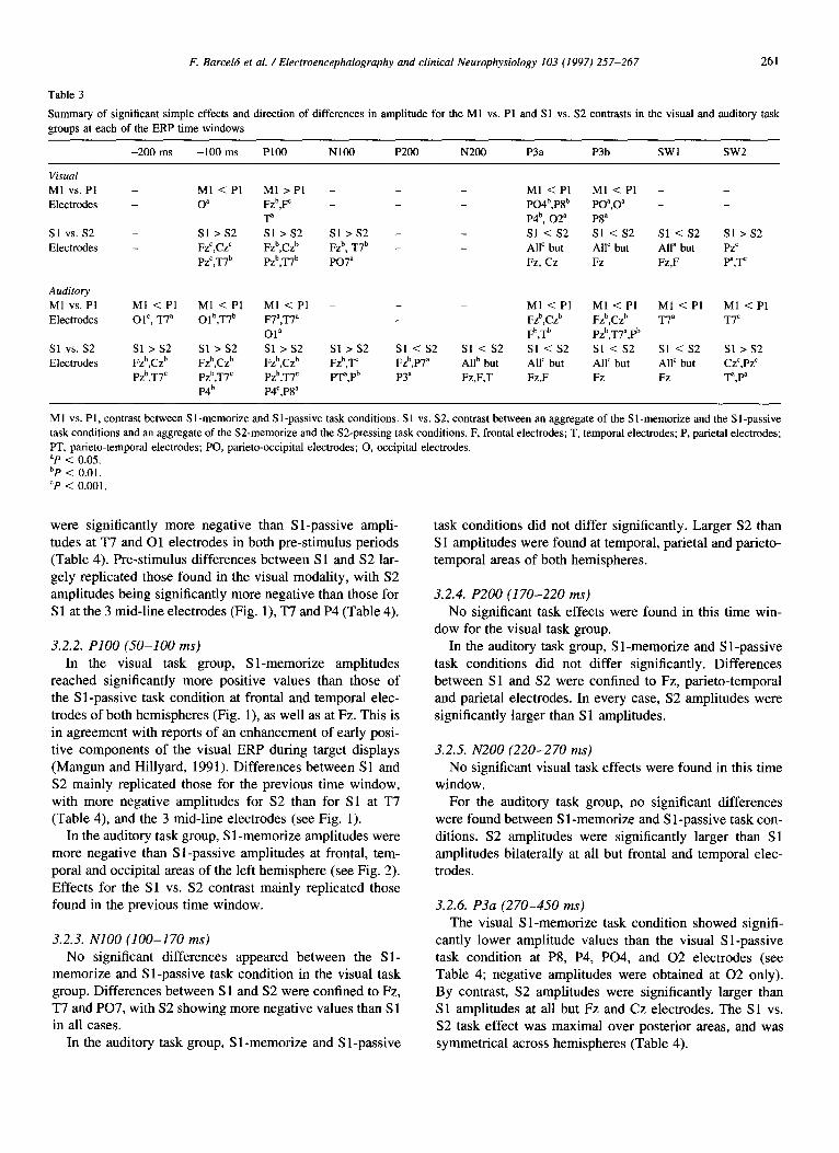
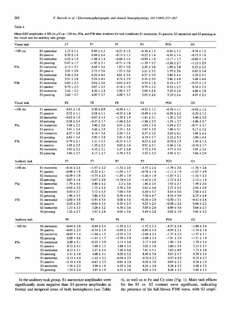
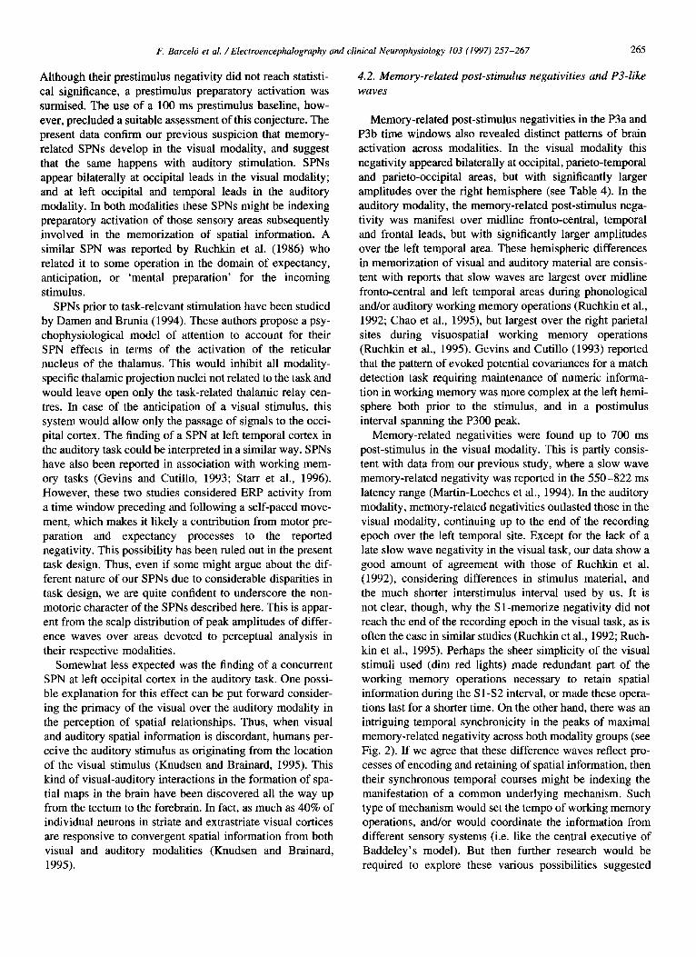
Results of the main ANOVA are shown in Table 2, and results of tests of simple effects are shown in Table 3. If both the Task x Electrode and Task x Electrode x Hemisphere interactions reached significance, only simple effects for the third-order interaction are presented in Table 3. Mean ERP amplitudes for time windows -100 ms, P3a, and P3b are presented in Table 4. Fig. 1 offers a detailed comparison between grand mean ERP averages for each of the 4 task conditions (S 1 -memorize, S l-passive, S2-memorize, and S2-pressing) at Fz, Cz, and Pz. These mid-line averages summarize the overall pattern of ERP responding in both visual and auditory modalities, and show that the largest ERP differences were found between Sl- and S2- task conditions. Fig. 2 shows memory-related difference waves which result from subtracting the Sl-passive from the Sl- memorize ERP averages in the visual and auditory task groups, respectively.
3.2.1. Pre-stimulus periods For the visual task group, significant differences between
the Sl-memorize and Sl-passive conditions appeared only during the -100 ms time window at both occipital electrodes (Table 4). Significant differences between Sl- and S2- task conditions were at all 3 mid-line electrodes (Fig. l), and at both temporal electrodes for the -100 ms time window (Table 4). In all cases, pre-S2 amplitudes were significantly more negative than pre-S 1 amplitudes.
For the auditory task group, Sl-memorize amplitudes
Main ANOVA results for the visual and auditory task groups at each of the different ERP time windows
df -200 ms -100 ms PI00 NlOO P200 N200 P3a P3b SW1 SW2
Visual T
TxH
TxE
TxExH
Auditory T
TxH
TxE
TxExH
357 - _ _ _ _ 16.12’ 29.21’ 4.98a -
357 - _ _ 5.15a - _ _
15,285 - 2&ra 3.078 _ 7.92’ 13.21’ 2.82a 3.96b
15,285 - 2.39a 3.22” 2.83’ - _ 6.25b - _ _
3,57 - _ _ 9.96b 31.52’ 43.75= 17.15c -
3,57 - _ _ _ _ _
15,285 8.62’ 10.42’ 11.15E 10.21C 13.97c 21.19’ 17.25’ 10.80’ 14.16b
15,285 9.83’ 13.83’ 16.26’ _ 7.16’ _ 3.20” - _
T, Task; H, Hemisphere: E, Electrode. “P < 0.05.
F. Barcelb et al. / Electroencephalography and clinical Neurophysiology 103 (1997) 257-267 261
Table 3
Summary of significant simple effects and direction of differences in amplitude for the Ml vs. Pl and Sl vs. S2 contrasts in tbe visual and auditory task
groups at each of the ERP time windows
-200 ms -100 ms Ploo NlOO P200 N200 P3a P3b SW1 SW2
Visual
Ml vs. Pl -
Electrodes _
Sl vs. s2 _
Electrodes -
Ml < Pl Ml>Pl - _ Ml < Pl Ml <Pl - _
0” Fzb,F _ _ PO4b,P8b PO”,O” - _
T P4b, 02” P8”
Sl > s2 Sl >s2 Sl>S2 - Sl <s2 Sl < s2 Sl < s2 Sl >s2 Fzc,CzC Fzb,Czb Fzb, T7b - AllC but All’ but Alla but Pz’
PzC,T7b Pz~,T~~ PO7” Fz, Cz Fz Fz,F pa,T
Auditory
Ml vs. Pl
Electrodes
Sl vs. s2
Electrodes
Ml < Pl Ml < Pl Ml<Pl - _ Ml < Pl Ml < Pl Ml < Pl Ml < Pl Ol’, T7a Olb,T7b F7=,T7” _ _ Fzb,Czb Fzb,Czb T7a T7C
Ola Fb,Tb pzb,T7a,pb Sl >s2 Sl > s2 Sl >s2 Sl >s2 Sl < s2 Sl <s2 Sl <s2 Sl <s2 Sl < s2 Sl >s2 Fzb,Czb Fzb,Czb Fzb,Czb Fzb,T Fz~,P~~ Allb but AllC but All’ but AllC but CzC,PzC Pz~,T~~ Pzb,T7C Pzb,T7C PT”,Pb P3” Fz,F,T Fz,F Fz Fz T”,p
P4b P4C,P8a
M 1 vs. Pl, contrast between S l-memorize and S l-passive task conditions. S 1 vs. S2, contrast between an aggregate of the S l-memorize and the S 1 -passive
task conditions and an aggregate of the S2-memorize and the S2-pressing task conditions. F, frontal electrodes; T, temporal electrodes; P, parietal electrodes;
PT. parieto-temporal electrodes; PO, parieto-occipital electrodes; 0, occipital electrodes. “P 2 0.05. bP < 0.01. cP < 0.001.
were significantly more negative than Sl-passive ampli- tudes at T7 and 01 electrodes in both pre-stimulus periods (Table 4). Pre-stimulus differences between Sl and S2 lar- gely replicated those found in the visual modality, with S2 amplitudes being significantly more negative than those for Sl at the 3 mid-line electrodes (Fig. l), T7 and P4 (Table 4).
3.2.2. PI00 (SO-100 ins) In the visual task group, Sl-memorize amplitudes
reached significantly more positive values than those of the Sl-passive task condition at frontal and temporal elec- trodes of both hemispheres (Fig. l), as well as at Fz. This is in agreement with reports of an enhancement of early posi- tive components of the visual ERP during target displays (Mangun and Hillyard, 1991). Differences between Sl and S2 mainly replicated those for the previous time window, with more negative amplitudes for S2 than for Sl at T7 (Table 4), and the 3 mid-line electrodes (see Fig. 1).
In the auditory task group, S 1 -memorize amplitudes were more negative than Sl-passive amplitudes at frontal, tem- poral and occipital areas of the left hemisphere (see Fig. 2). Effects for the Sl vs. S2 contrast mainly replicated those found in the previous time window.
3.2.3. NlOO (100-170 ms) No significant differences appeared between the Sl-
memorize and Sl-passive task condition in the visual task group. Differences between Sl and S2 were confined to Fz, T7 and P07, with S2 showing more negative values than S 1 in all cases.
In the auditory task group, Sl-memorize and Sl-passive
task conditions did not differ significantly. Larger S2 than S 1 amplitudes were found at temporal, parietal and parieto- temporal areas of both hemispheres.
3.2.4. P200 (170-220 ms) No significant task effects were found in this time win-
dow for the visual task group. In the auditory task group, Sl-memorize and Sl-passive
task conditions did not differ significantly. Differences between Sl and S2 were confined to Fz, parieto-temporal and parietal electrodes. In every case, S2 amplitudes were significantly larger than Sl amplitudes.
3.2.5. N200 (220-270 ms) No significant visual task effects were found in this time
window. For the auditory task group, no significant differences
were found between S 1 -memorize and S 1 -passive task con- ditions. S2 amplitudes were significantly larger than Sl amplitudes bilaterally at all but frontal and temporal elec- trodes.
3.2.6. P3a (270-450 ms) The visual Sl-memorize task condition showed signifi-
cantly lower amplitude values than the visual Sl-passive task condition at P8, P4, P04, and 02 electrodes (see Table 4; negative amplitudes were obtained at 02 only). By contrast, S2 amplitudes were significantly larger than Sl amplitudes at all but Fz and Cz electrodes. The Sl vs. S2 task effect was maximal over posterior areas, and was symmetrical across hemispheres (Table 4).
262 F. Barceld et al. / Electroencephalography and clinical Neurophysiology 103 (1997) 257-267
Table 4
Mean ERP amplitudes & SD (in pV) at -100 ms, P3a, and P3b time windows for task conditions Sl-memorize, Sl-passive, SZmemorize and S2-pressing in the visual and the auditory task groups
Visual task
-100 ms
P3a
P3b
S 1 -memorize S 1 -passive S2-memorize SZpressing S l-memorize S 1 -passive SZmemorize SZpressing S l-memorize S l-passive SZmemorize S2-pressing
F7 Tl PI P3 PO3 01
1.13 f 1.1 0.60 f 1.3 -0.27 f 1.2 -0.30 * 1.3 -0.54 f 1.2 -0.76 f 1.2 0.70 f 1.4 0.46 f 1.4 -0.13 + 1.3 -0.22 f 1.4 -0.34 f 1.4 -0.13 f 1.6 0.22 + 1.4 -1.08 f. 1.4 -0.68 f 1.4 -0.99 * 1.9 -1.17 f 1.7 -0.84 ? 1.4 0.47 f 1.7 -1.07 IL 2.1 -0.71 f 1.8 -1.29 f 2.7 -1.26 * 2.7 -1.12 * 2.5 4.13 f 2.7 4.69 f 3.4 1.82 f 3.0 2.45 * 3.0 1.90 f 3.8 0.33 f 3.2 3.51 + 2.8 3.72 f 3.0 1.53 + 3.0 2.21 f 3.3 1.79 + 3.6 0.65 + 3.4 5.46 f 2.4 6.22 f 4.4 4.61 f 3.4 6.57 f 3.9 5.80 f 4.4 4.20 f 4.1 5.51 + 2.8 5.94 f 4.4 4.74 f 3.9 6.34 f 5.0 5.86 f 4.8 4.04 f 4.6 0.69 f 2.3 0.94 f 2.6 XI.01 f 1.9 0.59 + 1.8 0.19 * 1.7 4.23 f 1.5 0.75 & 2.3 0.87 + 2.5 0.18 f 1.9 0.79 * 2.2 0.52 + 2.3 0.34 f 2.5 2.42 f 2.2 4.20 f 2.4 3.98 + 2.7 5.69 f 2.8 5.25 + 2.8 4.06 f 2.8 2.60 f 1.7 4.03 f 2.7 4.00 f 2.2 5.65 f 2.6 5.18 f 2.4 4.07 f 2.2
Visual task F8 T8 P8 P4 PO4 02
-100 ms
P3a
P3b
S l-memorize Sl-passive S2-memorize SZ-pressing S 1 -memorize S 1 -passive S2-memorize SZpressing S 1 -memorize S 1 -passive S2-memorize S2-pressing
Auditory task
0.63 + 1.0 0.30 f 0.9 ’ 0.32 f 1.1 0.28 f 1.1
-0.03 f 1.9 -0.67 k 1.4 0.28 + 2.4 -0.37 * 1.7 4.08 + 2.2 3.80 f 2.6 3.91 f 3.4 3.46 f 2.8 6.57 f 2.8 6.16 * 3.4 6.83 f 3.4 5.92 f 3.8 0.79 f 2.1 1.31 f 2.3 1.05 + 2.5 1.35 f 2.2 3.92 f 2.2 4.33 f 2.1 3.96 5 2.7 4.17 + 2.7
F7 T7
-0.50 f 1.1 ‘4.35 f 1.4 -1.29 + 1.9 -1.08 f 2.4
0.91 + 2.6 2.33 f 3.4 5.54 f 3.1 5.81 + 5.6
-0.31 + 1.3 0.65 f 1.8 4.17 f 2.4 4.78 f 3.5
w
4.52 f 1.2 -0.76 + 1.1 -0.49 + 1.4 -0.59 * 1.2 -1.81 f 2.1 -1.20 f 2.0 -1.88 f 2.5 -1.29 f 2.7
1.84 f 3.0 0.39 f 2.7 2.87 f 3.9 1.40 + 3.1 6.37 + 3.5 5.54 * 4.1 6.19 k 5.7 5.32 + 5.3 0.53 f 2.0 -0.70 * 1.5 0.91 * 2.1 0.36 f 1.6 5.72 f 3.9 4.77 + 3.4 5.52 f 3.3 4.92 + 3.1
P3 PO3
-0.92 + 1.2 -0.29 +_ 1.2 -1.06 f 2.0 -1.06 f 2.7 -0.71 + 3.1
0.17 f 3.2 3.94 f 4.1 3.90 f 4.1
-1.04 f 1.5 -0.10 f 1.7
3.85 + 2.4 3.96 +_ 2.9
01
-100 ms
P3a
P3b
S 1 -memorize Sl-passive SZmemorize SZpressing S l-memorize S l-passive SZ-memorize S2-pressing S 1 -memorize S 1 -passive S2-memorize SZpressing
Auditory task
-0.34 + 2.4 -0.08 + 1.9 -0.59 f 1.9
0.07 f 1.8 -1.79 f 4.4 -0.65 f 3.5
0.30 + 3.7 1.06 + 3.3
-2.60 f 3.8 -2.05 f 2.9
1.33 f 3.3 1.25 + 2.7
F8
-1.07 f 2.2 -1.52 f 2.6 -0.22 * 2.1 -1.03 f 1.7 -3.73 f 2.3 -1.85 f 1.9 -3.67 f 2.6 -1.74 * 2.1
0.40 f 4.2 2.3 rl: 3.6 1.73 f 4.3 2.76 + 3.0 3.12 f 4.3 7.68 f 3.9 3.86 f 5.2 8.59 + 4.8
-1.91 * 3.4 0.08 + 3.1 -0.64 * 3.1 0.59 f 2.3
3.28 + 3.2 6.38 f 3.6 3.57 f 2.8 6.04 f 3.6
T8 P8
-1.33 + 2.4 -0.78 + 1.9 -1.46 f 1.9 -1.40 f 1.8
1.86 f 3.5 2.62 f 3.0 6.40 f 3.7 7.50 * 4.7
-0.36 f 2.9 0.25 f 2.4 5.05 + 2.6 5.49 f 2.8
P4
-1.79 f 2.6 -1.11 f 1.9 -1.87 f 2.1 -1.72 + 2.2
1.93 f 3.7 2.27 f 3.4 8.14 IL 5.0 8.54 f 5.9
XI.50 f 3.1 -0.08 + 2.4
6.69 f 3.6 6.82 f 3.9
PO4
-1.76 f 2.6 -1.07 f 1.9 -1.34 + 2.2 -1.12 + 1.8
1.49 f 3.0 2.04 f 2.8 7.58 f 4.3 8.25 f 4.8
a.41 f 2.6 0.06 + 2.2 5.66 + 2.3 5.81 f 3.2
02
-100 ms
P3a
P3b
S 1 -memorize S 1 -passive SZmemorize SZpressing S l-memorize Sl-passive SZmemorize SZpressing S l-memorize S l-passive SZ-memorize SZpressing
-0.04 f 2.6 -0.05 f 2.3 -0.02 f 1.4
0.09 f 1.6 -2.09 XL 5.1 -0.72 f 4.1
0.13 * 3.1 1.11 f 3.4
-3.13 + 4.8 -2.18 f 3.8
1.36 + 2.2 1.24 + 2.4
-0.94 + 2.4 -1.50 f 2.3 -1.52 f 2.3 -0.74 f 1.9 -0.95 * 1.9 -0.85 + 1.9 -1.64 f 1.5 -2.23 + 2.2 -2.86 + 2.4 -1.41 f 1.7 -2.29 f 1.9 -2.88 f 2.4 -0.22 f 3.9 2.13 f 3.6 2.17 f 4.0
1 .oo I! 3.3 2.68 f 3.3 3.02 f 3.8 2.57 XL 2.4 7.46 f 4.6 7.61 f 5.1 4.00 + 3.1 8.20 f 5.2 8.40 f 5.8
-1.41 f 3.3 -0.50 + 2.9 -0.70 f 3.2 -0.67 f 2.5 -0.01 f 2.4 0.25 f 2.6
3.98 f 1.8 6.25 f 2.6 6.24 f 3.8 3.87 f 1.9 6.31 f 3.0 6.85 f 3.5
-1.57 ? 2.4 -1.60 f 2.4 -0.95 If: 2.1 -1.19 f 1.8 -1.77 f 2.3 -1.37 f 2.1 -1.81 f 2.0 -1.31 + 1.8
1.90 f 3.6 1.79 f 3.4 2.60 f 2.9 2.33 + 2.7 7.63 f 4.9 7.73 f 5.0 8.67 f 5.3 8.39 f 5.4
XI.57 f 2.9 a.32 f 2.7 0.05 f 2.2 0.16 f 1.9 6.26 + 3.5 5.48 + 3.2 6.18 f 3.2 5.85 f 3.3
In the auditory task group, S 1 -memorize amplitudes were significantly more negative than S l-passive amplitudes at frontal and temporal areas of both hemispheres (see Table
4), as well as at Fz and Cz sites (Fig. 1). Main task effects for the Sl vs. S2 contrast were significant, indicating the presence of the full-blown P300 wave, with S2 ampli-

F. Barcelo’ et al. / Electroencephalography and clinical Neurophysiology 103 (1997) 257-267 263
Visual Task
- SZ-memorize
Auditory Task
Fig. 1. Grand mean ERP averages at Fz, Cz, and Pz for each of the 4 task conditions (Sl-memorize, Sl-passive, SZmemorize, and S2-press). Upper panel: visual task group. Lower panel: auditory task group.
tudes being larger than Sl amplitudes bilaterally and at all except both frontal and mid-line Fz electrodes (Table 4, Fig. 1).
3.2.7. P3b (450-700 ms) Significantly lower amplitudes were found during the
visual S 1 -memorize as compared with the visual S 1 -passive task condition, which were confined to P8, as well as par- ieto-occipital and occipital electrodes of both hemispheres (see Table 4). Significant Sl vs. S2 effects appeared at all electrodes of both hemispheres (except for Fz, see Fig. l), with S2 amplitudes being larger than Sl amplitudes.
In the auditory task group, significant differences between S 1 -memorize and S 1 -passive task conditions were found at all mid-line electrodes (see Fig. l), as well
as at temporal and parietal areas, with a significant trend towards a left temporal asymmetry (see Table 4 and Fig. 2). Significant S 1 vs. S2 effects were obtained in all electrodes except Fz, with larger S2 than S 1 ERP amplitudes (Tables 3 and 4).
3.2.8. SW1 (700-900 ms) In the visual task group, only significant differences
between Sl and S2 were found in all brain areas except frontal ones. ERP differences were distributed symmetri- cally across hemispheres.
For the auditory task group, Sl-memorize amplitudes were more negative than Sl-passive amplitudes only at the T7 site (see Fig. 2). S2 elicited significantly larger amplitudes than Sl at all electrode sites.
::
P4
Fig. 2. Difference waveforms which result from subtracting the Sl-passive average from the Sl-memorize task conditions (Ml vs. Pl contrast) for the visual
and auditory task groups.

264 F. Barceld et al. / Electroencephalography and clinical lieurophysiology 103 (1997) 257-267
3.2.9. SW2 (900-1300 ms) A second-order interaction revealed significantly larger
amplitudes for the Sl than for the S2 task condition in the visual task group. These differences were distributed sym- metrically at parietal and temporal areas of both hemi- spheres.
In the auditory task group, S l-memorize showed signifi- cantly more negative amplitudes than Sl-passive at T7 (see Fig. 2). There was a change in the pattern of results from the previous time windows, so that S 1 showed
larger amplitudes than S2 at Cz and Pz mid-line electrodes, as well as at temporal and parietal areas of both hemi- spheres.
In summary, memory-related negativities revealed by the Ml vs. Pl contrast were confined to the -100 ms, P3a, and P3b time windows in the visual modality, and involved mainly occipital, parieto-occipital and parieto-temporal areas of the right hemisphere. In the auditory modality, memory-related negativities started earlier and lasted longer in the recording epoch, and were focused at fronto-central midline electrodes, as well as at frontal, temporal and occi- pital areas of the left hemisphere. The topographical distri- bution of effects for the Sl vs. S2 contrast did not conform to this pattern of effects. It is worthwhile noting the syn- chronicity of the peak amplitude of the memory-related negativities displayed in Fig. 2, which appeared almost simultaneously in both modalities around 450 ms post-sti- mulus. The scalp distribution of this peak clearly differs across modalities.
4. Discussion
There is a good deal of agreement between the present results and those reported by Martin-Leeches et al. (1994). This is in spite of considerable modifications in the type of stimuli used, the open-field mode of presentation, the use of a Sl-S2 passive-pressing control task, and even an alto- gether different EEG system of data acquisition and analy- sis. All in all, the two main conclusions from the previous study still hold. Namely, that the encoding into memory of spatial location is associated with a negative wave over the brain areas putatively associated with the sensory proces- sing of stimulus features. Also, that the P300 does not seem to be an important ERP feature related to spatial location encoding and retaining in memory. An improved task design allowed us to qualify and complement these two earlier conclusions in several respects. Firstly, memory- related negativities were experimentally dissociated from motor preparation negativities in the Sl-S2 interval, and both show distinct patterns of brain activation. Secondly, the brain topography of memory-related negativities dif- fered across modalities. Thus, a post-stimulus memory- related negative wave was centred over right parieto-occi- pital, parietal and occipital areas in the visual task, but it focused over midline fronto-central and left temporal areas
in the auditory task. Thirdly, these memory-related negativ- ities peaked synchronously in both the visual and auditory tasks at around 450 ms post-stimulus. Finally, the presence of stimulus-preceding negativities (SPN) suggests that sen- sory preparation processes manifest themselves over the same cortical areas activated during storage and mainte- nance of spatial information in working memory.
Match-to-sample tasks have been successfully employed in ERP studies of human memory (Lang et al., 1992; Ruch- kin et al., 1992; Ruchkin et al., 1995). However, slow ERP waves obtained during the S l-S2 interval of this kind of task also reflect motor preparation processes prior to S2 respond- ing (McCallum, 1988; Birbaumer et al., 1990; Rama et al., 1995; Starr et al., 1996). In this study, we assumed that both S l-memorize and S l-passive task conditions are equally affected by motor preparation processes, and therefore, their difference wave should reflect processes related to the encoding of spatial information into memory devoid of motor preparation processes (Fig. 2). Basically, these difference waves can be taken to reflect memory-related negativities at brain areas devoted to the perceptual analysis of stimulus features, and hence, can be regarded as modal- ity-specific. Such a topography is clearly in contrast with the modality-independent midline fronto-central maximum of CNV waves (McCallum, 1988) (see Fig. 1). Using a similar task design, Ruchkin et al. (1995) reported significant effects of information load upon the amplitudes and topo- graphy of slow waves during the S l-S2 interval, thus ruling out motor preparation as the sole explanation (see also Ruchkin et al., 1992).
The memory-related negativities revealed by Ml vs. Pl difference waves in Fig. 2 might reflect two distinct types of processes. On the one hand, stimulus-preceding negativities (SPN) may reflect anticipatory activation of primary sen- sory areas. On the other hand, poststimulus negativities dur- ing the 2 s memorization interval may reflect processes related to the storage and maintenance of spatial informa- tion. Both the prestimulus and poststimulus parts of these memory-related negativities show scalp distributions which are largely dependent on stimulus modality. Other working memory studies have analyzed ERP activity preceding a stimulus to which an open motor response is required (Gevins and Cutillo, 1993; Starr et al., 1996), thus leaving open the possibility of a contribution of motor preparation processes to their reported SPNs. By contrast, our Ml vs. PI difference waves reflect ERP activity around the first stimu- lus in the delayed Sl-S2 match-to-sample task, which demanded no motor response. The following discussion refers to ERP waves preceding and succeeding S 1.
4.1. Memory-related stimulus-preceding negativities (SPN)
The earliest significant negativity effects reported by Martin-Leeches et al. (1994) appeared 150 ms after stimulus onset, but there was a gradual development of this negativ- ity starting several milliseconds before stimulus onset.
F. Barcelo’ et al. / Electroencephalography and clinical Neurophysiology 103 (1997) 257-267 265
Although their prestimulus negativity did not reach statisti- cal significance, a prestimulus preparatory activation was surmised. The use of a 100 ms prestimulus baseline, how- ever, precluded a suitable assessment of this conjecture. The present data confirm our previous suspicion that memory- related SPNs develop in the visual modality, and suggest that the same happens with auditory stimulation. SPNs appear bilaterally at occipital leads in the visual modality; and at left occipital and temporal leads in the auditory modality. In both modalities these SPNs might be indexing preparatory activation of those sensory areas subsequently involved in the memorization of spatial information. A similar SPN was reported by Ruchkin et al. (1986) who related it to some operation in the domain of expectancy, anticipation, or ‘mental preparation’ for the incoming stimulus.
SPNs prior to task-relevant stimulation have been studied by Damen and Brunia (1994). These authors propose a psy- chophysiological model of attention to account for their SPN effects in terms of the activation of the reticular nucleus of the thalamus. This would inhibit all modality- specific thalamic projection nuclei not related to the task and would leave open only the task-related thalamic relay cen- tres. In case of the anticipation of a visual stimulus, this system would allow only the passage of signals to the occi- pital cortex. The finding of a SPN at left temporal cortex in the auditory task could be interpreted in a similar way. SPNs have also been reported in association with working mem- ory tasks (Gevins and Cutillo, 1993; Starr et al., 1996). However, these two studies considered ERP activity from a time window preceding and following a self-paced move- ment, which makes it likely a contribution from motor pre- paration and expectancy processes to the reported negativity. This possibility has been ruled out in the present task design. Thus, even if some might argue about the dif- ferent nature of our SPNs due to considerable disparities in task design, we are quite confident to underscore the non- motoric character of the SPNs described here. This is appar- ent from the scalp distribution of peak amplitudes of differ- ence waves over areas devoted to perceptual analysis in their respective modalities.
Somewhat less expected was the finding of a concurrent SPN at left occipital cortex in the auditory task. One possi- ble explanation for this effect can be put forward consider- ing the primacy of the visual over the auditory modality in the perception of spatial relationships. Thus, when visual and auditory spatial information is discordant, humans per- ceive the auditory stimulus as originating from the location of the visual stimulus (Knudsen and Brainard, 1995). This kind of visual-auditory interactions in the formation of spa- tial maps in the brain have been discovered all the way up from the tectum to the forebrain. In fact, as much as 40% of individual neurons in striate and extrastriate visual cortices are responsive to convergent spatial information from both visual and auditory modalities (Knudsen and Brainard, 1995).
4.2, Memory-related post-stimulus negativities and P3-like
waves
Memory-related post-stimulus negativities in the P3a and P3b time windows also revealed distinct patterns of brain activation across modalities. In the visual modality this negativity appeared bilaterally at occipital, parieto-temporal and parieto-occipital areas, but with significantly larger amplitudes over the right hemisphere (see Table 4). In the auditory modality, the memory-related post-stimulus nega- tivity was manifest over midline fronto-central, temporal and frontal leads, but with significantly larger amplitudes over the left temporal area. These hemispheric differences in memorization of visual and auditory material are consis- tent with reports that slow waves are largest over midline fronto-central and left temporal areas during phonological and/or auditory working memory operations (Ruchkin et al., 1992; Chao et al., 1995), but largest over the right parietal sites during visuospatial working memory operations (Ruchkin et al., 1995). Gevins and Cutillo (1993) reported that the pattern of evoked potential covariances for a match detection task requiring maintenance of numeric informa- tion in working memory was more complex at the left hemi- sphere both prior to the stimulus, and in a postimulus interval spanning the P300 peak.
Memory-related negativities were found up to 700 ms post-stimulus in the visual modality. This is partly consis- tent with data from our previous study, where a slow wave memory-related negativity was reported in the 550-822 ms latency range (Martin-Leeches et al., 1994). In the auditory modality, memory-related negativities outlasted those in the visual modality, continuing up to the end of the recording epoch over the left temporal site. Except for the lack of a late slow wave negativity in the visual task, our data show a good amount of agreement with those of Ruchkin et al. (1992), considering differences in stimulus material, and the much shorter interstimulus interval used by us. It is not clear, though, why the Sl-memorize negativity did not reach the end of the recording epoch in the visual task, as is often the case in similar studies (Ruchkin et al., 1992; Ruch- kin et al., 1995). Perhaps the sheer simplicity of the visual stimuli used (dim red lights) made redundant part of the working memory operations necessary to retain spatial information during the Sl-S2 interval, or made these opera- tions last for a shorter time. On the other hand, there was an intriguing temporal synchronicity in the peaks of maximal memory-related negativity across both modality groups (see Fig. 2). If we agree that these difference waves reflect pro- cesses of encoding and retaining of spatial information, then their synchronous temporal courses might be indexing the manifestation of a common underlying mechanism. Such type of mechanism would set the tempo of working memory operations, and/or would coordinate the information from different sensory systems (i.e. like the central executive of Baddeley’s model). But then further research would be required to explore these various possibilities suggested
266 F. Barcelb et al. / Electroencephalography and clinical Neurophysiology 103 (1997) 257-267
by the observed differences and similarities between mem- ory-related negativities across modalities.
In any case, neither P3a nor P3b waves seemed to be associated with the storing and retention of spatial informa- tion in working memory. This is consistent with reports from studies using match-to-sample paradigms that memory- related negative slow waves develop immediately after the P300 wave (Ruchkin et al., 1992). It also supports the view that working memory is largely independent of classical limbic memory networks associated with P300 elicitation (Chao et al., 1995; Knight and Grabowecky, 1995). The timing and scalp distribution of the peak amplitudes of memory-related negative waves are consistent with current neurophysiological models of working memory based on specialized brain systems for short-term, data-driven storage of sensory information (Fuster, 1995). These models con- sider that the memory of things cannot simply be allocated to one polymodal area just because that memory is usually anchored in several sense modalities (Damasio, 1990; Fuster, 1995). These data are also consistent with current psychological models of working memory in terms of sepa- rate systems for phonological and visuospatial information (Baddeley, 1992). It seems reasonable that such a simple stimulus feature as spatial location is encoded in cortical areas devoted to primary perceptual processing.
In agreement with our earlier study too, a P300 wave was elicited during the Sl-memorize task condition, which showed a significantly lower amplitude and a different brain distribution than the one elicited by the S2-memorize task condition. On the contrary, the Sl-passive task condi- tion also elicited a P3 wave which mimicked the amplitude and topography of the S 1 -memorize P300 wave. Although a P3a wave in the Sl-passive task condition might appear as counterintuitive, it should be noted that Sl forewarns the incoming target stimulus S2 and can hardly be ‘ignored’. These P3a waves may reflect the process of keeping count of the order of presentation in the S 1 -S2 paradigm, which of course was necessary both in the Sl-passive and in the Sl- memory task conditions alike. In this respect, the S l-passive task condition can be regarded as an adequate control for the Sl-memorize condition, and it can be assumed that both task conditions elicited the same amount of perceptual pro- cessing regarding S 1. Both the S 1 -passive and S 1 -memorize P300 waves displayed a maximum over fronto-central areas, and therefore, could be best described as P3a waves in clear contrast with the P3b waves elicited by the S2- memorize task condition (Knight and Grabowecky, 1995). In the visual task, P3a waves were more conspicuous over frontal areas and decreased in amplitude posteriorly (cf. Martin-Leeches et al., 1994). In the auditory task, P3a waves were more evenly distributed, with a maximum over centro-parietal areas.
The scalp distribution of post-stimulus memory-related negativities does not show a common frontal locus as would be expected from reports about the implication of the dorsal prefrontal cortex in working memory operations
(Smith et al., 1995; Ungerleider, 1995). One possible expla- nation for the lack of prefrontal effects could be that the encoding of spatial information during our simple match-to- sample task required the activation of neural networks in the lower levels of the hierarchy of perceptual processing, with minimum requirements made upon prefrontal working memory systems of higher levels of the hierarchy (Fuster, 1995). Most studies reporting prefrontal activation during working memory operations have typically employed more complex stimulus material and/or made higher demands during task performance. Also compatible with this expla- nation is that prefrontal cortex was activated to a similar extent both during the S l-passive and S l-memorize task conditions; hence the lack of frontal ERP differences between both task conditions.
5. Conclusions
The presence of distinct scalp topographies across mod- alities for the memory-related negativities reported here could be taken as an indication that different configurations of neural sources were active during storage and retention in working memory of visuo-spatial and auditory spatial infor- mation. In the main, the present data show a good deal of agreement with our previous study in that input to memory takes place in the same cortical areas devoted to feature extraction of simple stimulus characteristics (Martin- Leeches et al., 1994). This lends support to models of work- ing memory based on specialized brain systems for short- term, data-driven storage of visuo-spatial and audio-spatial information (Damasio, 1990; Baddeley, 1992; Fuster, 1995). Consequently, our ERP evidence is consistent with single cell studies of working memory which suggest that the storage and retaining of simple visuo-spatial and audio- spatial information recruit activity from neuronal networks in the primary and secondary visual cortices, rather than from third-order association and polysensory cortices. This reinforces the view that sensory processing and mem- ory representation probably share largely the same cortical substrates (Fuster, 1995).
Acknowledgements
This work was partly supported by the Spanish Ministry of Education and Science DGICYT PB94/0322, and by the Fundacidn Ramon Areces. An earlier version of this paper was presented at the 8th World Congress of the International Organization of Psychophysiology, Tampere, Finland, June 25-30, 1996.
References
American Electroencephalographic Society. Guidelines for standard elec-
trode position nomenclature. J. Clin. Neurophysiol., 1991, 3: 38-42.
F. Barceld et al. / Electroencephalography and clinical Neurophysiology 103 (1997) 257-267 261
Baddeley, A. Working memory: the interface between memory and cogni-
tion. J. Cognit. Neurosci., 1992, 4: 281-288.
Begleiter, H., Porjesz, B. and Wang, W. A neurophysiologic correlate of
visual short-term memory in humans. Electroenceph. clin. Neurophy-
siol., 1993, 87: 46-53.
Birbaumer, N., Elbert, T., Canavan, A.G.M. and Rockstroh, B. Slow
potentials of the cerebral cortex and behavior. Physiol. Rev., 1990,
70: 1-41.
Chao, L.L., Nielsen-Bohlman, L. and Knight, R.T. Auditory event-related
potentials dissociate early and late memory processes. Electroenceph.
clin. Neurophysiol., 1995, 96: 157-168.
Damasio, A.R. Synchronous activation in multiple cortical regions: a
mechanism for recall. Semin. Neurosci., 1990, 2: 287-296.
Damen, E.J.P. and Brunia, C.H.M. Is a stimulus conveying task-relevant
information a sufficient condition to elicit a stimulus-preceding nega-
tivity? Psychophysiology, 1994, 3 1: 129- 139.
Donchin, E. and Fabiani, M. The use of event-related brain potentials in
the study of memory: is P300 a measure of event distinctiveness? In:
J.R. Jennings and M. Coles (Eds.), Handbook of Cognitive Psychophy-
siology. Wiley, New York, 1991, pp. 471-510.
Fabiani, M., Karis, D. and Donchin, E. Effects of mnemonic strategy
manipulation in a Von Restorff paradigm. Electroenceph. clin. Neuro-
physiol., 1990, 75: 22-35.
Fuster, J.M. Memory in the Cerebral Cortex. An Empirical Approach to
Neural Networks in the Human and Nonhuman Primate. MIT Press,
Cambridge, MA, 1995. Gevins, A. and Cutillo, B. Spatiotemporal dynamics of component pro-
cesses in human working memory. Electroenceph. clin. Neurophysiol.,
1993, 87: 128-143.
Keselman, H.J. and Keselman, J.C. Comparing repeated measures means
in factorial designs. Psychophysiology, 1988, 5: 612-618.
Knight, R.T. and Grabowecky, M. Escape from linear time: prefrontal
cortex and conscious experience. In: M.S. Gazzaniga (Ed.), The Cogni-
tive Neurosciences. MIT Press, Cambridge, MA, 1995, pp. 1357-
1371.
Knudsen, E.I. and Brainard, M.S. Creating a unified representation of
visual and auditory space in the brain. Annu. Rev. Neurosci., 1995,
18: 19-43.
Lang, W., Starr, A., Lang, V., Lindinger, G. and Deecke, L. Cortical DC
potential shifts accompanying auditory and visual short-term memory.
Electroenceph. clin. Neurophysiol., 1992, 82: 285-295.
Mangun, G.R. and Hillyard, S.A. Modulations of sensory-evoked brain
potentials indicate changes in perceptual processing during visual-spa-
tial priming. J. Exp. Psychol. Hum. Percept. Perform., 1991, 17: 1057-
1074. Martin-Leeches, M. Gomez-Jarabo, G. and Rubia, F.J. Human brain poten-
tials of spatial location encoding into memory. Electroenceph. clin.
Neurophysiol., 1994, 91: 363-373.
McCallum, W.C. Potentials related to expectancy, preparation and motor
activity. In: T.W. Picton (Ed.), EEG Handbook (revised series, Vol. 3).
Elsevier, Amsterdam, 1988, pp. 427-534.
Patterson, J.V., Pratt, H. and Starr, A. Event-related potential correlates of
the serial position effect in short-term memory. Electroenceph. clin.
Neurophysiol., 1991, 78: 424-437.
Perrot, D.R., Costantino, B. and Cisneros, J. Auditory and visual localiza-
tion performance in a sequential discrimination task. J. Acoust. Sot.
Am., 1993, 93: 2134-2138.
Posner, M.I. and Boies, S.J. Components of attention. Psychol. Rev., 1971,
78: 391-408.
Rama, P., Carlson, S., Kekoni, J. and HBmiilXinen, H. A spatial oculomotor
memory-task performance produces a task-related slow shift in human
electroencephalography. Electroenceph. clin. Neurophysiol., 1995, 94:
371-380.
Riisler F., Heil, M. and Henninghausen, E. Distinct cortical activation
patterns during long-term memory retrieval of verbal, spatial, and
color information. J. Cognit. Neurosci., 1995, 7: 51-65.
Ruchkin, D.S., Sutton, S., Mahaffey, D. and Glasser, J. Terminal CNV in
the absence of motor response. Electroenceph. clin. Neurophysiol.,
1986.63: 445-463.
Ruchkin, D.S., Johnson, R., Grafman, J., Canoune, H. and Ritter, W.
Distinctions and similarities among working memory processes: an
event-related potential study. Cognit. Brain Res., 1992, 1: 53-66.
Ruchkin, D.S., Canoune, H.L., Johnson, R. and Ritter, W. Working mem-
ory and preparation elicit different patterns of slow wave event-related
brain potentials. Psychophysiology, 1995, 32: 399-410.
Semlitsch, H.V., Anderer, P., Schuster, P. and Presslich, 0. A solution for reliable and valid reduction of ocular artifacts applied to the P300 ERP.
Psychophysiology, 1986, 23: 695-703.
Shiffrin, R.M. and Schneider, W. Controlled and automatic human infor-
mation processing: II. Perceptual learning, automatic attending, and a
general theory. Psychol. Rev., 1977, 84: 127-190.
Smith, E.E., Jonides, J., Koeppe, R.A., Awh, E., Schumacher, E.H. and
Minoshima, S. Spatial versus object working memory: PET investiga- tions. J. Cognit. Neurosci., 1995, 7: 337-356.
Starr, A., Dong, C.J. and Michalewski, H.J. Brain potentials before and
during memory scanning. Electroenceph. clin. Neurophysiol., 1996, 99: 28-37.
Ungerleider, L.G. Functional brain imaging studies of cortical mechanisms
for memory. Science, 1995, 270: 769-775.
Vasey, M.W. and Thayer, J.F. The continuing problem of false positives in repeated measures ANOVA in psychophysiology: a multivariate solu-
tion. Psychophysiology, 1987, 24: 479-486.
Related Documents