Event-Related Desynchronization/ Synchronization- Based Brain-Computer Interface towards Volitional Cursor Control in a 2D Center-Out Paradigm Dandan Huang 1* , Kai Qian 1 , Simon Oxenham 2 , Ding-Yu Fei 1 , Ou Bai 1 1 Dept. of Biomedical Engineering Virginia Commonwealth University Richmond, US * E-mail: [email protected] 2 Dept. of Psychology University of the West of England Bristol, UK E-mail: [email protected] Abstract—To achieve reliable two-dimensional cursor control by noninvasive EEG-based brain-computer interface (BCI), users are typically required to receive long-term training to learn effective regulation of their brain rhythmic activities, and to maintain sustained attention during the operation. We proposed a two-dimensional BCI using event-related desynchronization and event-related synchronization associated with human natural behavior so that users no longer need long-term training or high mental loads to maintain concentration. In this study, we intended to investigate the performance of the proposed BCI associated with either physical movement or motor imagery with an online center-out cursor control paradigm. Genetic algorithm (GA)-based mahalanobis linear distance (MLD) classifier and decision tree classifier (DTC) were used in feature selection and classification and a model adaptation method was employed for better decoding of human movement intention from EEG activity. The results demonstrated effective control accuracy for this four-class classification: as high as 77.1% during online control with physical movement and 57.3% with motor imagery. This suggests that based on this preliminary testing, two-dimensional BCI control can be achieved without long- term training. Keywords: Brain Computer Interfaces; Movement Intention; Event-Related Desynchronization (ERD); Event-Related Synchronization (ERS); Two-Dimensional Cursor Control I. INTRODUCTION A brain-computer interface (BCI) can decode an individual’s intention by bypassing peripheral nerves and muscles to achieve direct control of external devices [1]. It provides a new communication pathway for people with severe motor disabilities. Performance of the BCI system is highly dependent on the signal-to-noise ratio (SNR) of the brain signal. Invasive and semi-invasive BCIs may provide better SNR than non-invasive BCI. Invasive BCI detects signals by implanting electrodes into the motor cortex using local field potential recorded from individual or small populations of motor neurons [2]. Semi-invasive BCI use subdural electrodes (ECoG) [3-8]. Widespread clinical use of invasive or semi-invasive BCIs in human beings is impeded however by the high risk of surgical procedures and the problems in achieving robust and stable long-term recordings [9]. Electroencephalography (EEG) which detects signals from the scalp is the most commonly used non-invasive method [10-15]. The critical challenge of EEG based BCI is maintaining robust performance with large variation of EEG signals. Studies in recent years show that EEG-based BCI has great potential in achieving two-dimensional or multi- dimensional cursor control [11, 12]. However, these systems usually require long-term training in regulating brain signals, and the performance in long-term use is often not robust [2]. The discovery of event-related desynchronization (ERD) or power decrease and event-related synchronization (ERS) or power increase has resulted in the development of new brain-computer interfaces [16]. Recent studies developed a paradigm to achieve two-dimensional cursor control using the ERD/ERS method to directly decode movement intention without long-term training [13, 14]. Human limbs are controlled by contralateral brain hemispheres, this has been confirmed by many studies [17-19]. During physical and motor imagery of right and left hand movements, beta band brain activation (15-30 Hz) ERD occurs predominantly over the contralateral left and right motor areas. The post movement ERS associated with ceasing to move can also be found over the contralateral motor areas. Therefore, reliably decoding the movement intention of the right and left hand, which are associated with different spatiotemporal patterns of ERD and ERS may potentially provide four reliable features for a two-dimensional control; e.g. left-hand ERD to command movement to the left, left-hand ERS to command movement up, right-hand ERD to command movement to the right, and right-hand ERS to command movement down. In this study we further investigated the performance of the BCI in multiple blocks, this was done online using a two dimensional (2D) center-out cursor control paradigm with a model adaptation method for better decoding of human movement intention from EEG activity.

Welcome message from author
This document is posted to help you gain knowledge. Please leave a comment to let me know what you think about it! Share it to your friends and learn new things together.
Transcript

Event-Related Desynchronization/ Synchronization-
Based Brain-Computer Interface towards Volitional
Cursor Control in a 2D Center-Out Paradigm
Dandan Huang1*
, Kai Qian1, Simon Oxenham
2, Ding-Yu Fei
1, Ou Bai
1
1Dept. of Biomedical Engineering
Virginia Commonwealth University
Richmond, US
*E-mail: [email protected]
2Dept. of Psychology
University of the West of England
Bristol, UK
E-mail: [email protected]
Abstract—To achieve reliable two-dimensional cursor control
by noninvasive EEG-based brain-computer interface (BCI),
users are typically required to receive long-term training to
learn effective regulation of their brain rhythmic activities, and
to maintain sustained attention during the operation. We
proposed a two-dimensional BCI using event-related
desynchronization and event-related synchronization
associated with human natural behavior so that users no longer
need long-term training or high mental loads to maintain
concentration. In this study, we intended to investigate the
performance of the proposed BCI associated with either
physical movement or motor imagery with an online center-out
cursor control paradigm. Genetic algorithm (GA)-based
mahalanobis linear distance (MLD) classifier and decision tree
classifier (DTC) were used in feature selection and
classification and a model adaptation method was employed
for better decoding of human movement intention from EEG
activity. The results demonstrated effective control accuracy
for this four-class classification: as high as 77.1% during online
control with physical movement and 57.3% with motor
imagery. This suggests that based on this preliminary testing,
two-dimensional BCI control can be achieved without long-
term training.
Keywords: Brain Computer Interfaces; Movement Intention;
Event-Related Desynchronization (ERD); Event-Related
Synchronization (ERS); Two-Dimensional Cursor Control
I. INTRODUCTION
A brain-computer interface (BCI) can decode an
individual’s intention by bypassing peripheral nerves and
muscles to achieve direct control of external devices [1]. It
provides a new communication pathway for people with
severe motor disabilities. Performance of the BCI system is
highly dependent on the signal-to-noise ratio (SNR) of the
brain signal.
Invasive and semi-invasive BCIs may provide better SNR
than non-invasive BCI. Invasive BCI detects signals by
implanting electrodes into the motor cortex using local field
potential recorded from individual or small populations of
motor neurons [2]. Semi-invasive BCI use subdural
electrodes (ECoG) [3-8].
Widespread clinical use of invasive or semi-invasive BCIs
in human beings is impeded however by the high risk of
surgical procedures and the problems in achieving robust and
stable long-term recordings [9].
Electroencephalography (EEG) which detects signals from
the scalp is the most commonly used non-invasive method
[10-15]. The critical challenge of EEG based BCI is
maintaining robust performance with large variation of EEG
signals. Studies in recent years show that EEG-based BCI
has great potential in achieving two-dimensional or multi-
dimensional cursor control [11, 12]. However, these systems
usually require long-term training in regulating brain signals,
and the performance in long-term use is often not robust [2].
The discovery of event-related desynchronization (ERD)
or power decrease and event-related synchronization (ERS)
or power increase has resulted in the development of new
brain-computer interfaces [16]. Recent studies developed a
paradigm to achieve two-dimensional cursor control using
the ERD/ERS method to directly decode movement intention
without long-term training [13, 14]. Human limbs are
controlled by contralateral brain hemispheres, this has been
confirmed by many studies [17-19]. During physical and
motor imagery of right and left hand movements, beta band
brain activation (15-30 Hz) ERD occurs predominantly over
the contralateral left and right motor areas. The post
movement ERS associated with ceasing to move can also be
found over the contralateral motor areas. Therefore, reliably
decoding the movement intention of the right and left hand,
which are associated with different spatiotemporal patterns
of ERD and ERS may potentially provide four reliable
features for a two-dimensional control; e.g. left-hand ERD to
command movement to the left, left-hand ERS to command
movement up, right-hand ERD to command movement to the
right, and right-hand ERS to command movement down.
In this study we further investigated the performance of
the BCI in multiple blocks, this was done online using a two
dimensional (2D) center-out cursor control paradigm with a
model adaptation method for better decoding of human
movement intention from EEG activity.

II. METHOD
A. Human Subjects
Three healthy subjects: a female at age 24 (S1), a male at
age 26 (S2) and a female at age 25 (3) were the BCI users in
this study. They were right-handed according to the
Edinburgh inventory [20] and they had no prior BCI
experience. The protocol was approved by the Institutional
Review Board, and each user gave the informed consent
before the study.
B. Study Protocal
Each subject participated in two parts of the study in a
single visit. The first part consisted of physical movement
i.e. motor execution; the second part consisted of imagined
movement i.e. motor imagery. Each part consisted of a first
6-min calibration period, containing 48 trials, followed by
between five and six 3-min blocks of online cursor control
separated by 1-min breaks with 16 trials each block. Subjects
finished 128 to 144 trials for each part and it took around 2.5
hours to complete both parts.
During BCI operation, subjects were seated in a chair
facing a computer screen, which was placed about 1.5 meters
in front of the subject. EEG activity was recorded from 27
(tin) surface electrodes (F3, F7, FC3, C1, C3, C5, T7, CP3,
P3, P7, F4, F8, FC4, C2, C4, C6, T8, CP4, P4, P8, FPZ, FZ,
FCZ, CZ, CPZ, PZ and OZ) (shown in Fig. 1) attached on an
elastic cap (Electro-Cap International, Inc., Eaton, OH,
U.S.A.) according to the international 10-20 system [21],
with reference from the right ear and ground from the
forehead. Surface electromyography (EMG) and
electrooculogram (EOG) signals were also recorded for
monitoring the muscle and eye movements. For surface
EMG, two electrodes were taped over the right and left wrist
extensors. Electrodes for bipolar EOG were pasted above the
left eye and below the right eye. Subjects were instructed to
keep all muscles relaxed and have the forearms semi-flexed,
supported by a pillow. They were also instructed to avoid
body movement during the BCI operation. The investigator
monitored the EMG activity continuously; if EMG activity
was observed during motor imagery, subjects were reminded
to relax their muscles. Trials with EMG contamination were
excluded based on visual inspection for further offline ERD
and ERS analysis and classification.
Signals from all the channels were amplified (g.tec
GmgH, Schiedlberg, Austria), filtered (0.1-100 Hz) and
digitized (sampling frequency was 250 Hz). The digital
signal was then sent to a HP PC workstation and was
processed online using a customized MATLAB
(MathWorks, Natick, MA) Toolbox: brain-computer
interface to virtual reality or BCI2VR [13]. The BCI2VR
programs provided both the visual stimulus for the
calibration and the two-dimensional cursor-control testing, as
well as online processing of the EEG signal.
C. Volitional Cursor Control in a 2D Center-Out
Paradigm
Similar experimental paradigms for calibration and 2D
center-out cursor control testing were adopted in this study.
Fig. 2 showed the procedures for both calibration and the
online test.
Figure 1. Locations of the 27 EEG surface electrodes (F3, F7, FC3, C1, C3, C5, T7, CP3, P3, P7, F4, F8, FC4, C2, C4, C6, T8, CP4, P4, P8, FPZ, FZ,
FCZ, CZ, CPZ, PZ and OZ, marked by red circles) and the ground (AFz).
1) Calibration
Every trial began when a target (in red) appeared at one of
the four locations on the periphery of the screen, together
with three non-target objects (in green) on the other three
sides (Fig. 2a). A target location was pseudo-randomized
(i.e. each occurred the same times in one block). In both
parts (physical movement and motor imagery), there were
four hint words in the task paradigm (a), ‘RYes’, ‘RNo’,
‘LYes’, and ‘LNo’ (‘R’ indicating right hand task, and ‘L’
for left hand task) on the four directions of the central cursor,
which was set in green initially. Subjects were instructed to
begin real or imagined repetitive wrist extensions of the right
arm, if the target was in the direction of ‘RYes’ or ‘RNo’; if
the target was in the direction of ‘LYes’ or ‘LNo’, they
performed real or imagined repetitive wrist extensions of the
left arm. After a period of 1s, the central cursor changed
color to blue (b), when the subject was instructed to continue
real or imagined movement with the ‘Yes’ case or abruptly
relax and stop moving with the ‘No’ case. After displaying
for a period of 1.5s, the configuration disappeared, indicating
that subject needed to stop the task, and the screen was blank
for 1.5s (f). The trial then restarted from (a).
2) 2D Center-Out Cursor Control Paradigm
Sustained movement is usually associated with persistent
event-related desynchronization (ERD), while cessation of
movement is followed by a beta band rebound above
baseline power levels, i.e. event-related synchronization
(ERS). Since we intended to discriminate ERD from ERS,
which occurs only after cessation of movement in the T2
window, we only extracted EEG signal in the T2 time
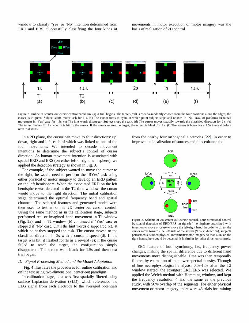
window to classify ‘Yes’ or ‘No’ intention determined from
ERD and ERS. Successfully classifying the four kinds of
movements in motor execution or motor imagery was the
basis of realization of 2D control.
Figure 2. Online 2D center-out cursor control paradigm. (a) A trial begins. The target (red) is pseudo-randomly chosen from the four positions along the edges; the
cursor is in green. Subject starts motor task for 1 s. (b) The cursor turns to cyan, at which point subject stops and relaxes in ‘No’ case, or performs sustained
movement in ‘Yes’ case for 1.5s. (c) The hint words disappear. Subject stops the task. (d) The cursor moves steadily towards the classified direction for 2 s. (e) The target flashes for 1 s when it is hit by the cursor. If the cursor misses the target, the screen is blank for 1 s. (f) The screen is blank for a 1.5s interval before
next trial starts.
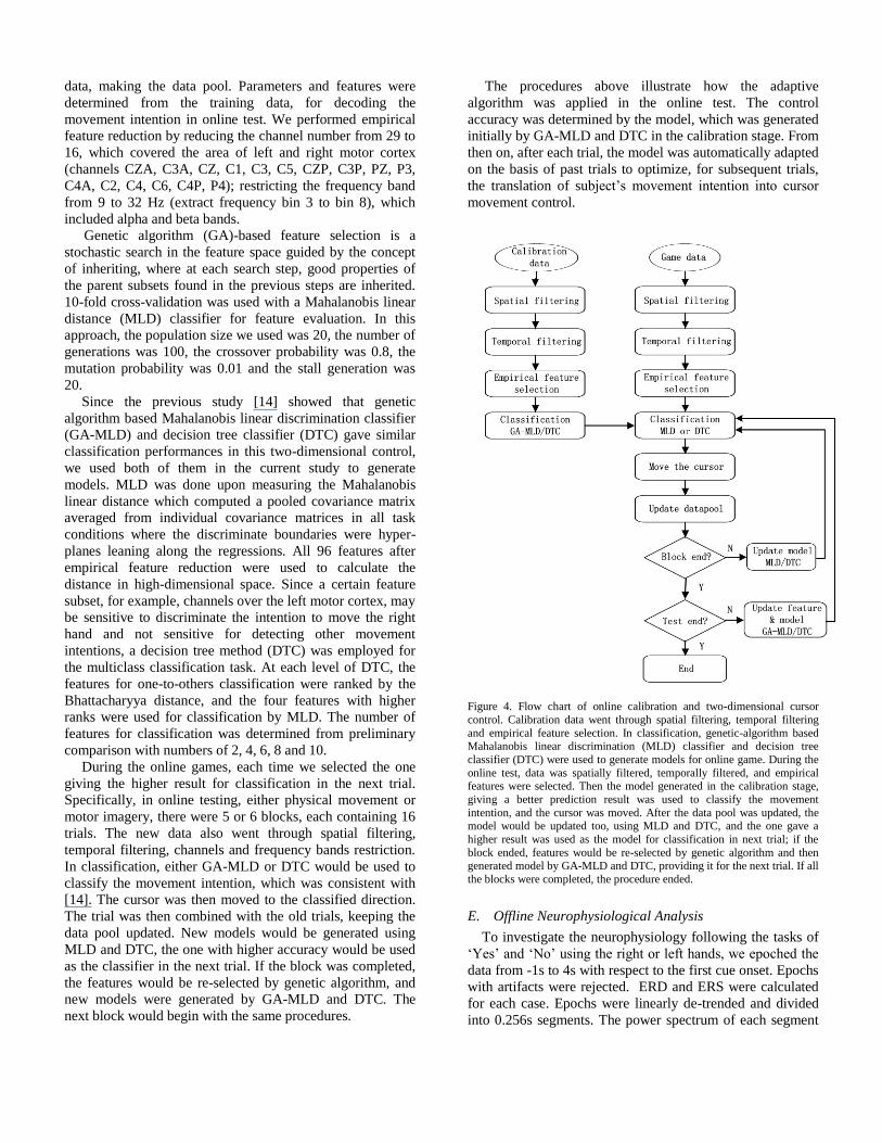
In a 2D plane, the cursor can move to four directions: up,
down, right and left, each of which was linked to one of the
four movements. We intended to decode movement
intentions to determine the subject’s control of cursor
direction. As human movement intention is associated with
spatial ERD and ERS (on either left or right hemisphere), we
applied the detection strategy as shown in Fig. 3.
For example, if the subject wanted to move the cursor to
the right, he would need to perform the ‘RYes’ task using
either physical or motor imagery to develop an ERD pattern
on the left hemisphere. When the associated ERD on the left
hemisphere was detected in the T2 time window, the cursor
would move to the right direction. The initial calibration
stage determined the optimal frequency band and spatial
channels. The selected features and generated model were
then used to test an online 2D center-out cursor control.
Using the same method as in the calibration stage, subjects
performed real or imagined hand movement in T1 window
(Fig. 2a), and in T2 window (b) continued if ‘Yes’ case or
stopped if ‘No’ case. Until the hint words disappeared (c), at
which point they stopped the task. The cursor moved to the
classified direction in 2s with a constant speed (d). If the
target was hit, it flashed for 1s as a reward (e); if the cursor
failed to reach the target, the configuration simply
disappeared. The screen went blank for 1.5s and then next
trial began.
D. Signal Processing Method and the Model Adaptation
Fig. 4 illustrates the procedures for online calibration and
online test using two-dimensional center-out paradigm.
In calibration stage, data was first spatially filtered using
surface Laplacian derivation (SLD), which referenced the
EEG signal from each electrode to the averaged potentials
from the nearby four orthogonal electrodes [22], in order to
improve the localization of sources and thus enhance the
Figure 3. Scheme of 2D center-out cursor control. Four directional control
by spatial detection of ERD/ERS on right/left hemisphere associated with intention to move or cease to move the left/right hand. In order to direct the
cursor move towards the left side of the screen (‘LYes’ direction), subjects
performed sustained physical movement/motor imagery so that ERD on the
right hemisphere could be detected. It is similar for other direction controls.
EEG feature of local synchrony, i.e., frequency power
changes, making the spatial difference due to different hand
movements more distinguishable. Data was then temporally
filtered by estimation of the power spectral density. Through
offline neurophysiological analysis, 0.5s-1.5s after the T2
window started, the strongest ERD/ERS was selected. We
applied the Welch method with Hamming window, and kept
the frequency resolution 4 Hz, the same as the previous
study, with 50% overlap of the segments. For either physical
movement or motor imagery, there were 48 trials for training
data, making the data pool. Parameters and features were
determined from the training data, for decoding the
movement intention in online test. We performed empirical
feature reduction by reducing the channel number from 29 to
16, which covered the area of left and right motor cortex
(channels CZA, C3A, CZ, C1, C3, C5, CZP, C3P, PZ, P3,
C4A, C2, C4, C6, C4P, P4); restricting the frequency band
from 9 to 32 Hz (extract frequency bin 3 to bin 8), which
included alpha and beta bands.
Genetic algorithm (GA)-based feature selection is a
stochastic search in the feature space guided by the concept
of inheriting, where at each search step, good properties of
the parent subsets found in the previous steps are inherited.
10-fold cross-validation was used with a Mahalanobis linear
distance (MLD) classifier for feature evaluation. In this
approach, the population size we used was 20, the number of
generations was 100, the crossover probability was 0.8, the
mutation probability was 0.01 and the stall generation was
20.
Since the previous study [14] showed that genetic
algorithm based Mahalanobis linear discrimination classifier
(GA-MLD) and decision tree classifier (DTC) gave similar
classification performances in this two-dimensional control,
we used both of them in the current study to generate
models. MLD was done upon measuring the Mahalanobis
linear distance which computed a pooled covariance matrix
averaged from individual covariance matrices in all task
conditions where the discriminate boundaries were hyper-
planes leaning along the regressions. All 96 features after
empirical feature reduction were used to calculate the
distance in high-dimensional space. Since a certain feature
subset, for example, channels over the left motor cortex, may
be sensitive to discriminate the intention to move the right
hand and not sensitive for detecting other movement
intentions, a decision tree method (DTC) was employed for
the multiclass classification task. At each level of DTC, the
features for one-to-others classification were ranked by the
Bhattacharyya distance, and the four features with higher
ranks were used for classification by MLD. The number of
features for classification was determined from preliminary
comparison with numbers of 2, 4, 6, 8 and 10.
During the online games, each time we selected the one
giving the higher result for classification in the next trial.
Specifically, in online testing, either physical movement or
motor imagery, there were 5 or 6 blocks, each containing 16
trials. The new data also went through spatial filtering,
temporal filtering, channels and frequency bands restriction.
In classification, either GA-MLD or DTC would be used to
classify the movement intention, which was consistent with
[14]. The cursor was then moved to the classified direction.
The trial was then combined with the old trials, keeping the
data pool updated. New models would be generated using
MLD and DTC, the one with higher accuracy would be used
as the classifier in the next trial. If the block was completed,
the features would be re-selected by genetic algorithm, and
new models were generated by GA-MLD and DTC. The
next block would begin with the same procedures.
The procedures above illustrate how the adaptive
algorithm was applied in the online test. The control
accuracy was determined by the model, which was generated
initially by GA-MLD and DTC in the calibration stage. From
then on, after each trial, the model was automatically adapted
on the basis of past trials to optimize, for subsequent trials,
the translation of subject’s movement intention into cursor
movement control.
Figure 4. Flow chart of online calibration and two-dimensional cursor
control. Calibration data went through spatial filtering, temporal filtering
and empirical feature selection. In classification, genetic-algorithm based Mahalanobis linear discrimination (MLD) classifier and decision tree
classifier (DTC) were used to generate models for online game. During the
online test, data was spatially filtered, temporally filtered, and empirical features were selected. Then the model generated in the calibration stage,
giving a better prediction result was used to classify the movement
intention, and the cursor was moved. After the data pool was updated, the model would be updated too, using MLD and DTC, and the one gave a
higher result was used as the model for classification in next trial; if the
block ended, features would be re-selected by genetic algorithm and then generated model by GA-MLD and DTC, providing it for the next trial. If all
the blocks were completed, the procedure ended.
E. Offline Neurophysiological Analysis
To investigate the neurophysiology following the tasks of
‘Yes’ and ‘No’ using the right or left hands, we epoched the
data from -1s to 4s with respect to the first cue onset. Epochs
with artifacts were rejected. ERD and ERS were calculated
for each case. Epochs were linearly de-trended and divided
into 0.256s segments. The power spectrum of each segment

was calculated using FFT with Hamming window resulting
in a bandwidth of about 4 Hz. ERD and ERS were obtained
by averaging the log power spectrum across epochs and
baseline corrected with respect to -1s to 0s.
Figure 5. Time-course and topography of ERD and ERS for S1, S2 and S3. For each subject, the left part is plotted for motor execution and the right part for motor imagery. The blue color stands for ERD; the red stands for ERS. T1 window is from 0 s to 1s and T2 window from 1 s to 2.5 s. For S1and S2, ERD and ERS were
clear for physical movement and motor imagery. For S3, ERD and ERS can only be clearly observed for physical movement.

III. RESULT
A. Neurophysiological Analysis of ERD/ERS
For each subject, all the calibration data and testing data
were included to do the spatiotemporal analysis. The study
differentiated the ERD and ERS patterns in two hemispheres
following hands movement or motor imagery using the
period after the ‘No’ cue onset. Fig. 5 shows the time-
frequency plots, head topographies of ERD and ERS for all
the three subjects, with physical movement (left half of each
sub-figure) and motor imagery (right half of each sub-
figure). For S1 and S3, channel C3 over the left
sensorimotor cortex and C4 over the right hemisphere were
selected to illustrate the strongest ERD and ERS patterns,
containing each of the four situations: ‘RYes’, ‘RNo’,
‘LYes’, and ‘LNo’. For S2, channel C1 on the left
hemisphere and channel C2 on the right hemisphere were
used for the same purpose. ERD was observed from around
0.2 – 0.5s after the cue onset. For S1 and S3, ERD centered
around 15Hz (lower beta band); for S2, ERD centered
around 22Hz. ERD was observed on both hemispheres for
all the subjects during physical movement, but more on one
side. ERS was observed around 20 Hz, over the contralateral
motor areas for S1 and S3, but also appeared slightly
ipsilateral for S2 on the central channels C2 or C1.
Compared with ERD patterns, ERS was more focal on the
contralateral hemisphere. Therefore, the ERD and ERS on
either left or right hemisphere provided four spatial patterns
to detect ‘RYes’, ‘RNo’, ‘LYes’, and ‘LNo’ intentions. For
motor imagery, ERD and ERS have similar patterns as for
physical movement, although the amplitudes were smaller.
ERD and ERS patterns were not clear to observe for S3.
B. Classification
Fig. 6 gives out the online cursor control test results for the three subjects, with physical movement (Block 1 to Block 6) and motor imagery (Block 1’ to Block 6’). All the subjects finished 6 blocks in physical part, containing 16 trials in each block, with four tasks evenly assigned. Either DTC or GA-MLD was used each time for the intention detection, depending on which one created a better model after model adaptation. Average online performances for each subject in the physical condition were 77.1%±8.54%, 70.8%±5.10%, and 57.0%±6.85%. We observed a trend that the overall performances increased across blocks, although correlation did not show significant difference (r=0.19, p-value=0.4468). In motor imagery online test, S1 and S2 had 6 blocks and S3 had 5.
Average online performances for each subject in motor imagery part were 57.3%±13.35%, 46.9%±8.62%, and 42.5%±5.23%. We also observed a trend that the overall performances increased across blocks, and the correlation showed a significant relationship between motor imagery performances and blocks (r=0.62, p-value=0.0057).
Offline analysis using 10-fold cross-validation was done
for each subject. All the calibration data and test data (total
128-144 trials per subject per part) were used. Table 1 listed
the results, evaluated by DTC and GA-MLD classifiers, for
physical and motor imagery parts. The two classifiers
provided similar results in each part.
Figure 6. Online two-dimensional cursor control accuracies of physical movement (Block 1 to Block 6) and motor imagery (Block 1’ to Block 6’) for S1, S2 and S3.
IV. DISCUSSION
A. Center-Out Paradigm
In our previous study [14], we used a goal oriented
paradigm in the online two dimensional cursor control test,
where the target randomly appeared in the 2D plane. The
subject was supposed to control the cursor moving to it and
avoid being trapped by a randomly assigned obstacle [14].
Most subjects found the paradigm interesting and easy to
learn, requiring little mental load. As we discussed before, in
motor imagery where no EMG was involved, subjects could
determine the route in each step moving the cursor to the
target by themselves, so, it was difficult for the computer to
tell whether the cursor really moved to the desired direction
without feedback, and therefore we did not report the
accuracy for motor imagery in the previous study, instead,
we reported overall target reaching rate. In this study, we
adopted the commonly used center-out paradigm [11, 23, 24]
to further investigate the performance of our proposed BCI
with motor imagery, where four tasks were evenly assigned
in each block and the calculation of control accuracy was
straightforward. The four-target center-out paradigm can be
generalized to an eight or more target paradigm, which
would be more ideal for testing further improved 2D control,
for example, continuous 2D control.
B. Decoding Rate and Accuracy
Information transfer rate (ITR) in bits per minute (bpm)
has been introduced by Wolpaw et al. to evaluate the
performance of BCI systems; both control accuracy and
control speed determine the BCI performance [10, 25]. In
this study, we used the classification accuracy given by the
best subject to calculate the ITR. For physical movement, the
classification accuracy was 83%, and for motor imagery
56.8%. For a four class task, ITR was 1.34 bits per trial for
physical movement and 1.01 bits per trial for motor imagery.
Previously the cuing period T1 was set to 2.5s, which left
enough time for subjects to prepare for the movement. In the
current study, we shortened it to 1s. Although the variance
was larger than before, subjects reported good attention

levels, and from the neurophysiological analysis we observed
clear ERD/ERS patterns and even shorter response delay
than before. Since in either part, the total duration for T1 and
T2 windows has been shortened to 2.5s, i.e. 24 trials per
minute. Therefore, ITR was 32.16 bits per minute for
physical movement and 24.24 bits per minute for motor
imagery. Compared with the results given in previous study
[14], ITR was greatly improved.
TABLE I. 10-FOLD CROSS-VALIDATION ACCURACY
Subject
Physical
DTC(%) GA-MLD(%)
Motor imagery
DTC(%) GA-MLD(%)
S1
S2
S3
78.5 ± 4.28
74.7 ± 3.15
59.1 ± 5.47
83.0 ± 4.59
78.2 ± 3.90
63.2 ± 4.83
52.4 ± 3.04
38.4 ± 4.02
40.8 ± 5.73
56.8 ± 2.76
49.1 ± 2.57
39.7 ± 4.88
Average 70.7 ±10.28 74.8 ± 10.33 43.9 ± 7.49 48.5 ± 8.56
DTC: decision tree classifier; GA-MLD: genetic algorithm-based
Mahalanobis linear discrimination.
C. Decoding Accuracies Changing with Time
To achieve four-directional classification for a two-
dimensional control associated with human natural behavior,
the BCI in this study was expected to show stable and robust
performance without intensive training of subjects. As the
results showed, a trend could be observed that the overall
performance for the three subjects improved across blocks, in
either physical part or motor imagery part. Since model
adaptation was used for each trial and the features were re-
selected for each new block, classification accuracy was
supposed to increase or stabilize when the tasks were done
consistently, although the increase may be insignificant. We
expect that in further study, with the model adaptation, the
accuracy can increase or at least stabilize in multiple visits,
with stable performances across blocks in each single visit. If
that is the case, the proposed BCI would be able to achieve
reliable control in both short and long-term use.
D. Spatiotemporal Features of ERD/ERS
As was expected for physical movement, we observed
clear ERD and ERS in beta band over the contralateral motor
cortex associated with the moving hand, ERD during the
sustained movement and ERS after the movement stopped.
We also observed ERD on the ipsilateral hemisphere and
even stronger than that on contralateral hemisphere. We
considered the reason might be that during the hand moving,
although the other hand was not moving, the automatic
urging of the movement also generated ERD activity, on the
contralateral motor cortex, which was the ipsilateral side of
the moving hand. Similar patterns appeared for motor
imagery. In this case, the discrimination between ‘RYes’ and
‘LYes’ could be difficult, since the movement of either hand
would generate ERD over both hemispheres, especially when
its variance was large. Although in this study, genetic
algorithm combined with adaptive method provided multiple
features for the classifier, which greatly helped with the
classification, ‘RYes’ and ‘LYes’ was still the most difficult
pair to distinguish compared with others. This issue could be
further improved upon either by adding another feature to
enhance classification or improving the paradigm to avoid
direct comparison of ‘RYes’ and ‘LYes’.
The present study further confirms the results presented in
our previous study which demonstrated that EEG activity
associated with human natural behavior delivers information
from which human volitional movement intention can be
decoded. This preliminary study provides evidence that EEG
based natural BCI supports 2D control, with a competitive
information transfer rate in terms of control accuracy and
control speed. In particular, 2D control can be easily
achieved within 3 hours in the experiment by imagining the
movement, long-termtraining is no longer needed.
Successfully decoding of movement intention is highly
dependent on the experimental design and optimization of
parameters in experimental and computational procedures.
Further research is needed to explore the reliability and
applicability of the natural BCI on larger populations. This
would include investigating healthy subjects and patients
over multiple visits to establish how well it can be
generalized to achieve fast continuous control. We anticipate
that such studies will further demonstrate that EEG is highly
capable of realizing continuous multi-dimensional control
with human natural behavior or thinking, which will
eventually benefit people in their daily life.
REFERENCES
[1] J. R. Wolpaw, "Brain-computer interfaces as new brain output pathways," J Physiol, vol. 579, pp. 613-9, 2007.
[2] J. Kubanek, K. J. Miller, J. G. Ojemann, J. R. Wolpaw, and G. Schalk, "Decoding flexion of individual fingers using electrocorticographic signals in humans," J Neural Eng, vol. 6, pp. 66001, 2009.
[3] E. C. Leuthardt, G. Schalk, J. R. Wolpaw, J. G. Ojemann, and D. W. Moran, "A brain-computer interface using electrocorticographic signals in humans," J Neural Eng, vol. 1, pp. 63-71, 2004.
[4] K. J. Miller, E. C. Leuthardt, G. Schalk, R. P. Rao, N. R. Anderson, D. W. Moran, J. W. Miller, and J. G. Ojemann, "Spectral changes in cortical surface potentials during motor movement," J Neurosci, vol. 27, pp. 2424-32, 2007.
[5] G. Schalk, J. Kubanek, K. J. Miller, N. R. Anderson, E. C. Leuthardt, J. G. Ojemann, D. Limbrick, D. Moran, L. A. Gerhardt, and J. R. Wolpaw, "Decoding two-dimensional movement trajectories using electrocorticographic signals in humans," J Neural Eng, vol. 4, pp. 264-75, 2007.
[6] E. C. Leuthardt, K. Miller, N. R. Anderson, G. Schalk, J. Dowling, J. Miller, D. W. Moran, and J. G. Ojemann, "Electrocorticographic frequency alteration mapping: a clinical technique for mapping the motor cortex," Neurosurgery, vol. 60, pp. 260-70; discussion 270-1, 2007.
[7] K. J. Miller, M. denNijs, P. Shenoy, J. W. Miller, R. P. Rao, and J. G. Ojemann, "Real-time functional brain mapping using electrocorticography," Neuroimage, vol. 37, pp. 504-7, 2007.
[8] G. Schalk, E. C. Leuthardt, P. Brunner, J. G. Ojemann, L. A. Gerhardt, and J. R. Wolpaw, "Real-time detection of event-related brain activity," Neuroimage, vol. 43, pp. 245-9, 2008.
[9] D. R. Kipke, W. Shain, G. Buzsaki, E. Fetz, J. M. Henderson, J. F. Hetke, and G. Schalk, "Advanced neurotechnologies for chronic neural interfaces: new horizons and clinical opportunities," J Neurosci, vol. 28, pp. 11830-8, 2008.

[10] J. R. Wolpaw, N. Birbaumer, D. J. McFarland, G. Pfurtscheller, and T. M. Vaughan, "Brain-computer interfaces for communication and control," Clin Neurophysiol, vol. 113, pp. 767-91, 2002.
[11] J. R. Wolpaw and D. J. McFarland, "Control of a two-dimensional movement signal by a noninvasive brain-computer interface in humans," Proc Natl Acad Sci U S A, vol. 101, pp. 17849-54, 2004.
[12] D. J. McFarland, D. J. Krusienski, W. A. Sarnacki, and J. R. Wolpaw, "Emulation of computer mouse control with a noninvasive brain-computer interface," J Neural Eng, vol. 5, pp. 101-10, 2008.
[13] O. Bai, P. Lin, S. Vorbach, M. K. Floeter, N. Hattori, and M. Hallett, "A high performance sensorimotor beta rhythm-based brain-computer interface associated with human natural motor behavior," J Neural Eng, vol. 5, pp. 24-35, 2008.
[14] D. Huang, P. Lin, D. Y. Fei, X. Chen, and O. Bai, "Decoding human motor activity from EEG single trials for a discrete two-dimensional cursor control," J Neural Eng, vol. 6, pp. 046005, 2009.
[15] V. Morash, O. Bai, S. Furlani, P. Lin, and M. Hallett, "Classifying EEG signals preceding right hand, left hand, tongue, and right foot movements and motor imageries," Clin Neurophysiol, vol. 119, pp. 2570-8, 2008.
[16] N. Birbaumer, C. Weber, C. Neuper, E. Buch, K. Haapen, and L. Cohen, "Physiological regulation of thinking: brain-computer interface (BCI) research," Prog Brain Res, vol. 159, pp. 369-91, 2006.
[17] O. Bai, Z. Mari, S. Vorbach, and M. Hallett, "Asymmetric spatiotemporal patterns of event-related desynchronization preceding voluntary sequential finger movements: a high-resolution EEG study," Clin Neurophysiol, vol. 116, pp. 1213-21, 2005.
[18] S. M. Rao, J. R. Binder, P. A. Bandettini, T. A. Hammeke, F. Z. Yetkin, A. Jesmanowicz, L. M. Lisk, G. L. Morris, W. M. Mueller, L. D. Estkowski, and et al., "Functional magnetic resonance imaging of complex human movements," Neurology, vol. 43, pp. 2311-8, 1993.
[19] S. Salenius, R. Salmelin, C. Neuper, G. Pfurtscheller, and R. Hari, "Human cortical 40 Hz rhythm is closely related to EMG rhythmicity," Neurosci Lett, vol. 213, pp. 75-8, 1996.
[20] R. C. Oldfield, "The assessment and analysis of handedness: the Edinburgh inventory," Neuropsychologia, vol. 9, pp. 97-113, 1971.
[21] H. H. Jasper and H. L. Andrews, "Electro-encephalography. III. Normal differentiation of occipital and precentral regions in man," Arch Neurol Psychiat, vol. 39, pp. 95-115, 1938.
[22] B. Hjorth, "An on-line transformation of EEG scalp potentials into orthogonal source derivations," Electroencephalogr Clin Neurophysiol, vol. 39, pp. 526-30, 1975.
[23] G. Schalk, K. J. Miller, N. R. Anderson, J. A. Wilson, M. D. Smyth, J. G. Ojemann, D. W. Moran, J. R. Wolpaw, and E. C. Leuthardt, "Two-dimensional movement control using electrocorticographic signals in humans," J Neural Eng, vol. 5, pp. 75-84, 2008.
[24] T. M. Vaughan, D. J. McFarland, G. Schalk, W. A. Sarnacki, D. J. Krusienski, E. W. Sellers, and J. R. Wolpaw, "The Wadsworth BCI Research and Development Program: at home with BCI," IEEE Trans Neural Syst Rehabil Eng, vol. 14, pp. 229-33, 2006.
[25] J. R. Wolpaw, N. Birbaumer, W. J. Heetderks, D. J. McFarland, P. H. Peckham, G. Schalk, E. Donchin, L. A. Quatrano, C. J. Robinson, and T. M. Vaughan, "Brain-computer interface technology: a review of the first international meeting," IEEE Trans Rehabil Eng, vol. 8, pp. 164-73, 2000.
Related Documents