Effect of insulin-like growth factor binding protein-1 on integrin signalling and the induction of apoptosis in human breast cancer cells C M Perks, P V Newcomb, M R Norman 1 and J M P Holly Division of Surgery, Department of Hospital Medicine, Level 7, Bristol Royal Infirmary, Bristol, BS2 8HW, UK 1 Department of Medicine, Upper Maudlin Street, Bristol Royal Infirmary, Bristol, BS2 8HW, UK (Requests for offprints should be addressed to C M Perks) ABSTRACT Interaction of epithelial cells with the extracellular matrix is mediated through integrin receptors, which transmit signals regulating cell growth, differentiation and death. Occupation of these receptors, via Arg-Gly-Asp (RGD) recognition sequences, leads to activation of focal adhesion kinase (FAK). We treated human breast cancer cell lines with RGD-containing peptides, which can disrupt integrin attachment, and investigated alterations in FAK phosphorylation, cell detachment and death. Cells grown in vitro were treated with insulin-like growth factor-binding protein-1 (IGFBP-1) and a small, synthetic RGD-containing peptide (Gly- Arg-Gly-Asp-Thr-Pro) and its negative control peptide RGE (Arg-Gly-Glu-Ser) for either 30 min followed by immunoprecipitation of cell lysates with anti-phosphotyrosine and Western immuno- blotting with anti-FAK or for 24 h followed by cell counting, immunocytochemistry and flow cytometry. Both IGFBP-1 (0–800 ng/ml) and the synthetic RGD-containing peptide (1–100 μg/ml) caused sig- nificant dephosphorylation of FAK. Furthermore, after 24 h both peptides caused detachment from the matrix and the induction of apoptosis. We conclude from these data that IGFBP-1 can interact with integrin receptors to induce FAK dephosphorylation and subsequently influence attachment and cell death. Journal of Molecular Endocrinology (1999) 22, 141–150 INTRODUCTION The normal growth of cells generally requires attachment to a substrate and the presence of growth factors (Ingber 1990). Cells which become transformed or malignant are characterised by the ability to undergo anchorage-independent growth (Freedman & Shin 1974, Tucker et al. 1981). Cell adhesion to the extracellular matrix (ECM) in vivo is mediated by integrin receptors. These bind to matrix proteins outside the cell and associate with cytoskeletal proteins within the cell. ECMs are composed of several macromolecules including fibronectin, laminin, collagens and proteoglycans (Ruoslahti & Pierschbacher 1987). A number of these adhesive proteins, for example fibronectin (Pierschbacher & Ruoslahti 1984), contain the three amino acid sequence Arg-Gly-Asp (RGD), which is specifically recognised by corresponding integrin receptors (Ruoslahti & Pierschbacher 1987). The binding of integrins to the ECM initiates assembly of actin microfilaments and the accumu- lation of numerous different proteins, including signalling molecules, integrins and components of the cytoskeleton, into structures called focal adhesions (Burridge et al. 1988). A number of these proteins, including focal adhesion kinase (FAK), paxillin (Burridge et al. 1992) and tensin (Bockholt & Burridge 1993), are tyrosine phospho- rylated upon integrin ligation, suggesting a role for these proteins in integrin signalling. Specifically, integrin-dependent signals have been shown to modulate the control of growth (Giancotti & Ruoslahti 1990) and cell survival (Frisch & Francis 1994). Maintenance of integrin linkages is essential for cell adhesion. It has been demonstrated previously that disruption of these attachments, via addition of antibodies or peptides, can induce cells to detach from the substratum (Knudson et al. 1981, 141 Journal of Molecular Endocrinology (1999) 22, 141–150 0952–5041/99/022–141 ? 1999 Society for Endocrinology Printed in Great Britain Online version via http://www.endocrinology.org

Welcome message from author
This document is posted to help you gain knowledge. Please leave a comment to let me know what you think about it! Share it to your friends and learn new things together.
Transcript
Effect of insulin-like growth factor binding protein-1 onintegrin signalling and the induction of apoptosis inhuman breast cancer cells
C M Perks, P V Newcomb, M R Norman1 and J M P HollyDivision of Surgery, Department of Hospital Medicine, Level 7, Bristol Royal Infirmary,
Bristol, BS2 8HW, UK1Department of Medicine, Upper Maudlin Street, Bristol Royal Infirmary, Bristol, BS2 8HW, UK
(Requests for offprints should be addressed to C M Perks)
ABSTRACT
Interaction of epithelial cells with the extracellularmatrix is mediated through integrin receptors,which transmit signals regulating cell growth,differentiation and death. Occupation of thesereceptors, via Arg-Gly-Asp (RGD) recognitionsequences, leads to activation of focal adhesionkinase (FAK).
We treated human breast cancer cell lines withRGD-containing peptides, which can disruptintegrin attachment, and investigated alterations inFAK phosphorylation, cell detachment and death.Cells grown in vitro were treated with insulin-likegrowth factor-binding protein-1 (IGFBP-1) and asmall, synthetic RGD-containing peptide (Gly-Arg-Gly-Asp-Thr-Pro) and its negative controlpeptide RGE (Arg-Gly-Glu-Ser) for either 30 min
followed by immunoprecipitation of cell lysateswith anti-phosphotyrosine and Western immuno-blotting with anti-FAK or for 24 h followed bycell counting, immunocytochemistry and flowcytometry.
Both IGFBP-1 (0–800 ng/ml) and the syntheticRGD-containing peptide (1–100 µg/ml) caused sig-nificant dephosphorylation of FAK. Furthermore,after 24 h both peptides caused detachment fromthe matrix and the induction of apoptosis.
We conclude from these data that IGFBP-1can interact with integrin receptors to induceFAK dephosphorylation and subsequently influenceattachment and cell death.Journal of Molecular Endocrinology (1999) 22, 141–150
INTRODUCTION
The normal growth of cells generally requiresattachment to a substrate and the presence ofgrowth factors (Ingber 1990). Cells which becometransformed or malignant are characterised by theability to undergo anchorage-independent growth(Freedman & Shin 1974, Tucker et al. 1981). Celladhesion to the extracellular matrix (ECM) in vivois mediated by integrin receptors. These bind tomatrix proteins outside the cell and associate withcytoskeletal proteins within the cell. ECMs arecomposed of several macromolecules includingfibronectin, laminin, collagens and proteoglycans(Ruoslahti & Pierschbacher 1987). A number ofthese adhesive proteins, for example fibronectin(Pierschbacher & Ruoslahti 1984), contain the threeamino acid sequence Arg-Gly-Asp (RGD), which isspecifically recognised by corresponding integrinreceptors (Ruoslahti & Pierschbacher 1987).
The binding of integrins to the ECM initiatesassembly of actin microfilaments and the accumu-lation of numerous different proteins, includingsignalling molecules, integrins and componentsof the cytoskeleton, into structures called focaladhesions (Burridge et al. 1988). A number ofthese proteins, including focal adhesion kinase(FAK), paxillin (Burridge et al. 1992) and tensin(Bockholt & Burridge 1993), are tyrosine phospho-rylated upon integrin ligation, suggesting a role forthese proteins in integrin signalling. Specifically,integrin-dependent signals have been shown tomodulate the control of growth (Giancotti &Ruoslahti 1990) and cell survival (Frisch & Francis1994).
Maintenance of integrin linkages is essential forcell adhesion. It has been demonstrated previouslythat disruption of these attachments, via additionof antibodies or peptides, can induce cells todetach from the substratum (Knudson et al. 1981,
141
Journal of Molecular Endocrinology (1999) 22, 141–1500952–5041/99/022–141 ? 1999 Society for Endocrinology Printed in Great Britain
Online version via http://www.endocrinology.org
Hayman et al. 1985) with a resultant induction ofprogrammed cell death.
Insulin-like growth factor binding protein-1(IGFBP-1) belongs to a family of six closely relatedproteins (IGFBP-1 to -6) that are known to modu-late the actions of insulin-like growth factors (IGFs)(Shimasaki et al. 1991, Oh et al. 1996). These pro-teins bind IGF-I and -II with high affinity, therebymodulating their actions on target cells (Clemmons1992). However, evidence now suggests that thesebinding proteins may be able to exert IGF-independent effects. Specifically, IGFBP-1, whichcontains an RGD integrin recognition sequence(Brewer et al. 1988, Hynes 1992), can stimulate cellmigration in CHO cells. This action of IGFBP-1 wasdemonstrated to be specifically via binding to theá5â1 integrin receptor and is independent of theIGFs (Jones et al. 1993). IGFBP-1 is expressedin a strictly tissue-specific manner, being nor-mally restricted to the liver and a small number ofother tissues, including decidualised endometrium(Julkunen et al. 1988), the ovary (Suikkari et al.1989) and regenerating liver (Mohn et al. 1991), all ofwhich exhibit high rates of tissue remodelling.
However, it has also been demonstrated thatIGFBP-1 is expressed in the breast both in vitro(Clemmons et al. 1990) and in vivo (Pekonen et al.1992). IGFBP-1 mRNA expression increased inhuman breast tumours in comparison to their ad-jacent normal tissue. The upregulation of IGFBP-1was associated with the malignant transformation ofbreast tissue (Pekonen et al. 1992).
Therefore, we chose to study the effects ofRGD-containing peptides, including IGFBP-1, onFAK phosphorylation, cell attachment and death inour human breast cancer cell lines, since IGFBP-1may have a significant pathological role in thistissue. Furthermore, one of our lines, Hs578T, is anideal model in which to study IGF-independenteffects of this binding protein, since it is non-responsive to IGFs and lacks a functional IGFreceptor. In fact, we have shown previously in thiscell line that IGFBP-3 could predispose cells toprogrammed cell death in an IGF-independentmanner (Gill et al. 1997). We also studied threeadditional breast cancer cell lines, MCF-7, T47Dand ZR-75–1, which are IGF-responsive.
The results of this study provide us with evidenceto suggest that IGFBP-1 is capable of elicitingintegrin-mediated IGF-independent cellular effects.
MATERIALS AND METHODS
Human IGFBP-1 peptide was a gift from Dr J Cox,Synergen Inc. (Boulder, CO, USA). The RGD-
containing synthetic peptide (Gly-Arg-Gly-Asp-Thr-Pro) together with its negative control peptide,RGE (Arg-Gly-Glu-Ser) were bought from SigmaChemical Co., Poole, Dorset, UK. All otherreagents were purchased from Sigma Chemical Co.or Merck Sharp and Dohme Ltd (Hoddesdon,Herts, UK) unless otherwise stated.
Cell culture
The human breast cancer cell lines T47D, Hs578T,MCF-7 and ZR-75–1 were purchased from ECACC(Porton Down, Wilts, UK) and grown in ahumidified 5% CO2 atmosphere at 37 )C. They werecultured in Dulbecco’s modified Eagle’s medium(DMEM) with glutamax-1, Minimum EssentialMedium (MEM), Eagle’s medium and RPMI 1640medium respectively supplemented with 10%foetal calf serum (Advanced Protein ProductsLtd, Brierley Hill, West Midlands, UK), peni-cillin (5000 IU/ml), streptomycin (5 mg/ml) and-glutamine (2 mM).
Immunoprecipitation and Westernimmunoblotting
Cells (1#106) were grown to 80% confluence in90 mm dishes (Nunclon, Rochester, NY, USA) andthen washed twice with PBS. The growth mediumwas replaced with serum-free Dulbecco’s MEM andHam’s nutrient mix F-12 buffered with Hepes andsupplemented with sodium bicarbonate (0·12%),BSA (0·2 mg/ml) and transferrin (0·01 mg/ml)(SFM) for 24 h. Cells were treated, incubated at37 )C for 30 min and then lysed on ice for 10 min(1 ml; 10 mM Tris–HCl, 5 mM EDTA, 50 mMNaCl, 30 mM sodium pyrophosphate, 50 mMsodium fluoride, 100 µM sodium orthovanadate, 1%Triton, 1 mM phenylmethylsulphonyl fluoride; pH7·6). Lysates were then centrifuged at 14 000 g for15 min at 4 )C. The supernatant fractions wereincubated at 4 )C with anti-phosphotyrosine anti-body (4 µg; Upstate Biotechnology, Lake Placid,NY, USA) for 2 h, goat anti-mouse IgG (5 µl;Calbiochem, Beeston, Nottingham, UK) for 1 h andthen 25 µl protein-A–Sepharose beads (Calbiochem)for 1 h. The samples were washed three times withlysis buffer (500 µl), centrifuged at 14 000 g for3 min and the supernatant removed. Laemmliloading buffer (2#) was added and proteins wereseparated by 8% SDS-PAGE and then transferredonto a nylon membrane. Non-specific binding siteswere blocked by washing the membranes in 5% milkand then they were probed with anti-FAK (1 µg/ml;Upstate Biotechnology) overnight. Following theremoval of excess unbound antibody, an anti-mouse
and others · Physiological effects of RGD-containing peptides in human breast cancer cells142
Journal of Molecular Endocrinology (1999) 22, 141–150
antibody conjugated to peroxidase (1:2000) wasadded for 1 h. Binding of the peroxidase wasvisualised by enhanced chemiluminescence accord-ing to the manufacturer’s instructions (AmershamInternational, Amersham, Bucks, UK). Opticaldensity measurements were determined using ascanning densitometer (Bio-Rad, Hemel Hemstead,Herts, UK) and analysed using Molecular Analystsoftware (Biorad).
Cell counting
Aliquots of cells (50 µl) were loaded onto ahaemocytometer and the total cell number wasdetermined.
Flow cytometry
This technique was used to assess the amount ofapoptosis in a given sample. Apoptotic cells have alower DNA stainability than normal cells andappear as a pre-G1 peak on a DNA cell cyclehistogram. Cells (1–2#106) were washed in PBSand fixed for 30 min by the addition of 70% ethanol(1 ml). Cells were pelleted (1600 g; 5 min) andwashed three times with PBS. The supernatant wasremoved and the cells were resuspended in reactionbuffer (propidium iodide, 0·05 mg/ml; sodiumcitrate, 0·1%; RNAse A, 0·02 mg/ml; NP-40, 0·3%;pH 8·3) vortexed and incubated at 4 )C for 30 min.All cells were then measured on a FACSCALIBURflow cytometer (Becton Dickinson, Cowley, Oxford,UK) with an argon laser at 488 nm for excitationand analysed using Cell Quest (Becton Dickinson).
MTT assay
MTT reagent (3-(4,5-dimethylthiazol-2-yl)-2,5-diphenyltetrazolium bromide; Thiazolyl blue) isconverted into a coloured water-insoluble formazansalt by the metabolic activity of viable cells and canbe used as a crude measure of cell viability. Cellswere seeded at 5#104/ml (150 µl growth medium)in 96-well plates and allowed to grow for 24 h.Growth medium was replaced with SFM (100 µl)24 h prior to the assay. MTT reagent (7·5 mg/ml) inPBS was added to the cells (10 µl/well) and thecultures were incubated for 30 min at 37 )C. Thereaction was stopped by the addition of acidifiedTriton buffer (0·1 M HCl, 10% (v/v) Triton X-100;50 µl/well) and the tetrazolium crystals weredissolved by mixing on a plate shaker for 20 min atroom temperature. The samples were measured ona Bio-Rad 450 plate reader at a test wavelength of595 nm and a reference wavelength of 650 nm.Results, which are expressed as a percentage opti-
cal density of SFM controls, represent the mean&... of five wells from one experiment which isrepresentative of experiments repeated at least threetimes.
Morphological appearance
To determine that IGFBP-1-induced cell deathgave rise to the classical morphological featuresassociated with apoptosis, aliquots of treated anduntreated cells were cytospun and stained withWright’s stain in an automated stainer (Lillie 1977).Photomicrographs of the cells were taken under oilimmersion at a magnification of#100.
Immunocytochemistry
Cells were cytospun (150 µl; 5#105/ml) onto aminopropyl triexothy silane-coated slides, fixed in coldacetone for 10 min and left to dry at roomtemperature for 10 min. Cells were incubated withhydrogen peroxide (0·6% in methanol) for 15 min,washed three times in Tris-buffered saline (TBS)for 5 min and incubated with 20% normal rabbitserum in TBS for 30 min. Serum was removed andcells were incubated with antibodies (anti-humanintegrin á5 IgG, 5·8 µg/ml; anti-human â1 integrinIgG, 10 µg/ml) at 4 )C overnight. Cells were thenwashed three times with TBS for 5 min andincubated with biotinylated rabbit anti-mouse IgG(1:300 in TBS) for 30 min. After a further threewashes in TBS, cells were incubated withstreptavidin–biotin complex (30 min) followed bythree TBS washes for 5 min. Cells were thenincubated with 3,3*-diaminobenzidene tetrahydro-chloride for 10 min prior to counterstaining withMayer’s haematoxylin.
Statistical analysis
The data were analysed using the Microsoft Excel4.0a software package. Significance was determinedusing Student’s t-test. A statistically significantdifference was considered to be present at P<0·05.
RESULTS
Measurement of FAK phosphorylation in cellstreated with RGD-containing peptides over30 min
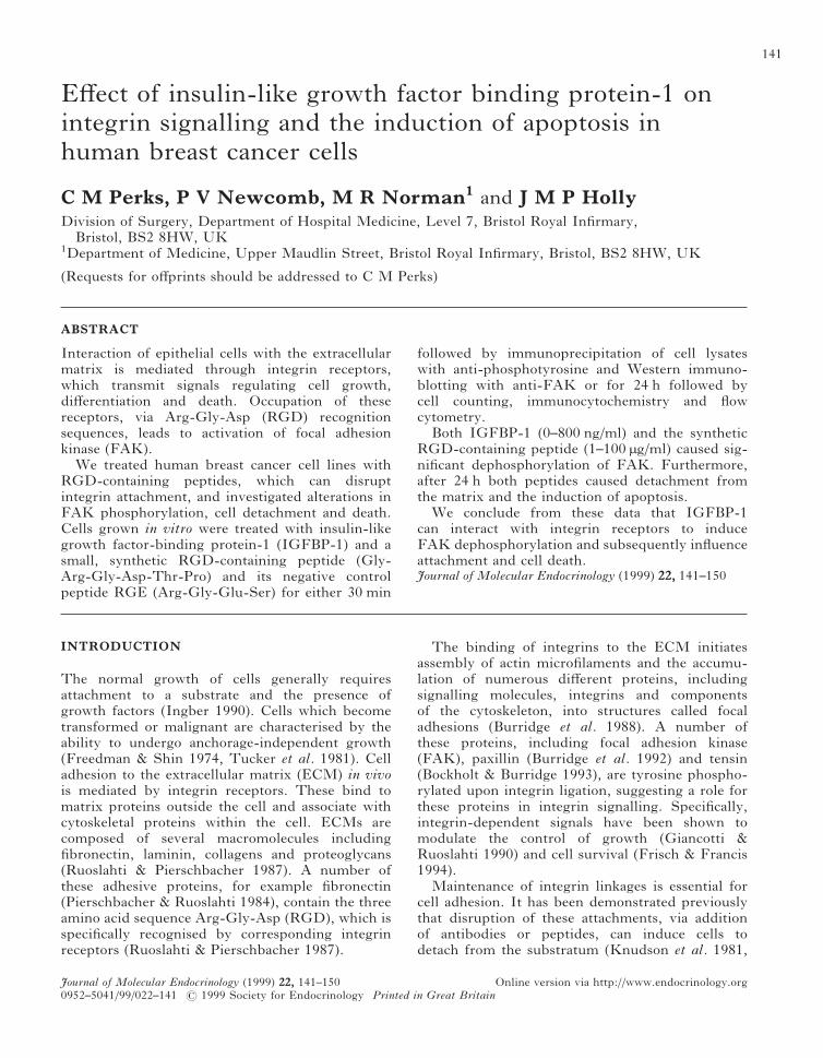
The RGD-containing peptides caused significantdose-dependent decreases in the phosphorylation ofFAK in the T47D cells (Fig. 1A and C). It isimportant to note that no cell detachment was
Physiological effects of RGD-containing peptides in human breast cancer cells · and others 143
Journal of Molecular Endocrinology (1999) 22, 141–150
evident after 30 min treatment and no signifi-cant differences were found between the totalprotein concentrations of the individual plates. Thesynthetic RGD-containing peptide (Fig. 1A and C)reduced FAK phosphorylation significantly by45·7% (P<0·01) and 66·5% (P<0·001) at 25 and100 µg/ml respectively relative to control values.This reduction in FAK phosphorylation was alsoevident in the T47Ds after treatment with IGFBP-1(Fig. 1A and C) at 400 ng/ml (19·1%), becomingsignificant at 800 ng/ml (39·4%; P<0·001). We alsoshowed that IGFBP-1 and the synthetic RGD-containing peptide can similarly decrease FAKphosphorylation in another breast cancer cell line,Hs578T (Fig. 1B). The RGD-containing peptidereduced FAK phosphorylation by 31·5 and 62% at25 and 100 µg/ml respectively. Similarly, IGFBP-1was also able to induce the dephosphorylation ofFAK in this cell line by 33·2 and 35% at 400 and800 ng/ml respectively. The control syntheticpeptide (RGE) had no significant effects on FAKphosphorylation in either cell line (Fig. 1D).
Measurement of detached cells treated withRGD-containing peptides for 24 h
Data demonstrated that treatment with both theRGD-containing synthetic peptide and IGFBP-1induced cells to detach from the plate over 24 h inT47D cultures (Fig. 2A and B). The RGD-containing synthetic peptide (Fig. 2A; 100 µg/ml)caused a significant (P<0·01) increase (35·3%) in the
1. Effects of a synthetic RGD-containingpeptide (0–100 µg/ml) and IGFBP-1 (0–800 ng/ml) onFAK phosphorylation in T47D and Hs578T cells aftertreatment for 30 min. Cells were plated in 90 mm Petridishes, grown to 80% confluence and switched to SFMfor 24 h prior to treatment. FAK phosphorylation wasassessed by immunoprecipitation and Westernimmunoblotting as outlined in Materials and Methods.(A and B) Show representative Western immunoblotsof T47D and Hs578T cells respectively; a=untreatedcells, b=RGD (25 µg/ml), c=RGD (100 µg/ml),d=IGFBP-1 (400 ng/ml) and e=IGFBP-1 (800 ng/ml).(C) Optical density measurements of FAKphosphorylation from Western immunoblots followingimmunoprecipitation in T47D cells. Results representthe mean&... of experiments performed at leastthree times, where ***P<0·001 and **P<0·01. (D)Optical density measurements of FAK phosphorylationfrom Western immunoblots following immuno-precipitation in T47D and Hs578T cells after treatmentwith the control RGD peptide (RGE) in comparisonto untreated control (CT) cells for 30 min. Resultsrepresent the mean&... of experiments performed atleast three times.
and others · Physiological effects of RGD-containing peptides in human breast cancer cells144
Journal of Molecular Endocrinology (1999) 22, 141–150
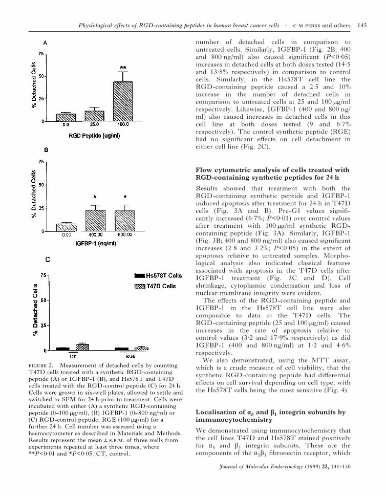
number of detached cells in comparison tountreated cells. Similarly, IGFBP-1 (Fig. 2B; 400and 800 ng/ml) also caused significant (P<0·05)increases in detached cells at both doses tested (14·5and 13·8% respectively) in comparison to controlcells. Similarly, in the Hs578T cell line theRGD-containing peptide caused a 2·5 and 10%increase in the number of detached cells incomparison to untreated cells at 25 and 100 µg/mlrespectively. Likewise, IGFBP-1 (400 and 800 ng/ml) also caused increases in detached cells in thiscell line at both doses tested (9 and 6·7%respectively). The control synthetic peptide (RGE)had no significant effects on cell detachment ineither cell line (Fig. 2C).
Flow cytometric analysis of cells treated withRGD-containing synthetic peptides for 24 h
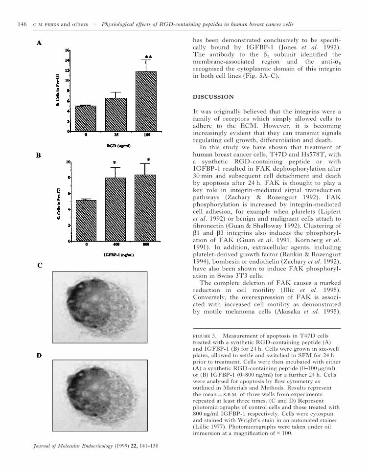
Results showed that treatment with both theRGD-containing synthetic peptide and IGFBP-1induced apoptosis after treatment for 24 h in T47Dcells (Fig. 3A and B). Pre-G1 values signifi-cantly increased (6·7%; P<0·01) over control valuesafter treatment with 100 µg/ml synthetic RGD-containing peptide (Fig. 3A). Similarly, IGFBP-1(Fig. 3B; 400 and 800 ng/ml) also caused significantincreases (2·8 and 3·2%; P<0·05) in the extent ofapoptosis relative to untreated samples. Morpho-logical analysis also indicated classical featuresassociated with apoptosis in the T47D cells afterIGFBP-1 treatment (Fig. 3C and D). Cellshrinkage, cytoplasmic condensation and loss ofnuclear membrane integrity were evident.
The effects of the RGD-containing peptide andIGFBP-1 in the Hs578T cell line were alsocomparable to data in the T47D cells. TheRGD-containing peptide (25 and 100 µg/ml) causedincreases in the rate of apoptosis relative tocontrol values (3·2 and 17·9% respectively) as didIGFBP-1 (400 and 800 ng/ml) at 1·2 and 4·6%respectively.
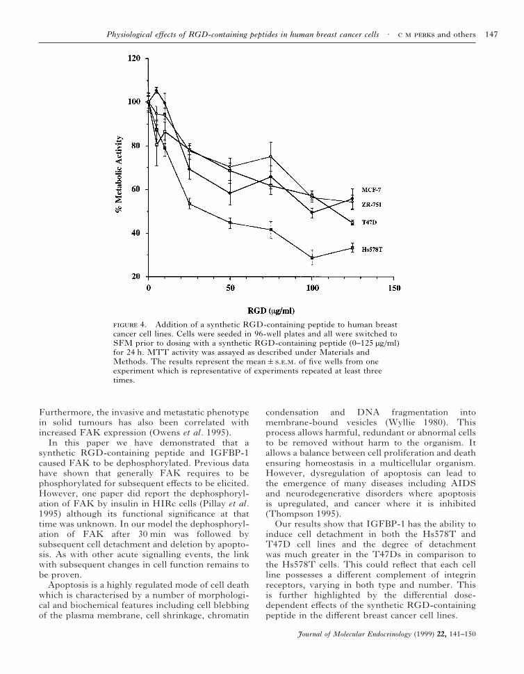
We also demonstrated, using the MTT assay,which is a crude measure of cell viability, that thesynthetic RGD-containing peptide had differentialeffects on cell survival depending on cell type, withthe Hs578T cells being the most sensitive (Fig. 4).
Localisation of á5 and â1 integrin subunits byimmunocytochemistry
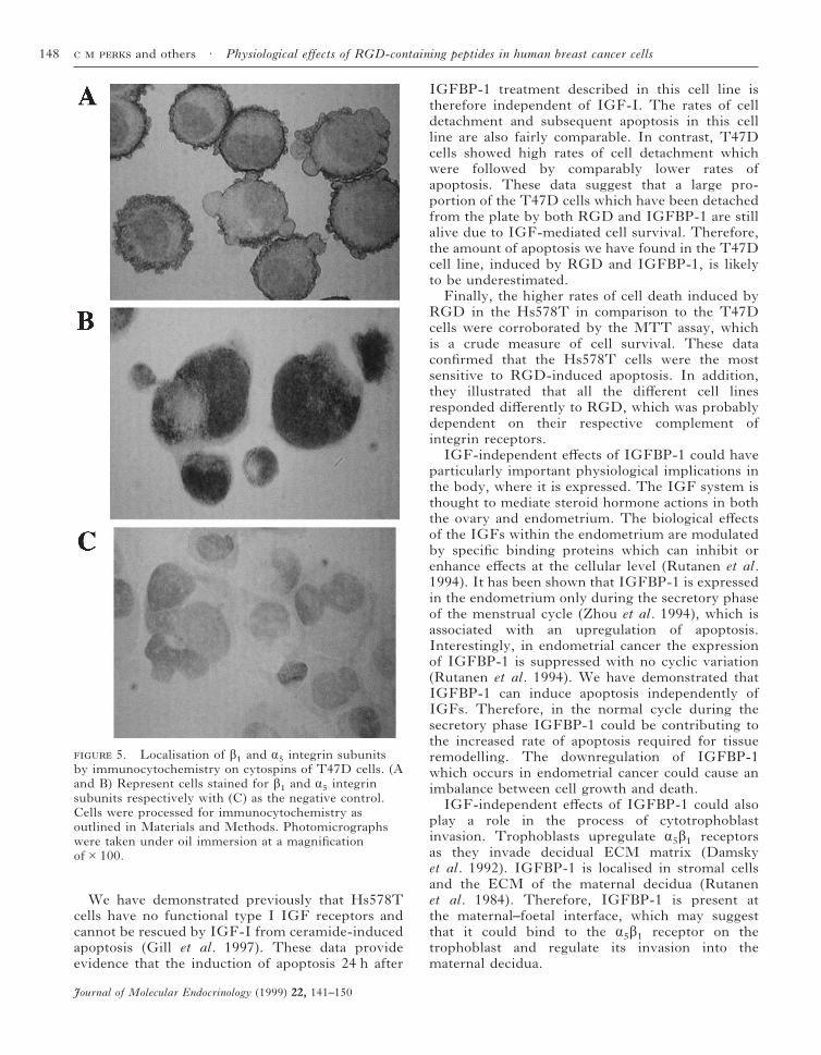
We demonstrated using immunocytochemistry thatthe cell lines T47D and Hs578T stained positivelyfor á5 and â1 integrin subunits. These are thecomponents of the á5â1 fibronectin receptor, which
2. Measurement of detached cells by countingT47D cells treated with a synthetic RGD-containingpeptide (A) or IGFBP-1 (B), and Hs578T and T47Dcells treated with the RGD-control peptide (C) for 24 h.Cells were grown in six-well plates, allowed to settle andswitched to SFM for 24 h prior to treatment. Cells wereincubated with either (A) a synthetic RGD-containingpeptide (0–100 µg/ml), (B) IGFBP-1 (0–800 ng/ml) or(C) RGD-control peptide, RGE (100 µg/ml) for afurther 24 h. Cell number was assessed using ahaemocytometer as described in Materials and Methods.Results represent the mean&... of three wells fromexperiments repeated at least three times, where**P<0·01 and *P<0·05. CT, control.
Physiological effects of RGD-containing peptides in human breast cancer cells · and others 145
Journal of Molecular Endocrinology (1999) 22, 141–150
has been demonstrated conclusively to be specifi-cally bound by IGFBP-1 (Jones et al. 1993).The antibody to the â1 subunit identified themembrane-associated region and the anti-á5recognised the cytoplasmic domain of this integrinin both cell lines (Fig. 5A–C).
DISCUSSION
It was originally believed that the integrins were afamily of receptors which simply allowed cells toadhere to the ECM. However, it is becomingincreasingly evident that they can transmit signalsregulating cell growth, differentiation and death.
In this study we have shown that treatment ofhuman breast cancer cells, T47D and Hs578T, witha synthetic RGD-containing peptide or withIGFBP-1 resulted in FAK dephosphorylation after30 min and subsequent cell detachment and deathby apoptosis after 24 h. FAK is thought to play akey role in integrin-mediated signal transductionpathways (Zachary & Rozengurt 1992). FAKphosphorylation is increased by integrin-mediatedcell adhesion, for example when platelets (Lipfertet al. 1992) or benign and malignant cells attach tofibronectin (Guan & Shalloway 1992). Clustering ofâ1 and â3 integrins also induces the phosphoryl-ation of FAK (Guan et al. 1991, Kornberg et al.1991). In addition, extracellular agents, includingplatelet-derived growth factor (Rankin & Rozengurt1994), bombesin or endothelin (Zachary et al. 1992),have also been shown to induce FAK phosphoryl-ation in Swiss 3T3 cells.
The complete deletion of FAK causes a markedreduction in cell motility (Illic et al. 1995).Conversely, the overexpression of FAK is associ-ated with increased cell motility as demonstratedby motile melanoma cells (Akasaka et al. 1995).
3. Measurement of apoptosis in T47D cellstreated with a synthetic RGD-containing peptide (A)and IGFBP-1 (B) for 24 h. Cells were grown in six-wellplates, allowed to settle and switched to SFM for 24 hprior to treatment. Cells were then incubated with either(A) a synthetic RGD-containing peptide (0–100 µg/ml)or (B) IGFBP-1 (0–800 ng/ml) for a further 24 h. Cellswere analysed for apoptosis by flow cytometry asoutlined in Materials and Methods. Results representthe mean&... of three wells from experimentsrepeated at least three times. (C and D) Representphotomicrographs of control cells and those treated with800 ng/ml IGFBP-1 respectively. Cells were cytospunand stained with Wright’s stain in an automated stainer(Lillie 1977). Photomicrographs were taken under oilimmersion at a magnification of#100.
and others · Physiological effects of RGD-containing peptides in human breast cancer cells146
Journal of Molecular Endocrinology (1999) 22, 141–150
Furthermore, the invasive and metastatic phenotypein solid tumours has also been correlated withincreased FAK expression (Owens et al. 1995).
In this paper we have demonstrated that asynthetic RGD-containing peptide and IGFBP-1caused FAK to be dephosphorylated. Previous datahave shown that generally FAK requires to bephosphorylated for subsequent effects to be elicited.However, one paper did report the dephosphoryl-ation of FAK by insulin in HIRc cells (Pillay et al.1995) although its functional significance at thattime was unknown. In our model the dephosphoryl-ation of FAK after 30 min was followed bysubsequent cell detachment and deletion by apopto-sis. As with other acute signalling events, the linkwith subsequent changes in cell function remains tobe proven.
Apoptosis is a highly regulated mode of cell deathwhich is characterised by a number of morphologi-cal and biochemical features including cell blebbingof the plasma membrane, cell shrinkage, chromatin
condensation and DNA fragmentation intomembrane-bound vesicles (Wyllie 1980). Thisprocess allows harmful, redundant or abnormal cellsto be removed without harm to the organism. Itallows a balance between cell proliferation and deathensuring homeostasis in a multicellular organism.However, dysregulation of apoptosis can lead tothe emergence of many diseases including AIDSand neurodegenerative disorders where apoptosisis upregulated, and cancer where it is inhibited(Thompson 1995).
Our results show that IGFBP-1 has the ability toinduce cell detachment in both the Hs578T andT47D cell lines and the degree of detachmentwas much greater in the T47Ds in comparison tothe Hs578T cells. This could reflect that each cellline possesses a different complement of integrinreceptors, varying in both type and number. Thisis further highlighted by the differential dose-dependent effects of the synthetic RGD-containingpeptide in the different breast cancer cell lines.
4. Addition of a synthetic RGD-containing peptide to human breastcancer cell lines. Cells were seeded in 96-well plates and all were switched toSFM prior to dosing with a synthetic RGD-containing peptide (0–125 µg/ml)for 24 h. MTT activity was assayed as described under Materials andMethods. The results represent the mean&... of five wells from oneexperiment which is representative of experiments repeated at least threetimes.
Physiological effects of RGD-containing peptides in human breast cancer cells · and others 147
Journal of Molecular Endocrinology (1999) 22, 141–150
We have demonstrated previously that Hs578Tcells have no functional type I IGF receptors andcannot be rescued by IGF-I from ceramide-inducedapoptosis (Gill et al. 1997). These data provideevidence that the induction of apoptosis 24 h after
IGFBP-1 treatment described in this cell line istherefore independent of IGF-I. The rates of celldetachment and subsequent apoptosis in this cellline are also fairly comparable. In contrast, T47Dcells showed high rates of cell detachment whichwere followed by comparably lower rates ofapoptosis. These data suggest that a large pro-portion of the T47D cells which have been detachedfrom the plate by both RGD and IGFBP-1 are stillalive due to IGF-mediated cell survival. Therefore,the amount of apoptosis we have found in the T47Dcell line, induced by RGD and IGFBP-1, is likelyto be underestimated.
Finally, the higher rates of cell death induced byRGD in the Hs578T in comparison to the T47Dcells were corroborated by the MTT assay, whichis a crude measure of cell survival. These dataconfirmed that the Hs578T cells were the mostsensitive to RGD-induced apoptosis. In addition,they illustrated that all the different cell linesresponded differently to RGD, which was probablydependent on their respective complement ofintegrin receptors.
IGF-independent effects of IGFBP-1 could haveparticularly important physiological implications inthe body, where it is expressed. The IGF system isthought to mediate steroid hormone actions in boththe ovary and endometrium. The biological effectsof the IGFs within the endometrium are modulatedby specific binding proteins which can inhibit orenhance effects at the cellular level (Rutanen et al.1994). It has been shown that IGFBP-1 is expressedin the endometrium only during the secretory phaseof the menstrual cycle (Zhou et al. 1994), which isassociated with an upregulation of apoptosis.Interestingly, in endometrial cancer the expressionof IGFBP-1 is suppressed with no cyclic variation(Rutanen et al. 1994). We have demonstrated thatIGFBP-1 can induce apoptosis independently ofIGFs. Therefore, in the normal cycle during thesecretory phase IGFBP-1 could be contributing tothe increased rate of apoptosis required for tissueremodelling. The downregulation of IGFBP-1which occurs in endometrial cancer could cause animbalance between cell growth and death.
IGF-independent effects of IGFBP-1 could alsoplay a role in the process of cytotrophoblastinvasion. Trophoblasts upregulate á5â1 receptorsas they invade decidual ECM matrix (Damskyet al. 1992). IGFBP-1 is localised in stromal cellsand the ECM of the maternal decidua (Rutanenet al. 1984). Therefore, IGFBP-1 is present atthe maternal–foetal interface, which may suggestthat it could bind to the á5â1 receptor on thetrophoblast and regulate its invasion into thematernal decidua.
5. Localisation of â1 and á5 integrin subunitsby immunocytochemistry on cytospins of T47D cells. (Aand B) Represent cells stained for â1 and á5 integrinsubunits respectively with (C) as the negative control.Cells were processed for immunocytochemistry asoutlined in Materials and Methods. Photomicrographswere taken under oil immersion at a magnificationof#100.
and others · Physiological effects of RGD-containing peptides in human breast cancer cells148
Journal of Molecular Endocrinology (1999) 22, 141–150
In summary, we have demonstrated thatIGFBP-1 and a synthetic RGD-containing peptidecan induce FAK dephosphorylation after 30 minwith subsequent cell detachment and death byapoptosis. Furthermore, these data suggest thatIGFBP-1 has the ability to evoke integrin-mediatedIGF-independent effects in vivo.
ACKNOWLEDGEMENTS
We wish to thank the UK Medical ResearchCouncil and Zeneca Pharmaceuticals for financialsupport and would also like to thank Eser Kilic forhis help with the measurement of focal adhesionkinase.
REFERENCES
Akasaka T, van Leeuwen IG, Yoshinaga IG, Mihm MC &Byers HR 1995 Focal adhesion kinase (p125FAK) expressioncorrelates with motility of human melanoma cell lines.Journal of Investigative Dermatology 105 104–108.
Bockholt SM & Burridge K 1993 Cell spreading onextracellular matrix proteins induces tyrosinephosphorylation of tensin. Journal of Biological Chemistry268 14565–14567.
Brewer MT, Stetler GL, Squires CH, Thompson RC, BusbyWH & Clemmons DR 1988 Cloning, characterization andexpression of a human insulin-like growth factor bindingprotein. Biochemical and Biophysical ResearchCommunications 152 1289–1297.
Burridge K, Fath K, Kelly T, Nuckolls G & Turner C 1988Focal adhesions: transmembrane junctions between theextracellular matrix and the cytoskeleton. Annual Review ofCell and Developmental Biology 4 487–525.
Burridge K, Turner CE & Romer LH 1992 Tyrosinephosphorylation of paxillin pp125FAK accompanies celladhesion to extracellular matrix: a role in cytoskeletalassembly. Journal of Cell Biology 119 893–903.
Clemmons DR 1992 IGF binding proteins: regulation ofcellular actions. Growth Regulation 2 80–87.
Clemmons DR, Camacho-Hubner C, Coronado E & OsborneCK 1990 Insulin-like growth factor binding protein secretionby breast carcinoma cell lines: correlation with estrogenreceptor status. Endocrinology 127 2679–2686.
Damsky CH, Fitzgerald ML & Fisher SJ 1992 Distributionpatterns of extracellular matrix components and adhesionreceptors are intricately modulated during first trimestercytotrophoblastic differentiation along the invasive pathway,in vivo. Journal of Clinical Investigation 89 210–222.
Freedman VH & Shin S 1974 Cellular tumorigenicity in nudemice: correlation with cell growth in semisolid medium. Cell3 353–359.
Frisch SM & Francis H 1994 Disruption of epithelialcell-matrix interactions induces apoptosis. Journal of CellBiology 124 619–626.
Giancotti FG & Ruoslahti E 1990 Elevated levels of á5â1,fibronectin receptor suppress the transformed phenotype ofCHO cells. Cell 60 849–859.
Gill ZP, Perks CM, Newcomb PV & Holly JMP 1997Insulin-like growth factor binding protein (IGFBP-3)predisposes breast cancer cells to programmed cell death in a
non-IGF dependent manner. Journal of Biological Chemistry272 25602–25607.
Guan JL & Shalloway D 1992 Regulation of pp125FAK both bycellular adhesion and by oncogenic transformation. Nature358 690–692.
Guan JL, Trevithick JE & Hynes RO 1991 Fibronectin/integrin interaction induces tyrosine phosphorylation of a120-kDa protein. Cell Regulation 2 951–964.
Hayman EG, Pierschbacher MD & Ruoslahti E 1985Detachment of cells from culture substrate by solublefibronectin peptides. Journal of Cell Biology 100 1948–1954.
Hynes RO 1992 Integrins: versatility, modulation andsignalling in cell adhesion. Cell 69 11–25.
Illic D, Furuta Y, Kanazawa S, Takeda N, Sobue K, NakatsujiN, Nomura S, Fujimoto, Okada M, Yamamoto T & AizawaS 1995 Reduced cell motility and enhanced focal adhesioncontact formation in cells from FAK-deficient mice. Nature377 539–544.
Ingber DE 1990 Fibronectin controls capillary endothelial cellgrowth based on its ability to modulate cell shape.Proceedings of the National Academy of Sciences of the USA87 3579–3583.
Jones JI, Gockerman A, Busby WH & Clemmons DR 1993IGFBP-1 stimulates cell migration and binds to the á5â1
integrin by means of its Arg-Gly-Asp sequence. Proceedingsof the National Academy of Sciences of the USA 9010553–10557.
Julkunen M, Koistinen R, Aalto-Setolo K, Seppolo M, JonneOA & Kontula K 1988 Primary structure of humaninsulin-like growth factor binding protein/placental protein12 and tissue-specific expression of its mRNA. FEBS Letters236 295–302.
Knudson KA, Rao PE, Damsky CH & Buck CA 1981Membrane glycoproteins involved in cell–substratuminteractions. Proceedings of the National Academy of Sciencesof the USA 78 6071–6075.
Kornberg LJ, Earp HS, Turner CE, Prockop C & Juliano RL1991 Signal transduction by integrins: increased proteintyrosine phosphorylation caused by integrin clustering.Proceedings of the National Academy of Sciences of the USA88 8392–8396.
Lillie RD 1977 HJ Conn’s Biological Stains, edn 9. Baltimore,USA: Williams and Wilkins Company.
Lipfert L, Haimovitch B, Schaller MD, Cobb BS, Parsons JT& Brugge JS 1992 Integrin dependent phosphorylation andactivation of the protein tyrosine kinase pp125FAK inplatelets. Journal of Cell Biology 119 905–912.
Mohn KL, Melby AE, Tewari DS, Laz TM & Taub R 1991The gene encoding rat insulin-like growth factor bindingprotein-1 is rapidly and highly induced in regenerating liver.Molecular and Cellular Biology 11 1393–1401.
Oh Y, Nagalla SR, Yamanaka Y, Kim HS, Wilson E &Rosenfeld RG 1996 Synthesis and characterisation ofinsulin-like growth factor-binding protein (IGFBP)-7.Journal of Biological Chemistry 271 30322–30325.
Owens LV, Hu L, Craven RJ, Dent GA, Weiner TM,Kornberg L, Liu ET & Cance WG 1995 Overexpression offocal adhesion kinase (p125FAK) in invasive human tumours.Cancer Research 55 2752–2755.
Pekonen F, Nyman T, Ilvesmoki V & Partanen S 1992Insulin-like growth factor binding proteins in human breastcancer tissue. Cancer Research 52 5204–5207.
Pierschbacher MD & Ruoslahti E 1984 Cell attachment activityof fibronectin can be duplicated by small synthetic fragmentsof the molecule. Nature 309 30–33.
Pillay TS, Sasaoka T & Olefsky JM 1995 Insulin stimulates thetyrosine dephosphorylation of pp125 focal adhesion kinase.Journal of Biological Chemistry 270 991–994.
Physiological effects of RGD-containing peptides in human breast cancer cells · and others 149
Journal of Molecular Endocrinology (1999) 22, 141–150
Rankin S & Rozengurt E 1994 Platelet-derived growth factormodulation of focal adhesion kinase (p125FAK) and paxillintyrosine phosphorylation in Swiss 3T3 cells. Journal ofBiological Chemistry 269 704–710.
Ruoslahti E & Pierschbacher MD 1987 New perspectives incell adhesion. Science 238 491–497.
Rutanen EM, Koistinen R, Wahlstrom T, Sjoberg J, StenmanUH & Seppala M 1984 Placental protein 12 (PP12) in thehuman endometrium: tissue concentration in relation tohistology and serum levels of PP12, progesterone andoestradiol. British Journal of Obstetrics and Gynaecology 91377–381.
Rutanen EM, Nyman T, Lehtovirta P, Ammala M & PekonenF 1994 Suppressed expression of insulin-like growth factorbinding protein-1 messenger RNA in the endometrium – amolecular mechanism associating endometrial cancer with itsrisk factors. International Journal of Cancer 59 307–312.
Shimasaki S, Shimonaka M, Zhang HP & Ling N 1991Identification of five different insulin-like growth factorbinding proteins (IGFBPs) from adult rat serum andmolecular cloning of a novel IGFBP-5 in rat and human.Journal of Biological Chemistry 266 10646–10653.
Suikkari AM, Jalkanen J, Koistinen R, Butzow R, Ritvos O,Ranta T & Seppolo M 1989 Human granulosa cells
synthesize low molecular weight insulin-like growthfactor-binding protein. Endocrinology 124 1088–1099.
Thompson CB 1995 Apoptosis in the pathogenesis andtreatment of disease. Science 267 1456–1462.
Tucker RW, Butterfield CE & Folkman J 1981 Interaction ofserum and cell spreading affects growth of neoplastic andnonneoplastic cells. Journal of Supramolecular Structure 1529–40.
Wyllie AH 1980 Cell death: the significance of apoptosis.International Review of Cytology 60 251–306.
Zachary I & Rozengurt E 1992 Focal adhesion kinase(p125FAK): a point of convergence in the action ofneuropeptides, integrins and oncogenes. Cell 71891–894.
Zachary I, Sinnett-Smith J & Rozengurt E 1992 Bombesin,vasopressin and endothelin stimulation of tyrosinephosphorylation in Swiss 3T3 cells. Journal of BiologicalChemistry 267 19031–19034.
Zhou J, Dsupin BA, Giudice LC & Bondy CA 1994Insulin-like growth-factor system gene expression in humanendometrium during the menstrual cycle. Journal of ClinicalEndocrinology and Metabolism 79 1723–1734.
30 September 1998
and others · Physiological effects of RGD-containing peptides in human breast cancer cells150
Journal of Molecular Endocrinology (1999) 22, 141–150
Related Documents