Evasion of Innate and Adaptive Immune Responses by Influenza A Virus Mirco Schmolke 1 and Adolfo García-Sastre 2,3,4,* Mirco Schmolke: [email protected] 1 Department of Microbiology, Mount Sinai School of Medicine, One Gustave L. Levy Place, New York, NY, 10029, USA 2 Department of Microbiology, Mount Sinai School of Medicine, One Gustave L. Levy Place, New York, NY, 10029, USA 3 Department of Medicine, Mount Sinai School of Medicine, One Gustave L. Levy Place, New York, NY, 10029, USA 4 Global Health and Emerging Pathogens Institute, Mount Sinai School of Medicine, One Gustave L. Levy Place, New York, NY, 10029, USA Summary Host organisms have developed sophisticated antiviral responses in order to defeat emerging influenza A viruses (IAV). At the same time IAV have evolved immune evasion strategies. The immune system of mammals provides several lines of defense to neutralize invading pathogens or limit their replication. Here, we summarize the mammalian innate and adaptive immune mechanisms involved in host defense against viral infection and review strategies by which IAV avoid, circumvent or subvert these mechanisms. We highlight well-characterized, as well as recently described features of this intriguing virus-host molecular battle. Introduction Influenza A viruses (IAV) belong to the family Orthomyxoviridae (reviewed in (Palese, 2007)). They are sub-typed according to the surface antigens hemagglutinin (HA or H) and neuraminidase (NA or N). So far 16 subtypes of HA and nine types of NA have been described in birds, where the majority of the IAV strains are found. IAV are characterized by a segmented RNA genome, organized into eight ribonucleoprotein (RNP) units per virion, that encodes for up to eleven proteins. In mammals, IAV primarily infect lung epithelial cells of the upper and lower respiratory tract. Innate immune sensors and antiviral signaling The innate immune system is the first and oldest line of defense against invading pathogens. It recognizes pathogen associated molecular patterns (PAMP), as well as endogenous danger signals (e.g. microbial nucleic acids or components, bacterial cell walls, extracellular ATP), by different families of germ line encoded pattern recognition receptors (PRR) and creates a fast and broadly reactive response that changes the infected tissue into an alerted state. Possible consequences of this alerted state are: secretion of cytokines (among them type I and type III interferons (IFNs)) to upregulate anti microbial gene products in neighboring cells; secretion of chemokines to attract and activate cytotoxic effector cells as well as * corresponding author: Tel: 212-241-7769, Fax: 212-534-1684, [email protected]. NIH Public Access Author Manuscript Cell Microbiol. Author manuscript; available in PMC 2011 July 1. Published in final edited form as: Cell Microbiol. 2010 July ; 12(7): 873–880. doi:10.1111/j.1462-5822.2010.01475.x. NIH-PA Author Manuscript NIH-PA Author Manuscript NIH-PA Author Manuscript

Welcome message from author
This document is posted to help you gain knowledge. Please leave a comment to let me know what you think about it! Share it to your friends and learn new things together.
Transcript

Evasion of Innate and Adaptive Immune Responses by InfluenzaA Virus
Mirco Schmolke1 and Adolfo García-Sastre2,3,4,*
Mirco Schmolke: [email protected] Department of Microbiology, Mount Sinai School of Medicine, One Gustave L. Levy Place, NewYork, NY, 10029, USA2 Department of Microbiology, Mount Sinai School of Medicine, One Gustave L. Levy Place, NewYork, NY, 10029, USA3 Department of Medicine, Mount Sinai School of Medicine, One Gustave L. Levy Place, NewYork, NY, 10029, USA4 Global Health and Emerging Pathogens Institute, Mount Sinai School of Medicine, One GustaveL. Levy Place, New York, NY, 10029, USA
SummaryHost organisms have developed sophisticated antiviral responses in order to defeat emerginginfluenza A viruses (IAV). At the same time IAV have evolved immune evasion strategies. Theimmune system of mammals provides several lines of defense to neutralize invading pathogens orlimit their replication. Here, we summarize the mammalian innate and adaptive immunemechanisms involved in host defense against viral infection and review strategies by which IAVavoid, circumvent or subvert these mechanisms. We highlight well-characterized, as well asrecently described features of this intriguing virus-host molecular battle.
IntroductionInfluenza A viruses (IAV) belong to the family Orthomyxoviridae (reviewed in (Palese,2007)). They are sub-typed according to the surface antigens hemagglutinin (HA or H) andneuraminidase (NA or N). So far 16 subtypes of HA and nine types of NA have beendescribed in birds, where the majority of the IAV strains are found. IAV are characterizedby a segmented RNA genome, organized into eight ribonucleoprotein (RNP) units pervirion, that encodes for up to eleven proteins. In mammals, IAV primarily infect lungepithelial cells of the upper and lower respiratory tract.
Innate immune sensors and antiviral signalingThe innate immune system is the first and oldest line of defense against invading pathogens.It recognizes pathogen associated molecular patterns (PAMP), as well as endogenous dangersignals (e.g. microbial nucleic acids or components, bacterial cell walls, extracellular ATP),by different families of germ line encoded pattern recognition receptors (PRR) and creates afast and broadly reactive response that changes the infected tissue into an alerted state.Possible consequences of this alerted state are: secretion of cytokines (among them type Iand type III interferons (IFNs)) to upregulate anti microbial gene products in neighboringcells; secretion of chemokines to attract and activate cytotoxic effector cells as well as
*corresponding author: Tel: 212-241-7769, Fax: 212-534-1684, [email protected].
NIH Public AccessAuthor ManuscriptCell Microbiol. Author manuscript; available in PMC 2011 July 1.
Published in final edited form as:Cell Microbiol. 2010 July ; 12(7): 873–880. doi:10.1111/j.1462-5822.2010.01475.x.
NIH
-PA Author Manuscript
NIH
-PA Author Manuscript
NIH
-PA Author Manuscript

antigen presenting cells (APC); and apoptosis of infected cells. Consequently the subsequentadaptive immune response is shaped by the quality of the initial innate response. Severalfamilies of PRRs have been described (Mogensen, 2009), among them: Toll-like receptors(TLR), nucleotide oligomerization domain (NOD)-like receptors (NLR) and retinoic acicinduced gene I (RIG-I)-like receptors (RLR).
The TLR pathway—The best-characterized family of PRRs is the toll like receptor (TLR)family (reviewed in (Akira et al., 2001, Iwasaki et al., 2004)). TLRs can be expressed eitheron the cell surface at the plasma membrane (TLR 1, 2, 4, 5, 6, 10 and 11) or on the inside ofendosomes (TLR3, 7, 8 and 9) and can bind to a variety of viral and bacterial molecularpatterns. Surface expressed TLRs recognize surface structures of microbes, while endosomalTLRs bind to microbe associated nucleic acids. For influenza virus infection TLR3(recognizes double stranded RNA species) (Le Goffic et al., 2006) and TLR7/8 (recognizessingle stranded RNA species) have been described to be involved in the recognition of viralRNA species (Diebold et al., 2004, Lund et al., 2004). Activation of TLR3 ultimately leadsto the activation of the transcription factors IFN regulatory factor (IRF) 3, activator protein 1(AP1) and p50/p65 (NFκB) (Kawai et al., 2007). These factors form the IFNβenhanceosome and initiate transcription of IFNβ (Kim et al., 1997). In addition, NFκB andAP1 are also involved in stimulating expression of pro-inflammatory cytokines.
TLR deficient mice show enhanced mortality, accompanied by a reduced inflammatoryresponse upon influenza A virus infection (Le Goffic et al., 2007). In consequence thesemice also showed a profound decrease in the adaptive response to virus infection. A casereport from 2006 correlated severe symptoms during IAV infection in humans with TLR3dependent production of IFNβ and proinflammatory cytokines and chemokines. Theinvestigators found a TLR3 miss-sense mutation, leading to a loss of function, in one patientwith IAV associated encephalopathy (Hidaka et al., 2006).
IAV infection in vivo results in robust expression of type I IFN, mainly produced byplasmacytoid dendritic cells (pDC). In contrast to myeloid DCs or fibroblasts, whichpredominantly recognize IAV infection via retinoic acid gene I (RIG-I), pDCs recognizeIAV RNA via TLR7 and PKR (Barchet et al., 2005, Diebold et al., 2004). TLR7 signalinginitiates activation of AP1, NFκB and IRF7. IRF7 is expressed in response to type-I IFNsignaling and can bind to type I IFN promoters. Interestingly, no direct viral mechanismantagonizing TLR signaling has been described for IAV so far. However, a recent studydemonstrated that PBMCs isolated from patients with severe IAV infection respond less tostimulation with TLR ligands compared to PBMCs from patients with mild or asymptomaticinfluenza. The underlying mechanism is not understood (Heltzer et al., 2009).
The NLR pathway—NLR signaling has been extensively studied in the context ofbacterial infection (review by (Franchi et al., 2009). Recently three independent studiesdemonstrated an involvement of inflammasome signaling in influenza A virus immunity.Ichinohe and colleagues used mice deficient in expression of inflammasome componentssuch as ASC or caspase-1 to investigate the importance of this signaling pathway for theCD4 and CD8 response, as well as IgA and IgG production against IAV, using low doseviral infection (Ichinohe et al., 2009). The authors conclude an essential role ofinflammasome signaling for a protective adaptive response against IAV. Interestingly, theydid not see these effects in NLRP3 deficient mice. In contrast, two studies demonstrated thatNLRP3−/−mice show defects in innate immunity against influenza A virus infection, whilehaving no effect on the adaptive immune response (Allen et al., 2009, Thomas et al., 2009).Despite these contradictions, the importance of the NLR pro-inflammatory response isundisputable. IAV infection in IL-1β or IL-18 deficient mice results in higher mortality andmorbidity (Kozak et al., 1995) (Liu et al., 2004) and IL-1R−/− mice show a reduced
Schmolke and García-Sastre Page 2
Cell Microbiol. Author manuscript; available in PMC 2011 July 1.
NIH
-PA Author Manuscript
NIH
-PA Author Manuscript
NIH
-PA Author Manuscript
inflammatory response, impaired neutrophil infiltration to the lung, deficiencies in CD4+ T-cell activation and recruitment as well as a decreased IgM response against IAV infection(Schmitz et al., 2005). Interestingly, mutant IAV with deletions in the NS1 provokeenhanced induction of caspase-1 activity upon infection of macrophages and accordinglyenhanced induction of IL-1β and IL18 levels (Stasakova et al., 2005) implicating anantagonizing function of NS1.
The RLR pathway—In recent years a multitude of studies have broadened ourunderstanding of RLR activation and signaling. RIG-I and the melanoma differentiationassociated protein (Mda) 5 are the best-characterized members of this PRR family (reviewedin (Yoneyama et al., 2009)). RIG-I is believed to be activated by cytoplasmic single strand5′-triphosphate RNA (Hornung et al., 2006), while Mda-5 preferentially binds long dsRNAspecies. In case of influenza virus replication, accumulating viral genomic 5-triphosphate(−) RNA is recognized by the cytosolic helicase RIG-I but not Mda5. Rehwinkel andcolleagues recently demonstrated that neither non-genomic viral transcripts nor shortreplication intermediates act as a RIG-I agonist under physiological conditions. Moreoverthey exclude cleaved self-RNAs to have RIG-activating function, as proposed earlier(Rehwinkel et al., 2010). It is presently unclear if RIG-I binds vRNA in intact RNPs, inwhich the RNA is in a complex with NP molecules, or if free or partially complexed vRNAoccurs as a byproduct of viral replication and is recognized as a PAMP. In its inactiveconformation the repressor domain (RD) of RIG-I is in close proximity to the two caspase-recruitment domains (CARD). Upon binding of the RD to viral RNA the conformationopens up and RIG-I interacts with the mitochondrial antiviral signaling protein MAVS viaCARD-CARD interactions. MAVS subsequently activates IRF3 as well as IRF7 and NFκB.The activated transcription factors initiate the expression of type I IFN and inflammatorycytokines.
Type I IFN signaling—Upon secretion type I IFN can act in an autocrine or paracrinefashion, by binding to the type I IFN receptor IFNAR on the infected cell or neighboringcells, respectively (Randall et al., 2008). IFNAR signaling activates transcription from IFNsensitive regulatory element (ISRE) containing promoters. By definition these genes arecalled IFN-stimulated genes (ISG). In recent years several hundred ISGs have beenidentified. Interestingly, most gene products involved in type I IFN expression are ISGs,among them RIG-I, Mda5, IRF7, TLR3/7, as well as STAT1, implicating positive feedbackmechanisms in the antiviral response. ISGs are involved in a variety of cellular processes,among them regulation of host cell transcription/translation, attraction of immune cells,regulation of apoptosis or recognition of PAMPs. The expression of ISGs converts theinfected cell, as well as neighboring cells into an alerted, antimicrobial state, that limitsmicrobial replication and prevents spread of the invaded pathogen.
For some ISGs a direct antiviral function has been described. The myxovirus resistance geneA (MxA) protein was shown to bind to cytoplasmic RNPs of orthomyxoviruses and blocknuclear import. Expression of Mx1 (the mouse homolog of human MxA) is a majordeterminant of the antiviral response against IAV. Interestingly not all IAV isolates respondequally to the inhibitory effects of MxA, implying viral escape mechanisms/mutations to theeffects of this ISG (Dittmann et al., 2008).
IAV infection also leads to up-regulation of negative host regulators of the type I IFNresponse. Recently, IAV induced SOCS3 dependent limitation of STAT1 signaling wasproposed (Pauli et al., 2008) (Fig. 1C). It is, however unclear, if this mechanism is activelyinduced by the virus or whether regular feedback signaling events of the host cell act to self-limit possible harmful effects caused by an exuberant antiviral response.
Schmolke and García-Sastre Page 3
Cell Microbiol. Author manuscript; available in PMC 2011 July 1.
NIH
-PA Author Manuscript
NIH
-PA Author Manuscript
NIH
-PA Author Manuscript

Antagonism of the innate immune response by IAV—IAV antagonize the innatehost response of infected cells mainly by expression of the non-structural protein 1 (NS1).This multifunctional virulence factors efficiently limits expression of type I IFN at differentlevels (reviewed in (Hale et al., 2008)). IAV with large deletions in the NS1 segment arepotent inducers of type I IFN and show massively reduced morbidity and mortality in animalmodels, compared to wild type viruses.
NS1 was shown to interfere with RIG-I dependent activation of IRF3 and NFκB signalingby forming a complex with RIG-I and potentially viral RNA. Additionally NS-1 interfereswith TRIM-25 oligomerization, thus blocking TRIM25 mediated ubiquitinylation of theRIG-I CARD, which is a required modification for RIG-I signaling (Gack et al., 2009) (Fig.1A).
The NS1 of most (but not all) IAV isolates can also block host gene expression byinterfering with the 3′-polyadenylation of newly synthesized host pre-mRNAs. This occursthrough interaction of NS1 with the cleavage and polyadenylation specific factor (CPSF) 30(Fig. 1B)(Nemeroff et al., 1998). Host gene expression is also limited by IAV polymerasemediated 5′-m7G-cap snatching of newly synthesized mRNAs. IAV uses these host derived5′-m7G-capped oligonucleotides to prime the synthesis of viral mRNA. Binding of thepolymerase complex to 5′-m7G-caps of host mRNAs is mediated by the PB2 subunit, whilePA harbors the necessary endonuclease function to cleave the mRNA 10–13 nucleotidesdownstream of the 5′-m7G-cap (Fig. 1B)(Dias et al., 2009). Early studies revealed that IAVinfection reduces the transcription of transfected plasmid DNA by 80% already 1h afterinfection (Katze et al., 1984). Since both mechanisms, interference with CPSF30 and 5′-m7G-cap-snatching, exclusively affects newly synthesized mRNA it has a major impact onthose gene products that have a high turnover rate of mRNA or are induced by viralinfection, like type I IFN and pro-inflammatory cytokines and chemokines.
Additionally NS1 was shown to directly interfere with the function of different ISGs.Different studies demonstrate a direct inhibitory effect of NS1 on the function of the doublestranded RNA activated protein kinase (PKR) and the 2–5-oligoadenylate synthetase (OAS)(Hale et al., 2008) (Fig. 1D). Interestingly, not all IAV strains contain all the describedfunctions in their NS1 protein albeit replicating well in immune competent hosts. Thisindicates host (presumably species) specific requirement for these functions.
The innate-adaptive immune response interphaseThe quality of the initial innate response against IAV has profound consequences for thefollowing adaptive response. Triggering the adaptive immune response against IAV requiresprofessional antigen presentating cells (APC) like DCs. Depletion of lung resident DCs wasshown to result in reduced recruitment of pDCs (areviewed in(McGill et al., 2009a)), whichare the main producers of type I IFN, and CD8+ cells upon IAV infection.
Residential alveolar macrophages and dendritic cells are among the first cells activated byinfluenza A virus infection. Depending on the virus strain a significant proportion ofinfected cells are alveolar macrophages (Nicholls et al., 2007). Upon infection with IAValveolar macrophages produce significant amounts of inflammatory cytokines like IL-6 andTNFα, and become highly phagocytic. Additionally, infected lung epithelial cells secretehigh levels of monocyte chemoattractant protein 1 (MCP-1), recruiting monocytes, whichcan differentiate into monocyte derived DCs and inflammatory macrophages (Herold et al.,2006).
Upon activation, DCs undergo maturation and migrate to the draining lymph nodes of thelung where they present peptide antigens to IAV specific naïve T-cells. This results in
Schmolke and García-Sastre Page 4
Cell Microbiol. Author manuscript; available in PMC 2011 July 1.
NIH
-PA Author Manuscript
NIH
-PA Author Manuscript
NIH
-PA Author Manuscript

proliferation and differentiation of these T-cells into effector cells. As shown for other celltypes, NS1 limits the production of type I IFN as well as that of cytokines and chemokinesupon IAV infection (Fernandez-Sesma et al., 2006, Haye et al., 2009).
Moreover IAV is capable of interfering with the maturation of DCs. Infected DCs showreduced upregulation of MHC class II and costimulatory receptors like CD80 and CD86,essential for T-cell priming. Young and colleagues demonstrated that infection of matureDCs by influenza virus interferes with the presentation of an endogenous viral antigen andcan thus impact the adaptive immune response (Young et al., 2007).
Up to 48h post infection no significant host response is detectable in mice infected withinfluenza A/PR/8/34, despite a profound virus load in the lung of infected animals after 24h(Moltedo et al., 2009). In this postulated “stealth phase” of infection, the host is unable toprime the adaptive immune response, by induction of a robust innate and inflammatoryresponse. The efficient inhibition of the innate antiviral host response by NS1 in directlyinfected APC could contribute to this delayed response. IAV infection was shown to delaythe induction of antibodies against an unrelated antigen, as long as this is given within 48hpost infection (Brimnes et al., 2003).
In summary, IAV are capable of limiting and delaying the innate host response, mainly byexpression of NS1, which inhibits the cascade of events leading to a robust innate responseby reducing the initial production of type I IFN. Probably, this opens a window forreplication and spreading of the virus to other hosts and also has profound consequences onthe priming and the quality of the adaptive response.
Adaptive immune response to IAV infectionThe humoral or antibody based response against IAV is essential to prevent infection of thehost. In contrast the cellular response is important for viral clearance in late stages ofinfection.
IAV are antigenically highly variable. Two major mechanisms determine this antigenicvariability of IAV, antigenic drift and antigenic shift (Fig. 2). The error prone RNA-dependent RNA-polymerase facilitates the generation of escape-mutants against neutralizingantibodies (antigenic drift), the CTL response or antiviral drugs. Moreover it allows theestablishment of mutant viruses that can pass species barriers, by overcoming certain hostspecific restriction factors.
Antigenic shift occurs by re-assortment of viral gene segments of two different virus strainsduring double infection of one host cell. This leads to a change of viral surface proteincomposition and can lead to the generation of pandemic IAV strains. The suddenintroduction of completely new antigenic features can allow these viruses to spread rapidlyin a naïve population, without preexisting immunity to former related isolates.
Humoral response—The humoral response against IAV has been extensively studied(reviewed in (Gerhard, 2001, Martinez et al., 2009)). Using B cell deficient μMT miceseveral studies could show the essential impact of the antibody response to lethal doses ofIAV. In concordance with this, passive transfer of influenza HA specific antibodies to severecombined immunodeficiency (SCID) mice protects these animals from a lethal IAVchallenge. Neutralizing antibodies are mainly directed against the viral exposed surfaceproteins HA (and to a lesser extend to NA) or to the membrane embedded ion channel M2(Nayak et al., 2009). Systematic analysis of H1 and H3 neutralizing antibody binding siteshas led to the establishment of antigenic maps, showing the distribution of antigenic sites onthe HA surface (reviewed in (Martinez et al., 2009)). Antibody mediated immunity can last
Schmolke and García-Sastre Page 5
Cell Microbiol. Author manuscript; available in PMC 2011 July 1.
NIH
-PA Author Manuscript
NIH
-PA Author Manuscript
NIH
-PA Author Manuscript

for several months up to a lifetime. Survivors of the Spanish influenza in 1918/19 had apopulation of HA specific B cells in their blood almost 90 years after the last exposure.
The RNA dependent RNA polymerase of IAV has a calculated error rate of approximately10−5 (Parvin et al., 1986). The resulting high genetic variability of the virus is the mainreason for annual updates of the seasonal vaccine due to antigenic drift and theestablishment of resistant viral strains as described for M2 blockers and neuraminidaseinhibitors.
Antigenic drift is mostly driven by the selective pressure of neutralizing antibodies,predominantly against the IAV HA. In the years from 1968 to 1999 antigenic drift caused anaccumulated rate of amino acid substitutions per year of 3.5 in the HA of H3 viruses derivedfrom the 1968 Hong Kong pandemic (Bean et al., 1992). A recent sequence analysis ofseveral hundred HA sequences of human H1N1 from 1918 to 2008 confirmed positive hostselection pressure on the evolution of HA and also highlighted the evolutionary trends onmutations in the HA1 residues 190 and 225, both involved in receptor binding (Shen et al.,2009). While epidemic H1N1 favor changes in residue 190, 1918 and swine HAs favorposition 225, suggesting interplay of antigenic adaptation and receptor binding in HAevolution. In agreement with this, it was suggested that HA receptor avidity is a majordeterminant driving antigenic drift in IAV infection (Hensley et al., 2009). Besides changesin the amino acid sequence, posttranscriptional modifications, mainly N-glycosylation,change the structure, charge and accessibility, as well as the function of viral surfaceproteins. Many human pathogenic viruses, among them IAV, HIV and West Nile virus, useN-glycosylation to evade the human immune system (Vigerust et al., 2007). Both IAV HAand NA are glycosylated. These posttranslational modifications are important for receptorbinding, viral infectivity, and virus release. The degree of glycosylation of HA varies fromisolate to isolate, ranging from five to eleven glycosylations. Interestingly, the degree ofglycosylation in H3N2 isolates has increased over the last 30 years, implicating a selectivepressure that favors higher glycosylated forms. It is believed that glycosylation of theglobular head region shields potential neutralizing epitopes from antibody recognition.Recently, Wang and colleagues demonstrated that antibodies raised against IAV HA-derivedantigens with a reduced number of structurally nonessential glycans showed enhancedneutralizing capacity compared to those raised against fully glycosylated antigens (Wang etal., 2009). On the other hand, addition of glycosylation sites in close proximity to theprotease cleavage site or the receptor-binding site can reduce viral pathogenicity. Overall theglycosylation of viral surface proteins needs to be balanced, to allow proper folding andfunction, as well as proper shielding against host antibodies.
Cytotoxic T-cell response—The virus specific cytotoxic T-cell response is essential toeliminate virus-infected cells, which present virus derived peptides by MHC class Imolecules. Consequently, mice lacking CD8+ T cells show delayed virus clearance (Benderet al., 1992). Upon primary infection of B6 mice the peak of predominant CD8+ T cells andviral clearance occur on day ten post inoculation. Memory T cell pools were shown to bestable for at least 500 days (Kedzierska et al., 2006). Priming of a CD8+ T cell responseagainst IAV infection requires the interaction of CD8+ T cells with professional antigenpresenting DCs (McGill et al., 2009a, McGill et al., 2009b).
CTL epitopes of IAV were extensively characterized in different mouse models (reviewed in(Stambas et al., 2008)). In general the predominance of certain CTL epitopes is broader inhumans due to the variation of HLA haplotypes. The predominance of certain CTL epitopesdepends on host factors (e.g. HLA haplotype, availability of CTLs, efficiency of antigenprocessing and presentation by APC) as well as on viral factors (affinity of viral peptides tothe present MHC class I, abundance of the antigen) (Yewdell et al., 1999). Escape mutations
Schmolke and García-Sastre Page 6
Cell Microbiol. Author manuscript; available in PMC 2011 July 1.
NIH
-PA Author Manuscript
NIH
-PA Author Manuscript
NIH
-PA Author Manuscript
in CTL epitopes seem to occur less often than in epitopes of neutralizing antibodies and caneither lead to loss of presentation by a certain HLA haplotype, by amino acid substitutions inthe anchor residue of the presented peptide, or a decrease in the avidity to the T cell antigenreceptor (TCR), by mutations in the interacting residues.
For H3N2 viruses, isolated over a period of ten years, variations in nucleoprotein derivedCTL epitopes were described recently (Rimmelzwaan et al., 2009). Amino acid substitutionsin the anchor residue resulted in a loss of binding to the respective MHC class I molecule.The selective advantage of these mutant viruses led to replacement of the ancestor strainwithin one season. Interestingly, the escape mutant viruses had to compensate the acquiredresistance by co-mutations to reestablish viral fitness, implicating functional constraintsassociated with the escape mutation. Moreover, mutations in the TCR interface occurred thatchanged the avidity of specific CD8+ T for these epitopes. Interestingly, IAV CTLs can alsobe highly conserved as shown for the immunodominant M158–66 presented by HLA-A*0201, a HLA allele found in more than 50% of the human population. Mutationalanalysis by reverse genetics revealed that this epitope is under highly functional constraints.
In summary, high genetic variability of IAV, due to its error prone replication machinery,allows the virus to adapt fast to selective pressure by the host immune response, but also byantiviral drugs. However this variability underlies functional constraints, which can limit theestablishment or require additional balancing mutations.
Concluding Remarks and Future PerspectivesDuring host-virus co-evolution IAV have developed remarkable strategies to avoid the hostimmune response. In infected cells, the virus mainly limits initial steps of PAMP detectionby expression of NS1 and shutting down the host gene expression. Additionally the highgenetic variability allows escape from preexisting immunity against other IAV strains. It hasto be pointed out that functional constrains limit the extent of mutations that are toleratedwithout affecting the viral replication.
AcknowledgmentsResearch on influenza viruses in the AG-S lab is supported by an NIAID Center for Research on InfluenzaPathogenesis (CRIP, HHSN266200700010C), and by NIAID grants R01AI046954, P01AI058113, U01AI070469,U01AI074539, U01AI082970 and U19AI083025. We thank Dr. Hale and Dr. Albrecht for their suggestions on thismanuscript.
ReferencesAkira S, Takeda K, Kaisho T. Toll-like receptors: critical proteins linking innate and acquired
immunity. Nat Immunol. 2001; 2:675–680. [PubMed: 11477402]Allen IC, Scull MA, Moore CB, Holl EK, McElvania-TeKippe E, Taxman DJ, et al. The NLRP3
inflammasome mediates in vivo innate immunity to influenza A virus through recognition of viralRNA. Immunity. 2009; 30:556–565. [PubMed: 19362020]
Barchet W, Krug A, Cella M, Newby C, Fischer JA, Dzionek A, et al. Dendritic cells respond toinfluenza virus through TLR7- and PKR- independent pathways. Eur J Immunol. 2005; 35:236–242. [PubMed: 15593126]
Bean WJ, Schell M, Katz J, Kawaoka Y, Naeve C, Gorman O, Webster RG. Evolution of the H3influenza virus hemagglutinin from human and nonhuman hosts. J Virol. 1992; 66:1129–1138.[PubMed: 1731092]
Bender BS, Croghan T, Zhang L, Small PA Jr. Transgenic mice lacking class I majorhistocompatibility complex-restricted T cells have delayed viral clearance and increased mortalityafter influenza virus challenge. J Exp Med. 1992; 175:1143–1145. [PubMed: 1552285]
Schmolke and García-Sastre Page 7
Cell Microbiol. Author manuscript; available in PMC 2011 July 1.
NIH
-PA Author Manuscript
NIH
-PA Author Manuscript
NIH
-PA Author Manuscript

Brimnes MK, Bonifaz L, Steinman RM, Moran TM. Influenza virus-induced dendritic cell maturationis associated with the induction of strong T cell immunity to a coadministered, normallynonimmunogenic protein. J Exp Med. 2003; 198:133–144. [PubMed: 12847140]
Dias A, Bouvier D, Crepin T, McCarthy AA, Hart DJ, Baudin F, et al. The cap-snatchingendonuclease of influenza virus polymerase resides in the PA subunit. Nature. 2009; 458:914–918.[PubMed: 19194459]
Diebold SS, Kaisho T, Hemmi H, Akira S, Reis e Sousa C. Innate antiviral responses by means ofTLR7-mediated recognition of single-stranded RNA. Science. 2004; 303:1529–1531. [PubMed:14976261]
Dittmann J, Stertz S, Grimm D, Steel J, Garcia-Sastre A, Haller O, Kochs G. Influenza A virus strainsdiffer in sensitivity to the antiviral action of Mx-GTPase. J Virol. 2008; 82:3624–3631. [PubMed:18199636]
Fernandez-Sesma A, Marukian S, Ebersole BJ, Kaminski D, Park MS, Yuen T, et al. Influenza virusevades innate and adaptive immunity via the NS1 protein. J Virol. 2006; 80:6295–6304. [PubMed:16775317]
Franchi L, Eigenbrod T, Munoz-Planillo R, Nunez G. The inflammasome: a caspase-1-activationplatform that regulates immune responses and disease pathogenesis. Nat Immunol. 2009; 10:241–247. [PubMed: 19221555]
Gack MU, Albrecht RA, Urano T, Inn KS, Huang IC, Carnero E, et al. Influenza A virus NS1 targetsthe ubiquitin ligase TRIM25 to evade recognition by the host viral RNA sensor RIG-I. Cell HostMicrobe. 2009; 5:439–449. [PubMed: 19454348]
Gerhard W. The role of the antibody response in influenza virus infection. Curr Top MicrobiolImmunol. 2001; 260:171–190. [PubMed: 11443873]
Hale BG, Randall RE, Ortin J, Jackson D. The multifunctional NS1 protein of influenza A viruses. JGen Virol. 2008; 89:2359–2376. [PubMed: 18796704]
Haye K, Burmakina S, Moran T, Garcia-Sastre A, Fernandez-Sesma A. The NS1 protein of a humaninfluenza virus inhibits type I interferon production and the induction of antiviral responses inprimary human dendritic and respiratory epithelial cells. J Virol. 2009; 83:6849–6862. [PubMed:19403682]
Heltzer ML, Coffin SE, Maurer K, Bagashev A, Zhang Z, Orange JS, Sullivan KE. Immunedysregulation in severe influenza. J Leukoc Biol. 2009; 85:1036–1043. [PubMed: 19276177]
Hensley SE, Das SR, Bailey AL, Schmidt LM, Hickman HD, Jayaraman A, et al. Hemagglutininreceptor binding avidity drives influenza A virus antigenic drift. Science. 2009; 326:734–736.[PubMed: 19900932]
Herold S, von Wulffen W, Steinmueller M, Pleschka S, Kuziel WA, Mack M, et al. Alveolar epithelialcells direct monocyte transepithelial migration upon influenza virus infection: impact ofchemokines and adhesion molecules. J Immunol. 2006; 177:1817–1824. [PubMed: 16849492]
Hidaka F, Matsuo S, Muta T, Takeshige K, Mizukami T, Nunoi H. A missense mutation of the Toll-like receptor 3 gene in a patient with influenza-associated encephalopathy. Clin Immunol. 2006;119:188–194. [PubMed: 16517210]
Hornung V, Ellegast J, Kim S, Brzozka K, Jung A, Kato H, et al. 5′-Triphosphate RNA is the ligandfor RIG-I. Science. 2006; 314:994–997. [PubMed: 17038590]
Ichinohe T, Lee HK, Ogura Y, Flavell R, Iwasaki A. Inflammasome recognition of influenza virus isessential for adaptive immune responses. J Exp Med. 2009; 206:79–87. [PubMed: 19139171]
Iwasaki A, Medzhitov R. Toll-like receptor control of the adaptive immune responses. Nat Immunol.2004; 5:987–995. [PubMed: 15454922]
Katze MG, Krug RM. Metabolism and expression of RNA polymerase II transcripts in influenza virus-infected cells. Mol Cell Biol. 1984; 4:2198–2206. [PubMed: 6095046]
Kawai T, Akira S. Antiviral signaling through pattern recognition receptors. J Biochem. 2007;141:137–145. [PubMed: 17190786]
Kedzierska K, Venturi V, Field K, Davenport MP, Turner SJ, Doherty PC. Early establishment ofdiverse T cell receptor profiles for influenza-specific CD8(+)CD62L(hi) memory T cells. ProcNatl Acad Sci U S A. 2006; 103:9184–9189. [PubMed: 16754852]
Schmolke and García-Sastre Page 8
Cell Microbiol. Author manuscript; available in PMC 2011 July 1.
NIH
-PA Author Manuscript
NIH
-PA Author Manuscript
NIH
-PA Author Manuscript

Kim TK, Maniatis T. The mechanism of transcriptional synergy of an in vitro assembledinterferon-beta enhanceosome. Mol Cell. 1997; 1:119–129. [PubMed: 9659909]
Kozak W, Zheng H, Conn CA, Soszynski D, van der Ploeg LH, Kluger MJ. Thermal and behavioraleffects of lipopolysaccharide and influenza in interleukin-1 beta-deficient mice. Am J Physiol.1995; 269:R969–977. [PubMed: 7503324]
Le Goffic R, Balloy V, Lagranderie M, Alexopoulou L, Escriou N, Flavell R, et al. Detrimentalcontribution of the Toll-like receptor (TLR)3 to influenza A virus-induced acute pneumonia. PLoSPathog. 2006; 2:e53. [PubMed: 16789835]
Le Goffic R, Pothlichet J, Vitour D, Fujita T, Meurs E, Chignard M, Si-Tahar M. Cutting Edge:Influenza A virus activates TLR3-dependent inflammatory and RIG-I-dependent antiviralresponses in human lung epithelial cells. J Immunol. 2007; 178:3368–3372. [PubMed: 17339430]
Liu B, Mori I, Hossain MJ, Dong L, Takeda K, Kimura Y. Interleukin-18 improves the early defencesystem against influenza virus infection by augmenting natural killer cell-mediated cytotoxicity. JGen Virol. 2004; 85:423–428. [PubMed: 14769900]
Lund JM, Alexopoulou L, Sato A, Karow M, Adams NC, Gale NW, et al. Recognition of single-stranded RNA viruses by Toll-like receptor 7. Proc Natl Acad Sci U S A. 2004; 101:5598–5603.[PubMed: 15034168]
Martinez O, Tsibane T, Basler CF. Neutralizing anti-influenza virus monoclonal antibodies:therapeutics and tools for discovery. Int Rev Immunol. 2009; 28:69–92. [PubMed: 19241254]
McGill J, Heusel JW, Legge KL. Innate immune control and regulation of influenza virus infections. JLeukoc Biol. 2009a; 86:803–812. [PubMed: 19643736]
McGill J, Legge KL. Cutting edge: contribution of lung-resident T cell proliferation to the overallmagnitude of the antigen-specific CD8 T cell response in the lungs following murine influenzavirus infection. J Immunol. 2009b; 183:4177–4181. [PubMed: 19767567]
Mogensen TH. Pathogen recognition and inflammatory signaling in innate immune defenses. ClinMicrobiol Rev. 2009; 22:240–273. Table of Contents. [PubMed: 19366914]
Moltedo B, Lopez CB, Pazos M, Becker MI, Hermesh T, Moran TM. Cutting edge: stealth influenzavirus replication precedes the initiation of adaptive immunity. J Immunol. 2009; 183:3569–3573.[PubMed: 19717515]
Nayak B, Kumar S, Dinapoli JM, Paldurai A, Perez DR, Collins PL, Samal SK. Contributions of theAvian Influenza Virus HA, NA and M2 Surface Proteins to the Induction of NeutralizingAntibodies and Protective Immunity. J Virol. 2009
Nemeroff ME, Barabino SM, Li Y, Keller W, Krug RM. Influenza virus NS1 protein interacts with thecellular 30 kDa subunit of CPSF and inhibits 3′end formation of cellular pre-mRNAs. Mol Cell.1998; 1:991–1000. [PubMed: 9651582]
Nicholls JM, Chan MC, Chan WY, Wong HK, Cheung CY, Kwong DL, et al. Tropism of avianinfluenza A (H5N1) in the upper and lower respiratory tract. Nat Med. 2007; 13:147–149.[PubMed: 17206149]
Palese, PaSML. Orthomyxoviridae: the viruses and their replication. In: Knipe, DaHPM., editor. FieldsVirology. Philadelphia: Lippincott Williams & Wilkins; 2007. p. 1647-1689.
Parvin JD, Moscona A, Pan WT, Leider JM, Palese P. Measurement of the mutation rates of animalviruses: influenza A virus and poliovirus type 1. J Virol. 1986; 59:377–383. [PubMed: 3016304]
Pauli EK, Schmolke M, Wolff T, Viemann D, Roth J, Bode JG, Ludwig S. Influenza A virus inhibitstype I IFN signaling via NF-kappaB-dependent induction of SOCS-3 expression. PLoS Pathog.2008; 4:e1000196. [PubMed: 18989459]
Randall RE, Goodbourn S. Interferons and viruses: an interplay between induction, signalling, antiviralresponses and virus countermeasures. J Gen Virol. 2008; 89:1–47. [PubMed: 18089727]
Rehwinkel J, Tan CP, Goubau D, Schulz O, Pichlmair A, Bier K, et al. RIG-I Detects Viral GenomicRNA during Negative-Strand RNA Virus Infection. Cell. 2010; 140:397–408. [PubMed:20144762]
Rimmelzwaan GF, Kreijtz JH, Bodewes R, Fouchier RA, Osterhaus AD. Influenza virus CTLepitopes, remarkably conserved and remarkably variable. Vaccine. 2009; 27:6363–6365.[PubMed: 19840674]
Schmolke and García-Sastre Page 9
Cell Microbiol. Author manuscript; available in PMC 2011 July 1.
NIH
-PA Author Manuscript
NIH
-PA Author Manuscript
NIH
-PA Author Manuscript

Schmitz N, Kurrer M, Bachmann MF, Kopf M. Interleukin-1 is responsible for acute lungimmunopathology but increases survival of respiratory influenza virus infection. J Virol. 2005;79:6441–6448. [PubMed: 15858027]
Shen J, Ma J, Wang Q. Evolutionary trends of A(H1N1) influenza virus hemagglutinin since 1918.PLoS One. 2009; 4:e7789. [PubMed: 19924230]
Stambas J, Guillonneau C, Kedzierska K, Mintern JD, Doherty PC, La Gruta NL. Killer T cells ininfluenza. Pharmacol Ther. 2008; 120:186–196. [PubMed: 18801385]
Stasakova J, Ferko B, Kittel C, Sereinig S, Romanova J, Katinger H, Egorov A. Influenza A mutantviruses with altered NS1 protein function provoke caspase-1 activation in primary humanmacrophages, resulting in fast apoptosis and release of high levels of interleukins 1beta and 18. JGen Virol. 2005; 86:185–195. [PubMed: 15604446]
Thomas PG, Dash P, Aldridge JR Jr, Ellebedy AH, Reynolds C, Funk AJ, et al. The intracellularsensor NLRP3 mediates key innate and healing responses to influenza A virus via the regulation ofcaspase-1. Immunity. 2009; 30:566–575. [PubMed: 19362023]
Vigerust DJ, Shepherd VL. Virus glycosylation: role in virulence and immune interactions. TrendsMicrobiol. 2007; 15:211–218. [PubMed: 17398101]
Wang CC, Chen JR, Tseng YC, Hsu CH, Hung YF, Chen SW, et al. Glycans on influenzahemagglutinin affect receptor binding and immune response. Proc Natl Acad Sci US A. 2009;106:18137–18142.
Yewdell JW, Bennink JR. Immunodominance in major histocompatibility complex class I-restricted Tlymphocyte responses. Annu Rev Immunol. 1999; 17:51–88. [PubMed: 10358753]
Yoneyama M, Fujita T. RNA recognition and signal transduction by RIG- I-like receptors. ImmunolRev. 2009; 227:54–65. [PubMed: 19120475]
Young LJ, Wilson NS, Schnorrer P, Mount A, Lundie RJ, La Gruta NL, et al. Dendritic cellpreactivation impairs MHC class II presentation of vaccines and endogenous viral antigens. ProcNatl Acad Sci U S A. 2007; 104:17753–17758. [PubMed: 17978177]
Schmolke and García-Sastre Page 10
Cell Microbiol. Author manuscript; available in PMC 2011 July 1.
NIH
-PA Author Manuscript
NIH
-PA Author Manuscript
NIH
-PA Author Manuscript
Figure 1. Viral evasion strategies in infected and bystander cells(A) IAV NS1 limits RIG-I activation by interference with TRIM25. (B) 5′m7G-capsnatching by the IAV polymerase complex and interference of NS1 with CPSF30 result inhost protein synthesis shut down. (C) Upregulation of SOCS3 limits IFN type I signaling inhost cells. (D) IAV NS1 directly antagonizes ISGs.
Schmolke and García-Sastre Page 11
Cell Microbiol. Author manuscript; available in PMC 2011 July 1.
NIH
-PA Author Manuscript
NIH
-PA Author Manuscript
NIH
-PA Author Manuscript
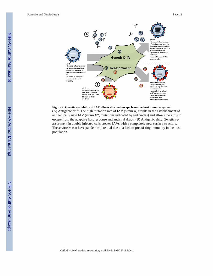
Figure 2. Genetic variability of IAV allows efficient escape from the host immune system(A) Antigenic drift: The high mutation rate of IAV (strain X) results in the establishment ofantigenically new IAV (strain X*, mutations indicated by red circles) and allows the virus toescape from the adaptive host response and antiviral drugs. (B) Antigenic shift: Genetic re-assortment in double infected cells creates IAVs with a completely new surface structure.These viruses can have pandemic potential due to a lack of preexisting immunity in the hostpopulation.
Schmolke and García-Sastre Page 12
Cell Microbiol. Author manuscript; available in PMC 2011 July 1.
NIH
-PA Author Manuscript
NIH
-PA Author Manuscript
NIH
-PA Author Manuscript
Related Documents