ARTICLE Evaluation of the fidelity of immunolabelling obtained with clone 5D8/1, a monoclonal antibody directed against the enteroviral capsid protein, VP1, in human pancreas Sarah J. Richardson & Pia Leete & Shalinee Dhayal & Mark A. Russell & Maarit Oikarinen & Jutta E. Laiho & Emma Svedin & Katharina Lind & Therese Rosenling & Nora Chapman & Adrian J. Bone & The nPOD-V Consortium & Alan K. Foulis & Gun Frisk & Malin Flodstrom-Tullberg & Didier Hober & Heikki Hyoty & Noel G. Morgan Received: 14 June 2013 /Accepted: 2 October 2013 # Springer-Verlag Berlin Heidelberg 2013 Abstract Aims/hypothesis Enteroviral infection has been implicated in the development of islet autoimmunity in type 1 diabetes and enteroviral antigen expression has been detected by immunohistochemistry in the pancreatic beta cells of patients with recent-onset type 1 diabetes. However, the immunohistochemical evidence relies heavily on the use of a monoclonal antibody, clone 5D8/1, raised against an enteroviral capsid protein, VP1. Recent data suggest that the clone 5D8/1 may also recognise non-viral antigens; in particular, a component of the mitochondrial ATP synthase (ATP5B) and an isoform of creatine kinase (CKB). Therefore, we evaluated the fidelity of immunolabelling by clone 5D8/1 in the islets of patients with type 1 diabetes. Methods Enteroviral VP1, CKB and ATP5B expression were analysed by western blotting, RT-PCR and immunocytochemistry in a range of cultured cell lines, isolated human islets and human tissue. Results Clone 5D8/1 labelled CKB, but not ATP5B, on western blots performed under denaturing conditions. In cultured human cell lines, isolated human islets and pancreas sections from patients with type 1 diabetes, the immunolabelling of ATP5B, CKB and VP1 by 5D8/1 was readily distinguishable. Moreover, in a human tissue microarray displaying more than 80 different cells and tissues, only two (stomach and colon; both of which are potential sites of enterovirus infection) were immunopositive when stained with clone 5D8/1. Electronic supplementary material The online version of this article (doi:10.1007/s00125-013-3094-7) contains peer-reviewed but unedited supplementary material, which is available to authorised users. S. J. Richardson (*) : P. Leete : S. Dhayal : M. A. Russell : N. G. Morgan (*) Institute of Biomedical and Clinical Sciences, University of Exeter Medical School, RILD Building, Barrack Road, Exeter EX2 5DW, UK e-mail: [email protected] e-mail: [email protected] M. Oikarinen : J. E. Laiho : H. Hyoty Department of Virology, Medical School, University of Tampere, Tampere, Finland E. Svedin : K. Lind : M. Flodstrom-Tullberg Department of Medicine HS, Center for Infectious Medicine, Karolinska Institutet, Stockholm, Sweden T. Rosenling : G. Frisk Division of Immunology, Genetics and Pathology, Uppsala University, Uppsala, Sweden N. Chapman Department of Pathology and Microbiology, University of Nebraska Medical Centre, Omaha, NE, USA A. J. Bone School of Pharmacy and Biomolecular Sciences, University of Brighton, Brighton, UK A. K. Foulis GG&C Pathology Department, Southern General Hospital, Glasgow, UK D. Hober University Lille 2, CHRU Lille Laboratory of Virology EA3610, Lille, France H. Hyoty Fimlab Laboratories, Pirkanmaa Hospital District, Tampere, Finland Diabetologia DOI 10.1007/s00125-013-3094-7

Welcome message from author
This document is posted to help you gain knowledge. Please leave a comment to let me know what you think about it! Share it to your friends and learn new things together.
Transcript
ARTICLE
Evaluation of the fidelity of immunolabelling obtainedwith clone 5D8/1, a monoclonal antibody directedagainst the enteroviral capsid protein, VP1, in human pancreas
Sarah J. Richardson & Pia Leete & Shalinee Dhayal & Mark A. Russell & Maarit Oikarinen &
Jutta E. Laiho & Emma Svedin & Katharina Lind & Therese Rosenling & Nora Chapman &
Adrian J. Bone & The nPOD-V Consortium & Alan K. Foulis & Gun Frisk &
Malin Flodstrom-Tullberg & Didier Hober & Heikki Hyoty & Noel G. Morgan
Received: 14 June 2013 /Accepted: 2 October 2013# Springer-Verlag Berlin Heidelberg 2013
AbstractAims/hypothesis Enteroviral infection has been implicated inthe development of islet autoimmunity in type 1 diabetes andenteroviral antigen expression has been detected byimmunohistochemistry in the pancreatic beta cells ofpatients with recent-onset type 1 diabetes. However,the immunohistochemical evidence relies heavily on the useof a monoclonal antibody, clone 5D8/1, raised against anenteroviral capsid protein, VP1. Recent data suggest that theclone 5D8/1 may also recognise non-viral antigens; inparticular, a component of the mitochondrial ATP synthase(ATP5B) and an isoform of creatine kinase (CKB). Therefore,we evaluated the fidelity of immunolabelling by clone 5D8/1in the islets of patients with type 1 diabetes.
Methods Enteroviral VP1, CKB and ATP5B expressionwere analysed by western blotting, RT-PCR andimmunocytochemistry in a range of cultured cell lines,isolated human islets and human tissue.Results Clone 5D8/1 labelled CKB, but not ATP5B, onwestern blots performed under denaturing conditions.In cultured human cell lines, isolated human islets andpancreas sections from patients with type 1 diabetes, theimmunolabelling of ATP5B, CKB and VP1 by 5D8/1was readily distinguishable. Moreover, in a human tissuemicroarray displaying more than 80 different cells and tissues,only two (stomach and colon; both of which arepotential sites of enterovirus infection) were immunopositivewhen stained with clone 5D8/1.
Electronic supplementary material The online version of this article(doi:10.1007/s00125-013-3094-7) contains peer-reviewed but uneditedsupplementary material, which is available to authorised users.
S. J. Richardson (*) : P. Leete : S. Dhayal :M. A. Russell :N. G. Morgan (*)Institute of Biomedical and Clinical Sciences, University of ExeterMedical School, RILDBuilding, Barrack Road, Exeter EX2 5DW,UKe-mail: [email protected]: [email protected]
M. Oikarinen : J. E. Laiho :H. HyotyDepartment of Virology, Medical School, University of Tampere,Tampere, Finland
E. Svedin :K. Lind :M. Flodstrom-TullbergDepartment of Medicine HS, Center for Infectious Medicine,Karolinska Institutet, Stockholm, Sweden
T. Rosenling :G. FriskDivision of Immunology, Genetics and Pathology, Uppsala University,Uppsala, Sweden
N. ChapmanDepartment of Pathology and Microbiology, University of NebraskaMedical Centre, Omaha, NE, USA
A. J. BoneSchool of Pharmacy and Biomolecular Sciences, University ofBrighton, Brighton, UK
A. K. FoulisGG&C Pathology Department, Southern General Hospital,Glasgow, UK
D. HoberUniversity Lille 2, CHRU Lille Laboratory of Virology EA3610,Lille, France
H. HyotyFimlab Laboratories, Pirkanmaa Hospital District, Tampere, Finland
DiabetologiaDOI 10.1007/s00125-013-3094-7
Conclusions/interpretation When used under carefullyoptimised conditions, the immunolabelling pattern detectedin sections of human pancreas with clone 5D8/1 did not reflectcross-reactivity with either ATP5B or CKB. Rather, 5D8/1 islikely to be representative of enteroviral antigen expression.
Keywords ATP5B . Coxsackievirus . Creatine kinase .
Dako 5D8/1 . Immunohistochemistry . Islets of Langerhans .
Pancreatic beta cell
AbbreviationsCKB Creatine kinase BCV CoxsackievirusMOI Multiplicity of infection
Introduction
Considerable evidence has accumulated to support thepossibility that type 1 diabetes may have a viral aetiology, atleast in some individuals. A number of viruses have beenimplicated, but the majority view suggests that one or moreenteroviruses are the most likely candidates [1, 2]. However,this evidence remains largely circumstantial and relies on a bodyof epidemiological data supported by immunohistochemicalstudies conducted with pathological specimens recovered frompatients diagnosed with type 1 diabetes [1, 3–5]. These haverevealed that a minority of islet cells of individuals with recent-onset disease (and some with longer-duration illness) displayevidence of enteroviral antigen expression [3–5]. In a smallnumber of cases, the immunocytochemical evidence has beensupported by more direct evidence of the presence of virus, viathe detection of positive in situ hybridisation signals indicativeof the presence of viral RNA [6]. On two occasions, apotentially diabetogenic strain of Coxsackievirus (CV)B4 hasbeen isolated directly from the pancreases of patients withdiabetes [3, 7]. GroupBCoxsackieviruses have also been linkedto type 1 diabetes in epidemiological studies [8, 9], and they cancause pancreatitis and diabetes in mice [10]. Collectively, thesestudies imply that, in a proportion of recently diagnosed patientswith type 1 diabetes, the development of a persistent enterovirusinfection (especially with CVB4) might underlie the changesthat lead to autoimmunity and ultimately to the loss ofpancreatic beta cells.
One important caveat is that the majority of studies in whichviral antigen expression has been detected in the islets of patientswith type 1 diabetes have employed a commercial monoclonalantiserum, clone 5D8/1 [3–5]. This antibody is directed againsta peptide sequence (EIPALTAVE [11–13]) encoded within oneof the viral capsid proteins, VP1, which is expressed by a rangeof enteroviral species. The antibody has been validatedextensively and is widely recognised to bind to VP1 with highavidity. This antibody has been used in several studies to detect
enteroviral VP1 in various human and mouse tissues, includingthyroid, myocardium and pancreas [3–5, 13–17]. However, it isaccepted that few antibodies display absolute specificity underall conditions and it is well known that 5D8/1 can bind toadditional proteins under some circumstances, especially whenit is used at high concentrations or after incomplete optimisationwithin the target tissue [18]. Recently, this issue has beenbrought to the fore by studies implying that 5D8/1 can bind totwo specific proteins, a component of the mitochondrial ATPasecomplex, ATP5B, and an isoform of creatine kinase, CKB, onwestern blots of human islet extracts [19]. This led theauthors to propose that previous work in which 5D8/1was used to define viral antigen expression in pancreassections by immunohistochemistry should be re-evaluated,since the conclusion that immunopositivity is indicative ofthe presence of virus protein might be unsafe.
In the present study we have undertaken this re-evaluationand show that, under carefully optimised conditions, 5D8/1does not label either ATP5B or CKB in fixed tissue samplesstudied by immunocytochemical methods. As such, this worklends further support to earlier conclusions that the detectionof VP1 in the islet cells of patients with type 1 diabetes isindicative of the presence of an enteroviral infection.
Methods
Viruses CVB4 (VD2921), CVB1 (CVB1-10802 CDC strain),CVB3 (Nancy) and CVB4E2 were used. VD2921 (GenBankaccession number AF328683) was originally isolated from thecerebrospinal fluid of a patient with aseptic meningitis [20,21]. The CVB1-10802 CDC strain was isolated in Argentinain 1998 by the Centers for Disease Control and Prevention(Atlanta, GA, USA). The CVB4E2 diabetogenic strain waskindly provided by Dr J.-W. Yoon (Calgary, AB, Canada).CVB3 (Nancy) was originally isolated in Connecticut in 1949and is available from the American Type Culture Collection(www.lgcstandards-atcc.org/).
Cell culture and virus infection HeLa and HepG2 cells werecultured in RPMI 1640 at 37°C and 5% CO2. CVB3 Nancywas propagated in HeLa cells and the titre was determinedusing a standard plaque assay. HeLa and HepG2 cells weremock infected or incubated with CVB3Nancy (multiplicity ofinfection [MOI] 20 or 30 for HeLa and HepG2 cells,respectively) for 1 h.
PANC-1 cells were cultured in DMEM and infected with0.01 MOI of CVB4E2. At 24 h postinfection, cells werewashed three times with DMEM, resuspended in freshmedium and incubated at 37°C.
Specimens Pancreases recovered from two patients with type1 diabetesmellitus were selected from cohorts used previously
Diabetologia
[5]. The specimens were studied with approval from GreaterGlasgow NHS Research Ethics Committee, and had beenfixed in buffered formalin and paraffin embedded.
Human tissue array A normal and cancer human tissue arraywas provided by the NHS Greater Glasgow and Clyde BioRepository (UK).
Isolation and culture of human islets Islets of Langerhanswere isolated, cultured and infected in Uppsala University,Sweden, using a protocol approved by the local ethicscommittee, as previously described [22, 23].
RNA extraction and real-time RT-PCR Total RNA from isletsstored in RNAlater was extracted using RNeasy Plus (QiagenAB, Sollentuna, Stockholm, Sweden). Up to 50 ng total RNAper sample was reverse transcribed using SuperScript IIReverse Transcriptase (Invitrogen, Stockholm, Sweden).Real-time PCRs were run with Power SYBR Green MasterMix (Applied Biosystems, Stockholm, Sweden). Predesignedgene-specific primer sets (QuantiTect Primer Assays, Qiagen)were used to detect CKB cDNA. PCR specificity was verifiedby melt-curve analysis of products.
Formalin-fixed paraffin embedding of islets or culturedcells Islets were fixed in 4% paraformaldehyde at roomtemperature for 4 h, washed in PBS and dehydratedwith increasing ethanol concentrations then xylene.Islets were paraffin embedded, sectioned (5 μm) andmounted on glass slides (Superfrost Plus, Thermo Scientific,Waltham, MA, USA).
Western blotting Cellular extracts or recombinant purifiedproteins were separated using SDS-PAGE, transferred tonitrocellulose or polyvinylidene fluoride membranes and thenincubated with the relevant primary antibodies overnight at4°C: VP1 (1:400, Dako clone 5D8/1), CKB (1:500, SigmaPrestige [#HPA001254], Sigma, Poole, UK) and ATP5B(1:250, Sigma Prestige [#HPA001520]). Actin (1:30,000,MPBiomedicals, Cambridge, UK) served as a loading control.Immunoreactive bands were detected using horseradishperoxidase-labelled secondary antibodies (1:1,000, Bio-Rad,Hemel Hempstead, UK) and visualised using SuperSignalenhanced chemiluminescent substrate (Thermo Scientific,Rockford, IL, USA).
Immunohistochemistry Sections of 4 μm were heated in10mmol/l citrate (pH 6.0) in a pressure cooker in a microwaveoven at 800W for 20min, then cooled at room temperature for20 min. Primary antibodies (Electronic SupplementaryMaterial [ESM] Table 1) were applied and the Dako REALEnVision Detection System (Dako, Ely, UK) was used forantigen detection. Clone 5D8/1 (1:2,000) was preincubated
overnight at 4°C with peptides (10 μg/ml) or recombinantprotein (at 1,000-fold molar excess) in blocking experiments.
Combined immunofluorescence Rabbit anti-CKB, anti-ATP5B and mouse anti-VP1 (Dako) were detected with AlexaFluor 568-conjugated anti-rabbit antibody or an Alexa Fluor488 conjugated anti-mouse antibody (Invitrogen). DAPI(1:1,000, Invitrogen) was included in the final secondaryincubation to stain cell nuclei. Sections were mounted inVectashield hard-set mounting medium (Vector Laboratories,Peterborough, UK) under glass coverslips. Images were capturedusing either a Nikon Eclipse 80i microscope (Nikon, Guildford,UK) and overlaid using NIS-Elements BR 3.0 software (Nikon)or a Zeiss LSM510meta confocalmicroscope (Zeiss, Cambridge,UK) to study the relative localisation of each antigen.
Peptide ELISA High-binding enzyme immunoassay plateswere coated with 1 μg/well of relevant peptide antigens(synthesised by GenScript, Piscataway, NJ, USA) in50 mmol/l sodium carbonate buffer (pH 9.4). Plates wereblocked with 5% normal goat serum (Vector Laboratories) inPBS and incubated for 2 h with varying dilutions of clone5D8/1 in 5% normal goat serum/PBS. The plates were washedwith PBS–Tween-20 (0.05%) and binding of the antibody wasdetected with alkaline phosphatase-conjugated anti-mouseIgG (1:4,000, Sigma) using p -nitrophenyl-phosphate(Sigma) as substrate. The reaction was stopped using 3 mol/lNaOH. Absorbance was read at 405 nm.
Results
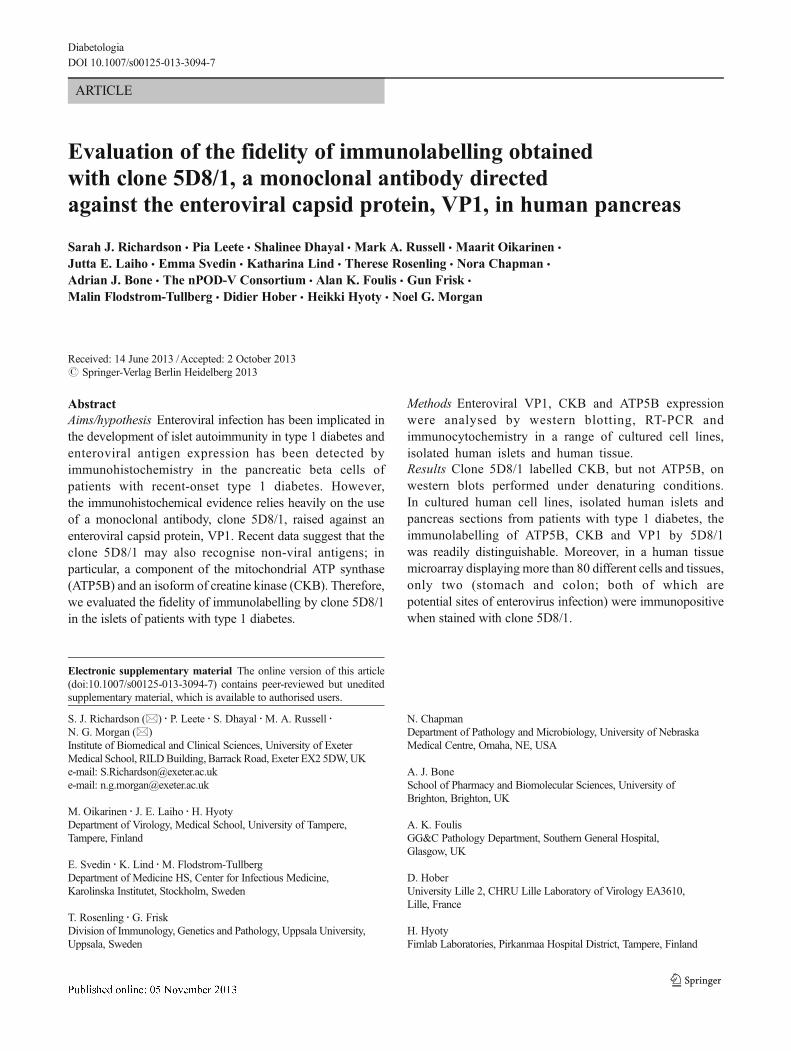
Analysis of the cross-reactivity of 5D8/1 with CKB and ATP5Bon western blots It was initially established that cultured celllines that are susceptible to infection with group BCoxsackieviruses express CKB and/or ATP5B (Fig. 1a, b). Inboth cell types, a strongly immunoreactive band of appropriatemolecular mass was detected in both uninfected cells and cellsharvested after infection with CVB3. The intensity of CKBlabelling declined with increasing time postinfection, whereasthe labelling of ATP5B was not altered during viral infection.When these extracts were probed with the enterovirus antibody5D8/1, an immunoreactive band corresponding to that detectedby the CKB antibody was again detected (Fig. 1a, b). Incontrast, no band migrating in the position expected for ATP5Bwas labelled by 5D8/1 in either cell line.
VP1 immunoreactivity was detected with clone 5D8/1within 4 h of CVB3 infection in HeLa cells and this wasmaintained until at least 8 h postinfection (Fig. 1a). In HepG2cells, the intensity of VP1 labelling was minimal at 4 h butincreased dramatically by 6 and 8 h postinfection (Fig. 1b).Neither of the antisera directed against CKB or ATP5Bdetected the time-dependent expression of VP1.
Diabetologia
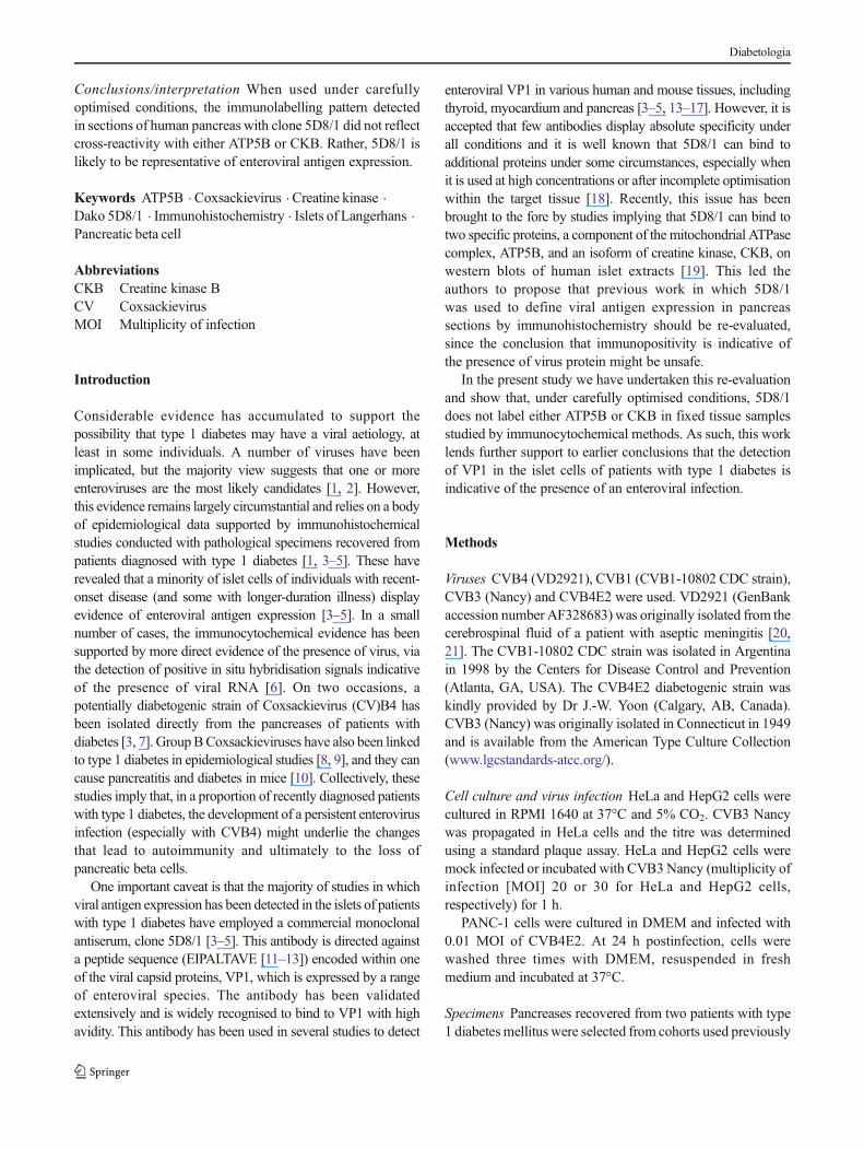
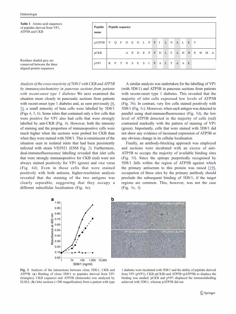
To confirm these data, purified recombinant proteins(rCKB and rATP5B) were examined by western blotting(Fig. 1c) and it was found that, under denaturing conditions,rCKB was detected by 5D8/1 whereas rATP5B was not. Inaddition, an ELISA assay was developed in which peptidesequences corresponding to the epitopes purportedlyrecognised by 5D8/1 in VP1, CKB and ATP5B [19] wereemployed (Table 1). This confirmed that the relevant peptideepitopes within both VP1 (pVP1) and CKB (pCKB) arebound by 5D8/1, although pVP1 was bound more avidly(Fig. 2a). No binding to pATP5B was detected (Fig. 2a). Inconfirmation, the addition of an excess of pVP1 or pCKB led
to displacement of the immunolabelling achieved with 5D8/1in islet cells, whereas pATP5B did not displace the binding of5D8/1 (Fig. 2b).
These results were confirmed using cell lines infected withCoxsackievirus (ESM Fig. 1), in which it was found thatpCKB and pVP1 each abrogated the immunolabelling ofVP1 by clone 5D8/1, while pATP5B was ineffective.
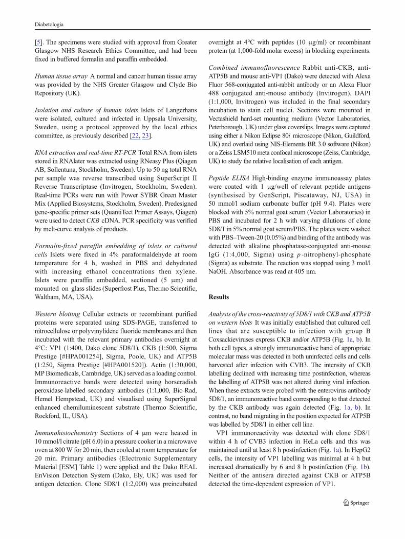
Analysis of the cross-reactivity of 5D8/1 with CKB and ATP5Bby immunocytochemistry in fixed cells To establish whetherclone 5D8/1 displays similar cross-reactivity under optimisedconditions in fixed, paraffin-embedded cells and tissue, theantibody was applied to both HeLa and HepG2 cells stained inparallel with antisera raised against either CKB or ATP5B(Fig. 3a, b). As expected from the western blottingexperiments, both CKB and ATP5B were present in the cells,although their expression was not uniform within the cellpopulation. Importantly, none of the uninfected HeLa orHepG2 cells were stained positively by 5D8/1, despite thefact that both cell lines expressed abundant levels of CKB andATP5B within the cell population. In contrast, when these celllines were infected with CVB3, the majority of cells thenbecame strongly immunopositive when stained with 5D8/1.
A third human cell line, PANC1, was also examined byimmunocytochemistry. LikeHeLa andHepG2 cells, the PANC1line also expressed CKB under control (uninfected) conditions,but these cells did not stain positively when 5D8/1 was applied.In contrast, when PANC1 cells were infected with CVB4E2,they became immunopositive for VP1 (Fig. 3d). Dual-immunofluorescence labelling revealed that some cells weredifferentially immunopositive when stained with both antiseratogether (Fig. 3d). Thus, detection of CKBwithAlexa Fluor 568(red) revealed the presence of strongly stained cells that wereimmunonegative for enteroviral VP1 (Alexa Fluor 488 [green]).Conversely, other cells within the population were clearlyimmunopositive for VP1 (green) but negative for CKB (red).
Analysis of the cross-reactivity of 5D8/1 with CKB byimmunocytochemistry in isolated human islets of LangerhansHaving established that, despite the possibility that 5D8/1 maycross-react with CKB on western blots and in ELISA assays, itdoes not do so in fixed cells under optimal conditions, we thenexamined the labelling patterns obtained in isolated humanislets following fixation (ESM Fig. 2). Control islets wereimmunonegative for both VP1 and CKB, whereas isletsinfected with a strain of Coxsackievirus that establishes a morepersistent infection (VD2921) were stained positively for CKB.Despite this, most cells did not stain positively when exposed to5D8/1 in islets infected with VD2921. In contrast, infection ofislets with an acutely lytic strain of Coxsackievirus (CVB1-11)resulted in the appearance of cells that were strongly positivefor VP1, but that remained largely immunonegative whenprobed for CKB (ESM Fig. 2).
70
4035
25
Mr(kDa)
Anti-VP1 (5D8/1)CVB3
Anti-CKBCVB3
Anti-ATP5BCVB3
C 0 2 4 6 8 h
Actin
Actin
C 0 2 4 6 8 h C 0 2 4 6 8 h
70
4035
25
Mr(kDa)
70
4035
25
CCVB3
0 2 4 6 8 h CCVB3
0 2 4 6 8 h CCVB3
0 2 4 6 8 h
70
4035
25
Anti-VP1(5D8/1) Anti-CKB Anti-ATP5B
~ 47
~ 95
rCKB rATP5B rCKB rATP5B rCKB rATP5B
a
b
c
Fig. 1 Western blotting of CBV3-infected and mock-infected cellextracts or purified recombinant CKB and ATP5B. HeLa (a) and HepG2(b) cells were infected with CVB3 Nancy (HeLa, 20 MOI; HepG2, 30MOI). Total protein was isolated at various time points (0–8 h, as shown)postinfection. A total of 5 μg protein from each sample was loaded ontoSDS–polyacrylamide gels and the expression of VP1, CKB and ATP5Bwas assessed on western blots. Actin was detected as the loading control.(c) A total of 100 ng recombinant CKB (rCKB) or ATP5B tagged withglutathione-S-transferase (rATP5B; molecular mass ∼95 kDa) was run onSDS–polyacrylamide gels and then electroblotted, and the membraneswere incubated with 5D8/1 (1:400) anti-CKB (1:500) or anti-ATP5B(1:250) as shown. Immunoreactive bands were detected withchemiluminescent substrate
Diabetologia
Analysis of the cross-reactivity of 5D8/1 with CKB and ATP5Bby immunocytochemistry in pancreas sections from patientswith recent-onset type 1 diabetes We next examined thesituation more closely in pancreatic sections from patientswith recent-onset type 1 diabetes and, as seen previously [4,5], a small minority of beta cells were labelled by 5D8/1(Figs 4, 5, 6). Some islets that contained only a few cells thatwere positive for VP1 also had cells that were stronglylabelled by anti-CKB (Fig. 4). However, both the intensityof staining and the proportion of immunopositive cells weremuch higher when the sections were probed for CKB thanwhen they were stained with 5D8/1. This is reminiscent of thesituation seen in isolated islets that had been persistentlyinfected with strain VD2921 (ESM Fig. 2). Furthermore,dual-immunofluorescence labelling revealed that islet cellsthat were strongly immunopositive for CKB (red) were notalways stained positively for VP1 (green) and vice versa(Fig. 4d). Even in those cells that were stainedpositively with both antisera, higher-resolution analysisrevealed that the staining of the two antigens wasclearly separable, suggesting that they occupy adifferent subcellular localisation (Fig. 4e).
A similar analysis was undertaken for the labelling of VP1(with 5D8/1) and ATP5B in pancreas sections from patientswith recent-onset type 1 diabetes. This revealed that themajority of islet cells expressed low levels of ATP5B(Fig. 5b). In contrast, very few cells stained positively with5D8/1 (Fig. 5c). Moreover, when each antigen was detected inparallel using dual-immunofluorescence (Fig. 5d), the lowlevel of ATP5B detected in the majority of cells (red)contrasted markedly with the pattern of staining of VP1(green). Importantly, cells that were stained with 5D8/1 didnot show any evidence of increased expression of ATP5B orany obvious change in its cellular localisation.
Finally, an antibody-blocking approach was employedand sections were incubated with an excess of anti-ATP5B to occupy the majority of available binding sites(Fig. 5f). Since the epitope purportedly recognised by5D8/1 falls within the region of ATP5B against whichthe primary antiserum to this protein was raised [19],occupation of these sites by the primary antibody shouldpreclude the subsequent binding of 5D8/1, if the targetregions are common. This, however, was not the case(Fig. 5e, f).
Table 1 Amino acid sequencesof peptides derived from VP1,ATP5B and CKB
Residues shaded grey areconserved between the threealigned protein sequences
Peptide
name
Peptide sequence
pATP5B V Q F D E G L P P I L N A L E V
pCKB A E D E F P D L S A H N N H M A
pVP1 R P T N S E S I P A L T A A E
VP1 (5D8/1) & pATP5BVP1 (5D8/1) alone
VP1 (5D8/1) & pCKB VP1 (5D8/1) & pVP1
0.00
0.10
0.20
0.30
0.40
0.50
0.60
0.70
0.80
0.90
1.00a b
1 10 100 1,000 10,000
Abs
orba
nce
at 4
05 n
m
5D8/1 (ng/ml)
Fig. 2 Analysis of the interactions between clone 5D8/1, CKB andATP5B. (a) Binding of clone 5D8/1 to peptides derived from VP1(triangles), CKB (squares) and ATP5B (diamonds) was analysed byELISA. (b) Islet sections (×200 magnification) from a patient with type
1 diabetes were incubated with 5D8/1 and the ability of peptides derivedfrom VP1 (pVP1), CKB (pCKB) and ATP5B (pATP5B) to displace thebinding was studied. pCKB and pVP1 displaced the immunolabellingachieved with 5D8/1, whereas pATP5B did not
Diabetologia
VP
1C
KB
AT
P5B
Control CVB3 Control CVB3
CKB VP1 MergedCKB VP1
a b
c d
Fig. 3 HeLa (a ; ×400 magnification) and HepG2 (b ; ×400magnification) cells were infected with CVB3 Nancy and cells werecollected 4 h (HeLa) or 6 h (HepG2) postinfection. Cells were fixed withformalin and embedded in paraffin and 5 μm sections were cut andmounted onto glass slides. Serial sections of mock and CVB3-infectedcells were immunostained for the presence of enterovirus VP1 protein(antibody dilution 1:2,000; equivalent to 55 ng/ml), CKB (1:500) and
ATP5B (1:500). (c ) Formalin-fixed, paraffin-embedded, CVB4E2-infected PANC1 cells were immunostained for CKB (1:500) and VP1(1:2,000). Images were captured at ×400 magnification (d )Photomicrographs (×600magnification) of representative CVB3-infectedPANC1 cells immunostained for CKB (red) and VP1 (green) revealedthat CKB (white arrow) and VP1 (orange arrow) did not co-localise.Nuclear (DAPI) staining is shown in blue in the merged image
VP1; CKB VP1; CKB
CKBVP1Insulina
d e
b c
Fig. 4 Photomicrographs (×200 magnification) of a representative isletfrom a patient with recent-onset type 1 diabetes. Serial sections wereimmunostained for (a) insulin, (b) VP1 and (c) CKB. (d) Fluorescencemicroscopy analysis (×400 magnification) of a representative islet from apatient with recent-onset type 1 diabetes revealed VP1-positive cells
(green arrow), CKB-positive cells (red arrows) and double-positive cells(yellow arrow). (e ) High-resolution analysis (×600 magnification)demonstrated that the staining of the two antigens (VP1 [green]and CKB [red]) was separable. (d , e ) Nuclear (DAPI) staining isshown in blue
Diabetologia
As a further confirmation of the specificity of labellingachieved with 5D8/1, we also examined the ability ofrecombinant CKB to displace the immunolabelling achievedwith this antibody in both islet sections (Fig. 6a) and inCoxsackievirus-infected cell lines (Fig. 6b). Importantly,rCKB failed to displace the immunolabelling achievedwith 5D8/1 in either the islets or infected cells although,as noted in infected cells (ESM Fig. 1), the peptidederived from CKB was able to displace binding.
Tissue expression of immunoreactive VP1 As a furtherverification, a human tissue array consisting of 154 samplescovering more than 80 cell and tissue types (including normaland tumour tissues) was probed with clone 5D8/1 (1:2,000).This yielded only two positive signals, in one of two samplesof gastric body and normal colon (ESM Table 2). We alsoexamined several mitochondria-rich tissues in greater detail,and such tissues (including liver and kidney) were uniformlynegative when stained with 5D8/1 at a dilution of 1:2,000(ESM Fig. 3).
Discussion
The concept that the triggering events leading to the initiationof islet cell autoimmunity and type 1 diabetes in humans mayinvolve the development of a sustained enteroviral infection ofislet beta cells has received widespread attention [1, 2].Epidemiological evidence supports this proposition and, inrare cases, enteroviruses have even been isolated from
pancreas samples recovered from patients with type 1 diabetes[1, 7]. In addition, a strong association has been foundbetween the expression of viral antigens in the beta cells ofpatients and disease development [3–5]. However, much ofthe immunohistological evidence has relied on the use ofclone 5D8/1 to detect the production of an enterovirus capsidprotein (VP1). This antibody is used preferentially because itbinds the target protein with high avidity in tissue samples andis capable of detecting a conserved sequence found in a widerange of different enterovirus serotypes [11, 24]. Accordingly,in the world’s largest study, more than 60% of pancreasesrecovered from patients with recent-onset type 1 diabetescontained islets that were immunopositive for enterovirusVP1 when stained with 5D8/1 [4]. This compared with only6% immunopositivity in the islets of age-matched controls [4].This evidence is striking, but it relies upon the specificity ofclone 5D8/1, and this has been a cause of concern because ofsuggestions that this antibody can, if used under suboptimallabelling conditions, provide false-positive signals [18].Importantly, Hansson et al have recently identified twopotentially cross-reactive antigens and have argued that, inthe beta cells of patients with recent-onset type 1 diabetes,labelling of these proteins could provide a basis for falsepositivity [19]. However, we now provide firm evidence toconfirm that the immunoreactivity seen in islet cells does notderive from a cross-reaction between 5D8/1 with either CKBor ATP5B.
First, we re-examined the recent conclusion [19] that 5D8/1labels CKB and ATP5B on western blots, and animmunoreactive band that co-migrated with a protein labelled
Insulin ATP5B VP1a
d e f
b c
ATP5B
VP1 VP1 VP1 (ATP5B block)
Fig. 5 Photomicrographs (×200magnification) of a representativeislet from a patient with recent-onset type 1 diabetes. Serialsections were immunostained for(a) insulin, (b) ATP5B and (c)VP1. (d) Fluorescencemicroscopy analysis (×400magnification) of a representativeislet revealed both VP1-positivecells (green) and ATP5B-positivecells (red). Nuclei were stainedwith DAPI (blue). Sections werepreincubated with (e) antibodydiluent alone or (f) ATP5Bantibody (10 μg/ml) overnight,prior to immunostaining for VP1(green; 1:2,000)
Diabetologia
by an antibody to CKB in various cultured cell lines was seen.Purified recombinant CKB was also detected by 5D8/1 onwestern blots, thereby confirming that clone 5D8/1 can labelthis enzyme under denaturing conditions. In contrast, nocross-reactivity was detected with a protein that co-migratedin the expected position of ATP5B and, when purifiedrecombinant ATP5B was probed with clone 5D8/1, noimmune-cross-reactivity was detected. In support of thesedata, we also found that the peptide antigens identified asthe likely epitopes recognised by clone 5D8/1 [19] weredifferentially detected in an ELISA assay. As expected, theantibody bound with highest affinity to the peptide from VP1,but it also recognised that from CKB. In contrast, no bindingwas detected to the relevant peptide from ATP5B.
When cultured cells were fixed and paraffin embedded foranalysis by immunohistochemistry using methods similar tothose employed to detect the presence of viral VP1 in thepancreases of type 1 diabetic patients, it was apparent that bothCKB and ATP5B are expressed in a majority of the population.
However, these cells were unstained by clone 5D8/1. Despitethis lack of cross-reactivity, the antibody labelled VP1 readily inenterovirus-infected cells. Thus, under optimal conditions, it isclear that neither CKB nor ATP5B are readily stained by clone5D8/1 in cultured cells after fixation and paraffin embedding.Similar conclusions were also drawn from the examination oftissue microarrays, in which 80 different tissues wereimmunostained in parallel. Of these, only colon and stomachtissues stained positively with clone 5D8/1, and both of theseare probable sites of infection by enteroviruses.
Our conclusionswere reinforced by dual-immunocytochemicalanalysis of cultured cells following infectionwith enteroviruses.This allowed the firm identification of a population of cellsexpressing VP1 in addition to CKB, as well as furthersubpopulations that expressed each of these antigens alone.Thus, the antibodies were not cross-reactive with commonantigens under the experimental conditions employed.
The study was then extended to include isolated humanislets. When these were immunostained with antisera directedagainst CKB and VP1, respectively, no evidence of cross-reactivity was found. However, whereas CKB was detectedonly weakly in uninfected human islets, it was present in isletsthat had been infected with a strain of Coxsackievirus type Bthat establishes a persistent sublytic infection. This impliesthat CKB might become upregulated under such conditions.To our knowledge, such upregulation has not been notedpreviously in cells infected with enteroviruses, although ithas been reported that CKB is required for the replication ofcertain strains of hepatitis C, another positive-strand RNAvirus [25, 26]. Thus, it seems possible that CKB might playa hitherto unrecognised role in the development of a sustainedinfection of islet cells. This will require additional verificationbut, if confirmed, may have implications for the progressionof such infections in the beta cells of patients developing type1 diabetes. However, irrespective of this conclusion, it is clearthat the altered expression of CKB does not lead to thedevelopment of a corresponding immunopositivity when theislets are stained with 5D8/1. This confirms that CKB is not atarget for clone 5D8/1 under such conditions and also revealsthat the synthesis of VP1 capsid protein is minimal underconditions of persistent enterovirus infection. This may, inturn, have relevance to the situation found in pancreas sectionsfrom patients with type 1 diabetes, where the proportion ofimmunopositive cells is very low (∼5% at best [5]), implyingthat production of VP1 occurs in only a few cells at any giventime. As such, this would be consistent with a mechanism inwhich the majority of islet cells sustain a persistent sublyticinfection and very few are involved in the active synthesis ofviral capsid proteins.
Finally, dual-immunofluorescence detection of each proteinin turn was undertaken and compared with the pattern oflabelling achieved with clone 5D8/1. The data again impliedthat all of the antigens are separate and are detected
No additionb
a
rCKB
VP
1 (5
D8/
1) a
lone
VP
1 (5
D8/
1) &
CK
B
pCKB rCKB
Fig. 6 Effects of the addition of an excess of either recombinant (r)CKB ora peptide epitope derived from CKB (pCKB) on the immunolabellingachieved with 5D8/1 in the islets of a patient with type 1 diabetes and incultured cells infected with Coxsackievirus. (a ) Islet sections wereincubated with clone 5D8/1 (1:2,000) either alone or in the presence ofan excess of pCKB or rCKB prior to immunodetection. (b) CVB5-infectedVero cells were immunostained with 5D8/1 (1:2,000) either alone or incombination with an excess of rCKB prior to immunodetection. Imageswere captured at ×200 magnification
Diabetologia
independently by each antiserum employed. Most significantly,some cells were found to display strong immunoreactivity bothto an antibody raised against CKB as well as to that againstVP1. However, high-resolution analysis of the images clearlyshowed that the antigens were not co-localised within the cells.
These considerations raise the important question of whyCKB is detected by clone 5D8/1 on western blots (and inELISA assays) but not in tissue samples labelled in situ.Hansson et al [19] have argued that this may be because clone5D8/1 only recognises the relevant epitopes present in CKB(and ATP5B) when the protein is expressed in stressed isletcells. However, they offer no molecular basis for thisproposition, except to suggest that this may reflect a level ofmitochondrial stress. We consider this to be unlikely, not leastbecause CKB is a cytosolic protein [27, 28] and not normallylocalised within mitochondria. Rather, we suspect that, byreference to the crystal structure of CKB [29], the epitopedetected by clone 5D8/1 is not normally accessible to theantibody in tissue sections. CKB is a dimer that, as noted byEder et al [29], can only be dissociated under stronglydenaturing conditions (i.e. similar to those used for westernblotting). Thus, in tissue sections, it is probable that the proteinstructure is not disrupted sufficiently to allow access of theantibody, even after antigen retrieval. As a result, the selectivityfor VP1 is maintained during immunohistochemical analysiswhereas it may be lost on western blots. This hypothesis issupported by data revealing that a peptide epitope derived fromCKB was capable of displacing the binding of 5D8/1 to bothCoxsackievirus-infected cells and islet sections, whereas thefull-length recombinant protein was not.
A further argument against the stress hypothesis of Hanssonet al [19] is that infection of cells with enteroviruses thatproduce isoforms of VP1 that are not recognised by 5D8/1 donot stain positively with the antiserum [12, 13]. This confirmsthat the high level of stress imposed under these conditions doesnot lead to aberrant labelling of ATP5B or CKB.
Finally, we have recently had opportunity to conduct a moredetailed concordance analysis across a range of different well-preserved pancreas samples from the JDRF’s nPOD (Networkfor Pancreatic Organ Donors with Diabetes) collection. In thisanalysis, the immunohistochemical labelling of VP1 wascorrelated with detection of the viral genome by in situhybridisation. The results revealed a very high level ofconcordance (S. J. Richardson, G. Frisk, M. Oikarinen,J. E. Laiho, H. Hyoty, and N. G. Morgan; manuscript inpreparation) and also showed that immunopositivity for VP1correlated positively with hyperexpression of MHC class Imolecules. Since MHC class I hyperexpression is likely to bedriven by release of interferons, and this occurs in response toviral infection, these results add further weight to the view thatdetection of positive immunostaining of human beta cells byclone 5D8/1 under stringent conditions may reflect anunderlying enteroviral infection.
Acknowledgements We thank D. Lobert (University Lille 2, Lille,France) for culture and infection of PANC1 cells. Details of the nPOD-VConsortium can be found at www.jdrfnpod.org.
Funding This work was supported by funding from the EuropeanUnion’s Seventh Framework Programme PEVNET (FP7/2007-2013)under grant agreement number 261441. Additional support was from aDiabetes Research and Wellness Foundation non-clinical researchfellowship to SJR and from the Karolinska Institutet (KL), the StrategicResearch Programme in Diabetes at the Karolinska Institutet (MF-T) andthe Swedish Research Council (MF-T). The research was also performedwith the support of the Network for Pancreatic OrganDonors with Diabetes(nPOD), a collaborative type 1 diabetes research project sponsored by theJDRF, and with a JDRF research grant awarded to the nPOD-VConsortium. Organ procurement organisations partnering with nPOD toprovide research resources are listed at www.jdrfnpod.org/our-partners.php.
Duality of interest HH is a minor (<5%) shareholder and member ofthe board of Vactech, which develops vaccines against picornaviruses. Allother authors declare that there is no duality of interest associated withthis manuscript.
Contribution statement SJR was responsible for the study design andperformed experiments, analysed data and wrote the manuscript. PL, SD,MAR, MO, JEL, KL, ES and TR performed experiments, analysed dataand edited the manuscript. NC, DH, GF, HH, MF-T and AJB, contributedto the study design and reviewed and edited the manuscript. AKF collectedsamples, contributed to the study design and reviewed and edited themanuscript. NGM was responsible for the study design and wrote themanuscript. All authors reviewed and approved the final manuscript.
References
1. CraigME, Nair S, Stein H, RawlinsonWD (2013) Viruses and type 1diabetes: a new look at an old story. Pediatr Diabetes 14:149–158
2. Taylor K, Hyöty H, Toniolo A, Zuckerman AJ (eds) (2013) Diabetesand viruses. Springer, New York
3. Dotta F, Censini S, van Halteren AG et al (2007) Coxsackie B4 virusinfection of beta cells and natural killer cell insulitis in recent-onsettype 1 diabetic patients. Proc Natl Acad Sci U S A 104:5115–5120
4. Richardson SJ, Willcox A, Bone AJ, Foulis AK, Morgan NG (2009)The prevalence of enteroviral capsid protein vp1 immunostaining inpancreatic islets in human type 1 diabetes. Diabetologia 52:1143–1151
5. Richardson SJ, Leete P, Bone AJ, Foulis AK, Morgan NG (2013)Expression of the enteroviral capsid protein VP1 in the islet cells ofpatients with type 1 diabetes is associated with induction of proteinkinase R and downregulation of Mcl-1. Diabetologia 56:185–193
6. Ylipaasto P, Klingel K, Lindberg AM et al (2004) Enterovirusinfection in human pancreatic islet cells, islet tropism in vivo andreceptor involvement in cultured islet beta cells. Diabetologia 47:225–239
7. Yoon JW, Austin M, Onodera T, Notkins AL (1979) Isolation of avirus from the pancreas of a child with diabetic ketoacidosis.N Engl J Med 300:1173–1179
8. Gamble DR, Kinsley ML, FitzGerald MG, Bolton R, Taylor KW(1969) Viral antibodies in diabetes mellitus. Br Med J 3:627–630
9. Yeung WC, Rawlinson WD, Craig ME (2011) Enterovirus infectionand type 1 diabetes mellitus: systematic review and meta-analysis ofobservational molecular studies. BMJ 342:d35
10. Toniolo A, Onodera T, Jordan G, Yoon JW, Notkins AL (1982)Virus-induced diabetes mellitus. Glucose abnormalities produced in
Diabetologia
mice by the six members of the Coxsackie B virus group. Diabetes31:496–499
11. Samuelson A, ForsgrenM, SallbergM (1995) Characterization of therecognition site and diagnostic potential of an enterovirus group-reactive monoclonal antibody. Clin Diagn Lab Immunol 2:385–386
12. Oikarinen M, Tauriainen S, Penttila P et al (2010) Evaluation ofimmunohistochemistry and in situ hybridization methods forthe detection of enteroviruses using infected cell culture samples.J Clin Virol 47:224–228
13. Richardson SJ, Willcox A, Hilton DA et al (2010) Use of antiseradirected against dsRNA to detect viral infections in formalin-fixedparaffin-embedded tissue. J Clin Virol 49:180–185
14. Oikarinen M, Tauriainen S, Honkanen T et al (2008) Detection ofenteroviruses in the intestine of type 1 diabetic patients. Clin ExpImmunol 151:71–75
15. Oikarinen M, Tauriainen S, Honkanen T et al (2008) Analysis ofpancreas tissue in a child positive for islet cell antibodies.Diabetologia 51:1796–1802
16. Li Y, Bourlet T, Andreoletti L et al (2000) Enteroviral capsidprotein VP1 is present in myocardial tissues from somepatients with myocarditis or dilated cardiomyopathy. Circulation101:231–234
17. Hammerstad SS, Tauriainen S, Hyoty H, Paulsen T, Norheim I,Dahl-Jorgensen K (2013) Detection of enterovirus in thethyroid tissue of patients with Graves’ disease. J Med Virol85:512–518
18. Roivainen M, Klingel K (2009) Role of enteroviruses in thepathogenesis of type 1 diabetes. Diabetologia 52:995–996
19. Hansson SF, Korsgren S, Ponten F, Korsgren O (2013) Enterovirusesand the pathogenesis of type 1 diabetes revisited: cross-reactivity ofenterovirus capsid protein (VP1) antibodies with humanmitochondrial proteins. J Pathol 229:719–728
20. Frisk G, Diderholm H (2000) Tissue culture of isolated humanpancreatic islets infected with different strains of coxsackievirus
B4: assessment of virus replication and effects on islet morphologyand insulin release. Int J Exp Diabetes Res 1:165–175
21. Yin H, Berg AK, Westman J, Hellerstrom C, Frisk G (2002)Complete nucleotide sequence of a Coxsackievirus B-4 straincapable of establishing persistent infection in human pancreatic isletcells: effects on insulin release, proinsulin synthesis, and cellmorphology. J Med Virol 68:544–557
22. Skog O, Korsgren O, Frisk G (2011) Modulation of innateimmunity in human pancreatic islets infected with enterovirus in vitro.J Med Virol 83:658–664
23. Moell A, Skog O, Ahlin E, Korsgren O, Frisk G (2009) Antiviraleffect of nicotinamide on enterovirus-infected human islets in vitro:effect on virus replication and chemokine secretion. J Med Virol 81:1082–1087
24. Samuelson A, Forsgren M, Johansson B, Wahren B, Sallberg M(1994) Molecular basis for serological cross-reactivity betweenenteroviruses. Clin Diagn Lab Immunol 1:336–341
25. Hara H, Aizaki H, Matsuda M et al (2009) Involvement of creatinekinase B in hepatitis C virus genome replication through interactionwith the viral NS4A protein. J Virol 83:5137–5147
26. Vassilaki N, Kalliampakou KI, Kotta-Loizou I et al (2013) Lowoxygen tension enhances hepatitis C virus replication. J Virol 87:2935–2948
27. Wallimann T, Tokarska-Schlattner M, Schlattner U (2011) Thecreatine kinase system and pleiotropic effects of creatine.Amino Acids 40:1271–1296
28. Wallimann T, Wyss M, Brdiczka D, Nicolay K, Eppenberger HM(1992) Intracellular compartmentation, structure and function ofcreatine kinase isoenzymes in tissues with high and fluctuatingenergy demands: the ‘phosphocreatine circuit’ for cellular energyhomeostasis. Biochem J 281:21–40
29. EderM, Schlattner U, Becker A,Wallimann T, KabschW, Fritz-Wolf K(1999) Crystal structure of brain-type creatine kinase at 1.41 Aresolution. Protein Sci 8:2258–2269
Diabetologia
Related Documents


