ONLINE FIRST This is a provisional PDF only. Copyedited and fully formatted version will be made available soon. ISSN: 0015-5659 e-ISSN: 1644-3284 Evaluation of the effects of Ankaferd hemostat application on bone regeneration in rats with calvarial defects: histochemical, immunohistochemical and scintigraphic study Authors: M. Turgut, S. Karademir, H. K. Başaoğlu, C. Tomruk, E. O. Cetin, Y. Uyanikgil, A. Cengiz DOI: 10.5603/FM.a2021.0074 Article type: Original article Submitted: 2021-05-31 Accepted: 2021-07-09 Published online: 2021-08-03 This article has been peer reviewed and published immediately upon acceptance. It is an open access article, which means that it can be downloaded, printed, and distributed freely, provided the work is properly cited. Articles in "Folia Morphologica" are listed in PubMed. Powered by TCPDF (www.tcpdf.org)

Welcome message from author
This document is posted to help you gain knowledge. Please leave a comment to let me know what you think about it! Share it to your friends and learn new things together.
Transcript
ONLINE FIRST
This is a provisional PDF only. Copyedited and fully formatted version will be made available soon.
ISSN: 0015-5659
e-ISSN: 1644-3284
Evaluation of the effects of Ankaferd hemostat application onbone regeneration in rats with calvarial defects: histochemical,
immunohistochemical and scintigraphic study
Authors: M. Turgut, S. Karademir, H. K. Başaoğlu, C. Tomruk, E. O. Cetin, Y.Uyanikgil, A. Cengiz
DOI: 10.5603/FM.a2021.0074
Article type: Original article
Submitted: 2021-05-31
Accepted: 2021-07-09
Published online: 2021-08-03
This article has been peer reviewed and published immediately upon acceptance.It is an open access article, which means that it can be downloaded, printed, and distributed freely,
provided the work is properly cited.Articles in "Folia Morphologica" are listed in PubMed.
Powered by TCPDF (www.tcpdf.org)

Evaluation of the effects of Ankaferd hemostat application on bone regeneration in rats
with calvarial defects
M. Turgut et al., Evaluation of the effects of Ankaferd hemostat application on bone
regeneration in rats with calvarial defects
M. Turgut1, 2, S. Karademir2, H.K. Başaloğlu2, 3, C. Tomruk4, E.O. Cetin5, Y. Uyanikgil4, 6, 7, A.
Cengiz8
1Department of Neurosurgery, Aydın Adnan Menderes University Faculty of Medicine, Aydın,
Turkey2Department of Histology and Embryology, Aydın Adnan Menderes University Health
Sciences Institute, Aydın, Turkey3Department of Histology and Embryology, Aydın Adnan Menderes University Faculty of
Medicine, Aydın, Turkey4Department of Histology and Embryology, Ege University, Faculty of Medicine, Izmir,
Turkey5Department of Pharmaceutical Technology, Department of Biopharmaceutics and
Pharmacokinetics, EgeUniversity Faculty of Pharmacy, Izmir, Turkey6Department of Stem Cell, Ege University, Health Science Institue, Izmir, Turkey7Cord Blood, Cell and Tissue Research and Application Centre, Ege University, Izmir, Turkey8Department of Nuclear Medicine, Aydın Adnan Menderes University Faculty of Medicine,
Aydın, Turkey
Address for correspondence: Emel Oyku Cetin, PhD, Department of Pharmaceutical
Technology, Department of Biopharmaceutics and Pharmacokinetics, Ege University Faculty
of Pharmacy, Bornova, Izmir, Turkey, e-mail: [email protected]
Abstract
Background: Bone wax, a hemostatic agent, is widely used in craniospinal surgical
procedures for a long time, inspite of controversial results regarding its negative influence
upon bone regeneration. In this experimental study, the effects of Ankaferd Blood Stopper
(ABS), as an alternative hemostatic agent, were evaluated through histochemical,
immunohistochemical and scintigraphic studies.

Materials and methods: The total of 30 adult female Wistar-Albino rats was randomly
divided into three groups: intact control group (n=10), bone wax group (n=10), and ABS
group (n=10). Surgically, a 3.0 mm hole in diameter was drilled on the right side of calvarium
of the rats using a Class Mini Grinder set in all three groups, as described previously. At the
end of 8 weeks, bone healing and connective tissue alterations surrounding drilled calvarial
defect areas of the rats were determined via Hematoxylin and Eosin (H&E) and the Mallory's
trichrome staining and anti-bone sialoprotein (BSP) immunohistochemistry. Image Pro
Express 4.5 program was used for histomorphometric calculation of new bone and fibrotic
tissue areas. All statistical analyzes were made with SPSS 25.0 and analysis of variance (one-
way ANOVA) followed by Bonferroni post hoc test was performed, p<0.001 was considered
as significance level.
Results: Histomorphometrically, it was found that he had the largest hole diameter and the
least fibrotic scar area in the bone-wax group. In the bone wax group, it was observed that the
material closed the hole and there was only a fibrotic scar tissue in the area between the bone
tissue at the edge of the hole and bone wax, and a fibrotic tissue was formed in the bone wax
area. During the histological procedure, this bone-wax material was poured and the sections
were seen as a gap in this area. In the ABS hemostat group, the smallest hole diameter and the
least fibrotic scar tissue were observed. Fibrotic scar tissue close to each other was found in
the ABS hemostat and bone wax groups. Histological analysis of samples also showed a
statistical significance for fibrotic connective tissue area between groups (p <0.05).
Scintigraphically, osteoblastic activity related to blood flow in the animal taken from the
group with application of ABS hemostat was more pronounced compared to the other two
groups.
Conclusions: In our study, it has been concluded that the ABS yields affirmative effects on
the bone healing, while bone wax leads to negative impact on the bone regeneration.
Scintigraphic, histochemical and immunohistochemical data support the affirmative impact of
the ABS hemostat application upon the bone regeneration apart from the quick stop of
hemorrhage.
Key words: Ankaferd hemostat, bone wax, calvarium, osteogenesis, rat
INTRODUCTION
Today, cranial defect is one of the most frequently encountered problems in
craniofacial reconstructive surgery, but there is still controversy about repair of cranial
defects. Bone wax, which is frequently used as a hemostatic agent in the control of bone
bleeding in surgical operations, is the oldest known and cheapest absorbable substance
prepared by Horsley in 1892 [1, 14]. It is a mixture of bees wax and isopropyl palmitate and is
used in small pieces. Bone wax, which does not have any coagulation mechanism, stops the
osseous bleeding in a physical way and it must be removed from the cavity properly after
application because it delays bone regeneration [21, 31]. Therefore, it has been suggested that
this material should not be used in places exposed to pressure and expected to heal quickly.
On the other hand, Ankaferd Blood Stopper (ABS), consisting of Thymus vulgaris (thyme),
Glycyrrhiza glabra (liquorice), Vitis vinifera (grape vine), Alpinia officinarum (blue ginger),
and Urtica dioica (nettle) plant extracts, has been used as an alternative hemostatic agent in
folk medicine for many years [5, 6, 12]. Recently, it has been widely used in some clinical
trials for various tissue injuries as a topical agent for control of minor or major hemorrhages
that occur spontaneously, such as gastrointestinal bleeding, or during or following some
surgical interventions, including adenoidectomy and tyroidectomy [5, 7]. Thus, it is well-
known that it has a very rapid hemostatic effect in clinical practice, but few studies have
assessed the influence of ABS hemostat on connective tissue and bone healing. In a previous
experimental study by Bulut et al. (2014), it has been reported that use of ABS as a hemostatic
agent caused a negative effect on proliferation, number, viability and morphology of
fibroblast cells [4]. In similar, Gul et al. (2020) found that fibrosis was significantly higher in
the ABS group in mucosal tissue, suggesting its positive effect on wound healing in rats [13].
Recent studies have revealed that bone sialoprotein (BSP) and osteocalcin (OC) are
expressed with the initiation of mineralization with type I, type II and type X collagens in the
early period of bone formation [18,29]. In past, some researchers have suggested that bone
formation is reduced by impaired mineralization in mice with BSP deficiency [18].
Furthermore, it has also been suggested that osteonectin (ON) and alkaline phosphatase (AP)
in cells have important roles in bone healing process. Due to their effects on bone cell
differentiation and mineral formation, therefore, ABS hemostat can be used for stimulation of
bone regeneration and its effects upon bone cells such as osteoblasts and osteoclasts will be
determined, if noncollagenous bone related proteins BSP, OC, ON and osteopontin (OP) are
evaluated immunohistochemically as bone formation markers.
On the other hand, three-phase bone scintigraphy is a non-invasive technique that
shows the vascularity and osteoblastic activity of the bone. In their study using different bone
grafts, Aygit et al. (1999) evaluated vascularity and osseous changes with bone scintigraphy.
In this study, they noted that bone scintigraphy could be used as a non-invasive method to
evaluate implant vascularity because of histological changes and scintigraphic findings [2].
Therefore, the objective of this study was to analyze the changes that occur in the bone
tissue of rats subjected to bone wax and ABS hemostat applications, which are used as
hemostatic agents in various surgical procedures for a long time, comparatively through
histochemical, immunohistochemical and scintigraphic studies.
MATERIALS AND METHODS
Animals and experimental groups
In this study, 30 adult 200 gr female Wistar-Albino rats were used for the experiment.
The experimental protocol was approved by the Ethical Committee of Aydın Adnan Menderes
University (HADYEK 64583101/2016/78). Animals were housed in rat cages in standard
conditions (24±2°C and 50±5% humidity), exposed to 12:12-h light/dark cycle, fed with
standardized rodent chow and tap water ad libitum access. Rats were divided into three groups
as intact control group (n=10), bone wax group (n=10), and ABS hemostat group (n=10).
Then, critical-sized burr-hole defects (diameter = 3.0 mm) were produced in the calvarium of
all rats under general anesthesia. At the end of the 8 weeks all animals were sacrificed
following scintigraphical study and cranial tissue samples were taken from the defects for
histological histochemical and immunohistochemical analyses.
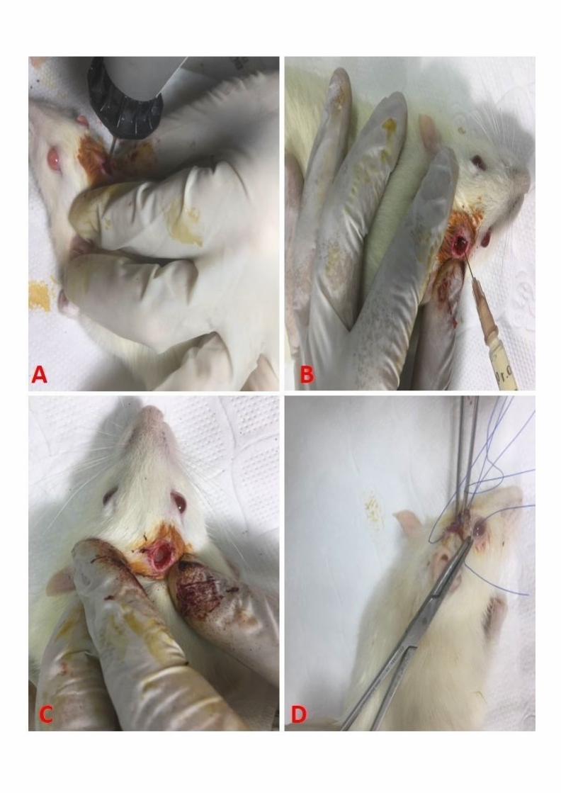
Surgical procedure for burr hole production in rat calvarium
In the current study, all surgical procedures were performed under general anesthesia
provided with intraperitoneal combination of ketamine (50 mg/kg) and xylasine (5 mg/kg)
anesthesia. Following heads of rats were shaved in an area of about 2 cm in diameter and
cleaned with baticon, a hole 3.0 mm in diameter was opened on the right sides of the rats’
calvarium under sterile conditions, using an electric drill (CLASS Mini Grinder sets, PRC),
and then the wound was closed, as described previously by Başaloğlu et al. (2021) [3] (Fig.
1).
Three-phase bone scintigraphy
Before sacrification of the animals for histological examination, one rat was
randomly selected from each group for scintigraphic study of the calvarium defect.
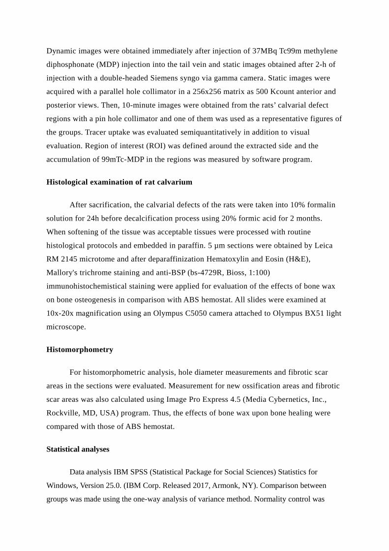
Dynamic images were obtained immediately after injection of 37MBq Tc99m methylene
diphosphonate (MDP) injection into the tail vein and static images obtained after 2-h of
injection with a double-headed Siemens syngo via gamma camera. Static images were
acquired with a parallel hole collimator in a 256x256 matrix as 500 Kcount anterior and
posterior views. Then, 10-minute images were obtained from the rats’ calvarial defect
regions with a pin hole collimator and one of them was used as a representative figures of
the groups. Tracer uptake was evaluated semiquantitatively in addition to visual
evaluation. Region of interest (ROI) was defined around the extracted side and the
accumulation of 99mTc-MDP in the regions was measured by software program.
Histological examination of rat calvarium
After sacrification, the calvarial defects of the rats were taken into 10% formalin
solution for 24h before decalcification process using 20% formic acid for 2 months.
When softening of the tissue was acceptable tissues were processed with routine
histological protocols and embedded in paraffin. 5 µm sections were obtained by Leica
RM 2145 microtome and after deparaffinization Hematoxylin and Eosin (H&E),
Mallory's trichrome staining and anti-BSP (bs-4729R, Bioss, 1:100)
immunohistochemistical staining were applied for evaluation of the effects of bone wax
on bone osteogenesis in comparison with ABS hemostat. All slides were examined at
10x-20x magnification using an Olympus C5050 camera attached to Olympus BX51 light
microscope.
Histomorphometry
For histomorphometric analysis, hole diameter measurements and fibrotic scar
areas in the sections were evaluated. Measurement for new ossification areas and fibrotic
scar areas was also calculated using Image Pro Express 4.5 (Media Cybernetics, Inc.,
Rockville, MD, USA) program. Thus, the effects of bone wax upon bone healing were
compared with those of ABS hemostat.
Statistical analyses
Data analysis IBM SPSS (Statistical Package for Social Sciences) Statistics for
Windows, Version 25.0. (IBM Corp. Released 2017, Armonk, NY). Comparison between
groups was made using the one-way analysis of variance method. Normality control was
performed using the Shapiro-Wilk test, one of the obtained error estimates. Since there was no
problem of adaptation to normal distribution, the study continued with one-way analysis of
variance. Variance homogeneities were examined with Levene test, when there was no
homogeneity problem, multi-group comparison was performed with F test, Bonferroni test
was used as post hoc tests. In cases where variance homogeneity was not provided, Welch’s
test was used for multi-group comparison, Dunnett's T3 method was preferred as the post hoc
test. All hypothesis checks were carried out at the 0.05 significance level, so p <0.05 was
considered significant.
RESULTS
Macroscopic examination
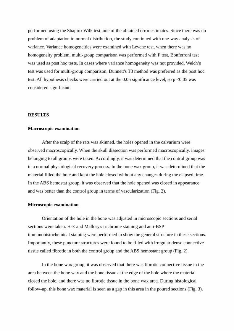
After the scalp of the rats was skinned, the holes opened in the calvarium were
observed macroscopically. When the skull dissection was performed macroscopically, images
belonging to all groups were taken. Accordingly, it was determined that the control group was
in a normal physiological recovery process. In the bone wax group, it was determined that the
material filled the hole and kept the hole closed without any changes during the elapsed time.
In the ABS hemostat group, it was observed that the hole opened was closed in appearance
and was better than the control group in terms of vascularization (Fig. 2).
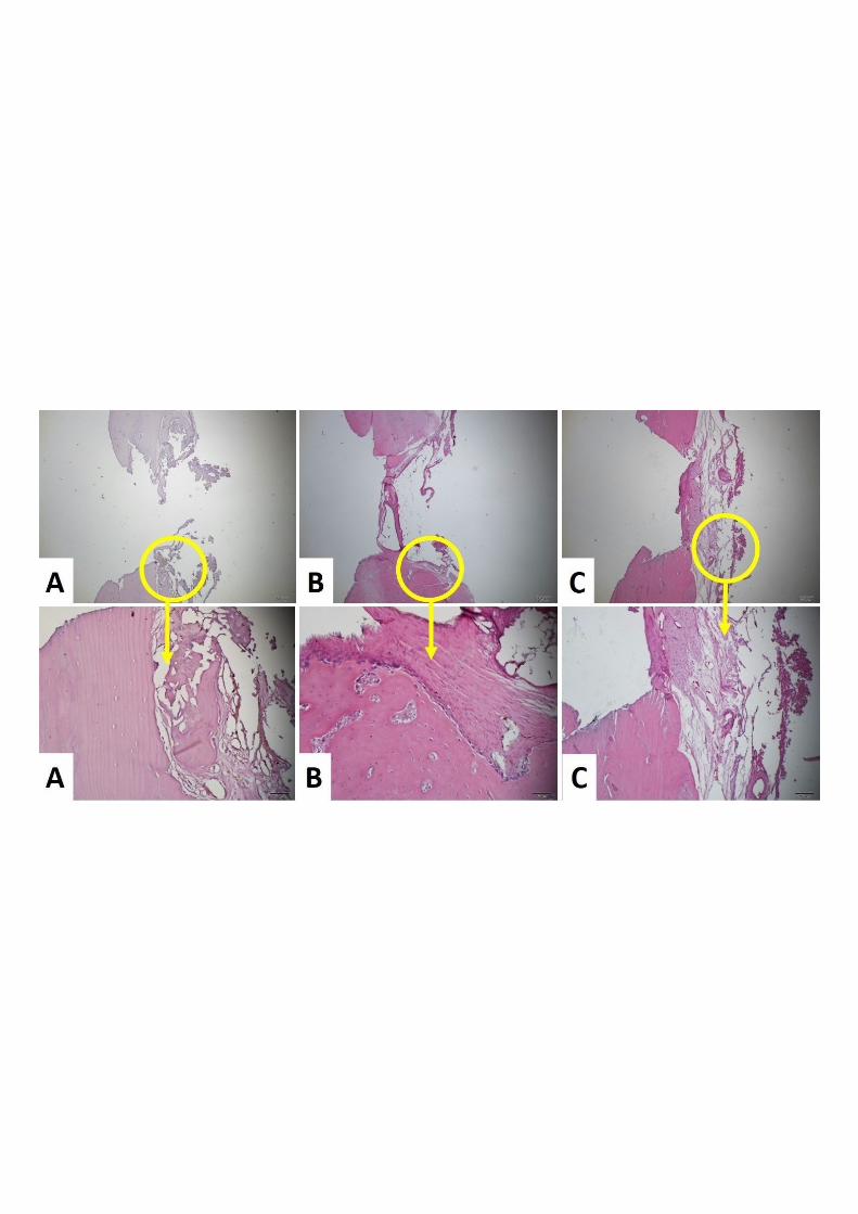
Microscopic examination
Orientation of the hole in the bone was adjusted in microscopic sections and serial
sections were taken. H-E and Mallory's trichrome staining and anti-BSP
immunohistochemical staining were performed to show the general structure in these sections.
Importantly, these puncture structures were found to be filled with irregular dense connective
tissue called fibrotic in both the control group and the ABS hemostant group (Fig. 2).
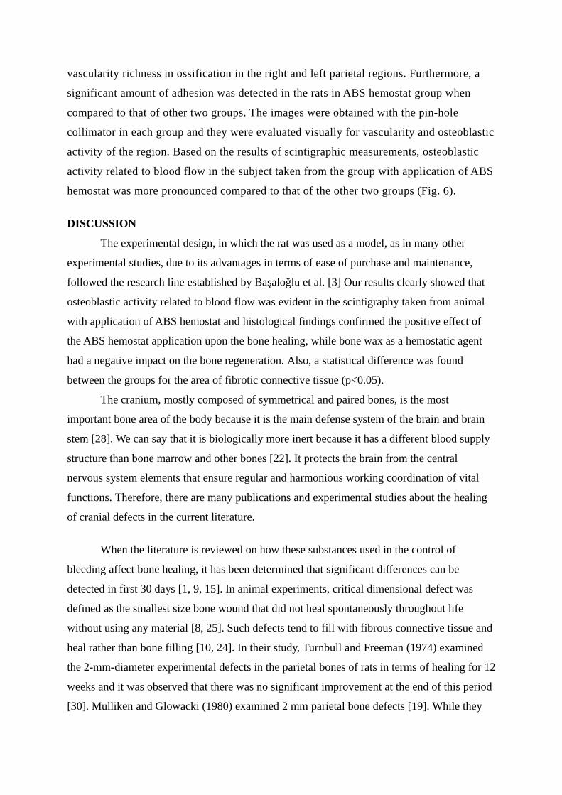
In the bone wax group, it was observed that there was fibrotic connective tissue in the
area between the bone wax and the bone tissue at the edge of the hole where the material
closed the hole, and there was no fibrotic tissue in the bone wax area. During histological
follow-up, this bone wax material is seen as a gap in this area in the poured sections (Fig. 3).

When the fibrotic connective tissue structure was examined at higher magnification, it
was found that they contained fibroblast-like cells and type I collagen bundles. In the control
group, new bone spicules were found, probably formed by intramembranous ossification,
where they settled in different regions within the connective tissue. More importantly, it was
noted that this connective tissue area was small in the ABS hemostat group and ossification
developed in most of the structure. With this appearance, it became the experimental group
closest to the normal histological structure of the other groups. Mallory's trichrome staining
showed that type I collagen bundles were stained blue, while the newly formed bone areas
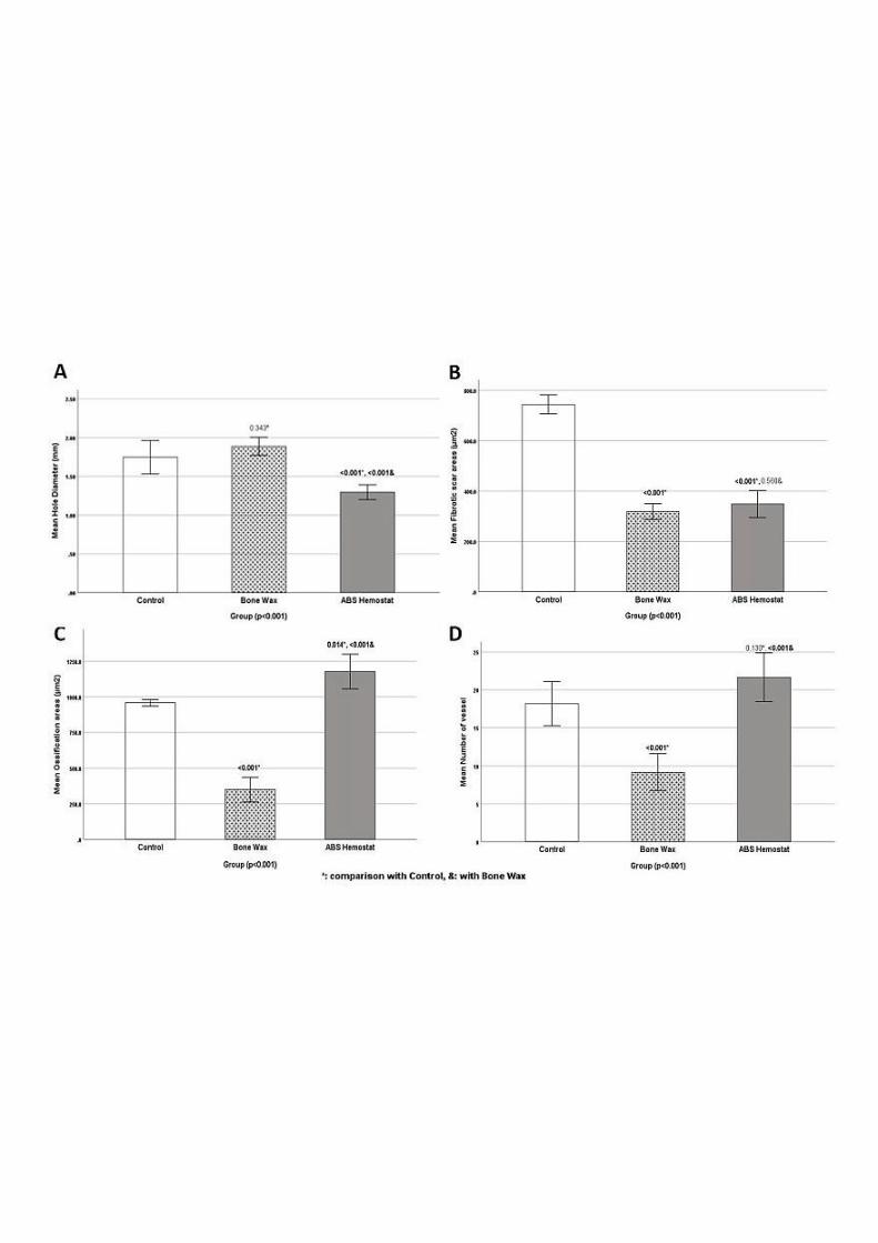
were red (Fig. 4). When the hole diameters were evaluated statistically, it was seen that the
bone wax group had the largest hole diameter. It was found that the smallest hole diameter
was in the ABS hemostat group. When the groups were compared with each other, a
significant difference was found between control group and ABS hemostat group (p <0.001)
as well as bone wax group and ABS hemostat group (p <0.001) (Fig. 5A).
When evaluated in terms of fibrotic scar area, it was found that the lowest scar area
was in the bone wax group. We think that the reason for this is that the bone wax material has
not been lost yet, which delays wound healing. There is a statistical significance between
control group and bone wax group (p <0.001) as well as bone wax group and ABS hemostat
group (p <0.001) (Fig. 5B). When evaluated in terms of ossification areas, it was seen that the
most ossification area was in the ABS hemostat group, while the least ossified area was in the
bone wax group. There is a statistical significance between control group and bone wax group
(p <0.001) as well as bone wax group and ABS hemostat group (p <0.001) (Fig. 5C). In terms
of vascularization, among the scar areas and ossification areas, it was seen that the most
vascular structure was in the ABS hemostat group and the least in the bone wax group. The
difference between control group and bone wax group (p <0.001) as well as bone wax group
and ABS hemostat group (p <0.001) was found to be significant (Fig. 5D).
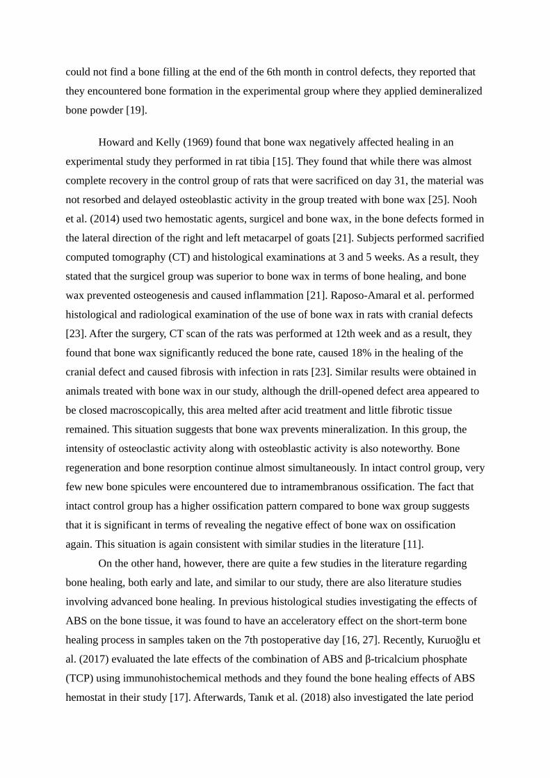
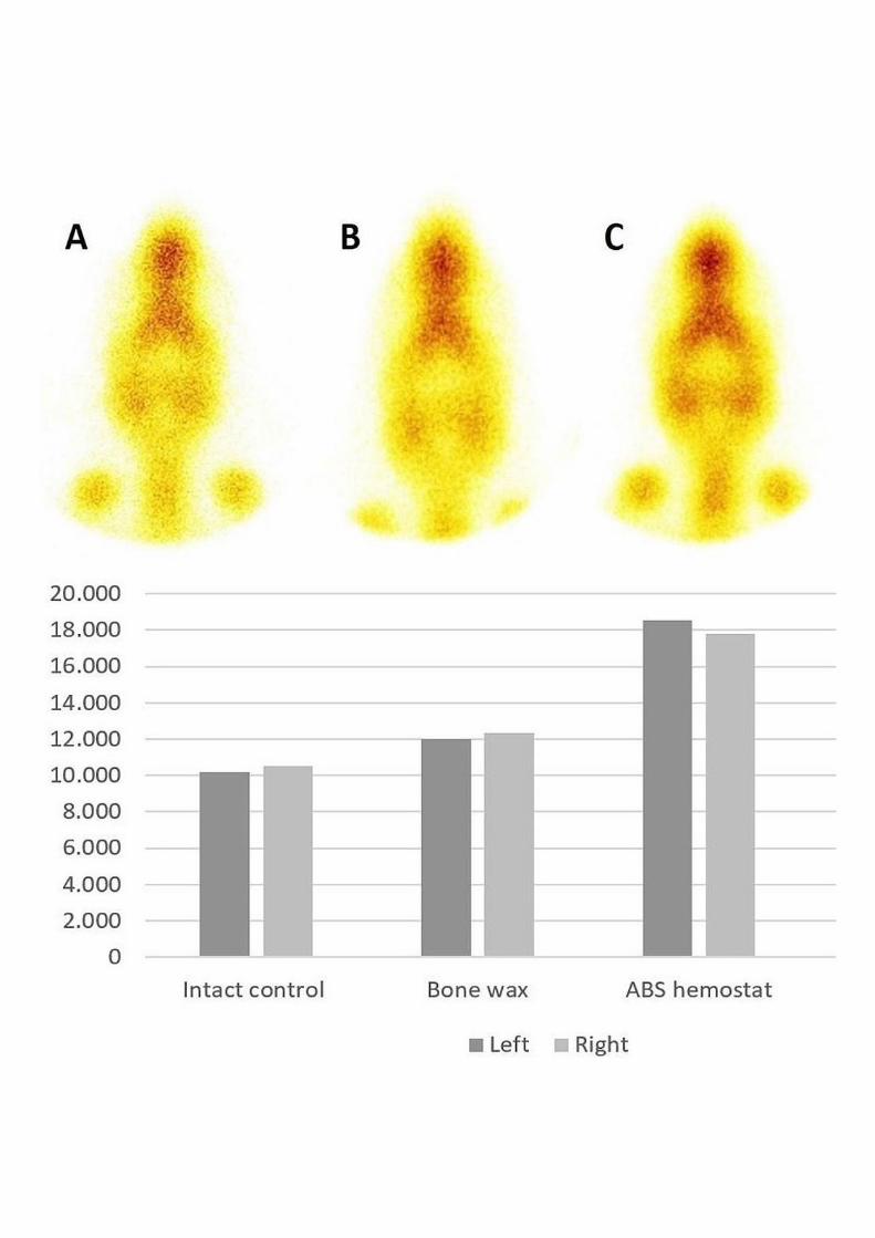
Bone scintigraphy
As a result of quantitative examination of the right and left parietal regions of the
calvarium of the rats; in the rat in intact control left 10.158 count, right 10.541 count, in
bone wax group, left 12.008 count right 12.325 count; and, in the rat in group with
application of ABS hemostat, 18.513 count in the left was found as 17.786 count in the
right (Fig. 6). In the same rat, however, no significant result was obtained in terms of
vascularity richness in ossification in the right and left parietal regions. Furthermore, a
significant amount of adhesion was detected in the rats in ABS hemostat group when
compared to that of other two groups. The images were obtained with the pin-hole
collimator in each group and they were evaluated visually for vascularity and osteoblastic
activity of the region. Based on the results of scintigraphic measurements, osteoblastic
activity related to blood flow in the subject taken from the group with application of ABS
hemostat was more pronounced compared to that of the other two groups (Fig. 6).
DISCUSSION
The experimental design, in which the rat was used as a model, as in many other
experimental studies, due to its advantages in terms of ease of purchase and maintenance,
followed the research line established by Başaloğlu et al. [3] Our results clearly showed that
osteoblastic activity related to blood flow was evident in the scintigraphy taken from animal
with application of ABS hemostat and histological findings confirmed the positive effect of
the ABS hemostat application upon the bone healing, while bone wax as a hemostatic agent
had a negative impact on the bone regeneration. Also, a statistical difference was found
between the groups for the area of fibrotic connective tissue (p<0.05).
The cranium, mostly composed of symmetrical and paired bones, is the most
important bone area of the body because it is the main defense system of the brain and brain
stem [28]. We can say that it is biologically more inert because it has a different blood supply
structure than bone marrow and other bones [22]. It protects the brain from the central
nervous system elements that ensure regular and harmonious working coordination of vital
functions. Therefore, there are many publications and experimental studies about the healing
of cranial defects in the current literature.
When the literature is reviewed on how these substances used in the control of
bleeding affect bone healing, it has been determined that significant differences can be
detected in first 30 days [1, 9, 15]. In animal experiments, critical dimensional defect was
defined as the smallest size bone wound that did not heal spontaneously throughout life
without using any material [8, 25]. Such defects tend to fill with fibrous connective tissue and
heal rather than bone filling [10, 24]. In their study, Turnbull and Freeman (1974) examined
the 2-mm-diameter experimental defects in the parietal bones of rats in terms of healing for 12
weeks and it was observed that there was no significant improvement at the end of this period
[30]. Mulliken and Glowacki (1980) examined 2 mm parietal bone defects [19]. While they
could not find a bone filling at the end of the 6th month in control defects, they reported that
they encountered bone formation in the experimental group where they applied demineralized
bone powder [19].
Howard and Kelly (1969) found that bone wax negatively affected healing in an
experimental study they performed in rat tibia [15]. They found that while there was almost
complete recovery in the control group of rats that were sacrificed on day 31, the material was
not resorbed and delayed osteoblastic activity in the group treated with bone wax [25]. Nooh
et al. (2014) used two hemostatic agents, surgicel and bone wax, in the bone defects formed in
the lateral direction of the right and left metacarpel of goats [21]. Subjects performed sacrified
computed tomography (CT) and histological examinations at 3 and 5 weeks. As a result, they
stated that the surgicel group was superior to bone wax in terms of bone healing, and bone
wax prevented osteogenesis and caused inflammation [21]. Raposo-Amaral et al. performed
histological and radiological examination of the use of bone wax in rats with cranial defects
[23]. After the surgery, CT scan of the rats was performed at 12th week and as a result, they
found that bone wax significantly reduced the bone rate, caused 18% in the healing of the
cranial defect and caused fibrosis with infection in rats [23]. Similar results were obtained in
animals treated with bone wax in our study, although the drill-opened defect area appeared to
be closed macroscopically, this area melted after acid treatment and little fibrotic tissue
remained. This situation suggests that bone wax prevents mineralization. In this group, the
intensity of osteoclastic activity along with osteoblastic activity is also noteworthy. Bone
regeneration and bone resorption continue almost simultaneously. In intact control group, very
few new bone spicules were encountered due to intramembranous ossification. The fact that
intact control group has a higher ossification pattern compared to bone wax group suggests
that it is significant in terms of revealing the negative effect of bone wax on ossification
again. This situation is again consistent with similar studies in the literature [11].
On the other hand, however, there are quite a few studies in the literature regarding
bone healing, both early and late, and similar to our study, there are also literature studies
involving advanced bone healing. In previous histological studies investigating the effects of
ABS on the bone tissue, it was found to have an acceleratory effect on the short-term bone
healing process in samples taken on the 7th postoperative day [16, 27]. Recently, Kuruoğlu et
al. (2017) evaluated the late effects of the combination of ABS and β-tricalcium phosphate
(TCP) using immunohistochemical methods and they found the bone healing effects of ABS
hemostat in their study [17]. Afterwards, Tanık et al. (2018) also investigated the late period
effects of the combination of ABS and β-TCP on the regeneration of bone tissue in rats [29].
They found that both β-TCP and ABS had positive histological and radiological effects on
wound healing and bone formation in samples taken on day 56 after surgery [29]. In the first
macroscopic examination we observed in our study, it was determined that the group with the
closest appearance of scar tissue to normal structure was group with application of ABS
hemostat, suggesting a positive effect of ABS hemostat on wound healing.
Histologically, in the Mallory's trichrome staining, type I collagen bundles were
stained in blue, and the newly formed bone areas were red in all groups in our study. In terms
of hole diameters, it was determined that the bone wax group had the largest hole diameter
and the ABS hemostat group had the smallest hole diameter. A statistically significant
difference was found between control group and ABS hemostat group (p <0.001) as well as
bone wax group and ABS hemostat group (p <0.001). When evaluated in terms of fibrotic scar
area, it was found that the lowest scar area was in the bone wax group. We think that the
reason for this is that the bone wax material has not been lost yet, which delays wound
healing. There is a statistical significance between control group and bone wax group (p
<0.001) as well as bone wax group and ABS hemostat group (p <0.001). In the literature
review, however, immunohistochemically studies on bone repair of ABS hemostat are limited.
In their experimental study in which they evaluated the use of platelet-rich plasma (PRP)
together with autogenous bone graft in the repair of bone defects by immunohistochemical
analysis, Nagata et al. (2009) found that the expression of OC and OP was significantly higher
in the PRP group [20]. In our study, ossification is present in all three groups, although it
seems that the mineralization has not yet been completed. Therefore, absence of any analysis
for inorganic material content of bone tissue mineralization and difference of drilled defects
from those in humans in terms of bone remodelling are limitations of our study. Moreover,
lack of any imaging or mechanical study investigating the bone healing is another limitation
of the study. Future studies including immunohistochemical determination of bone related
proteins in the early stages of bone healing are needed for beter understanding of impact of
the ABS hemostat application upon bone remodeling apart from the quick stop of
hemorrhage.
It is well-known that three-phase bone scintigraphy is a non-invasive technique
showing the vascularity and osteoblastic activity of the bone. Based on their findings in a
study, Aygit et al. (1999) stated that bone scintigraphy could be used as a noninvasive method
in evaluating implant vascularity because histological changes and scintigraphic findings were
paralel [2]. Tc-99m MDP is a radiopharmaceutical that can chemically adhere to all bones in
the skeletal system after intravenous administration. It shows a significant accumulation
especially at the points where bone metabolism increases. The common features of
radiopharmaceuticals such as MDP, hydroxy diphosphonate (HDP) and hydroxymethylene
diphosphonate (HMDP) marked with Tc-99m are that the diphosphonate core in their
structure acts like hydroxyapatite, which is the basic building block of bone, and accumulates
reversibly on bone surfaces and shows osteoblastic activity depending on blood flow. In this
way, all bones belonging to the skeletal system get rid of the soft tissue effect and become
visible in a plain state [26]. Diphosphonates attach to hydroxy apatite crystals, one of the main
components of the bone matrix, by physicochemical absorption. This amount of adhesion
varies according to the blood supply and osteoblastic activity in that area. Organization of
hemostasis is primary in fracture or defect repair. When scintigraphic imaging was performed
it was not clear between the groups visually. Although MDP is a chemoabsorbable substance
in all bones of the skeletal system, it shows a significant accumulation in places where bone
metabolism is increased. Radiopharmaceuticals such as MDP, HDP, and HMDP act as
hydroxyapatite, which is the main component of bone, and show osteoblastic activity due to a
reversible blood flow at the bone surfaces [28]. Although there are not many such studies in
the literature, we think that performing bone scintigraphy in weekly periods starting from the
defect opening will be more beneficial as it will provide comparable and observable data.
CONCLUSIONS
Bone fractures due to trauma or surgical operations are conditions that can be encountered
in all stages of life. The primary functions of ABS hemostat and bone wax, which are used as
hemostatic agents in neurosurgery, orthopedic and traumatology surgery, oral and
maxillofacial surgery, are to stop bleeding quickly. Taken as whole, our results showed that:
1. Histological, immunohistochemical and bone scintigraphic data support the positive
effects of ABS, consisting of folkloric medicinal plant extracts, on bone healing as
well as stopping bleeding rapidly. It was concluded that ABS hemostat also had a
positive effect on bone regeneration, while bone wax it had a negative effect on
regeneration of the rats’ calvarial defects.
2. In various surgical procedures, we think that it is important to choose a blood stopping
agent, such as ABS, that will both provide rapid control of bleeding and accelerate
regeneration by helping bone repair. However, the effectiveness of this product as a
therapeutic modality for possible future clinical applications can interest all types of
bones, including extracranial, spongy and cortical, apart from cranial ones.

3. Furthermore, this study also suggests that the anti-inflammatory and antimicrobial
properties of the plant ingredients of antihemorrhagic ABS should be evaluated in
terms of preventing various complications during the operation and postoperative
period in future studies.
Acknowledgements
The authors are grateful to Mr. Rifat Aydın for technical support and Aydın Adnan
Menderes University Scientific Research Projects (BAP) Unit for financial support. We would
like to thank Professor Ali Akhaddar for his suggestions following review of the article draft.
Competing interests
The authors declare that they have no competing interests.
REFERENCES
1. Alberius P, Klinge B, Sjögren S. Effects of bone wax on rabbit cranial bone lesions. J
Craniomaxillofac Surg 1987; 15:63-7.2. Aygit A, Sarikaya A, Candan L. Ayhan MS, Cermik TF. Comparison of alloplastic
implants for facial bones by scintigraphy and histology: an experimental study. Eur J
Plast Surg 1999; 22:102-6.3. Başaloglu HK, Turgut M, Uyanıkgil Y, Sirin C, Demirci B, Cetin EÖ. The influence of
functional pinealectomy and exogenous melatonin application on healing of burr hole
in adult rat calvaria: a histological and immunohistochemical study. Folia Morphol
2021 (in press).4. Bulut E, Baş B, Altunkaynak BZ, Bekçioğlu B, Erdem Koç G, Gönülol E, Önger ME,
Kaplan S. Efficacy of Ankaferd Blood Stopper on bone healing in diabetic rats: a
stereological and histopathological study. Biotech Histochem 2014; 89:535-43.
5. Ciftciler R, Ciftciler AE, Malkan UY, Haznedaroglu IC. Pharmacobiological
management of hemostasis within clinical backgrounds via Ankaferd hemostat
(Ankaferd blood stopper). SAGE Open Med 2020; 8:2050312120907811.
6. Ciftciler R, Haznedaroglu IC. Ankaferd hemostat: from molecules to medicine. Turk J
Med Sci 2020; 50:1739-50.
7. Cipil HS, Kosar A, Kaya A, Uz B, Haznedaroglu IC, Goker H, Ozdemir O, Koroglu
M, Kirazli S, Firat HC. In vivo hemostatic effect of the medicinal plant extract
Ankaferd Blood Stopper in rats pretreated with warfarin. Clin Appl Thromb Hemost
2009; 15:270-6.
8. Cook JL, Hung CT, Kuroki K, Stoker AM, Cook CR, Pfeiffer FM, Sherman SL,
Stannard JP. Animal models of cartilage repair. Bone Joint Res 2014; 3:89-94.9. Finn MD, Schow SR, Schneiderman ED. Osseous regeneration in the presence of four
common hemostatic agents. J Oral Maxillofac Surg 1992; 50:608-12.10. Frame JW. A convenient animal model for testing bone substitute materials. J Oral
Surg 1980; 38:176-80.11. Geary JR, Frantz VK. New absorbable hemastatik bonewax. Ann Surg 1950;
132:1128-37.12. Goker H, Haznedaroglu IC, Ercetin S, Kirazli S, Akman U, Ozturk Y, Firat HC.
Haemostatic actions of the folkloric medicinal plant extract Ankaferd Blood Stopper. J
Int Med Res 2008; 36:163-70.13. Gul M, Gunay A, Tanik A. An evaluation of the effects of caffeic acid phenethyl ester
and Ankaferd blood stopper on secondary wound healing of oral mucosal tissue. Turk
J Med Sci 2020; 50:248-57. 14. Gupta G, Prestigiacomo CJ. From sealing wax to bone wax: predecessors to Horsley's
development. Neurosurg Focus 2007; 23:E16.15. Howard TC, Kelley RR. The effect of bone wax on the healing of experimental rat
tibial lesions. Clin Orthop Relat Res 1969; 63:226-32.16. Isler SC, Demircan S, Cakarer S, Cebi Z, Keskin C, Soluk M, Yuzbasioglu E. Effects
of folk medicinal plant extract Ankaferd Blood Stopper on early bone healing. J Appl
Oral Sci 2010; 18:409-14.17. Kuruoglu E, Onger ME, Marangoz AH, Kocacan SE, Cokluk C, Kaplan S.
Postlaminectomy bone and scar formations in presence of Ankaferd Blood Stopper
and Bitter Melon (Momordica Charantia): An experimental study. Turk Neurosurg
2017; 27:441-6.18. Malaval L, Wade-Guéye NM, Boudiffa M, Fei J, Zirngibl R, Chen F, Laroche N, Roux
JP, Burt-Pichat B, Duboeuf F, Boivin G, Jurdic P, Lafage-Proust MH, Amédée J, Vico
L, Rossant J, Aubin JE. Bone sialoprotein plays a functional role in bone formation
and osteoclastogenesis. J Exp Med 2008; 205:1145–53.19. Mulliken JB, Glowacki J. Induced osteogenesis for repair and construction in the
craniofacial region. Plast Reconstr Surg 1980; 65:553-60.20. Nagata M, Messora M, Okamoto R, Campos N, Pola N, Esper L, Sbrana M, Fucini S,
Garcia V, Bosco A. Influence of the proportion of particulate autogenous bone
graft/platelet-rich plasma on bone healing in critical-size defects: an
immunohistochemical analysis in rat calvaria. Bone 2009; 45:339-45.21. Nooh N, Abdullah WA, Grawish Mel-A, Ramalingam S, Javed F, Al-Hezaimi K. The
effects of surgicel and bonewax hemostatik agents on bone healing. An experimental
study. Indian J Orthop 2014; 48:319-25.
22. Prolo DJ, Gutierrez RV, DeVine JS, Oklund SA. Clinical utility of allogeneic skull
discs in human craniotomy. Neurosurgery 1984; 14:183-6.23. Raposo-Amaral CE, Almeida AB, Paschoal G, Bueno DF, Vulcano LC, Passos-Bueno
MR, Alonso N. Histological and radiological changes in cranial bone in the presence
of bone wax. Acta Cir Bras 2011; 26:274-8.24. Rowe NL. Nonunion of the mandible and maxilla. J Oral Surg 1969; 27:520-9.
25. Schmitz JP, Hollinger JO. The critical size defect as an experimental model for
craniomandibulofacial nonunions. Clin Orthop Relat Res 1986; 205:299-308.
26. Sengöz T, Karaçalıoğlu AÖ, İnce S, Emer MÖ, Özgüven MA. Cardiac Tc-99m
metylene diphosphanate uptake in bone scintigraphy (in Turkish). Gülhane Tıp Dergisi
2015; 57:81-3.27. Simsek HO, Tüzüm MŞ, Baykul T, Gürer IE, Başsorgun Cİ. Experimental
investigation of the effects of a blood stopper agent (ankaferd blood stopper) on bone
surfaces. Turk J Haematol 2013; 30:177-83.28. Sırola K. Regeneration of defects in the calvaria. An experimental study. Ann Med
Exp Biol Fenn 1960; 38(Suppl 2):1-87.29. Tanık A, Güler Doğru A, Akpolat V, Acun Kaya F, Sarıbaş E, Gül M, İrtegün
Kandemir S, Deveci E. Investigation of the effect of combined use of alloplastic-based
tricalcium phosphate bone graft and antihemorrhagic plant extract (ABS) on bone
regeneration in surgically induced bone defects in nondiabetic rats: an experimental
animal study. Turk J Med Sci 2018; 48:1302-1314. 30. Turnbull RS, Freeman E. Use of wounds in the parietal bone of the rat for evaluating
bone marrow for grafting into periodontal defects. J Periodontal Res 1974; 9:39-43.
31. Zhou H, Ge J, Bai Y, Liang C, Yang L. Translation of bone wax and its substitutes:
history, clinical status and future directions. J Orthop Translat 2019; 17:64-72.
Figure 1. The process of establishing the animal model of bone defect as burr hole on the
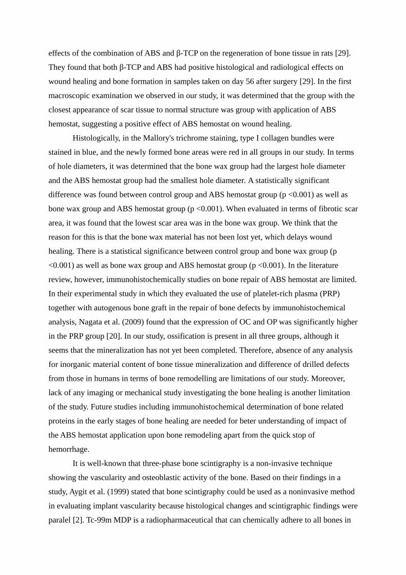
right side of the rat calvarium using a drill with 3 mm in diameter.
Figure 2. Demonstration of the experimentally produced bone lesions in all rat groups. (A)
Control group; (B) Bone wax group; and (C) Ankaferd (ABS) hemostat group.
Figure 3. Microscopic evaluation of all groups (A) Control group; (B) Bone wax group; and
(C) ABS hemostat group. H&E staining.

Figure 4. Microscopic evaluation of all groups showing (A) Control group; (B) Bone wax
group; and (C) ABS hemostat group. Top row: Mallory's Trichrome staining. Up row: Anti-
BSP staining.
Figure 5. Statistical evaluation graph of histomorphometric analysis. Graphical representation
of hole diameter (A), fibrotic scar area (B), ossification area (C) and number of vessels (D) in
all groups.
Figure 6. Scintigraphic imaging in rats. (A) Control group; (B) Bone wax group; and (C)
ABS hemostat group. Results of scintigraphic measurements show more pronounced
osteoblastic activity depending on blood flow. In the lower part, the results obtained are also
shown graphically.


Related Documents