Dental Press J Orthod 125 2012 Jan-Feb;17(1):125-37 Paulo Estevão Scanavini*, Luiz Renato Paranhos**, Fernando César Torres**, Maria Helena Ferreira Vasconcelos**, Renata Pilli Jóias*, Marco Antonio Scanavini*** O RIGINAL A RTICLE Evaluation of the dental arch asymmetry in natural normal occlusion and Class II malocclusion individuals Objective: To verify the presence and degree of asymmetry of dental arches in Brazilian indi- viduals with natural normal occlusion and Class II, Divisions 1 and 2 malocclusions. Methods: The study evaluated the symmetry of the maxillary and mandibular dental arches of 180 pairs of dental casts, divided into: Group I = 60 pairs of natural normal occlusion individuals; Group II = 60 pairs of Class II, Division 1 malocclusion individuals; and Group III = 60 pairs of Class II, Division 2 malocclusion individuals. A device was used to measure dental midline deviation and the canine tip in the dental arches (in degrees). It was also verified the distance of the upper canines from the palatal suture, intercanine distance, and anteroposterior upper and lower first molar position. Results: Dental arches of individuals from all groups presented asymmetry, regardless of the presence of malocclusion. Group I showed a lower asymmetry degree in relation to Groups II and III. The asymmetry in Groups II and III was similar. Con- clusion: The dental arches of individuals with natural normal occlusion and with Class II, Di- vision 1 and Division 2 malocclusions showed asymmetry. The asymmetry degree was higher in the mandibular dental arches than in the maxillary dental arches in all 3 evaluated groups. Abstract Keywords: Dental arch. Abnormalities. Growth and development. Anatomy and histology. Malocclusion. * MSc in Orthodontics, Methodist University of São Paulo (UMESP). ** Professor, Orthodontics Graduate Program, UMESP. *** PhD in Orthodontics, School of Dentistry, University of São Paulo (USP). Head of the Orthodontics Graduate Program, UMESP. How to cite this article: Scanavini PE, Paranhos LR, Torres FC, Vasconcelos MHF, Jóias RP, Scanavini MA. Evaluation of the dental arch asymmetry in natural normal occlusion and Class II malocclusion individuals. Dental Press J Orthod. 2012 Jan-Feb;17(1):125-37. » The authors report no commercial, proprietary, or financial interest in the products or companies described in this article.

Welcome message from author
This document is posted to help you gain knowledge. Please leave a comment to let me know what you think about it! Share it to your friends and learn new things together.
Transcript
Dental Press J Orthod 125 2012 Jan-Feb;17(1):125-37
Paulo Estevão Scanavini*, Luiz Renato Paranhos**, Fernando César Torres**, Maria Helena Ferreira Vasconcelos**, Renata Pilli Jóias*, Marco Antonio Scanavini***
O r i g i n a l a r t i c l e
Evaluation of the dental arch asymmetry in natural normal occlusion and Class II malocclusion individuals
Objective: To verify the presence and degree of asymmetry of dental arches in Brazilian indi-viduals with natural normal occlusion and Class II, Divisions 1 and 2 malocclusions. Methods: The study evaluated the symmetry of the maxillary and mandibular dental arches of 180 pairs of dental casts, divided into: Group I = 60 pairs of natural normal occlusion individuals; Group II = 60 pairs of Class II, Division 1 malocclusion individuals; and Group III = 60 pairs of Class II, Division 2 malocclusion individuals. A device was used to measure dental midline deviation and the canine tip in the dental arches (in degrees). It was also verified the distance of the upper canines from the palatal suture, intercanine distance, and anteroposterior upper and lower first molar position. Results: Dental arches of individuals from all groups presented asymmetry, regardless of the presence of malocclusion. Group I showed a lower asymmetry degree in relation to Groups II and III. The asymmetry in Groups II and III was similar. Con-clusion: The dental arches of individuals with natural normal occlusion and with Class II, Di-vision 1 and Division 2 malocclusions showed asymmetry. The asymmetry degree was higher in the mandibular dental arches than in the maxillary dental arches in all 3 evaluated groups.
Abstract
Keywords: Dental arch. Abnormalities. Growth and development. Anatomy and histology. Malocclusion.
* MSc in Orthodontics, Methodist University of São Paulo (UMESP). ** Professor, Orthodontics Graduate Program, UMESP. *** PhD in Orthodontics, School of Dentistry, University of São Paulo (USP). Head of the Orthodontics Graduate Program, UMESP.
How to cite this article: Scanavini PE, Paranhos LR, Torres FC, Vasconcelos MHF, Jóias RP, Scanavini MA. Evaluation of the dental arch asymmetry in natural normal occlusion and Class II malocclusion individuals. Dental Press J Orthod. 2012 Jan-Feb;17(1):125-37.
» The authors report no commercial, proprietary, or financial interest in the products or companies described in this article.
Dental Press J Orthod 126 2012 Jan-Feb;17(1):125-37
Evaluation of the dental arch asymmetry in natural normal occlusion and Class II malocclusion individuals
IntROduCtIOnThe study of form and transverse dimensions
of dental arches is of great relevance to orthodon-tists. Correcting skeletal and dental midlines, as well as coordinating the position of teeth in each side of the arch, lead to maximum intercuspation, correct function, stability of achieved results, an-terior and facial dental aesthetics and reduced po-tential for temporomandibular joint dysfunction.1
The midpalatal suture and the center of the maxillary dental arch are almost coincident, vali-dating the use of the suture as a symmetry axis.2,3 Dental arch asymmetry can be caused by a com-bination of genetic3,4 and environmental3 factors, with skeletal, dental or functional repercussions.4 In individuals with symmetric development, the slight differences between the right and left sides may be due to external environmental fac-tors, such as: Thumb sucking, unilateral chew-ing, loss of contact due to cavities, extraction or trauma.3 Children can also feature asymmetric dental arches,5 and older individuals tend to have greater arch asymmetry, resulting from lifelong external environmental factors.6
Even symmetrical faces feature skeletal asymmetry, suggesting that soft tissues mini-mize the existing asymmetry. Dentoalveolar re-gions feature symmetry between the right and left sides, probably due to muscle balance of lingual and labial forces.7
It is rare to find a totally symmetric individu-al. Therefore, small asymmetries are regarded as normal.8 Most individuals with normal occlusion may show almost coinciding midlines (deviation smaller than 1 mm), and many can have molar asymmetry greater than 1 mm in transversal and anteroposterior directions.9 Dental midline devia-tions greater than 2 mm are easily detected by lay persons, and should therefore be considered when planning orthodontic treatments.10
Dental asymmetries and a variety of functional deviations can be treated with orthodontics, but significant asymmetries of facial structures may
require orthopedic correction during growth pe-riod and/or later surgical treatment.4
Some authors have observed skeletal asym-metries both in normal occlusion and malocclu-sion groups,2,11 with pre-orthodontic treatment patients showing more symmetrical arches.2 Con-versely, other authors revealed a tendency for pos-terior crossbite in individuals with malocclusion,12 and a greater tendency towards dental arch asym-metries in individuals with Angle Class II and/or Class III malocclusions.13,14 Other studies showed asymmetries in the dental arches of individuals with normal occlusion, in the passage from ado-lescence to adult age, further questioning the pos-sibility of achieving post-treatment stability.5
The verification of dental arch asymmetries during diagnosis makes it possible to choose the appropriate mechanics for orthodontic treat-ment—for instance, whether to recommend extractions. When opting for tooth extractions, arch symmetry should be controlled throughout orthodontic movement.
In cases of dental arch asymmetry, therapy with asymmetric extractions can reduce complications resulting from patient compliance to the use of elastic bands, as well as reducing the length of orth-odontic treatment. Atypical extractions can also benefit pre-surgery orthodontic preparation.16
Caution must be taken when planning orth-odontic treatments, as there are cases when trans-versal asymmetry of dental arches is not corrected after orthodontic treatment, and orthodontic cases originate dental arches that are larger than those without extraction.17
There is great ethnic diversity among the Brazil-ian population, raising questions on the applicabil-ity of standards and measurements previously es-tablished in more homogenous populations. Based on that premise, the objective of this study was to evaluate the presence and degree of asymmetry of the maxillary and mandibular dental arches in Bra-zilian individuals with normal occlusion or Angle Class II, Divisions 1 and 2 malocclusion.
Scanavini PE, Paranhos LR, Torres FC, Vasconcelos MHF, Jóias RP, Scanavini MA
Dental Press J Orthod 127 2012 Jan-Feb;17(1):125-37
MAtERIAL And MEtHOdSMaterial
The experimental group consisted of plaster cast models of mandibular and maxillary dental arches from 80 patients with ages varying be-tween 12 and 21 years. The experimental group was composed using the following selection cri-teria: a) Permanent dentition, with exception of third molars; b) Brazilian ethnicity; c) Not subjected to any orthodontic intervention. They were then divided into three groups:
» Group 1: 60 pairs of models from individuals with natural normal occlusion, featuring at least 4 of Andrews’ 6 keys to occlusion.
» Group 2: 60 pairs of models from individ-uals with Angle Class II, Division 1 maloc-clusion.
» Group 3: 60 pairs of models from individuals with Angle Class II, Division 2 malocclusion.
The plaster models are part of the collection belonging to the Orthodontics Graduate Pro-gram of the Methodist University of São Paulo (UMESP), São Bernardo do Campo/SP, Brazil.
MethodsModel measurements were obtained from
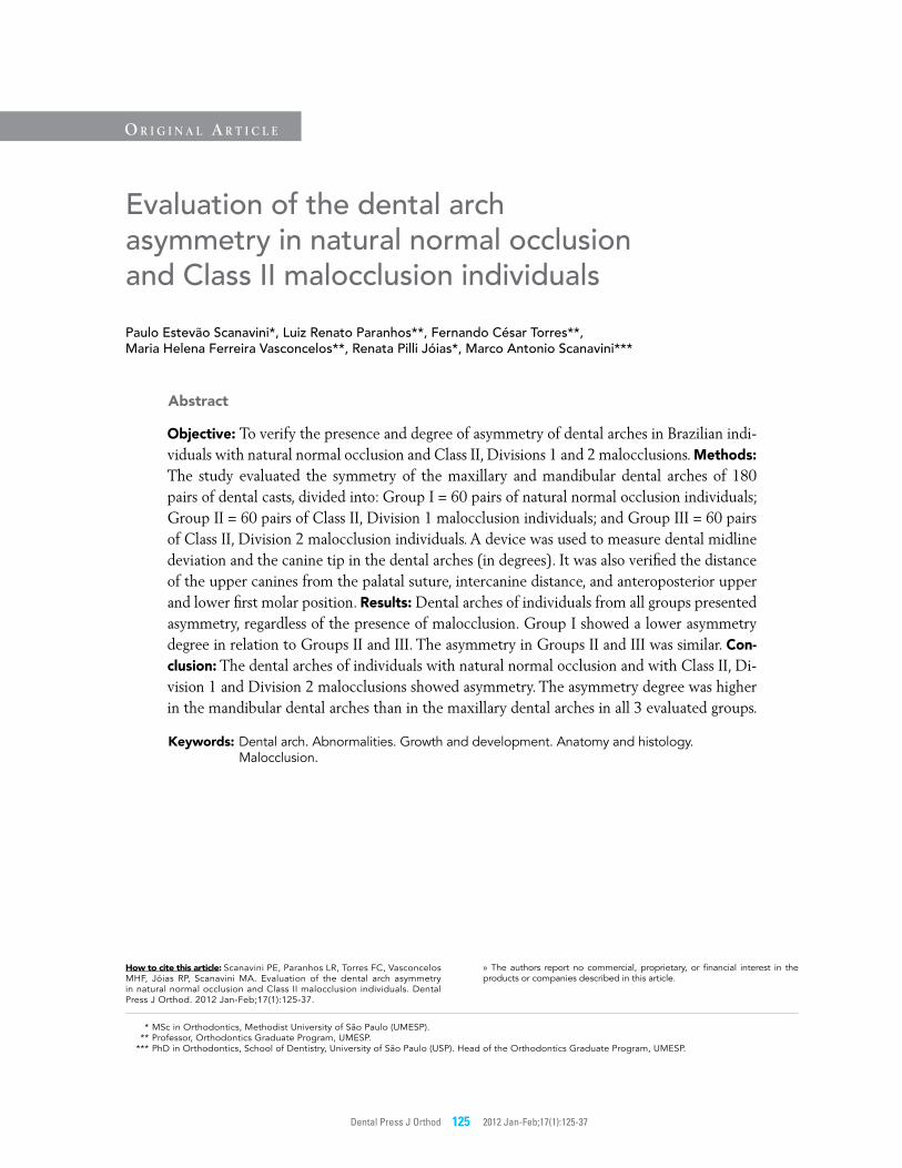
an original device, developed specifically to analyze dental arch asymmetries. It was con-ceived and designed at UMESP, in the Ortho-dontics Department, manufactured in steel and aluminum. A millimeter ruler and metallic protractor were adapted to obtain measure-ments. The models were positioned in a delin-eator base (used in the manufacture of remov-able partial prostheses) fixed in the base of the appliance using a screw manufactured specifi-cally for that purpose (Fig 1).
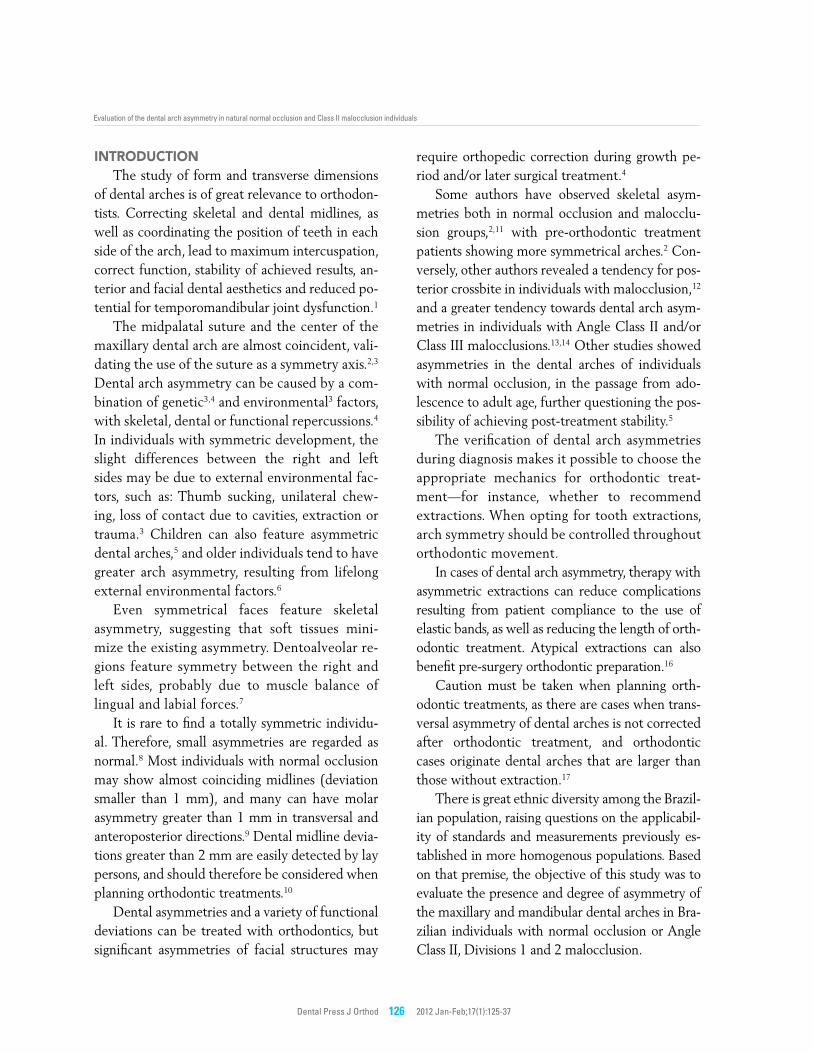
The maxillary model midline was deter-mined by marking points over the midpalatal suture, from the incisive papilla to the most visible posterior landmark.5,18 By connecting these points, the symmetry axis is obtained, which if prolonged anteriorly up to the incisal
edge of the maxillary incisor determined point As (anterior-superior), and posteriorly, up to the posterior surface of the maxillary model, determining point Ps (posterior-superior), as shown in Figure 2.
To determine the mandibular midline, the midline projection obtained in the maxillary dental arch was used, according to Alavi.19 The maxillary midline was transferred to the mandib-ular model, using the As and Ps reference points.
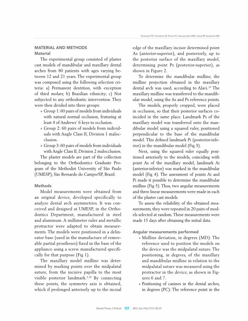
The models, properly cropped, were placed in occlusion, so that their posterior surfaces co-incided in the same place. Landmark Ps of the maxillary model was transferred onto the man-dibular model using a squared ruler, positioned perpendicular to the base of the mandibular model. This defined landmark Pi (posterior-infe-rior) in the mandibular model (Fig 3).
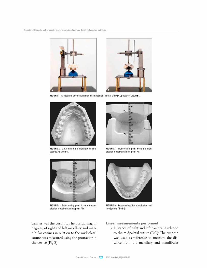
Next, using the squared ruler equally posi-tioned anteriorly to the models, coinciding with point As of the maxillary model, landmark Ai (anterior-inferior) was marked in the mandibular model (Fig 4). The assessment of points Ai and Pi made it possible to determine the mandibular midline (Fig 5). Thus, two angular measurements and three linear measurements were made in each of the plaster cast models.
To assess the reliability of the obtained mea-surements, they were repeated in 20 pairs of mod-els selected at random. These measurements were made 15 days after obtaining the initial data.
Angular measurements performed» Midline deviation, in degrees (MD): The
reference used to position the models on the device was the midpalatal suture. The positioning, in degrees, of the maxillary and mandibular midline in relation to the midpalatal suture was measured using the protractor in the device, as shown in Fig-ures 6 and 7.
» Positioning of canines in the dental arches, in degrees (PC): The reference point in the
Dental Press J Orthod 128 2012 Jan-Feb;17(1):125-37
A B
Evaluation of the dental arch asymmetry in natural normal occlusion and Class II malocclusion individuals
FigurE 1 - Measuring device with models in position: frontal view (A), posterior view (B).
FigurE 2 - Determining the maxillary midline (points As and Ps).
FigurE 4 - Transferring point As to the man-dibular model (obtaining point Ai).
FigurE 3 - Transferring point Ps to the man-dibular model (obtaining point Pi).
FigurE 5 - Determining the mandibular mid-line (points Ai e Pi).
canines was the cusp tip. The positioning, in degrees, of right and left maxillary and man-dibular canines in relation to the midpalatal suture, was measured using the protractor in the device (Fig 8).
Linear measurements performed» Distance of right and left canines in relation
to the midpalatal suture (DC): The cusp tip was used as reference to measure the dis-tance from the maxillary and mandibular

Scanavini PE, Paranhos LR, Torres FC, Vasconcelos MHF, Jóias RP, Scanavini MA
Dental Press J Orthod 129 2012 Jan-Feb;17(1):125-37
A B
A B
A B
FigurE 6 - Model positioned with the protractor in the zero position over the palatine raphe (A); protractor in the 90º position – initial position (B).
FigurE 8 - Model positioned with the hand over the cusp of the right maxillary canine (A); protractor pointing the position of the right maxillary canine at 33.5º (B).
FigurE 9 - Model positioned for measurement of DC (right and left) and iCD (A); close-up view of the model and ruler used to measure DC and iDC (B).
FigurE 7 - Model positioned with the hand over the dental midline.
canines to the midpalatal suture (Fig 9).» Intercanine distance (ICD): The cusp tip
of canines was used as reference point to measure intercanine distance, in the max-illary and mandibular models (Fig 9).
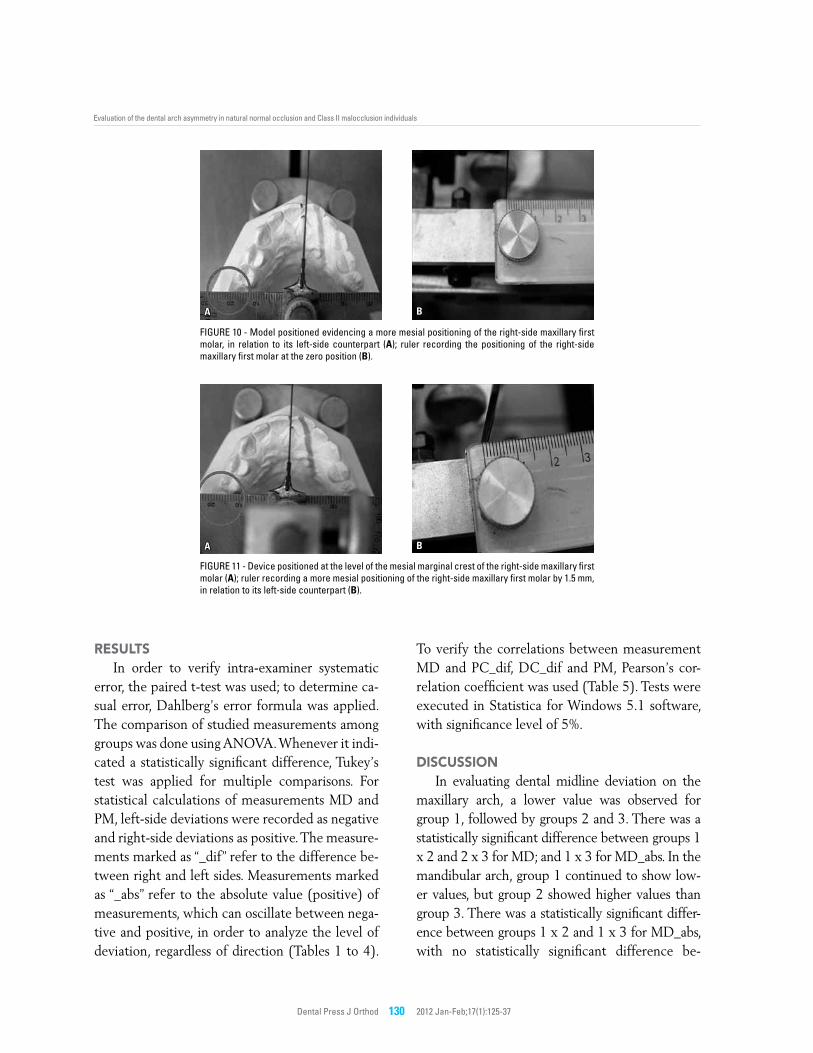
» Position of the maxillary and mandibular
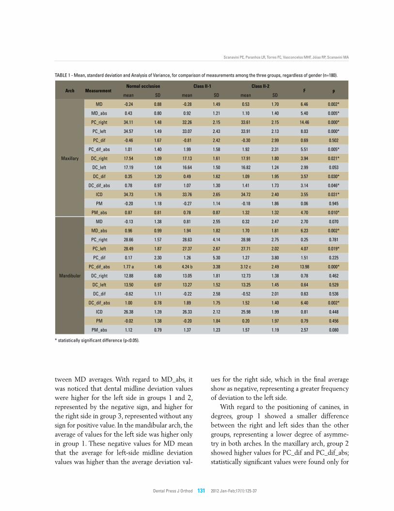
first molars, in anteroposterior direction (PM): The distance was measured longi-tudinally from the mesial marginal crest of the molar positioned more distally to the mesial marginal crest of opposite side molar, as shown in Figures 10 and 11.
Dental Press J Orthod 130 2012 Jan-Feb;17(1):125-37
A B
A B
Evaluation of the dental arch asymmetry in natural normal occlusion and Class II malocclusion individuals
RESuLtSIn order to verify intra-examiner systematic
error, the paired t-test was used; to determine ca-sual error, Dahlberg’s error formula was applied. The comparison of studied measurements among groups was done using ANOVA. Whenever it indi-cated a statistically significant difference, Tukey’s test was applied for multiple comparisons. For statistical calculations of measurements MD and PM, left-side deviations were recorded as negative and right-side deviations as positive. The measure-ments marked as “_dif” refer to the difference be-tween right and left sides. Measurements marked as “_abs” refer to the absolute value (positive) of measurements, which can oscillate between nega-tive and positive, in order to analyze the level of deviation, regardless of direction (Tables 1 to 4).
To verify the correlations between measurement MD and PC_dif, DC_dif and PM, Pearson’s cor-relation coefficient was used (Table 5). Tests were executed in Statistica for Windows 5.1 software, with significance level of 5%.
dISCuSSIOnIn evaluating dental midline deviation on the
maxillary arch, a lower value was observed for group 1, followed by groups 2 and 3. There was a statistically significant difference between groups 1 x 2 and 2 x 3 for MD; and 1 x 3 for MD_abs. In the mandibular arch, group 1 continued to show low-er values, but group 2 showed higher values than group 3. There was a statistically significant differ-ence between groups 1 x 2 and 1 x 3 for MD_abs, with no statistically significant difference be-
FigurE 10 - Model positioned evidencing a more mesial positioning of the right-side maxillary first molar, in relation to its left-side counterpart (A); ruler recording the positioning of the right-side maxillary first molar at the zero position (B).
FigurE 11 - Device positioned at the level of the mesial marginal crest of the right-side maxillary first molar (A); ruler recording a more mesial positioning of the right-side maxillary first molar by 1.5 mm, in relation to its left-side counterpart (B).
Scanavini PE, Paranhos LR, Torres FC, Vasconcelos MHF, Jóias RP, Scanavini MA
Dental Press J Orthod 131 2012 Jan-Feb;17(1):125-37
tween MD averages. With regard to MD_abs, it was noticed that dental midline deviation values were higher for the left side in groups 1 and 2, represented by the negative sign, and higher for the right side in group 3, represented without any sign for positive value. In the mandibular arch, the average of values for the left side was higher only in group 1. These negative values for MD mean that the average for left-side midline deviation values was higher than the average deviation val-
ues for the right side, which in the final average show as negative, representing a greater frequency of deviation to the left side.
With regard to the positioning of canines, in degrees, group 1 showed a smaller difference between the right and left sides than the other groups, representing a lower degree of asymme-try in both arches. In the maxillary arch, group 2 showed higher values for PC_dif and PC_dif_abs; statistically significant values were found only for
Arch MeasurementNormal occlusion Class II-1 Class II-2
F pmean SD mean SD mean SD
Maxillary
MD -0.24 0.88 -0.28 1.49 0.53 1.70 6.46 0.002*
MD_abs 0.43 0.80 0.92 1.21 1.10 1.40 5.40 0.005*
PC_right 34.11 1.48 32.26 2.15 33.61 2.15 14.46 0.000*
PC_left 34.57 1.49 33.07 2.43 33.91 2.13 8.03 0.000*
PC_dif -0.46 1.67 -0.81 2.42 -0.30 2.99 0.69 0.502
PC_dif_abs 1.01 1.40 1.99 1.58 1.92 2.31 5.51 0.005*
DC_right 17.54 1.09 17.13 1.61 17.91 1.80 3.94 0.021*
DC_left 17.19 1.04 16.64 1.50 16.82 1.24 2.99 0.053
DC_dif 0.35 1.20 0.49 1.62 1.09 1.95 3.57 0.030*
DC_dif_abs 0.78 0.97 1.07 1.30 1.41 1.73 3.14 0.046*
iCD 34.73 1.76 33.76 2.65 34.72 2.40 3.55 0.031*
PM -0.20 1.18 -0.27 1.14 -0.18 1.86 0.06 0.945
PM_abs 0.87 0.81 0.78 0.87 1.32 1.32 4.70 0.010*
Mandibular
MD -0.13 1.38 0.81 2.55 0.32 2.47 2.70 0.070
MD_abs 0.96 0.99 1.94 1.82 1.70 1.81 6.23 0.002*
PC_right 28.66 1.57 28.63 4.14 28.98 2.75 0.25 0.781
PC_left 28.49 1.87 27.37 2.67 27.71 2.02 4.07 0.019*
PC_dif 0.17 2.30 1.26 5.30 1.27 3.80 1.51 0.225
PC_dif_abs 1.77 a 1.46 4.24 b 3.38 3.12 c 2.49 13.98 0.000*
DC_right 12.88 0.80 13.05 1.81 12.73 1.38 0.78 0.462
DC_left 13.50 0.97 13.27 1.52 13.25 1.45 0.64 0.529
DC_dif -0.62 1.11 -0.22 2.58 -0.52 2.01 0.63 0.536
DC_dif_abs 1.00 0.78 1.89 1.75 1.52 1.40 6.40 0.002*
iCD 26.38 1.39 26.33 2.12 25.98 1.99 0.81 0.448
PM -0.02 1.38 -0.20 1.84 0.20 1.97 0.79 0.456
PM_abs 1.12 0.79 1.37 1.23 1.57 1.19 2.57 0.080
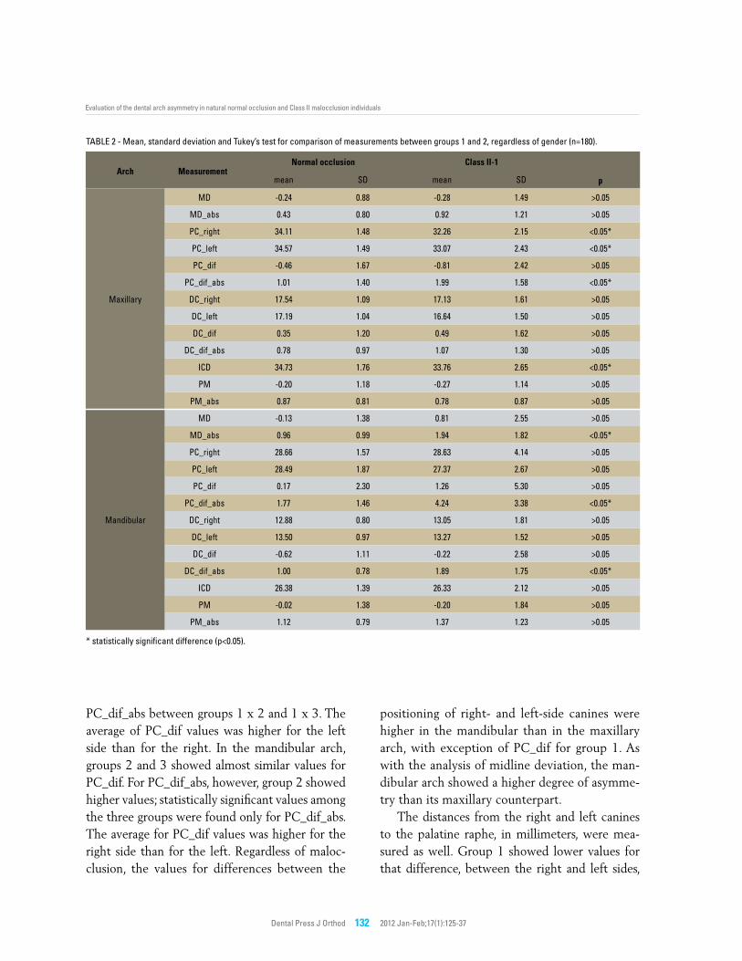
TAblE 1 - Mean, standard deviation and Analysis of Variance, for comparison of measurements among the three groups, regardless of gender (n=180).
* statistically significant difference (p<0.05).
Dental Press J Orthod 132 2012 Jan-Feb;17(1):125-37
Evaluation of the dental arch asymmetry in natural normal occlusion and Class II malocclusion individuals
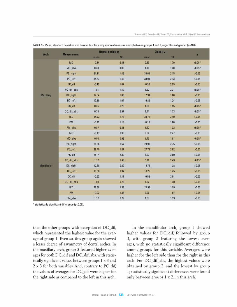
TAblE 2 - Mean, standard deviation and Tukey’s test for comparison of measurements between groups 1 and 2, regardless of gender (n=180).
* statistically significant difference (p<0.05).
Arch MeasurementNormal occlusion Class II-1
mean SD mean SD p
Maxillary
MD -0.24 0.88 -0.28 1.49 >0.05
MD_abs 0.43 0.80 0.92 1.21 >0.05
PC_right 34.11 1.48 32.26 2.15 <0.05*
PC_left 34.57 1.49 33.07 2.43 <0.05*
PC_dif -0.46 1.67 -0.81 2.42 >0.05
PC_dif_abs 1.01 1.40 1.99 1.58 <0.05*
DC_right 17.54 1.09 17.13 1.61 >0.05
DC_left 17.19 1.04 16.64 1.50 >0.05
DC_dif 0.35 1.20 0.49 1.62 >0.05
DC_dif_abs 0.78 0.97 1.07 1.30 >0.05
iCD 34.73 1.76 33.76 2.65 <0.05*
PM -0.20 1.18 -0.27 1.14 >0.05
PM_abs 0.87 0.81 0.78 0.87 >0.05
Mandibular
MD -0.13 1.38 0.81 2.55 >0.05
MD_abs 0.96 0.99 1.94 1.82 <0.05*
PC_right 28.66 1.57 28.63 4.14 >0.05
PC_left 28.49 1.87 27.37 2.67 >0.05
PC_dif 0.17 2.30 1.26 5.30 >0.05
PC_dif_abs 1.77 1.46 4.24 3.38 <0.05*
DC_right 12.88 0.80 13.05 1.81 >0.05
DC_left 13.50 0.97 13.27 1.52 >0.05
DC_dif -0.62 1.11 -0.22 2.58 >0.05
DC_dif_abs 1.00 0.78 1.89 1.75 <0.05*
iCD 26.38 1.39 26.33 2.12 >0.05
PM -0.02 1.38 -0.20 1.84 >0.05
PM_abs 1.12 0.79 1.37 1.23 >0.05
PC_dif_abs between groups 1 x 2 and 1 x 3. The average of PC_dif values was higher for the left side than for the right. In the mandibular arch, groups 2 and 3 showed almost similar values for PC_dif. For PC_dif_abs, however, group 2 showed higher values; statistically significant values among the three groups were found only for PC_dif_abs. The average for PC_dif values was higher for the right side than for the left. Regardless of maloc-clusion, the values for differences between the
positioning of right- and left-side canines were higher in the mandibular than in the maxillary arch, with exception of PC_dif for group 1. As with the analysis of midline deviation, the man-dibular arch showed a higher degree of asymme-try than its maxillary counterpart.
The distances from the right and left canines to the palatine raphe, in millimeters, were mea-sured as well. Group 1 showed lower values for that difference, between the right and left sides,
Scanavini PE, Paranhos LR, Torres FC, Vasconcelos MHF, Jóias RP, Scanavini MA
Dental Press J Orthod 133 2012 Jan-Feb;17(1):125-37
Arch MeasurementNormal occlusion Class II-2
pmean SD mean SD
Maxillary
MD -0.24 0.88 0.53 1.70 <0.05*
MD_abs 0.43 0.80 1.10 1.40 <0.05*
PC_right 34.11 1.48 33.61 2.15 >0.05
PC_left 34.57 1.49 33.91 2.13 >0.05
PC_dif -0.46 1.67 -0.30 2.99 >0.05
PC_dif_abs 1.01 1.40 1.92 2.31 <0.05*
DC_right 17.54 1.09 17.91 1.80 >0.05
DC_left 17.19 1.04 16.82 1.24 >0.05
DC_dif 0.35 1.20 1.09 1.95 <0.05*
DC_dif_abs 0.78 0.97 1.41 1.73 <0.05*
iCD 34.73 1.76 34.72 2.40 >0.05
PM -0.20 1.18 -0.18 1.86 >0.05
PM_abs 0.87 0.81 1.32 1.32 <0.05*
Mandibular
MD -0.13 1.38 0.32 2.47 >0.05
MD_abs 0.96 0.99 1.70 1.81 <0.05*
PC_right 28.66 1.57 28.98 2.75 >0.05
PC_left 28.49 1.87 27.71 2.02 >0.05
PC_dif 0.17 2.30 1.27 3.80 >0.05
PC_dif_abs 1.77 1.46 3.12 2.49 <0.05*
DC_right 12.88 0.80 12.73 1.38 >0.05
DC_left 13.50 0.97 13.25 1.45 >0.05
DC_dif -0.62 1.11 -0.52 2.01 >0.05
DC_dif_abs 1.00 0.78 1.52 1.40 >0.05
iCD 26.38 1.39 25.98 1.99 >0.05
PM -0.02 1.38 0.20 1.97 >0.05
PM_abs 1.12 0.79 1.57 1.19 >0.05
TAblE 3 - Mean, standard deviation and Tukey’s test for comparison of measurements between groups 1 and 3, regardless of gender (n=180).
* statistically significant difference (p<0.05).
than the other groups, with exception of DC_dif, which represented the highest value for the aver-age of group 1. Even so, this group again showed a lesser degree of asymmetry of dental arches. In the maxillary arch, group 3 featured higher aver-ages for both DC_dif and DC_dif_abs, with statis-tically significant values between groups 1 x 3 and 2 x 3 for both variables. And, contrary to PC_dif, the values of averages for DC_dif were higher for the right side as compared to the left in this arch.
In the mandibular arch, group 1 showed higher values for DC_dif, followed by group 3, with group 2 featuring the lowest aver-ages, with no statistically significant difference among groups for this variable. Averages were higher for the left side than for the right in this arch. For DC_dif_abs, the highest values were obtained by group 2, and the lowest by group 1; statistically significant differences were found only between groups 1 x 2, in this arch.
Dental Press J Orthod 134 2012 Jan-Feb;17(1):125-37
Evaluation of the dental arch asymmetry in natural normal occlusion and Class II malocclusion individuals
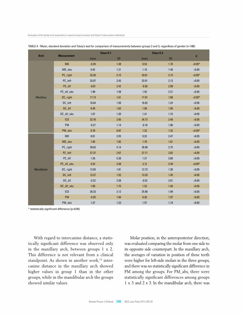
TAblE 4 - Mean, standard deviation and Tukey’s test for comparison of measurements between groups 2 and 3, regardless of gender (n=180).
* statistically significant difference (p<0.05).
Arch MeasurementClass II-1 Class II-2
pmean SD mean SD
Maxillary
MD -0.28 1.49 0.53 1.70 <0.05*
MD_abs 0.92 1.21 1.10 1.40 >0.05
PC_right 32.26 2.15 33.61 2.15 <0.05*
PC_left 33.07 2.43 33.91 2.13 >0.05
PC_dif -0.81 2.42 -0.30 2.99 >0.05
PC_dif_abs 1.99 1.58 1.92 2.31 >0.05
DC_right 17.13 1.61 17.91 1.80 <0.05*
DC_left 16.64 1.50 16.82 1.24 >0.05
DC_dif 0.49 1.62 1.09 1.95 >0.05
DC_dif_abs 1.07 1.30 1.41 1.73 >0.05
iCD 33.76 2.65 34.72 2.40 >0.05
PM -0.27 1.14 -0.18 1.86 >0.05
PM_abs 0.78 0.87 1.32 1.32 <0.05*
Mandibular
MD 0.81 2.55 0.32 2.47 >0.05
MD_abs 1.94 1.82 1.70 1.81 >0.05
PC_right 28.63 4.14 28.98 2.75 >0.05
PC_left 27.37 2.67 27.71 2.02 >0.05
PC_dif 1.26 5.30 1.27 3.80 >0.05
PC_dif_abs 4.24 3.38 3.12 2.49 <0.05*
DC_right 13.05 1.81 12.73 1.38 >0.05
DC_left 13.27 1.52 13.25 1.45 >0.05
DC_dif -0.22 2.58 -0.52 2.01 >0.05
DC_dif_abs 1.89 1.75 1.52 1.40 >0.05
iCD 26.33 2.12 25.98 1.99 >0.05
PM -0.20 1.84 0.20 1.97 >0.05
PM_abs 1.37 1.23 1.57 1.19 >0.05
With regard to intercanine distance, a statis-tically significant difference was observed only in the maxillary arch, between groups 1 x 2. This difference is not relevant from a clinical standpoint. As shown in another work,12 inter-canine distance in the maxillary arch showed higher values in group 1 than in the other groups, while in the mandibular arch the groups showed similar values.
Molar position, in the anteroposterior direction, was evaluated comparing the molar from one side to its opposite side counterpart. In the maxillary arch, the averages of variation in position of these teeth were higher for left-side molars in the three groups, and there was no statistically significant difference in PM among the groups. For PM_abs, there were statistically significant differences among groups 1 x 3 and 2 x 3. In the mandibular arch, there was
Scanavini PE, Paranhos LR, Torres FC, Vasconcelos MHF, Jóias RP, Scanavini MA
Dental Press J Orthod 135 2012 Jan-Feb;17(1):125-37
Group Arch Measurement r p
Normal
Maxillary
PC_dif 0.58 0.000*
DC_dif 0.35 0.006*
PM -0.29 0.022*
Mandibular
PC_dif 0.71 0.000*
DC_dif 0.78 0.000*
PM -0.60 0.000*
Class ii-1
Maxillary
PC_dif 0.46 0.000*
DC_dif 0.43 0.001*
PM 0.13 0.328
Mandibular
PC_dif 0.84 0.000*
DC_dif 0.67 0.000*
PM -0.55 0.000*
Class ii-2
Maxillary
PC_dif 0.27 0.037*
DC_dif 0.54 0.000*
PM -0.34 0.008*
Mandibular
PC_dif 0.82 0.000*
DC_dif 0.77 0.000*
PM -0.61 0.000*
TAblE 5 - Pearson’s correlation coefficient between MD and measure-ments PC_dif, DC_dif and PM.
* statistically significant correlation (p<0.05).
no statistically significant difference among the three groups, either for PM or PM_abs. In groups 1 and 2, PM values were higher for left-side molars, while in group 3 they were higher for the right side.
Corroborating the works of other authors,2-5,8,9 individuals with natural normal occlusion (group 1) showed dental arch asymmetry, even if in a lesser degree than individuals with Angle Class II, Division 1 malocclusion (group 2) and Angle Class II, Division 2 malocclusion (group 3).
Although group 1 showed a smaller degree of asymmetry than the other two groups, a small dif-ference was noticed between the degrees of asym-metry shown by groups 2 and 3; in other words, the results do not show an expressive difference, clinically speaking, among the measured variables, when comparing only groups 2 and 3. This indi-cates a greater degree of asymmetry for groups with malocclusion, in agreement with other authors.13
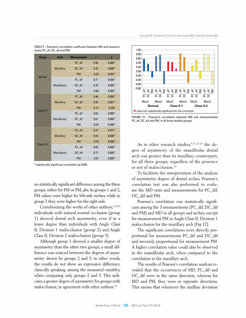
As in other research studies,9,11,13,14 the de-gree of asymmetry of the mandibular dental arch was greater than its maxillary counterpart, for all three groups, regardless of the presence or not of malocclusion.12
To facilitate the interpretation of the analysis of asymmetry degree of dental arches, Pearson’s correlation test was also performed to evalu-ate the MD ratio and measurements for PC_dif, DC_dif and PM.
Pearson’s correlation was statistically signifi-cant among the 3 measurements (PC_dif, DC_dif and PM) and MD in all groups and arches, except for measurement PM in Angle Class II, Division 1 malocclusion for the maxillary arch (Fig 12).
The significant correlations were directly pro-portional for measurements PC_dif and DC_dif and inversely proportional for measurement PM. A higher correlation value could also be observed in the mandibular arch, when compared to the correlation in the maxillary arch.
The results of Pearson’s correlation analysis re-vealed that the occurrences of MD, PC_dif and DC_dif were in the same direction, whereas for MD and PM, they were in opposite directions. This means that whenever the midline deviation
FigurE 12 - Pearson’s correlation between MD and measurements PC_dif, DC_dif and PM, in all three studied groups.
1.000.800.600.400.200.00
-0.20-0.40-0.60-0.80
Maxil.Normal Class II-1 Class II-2
Mand. Maxil. Mand. Maxil. Mand.
PC_d
ifDC
_dif
PMPC
_dif
DC_d
ifPM
PC_d
ifDC
_dif
PMPC
_dif
DC_d
ifPM
PC_d
ifDC
_dif
PMPC
_dif
DC_d
ifPM
value non-statistically significant for the correction
Dental Press J Orthod 136 2012 Jan-Feb;17(1):125-37
Evaluation of the dental arch asymmetry in natural normal occlusion and Class II malocclusion individuals
was towards the right side, the left-side molar was more mesial in relation to the right-side molar and vice-versa, in both arches.
With regard to the maxillary arch of group 2, factors such as tooth rotation and more severe crowding, found in the entire group, can explain the fact that this correlation is different than the other groups only in this arch of this group.
Dental arch asymmetry is a widely discussed subject in orthodontic literature, from its possible causes (such as heredity,3 chewing3,6,7 and posture3,6 habits, early tooth loss and agenesis with resulting movement of adjacent teeth3,4,17) to the several dif-ferent diagnostic resources4,10,16,17 and treatment possibilities1,10,16,17 in the first phases of orthodon-tic mechanics, aiming for a successful orthodontic treatment resulting in an occlusion with better post-treatment stability. This stability also deserves special attention, because it is questioned over time, as individuals reach adult age.1,2,6,15,16,17
This study introduced a new methodology, with measurements in plaster cast models, to analyze the asymmetry of dental arches. The obtained results will provide important data that can be used by clinical professionals and researchers in orthodon-tics, both in the diagnosis and planning or new cas-es to be treated, as in devising new research studies.
COnCLuSIOnS After a judicious analysis of the applied meth-
odology and the results obtained in this study, it can be concluded that:
1) Asymmetry in the dental arches was found in all individuals, regardless of the pres-ence of malocclusion.
2) Individuals with natural normal occlu-sion showed a smaller degree of asymme-try than individuals with Angle Class II, Division 1 and Angle Class II, Division 2 malocclusions, and the latter two featured similar degrees of asymmetry.
3) The degree of asymmetry in mandibular dental arches was greater than in maxillary dental arches for all 3 evaluated groups.
4) The direction of midline deviation showed a correlation directly proportional to the difference in position of the canines and to the difference in the distances from the canines to the palatine raphe.
5) The direction of midline deviation showed a correlation inversely proportional to the side of the molar positioned most mesially, in both arches of all three groups, with ex-ception of the maxillary arch in group 2 (Angle Class II, Division 1).
Scanavini PE, Paranhos LR, Torres FC, Vasconcelos MHF, Jóias RP, Scanavini MA
Dental Press J Orthod 137 2012 Jan-Feb;17(1):125-37
Contact addressPaulo Estevão Scanavini Rua Antonio Pereira de Camargo, 129 – CentroZip code: 13.800-033 – Sumaré/SP, BrazilE-mail: [email protected]
1. Jerrold L, Lowenstein J. The midline: diagnosis and treatment. Am J Orthod Dentofacial Orthop. 1990;97(6):453-62.
2. Hechter FJ. Symmetry and dental arch form of orthodontically treated patients. Dent J. 1978 Apr;44(4):173-84.
3. Lundstrom A. Some asymmetries of dental arches, jaws, and skull, and their etiological significance. Am J Orthod. 1961;47(2):81-106.
4. Bishara SE, Burkey PS, Kharouf JG. Dental and facial asymmetries: a review. Angle Orthod. 1994;64(2):89-98.
5. Maurice TJ, Kula K. Dental arch asymmetry in the mixed dentition. Angle Orthod. 1998;68(1):37-44.
6. Smith R, Bailit H. Prevalence and etiology of asymmetries in occlusion. Angle Orthod. 1979;49(3):199-204.
7. Shah MS, Joshi MR. An assessment of asymmetry in the normal craniofacial complex. Angle Orthod. 1978;48(2):141-8.
8. Ferrario VF, Sforza C, Miani A, D’Addona A. Position and asymmetry of teeth in untreated dental arches. Int J Adult Orthod Orthognath Surg. 1993;8(4):277-85.
9. Araújo TM, Wilhelm RS, Almeida MA. Skeletal and dental arch asymmetries in individuals with normal dental occlusion. Int J Adult Orthod Orthog Surg. 1994;9(2):111-8.
10. Beyer JW, Lindauer SJ. Evaluation of dental midline position. Semin Orthod. 1998;4(3):146-52.
11. Rose JM, Sadowsky C, BeGole EA, Moles R. Mandibular skeletal and dental asymmetry in Class II malocclusions. Am J Orthod Dentofacial Orthop. 1994 May;105(5):489-95.
REfEREnCES
12. Staley RN, Stuntz WR, Peterson LC. A comparison of arch widths in adults with normal occlusion and adults with Class II, Division 1 malocclusion. Am J Orthod. 1985;8(2):163-9.
13. Nie Q, Lin J. [Analysis and comparison of dental arch symmetry between different Angle’s malocclusion categories and normal occlusion. Zhonghua Kou Qiang Yi Xue Za Zhi. 2000 Mar;35(2):105-7.
14. Janson GR, Metaxas A, Woodside DG, de Freitas MR, Pinzan A. Three-dimensional evaluation of skeletal and dental asymmetries in Class II subdivision malocclusions. Am J Orthod Dentofacial Orthop. 2001 Apr;119(4):406-18.
15. Henrikson J, Persson M, Thilander B. Long-term stability of dental arch form in normal occlusion from 13 to 31 years of age. Eur J Orthod. 2001;23(1):51-61.
16. Rebellato J. Asymmetric extractions used in the treatment of patients with asymmetries. Semin Orthod. 1998;4(3):180-8.
17. Kusnoto J, Evans CA, BeGole EA, Obrez A. Orthodontic correction of transverse arch asymmetries. Am J Orthod Dentofacial Orthop. 2002;121(1):38-45.
18. Wertz RA. Diagnosis and treatment planning of unilateral Class II malocclusions. Angle Orthod. 1975;45(2):85-94.
19. Alavi DG, Begole EA, Shneider BJ. Facial and dental arch asymmetries in Class II subdivision malocclusion. Am J Orthod Dentofacial Orthop. 1988;93(1):38-46.
Submitted: December 18, 2008Revised and accepted: August 16, 2009
Related Documents