RESEARCH ARTICLE Open Access Evaluation of the complexity of indoor air in hospital wards based on PM2.5, real-time PCR, adenosine triphosphate bioluminescence assay, microbial culture and mass spectrometry Shao Ling and Liu Hui * Abstract Background: The aim of this study was to establish a set of assessment methods suitable for evaluating the complex indoor environment of hospital wards and to ascertain the composition of bacteria and microbial ecology of hospital wards. Methods: Colony-forming units (CFUs), PM2.5 detection, real-time PCR, and adenosine triphosphate (ATP) bioluminescence assay were employed to evaluate the complexity of indoor air in 18 wards of nine departments in a hospital and two student dormitories in a university. Subsequently, the microbial samples were quantified and identified using matrix-assisted laser desorption/ionization time-of-flight mass spectrometry (MALDI-TOF MS). Results: Although the studied indices were relatively independent, the PM2.5 content was correlated with bacterial CFUs determined by passive sedimentation method, bacterial and fungal counts measured by real-time PCR, and ATP bioluminescence assay. The composition of microorganisms in the air of hospital wards differed from that in the air of student dormitories. The dominant genera in hospital wards were Staphylococcus (39.4%), Micrococcus (21.9%), Corynebacterium (11.7%), Kocuria (4.4%), Bacillus (2.9%), Streptococcus (1.6%), Moraxella (1.6%), and Enterococcus (1.3%), and the microbial ecology differed between Respiration Dept. III and other hospital departments. Additionally, 11.1 and 27.3% of bacteria in hospital wards and student dormitories were not identified, respectively. Conclusions: Assessment of environmental quality of hospital wards should be based on comprehensive analysis with multiple indicators. There may be imbalances in the microbial diversity in the hospital wards, therefore, monitoring of the environmental quality of hospitals is important in the prevention of nosocomial infections. Keywords: PM2.5, Real-time PCR, ATP bioluminescence assay, Passive sedimentation, Microbial ecology, MALDI- TOF MS © The Author(s). 2019 Open Access This article is distributed under the terms of the Creative Commons Attribution 4.0 International License (http://creativecommons.org/licenses/by/4.0/), which permits unrestricted use, distribution, and reproduction in any medium, provided you give appropriate credit to the original author(s) and the source, provide a link to the Creative Commons license, and indicate if changes were made. The Creative Commons Public Domain Dedication waiver (http://creativecommons.org/publicdomain/zero/1.0/) applies to the data made available in this article, unless otherwise stated. * Correspondence: [email protected] College of Medical Laboratory, Dalian Medical University, No.9 West Section Lvshun South Road, Dalian 116044, China Ling and Hui BMC Infectious Diseases (2019) 19:646 https://doi.org/10.1186/s12879-019-4249-z

Welcome message from author
This document is posted to help you gain knowledge. Please leave a comment to let me know what you think about it! Share it to your friends and learn new things together.
Transcript

RESEARCH ARTICLE Open Access
Evaluation of the complexity of indoor airin hospital wards based on PM2.5, real-timePCR, adenosine triphosphatebioluminescence assay, microbial cultureand mass spectrometryShao Ling and Liu Hui*
Abstract
Background: The aim of this study was to establish a set of assessment methods suitable for evaluating thecomplex indoor environment of hospital wards and to ascertain the composition of bacteria and microbial ecologyof hospital wards.
Methods: Colony-forming units (CFUs), PM2.5 detection, real-time PCR, and adenosine triphosphate (ATP)bioluminescence assay were employed to evaluate the complexity of indoor air in 18 wards of nine departments ina hospital and two student dormitories in a university. Subsequently, the microbial samples were quantified andidentified using matrix-assisted laser desorption/ionization time-of-flight mass spectrometry (MALDI-TOF MS).
Results: Although the studied indices were relatively independent, the PM2.5 content was correlated with bacterialCFUs determined by passive sedimentation method, bacterial and fungal counts measured by real-time PCR, andATP bioluminescence assay. The composition of microorganisms in the air of hospital wards differed from that inthe air of student dormitories. The dominant genera in hospital wards were Staphylococcus (39.4%), Micrococcus(21.9%), Corynebacterium (11.7%), Kocuria (4.4%), Bacillus (2.9%), Streptococcus (1.6%), Moraxella (1.6%), andEnterococcus (1.3%), and the microbial ecology differed between Respiration Dept. III and other hospitaldepartments. Additionally, 11.1 and 27.3% of bacteria in hospital wards and student dormitories were not identified,respectively.
Conclusions: Assessment of environmental quality of hospital wards should be based on comprehensive analysiswith multiple indicators. There may be imbalances in the microbial diversity in the hospital wards, therefore,monitoring of the environmental quality of hospitals is important in the prevention of nosocomial infections.
Keywords: PM2.5, Real-time PCR, ATP bioluminescence assay, Passive sedimentation, Microbial ecology, MALDI-TOF MS
© The Author(s). 2019 Open Access This article is distributed under the terms of the Creative Commons Attribution 4.0International License (http://creativecommons.org/licenses/by/4.0/), which permits unrestricted use, distribution, andreproduction in any medium, provided you give appropriate credit to the original author(s) and the source, provide a link tothe Creative Commons license, and indicate if changes were made. The Creative Commons Public Domain Dedication waiver(http://creativecommons.org/publicdomain/zero/1.0/) applies to the data made available in this article, unless otherwise stated.
* Correspondence: [email protected] of Medical Laboratory, Dalian Medical University, No.9 West SectionLvshun South Road, Dalian 116044, China
Ling and Hui BMC Infectious Diseases (2019) 19:646 https://doi.org/10.1186/s12879-019-4249-z

BackgroundThe indoor environment of hospitals, with higher risk ofmicrobial contamination, is complicated and differentfrom other occupational or residential indoor settings.Most of the hospitalized patients spend two-third oftheir time in wards every day; in particular, bed-riddenpatients spend almost all their time in the ward. There-fore, the air quality of hospitals may be an importantrisk factor affecting the health of patients, and assess-ment of hospital environment is particularly significant.Airborne microflora in hospital as a potential cause of
hospital infections has been the subject of numerousstudies. Some researches have been showed that envir-onmental contamination may promote the infection andtransmission of healthcare-associated pathogens, includ-ing methicillin-resistant Staphylococcus aureus (MRSA),vancomycin-resistant Enterococcus spp. (VRE), Clostri-dium difficile, Acinetobacter spp., and norovirus [1, 2].Also, some studies suggested that if the prior room wasoccupied by several important healthcare-associatedpathogens, the risk of acquiring these bacteria by a newadmission was significantly increased [3–6]. In addition,bioaerosol infection is common in hospitals and re-cognized to be the causation of agents and illness suchas aspergillosis and tuberculosis, as a result, the levels,sources and characteristics of bioaerosols have beenextensively investigated [7–11]. PM2.5 is an air pollutantwith a diameter of ≤2.5 μm, which can be easily inhaledand can penetrate deep into the airway [12]. Many stud-ies have confirmed that inhalation exposure to ambientPM in the environment, especially PM2.5, is related tocertain systemic diseases and cancer [13–15]. Long- andshort-term exposure to PM2.5 has been consistently as-sociated with a number of outcomes, including morta-lity, cardiovascular and cerebrovascular events and lungcancer [16–20]. Calderón-Garcidueñas et al. highlightedthat air pollution can be considered as a risk factor forboth Alzheimer’s disease and Parkinson’s disease [21].Currently, although the pathogenicity of PM2.5 in aero-sol chemistry and physics has been extensively studied, alittle is known about the inhalable biological particlessuch as bacteria, fungi, viruses, pollen, and cell debris[22–24]. In addition, airborne PM2.5 in indoor environ-ments has been widely investigated because of its abilityto cause adverse health effects [25]. A study conductedin the USA about hospital nurses confirmed that PM2.5was related to the risk of all-cause mortality [26]. Hence,monitoring of indoor environmental quality is the firststep to reduce the chance of nosocomial infections. Thetraditionally applied methods, to assess microbial aircontamination and cleanliness on surfaces in hospitalare based on microbiological cultural technology, inclu-ding active, passive and surfaces sampling [27]. In recentdecade, adenosine triphosphate (ATP) bioluminescence
assay, which was used to assess environmental surfacecleanliness has been proposed [28]. Previous studieshave shown that the number of microorganisms thatcan be cultured only accounts for < 5% of the totalmicroorganisms in air [29–31]. Therefore, relyingsolely on the method of cultivation may not help inaccurate assessment of hospital environment. Hence,comprehensive indicators, including fine particles,total microorganisms (including dead microorga-nisms), culturable microorganisms, and number ofliving cells, are needed to analyze the complex hos-pital wards environment.Conventional microbiological methods for bacterial
identification involve culture, microscopic exa-mination, and biochemical testing. Although theseprocedures can be accurate and reliable, they aretime-consuming and require well-trained techniciansto interpret the results [32]. Currently, whole genomesequencing (WGS), an analytical technology thatallows sequencing of the whole genomic content ofbacteria, would be the best approach to bacterial identi-fication. Matrix-assisted laser desorption/ionization time-of-flight mass spectrometry (MALDI-TOF MS) hasemerged as a tool for microbial identification and diagno-sis owing to its specificity, high speed, and low costs. Thistechnology has been widely used for microbial identifica-tion and strain typing, detection of antibiotic resistance,detection of blood and urinary tract pathogens, and detec-tion of water- and food-borne pathogens, etc. [33].In the present study, we established a set of assess-
ment methods suitable for evaluating the complexindoor environment of hospital wards by using both cul-tivation and non-cultivation approaches. Subsequently,MALDI-TOF MS was employed to identify the isolatedorganisms to assess the microbial ecology of the studiedenvironment.
MethodsSetting and samplingThe hospital selected in this study is a comprehensiveteaching hospital with three buildings and 1,788 beds inNortheast China. The wards of two hospital buildings(Nos. 1 and 9 buildings), which have the same centralHVAC system and the area of each ward was 30 m2,were selected for the investigation. The two buildingswere completed in 2016 and 2000, respectively. To com-pare the differences between the hospital and naturalenvironment, two student dormitories in a universitywere selected as control. The indoor environmentalindicators were monitored in the general hospital inspring between 1/3/2018 and 15/3/2018. The researchwas approved by the Ethical Committee of DalianMedical University, China.
Ling and Hui BMC Infectious Diseases (2019) 19:646 Page 2 of 10

Detection of PM2.5 and PM10The indoor PM2.5 concentrations were detected usingoptical particle counter PC-5A (Laser Research Institute,Jiangsu, China), which employs light scattering technol-ogy to deliver real-time measurements [34]. In additionto the seventh floor (Obstetrics Dept.) in No. 1 building,five wards of each department from floor 6 to 17 wereselected for PM10 detection. Likewise, in No. 9 building,five wards of each department from floor 1 to 11 wereselected for PM10 detection. In No. 1 building, threedepartments with the highest concentration of PM10,namely, General Surgery Dept. VI Ward, GI SurgeryDept. Ward, and GI Dept. I Ward, and two departmentswith the lowest concentration of PM10, namely, ENTDept. Ward and Nephrology Dept. Ward, were selected.In No. 9 building, two departments with the highestconcentration of PM10, namely, Respiration Dept. IWard on floor 11 and Respiration Dept. III Ward onfloor 9, and two departments with the lowest PM10 con-centration, namely, Neurology Dept. VI Ward on floor 3and Neurology Dept. VII Ward on floor 2, were chosen.Among these nine selected departments, two wards werechosen for each department (totaling to 18 wards), andtheir PM2.5 content was ascertained. Two measured fac-tors, namely, the use of a humidifier and presence ofhouseplants, were invariant and therefore not includedin the analysis. The air temperature and relative humid-ity were measured for the duration of the samplingperiod. The student dormitories, as control, were simul-taneously subjected to PM2.5 and PM10 analysis.
Passive sedimentation and dust sample collectionIn the 18 wards of the selected nine departments, thebacterial and fungal cultures were accomplished by trad-itional passive sedimentation method according to theHospital Sanitary Disinfection Standard (GB15982–2012). The inner, middle, and outer diagonal lines wereset up at three points. The inner and outer points werelocated at 1 m from the wall and at a height of 0.8 m.Three Columbia blood agar plates (BIOMERIEUX,France) for bacteria and three Sabouraud medium plates(Yancheng biology, China) for fungi were placed at thesampling points in each ward. During sampling, theplates were opened for 5 min.A modified method based on the standard ASTM
(method E1728–03) [35], combined with the protocol
developed by Yamamoto [36] and Rachel I. Adams [37],was employed to collect indoor dust. Sterile cotton swabspre-wetted with 0.15M NaCl and 0.05% Tween 20 sam-pling buffer were swabbed for 5–10 s on the four cornersof the floor, each with an area of 5 × 5 cm, to collect thedust. Then, the swabs were placed in sampling tubes andstored at − 20 °C until DNA extraction. In general, fourswabs were obtained for each ward. During the DNAextraction process, two swabs from the same ward werecombined into one DNA extraction reaction mixture toobtain sufficient biomass for analysis.
DNA extractionThe head of the swab was cut using sterile scissor (steril-ized using 75% alcohol) and placed into a 2-ml DNeasyPowerSoil extraction tube (Hilden, Germany). Then, so-lution C1 was added to the extraction tube and incu-bated at 65 °C for 10 min. After incubation, the tube wasvortexed with a vortex mixer for 20 min, and the DNAwas extracted using DNeasy PowerSoil Kit (Hilden,Germany) according to the manufacturer’s protocol.The bacterial universal primer p1370/p201 [38] and
fungal universal primer NL1/260R [39] were used toamplify the fragment of bacterial 16S rDNA subunit andfungal 28S rDNA subunit, respectively. All the primersused were synthesized by Takara (Japan), and theirsequence details are listed in Table 1.The amplified fragments were run on a Real-Time PCR
System (Thermal Cycler Dice Real Time System TP800,Takara, Japan) containing 12.5 μl of SYBR Premix Ex TaqII (Tli RNaseH Plus) (2×) (Takara, Japan), 0.5 μl of each0.4 μM primer, 2 μl of genomic DNA, and water to a finalvolume of 25 μl. For bacteria, the thermal cycling condi-tions consisted of an initial denaturation at 95 °C for 30 s,followed by 40 cycles of denaturation at 95 °C for 5 s,annealing at 55 °C for 15 s, and extension at 72 °C for 10 s.For fungi, the PCR conditions were as follows: 95 °C for30 s, followed by 40 cycles of denaturation at 95 °C for 10s, annealing at 60 °C for 35 s, and extension at 72 °C for20 s. Standard curves for bacteria and fungi were based onextraction of a known quantity of Escherichia coli andAspergillus oryzae spores, respectively.
ATP bioluminescence assayFor ATP bioluminescence assay, Clean-Trace TM NGIinstrument, along with Clean-Trace TM Test Kit (3M,
Table 1 Sequences of the primers used
Name of the targeted species Sequences
Bacteria Forward primer p1370 5′-AGICCCGIGAACGTATTCAC-3′
Reverse primer p201 5′-GAGGAAGGIGIGGAIGACGT-3′
Fungi Forward primer NL1 5′-GCATATCAATAAGCGGAGGAAAAG-3′
Reverse primer 260R 5′-TTAGCTTTAGATGRARTTTACCACC-3′
Ling and Hui BMC Infectious Diseases (2019) 19:646 Page 3 of 10

UK), was used. In the 18 wards of the nine selecteddepartments and two student dormitories, a luciferase-containing sampling swab was swabbed at an area of10 × 10 cm at the end of each bed, and the swab wasinserted into the Clean-Trace tube. Then, the handlewas pushed so that the sampling rod was completelyinserted into the bottom. After shaking for 5 s, thebioluminescence was measured immediately.
Identification by MALDI-TOF MSSingle colonies freshly grown on agar plates were pickedand smeared as a thin film directly onto a MALDI steeltarget plate. Subsequently, 10 μl of 70% formic acid wereadded and the microbial film was overlaid with 10 μl ofMALDI HCCA matrix (50% acetonitrile, 2.5% trifluoroa-cetic acid, and 47.5% water), as recommended by theMALDI-TOF MS manufacturer. The sample-matrix mix-ture was dried at room temperature and analyzed using aMALDI-TOF MS analyzer (Bruker Daltonics, Bremen,Germany). The data were processed using MALDI Bioty-per 2.3 software (Bruker Daltronics) and the spectra werecompared with reference libraries for bacterial identifica-tion. A total of three rounds of MALDI-TOF MS wereperformed. The unidentified bacteria in the first roundwere subcultured for 24 h and a second round of MALDI-TOF MS identification was performed. The bacteria thatstill remained unidentified were subcultured for a further24 h and a third round of MALDI-TOF MS wasperformed.
Statistical analysisThe mean values of CFUs, PM2.5 and PM10 contents,CT values, and ATP contents in the 18 wards and twostudent dormitories were calculated, and correlationanalyses were performed to determine the relationshipsamong these index. Wilcoxon sum rank test was used tocompare the biomass of bacteria and fungi in the wards,and one-sample t-test was employed to assess the differ-ence between the means of wards and dormitories. Thestatistical analysis was performed using SPSS 24.0 statis-tical software, and p < 0.05 was considered significant.
ResultsContents of PM2.5 and PM10, bacteria, fungi, and ATP inthe study sitesAccording to the Chinese Indoor Environment and En-vironmental Product Quality Supervision and InspectionCenter, which issued the first indoor environment anddevelopment trend of the indoor environment industry,the indoor PM2.5 test standard is the first-class con-centration limit of the “Ambient Air Quality Standard”published in 2012, and the daily average PM2.5 concen-tration is 35 μg/m3. In the present study, the concentra-tion of PM2.5 in the wards of four departments,
including ENT Dept., Nephrology Dept., NeurologyDept. VI, and Neurology Dept. VII, were in accordancewith the standard. However, the concentrations ofPM2.5 in the wards of other departments, includingGeneral Surgery Dept. VI, GI Surgery Dept., GI Dept.,Respiration Dept. I, and Respiration Dept. III, as well asstudent dormitories exceeded the standard. Amongthem, the concentration of PM2.5 (89 μg/m3) in the Res-piration Dept. III Ward was the highest, whereas thelowest PM2.5 concentration (10 μg/m3) was noted in theNeurology Dept. VI Ward. According to the “AmbientAir Quality Standard” (GB 3095–2012), the average dailyPM10 concentration limit is 150 μg/m3. The PM10 con-centration in all the wards and dormitories examined inthis study did not exceed the limit, and the highestPM10 content was noted in the GI Surgery Dept. Ward(118 μg/m3), and the lowest content of PM10 was foundin the Neurology Dept. VI Ward (13 μg/m3) (Fig. 1A).In accordance with the Hospital Sanitary Standards for
Sanitization (15982–2012), the average number of bac-terial colonies in the Class III general wards was < 4CFU/petri dish× 5min. The number of bacterial coloniesdetected in the wards of four departments, includingENT Dept., Nephrology Dept., Neurology Dept. VI, andNeurology Dept. VII, was consistent with the standard,and the qualification rate was 44.4%; however, the bac-terial colonies quantified in the wards of other depart-ments examined exceeded the standard. Furthermore, asstudent dormitories do not qualify as medical environ-ment, Indoor Air Quality Standard (GB/T18883–2002)was applied for the assessment, which stipulates < 2500CFU/m3 as the standard total number of bacteria in in-door air. The total number of bacteria can be convertedto the concentration of microorganisms in air, accordingto ORM’s formula as follows: C = 100/A × 5/t × 1000/10 ×N = 50000 N/At, where C is the microbial concen-tration in air (CFU/m3), A is the culture dish area (cm2),t is the sampling time (min), and N is the number ofcolonies in each dish (CFU). In the present study, theconcentration of microorganisms in the student dormi-tories was 1730 CFU/m3, which was consistent with thestandard (Fig. 1B). In addition, the concentrations ofmicroorganisms in the selected nine hospital depart-ments were also calculated.Based on the standard provided by the ATP biolu-
minescence manufacturer, the qualified, critical, andunqualified detection values for the ATP content were0–250, 251–499, and ≥ 500 RLU, respectively, in the gen-eral ward area (patients’ active area, rehabilitation area,and family waiting room). Furthermore, Neurology Dept.VII was the only department that presented qualifiedvalues, whereas the General Surgery Dept. VI, GI Dept.I, ENT Dept., Nephrology Dept., Respiration Dept. I,Respiration Dept. III, and Neurology Dept. VI exhibited
Ling and Hui BMC Infectious Diseases (2019) 19:646 Page 4 of 10
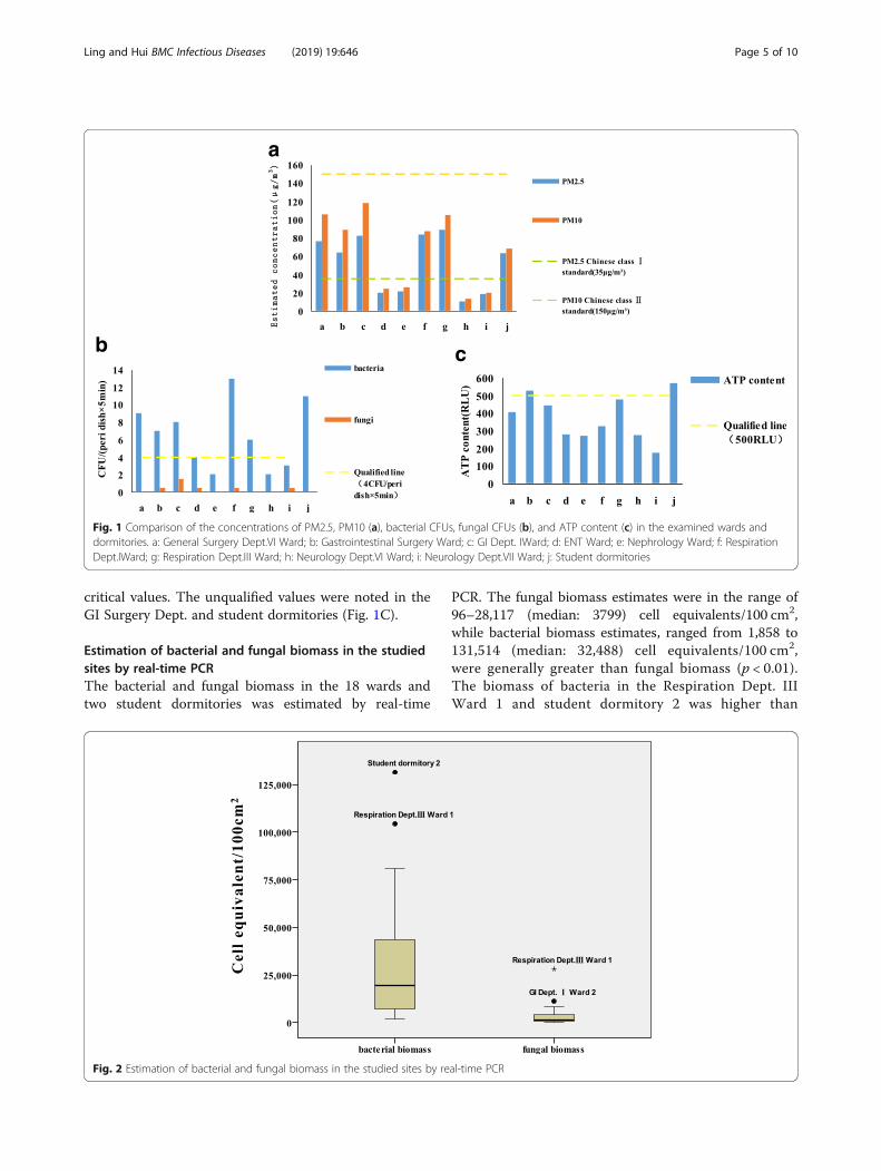
critical values. The unqualified values were noted in theGI Surgery Dept. and student dormitories (Fig. 1C).
Estimation of bacterial and fungal biomass in the studiedsites by real-time PCRThe bacterial and fungal biomass in the 18 wards andtwo student dormitories was estimated by real-time
PCR. The fungal biomass estimates were in the range of96–28,117 (median: 3799) cell equivalents/100 cm2,while bacterial biomass estimates, ranged from 1,858 to131,514 (median: 32,488) cell equivalents/100 cm2,were generally greater than fungal biomass (p < 0.01).The biomass of bacteria in the Respiration Dept. IIIWard 1 and student dormitory 2 was higher than
Fig. 2 Estimation of bacterial and fungal biomass in the studied sites by real-time PCR
a
b c
Fig. 1 Comparison of the concentrations of PM2.5, PM10 (a), bacterial CFUs, fungal CFUs (b), and ATP content (c) in the examined wards anddormitories. a: General Surgery Dept.VI Ward; b: Gastrointestinal Surgery Ward; c: GI Dept. IWard; d: ENT Ward; e: Nephrology Ward; f: RespirationDept.IWard; g: Respiration Dept.III Ward; h: Neurology Dept.VI Ward; i: Neurology Dept.VII Ward; j: Student dormitories
Ling and Hui BMC Infectious Diseases (2019) 19:646 Page 5 of 10
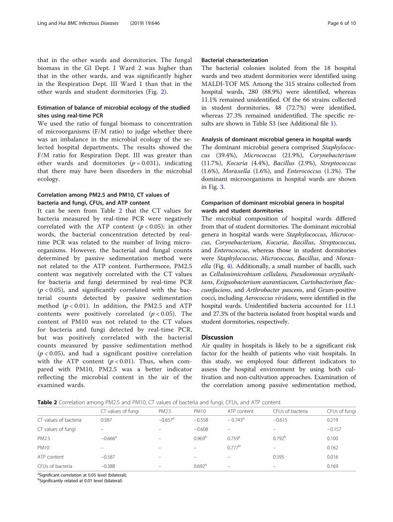
that in the other wards and dormitories. The fungalbiomass in the GI Dept. I Ward 2 was higher thanthat in the other wards, and was significantly higherin the Respiration Dept. III Ward 1 than that in theother wards and student dormitories (Fig. 2).
Estimation of balance of microbial ecology of the studiedsites using real-time PCRWe used the ratio of fungal biomass to concentrationof microorganisms (F/M ratio) to judge whether therewas an imbalance in the microbial ecology of the se-lected hospital departments. The results showed theF/M ratio for Respiration Dept. III was greater thanother wards and dormitories (p = 0.031), indicatingthat there may have been disorders in the microbialecology.
Correlation among PM2.5 and PM10, CT values ofbacteria and fungi, CFUs, and ATP contentIt can be seen from Table 2 that the CT values forbacteria measured by real-time PCR were negativelycorrelated with the ATP content (p < 0.05); in otherwords, the bacterial concentration detected by real-time PCR was related to the number of living micro-organisms. However, the bacterial and fungal countsdetermined by passive sedimentation method werenot related to the ATP content. Furthermore, PM2.5content was negatively correlated with the CT valuesfor bacteria and fungi determined by real-time PCR(p < 0.05), and significantly correlated with the bac-terial counts detected by passive sedimentationmethod (p < 0.01). In addition, the PM2.5 and ATPcontents were positively correlated (p < 0.05). Thecontent of PM10 was not related to the CT valuesfor bacteria and fungi detected by real-time PCR,but was positively correlated with the bacterialcounts measured by passive sedimentation method(p < 0.05), and had a significant positive correlationwith the ATP content (p < 0.01). Thus, when com-pared with PM10, PM2.5 was a better indicatorreflecting the microbial content in the air of theexamined wards.
Bacterial characterizationThe bacterial colonies isolated from the 18 hospitalwards and two student dormitories were identified usingMALDI-TOF MS. Among the 315 strains collected fromhospital wards, 280 (88.9%) were identified, whereas11.1% remained unidentified. Of the 66 strains collectedin student dormitories, 48 (72.7%) were identified,whereas 27.3% remained unidentified. The specific re-sults are shown in Table S3 (see Additional file 1).
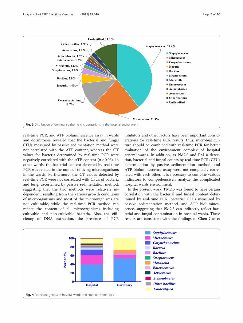
Analysis of dominant microbial genera in hospital wardsThe dominant microbial genera comprised Staphylococ-cus (39.4%), Micrococcus (21.9%), Corynebacterium(11.7%), Kocuria (4.4%), Bacillus (2.9%), Streptococcus(1.6%), Moraxella (1.6%), and Enterococcus (1.3%). Thedominant microorganisms in hospital wards are shownin Fig. 3.
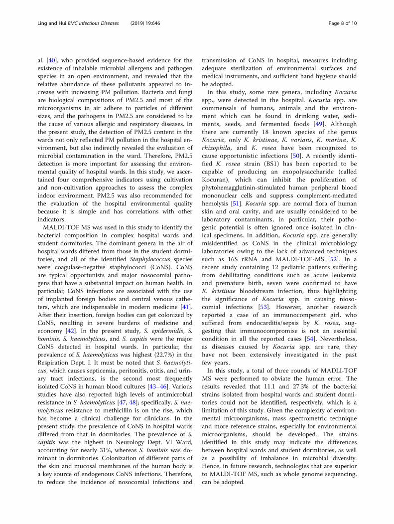
Comparison of dominant microbial genera in hospitalwards and student dormitoriesThe microbial composition of hospital wards differedfrom that of student dormitories. The dominant microbialgenera in hospital wards were Staphylococcus, Micrococ-cus, Corynebacterium, Kocuria, Bacillus, Streptococcus,and Enterococcus, whereas those in student dormitorieswere Staphylococcus, Micrococcus, Bacillus, and Morax-ella (Fig. 4). Additionally, a small number of bacilli, suchas Cellulosimicrobium cellulans, Pseudomonas oryzihabi-tans, Exiguobacterium aurantiacum, Curtobacterium flac-cumfaciens, and Arthrobacter pascens, and Gram-positivecocci, including Aerococcus viridans, were identified in thehospital wards. Unidentified bacteria accounted for 11.1and 27.3% of the bacteria isolated from hospital wards andstudent dormitories, respectively.
DiscussionAir quality in hospitals is likely to be a significant riskfactor for the health of patients who visit hospitals. Inthis study, we employed four different indicators toassess the hospital environment by using both cul-tivation and non-cultivation approaches. Examination ofthe correlation among passive sedimentation method,
Table 2 Correlation among PM2.5 and PM10, CT values of bacteria and fungi, CFUs, and ATP content
CT values of fungi PM2.5 PM10 ATP content CFUs of bacteria CFUs of fungi
CT values of bacteria 0.587 −0.657a −0.558 − 0.743a −0.615 0.219
CT values of fungi – – −0.608 – – −0.157
PM2.5 −0.666a – 0.969b 0.759a 0.792b 0.100
PM10 – – – 0.777b – 0.162
ATP content −0.587 – – – 0.595 0.016
CFUs of bacteria −0.388 – 0.692a – – 0.169aSignificant correlation at 0.05 level (bilateral);bSignificantly related at 0.01 level (bilateral)
Ling and Hui BMC Infectious Diseases (2019) 19:646 Page 6 of 10
real-time PCR, and ATP bioluminescence assay in wardsand dormitories revealed that the bacterial and fungalCFUs measured by passive sedimentation method werenot correlated with the ATP content, whereas the CTvalues for bacteria determined by real-time PCR werenegatively correlated with the ATP content (p < 0.05). Inother words, the bacterial content detected by real-timePCR was related to the number of living microorganismsin the wards. Furthermore, the CT values detected byreal-time PCR were not correlated with CFUs of bacteriaand fungi ascertained by passive sedimentation method,suggesting that the two methods were relatively in-dependent, resulting from the various growth conditionsof microorganisms and most of the microorganisms arenot culturable, while the real-time PCR method canreflect the content of all microorganisms includingcultivable and non-cultivable bacteria. Also, the effi-ciency of DNA extraction, the presence of PCR
inhibitors and other factors have been important consid-erations for real-time PCR results, thus, microbial cul-ture should be combined with real-time PCR for betterevaluation of the environment complex of hospitalgeneral wards. In addition, as PM2.5 and PM10 detec-tion, bacterial and fungal counts by real-time PCR, CFUsdetermination by passive sedimentation method, andATP bioluminescence assay were not completely corre-lated with each other, it is necessary to combine variousindicators to comprehensively analyze the complicatedhospital wards environment.In the present work, PM2.5 was found to have certain
correlation with the bacterial and fungal content deter-mined by real-time PCR, bacterial CFUs measured bypassive sedimentation method, and ATP biolumines-cence, suggesting that PM2.5 can indirectly reflect bac-terial and fungal contamination in hospital wards. Theseresults are consistent with the findings of Chen Cao et
Fig. 3 Distribution of dominant airborne microorganisms in the hospital environment
Fig. 4 Dominant genera in hospital wards and student dormitories
Ling and Hui BMC Infectious Diseases (2019) 19:646 Page 7 of 10
al. [40], who provided sequence-based evidence for theexistence of inhalable microbial allergens and pathogenspecies in an open environment, and revealed that therelative abundance of these pollutants appeared to in-crease with increasing PM pollution. Bacteria and fungiare biological compositions of PM2.5 and most of themicroorganisms in air adhere to particles of differentsizes, and the pathogens in PM2.5 are considered to bethe cause of various allergic and respiratory diseases. Inthe present study, the detection of PM2.5 content in thewards not only reflected PM pollution in the hospital en-vironment, but also indirectly revealed the evaluation ofmicrobial contamination in the ward. Therefore, PM2.5detection is more important for assessing the environ-mental quality of hospital wards. In this study, we ascer-tained four comprehensive indicators using cultivationand non-cultivation approaches to assess the complexindoor environment. PM2.5 was also recommended forthe evaluation of the hospital environmental qualitybecause it is simple and has correlations with otherindicators.MALDI-TOF MS was used in this study to identify the
bacterial composition in complex hospital wards andstudent dormitories. The dominant genera in the air ofhospital wards differed from those in the student dormi-tories, and all of the identified Staphylococcus specieswere coagulase-negative staphylococci (CoNS). CoNSare typical opportunists and major nosocomial patho-gens that have a substantial impact on human health. Inparticular, CoNS infections are associated with the useof implanted foreign bodies and central venous cathe-ters, which are indispensable in modern medicine [41].After their insertion, foreign bodies can get colonized byCoNS, resulting in severe burdens of medicine andeconomy [42]. In the present study, S. epidermidis, S.hominis, S. haemolyticus, and S. capitis were the majorCoNS detected in hospital wards. In particular, theprevalence of S. haemolyticus was highest (22.7%) in theRespiration Dept. I. It must be noted that S. haemolyti-cus, which causes septicemia, peritonitis, otitis, and urin-ary tract infections, is the second most frequentlyisolated CoNS in human blood cultures [43–46]. Variousstudies have also reported high levels of antimicrobialresistance in S. haemolyticus [47, 48]; specifically, S. hae-molyticus resistance to methicillin is on the rise, whichhas become a clinical challenge for clinicians. In thepresent study, the prevalence of CoNS in hospital wardsdiffered from that in dormitories. The prevalence of S.capitis was the highest in Neurology Dept. VI Ward,accounting for nearly 31%, whereas S. hominis was do-minant in dormitories. Colonization of different parts ofthe skin and mucosal membranes of the human body isa key source of endogenous CoNS infections. Therefore,to reduce the incidence of nosocomial infections and
transmission of CoNS in hospital, measures includingadequate sterilization of environmental surfaces andmedical instruments, and sufficient hand hygiene shouldbe adopted.In this study, some rare genera, including Kocuria
spp., were detected in the hospital. Kocuria spp. arecommensals of humans, animals and the environ-ment which can be found in drinking water, sedi-ments, seeds, and fermented foods [49]. Althoughthere are currently 18 known species of the genusKocuria, only K. kristinae, K. varians, K. marina, K.rhizophila, and K. rosea have been recognized tocause opportunistic infections [50]. A recently identi-fied K. rosea strain (BS1) has been reported to becapable of producing an exopolysaccharide (calledKocuran), which can inhibit the proliferation ofphytohemagglutinin-stimulated human peripheral bloodmononuclear cells and suppress complement-mediatedhemolysis [51]. Kocuria spp. are normal flora of humanskin and oral cavity, and are usually considered to belaboratory contaminants, in particular, their patho-genic potential is often ignored once isolated in clin-ical specimens. In addition, Kocuria spp. are generallymisidentified as CoNS in the clinical microbiologylaboratories owing to the lack of advanced techniquessuch as 16S rRNA and MALDI-TOF-MS [52]. In arecent study containing 12 pediatric patients sufferingfrom debilitating conditions such as acute leukemiaand premature birth, seven were confirmed to haveK. kristinae bloodstream infection, thus highlightingthe significance of Kocuria spp. in causing nioso-comial infections [53]. However, another researchreported a case of an immunocompetent girl, whosuffered from endocarditis/sepsis by K. rosea, sug-gesting that immunocompromise is not an essentialcondition in all the reported cases [54]. Nevertheless,as diseases caused by Kocuria spp. are rare, theyhave not been extensively investigated in the pastfew years.In this study, a total of three rounds of MADLI-TOF
MS were performed to obviate the human error. Theresults revealed that 11.1 and 27.3% of the bacterialstrains isolated from hospital wards and student dormi-tories could not be identified, respectively, which is alimitation of this study. Given the complexity of environ-mental microorganisms, mass spectrometric techniqueand more reference strains, especially for environmentalmicroorganisms, should be developed. The strainsidentified in this study may indicate the differencesbetween hospital wards and student dormitories, as wellas a possibility of imbalance in microbial diversity.Hence, in future research, technologies that are superiorto MALDI-TOF MS, such as whole genome sequencing,can be adopted.
Ling and Hui BMC Infectious Diseases (2019) 19:646 Page 8 of 10
ConclusionsAssessment of environmental quality of hospital wardsshould be based on comprehensive analysis with mul-tiple indicators. There may be imbalances in the micro-bial diversity in the hospital wards, therefore, monitoringof the environmental quality of hospitals is important inthe prevention of nosocomial infections.
Additional file
Additional file 1: Table S3. Bacterial characterization and proportion ofbacterial species identified in hospital wards and student dormitoriesusing MALDI-TOF MS. Description of data: The data have showed thespecific proportion of bacterial species identified in hospital wards andstudent dormitories using MALDI-TOF MS. (DOC 145 kb)
AbbreviationsATP: Adenosine triphosphate; CFUs: Colony-forming units; CoNS: Coagulase-negative staphylococci; MALDI-TOF MS: Matrix assisted laser desorption/ionization time-of-flight mass spectrometry; MRSA: Methicillin-resistantStaphylococcus aureus; VRE: Vancomycin-resistant Enterococcus spp
AcknowledgementsThe authors are grateful to Kejun Han and Liwen Liu for their help duringthe sampling.
Authors’ contributionsSL conducted the data organization and drafted the manuscript. LH contributedto study design, data analysis, data interpretation, and manuscript preparation.Both authors approved of the submitted manuscript.
FundingWe gratefully acknowledge funding from the Special Grant for Scientific andTechnological Innovation of Dalian (2018J12SN084). The funding body hadno role in the design of the study or collection, analysis or interpretation ofdata or in writing the manuscript.
Availability of data and materialsThe datasets used and/or analysed during the current study are availablefrom the corresponding author on reasonable request.
Ethics approval and consent to participateThe research study was approved by Ethical Committee of Dalian MedicalUniversity.
Consent for publicationNot applicable.
Competing interestsThe authors declare that they have no competing interests.
Received: 26 December 2018 Accepted: 2 July 2019
References1. Dancer SJ. Controlling hospital-acquired infection: focus on the role of the
environment and new technologies for decontamination. Clin MicrobiolRev. 2014;27(4):665–90.
2. Weber DJ, Anderson D, Rutala WA. The role of the surface environment inhealthcare-associated infections. Curr Opin Infect Dis. 2013;26(4):338–44.
3. Drees M, Snydman DR, Schmid CH, Barefoot L, Hansjosten K, Vue PM,Cronin M, Nasraway SA, Golan Y. Prior environmental contaminationincreases the risk of acquisition of vancomycin-resistantenterococci.Clin Infect Dis. 2008;46(5):678–85.
4. Wilks M, Wilson A, Warwick S, Price E, Kennedy D, Ely A, Millar MR. Controlof an outbreak of multidrug-resistant Acinetobacter baumannii-calcoaceticus colonization and infection in an intensive care unit (ICU)
without closing the ICU or placing patients in isolation. Infect Control HospEpidemiol. 2006;27(7):654–8.
5. Nseir S, Blazejewski C, Lubret R, Wallet F, Courcol R, Durocher A. Risk ofacquiring multidrug-resistant gram-negative bacilli from prior roomoccupants in the intensive care unit. Clin Microbiol Infect. 2011;17(8):1201–8.
6. Shaughnessy MK, Micielli RL, DePestel DD, Arndt J, Strachan CL, Welch KB,Chenoweth CE. Evaluation of hospital room assignment and acquisition ofClostridium difficile infection. Infect Control Hosp Epidemiol. 2011;32(3):201–6.
7. Jung C-C, Wu P-C, Tseng CH, Su HJ. Indoor air quality varies with ventilationtypes and working areas in hospitals. Build Environ. 2015;85:190–5https://doi.org/10.1016/j.buildenv.2014.11.026.
8. Kedjarune U, Kukiattrakoon B, Yapong B, Chowanadisai S, Leggat P. Bacterialaerosols in the dental clinic: effect of time, position and type of treatment.Int Dent J. 2000;50(2):103–7.
9. Mirzaei R, Shahriary E, Qureshi MI, Rakhshkhorshid A, Khammary A,Mohammadi M. Quantitative and qualitative evaluation of bio-aerosols insurgery rooms and emergency department of an educational hospital.Jundishapur J Microbiol. 2014;7(10):e11688.
10. Okten S, Asan A. Airborne fungi and bacteria in indoor and outdoorenvironment of the pediatric unit of Edirne government hospital. EnvironMonit Assess. 2012;184(3):1739–51.
11. Sautour M, Sixt N, Dalle F, L'Ollivier C, Fourquenet V, Calinon C, Paul K,Valvin S, Maurel A, Aho S, Couillault G, Cachia C, Vagner O, Cuisenier B,Caillot D, Bonnin A. Profiles and seasonal distribution of airborne fungi inindoor and outdoor environments at a French hospital. Sci Total Environ.2009;407(12):3766–71.
12. Fan J, Li S, Fan C, Bai Z, Yang K. The impact of PM2.5 on asthma emergencydepartment visits: a systematic review and meta-analysis. Environ Sci PollutRes Int. 2016;23(1):843–50.
13. Han Y, Qi M, Chen Y, Shen H, Liu J, Huang Y, Chen H, Liu W, Wang X, Liu J,Xing B, Tao S. Influences of ambient air PM2.5 concentration andmeteorological condition on the indoor PM2.5 concentrations in aresidential apartment in Beijing using a new approach. Environ Pollut. 2015;205:307–14. https://doi.org/10.1016/j.envpol.2015.04.026.
14. Pope CA 3rd, Dockery DW. Health effects of fine particulate air pollution:lines that connect. J Air Waste Manag Assoc. 2006;56(6):709–42.
15. Brook RD, Rajagopalan S, Pope CA 3rd, Brook JR, Bhatnagar A, Diez-Roux AV,Holguin F, Hong Y, Luepker RV, Mittleman MA, Peters A, Siscovick D, SmithSC Jr, Whitsel L, Kaufman JD. American Heart Association Council onEpidemiology and Prevention, Council on the Kidney in CardiovascularDisease, and Council on Nutrition, Physical Activity and Metabolism.Particulate matter air pollution and cardiovascular disease: An update to thescientific statement from the American Heart Association. Circulation. 2010;121(21):2331–78.
16. Kioumourtzoglou MA, Schwartz JD, Weisskopf MG, Melly SJ, Wang Y,Dominici F, Zanobetti A. Long-term PM2.5 exposure and neurologicalhospital admissions in the northeastern United States. Environ HealthPerspect. 2016;124(1):23–9. https://doi.org/10.1289/ehp.1408973.
17. Erqou S, Clougherty JE, Olafiranye O, Magnani JW, Aiyer A, Tripathy S,Kinnee E, Kip KE, Reis SE. Particulate matter air pollution and racialdifferences in cardiovascular disease risk. Arterioscler Thromb Vasc Biol.2018;38(4):935–42.
18. Puett RC, Hart JE, Yanosky JD, Paciorek C, Schwartz J, Suh H, Speizer FE,Laden F. Chronic fine and coarse particulate exposure, mortality, andcoronary heart disease in the Nurses' health study. Environ Health Perspect.2009;117(11):1697–701.
19. Stafoggia M, Cesaroni G, Peters A, Andersen ZJ, Badaloni C, Beelen R,Caracciolo B, Cyrys J, de Faire U, de Hoogh K, et al. Long-term exposure toambient air pollution and incidence of cerebrovascular events: results from11 European cohorts within the ESCAPE project. Environ Health Perspect.2014;122(9):919–25.
20. Hamra GB, Guha N, Cohen A, Laden F, Raaschou-Nielsen O, Samet JM,Vineis P, Forastiere F, Saldiva P, Yorifuji T, et al. Outdoor particulate matterexposure and lung cancer: a systematic review and meta-analysis. EnvironHealth Perspect. 2014;122(9):906–11.
21. Calderón-Garcidueñas L, Solt AC, Henríquez-Roldán C, Torres-Jardón R, Nuse B,Herritt L, Villarreal-Calderón R, Osnaya N, Stone I, García R, et al. Long-term airpollution exposure is associated with neuroinflammation, an altered innateimmune response, disruption of the blood-brain barrier, ultrafine particulatedeposition, and accumulation of amyloid beta-42 and alpha-synuclein inchildren and young adults. Toxicol Pathol. 2008;36(2):289–310.
Ling and Hui BMC Infectious Diseases (2019) 19:646 Page 9 of 10

22. He K, Yang F, Ma Y, Zhang Q, Yao X, Chan CK, Cadle S, Chan T, Mulawa P.The characteristics of PM2.5 in Beijing, China. Atmos Environ. 2001;35:4959–70 https://doi.org/10.1016/S1352-2310(01)00301-6.
23. Han B, Zhang R, Yang W, Bai Z, Ma Z, Zhang W. Heavy haze episodes inBeijing during January 2013: inorganic ion chemistry and source analysisusing highly time-resolved measurements from an urban site. Sci TotalEnviron. 2016;544:319–29.
24. Wang J, Hang Ho SS, Huang R, Gao M, Liu S, Zhao S, Cao J, Wang G, ShenZ, Han Y. Characterization of parent and oxygenated-polycyclic aromatichydrocarbons(PAHs) in Xi’an, China during heating period: an investigationof spatial distribution and transformation. Chemosphere. 2016;159:367–77.
25. Loupa G, Zarogianni AM, Karali D, Kosmadakis I, Rapsomanikis S. Indoor/outdoor PM2.5 elemental composition and organic fraction medications, ina Greek hospital. Sci Total Environ. 2016;550:727–35 https://doi.org/10.1016/j.scitotenv.2016.01.070.
26. Hart JE, Liao X, Hong B, Puett RC, Yanosky JD, Suh H, Kioumourtzoglou MA,Spiegelman D, Laden F. The association of long-term exposure to PM2.5 onall-cause mortality in the Nurses' health study and the impact ofmeasurement-error correction. Environ Health. 2015;14:38 https://doi.org/10.1186/s12940-015-0027-6.
27. Napoli C, Marcotrigiano V, Montagna MT. Air sampling procedures toevaluate microbial contamination: a comparison between active andpassive methods in operating theatres. BMC Public Health. 2012;12:594.
28. Sanna T, Dallolio L, Raggi A, Mazzetti M, Lorusso G, Zanni A, FarruggiaP, Leoni E. ATP bioluminescence assay for evaluating cleaning practicesin operating theatres: applicability and limitations. BMC Infect Dis.2018;18(1):583.
29. Radosevich JL, Wilson WJ, Shinn JH, DeSantis TZ, Andersen GL. Developmentof a high-volume aerosol collection system for the identification of air-bornemicro-organisms. Lett Appl Microbiol. 2002;34(3):162–7.
30. Toivola M, Alm S, Reponen T, Kolari S, Nevalainen A. Personal exposures andmicroenvironmental concentrations of particles and bioaerosols. J EnvironMonit. 2002;4(1):166–74.
31. Eduard W, Heederik D. Methods for quantitative assessment of airbornelevels of noninfectious microorganisms in highly contaminated workenvironments. Am Ind Hyg Assoc J. 1998;59(2):113–27.
32. Croxatto A, Prod'hom G, Greub G. Applications of MALDI-TOF massspectrometry in clinical diagnostic microbiology. FEMS Microbiol Rev. 2012;36(2):380–407.
33. Singhal N, Kumar M, Kanaujia PK, Virdi JS. MALDI-TOF mass spectrometry: anemerging technology for microbial identification and diagnosis. FrontMicrobiol. 2015;6:791.
34. Wang F, Meng D, Li X, Tan J. Indoor-outdoor relationships of PM2.5 in fourresidential dwellings in winter in the Yangtze River Delta, China. EnvironPollut. 2016;215:280–9 https://doi.org/10.1016/j.envpol.2016.05.023.
35. ASTM Standard E1792–-03(2016). Standard Specification for Wipe SamplingMaterials for Lead in Surface Dust. West Conshohocken: ASTM International;2016. https://www.astm.org.
36. Yamamoto N, Shendell DG, Peccia J. Assessing allergenic fungi in housedust by floor wipe sampling and quantitative PCR. Indoor Air. 2011;21(6):521–30.
37. Adams RI, Tian Y, Taylor JW, Bruns TD, Hyvärinen A, Täubel M. Passive dustcollectors for assessing airborne microbial material. Microbiome. 2015;3:46.
38. Tseng CP, Cheng JC, Tseng CC, Wang C, Chen YL, Chiu DT, Liao HC,Chang SS. Broad-range ribosomal RNA real-time PCR after removal ofDNA from reagents: melting profiles for clinically important bacteria.Clin Chem. 2003;49(2):306–9.
39. Vollmer T, Störmer M, Kleesiek K, Dreier J. Evaluation of novel broad-rangereal-time PCR assay for rapid detection of human pathogenic fungi invarious clinical specimens. J Clin Microbiol. 2008;46(6):1919–26.
40. Cao C, Jiang W, Wang B, Fang J, Lang J, Tian G, Jiang J, Zhu TF. Inhalablemicroorganisms in Beijing’s PM2.5 and PM10 pollutants during a severesmog event. Environ Sci Technol. 2014;48(3):1499–507https://doi.org/10.1021/es4048472.
41. Liakopoulos V, Petinaki E, Efthimiadi G, Klapsa D, Giannopoulou M,Dovas S, Eleftheriadis T, Mertens PR, Stefanidis I. Clonal relatedness ofmethicillin-resistant coagulase-negative staphylococci in thehaemodialysis unit of a single university Centre in Greece. Nephrol DialTransplant. 2008;23(8):2599–603.
42. Becker K, Heilmann C, Peters G. Coagulase-negative staphylococci. ClinMicrobiol Rev. 2014;27:870–926.
43. Takeuchi F, Watanabe S, Baba T, Yuzawa H, Ito T, Morimoto Y, Kuroda M, CuiL, Takahashi M, Ankai A, Baba S, Fukui S, Lee JC, Hiramatsu K. Whole-genome sequencing of staphylococcus haemolyticus uncovers the extremeplasticity of its genome and the evolution of human-colonizingstaphylococcal species. J Bacteriol. 2005;187(21):7292–308.
44. Silva PV, Cruz RS, Keim LS, Paula GR, Carvalho BT, Coelho LR, Carvalho MC,Rosa JM, Figueiredo AM, Teixeira LA. The antimicrobial susceptibility, biofilmformation and genotypic profiles of staphylococcus haemolyticus frombloodstream infections. Mem Inst Oswaldo Cruz. 2013;108(6):812–3.
45. Barros EM, Ceotto H, Bastos MC, Dos Santos KR, Giambiagi-Demarval M.Staphylococcus haemolyticus as an important hospital pathogen and carrierof methicillin resistance genes. J Clin Microbiol. 2012;50(1):166–8.
46. Czekaj T, Ciszewski M, Szewczyk EM. Staphylococcus haemolyticus - anemerging threat in the twilight of the antibiotics age. Microbiology. 2015;161(11):2061–8.
47. Froggatt JW, Johnston JL, Galetto DW, Archer GL. Antimicrobial resistance innosocomial isolates of staphylococcus haemolyticus. Antimicrob AgentsChemother. 1989;33(4):460–6.
48. Chiew YF, Charles M, Johnstone MC, Thompson KM, Parnell KD, Penno EC.Detection of vancomycin heteroresistant staphylococcus haemolyticus andvancomycin intermediate resistant Staphylococcus epidermidis by means ofvancomycin screening agar. Pathology. 2007;39(3):375–7.
49. Purty S, Saranathan R, Prashanth K, Narayanan K, Asir J, Sheela Devi C,Kumar AS. The expanding spectrum of human infections caused by Kocuriaspecies: a case report and literature review. Emerg Microbes Infect. 2013;2(10):e71. https://doi.org/10.1038/emi.2013.71.
50. Savini V, Catavitello C, Masciarelli G, Astolfi D, Balbinot A, Bianco A, Febbo F,D'Amario C, D'Antonio D. Drug sensitivity and clinical impact of membersof the genus Kocuria. J Med Microbiol. 2010;59(Pt 12):1395–402.
51. Kumar CG, Sujitha P. Kocuran, an exopolysaccharide isolated from Kocuriarosea strain BS-1 and evaluation of its in vitro immunosuppression activities.Enzym Microb Technol. 2014;55:113–20.
52. Kandi V, Palange P, Vaish R, Bhatti AB, Kale V, Kandi MR, Bhoomagiri MR.Emerging bacterial infection: identification and clinical significance ofKocuria species. Cureus. 2016;8(8):e731.
53. Chen HM, Chi H, Chiu NC, Huang FY. Kocuria kristinae: a true pathogen inpediatric patients. J Microbiol Immunol Infect. 2015;48(1):80–4.
54. Moreira JS, Riccetto AG, Silva MT, Vilela MM. Study group Centro Médico deCampinas/Franceschi Medicina laboratorial. Endocarditis by Kocuria rosea inan immunocompetent child. Braz J Infect Dis. 2015;19(1):82–4.
Publisher’s NoteSpringer Nature remains neutral with regard to jurisdictional claims inpublished maps and institutional affiliations.
Ling and Hui BMC Infectious Diseases (2019) 19:646 Page 10 of 10
Related Documents