Open Journal of Stomatology, 2014, 4, 507-517 Published Online December 2014 in SciRes. http://www.scirp.org/journal/ojst http://dx.doi.org/10.4236/ojst.2014.412068 How to cite this paper: Ajeti, N., Pustina-Krasniqi, T., Kelmendi, T., Murtezani, A., Vula, V. and Bicaj, T. (2014) Evaluation of Teeth Discoloration Induced by Endomethasone, AH+, Canason and Apexit Paste. Open Journal of Stomatology, 4, 507-517. http://dx.doi.org/10.4236/ojst.2014.412068 Evaluation of Teeth Discoloration Induced by Endomethasone, AH+, Canason and Apexit Paste Nexhmije Ajeti 1 , Teuta Pustina-Krasniqi 2 , Tringa Kelmendi 1 , Arben Murtezani 3 , Violeta Vula 1 , Teuta Bicaj 2 1 Department of Endodontic and Dental Pathology, Dental Branch, Medical Faculty, University of Prishtina, Prishtina, Kosovo 2 Department of Prosthetic Dentistry, Dental Branch, Medical Faculty, University of Prishtina, Prishtina, Kosovo 3 Department of Oral Surgery, Dental Branch, Medical Faculty, University of Prishtina, Prishtina, Kosovo Email: [email protected] Received 4 October 2014; revised 21 November 2014; accepted 6 December 2014 Copyright © 2014 by authors and Scientific Research Publishing Inc. This work is licensed under the Creative Commons Attribution International License (CC BY). http://creativecommons.org/licenses/by/4.0/ Abstract Objective: The aim of this study was to investigate, in vitro, the color changes of the teeth, induced by endodontic sealers. Materials and Methods: Forty-five mature maxillary and mandibular ante- rior teeth, extracted for periodontal reasons, were collected. After the chemo-mechanical instru- mentation of the root canal, teeth were filled with four endodontic sealers (Endomethason, AH+, Canason and Apexit). Depending on canal sealers and the CEJ (Cement Enamel Junction), teeth were divided into 8 experimental groups (n = 5) and one control group/CG (n = 5). Teeth color changes (L*a*b*/CIE Commission Internationaled’ Eclaraige) were determinated by a spectro- photometer Vita Easyshade in 4 stages (Baseline, Week 0, 4 and 12). Results: Between the EOCEJ and CG for the parameter L*, there was a statistical significance (p < 0.05). The L*a*b* results were: L* (82.5 ± 4.2 → 77.2 ± 1.2); a* (−2.38 ± 0.93 → 0.36 ± 1.41) and b* (15.5 ± 2.0 → 19.0 ± 3.2). For AOCEJ and CG there was not a statistical significance (p > 0.05). The L*a*b* results were: L* (83.6 ± 4.8 → 83.6 ± 4.9); a* (−2.68 ± 1.02 → −1.12 ± 0.72) and b* (20.2 ± 4.5 → 24.4 ± 4.2). Conclusions: All endodontic sealers may cause teeth discoloration. Keywords Discoloration, Endodontic Sealers, Spectrophotometer

Welcome message from author
This document is posted to help you gain knowledge. Please leave a comment to let me know what you think about it! Share it to your friends and learn new things together.
Transcript
Open Journal of Stomatology, 2014, 4, 507-517 Published Online December 2014 in SciRes. http://www.scirp.org/journal/ojst http://dx.doi.org/10.4236/ojst.2014.412068
How to cite this paper: Ajeti, N., Pustina-Krasniqi, T., Kelmendi, T., Murtezani, A., Vula, V. and Bicaj, T. (2014) Evaluation of Teeth Discoloration Induced by Endomethasone, AH+, Canason and Apexit Paste. Open Journal of Stomatology, 4, 507-517. http://dx.doi.org/10.4236/ojst.2014.412068
Evaluation of Teeth Discoloration Induced by Endomethasone, AH+, Canason and Apexit Paste Nexhmije Ajeti1, Teuta Pustina-Krasniqi2, Tringa Kelmendi1, Arben Murtezani3, Violeta Vula1, Teuta Bicaj2 1Department of Endodontic and Dental Pathology, Dental Branch, Medical Faculty, University of Prishtina, Prishtina, Kosovo 2Department of Prosthetic Dentistry, Dental Branch, Medical Faculty, University of Prishtina, Prishtina, Kosovo 3Department of Oral Surgery, Dental Branch, Medical Faculty, University of Prishtina, Prishtina, Kosovo Email: [email protected] Received 4 October 2014; revised 21 November 2014; accepted 6 December 2014
Copyright © 2014 by authors and Scientific Research Publishing Inc. This work is licensed under the Creative Commons Attribution International License (CC BY). http://creativecommons.org/licenses/by/4.0/
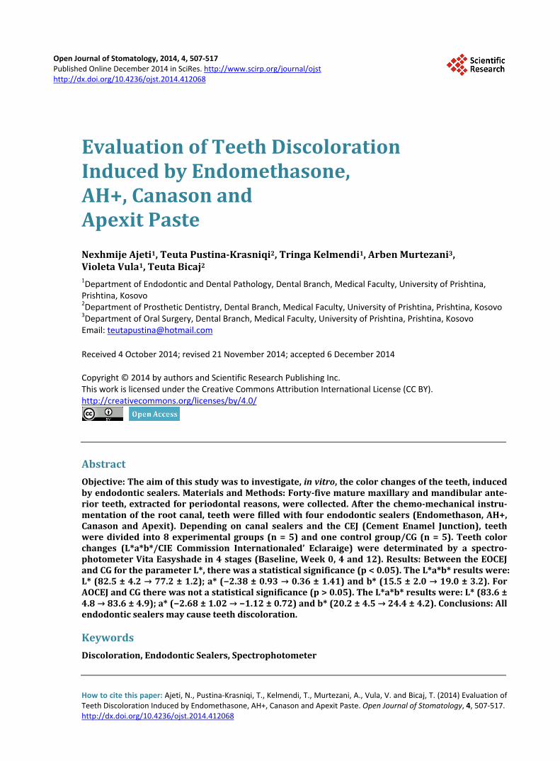
Abstract Objective: The aim of this study was to investigate, in vitro, the color changes of the teeth, induced by endodontic sealers. Materials and Methods: Forty-five mature maxillary and mandibular ante-rior teeth, extracted for periodontal reasons, were collected. After the chemo-mechanical instru-mentation of the root canal, teeth were filled with four endodontic sealers (Endomethason, AH+, Canason and Apexit). Depending on canal sealers and the CEJ (Cement Enamel Junction), teeth were divided into 8 experimental groups (n = 5) and one control group/CG (n = 5). Teeth color changes (L*a*b*/CIE Commission Internationaled’ Eclaraige) were determinated by a spectro-photometer Vita Easyshade in 4 stages (Baseline, Week 0, 4 and 12). Results: Between the EOCEJ and CG for the parameter L*, there was a statistical significance (p < 0.05). The L*a*b* results were: L* (82.5 ± 4.2 → 77.2 ± 1.2); a* (−2.38 ± 0.93 → 0.36 ± 1.41) and b* (15.5 ± 2.0 → 19.0 ± 3.2). For AOCEJ and CG there was not a statistical significance (p > 0.05). The L*a*b* results were: L* (83.6 ± 4.8 → 83.6 ± 4.9); a* (−2.68 ± 1.02 → −1.12 ± 0.72) and b* (20.2 ± 4.5 → 24.4 ± 4.2). Conclusions: All endodontic sealers may cause teeth discoloration.
Keywords Discoloration, Endodontic Sealers, Spectrophotometer
N. Ajeti et al.
508
1. Introduction One of the most frequent reasons why patients seek dental care is the teeth discoloration, especially in the ante-rior region. These changes compromise the appearance of the patients.
Crown discoloration after endodontic treatment is considered a common esthetic problem for the patient and dentist, particularly for anterior teeth [1].
A lot of authors concluded that endodontic sealers can produce teeth discoloration (van der Burgt [2], Zareba et al. [3], Pumarola et al. [4], Orstavik [5], Kaplan et al. [6], Davis et al. [7], Tina et al. [8], Walsh et al. [9], Elkhazin [10], Lenher et al. [11], Kim et al. [12], White et al. [13], Meincke et al. [14], Zare Jahromi [15], Mo-hamed Abdel Aziz El Sayed [16]).
There are a lot of endodontic sealers that are used in the root canals treatment. Endomethasone (Septodont, France) is an endodontic sealer which was used for decades. Unfortunately, it
was concluded that this sealer causes teeth discoloration (Burgt et al. [17] [18]). It is consisted of zinc oxide, paraformaldehide and corticosteroides (hydrocortisone acetate, magnesium stearat, tymol iodide and barium sulfat). Furthermore, Endomethasone is characterized, for its antibacterial activity (Zareba et al. [3], Pumarola et al. [4], Orstavik [5]).
AH+ (Dentsply-Maille), a root canal sealer, is characterized with very good mechanical properties. It is deli-vered into two tubes: Epoxy Paste (diepoxide, calcium tungstate, zirconium oxide, aerosol and pigments) and Amin Paste (1-adamantine amine, N’N-dibenzyl P-5-ara-nonandiamine-1,9, TCD-diamine, calcium tungstate, zir-conium oxide, aerosol and silicone oil). The literature review proved that AH Plus (resin-based sealer) has good sealing ability and good biocompatibility [19] [20].
Based on Kaplan et al. [6], AH+ is a sealer that doesn’t cause teeth discoloration, but can cause sensitivity in patients, because of epoxy paste.
Canason (Vocco, Germany) is a root canal sealer based on zinc oxide. It is characterized for its antibacterial effect [3], antiseptic and anti-inflammatory properties. The literature is poor regarding the effect of Canason paste, but there are other studies, based on ZOE pastes. It was reported that Sultan sealer (ZOE) displayed the greatest overall coronal discoloration [16].
Apexit (Ivoclar, Viva Dent) is hydroxide calcium paste, which has had a wide use in dentistry. It is produced as two components: base and activator. The base contents include hydroxide calcium, zinc oxide, calcium oxide, silica oxide, zinc calcium phosphate, butan-diodilsalicilate and phosphate-3-calcium.
Limited data are available about the coronal discoloration effect of Apexit. Apexit Plus sealer showed the lowest coronal discoloration effect [16].
Teeth color determination Teeth color determination has attracted attention in the field of dentistry. It can be measured by a visual per-
ception and via digital instruments. As early as 1905, Albert H. Munsell created the Munsell system, which is consisted in terms of hue, lightness and chromic attributes [21].
H (Hue)—is the common color (red, yellow, green, and blue). L (Lightness)—is a measure of the lightness of an object, ranging from 0 (black) to 100 (white) [13]. C (Chrome)—is the saturation of the color. It is sometimes described as the purity of the color. In the shade guide Vita 3D Master® (Vita-Zahnfabrik, Germany), the range of normal lightness for human
teeth is represented by value groups ranging from 0 (the lightest) to 5 (darkest). Within a given value group the chroma is represented by a number ranging from 1 to 3. Also with a given value group, hue is represented by L (yellowish), M (neutral, middle) or R (slightly red). In this way the complex shades are created: 2M2, 2L1.5 etc.
When selecting a shade tab based on visual criteria, it is recommended that the lightness should be determined first, followed by the chrome. The hue is determined last by matching to selected shade tabs for which lightness and chrome have been previously determined. However, it has been reported that visual evaluation of shade is unreliable and inconsistent [22]. An understanding of appearance attributes of natural teeth is required along with new shade guides and shade taking instruments in order to maximize shade-matching results [23].
Instrumental devices for tooth color measurement are: dental colorimeters, spectroradiometers, spectropho-tometers and digital cameras.
Instrumental devices commonly use the Commision International de l’ Eclairage’s (CIE) xyz (x = red, y = green, z = blue) or CIE (L*a*b*) (L* = lightness, a* = red-green, b* = blue) color systems, where: a* is a meas-
N. Ajeti et al.
509
ure of redness (a > 0) or greenness (a < 0) and b* is a measure of yellowness (b > 0) or blueness (b < 0) [24].
2. Objective The objective of this study was to investigate, in vitro, the tooth color changes, induced by endodontic sealers (Endomethason, AH+, Canason and Apexit) and determined by a spectrophotometer Vita Easy Shade.
3. Material and Methods Investigation was carried out at the Department of Dental Pathology and Endodontic and Prosthetic Dentistry in University Dental Clinical Center of Kosovo (UDCCK), Prishtina, Republic of Kosovo. Forty five mature max-illary and mandibular anterior teeth, extracted in the department of Oral Surgery (UDCCK), for periodontal rea-sons were collected. The age and gender of the patients were unknown. Teeth with cracks, caries, large restora-tions and multiple canals were excluded. The calculus and stains were removed using hand scalars and polishing brushes. Teeth were initially stored in 0.9% NaCl, until they were filled. The container was stored away from sunlight in the room temperature. Each tooth was placed in a separate Ependorf’s tubes with human saliva, col-lected voluntary, without any stimulation. The teeth color determination was matched by a spectrophotometer Vita Easyshade® (Vita Zahnfabrik H. Rauter GmbH & Co. KG, Bad Sackingen, Germany).
The Vita Easyshade is a handheld spectrophotometer for tooth shade matching. The instrument consists of a handpiece and base units, which are connected by a monocor fiberoptic cable assemble. The tip of the handpiece is covered with a thin polymethane disposable cover and is brought into direct contact with the tooth surface when a measurement is made. The fiber optic tip is approximately 5 mm in diameter and contains nineteen 1-mm diameter fiber optic fibers. The light source is a continuous halogen stabilized tungsten lamp, located in the base unit; it is controlled by a shutter and delivered through the fiberoptic cable. The illumination is provided by the halogen bulb directed into the tooth surface of the probe tip [21].
Vita Easyshade provides accurate shade determination for natural and bleached teeth and a variety of restora-tion. Easyshade displays it output on a touch screen that is also used to make menu selections and enter data [24].
The procedure: after Vita Easyshade has turned on, and the lamp has warmed up the Infection Control Shield was inserted to the probe tip. After the calibration, the normal measurement mode was used, which gives a possibility to measure the base shade of a tooth, with the selection of “Tooth Single” on the measurement style menu. The probe tip was holed at 90˚ to the third middle segment of vestibular surface of the tooth. The 3D Master shades were collected. After touching the screen in the reported 3D-Master shade the screen showed the values L*, a*, b*, C and H.
Prior the instrumentation and obturation of the root canal, the teeth color was matched with the spectropho-tometer. This part was marked as Baseline.
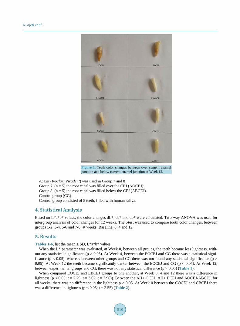
After the Baseline, in each tooth the pulpectomy was carried out. The teeth were instrumented with the con-ventional technique with K-Files (K-Reamers, Edenta le, Swiss), with different sizes from 15#60, depended on the canal size. The organic compounds were removed and rinsed using a 5.25% NaOCl (Histolith, LegeArtis, Detenhausen), after each instrumentation. The final irrigation was done with 0.9% NaCl. Before the obturation root canals were dried with sterile paper-points. Then the canals, were filled with the endodontic sealers (Endo-methason, AH+, Canason and Apexit) over and below the CEJ (Cement Enamel Junction) and all teeth were di-vided into 8 experimental groups and 1 control group. The teeth color determination was done immediately after the filling (Week 0) and after 4 weeks (Week 4) and 12 weeks (Week 12) (Figure 1).
Endomethason (Septodont, France) was used in Group 1 and 2 Group 1. (n = 5) the root canal was filled over the CEJ (EOCEJ); Group 2. (n = 5) the root canal was filled below the CEJ (EBCEJ). AH+ (Dentsply, Germany) was used in Group 3 and 4 Group 3. (n = 5) the root canal was filled over the CEJ (AH+ OCEJ); Group 4. (n = 5) the root canal was filled below the CEJ (AH+ BCEJ). Canason (Vocco, Germany) was used in Group 5 and 6 Group 5. (n = 5) the root canal was filled over the CEJ (COCEJ); Group 6. (n = 5) the root canal was filled below the CEJ (CBCEJ).
N. Ajeti et al.
510
Figure 1. Teeth color changes between over cement enamel junction and below cement enamel junction at Week 12.
Apexit (Ivoclar, Vivadent) was used in Group 7 and 8 Group 7. (n = 5) the root canal was filled over the CEJ (AOCEJ); Group 8. (n = 5) the root canal was filled below the CEJ (ABCEJ). Control group (CG) Control group consisted of 5 teeth, filled with human saliva.
4. Statistical Analysis Based on L*a*b* values, the color changes dL*, da* and db* were calculated. Two-way ANOVA was used for intergroup analysis of color changes for 12 weeks. The t-test was used to compare tooth color changes, between groups 1-2, 3-4, 5-6 and 7-8, at weeks: Baseline, 0, 4 and 12.
5. Results Tables 1-6, list the mean ± SD, L*a*b* values.
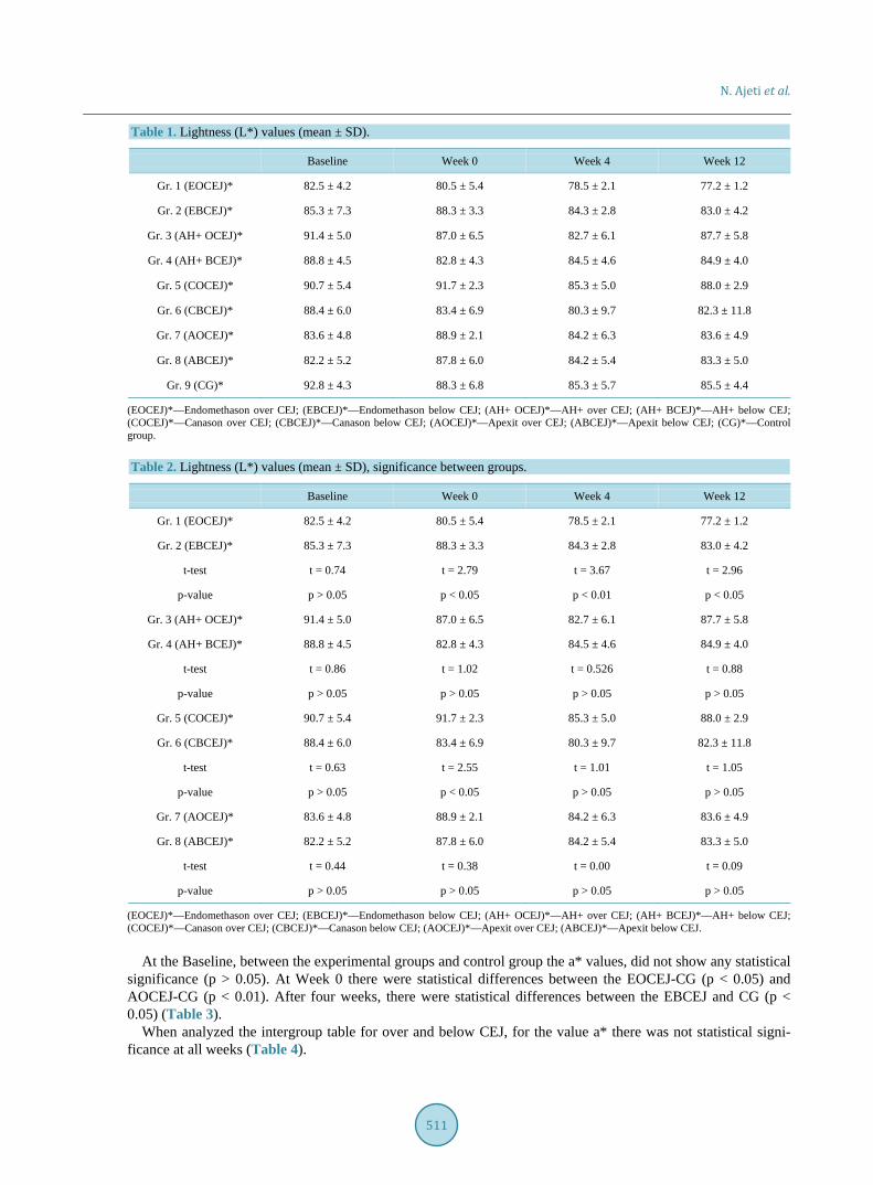
When the L* parameter was evaluated, at Week 0, between all groups, the teeth became less lightness, with-out any statistical significance (p > 0.05). At Week 4, between the EOCEJ and CG there was a statistical signi-ficance (p < 0.05), whereas between other groups and CG there was not found any statistical significance (p > 0.05). At Week 12 the teeth became significantly darker between the EOCEJ and CG (p < 0.05). At Week 12, between experimental groups and CG, there was not any statistical difference (p > 0.05) (Table 1).
When compared EOCEJ and EBCEJ groups to one another, at Week 0, 4 and 12 there was a difference in lightness (p < 0.05; t = 2.79; t = 3.67; t = 2.96)). Between the AH+ OCEJ; AH+ BCEJ and AOCEJ-ABCEJ, for all weeks, there was no difference in the lightness p > 0.05. At Week 0 between the COCEJ and CBCEJ there was a difference in lightness (p < 0.05; t = 2.55) (Table 2).
N. Ajeti et al.
511
Table 1. Lightness (L*) values (mean ± SD).
Baseline Week 0 Week 4 Week 12
Gr. 1 (EOCEJ)* 82.5 ± 4.2 80.5 ± 5.4 78.5 ± 2.1 77.2 ± 1.2
Gr. 2 (EBCEJ)* 85.3 ± 7.3 88.3 ± 3.3 84.3 ± 2.8 83.0 ± 4.2
Gr. 3 (AH+ OCEJ)* 91.4 ± 5.0 87.0 ± 6.5 82.7 ± 6.1 87.7 ± 5.8
Gr. 4 (AH+ BCEJ)* 88.8 ± 4.5 82.8 ± 4.3 84.5 ± 4.6 84.9 ± 4.0
Gr. 5 (COCEJ)* 90.7 ± 5.4 91.7 ± 2.3 85.3 ± 5.0 88.0 ± 2.9
Gr. 6 (CBCEJ)* 88.4 ± 6.0 83.4 ± 6.9 80.3 ± 9.7 82.3 ± 11.8
Gr. 7 (AOCEJ)* 83.6 ± 4.8 88.9 ± 2.1 84.2 ± 6.3 83.6 ± 4.9
Gr. 8 (ABCEJ)* 82.2 ± 5.2 87.8 ± 6.0 84.2 ± 5.4 83.3 ± 5.0
Gr. 9 (CG)* 92.8 ± 4.3 88.3 ± 6.8 85.3 ± 5.7 85.5 ± 4.4
(EOCEJ)*—Endomethason over CEJ; (EBCEJ)*—Endomethason below CEJ; (AH+ OCEJ)*—AH+ over CEJ; (AH+ BCEJ)*—AH+ below CEJ; (COCEJ)*—Canason over CEJ; (CBCEJ)*—Canason below CEJ; (AOCEJ)*—Apexit over CEJ; (ABCEJ)*—Apexit below CEJ; (CG)*—Control group. Table 2. Lightness (L*) values (mean ± SD), significance between groups.
Baseline Week 0 Week 4 Week 12
Gr. 1 (EOCEJ)* 82.5 ± 4.2 80.5 ± 5.4 78.5 ± 2.1 77.2 ± 1.2
Gr. 2 (EBCEJ)* 85.3 ± 7.3 88.3 ± 3.3 84.3 ± 2.8 83.0 ± 4.2
t-test t = 0.74 t = 2.79 t = 3.67 t = 2.96
p-value p > 0.05 p < 0.05 p < 0.01 p < 0.05
Gr. 3 (AH+ OCEJ)* 91.4 ± 5.0 87.0 ± 6.5 82.7 ± 6.1 87.7 ± 5.8
Gr. 4 (AH+ BCEJ)* 88.8 ± 4.5 82.8 ± 4.3 84.5 ± 4.6 84.9 ± 4.0
t-test t = 0.86 t = 1.02 t = 0.526 t = 0.88
p-value p > 0.05 p > 0.05 p > 0.05 p > 0.05
Gr. 5 (COCEJ)* 90.7 ± 5.4 91.7 ± 2.3 85.3 ± 5.0 88.0 ± 2.9
Gr. 6 (CBCEJ)* 88.4 ± 6.0 83.4 ± 6.9 80.3 ± 9.7 82.3 ± 11.8
t-test t = 0.63 t = 2.55 t = 1.01 t = 1.05
p-value p > 0.05 p < 0.05 p > 0.05 p > 0.05
Gr. 7 (AOCEJ)* 83.6 ± 4.8 88.9 ± 2.1 84.2 ± 6.3 83.6 ± 4.9
Gr. 8 (ABCEJ)* 82.2 ± 5.2 87.8 ± 6.0 84.2 ± 5.4 83.3 ± 5.0
t-test t = 0.44 t = 0.38 t = 0.00 t = 0.09
p-value p > 0.05 p > 0.05 p > 0.05 p > 0.05
(EOCEJ)*—Endomethason over CEJ; (EBCEJ)*—Endomethason below CEJ; (AH+ OCEJ)*—AH+ over CEJ; (AH+ BCEJ)*—AH+ below CEJ; (COCEJ)*—Canason over CEJ; (CBCEJ)*—Canason below CEJ; (AOCEJ)*—Apexit over CEJ; (ABCEJ)*—Apexit below CEJ.
At the Baseline, between the experimental groups and control group the a* values, did not show any statistical significance (p > 0.05). At Week 0 there were statistical differences between the EOCEJ-CG (p < 0.05) and AOCEJ-CG (p < 0.01). After four weeks, there were statistical differences between the EBCEJ and CG (p < 0.05) (Table 3).
When analyzed the intergroup table for over and below CEJ, for the value a* there was not statistical signi-ficance at all weeks (Table 4).
N. Ajeti et al.
512
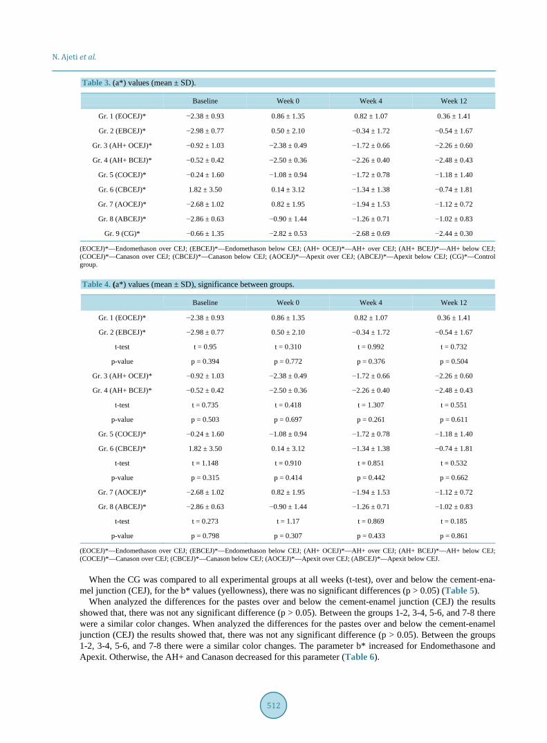
Table 3. (a*) values (mean ± SD).
Baseline Week 0 Week 4 Week 12
Gr. 1 (EOCEJ)* −2.38 ± 0.93 0.86 ± 1.35 0.82 ± 1.07 0.36 ± 1.41
Gr. 2 (EBCEJ)* −2.98 ± 0.77 0.50 ± 2.10 −0.34 ± 1.72 −0.54 ± 1.67
Gr. 3 (AH+ OCEJ)* −0.92 ± 1.03 −2.38 ± 0.49 −1.72 ± 0.66 −2.26 ± 0.60
Gr. 4 (AH+ BCEJ)* −0.52 ± 0.42 −2.50 ± 0.36 −2.26 ± 0.40 −2.48 ± 0.43
Gr. 5 (COCEJ)* −0.24 ± 1.60 −1.08 ± 0.94 −1.72 ± 0.78 −1.18 ± 1.40
Gr. 6 (CBCEJ)* 1.82 ± 3.50 0.14 ± 3.12 −1.34 ± 1.38 −0.74 ± 1.81
Gr. 7 (AOCEJ)* −2.68 ± 1.02 0.82 ± 1.95 −1.94 ± 1.53 −1.12 ± 0.72
Gr. 8 (ABCEJ)* −2.86 ± 0.63 −0.90 ± 1.44 −1.26 ± 0.71 −1.02 ± 0.83
Gr. 9 (CG)* −0.66 ± 1.35 −2.82 ± 0.53 −2.68 ± 0.69 −2.44 ± 0.30
(EOCEJ)*—Endomethason over CEJ; (EBCEJ)*—Endomethason below CEJ; (AH+ OCEJ)*—AH+ over CEJ; (AH+ BCEJ)*—AH+ below CEJ; (COCEJ)*—Canason over CEJ; (CBCEJ)*—Canason below CEJ; (AOCEJ)*—Apexit over CEJ; (ABCEJ)*—Apexit below CEJ; (CG)*—Control group. Table 4. (a*) values (mean ± SD), significance between groups.
Baseline Week 0 Week 4 Week 12
Gr. 1 (EOCEJ)* −2.38 ± 0.93 0.86 ± 1.35 0.82 ± 1.07 0.36 ± 1.41
Gr. 2 (EBCEJ)* −2.98 ± 0.77 0.50 ± 2.10 −0.34 ± 1.72 −0.54 ± 1.67
t-test t = 0.95 t = 0.310 t = 0.992 t = 0.732
p-value p = 0.394 p = 0.772 p = 0.376 p = 0.504
Gr. 3 (AH+ OCEJ)* −0.92 ± 1.03 −2.38 ± 0.49 −1.72 ± 0.66 −2.26 ± 0.60
Gr. 4 (AH+ BCEJ)* −0.52 ± 0.42 −2.50 ± 0.36 −2.26 ± 0.40 −2.48 ± 0.43
t-test t = 0.735 t = 0.418 t = 1.307 t = 0.551
p-value p = 0.503 p = 0.697 p = 0.261 p = 0.611
Gr. 5 (COCEJ)* −0.24 ± 1.60 −1.08 ± 0.94 −1.72 ± 0.78 −1.18 ± 1.40
Gr. 6 (CBCEJ)* 1.82 ± 3.50 0.14 ± 3.12 −1.34 ± 1.38 −0.74 ± 1.81
t-test t = 1.148 t = 0.910 t = 0.851 t = 0.532
p-value p = 0.315 p = 0.414 p = 0.442 p = 0.662
Gr. 7 (AOCEJ)* −2.68 ± 1.02 0.82 ± 1.95 −1.94 ± 1.53 −1.12 ± 0.72
Gr. 8 (ABCEJ)* −2.86 ± 0.63 −0.90 ± 1.44 −1.26 ± 0.71 −1.02 ± 0.83
t-test t = 0.273 t = 1.17 t = 0.869 t = 0.185
p-value p = 0.798 p = 0.307 p = 0.433 p = 0.861
(EOCEJ)*—Endomethason over CEJ; (EBCEJ)*—Endomethason below CEJ; (AH+ OCEJ)*—AH+ over CEJ; (AH+ BCEJ)*—AH+ below CEJ; (COCEJ)*—Canason over CEJ; (CBCEJ)*—Canason below CEJ; (AOCEJ)*—Apexit over CEJ; (ABCEJ)*—Apexit below CEJ.
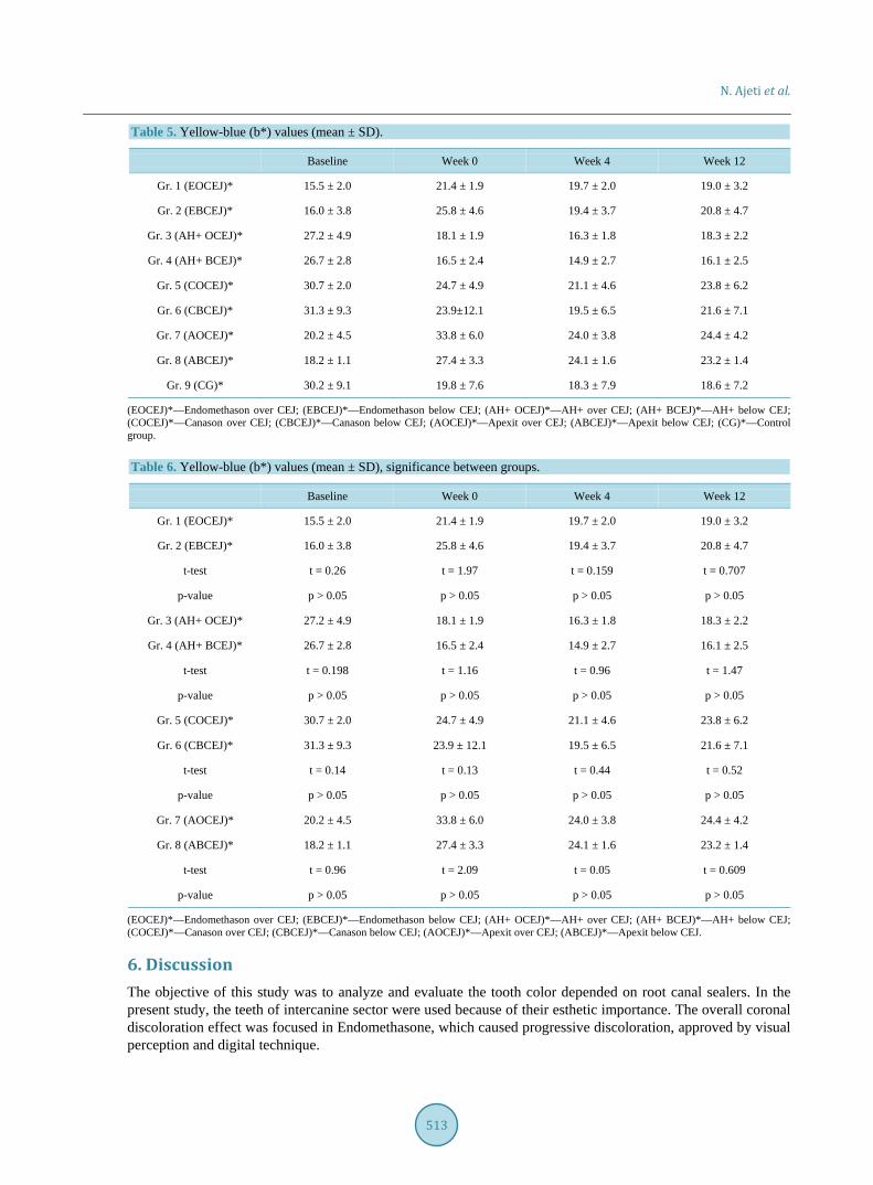
When the CG was compared to all experimental groups at all weeks (t-test), over and below the cement-ena- mel junction (CEJ), for the b* values (yellowness), there was no significant differences (p > 0.05) (Table 5).
When analyzed the differences for the pastes over and below the cement-enamel junction (CEJ) the results showed that, there was not any significant difference (p > 0.05). Between the groups 1-2, 3-4, 5-6, and 7-8 there were a similar color changes. When analyzed the differences for the pastes over and below the cement-enamel junction (CEJ) the results showed that, there was not any significant difference (p > 0.05). Between the groups 1-2, 3-4, 5-6, and 7-8 there were a similar color changes. The parameter b* increased for Endomethasone and Apexit. Otherwise, the AH+ and Canason decreased for this parameter (Table 6).
N. Ajeti et al.
513
Table 5. Yellow-blue (b*) values (mean ± SD).
Baseline Week 0 Week 4 Week 12
Gr. 1 (EOCEJ)* 15.5 ± 2.0 21.4 ± 1.9 19.7 ± 2.0 19.0 ± 3.2
Gr. 2 (EBCEJ)* 16.0 ± 3.8 25.8 ± 4.6 19.4 ± 3.7 20.8 ± 4.7
Gr. 3 (AH+ OCEJ)* 27.2 ± 4.9 18.1 ± 1.9 16.3 ± 1.8 18.3 ± 2.2
Gr. 4 (AH+ BCEJ)* 26.7 ± 2.8 16.5 ± 2.4 14.9 ± 2.7 16.1 ± 2.5
Gr. 5 (COCEJ)* 30.7 ± 2.0 24.7 ± 4.9 21.1 ± 4.6 23.8 ± 6.2
Gr. 6 (CBCEJ)* 31.3 ± 9.3 23.9±12.1 19.5 ± 6.5 21.6 ± 7.1
Gr. 7 (AOCEJ)* 20.2 ± 4.5 33.8 ± 6.0 24.0 ± 3.8 24.4 ± 4.2
Gr. 8 (ABCEJ)* 18.2 ± 1.1 27.4 ± 3.3 24.1 ± 1.6 23.2 ± 1.4
Gr. 9 (CG)* 30.2 ± 9.1 19.8 ± 7.6 18.3 ± 7.9 18.6 ± 7.2
(EOCEJ)*—Endomethason over CEJ; (EBCEJ)*—Endomethason below CEJ; (AH+ OCEJ)*—AH+ over CEJ; (AH+ BCEJ)*—AH+ below CEJ; (COCEJ)*—Canason over CEJ; (CBCEJ)*—Canason below CEJ; (AOCEJ)*—Apexit over CEJ; (ABCEJ)*—Apexit below CEJ; (CG)*—Control group. Table 6. Yellow-blue (b*) values (mean ± SD), significance between groups.
Baseline Week 0 Week 4 Week 12
Gr. 1 (EOCEJ)* 15.5 ± 2.0 21.4 ± 1.9 19.7 ± 2.0 19.0 ± 3.2
Gr. 2 (EBCEJ)* 16.0 ± 3.8 25.8 ± 4.6 19.4 ± 3.7 20.8 ± 4.7
t-test t = 0.26 t = 1.97 t = 0.159 t = 0.707
p-value p > 0.05 p > 0.05 p > 0.05 p > 0.05
Gr. 3 (AH+ OCEJ)* 27.2 ± 4.9 18.1 ± 1.9 16.3 ± 1.8 18.3 ± 2.2
Gr. 4 (AH+ BCEJ)* 26.7 ± 2.8 16.5 ± 2.4 14.9 ± 2.7 16.1 ± 2.5
t-test t = 0.198 t = 1.16 t = 0.96 t = 1.47
p-value p > 0.05 p > 0.05 p > 0.05 p > 0.05
Gr. 5 (COCEJ)* 30.7 ± 2.0 24.7 ± 4.9 21.1 ± 4.6 23.8 ± 6.2
Gr. 6 (CBCEJ)* 31.3 ± 9.3 23.9 ± 12.1 19.5 ± 6.5 21.6 ± 7.1
t-test t = 0.14 t = 0.13 t = 0.44 t = 0.52
p-value p > 0.05 p > 0.05 p > 0.05 p > 0.05
Gr. 7 (AOCEJ)* 20.2 ± 4.5 33.8 ± 6.0 24.0 ± 3.8 24.4 ± 4.2
Gr. 8 (ABCEJ)* 18.2 ± 1.1 27.4 ± 3.3 24.1 ± 1.6 23.2 ± 1.4
t-test t = 0.96 t = 2.09 t = 0.05 t = 0.609
p-value p > 0.05 p > 0.05 p > 0.05 p > 0.05
(EOCEJ)*—Endomethason over CEJ; (EBCEJ)*—Endomethason below CEJ; (AH+ OCEJ)*—AH+ over CEJ; (AH+ BCEJ)*—AH+ below CEJ; (COCEJ)*—Canason over CEJ; (CBCEJ)*—Canason below CEJ; (AOCEJ)*—Apexit over CEJ; (ABCEJ)*—Apexit below CEJ.
6. Discussion The objective of this study was to analyze and evaluate the tooth color depended on root canal sealers. In the present study, the teeth of intercanine sector were used because of their esthetic importance. The overall coronal discoloration effect was focused in Endomethasone, which caused progressive discoloration, approved by visual perception and digital technique.
N. Ajeti et al.
514
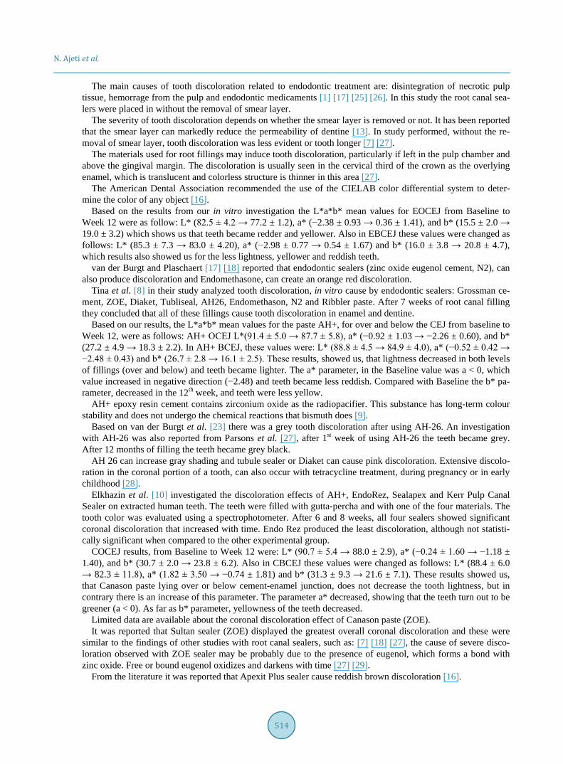
The main causes of tooth discoloration related to endodontic treatment are: disintegration of necrotic pulp tissue, hemorrage from the pulp and endodontic medicaments [1] [17] [25] [26]. In this study the root canal sea-lers were placed in without the removal of smear layer.
The severity of tooth discoloration depends on whether the smear layer is removed or not. It has been reported that the smear layer can markedly reduce the permeability of dentine [13]. In study performed, without the re-moval of smear layer, tooth discoloration was less evident or tooth longer [7] [27].
The materials used for root fillings may induce tooth discoloration, particularly if left in the pulp chamber and above the gingival margin. The discoloration is usually seen in the cervical third of the crown as the overlying enamel, which is translucent and colorless structure is thinner in this area [27].
The American Dental Association recommended the use of the CIELAB color differential system to deter-mine the color of any object [16].
Based on the results from our in vitro investigation the L*a*b* mean values for EOCEJ from Baseline to Week 12 were as follow: L* (82.5 ± 4.2 → 77.2 ± 1.2), a* (−2.38 ± 0.93 → 0.36 ± 1.41), and b* (15.5 ± 2.0 → 19.0 ± 3.2) which shows us that teeth became redder and yellower. Also in EBCEJ these values were changed as follows: L* (85.3 ± 7.3 → 83.0 ± 4.20), a* (−2.98 ± 0.77 → 0.54 ± 1.67) and b* (16.0 ± 3.8 → 20.8 ± 4.7), which results also showed us for the less lightness, yellower and reddish teeth.
van der Burgt and Plaschaert [17] [18] reported that endodontic sealers (zinc oxide eugenol cement, N2), can also produce discoloration and Endomethasone, can create an orange red discoloration.
Tina et al. [8] in their study analyzed tooth discoloration, in vitro cause by endodontic sealers: Grossman ce-ment, ZOE, Diaket, Tubliseal, AH26, Endomethason, N2 and Ribbler paste. After 7 weeks of root canal filling they concluded that all of these fillings cause tooth discoloration in enamel and dentine.
Based on our results, the L*a*b* mean values for the paste AH+, for over and below the CEJ from baseline to Week 12, were as follows: AH+ OCEJ L*(91.4 ± 5.0 → 87.7 ± 5.8), a* (−0.92 ± 1.03 → −2.26 ± 0.60), and b* (27.2 ± 4.9 → 18.3 ± 2.2). In AH+ BCEJ, these values were: L* (88.8 ± 4.5 → 84.9 ± 4.0), a* (−0.52 ± 0.42 → −2.48 ± 0.43) and b* (26.7 ± 2.8 → 16.1 ± 2.5). These results, showed us, that lightness decreased in both levels of fillings (over and below) and teeth became lighter. The a* parameter, in the Baseline value was a < 0, which value increased in negative direction (−2.48) and teeth became less reddish. Compared with Baseline the b* pa-rameter, decreased in the 12th week, and teeth were less yellow.
AH+ epoxy resin cement contains zirconium oxide as the radiopacifier. This substance has long-term colour stability and does not undergo the chemical reactions that bismuth does [9].
Based on van der Burgt et al. [23] there was a grey tooth discoloration after using AH-26. An investigation with AH-26 was also reported from Parsons et al. [27], after 1st week of using AH-26 the teeth became grey. After 12 months of filling the teeth became grey black.
AH 26 can increase gray shading and tubule sealer or Diaket can cause pink discoloration. Extensive discolo-ration in the coronal portion of a tooth, can also occur with tetracycline treatment, during pregnancy or in early childhood [28].
Elkhazin et al. [10] investigated the discoloration effects of AH+, EndoRez, Sealapex and Kerr Pulp Canal Sealer on extracted human teeth. The teeth were filled with gutta-percha and with one of the four materials. The tooth color was evaluated using a spectrophotometer. After 6 and 8 weeks, all four sealers showed significant coronal discoloration that increased with time. Endo Rez produced the least discoloration, although not statisti-cally significant when compared to the other experimental group.
COCEJ results, from Baseline to Week 12 were: L* (90.7 ± 5.4 → 88.0 ± 2.9), a* (−0.24 ± 1.60 → −1.18 ± 1.40), and b* (30.7 ± 2.0 → 23.8 ± 6.2). Also in CBCEJ these values were changed as follows: L* (88.4 ± 6.0 → 82.3 ± 11.8), a* (1.82 ± 3.50 → −0.74 ± 1.81) and b* (31.3 ± 9.3 → 21.6 ± 7.1). These results showed us, that Canason paste lying over or below cement-enamel junction, does not decrease the tooth lightness, but in contrary there is an increase of this parameter. The parameter a* decreased, showing that the teeth turn out to be greener (a < 0). As far as b* parameter, yellowness of the teeth decreased.
Limited data are available about the coronal discoloration effect of Canason paste (ZOE). It was reported that Sultan sealer (ZOE) displayed the greatest overall coronal discoloration and these were
similar to the findings of other studies with root canal sealers, such as: [7] [18] [27], the cause of severe disco-loration observed with ZOE sealer may be probably due to the presence of eugenol, which forms a bond with zinc oxide. Free or bound eugenol oxidizes and darkens with time [27] [29].
From the literature it was reported that Apexit Plus sealer cause reddish brown discoloration [16].
N. Ajeti et al.
515
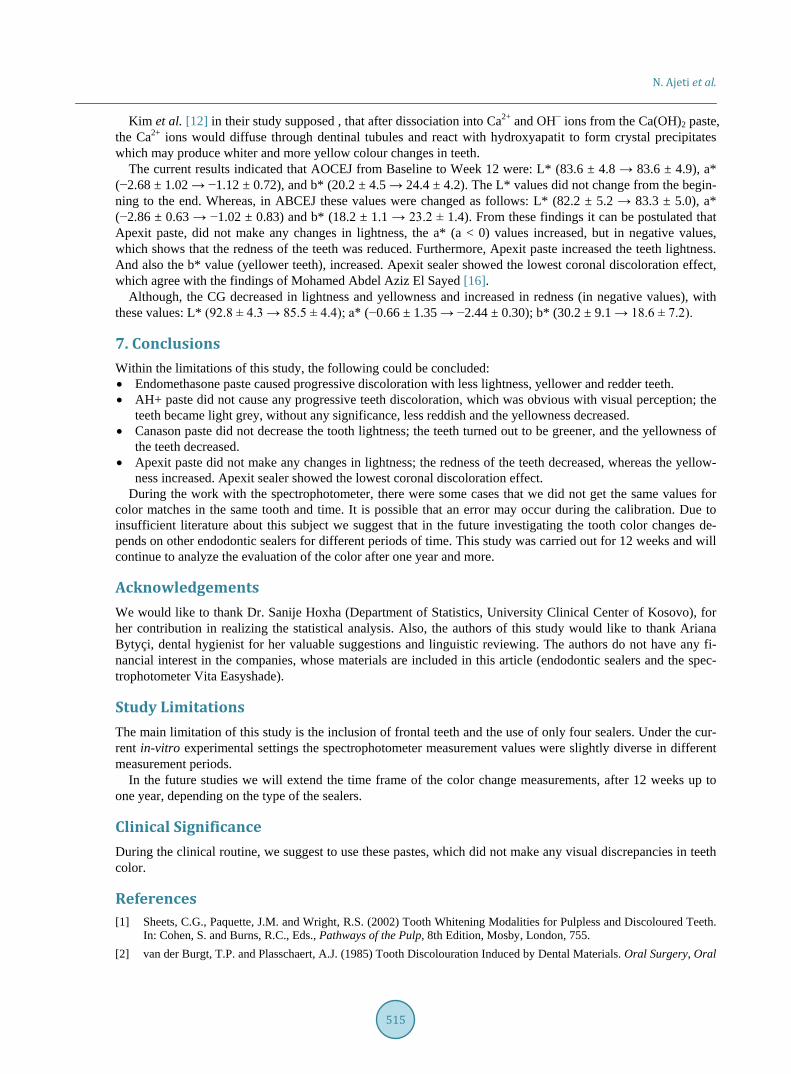
Kim et al. [12] in their study supposed , that after dissociation into Ca2+ and OH− ions from the Ca(OH)2 paste, the Ca2+ ions would diffuse through dentinal tubules and react with hydroxyapatit to form crystal precipitates which may produce whiter and more yellow colour changes in teeth.
The current results indicated that AOCEJ from Baseline to Week 12 were: L* (83.6 ± 4.8 → 83.6 ± 4.9), a* (−2.68 ± 1.02 → −1.12 ± 0.72), and b* (20.2 ± 4.5 → 24.4 ± 4.2). The L* values did not change from the begin-ning to the end. Whereas, in ABCEJ these values were changed as follows: L* (82.2 ± 5.2 → 83.3 ± 5.0), a* (−2.86 ± 0.63 → −1.02 ± 0.83) and b* (18.2 ± 1.1 → 23.2 ± 1.4). From these findings it can be postulated that Apexit paste, did not make any changes in lightness, the a* (a < 0) values increased, but in negative values, which shows that the redness of the teeth was reduced. Furthermore, Apexit paste increased the teeth lightness. And also the b* value (yellower teeth), increased. Apexit sealer showed the lowest coronal discoloration effect, which agree with the findings of Mohamed Abdel Aziz El Sayed [16].
Although, the CG decreased in lightness and yellowness and increased in redness (in negative values), with these values: L* (92.8 ± 4.3 → 85.5 ± 4.4); a* (−0.66 ± 1.35 → −2.44 ± 0.30); b* (30.2 ± 9.1 → 18.6 ± 7.2).
7. Conclusions Within the limitations of this study, the following could be concluded: • Endomethasone paste caused progressive discoloration with less lightness, yellower and redder teeth. • AH+ paste did not cause any progressive teeth discoloration, which was obvious with visual perception; the
teeth became light grey, without any significance, less reddish and the yellowness decreased. • Canason paste did not decrease the tooth lightness; the teeth turned out to be greener, and the yellowness of
the teeth decreased. • Apexit paste did not make any changes in lightness; the redness of the teeth decreased, whereas the yellow-
ness increased. Apexit sealer showed the lowest coronal discoloration effect. During the work with the spectrophotometer, there were some cases that we did not get the same values for
color matches in the same tooth and time. It is possible that an error may occur during the calibration. Due to insufficient literature about this subject we suggest that in the future investigating the tooth color changes de-pends on other endodontic sealers for different periods of time. This study was carried out for 12 weeks and will continue to analyze the evaluation of the color after one year and more.
Acknowledgements We would like to thank Dr. Sanije Hoxha (Department of Statistics, University Clinical Center of Kosovo), for her contribution in realizing the statistical analysis. Also, the authors of this study would like to thank Ariana Bytyçi, dental hygienist for her valuable suggestions and linguistic reviewing. The authors do not have any fi-nancial interest in the companies, whose materials are included in this article (endodontic sealers and the spec-trophotometer Vita Easyshade).
Study Limitations The main limitation of this study is the inclusion of frontal teeth and the use of only four sealers. Under the cur-rent in-vitro experimental settings the spectrophotometer measurement values were slightly diverse in different measurement periods.
In the future studies we will extend the time frame of the color change measurements, after 12 weeks up to one year, depending on the type of the sealers.
Clinical Significance During the clinical routine, we suggest to use these pastes, which did not make any visual discrepancies in teeth color.
References [1] Sheets, C.G., Paquette, J.M. and Wright, R.S. (2002) Tooth Whitening Modalities for Pulpless and Discoloured Teeth.
In: Cohen, S. and Burns, R.C., Eds., Pathways of the Pulp, 8th Edition, Mosby, London, 755. [2] van der Burgt, T.P. and Plasschaert, A.J. (1985) Tooth Discolouration Induced by Dental Materials. Oral Surgery, Oral
N. Ajeti et al.
516
Medicine, Oral Pathology, 60, 666-669. http://dx.doi.org/10.1016/0030-4220(85)90373-1 [3] Zareba, T., Bojar, W. and Wasaw, P.L. (2004) Antimicrobial Activity of Root Canal Sealers—In Vitro Evaluation.
Clinical Microbiology and Infection, 10, 55-59. [4] Pumarola, J., Berastegui, E., Brau, E., Cunalda, C. and Jimenez de Ante, M.T. (1992) Antimicrobial Activity of Seven
Root canal Sealers. Results of Agar Diffusion and Agar Dilution Tests. Oral Surgery, Oral Medicine, Oral Pathology, 72, 216-220. http://dx.doi.org/10.1016/0030-4220(92)90386-5
[5] Orstavik, D.A. (1981) Antibacterial Properties of Root Canal Sealers, Cements and Pastes. International Endodontic Journal, 14, 125-133. http://dx.doi.org/10.1111/j.1365-2591.1981.tb01073.x
[6] Kaplan, A.E., Picca, M., Gonzalez, M., Macchi, P.L. and Molgatini, S.L. (1999) Antimicrobial Effect of Six Endodon-tic Sealers: An in Vitro Evaluation. Endodontics & Dental Traumatology, 15, 42-45. http://dx.doi.org/10.1111/j.1600-9657.1999.tb00748.x
[7] Davis, M.C., Walton, R.E. and Rivera, E.M. (2002) Sealer Distribution in Coronal Dentin. Journal of Endodontics, 28, 464-466. http://dx.doi.org/10.1097/00004770-200206000-00012
[8] Tina, P.A., Cemal, A., Burgt, A., Eronat, J.M. and Plasschaert, J.M.A. (1996) Staining Patterns in Teeth Discolored by Endodontic Sealers. Journal of Endodontics, 12, 187-191.
[9] Walsh, L.J. and Athanassiadis, B. (2007) Endodontic Aesthetic Iatrodontics. Australian Dental Practice, 18, 16-24. [10] Elkhazin, M. (2011) Analysis of Coronal Discoloration from Common Obturation Materials. An in Vitro Spectropho-
tometry Study. Lambert Academic Publishing, Saarbruecken. [11] Lenherr, P., Allgayer, N., Weiger, R., Filippi, A., Attin, T. and Krasti, G. (2012) Tooth Discoloration Induced by En-
dodontic Materials: A Laboratory Study. International Endodontic Journal, 45, 942-949. http://dx.doi.org/10.1111/j.1365-2591.2012.02053.x
[12] Kim, S.T., Abbott, P.V. and McGinley, P. (2000) The Effects of Ledermix Paste on Discolouration of Mature Teeth. International Endodontic Journal, 33, 227-232. http://dx.doi.org/10.1046/j.1365-2591.2000.00278.x
[13] White, R.R., Goldman, M. and Lin, P.S. (1987) The Influence of the Smeared Layer upon Dentinal Tubule Penetration by Endodontic Filling Materials. Part II. Journal of Endodontics, 13, 369-374. http://dx.doi.org/10.1016/S0099-2399(87)80195-4
[14] Meincke, D.K., Prado, M., Gomes, B.P., Bona, A.D. and Sousa, E.L. (2013) Effect of Endodontic Sealers on Tooth Color. Journal of Dentistry, 41, 93-96.
[15] Zare Jahromi, M., Navabi, A.A. and Ekhtiari, M. (2011) Comparing Coronal Discoloration between AH26 and ZOE Sealers. Iranian Endodontic Journal, 6, 146-149.
[16] El Sayed, M.A.A. and Etemadi, H. (2013) Coronal Discoloration Effect of Three Endodontic Sealers: An in Vitro Spectrophotometric Analysis. Journal of Conservative Dentistry, 16, 347-351. http://dx.doi.org/10.4103/0972-0707.114369
[17] van der Burgt, T.P., Mullaney, T.P. and Plasschaert, A.J. (1986) Tooth Discoloration Induced by Endodontic Sealers. Oral Surgery, Oral Medicine, Oral Pathology, 61, 84-89. http://dx.doi.org/10.1016/0030-4220(86)90208-2
[18] van der Burgt, T.P., Eronat, C. and Plasschaert, A.J. (1986) Staining Patterns in Teeth Discolored by Endodontic Sea-lers. Journal of Endodontics, 12, 187-191. http://dx.doi.org/10.1016/S0099-2399(86)80152-2
[19] Saunders, E.M. and Saunders, W.P. (1995) Long-Term Coronal Leakage of JS Quickfill Root Fillings with Sealapex and Apexit Sealers. Dental Traumatology, 11, 181-185. http://dx.doi.org/10.1111/j.1600-9657.1995.tb00484.x
[20] McMichen, F.R., Pearson, G., Rahbaran, S. and Gulabivala, K. (2003) A Comparative Study of Selected Physical Properties of Five Root-Canal Sealers. International Endodontic Journal, 36, 629-635. http://dx.doi.org/10.1046/j.1365-2591.2003.00701.x
[21] Paravina, R.D. and Powers, J.M. (2004) Esthetic Color Training in Dentistry. Elsevier Mosby, Amsterdam. [22] Brewer, J.D., Wee, A. and Seyhi, R. (2004) Advances in Color Matching. Dental Clinics of North America, 48, 341-
358. http://dx.doi.org/10.1016/j.cden.2004.01.004 [23] Burgt, G. (1993) Clinical Evaluation of an Experimental Body and Incisal Shade Guide Based on in Vivo Tooth Colour
Measurements. MS Thesis, University of Minnesota, Minneapolis. [24] (2004) Operating Manual: Vita Easyshade. [25] Pittford, T.R. (1986) Apexification and Apexogenesis. In: Walton, R.E. and Torabinejad, M., Eds., Principles and
Practice of Endodontics, WB Saunders, Philadelphia. [26] Walton, R.E. and Rotstein, I. (1996) Bleaching Discolored Teeth: Internal and External. In: Walton, R.E. and Torabi-
nejad, M., Eds., Principles and Practice of Endodontics, 2nd Edition, WB Saunders, Philadelphia, 385. [27] Parsons, J.R., Walton, R.E. and Ricks-Williamson, L. (2001) In Vitro Longitudinal Assessment of Coronal Discolora-
N. Ajeti et al.
517
tion from Endodontic Sealers. Journal of Endodontics, 27, 699-702. http://dx.doi.org/10.1097/00004770-200111000-00012
[28] Chiappinelli, J.A. and Walton, R.E. (1992) Tooth Discoloration Resulting from Long-Term Tetracycline Therapy: A Case Report. Quintessence International, 23, 539-541.
[29] Weinberg, J.E., Rabinowitz, R.L., Zainger, M. and Gennaro, A.F. (1972) 14C-Eugenol: 1. Synthesis, Polymerization, and Use. Journal of Dental Research, 51, 1055-1061. http://dx.doi.org/10.1177/00220345720510041101
Note List of Abbreviations (EOCEJ)*—Endomethason over Cement Enamel Junction; (EBCEJ)*—Endomethason below Cement Enamel Junction; (AH+ OCEJ)*—AH+ over Cement Enamel Junction; (AH+ BCEJ)*—AH+ below Cement Enamel Junction; (COCEJ)*—Canason over Cement Enamel Junction; (CBCEJ)*—Canason below Cement Enamel Junction; (AOCEJ)*—Apexit over Cement Enamel Junction; (ABCEJ)*—Apexit below Cement Enamel Junction; (CG)*—Control Group; L*—Lightness; a*—red-green; b*—blue; CIE—Commission Internationaled’ Eclaraige; UDCCK—University Dental Clinical Center of Kosovo (UDCCK); NaOCl—Natrium hypochlorite; NaCl—Natrium chloride.

Related Documents