RESEARCH ARTICLE Evaluation of protective efficacy of Spirulina platensis against collagen-induced arthritis in rats Narendra Kumar Surendra Singh Nisha Patro Ishan Patro Received: 2 March 2009 / Accepted: 23 March 2009 Ó Birkha ¨user Verlag, Basel/Switzerland 2009 Abstract Aim To assess the protective efficacy of Spirulina plat- ensis against collagen-induced arthritis (CIA) in female Wistar rats based on the changes in paws thickness, serum albumin, cholesterol, lipid peroxidation, alkaline phos- phatase and acid phosphatase activities and histology of paw joints. Methods Arthritis was induced by intradermal injection of Collagen and Freund’s adjuvant incomplete suspension at several sites on the back with a dose of 2 mg kg -1 of body weight and boosted with 0.1 ml intradermally at the base of the tail. CIA rats were orally treated with 200 and 400 mg kg -1 per oral of S. platensis from 0 to 45th day. Results S. platensis at 400 mg kg -1 per oral significantly elevates serum albumin and decreases the serum choles- terol, alkaline phosphatase and acid phosphatase activities, lipid peroxidation, paw thickness as well as normalize the joint histopathology of CIA rats. Conclusions S. platensis (400 mg kg -1 ) significantly normalizes changes observed in arthritic rats to near nor- mal conditions, indicates that S. platensis has promising protective efficacy against CIA rats. Keywords Spirulina platensis Á Collagen Á Alkaline phosphatase Á Lipid peroxidation Á Pannus Introduction Rheumatoid arthritis (RA) is traditionally considered a chronic, inflammatory autoimmune disorder that causes the immune system to attack the joints. It is a disabling and painful inflammatory condition, which can lead to sub- stantial loss of mobility due to pain and joint destruction. Most commonly, small joints are affected, but larger joints can also be involved; the pattern of joint involvement can differ from patient to patient (Majithia and Geraci 2007). RA affects women three times more often than men and it can first develop at any age. The risk of first developing the disease appears to be greatest for women between 40 and 50 years of age, and for men somewhat later (Alamanos et al. 2006). There is a significant increase in the lifetime cost and a decrease in the quality of life (Kobelt et al. 2002). The disease is symmetric, especially as it pro- gresses, and favors the interphalangeal joints of the hands and feet, such as the proximal interphalangeal, metacar- pophalangeal, and metatarsophalangeal joints, as well as the wrist and ankle (Lee and Weinblatt 2001). Joint swelling, deformity, pain, stiffness, and weakness are classic symptoms. Other includes tenderness, synovial thickening, effusion, erythema, and decreased range of motion, ankylosis, and subluxation (Lee and Weinblatt 2001). RA is the commonest inflammatory arthropathy worldwide and affects up to 0.75% of the Indian population (Malaviya et al. 1993). The collagen-induced arthritis (CIA) model in the rat is in many aspects similar to RA, which is perhaps the most commonly used model for RA today. Intradermal injection in rats with collagen N. Kumar School of Studies in Microbiology, Jiwaji University, Gwalior 474011, Madhya Pradesh, India S. Singh Department of Botany, Banaras Hindu University, Varanasi 221005, Uttar Pradesh, India N. Patro Á I. Patro (&) School of Studies in Neuroscience, Jiwaji University, Gwalior 474011, Madhya Pradesh, India e-mail: [email protected] Inflammopharmacol DOI 10.1007/s10787-009-0004-1 Inflammopharmacology

Welcome message from author
This document is posted to help you gain knowledge. Please leave a comment to let me know what you think about it! Share it to your friends and learn new things together.
Transcript
RESEARCH ARTICLE
Evaluation of protective efficacy of Spirulina platensis againstcollagen-induced arthritis in rats
Narendra Kumar Æ Surendra Singh ÆNisha Patro Æ Ishan Patro
Received: 2 March 2009 / Accepted: 23 March 2009
� Birkhauser Verlag, Basel/Switzerland 2009
Abstract
Aim To assess the protective efficacy of Spirulina plat-
ensis against collagen-induced arthritis (CIA) in female
Wistar rats based on the changes in paws thickness, serum
albumin, cholesterol, lipid peroxidation, alkaline phos-
phatase and acid phosphatase activities and histology of
paw joints.
Methods Arthritis was induced by intradermal injection
of Collagen and Freund’s adjuvant incomplete suspension
at several sites on the back with a dose of 2 mg kg-1 of
body weight and boosted with 0.1 ml intradermally at the
base of the tail. CIA rats were orally treated with 200 and
400 mg kg-1 per oral of S. platensis from 0 to 45th day.
Results S. platensis at 400 mg kg-1 per oral significantly
elevates serum albumin and decreases the serum choles-
terol, alkaline phosphatase and acid phosphatase activities,
lipid peroxidation, paw thickness as well as normalize the
joint histopathology of CIA rats.
Conclusions S. platensis (400 mg kg-1) significantly
normalizes changes observed in arthritic rats to near nor-
mal conditions, indicates that S. platensis has promising
protective efficacy against CIA rats.
Keywords Spirulina platensis � Collagen � Alkaline
phosphatase � Lipid peroxidation � Pannus
Introduction
Rheumatoid arthritis (RA) is traditionally considered a
chronic, inflammatory autoimmune disorder that causes the
immune system to attack the joints. It is a disabling and
painful inflammatory condition, which can lead to sub-
stantial loss of mobility due to pain and joint destruction.
Most commonly, small joints are affected, but larger joints
can also be involved; the pattern of joint involvement can
differ from patient to patient (Majithia and Geraci 2007).
RA affects women three times more often than men and it
can first develop at any age. The risk of first developing the
disease appears to be greatest for women between 40 and
50 years of age, and for men somewhat later (Alamanos
et al. 2006). There is a significant increase in the lifetime
cost and a decrease in the quality of life (Kobelt et al.
2002). The disease is symmetric, especially as it pro-
gresses, and favors the interphalangeal joints of the hands
and feet, such as the proximal interphalangeal, metacar-
pophalangeal, and metatarsophalangeal joints, as well as
the wrist and ankle (Lee and Weinblatt 2001). Joint
swelling, deformity, pain, stiffness, and weakness are
classic symptoms. Other includes tenderness, synovial
thickening, effusion, erythema, and decreased range of
motion, ankylosis, and subluxation (Lee and Weinblatt
2001). RA is the commonest inflammatory arthropathy
worldwide and affects up to 0.75% of the Indian population
(Malaviya et al. 1993). The collagen-induced arthritis
(CIA) model in the rat is in many aspects similar to RA,
which is perhaps the most commonly used model for
RA today. Intradermal injection in rats with collagen
N. Kumar
School of Studies in Microbiology, Jiwaji University,
Gwalior 474011, Madhya Pradesh, India
S. Singh
Department of Botany, Banaras Hindu University,
Varanasi 221005, Uttar Pradesh, India
N. Patro � I. Patro (&)
School of Studies in Neuroscience, Jiwaji University,
Gwalior 474011, Madhya Pradesh, India
e-mail: [email protected]
Inflammopharmacol
DOI 10.1007/s10787-009-0004-1 Inflammopharmacology
emulsified in IFA leads to a severe, erosive poly-arthritis
developing within 2–3 weeks after immunization followed
by a subsequent chronic relapsing phase (Holmdahl et al.
1994).
S. platensis is a cyanobacterium, or more commonly
blue-green alga, appeared on the earth 3,500 million years
ago. Before Columbus, Mexicans (Aztecs) exploited this
microorganism as human food; presently, African tribes
(Kanembu) use it for the same purpose. S. platensis has
been used as food and nutritional supplements for a long
time (Dillon et al. 1995). It is generally regarded as a rich
source of proteins, vitamins, essential amino acids, min-
erals, essential fatty acids such as c-linolenic acid and
sulfolipids (Mendes et al. 2003). It also contains phenolic
acids, tocopherols and beta-carotene that are known to
exhibit antioxidant properties.
Over the last few years, S. platensis has been found to
have many additional pharmacological properties (Belay
et al. 2002; Chamorro et al. 2002; Rathore et al. 2004). S.
platensis also exhibits antiviral (Hernandez-Corona et al.
2002), anti-platelet (Hsiao et al. 2005), anti-cardiotoxic
(Khan et al. 2005), hypocholesterolaemic (Nagaoka et al.
2005), anti-nephrotoxic (Khan et al. 2006), anti-hepato-
toxic (Mohan et al. 2006) and anti-acute allergic rhinitis
(Mao et al. 2005) effects. To the best of our knowledge, the
protective effect of S. platensis has never been reported in
CIA in rats. Keeping in view the nutritive and pharmaco-
logical properties of S. platensis, present investigation was
undertaken to assess the protective effects of S. platensis
against CIA in rats.
Materials and methods
Reagents
All the inorganic and organic chemicals used were of
analytical grade and unless otherwise stated were pur-
chased from the Span Diagnostics Ltd., Surat, India, Merck
India Ltd, Sigma Chemical Co., St. Louis, Missouri (USA)
and BDH, Poole, England.
Mass cultivation and biomass preparation
of S. platensis
S. platensis was axenically grown in Zarrouk’s medium
(Zarrouk 1966). The exponentially growing cells of S.
platensis were harvested by filtration through screen-
printing filter with pore size 305 nm (1,400 pore/cm2) and
the biomass was dried at 50�C. The dried S. platensis
biomass was collected, weighed and used to feed the
experimental animals (rats).
Experimental model
Albino female rats (Wistar strain) of 6–10 weeks of age
were taken as the experimental animals for conducting the
proposed study. The choice of sex of the animals, i.e.,
females, is based on the findings of Holmdahl (1995) that
autoimmune arthritis is mediated by sex hormones and is
associated with a female preponderance for development of
arthritis. In addition, Van den Berg (2004) holds that the
female rats are more susceptible to induced arthritis as
compared to the males. A rat colony was maintained in the
animal house with 12:12 h light:dark schedule and free
access to food and water was given ad libitum. Rats were
subdivided into the following groups: normal rats (n = 6);
Arthritic control rats (n = 6); S. platensis treated arthritic
rats 200 mg kg-1 (n = 6); S. platensis treated arthritic rats
400 mg kg-1 (n = 6). All the experimental protocols were
pre-approved by the animal ethical committee, Jiwaji
University, Gwalior, Madhya Pradesh, India.
Induction of collagen-induced arthritis
Collagen-induced arthritis (CIA) in rats was developed
according to Remmers et al. (2002). Collagen from bovine
tracheal cartilage type II obtained from Sigma chemical
company St. Louis, Missouri, USA (CII) was dissolved in
cold 0.1 N acetic acid (2 mg ml-1) and was emulsified
with an equal volume of freshly opened, cold Freund’s
adjuvant incomplete (IFA) (Sigma, USA). Rats (6–
10 weeks old) were injected intradermally at several sites
on the back with a dose of 2 mg kg-1 of body weight. On
the seventh day after the primary immunization, the rats
were boosted with 0.1 ml (100 lg) of similarly prepared
collagen/IFA emulsion injected intradermally at the base of
the tail.
Treatment of animals with S. platensis biomass
The water suspension of S. platensis (200 and 400 mg kg-1)
was administered orally to arthritic rats with the help of
syringe cannula on a daily basis, whereas sterile water was
given to the control as well as untreated arthritic control rats.
The treatment was started from 0 day up to 45th day.
Measurement of body weight and paw thickness
changes
Severity of arthritis in hind paw was assessed by the
quantification of the changes in paw thickness. Measure-
ments were made with a dial gauge caliper from 0 to 45th
day at an interval of every 5th day. The body weight of rats
N. Kumar et al.
was measured from 0 to 45th day at an interval of every 5th
day.
Sample collection and processing
The blood samples were drawn from the retro-orbital
bleeding. The blood was collected in tubes, each blood
sample was centrifuged for 10 min at 5,000 rpm and at
4�C. The serum were collected and stored at -80�C for
further investigation.
Total serum cholesterol and albumin
Serum cholesterol level was estimated by CHOD-PAP
method using kit (Merck India Ltd.). Total serum albumin
was assayed according to Doumas et al. (1971) by using kit
(Merck India Ltd.). Albumin forms blue-green complex
with bromocresol green at slightly acidic pH, which is
measured spectrophotometrically at 540 nm.
Serum alkaline phosphatase activity
Serum alkaline phosphatase activity was assayed according
to Kind and King’s (1954) using commercially accessible
kits (Span Diagnostics Ltd., Surat, India).
Serum alkaline phosphatase converts phenyl phosphate
to inorganic phosphate and phenol at pH 10.0. Phenol, so
formed, reacts in alkaline medium with 4-aminoantipyrine
in the presence of the oxidizing agent potassium ferricya-
nide and forms an orange-red colored complex, which is
measured spectrophotometrically at 510 nm. The color
intensity is proportional to the enzyme activity, which is
expressed as Kings and Armstrong Unit (KAU).
Serum acid phosphatase assay
Serum acid phosphatase activity was assayed according to
King and Jegatheesan (1959) using commercially accessi-
ble kits (Span Diagnostics Ltd., Surat, India). Acid
phosphatase from serum converts phenyl phosphate to
inorganic phosphate and phenol at pH 4.9. Phenol, so
formed, reacts in alkaline medium with aminoantipyrine in
the presence of oxidizing agent potassium ferricyanide and
forms an orange red colored complex, which is measured
spectrophotometrically at 510 nm. The color intensity is
proportional to the enzyme activity, which is expressed as
Kings and Armstrong Unit (KAU).
Serum lipid peroxidation
Serum lipid peroxidation was measured following the
method of Okhawa et al. (1979). In this method, the
released malondialdehyde (MDA) serves as an index of
lipid peroxidation. To 0.2 ml of serum sample, 0.2 ml of
8.1% sodium dodecyl sulfate, 1.5 ml of 20% acetic acid
(pH 3.5) and 1.5 ml of 0.8% thiobarbituric acid (TBA)
aqueous solution were added. Distilled water (0.6 ml) was
added to make up the final volume 4 ml.
The content was vortexed, kept in water bath at 100�C
for 60 min and cooled in ice bath or in tap water for 5 min.
To the content, 1 ml water and 5 ml n-butanol/pyridine
mixture (15:1 v/v) were added and the content was shaken
vigorously. The content was centrifuged at 4,000 rpm for
10 min at 4�C and the absorbance of organic upper layer
was recorded at 532 nm against a reagent blank. The
concentration of thiobarbituric acid reactive substance was
expressed as nmol MDA ml-1 of serum using 1,1,3,3-tet-
raethoxypropane (TEP) as the standard.
Histopathological examination
Paw joints were collected at the end of experiment (45th
day) and after that tissue samples of the joints
(10 mm 9 5 mm thick pieces) were fixed in 10% (v/v)
neutral formalin. The formalin fixed tissues were cut into
thin pieces (2–3 mm thick) and decalcified in 10% EDTA
for 21 days. The decalcified tissue pieces were washed in
running tap water for overnight and washed twice with
distilled water for 30 min. The decalcified tissues were
dehydrated in ascending grades of alcohol and embedded
in paraffin blocks, 7l thick sections were cut with Leica
RM 2135 rotary microtome and mounted on slides and
stained with haematoxylin and eosin (Humason 1972) to
study the histopathological changes associated with CIA
and S. platensis treatment. The stained slides were visual-
ized using a Leica DM 6000 (Germany) equipped with
digital camera and the images were captured using a Leica
Application Suite software.
Statistical analysis
The values were presented as mean ± SE of six rats per
group. Results were analyzed by one-way analysis of var-
iance (ANOVA) followed by Tukey test (all pair wise
multiple comparison procedure) (Sigma Stat 3.5, Systat
Software Inc. USA). A value of *P \ 0.05 was considered
significant to arthritic control versus normal and treated
groups.
Results
Effect of S. platensis on arthritic symptoms
The sign of arthritis appeared from 20–22 days after
immunization and reached its maximum level at 45th day.
Protection of arthritis by Spirulina platensis
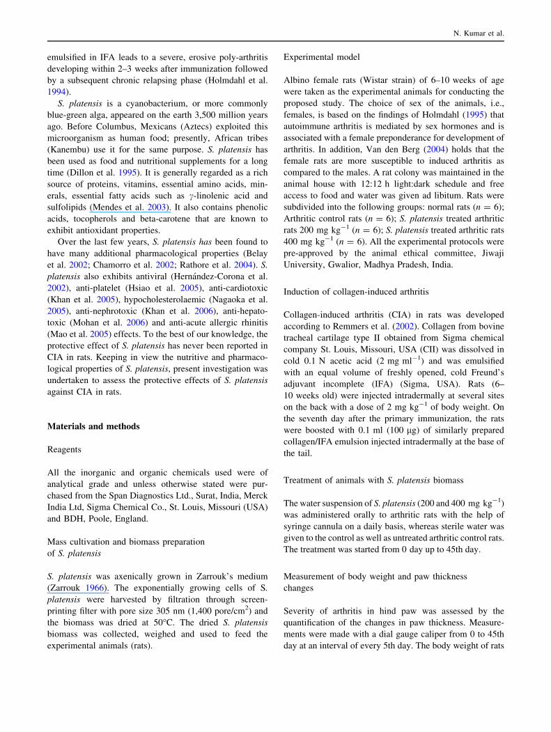
The macroscopic sign of severe arthritis at 45th day
included swelling, redness deformity and ankylosis in hind
paw and ankle joints. The symptoms of arthritic control
rats showed significant difference as compared to the
hind paw of normal rats. Such symptoms were, however,
found to be very less in the forelimbs. Whereas S. platensis
(200 mg kg-1) treated arthritic rats showed redness and
swelling only with moderate arthritis, the arthritic rats
treated with S. platensis (400 mg kg-1), however, showed
almost no sign of arthritis and appeared essentially similar
to normal rats (Fig. 1).
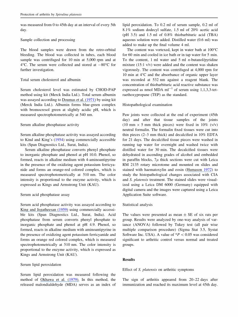
Effect of S. platensis on paw thickness changes
A significant increment in hind paw thickness from
4.17 ± 0.05 to 5.35 ± 0.20 mm was observed in arthritic
control rats from 0 to 45th day during development
of arthritis. Arthritic control rats, however, showed a
significant difference in paw thickness from normal rats
from 4.27 ± 0.06 to 4.38 ± 0.04 mm on 30th–45th day,
respectively. Whereas, S. platensis (400 mg kg-1) treatment
resulted in significant decline of paw thickness (4.60 ±
0.17 mm) in arthritic treated rats at 45th day, the arthritic rats
treated with S. platensis (200 mg kg-1), however, did not
show significant decline in paw thickness during develop-
ment of arthritis as compared to their arthritic control
counterpart (Fig. 2).
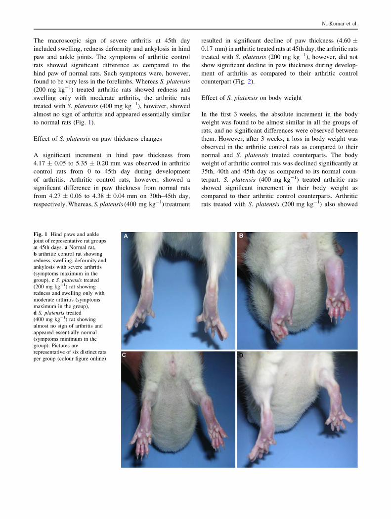
Effect of S. platensis on body weight
In the first 3 weeks, the absolute increment in the body
weight was found to be almost similar in all the groups of
rats, and no significant differences were observed between
them. However, after 3 weeks, a loss in body weight was
observed in the arthritic control rats as compared to their
normal and S. platensis treated counterparts. The body
weight of arthritic control rats was declined significantly at
35th, 40th and 45th day as compared to its normal coun-
terpart. S. platensis (400 mg kg-1) treated arthritic rats
showed significant increment in their body weight as
compared to their arthritic control counterparts. Arthritic
rats treated with S. platensis (200 mg kg-1) also showed
Fig. 1 Hind paws and ankle
joint of representative rat groups
at 45th days. a Normal rat,
b arthritic control rat showing
redness, swelling, deformity and
ankylosis with severe arthritis
(symptoms maximum in the
group), c S. platensis treated
(200 mg kg-1) rat showing
redness and swelling only with
moderate arthritis (symptoms
maximum in the group),
d S. platensis treated
(400 mg kg-1) rat showing
almost no sign of arthritis and
appeared essentially normal
(symptoms minimum in the
group). Pictures are
representative of six distinct rats
per group (colour figure online)
N. Kumar et al.
increment in their body weight as compared to arthritic
control rats, however, the difference was found to be sta-
tistically non-significant (Fig. 3).
Biochemical studies
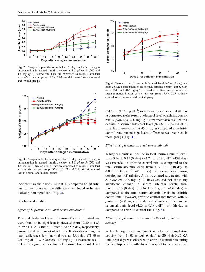
Effect of S. platensis on total serum cholesterol
The total cholesterol levels in serum of arthritic control rats
were found to be significantly elevated from 72.30 ± 1.83
to 89.64 ± 2.23 mg dl-1 from 0 to 45th day, respectively,
during the development of arthritis. It also showed signif-
icant difference form normal rats at 45th day (71.60 ±
2.57 mg dl-1). S. platensis (400 mg kg-1) treatment resul-
ted in a significant decline of serum cholesterol level
(74.53 ± 2.14 mg dl-1) in arthritic treated rats at 45th day
as compared to the serum cholesterol level of arthritic control
rats. S. platensis (200 mg kg-1) treatment also resulted in a
decline in serum cholesterol level (82.66 ± 2.54 mg dl-1)
in arthritic treated rats at 45th day as compared to arthritic
control rats, but no significant difference was recorded in
these groups (Fig. 4).
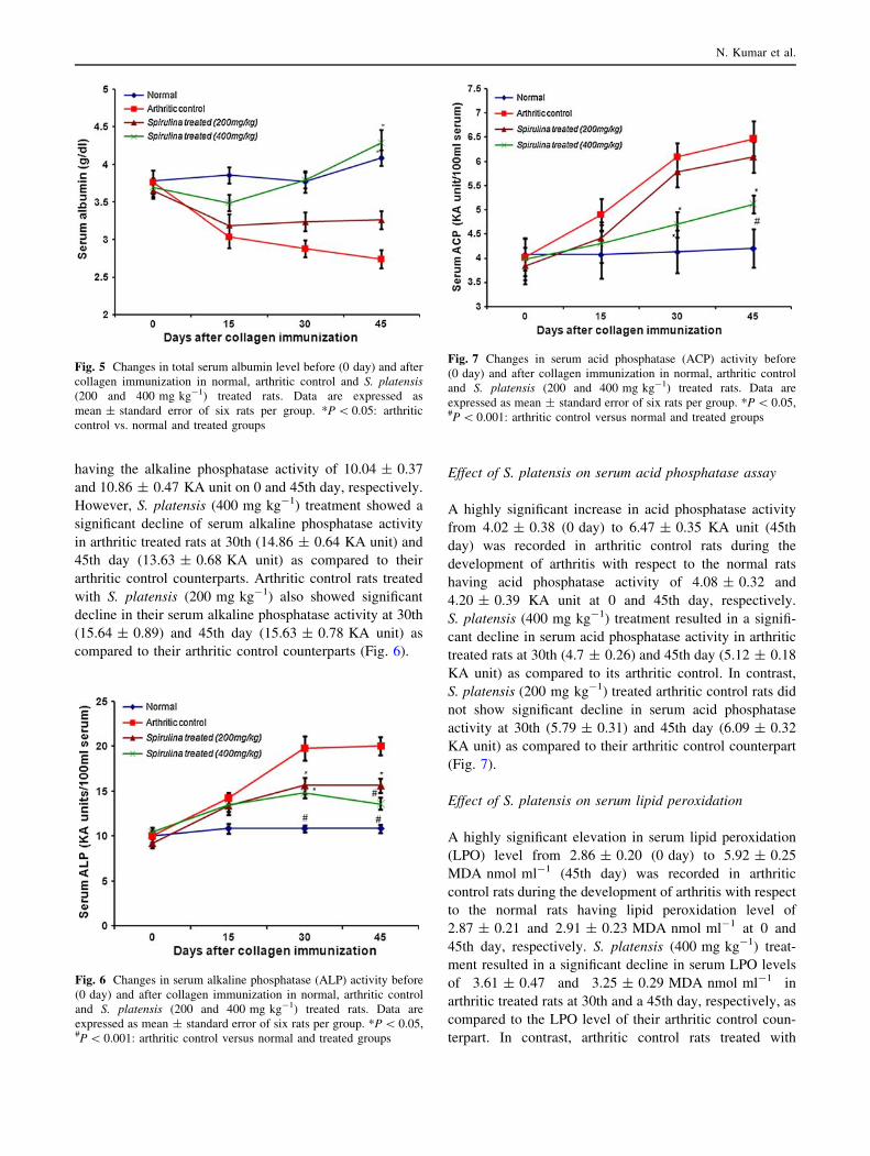
Effect of S. platensis on total serum albumin
A highly significant decline in total serum albumin levels
from 3.76 ± 0.15 (0 day) to 2.74 ± 0.12 g dl-1 (45th day)
was recorded in arthritic control rats as compared to the
total serum albumin levels from 3.77 ± 0.30 (0 day) to
4.08 ± 0.34 g dl-1 (45th day) in normal rats during
development of arthritis. Arthritic control rats treated with
S. platensis (200 mg kg-1), however, did not show any
significant change in serum albumin levels from
3.64 ± 0.10 (0 day) to 3.26 ± 0.11 g dl-1 (45th day) as
compared to the total serum albumin levels in arthritic
control rats. However, arthritic control rats treated with S.
platensis (400 mg kg-1) showed significant increase in
serum albumin level (4.28 ± 0.18 g dl-1) at 45th day as
compared to arthritic control rats (Fig. 5).
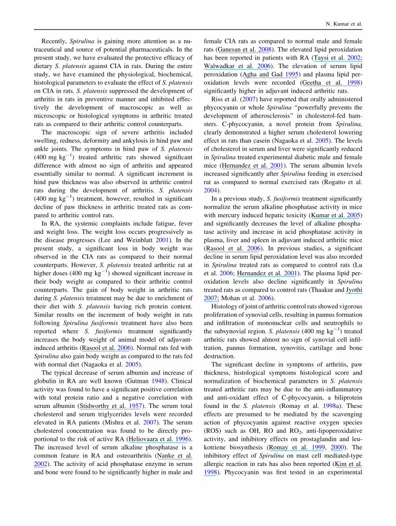
Effect of S. platensis on serum alkaline phosphatase
activity
A highly significant increment in alkaline phosphatase
activity from 10.02 ± 0.63 (0 day) to 20.04 ± 0.98 KA
unit (45th day) was observed in arthritic control rats during
the development of arthritis with respect to the normal rats
Fig. 2 Changes in paw thickness before (0 day) and after collagen
immunization in normal, arthritic control and S. platensis (200 and
400 mg kg-1) treated rats. Data are expressed as mean ± standard
error of six rats per group. *P \ 0.05: arthritic control versus normal
and treated groups
Fig. 3 Changes in the body weight before (0 day) and after collagen
immunization in normal, arthritic control and S. platensis (200 and
400 mg kg-1) treated group. Data are expressed as mean ± standard
error of six rats per group. *P \ 0.05, #P \ 0.001: arthritic control
versus normal and treated groups
Fig. 4 Changes in total serum cholesterol level before (0 day) and
after collagen immunization in normal, arthritic control and S. plat-ensis (200 and 400 mg kg-1) treated rats. Data are expressed as
mean ± standard error of six rats per group. *P \ 0.05: arthritic
control versus normal and treated groups
Protection of arthritis by Spirulina platensis
having the alkaline phosphatase activity of 10.04 ± 0.37
and 10.86 ± 0.47 KA unit on 0 and 45th day, respectively.
However, S. platensis (400 mg kg-1) treatment showed a
significant decline of serum alkaline phosphatase activity
in arthritic treated rats at 30th (14.86 ± 0.64 KA unit) and
45th day (13.63 ± 0.68 KA unit) as compared to their
arthritic control counterparts. Arthritic control rats treated
with S. platensis (200 mg kg-1) also showed significant
decline in their serum alkaline phosphatase activity at 30th
(15.64 ± 0.89) and 45th day (15.63 ± 0.78 KA unit) as
compared to their arthritic control counterparts (Fig. 6).
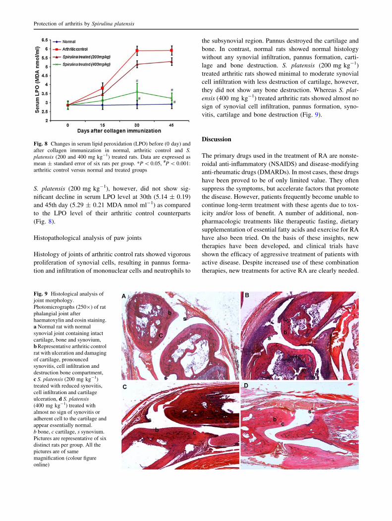
Effect of S. platensis on serum acid phosphatase assay
A highly significant increase in acid phosphatase activity
from 4.02 ± 0.38 (0 day) to 6.47 ± 0.35 KA unit (45th
day) was recorded in arthritic control rats during the
development of arthritis with respect to the normal rats
having acid phosphatase activity of 4.08 ± 0.32 and
4.20 ± 0.39 KA unit at 0 and 45th day, respectively.
S. platensis (400 mg kg-1) treatment resulted in a signifi-
cant decline in serum acid phosphatase activity in arthritic
treated rats at 30th (4.7 ± 0.26) and 45th day (5.12 ± 0.18
KA unit) as compared to its arthritic control. In contrast,
S. platensis (200 mg kg-1) treated arthritic control rats did
not show significant decline in serum acid phosphatase
activity at 30th (5.79 ± 0.31) and 45th day (6.09 ± 0.32
KA unit) as compared to their arthritic control counterpart
(Fig. 7).
Effect of S. platensis on serum lipid peroxidation
A highly significant elevation in serum lipid peroxidation
(LPO) level from 2.86 ± 0.20 (0 day) to 5.92 ± 0.25
MDA nmol ml-1 (45th day) was recorded in arthritic
control rats during the development of arthritis with respect
to the normal rats having lipid peroxidation level of
2.87 ± 0.21 and 2.91 ± 0.23 MDA nmol ml-1 at 0 and
45th day, respectively. S. platensis (400 mg kg-1) treat-
ment resulted in a significant decline in serum LPO levels
of 3.61 ± 0.47 and 3.25 ± 0.29 MDA nmol ml-1 in
arthritic treated rats at 30th and a 45th day, respectively, as
compared to the LPO level of their arthritic control coun-
terpart. In contrast, arthritic control rats treated with
Fig. 5 Changes in total serum albumin level before (0 day) and after
collagen immunization in normal, arthritic control and S. platensis(200 and 400 mg kg-1) treated rats. Data are expressed as
mean ± standard error of six rats per group. *P \ 0.05: arthritic
control vs. normal and treated groups
Fig. 6 Changes in serum alkaline phosphatase (ALP) activity before
(0 day) and after collagen immunization in normal, arthritic control
and S. platensis (200 and 400 mg kg-1) treated rats. Data are
expressed as mean ± standard error of six rats per group. *P \ 0.05,#P \ 0.001: arthritic control versus normal and treated groups
Fig. 7 Changes in serum acid phosphatase (ACP) activity before
(0 day) and after collagen immunization in normal, arthritic control
and S. platensis (200 and 400 mg kg-1) treated rats. Data are
expressed as mean ± standard error of six rats per group. *P \ 0.05,#P \ 0.001: arthritic control versus normal and treated groups
N. Kumar et al.
S. platensis (200 mg kg-1), however, did not show sig-
nificant decline in serum LPO level at 30th (5.14 ± 0.19)
and 45th day (5.29 ± 0.21 MDA nmol ml-1) as compared
to the LPO level of their arthritic control counterparts
(Fig. 8).
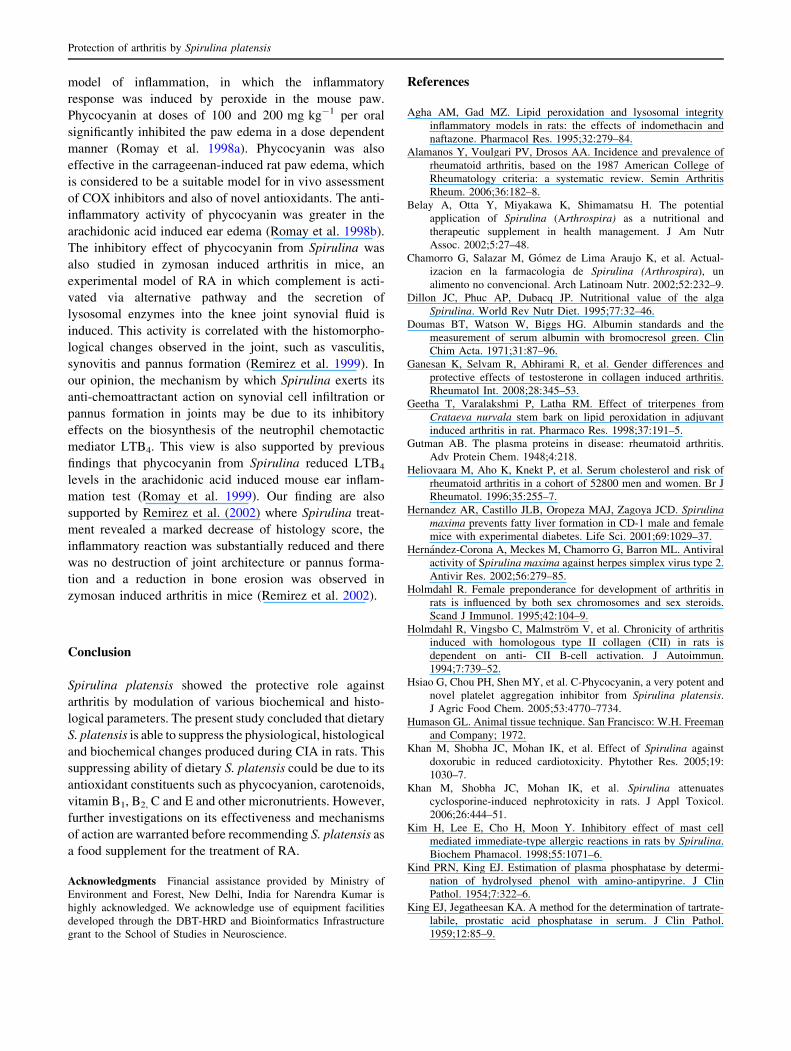
Histopathological analysis of paw joints
Histology of joints of arthritic control rats showed vigorous
proliferation of synovial cells, resulting in pannus forma-
tion and infiltration of mononuclear cells and neutrophils to
the subsynovial region. Pannus destroyed the cartilage and
bone. In contrast, normal rats showed normal histology
without any synovial infiltration, pannus formation, carti-
lage and bone destruction. S. platensis (200 mg kg-1)
treated arthritic rats showed minimal to moderate synovial
cell infiltration with less destruction of cartilage, however,
they did not show any bone destruction. Whereas S. plat-
ensis (400 mg kg-1) treated arthritic rats showed almost no
sign of synovial cell infiltration, pannus formation, syno-
vitis, cartilage and bone destruction (Fig. 9).
Discussion
The primary drugs used in the treatment of RA are nonste-
roidal anti-inflammatory (NSAIDS) and disease-modifying
anti-rheumatic drugs (DMARDs). In most cases, these drugs
have been proved to be of only limited value. They often
suppress the symptoms, but accelerate factors that promote
the disease. However, patients frequently become unable to
continue long-term treatment with these agents due to tox-
icity and/or loss of benefit. A number of additional, non-
pharmacologic treatments like therapeutic fasting, dietary
supplementation of essential fatty acids and exercise for RA
have also been tried. On the basis of these insights, new
therapies have been developed, and clinical trials have
shown the efficacy of aggressive treatment of patients with
active disease. Despite increased use of these combination
therapies, new treatments for active RA are clearly needed.
Fig. 8 Changes in serum lipid peroxidation (LPO) before (0 day) and
after collagen immunization in normal, arthritic control and S.platensis (200 and 400 mg kg-1) treated rats. Data are expressed as
mean ± standard error of six rats per group. *P \ 0.05, #P \ 0.001:
arthritic control versus normal and treated groups
Fig. 9 Histological analysis of
joint morphology.
Photomicrographs (2509) of rat
phalangial joint after
haematoxylin and eosin staining.
a Normal rat with normal
synovial joint containing intact
cartilage, bone and synovium,
b Representative arthritic control
rat with ulceration and damaging
of cartilage, pronounced
synovitis, cell infiltration and
destruction bone compartment,
c S. platensis (200 mg kg-1)
treated with reduced synovitis,
cell infiltration and cartilage
ulceration, d S. platensis(400 mg kg-1) treated with
almost no sign of synovitis or
adherent cell to the cartilage and
appear essentially normal.
b bone, c cartilage, s synovium.
Pictures are representative of six
distinct rats per group. All the
pictures are of same
magnification (colour figure
online)
Protection of arthritis by Spirulina platensis
Recently, Spirulina is gaining more attention as a nu-
traceutical and source of potential pharmaceuticals. In the
present study, we have evaluated the protective efficacy of
dietary S. platensis against CIA in rats. During the entire
study, we have examined the physiological, biochemical,
histological parameters to evaluate the effect of S. platensis
on CIA in rats. S. platensis suppressed the development of
arthritis in rats in preventive manner and inhibited effec-
tively the development of macroscopic as well as
microscopic or histological symptoms in arthritic treated
rats as compared to their arthritic control counterparts.
The macroscopic sign of severe arthritis included
swelling, redness, deformity and ankylosis in hind paw and
ankle joints. The symptoms in hind paw of S. platensis
(400 mg kg-1) treated arthritic rats showed significant
difference with almost no sign of arthritis and appeared
essentially similar to normal. A significant increment in
hind paw thickness was also observed in arthritic control
rats during the development of arthritis. S. platensis
(400 mg kg-1) treatment, however, resulted in significant
decline of paw thickness in arthritic treated rats as com-
pared to arthritic control rats.
In RA, the systemic complaints include fatigue, fever
and weight loss. The weight loss occurs progressively as
the disease progresses (Lee and Weinblatt 2001). In the
present study, a significant loss in body weight was
observed in the CIA rats as compared to their normal
counterparts. However, S. platensis treated arthritic rat at
higher doses (400 mg kg-1) showed significant increase in
their body weight as compared to their arthritic control
counterparts. The gain of body weight in arthritic rats
during S. platensis treatment may be due to enrichment of
their diet with S. platensis having rich protein content.
Similar results on the increment of body weight in rats
following Spirulina fusiformis treatment have also been
reported where S. fusiformis treatment significantly
increases the body weight of animal model of adjuvant-
induced arthritis (Rasool et al. 2006). Normal rats fed with
Spirulina also gain body weight as compared to the rats fed
with normal diet (Nagaoka et al. 2005).
The typical decrease of serum albumin and increase of
globulin in RA are well known (Gutman 1948). Clinical
activity was found to have a significant positive correlation
with total protein ratio and a negative correlation with
serum albumin (Stidworthy et al. 1957). The serum total
cholesterol and serum triglycerides levels were recorded
elevated in RA patients (Mishra et al. 2007). The serum
cholesterol concentration was found to be directly pro-
portional to the risk of active RA (Heliovaara et al. 1996).
The increased level of serum alkaline phosphatase is a
common feature in RA and osteoarthritis (Nanke et al.
2002). The activity of acid phosphatase enzyme in serum
and bone were found to be significantly higher in male and
female CIA rats as compared to normal male and female
rats (Ganesan et al. 2008). The elevated lipid peroxidation
has been reported in patients with RA (Taysi et al. 2002;
Walwadkar et al. 2006). The elevation of serum lipid
peroxidation (Agha and Gad 1995) and plasma lipid per-
oxidation levels were recorded (Geetha et al. 1998)
significantly higher in adjuvant induced arthritic rats.
Riss et al. (2007) have reported that orally administered
phycocyanin or whole Spirulina ‘‘powerfully prevents the
development of atherosclerosis’’ in cholesterol-fed ham-
sters. C-phycocyanin, a novel protein from Spirulina,
clearly demonstrated a higher serum cholesterol lowering
effect in rats than casein (Nagaoka et al. 2005). The levels
of cholesterol in serum and liver were significantly reduced
in Spirulina treated experimental diabetic male and female
mice (Hernandez et al. 2001). The serum albumin levels
increased significantly after Spirulina feeding in exercised
rat as compared to normal exercised rats (Rogatto et al.
2004).
In a previous study, S. fusiformis treatment significantly
normalize the serum alkaline phosphatase activity in mice
with mercury induced hepatic toxicity (Kumar et al. 2005)
and significantly decreases the level of alkaline phospha-
tase activity and increase in acid phosphatase activity in
plasma, liver and spleen in adjuvant induced arthritic mice
(Rasool et al. 2006). In previous studies, a significant
decline in serum lipid peroxidation level was also recorded
in Spirulina treated rats as compared to control rats (Lu
et al. 2006; Hernandez et al. 2001). The plasma lipid per-
oxidation levels also decline significantly in Spirulina
treated rats as compared to control rats (Thaakur and Jyothi
2007; Mohan et al. 2006).
Histology of joint of arthritic control rats showed vigorous
proliferation of synovial cells, resulting in pannus formation
and infiltration of mononuclear cells and neutrophils to
the subsynovial region. S. platensis (400 mg kg-1) treated
arthritic rats showed almost no sign of synovial cell infil-
tration, pannus formation, synovitis, cartilage and bone
destruction.
The significant decline in symptoms of arthritis, paw
thickness, histological symptoms histological score and
normalization of biochemical parameters in S. platensis
treated arthritic rats may be due to the anti-inflammatory
and anti-oxidant effect of C-phycocyanin, a biliprotein
found in the S. platensis (Romay et al. 1998a). These
effects are presumed to be mediated by the scavenging
action of phycocyanin against reactive oxygen species
(ROS) such as OH, RO and RO2, anti-lipoperoxidative
activity, and inhibitory effects on prostaglandin and leu-
kotriene biosynthesis (Romay et al. 1999, 2000). The
inhibitory effect of Spirulina on mast cell mediated-type
allergic reaction in rats has also been reported (Kim et al.
1998). Phycocyanin was first tested in an experimental
N. Kumar et al.
model of inflammation, in which the inflammatory
response was induced by peroxide in the mouse paw.
Phycocyanin at doses of 100 and 200 mg kg-1 per oral
significantly inhibited the paw edema in a dose dependent
manner (Romay et al. 1998a). Phycocyanin was also
effective in the carrageenan-induced rat paw edema, which
is considered to be a suitable model for in vivo assessment
of COX inhibitors and also of novel antioxidants. The anti-
inflammatory activity of phycocyanin was greater in the
arachidonic acid induced ear edema (Romay et al. 1998b).
The inhibitory effect of phycocyanin from Spirulina was
also studied in zymosan induced arthritis in mice, an
experimental model of RA in which complement is acti-
vated via alternative pathway and the secretion of
lysosomal enzymes into the knee joint synovial fluid is
induced. This activity is correlated with the histomorpho-
logical changes observed in the joint, such as vasculitis,
synovitis and pannus formation (Remirez et al. 1999). In
our opinion, the mechanism by which Spirulina exerts its
anti-chemoattractant action on synovial cell infiltration or
pannus formation in joints may be due to its inhibitory
effects on the biosynthesis of the neutrophil chemotactic
mediator LTB4. This view is also supported by previous
findings that phycocyanin from Spirulina reduced LTB4
levels in the arachidonic acid induced mouse ear inflam-
mation test (Romay et al. 1999). Our finding are also
supported by Remirez et al. (2002) where Spirulina treat-
ment revealed a marked decrease of histology score, the
inflammatory reaction was substantially reduced and there
was no destruction of joint architecture or pannus forma-
tion and a reduction in bone erosion was observed in
zymosan induced arthritis in mice (Remirez et al. 2002).
Conclusion
Spirulina platensis showed the protective role against
arthritis by modulation of various biochemical and histo-
logical parameters. The present study concluded that dietary
S. platensis is able to suppress the physiological, histological
and biochemical changes produced during CIA in rats. This
suppressing ability of dietary S. platensis could be due to its
antioxidant constituents such as phycocyanion, carotenoids,
vitamin B1, B2, C and E and other micronutrients. However,
further investigations on its effectiveness and mechanisms
of action are warranted before recommending S. platensis as
a food supplement for the treatment of RA.
Acknowledgments Financial assistance provided by Ministry of
Environment and Forest, New Delhi, India for Narendra Kumar is
highly acknowledged. We acknowledge use of equipment facilities
developed through the DBT-HRD and Bioinformatics Infrastructure
grant to the School of Studies in Neuroscience.
References
Agha AM, Gad MZ. Lipid peroxidation and lysosomal integrity
inflammatory models in rats: the effects of indomethacin and
naftazone. Pharmacol Res. 1995;32:279–84.
Alamanos Y, Voulgari PV, Drosos AA. Incidence and prevalence of
rheumatoid arthritis, based on the 1987 American College of
Rheumatology criteria: a systematic review. Semin Arthritis
Rheum. 2006;36:182–8.
Belay A, Otta Y, Miyakawa K, Shimamatsu H. The potential
application of Spirulina (Arthrospira) as a nutritional and
therapeutic supplement in health management. J Am Nutr
Assoc. 2002;5:27–48.
Chamorro G, Salazar M, Gomez de Lima Araujo K, et al. Actual-
izacion en la farmacologia de Spirulina (Arthrospira), un
alimento no convencional. Arch Latinoam Nutr. 2002;52:232–9.
Dillon JC, Phuc AP, Dubacq JP. Nutritional value of the alga
Spirulina. World Rev Nutr Diet. 1995;77:32–46.
Doumas BT, Watson W, Biggs HG. Albumin standards and the
measurement of serum albumin with bromocresol green. Clin
Chim Acta. 1971;31:87–96.
Ganesan K, Selvam R, Abhirami R, et al. Gender differences and
protective effects of testosterone in collagen induced arthritis.
Rheumatol Int. 2008;28:345–53.
Geetha T, Varalakshmi P, Latha RM. Effect of triterpenes from
Crataeva nurvala stem bark on lipid peroxidation in adjuvant
induced arthritis in rat. Pharmaco Res. 1998;37:191–5.
Gutman AB. The plasma proteins in disease: rheumatoid arthritis.
Adv Protein Chem. 1948;4:218.
Heliovaara M, Aho K, Knekt P, et al. Serum cholesterol and risk of
rheumatoid arthritis in a cohort of 52800 men and women. Br J
Rheumatol. 1996;35:255–7.
Hernandez AR, Castillo JLB, Oropeza MAJ, Zagoya JCD. Spirulinamaxima prevents fatty liver formation in CD-1 male and female
mice with experimental diabetes. Life Sci. 2001;69:1029–37.
Hernandez-Corona A, Meckes M, Chamorro G, Barron ML. Antiviral
activity of Spirulina maxima against herpes simplex virus type 2.
Antivir Res. 2002;56:279–85.
Holmdahl R. Female preponderance for development of arthritis in
rats is influenced by both sex chromosomes and sex steroids.
Scand J Immunol. 1995;42:104–9.
Holmdahl R, Vingsbo C, Malmstrom V, et al. Chronicity of arthritis
induced with homologous type II collagen (CII) in rats is
dependent on anti- CII B-cell activation. J Autoimmun.
1994;7:739–52.
Hsiao G, Chou PH, Shen MY, et al. C-Phycocyanin, a very potent and
novel platelet aggregation inhibitor from Spirulina platensis.
J Agric Food Chem. 2005;53:4770–7734.
Humason GL. Animal tissue technique. San Francisco: W.H. Freeman
and Company; 1972.
Khan M, Shobha JC, Mohan IK, et al. Effect of Spirulina against
doxorubic in reduced cardiotoxicity. Phytother Res. 2005;19:
1030–7.
Khan M, Shobha JC, Mohan IK, et al. Spirulina attenuates
cyclosporine-induced nephrotoxicity in rats. J Appl Toxicol.
2006;26:444–51.
Kim H, Lee E, Cho H, Moon Y. Inhibitory effect of mast cell
mediated immediate-type allergic reactions in rats by Spirulina.
Biochem Phamacol. 1998;55:1071–6.
Kind PRN, King EJ. Estimation of plasma phosphatase by determi-
nation of hydrolysed phenol with amino-antipyrine. J Clin
Pathol. 1954;7:322–6.
King EJ, Jegatheesan KA. A method for the determination of tartrate-
labile, prostatic acid phosphatase in serum. J Clin Pathol.
1959;12:85–9.
Protection of arthritis by Spirulina platensis
Kobelt G, Jonsson L, Lindgren P, et al. Modeling the progression of
rheumatoid arthritis: a two country model to estimate costs and
consequences of rheumatoid arthritis. Arthritis Rheum.
2002;46:2310–9.
Kumar M, Sharma MK, Kumar A. Spirulina fusiformis: a food
supplement against mercury induced hepatic toxicity. J Health
Sci. 2005;51:424–30.
Lee DM, Weinblatt ME. Rheumatoid arthritis. Lancet. 2001;358:
903–11.
Lu HK, Hsieh C, Hsu JJ, et al. Preventive effects of Spirulinaplatensis on skeletal muscle damage under exercise-induced
oxidative stress. Eur J Appl Physiol. 2006;98:220–6.
Majithia V, Geraci SA. Rheumatoid arthritis: diagnosis and manage-
ment. Am J Med. 2007;120:936–9.
Malaviya AN, Kapoor SK, Singh RR, et al. Prevalence of rheumatoid
arthritis in the adult Indian Population. Rheumatol Int. 1993;13:
131–4.
Mao, K., Van de, W.J., Gershwin, M.E. (2005). Effects of Spirulina-
based dietary supplement on cytokine production from allergic
rhinitis patients. J Med Food. 8, 27–30.
Mendes RL, Nobre BP, Cardoso MT, et al. Supercritical carbon
dioxide extraction of compounds with pharmaceutical impor-
tance from microalgae. Inorganica Chim Acta. 2003;356:
328–34.
Mishra KK, Pandey HP, Singh RH. A clinical study on cortisol and
certain metabolites in some chronic psychosomatic disorders.
Ind J Clin Biochem. 2007;22:41–3.
Mohan IK, Khan M, Shobha JC, et al. Protection against cisplatin
induced nephrotoxicity by Spirulina in rats. Cancer Chemother
Pharmacol. 2006;58:802–8.
Nagaoka S, Shimizu K, Kaneko H, et al. A novel protein C-
phycocyanin plays crucial role in the hypocholesterolemic action
of Spirulina platensis concentrate in rats. J Nutr. 2005;135:
2425–30.
Nanke Y, Kotake S, Akama H, Kamatani N. Alkaline phosphatase in
rheumatoid arthritis patients: possible contribution of bone-type
ALP to the raised activities of ALP in rheumatoid arthritis
patients. Clin Rheumatol. 2002;21:198–202.
Okhawa H, Ohishi N, Yagi K. Assay for lipid peroxidation in animal
tissue by thiobarbituric acid reaction. Ann Biochem. 1979;95:
351–8.
Rasool M, Sabina EP, Lavanya B. Anti-inflammatory effect of
Spirulina fusiformis on adjuvant-induced arthritis in mice. Biol
Pharm Bull. 2006;29:2483–7.
Rathore NK, Singh S, Singh V. Spirulina as immuno-enhancer and
biomodulator. J. Immunol. Immunopathol. 2004;6:1–10.
Remirez D, Gonzalez A, Merino N, et al. Effect of phycocyanin in
zymosan induced arthritis in mice. Drug Dev Res. 1999;48:70–5.
Remirez D, Gonzalez R, Merino N, et al. Inhibitory effects of
Spirulina in zymosan-induced arthritis in mice. Mediators
Inflamm. 2002;11:75–9.
Remmers EF, Joe B, Griffiths MM, et al. Modulation of multiple
experimental arthritis models by collagen-induced arthritis
quantitative trait loci isolated in congenic rat lines. Arthritis
Rheum. 2002;46:2225–34.
Riss J, Decorde K, Sutra T, et al. Phycobiliprotein C-phycocyanin
from Spirulina platensis is powerfully responsible for reducing
oxidative stress and NADPH oxidase expression induced by an
atherogenic diet in hamsters. J Agri Food Chem. 2007;55:7962–7.
Rogatto GP, de Oliveira CAM, dos Santos JW, et al. Influence of
Spirulina intake on metabolism of exercised rats. Rev Bras Med
Esporte. 2004;10:264–8.
Romay C, Armesto J, Remirez D, et al. Antioxidant and anti-
inflammatory properties of C-phycocyanin from blue-green
algae. Inflamm Res. 1998a;47:36–41.
Romay C, Ledon N, Gonzalez R. Further studies on anti-inflamma-
tory activity of phycocyanin in some animal models of
inflammation. Inflamm Res. 1998b;47:334–8.
Romay C, Lendon N, Gonzalez R. Phycocyanin extract reduces
leukotriene B4 levels in arachidonic acid induced mouse ear
inflammation test. J Pharm Pharmacol. 1999;51:641–2.
Romay C, Lendon N, Gonzalez R. Effects of phycocyanin extract on
prostaglandin E2 levels in mouse ear inflammation test. Arz
Forsch Drug Res. 2000;50:1106–9.
Stidworthy G, Payne RW, Shetlar CL, Shetlar MR. Objective
evaluation of patients with rheumatic diseases. II. Paper
electrophoretic studies of serum glycoprotein and protein from
patients with rheumatoid arthritis. J Clin Invest. 1957;36:
309–13.
Taysi S, Polat F, Gul M, et al. Lipid peroxidation, some extracellular
antioxidants and antioxidants enzymes in serum of patients with
rheumatoid arthritis. Rheumatol Int. 2002;21:200–4.
Thaakur SR, Jyothi B. Effect of Spirulina maxima on the haloperidol
induced tardive dyskinesia and oxidative stress in rats. J Neur
Transmis. 2007;114:1217–25.
Van den Berg WB. Animal models of arthritis. In: Isenberg DA,
Maddision PJ, Davidglass PW, Breedveld FC, editors. Oxford
text book of rheumatology. UK: Oxford University Press; 2004.
p. 433–41.
Walwadkar SD, Suryakar AN, Katkam RV, et al. Oxidative stress and
calcium-phosphorus levels in rheumatoid arthritis. Ind J Clin
Biochem. 2006;21:134–7.
Zarrouk C. Contribution a l’etude d; une cyanophycee. Influene de
divers facteurs physiques et chimiques sur la croissance et la
photosynthese de Spirulina maxima (Setch. et Gardner) Geitler.
Ph.D.thesis. France: University of Paris; 1966.
N. Kumar et al.
Related Documents