Journal of the Peripheral Nervous System 16:199–212 (2011) RESEARCH REPORT Evaluation of pre-existing neuropathy and bortezomib retreatment as risk factors to develop severe neuropathy in a mouse model Jordi Bruna 1,2,3 , Albert Al ´ e 1,2 , Roser Velasco 1,2,3 , Jessica Jaramillo 1,2 , Xavier Navarro 1,2 , and Esther Udina 1,2 1 Group of Neuroplasticity and Regeneration, Institute of Neurosciences and Department of Cell Biology, Physiology and Immunology, Universitat Aut ` onoma de Barcelona, Bellaterra, Spain; 2 Centro de Investigaci ´ on Biom ´ edica en Red sobre Enfermedades Neurodegenerativas (CIBERNED), Barcelona, Spain; and 3 Unit of Neuro-Oncology, Department of Neurology, Hospital Universitari de Bellvitge, Hospitalet de Llobregat, Barcelona, Spain Abstract Pre-existing neuropathy, a not uncommon feature in oncologic patients, is a potential but non-confirmed risk factor to develop early or severe chemotherapy-induced neuropathy. The main goal of this study is to evaluate the role of pre-existing neuropathy induced by vincristine (VNC) or bortezomib (BTZ) as a risk factor to develop more severe BTZ-induced neuropathy in a mouse model. VNC, at doses of 1 and 1.5 mg/kg given twice per week for 4 weeks, induced a moderate and severe sensory-motor neuropathy, primarily axonal, with predominant involvement of myelinated sensory axons. The neuropathy induced by BTZ at dose of 1 mg/kg given twice per week for 6 weeks was a mild axonal sensory neuropathy involving myelinated and unmyelinated fibers. The neuropathy in mice previously treated and retreated with the same schedule of BTZ after 4 weeks of washout period was similar in profile and severity to the one observed after the first treatment. When basal neuropathy was classified as moderate (most of BTZ-treated animals) or severe (all VNC-treated animals and two BTZ-treated animals), there was a more marked decline in sensory nerve function during BTZ retreatment in the group with basal severe neuropathy (−86%) than in the groups with basal mild (−57%) or without neuropathy (−52%; p < 0.001). Histopathological findings supported the functional results. Therefore, this study shows that the presence of a severe neuropathy previous to treatment with an antitumoral agent, such as BTZ, results in a more marked involvement of peripheral nerves. Key words: bortezomib, mice, pre-existing neuropathy, vincristine Introduction Peripheral neuropathy (PN) has become the prin- cipal dose-limiting side effect of many employed chemotherapeutic agents with the advent of hematopoietic growth factors that ameliorate the Address correspondence to: Dr. Esther Udina, Unitat Fisiologia M` edica, Facultat de Medicina, Universitat Aut ` onoma de Barcelona, E-08193 Bellaterra, Spain. Tel: +(34)-935811966; Fax: +(34)- 935812986; E-mail: [email protected] hematologic toxicity also associated with chemother- apy (Windebank and Grisold, 2008; Velasco and Bruna, 2010). It has been suggested that the pre-existing neuropathy can be a risk factor to develop more severe chemotherapy-induced PN (CIPN) based on clinical experience and few studies (Chaudhry et al., 2003; Windebank and Grisold, 2008). However, in most reported cases, patients had an unsuspected underlying inherited or inflammatory neuropathy (Graf et al., 1996; Kalfakis et al., 2002; Trobaugh-Lotrario © 2011 Peripheral Nerve Society 199

Welcome message from author
This document is posted to help you gain knowledge. Please leave a comment to let me know what you think about it! Share it to your friends and learn new things together.
Transcript
Journal of the Peripheral Nervous System 16:199–212 (2011)
RESEARCH REPORT
Evaluation of pre-existing neuropathy and bortezomibretreatment as risk factors to develop severe
neuropathy in a mouse model
Jordi Bruna1,2,3, Albert Ale1,2, Roser Velasco1,2,3, Jessica Jaramillo1,2, Xavier Navarro1,2,and Esther Udina1,2
1Group of Neuroplasticity and Regeneration, Institute of Neurosciences and Department of Cell Biology, Physiology andImmunology, Universitat Autonoma de Barcelona, Bellaterra, Spain; 2Centro de Investigacion Biomedica en Red sobre
Enfermedades Neurodegenerativas (CIBERNED), Barcelona, Spain; and 3Unit of Neuro-Oncology, Department of Neurology,Hospital Universitari de Bellvitge, Hospitalet de Llobregat, Barcelona, Spain
Abstract Pre-existing neuropathy, a not uncommon feature in oncologic patients, is apotential but non-confirmed risk factor to develop early or severe chemotherapy-inducedneuropathy. The main goal of this study is to evaluate the role of pre-existing neuropathyinduced by vincristine (VNC) or bortezomib (BTZ) as a risk factor to develop more severeBTZ-induced neuropathy in a mouse model. VNC, at doses of 1 and 1.5 mg/kg given twiceper week for 4 weeks, induced a moderate and severe sensory-motor neuropathy, primarilyaxonal, with predominant involvement of myelinated sensory axons. The neuropathyinduced by BTZ at dose of 1 mg/kg given twice per week for 6 weeks was a mild axonalsensory neuropathy involving myelinated and unmyelinated fibers. The neuropathy in micepreviously treated and retreated with the same schedule of BTZ after 4 weeks of washoutperiod was similar in profile and severity to the one observed after the first treatment.When basal neuropathy was classified as moderate (most of BTZ-treated animals) orsevere (all VNC-treated animals and two BTZ-treated animals), there was a more markeddecline in sensory nerve function during BTZ retreatment in the group with basal severeneuropathy (−86%) than in the groups with basal mild (−57%) or without neuropathy(−52%; p < 0.001). Histopathological findings supported the functional results. Therefore,this study shows that the presence of a severe neuropathy previous to treatment with anantitumoral agent, such as BTZ, results in a more marked involvement of peripheral nerves.
Key words: bortezomib, mice, pre-existing neuropathy, vincristine
IntroductionPeripheral neuropathy (PN) has become the prin-
cipal dose-limiting side effect of many employedchemotherapeutic agents with the advent ofhematopoietic growth factors that ameliorate the
Address correspondence to: Dr. Esther Udina, Unitat FisiologiaMedica, Facultat de Medicina, Universitat Autonoma de Barcelona,E-08193 Bellaterra, Spain. Tel: +(34)-935811966; Fax: +(34)-935812986; E-mail: [email protected]
hematologic toxicity also associated with chemother-apy (Windebank and Grisold, 2008; Velasco and Bruna,2010). It has been suggested that the pre-existingneuropathy can be a risk factor to develop moresevere chemotherapy-induced PN (CIPN) based onclinical experience and few studies (Chaudhry et al.,2003; Windebank and Grisold, 2008). However, inmost reported cases, patients had an unsuspectedunderlying inherited or inflammatory neuropathy (Grafet al., 1996; Kalfakis et al., 2002; Trobaugh-Lotrario
© 2011 Peripheral Nerve Society 199
Bruna et al. Journal of the Peripheral Nervous System 16:199–212 (2011)
et al., 2003). Moreover, indirect evidence usuallyemployed to justify this hypothesis, like prior expo-sure to neurotoxic agents, presence of diabetes, andalcohol intake, is also scarce or absent (Thant et al.,1982; Mollman et al., 1988; Van der Hoop et al., 1990;Ramanathan et al., 2010). In contrast, it has also beendescribed that patients with pre-existing neuropathymay undergo chemotherapy with neurotoxic agentswithout experiencing serious exacerbations (Van denBent et al., 2002; Goetz et al., 2003).
Bortezomib (BTZ) is a proteasome inhibitor used inthe treatment of multiple myeloma (Richardson et al.,2003; San Miguel et al., 2008). The relationship of BTZ-induced PN with the presence of a prior neuropathyhas been extensively evaluated during the last years,but clinical studies have reported contradictory results(Richardson et al., 2006; Stubblefield et al., 2006;Badros et al., 2007; Argyriou et al., 2008; El-Cheikhet al., 2008; Lanzani et al., 2008; Corso et al., 2010;Velasco et al., 2010; Dimopoulos et al., 2011). Severalreasons have contributed to this controversy; first,multiple myeloma itself can cause PN in approximately11%–54% of patients (Plasmati et al., 2007; Chaudhryet al., 2003; Richardson et al., 2009); second, therapiescommonly employed in these patients in the pasthave neurotoxic profile [thalidomide, vincristine (VNC)];third, multiple myeloma is usually a disease affectingold patients (Mohty et al., 2010); and last, retreatmentwith BTZ can be offered to the same patient whenfavorable responses are observed. The main questionin this regard is if patients with pre-existing neuropathy,and thus worst neurologic and neurophysiologicbaseline status, will have more severe neurologicaldysfunction when treated with neurotoxic agentsthan patients without pre-existing neuropathy. The
answer to this question will contribute to optimizethe therapies that can be offered to oncologic patients,mainly in the setting of advanced or recurrently treatedpatients. Furthermore, this knowledge may be usefulto extend the findings of clinical trials to the generalpopulation.
The first goal of this study is to evaluate therole of pre-existing neuropathy, as a risk factorto develop more severe BTZ-induced PN in mice,using a recently described model (Bruna et al., 2010).Neuropathic involvement of different severity wasinduced by treating the animals previously with thesame schedule of BTZ or with VNC. The studyfurther describes the neurophysiological, histological,and immunohistochemical features of VNC-inducedneuropathy in the mouse model.
Materials and MethodsAnimal groups, treatment, and dose schedule
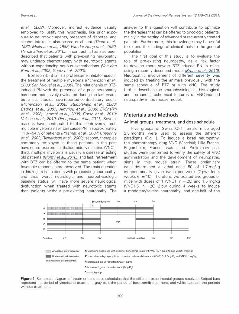
Five groups of Swiss OF1 female mice aged2.5 months were used to assess the differentparadigms (Fig. 1). To induce a basal neuropathy,the chemotherapy drug VNC (Vincrisul; Lilly France,Fegersheim, France) was used. Preliminary pilotstudies were performed to verify the safety of VNCadministration and the development of neuropathicsigns in this mouse strain. These preliminarydata determined a lethal dose 50 of 1.7 mg/kgintraperitoneally given twice per week (2 pw) for 4weeks (n = 10). Therefore, we treated two groups ofmice with doses of 1 (VNC1, n = 20) and 1.5 mg/kg(VNC1.5, n = 26) 2 pw during 4 weeks to inducea moderate/severe neuropathy, and one-half of the
A
A: vincristine subgroups with posterior bortezomib treatment (VNC1.5: 1.5mg/Kg and VNC1: 1mg/Kg)
B:
C:
D:
A’: vincristine subgroups without posterior bortezomib treatment (VNC1.5: 1.5mg/Kg and VNC1: 1mg/Kg)
B
C
D
Baseline 2w
Vincristine administration
Bortezomib administration
w-p: washout period.w:week
4w 6w
2w 4w 6wSecond Baseline
2w’ 4w’ 6w’Second Baseline
w-p
w-p
A’
bortezomib group retreated twice (1mg/Kg)
bortezomib group retreated once (1mg/Kg)
control group
Figure 1. Schematic diagram of treatment and dose schedules that the different experimental groups received. Striped barsrepresent the period of vincristine treatment, gray bars the period of bortezomib treatment, and white bars are the periodswithout treatment.
200
Bruna et al. Journal of the Peripheral Nervous System 16:199–212 (2011)
animals were left untreated during two additionalweeks before retreatment with BTZ (1 mg/kg 2 pwfor 6 weeks, provided by Janssen PharmaceuticalCompanies of Johnson & Johnson PharmaceuticalR&D, Beerse, Belgium), whereas the other half wasfollowed with no further treatment until the 16th week.
Another group of animals was treated with BTZat a dose of 1 mg/kg 2 pw subcutaneously during 6weeks and left untreated for 4 more weeks beforeretreatment (BTZ-R, n = 10). Other animals weretreated with the same dose of BTZ for only 6 weeks(BTZ, n = 20). In parallel, an untreated group of micewas followed and served as control (CTROL, n = 15).
During the study, animals were housed understandard conditions in cages with soft bedding. Thegeneral condition of the animals was assessed dailyand body weight was recorded before each drugadministration. The experimental procedures wereapproved by the Ethical Committee of our institution,and followed the rules of the European CommunitiesCouncil Directive 86/609/EEC.
Functional tests
Functional evaluation was performed at baseline(before the first treatment regimen and the day beforethe onset of the second treatment regimen) and thenevery 2 weeks during the periods of treatment (Fig. 1).The tests performed were aimed to quantitativelyevaluate motor and sensory nerve conduction, painsensibility, and integration of sensory-motor function.
Nerve conduction studies
For nerve conduction studies, the sciatic nervewas stimulated percutaneously through a pair of nee-dle electrodes placed at the sciatic notch (proximalsite) and at the ankle (distal site). Rectangular electri-cal pulses (Grass S88 stimulator, Quincy, MA, USA)of 0.01 ms duration were applied up to 25% abovethe voltage that gave a maximal response (Navarroet al., 1994; Verdu et al., 1999; Bruna et al., 2010).The compound muscle action potentials (CMAPs)were recorded from the tibialis anterior and the thirdinterosseus muscle with microneedle electrodes. Sim-ilarly, the sensory compound nerve action potential(SNAP) was recorded by electrodes placed at the fourthtoe near the digital nerves. The onset latency and themaximal amplitude of the action potentials were mea-sured and nerve conduction velocity (NCV) of motorand sensory nerve fibers was estimated. During elec-trophysiological tests, the animals were under anes-thesia (pentobarbital 40 mg/kg i.p.) and placed over awarm flat steamer controlled by a hot water circulatingpump to maintain the body temperature constant.
To classify the grade of neuropathy induced bythe first drug treatment, mice were arbitrarily grouped
before the second treatment based on the amplitudereduction of the digital SNAP with respect to the firstbaseline recording. An amplitude reduction >50% wasconsidered as a criteria of severe neuropathy, whereasa reduction between 40% and 50% was classified asmoderate neuropathy.
Pain sensibility tests
The plantar algesimetry technique was used toevaluate the functional status of nociceptive C fibers(Hargreaves et al., 1988). Mice were placed into a plas-tic box with an elevated glass floor (Plantar Algesime-ter, Ugo Basile, Comerio, Italy) and the light of a projec-tion lamp was focused directly onto the plantar surfaceof one hindpaw. The time to withdrawal of the heatedpaw was obtained from a time meter coupled withinfrared detectors. The value for a test was the meanof three trials separated by 10-min resting periods.
Sensory-motor function
A rotarod apparatus for small rodents (Letica,Panlab, Barcelona, Spain) was used. Mice were placedon the rod, turning at 8 rpm, and the time that eachanimal remained on it before falling was measured. Thevalue for a test was the mean of three trials separatedby 10-min resting intervals. Before treatment, micewere trained for 5 days. The ability to remain on therotarod for 120 s was taken as an index of normalsensory-motor function (Verdu et al., 1999).
Histological methods
At the end of the studies, all animals were anes-thetized and intracardially perfused with paraformalde-hyde [4% in phosphate-buffered saline (PBS)]. A seg-ment of the sciatic nerve at mid thigh and of the tibialnerve at the ankle level was removed in each animal.Dorsal root ganglia (DRG) from the fourth lumbar rootwere also harvested. The samples were fixed in glu-taraldehyde–paraformaldehyde (3% : 3%), washed incacodylate buffer, then post-fixed with 2% osmiumtetroxide, dehydrated in graded concentrations ofethanol, and embedded in epon. Light microscopyobservations were performed on 0.5 μm semithinsections stained with toluidine blue. Myelinated fibercounts were made from systematically selected fieldsat ×1,000 final magnification, covering a representativepart of the cross-sectional nerve profile (at least 15%of the total). The density of myelinated nerve fiberswas then derived according to a previously reportedmethod (Gomez et al., 1996). The fiber counts weremade at both proximal (sciatic) and distal (tibial) sitesusing Image J software (NIH, Bethesda, MA, USA).A morphometrical evaluation, including cross-sectionalarea, axonal counts, myelinated axon and fiber perime-ters and diameters, and calculated myelin thickness
201
Bruna et al. Journal of the Peripheral Nervous System 16:199–212 (2011)
was made from systematically selected fields fromthe sciatic nerve sections using Object Image software(NIH, Bethesda, MA, USA).
On the basis of the light microscopy findings,ultrathin sections were prepared from nerve and DRGselected blocks, counterstained with lead citrate, andexamined under a Hitachi 7000 electron microscope(Hitachi, Japan).
Immunohistochemistry
Plantar pads removed from perfused mice after theend of treatments were stored in Zamboni’s fixativeovernight and thereafter cryoprotected. Cryotome padsections 40-μm-thick were washed free-floating in PBSwith 0.3% Triton-X100 and 1% normal goat serum for1 h, then incubated in primary rabbit antiserum againstprotein gene product 9.5 (PGP, 1 : 800; AbD Serotec,Dusseldorf, Germany). After washes, sections wereincubated in secondary antiserum and processed asdescribed previously (Navarro et al., 1995). Sampleswere viewed under an epifluorescence microscopeusing appropriate filters. Five sections from eachsample were used to quantify skin pad innervationby analyzing the number and density of epidermalnerve fibers (Verdu et al., 1999).
Statistical analysis
Comparisons among experimental group valuesin functional tests were made using repeated mea-sures analysis of variance (ANOVA) test, whereasone-way ANOVA was used to assess differencesbetween groups in histological and immunohistochem-ical results. The Bonferroni test was used as thepost hoc test in each case. Results are expressedas mean and standard error of the mean. For normal-ization, the results of functional tests during the studyperiod are expressed as the percentage with respect tobaseline values for each mouse. All calculations wereperformed using the SPSS software package version15.0 (SPSS Inc.) and graphics using software packageGraphPad Prism version 4 (GraphPad Software Inc., LaJolla, CA, USA).
ResultsVNC-induced PNAssessment of general toxicity
VNC treatments showed a moderate toxicityprofile. In the VNC1.5 group, body weight wassignificantly decreased (mean loss of 13%) comparedto CTROLs (p < 0.001), whereas in the VNC1 group,the mean body weight was stable but did not follow thenormal gain of CTROL mice of about 12% (p = 0.003).Despite body weight changes, mice did not show
prostration or behavioral abnormalities. With the highdose of VNC three mice died (11.5%), whereas withthe lower dose only one died (5%) during the 4weeks of treatment. After 2 weeks of washout periodboth groups recovered body weight compared to theCTROL group, although another three and two micedied in groups VNC1.5 and VNC1, respectively.
Neurophysiological assessmentThe results of motor nerve conduction tests
showed that the CMAP amplitude of both tibialisanterior and plantar muscles and the motor NCVwere similar in VNC1 and CTROL groups. In contrast,VNC1.5 group suffered a significant decrease in CMAPamplitude in both muscles compared to CTROL (tib-ialis anterior: p = 0.033, plantar: p = 0.014) and VNC1groups (tibialis anterior: p = 0.001, plantar: p < 0.001;Fig. 2A). The reduction of CMAP amplitudes was morepronounced in the plantar (−37%) than in the tibialisanterior muscle (−16%), indicating a distal to proximalgradient. Motor NCV was also significantly decreasedat the end of treatment in the VNC1.5 group comparedto the CTROL group (p = 0.037; Fig. 2B).
In the sensory nerve conduction tests, the digitalSNAP amplitude showed a significant decrease inboth VNC-treated groups. At the end of treatment,the SNAP amplitude averaged about 16% (p < 0.001)and 21% (p < 0.001) of baseline values in the VNC1.5and VNC1 groups, respectively (Fig. 2C). Sensory NCVwas also significantly decreased in distal and proximalsegments of the sciatic nerve in VNC1.5 (p < 0.001)and VNC1 (p < 0.001) groups compared to the CTROLgroup (Fig. 2D). Likewise, increased latencies of theH waves were also found in treated groups (VNC1.5:p = 0.002, VNC1: p = 0.013) compared to CTROLs.
During the washout period, there was a recoveryof the amplitude of CMAPs of the VNC1.5 group thatbecame closer to normal values after 4 weeks oftreatment withdrawal, whereas motor NCV normalizedat 2 weeks. In contrast, the SNAP amplitude of thedigital nerves in both the VNC-treated groups remainedsignificantly reduced after 10 weeks of follow-up. Thesensory NCV recovered to normal values at 8 weeksafter treatment in the VNC1 group, but remained lowerthan normal in the VNC1.5 group (Fig. 2D).
Pain sensibilityPain sensibility, evaluated by hot thermal
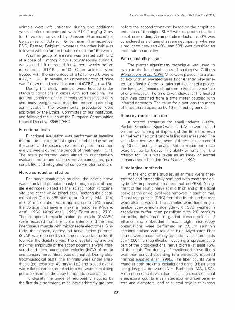
algesimetry, was unaffected with VNC treatmentduring all the follow-up, although there was a non-significant slight increase of the mean withdrawalthreshold in the VNC1.5 group at the highestaccumulated dose (Fig. 3A).
202

Bruna et al. Journal of the Peripheral Nervous System 16:199–212 (2011)
A B
C D
Figure 2. Percentage of amplitude changes vs. baseline of (A) the compound muscle action potential of plantar muscles,(B) motor nerve conduction velocity, (C) sensory compound nerve action potential of digital nerves, and (D) sensory nerveconduction velocity during follow-up of the experimental groups treated with VNC. Animals were treated during 4 weeks with 1(VNC1) and 1.5 mg/kg (VNC1.5) of VNC and followed-up during 8 more weeks (washout). Values are expressed as percentageswith respect to baseline. Error bars: SEM. #p < 0.05 in VNC1 vs. CTROL; ∗p < 0.05 in VNC1.5 vs. CTROL; & p < 0.05 inVNC1.5 vs. VNC1. CTROL, control; VNC, vincristine.
Sensory-motor functionIn the rotarod test, VNC-treated mice showed
a significant decline in the time of maintenance inthe rotating rod at the end of treatment. Animals inthe VNC1.5 group were almost unable to maintain theposition on the rod (p < 0.001) whereas animals in theVNC1 group were less affected (p < 0.001), their timeof maintenance on the rod about 60% of baseline. Afterthe washout period, the VNC1.5 and VNC1 groupsslightly recovered sensory-motor function (+12% and+13%, respectively) but without reaching normal val-ues (Fig. 3B).
Histopathological examinationThe sciatic and tibial nerves of VNC-treated mice
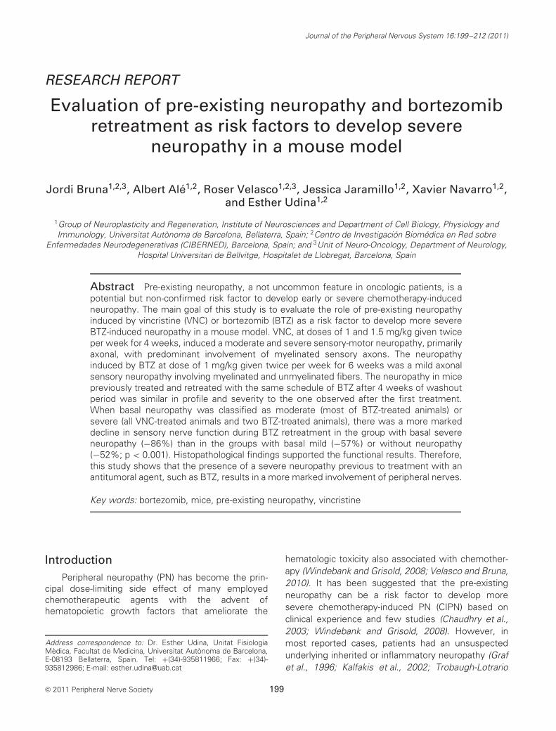
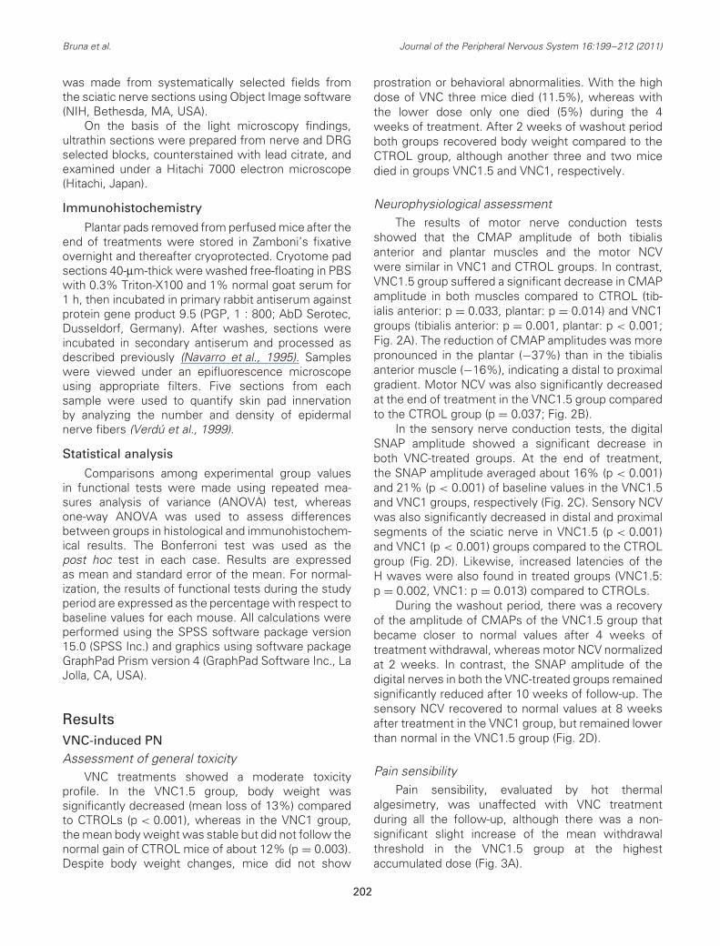
showed a lower density of myelinated large fibers,with evident signs of Wallerian degeneration andmacrophage infiltration in both animal groups, althoughthese signs were more obvious in the VNC1.5than in the VNC1 group (compare Figs. 4C and 4B,respectively). The estimated counts of myelinatedfibers were significantly lower in sciatic nerves ofVNC1.5-treated animals when compared to CTROLs(p = 0.027; Table 1).
Evaluation of skin innervationPlantar pads of treated animals did not show
obvious signs of skin atrophy. The number and the
density of the intraepidermal axonal profiles, labeledby PGP, showed only a significant decrease in theVNC1.5 group (p = 0.006) with respect to the CTROLgroup (Table 1).
PN induced by BTZ and BTZ retreatment
Mice treated with BTZ at 1 mg/kg 2 pw for6 weeks showed a sensory neuropathy, with asignificant decrease in the amplitude of SNAP (−52%,p = 0.013) and the conduction velocity of sensorynerve fibers (−17%, p = 0.049) without changes inmotor conduction tests. Mice retreated with BTZ after4 weeks of washout presented a similar neurotoxicpattern (reduction of 34% in the amplitude of SNAP:p = 0.014 and of 12% in the NCV: p = 0.03); thus,there were no differences in the percentage of declinewith respect to the corresponding baseline valuesbetween first and second treatments (Figs. 5A and5B). Surprisingly, the impairment of SNAP amplitudeswas more marked during first treatment than secondtreatment, although the trend was not significant(p = 0.072).
Pain sensibility showed a progressive increase ofthe withdrawal threshold during the first treatmentfollowed by recovery during the washout period, and asimilar behavior during the second BTZ treatment,although the changes in pain threshold were notsignificant with respect to CTROLs (Fig. 5C).
203
Bruna et al. Journal of the Peripheral Nervous System 16:199–212 (2011)
A
B
Figure 3. Percentage of changes vs. baseline of(A) algesimetry test results, expressed as time to with-drawal from hot pain stimulation and (B) rotarod test results,expressed as time of maintenance in a rotating rod. Ani-mals were treated during 4 weeks with 1 (VNC1) and1.5 mg/kg (VNC1.5) of VNC and followed-up during 8 moreweeks (washout). Values are expressed as percentages withrespect to baseline. Error bars: SEM. ∗p < 0.001 VNC groupsvs. CTROL. CTROL, control; VNC, vincristine.
A significant decline in the time of rotarod testwas also observed during the first (p = 0.027) and thesecond BTZ treatments (p = 0.006), although therewere no differences between the two treatmentadministrations (Fig. 5D).
When analyzing the skin of the paws byimmunohistochemistry, we observed a significant lossof intraepidermal fibers in both the animals treatedand retreated than in CTROLs (p < 0.001; Table 1).Sciatic and distal tibial nerve cross sections showeda preserved architecture in all BTZ-treated animals,with no clear signs of degeneration but a lowerdensity of myelinated fibers than in CTROLs. Thereduction in the number of myelinated fibers wasof about 12% and 11% at mid thigh (sciatic) andankle (tibial) levels, respectively, although the decreaseonly reached significance at the sciatic level (Table 1).The counts of myelinated fibers in animals retreatedwith BTZ were similar to the ones that receivedsingle treatment. Morphometrical analysis showedthat the mean area of the axons was significantly
A
B
C
Figure 4. Semithin cross sections of representative sciaticnerves of a control animal (A), an animal treated with 1 mg/kgVNC (B), and one treated with 1.5 mg/kg VNC (C) during 4weeks. There is a reduction in the density of myelinatedfibers, with signs of degeneration after VNC treatment,more marked in the animals that received higher doses. Smallaxons with thin myelin sheaths, compatible with regenerativeclusters, can be observed in treated animals. Bar = 10 μm.VNC, vincristine.
reduced in both BTZ (7.6 ± 0.2 μm2) and BTZ-R animals(7.1 ± 0.3 μm2) compared to CTROLs (8.0 ± 0.9 μm2;Table 2). Both treated groups showed a shift to theright in the fiber diameter distribution graph, althoughmore marked in the BTZ-R animals (Fig. 6).
Pre-existing neuropathy as a risk factor todevelop BTZ-induced neuropathy
On the basis of pre-defined criteria of SNAPamplitude loss, we grouped the animals depending
204
Bruna et al. Journal of the Peripheral Nervous System 16:199–212 (2011)
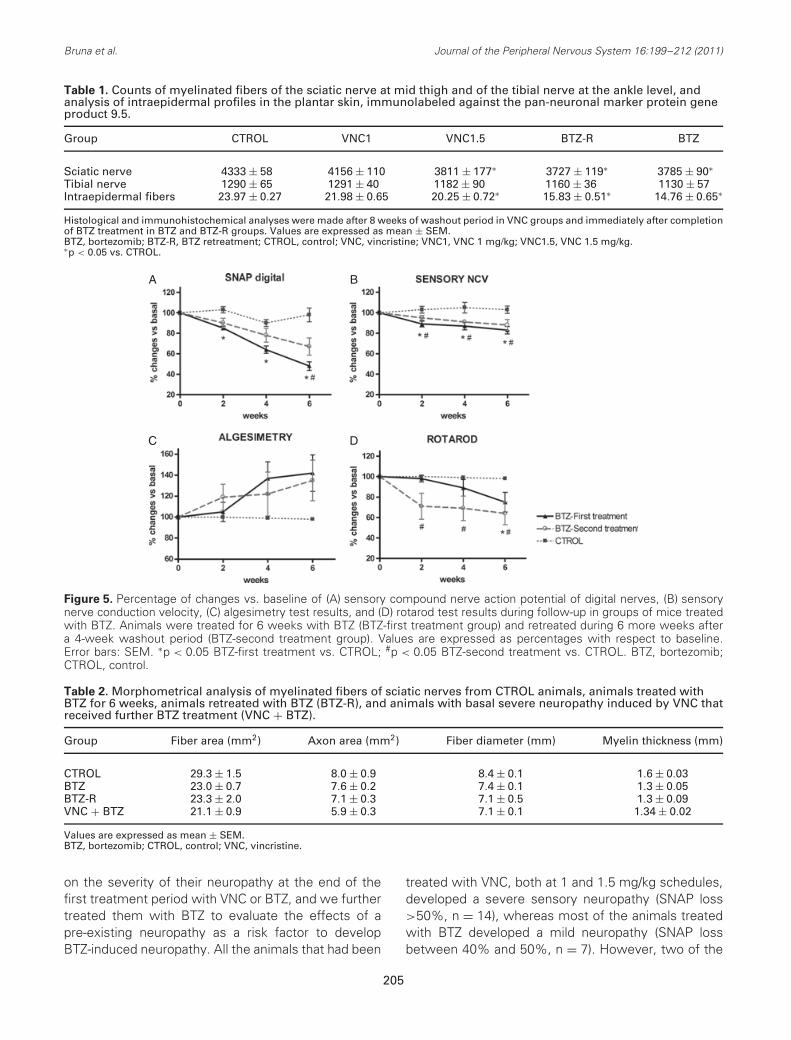
Table 1. Counts of myelinated fibers of the sciatic nerve at mid thigh and of the tibial nerve at the ankle level, andanalysis of intraepidermal profiles in the plantar skin, immunolabeled against the pan-neuronal marker protein geneproduct 9.5.
Group CTROL VNC1 VNC1.5 BTZ-R BTZ
Sciatic nerve 4333 ± 58 4156 ± 110 3811 ± 177∗ 3727 ± 119∗ 3785 ± 90∗Tibial nerve 1290 ± 65 1291 ± 40 1182 ± 90 1160 ± 36 1130 ± 57Intraepidermal fibers 23.97 ± 0.27 21.98 ± 0.65 20.25 ± 0.72∗ 15.83 ± 0.51∗ 14.76 ± 0.65∗
Histological and immunohistochemical analyses were made after 8 weeks of washout period in VNC groups and immediately after completionof BTZ treatment in BTZ and BTZ-R groups. Values are expressed as mean ± SEM.BTZ, bortezomib; BTZ-R, BTZ retreatment; CTROL, control; VNC, vincristine; VNC1, VNC 1 mg/kg; VNC1.5, VNC 1.5 mg/kg.∗p < 0.05 vs. CTROL.
A B
C D
Figure 5. Percentage of changes vs. baseline of (A) sensory compound nerve action potential of digital nerves, (B) sensorynerve conduction velocity, (C) algesimetry test results, and (D) rotarod test results during follow-up in groups of mice treatedwith BTZ. Animals were treated for 6 weeks with BTZ (BTZ-first treatment group) and retreated during 6 more weeks aftera 4-week washout period (BTZ-second treatment group). Values are expressed as percentages with respect to baseline.Error bars: SEM. ∗p < 0.05 BTZ-first treatment vs. CTROL; #p < 0.05 BTZ-second treatment vs. CTROL. BTZ, bortezomib;CTROL, control.
Table 2. Morphometrical analysis of myelinated fibers of sciatic nerves from CTROL animals, animals treated withBTZ for 6 weeks, animals retreated with BTZ (BTZ-R), and animals with basal severe neuropathy induced by VNC thatreceived further BTZ treatment (VNC + BTZ).
Group Fiber area (mm2) Axon area (mm2) Fiber diameter (mm) Myelin thickness (mm)
CTROL 29.3 ± 1.5 8.0 ± 0.9 8.4 ± 0.1 1.6 ± 0.03BTZ 23.0 ± 0.7 7.6 ± 0.2 7.4 ± 0.1 1.3 ± 0.05BTZ-R 23.3 ± 2.0 7.1 ± 0.3 7.1 ± 0.5 1.3 ± 0.09VNC + BTZ 21.1 ± 0.9 5.9 ± 0.3 7.1 ± 0.1 1.34 ± 0.02
Values are expressed as mean ± SEM.BTZ, bortezomib; CTROL, control; VNC, vincristine.
on the severity of their neuropathy at the end of thefirst treatment period with VNC or BTZ, and we furthertreated them with BTZ to evaluate the effects of apre-existing neuropathy as a risk factor to developBTZ-induced neuropathy. All the animals that had been
treated with VNC, both at 1 and 1.5 mg/kg schedules,developed a severe sensory neuropathy (SNAP loss>50%, n = 14), whereas most of the animals treatedwith BTZ developed a mild neuropathy (SNAP lossbetween 40% and 50%, n = 7). However, two of the
205

Bruna et al. Journal of the Peripheral Nervous System 16:199–212 (2011)
Figure 6. Frequency distribution (percentage) of the diam-eters of myelinated fibers from sciatic nerves of controlanimals (CTROL), animals treated with bortezomib (BTZ) for6 weeks, animals retreated with BTZ (BTZ-R), and animalswith basal severe neuropathy-induced by vincristine (VNC)that received further BTZ treatment (VNC + BTZ).
animals treated with BTZ showed a marked decreasein the SNAP amplitude reaching criteria of severeneuropathy. These two animals were thus includedin the subgroup with severe neuropathy (final n = 16).The group of mice treated once with BTZ (n = 20)was used as a group of mice without previous basalneuropathy.
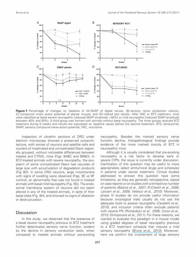
After BTZ treatment, the SNAP amplitude ofdigital nerves showed a significant decrease in allgroups compared to the CTROL group. However, thedecline of the SNAP amplitude was significantly moremarked in the group with severe neuropathy (86%decline) than in the groups with mild neuropathy(57%) and without basal neuropathy (52% decline,p < 0.001). The sensory NCV was also significantlyreduced in BTZ-treated animals with basal severeneuropathy when compared to CTROLs and BTZ-treated animals without basal neuropathy (p = 0.002;Fig. 6). BTZ-treated mice with basal severe neuropathyalso showed a more marked decrease in the averagetime of maintenance in the rotarod compared to treatedanimals without pre-existing neuropathy (−67% vs.−22%, p < 0.001; Fig. 7D). However, no differenceswere observed in the algesimetry test betweengroups. As expected, CMAP swere not affected bythe second BTZ treatment because it induces apure sensory neuropathy. In contrast, previous VNCtreatment impaired plantar CMAP amplitude in mostof the animals (see above), and after VNC withdrawal,the amplitudes progressively recovered even inanimals that received further treatment with BTZ(Fig. 7C).
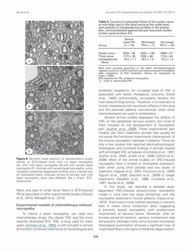
The histopathological analysis at the end of thesecond BTZ treatment revealed that animals with basalsevere neuropathy had more affected nerves, withlower density of myelinated fibers, marked signs ofdegeneration, and small clusters of thin myelinatedaxons compatible with regenerative units (Fig. 8C)than nerves from the groups with basal mild or noneuropathy (Figs. 8A and 8B). The estimated count ofsciatic myelinated fibers was lower (3626 ± 251) inthe group with severe basal neuropathy than in groupswith basal mild neuropathy (4033 ± 240, p = 0.02),no pre-existing neuropathy (3898 ± 321, p = 0.02),and CTROLs (4333 ± 153, p = 0.024, Table 3). Theestimated counts of myelinated fibers at the anklelevel showed a similar trend but differences were notsignificant.
Morphometrical analysis of the myelinated fibersshowed a significant reduction in the area of fibersand axons in animals with basal neuropathy treatedwith BTZ compared to CTROLs (29.3 ± 1.5 and8 ± 0.9 μm2, respectively), although the reduction wasmore marked in animals with basal severe neuropathy(21 ± 0.9 and 5.9 ± 0.29 μm2) than in animals withbasal mild neuropathy (23 ± 2.0 and 7.8 ± 0.8 μm2) orno neuropathy (23 ± 0.7 and 7.6 ± 0.2 μm2; Table 2).Fiber diameter distribution plots showed a shift towardthe left in both treated groups with basal neuropathy,with an increased proportion of smaller fibers than inanimals without basal neuropathy and in the CTROLgroup (Fig. 6).
206
Bruna et al. Journal of the Peripheral Nervous System 16:199–212 (2011)
A B
C D
Figure 7. Percentage of changes vs. baseline of (A) SNAP of digital nerves, (B) sensory nerve conduction velocity,(C) compound motor action potential of plantar muscle, and (D) rotarod test results. After VNC or BTZ treatment, micewere classified as basal severe neuropathic (reduced SNAP amplitude >50%) or mild neuropathic (reduced SNAP amplitudebetween 40% and 50%). A third group was formed with animals without basal neuropathy. The three groups received BTZtreatment during 6 weeks and results are expressed vs. baseline values before the second treatment. BTZ, bortezomib;SNAP; sensory compound nerve action potential; VNC, vincristine.
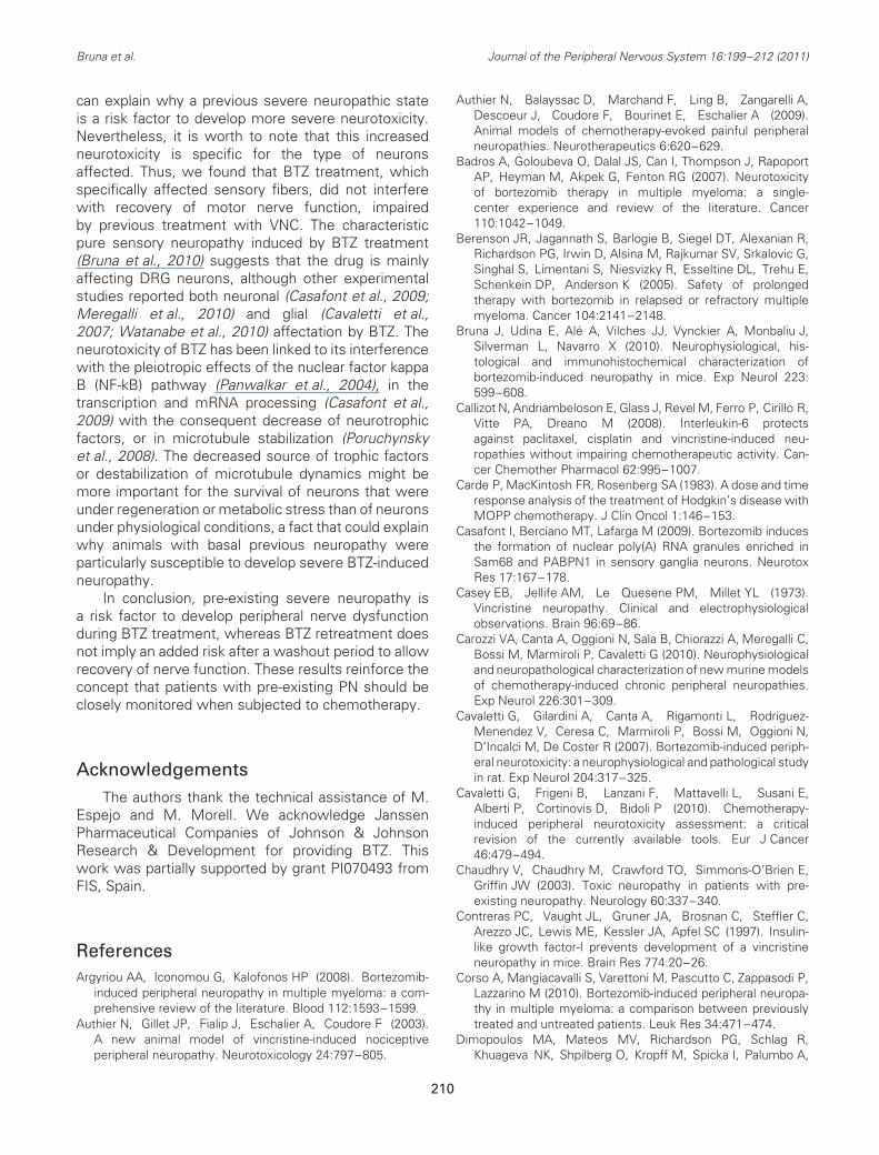
Inspection of ultrathin sections of DRG underelectron microscopy showed a preserved cytoarchi-tecture, with somas of neurons and satellite cells andclusters of myelinated and unmyelinated fibers region-ally grouped, without noticeable differences betweentreated and CTROL mice (Figs. 9A&C and 9B&D). InBTZ-treated animals with severe neuropathy, the axo-plasm of some unmyelinated fibers had vacuoles oflarge size with accumulation of degradation products(Fig. 9D). In some DRG neurons, large mitochondriawith signs of swelling were observed (Figs. 9E vs 9Fcontrol), an abnormality that was not found in treatedanimals with basal mild neuropathy (Fig. 9G). The endo-somal membrane system of neurons did not seemaltered in any of the treated animals, in spite of theirbasal state (Fig. 9H), and showed no signs of dilatationor destructuration.
DiscussionIn this study, we observed that the presence of
a basal severe neuropathy previous to BTZ treatmentfurther deteriorates sensory nerve function, evidentby the decline in sensory conduction tests, whencompared to treated animals without pre-existing
neuropathy. Besides the marked sensory nervefunction decline, histopathological findings provideevidence of the more marked toxicity of BTZ inneuropathic mice.
Although it is usually considered that pre-existingneuropathy is a risk factor to develop early orsevere CIPN, the issue is currently under discussion.Clarification of this question may be useful to moreappropriately select antitumoral drugs and schedulesin patients under cancer treatment. Clinical studiesaddressed to answer this question have somelimitations, as they are generally retrospective, basedon case reports or on studies with a limited low numberof patients (Badros et al., 2007; El-Cheikh et al., 2008;Lanzani et al., 2008; Velasco et al., 2010). Moreover,phase III studies do not provide useful informationbecause oncological trials usually do not use theadequate tools to assess neuropathy (Cavaletti et al.,2010), and inclusion criteria often exclude patientswith severe PN (Richardson et al., 2006; Corso et al.,2010; Dimopoulos et al., 2011). For these reasons, wewanted to evaluate this paradigm in a mouse modelusing graded degrees of basal neuropathy previousto a BTZ treatment schedule that induces a mildsensory neuropathy (Bruna et al., 2010). Moreover,here we confirm the involvement of large sensory
207
Bruna et al. Journal of the Peripheral Nervous System 16:199–212 (2011)
A
B
C
Figure 8. Semithin cross sections of representative sciaticnerves of BTZ-treated mice with no basal neuropathy(A), with mild basal neuropathy (B) and with severe basalneuropathy (C). Animals with severe basal neuropathy, whenretreated, presented degenerate profiles and a marked lossof myelinated fibers, whereas nerves of animals with mildbasal neuropathy were less affected. Bar = 10 μm. BTZ,bortezomib.
fibers and also of small nerve fibers in BTZ-inducedPN as described in other experimental studies (Carozziet al., 2010; Meregalli et al., 2010).
Experimental models of chemotherapy-inducedneuropathy
To induce a basal neuropathy, we used twochemotherapy drugs: the classic VNC and the morerecently developed BTZ. VNC, a drug used for manyyears (Johnson et al., 1963), is still included in almostall frontline combined treatments of hematological and
Table 3. Counts of myelinated fibers of the sciatic nerveat mid thigh and of the tibial nerve at the ankle level,and analysis of intraepidermal profiles in the plantarskin, immunolabeled against the pan-neuronal markerprotein gene product 9.5.
Group
Severebasal PN(n = 16)
Mild basalPN (n = 7)
Non-basalPN (n = 20)
Sciatic nerve 3626 ± 65 4033 ± 135∗ 3898 ± 91∗Tibial nerve 1172 ± 38 1092 ± 66 1133 ± 44Intraepidermal
fibers19.5 ± 1.1 18.5 ± 1.0 19.2 ± 1.2
Mice were grouped according to the basal neurophysiologicalregisters previous to BTZ treatment and were analyzed immediatelyafter completion of BTZ treatment. Values are expressed asmean ± SEM.BTZ, bortezomib; PN, peripheral neuropathy.∗p < 0.05 vs. severe basal PN.
lymphatic neoplasms. An increased dose of VNC isassociated with better therapeutic outcome (Cardeet al., 1983); unfortunately, neuropathy remains themain dose-limiting toxicity. Therefore, it is important tofurther characterize the neurotoxic effects of this drugand the potential additive neurotoxicity when otherchemotherapies are used in combination.
Several animal studies evaluated the effects ofVNC on the peripheral nervous system, but most ofthem focused on the development of neuropathicpain (Authier et al., 2009). These experimental painmodels use short treatment periods that usually donot cause the functional impairments characteristic ofthe chronic neuropathy reported in patients. There areonly a few studies that reported electrophysiological,histological, and functional findings in animals treatedwith prolonged VNC schedules (Contreras et al., 1997;Authier et al., 2003; Ja’afer et al., 2006; Callizot et al.,2008). Most of the animal studies on VNC-inducedneuropathy have a limited or incomplete evaluation,both when using short periods and low doses oftreatment (Ogawa et al., 2001; Fukuizumi et al., 2003;Kiguchi et al., 2008; Gauchan et al., 2009) or longertreatments (Djaldetti et al., 1996; Contreras et al.,1997; Kamei et al., 2006).
In this study, we describe a detailed dose-dependent VNC-induced sensory-motor neuropathymodel in mice that has close similarities with theneuropathy observed in human patients (Casey et al.,1973). There was a more marked decrease in sensorythan in motor nerve conduction tests suggestinga primarily axonal neuropathy with predominantinvolvement of sensory axons. Moreover, after an8-week period of washout, sensory involvement wasstill present, whereas motor nerve function recovered.Histological examination showed a significant loss ofmyelinated fibers with signs of Wallerian degeneration.
208

Bruna et al. Journal of the Peripheral Nervous System 16:199–212 (2011)
In contrast to the lesion of large sensory fibers,small sensory fibers were preserved. Therefore,nociceptive functional responses were normal andthe density of unmyelinated intraepidermal fiberswas only slightly decreased with high doses of VNC(1.5 mg/kg). The involvement of the different types offibers could be related to a different composition oftubulin isotypes between myelinated sensory, motor,and unmyelinated axons, which provide differentsusceptibility to the action of VNC. Alternatively, itis also possible that VNC carriers are mainly present inmyelinated axons, with a higher distribution in sensorythan motor ones that would facilitate VNC access tothe axolemma and thus a selective toxicity.
We also used the more recently discoveredchemotherapy drug BTZ to induce a basal neuropathy.The present results following a single course of BTZ arein agreement with those previously described in detailin the mouse model of BTZ-induced neuropathy (Brunaet al., 2010). Retreatment with the same scheduleof BTZ after 4 weeks of washout period led to adecline in sensory nerve conduction and functionaltests similar to that observed when animals weretreated for the first time with the drug. In fact, therewas a non-significant better tolerance to the drugduring the second treatment. Furthermore, the loss
of myelinated fibers after the second treatment wassimilar to that observed in mice with only one BTZschedule. Morphometrical results were also similarbetween BTZ and BTZ-R animals, although the groupthat received twice the drug showed a reduction inthe percentage of large myelinated fibers, similar tothe one observed in the VNC + BTZ group. Whenanalyzing the ultrastructure of DRG neurons, animalswith BTZ retreatment did not show the markedsigns of toxicity observed in animals that receivedBTZ after a VNC treatment. Mitochondrial swelling,a sign described also after treatment with otherchemotherapeutic drugs (Flatters and Bennett, 2006),was common in VNC–BTZ-treated mice but not seenin BTZ-retreated animals (Fig. 8). Our observationsindicate that retreatment with BTZ does not causesevere neuropathy in the mouse model. Thus, thisstudy supports the findings of a few clinical reportsin which retreatment of patients with relapsing orrefractory multiple myeloma was explored (Berensonet al., 2005; Wolf et al., 2008; Sood et al., 2009).
Pre-existing PN as a risk factor to developBTZ-induced neuropathy
As the pathogenesis of BTZ-induced PN is largelyunknown, it is difficult to establish the reasons that
A
B
C
D
E
F
G
H
Figure 9. Ultrathin cross sections of DRGs from an intact animal (A, C, D), and animals treated with BTZ with basal mildneuropathy (BTZ retreatment, E) or severe neuropathy (VNC-BTZ treatment, B, D, F, G). Low magnification view (×3,000) of anintact DRG (A) and a DRG from a BTZ-treated animal with severe basal neuropathy (B). In the axoplasm of some unmyelinatedfibers, there are vacuoles with accumulation of degradation products (×6,000 in D) not observed in intact unmyelinated fibers(C). Mitochondria were larger and swollen (×20,000 in F), compared to mitochondria of control animals (E) or BTZ-treatedanimals with basal minor neuropathy (G). The endosomal membrane system from neurons of BTZ-treated animals with basalneuropathy, such as the rough reticule, had a normal appearance without signs of dilatation (H, ×40,000). BTZ, bortezomib;DRG, dorsal root ganglia; VNC, vincristine.
209
Bruna et al. Journal of the Peripheral Nervous System 16:199–212 (2011)
can explain why a previous severe neuropathic stateis a risk factor to develop more severe neurotoxicity.Nevertheless, it is worth to note that this increasedneurotoxicity is specific for the type of neuronsaffected. Thus, we found that BTZ treatment, whichspecifically affected sensory fibers, did not interferewith recovery of motor nerve function, impairedby previous treatment with VNC. The characteristicpure sensory neuropathy induced by BTZ treatment(Bruna et al., 2010) suggests that the drug is mainlyaffecting DRG neurons, although other experimentalstudies reported both neuronal (Casafont et al., 2009;Meregalli et al., 2010) and glial (Cavaletti et al.,2007; Watanabe et al., 2010) affectation by BTZ. Theneurotoxicity of BTZ has been linked to its interferencewith the pleiotropic effects of the nuclear factor kappaB (NF-kB) pathway (Panwalkar et al., 2004), in thetranscription and mRNA processing (Casafont et al.,2009) with the consequent decrease of neurotrophicfactors, or in microtubule stabilization (Poruchynskyet al., 2008). The decreased source of trophic factorsor destabilization of microtubule dynamics might bemore important for the survival of neurons that wereunder regeneration or metabolic stress than of neuronsunder physiological conditions, a fact that could explainwhy animals with basal previous neuropathy wereparticularly susceptible to develop severe BTZ-inducedneuropathy.
In conclusion, pre-existing severe neuropathy isa risk factor to develop peripheral nerve dysfunctionduring BTZ treatment, whereas BTZ retreatment doesnot imply an added risk after a washout period to allowrecovery of nerve function. These results reinforce theconcept that patients with pre-existing PN should beclosely monitored when subjected to chemotherapy.
AcknowledgementsThe authors thank the technical assistance of M.
Espejo and M. Morell. We acknowledge JanssenPharmaceutical Companies of Johnson & JohnsonResearch & Development for providing BTZ. Thiswork was partially supported by grant PI070493 fromFIS, Spain.
ReferencesArgyriou AA, Iconomou G, Kalofonos HP (2008). Bortezomib-
induced peripheral neuropathy in multiple myeloma: a com-prehensive review of the literature. Blood 112:1593–1599.
Authier N, Gillet JP, Fialip J, Eschalier A, Coudore F (2003).A new animal model of vincristine-induced nociceptiveperipheral neuropathy. Neurotoxicology 24:797–805.
Authier N, Balayssac D, Marchand F, Ling B, Zangarelli A,Descoeur J, Coudore F, Bourinet E, Eschalier A (2009).Animal models of chemotherapy-evoked painful peripheralneuropathies. Neurotherapeutics 6:620–629.
Badros A, Goloubeva O, Dalal JS, Can I, Thompson J, RapoportAP, Heyman M, Akpek G, Fenton RG (2007). Neurotoxicityof bortezomib therapy in multiple myeloma: a single-center experience and review of the literature. Cancer110:1042–1049.
Berenson JR, Jagannath S, Barlogie B, Siegel DT, Alexanian R,Richardson PG, Irwin D, Alsina M, Rajkumar SV, Srkalovic G,Singhal S, Limentani S, Niesvizky R, Esseltine DL, Trehu E,Schenkein DP, Anderson K (2005). Safety of prolongedtherapy with bortezomib in relapsed or refractory multiplemyeloma. Cancer 104:2141–2148.
Bruna J, Udina E, Ale A, Vilches JJ, Vynckier A, Monbaliu J,Silverman L, Navarro X (2010). Neurophysiological, his-tological and immunohistochemical characterization ofbortezomib-induced neuropathy in mice. Exp Neurol 223:599–608.
Callizot N, Andriambeloson E, Glass J, Revel M, Ferro P, Cirillo R,Vitte PA, Dreano M (2008). Interleukin-6 protectsagainst paclitaxel, cisplatin and vincristine-induced neu-ropathies without impairing chemotherapeutic activity. Can-cer Chemother Pharmacol 62:995–1007.
Carde P, MacKintosh FR, Rosenberg SA (1983). A dose and timeresponse analysis of the treatment of Hodgkin’s disease withMOPP chemotherapy. J Clin Oncol 1:146–153.
Casafont I, Berciano MT, Lafarga M (2009). Bortezomib inducesthe formation of nuclear poly(A) RNA granules enriched inSam68 and PABPN1 in sensory ganglia neurons. NeurotoxRes 17:167–178.
Casey EB, Jellife AM, Le Quesene PM, Millet YL (1973).Vincristine neuropathy. Clinical and electrophysiologicalobservations. Brain 96:69–86.
Carozzi VA, Canta A, Oggioni N, Sala B, Chiorazzi A, Meregalli C,Bossi M, Marmiroli P, Cavaletti G (2010). Neurophysiologicaland neuropathological characterization of new murine modelsof chemotherapy-induced chronic peripheral neuropathies.Exp Neurol 226:301–309.
Cavaletti G, Gilardini A, Canta A, Rigamonti L, Rodriguez-Menendez V, Ceresa C, Marmiroli P, Bossi M, Oggioni N,D’Incalci M, De Coster R (2007). Bortezomib-induced periph-eral neurotoxicity: a neurophysiological and pathological studyin rat. Exp Neurol 204:317–325.
Cavaletti G, Frigeni B, Lanzani F, Mattavelli L, Susani E,Alberti P, Cortinovis D, Bidoli P (2010). Chemotherapy-induced peripheral neurotoxicity assessment: a criticalrevision of the currently available tools. Eur J Cancer46:479–494.
Chaudhry V, Chaudhry M, Crawford TO, Simmons-O’Brien E,Griffin JW (2003). Toxic neuropathy in patients with pre-existing neuropathy. Neurology 60:337–340.
Contreras PC, Vaught JL, Gruner JA, Brosnan C, Steffler C,Arezzo JC, Lewis ME, Kessler JA, Apfel SC (1997). Insulin-like growth factor-I prevents development of a vincristineneuropathy in mice. Brain Res 774:20–26.
Corso A, Mangiacavalli S, Varettoni M, Pascutto C, Zappasodi P,Lazzarino M (2010). Bortezomib-induced peripheral neuropa-thy in multiple myeloma: a comparison between previouslytreated and untreated patients. Leuk Res 34:471–474.
Dimopoulos MA, Mateos MV, Richardson PG, Schlag R,Khuageva NK, Shpilberg O, Kropff M, Spicka I, Palumbo A,
210
Bruna et al. Journal of the Peripheral Nervous System 16:199–212 (2011)
Wu KL, Esseltine DL, Liu K, Deraedt W, Cakana A, VanDe Velde H, San Miguel JF (2011). Risk factors for,and reversibility of, peripheral neuropathy associatedwith bortezomib-melphalan-prednisone in newly diagnosedpatients with multiple myeloma: subanalysis of the phase 3VISTA study. Eur J Haematol 86:23–31.
Djaldetti R, Hart J, Alexandrova S, Cohen S, Beilin BZ,Djaldetti M, Bessler H (1996). Vincristine-induced alterationsin Schwann cells of mouse peripheral nerve. Am J Hematol52:254–257.
El-Cheikh J, Stoppa AM, Bouabdallah R, de Lavallade H, Coso D,de Collela JM, Auran-Schleinitz T, Gastaut JA, Blaise D,Mohty M (2008). Features and risk factors of peripheralneuropathy during treatment with bortezomib for advancedmyeloma. Clin Lymphoma Myeloma 8:146–152.
Flatters SJ, Bennett GJ (2006). Studies of peripheral sensorynerves in paclitaxel-induced painful peripheral neuropathy:evidence for mitochondrial dysfunction. Pain 122:245–257.
Fukuizumi T, Ohkubo T, Kitamura K (2003). Spinal sensitizationmechanism in vincristine-induced hyperalgesia in mice.Neurosci Lett 343:89–92.
Gauchan P, Andoh T, Ikeda K, Fujita M, Sasaki A, Kato A,Kuraishi Y (2009). Mechanical allodynia induced by paclitaxel,oxaliplatin and vincristine: different effectiveness ofgabapentin and different expression of voltage-dependentcalcium channel alpha (2) delta-1 subunit. Biol Pharm Bull32:732–734.
Goetz MP, Erlichman C, Windebank AJ, Reid JM, Sloan JA,Atherton P, Adjei AA, Rubin J, Pitot H, Galanis E, Ames MM,Goldberg RM (2003). Phase I and pharmacokinetic study oftwo different schedules of oxaliplatin, irinotecan, Fluorouracil,and leucovorin in patients with solid tumors. J Clin Oncol21:3761–3769.
Gomez N, Cuadras J, Butı M, Navarro X (1996). Histologicassessment of sciatic nerve regeneration following resectionand graft or tube repair in the mouse. Restor Neurol Neurosci10:187–196.
Graf WD, Chance PF, Lensch MW, Eng LJ, Lipe HP, Bird TD(1996). Severe vincristine neuropathy in Charcot-Marie-Toothdisease type 1A. Cancer 77:1356–1362.
Hargreaves K, Dubner R, Brown F, Flores C, Joris J (1988). Anew and sensitive method for measuring thermal nociceptionin cutaneous hyperalgesia. Pain 32:77–88.
Ja’afer FM, Hamdan FB, Mohammed FH (2006). Vincristine-induced neuropathy in rat: electrophysiological and histo-logical study. Exp Brain Res 173:334–345.
Johnson IS, Armstrong JG, Gorman M, Burnett JP (1963). Thevinca alkaloids: a new class of oncolytic agents. Cancer Res23:1390–1427.
Kalfakis N, Panas M, Karadima G, Floroskufi P, Kokolakis N,Vassilopoulos D (2002). Hereditary neuropathy with liabilityto pressure palsies emerging during vincristine treatment.Neurology 59:1470–1471.
Kamei J, Nozaki C, Saitoh A (2006). Effect of mexiletineon vincristine-induced painful neuropathy in mice. EurJ Pharmacol 536:123–127.
Kiguchi N, Maeda T, Kobayashi Y, Kondo T, Ozaki M, Kishioka S(2008). The critical role of invading peripheral macrophage-derived interleukin-6 in vincristine-induced mechanicalallodynia in mice. Eur J Pharmacol 592:87–92.
Lanzani F, Mattavelli L, Frigeni B, Rossini F, Cammarota S,Petro D, Jann S, Cavaletti G (2008). Role of a pre-existing
neuropathy on the course of bortezomib-induced peripheralneurotoxicity. J Peripher Nerv Syst 13:267–274.
Meregalli C, Canta A, Carozzi VA, Chiorazzi A, Oggioni N,Gilardini A, Ceresa C, Avezza F, Crippa L, Marmiroli P,Cavaletti G (2010). Bortezomib-induced painful neuropathy inrats: a behavioral, neurophysiological and pathological studyin rats. Eur J Pain 14:343–350.
Mohty B, El-Cheikh J, Yakoub-Agha I, Moreau P, Harousseau JL,Mohty M (2010). Peripheral neuropathy and new treatmentsfor multiple myeloma: background and practical recommen-dations. Haematologica 95:311–9.
Mollman JE, Glover DJ, Hogan WM, Furman RE (1988). Cis-platin neuropathy. Risk factors, prognosis, and protection byWR-2721. Cancer 61:2192–2195.
Navarro X, Verdu E, Butı M (1994). Comparison of regeneratingand reinnervating capabilities of different functional types ofnerve fibers. Exp Neurol 129:217–224.
Navarro X, Verdu E, Wendelschafer-Crabb G, Kennedy WR(1995). Innervation of cutaneous structures in the mouse hindpaw: a confocal microscopy immunohistochemical study. JNeurosci Res 41:111–120.
Ogawa T, Mimura Y, Isowa K, Kato H, Mitsuishi M, Toyoshi T,Kuwayama N, Morimoto H, Murakoshi M, Nakayama T(2001). An antimicrotubule agent, TZT-1027, does not induceneuropathologic alterations which are detected after admin-istration of vincristine or paclitaxel in animal models. ToxicolLett 121:97–106.
Panwalkar A, Verstovsek S, Giles F (2004). Nuclear factor-kappaB modulation as a therapeutic approach in hematologicmalignancies. Cancer 100:1578–1589.
Plasmati R, Pastorelli F, CAvo M, Petracci E, Zamagnani E, TosiP, Cangini D, TAcchetti P, Salvi F, Bartolomei I, MichelucciR, Tassinari CA (2007). Neuropathy in multiple myelomatreated with thalidomide: a prospective study. Neurology69:573–581.
Poruchynsky MS, Sackett DL, Robey RW, Ward Y, Annunzi-ata C, Fojo T (2008). Proteasome inhibitors increase tubulinpolymerization and stabilization in tissue culture cells: a pos-sible mechanism contributing to peripheral neuropathy andcellular toxicity following proteasome inhibition. Cell Cycle7:940–949.
Ramanathan RK, Rothenberg ML, Gramont A, Tournigand C,Goldberg RM, Gupta S, Andre T (2010). Incidence and evolu-tion of oxaliplatin-induced peripheral sensory neuropathy indiabetic patients with colorectal cancer: a pooled analysis ofthree phase III studies. Ann Oncol 21:754–758.
Richardson PG, Barlogie B, Berenson J, Singhal S, Jagan-nath S, Irwin D, Rajkumar SV, Srkalovic G, Alsina M, Alex-anian R, Siegel D, Orlowski RZ, Kuter D, Limentani SA,Lee S, Hideshima T, Esseltine DL, Kauffman M, Adams J,Schenkein DP, Anderson KC (2003). A phase 2 study ofbortezomib in relapsed, refractory myeloma. N Engl J Med348:2609–2617.
Richardson PG, Briemberg H, Jagannath S, Wen PY, Bar-logie B, Berenson J, Singhal S, Siegel DS, Irwin D, Schus-ter M, Srkalovic G, Alexanian R, Rajkumar SV, Limentani S,Alsina M, Orlowski RZ, Najarian K, Esseltine D, Ander-son KC, Amato AA (2006). Frequency, characteristics, andreversibility of peripherals neuropathy during treatment ofadvanced multiple myeloma with bortezomib. J Clin Oncol24:3113–3120.
Richardson PG, Xie W, Mitsiades C, Chanan-Khan AA, Lonial S,Hassoun H, Avigan DE, Oaklander AL, Kuter DJ, Wen PY,
211
Bruna et al. Journal of the Peripheral Nervous System 16:199–212 (2011)
Kesari S, Briemberg HR, Schlossman RL, Munshi NC, HeffnerLT, Doss D, Esseltine DL, Weller E, Anderson KC, Amato AA(2009). Single-agent bortezomib in previously untreatedmultiple myeloma: efficacy, characterization of peripheralneuropathy, and molecular correlations with response andneuropathy. J Clin Oncol 27:3518–3525.
San Miguel JF, Schlag R, Khuageva NK, Dimopoulos MA,Shpilberg O, Kropff M, Spicka I, Petrucci MT, Palumbo A,Samoilova OS, Dmoszynska A, Abdulkadyrov KM, Schots R,Jiang B, Mateos MV, Anderson KC, Esseltine DL, Liu K,Cakana A, van de Velde H, Richardson PG (2008). Bortezomibplus melphalan and prednisone for initial treatment of multiplemyeloma. N Engl J Med 359:906–917.
Sood R, Carloss H, Kerr R, Lopez J, Lee M, Druck M, Walters IB,Noga SJ (2009). Retreatment with bortezomib alone or incombination for patients with multiple myeloma followingan initial response to bortezomib. Am J Hematol 84:657–660.
Stubblefield MD, Slovin S, MacGregor-Cortelli B, Muzzy J,Scher H, Wright J, Esseltine D, Schenkein D, O’Connor OA(2006). An electrodiagnostic evaluation of the effect of pre-existing peripheral nervous system disorders in patientstreated with the novel proteasome inhibitor bortezomib. ClinOncol (R Coll Radiol) 18:410–418.
Thant M, Hawley RJ, Smith M, Cohen MH, Minna JD, Bunn PA,Ihde DC, West W, Matthews MJ (1982). Possible enhance-ment of vincristine neuropathy by VP-16. Cancer 49:859–864.
Trobaugh-Lotrario AD, Smith AA, Odom LF (2003). Vincristineneurotoxicity in the presence of hereditary neuropathy. MedPediatr Oncol 40:39–43.
Van den Bent MJ, van Putten WLJ, Hilkens PHE, de Wit R,van der Burg MEL (2002). Retreatment with dose-denseweekly cisplatin after previous cisplatin chemotherapy isnot complicated by significant neuro-toxicity. Eur J Cancer38:387–391.
Van der Hoop RG, van der Burg ME, ten Bokkel Huinink WW,van Houwelingen C, Neijt JP (1990). Incidence of neuropathyin 395 patients with ovarian cancer treated with or withoutcisplatin. Cancer 66:1697–1702.
Velasco R, Bruna J (2010). Chemotherapy-induced peripheralneuropathy: an unresolved issue. Neurologia 25:116–131.
Velasco R, Petit J, Clapes V, Verdu E, Navarro X, Bruna J (2010).Neurological monitoring reduces the incidence of bortezomib-induced peripheral neuropathy in multiple myeloma patients.J Peripher Nerv Syst 15:17–25.
Verdu E, Vilches JJ, Rodrıguez FJ, Ceballos D, Valero A, NavarroX (1999). Physiological and immunohistochemical characteri-zation of cisplatin-induced neuropathy in mice. Muscle Nerve22:329–340.
Watanabe T, Nagase K, Chosa M, Tobinai K (2010). Schwanncell autophagy induced by SAHA, 17-AAG, or clonazepamcan reduce bortezomib-induced peripheral neuropathy. BrJ Cancer 103:1580–1587.
Windebank AJ, Grisold W (2008). Chemotherapy-induced neu-ropathy. J Peripher Nerv Syst 13:27–46.
Wolf J, Richardson PG, Schuster M, LeBlanc A, Walters IB,Battleman DS (2008). Utility of bortezomib retreatmentin relapsed or refractory multiple myeloma patients: amulticenter case series. Clin Adv Hematol Oncol 6:755–760.
212
Related Documents