Evaluation of Cystoid Change Phenotypes in Ocular Toxoplasmosis Using Optical Coherence Tomography Yanling Ouyang 1. , Uwe Pleyer 1. , Qing Shao 1 , Pearse A. Keane 2 , Nicole Stu ¨ biger 1 , Antonia M. Joussen 1 , Srinivas R. Sadda 3 , Florian M. Heussen 1 * 1 Charite ´, University Medicine Berlin, Department of Ophthalmology, Berlin, Germany, 2 NIHR Biomedical Research Centre for Ophthalmology, Moorfields Eye Hospital NHS Foundation Trust and UCL Institute of Ophthalmology, London, United Kingdom, 3 Doheny Eye Institute and Department of Ophthalmology, Keck School of Medicine of the University of Southern California, Los Angeles, California, United States of America Abstract Purpose: To present unique cystoid changes occurring in patients with ocular toxoplasmosis observed in spectral domain optical coherence tomography (OCT). Methods: Forty-six patients (80 eyes) with a diagnosis of ocular toxoplasmosis, who underwent volume OCT examination between January 2005 and October 2012, were retrospectively collected. Review of clinical examination findings, fundus photographs, fluorescein angiograms (FA) and OCT image sets obtained at initial visits and follow-up. Qualitative and quantitative analyses of cystoid space phenotypes visualized using OCT. Results: Of the 80 eyes included, 17 eyes (15 patients) demonstrated cystoid changes in the macula on OCT. Six eyes (7.5%) had cystoid macular edema (CME), 2 eyes (2.5%) had huge outer retinal cystoid space (HORC), 12 eyes (15%) had cystoid degeneration and additional 3 eyes (3.75%) had outer retinal tubulation due to age related macular degeneration. In one eye with HORC, the lesion was seen in the photoreceptor outer segment, accompanied by photoreceptor elongation and splitting. Three eyes presented with paravascular cystoid degeneration in the inner retina without other macular OCT abnormality. Conclusions: In this study, different phenotypes of cystoid spaces seen in eyes with ocular toxoplasmosis using spectral domain OCT (SD-OCT) were demonstrated. CME presented as an uncommon feature, consistently with previous findings. Identification of rare morphological cystoid features (HORC with/without photoreceptor enlongation or splitting) on clinical examination had provided evidence to previous experimental models, which may also expand the clinical spectrum of the disease. Cystoid degeneration in the inner retina next to the retinal vessels in otherwise ‘‘normal’’ looking macula was observed, which may suggest more often clinical evaluation for those patients. Further studies are needed to verify the relevance of cystoid features seen on SD-OCT in assisting with the diagnosis and management of ocular toxoplasmosis. Citation: Ouyang Y, Pleyer U, Shao Q, Keane PA, Stu ¨ biger N, et al. (2014) Evaluation of Cystoid Change Phenotypes in Ocular Toxoplasmosis Using Optical Coherence Tomography. PLoS ONE 9(2): e86626. doi:10.1371/journal.pone.0086626 Editor: Andreas Wedrich, Medical University Graz, Austria Received September 27, 2013; Accepted December 11, 2013; Published February 5, 2014 Copyright: ß 2014 Ouyang et al. This is an open-access article distributed under the terms of the Creative Commons Attribution License, which permits unrestricted use, distribution, and reproduction in any medium, provided the original author and source are credited. Funding: Dr. Keane has received a proportion of his funding from the Department of Health’s NIHR Biomedical Research Centre for Ophthalmology at Moorfields Eye Hospital and UCL Institute of Ophthalmology. The views expressed in the publication are those of the author and not necessarily those of the Department of Health. Drs. Keane and Heussen have received travel grants from the Allergan European Retina Panel. Dr. Joussen received financial compensation for lectures from Novartis, Allergan Inc. and Bayer AG. Dr. Pleyer received funding from the federal ministry of education and research of Germany (BMBF) through the Toxonet 02 research collaboration and has been the Principal Investigator/Consultant for Abbott, Alcon, Allergan, Amgen, Bausch and Lomb, Bayer/Schering, Centocor, Esba Tech, Novartis but no commercial interests or financial relationships to any agent, device, or technique. Dr. Sadda is a co-inventor of Doheny intellectual property related to optical coherence tomography that has been licensed by Topcon Medical Systems, and is a member of the scientific advisory board for Heidelberg Engineering. Dr Sadda also receives research support from Carl Zeiss Meditec, Optos, and Optovue, Inc. The authors have no commercial interests for the manuscript. This work was partially supported by the funding from the federal ministry of education and research of Germany (BMBF) through the Toxonet 02 research collaboration and the Department of Health’s NIHR Biomedical Research Centre for Ophthalmology at Moorfields Eye Hospital and UCL Institute of Ophthalmology. The funders had no role in study design, data collection and analysis, decision to publish, or preparation of the manuscript. Competing Interests: Dr. Keane has received a proportion of his funding from the Department of Health’s NIHR Biomedical Research Centre for Ophthalmology at Moorfields Eye Hospital and UCL Institute of Ophthalmology. Drs. Keane and Heussen have received travel grants from the Allergan European Retina Panel. Dr. Joussen received financial compensation for lectures from Novartis, Allergan Inc. and Bayer AG. Dr. Pleyer has been the Principal Investigator/Consultant for Abbott, Alcon, Allergan, Amgen, Bausch and Lomb, Bayer/Schering, Centocor, Esba Tech, Novartis but has no commercial interests or financial relationships to any agent, device, or technique. Dr. Sadda is a co-inventor of Doheny intellectual property related to optical coherence tomography that has been licensed by Topcon Medical Systems. Dr Sadda is a member of the scientific advisory board for Heidelberg Engineering. Dr Sadda also receives research support from Carl Zeiss Meditec, Optos, and Optovue, Inc. Please note, although Dr. Sadda is a co-inventor of Doheny intellectual property related to optical coherence tomography (OCT) that has been licensed by Topcon Medical Systems, the authors have used a Spectralis OCT system (Spectralis, Heidelberg Engineering, Germany) in this study. Thus, there is no patent relating to material pertinent to this article. There are no further patents, products in development or marketed products to declare. This does not alter the authors’ adherence to all the PLoS ONE policies on sharing data and materials. * E-mail: [email protected] . These authors contributed equally to this work. PLOS ONE | www.plosone.org 1 February 2014 | Volume 9 | Issue 2 | e86626

Welcome message from author
This document is posted to help you gain knowledge. Please leave a comment to let me know what you think about it! Share it to your friends and learn new things together.
Transcript
Evaluation of Cystoid Change Phenotypes in OcularToxoplasmosis Using Optical Coherence TomographyYanling Ouyang1., Uwe Pleyer1., Qing Shao1, Pearse A. Keane2, Nicole Stubiger1, Antonia M. Joussen1,
Srinivas R. Sadda3, Florian M. Heussen1*
1 Charite, University Medicine Berlin, Department of Ophthalmology, Berlin, Germany, 2 NIHR Biomedical Research Centre for Ophthalmology, Moorfields Eye Hospital
NHS Foundation Trust and UCL Institute of Ophthalmology, London, United Kingdom, 3 Doheny Eye Institute and Department of Ophthalmology, Keck School of
Medicine of the University of Southern California, Los Angeles, California, United States of America
Abstract
Purpose: To present unique cystoid changes occurring in patients with ocular toxoplasmosis observed in spectral domainoptical coherence tomography (OCT).
Methods: Forty-six patients (80 eyes) with a diagnosis of ocular toxoplasmosis, who underwent volume OCT examinationbetween January 2005 and October 2012, were retrospectively collected. Review of clinical examination findings, fundusphotographs, fluorescein angiograms (FA) and OCT image sets obtained at initial visits and follow-up. Qualitative andquantitative analyses of cystoid space phenotypes visualized using OCT.
Results: Of the 80 eyes included, 17 eyes (15 patients) demonstrated cystoid changes in the macula on OCT. Six eyes (7.5%)had cystoid macular edema (CME), 2 eyes (2.5%) had huge outer retinal cystoid space (HORC), 12 eyes (15%) had cystoiddegeneration and additional 3 eyes (3.75%) had outer retinal tubulation due to age related macular degeneration. In oneeye with HORC, the lesion was seen in the photoreceptor outer segment, accompanied by photoreceptor elongation andsplitting. Three eyes presented with paravascular cystoid degeneration in the inner retina without other macular OCTabnormality.
Conclusions: In this study, different phenotypes of cystoid spaces seen in eyes with ocular toxoplasmosis using spectraldomain OCT (SD-OCT) were demonstrated. CME presented as an uncommon feature, consistently with previous findings.Identification of rare morphological cystoid features (HORC with/without photoreceptor enlongation or splitting) on clinicalexamination had provided evidence to previous experimental models, which may also expand the clinical spectrum of thedisease. Cystoid degeneration in the inner retina next to the retinal vessels in otherwise ‘‘normal’’ looking macula wasobserved, which may suggest more often clinical evaluation for those patients. Further studies are needed to verify therelevance of cystoid features seen on SD-OCT in assisting with the diagnosis and management of ocular toxoplasmosis.
Citation: Ouyang Y, Pleyer U, Shao Q, Keane PA, Stubiger N, et al. (2014) Evaluation of Cystoid Change Phenotypes in Ocular Toxoplasmosis Using OpticalCoherence Tomography. PLoS ONE 9(2): e86626. doi:10.1371/journal.pone.0086626
Editor: Andreas Wedrich, Medical University Graz, Austria
Received September 27, 2013; Accepted December 11, 2013; Published February 5, 2014
Copyright: � 2014 Ouyang et al. This is an open-access article distributed under the terms of the Creative Commons Attribution License, which permitsunrestricted use, distribution, and reproduction in any medium, provided the original author and source are credited.
Funding: Dr. Keane has received a proportion of his funding from the Department of Health’s NIHR Biomedical Research Centre for Ophthalmology at MoorfieldsEye Hospital and UCL Institute of Ophthalmology. The views expressed in the publication are those of the author and not necessarily those of the Department ofHealth. Drs. Keane and Heussen have received travel grants from the Allergan European Retina Panel. Dr. Joussen received financial compensation for lecturesfrom Novartis, Allergan Inc. and Bayer AG. Dr. Pleyer received funding from the federal ministry of education and research of Germany (BMBF) through theToxonet 02 research collaboration and has been the Principal Investigator/Consultant for Abbott, Alcon, Allergan, Amgen, Bausch and Lomb, Bayer/Schering,Centocor, Esba Tech, Novartis but no commercial interests or financial relationships to any agent, device, or technique. Dr. Sadda is a co-inventor of Dohenyintellectual property related to optical coherence tomography that has been licensed by Topcon Medical Systems, and is a member of the scientific advisoryboard for Heidelberg Engineering. Dr Sadda also receives research support from Carl Zeiss Meditec, Optos, and Optovue, Inc. The authors have no commercialinterests for the manuscript. This work was partially supported by the funding from the federal ministry of education and research of Germany (BMBF) throughthe Toxonet 02 research collaboration and the Department of Health’s NIHR Biomedical Research Centre for Ophthalmology at Moorfields Eye Hospital and UCLInstitute of Ophthalmology. The funders had no role in study design, data collection and analysis, decision to publish, or preparation of the manuscript.
Competing Interests: Dr. Keane has received a proportion of his funding from the Department of Health’s NIHR Biomedical Research Centre for Ophthalmologyat Moorfields Eye Hospital and UCL Institute of Ophthalmology. Drs. Keane and Heussen have received travel grants from the Allergan European Retina Panel. Dr.Joussen received financial compensation for lectures from Novartis, Allergan Inc. and Bayer AG. Dr. Pleyer has been the Principal Investigator/Consultant forAbbott, Alcon, Allergan, Amgen, Bausch and Lomb, Bayer/Schering, Centocor, Esba Tech, Novartis but has no commercial interests or financial relationships to anyagent, device, or technique. Dr. Sadda is a co-inventor of Doheny intellectual property related to optical coherence tomography that has been licensed by TopconMedical Systems. Dr Sadda is a member of the scientific advisory board for Heidelberg Engineering. Dr Sadda also receives research support from Carl ZeissMeditec, Optos, and Optovue, Inc. Please note, although Dr. Sadda is a co-inventor of Doheny intellectual property related to optical coherence tomography(OCT) that has been licensed by Topcon Medical Systems, the authors have used a Spectralis OCT system (Spectralis, Heidelberg Engineering, Germany) in thisstudy. Thus, there is no patent relating to material pertinent to this article. There are no further patents, products in development or marketed products todeclare. This does not alter the authors’ adherence to all the PLoS ONE policies on sharing data and materials.
* E-mail: [email protected]
. These authors contributed equally to this work.
PLOS ONE | www.plosone.org 1 February 2014 | Volume 9 | Issue 2 | e86626
Introduction
Ocular toxoplasmosis is the most common form of posterior
uveitis in otherwise healthy individuals, leading to legal blindness
in at least one eye in approximately 25% of cases [1]. It occurs
mainly in children and young adults, with significant morbidity,
and thus has considerable socio-economic implications. In
addition, more severe disease commonly develops in immunosup-
pressed patient populations, particularly those with the acquired
immune deficiency syndrome. In such patients, the occurrence of
ocular toxoplasmosis may be an important indicator of intracra-
nial involvement [2].
The pathogenesis of ocular toxoplasmosis – in particular
pertaining to mechanisms of disease recurrence – is poorly
understood [3]. In humans, the majority of recurrent retinal
lesions arise at the borders of old retinochoroidal scars. The
appearance of such lesions may be the result of parasite release
from tissue cysts, with invasion and destruction of adjacent cells
[3], [4]. The immune response induced by these parasites may also
play a significant role [3]. Advances in in vivo imaging of retinal
lesion morphology, especially optical coherence tomography
(OCT), combined with known histopathology, may thus enable
further understanding of disease progression mechanisms.
The introduction of spectral domain OCT (SD-OCT) has
significantly improved the speed and sensitivity of OCT devices.
These improvements allow excellent visualization of the neuro-
sensory retina and have facilitated identification of numerous,
hitherto unknown, retinal disease features [5]. In this study, we
analyse the phenotype of cystoid changes seen on macular OCT,
in an effort to gain a better understanding of the evolution of these
lesions and the pathogenesis of ocular toxoplasmosis.
Materials and Methods
Baseline Data CollectionData were retrospectively collected from consecutive patients
attending the Department of Ophthalmology at Charite, Univer-
sity Berlin, between January 2005 and October 2012, with a
diagnosis of ocular toxoplasmosis. For inclusion in the study, all
patients were required to have undergone volume OCT scanning
with spectral domain OCT (Spectralis, Heidelberg Engineering,
Germany). Diagnoses were based on clinical characteristics
consistent with toxoplasma retinochoroiditis (foci of retinal
necrosis with associated retinal inflammation), in the absence of
other identifiable causes. In addition, confirmatory laboratory
testing included serology and/or aqueous humor analysis (detec-
tion of Toxoplasma antibodies or detection of T. gondii DNA
using polymerase chain reaction). Patients with AIDS or other
immune system diseases, or receiving immunosuppressive drugs
(other than corticosteroids specifically for ocular toxoplasmosis),
were excluded [6]. Information regarding age, sex, ethnicity,
history of ophthalmic diseases or surgeries, ophthalmic diagnosis,
and lens status were also collected. Best-corrected visual acuity
(VA) was measured using Snellen VA charts. Approval for data
collection and analysis was obtained from the institutional review
board of the Gottingen ethics committee. The research adhered to
the tenets set forth in the Declaration of Helsinki. Written consent
was given by the patients for their information to be stored in the
hospital database and used for research.
OCT Scanning Protocols and AnalysisIn each case, macular OCT scans were obtained (i.e., image sets
centered approximately on the fovea) using Spectralis OCT. Raw
OCT data were exported from the imaging system and imported
into validated custom grading software (3D-OCTOR, Doheny
Image Reading Center (DIRC), Los Angeles, CA) [7] for further
review.
OCT Grading MethodologyTwo graders (Y.O., F.M.H.), certified for the assessment of
OCT images at the DIRC, evaluated each OCT image set
independently. The presence or absence of cystoid changes was
firstly assessed [8]. When cystoid changes were identified, their
axial location was then documented according to a pre-existing
OCT grading methodology [9]. The phenotypes of cystoid lesions
were also assessed as 1) cystoid macular edema (CME), 2) outer
retinal cystoid degeneration due to atrophy (i.e. outer retinal
tubulation), [5] 3) cystoid degeneration due to other causes
(Figure 1), 4) huge outer retinal cystoid space (CS) (Figure 2,3).
A reading center open adjudication process was applied to
obtain the final results from the dual grading [10].
Quantitative Analysis of OCT Images:For OCT sets containing huge outer retinal CS (HORC),
manual segmentation was performed, which allowed both
quantification of retinal thickness and multiple measurements of
HORC and subretinal fluid (SRF) (including maximum and
minimum height, area and volume). Quantitative parameters were
then reported based on the Early Treatment Diabetic Retinopathy
Study (ETDRS) grid or the entirety of the scanned area.
In Patient 1 (Figure 2), a membranous structure was present in
addition to the HORC. For this case, the thickness of membra-
nous structure, PR thicknesses on the left and PR thicknesses on
the right were documented. The thickness of the membranous
structure was measured at the center of HORC in each available
B-scan (example in Figure 2B as M). PR thickness was defined as
the distance between the outer boundary of the external limiting
membrane (ELM) and the inner boundary of the RPE. PR
thickness on the left/right was measured on the left/right side of
the area unaffected by HORC with distinct ELM and closest to
the center of HORC (example in Figure 2B as L and R,
respectively). In B-scans without HORC, PR thickness in the
center of the B-scan was also measured.
Data Analysis and Statistical Methods:Clinical and imaging data were analyzed with frequency and
descriptive statistics. Statistical analysis was performed using
commercially available software SPSS (version 19.0, SPSS Inc.,
Chicago, Illinois, United States). P values,0.05 were considered
statistically significant.
Results
Baseline CharacteristicsOne hundred and fifty patients with a diagnosis of ocular
toxoplasmosis were seen in our tertiary center within the study
period. Altogether, 46 patients (80 eyes; 204 visits) had undergone
volume OCT scanning and were included in the study. Of these,
all had clinical characteristics consistent with ocular toxoplasmosis.
The mean patient age was 39.5616.3 years (range, 15–78 years).
23 patients (50%) were female, while 23 (50%) were male.
All OCT images included in the study met reading center
criteria for sufficient image quality. A total of 137 visits had
abnormalities seen on OCT scans, out of which, 112 visits had
lesions involving the fovea.
Cystoid Changes in Ocular Toxoplasmosis by SD-OCT
PLOS ONE | www.plosone.org 2 February 2014 | Volume 9 | Issue 2 | e86626
Range of Cystoid ChangesOut of 80 eyes included in the study, 17 eyes (15 patients, 39
visits) had cystoid changes seen in the macula on volume OCT
scanning: 6 (7.5%) with CME, 3 (3.75%) with outer retinal
tubulation, 2 (2.5%) with HORC, and 12 (15%) with cystoid
degeneration due to other causes (6 eyes with more than one
cystoid phenotype present in the same OCT image set).
Three eyes with outer retinal tubulation were due to late
degenerative change after treatment of choroidal neovasculariza-
tion (CNV) secondary to age-related macular degeneration
(AMD). One eye presented with HORCs located in the
photoreceptor outer segment (OS) accompanied with an outer
plexiform layer (OPL) cyst and photoreceptor elongation and
splitting. Three eyes presented with cystoid degeneration in the
inner retina next to the retinal vessels without other retinal/
choroidal abnormality in the macula.
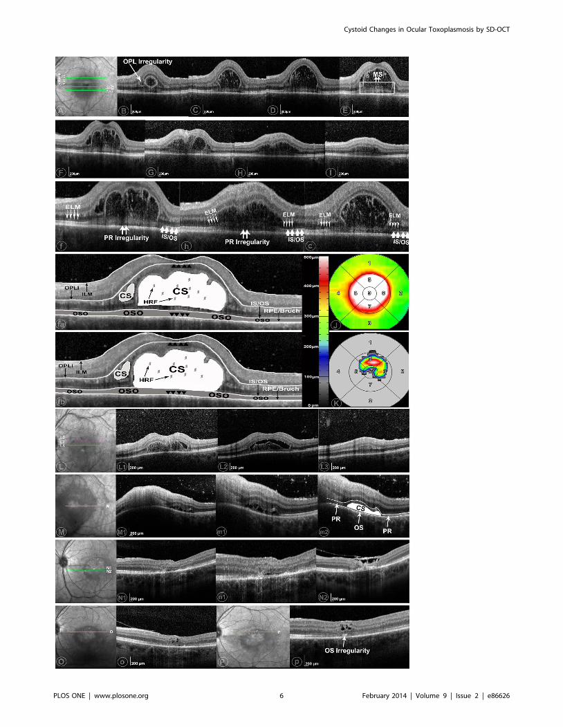
HORC – Representative Case StudiesPatient 1. A 28 year-old Caucasian male, with a previous
diagnosis of recurrent ocular toxoplasmosis in the right eye,
presented with a new onset scotoma in the same eye. On
presentation, VA was 20/20 in each eye. Dilated fundus
examination revealed a whitish-yellow inflammatory lesion near
an atrophic and pigmented retinochoroidal scar in the right eye,
with no obvious abnormality in the left eye. The patient was
commenced on oral clindamycin. Two days later, the patient
represented with decreased VA (20/200) in the right eye. Fundal
examination revealed the presence of new retinal hemorrhage and
edema accompanying the inflammatory lesion and involving the
fovea (Figure 2A).
On OCT scanning, the inner retina was largely unremarkable
with the exception of a small CS seen on a single B-scan. In the
outer retina, a huge CS posterior to the inner boundary of the
OPL was seen, termed HORC. Only a few spots of hyper
reflective foci (HRF) were observed inside the HORC. Irregularity
of the photoreceptor OS was also seen. For measurements,
HORC was observed in 8 B-scans, with a mean thickness of
membranous structure as 67.163.7 mm (range: 61–70 mm, n = 8),
mean PR thicknesses on the left as 67.062.2 mm (range: 62–
69 mm, n = 8) and mean PR thicknesses on the right as
66.163.3 mm (range: 61–69 mm, n = 8). Statistically, there was
no significant difference among PR thicknesses on the left, PR
thicknesses on the right and the mean thickness of membranous
structure (p = 0.119, nonparametric testing). The mean thickness
of the membranous structure was also similar to subfoveal PR
thickness in the fellow eye (67 mm).
The patient continued treatment with oral clindamycin.
Approximately three weeks following initial presentation, HORC
was no longer seen on OCT. Approximately two months following
initial presentation, subfoveal PR thickness increased to 68 mm. At
the most recent follow-up visit, the patient’s VA had recovered to
20/20 (Table 1, Figure 2).
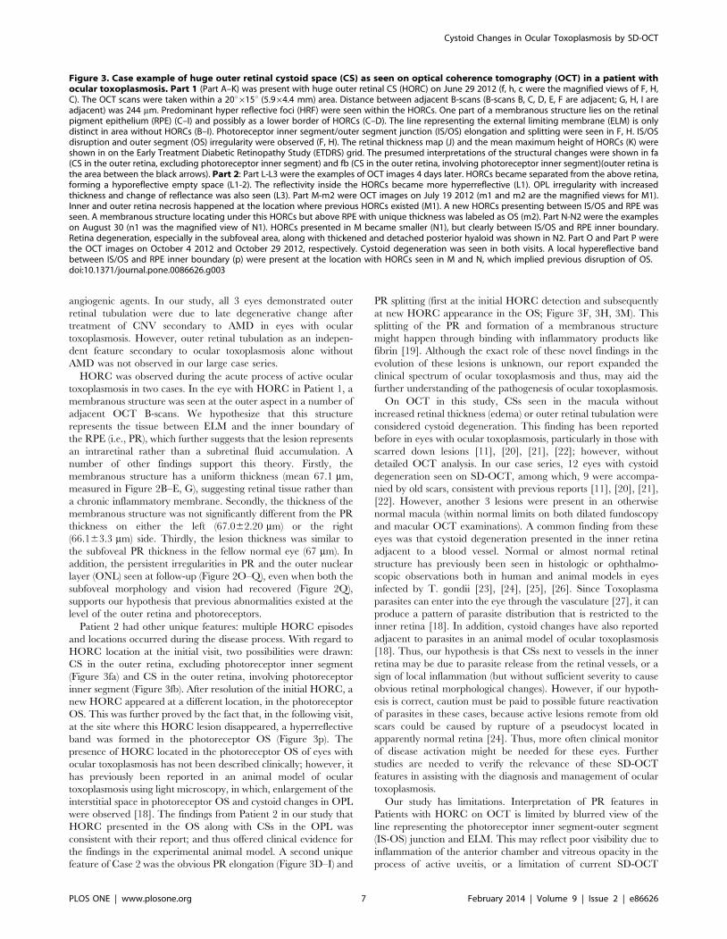
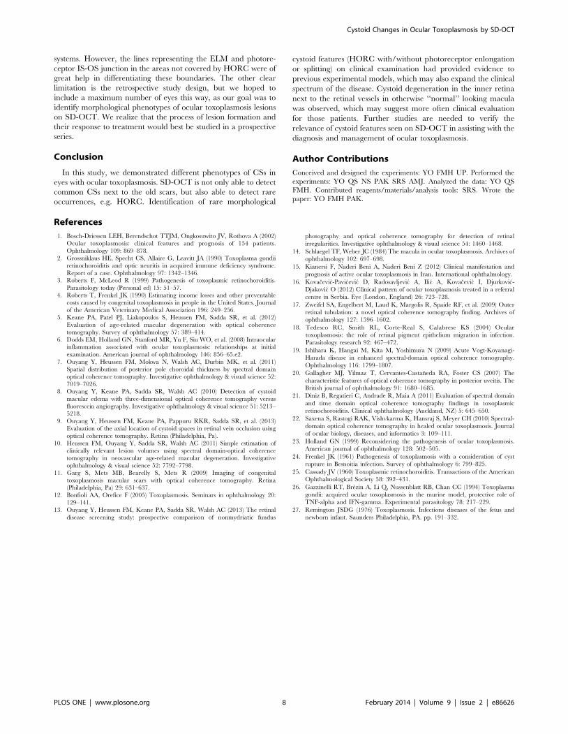
Patient 2. A 23 year-old Caucasian woman, with previous
history of recurrent ocular toxoplasmosis affecting the left eye,
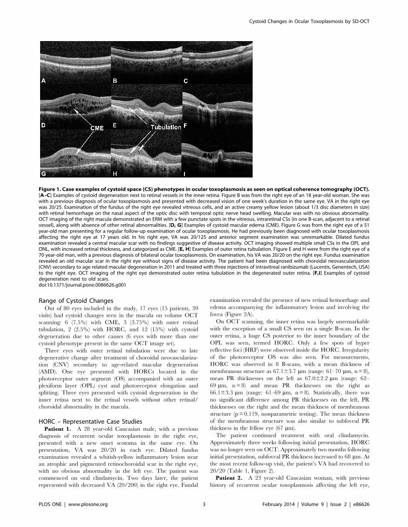
Figure 1. Case examples of cystoid space (CS) phenotypes in ocular toxoplasmosis as seen on optical coherence tomography (OCT).[A–C] Examples of cystoid degeneration next to retinal vessels in the inner retina. Figure B was from the right eye of an 18 year-old woman. She waswith a previous diagnosis of ocular toxoplasmosis and presented with decreased vision of one week’s duration in the same eye. VA in the right eyewas 20/25. Examination of the fundus of the right eye revealed vitreous cells, and an active creamy yellow lesion (about 1/3 disc diameters in size)with retinal hemorrhage on the nasal aspect of the optic disc with temporal optic nerve head swelling. Macular was with no obvious abnormality.OCT imaging of the right macula demonstrated an ERM with a few punctate spots in the vitreous, intraretinal CSs (in one B-scan, adjacent to a retinalvessel), along with absence of other retinal abnormalities. [D, G] Examples of cystoid macular edema (CME). Figure G was from the right eye of a 51year-old man presenting for a regular follow-up examination of ocular toxoplasmosis. He had previously been diagnosed with ocular toxoplasmosisaffecting the right eye at 17 years old. In his right eye, VA was 20/125 and anterior segment examination was unremarkable. Dilated fundusexamination revealed a central macular scar with no findings suggestive of disease activity. OCT imaging showed multiple small CSs in the OPL andONL, with increased retinal thickness, and categorized as CME. [E, H] Examples of outer retina tubulation. Figure E and H were from the right eye of a70 year-old man, with a previous diagnosis of bilateral ocular toxoplasmosis. On examination, his VA was 20/20 on the right eye. Fundus examinationrevealed an old macular scar in the right eye without signs of disease activity. The patient had been diagnosed with choroidal neovascularization(CNV) secondary to age related macular degeneration in 2011 and treated with three injections of intravitreal ranibizumab (Lucentis, Genentech, USA)to the right eye. OCT imaging of the right eye demonstrated outer retina tubulation in the degenerated outer retina. [F,I] Examples of cystoiddegeneration next to old scars.doi:10.1371/journal.pone.0086626.g001
Cystoid Changes in Ocular Toxoplasmosis by SD-OCT
PLOS ONE | www.plosone.org 3 February 2014 | Volume 9 | Issue 2 | e86626
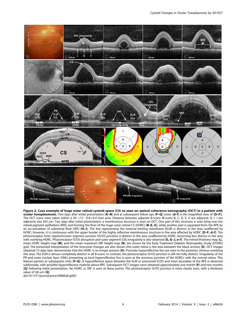
Figure 2. Case example of huge outer retinal cystoid space (CS) as seen on optical coherence tomography (OCT) in a patient withocular toxoplasmosis. Two days after initial presentation [A–N] and at subsequent follow-ups [P–Q] (note: [d–f] is the magnified view of [D–F]).The OCT scans were taken within a 20u615u (5.864.4 mm) area. Distance between adjacent B-scans (B-scans B, C, D, E, G are adjacent; H, I, J areadjacent) was 243 mm. Two days after initial presentation, a membranous structure is seen on OCT. One part of this structure is seen lying over theretinal pigment epithelium (RPE) and forming the floor of the huge outer retinal CS (HORC) [B–E, G], while another part is separated from the RPE byan accumulation of subretinal fluid (SRF) [H–J]. The line representing the external limiting membrane (ELM) is distinct in the area unaffected byHORC; however, it is continuous with the upper border of the highly reflective membranous structure in the area affected by HORC [D–F, d–f]. Thephotoreceptor inner segment/outer segment junction (IS/OS junction) is distinct in the area unaffected by HORC, becoming less distinct in the areawith overlying HORC. Photoreceptor IS/OS disruption and outer segment (OS) irregularity is also observed [E, G–J, e–f]. The retinal thickness map [L],mean HORC height map [M], and the mean maximum SRF height map [N], are shown for the Early Treatment Diabetic Retinopathy Study (ETDRS)grid. The presumed interpretation of the structural changes are also shown (the outer retina is the area between the black arrows) [K]. OCT imagesobtained 15 days later demonstrate that the HORC is no longer present [O]. Punctate hyperreflective foci are seen in the posterior vitreous overlyingthis area. The ELM is almost completely distinct in all B-scans. In contrast, the photoreceptor IS/OS junction is still not fully distinct. Irregularity of thePR and outer nuclear layer (ONL) presenting as local hyperreflective foci is seen at the previous junction of the HORCs with the normal retina. Thisfeature persists at subsequent visits [P–Q]. A hyporeflective space between the ELM or presumed IS/OS and inner boundary of the RPE is observedsubfoveally, with possible hyperreflective material above RPE. Subsequent OCT images were obtained approximately one month [P] and two months[Q] following initial presentation. No HORC or SRF is seen at these points. The photoreceptor IS/OS junction is more clearly seen, with a thicknessvalue of 68 mm [Q].doi:10.1371/journal.pone.0086626.g002
Cystoid Changes in Ocular Toxoplasmosis by SD-OCT
PLOS ONE | www.plosone.org 4 February 2014 | Volume 9 | Issue 2 | e86626
presented with decreased vision in the same eye. On presentation,
VA was 20/15 in the right eye and 20/25 in the left eye. Fundus
examination of the left eye showed pigmentary changes in the
macula unchanged from previous visits, but was otherwise
unremarkable. Right fundus examination was normal. OCT
imaging of the left eye revealed only the presence of an ERM.
Subfoveal PR thickness was 83 mm.
On examination approximately two months later, the patient
had a VA of 20/160 in the left eye. On OCT, ERM and punctate
hyperreflective spots were seen in the posterior vitreous. The OPL
was thickened with focal areas of hyporeflectivity consistent with
CSs. Again, a HORC was seen. Inside the HORC, many discrete
HRF of irregular shape were observed. The mean retinal thickness
in the foveal central subfield was 533.2626.9 (range, 475.9–600.6)
mm. The mean height of the HORC in the foveal central subfield
was 152.5695.2 (0.09–385.0) mm (Figure 3).
On examination four days later, necrosis of the outer retina,
consisting of a hyporeflective empty space, was seen on OCT. The
mean retinal thickness in the foveal central subfield was
480.7632.7 (range, 405.8–526.4) mm. The mean height of the
HORC was 249.8645.0 (range, 70.2–307.4) mm. On examination
two weeks later, the previously visible HORC had disappeared
completely; however, a new hyporeflective space appeared within
PR, more precisely, within OS. Necrosis in the inner and outer
retina in the fovea was seen, with total retinal destruction in the
fovea at subsequent visits. The patient’s final VA was 20/125
(Figure 3).
Discussion
In this study, the presence and phenotypes of CSs in eyes with
ocular toxoplasmosis were evaluated using SD-OCT. Out of 80
eyes included in the study, 17 eyes of 15 patients had cystoid
changes seen in the macula. Among these, CME (6/80, or 7.5%),
cystoid degeneration (12/80, 15%) HORC (2/80, or 2.5%), and
outer retinal tubulation (3/80, or 3.75%) due to AMD were
observed.
CME is considered a severe complication of ocular toxoplas-
mosis [11], [12]. FA has been the traditional gold standard for the
detection of CME in the past years; however, with the advent of
OCT, more CME could be possibly observed with better
reproducibility [8], [13]. Although CME in ocular toxoplasmosis
was reported in previous publications, none of them evaluated
CME with SD-OCT [14], [15], [16]. Thus, in the current study,
we tried to use SD-OCT to maximize the detection of CME. As
shown in our study, out of a total of 80 eyes with ocular
toxoplasmosis with SD-OCT examinations, 6 eyes were present
with CME. Although with the application of SD-OCT, more eyes
with CME probably could have been detected, our study had
shown that CME is still rather uncommon [14], [15], [16].
Outer retinal tubulation was first reported by Zweifel and
colleges and is now a well-accepted concept [5], [17]. Degener-
ating photoreceptors may become arranged in a circular or ovoid
fashion during a process of tubulation in AMD or other atrophic
disorders or in eyes with CNV after treatment with anti-
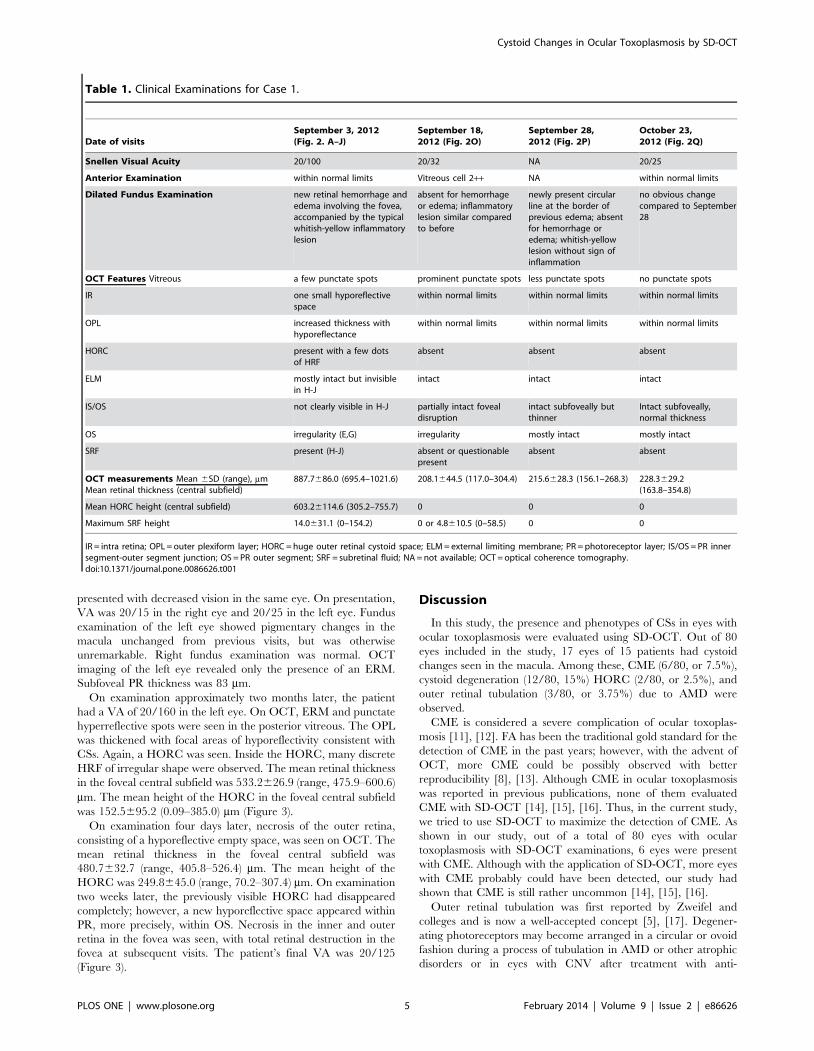
Table 1. Clinical Examinations for Case 1.
Date of visitsSeptember 3, 2012(Fig. 2. A–J)
September 18,2012 (Fig. 2O)
September 28,2012 (Fig. 2P)
October 23,2012 (Fig. 2Q)
Snellen Visual Acuity 20/100 20/32 NA 20/25
Anterior Examination within normal limits Vitreous cell 2++ NA within normal limits
Dilated Fundus Examination new retinal hemorrhage andedema involving the fovea,accompanied by the typicalwhitish-yellow inflammatorylesion
absent for hemorrhageor edema; inflammatorylesion similar comparedto before
newly present circularline at the border ofprevious edema; absentfor hemorrhage oredema; whitish-yellowlesion without sign ofinflammation
no obvious changecompared to September28
OCT Features Vitreous a few punctate spots prominent punctate spots less punctate spots no punctate spots
IR one small hyporeflectivespace
within normal limits within normal limits within normal limits
OPL increased thickness withhyporeflectance
within normal limits within normal limits within normal limits
HORC present with a few dotsof HRF
absent absent absent
ELM mostly intact but invisiblein H-J
intact intact intact
IS/OS not clearly visible in H-J partially intact fovealdisruption
intact subfoveally butthinner
Intact subfoveally,normal thickness
OS irregularity (E,G) irregularity mostly intact mostly intact
SRF present (H-J) absent or questionablepresent
absent absent
OCT measurements Mean 6SD (range), mmMean retinal thickness (central subfield)
887.7686.0 (695.4–1021.6) 208.1644.5 (117.0–304.4) 215.6628.3 (156.1–268.3) 228.3629.2(163.8–354.8)
Mean HORC height (central subfield) 603.26114.6 (305.2–755.7) 0 0 0
Maximum SRF height 14.0631.1 (0–154.2) 0 or 4.8610.5 (0–58.5) 0 0
IR = intra retina; OPL = outer plexiform layer; HORC = huge outer retinal cystoid space; ELM = external limiting membrane; PR = photoreceptor layer; IS/OS = PR innersegment-outer segment junction; OS = PR outer segment; SRF = subretinal fluid; NA = not available; OCT = optical coherence tomography.doi:10.1371/journal.pone.0086626.t001
Cystoid Changes in Ocular Toxoplasmosis by SD-OCT
PLOS ONE | www.plosone.org 5 February 2014 | Volume 9 | Issue 2 | e86626
Cystoid Changes in Ocular Toxoplasmosis by SD-OCT
PLOS ONE | www.plosone.org 6 February 2014 | Volume 9 | Issue 2 | e86626
angiogenic agents. In our study, all 3 eyes demonstrated outer
retinal tubulation were due to late degenerative change after
treatment of CNV secondary to AMD in eyes with ocular
toxoplasmosis. However, outer retinal tubulation as an indepen-
dent feature secondary to ocular toxoplasmosis alone without
AMD was not observed in our large case series.
HORC was observed during the acute process of active ocular
toxoplasmosis in two cases. In the eye with HORC in Patient 1, a
membranous structure was seen at the outer aspect in a number of
adjacent OCT B-scans. We hypothesize that this structure
represents the tissue between ELM and the inner boundary of
the RPE (i.e., PR), which further suggests that the lesion represents
an intraretinal rather than a subretinal fluid accumulation. A
number of other findings support this theory. Firstly, the
membranous structure has a uniform thickness (mean 67.1 mm,
measured in Figure 2B–E, G), suggesting retinal tissue rather than
a chronic inflammatory membrane. Secondly, the thickness of the
membranous structure was not significantly different from the PR
thickness on either the left (67.062.20 mm) or the right
(66.163.3 mm) side. Thirdly, the lesion thickness was similar to
the subfoveal PR thickness in the fellow normal eye (67 mm). In
addition, the persistent irregularities in PR and the outer nuclear
layer (ONL) seen at follow-up (Figure 2O–Q), even when both the
subfoveal morphology and vision had recovered (Figure 2Q),
supports our hypothesis that previous abnormalities existed at the
level of the outer retina and photoreceptors.
Patient 2 had other unique features: multiple HORC episodes
and locations occurred during the disease process. With regard to
HORC location at the initial visit, two possibilities were drawn:
CS in the outer retina, excluding photoreceptor inner segment
(Figure 3fa) and CS in the outer retina, involving photoreceptor
inner segment (Figure 3fb). After resolution of the initial HORC, a
new HORC appeared at a different location, in the photoreceptor
OS. This was further proved by the fact that, in the following visit,
at the site where this HORC lesion disappeared, a hyperreflective
band was formed in the photoreceptor OS (Figure 3p). The
presence of HORC located in the photoreceptor OS of eyes with
ocular toxoplasmosis has not been described clinically; however, it
has previously been reported in an animal model of ocular
toxoplasmosis using light microscopy, in which, enlargement of the
interstitial space in photoreceptor OS and cystoid changes in OPL
were observed [18]. The findings from Patient 2 in our study that
HORC presented in the OS along with CSs in the OPL was
consistent with their report; and thus offered clinical evidence for
the findings in the experimental animal model. A second unique
feature of Case 2 was the obvious PR elongation (Figure 3D–I) and
PR splitting (first at the initial HORC detection and subsequently
at new HORC appearance in the OS; Figure 3F, 3H, 3M). This
splitting of the PR and formation of a membranous structure
might happen through binding with inflammatory products like
fibrin [19]. Although the exact role of these novel findings in the
evolution of these lesions is unknown, our report expanded the
clinical spectrum of ocular toxoplasmosis and thus, may aid the
further understanding of the pathogenesis of ocular toxoplasmosis.
On OCT in this study, CSs seen in the macula without
increased retinal thickness (edema) or outer retinal tubulation were
considered cystoid degeneration. This finding has been reported
before in eyes with ocular toxoplasmosis, particularly in those with
scarred down lesions [11], [20], [21], [22]; however, without
detailed OCT analysis. In our case series, 12 eyes with cystoid
degeneration seen on SD-OCT, among which, 9 were accompa-
nied by old scars, consistent with previous reports [11], [20], [21],
[22]. However, another 3 lesions were present in an otherwise
normal macula (within normal limits on both dilated fundoscopy
and macular OCT examinations). A common finding from these
eyes was that cystoid degeneration presented in the inner retina
adjacent to a blood vessel. Normal or almost normal retinal
structure has previously been seen in histologic or ophthalmo-
scopic observations both in human and animal models in eyes
infected by T. gondii [23], [24], [25], [26]. Since Toxoplasma
parasites can enter into the eye through the vasculature [27], it can
produce a pattern of parasite distribution that is restricted to the
inner retina [18]. In addition, cystoid changes have also reported
adjacent to parasites in an animal model of ocular toxoplasmosis
[18]. Thus, our hypothesis is that CSs next to vessels in the inner
retina may be due to parasite release from the retinal vessels, or a
sign of local inflammation (but without sufficient severity to cause
obvious retinal morphological changes). However, if our hypoth-
esis is correct, caution must be paid to possible future reactivation
of parasites in these cases, because active lesions remote from old
scars could be caused by rupture of a pseudocyst located in
apparently normal retina [24]. Thus, more often clinical monitor
of disease activation might be needed for these eyes. Further
studies are needed to verify the relevance of these SD-OCT
features in assisting with the diagnosis and management of ocular
toxoplasmosis.
Our study has limitations. Interpretation of PR features in
Patients with HORC on OCT is limited by blurred view of the
line representing the photoreceptor inner segment-outer segment
(IS-OS) junction and ELM. This may reflect poor visibility due to
inflammation of the anterior chamber and vitreous opacity in the
process of active uveitis, or a limitation of current SD-OCT
Figure 3. Case example of huge outer retinal cystoid space (CS) as seen on optical coherence tomography (OCT) in a patient withocular toxoplasmosis. Part 1 (Part A–K) was present with huge outer retinal CS (HORC) on June 29 2012 (f, h, c were the magnified views of F, H,C). The OCT scans were taken within a 20u615u (5.964.4 mm) area. Distance between adjacent B-scans (B-scans B, C, D, E, F are adjacent; G, H, I areadjacent) was 244 mm. Predominant hyper reflective foci (HRF) were seen within the HORCs. One part of a membranous structure lies on the retinalpigment epithelium (RPE) (C–I) and possibly as a lower border of HORCs (C–D). The line representing the external limiting membrane (ELM) is onlydistinct in area without HORCs (B–I). Photoreceptor inner segment/outer segment junction (IS/OS) elongation and splitting were seen in F, H. IS/OSdisruption and outer segment (OS) irregularity were observed (F, H). The retinal thickness map (J) and the mean maximum height of HORCs (K) wereshown in on the Early Treatment Diabetic Retinopathy Study (ETDRS) grid. The presumed interpretations of the structural changes were shown in fa(CS in the outer retina, excluding photoreceptor inner segment) and fb (CS in the outer retina, involving photoreceptor inner segment)(outer retina isthe area between the black arrows). Part 2: Part L-L3 were the examples of OCT images 4 days later. HORCs became separated from the above retina,forming a hyporeflective empty space (L1-2). The reflectivity inside the HORCs became more hyperreflective (L1). OPL irregularity with increasedthickness and change of reflectance was also seen (L3). Part M-m2 were OCT images on July 19 2012 (m1 and m2 are the magnified views for M1).Inner and outer retina necrosis happened at the location where previous HORCs existed (M1). A new HORCs presenting between IS/OS and RPE wasseen. A membranous structure locating under this HORCs but above RPE with unique thickness was labeled as OS (m2). Part N-N2 were the exampleson August 30 (n1 was the magnified view of N1). HORCs presented in M became smaller (N1), but clearly between IS/OS and RPE inner boundary.Retina degeneration, especially in the subfoveal area, along with thickened and detached posterior hyaloid was shown in N2. Part O and Part P werethe OCT images on October 4 2012 and October 29 2012, respectively. Cystoid degeneration was seen in both visits. A local hypereflective bandbetween IS/OS and RPE inner boundary (p) were present at the location with HORCs seen in M and N, which implied previous disruption of OS.doi:10.1371/journal.pone.0086626.g003
Cystoid Changes in Ocular Toxoplasmosis by SD-OCT
PLOS ONE | www.plosone.org 7 February 2014 | Volume 9 | Issue 2 | e86626
systems. However, the lines representing the ELM and photore-
ceptor IS-OS junction in the areas not covered by HORC were of
great help in differentiating these boundaries. The other clear
limitation is the retrospective study design, but we hoped to
include a maximum number of eyes this way, as our goal was to
identify morphological phenotypes of ocular toxoplasmosis lesions
on SD-OCT. We realize that the process of lesion formation and
their response to treatment would best be studied in a prospective
series.
Conclusion
In this study, we demonstrated different phenotypes of CSs in
eyes with ocular toxoplasmosis. SD-OCT is not only able to detect
common CSs next to the old scars, but also able to detect rare
occurrences, e.g. HORC. Identification of rare morphological
cystoid features (HORC with/without photoreceptor enlongation
or splitting) on clinical examination had provided evidence to
previous experimental models, which may also expand the clinical
spectrum of the disease. Cystoid degeneration in the inner retina
next to the retinal vessels in otherwise ‘‘normal’’ looking macula
was observed, which may suggest more often clinical evaluation
for those patients. Further studies are needed to verify the
relevance of cystoid features seen on SD-OCT in assisting with the
diagnosis and management of ocular toxoplasmosis.
Author Contributions
Conceived and designed the experiments: YO FMH UP. Performed the
experiments: YO QS NS PAK SRS AMJ. Analyzed the data: YO QS
FMH. Contributed reagents/materials/analysis tools: SRS. Wrote the
paper: YO FMH PAK.
References
1. Bosch-Driessen LEH, Berendschot TTJM, Ongkosuwito JV, Rothova A (2002)Ocular toxoplasmosis: clinical features and prognosis of 154 patients.
Ophthalmology 109: 869–878.2. Grossniklaus HE, Specht CS, Allaire G, Leavitt JA (1990) Toxoplasma gondii
retinochoroiditis and optic neuritis in acquired immune deficiency syndrome.
Report of a case. Ophthalmology 97: 1342–1346.3. Roberts F, McLeod R (1999) Pathogenesis of toxoplasmic retinochoroiditis.
Parasitology today (Personal ed) 15: 51–57.4. Roberts T, Frenkel JK (1990) Estimating income losses and other preventable
costs caused by congenital toxoplasmosis in people in the United States. Journalof the American Veterinary Medical Association 196: 249–256.
5. Keane PA, Patel PJ, Liakopoulos S, Heussen FM, Sadda SR, et al. (2012)
Evaluation of age-related macular degeneration with optical coherencetomography. Survey of ophthalmology 57: 389–414.
6. Dodds EM, Holland GN, Stanford MR, Yu F, Siu WO, et al. (2008) Intraocularinflammation associated with ocular toxoplasmosis: relationships at initial
examination. American journal of ophthalmology 146: 856–65.e2.
7. Ouyang Y, Heussen FM, Mokwa N, Walsh AC, Durbin MK, et al. (2011)Spatial distribution of posterior pole choroidal thickness by spectral domain
optical coherence tomography. Investigative ophthalmology & visual science 52:7019–7026.
8. Ouyang Y, Keane PA, Sadda SR, Walsh AC (2010) Detection of cystoidmacular edema with three-dimensional optical coherence tomography versus
fluorescein angiography. Investigative ophthalmology & visual science 51: 5213–
5218.9. Ouyang Y, Heussen FM, Keane PA, Pappuru RKR, Sadda SR, et al. (2013)
Evaluation of the axial location of cystoid spaces in retinal vein occlusion usingoptical coherence tomography. Retina (Philadelphia, Pa).
10. Heussen FM, Ouyang Y, Sadda SR, Walsh AC (2011) Simple estimation of
clinically relevant lesion volumes using spectral domain-optical coherencetomography in neovascular age-related macular degeneration. Investigative
ophthalmology & visual science 52: 7792–7798.11. Garg S, Mets MB, Bearelly S, Mets R (2009) Imaging of congenital
toxoplasmosis macular scars with optical coherence tomography. Retina(Philadelphia, Pa) 29: 631–637.
12. Bonfioli AA, Orefice F (2005) Toxoplasmosis. Seminars in ophthalmology 20:
129–141.13. Ouyang Y, Heussen FM, Keane PA, Sadda SR, Walsh AC (2013) The retinal
disease screening study: prospective comparison of nonmydriatic fundus
photography and optical coherence tomography for detection of retinal
irregularities. Investigative ophthalmology & visual science 54: 1460–1468.
14. Schlaegel TF, Weber JC (1984) The macula in ocular toxoplasmosis. Archives of
ophthalmology 102: 697–698.
15. Kianersi F, Naderi Beni A, Naderi Beni Z (2012) Clinical manifestation and
prognosis of active ocular toxoplasmosis in Iran. International ophthalmology.
16. Kovacevic-Pavicevic D, Radosavljevic A, Ilic A, Kovacevic I, Djurkovic-
Djakovic O (2012) Clinical pattern of ocular toxoplasmosis treated in a referral
centre in Serbia. Eye (London, England) 26: 723–728.
17. Zweifel SA, Engelbert M, Laud K, Margolis R, Spaide RF, et al. (2009) Outer
retinal tubulation: a novel optical coherence tomography finding. Archives of
ophthalmology 127: 1596–1602.
18. Tedesco RC, Smith RL, Corte-Real S, Calabrese KS (2004) Ocular
toxoplasmosis: the role of retinal pigment epithelium migration in infection.
Parasitology research 92: 467–472.
19. Ishihara K, Hangai M, Kita M, Yoshimura N (2009) Acute Vogt-Koyanagi-
Harada disease in enhanced spectral-domain optical coherence tomography.
Ophthalmology 116: 1799–1807.
20. Gallagher MJ, Yilmaz T, Cervantes-Castaneda RA, Foster CS (2007) The
characteristic features of optical coherence tomography in posterior uveitis. The
British journal of ophthalmology 91: 1680–1685.
21. Diniz B, Regatieri C, Andrade R, Maia A (2011) Evaluation of spectral domain
and time domain optical coherence tomography findings in toxoplasmic
retinochoroiditis. Clinical ophthalmology (Auckland, NZ) 5: 645–650.
22. Saxena S, Rastogi RAK, Vishvkarma K, Hansraj S, Meyer CH (2010) Spectral-
domain optical coherence tomography in healed ocular toxoplasmosis. Journal
of ocular biology, diseases, and informatics 3: 109–111.
23. Holland GN (1999) Reconsidering the pathogenesis of ocular toxoplasmosis.
American journal of ophthalmology 128: 502–505.
24. Frenkel JK (1961) Pathogenesis of toxoplasmosis with a consideration of cyst
rupture in Besnoitia infection. Survey of ophthalmology 6: 799–825.
25. Cassady JV (1960) Toxoplasmic retinochoroiditis. Transactions of the American
Ophthalmological Society 58: 392–431.
26. Gazzinelli RT, Brezin A, Li Q, Nussenblatt RB, Chan CC (1994) Toxoplasma
gondii: acquired ocular toxoplasmosis in the murine model, protective role of
TNF-alpha and IFN-gamma. Experimental parasitology 78: 217–229.
27. Remington JSDG (1976) Toxoplasmosis. Infections diseases of the fetus and
newborn infant. Saunders Philadelphia, PA. pp. 191–332.
Cystoid Changes in Ocular Toxoplasmosis by SD-OCT
PLOS ONE | www.plosone.org 8 February 2014 | Volume 9 | Issue 2 | e86626
Related Documents