Evaluation of CN iron in Huntington’s Disease using Susceptibility Weighted Imaging Lakshman Gollapalli

Evaluation of CN iron in Huntington’s Disease using Susceptibility Weighted Imaging Lakshman Gollapalli.
Dec 21, 2015
Welcome message from author
This document is posted to help you gain knowledge. Please leave a comment to let me know what you think about it! Share it to your friends and learn new things together.
Transcript

Evaluation of CN iron in Huntington’s Disease using Susceptibility Weighted
Imaging
Lakshman Gollapalli

Huntington’s Disease (HD)
• Huntington’s disease is a dominantly inherited degenerative disease of the CNS
• The pathological features include atrophy of the CN, chorea and neuronal loss
• Changes in caudate volume have been documented in MRI studies
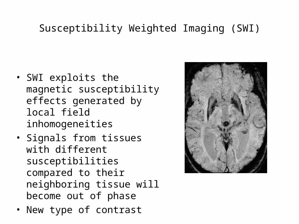
Susceptibility Weighted Imaging (SWI)
• SWI exploits the magnetic susceptibility effects generated by local field inhomogeneities
• Signals from tissues with different susceptibilities compared to their neighboring tissue will become out of phase
• New type of contrast
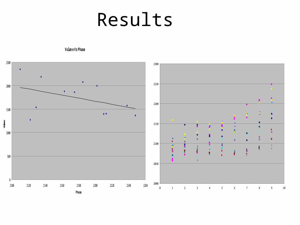
ResultsVolume Vs Phase
0
500
1000
1500
2000
2500
2100 2120 2140 2160 2180 2200 2220 2240 2260
Phase
Volum
e
2000
2050
2100
2150
2200
2250
2300
0 1 2 3 4 5 6 7 8 9 10
PHASE PLOT

Using SWI for noninvasive tracking of SVZ labeled cells in Rat subjected to stroke
• SVZ cells labeled with iron particles were injected in Rat subjected to stroke
• Using SWI as a means to track the cell migration and identify angiogenesis
Related Documents