INTRODUCTION Malignant tumours are the second most often cause of mortality in Western society and the third in the rank of death causes in developing countries, thus bringing a serious threat to public health. The 2005 WHO report shows a rising number of deaths from colon, rectum and prostate cancers; however, mortality from stomach and liver malignancies has been decreasing. Commonly applied chemotherapies are usually linked to severe adverse reactions, thus agents capable of directing the cancer cells to apoptosis would be of great value (1). The aim of the current study was to examine the pro- apoptotic activities of two novel synthetic pyrazoles: 5-(p- toluenesulfonyl)pyrazolo[4,3-f]quinoline (tospyrquin) and 5- chloro-3-(p-toluenesulfonyl)indazole (tosind) in the cultured HT29 colon cancer cell line. Caspase-8 is activated in response to DNA damage which triggers apoptosis via death receptors (2). Mitochondrial apoptotic pathway is initiated by the cytochrome c and other pro-apoptotic proteins release into the cytosol, resulting in caspase-9 activation. Both enzymes then proteolytically decompose procaspase-3 to yield active product which in turn cleaves poly-ADP-ribose polymerase (PARP-1) which is cell death triggering signal (3). Recently, attention has been directed to the role of the Bcl-2 protein family in apoptosis regulation. These proteins, converging on the mitochondria, are divided into three groups depending on the Bcl-2 homology domains (BH1-BH4). Each of the anti- apoptotic proteins in the family, such as Bcl-2 and Bcl-xL, Bcl-w, Mcl-1 or Bcl-B, possesses two or four distinct Bcl-2-homology domains, and their binding groove links the BH3 domain of the pro-apoptotic family members, thus causing their sequestration, preventing induction of the apoptosis cascade and causing cancer cell survival in response to therapy (4, 5). This protein-protein interaction may be a target of small-molecule inhibitors (BH3 mimetics) which sensitises cancer cells to apoptosis, as was shown in the case of ABT-737. Mechanistic studies revealed that ABT- 737 did not directly initiate apoptosis, but enhanced the action of death signals and displayed synergistic cytotoxic effects of chemotherapy and radiation (6). The phenylpyrazole molecule described by Porter et al. (7) was bound to a similar region of the Bcl-2 proteins as ABT-737 and was capable of selectively inhibiting the anti-apoptotic Bcl-2 and Bcl-xL. Cyclic pyrazole derivatives deserve attention due to their great chemical plasticity and broad range of biological activities exerted when applied practically. Cyclic pyrazoles have been used as antiviral/antibacterial (8, 9), neuroleptic, antidepressant, immuno- suppressant and anti-inflammatory remedies (10-12). Similar agents, i.e. anthrapyrazoles, are non-cardiotoxic alternatives to anthracyclines (13) and have found application as chemotherapeutics (14) and photosensitisers in hypoxic cells (15). Recently, several novel heterocyclic pyrazoles have been synthesised, and their anticancer potential has been reported (16, 17). Intercalation into the DNA structure is a common mechanism of cyclic pyrazole anticancer activity which leads to errors in the JOURNAL OF PHYSIOLOGY AND PHARMACOLOGY 2013, 64, 1, 115-123 www.jpp.krakow.pl E. TOTON 1 , E. IGNATOWICZ 2 , M.K. BERNARD 3 , J. KUJAWSKI 3 , M. RYBCZYNSKA 1 EVALUATION OF APOPTOTIC ACTIVITY OF NEW CONDENSED PYRAZOLE DERIVATIVES 1 Department of Clinical Chemistry and Molecular Diagnostics, Poznan University of Medical Sciences, Poznan, Poland; 2 Department of Pharmaceutical Biochemistry, Poznan University of Medical Sciences, Poznan, Poland; 3 Department of Organic Chemistry, Poznan University of Medical Sciences, Poznan, Poland Cyclic pyrazoles exhibit cytotoxicity to human cancer cells through apoptosis induction. We investigated the pro- apoptotic activities of two novel synthetic pyrazoles: 5-(p-toluenesulfonyl)pyrazolo[4,3-f]quinoline (tospyrquin) and 5- chloro-3-(p-toluenesulfonyl)indazole (tosind) in HT29 colon cancer cells which are characterised by point mutation (G/A in codon 273) in the p53 gene, which causes the lack of functionality of the p53 protein. Cell viability was evaluated in the MTT assay, cell morphology was assessed by DAPI staining, flow cytometry was used to study the cell cycle, Western blot techniques were applied for measurements of the Bax, Bcl-2, caspase-8, caspase-9 and PARP-1 proteins and DNA damage was evaluated in the Comet assay. Tospyrquin or tosind in a concentration range of 2.5 μM- 15 μM caused an approximately 20% diminishment in cell growth, but in higher concentrations (25–100 μM) the observed effect depended on the pyrazole structure and time of treatment. In cell cycle analysis, tosind caused 23.7% of apoptotic death and tospyrquin - 14.9%. These data were supported by an increased level of the pro-apoptotic protein Bax, a decreased level of the anti-apoptotic Bcl-2 and enhanced caspase-8, caspase-9, PARP-1 cleavage. DNA damage was dose-dependent for both tested compounds. The results suggest that the pro-apoptotic activity of tospyrquin and tosind is probably regulated by the extrinsic and the intrinsic pathways. Key words: DNA - intercalators, pyrazole derivatives, cytotoxic activity, apoptosis, caspase, poly-ADP-ribose polymerase

Welcome message from author
This document is posted to help you gain knowledge. Please leave a comment to let me know what you think about it! Share it to your friends and learn new things together.
Transcript
INTRODUCTION
Malignant tumours are the second most often cause ofmortality in Western society and the third in the rank of deathcauses in developing countries, thus bringing a serious threat topublic health. The 2005 WHO report shows a rising number ofdeaths from colon, rectum and prostate cancers; however,mortality from stomach and liver malignancies has beendecreasing. Commonly applied chemotherapies are usuallylinked to severe adverse reactions, thus agents capable ofdirecting the cancer cells to apoptosis would be of great value (1).
The aim of the current study was to examine the pro-apoptotic activities of two novel synthetic pyrazoles: 5-(p-toluenesulfonyl)pyrazolo[4,3-f]quinoline (tospyrquin) and 5-chloro-3-(p-toluenesulfonyl)indazole (tosind) in the culturedHT29 colon cancer cell line.
Caspase-8 is activated in response to DNA damage whichtriggers apoptosis via death receptors (2). Mitochondrial apoptoticpathway is initiated by the cytochrome c and other pro-apoptoticproteins release into the cytosol, resulting in caspase-9 activation.Both enzymes then proteolytically decompose procaspase-3 toyield active product which in turn cleaves poly-ADP-ribosepolymerase (PARP-1) which is cell death triggering signal (3).Recently, attention has been directed to the role of the Bcl-2protein family in apoptosis regulation. These proteins, convergingon the mitochondria, are divided into three groups depending onthe Bcl-2 homology domains (BH1-BH4). Each of the anti-
apoptotic proteins in the family, such as Bcl-2 and Bcl-xL, Bcl-w,Mcl-1 or Bcl-B, possesses two or four distinct Bcl-2-homologydomains, and their binding groove links the BH3 domain of thepro-apoptotic family members, thus causing their sequestration,preventing induction of the apoptosis cascade and causing cancercell survival in response to therapy (4, 5). This protein-proteininteraction may be a target of small-molecule inhibitors (BH3mimetics) which sensitises cancer cells to apoptosis, as was shownin the case of ABT-737. Mechanistic studies revealed that ABT-737 did not directly initiate apoptosis, but enhanced the action ofdeath signals and displayed synergistic cytotoxic effects ofchemotherapy and radiation (6). The phenylpyrazole moleculedescribed by Porter et al. (7) was bound to a similar region of theBcl-2 proteins as ABT-737 and was capable of selectivelyinhibiting the anti-apoptotic Bcl-2 and Bcl-xL.
Cyclic pyrazole derivatives deserve attention due to their greatchemical plasticity and broad range of biological activities exertedwhen applied practically. Cyclic pyrazoles have been used asantiviral/antibacterial (8, 9), neuroleptic, antidepressant, immuno-suppressant and anti-inflammatory remedies (10-12). Similaragents, i.e. anthrapyrazoles, are non-cardiotoxic alternatives toanthracyclines (13) and have found application aschemotherapeutics (14) and photosensitisers in hypoxic cells (15).Recently, several novel heterocyclic pyrazoles have beensynthesised, and their anticancer potential has been reported (16,17). Intercalation into the DNA structure is a common mechanismof cyclic pyrazole anticancer activity which leads to errors in the
JOURNAL OF PHYSIOLOGY AND PHARMACOLOGY 2013, 64, 1, 115-123www.jpp.krakow.pl
E. TOTON1, E. IGNATOWICZ2, M.K. BERNARD3, J. KUJAWSKI3, M. RYBCZYNSKA1
EVALUATION OF APOPTOTIC ACTIVITY OF NEW CONDENSED PYRAZOLE DERIVATIVES
1Department of Clinical Chemistry and Molecular Diagnostics, Poznan University of Medical Sciences, Poznan, Poland;2Department of Pharmaceutical Biochemistry, Poznan University of Medical Sciences, Poznan, Poland;
3Department of Organic Chemistry, Poznan University of Medical Sciences, Poznan, Poland
Cyclic pyrazoles exhibit cytotoxicity to human cancer cells through apoptosis induction. We investigated the pro-apoptotic activities of two novel synthetic pyrazoles: 5-(p-toluenesulfonyl)pyrazolo[4,3-f]quinoline (tospyrquin) and 5-chloro-3-(p-toluenesulfonyl)indazole (tosind) in HT29 colon cancer cells which are characterised by point mutation(G/A in codon 273) in the p53 gene, which causes the lack of functionality of the p53 protein. Cell viability wasevaluated in the MTT assay, cell morphology was assessed by DAPI staining, flow cytometry was used to study the cellcycle, Western blot techniques were applied for measurements of the Bax, Bcl-2, caspase-8, caspase-9 and PARP-1proteins and DNA damage was evaluated in the Comet assay. Tospyrquin or tosind in a concentration range of 2.5 µM-15 µM caused an approximately 20% diminishment in cell growth, but in higher concentrations (25–100 µM) theobserved effect depended on the pyrazole structure and time of treatment. In cell cycle analysis, tosind caused 23.7% ofapoptotic death and tospyrquin - 14.9%. These data were supported by an increased level of the pro-apoptotic proteinBax, a decreased level of the anti-apoptotic Bcl-2 and enhanced caspase-8, caspase-9, PARP-1 cleavage. DNA damagewas dose-dependent for both tested compounds. The results suggest that the pro-apoptotic activity of tospyrquin andtosind is probably regulated by the extrinsic and the intrinsic pathways.
K e y w o r d s : DNA - intercalators, pyrazole derivatives, cytotoxic activity, apoptosis, caspase, poly-ADP-ribose polymerase
recognition and functioning of DNA-linked proteins, i.e.polymerases, transcription factors and DNA repair systems, mainlytopoisomerases I and II, and to the subsequent direction of cellsinto the apoptotic pathways (18-20). This mode of action has beencorroborated for pyrazolacridines (21) and anthrapyrazoles (16, 17,19). Anticancer potential has also been found in other groups ofcyclic pyrazoles, e.g. in 2-pyrazoline (22), pyrazole triazine (23)and pyrazole pyrimidine derivatives (24). The latter have beenreported to suppress mitosis through inhibition of tubulinpolymerisation, which results in cell cycle arrest in G2/M andapoptosis induction (25). A 1,5-diarylpyrazole derivative capableof selective COX-2 inhibition and currently applied as an arthritiscure (Celecoxib), has been licensed for use in colon/rectal cancerprevention in patients with familial adenomatous polyposis (26).
MATERIALS AND METHODS
Synthesis of tested compounds
Melting points were determined on a Boetius apparatus and areuncorrected. 1H NMR were recorded at 300 MHz (Varian Mercury300) in DMSO-d6 with tetramethylsilane as an internal standard.Elemental analyses were carried out on a Vario EL III instrument.
6-Nitro-5-(p-toluenesulfonylmethyl)quinoline (27) or 4-chloro-2-(p-toluenesulfonylmethyl) nitrobenzene (28) wasreduced to an appropriate amine (quinamine or benzamine) usingNH2NH2/Raney Ni. Quinamine or benzamine (1 mmol,quinamine: 296 mg; benzamine: 275 mg) was dissolved in aceticacid (quinamine: 3 ml; benzamine: 5 ml) and the solution wasplaced in a pressure tube. 3-Methylbutyl nitrite (3 mmol, 352 mg)was added, the tube was closed and heated in a microwavereactor (Plazmatronika RM800) for 10 or 15 min at 500 W(reaction temperature 100–120 °C) to give 5-(p-toluenesulfonyl)pyrazolo[4,3-f]quinoline (tospyrquin) or 5-chloro-3-(p-toluenesulfonyl)indazole (tosind), respectively. The tube wascooled and the reaction mixture was poured into water. Theresulted mixture was extracted with dichloromethane (2×30 ml),the organic extract was washed three times with water and driedover MgSO4.
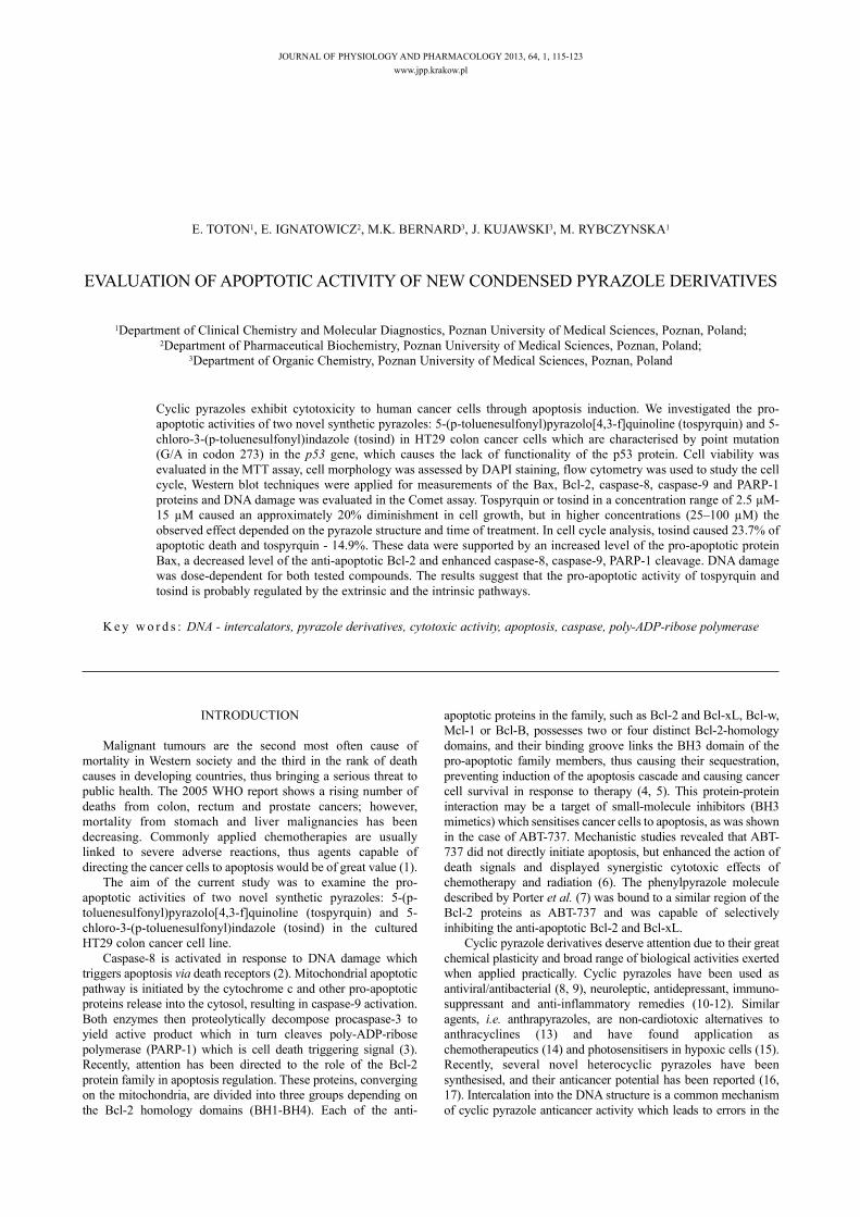
The solvent was removed and the residue was crystallisedfrom ethanol to yield:a) 5-(p-toluenesulfonyl)pyrazolo[4,3-f]quinoline (tospyrquin,Fig. 1A) as yellow needles, mp 238–240°C (decomposition).Yield: 123 mg (38%). 1H NMR, δ 2.39 (s, 1H, CH3), 7.45–7.48(m, 2H, Ts), 7.78 (dd, 3J34 = 8.5 Hz, 3J23 = 4.4 Hz, 1H, 3-H)7.91–7.94 (m, 2H, Ts), 8.02 (s, 2H, 8-H and 9-H), 8.94 (dd, 3J23 =4.4 Hz, 4J24 = 1.6 Hz, 1H, 2-H), 9.41 (dd, 3J34 = 8.5 Hz, 4J24 = 1.6Hz, 1H, 4-H), ~15.0 (bs, 1H, NH, disappeared after addition ofD2O). Anal. Calc. for C17H13N3O2S (323.4): C, 63.13; H, 4.05; N,13.00%. Found: C, 63.48; H, 3.92; N, 12.81%.b) 5-chloro-3-(p-toluenesulfonyl)indazole (tosind, Fig. 1B) ascream needles, mp 247–249°C. Yield: 166 mg (54%). 1H NMR, δ2.41 (s, 1H, CH3), 7.42–7.45 (m, 2H, Ts), 7.53 (dd, 3J67 = 9.0 Hz,4J46 = 2.1 Hz, 1H, 6-H), 7.75 (dd, 3J67 = 9.0 Hz, 5J47 = 0.6 Hz, 1H,7-H), 7.91–7.95 (m, 2H, Ts), 8.04 (dd, 4J46 = 2.1 Hz, 5J47 = 0.6 Hz,1H, 4-H), 14.4 (bs, 1H, NH, disappeared after addition of D2O).
For diverse techniques of biological activity testing we used40 mM stock solutions of tospyrquin and tosind in DMSO. Thefinal concentration of the solvent did not exceed 0.125%.
Cell culture
The human colorectal cancer cell line, HT29, was kindlyprovided by Professor Johann Hofmann (Biocenter, Division ofMedical Biochemistry, Innsbruck Medical University, Austria).The cells were cultured in RPMI 1640 medium containing 10%
heat-inactivated FBS, 2 mM glutamine and 50 g/ml gentamicin(Sigma Chemicals, Munich, Germany). The cultures were grownat 37°C in a humidified incubator with 5% CO2.
Cell viability analysis
Logarithmically growing cells were washed once with PBS(Sigma), trypsinised with Tripsin-EDTA (Sigma) andresuspended in a 10 ml volume of the growth medium. Then theywere seeded at a density of 3×10⁴ per well in 96-wellmicroplates. After overnight incubation to reach sedimentationin standard culture conditions, the cells were treated with variousconcentrations (2.5–100 µM) of tospyrquin or tosind, or 0.125%DMSO. After 24 , 48 and 72 hours of incubation, an MTTsolution was added to each well (the final concentration was 0.5mg/ml), and culture was continued in standard conditions foranother 4 hours. Then the cells were solubilised overnight with10% SDS in 0.01 M HCl and the obtained formazan wasquantified spectrophotometrically using a Labsystems MultiscanRC spectrophotometer. Absorbance was recorded at 570 nm (testwavelength) and 690 nm (reference wavelength). Average valuesof IC50 were calculated from four experiments run in duplicate.Viability of the cells was calculated with Excel software, whileIC50 was calculated with CalculSyn V1.1.
Flow cytometry analysis
HT29 cells (3×10⁵/ml) were incubated overnight to reachsedimentation and then were treated with tospyrquin (110 µM),tosind (90 µM), or CPT (25 µM). Cell cycle analysis wasperformed after incubation for 24 hours. Cells were collectedusing 0.25% trypsin (Sigma, Munich, Germany) and cell pelletswere resuspended in 100 µl of PBS containing 250 µg/mlpropidium iodide, 1% Triton X-100 and 25 µl ribonuclease A (10mg/ml; Sigma, Munich, Germany). Flow cytometry analysis wasperformed (FacSCAN, Becton-Dickinson, Franklin Lakes, NJ)after incubation in the dark at room temperature for 1 hour.
Western blot analysis
Confluent HT29 monolayers were incubated with tospyrquin(37–110 µM) or tosind (30–90 µM ), or 25 µM CPT for 24 hours.Whole-cell extracts were prepared with lysis buffer containing 50mM Tris-HCl (pH 7.5), 150 mM NaCl, 2 mM EDTA, 1 mMEGTA, 1% Triton X-100, 100 mM PMSF, 25 g/ml Na3VO4, 25µg/ml NaF, 25 µg/ml leupeptin and 25 µg/ml aprotinin, aspreviously described (29). The protein concentration wasmeasured with the Bradford assay (Sigma, Munich, Germany)and 30–60 µg of each extract was loaded onto (10–12%) SDS-PAGE ready gels (BioRad, Hercules, CA). Western blotting wasperformed by standard procedure using a PVDF membrane(Pierce Biotechnology, Rockford, USA). For the detection ofproteins the following antibodies were used: rabbit anti-Bax,rabbit anti-Bcl-2, rabbit anti-PARP-1, rabbit anti-GAPDH (fromSanta Cruz Biotechnology INC, concentration of antibodies was1 µg/ml) and mouse anti-Caspase-8, rabbit anti-Caspase-9 (fromCell Signaling Technology INC, concentration of antibodies was1 µg/ml). The proteins were visualised using SuperSignal® WestPico Chemiluminescent Substrate and CL-X PosureTM film(Pierce Biotechnology, Rockford, USA). LabWorks software(UVP, Upland, CA) was used for measurement of the opticaldensity (Arbitrary Units) of the bands.
Morphological analysis of apoptotic cells
The study was carried out in 6-well microplates with a glasscoverslide. The HT29 cells (3×10⁴/ml) were incubated overnight
116
to reach sedimentation and then were treated with tospyrquin (74µM), tosind (60 µM), or CPT (25 µM) for 24 and 48 hours. Afterthis time period the cells were washed with PBS, fixed with3.7% paraformaldehyde and permeabilised with 0.1% Triton X-100. The cells were incubated for 5 min with DAPI (the finalconcentration was 250 ng/ml) in the dark. The stained cells wereimaged with a fluorescent microscope - Axiostar Plus (Zeiss,Germany). The percentage of apoptotic cells was calculated asthe ratio of apoptotic cells to total cells counted, and a minimumof 100 cells/field and at least 3 fields in each well were counted.
Comet assay
Confluent monolayers of HT29 cells were treated for 24 hourswith either tospyrquin (37, 74, 110 µM) or tosind (30, 60, 90 µM),or CPT (25 µM). The cells were then trypsinised and rinsed twicewith the culture medium. Comet assay in alkaline conditions(pH>13) was performed in suspended cells according to themethod presented by Hartmann et al. (30). Samples embedded in
LMP agarose were submitted to cell lysis, DNA unwinding,electrophoresis and neutralisation and then were dehydrated inabsolute ethanol, dried and stored in room temperature, andprotected from light. Immediately before microscopic evaluationthe slides were rehydrated and stained with ethidium bromide(0.05 mg/ml). Images of the comets were captured with a digitalcamera. The comets were divided into 5 groups according to thedegree of DNA damage (31). A total damage score for eachsample on the slide was calculated by multiplying the number ofcells classified to each grade of damage by the numeric value ofthe grade and summing over all grades. For each sample 100comets were scored in two separate slides.
Statistical analysis
Statistical analysis was performed by one-way ANOVA(GraphPad Prism, San Diego, CA). The statistical significancebetween the experimental groups and control was assessed bythe t-Student test (Tukey's Multiple Comparison Test), with *
117
Fig. 1. Structure and 1H NMR data of the tested cyclic pyrazoles: (A) 5-(p-toluenesulfonyl)pyrazolo[4,3-f]quinoline (tospyrquin);(B) 5-chloro-3-(p-toluenesulfonyl)indazole (tosind).
NNH
S
O
O CH3
Cl
5-chloro-3-(p-toluenesulfonyl)indazole (tosind)B)
N
NHNS
O
O
CH3
5-(p-toluenesulfonyl)pyrazolo[4,3-f]quinoline (tospyrquin)A)
p<0.05. The correlation coefficient between the dose andBax/Bcl-2 proteins was Pearson's r2.
RESULTS
Investigated pyrazole derivatives
Tospyrquin and tosind were obtained by a three-step synthesisincluding vicarious nucleophilic substitution of hydrogen ortho tothe nitro group, catalytic hydrogenation of the nitro group, anddiazotizatio-cyclization leading to a pyrazole ring fused with theexisting quinoline or benzene moiety (Fig. 1A and 1B).
Cytotoxicity of tospyrquin and tosind in HT29 cells
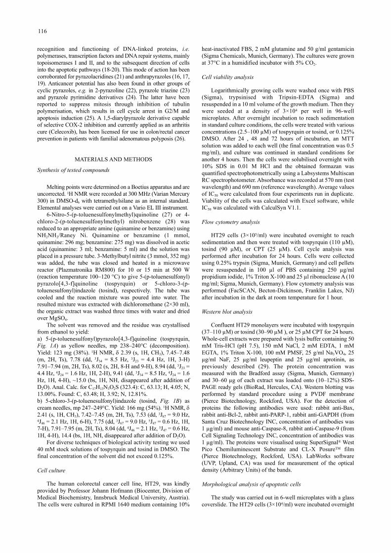
The viability of cells after 24, 48 and 72 h treatment with abroad concentration range (2.5–100 µM) of tospyrquin or tosindwas assessed with the MTT test. The pyrazoles were dissolved inDMSO and the final solvent concentration (0.125%) did notaffect cell viability. The obtained results are expressed as apercentage of viability stated in the untreated cells and arepresented in Fig. 2A and 2B. Both pyrazoles tested in theconcentration range of 2.5–15 µM caused an approximately 20%reduction in cell growth; however, in higher concentrations(25–100 µM) the observed effect depended on the pyrazolestructure and treatment time. After 72 h treatment, the IC50 valueshowed that tosind showed significantly greater (p=0.0104)cytotoxic activity towards HT29 cells than tospyrquin (Fig. 2C).
Tospyrquin and tosind induce apoptosis in HT29 cells
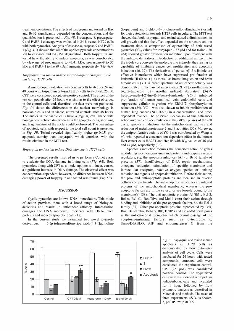
We performed flow cytometry analysis in order to determinewhether the tested pyrazoles could affect the cell cycle andinduce apoptosis in HT29 cells. As concentrations equal to oneor two multiplications of the respective IC50 values did not exertan apoptotic effect (data not shown), we continued experimentswith three multiplications of IC50 (tospyrquin - 110 µM andtosind - 90 µM). The results after 24 h treatment of cells areshown in Fig. 3. Apoptosis-positive control were cells treatedwith 25 µM CPT. Untreated HT29 cells in the G0/G1, S andG2/M phases were considered 100% (control). We observed thatthe pro-apoptotic effect depended on the structure. Tospyrquincaused 14.9% apoptotic cell death, which was similar to theeffect observed in the control cells, whereas tosind had higheractivity and caused 23.7% apoptotic cell death (p<0.05).However, neither tospyrquin nor tosind inhibited the cell cycle,as no accumulation of cells was found in any of the cycle phases.
Tospyrquin and tosind induce apoptosis in HT29 by functionalprotein modulation
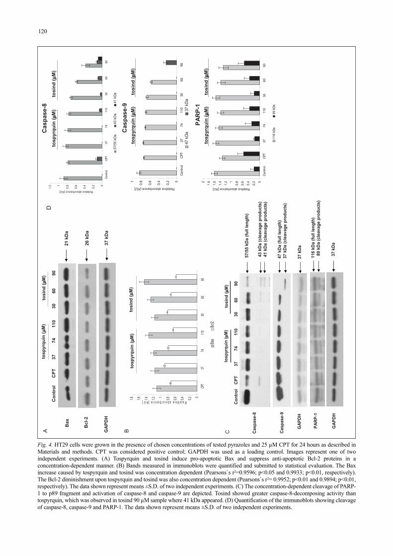
To elucidate the mechanism of apoptosis induction by thetested pyrazoles, we analysed expression of the pro- and anti-apoptotic proteins (Fig. 4A). In cells treated for 24 hours withincreasing concentrations of both pyrazoles (1, 2 and 3multiplications of the respective IC50 values), a rise in the level ofthe pro-apoptotic Bax protein was found. On the contrary, adecrease of the anti-apoptotic Bcl-2 was noticed under the same
118
A B
0
20
40
60
80
100
120
0 25 50 75 100
tosind [uM]
Rel
ativ
e vi
abili
ty [%
]
72 h48 h24 h
0
20
40
60
80
100
120
0 25 50 75 100
tospyrquin [uM]
Rel
ativ
e vi
abili
ty [%
]
72 h48 h24 h
C)
29,9 ± 3,5tosind
37,3 ± 2,0tospyrquin
IC50 ± SD (µM) from 72 h treatmentCompound
Fig. 2. Cytotoxicity of tospyrquin (A) and tosind (B) in HT29 cells. Cells were treated either with tospyrquin or with tosind for 24, 48, and72 hours in concentration range from 2.5 to 100 µM as described in Materials and methods. Viability was quantified spectrophotometricallyby the MTT assay. Percentage of cell viability was expressed in relation to untreated control cells. The applied DMSO concentration(0.125%) had no effect on control cells. Means±S.D. from four independent experiments run in duplicate are shown. (C) Cytotoxicefficacies of tospyrquin and tosind on the HT29 cells. IC50 denotes tospyrquin and tosind concentrations that result in a 50% decrease inthe cell number compared to non-treated controls and were derived from the 72 h treatment. Results from four independent experimentsare presented.
treatment conditions. The effects of tospyrquin and tosind on Baxand Bcl-2 significantly depended on the concentration, and thequantification is presented in Fig. 4B. Procaspase-8, procaspase-9 and PARP-1 cleavage was evaluated in 24-h-treated HT29 cellswith both pyrazoles. Analysis of caspase-8, caspase-9 and PARP-1 (Fig. 4C) showed that all of the applied pyrazole concentrationsled to caspases and PARP-1 degradation. Both tospyrquin andtosind have the ability to induce apoptosis, as was corroboratedby cleavage of procaspase-8 to 43/41 kDa, procaspase-9 to 37kDa and PARP-1 to the 89 kDa fragments, respectively (Fig. 4D).
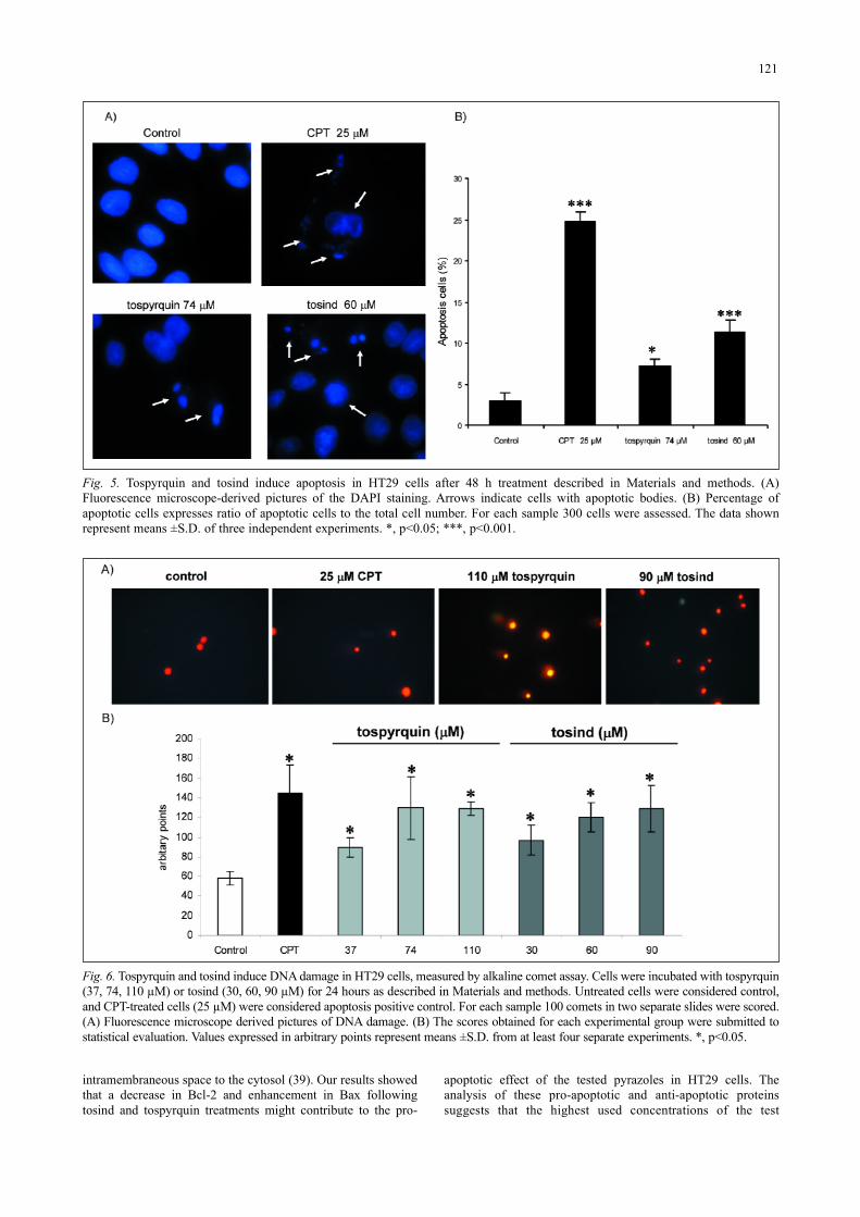
Tospyrquin and tosind induce morphological changes in thenuclei of HT29 cells
A microscopic evaluation was done in cells treated for 24 and48 hours with tospyrquin or tosind. HT29 cells treated with 25 µMCPT were considered apoptosis-positive control. The effect of thetest compounds after 24 hours was similar to the effect observedin the control cells and, therefore, the data were not published.Fig. 5A shows the differences in the nuclear morphology innonviable cells and in viable, untreated controls after 48 hours.The nuclei in the viable cells have a regular, oval shape withhomogeneous chromatin, whereas in the apoptotic cells, shrinkingand fragmentation of the nuclei could be observed. The percentageof apoptotic cells with respect to the total cell count is presentedin Fig. 5B. Tosind revealed significantly higher (p<0.05) pro-apoptotic activity than tospyrquin, which correlates with theresults obtained in the MTT test.
Tospyrquin and tosind induce DNA damage in HT29 cells
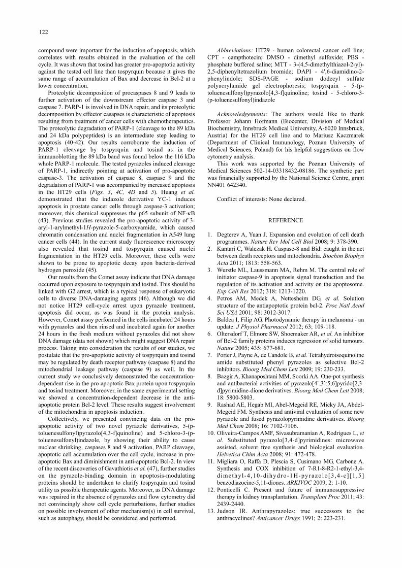
The presented results inspired us to perform a Comet assayto evaluate the DNA damage in living cells (Fig. 6A). Bothpyrazoles, along with CPT as a model apoptosis inducer, causeda significant increase in DNA damage. The observed effect wasconcentration-dependent, however, no difference between DNA-damaging power of tospyrquin and tosind was found (Fig. 6B).
DISCUSSION
Cyclic pyrazoles are known DNA intercalators. This modeof action provides them with a broad range of biologicalactivities and results in anticancer efficacy. Intercalationdamages the DNA molecule, interferes with DNA-linkedproteins and induces apoptotic death (18).
In the current study we examined two novel pyrazolederivatives, 5-(p-toluenesulfonyl)pyrazolo[4,3-f]quinoline
(tospyrquin) and 5-chloro-3-(p-toluenesulfonyl)indazole (tosind)for their cytotoxicity towards HT29 cells in culture. The MTT testshowed that both tospyrquin and tosind caused a diminishment incell growth and that the effect depended on the structure and ontreatment time. A comparison of cytotoxicity of both testedpyrazoles (IC50 values for tospyrquin - 37 µM and for tosind - 30µM) showed greater proliferation inhibition upon treatment withthe indazole derivatives. Introduction of additional nitrogen intothe indole core converts the molecule into indazole, thus raising itscapability of inhibiting cancer cell proliferation and apoptosisinduction (16, 32). The derivatives of pyrazole[1,5-α]indole areeffective intercalators which have suppressed proliferation ofleukemic HL60 cells (16) as well as breast, lung, colon and braintumour cells (33). A broad spectrum of anticancer activity wasdemonstrated in the case of intercalating 2H-[1]benzothiopyrano[4,3,2-]indazole (32). Another indazole derivative, [3-(5'-hydroxymethyl-2'-furyl)-1-benzyl-indazole, YC-1] inhibitedproliferation of human umbilical vein endothelial cells andsuppressed cellular migration via ERK1/2 phosphorylationreduction (34). YC-1 was also shown to inhibit proliferation ofhuman lung cancer (NCI-H226) in a concentration- and time-dependent manner. The observed mechanism of this anticanceraction involved cell accumulation in the G0/G1 phases of the cellcycle, apoptosis induction via the mitochondrial pathway andreduction of metalloproteinase 2 and 9 activities (35). Moreover,the antiproliferative activity of YC-1 was corroborated by Wang etal., who reported a concentration-dependent effect in the humanliver cancer cells HA22T and Hep3B with IC50 values of 46 µMand 47 µM, respectively (36).
Apoptosis induction requires the concerted action of genesmodulating receptors, enzymes and proteins and caspase cascaderegulators, e.g. the apoptosis inhibitor (IAP) or Bcl-2 family ofproteins (37). Insufficiency of DNA repair mechanisms,oncogene activation, stimulation of specific membrane andintracellular receptors, reactive oxygen species or ionisingradiation are signals of apoptosis initiation. Before their action,the pro- and anti-apoptotic proteins are localised in diversecellular compartments. The anti-apoptotic molecules are integralproteins of the mitochondrial membrane, whereas the pro-apoptotic factors are in the cytosol or are loosely bound to themembrane(s) (38). The anti-apoptotic proteins A1/Bfl1, Bcl-2,Bcl-w, Bcl-xL, Boo/Diva and Mcl-1 exert their action throughbinding and inhibition of the pro-apoptotic factors, i.e. the Bcl-2family (37). Other pro-apoptotic proteins represented by Bak,Bax, Bcl-rambo, Bcl-xS, Blk, BNIP3 and Bok/Mtd form poresin the mitochondrial membrane which permit passage of theapoptosis-initiating factors such as cytochrome c,Smac/DIABLO, AIF and endonucleases G from the
119
0
10
20
30
40
50
60
70
Control CPT 25uM tospy rquin 110 uM tosind 90 uM
% c
ells
G0/G1
S
G2/M
Apoptosis∗∗∗∗
∗∗∗∗∗∗∗∗
Fig. 3. Tospyrquin and tosind induceapoptosis in HT29 cells asdemonstrated by flow cytometryanalysis of cell cycle. Cells wereincubated for 24 hours with testedcompounds, untreated cells wereconsidered the experiment control.CPT (25 µM) was consideredpositive control. The trypsinizedcells were resuspended in propidiumiodide/ribonuclease and incubatedfor 1 hour, followed by flowcytometry analysis as described inMaterials and methods. The mean ofthree experiments ±S.D. is shown.*, p<0.05, **, p<0.005.
120
Fig. 4. HT29 cells were grown in the presence of chosen concentrations of tested pyrazoles and 25 µM CPT for 24 hours as described inMaterials and methods. CPT was considered positive control; GAPDH was used as a loading control. Images represent one of twoindependent experiments. (A) Tospyrquin and tosind induce pro-apoptotic Bax and suppress anti-apoptotic Bcl-2 proteins in aconcentration-dependent manner. (B) Bands measured in immunoblots were quantified and submitted to statistical evaluation. The Baxincrease caused by tospyrquin and tosind was concentration dependent (Pearsons`s r2=0.9596; p<0.05 and 0.9933; p<0.01, respectively).The Bcl-2 diminishment upon tospyrquin and tosind was also concentration dependent (Pearsons`s r2= 0.9952; p<0.01 and 0.9894; p<0.01,respectively). The data shown represent means ±S.D. of two independent experiments. (C) The concentration-dependent cleavage of PARP-1 to p89 fragment and activation of caspase-8 and caspase-9 are depicted. Tosind showed greater caspase-8-decomposing activity thantospyrquin, which was observed in tosind 90 µM sample where 41 kDa appeared. (D) Quantification of the immunoblots showing cleavageof caspase-8, caspase-9 and PARP-1. The data shown represent means ±S.D. of two independent experiments.
D
0
0,2
0,4
0,6
0,81
1,2
Control
CPT
3774
110
3060
90
Relative absorbance [AU]
57/5
5 kD
a43
kDa
41 k
Datosind
(µ µµµM)
tospyrquin(µ µµµM)
Caspase-8
0
0,2
0,4
0,6
0,81
Control
CPT
3774
110
3060
90
Relative absorbance [AU]
47 k
Da37
kDa
Caspase-9
tosind
(µ µµµM)
tospyrquin(µ µµµM)
0
0,2
0,4
0,6
0,81
1,2
1,4
1,6
1,82
Control
CPT
3774
110
3060
90
Relative absorbance [AU]
116
kDa
89 k
Da
PARP-1
tosind
(µ µµµM)
tospyrquin(µ µµµM)
C
47 k
Da(fu
llle
ngth
)37
kDa
(cle
avag
epr
oduc
ts)
43 k
Da(c
leav
age
prod
ucts
)41
kDa
(cle
avag
epr
oduc
ts)
GAPDH
37 k
Da
tosp
yrqu
in(µ µµµM)
tosind
(µ µµµM)
Cont
rol
CPT
37
74
110
3
0
6
0
90
PARP-1
116
kDa
(full
leng
th)
89 k
Da(c
leav
age
prod
ucts
)
GAPDH
37 k
Da
Caspase-9
Caspase-8
57/5
5 kD
a(fu
llle
ngth
)
00,20,40,60,811,21,41,61,8
CPT
3774
110
3060
90
Realitive absorbance [AU]
Bax
Bcl2
26 k
Da
Bcl
-2
Bax
21 k
Da
tosp
yrqu
in(µ µµµM)
tosind
(µ µµµM)
Con
trol
CPT
37
74
110
30
60
90
GAPDH
37 k
Da
A Btospyrquin(µ µµµM)
tosind
( µ µµµM)
intramembraneous space to the cytosol (39). Our results showedthat a decrease in Bcl-2 and enhancement in Bax followingtosind and tospyrquin treatments might contribute to the pro-
apoptotic effect of the tested pyrazoles in HT29 cells. Theanalysis of these pro-apoptotic and anti-apoptotic proteinssuggests that the highest used concentrations of the test
121
Fig. 6. Tospyrquin and tosind induce DNA damage in HT29 cells, measured by alkaline comet assay. Cells were incubated with tospyrquin(37, 74, 110 µM) or tosind (30, 60, 90 µM) for 24 hours as described in Materials and methods. Untreated cells were considered control,and CPT-treated cells (25 µM) were considered apoptosis positive control. For each sample 100 comets in two separate slides were scored.(A) Fluorescence microscope derived pictures of DNA damage. (B) The scores obtained for each experimental group were submitted tostatistical evaluation. Values expressed in arbitrary points represent means ±S.D. from at least four separate experiments. *, p<0.05.
Fig. 5. Tospyrquin and tosind induce apoptosis in HT29 cells after 48 h treatment described in Materials and methods. (A)Fluorescence microscope-derived pictures of the DAPI staining. Arrows indicate cells with apoptotic bodies. (B) Percentage ofapoptotic cells expresses ratio of apoptotic cells to the total cell number. For each sample 300 cells were assessed. The data shownrepresent means ±S.D. of three independent experiments. *, p<0.05; ***, p<0.001.
compound were important for the induction of apoptosis, whichcorrelates with results obtained in the evaluation of the cellcycle. It was shown that tosind has greater pro-apoptotic activityagainst the tested cell line than tospyrquin because it gives thesame range of accumulation of Bax and decrease in Bcl-2 at alower concentration.
Proteolytic decomposition of procaspases 8 and 9 leads tofurther activation of the downstream effector caspase 3 andcaspase 7. PARP-1 is involved in DNA repair, and its proteolyticdecomposition by effector casapses is characteristic of apoptosisresulting from treatment of cancer cells with chemotherapeutics.The proteolytic degradation of PARP-1 (cleavage to the 89 kDaand 24 kDa polypeptides) is an intermediate step leading toapoptosis (40-42). Our results corroborate the induction ofPARP-1 cleavage by tospyrquin and tosind as in theimmunoblotting the 89 kDa band was found below the 116 kDawhole PARP-1 molecule. The tested pyrazoles induced cleavageof PARP-1, indirectly pointing at activation of pro-apoptoticcaspase-3. The activation of caspase 8, caspase 9 and thedegradation of PARP-1 was accompanied by increased apoptosisin the HT29 cells (Figs. 3, 4C, 4D and 5). Huang et al.demonstrated that the indazole derivative YC-1 inducesapoptosis in prostate cancer cells through caspase-3 activation;moreover, this chemical suppresses the p65 subunit of NF-κB(43). Previous studies revealed the pro-apoptotic activity of 3-aryl-1-arylmethyl-1H-pyrazole-5-carboxyamide, which causedchromatin condensation and nuclei fragmentation in A549 lungcancer cells (44). In the current study fluorescence microscopyalso revealed that tosind and tospyrquin caused nucleifragmentation in the HT29 cells. Moreover, these cells wereshown to be prone to apoptotic decay upon bacteria-derivedhydrogen peroxide (45).
Our results from the Comet assay indicate that DNA damageoccurred upon exposure to tospyrquin and tosind. This should belinked with G2 arrest, which is a typical response of eukaryoticcells to diverse DNA-damaging agents (46). Although we didnot notice HT29 cell-cycle arrest upon pyrazole treatment,apoptosis did occur, as was found in the protein analysis.However, Comet assay performed in the cells incubated 24 hourswith pyrazoles and then rinsed and incubated again for another24 hours in the fresh medium without pyrazoles did not showDNA damage (data not shown) which might suggest DNA repairprocess. Taking into consideration the results of our studies, wepostulate that the pro-apoptotic activity of tospyrquin and tosindmay be regulated by death receptor pathway (caspase 8) and themitochondrial leakage pathway (caspase 9) as well. In thecurrent study we conclusively demonstrated the concentration-dependent rise in the pro-apoptotic Bax protein upon tospyrquinand tosind treatment. Moreover, in the same experimental settingwe showed a concentration-dependent decrease in the anti-apoptotic protein Bcl-2 level. These results suggest involvementof the mitochondria in apoptosis induction.
Collectively, we presented convincing data on the pro-apoptotic activity of two novel pyrazole derivatives, 5-(p-toluenesulfonyl)pyrazolo[4,3-f]quinoline) and 5-chloro-3-(p-toluenesulfonyl)indazole, by showing their ability to causenuclear shrinking, caspases 8 and 9 activation, PARP cleavage,apoptotic cell accumulation over the cell cycle, increase in pro-apoptotic Bax and diminishment in anti-apoptotic Bcl-2. In viewof the recent discoveries of Gavathiotis et al. (47), further studieson the pyrazole-binding domain in apoptosis-modulatingproteins should be undertaken to clarify tospyrquin and tosindutility as possible therapeutic agents. Moreover, as DNA damagewas repaired in the absence of pyrazoles and flow cytometry didnot convincingly show cell cycle perturbations, further studieson possible involvement of other mechanism(s) in cell survival,such as autophagy, should be considered and performed.
Abbreviations: HT29 - human colorectal cancer cell line;CPT - campthotecin; DMSO - dimethyl sulfoxide; PBS -phosphate buffered saline; MTT - 3-(4,5-dimethylthiazol-2-yl)-2,5-diphenyltetrazolium bromide; DAPI - 4',6-diamidino-2-phenylindole; SDS-PAGE - sodium dodecyl sulfatepolyacrylamide gel electrophoresis; tospyrquin - 5-(p-toluenesulfonyl)pyrazolo[4,3-f]quinoline; tosind - 5-chloro-3-(p-toluenesulfonyl)indazole
Acknowledgements: The authors would like to thankProfessor Johann Hofmann (Biocenter, Division of MedicalBiochemistry, Innsbruck Medical University, A-6020 Innsbruck,Austria) for the HT29 cell line and to Mariusz Kaczmarek(Department of Clinical Immunology, Poznan University ofMedical Sciences, Poland) for his helpful suggestions on flowcytometry analysis.
This work was supported by the Poznan University ofMedical Sciences 502-14-03318432-08186. The synthetic partwas financially supported by the National Science Centre, grantNN401 642340.
Conflict of interests: None declared.
REFERENCE
1. Degterev A, Yuan J. Expansion and evolution of cell deathprogrammes. Nature Rev Mol Cell Biol 2008; 9: 378-390.
2. Kantari C, Walczak H. Caspase-8 and Bid: caught in the actbetween death receptors and mitochondria. Biochim BiophysActa 2011; 1813: 558-563.
3. Wurstle ML, Laussmann MA, Rehm M. The central role ofinitiator caspase-9 in apoptosis signal transduction and theregulation of its activation and activity on the apoptosome.Exp Cell Res 2012; 318: 1213-1220.
4. Petros AM, Medek A, Nettesheim DG, et al. Solutionstructure of the antiapoptotic protein bcl-2. Proc Natl AcadSci USA 2001; 98: 3012-3017.
5. Baldea I, Filip AG. Photodynamic therapy in melanoma - anupdate. J Physiol Pharmacol 2012; 63; 109-118.
6. Oltersdorf T, Elmore SW, Shoemaker AR, et al. An inhibitorof Bcl-2 family proteins induces regression of solid tumours.Nature 2005; 435: 677-681.
7. Porter J, Payne A, de Candole B, et al. Tetrahydroisoquinolineamide substituted phenyl pyrazoles as selective Bcl-2inhibitors. Bioorg Med Chem Lett 2009; 19: 230-233.
8. Bazgir A, Khanaposhtani MM, Soorki AA. One-pot synthesisand antibacterial activities of pyrazolo[4`,3`:5,6]pyrido[2,3-d]pyrimidine-dione derivatives. Bioorg Med Chem Lett 2008;18: 5800-5803.
9. Rashad AE, Hegab MI, Abel-Megeid RE, Micky JA, Abdel-Megeid FM. Synthesis and antiviral evaluation of some newpyrazole and fused pyrazolopyrimidine derivatives. BioorgMed Chem 2008; 16: 7102-7106.
10. Oliveira-Campos AMF, Sivasubramanian A, Rodrigues L, etal. Substituted pyrazolo[3,4-d]pyrimidines: microwaveassisted, solvent free synthesis and biological evaluation.Helvetica Chim Acta 2008; 91: 472-478.
11. Migliara O, Raffa D, Plescia S, Cusimano MG, Carbone A.Synthesis and COX inhibition of 7-R1-8-R2-1-ethyl-3,4-d imeth y l -4 ,10-d ih ydro-1 H-pyrazo lo[3 ,4-c ] [1 ,5 ]benzodiazocine-5,11-diones. ARKIVOC 2009; 2: 1-10.
12. Ponticelli C. Present and future of immunosuppressivetherapy in kidney transplantation. Transplant Proc 2011; 43:2439-2440.
13. Judson IR. Anthrapyrazoles: true successors to theanthracyclines? Anticancer Drugs 1991; 2: 223-231.
122
14. Sekine I, Fukuda H, Kunitoh H, Saijo N. Cancer chemotherapyin the elderly. Jpn J Clin Oncol 1998; 28: 463-473.
15. Reszka K, Hartley JA, Lown JW. Photosensitization byselected anticancer agents. Biophys Chem 1990; 35: 313-323.
16. Umemura K, Mizushima T, Katayama M, Kiryu Y, Yamoro T,Andoh T. Inhibition of DNA topoisomerases II and/or I bypyrazolo[1,5-?]indole derivatives and their growth inhibitoryactivities. Mol Pharmacol 2002; 62: 873-880.
17. Hasinoff BB, Liang H, Wu X, et al. The structure-baseddesign, synthesis and biological evaluation of DNA-bindingbisintercalating bisanthrapyrazole anticancer compounds.Bioorg Med Chem 2008; 16: 3959-3968.
18. Palchaudhuri R, Hergenrother PJ. DNA as a target foranticancer compounds: methods to determine the mode ofbinding and the mechanism of action. Curr Opin Biotechnol2007; 18: 497-503.
19. Shaw AY, Liau HH, Lu PJ, et al. 3,5-Diaryl-1H-pyrazole asa molecular scaffold for the synthesis of apoptosis-inducingagents. Bioorg Med Chem 2010; 18: 3270-3278.
20. Martinez R, Chacon-Garcia L. The search of DNA-intercalators as antitumoral drugs: what it worked and whatdid not work. Curr Med Chem 2005; 12: 127-151.
21. Adjei AA. Current status of pyrazoloacridine as ananticancer agent. Invest New Drugs 1999; 17: 43-48.
22. Dipankar B, Hirakmoy C, Asish B, Abhijit C. 2-Pyrazoline:a pharmacologically active moiety. Int Res J Pharm App Sci2011; 1: 68-80.
23. Pevarello P, Brasca MG, Amici R, et al. 3-Aminopyrazoleinhibitors of CDK2/cyclin A as antitumor agents. 1. LeadFinding. J Med Chem 2004; 47: 3367-3380.
24. Spreafico A, Schenone S, Serchi T, et al. Antiproliferativeand proapoptotic activities of new pyrazolo[3,4-d]pyrimidine derivative Src kinase. FASEB J 2008; 22:1560-1571.
25. Naito H, Ohsuki S, Atsumi R, et al. Synthesis and antitumoractivity of novel pyrimidinyl pyrazole derivatives. III.Synthesis and antitumor activity of 3-phenylpiperazinyl-1-trans-propenes. Chem Pharm Bull 2005; 53: 153-163.
26. Bertagnolli M, Eagle C, Zauber A, et al. Celecoxib for theprevention of sporadic colorectal adenomas. New Eng J Med2006; 355: 873-884.
27. Makosza M, Kinowski A, Danikiewicz W, Mudryk B.Vicarious nucleophilic substitution in nitroquinolines1.Liebigs Ann Chem 1986; 1: 69-77.
28. Wojciechowski K, Makosza M. A facile synthesis of 3-sulfonyl-substituted indole derivatives. Synthesis 1986; 8:651-653.
29. Garczarczyk D, Toton E, Biedermann V, et al. Signaltransduction of constitutively active protein kinase Cepsilon. Cell Signal 2009; 21: 745-752.
30. Hartmann A, Agurell E, Beevers C, et al. Recommendationsfor conducting the in vivo alkaline comet assay. Mutagenesis2003; 18: 45-51.
31. Collins AR. The comet assay for DNA damage and repair:principles, applications, and limitations. Mol Biotechnol2004; 26: 249-261.
32. Dees EC, Whitfield LR, Grove WR, Rummel S, GrochowLB, Donehower RC. A phase I and pharmacologic evaluationof the DNA intercalator CI-958 in patients with advancedsolid tumors. Clin Cancer Res 2000; 6: 3885-3894.
33. Yamori T, Matsunaga A, Sato S, et al. Potent antitumoractivity of MS-247, a novel DNA minor groove binder,evaluated by an in vitro and in vivo human cancer cell linepanel. Cancer Res 1999; 59: 4042-4049.
34. Pan SL, Guh JH, Peng CY, et al. YC-1 [3-(5'-hydroxymethyl-2'-furyl)-1-benzyl indazole] inhibitsendothelial cell functions induced by angiogenic factors invitro and angiogenesis in vivo models. J Pharmacol ExpTher 2005; 31: 35-42.
35. Chen CJ, Hsu MH, Huang LJ, et al. Anticancer mechanismsof YC-1 in human lung cancer cell line, NCI-H226. BiochemPharmacol 2008; 75: 360-368.
36. Wang SW, Pan SL, Guh JH, et al. YC-1 [3-(5'-hydroxymethyl-2'-furyl)-1-benzyl indazole] exhibits a novel antiproliferativeeffect and arrests the cell cycle in G0-G1 in humanhepatocellular carcinoma cells. J Pharmacol Exp Ther 2005;312: 917-925.
37. Fan T, Han L, Cong R, Liang J. Caspase family proteasesand apoptosis. Acta Biochim Biophys Sin 2005; 11: 719-727.
38. Gross A, McDonnell JM, Korsmeyer SJ. BCL-2 familymembers and the mitochondria in apoptosis. Genes Dev1999; 13: 1899-1911.
39. Hossini AM, Eberle J. Apoptosis induction by Bcl-2 proteinsindependent of the BH3 domain. Biochem Pharmacol 2008;76: 1612-1619.
40. Kaufmann FC, Desnoyer S, Taviano Y, Davison NE, PoirierGG. Specific cleavage of PARP: an early marker ofchemotherapy-induced apoptosis. Cancer Res 1993; 53:3976-3985.
41. Duriez PJ, Shah GM. Cleavage of poly(ADP-ribose)polymerase: a sensitive parameter to study cell death.Biochem Cell Biol 1997; 75: 337-349.
42. Pierzchalski P, Krawiec A, Gawelko J, Pawlik WW,Konturek SJ, Gonciarz M. Molecular mechanism ofprotection against chemically and γ-radiation inducedapoptosis in human colon cancer calls. J Physiol Pharmacol2008; 59: 191-202.
43. Huang YT, Pan SL, Guh JH, et al. YC-1 suppressesconstitutive nuclear factor-κB activation and inducesapoptosis in human prostate cancer cells. Mol Cancer Ther2005; 4: 1628-1635.
44. Ding XL, Zhang HY, Qi L, Zhao BX, Lv HS, Miao JY.Synthesis of novel pyrazole carboxamide derivatives anddiscovery of modulators for apoptosis or autophagy in A549lung cancer cells. Bioorg Med Chem Lett 2009; 19: 5325-5328.
45. Strus M, Janczyk A, Gonet-Surowka A, et al. Effect ofhydrogen peroxide of bacterial origin on apoptosis andnecrosis of gut mucosa epithelial cells as a possiblepathomechanism of inflammatory bowel disease and cancer.J Physiol Pharmacol 2009; 60: 55-60.
46. Han Z, Chatterjee D, He DM, et al. Evidence for a G2checkpoint in p53-independent apoptosis induction by X-irradiation. Mol Cell Biol 1995; 15: 5849-5857.
47. Gavathiotis E, Reyna DE, Bellairs JA, Leshchiner ES,Walensky LD. Direct and selective small-molecule activationof proapoptotic BAX. Nat Chem Biol 2012; 8; 639-645.
R e c e i v e d : Novenber 6, 2012A c c e p t e d : February 25, 2013
Author's address: Prof. Maria Rybczynska, Department ofClinical Chemistry and Molecular Diagnostics, PoznanUniversity of Medical Sciences, Przybyszewskiego 49 St, 60-355 Poznan, Poland.E-mail: [email protected]
123
Related Documents