FROM THE AMERICAN ACADEMY OF PEDIATRICS PEDIATRICS Volume 138, number 6, December 2016:e20163107 Evaluation and Referral for Developmental Dysplasia of the Hip in Infants Brian A. Shaw, MD, FAAOS, FAAP, Lee S. Segal, MD, FAAOS, FAAP, SECTION ON ORTHOPAEDICS This document is copyrighted and is property of the American Academy of Pediatrics and its Board of Directors. All authors have filed conflict of interest statements with the American Academy of Pediatrics. Any conflicts have been resolved through a process approved by the Board of Directors. The American Academy of Pediatrics has neither solicited nor accepted any commercial involvement in the development of the content of this publication. Clinical reports from the American Academy of Pediatrics benefit from expertise and resources of liaisons and internal (AAP) and external reviewers. However, clinical reports from the American Academy of Pediatrics may not reflect the views of the liaisons or the organizations or government agencies that they represent. The guidance in this report does not indicate an exclusive course of treatment or serve as a standard of medical care. Variations, taking into account individual circumstances, may be appropriate. All clinical reports from the American Academy of Pediatrics automatically expire 5 years after publication unless reaffirmed, revised, or retired at or before that time. DOI: 10.1542/peds.2016-3107 PEDIATRICS (ISSN Numbers: Print, 0031-4005; Online, 1098-4275). Copyright © 2016 by the American Academy of Pediatrics FINANCIAL DISCLOSURE: The authors have indicated they do not have a financial relationship relevant to this article to disclose. FUNDED: No external funding. POTENTIAL CONFLICT OF INTEREST: The authors have indicated they have no potential conflicts of interest to disclose. abstract Developmental dysplasia of the hip (DDH) encompasses a wide spectrum of clinical severity, from mild developmental abnormalities to frank dislocation. Clinical hip instability occurs in 1% to 2% of full-term infants, and up to 15% have hip instability or hip immaturity detectable by imaging studies. Hip dysplasia is the most common cause of hip arthritis in women younger than 40 years and accounts for 5% to 10% of all total hip replacements in the United States. Newborn and periodic screening have been practiced for decades, because DDH is clinically silent during the first year of life, can be treated more effectively if detected early, and can have severe consequences if left untreated. However, screening programs and techniques are not uniform, and there is little evidence-based literature to support current practice, leading to controversy. Recent literature shows that many mild forms of DDH resolve without treatment, and there is a lack of agreement on ultrasonographic diagnostic criteria for DDH as a disease versus developmental variations. The American Academy of Pediatrics has not published any policy statements on DDH since its 2000 clinical practice guideline and accompanying technical report. Developments since then include a controversial US Preventive Services Task Force “inconclusive” determination regarding usefulness of DDH screening, several prospective studies supporting observation over treatment of minor ultrasonographic hip variations, and a recent evidence-based clinical practice guideline from the American Academy of Orthopaedic Surgeons on the detection and management of DDH in infants 0 to 6 months of age. The purpose of this clinical report was to provide literature-based updated direction for the clinician in screening and referral for DDH, with the primary goal of preventing and/or detecting a dislocated hip by 6 to 12 months of age in an otherwise healthy child, understanding that no screening program has eliminated late development or presentation of a dislocated hip and that the diagnosis and treatment of milder forms of hip dysplasia remain controversial. CLINICAL REPORT Guidance for the Clinician in Rendering Pediatric Care To cite: Shaw BA, Segal LS, AAP SECTION ON ORTHOPAEDICS. Evaluation and Referral for Developmental Dysplasia of the Hip in Infants. Pediatrics. 2016;138(6):e20163107 by guest on August 18, 2018 www.aappublications.org/news Downloaded from

Welcome message from author
This document is posted to help you gain knowledge. Please leave a comment to let me know what you think about it! Share it to your friends and learn new things together.
Transcript
FROM THE AMERICAN ACADEMY OF PEDIATRICSPEDIATRICS Volume 138 , number 6 , December 2016 :e 20163107
Evaluation and Referral for Developmental Dysplasia of the Hip in InfantsBrian A. Shaw, MD, FAAOS, FAAP, Lee S. Segal, MD, FAAOS, FAAP, SECTION ON ORTHOPAEDICS
This document is copyrighted and is property of the American Academy of Pediatrics and its Board of Directors. All authors have fi led confl ict of interest statements with the American Academy of Pediatrics. Any confl icts have been resolved through a process approved by the Board of Directors. The American Academy of Pediatrics has neither solicited nor accepted any commercial involvement in the development of the content of this publication.
Clinical reports from the American Academy of Pediatrics benefi t from expertise and resources of liaisons and internal (AAP) and external reviewers. However, clinical reports from the American Academy of Pediatrics may not refl ect the views of the liaisons or the organizations or government agencies that they represent.
The guidance in this report does not indicate an exclusive course of treatment or serve as a standard of medical care. Variations, taking into account individual circumstances, may be appropriate.
All clinical reports from the American Academy of Pediatrics automatically expire 5 years after publication unless reaffi rmed, revised, or retired at or before that time.
DOI: 10.1542/peds.2016-3107
PEDIATRICS (ISSN Numbers: Print, 0031-4005; Online, 1098-4275).
Copyright © 2016 by the American Academy of Pediatrics
FINANCIAL DISCLOSURE: The authors have indicated they do not have a fi nancial relationship relevant to this article to disclose.
FUNDED: No external funding.
POTENTIAL CONFLICT OF INTEREST: The authors have indicated they have no potential confl icts of interest to disclose.
abstractDevelopmental dysplasia of the hip (DDH) encompasses a wide spectrum
of clinical severity, from mild developmental abnormalities to frank
dislocation. Clinical hip instability occurs in 1% to 2% of full-term infants,
and up to 15% have hip instability or hip immaturity detectable by imaging
studies. Hip dysplasia is the most common cause of hip arthritis in
women younger than 40 years and accounts for 5% to 10% of all total hip
replacements in the United States. Newborn and periodic screening have
been practiced for decades, because DDH is clinically silent during the fi rst
year of life, can be treated more effectively if detected early, and can have
severe consequences if left untreated. However, screening programs and
techniques are not uniform, and there is little evidence-based literature to
support current practice, leading to controversy. Recent literature shows
that many mild forms of DDH resolve without treatment, and there is a lack
of agreement on ultrasonographic diagnostic criteria for DDH as a disease
versus developmental variations. The American Academy of Pediatrics has
not published any policy statements on DDH since its 2000 clinical practice
guideline and accompanying technical report. Developments since then
include a controversial US Preventive Services Task Force “inconclusive”
determination regarding usefulness of DDH screening, several prospective
studies supporting observation over treatment of minor ultrasonographic
hip variations, and a recent evidence-based clinical practice guideline
from the American Academy of Orthopaedic Surgeons on the detection
and management of DDH in infants 0 to 6 months of age. The purpose of
this clinical report was to provide literature-based updated direction for
the clinician in screening and referral for DDH, with the primary goal of
preventing and/or detecting a dislocated hip by 6 to 12 months of age in
an otherwise healthy child, understanding that no screening program
has eliminated late development or presentation of a dislocated hip and
that the diagnosis and treatment of milder forms of hip dysplasia remain
controversial.
CLINICAL REPORT Guidance for the Clinician in Rendering Pediatric Care
To cite: Shaw BA, Segal LS, AAP SECTION ON ORTHOPAEDICS.
Evaluation and Referral for Developmental Dysplasia of the
Hip in Infants. Pediatrics. 2016;138(6):e20163107
by guest on August 18, 2018www.aappublications.org/newsDownloaded from
FROM THE AMERICAN ACADEMY OF PEDIATRICS
INTRODUCTION
Early diagnosis and treatment of
developmental dysplasia of the
hip (DDH) is important to provide
the best possible clinical outcome.
DDH encompasses a spectrum of
physical and imaging findings, from
mild instability and developmental
variations to frank dislocation. DDH
is asymptomatic during infancy
and early childhood, and, therefore,
screening of otherwise healthy
infants is performed to detect this
uncommon condition. Traditional
methods of screening have included
the newborn and periodic physical
examination and selected use of
radiographic imaging. The American
Academy of Pediatrics (AAP)
promotes screening as a primary
care function. However, screening
techniques and definitions of
clinically important clinical findings
are controversial, and despite
abundant literature on the topic,
quality evidence-based literature is
lacking.
The AAP last published a clinical
practice guideline on DDH in
2000 titled “Early Detection of
Developmental Dysplasia of the
Hip.” 1 The purpose of this clinical
report is to provide the pediatrician
with updated information for DDH
screening, surveillance, and referral
based on recent literature, expert
opinion, policies, and position
statements of the AAP and the
Pediatric Orthopaedic Society of
North America (POSNA), and the
2014 clinical practice guideline of the
American Academy of Orthopaedic
Surgeons (AAOS). 1 – 3
DEFINITIONS
A contributing factor to the DDH
screening debate is lack of a uniform
definition of DDH. DDH encompasses
a spectrum of pathologic hip
disorders in which hips are unstable,
subluxated, or dislocated and/
or have malformed acetabula. 1
However, imaging advancements,
primarily ultrasonography, have
created uncertainty regarding
whether minor degrees of anatomic
and physiologic variability are
clinically significant or even
abnormal, particularly in the first few
months of life.
Normal development of the femoral
head and acetabulum is codependent;
the head must be stable in the hip
socket for both to form spherically
and concentrically. If the head is
loose in the acetabulum, or if either
component is deficient, the entire
hip joint is at risk for developing
incongruence and lack of sphericity.
Most authorities refer to looseness
as instability or subluxation and
the actual physical deformity of the
femoral head and/or acetabulum
as dysplasia, but some consider
hip instability itself to be dysplasia.
Further, subluxation can be static (in
which the femoral head is relatively
uncovered without stress) or
dynamic (the hip partly comes out of
the socket with stress). The Ortolani
maneuver, in which a subluxated or
dislocated femoral head is reduced
into the acetabulum with gentle
hip abduction by the examiner, is
the most important clinical test for
detecting newborn dysplasia. In
contrast, the Barlow maneuver, in
which a reduced femoral head is
gently adducted until it becomes
subluxated or dislocated, is a test
of laxity or instability and has less
clinical significance than the Ortolani
maneuver. In a practical sense, both
maneuvers are performed seamlessly
in the clinical assessment of an
infant’s hip. Mild instability and
morphologic differences at birth are
considered by some to be pathologic
and by others to be normal
developmental variants.
In summary, there is lack of universal
agreement on what measurable
parameters at what age constitute
developmental variation versus
actual disease. Despite these
differences in definition, there is
universal expert agreement that a
hip will fare poorly if it is unstable
and morphologically abnormal by
2 to 3 years of age. It is the opinion
of the AAP that DDH fulfills most
screening criteria outlined by Wilson
and Jungner 4 and that screening
efforts are worthwhile to prevent a
subluxated or dislocated hip by 6 to
12 months of age.
INCIDENCE, RISK FACTORS, AND NATURAL HISTORY
Incidence
The incidence of developmental
dislocation of the hip is
approximately 1 in 1000 live births.
The incidence of the entire spectrum
of DDH is undoubtedly higher but not
truly known because of the lack of
a universal definition. Rosendahl
et al 5 noted a prevalence of dysplastic
but stable hips of 1.3% in the general
population. A study from the United
Kingdom reported a 2% prevalence
of DDH in girls born in the breech
position. 6
Risk Factors
Important risk factors for DDH
include breech position, female sex,
incorrect lower-extremity swaddling,
and positive family history. These
risk factors are thought to be
additive. Other suggested findings,
such as being the first born or having
torticollis, foot abnormalities, or
oligohydramnios, have not been
proven to increase the risk of
“nonsyndromic” DDH. 3, 7
Breech presentation may be the
most important single risk factor,
with DDH reported in 2% to 27%
of boys and girls presenting in the
breech position. 6, 8, 9 Frank breech
e2
The Ortolani maneuver, in
which a subluxated or dislocated
femoral head is reduced into
the acetabulum with gentle hip
abduction by the examiner, is the
most important clinical test for
detecting newborn hip dysplasia.
by guest on August 18, 2018www.aappublications.org/newsDownloaded from
PEDIATRICS Volume 138 , number 6 , December 2016
presentation in a girl (sacral
presentation with hips flexed and
knees extended) appears to have
the highest risk. 1 Most evidence
supports the breech position toward
the end of pregnancy rather than
breech delivery that contributes to
DDH. There is no clear demarcation
of timing of this risk; in other words,
the point during pregnancy when
the DDH risk is normalized by
spontaneous or external version
from breech to vertex position.
Mode of delivery (cesarean) may
decrease the risk of DDH with breech
positioning.10 – 12 A recent study
suggested that breech-associated
DDH is a milder form than DDH
that is not associated with breech
presentation, with more rapid
spontaneous normalization. 13
Genetics may contribute more to
the risk of DDH than previously
considered “packaging effects.” If a
monozygotic twin has DDH, the risk
to the other twin is approximately
40%, and the risk to a dizygotic
twin is 3%. 14, 15 Recent research has
confirmed that the familial relative
risk of DDH is high, with first-degree
relatives having 12 times the risk of
DDH over controls. 16 –18 The left hip
is more likely to be dysplastic than
the right, which may be because
of the more common in utero
left occiput anterior position in
nonbreech infants. 1 The AAOS clinical
practice guideline considers breech
presentation and family history to be
the 2 most important risk factors in
DDH screening. 3
A lesser-known but important
risk factor is the practice of
swaddling, which has been gaining
popularity in recent years for its
noted benefits of enhancing better
sleep patterns and duration and
minimizing hypothermia. However,
these benefits are countered by the
apparent increased rates of DDH
observed in several ethnic groups,
such as Navajo Indian and Japanese
populations, that have practiced
traditional swaddling techniques.
Traditional swaddling maintains the
hips in an extended and adducted
position, which increases the risk
of DDH. However, the concept of
“safe swaddling, ” which allows for
hip flexion and abduction and knee
flexion, has been shown to lessen the
risk of DDH (http:// hipdysplasia. org/
developmental- dysplasia- of- the- hip/
hip- healthy- swaddling/ ). Parents can
be taught the principles of safe infant
sleep, including supine position in the
infant’s own crib and not the parent’s
bed, with no pillows, bumpers, or
loose blankets. 19 – 24 The POSNA,
International Hip Dysplasia Institute,
AAOS, United States Bone and Joint
Initiative, and Shriners Hospitals
for Children have published a joint
statement regarding the importance
of safe swaddling in preventing
DDH. 25
In general, risk factors are poor
predictors of DDH. Female sex,
alone without other known risk
factors, accounts for 75% of DDH.
This emphasizes the importance of
a careful physical examination of all
infants in detecting DDH. 6 A recent
survey showed poor consensus on
risk factors for DDH from a group of
experts. 26
Natural History
Clinical and imaging studies show
that the natural history of mild
dysplasia and instability noted
in the first few weeks of life is
typically benign. Barlow-positive
(subluxatable and dislocatable) hips
resolve spontaneously, and Barlow
himself noted that the mild dysplasia
in all 250 newborn infants with
positive test results in his original
study resolved spontaneously. 27 – 32
Conversely, the natural history of a
child with hip dysplasia at the more
severe end of the disease spectrum
(subluxation or dislocation) by
walking age is less satisfactory than
children treated successfully at a
younger age. Without treatment,
these children will likely develop a
limp, limb length discrepancy, and
limited hip abduction. This may
result in premature degenerative
arthritis in the hip, knee, and low
back. The burden of disability is high,
because most affected people become
symptomatic in their teens and
early adult years, and most require
complex hip salvage procedures and/
or replacement at an early age.
SCREENING AND DIAGNOSIS
The 2000 AAP clinical practice
guideline recommended that all
newborn infants be screened for
DDH by physical examination, with
follow-up at scheduled well-infant
periodic examinations. The POSNA,
the Canadian Task Force on DDH,
and the AAOS have also advocated
newborn and periodic screening. A
2006 report by the US Preventive
Services Task Force (USPSTF)
resulted in controversy regarding
DDH screening. By using a data-
driven model and a strong emphasis
on the concept on predictors of poor
health, the USPSTF report gave an
“I” recommendation, meaning that
the evidence was insufficient to
recommend routine screening for
DDH in infants as a means to prevent
adverse outcomes. 1 – 3, 33– 35 However,
on the basis of the body of evidence
when evaluated from the perspective
of a clinical practice model, the AAP
advocates for DDH screening.
In its report, the USPSTF noted that
avascular necrosis (AVN) is the most
common (up to 60%) and severe
potential harm of both surgical and
nonsurgical interventions. 33 Williams
et al 36 reported the risk of AVN to be
less than 1% with screening, early
detection, and the use of the Pavlik
harness. In a long-term follow-up
study of a randomized controlled
e3
In general, risk factors are poor
predictors of DDH. Female sex,
alone without other known risk
factors, accounts for 75% of DDH.
by guest on August 18, 2018www.aappublications.org/newsDownloaded from
FROM THE AMERICAN ACADEMY OF PEDIATRICS
trial from Norway, the authors
reported no cases of AVN and no
increased risk of harm with increased
treatment. 37 The USPSTF also raised
concerns about the psychological
consequences or stresses with
early diagnosis and intervention.
Gardner et al 38 found that the use
of hip ultrasonography allowed for
reduction of treatment rates without
adverse clinical or psychological
outcomes. Thus, the concerns of
AVN and psychological distress or
potential predictors of poor health
have not been supported in literature
not referenced in the USPSTF report.
In 2 well-designed, randomized
controlled trial studies from
Norway, the prevalence of late DDH
presentation was reduced from 2.6 to
3.0 per 1000 to 0.7 to 1.3 per 1000 by
using either selective or universal hip
ultrasonographic screening. Neither
study reached statistical significance
because of the inadequate sample
size on the basis of prestudy rates
of late-presentation DDH. Despite
this, both centers have introduced
selective hip ultrasonography as part
of their routine newborn screening. 39, 40
Clarke et al 32 also demonstrated a
decrease in late DDH presentation
from 1.28 per 1000 to 0.74 per 1000
by using selective hip ultrasonography
in a prospective cohort of patients
over a 20-year period.
The term “surveillance” may be
useful nomenclature to consider
in place of screening, because,
by definition, it means the close
monitoring of someone or something
to prevent an adverse outcome.
The term surveillance reinforces
the concept of periodic physical
examinations as part of well-child
care visits until 6 to 9 months of
age and the use of selective hip
ultrasonography as an adjunct
imaging tool or an anteroposterior
radiograph of the pelvis after 4
months of age for infants with
identified risk factors. 3, 5, 32, 41
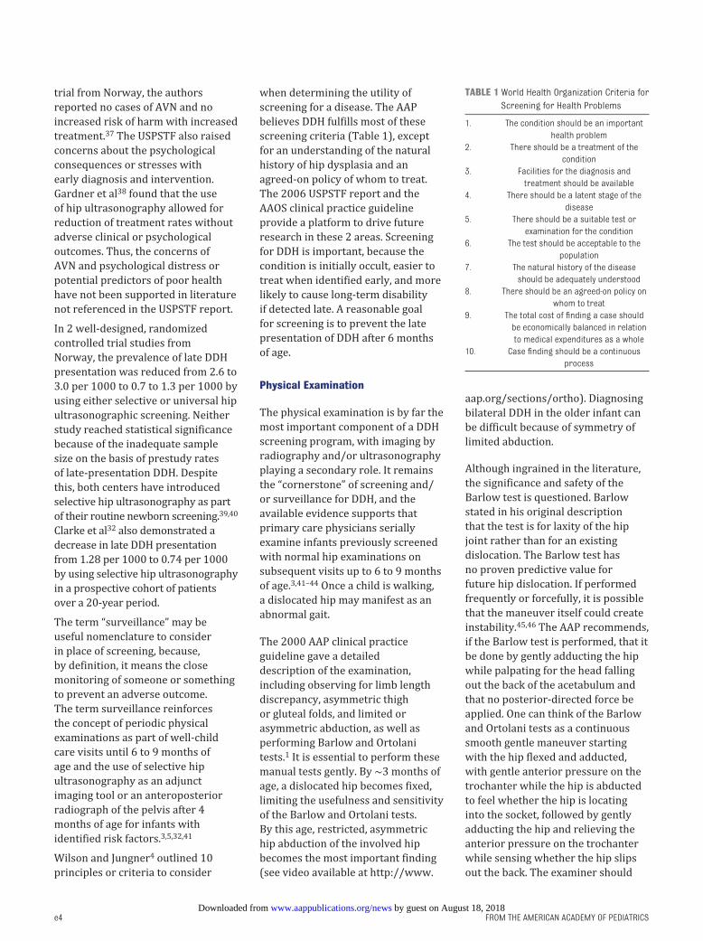
Wilson and Jungner 4 outlined 10
principles or criteria to consider
when determining the utility of
screening for a disease. The AAP
believes DDH fulfills most of these
screening criteria ( Table 1), except
for an understanding of the natural
history of hip dysplasia and an
agreed-on policy of whom to treat.
The 2006 USPSTF report and the
AAOS clinical practice guideline
provide a platform to drive future
research in these 2 areas. Screening
for DDH is important, because the
condition is initially occult, easier to
treat when identified early, and more
likely to cause long-term disability
if detected late. A reasonable goal
for screening is to prevent the late
presentation of DDH after 6 months
of age.
Physical Examination
The physical examination is by far the
most important component of a DDH
screening program, with imaging by
radiography and/or ultrasonography
playing a secondary role. It remains
the “cornerstone” of screening and/
or surveillance for DDH, and the
available evidence supports that
primary care physicians serially
examine infants previously screened
with normal hip examinations on
subsequent visits up to 6 to 9 months
of age. 3, 41 –44 Once a child is walking,
a dislocated hip may manifest as an
abnormal gait.
The 2000 AAP clinical practice
guideline gave a detailed
description of the examination,
including observing for limb length
discrepancy, asymmetric thigh
or gluteal folds, and limited or
asymmetric abduction, as well as
performing Barlow and Ortolani
tests. 1 It is essential to perform these
manual tests gently. By ∼3 months of
age, a dislocated hip becomes fixed,
limiting the usefulness and sensitivity
of the Barlow and Ortolani tests.
By this age, restricted, asymmetric
hip abduction of the involved hip
becomes the most important finding
(see video available at http:// www.
aap. org/ sections/ ortho). Diagnosing
bilateral DDH in the older infant can
be difficult because of symmetry of
limited abduction.
Although ingrained in the literature,
the significance and safety of the
Barlow test is questioned. Barlow
stated in his original description
that the test is for laxity of the hip
joint rather than for an existing
dislocation. The Barlow test has
no proven predictive value for
future hip dislocation. If performed
frequently or forcefully, it is possible
that the maneuver itself could create
instability. 45, 46 The AAP recommends,
if the Barlow test is performed, that it
be done by gently adducting the hip
while palpating for the head falling
out the back of the acetabulum and
that no posterior-directed force be
applied. One can think of the Barlow
and Ortolani tests as a continuous
smooth gentle maneuver starting
with the hip flexed and adducted,
with gentle anterior pressure on the
trochanter while the hip is abducted
to feel whether the hip is locating
into the socket, followed by gently
adducting the hip and relieving the
anterior pressure on the trochanter
while sensing whether the hip slips
out the back. The examiner should
e4
TABLE 1 World Health Organization Criteria for
Screening for Health Problems
1. The condition should be an important
health problem
2. There should be a treatment of the
condition
3. Facilities for the diagnosis and
treatment should be available
4. There should be a latent stage of the
disease
5. There should be a suitable test or
examination for the condition
6. The test should be acceptable to the
population
7. The natural history of the disease
should be adequately understood
8. There should be an agreed-on policy on
whom to treat
9. The total cost of fi nding a case should
be economically balanced in relation
to medical expenditures as a whole
10. Case fi nding should be a continuous
process
by guest on August 18, 2018www.aappublications.org/newsDownloaded from
PEDIATRICS Volume 138 , number 6 , December 2016
not attempt to forcefully dislocate
the femoral head (see video available
at http:// www. aap. org/ sections/
ortho).
“Hip clicks” without the sensation
of instability are clinically
insignificant. 47 Whereas the Ortolani
sign represents the palpable
sensation of the femoral head
moving into the acetabulum over the
hypertrophied rim of the acetabular
cartilage (termed neolimbus),
isolated high-pitched clicks represent
the movement of myofascial tissues
over the trochanter, knee, or other
bony prominences and are not a sign
of hip dysplasia or instability.
Radiography
Plain radiography becomes most
useful by 4 to 6 months of age, when
the femoral head secondary center
of ossification forms. 48 Limited
evidence supports obtaining a
properly positioned anteroposterior
radiograph of the pelvis. 3 If the
pelvis is rotated or if a gonadal
shield obscures the hip joint, then
the radiograph should be repeated.
Hip asymmetry, subluxation, and
dislocation can be detected on
radiographs when dysplasia is
present. There is debate about
whether early minor radiographic
variability (such as increased
acetabular index) constitutes
actual disease. 31 Radiography is
traditionally indicated for diagnosis
of the infant with risk factors or
an abnormal examination after 4
months of age. 1, 2, 8, 49
Ultrasonography
Ultrasonography can provide detailed
static and dynamic imaging of the
hip before femoral head ossification.
The American Institute of Ultrasound
in Medicine and the American
College of Radiology published a
joint guideline for the standardized
performance of the infantile hip
ultrasonographic examination. 50
Static ultrasonography shows
coverage of the femoral head by the
cartilaginous acetabulum (α angle) at
rest, and dynamic ultrasonography
demonstrates a real-time image of
the Barlow and Ortolani tests.
Ultrasonographic imaging can be
universal for all infants or selective
for those at risk for having DDH.
Universal newborn ultrasonographic
screening is not recommended
in North America because of
the expense, inconvenience,
inconsistency, subjectivity, and
high false-positive rates, given
an overall population disease
prevalence of 1% to 2%. 3 Rather,
selective ultrasonographic screening
is recommended either to clarify
suspicious findings on physical
examination after 3 to 4 weeks of
age or to detect clinically silent DDH
in the high-risk infant from 6 weeks
to 4 to 6 months of age. 1, 2, 35, 50 Two
prospective randomized clinical
trials from Norway support selective
ultrasonographic imaging when used
in conjunction with high-quality
clinical screening. 39, 40
Roposch and colleagues 51, 52
contend that experts cannot reach
a consensus on what is normal,
abnormal, developmental variation,
or simply uncertain regarding much
ultrasonographic imaging, thereby
confounding referral and treatment
recommendations. Several studies
have demonstrated that mild
ultrasonographic abnormalities
usually resolve spontaneously,
fueling the controversy over what
imaging findings constitute actual
disease requiring treatment. 5, 30, 51, 53 – 56
The concept of surveillance for
DDH emphasizes the importance
of repeated physical examinations
and the adjunct use of selective hip
ultrasonography after 6 weeks of age
or an anteroposterior radiograph
of the pelvis after 4 months of
age for infants with questionable
or abnormal findings on physical
examination or with identified risk
factors. Ultrasonography is not
necessary for a frankly dislocated
hip (Ortolani positive) but may be
desired by the treating physician.
Physiologic joint capsular laxity and
immature acetabular development
before 6 weeks of age may limit the
accuracy of hip ultrasonography
interpretations. 39, 40 There is no
consensus on exact timing of and
indications for ultrasonography
among expert groups. 26, 57 However,
ultrasonographic imaging does
have a management role in infants
younger than 6 weeks undergoing
abduction brace treatment of
unstable hips identified on physical
examination.3
REFERRAL, ADJUNCTIVE IMAGING, AND TREATMENT
Referral
Early detection and referral of
infants with DDH allows appropriate
intervention with bracing or casting,
which may prevent the need for
reconstructive surgery. Primary
indications for referral include an
unstable (positive Ortolani test
result) or dislocated hip on clinical
examination. Because most infants
with a positive Barlow test result
at either the newborn or 2-week
examination stabilize on their own,
these infants should have sequential
follow-up examinations as part of
the concept of surveillance. This
recommendation differs from the
2000 AAP clinical practice guideline. 1
Any child with limited hip abduction
or asymmetric hip abduction after
the neonatal period (4 weeks) should
be referred. Relative indications
for referral include infants with
risk factors for DDH, a questionable
examination, and pediatrician or
parental concern. 1
Adjunctive Imaging
Recommendations for the evaluation
and management of infants with risk
factors for DDH but with normal
findings on physical examination
continue to evolve. The 2000
AAP clinical practice guideline
recommended hip ultrasonography
e5 by guest on August 18, 2018www.aappublications.org/newsDownloaded from
FROM THE AMERICAN ACADEMY OF PEDIATRICS
at 6 weeks of age or radiography of
the pelvis and hips at 4 months of age
in girls with a positive family history
of DDH or breech presentation. The
AAP clinical practice guideline also
stated that hip ultrasonographic
examinations remain an option for
all infants born breech. 1 The recent
AAOS report found that moderate
evidence supports an imaging study
before 6 months of age in infants
with breech presentation, family
history, and/or history of clinical
instability. 3, 58 –60
Refinement in the term “breech
presentation” as a risk factor for
DDH is needed to determine whether
selective hip ultrasonography at
6 weeks or radiography before
6 months of age is needed for an
infant with a normal clinical hip
examination. More specific variables,
such as mode of delivery, type of
breech position, or breech position
at any time during the pregnancy
or in the third trimester, have
received little attention to date. The
AAOS clinical practice guideline
reported 6 studies addressing breech
presentation, but all were considered
low-strength evidence. 3 Thus, the
literature is not adequate enough
to allow specific guidance. The risk
is thought to be greater for frank
breech (hips flexed, knees extended)
in the last trimester. 1
Lacking expert consensus of risk
factors for DDH, 26 the questions of
whether to obtain additional imaging
studies with a normal clinical hip
examination is ultimately best left
to one’s professional judgment. One
must consider, however, that the
overall probability of a clinically
stable hip to later dislocate is very
low.
Because of the variability in
performance and interpretation of
the hip ultrasonographic examination
and varying thresholds for treatment,
the requesting physician might
consider developing a regional
protocol in conjunction with a
consulting pediatric orthopedist
and pediatric radiologist. Specific
criteria for imaging and referral
based on local resources can
promote consistency in evaluation
and treatment of suspected DDH.
Realistically, many families may not
have ready access to quality infant
hip ultrasonography, and this may
determine the choice of obtaining
a pelvic radiograph instead of an
ultrasound. 61
Treatment
Recommendations for treatment are
based on the clinical hip examination
and the presence or absence of
imaging abnormalities. Infants with
a stable clinical hip examination
but with abnormalities noted on
ultrasonography can be observed
without a brace. 3, 56
The initiation of abduction brace
treatment, either immediate or
delayed, for clinically unstable hips is
supported by several studies. 3, 62 – 64 In
a randomized clinical trial, Gardiner
and Dunn62 found no difference in hip
ultrasonography findings or clinical
outcome for infants with dislocatable
hips treated with either immediate or
delayed abduction bracing at 6- and
12-month follow-up. The infants in
the delayed group (2 weeks) were
treated with abduction bracing if
hip instability persisted or the hip
ultrasonographic abnormalities did
not improve. 62
RISKS OF TREATMENT
Treatment of clinically unstable
hips usually consists of bracing
when discovered in early infancy
and closed reduction with
adductor tenotomy and spica cast
immobilization when noted later.
After 18 months of age, open surgery
is generally recommended.
As previously noted, the 2006
USPSTF report noted a high rate of
AVN, up to 60% with both surgical
and nonsurgical intervention. 33 Other
studies have reported much lower
rates of AVN. 36, 37 One prospective
study reported a zero prevalence
of AVN by 6 years of age in mildly
dysplastic hips treated with
bracing. 30
However, abduction brace treatment
is not innocuous. The potential risks
include AVN, temporary femoral
nerve palsy, and obturator (inferior)
hip dislocation. 65 – 67 One study
demonstrated a 7% to 14% risk
of complications after treatment
in a Pavlik harness. The risk was
greater in hips that did not reduce
in the brace. 33 Precautions such as
avoiding forced abduction in the
harness, stopping treatment after 3
weeks if the hip does not reduce, and
proper strap placement with weekly
monitoring is important to minimize
the risks associated with brace
treatment.68, 69 Double diapering is
a probably harmless but ineffective
treatment of true DDH.
What remains controversial
is whether the selective use of
ultrasonography reduces or increases
treatment. A randomized controlled
study from the United Kingdom
showed that approximately half of
all positive physical examination
findings were falsely positive (ie,
normal ultrasonography results)
and that the use of ultrasonography
in clinically suspect hips actually
e6
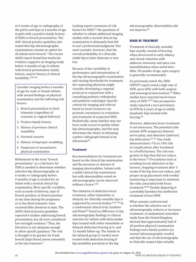
Consider imaging before 6 months
of age for male or female infants
with normal findings on physical
examination and the following risk
factors:
1. Breech presentation in third
trimester (regardless of
cesarean or vaginal delivery)
2. Positive family history
3. History of previous clinical
instability
4. Parental concern
5. History of improper swaddling
6. Suspicious or inconclusive
physical examination
by guest on August 18, 2018www.aappublications.org/newsDownloaded from
PEDIATRICS Volume 138 , number 6 , December 2016
reduced DDH treatment. 60 However,
in the United States and Canada, 21
the reverse appears to be true. In
the current medicolegal climate that
encourages a defensive approach,
liberal use of ultrasonography in
the United States and Canada has
clearly fostered overdiagnosis and
overtreatment of DDH, despite
best-available literature supporting
observation of mild dysplasia. 33 –35, 70
MEDICOLEGAL RISK TO THE PEDIATRICIAN
Undetected or late-developing
DDH is a liability concern for the
pediatrician, generating anxiety and a
desire for guidance in best screening
methodology. 71 Unfortunately, this
fear may also provoke overdiagnosis
and overtreatment. “Late-presenting”
DDH is a more accurate term
than “missed” to use when DDH is
first diagnosed in a walking-aged
child who had appropriate clinical
examinations during infancy. 72, 73
Although there is no universally
recognized DDH screening standard,
the AAP endorses the concept of
surveillance or periodic physical
examinations until walking age,
with selective use of either hip
ultrasonography or radiography,
depending on age. The AAP
cautions against overreliance on
ultrasonography as a diagnostic test
and encourages its use as an adjunctive
secondary screen and an aid to
treatment of established DDH. Notably,
no screening program has been shown
to completely eliminate the risk of a
late-presenting dislocated hip. 69
The electronic health record can
be used to provide a template,
reminder, and documentation tool
for the periodic examination. It
also can be useful in the transition
and comanagement of children
with suspected DDH by providing
effective information transfer
between consultants and primary
care physicians and ensuring
follow-up. Accurate documented
communication between providers
is important to provide continuity of
care for this condition, and it is also
important to explain to the parent(s)
and document those instances when
observation is used as a planned
strategy so it is less likely to be
misinterpreted as negligence.
BEST PRACTICES AND STATE OF THE ART
e7
1. The AAP, POSNA, AAOS, and
Canadian DDH Task Force
recommend newborn and
periodic surveillance physical
examinations for DDH to
include detection of limb length
discrepancy, examination for
asymmetric thigh or buttock
(gluteal) creases, performing
the Ortolani test for stability
(performed gently and which is
usually negative after 3 months
of age), and observing for
limited abduction (generally
positive after 3 months of
age). Use of electronic health
records can be considered to
prompt and record the results
of periodic hip examinations.
The AAP recommends against
universal ultrasonographic
screening.
2. Selective hip ultrasonography
can be considered between
the ages of 6 weeks and 6
months for “high-risk” infants
without positive physical
findings. High risk is a relative
and controversial term, but
considerations include male
or female breech presentation,
a positive family history,
parental concern, suspicious
but inconclusive periodic
examination, history of a
previous positive instability
physical examination, and
history of tight lower-extremity
swaddling. Because most DDH
occurs in children without risk
factors, physical examination
remains the primary screening
tool.
3. It is important that infantile hip
ultrasonography be performed
and interpreted per American
Institute of Ultrasound in
Medicine and the American
College of Radiology guidelines
by experienced, trained
examiners. Developing local
criteria for screening imaging
and referral based on best
resources may promote more
uniform and cost-effective
treatment. Regional variability
of ultrasonographic imaging
quality can lead to under- or
overtreatment.
4. Most minor hip anomalies
observed on ultrasonography at
6 weeks to 4 months of age will
resolve spontaneously. These
include minor variations in α
and β angles and subluxation
(“uncoverage”) with stress
maneuvers. Current levels
of evidence do not support
recommendations for
treatment versus observation
in any specific case of minor
ultrasonographic variation.
Care is, therefore, individualized
through a process of shared
decision-making in this setting
of inadequate information.
5. Radiography (anteroposterior
and frog pelvis views) can be
considered after 4 months of
age for the high-risk infant
without physical findings or
any child with positive clinical
findings. Age 4 to 6 months
is a watershed during which
either imaging modality may
be used; radiography is more
readily available, has a lower
rate of false-positive results,
and is less expensive than
ultrasonography but involves a
very low dose of radiation.
6. A referral to an orthopedist
for DDH does not require
by guest on August 18, 2018www.aappublications.org/newsDownloaded from
FROM THE AMERICAN ACADEMY OF PEDIATRICS
ACKNOWLEDGMENTS
The authors thank Charles Price,
MD, FAAP, Ellen Raney, MD, FAAP,
Joshua Abzug, MD, FAAP, and William
Hennrikus, MD, FAAP, for their
valuable contributions to this report.
LEAD AUTHORS
Brian A. Shaw, MD, FAAOS, FAAP
Lee S. Segal, MD, FAAP
SECTION ON ORTHOPAEDICS EXECUTIVE COMMITTEE, 2014–2015
Norman Y. Otsuka, MD, FAAP, Chairperson
Richard M. Schwend, MD, FAAP, Immediate Past
Chairperson
Theodore John Ganley, MD, FAAP
Martin Joseph Herman, MD, FAAP
Joshua E. Hyman, MD, FAAP
Brian A. Shaw, MD, FAAOS, FAAP
Brian G. Smith, MD, FAAP
STAFF
Niccole Alexander, MPP
ABBREVIATIONS
AAOS: American Academy of
Orthopaedic Surgeons
AAP: American Academy of
Pediatrics
AVN: avascular necrosis
DDH: developmental dysplasia of
the hip
POSNA: Pediatric Orthopaedic
Society of North America
USPSTF: US Preventive Services
Task Force
REFERENCES
1. American Academy of Pediatrics.
Clinical practice guideline: early
detection of developmental dysplasia
of the hip. Committee on Quality
Improvement, Subcommittee on
Developmental Dysplasia of the Hip.
Pediatrics. 2000;105(4 pt 1):896–905
2. Schwend RM, Schoenecker P,
Richards BS, Flynn JM, Vitale M;
Pediatric Orthopaedic Society of North
America. Screening the newborn for
developmental dysplasia of the hip:
now what do we do?J Pediatr Orthop.
2007;27(6):607–610
3. American Academy of Orthopaedic
Surgeons. Detection and Nonoperative
Management of Pediatric
Developmental Dysplasia of the Hip
in Infants Up to Six Months of Age.
Evidence-Based Clinical Practice
Guideline. Rosemont, IL: American
Academy of Orthopaedic Surgeons;
2014
4. Wilson JMG, Jungner G. Principles and
Practice of Screening for Disease.
Geneva, Switzerland: World Health
Organization; 1968
5. Rosendahl K, Dezateux C, Fosse KR,
et al. Immediate treatment versus
sonographic surveillance for mild
hip dysplasia in newborns.Pediatrics.
2010;125(1). Available at: www.
pediatrics. org/ cgi/ content/ full/ 125/ 1/ e9
6. Bache CE, Clegg J, Herron M. Risk
factors for developmental dysplasia of
the hip: ultrasonographic fi ndings in
the neonatal period.J Pediatr Orthop B.
2002;11(3):212–218
7. Barr LV, Rehm A. Should all twins and
multiple births undergo ultrasound
examination for developmental
dysplasia of the hip? A retrospective
study of 990 multiple births.Bone Joint
J. 2013;95-B(1):132–134
8. Imrie M, Scott V, Stearns P, Bastrom T,
Mubarak SJ. Is ultrasound screening
for DDH in babies born breech
suffi cient?J Child Orthop. 2010;4(1):3–8
9. Suzuki S, Yamamuro T. Avascular
necrosis in patients treated with
the Pavlik harness for congenital
dislocation of the hip.J Bone Joint Surg
Am. 1990;72(7):1048–1055
10. Fox AE, Paton RW. The relationship
between mode of delivery and
e8
ultrasonography or
radiography. The primary
indication for referral includes
an unstable (positive Ortolani
test result) or dislocated hip on
clinical examination. Any child
with limited hip abduction or
asymmetric hip abduction after
the neonatal period (4 weeks
of age) should be referred
for evaluation. Relative
indications for referral include
infants with risk factors
for DDH, a questionable
examination, and pediatrician
or parental concern.
7. Evidence strongly supports
screening for and treatment
of hip dislocation (positive
Ortolani test result) and
initially observing milder
early forms of dysplasia and
instability (positive Barlow
test result). Depending on
local custom, either the
pediatrician or the orthopedist
can observe mild forms by
periodic examination and
possible follow-up imaging,
but actual treatment should be
performed by an orthopedist.
8. A reasonable goal for the
primary care physician
should be to diagnose hip
subluxation or dislocation by
6 months of age by using the
periodic physical examination.
Selective ultrasonography or
radiography may be used in
consultation with a pediatric
radiologist and/or orthopedist.
No screening program has been
shown to completely eliminate
the risk of a late presentation
of DDH. There is no high-level
evidence that milder forms of
dysplasia can be prevented by
screening and early treatment.
9. Tight swaddling of the lower
extremities with the hips
adducted and extended should
be avoided. The concept
of “safe” swaddling, which
does not restrict hip motion,
minimizes the risk of DDH.
10. Treatment of neonatal DDH
is not an emergency, and
in-hospital initiation of bracing
is not required. Orthopaedic
consultation can be safely
obtained within several weeks
of discharge for an infant
with a positive Ortolani test
result. Infants with a positive
Barlow test results should be
reexamined and referred to an
orthopedist if they continue to
show clinical instability.
by guest on August 18, 2018www.aappublications.org/newsDownloaded from

PEDIATRICS Volume 138 , number 6 , December 2016
developmental dysplasia of the hip in
breech infants: a four-year prospective
cohort study.J Bone Joint Surg Br.
2010;92(12):1695–1699
11. Lowry CA, Donoghue VB, O’Herlihy C,
Murphy JF. Elective Caesarean section
is associated with a reduction in
developmental dysplasia of the hip in
term breech infants.J Bone Joint Surg
Br. 2005;87(7):984–985
12. Panagiotopoulou N, Bitar K, Hart WJ.
The association between mode of
delivery and developmental dysplasia
of the hip in breech infants: a
systematic review of 9 cohort studies.
Acta Orthop Belg. 2012;78(6):697–702
13. Sarkissian EJ, Sankar WN, Baldwin
K, Flynn JM. Is there a predilection
for breech infants to demonstrate
spontaneous stabilization of
DDH instability?J Pediatr Orthop.
2014;34(5):509–513
14. Dodinval P. Hérédité de la maladie
luxante de la hanche (MLH) [Heredity
in congenital dislocation of the hip].
Acta Orthop Belg. 1990;56(1 pt A):7–11
15. Tönnis D. Inheritence. In: Tönnis D, ed.
Congenital Dysplasia and Dislocation
of the Hip in Children and Adults.
Berlin, Germany: Springer-Verlag;
1984:61–62
16. Stevenson DA, Mineau G, Kerber
RA, Viskochil DH, Schaefer C, Roach
JW. Familial predisposition to
developmental dysplasia of the hip.J
Pediatr Orthop. 2009;29(5):463–466
17. Schiffern AN, Stevenson DA, Carroll
KL, et al. Total hip arthroplasty, hip
osteoarthritis, total knee arthroplasty,
and knee osteoarthritis in patients
with developmental dysplasia of
the hip and their family members:
a kinship analysis report.J Pediatr
Orthop. 2012;32(6):609–612
18. Carroll KL, Schiffern AN, Murray KA, et al.
The occurrence of occult acetabular
dysplasia in relatives of individuals
with developmental dysplasia of the
hip.J Pediatr Orthop. 2016;36(1):96–100
19. Mahan ST, Kasser JR. Does
swaddling infl uence developmental
dysplasia of the hip?Pediatrics.
2008;121(1):177–178
20. van Sleuwen BE, Engelberts AC, Boere-
Boonekamp MM, Kuis W, Schulpen TW,
L’Hoir MP. Swaddling: a systematic
review.Pediatrics. 2007;120(4).
Available at: www. pediatrics. org/ cgi/
content/ full/ 120/ 4/ e1097
21. Wang E, Liu T, Li J, et al. Does swaddling
infl uence developmental dysplasia
of the hip? An experimental study of
the traditional straight-leg swaddling
model in neonatal rats.J Bone Joint
Surg Am. 2012;94(12):1071–1077
22. Gerard CM, Harris KA, Thach BT.
Physiologic studies on swaddling: an
ancient child care practice, which may
promote the supine position for infant
sleep.J Pediatr. 2002;141(3):398–403
23. Oden RP, Powell C, Sims A, Weisman
J, Joyner BL, Moon RY. Swaddling:
will it get babies onto their backs
for sleep?Clin Pediatr (Phila).
2012;51(3):254–259
24. Canillas F, Delgado-Martos MJ, Martos-
Rodriguez A, Quintana-Villamandos B,
Delgado-Baeza E. Contribution to the
initial pathodynamics of hip luxation
in young rats.J Pediatr Orthop.
2012;32(6):613–620
25. Pediatric Orthopaedic Society of
North America, International Hip
Dysplasia Institute, American Academy
of Orthopaedic Surgeons, United
States Bone and Joint Initiative,
Shriners Hospitals for Children.
Position Statement: Swaddling and
Developmental Hip Dysplasia. Rosemont,
IL: Pediatric Orthopaedic Society of
North America; 2015. Available at: www.
aaos. org/ uploadedFiles/ PreProduction/
About/ Opinion_ Statements/
position/ 1186%20 Swaddling%20
and%20 Developmental%20 Hip%20
Dysplasia%281 %29 . pdf. Accessed
January 12, 2016
26. Roposch A, Liu LQ, Protopapa E.
Variations in the use of diagnostic
criteria for developmental dysplasia
of the hip.Clin Orthop Relat Res.
2013;471(6):1946–1954
27. Barlow TG. Early diagnosis and
treatment of congenital dislocation
of the hip.J Bone Joint Surg Br.
1962;44B(2):292–301
28. Barlow TG. Congenital dislocation of
the hip in the newborn.Proc R Soc Med.
1966;59(11 part 1):1103–1106
29. Barlow TG. Neonatal hip dysplasia—
treatment, results and complications.
Proc R Soc Med. 1975;68(8):475
30. Brurås KR, Aukland SM, Markestad
T, Sera F, Dezateux C, Rosendahl K.
Newborns with sonographically
dysplastic and potentially unstable
hips: 6-year follow-up of an RCT.
Pediatrics. 2011;127(3). Available at:
www. pediatrics. org/ cgi/ content/ full/
127/ 3/ e661
31. Mladenov K, Dora C, Wicart P,
Seringe R. Natural history of hips
with borderline acetabular index
and acetabular dysplasia in infants.
J Pediatr Orthop. 2002;22(5):
607–612
32. Clarke NM, Reading IC, Corbin C,
Taylor CC, Bochmann T. Twenty years’
experience of selective secondary
ultrasound screening for congenital
dislocation of the hip.Arch Dis Child.
2012;97(5):423–429
33. Shipman SA, Helfand M, Moyer VA,
Yawn BP. Screening for developmental
dysplasia of the hip: a systematic
literature review for the US Preventive
Services Task Force.Pediatrics.
2006;117(3). Available at: www.
pediatrics. org/ cgi/ content/ full/ 117/ 3/
e557
34. US Preventive Services Task Force.
Screening for developmental dysplasia
of the hip: recommendation statement.
Pediatrics. 2006;117(3):898–902
35. Patel H; Canadian Task Force on
Preventive Health Care. Preventive
health care, 2001 update: screening
and management of developmental
dysplasia of the hip in newborns.CMAJ.
2001;164(12):1669–1677
36. Williams PR, Jones DA, Bishay M.
Avascular necrosis and the Aberdeen
splint in developmental dysplasia
of the hip.J Bone Joint Surg Br.
1999;81(6):1023–1028
37. Laborie LB, Engesæter IØ, Lehmann
TG, Eastwood DM, Engesæter LB,
Rosendahl K. Screening strategies for
hip dysplasia: long-term outcome of a
randomized controlled trial.Pediatrics.
2013;132(3):492–501
38. Gardner F, Dezateaux C, Elbourne D,
Gray A, King A, Quinn A; Collaborative
Hip Trial Group. The hip trial:
psychological consequences for
mothers of using ultrasound to
manage infants with developmental
hip dysplasia.Arch Dis Child Fetal
Neonatal Ed. 2005;90(1):F17–F24
e9 by guest on August 18, 2018www.aappublications.org/newsDownloaded from

FROM THE AMERICAN ACADEMY OF PEDIATRICS
39. Rosendahl K, Markestad T, Lie
RT. Ultrasound screening for
developmental dysplasia of the hip in
the neonate: the effect on treatment
rate and prevalence of late cases.
Pediatrics. 1994;94(1):47–52
40. Holen KJ, Tegnander A, Bredland T, et
al. Universal or selective screening of
the neonatal hip using ultrasound? A
prospective, randomised trial of 15, 529
newborn infants.J Bone Joint Surg Br.
2002;84(6):886–890
41. Hagans JF, Shaw JS, Duncan P, eds.
Bright Futures: Guidelines for Health
Supervision of Infants, Children, and
Adolescents, Third Edition. Pocket
Guide. Elk Grove Village, IL: American
Academy of Pediatrics; 2008
42. Catterall A. The early diagnosis of
congenital dislocation of the hip.J Bone
Joint Surg Br. 1994;76(4):515–516
43. Wirth T, Stratmann L, Hinrichs
F. Evolution of late presenting
developmental dysplasia of the hip
and associated surgical procedures
after 14 years of neonatal ultrasound
screening.J Bone Joint Surg Br.
2004;86(4):585–589
44. Myles JW. Secondary screening for
congenital displacement of the hip.J
Bone Joint Surg Br. 1990;72(2):326–327
45. Moore FH. Examining infants’ hips—
can it do harm?J Bone Joint Surg Br.
1989;71(1):4–5
46. Jones DA. Neonatal hip stability
and the Barlow test. A study in
stillborn babies.J Bone Joint Surg Br.
1991;73(2):216–218
47. Bond CD, Hennrikus WL, DellaMaggiore
ED. Prospective evaluation of
newborn soft-tissue hip “clicks”
with ultrasound.J Pediatr Orthop.
1997;17(2):199–201
48. Scoles PV, Boyd A, Jones PK.
Roentgenographic parameters of the
normal infant hip.J Pediatr Orthop.
1987;7(6):656–663
49. Karmazyn BK, Gunderman RB,
Coley BD, et al; American College
of Radiology. ACR Appropriateness
Criteria on developmental dysplasia
of the hip—child.J Am Coll Radiol.
2009;6(8):551–557
50. American Institute of Ultrasound
in Medicine; American College of
Radiology. AIUM practice guideline
for the performance of an ultrasound
examination for detection and
assessment of developmental
dysplasia of the hip.J Ultrasound Med.
2009;28(1):114–119
51. Roposch A, Wright JG. Increased
diagnostic information and
understanding disease: uncertainty
in the diagnosis of developmental
hip dysplasia.Radiology.
2007;242(2):355–359
52. Roposch A, Moreau NM, Uleryk E,
Doria AS. Developmental dysplasia
of the hip: quality of reporting of
diagnostic accuracy for US.Radiology.
2006;241(3):854–860
53. Burger BJ, Burger JD, Bos CF,
Obermann WR, Rozing PM,
Vandenbroucke JP. Neonatal
screening and staggered early
treatment for congenital dislocation
or dysplasia of the hip.Lancet.
1990;336(8730):1549–1553
54. Dunn PM, Evans RE, Thearle MJ,
Griffi ths HE, Witherow PJ. Congenital
dislocation of the hip: early and late
diagnosis and management compared.
Arch Dis Child. 1985;60(5):407–414
55. Sampath JS, Deakin S, Paton RW.
Splintage in developmental dysplasia
of the hip: how low can we go?J
Pediatr Orthop. 2003;23(3):352–355
56. Wood MK, Conboy V, Benson MK. Does
early treatment by abduction splintage
improve the development of dysplastic
but stable neonatal hips?J Pediatr
Orthop. 2000;20(3):302–305
57. Shorter D, Hong T, Osborn DA.
Screening programmes for
developmental dysplasia of the hip in
newborn infants.Cochrane Database
Syst Rev. 2011;(9):CD004595
58. Paton RW, Srinivasan MS, Shah B,
Hollis S. Ultrasound screening for hips
at risk in developmental dysplasia.
Is it worth it?J Bone Joint Surg Br.
1999;81(2):255–258
59. Paton RW, Hinduja K, Thomas CD.
The signifi cance of at-risk factors
in ultrasound surveillance of
developmental dysplasia of the hip. A
ten-year prospective study.J Bone Joint
Surg Br. 2005;87(9):1264–1266
60. Elbourne D, Dezateux C, Arthur R,
et al; UK Collaborative Hip Trial Group.
Ultrasonography in the diagnosis and
management of developmental hip
dysplasia (UK Hip Trial): clinical and
economic results of a multicentre
randomised controlled trial.Lancet.
2002;360(9350):2009–2017
61. Pacana MJ, Hennrikus WL, Slough
J, Curtin W. Ultrasound examination
for infants born breech by elective
cesarean section with a normal hip
exam for instability [published online
ahead of print October 21, 2015].J
Pediatr Orthop. Doi: 10. 1097/ BPO.
0000000000000668
62. Gardiner HM, Dunn PM. Controlled
trial of immediate splinting versus
ultrasonographic surveillance in
congenitally dislocatable hips.Lancet.
1990;336(8730):1553–1556
63. Paton RW, Hopgood PJ, Eccles K.
Instability of the neonatal hip: the role
of early or late splintage.Int Orthop.
2004;28(5):270–273
64. Lorente Moltó FJ, Gregori AM, Casas
LM, Perales VM. Three-year prospective
study of developmental dysplasia of
the hip at birth: should all dislocated
or dislocatable hips be treated?J
Pediatr Orthop. 2002;22(5):613–621
65. Suzuki S, Kashiwagi N, Kasahara Y,
Seto Y, Futami T. Avascular necrosis
and the Pavlik harness. The incidence
of avascular necrosis in three types
of congenital dislocation of the hip as
classifi ed by ultrasound.J Bone Joint
Surg Br. 1996;78(4):631–635
66. Murnaghan ML, Browne RH, Sucato DJ,
Birch J. Femoral nerve palsy in Pavlik
harness treatment for developmental
dysplasia of the hip.J Bone Joint Surg
Am. 2011;93(5):493–499
67. Rombouts JJ, Kaelin A. Inferior
(obturator) dislocation of the hip in
neonates. A complication of treatment
by the Pavlik harness.J Bone Joint
Surg Br. 1992;74(5):708–710
68. Mubarak S, Garfi n S, Vance R, McKinnon
B, Sutherland D. Pitfalls in the use of
the Pavlik harness for treatment of
congenital dysplasia, subluxation, and
dislocation of the hip.J Bone Joint Surg
Am. 1981;63(8):1239–1248
69. Kitoh H, Kawasumi M, Ishiguro N.
Predictive factors for unsuccessful
treatment of developmental dysplasia
of the hip by the Pavlik harness.J
Pediatr Orthop. 2009;29(6):552–557
e10 by guest on August 18, 2018www.aappublications.org/newsDownloaded from

PEDIATRICS Volume 138 , number 6 , December 2016
70. Sucato DJ, Johnston CE II, Birch
JG, Herring JA, Mack P. Outcome of
ultrasonographic hip abnormalities in
clinically stable hips.J Pediatr Orthop.
1999;19(6):754–759
71. McAbee GN, Donn SM, Mendelson RA,
McDonnell WM, Gonzalez JL,
Ake JK. Medical diagnoses
commonly associated with
pediatric malpractice lawsuits
in the United States.Pediatrics.
2008;122(6). Available at: www.
pediatrics. org/ cgi/ content/ full/ 122/ 6/
e1282
72. Davies SJ, Walker G. Problems in the
early recognition of hip dysplasia.J
Bone Joint Surg Br. 1984;66(4):479–484
73. Ilfeld FW, Westin GW, Makin M. Missed
or developmental dislocation of
the hip.Clin Orthop Relat Res. 1986;
(203):276–281
e11 by guest on August 18, 2018www.aappublications.org/newsDownloaded from

DOI: 10.1542/peds.2016-3107 originally published online November 21, 2016; 2016;138;Pediatrics
Brian A. Shaw, Lee S. Segal and SECTION ON ORTHOPAEDICSEvaluation and Referral for Developmental Dysplasia of the Hip in Infants
ServicesUpdated Information &
http://pediatrics.aappublications.org/content/138/6/e20163107including high resolution figures, can be found at:
Referenceshttp://pediatrics.aappublications.org/content/138/6/e20163107#BIBLThis article cites 68 articles, 18 of which you can access for free at:
Subspecialty Collections
subhttp://www.aappublications.org/cgi/collection/orthopedic_medicine_Orthopaedic Medicineshttp://www.aappublications.org/cgi/collection/section-on-orthopaedicSection on Orthopaedicshttp://www.aappublications.org/cgi/collection/current_policyCurrent Policyfollowing collection(s): This article, along with others on similar topics, appears in the
Permissions & Licensing
http://www.aappublications.org/site/misc/Permissions.xhtmlin its entirety can be found online at: Information about reproducing this article in parts (figures, tables) or
Reprintshttp://www.aappublications.org/site/misc/reprints.xhtmlInformation about ordering reprints can be found online:
by guest on August 18, 2018www.aappublications.org/newsDownloaded from
DOI: 10.1542/peds.2016-3107 originally published online November 21, 2016; 2016;138;Pediatrics
Brian A. Shaw, Lee S. Segal and SECTION ON ORTHOPAEDICSEvaluation and Referral for Developmental Dysplasia of the Hip in Infants
http://pediatrics.aappublications.org/content/138/6/e20163107located on the World Wide Web at:
The online version of this article, along with updated information and services, is
1073-0397. ISSN:60007. Copyright © 2016 by the American Academy of Pediatrics. All rights reserved. Print
the American Academy of Pediatrics, 141 Northwest Point Boulevard, Elk Grove Village, Illinois,has been published continuously since 1948. Pediatrics is owned, published, and trademarked by Pediatrics is the official journal of the American Academy of Pediatrics. A monthly publication, it
by guest on August 18, 2018www.aappublications.org/newsDownloaded from
Related Documents