Evaluation and Management of Cleft Lip and Palate A Developmental Perspective David J. Zajac, PhD, CCC-SLP Linda D. Vallino, PhD, CCC-SLP/A

Welcome message from author
This document is posted to help you gain knowledge. Please leave a comment to let me know what you think about it! Share it to your friends and learn new things together.
Transcript
Evaluation and Management of Cleft Lip and Palate
A Developmental Perspective
David J. Zajac, PhD, CCC-SLPLinda D. Vallino, PhD, CCC-SLP/A
v
Contents
Preface viiAcknowledgements ixContributors xi
Part I. Fundamentals 1
1 Orofacial and Velopharyngeal Structure and Function 3Jamie Perry and David J. Zajac
2 Clefts of the Lip and Palate 23
3 Syndromes and Associated Anomalies 49
Part II. Birth to Age Three 111
4 Feeding the Newborn 113
5 Presurgical and Surgical Management 129
6 Hearing and Otologic Management 151
7 Early Linguistic Development and Intervention 177Nancy J. Scherer
Part III. Early to Middle School Age 191
8 Speech and Resonance Characteristics 193
9 Assessment of Speech and Velopharyngeal Function 227
10 School-Based Intervention 281Dennis M. Ruscello
11 Secondary Management of Velopharyngeal Inadequacy 319
12 Alveolar Cleft Repair 339Joseph A. Napoli
Part IV. Adolescents and Adults 355
13 Maxillary Advancement 357Joseph A. Napoli and Linda D. Vallino
14 The Adult With Cleft Lip and Palate 379
Glossary of Terms 391Index 401
vii
Preface
This book is intended to be a concise, practical, and evidence-based text on cleft lip and palate and related craniofacial disorders for advanced undergraduate students, graduate students, and profes-sionals in speech-language pathology. Students and professionals in related dis-ciplines such as dentistry, medicine, psy-chology, and social work also may find this book useful in providing information on individuals with craniofacial condi-tions. Cleft palate with or without cleft lip is a congenital defect that varies both in its severity and impact on facial and oral structures and communication. Treat-ment of individuals is typically a long and often-complicated process, extending into early adulthood and beyond, that is best accomplished in a team setting. In addition to the speech-language pathologist, other team members typically include a plastic surgeon, dentist, orthodontist, oral and maxillofacial surgeon, otolaryngologist, audiologist, geneticist, social worker, and psychologist. Even though all members of the team contribute to the habilitation of the individual througout his or her life span, certain team members assume critical roles at specific times of life. The purpose of the book is to provide the stu-dent and professional in speech-language pathology the information needed to (a) evaluate and treat communication disor-ders associated with cleft palate regard-less of their primary place of employment (i.e., craniofacial team, hospital, school, or private practice), and (b) understand the complex — and sometimes controversial — surgical and dental management of indi-viduals across the life span.
To help achieve these goals, the mate-rial in the book is presented in a devel-opmental framework that emphasizes the most critical needs of the individual from birth to adulthood. This organizational approach has both practical and concep-tual advantages. Practically, it allows the reader to access information more read-ily according to the age and presenting condition of the individual (i.e., birth, lip repair, palate repair, alveolar cleft repair or bone grafting, maxillary advancement). Conceptually, it chronicles the lifelong impact of craniofacial birth defects on the individual and elucidates the timing and rationale of surgical, dental, and behav-ioral interventions.
Part I provides necessary funda-mentals for the student and professional. Chapter 1 reviews anatomy and physiol-ogy of the facial, oral, and velopharyn-geal structures that are affected by clefts. Chapter 2 describes the types and causes of clefts with an emphasis on embryologi-cal development and classification. The controversy regarding timing of palate repair is discussed from the perspective of speech and language development. Chapter 3 provides an overview of genet-ics and the terminology used to catego-rize and identify congenital anomalies. A select group of craniofacial anomalies most likely to be encountered by the pro-fessional is reviewed.
Part II focuses on evaluation and management of the individual from birth to 3 years of age. Chapter 4 describes nor-mal feeding physiology, feeding prob-lems associated with cleft palate, and approaches to facilitate feeding prior to
Evaluation and Management of Cleft Lip and Palate: A Developmental Perspectiveviii
palate repair. Chapter 5 describes presur-gical and surgical management of cleft lip and palate. Chapter 6 covers the almost universal occurrence of otitis media with effusion and conductive hearing loss that occurs in infants with cleft palate and cur-rent methods of management. Chapter 7 reviews early linguistic development in infants with cleft palate and intervention strategies before and after palate repair.
Part III focuses on evaluation and management of the individual from age 3 throughout the middle school years. Chapter 8 describes the resonance, nasal emission, articulation, voice, fluency, and intelligibility characteristics of children with repaired cleft palate. Chapter 9 pro-vides detailed coverage of perceptual assessment and an overview of instru-mental assessment techniques. Chapter 10 describes practical approaches to treat-ing children with articulation problems in the school setting. Chapter 11 presents an overview of behavioral, surgical, and prosthetic options to manage velopharyn-geal inadequacy that persists following initial palate surgery. Chapter 12 describes the orthodontic preparation and surgical correction of clefts of the alveolus.
Part IV focuses on evaluation and man- agement of adolescents and adults with cleft palate. Chapter 13 describes the rationale and timing of maxillary advancement in adolescents to improve facial aesthetics, dental occlusion, and articulation. Chapter 14 discusses issues facing adult patients.
Although clefts of the lip and palate are among the most frequently occurring birth defects, the actual number of indi-
viduals affected in the United States is relatively low. It is not unusual, therefore, for speech-language pathologists working in the schools to infrequently see children with clefts. It is our hope that the materi-als in this book will be a valuable resource for school-based clinicians when they do encounter children with clefts. Some mate-rial traditionally covered in other texts is, unfortunately, omitted in the book. Due to a goal to be concise and follow a develop-mental framework, separate chapters on craniofacial team function and psycho-social aspects of individuals with cranio-facial anomalies are not included. These are obviously important areas. We have, however, interweaved these materials in various chapters throughout the book. Maternal reactions to an infant born with a cleft are covered in Chapter 4, learning disabilities of children with cleft palate and treatment collaboration models with teams are reviewed in Chapter 10, and the last chapter deals exclusively with quality of life issues facing the adult.
Finally, the book has been written with a goal to be concise and cite evidence-based sources to support intervention approaches. In some areas, there is little objective evidence available to guide clinical decision making. In those areas, we note the lack of evidence and suggest directions for future research. It is our hope that this book will not only inform but also challenge clinicians in speech-language pathology to provide the best evidence-based evaluation and manage-ment of individuals with craniofacial anomalies as possible.

ix
Acknowledgments
I am indebted to many who have contrib-uted either directly or indirectly to this book. While at the University of Pitts-burgh, I was fortunate to have Betty Jane McWilliams as an early mentor. Dr. McWil-liams, the first and longtime director of the Cleft Palate Center, was an inexhaust-ible clinician and researcher who first and foremost put the welfare of the patient above all else. I have been truly influ-enced by her wisdom, knowledge, and caring. Campbell C. Yates and Raymond Linville also were mentors at Pittsburgh. Professor Yates, chair and emeritus pro-fessor of mechanical engineering, volun-teered his time to establish aerodynamic assessment procedures at the center. Pro-fessor Yates helped me to understand and appreciate the nuances of fluid dynamics when applied to the complex geometry of the human upper airways. Raymond Lin-ville, a mentor and friend, was influential in guiding my early research endeavors.
Many colleagues have been influen-tial during the past 20 years at the Uni-versity of North Carolina at Chapel Hill. Donald W. Warren, the Craniofacial Cen-ter’s first director, was a prolific researcher who applied aerodynamic techniques to the evaluation of nasal and palatal function. Don was a role model whom
I strived to emulate relative to scholar-ship and research productivity. Others at the University of North Carolina at Cha-pel Hill who have been influential include Amelia Drake, Tim Turvey, Gerald Sloan, Wolf Losken (also at Pittsburgh), and John van Aalst. They were instrumental in helping me understand surgical treat-ment of patients.
I am most appreciative of Linda Val-lino, co-author of the book, and Jamie Perry, Nancy Scherer, Dennis Ruscello, and Joseph Napoli, contributors to the book. They are outstanding clinicians and researchers in their respective areas. This book would not be what it is without their input. A special thanks is also due to Marziye Eshghi, Jacqueline Dorry, and Ramona Hutton-Howe for their help with figures, tables, and photography.
On a personal note, I am indebted to my family for their support and encour-agement. They graciously endured count-less dinnertime book conversations — and dinners without me — during the writing of the book. I am grateful to my loving wife, Robin, children, Jared and Rachel, daughter-in-law Elizabeth, son-in-law, Matt, and grandson, William, for their encouragement. The thought of William’s wonderful smile was the best inspiration.
— David J. Zajac Chapel Hill, North Carolina
This book would not have been possible without the encouragement, mentor-ship, and friendship of some very special people. Like David, I received my train-
ing and doctoral degree at the University of Pittsburgh, under the direction of Dr. Betty Jane McWilliams. Dr. McWilliams was the quintessential teacher, researcher

Evaluation and Management of Cleft Lip and Palate: A Developmental Perspectivex
and clinician. She cared deeply for her patients and their families. I value all that she has taught me.
I would like to express my grati-tude to Drs. William Garret (University of Pittsburgh), John B. Mulliken (Boston Children’s Hospital), Ronald R. Zuker (Hospital for Sick Children), and Joseph A. Napoli (Nemours/Alfred I. DuPont Hospital for Children) for teaching me about cleft and craniofacial surgery and enhancing my education. They are tal-ented and compassionate surgeons.
I am appreciative of David Zajac, my colleague and friend, who graciously invited me to coauthor this book with him, and our contributors, Jamie Perry, Nancy Scherer, Dennis Ruscello, and Joseph Napoli. They are among the best at what they do.
My patients and their caregivers have also been my teachers. I have learned
much from all of them. Over the years, these families allowed me the opportu-nity to be a part of their children’s care, and trusted that I would always do the best I could for them. I hope I have served them well.
Thanks to Cindy Brodoway and Brad Gelman, medical photographers at Nemours, for capturing the beautiful smiles of some of the children shown in this book.
My father always thought I should write a book, my mother agreed, and so I wrote one. This book is for both of you. To my sister Rita, and brother-in-law, Boyd — thank you always for caring about me. A special thank you to my wonder-ful family, Nicholas, Caroline, John, and Eleanor. You four are the best ever. Above all, my heartfelt thanks to my husband, Joe for his endless love and support for everything I do. It means the world to me.
— Linda D. Vallino Wilmington, Delaware
xi
Contributors
Joseph A. Napoli, MD, DDSAssociate Professor of SurgerySidney Kimmel Medical CollegeThomas Jefferson UniversityChief, Division of Plastic and
Maxillofacial SurgeryNemours Children’s ClinicDirector, Cleft Lip and Palate/
Craniofacial Anomalies ProgramAlfred I. duPont Hospital for ChildrenWilmington, DelawareChapters 12 and 13
Jamie Perry, PhDAssociate ProfessorDepartment of Communication Sciences
and DisordersEast Carolina UniversityGreenville, North CarolinaChapter 1
Dennis M. Ruscello, PhDProfessor of Communication Sciences
and DisordersAdjunct Professor of OtolaryngologyWest Virginia UniversityMorgantown, West VirginiaChapter 10
Nancy J. Scherer, PhD, CCC-SLPASHA Fellow
Professor and ChairDepartment of Speech and Hearing
SciencesArizona State UniversityTempe, ArizonaChapter 7
Linda D. Vallino, PhD, CCC-SLP/AHead of the Craniofacial Outcomes
Research LaboratoryCenter for Pediatric Auditory and Speech
SciencesAlfred I. duPont Hospital for ChildrenWilmington, DelawareProfessor of PediatricsSidney Kimmel Medical CollegeThomas Jefferson UniversityPhiladelphia, PAAdjunct Associate ProfessorUniversity of DelawareNewark, Delaware
David J. Zajac, PhD, CCC-SLPProfessor, Department of Dental
EcologyAdjunct Associate Professor, Division
of Speech and Hearing Sciences, Department of Allied Health Sciences
University of North Carolina at Chapel Hill
Chapel Hill, North Carolina

Part IFundamentals

Evaluation and Management of Cleft Lip and Palate: A Developmental Perspective2
We intend this book to serve as a roadmap to the diagnosis and care of individuals with orofacial clefts and other craniofacial conditions from birth through adulthood. To do so, the speech-language patholo-gist (SLP) and other health care providers must know certain fundamentals. Chapter 1, Orofacial and Velopharyngeal Structure and Function, provides information on normal anatomy and physiology. Major landmarks of the face, nose, and oral cav-ity are identified, and detailed descrip-tions of the velopharyngeal muscles and functions are provided. Chapter 2, Clefts of the Lip and Palate, describes the types
of clefts that are commonly encountered in the clinic. Special attention is given to sub-mucous clefts as these may be subtle and difficult to identify. Causes of clefts and epidemiology relative to prevalence and recurrence are reviewed. Finally, Chapter 3, Syndromes and Associated Anomalies, provides an overview of genetics and the terminology used to categorize and iden-tify congenital anomalies. A select group of craniofacial anomalies that impact com-munication is presented. The information in Part I sets the stage for the remainder of the book.

3
1Orofacial and Velopharyngeal
Structure and FunctionJamie Perry and David J. Zajac
IntroductIon
Orofacial clefts can involve structural anomalies of the upper lip and gum ridge, nose, hard palate, and soft palate to vari-ous degrees. If a cleft is part of a syndrome or sequence, then additional craniofacial anomalies may be present involving the lower jaw, face, ears, and skull. In order to understand the nature and management of clefts — including embryological devel-opment, anomalies at birth, impact on feeding, hearing and speech, and surgical repair — the speech-language pathologist must have a fundamental understanding of orofacial and velopharyngeal struc-tures and function. The purpose of this chapter is to review (a) structures of the face, nasal cavity, oral cavity, pharynx, and velopharynx that may be affected by clefts, and (b) velopharyngeal function of normal speech production.
the Face
The face is part of the skull that contains the forehead and bony framework for the eyes, nose, and mouth. Facial landmarks of the nose and upper lip can be seen in Figure 1–1. The nasion is the bony struc-ture at the root of the nose between the eyes. The nasal columella consists of skin and underlying tissue that separates the nose into two nostrils. It courses from the anterior nasal spine (shown in Figure 1–3) to the nasal tip. Nostrils, also called nares, are openings bounded laterally by a cartilaginous ala nasi (curved lateral portion of the nose) and alar rim (outer rim). The alar base connects the alar rim to the upper lip. The philtrum, also called philtral dimple or groove, is a midline indentation that courses from the nose to the upper lip and is bounded laterally by the philtral ridges, also called philtral

Evaluation and Management of Cleft Lip and Palate: A Developmental Perspective4
columns. The philtral ridges are created by fusion of the maxillary, medial nasal, and lateral nasal processes during embry-ological development (see Chapter 2). The vermilion zone is the pigmented portion of the lips. The Cupid’s bow is defined by a double curve along the superior edge of the upper lip. The white roll is a distinc-tive landmark that surrounds the vermil-ion zone. When a cleft of the lip occurs, symmetrical reconstruction of the Cupid’s bow presents a particular challenge for the surgeon (see Chapter 5).
nasal cavity
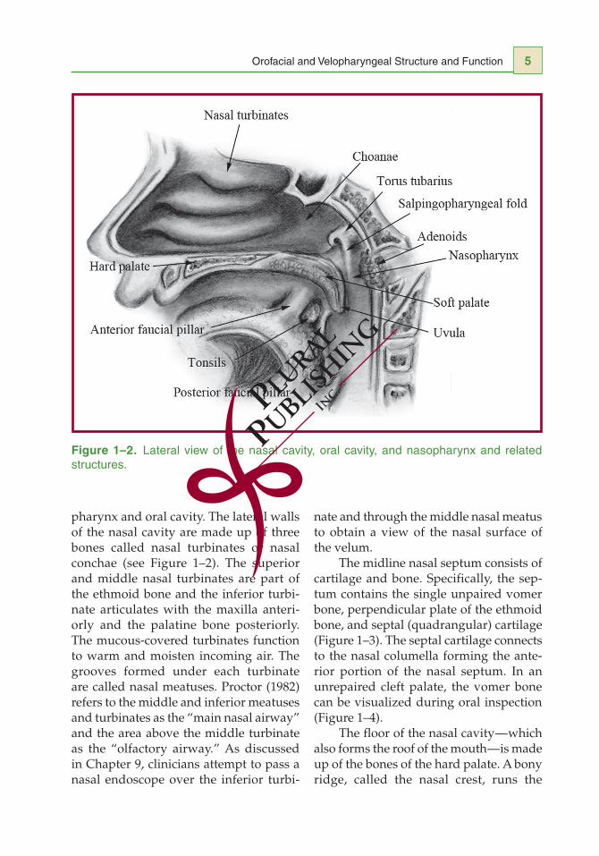
The nasal cavity consists of the airway from the nares to the nasopharynx (Fig-ure 1–2). The nasal cavity is the first and last point of airflow to and from the lungs and provides important physiologic func-
tions such as filtering, humidification, and temperature regulation of inspired air. Dur-ing speech production, the nasal cavity also functions as an air-filled resonator for nasal consonants (see Chapter 8). The smallest cross-sectional area of the nasal cavity is called the internal nasal valve (Hixon, Weismer, & Hoit, 2008; Proctor, 1982). This valve is located approximately 1 cm from the vestibule (entrance) of the nose and is bounded by the upper lateral cartilage, the medial wall of the septum, and the anterior part of the inferior turbinate. The internal nasal valve provides the greatest resistance to inspired airflow, accounting for approxi-mately two-thirds of the total resistance of the nasal airway (Foster, 1962). Just before entering the nasopharynx, the nasal airway narrows again into a funnel-like structure called the choanae.
The nasal cavity is lined with mucous membrane that is continuous with the
Figure 1–1. Facial landmarks.
Orofacial and Velopharyngeal Structure and Function 5
pharynx and oral cavity. The lateral walls of the nasal cavity are made up of three bones called nasal turbinates or nasal conchae (see Figure 1–2). The superior and middle nasal turbinates are part of the ethmoid bone and the inferior turbi-nate articulates with the maxilla anteri-orly and the palatine bone posteriorly. The mucous-covered turbinates function to warm and moisten incoming air. The grooves formed under each turbinate are called nasal meatuses. Proctor (1982) refers to the middle and inferior meatuses and turbinates as the “main nasal airway” and the area above the middle turbinate as the “olfactory airway.” As discussed in Chapter 9, clinicians attempt to pass a nasal endoscope over the inferior turbi-
nate and through the middle nasal meatus to obtain a view of the nasal surface of the velum.
The midline nasal septum consists of cartilage and bone. Specifically, the sep-tum contains the single unpaired vomer bone, perpendicular plate of the ethmoid bone, and septal (quadrangular) cartilage (Figure 1–3). The septal cartilage connects to the nasal columella forming the ante-rior portion of the nasal septum. In an unrepaired cleft palate, the vomer bone can be visualized during oral inspection (Figure 1–4).
The floor of the nasal cavity — which also forms the roof of the mouth — is made up of the bones of the hard palate. A bony ridge, called the nasal crest, runs the
Figure 1–2. Lateral view of the nasal cavity, oral cavity, and nasopharynx and related structures.

Evaluation and Management of Cleft Lip and Palate: A Developmental Perspective6
length of the superior nasal surface of the hard palate and serves as the attachment of the nasal septum (vomer bone) to the nasal floor. As shown in Figure 1–4, how-
ever, the vomer bone does not attach to the hard palate when a complete bilateral cleft of the secondary palate occurs (see Chapter 2).
Figure 1–3. Structures of the midline nasal septum.
Figure 1–4. Oral view of a complete cleft palate. Note the midline nasal septum visible through oral inspection.
Orofacial and Velopharyngeal Structure and Function 7
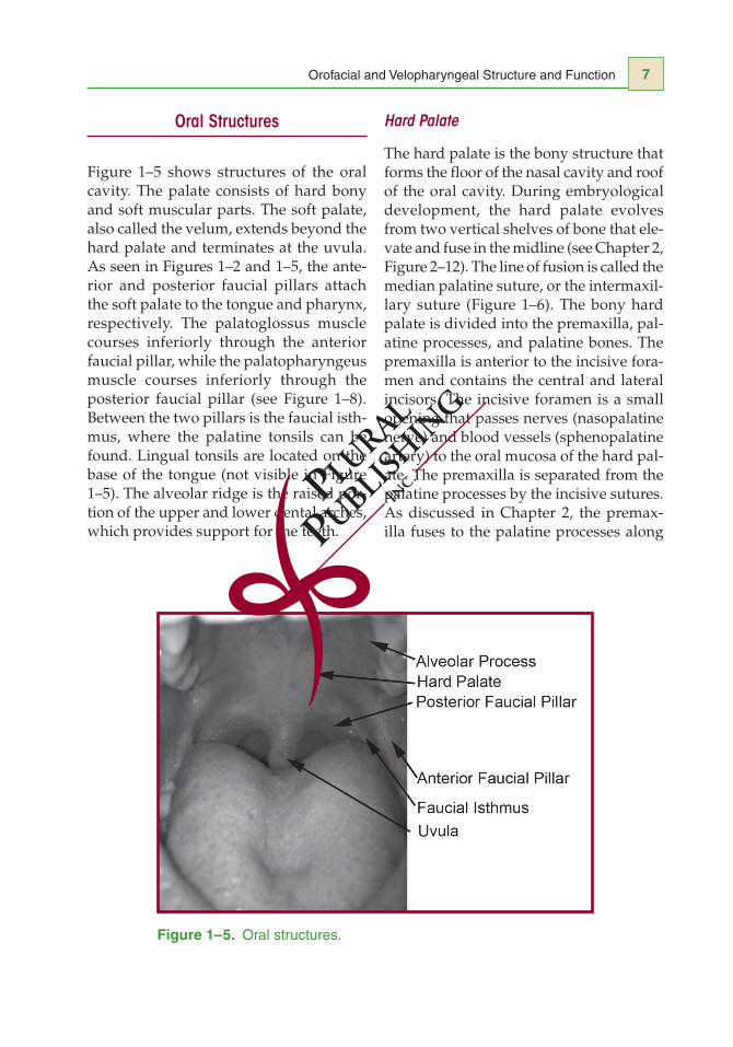
oral Structures
Figure 1–5 shows structures of the oral cavity. The palate consists of hard bony and soft muscular parts. The soft palate, also called the velum, extends beyond the hard palate and terminates at the uvula. As seen in Figures 1–2 and 1–5, the ante-rior and posterior faucial pillars attach the soft palate to the tongue and pharynx, respectively. The palatoglossus muscle courses inferiorly through the anterior faucial pillar, while the palatopharyngeus muscle courses inferiorly through the posterior faucial pillar (see Figure 1–8). Between the two pillars is the faucial isth-mus, where the palatine tonsils can be found. Lingual tonsils are located on the base of the tongue (not visible in Figure 1–5). The alveolar ridge is the raised por-tion of the upper and lower dental arches, which provides support for the teeth.
Hard Palate
The hard palate is the bony structure that forms the floor of the nasal cavity and roof of the oral cavity. During embryological development, the hard palate evolves from two vertical shelves of bone that ele-vate and fuse in the midline (see Chapter 2, Figure 2–12). The line of fusion is called the median palatine suture, or the intermaxil-lary suture (Figure 1–6). The bony hard palate is divided into the premaxilla, pal-atine processes, and palatine bones. The premaxilla is anterior to the incisive fora-men and contains the central and lateral incisors. The incisive foramen is a small opening that passes nerves (nasopalatine nerve) and blood vessels (sphenopalatine artery) to the oral mucosa of the hard pal-ate. The premaxilla is separated from the palatine processes by the incisive sutures. As discussed in Chapter 2, the premax-illa fuses to the palatine processes along
Figure 1–5. Oral structures.
Related Documents

![Cleft Lip Palate[1]](https://static.cupdf.com/doc/110x72/577cdb8f1a28ab9e78a88308/cleft-lip-palate1.jpg)