doi: 10.1098/rstb.2009.0257 , 617-630 365 2010 Phil. Trans. R. Soc. B Abderrahman Khila and Ehab Abouheif evolution Evaluating the role of reproductive constraints in ant social References http://rstb.royalsocietypublishing.org/content/365/1540/617.full.html#ref-list-1 This article cites 72 articles, 19 of which can be accessed free Rapid response http://rstb.royalsocietypublishing.org/letters/submit/royptb;365/1540/617 Respond to this article Subject collections (2263 articles) evolution (216 articles) developmental biology (97 articles) cellular biology (308 articles) molecular biology Articles on similar topics can be found in the following collections Email alerting service here right-hand corner of the article or click Receive free email alerts when new articles cite this article - sign up in the box at the top http://rstb.royalsocietypublishing.org/subscriptions go to: Phil. Trans. R. Soc. B To subscribe to This journal is © 2010 The Royal Society on December 16, 2010 rstb.royalsocietypublishing.org Downloaded from

Welcome message from author
This document is posted to help you gain knowledge. Please leave a comment to let me know what you think about it! Share it to your friends and learn new things together.
Transcript
doi: 10.1098/rstb.2009.0257, 617-630365 2010 Phil. Trans. R. Soc. B
Abderrahman Khila and Ehab Abouheif evolutionEvaluating the role of reproductive constraints in ant social
Referenceshttp://rstb.royalsocietypublishing.org/content/365/1540/617.full.html#ref-list-1
This article cites 72 articles, 19 of which can be accessed free
Rapid responsehttp://rstb.royalsocietypublishing.org/letters/submit/royptb;365/1540/617
Respond to this article
Subject collections
(2263 articles)evolution � (216 articles)developmental biology �
(97 articles)cellular biology � (308 articles)molecular biology �
Articles on similar topics can be found in the following collections
Email alerting service hereright-hand corner of the article or click Receive free email alerts when new articles cite this article - sign up in the box at the top
http://rstb.royalsocietypublishing.org/subscriptions go to: Phil. Trans. R. Soc. BTo subscribe to
This journal is © 2010 The Royal Society
on December 16, 2010rstb.royalsocietypublishing.orgDownloaded from
on December 16, 2010rstb.royalsocietypublishing.orgDownloaded from
* Authoabderrah
One concomplex
Phil. Trans. R. Soc. B (2010) 365, 617–630
doi:10.1098/rstb.2009.0257
Evaluating the role of reproductiveconstraints in ant social evolution
Abderrahman Khila* and Ehab Abouheif*
Department of Biology, McGill University, avenue Dr Penfield, Montreal, Quebec H3A 1B1, Canada
The reproductive division of labour is a key feature of eusociality in ants, where queen and workercastes show dramatic differences in the development of their reproductive organs. To understandthe developmental and genetic basis underlying this division of labour, we performed a molecularanalysis of ovary function and germ cell development in queens and workers. We show that theprocesses of ovarian development in queens have been highly conserved relative to the fruitflyDrosophila melanogaster. We also identify specific steps during oogenesis and embryogenesis inwhich ovarian and germ cell development have been evolutionarily modified in the workers.These modifications, which we call ‘reproductive constraints’, are often assumed to represent neu-tral degenerations that are a consequence of social evolutionary forces. Based on our developmentaland functional analysis of these constraints, however, we propose and discuss the alternative hypoth-esis that reproductive constraints represent adaptive proximate mechanisms or traits for maintainingsocial harmony in ants. We apply a multi-level selection framework to help understand the role ofthese constraints in ant social evolution. A complete understanding of how cooperation, conflictand developmental systems evolve in social groups requires a ‘socio-evo-devo’ approach thatintegrates social evolutionary and developmental biology.
Keywords: ants; reproduction; development; germ cells; evo-devo; social evolution
1. INTRODUCTIONEvolutionary developmental biology (evo-devo) isbringing development into the modern synthesis andthe mainstream of evolutionary theory (Hall 1999;Wilkins 2002; Eberhard 2003; Carroll et al. 2004).Evo-devo, however, has yet to be integrated with thefield of social evolution, which was a critical themearising from the modern synthesis. This is in spite ofthe fact that the social environment of individuals ina population can influence their development, and inturn development can feed back to influence theirsocial environment (Toth & Robinson 2007; Smithet al. 2008). Thus, a ‘socio-evo-devo’ approach isneeded for a complete understanding of howcooperation, conflict and developmental systemsevolve in social groups (Toth & Robinson 2007). Tounderstand the crosstalk between social evolutionaryforces (ultimate mechanisms) and developmental pro-cesses (proximate mechanisms), this socio-evo-devoapproach must integrate the concepts and approachesof both social evolutionary and developmental biology.
Here, we focus on a key feature of eusociality inants—the reproductive division of labour betweenqueens and workers—where queens perform most ofthe reproduction and workers perform the rest of thetasks such as foraging and brood rearing (Holldobler &Wilson 1990, 2008). Workers in the majority of antspecies have retained their ovaries, but do not have
rs for correspondence ([email protected];[email protected]).
tribution of 12 to a Theme Issue ‘From polyphenism tometazoan life cycles’.
617
the same reproductive capacity as the queen becausethere exist several constraints on worker reproduction(Holldobler & Wilson 1990, 2008). We call these con-straints ‘reproductive constraints’, and define them asany process that quantitatively or qualitatively reducesthe ability of workers to reproduce relative to thequeen. Reproductive constraints can act through devel-opment or behaviour. Many behavioural reproductiveconstraints exist, which include policing (where workersaggress reproducing workers or kill each other’s eggs) orself-restraint (where workers would refrain from repro-ducing). Here, we focus only on reproductiveconstraints that involve changes in the development ofreproductive organs in worker ants. We consider con-straints to be different from one another if theyoriginate from the modification of distinct developmen-tal processes in the workers. Based on this criterion, weidentified five types of reproductive constraints,which we call RC1–RC5: RC1 acts through the mis-localization of maternal determinants (mRNAs andproteins) during late oogenesis and leads to embryonicfailure of worker offspring (Khila & Abouheif 2008);RC2 affects the quantitative activity of the ovaries inadult workers; RC3 refers to loss of the sperm storageorgan (spermatheca); RC4 refers to the reduction ofovariole number in the workers; and finally, RC5refers to the complete loss of worker reproductiveorgans. Our general goal is to analyse the molecularand developmental basis of these constraints onworker reproduction and attempt to place them in thecontext of social evolutionary theory.
Inclusive fitness or kin selection theory has been keyto understanding the conditions under which naturalselection will favour cooperation within a group of
This journal is q 2010 The Royal Society
618 A. Khila & E. Abouheif The socio-evo-devo of ants
on December 16, 2010rstb.royalsocietypublishing.orgDownloaded from
relatives. It can be summed in a mathematicalexpression called Hamilton’s (1964a,b) rule:
rb � c;
where r is the genetic relatedness between individualsin a population, b is the benefit, in terms of extra off-spring that the beneficiary gains and c is the cost interms of offspring loss that the altruist suffers. Thus,if Hamilton’s inequality is satisfied, then the gene foraltruism will spread throughout a group of individuals.In general, Hamilton’s rule highlights both the geneticfactors (as they affect relatedness) and ecologicalfactors (as they affect costs and benefits) that promotecooperation and conflict in social groups (Bourke &Franks 1995). Although kin selection theory has longdominated the field of social evolution, it remains ani-mated with controversy—most often ignited by theideas and semantic confusion surrounding the alterna-tive theoretical framework of group selection (Wilson &Holldobler 2005; Fletcher et al. 2006; Foster et al.2006a,b; Lehmann et al. 2007; West et al. 2007,2008; Wilson & Wilson 2007; Kohn 2008; Wilson2008a,b; Pennisi 2009). This is in spite of the factthat theorists in social evolution have for a long timeagreed that, if certain conditions are satisfied,the theories of multi-level selection and kin selectionare mathematically equivalent to one another andgive the same predictions (Bourke & Franks 1995;Lehmann et al. 2007). These conditions are satisfiedas long as in multi-level selection theory a ‘within-group’ and a ‘between-group’ component of selectionare specified and individuals within these groups forma physical group of relatives, which is the case for socialinsects (Bourke & Franks 1995; Korb & Heinze 2004).Under this multi-level selection perspective, a genepromoting altruism will spread if the within-groupconflict is weaker than between-group selection(Bourke & Franks 1995). Although there is stilldebate about which framework is the most use-ful (West et al. 2007, 2008; Wilson 2008a,b), wediscuss the role of reproductive constraints usinga multi-level selection framework (Korb & Heinze2004).
However, several theoretical models of social evo-lution (Bourke 1999; Jeon & Choe 2003; Wenseleerset al. 2004; Reeve & Holldobler 2007; Gardner &Grafen 2009) implicitly or explicitly assume thatreproductive constraints that affect development areneutral degenerations of worker ovaries in responseto either kin or multi-level selection. Bourke (1999),for example, hypothesizes that as the size of a colonyincreases, it reduces the probability that any particularworker in the colony can replace the queen. This inturn increases selection for workers to mutually inhibiteach other’s reproduction through worker policing.This leads to the eventual decay or loss of workerreproductive potential, which allows for greater mor-phological differences to evolve between the queenand the workers.
In this article we propose an alternative hypothesisfor the role of reproductive constraints—that they areadaptive proximate mechanisms or traits that reinforcesocial harmony in ants. To support our hypothesis, we
Phil. Trans. R. Soc. B (2010)
first show that the processes of ovarian developmentand function in queens have been highly conservedrelative to the fruitfly Drosophila melanogaster. Wethen identify the likely developmental genetic basis ofseveral reproductive constraints, which occur atdefined steps during development. Based on theseresults, we argue that the development of workerreproductive organs has been modified through aspecific and narrow range of developmental par-ameters, which may not be expected if ovarymodifications in the workers were a result of neutraldecay mechanisms. Given this narrow range of devel-opmental parameters, we also propose that workerovaries may have been selectively modified to fulfil arange of alternative functions, including trophic eggproduction (Crespi 1992) and signalling physiologicalinstructive cues to the behavioural response systems(Amdam et al. 2006).
2. MATERIAL AND METHODS(a) Preparation of whole reproductive organs
Females were anaesthetized with carbon dioxide andimmersed in PTW (1�phosphate buffered saline(PBS); 0.05% Tween-20). Reproductive organs canbe recovered by pulling with forceps on the last tergite.After removing fat and unrelated tissues, images ofreproductive organs were captured using a Zeissscope and the AXIOVISION software.
(b) Cloning and in situ hybridization
of Aphaenogaster rudis torTotal RNA was extracted from queen ovaries, and usedas template in a first-strand complementary DNA syn-thesis reaction (Invitrogen). This first-strand cDNApool was used as a PCR template to amplify a 1600base pairs (bp) tor fragment. The following forwardand reverse tor primers were synthesized based on ator sequence alignment from closely related insects:forward: 50-TAGCCGAGTTTATGGAACATTGCGA-30 and reverse: 50-TGCGGTATAACTTGTAGCCAAGTGTTT-30. Sequence alignment usingClustalW showed high levels of amino acid similaritybetween Aphaenogaster rudis and honeybee TORprotein, indicating that the cloned fragment representsA. rudis tor locus. Aphaenogaster rudis tor sequence canbe retrieved in GenBank under the following accessionnumber: GQ892187. In situ hybridization was con-ducted following the protocol in Khila & Abouheif(2009).
(c) Ovary and embryo dissection and fixation
Ovaries were dissected in PTW (1�PBS; 0.1%Tween-20) and kept on ice during the dissection pro-cess. The peritoneal sheet covering each ovariole wasremoved using fine forceps. The ovarioles were fixedin 4 per cent paraformaldehyde (200 ml) sup-plemented with 10 per cent DMSO (20 ml) andheptane (600 ml) for 20 min at room temperature.The fixed ovarioles were then washed three times inPBT (1 � PBS; 0.3% Triton X-100) and processedfor subsequent staining. When processed for actin ormicrotubule staining, tissue should not be treatedwith methanol at any stage of the fixation process.
Vasa, actin,nuclei
α-tubulinactinnuclei
α-tubulin, actin,nuclei α-tubulin, actin, nuclei α-tubulin, actin, nuclei
Bicaudal-Cnuclei
Bicaudal-Cnuclei
α-tubulin, actin, nuclei Vasa, actin, nuclei
pole plasm
spermathecagenitalia
oviducts
ovarioles
oocytes
TFGSC
Cb
oocytes
oocytes
follicle
follicle
folliclecells
nursecells
uterus
dividing cysts
oocytefoliclecells
stem cellsring canal nurse cells
cystoblast
terminalfilament
pro-oocyte determined oocytes egg chamber(follicle)
cysts
follicle cells
ringcanal
pole plasm pole cells
gonad
gonadsVasa; nuclei Vasa; nuclei
nuclei
Vasa; nuclei
Vasa;nuclei
(a)
(i)
(o)(n) (p) (q) (r)
(s)
(k)( j) (l) (m)
(h)
( f ) (g)
(b) (c) (d) (e)
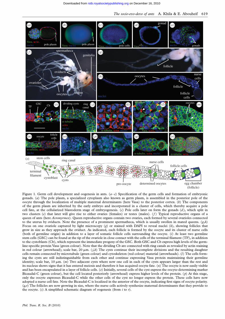
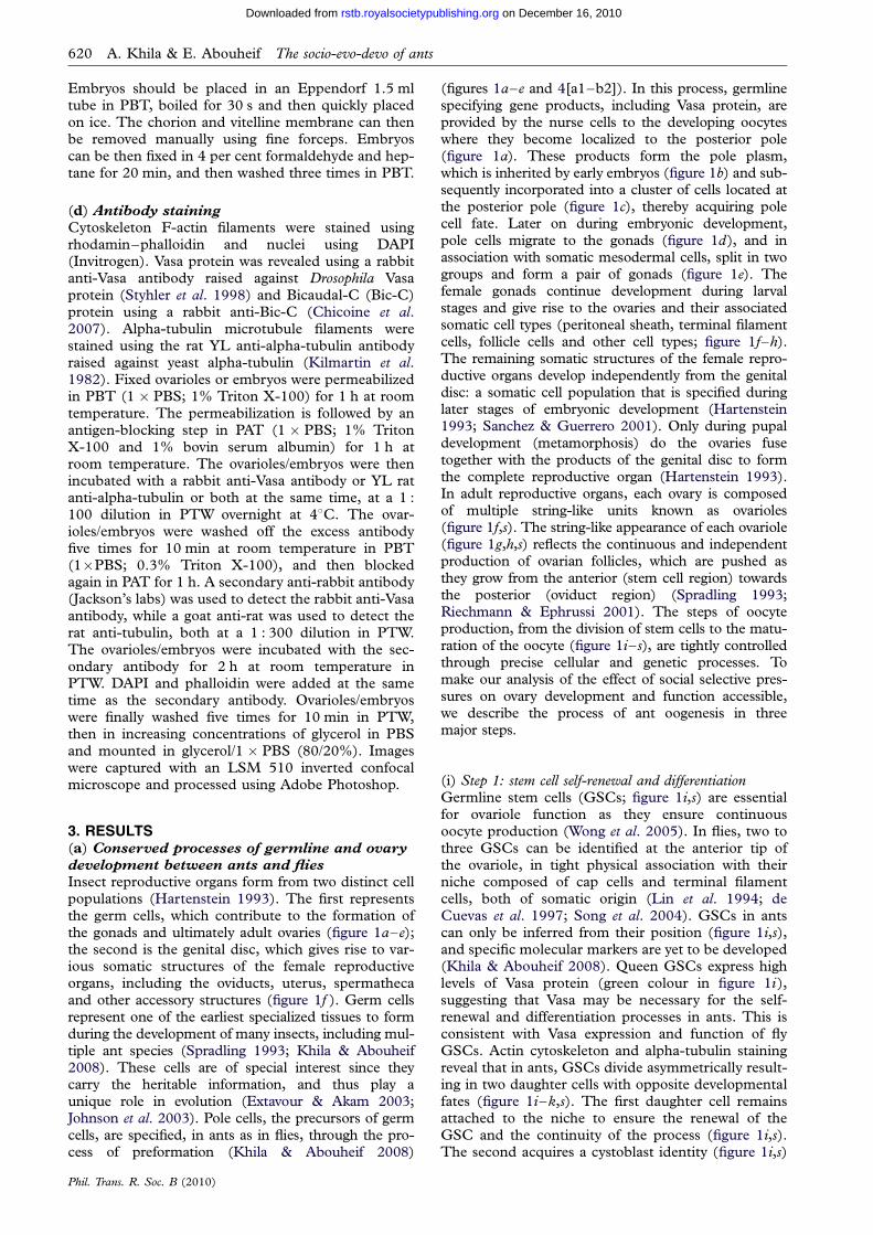
Figure 1. Germ cell development and oogenesis in ants. (a–e) Specification of the germ cells and formation of embryonic
gonads. (a) The pole plasm, a specialized cytoplasm also known as germ plasm, is assembled in the posterior pole of theoocyte through the localization of multiple maternal determinants (here Vasa) to the posterior cortex. (b) The componentsof the germ plasm are inherited by the early embryo and incorporated in a cluster of cells, which thereby acquire a polecell fate, at the cellularized blastoderm stage of embryogenesis. (c) Pole cells later on form the gonads (d ), which split intwo clusters (e) that later will give rise to either ovaries (females) or testes (males). ( f ) Typical reproductive organs of a
queen of ants (here Acromyrmex). Queen reproductive organs contain two ovaries, each formed by several ovarioles connectedto the uterus by oviducts. Note the presence of a prominent spermatheca, which is usually swollen in mated queens. (g,h)Focus on one ovariole captured by light microscopy (g) or stained with DAPI to reveal nuclei (h), showing follicles thatgrow in size as they approach the oviduct. As indicated, each follicle is formed by the oocyte and its cluster of nurse cells
(both of germline origin) in addition to a layer of somatic follicle cells surrounding the oocyte. (i) At least two germlinestem cells (GSC) can be found at the tip of the ovariole in close contact with the cells of the terminal filament (TF), in additionto the cystoblasts (Cb), which represent the immediate progeny of the GSC. Both GSC and Cb express high levels of the germ-line-specific protein Vasa (green colour). Note that the dividing Cb are connected with ring canals as revealed by actin stainingin red colour (arrowhead); scale bar, 20 mm. ( j,k) The cysts continue their incomplete divisions and the resulting daughter
cells remain connected by microtubule (green colour) and cytoskeleton (red colour) material (arrowheads). (l ) The cells form-ing the cysts are still indistinguishable from each other and continue expressing Vasa protein maintaining their germlineidentity; scale bar, 10 mm. (m) Two adjacent cysts where now one cell in each of the cysts appears larger than the rest andits nucleus shows signs that it has entered meiosis and therefore it has acquired oocyte fate. (n) The oocyte is now easily visibleand has been encapsulated in a layer of follicle cells. (o) Initially, several cells of the cyst express the oocyte-determining marker
Bicaudal-C (green colour), but the cell located posteriorly (arrowhead) express higher levels of the protein. (p) At this stage,only the oocyte expresses Bicaudal-C while the other cells of the cyst no longer express the protein. These cells have nowadopted a nurse cell fate. Note the Bicaudal-C is localized in the anterior of the oocyte, indicating first signs of oocyte polarity.(q,r) The follicles are now growing in size, where the nurse cells actively synthesize maternal determinants that they provide tothe oocyte. (s) A simplified schematic diagram of oogenesis (from i to r).
The socio-evo-devo of ants A. Khila & E. Abouheif 619
Phil. Trans. R. Soc. B (2010)
on December 16, 2010rstb.royalsocietypublishing.orgDownloaded from
620 A. Khila & E. Abouheif The socio-evo-devo of ants
on December 16, 2010rstb.royalsocietypublishing.orgDownloaded from
Embryos should be placed in an Eppendorf 1.5 mltube in PBT, boiled for 30 s and then quickly placedon ice. The chorion and vitelline membrane can thenbe removed manually using fine forceps. Embryoscan be then fixed in 4 per cent formaldehyde and hep-tane for 20 min, and then washed three times in PBT.
(d) Antibody staining
Cytoskeleton F-actin filaments were stained usingrhodamin–phalloidin and nuclei using DAPI(Invitrogen). Vasa protein was revealed using a rabbitanti-Vasa antibody raised against Drosophila Vasaprotein (Styhler et al. 1998) and Bicaudal-C (Bic-C)protein using a rabbit anti-Bic-C (Chicoine et al.2007). Alpha-tubulin microtubule filaments werestained using the rat YL anti-alpha-tubulin antibodyraised against yeast alpha-tubulin (Kilmartin et al.1982). Fixed ovarioles or embryos were permeabilizedin PBT (1 � PBS; 1% Triton X-100) for 1 h at roomtemperature. The permeabilization is followed by anantigen-blocking step in PAT (1 � PBS; 1% TritonX-100 and 1% bovin serum albumin) for 1 h atroom temperature. The ovarioles/embryos were thenincubated with a rabbit anti-Vasa antibody or YL ratanti-alpha-tubulin or both at the same time, at a 1 :100 dilution in PTW overnight at 48C. The ovar-ioles/embryos were washed off the excess antibodyfive times for 10 min at room temperature in PBT(1�PBS; 0.3% Triton X-100), and then blockedagain in PAT for 1 h. A secondary anti-rabbit antibody(Jackson’s labs) was used to detect the rabbit anti-Vasaantibody, while a goat anti-rat was used to detect therat anti-tubulin, both at a 1 : 300 dilution in PTW.The ovarioles/embryos were incubated with the sec-ondary antibody for 2 h at room temperature inPTW. DAPI and phalloidin were added at the sametime as the secondary antibody. Ovarioles/embryoswere finally washed five times for 10 min in PTW,then in increasing concentrations of glycerol in PBSand mounted in glycerol/1 � PBS (80/20%). Imageswere captured with an LSM 510 inverted confocalmicroscope and processed using Adobe Photoshop.
3. RESULTS(a) Conserved processes of germline and ovary
development between ants and flies
Insect reproductive organs form from two distinct cellpopulations (Hartenstein 1993). The first representsthe germ cells, which contribute to the formation ofthe gonads and ultimately adult ovaries (figure 1a–e);the second is the genital disc, which gives rise to var-ious somatic structures of the female reproductiveorgans, including the oviducts, uterus, spermathecaand other accessory structures (figure 1f ). Germ cellsrepresent one of the earliest specialized tissues to formduring the development of many insects, including mul-tiple ant species (Spradling 1993; Khila & Abouheif2008). These cells are of special interest since theycarry the heritable information, and thus play aunique role in evolution (Extavour & Akam 2003;Johnson et al. 2003). Pole cells, the precursors of germcells, are specified, in ants as in flies, through the pro-cess of preformation (Khila & Abouheif 2008)
Phil. Trans. R. Soc. B (2010)
(figures 1a–e and 4[a1–b2]). In this process, germlinespecifying gene products, including Vasa protein, areprovided by the nurse cells to the developing oocyteswhere they become localized to the posterior pole(figure 1a). These products form the pole plasm,which is inherited by early embryos (figure 1b) and sub-sequently incorporated into a cluster of cells located atthe posterior pole (figure 1c), thereby acquiring polecell fate. Later on during embryonic development,pole cells migrate to the gonads (figure 1d), and inassociation with somatic mesodermal cells, split in twogroups and form a pair of gonads (figure 1e). Thefemale gonads continue development during larvalstages and give rise to the ovaries and their associatedsomatic cell types (peritoneal sheath, terminal filamentcells, follicle cells and other cell types; figure 1f–h).The remaining somatic structures of the female repro-ductive organs develop independently from the genitaldisc: a somatic cell population that is specified duringlater stages of embryonic development (Hartenstein1993; Sanchez & Guerrero 2001). Only during pupaldevelopment (metamorphosis) do the ovaries fusetogether with the products of the genital disc to formthe complete reproductive organ (Hartenstein 1993).In adult reproductive organs, each ovary is composedof multiple string-like units known as ovarioles(figure 1f,s). The string-like appearance of each ovariole(figure 1g,h,s) reflects the continuous and independentproduction of ovarian follicles, which are pushed asthey grow from the anterior (stem cell region) towardsthe posterior (oviduct region) (Spradling 1993;Riechmann & Ephrussi 2001). The steps of oocyteproduction, from the division of stem cells to the matu-ration of the oocyte (figure 1i–s), are tightly controlledthrough precise cellular and genetic processes. Tomake our analysis of the effect of social selective pres-sures on ovary development and function accessible,we describe the process of ant oogenesis in threemajor steps.
(i) Step 1: stem cell self-renewal and differentiationGermline stem cells (GSCs; figure 1i,s) are essentialfor ovariole function as they ensure continuousoocyte production (Wong et al. 2005). In flies, two tothree GSCs can be identified at the anterior tip ofthe ovariole, in tight physical association with theirniche composed of cap cells and terminal filamentcells, both of somatic origin (Lin et al. 1994; deCuevas et al. 1997; Song et al. 2004). GSCs in antscan only be inferred from their position (figure 1i,s),and specific molecular markers are yet to be developed(Khila & Abouheif 2008). Queen GSCs express highlevels of Vasa protein (green colour in figure 1i),suggesting that Vasa may be necessary for the self-renewal and differentiation processes in ants. This isconsistent with Vasa expression and function of flyGSCs. Actin cytoskeleton and alpha-tubulin stainingreveal that in ants, GSCs divide asymmetrically result-ing in two daughter cells with opposite developmentalfates (figure 1i–k,s). The first daughter cell remainsattached to the niche to ensure the renewal of theGSC and the continuity of the process (figure 1i,s).The second acquires a cystoblast identity (figure 1i,s)
The socio-evo-devo of ants A. Khila & E. Abouheif 621
on December 16, 2010rstb.royalsocietypublishing.orgDownloaded from
and activates differentiation programmes, moves awayfrom the niche and engages in successive incompletedivisions to become a cyst (figure 1i–n,s) (de Cuevaset al. 1996). This distinct fate of GSC progeny inflies is instructed by molecular signals emanatingfrom the neighbouring niche cells (Wong et al.2005). This suggests that similar signals may beresponsible for GSC self-renewal and differentiationin ants. Each cystoblast undergoes five incomplete div-isions in ants, resulting in 32 interconnected cells thatform the cyst, whereas in Drosophila only four divisionsoccur, resulting in 16-cell cysts (van Eeden &St Johnston 1999).
(ii) Step 2: determination of oocyte fate and polarityAt each division of the cystoblast, the resulting daugh-ter cells remain connected by cytoplasmic bridgescalled ring canals, which ensure communicationbetween the cells forming the cyst (figure 1i–m,s)(van Eeden & St Johnston 1999; Huynh & St Johnston2004). In ants, four to five cells located at the posteriorof the cyst accumulate Bic-C protein (figure 1o).Shortly after, Bic-C now accumulates only in the celllocated most posteriorly in the cyst (figure 1p), indica-ting that this cell has acquired the oocyte fate, consistentwith this process in Drosophila (van Eeden &St Johnston 1999; Riechmann & Ephrussi 2001;Huynh & St Johnston 2004). The remaining cells ofthe cyst now exhibit large nuclei, indicating that thesecells have adopted a nurse cell fate (figure 1p–s).In both ants and flies, only one cell is chosen as anoocyte and accumulates products of polarity genes(in addition to other cell cycle components) at itsanterior pole (Huynh & St Johnston 2004). This cellenters meiosis, whereas the remaining 31 cellsengage in multiple chromosomal endoreplicationsand become polyploid nurse cells (figure 1p– s). Thisresults in the meroistic polytrophic mode of oogenesis,where a cluster of nurse cells assists each oocyte(figure 1q,s). The oocyte fate, after being specified,has to be maintained in order for oogenesis to progress(Cox et al. 2001; Huynh & St Johnston 2004). In ants,during the encapsulation of the cyst by follicle cells(figure 1o), four neighbouring cells in the cyst showa perinuclear accumulation of the Bic-C protein. How-ever, the cell located most posteriorly in the cystaccumulates higher amounts of the Bic-C protein,indicating that this cell has been defined as an oocyte(arrowhead in figure 1o). Shortly thereafter, only thiscell shows Bic-C accumulation, with a clear transloca-tion of the protein to the anterior pole suggesting thatthis oocyte is already polarized at such an early stage.This indicates that the processes of oocyte deter-mination and polarity establishment are highlyconserved between ants and Drosophila.
(iii) Step 3: patterning of the oocyteThe active molecular crosstalk between the oocyte andthe surrounding layer of somatic follicle cells is essen-tial for establishing the polarity of the egg chamber.The localization of three main molecules in Drosophila,Gurken protein, bicoid mRNA and Oskar protein,initiates the major events leading to the patterning of
Phil. Trans. R. Soc. B (2010)
the oocyte in preparation for early embryogenesis(Steinhauer & Kalderon 2006). The patterning ofthe posterior compartment in ants involves the local-ization of conserved maternal determinants, includingVasa protein and nanos mRNA (Khila & Abouheif2008), and the assembly of the pole plasm in the pos-terior pole (figure 1a). These maternal determinantsare synthesized by the nurse cells and transported tothe oocyte through microtubule cytoskeleton to theirrespective locations (figure 1n,q–r). How the othercompartments are established in ant oocytes remainsunknown.
(b) Reproductive function of worker ovaries
is blocked or reduced at specific steps
during oogenesis
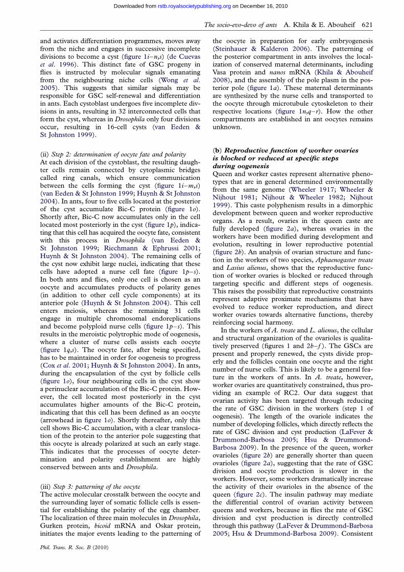
Queen and worker castes represent alternative pheno-types that are in general determined environmentallyfrom the same genome (Wheeler 1917; Wheeler &Nijhout 1981; Nijhout & Wheeler 1982; Nijhout1999). This caste polyphenism results in a dimorphicdevelopment between queen and worker reproductiveorgans. As a result, ovaries in the queen caste arefully developed (figure 2a), whereas ovaries in theworkers have been modified during development andevolution, resulting in lower reproductive potential(figure 2b). An analysis of ovarian structure and func-tion in the workers of two species, Aphaenogaster treateand Lasius alienus, shows that the reproductive func-tion of worker ovaries is blocked or reduced throughtargeting specific and different steps of oogenesis.This raises the possibility that reproductive constraintsrepresent adaptive proximate mechanisms that haveevolved to reduce worker reproduction, and directworker ovaries towards alternative functions, therebyreinforcing social harmony.
In the workers of A. treate and L. alienus, the cellularand structural organization of the ovarioles is qualita-tively preserved (figures 1 and 2b– f ). The GSCs arepresent and properly renewed, the cysts divide prop-erly and the follicles contain one oocyte and the rightnumber of nurse cells. This is likely to be a general fea-ture in the workers of ants. In A. treate, however,worker ovaries are quantitatively constrained, thus pro-viding an example of RC2. Our data suggest thatovarian activity has been targeted through reducingthe rate of GSC division in the workers (step 1 ofoogenesis). The length of the ovariole indicates thenumber of developing follicles, which directly reflects therate of GSC division and cyst production (LaFever &Drummond-Barbosa 2005; Hsu & Drummond-Barbosa 2009). In the presence of the queen, workerovarioles (figure 2b) are generally shorter than queenovarioles (figure 2a), suggesting that the rate of GSCdivision and oocyte production is slower in theworkers. However, some workers dramatically increasethe activity of their ovarioles in the absence of thequeen (figure 2c). The insulin pathway may mediatethe differential control of ovarian activity betweenqueens and workers, because in flies the rate of GSCdivision and cyst production is directly controlledthrough this pathway (LaFever & Drummond-Barbosa2005; Hsu & Drummond-Barbosa 2009). Consistent
direction of oocyte maturation
f1 g1 g2
nursecells
oocyte
tor mRNA expression
developing cysts
*
12
3 4
12 3 4
e1
h1
h2
i1
i2
(a)
(d)
(b) (c)
(e)
( f )
(h) (i)
(g)
Figure 2. Functions of female reproductive organs in ants. (a) Reproductive organs of a queen of A. rudis showing the highnumber of ovarioles in the ovaries. (b) Reproductive organs of a worker, from a queened colony, where each ovary containsonly one short ovariole. (c) Reproductive organs of a worker from a colony without a queen, showing that the ovarioles arenow longer, which indicates much higher activity. (d) Ovariole from an A. rudis worker showing the expression of tor mRNAin the early developing oocytes and in the nurse cells. tor mRNA is transported from the nurse cells to the oocyte in older follicles.
(e) Ovariole of a worker from a queened colony, stained for nuclei (blue colour) and Vasa protein (green colour). Note in themature oocyte the absence of Vasa localization at the posterior pole (e1), indicating that this oocyte is trophic. ( f,g) Same ovarioleas in (c), of a worker from a colony without a queen, stained for nuclei (blue colour) and Vasa protein (green colour). In additionto the higher number of follicles, many oocytes now localize Vasa to the posterior pole (f1 and g2) while other do not (g1). Thisindicates that the same ovariole is producing both trophic and reproductive oocytes in the absence of the queen. (h,i ) Ovarioles of
L. alienus workers from a colony without a queen, stained for nuclei (blue colour), actin (red colour) and Vasa (green colour).Note that these workers make oocytes with mis-localized Vasa (h2) and others with correct Vasa localization (h1, i1 and i2).
622 A. Khila & E. Abouheif The socio-evo-devo of ants
Phil. Trans. R. Soc. B (2010)
on December 16, 2010rstb.royalsocietypublishing.orgDownloaded from
Mon
omor
ium
em
erso
niM
esso
r pe
rgan
dei
CNS
gonads
no gonads
degenerating GCs
GCs
germplasm
degeneratingGCs
(a) (b) (c)
(d) (e) ( f )
Figure 3. The developmental basis of germ cell reduction or loss in the workers. (a–c) Worker-destined embryos of the ant M.pergandei stained for nanos mRNA to reveal the germ cells (GCs). (a) Embryo at early gastrulation, a stage where the germ cellsare still developing properly. (b) A slightly older embryo showing that the germ cells are now degenerating. (c) A late embryo
showing the remainder of germ cells, which have populated the gonads. (d– f ) Embryos of the ant M. emersoni stained for Vasaprotein to reveal the germ cells. (d) Early embryo showing the proper formation of the germ plasm at the posterior pole. (e)Embryo at gastrulation showing that the germ cells are undergoing degeneration. ( f ) A late embryo showing that the gonadsare absent, and that Vasa now accumulates in the central nervous system (CNS).
The socio-evo-devo of ants A. Khila & E. Abouheif 623
on December 16, 2010rstb.royalsocietypublishing.orgDownloaded from
with this hypothesis, we found that the ovariolesof the ant A. rudis express mRNA of the proteinkinase tor (target of rapamycin) throughout oogenesis(figure 2d). tor is a major component of the insulinsignalling pathway (Fingar & Blenis 2004), and itsexpression represents a preliminary indication thatthis pathway is conserved and active in ants. There-fore, RC2 represents the ability to quantitativelyregulate worker ovaries in response to their socialenvironment.
This RC2 in A. rudis has evolved such that ovarianactivity is partially maintained in the workers when thequeen is present. This allows the production of trophiceggs in colonies with functional queens. Indeed, ovar-ies of these workers contain only oocytes that lack Vasaprotein in their posterior pole (figure 2e,e1). We havepreviously established that oocytes that lack maternaldeterminant localization in the posterior pole willbecome trophic eggs (Khila & Abouheif 2008). Inthe absence of the queen, ovaries of some workersbecome much longer, indicating higher activity(figure 3c). These ovaries, in the absence of thequeen, produce both trophic and reproductiveoocytes. This is shown by the presence of oocytesboth with (figure 2f,f1,g,g2) and without (figure 2g,g1)Vasa localization in the posterior pole (Khila &Abouheif 2008). Therefore, RC2 allows some workersto switch their ovarian activity towards reproduction,probably to produce one last cohort of males in thecase where the queen is lost. In addition to RC2,workers in this species also produce oocytes withmis-localized maternal determinants (RC1), havelost the spermatheca (RC3; figure 2b,c) and have ovar-ies with a reduced number of ovarioles (RC4;figure 2b,c).
Phil. Trans. R. Soc. B (2010)
In L. alienus, worker ovaries are constrainedthrough maternal determinant mis-localization provid-ing an example of RC1 (figure 2h– i). Our data showthat oogenesis in these workers has been targeted atlate stages (step 3). The workers of L. alienus produceoocytes that either fail to localize Vasa (failed oocytes;figure 2h,h2) or show a correctly localized Vasa pattern(viable oocytes; figure 2h,h1, i,i1 and i2). Furthermore,even when Vasa shows a correct localization in youngoocytes, this localization can be lost later during oogen-esis. We found that in the same ovariole, young oocytesshow correctly localized Vasa (figure 2h,h1), whereasolder oocytes lose this localization (figure 2h,h2).Therefore, RC1 represents the mis-localization ofmaternal determinants, such as Vasa, which resultsin developmental failures of the future embryos, andthus, reduced reproduction (Khila & Abouheif2008). The fact that we did not observe any trophiceggs in this species, together with the presence ofRC1, raises the possibility that these ovaries mayhave been maintained for two possible functions: (i)the production of a last cohort of males if the queenis lost and/or (ii) may perform alternative functionsother than trophic egg production. This second possi-bility is consistent with our previous finding in anotherant, Myrmica americana, where the workers producehigh percentages of failed oocytes and low percentagesof trophic oocytes (Khila & Abouheif 2008). This con-clusion is also consistent with the function of workerovaries in the honeybees, where functionally sterileforagers will decide to collect either nectar or pollenbased on the status of their reproductive organs(Amdam et al. 2006). It is possible that in the workersof ants, ovaries emanate or receive endocrine signalsthat regulate the worker’s behavioural status.
624 A. Khila & E. Abouheif The socio-evo-devo of ants
on December 16, 2010rstb.royalsocietypublishing.orgDownloaded from
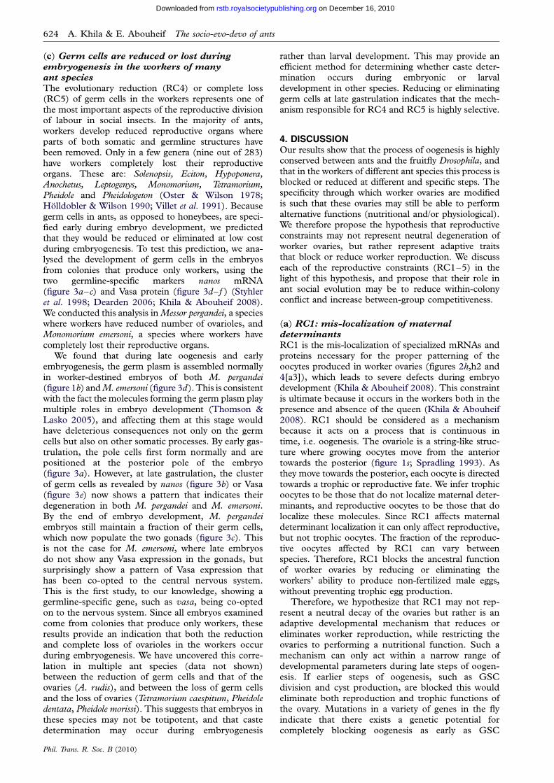
(c) Germ cells are reduced or lost during
embryogenesis in the workers of many
ant species
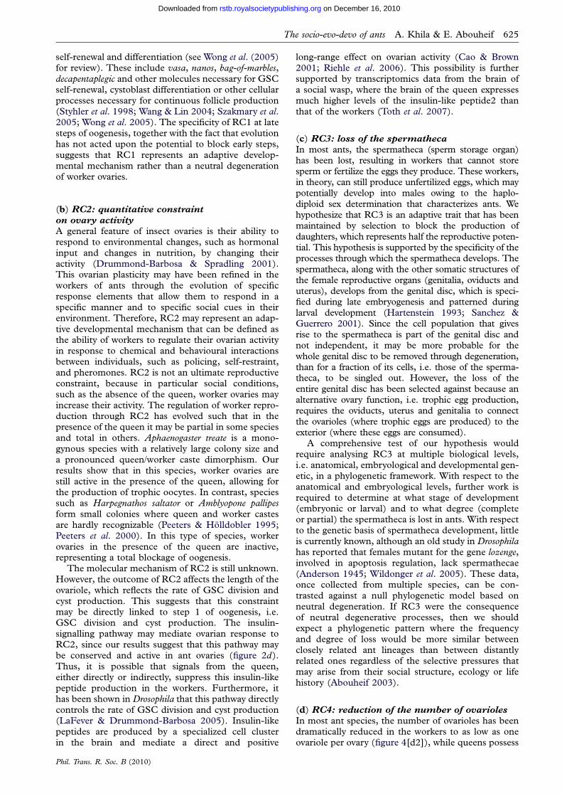
The evolutionary reduction (RC4) or complete loss(RC5) of germ cells in the workers represents one ofthe most important aspects of the reproductive divisionof labour in social insects. In the majority of ants,workers develop reduced reproductive organs whereparts of both somatic and germline structures havebeen removed. Only in a few genera (nine out of 283)have workers completely lost their reproductiveorgans. These are: Solenopsis, Eciton, Hypoponera,Anochetus, Leptogenys, Monomorium, Tetramorium,Pheidole and Pheidologeton (Oster & Wilson 1978;Holldobler & Wilson 1990; Villet et al. 1991). Becausegerm cells in ants, as opposed to honeybees, are speci-fied early during embryo development, we predictedthat they would be reduced or eliminated at low costduring embryogenesis. To test this prediction, we ana-lysed the development of germ cells in the embryosfrom colonies that produce only workers, using thetwo germline-specific markers nanos mRNA(figure 3a–c) and Vasa protein (figure 3d– f ) (Styhleret al. 1998; Dearden 2006; Khila & Abouheif 2008).We conducted this analysis in Messor pergandei, a specieswhere workers have reduced number of ovarioles, andMonomorium emersoni, a species where workers havecompletely lost their reproductive organs.
We found that during late oogenesis and earlyembryogenesis, the germ plasm is assembled normallyin worker-destined embryos of both M. pergandei(figure 1b) and M. emersoni (figure 3d). This is consistentwith the fact the molecules forming the germ plasm playmultiple roles in embryo development (Thomson &Lasko 2005), and affecting them at this stage wouldhave deleterious consequences not only on the germcells but also on other somatic processes. By early gas-trulation, the pole cells first form normally and arepositioned at the posterior pole of the embryo(figure 3a). However, at late gastrulation, the clusterof germ cells as revealed by nanos (figure 3b) or Vasa(figure 3e) now shows a pattern that indicates theirdegeneration in both M. pergandei and M. emersoni.By the end of embryo development, M. pergandeiembryos still maintain a fraction of their germ cells,which now populate the two gonads (figure 3c). Thisis not the case for M. emersoni, where late embryosdo not show any Vasa expression in the gonads, butsurprisingly show a pattern of Vasa expression thathas been co-opted to the central nervous system.This is the first study, to our knowledge, showing agermline-specific gene, such as vasa, being co-optedon to the nervous system. Since all embryos examinedcome from colonies that produce only workers, theseresults provide an indication that both the reductionand complete loss of ovarioles in the workers occurduring embryogenesis. We have uncovered this corre-lation in multiple ant species (data not shown)between the reduction of germ cells and that of theovaries (A. rudis), and between the loss of germ cellsand the loss of ovaries (Tetramorium caespitum, Pheidoledentata, Pheidole morissi). This suggests that embryos inthese species may not be totipotent, and that castedetermination may occur during embryogenesis
Phil. Trans. R. Soc. B (2010)
rather than larval development. This may provide anefficient method for determining whether caste deter-mination occurs during embryonic or larvaldevelopment in other species. Reducing or eliminatinggerm cells at late gastrulation indicates that the mech-anism responsible for RC4 and RC5 is highly selective.
4. DISCUSSIONOur results show that the process of oogenesis is highlyconserved between ants and the fruitfly Drosophila, andthat in the workers of different ant species this process isblocked or reduced at different and specific steps. Thespecificity through which worker ovaries are modifiedis such that these ovaries may still be able to performalternative functions (nutritional and/or physiological).We therefore propose the hypothesis that reproductiveconstraints may not represent neutral degeneration ofworker ovaries, but rather represent adaptive traitsthat block or reduce worker reproduction. We discusseach of the reproductive constraints (RC1–5) in thelight of this hypothesis, and propose that their role inant social evolution may be to reduce within-colonyconflict and increase between-group competitiveness.
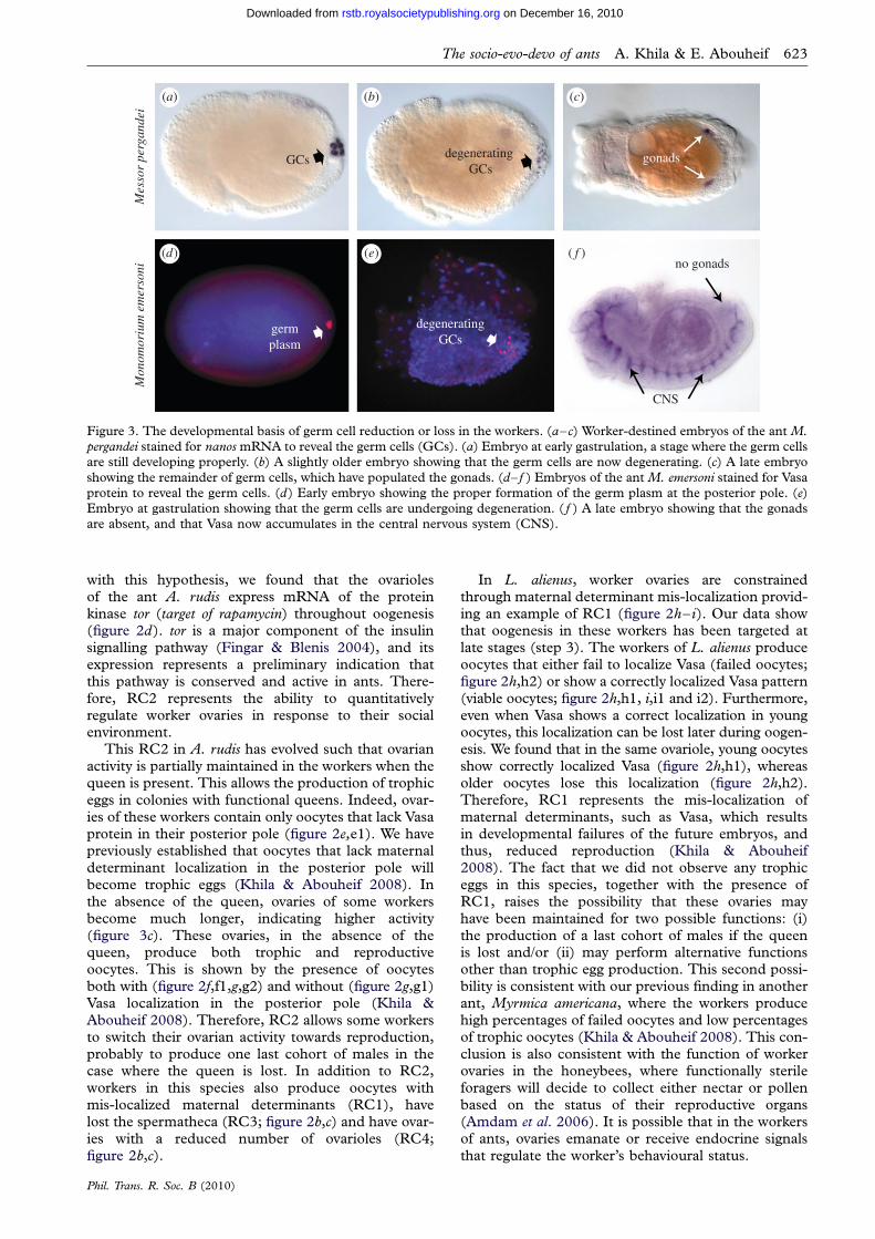
(a) RC1: mis-localization of maternal
determinants
RC1 is the mis-localization of specialized mRNAs andproteins necessary for the proper patterning of theoocytes produced in worker ovaries (figures 2h,h2 and4[a3]), which leads to severe defects during embryodevelopment (Khila & Abouheif 2008). This constraintis ultimate because it occurs in the workers both in thepresence and absence of the queen (Khila & Abouheif2008). RC1 should be considered as a mechanismbecause it acts on a process that is continuous intime, i.e. oogenesis. The ovariole is a string-like struc-ture where growing oocytes move from the anteriortowards the posterior (figure 1s; Spradling 1993). Asthey move towards the posterior, each oocyte is directedtowards a trophic or reproductive fate. We infer trophicoocytes to be those that do not localize maternal deter-minants, and reproductive oocytes to be those that dolocalize these molecules. Since RC1 affects maternaldeterminant localization it can only affect reproductive,but not trophic oocytes. The fraction of the reproduc-tive oocytes affected by RC1 can vary betweenspecies. Therefore, RC1 blocks the ancestral functionof worker ovaries by reducing or eliminating theworkers’ ability to produce non-fertilized male eggs,without preventing trophic egg production.
Therefore, we hypothesize that RC1 may not rep-resent a neutral decay of the ovaries but rather is anadaptive developmental mechanism that reduces oreliminates worker reproduction, while restricting theovaries to performing a nutritional function. Such amechanism can only act within a narrow range ofdevelopmental parameters during late steps of oogen-esis. If earlier steps of oogenesis, such as GSCdivision and cyst production, are blocked this wouldeliminate both reproduction and trophic functions ofthe ovary. Mutations in a variety of genes in the flyindicate that there exists a genetic potential forcompletely blocking oogenesis as early as GSC
The socio-evo-devo of ants A. Khila & E. Abouheif 625
on December 16, 2010rstb.royalsocietypublishing.orgDownloaded from
self-renewal and differentiation (see Wong et al. (2005)for review). These include vasa, nanos, bag-of-marbles,decapentaplegic and other molecules necessary for GSCself-renewal, cystoblast differentiation or other cellularprocesses necessary for continuous follicle production(Styhler et al. 1998; Wang & Lin 2004; Szakmary et al.2005; Wong et al. 2005). The specificity of RC1 at latesteps of oogenesis, together with the fact that evolutionhas not acted upon the potential to block early steps,suggests that RC1 represents an adaptive develop-mental mechanism rather than a neutral degenerationof worker ovaries.
(b) RC2: quantitative constraint
on ovary activity
A general feature of insect ovaries is their ability torespond to environmental changes, such as hormonalinput and changes in nutrition, by changing theiractivity (Drummond-Barbosa & Spradling 2001).This ovarian plasticity may have been refined in theworkers of ants through the evolution of specificresponse elements that allow them to respond in aspecific manner and to specific social cues in theirenvironment. Therefore, RC2 may represent an adap-tive developmental mechanism that can be defined asthe ability of workers to regulate their ovarian activityin response to chemical and behavioural interactionsbetween individuals, such as policing, self-restraint,and pheromones. RC2 is not an ultimate reproductiveconstraint, because in particular social conditions,such as the absence of the queen, worker ovaries mayincrease their activity. The regulation of worker repro-duction through RC2 has evolved such that in thepresence of the queen it may be partial in some speciesand total in others. Aphaenogaster treate is a mono-gynous species with a relatively large colony size anda pronounced queen/worker caste dimorphism. Ourresults show that in this species, worker ovaries arestill active in the presence of the queen, allowing forthe production of trophic oocytes. In contrast, speciessuch as Harpegnathos saltator or Amblyopone pallipesform small colonies where queen and worker castesare hardly recognizable (Peeters & Holldobler 1995;Peeters et al. 2000). In this type of species, workerovaries in the presence of the queen are inactive,representing a total blockage of oogenesis.
The molecular mechanism of RC2 is still unknown.However, the outcome of RC2 affects the length of theovariole, which reflects the rate of GSC division andcyst production. This suggests that this constraintmay be directly linked to step 1 of oogenesis, i.e.GSC division and cyst production. The insulin-signalling pathway may mediate ovarian response toRC2, since our results suggest that this pathway maybe conserved and active in ant ovaries (figure 2d).Thus, it is possible that signals from the queen,either directly or indirectly, suppress this insulin-likepeptide production in the workers. Furthermore, ithas been shown in Drosophila that this pathway directlycontrols the rate of GSC division and cyst production(LaFever & Drummond-Barbosa 2005). Insulin-likepeptides are produced by a specialized cell clusterin the brain and mediate a direct and positive
Phil. Trans. R. Soc. B (2010)
long-range effect on ovarian activity (Cao & Brown2001; Riehle et al. 2006). This possibility is furthersupported by transcriptomics data from the brain ofa social wasp, where the brain of the queen expressesmuch higher levels of the insulin-like peptide2 thanthat of the workers (Toth et al. 2007).
(c) RC3: loss of the spermatheca
In most ants, the spermatheca (sperm storage organ)has been lost, resulting in workers that cannot storesperm or fertilize the eggs they produce. These workers,in theory, can still produce unfertilized eggs, which maypotentially develop into males owing to the haplo-diploid sex determination that characterizes ants. Wehypothesize that RC3 is an adaptive trait that has beenmaintained by selection to block the production ofdaughters, which represents half the reproductive poten-tial. This hypothesis is supported by the specificity of theprocesses through which the spermatheca develops. Thespermatheca, along with the other somatic structures ofthe female reproductive organs (genitalia, oviducts anduterus), develops from the genital disc, which is speci-fied during late embryogenesis and patterned duringlarval development (Hartenstein 1993; Sanchez &Guerrero 2001). Since the cell population that givesrise to the spermatheca is part of the genital disc andnot independent, it may be more probable for thewhole genital disc to be removed through degeneration,than for a fraction of its cells, i.e. those of the sperma-theca, to be singled out. However, the loss of theentire genital disc has been selected against because analternative ovary function, i.e. trophic egg production,requires the oviducts, uterus and genitalia to connectthe ovarioles (where trophic eggs are produced) to theexterior (where these eggs are consumed).
A comprehensive test of our hypothesis wouldrequire analysing RC3 at multiple biological levels,i.e. anatomical, embryological and developmental gen-etic, in a phylogenetic framework. With respect to theanatomical and embryological levels, further work isrequired to determine at what stage of development(embryonic or larval) and to what degree (completeor partial) the spermatheca is lost in ants. With respectto the genetic basis of spermatheca development, littleis currently known, although an old study in Drosophilahas reported that females mutant for the gene lozenge,involved in apoptosis regulation, lack spermathecae(Anderson 1945; Wildonger et al. 2005). These data,once collected from multiple species, can be con-trasted against a null phylogenetic model based onneutral degeneration. If RC3 were the consequenceof neutral degenerative processes, then we shouldexpect a phylogenetic pattern where the frequencyand degree of loss would be more similar betweenclosely related ant lineages than between distantlyrelated ones regardless of the selective pressures thatmay arise from their social structure, ecology or lifehistory (Abouheif 2003).
(d) RC4: reduction of the number of ovarioles
In most ant species, the number of ovarioles has beendramatically reduced in the workers to as low as oneovariole per ovary (figure 4[d2]), while queens possess
viable
[a1] MDs properlylocalized
[b1] germ plasm localizedin embryo posterior
[a2] MDs non-localized: food
[a3] MDs mis-localized:reproductive constraint
[b2] germ plasm inheritedby pole cells
[b4] germ cells degenerate
[b3] germ cells develop
[c1] gonaddifferentiation
[d3] ovary loss= workers
[d2] ovariole numberreduced = workers
reproduction
males(testes)
[d1] ovaries fullydeveloped = queen
adultlarval development
[d1a] reproducively
active ovariole
[d2a] worker-like
ovariole
colonygrowth
germ cells
trophic
failed
ooge
nesi
s
*
embryogenesis
Figure 4. Summary of germ cell development and ovary function in ants. [a1–a3] Types of oocytes produced in ant ovaries.
[a1] Viable with correct maternal determinant localization, [a2] trophic with no localization and [a3] failed where Vasa proteinaccumulates in the oocyte but its localization is not maintained at the posterior pole. Only oocytes with correct localization [a1]possess functional pole plasm and will undergo proper development. The pole plasm is inherited by the embryo [b1] and func-tions to specify the germ cells [b2]. Germ cells may develop fully [b3] and lead to the formation of complete gonads [b3 andc1] and therefore fully developed queen ovaries [d1]. Alternatively, germ cells may undergo degeneration during embryo devel-
opment [b4] and lead to either reduced number of worker ovarioles [d2] or complete loss of worker ovarioles [d3]. Ovarioles ofqueens (or workers that are reproductively active) [d1a] are more active than normal worker ovarioles [d2a]. Queen ovariolescan produce both trophic and reproductive oocytes [a1–a2], whereas worker ovarioles are constrained at the molecular leveland produce failed oocytes [a3] in addition to viable [a1] and trophic ones [a2]. Green, Vasa; blue, nuclei; red, actin.
626 A. Khila & E. Abouheif The socio-evo-devo of ants
on December 16, 2010rstb.royalsocietypublishing.orgDownloaded from
several to several thousand ovarioles per ovary(figure 4[d1]). RC4 is a trait that is generallyaccompanied by the loss of the spermatheca (RC3).Embryonic pole cells, the precursors of the germlinepart of adult ovarioles, are specified in ants duringoogenesis and early embryogenesis, representing oneof the earliest tissues to be specified (figure 4[a1]–[b2]). We present data indicating that in ants thereduction of ovariole number may be caused by thedegeneration of pole cells during the development ofworker-destined embryos (figure 3). The developmen-tal basis of RC4 is different in honeybees, whereovariole number is reduced in worker larvae ratherthan in embryos (Schmidt Capella & Hartfelder2002; Reginato & Cruz-Landim 2003). This is con-sistent with the much later timing of germ cellspecification in bees relative to ants (Dearden 2006).
Phil. Trans. R. Soc. B (2010)
We hypothesize that RC4 is an adaptive trait thathas evolved to reduce worker reproduction with-out interfering with possible alternative functions ofworker ovaries. Because the early stages of embryonicdevelopment are quite sensitive and involve simul-taneous specification of various tissues usingcommon gene batteries (Thomson & Lasko 2005),germ cell degeneration can only occur within anarrow range of developmental parameters. The pro-cess of germ cell specification and that of patterningthe posterior of the embryo are tightly interdependent(Thomson & Lasko 2005). Many of the molecules thatform the germ plasm, including Nanos and Vasa, areknown to have pleiotropic functions in the develop-ment of both germ cells and posterior segments inaddition to other key developmental processes. Ourresults indeed show that this reduction of germ cells
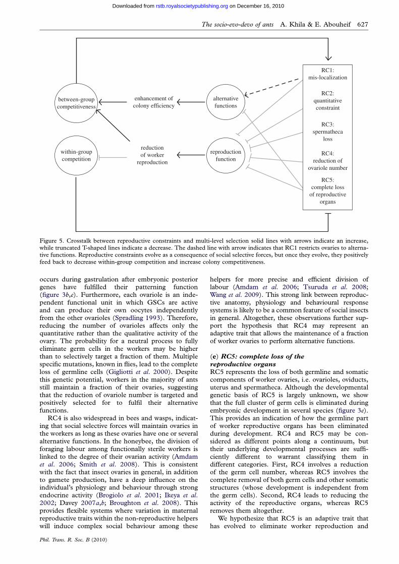
between-groupcompetitiveness
within-groupcompetition
reductionof worker
reproduction
reproductionfunction
alternativefunctions
RC1:mis-localization
RC2:quantitativeconstraint
RC3:spermatheca
loss
RC4:reduction of
ovariole number
RC5:complete loss
of reproductiveorgans
enhancement ofcolony efficiency
Figure 5. Crosstalk between reproductive constraints and multi-level selection solid lines with arrows indicate an increase,while truncated T-shaped lines indicate a decrease. The dashed line with arrow indicates that RC1 restricts ovaries to alterna-tive functions. Reproductive constraints evolve as a consequence of social selective forces, but once they evolve, they positively
feed back to decrease within-group competition and increase colony competitiveness.
The socio-evo-devo of ants A. Khila & E. Abouheif 627
on December 16, 2010rstb.royalsocietypublishing.orgDownloaded from
occurs during gastrulation after embryonic posteriorgenes have fulfilled their patterning function(figure 3b,e). Furthermore, each ovariole is an inde-pendent functional unit in which GSCs are activeand can produce their own oocytes independentlyfrom the other ovarioles (Spradling 1993). Therefore,reducing the number of ovarioles affects only thequantitative rather than the qualitative activity of theovary. The probability for a neutral process to fullyeliminate germ cells in the workers may be higherthan to selectively target a fraction of them. Multiplespecific mutations, known in flies, lead to the completeloss of germline cells (Gigliotti et al. 2000). Despitethis genetic potential, workers in the majority of antsstill maintain a fraction of their ovaries, suggestingthat the reduction of ovariole number is targeted andpositively selected for to fulfil their alternativefunctions.
RC4 is also widespread in bees and wasps, indicat-ing that social selective forces will maintain ovaries inthe workers as long as these ovaries have one or severalalternative functions. In the honeybee, the division offoraging labour among functionally sterile workers islinked to the degree of their ovarian activity (Amdamet al. 2006; Smith et al. 2008). This is consistentwith the fact that insect ovaries in general, in additionto gamete production, have a deep influence on theindividual’s physiology and behaviour through strongendocrine activity (Brogiolo et al. 2001; Ikeya et al.2002; Davey 2007a,b; Broughton et al. 2008). Thisprovides flexible systems where variation in maternalreproductive traits within the non-reproductive helperswill induce complex social behaviour among these
Phil. Trans. R. Soc. B (2010)
helpers for more precise and efficient division oflabour (Amdam et al. 2006; Tsuruda et al. 2008;Wang et al. 2009). This strong link between reproduc-tive anatomy, physiology and behavioural responsesystems is likely to be a common feature of social insectsin general. Altogether, these observations further sup-port the hypothesis that RC4 may represent anadaptive trait that allows the maintenance of a fractionof worker ovaries to perform alternative functions.
(e) RC5: complete loss of the
reproductive organs
RC5 represents the loss of both germline and somaticcomponents of worker ovaries, i.e. ovarioles, oviducts,uterus and spermatheca. Although the developmentalgenetic basis of RC5 is largely unknown, we showthat the full cluster of germ cells is eliminated duringembryonic development in several species (figure 3e).This provides an indication of how the germline partof worker reproductive organs has been eliminatedduring development. RC4 and RC5 may be con-sidered as different points along a continuum, buttheir underlying developmental processes are suffi-ciently different to warrant classifying them indifferent categories. First, RC4 involves a reductionof the germ cell number, whereas RC5 involves thecomplete removal of both germ cells and other somaticstructures (whose development is independent fromthe germ cells). Second, RC4 leads to reducing theactivity of the reproductive organs, whereas RC5removes them altogether.
We hypothesize that RC5 is an adaptive trait thathas evolved to eliminate worker reproduction and
628 A. Khila & E. Abouheif The socio-evo-devo of ants
on December 16, 2010rstb.royalsocietypublishing.orgDownloaded from
reduce within-colony conflict. As discussed in RC4,there is a substantial genetic potential for completelyeliminating reproductive organs. However, RC5 hasonly evolved in nine out of 283 ant genera. If RC5were a consequence of neutral degeneration, we mayexpect the complete loss of worker reproductiveorgans to occur much more frequently in ant evo-lution. We have argued that alternative functions(trophic and/or physiological) are central for workerovaries. For a species to evolve RC5, the cost associ-ated with the ancestral reproductive function ofworker ovaries should outweigh the benefit gainedfrom maintaining worker ovaries for alternativefunctions.
5. REPRODUCTIVE CONSTRAINTSAND MULTI-LEVEL SELECTIONOur hypothesis that reproductive constraints representadaptive mechanisms or traits that have evolved toactively maintain social harmony, rather than degenera-tive by-products of social selective forces, may beintuitively understood in a multi-level selection frame-work. Figure 5 attempts to summarize how theevolution of reproductive constraints is influenced bysocial selective forces and how they positively feedback to influence the social evolutionary dynamics ofant colonies. RC1–RC5 act to suppress worker repro-duction at specific steps during development, therebyreducing within-colony competition. RC2–RC4 sup-press or reduce the reproductive function of ovaries ina way that does not interfere with other alternative func-tions. RC2 (quantitative constraint) is a non-ultimateconstraint that may result in either complete or partialblockage of the quantitative activity of worker ovariesdepending on species. RC2 is a mechanism, whereasRC3 and RC4 are adaptive traits consisting of modifi-cations of embryonic or larval development thatresults in the permanent loss or reduction of adultstructures. RC1 (maternal determinant mis-localiz-ation) and RC5 (complete loss of reproductiveorgans) are both ultimate constraints that are fixed inthe workers. RC5 suppresses both reproductive andalternative functions of the ovaries. RC1, however, isdistinct from the other reproductive constraints becauseit represents a mechanism that blocks or reduces workerreproduction while restricting ovaries to performingalternative functions. Thus, reproductive constraintsmay reduce within-colony competition (reduced or noworker reproduction), increase cooperation betweencolony members (alternative functions) and thereforeincrease the ability of the colony to compete withother groups. Once they evolve, these constraints canpositively feed back and may allow an increase incolony size and a more complex state of behaviouraland morphological division of labour. Further-more, they may render colonies robust to variations intheir relatedness structure, potentially increasing theirability to cope with harsh environments (Korb &Heinze 2004; Smith et al. 2008; Wilson 2008a,b). Acomplete understanding of the roles of reproductiveconstraints in ant evolution will require integratingboth ultimate (evolutionary) and proximate (develop-mental) causations. This socio-evo-devo approach
Phil. Trans. R. Soc. B (2010)
holds much promise for gaining further insight intohow cooperation, conflict and developmental systemsevolve in social groups.
We thank Rajendhran Rajakumar and Nella Mauro for helpwith tor cloning, Paul Lasko and Jarred Chicoine for the Vasaand Bicaudal-C antibodies, Laura Nilson and Lucia Caceresfor the alpha-tubulin antibody, Abouheif lab Membersand two anonymous reviewers for comments on themanuscript. This work was supported by an NSERCDiscovery Grant to E.A.
REFERENCESAbouheif, E. 2003 A framework for studying the evolution of
gene networks underlying polyphenism: insights from
winged and wingless ant castes. In Environment, develop-ment, and evolution: toward a synthesis (eds B. K. Hall,R. D. Pearson & G. B. Muller). Cambridge, MA: MITPress.
Amdam, G. V., Csondes, A., Fondrk, M. K. & Page Jr, R. E.
2006 Complex social behaviour derived from maternalreproductive traits. Nature 439, 76–78. (doi:10.1038/nature04340)
Anderson, R. C. 1945 A study of the factors affectingfertility of lozenge females of Drosophila melanogaster.Genetics 30, 280–296.
Bourke, A. F. G. 1999 Colony size, social complexity andreproductive conflict in social insects. J. Evol. Biol. 12,245–257. (doi:10.1046/j.1420-9101.1999.00028.x)
Bourke, A. & Franks, N. 1995 Social evolution in ants.Princeton, NJ: Princeton University Press.
Brogiolo, W., Stocker, H., Ikeya, T., Rintelen, F.,Fernandez, R. & Hafen, E. 2001 An evolutionarilyconserved function of the Drosophila insulin receptor
and insulin-like peptides in growth control. Curr. Biol.11, 213–221. (doi:10.1016/S0960-9822(01)00068-9)
Broughton, S. et al. 2008 Reduction of DILP2 in Drosophilatriages a metabolic phenotype from lifespan revealingredundancy and compensation among DILPs. PLoS ONE3, e3721. (doi:10.1371/journal.pone.0003721)
Cao, C. & Brown, M. R. 2001 Localization of an insulin-like peptide in brains of two flies. Cell Tissue Res. 304,317–321. (doi:10.1007/s004410100367)
Carroll, S. B., Grenier, J. & Weatherbee, S. 2004 From DNAto diversity. Oxford, UK: Blackwell Science.
Chicoine, J., Benoit, P., Gamberi, C., Paliouras, M.,Simonelig, M. & Lasko, P. 2007 Bicaudal-C recruitsCCR4-NOT deadenylase to target mRNAs and regulates
oogenesis, cytoskeletal organization, and its ownexpression. Dev. Cell 13, 691–704. (doi:10.1016/j.devcel.2007.10.002)
Cox, D. N., Lu, B., Sun, T. Q., Williams, L. T. & Jan, Y. N.2001 Drosophila par-1 is required for oocyte differen-
tiation and microtubule organization. Curr. Biol. 11,75–87. (doi:10.1016/S0960-9822(01)00027-6)
Crespi, B. J. 1992 Cannibalism and trophic eggs in subsocialand eusocial insects. Cannibalism: ecology and evolutionamong diverse taxa, pp. 176–213. Oxford, UK: Oxford
University Press.Davey, K. 2007a From insect ovaries to sheep red blood
cells: a tale of two hormones. J. Insect Physiol. 53, 1–10.(doi:10.1016/j.jinsphys.2006.10.005)
Davey, K. 2007b The interaction of feeding and mating in
the hormonal control of egg production in Rhodniusprolixus. J. Insect Physiol. 53, 208–215. (doi:10.1016/j.jinsphys.2006.10.002)
Dearden, P. K. 2006 Germ cell development in the honeybee
(Apis mellifera); Vasa and Nanos expression. BMC Dev.Biol. 6, 6. (doi:10.1186/1471-213X-6-6)
The socio-evo-devo of ants A. Khila & E. Abouheif 629
on December 16, 2010rstb.royalsocietypublishing.orgDownloaded from
de Cuevas, M., Lee, J. K. & Spradling, A. C. 1996Alpha-spectrin is required for germline cell division anddifferentiation in the Drosophila ovary. Development 122,
3959–3968.de Cuevas, M., Lilly, M. A. & Spradling, A. C. 1997 Germ-
line cyst formation in Drosophila. Annu. Rev. Genet. 31,405–428. (doi:10.1146/annurev.genet.31.1.405)
Drummond-Barbosa, D. & Spradling, A. C. 2001 Stem cells
and their progeny respond to nutritional changes duringDrosophila oogenesis. Dev. Biol. 231, 265–278. (doi:10.1006/dbio.2000.0135)
Eberhard, M. J. W. 2003 Developmental plasticity andevolution. Oxford, UK: Oxford University Press.
Extavour, C. G. & Akam, M. 2003 Mechanisms of germ cellspecification across the metazoans: epigenesis and prefor-mation. Development 130, 5869–5884. (doi:10.1242/dev.00804)
Fingar, D. C. & Blenis, J. 2004 Target of rapamycin (TOR):an integrator of nutrient and growth factorsignals and coordinator of cell growth and cell cycle pro-gression. Oncogene 23, 3151–3171. (doi:10.1038/sj.onc.1207542)
Fletcher, J. A., Zwick, M., Doebeli, M. & Wilson, D. S.2006 What’s wrong with inclusive fitness? Trends Ecol.Evol. 21, 597–598 (author reply 599–600). (doi:10.1016/j.tree.2006.08.008)
Foster, K. R., Wenseleers, T. & Ratnieks, F. L. 2006a Kin
selection is the key to altruism. Trends Ecol. Evol. 21,57–60. (doi:10.1016/j.tree.2005.11.020)
Foster, K. R., Wenseleers, T., Ratnieks, F. L. & Queller,D. C. 2006b There is nothing wrong with inclusive
fitness. Trends Ecol. Evol. 21, 599–600. (doi:10.1016/j.tree.2006.08.010)
Gardner, A. & Grafen, A. 2009 Capturing the superorgan-ism: a formal theory of group adaptation. J. Evol. Biol.22, 659–671. (doi:10.1111/j.1420-9101.2008.01681.x)
Gigliotti, S., Rotoli, D., Manzi, A., Graziani, F. & Malva, C.2000 Female sterile mutations and egg chamber develop-ment in Drosophila melanogaster. Int. J. Dev. Biol. 44,581–589.
Hall, B. K. 1999 Evolutionary developmental biology.Dordrecht, The Netherlands: Kluwer AcademicPublishers.
Hamilton, W. D. 1964a Genetical evolution of socialbehaviour 2. J. Theor. Biol. 7, 17–52. (doi:10.1016/0022-5193(64)90039-6)
Hamilton, W. D. 1964b Genetical evolution of socialbehaviour I. J. Theor. Biol. 7, 1–16. (doi:10.1016/0022-5193(64)90038-4)
Hartenstein, V. 1993 Atlas of Drosophila development. Cold
Spring Harbor, NY: Cold Spring Harbor LaboratoryPress.
Holldobler, B. & Wilson, E. 1990 The ants. Cambridge, MA:Harvard University Press.
Holldobler, B. & Wilson, E. O. 2008 The superorganism: thebeauty, elegance, and strangeness of insect societies, 1st edn.New York, NY: W. W. Norton.
Hsu, H. J. & Drummond-Barbosa, D. 2009 Insulin levelscontrol female germline stem cell maintenance via theniche in Drosophila. Proc. Natl Acad. Sci. USA 106,
1117–1121. (doi:10.1073/pnas.0809144106)Huynh, J. R. & St Johnston, D. 2004 The origin of asym-
metry: early polarisation of the Drosophila germline cystand oocyte. Curr. Biol. 14, R438–R449. (doi:10.1016/j.cub.2004.05.040)
Ikeya, T., Galic, M., Belawat, P., Nairz, K. & Hafen, E. 2002Nutrient-dependent expression of insulin-like peptidesfrom neuroendocrine cells in the CNS contributes togrowth regulation in Drosophila. Curr. Biol. 12,1293–1300. (doi:10.1016/S0960-9822(02)01043-6)
Phil. Trans. R. Soc. B (2010)
Jeon, J. & Choe, J. C. 2003 Reproductive skew and the originof sterile castes. Am. Nat. 161, 206–224. (doi:10.1086/346099)
Johnson, A. D., Drum, M., Bachvarova, R. F., Masi, T.,White, M. E. & Crother, B. I. 2003 Evolution of predeter-mined germ cells in vertebrate embryos: implications formacroevolution. Evol. Dev. 5, 414–431. (doi:10.1046/j.1525-142X.2003.03048.x)
Khila, A. & Abouheif, E. 2008 Reproductive constraint is adevelopmental mechanism that maintains socialharmony in advanced ant societies. Proc. Natl Acad. Sci.USA 105, 17 884–17 889. (doi:10.1073/pnas.08
07351105)Khila, A. & Abouheif, E. 2009 In situ hybridization on ant
ovaries and embryos. Emerging model organisms. ColdSpring Harbor, NY: Cold Spring Harbor LaboratoryPress.
Kilmartin, J. V., Wright, B. & Milstein, C. 1982 Rat mono-clonal antitubulin antibodies derived by using a newnonsecreting rat cell line. J. Cell Biol. 93, 576–582.(doi:10.1083/jcb.93.3.576)
Kohn, M. 2008 The needs of the many. Nature 456,
296–299. (doi:10.1038/456296a)Korb, J. & Heinze, J. 2004 Multilevel selection and social
evolution of insect societies. Naturwissenschaften 91,291–304.
LaFever, L. & Drummond-Barbosa, D. 2005 Direct control
of germline stem cell division and cyst growth by neuralinsulin in Drosophila. Science 309, 1071–1073. (doi:10.1126/science.1111410)
Lehmann, L., Keller, L., West, S. & Roze, D. 2007 Group
selection and kin selection: two concepts but one process.Proc. Natl Acad. Sci. USA 104, 6736–6739. (doi:10.1073/pnas.0700662104)
Lin, H., Yue, L. & Spradling, A. C. 1994 The Drosophilafusome, a germline-specific organelle, contains mem-
brane skeletal proteins and functions in cyst formation.Development 120, 947–956.
Nijhout, H. F. 1999 Control mechanisms of polyphenicdevelopment in insects—in polyphenic development,environmental factors alter same aspects of development
in an orderly and predictable way. Bioscience 49,181–192. (doi:10.2307/1313508)
Nijhout, H. F. & Wheeler, D. E. 1982 Juvenile-hormone andthe physiological-basis of insect polymorphisms. Q. Rev.Biol. 57, 109–133.
Oster, G. F. & Wilson, E. O. 1978 Caste and ecology in thesocial insects. Monogr. Popul. Biol. 12, 1–352.
Peeters, C. & Holldobler, B. 1995 Reproductive cooperationbetween queens and their mated workers: the complex life
history of an ant with a valuable nest. Proc. Natl Acad.Sci. USA 92, 10 977–10 979. (doi:10.1073/pnas.92.24.10977)
Peeters, C., Liebg, J. & Holldobler, B. 2000 Sexual repro-duction by both queens and workers in the ponerine ant
Harpegnathos saltator. Insect Soc. 47, 325–332. (doi:10.1007/PL00001724)
Pennisi, E. 2009 Agreeing to disagree. Science 323, 706–708.(doi:10.1126/science.323.5915.706)
Reeve, H. K. & Holldobler, B. 2007 The emergence of a
superorganism through intergroup competition. Proc.Natl Acad. Sci. USA 104, 9736–9740. (doi:10.1073/pnas.0703466104)
Reginato, R. D. & Cruz-Landim, C. 2003 Ovarian growthduring larval development of queen and worker of Apismellifera (Hymenoptera: Apidae): a morphometric andhistological study. Braz. J. Biol. 63, 121–127.
Riechmann, V. & Ephrussi, A. 2001 Axis formation duringDrosophila oogenesis. Curr. Opin. Genet. Dev. 11,374–383. (doi:10.1016/S0959-437X(00)00207-0)
630 A. Khila & E. Abouheif The socio-evo-devo of ants
on December 16, 2010rstb.royalsocietypublishing.orgDownloaded from
Riehle, M. A., Fan, Y., Cao, C. & Brown, M. R. 2006 Mol-ecular characterization of insulin-like peptides in theyellow fever mosquito, Aedes aegypti: expression, cellular
localization, and phylogeny. Peptides 27, 2547–2560.(doi:10.1016/j.peptides.2006.07.016)
Sanchez, L. & Guerrero, I. 2001 The development of theDrosophila genital disc. BioEssays 23, 698–707. (doi:10.1002/bies.1099)
Schmidt Capella, I. C. & Hartfelder, K. 2002 Juvenile-hormone-dependent interaction of actin and spectrin iscrucial for polymorphic differentiation of the larvalhoney bee ovary. Cell Tissue Res. 307, 265–272. (doi:10.
1007/s00441-001-0490-y)Smith, C. R., Toth, A. L., Suarez, A. V. & Robinson, G. E.
2008 Genetic and genomic analyses of the division oflabour in insect societies. Nat. Rev. Genet. 9, 735–748.(doi:10.1038/nrg2429)
Song, X., Wong, M. D., Kawase, E., Xi, R., Ding, B. C.,McCarthy, J. J. & Xie, T. 2004 Bmp signals from nichecells directly repress transcription of a differentiation-promoting gene, bag of marbles, in germline stem cells inthe Drosophila ovary. Development 131, 1353–1364.
(doi:10.1242/dev.01026)Spradling, A. C. 1993 Developmental genetics of oogenesis.
In The development of Drosophila melanogaster, vol. 1 (edsM. Bate & A. Martinez-Arias), pp. 1–70. Cold SpringHarbor, NY: Cold Spring Harbor Laboratory Press.
Steinhauer, J. & Kalderon, D. 2006 Microtubule polarityand axis formation in the Drosophila oocyte. Dev. Dyn.235, 1455–1468. (doi:10.1002/dvdy.20770)
Styhler, S., Nakamura, A., Swan, A., Suter, B. & Lasko, P.
1998 vasa is required for GURKEN accumulation inthe oocyte, and is involved in oocyte differentiation andgermline cyst development. Development 125,1569–1578.
Szakmary, A., Cox, D. N., Wang, Z. & Lin, H. 2005
Regulatory relationship among piwi, pumilio, and bag-of-marbles in Drosophila germline stem cell self-renewaland differentiation. Curr. Biol. 15, 171–178. (doi:10.1016/j.cub.2005.01.005)
Thomson, T. & Lasko, P. 2005 Tudor and its domains: germ
cell formation from a Tudor perspective. Cell Res. 15,281–291. (doi:10.1038/sj.cr.7290297)
Toth, A. L. & Robinson, G. E. 2007 Evo-devo and theevolution of social behavior. Trends Genet. 23, 334–341.(doi:10.1016/j.tig.2007.05.001)
Toth, A. L. et al. 2007 Wasp gene expression supports anevolutionary link between maternal behavior and euso-ciality. Science 318, 441–444. (doi:10.1126/science.1146647)
Tsuruda, J. M., Amdam, G. V. & Page Jr, R. E. 2008 Sen-sory response system of social behavior tied to femalereproductive traits. PLoS ONE 3, e3397. (doi:10.1371/journal.pone.0003397)
Phil. Trans. R. Soc. B (2010)
van Eeden, F. & St Johnston, D. 1999 The polarisation of theanterior–posterior and dorsal–ventral axes duringDrosophila oogenesis. Curr. Opin. Genet. Dev. 9, 396–404.
Villet, M. H., Crewe, R. M. & Duncan, F. D. 1991 Evo-lutionary trends in the reproductive biology of ponerineants (Hymenoptera: Formicidae). J. Nat. Hist. 25,1603–1610. (doi:10.1080/00222939100770991)
Wang, Z. & Lin, H. 2004 Nanos maintains germline stem
cell self-renewal by preventing differentiation. Science303, 2016–2019. (doi:10.1126/science.1093983)
Wang, Y., Amdam, G. V., Rueppell, O., Wallrichs, M. A.,Fondrk, M. K., Kaftanoglu, O. & Page Jr, R. E. 2009
PDK1 and HR46 gene homologs tie social behavior toovary signals. PLoS ONE 4, e4899. (doi:10.1371/journal.pone.0004899)
Wenseleers, T., Hart, A. G. & Ratnieks, F. L. W. 2004When resistance is useless: policing and the evolution of
reproductive acquiescence in insect societies. Am. Nat.164, E154–E167. (doi:10.1086/425223)
West, S. A., Griffin, A. S. & Gardner, A. 2007 Social seman-tics: altruism, cooperation, mutualism, strong reciprocityand group selection. J. Evol. Biol. 20, 415–432. (doi:10.
1111/j.1420-9101.2006.01258.x)West, S. A., Griffin, A. S. & Gardener, A. 2008 Social
semantics: how useful has group selection been? J. Evol.Biol. 21, 374–385.
Wheeler, W. 1917 The phylogenetic development of subap-
terous and apterous castes in the Formicidae. Proc. NatlAcad. Sci. USA 3, 109–117.
Wheeler, D. E. & Nijhout, H. F. 1981 Soldier determinationin ants—new role for juvenile-hormone. Science 213,
361–363. (doi:10.1126/science.213.4505.361)Wildonger, J., Sosinsky, A., Honig, B. & Mann, R. S. 2005
Lozenge directly activates argos and klumpfuss to regulateprogrammed cell death. Genes Dev. 19, 1034–1039.(doi:10.1101/gad.1298105)
Wilkins, A. S. 2002 The evolution of developmental pathways.Sunderland, UK: Sinauer.
Wilson, D. S. 2008a Social semantics: toward a genuinepluralism in the study of social behaviour. J. Evol. Biol.21, 368–373.
Wilson, E. O. 2008b One giant leap: how insects achievedaltruism and colonial life. Bioscience 58, 17–25. (doi:10.1641/B580106)
Wilson, D. S. & Wilson, E. O. 2007 Rethinking the theoreti-cal foundation of sociobiology. Q. Rev. Biol. 82, 327–348.
(doi:10.1086/522809)Wilson, E. O. & Holldobler, B. 2005 Eusociality: origin
and consequences. Proc. Natl Acad. Sci. USA 102,13 367–13 371. (doi:10.1073/pnas.0505858102)
Wong, M. D., Jin, Z. & Xie, T. 2005 Molecular mechanismsof germline stem cell regulation. Annu. Rev. Genet.39, 173–195. (doi:10.1146/annurev.genet.39.073003.105855)
Related Documents


![Blood Biochemistry Reflects Seasonal Nutritional and Reproductive Constraints in the Eurasian Badger ([ITAL]Meles meles[/ITAL])](https://static.cupdf.com/doc/110x72/6337ac305b59cce6920b34c6/blood-biochemistry-reflects-seasonal-nutritional-and-reproductive-constraints-in.jpg)
