IOSR Journal of Dental and Medical Sciences (IOSR-JDMS) e-ISSN: 2279-0853, p-ISSN: 2279-0861.Volume 14, Issue 3 Ver. V (Mar. 2015), PP 76-85 www.iosrjournals.org DOI: 10.9790/0853-14357685 ww.iosrjournals.org 76 | Page Evaluating the Frequency of Urological Causes of Flank Pain in Adult Patients that refer to Urology Ward in Shahid Motahari Hospital (Marvdasht, Iran) during 2013-2014 Shima Cyrus 1 and Kamran Etaati 2 1 (MD, School of Medicine, International Branch, Shiraz University of Medical Sciences, Iran) 2 (Assistant Professor, School of Medicine, International Branch, Shiraz University of Medical Sciences, Iran) Abstract: Flank pain is the basic problem in medicine. Finding the causes of flank pain can have important role on screening and treatment. Methods: This research was performed on 350 cases that referred with chief complain of flank pain in Shahid Motahari Hospital (Marvdasht, Iran) during 2013 to 2014. For each cases urinalysis and Ultrasonography is requested and the data was collected by questionnaire and frequency of different causes was investigated. Results: In 350 cases, 126 cases had urinary tract stones with hydronephrosis, 194 cases had musculoskeletal pain and 30 cases had pyelonephritis. Renal abcess, renal tumor, urinary pelvic junction obstruction, ureteral stenosis, acute renal infarction and renal vessels diseases did not have any frequencies. Conclusion: By this research we concluded that most people who refer to clinic of urology with chief complain of flank pain have musculoskeletal pain. So by screening of these patients before referring to the clinic of urology, we can differentiate the urologic causes and non urologic causes of flank pain and it is time and cost beneficial. Keywords: Acute renal infarction, musculoskeletal pain, pyelonephritis, renal vessels diseases, renal abscess, renal tumor, urinary-pelvic junction obstruction, urinary tract stones, urethral stenosis. I. Introduction I.1. Flank Pain Flank pain as a fundamental and complex problem exists in the medical world. Many diseases of the genitourinary tract and genitals can cause flank pain. Flank pain is a common complaint in the emergency and general urology. Several factors cause this painful complaint that accurate and rapid differential diagnosis for early diagnosis and therapeutic intervention is necessary to reduce the risk. Flank pain can be origin from the superficial or deep parts. Painful sores on the skin surface can be followed, but the pain can seek deeper parenchymal lesions in internal organs such as intestines, biliary system, ureter, urethra, bladder and other hollow cavity of the abdomen. Recording adequate patient's history and perform a thorough physical examination is helpful in diagnosis. Symptoms such as dysuria and urinary frequency, urgency in urinating, fever, blood and urine can be helpful in diagnosis. Performing a simple urine test and counting evaluation of blood cells, white blood cells and bacteria in the urinary nitrite can also be helpful. (1, 2 & 3) Obstruction and inflammation are two main pathological mechanisms in flank pain. Many of the pathologic process can be classified into two groups inside and outside the urinary tract.The first flank pain should be evaluated to determine the presence or absence of obstruction and if approved the cause should be reported. (4) First abdominal radiography imaging are for patients with suspected diagnosis of renal colic that its sensitivity for detecting ureteral stones is 45 to 62 percent and specificity of 67 to 77 percent. Unenhanced Helical Computed Tomography (UHCT) as a reliable, fast and high precision is necessary for the evaluation of flank pain. Recent studies on the evaluation of this method shows that the most common cause of urinary stones and ureteral obstruction colicky flank pain that are detected by this method, are highly accurate. Due to the availability of suitable, less time, cost, precision, high sensitivity for the detection of micrometeoroids and radiolucent, accurate determination of the exact location and size of stones, UHCT as a diagnostic method is introduced primarily. (5, 6 & 7) Of great advantages of this method is the diagnosis of secondary signs of obstruction such as nephromegaly, dilatation of the collecting system with high positive predictive value implies the possible presence of kidney stones or other obstruction such as differential diagnosis of pyelonephritis or renal artery thrombosis. This ability is extremely important to detect small stones less than 4 mm that easily excreted. (8, 9 & 10) But if the ultrasound or CT scan detects abnormalities in the genitourinary tract in the kidney, thus, we should look for hydronephrosis.

Welcome message from author
This document is posted to help you gain knowledge. Please leave a comment to let me know what you think about it! Share it to your friends and learn new things together.
Transcript
IOSR Journal of Dental and Medical Sciences (IOSR-JDMS)
e-ISSN: 2279-0853, p-ISSN: 2279-0861.Volume 14, Issue 3 Ver. V (Mar. 2015), PP 76-85 www.iosrjournals.org
DOI: 10.9790/0853-14357685 ww.iosrjournals.org 76 | Page
Evaluating the Frequency of Urological Causes of Flank Pain in Adult
Patients that refer to Urology Ward in Shahid Motahari Hospital
(Marvdasht, Iran) during 2013-2014
Shima Cyrus 1 and Kamran Etaati
2
1(MD, School of Medicine, International Branch, Shiraz University of Medical Sciences, Iran) 2(Assistant Professor, School of Medicine, International Branch, Shiraz University of Medical Sciences, Iran)
Abstract: Flank pain is the basic problem in medicine. Finding the causes of flank pain can have important
role on screening and treatment.
Methods: This research was performed on 350 cases that referred with chief complain of flank pain in Shahid
Motahari Hospital (Marvdasht, Iran) during 2013 to 2014. For each cases urinalysis and Ultrasonography is
requested and the data was collected by questionnaire and frequency of different causes was investigated.
Results: In 350 cases, 126 cases had urinary tract stones with hydronephrosis, 194 cases had musculoskeletal pain and 30 cases had pyelonephritis. Renal abcess, renal tumor, urinary pelvic junction obstruction, ureteral
stenosis, acute renal infarction and renal vessels diseases did not have any frequencies.
Conclusion: By this research we concluded that most people who refer to clinic of urology with chief complain
of flank pain have musculoskeletal pain. So by screening of these patients before referring to the clinic of
urology, we can differentiate the urologic causes and non urologic causes of flank pain and it is time and cost
beneficial.
Keywords: Acute renal infarction, musculoskeletal pain, pyelonephritis, renal vessels diseases, renal abscess,
renal tumor, urinary-pelvic junction obstruction, urinary tract stones, urethral stenosis.
I. Introduction I.1. Flank Pain
Flank pain as a fundamental and complex problem exists in the medical world. Many diseases of the
genitourinary tract and genitals can cause flank pain. Flank pain is a common complaint in the emergency and
general urology. Several factors cause this painful complaint that accurate and rapid differential diagnosis for
early diagnosis and therapeutic intervention is necessary to reduce the risk. Flank pain can be origin from the
superficial or deep parts. Painful sores on the skin surface can be followed, but the pain can seek deeper parenchymal lesions in internal organs such as intestines, biliary system, ureter, urethra, bladder and other
hollow cavity of the abdomen. Recording adequate patient's history and perform a thorough physical
examination is helpful in diagnosis. Symptoms such as dysuria and urinary frequency, urgency in urinating,
fever, blood and urine can be helpful in diagnosis. Performing a simple urine test and counting evaluation of
blood cells, white blood cells and bacteria in the urinary nitrite can also be helpful. (1, 2 & 3)
Obstruction and inflammation are two main pathological mechanisms in flank pain. Many of the
pathologic process can be classified into two groups inside and outside the urinary tract.The first flank pain
should be evaluated to determine the presence or absence of obstruction and if approved the cause should be
reported. (4) First abdominal radiography imaging are for patients with suspected diagnosis of renal colic that its
sensitivity for detecting ureteral stones is 45 to 62 percent and specificity of 67 to 77 percent. Unenhanced
Helical Computed Tomography (UHCT) as a reliable, fast and high precision is necessary for the evaluation of
flank pain. Recent studies on the evaluation of this method shows that the most common cause of urinary stones and ureteral obstruction colicky flank pain that are detected by this method, are highly accurate. Due to the
availability of suitable, less time, cost, precision, high sensitivity for the detection of micrometeoroids and
radiolucent, accurate determination of the exact location and size of stones, UHCT as a diagnostic method is
introduced primarily. (5, 6 & 7) Of great advantages of this method is the diagnosis of secondary signs of
obstruction such as nephromegaly, dilatation of the collecting system with high positive predictive value implies
the possible presence of kidney stones or other obstruction such as differential diagnosis of pyelonephritis or
renal artery thrombosis. This ability is extremely important to detect small stones less than 4 mm that easily
excreted. (8, 9 & 10) But if the ultrasound or CT scan detects abnormalities in the genitourinary tract in the
kidney, thus, we should look for hydronephrosis.
Evaluating the frequency of urological causes of flank pain in adult patients that refer…
DOI: 10.9790/0853-14357685 ww.iosrjournals.org 77 | Page
Two things may have happened in case of hydronephrosis:
1. Presence of stones that may be associated with urinary tract infection or not.
2. In case of absence of stone obstruction, we look for followings:
I.2. Ureteral tumor Ureteral tumors like other tumors might be benign or malignant. If the tumors occur near the entrance
of ureter to the bladder, it may block kidney and causes pain and swelling of the kidneys (hydronephrosis).
Ureter and renal pelvis cancers are rare and responsible for only 4% of all cancers of the lining of the urinary
tract (bladder). The mean age of patients is 65 years and the disease is more common in men than in women. Risk of having bladder tumors in patient who have had the upper urinary tract tumors are increased to 30 to 50
percent, but the reverse is not true, i.e., the probability is less than 2% of bladder tumors associated with upper
urinary tract tumors. Smoking can be named as important risk factors.
In Pathology 97% of ureter cancers, are bladder transitional cell carcinomas. Squamous cell carcinoma
of the ureter are rare (especially in persons with a history of chronic inflammation or infection of the rock) and
adenocarcinoma tumors of mesodermal Bsyarnadrast are also rare. Of its clinical symptom, hematuria (blood in
urine) and flank pain and a mass clear-side or flank area (hydronephrosis or damage) can be named. The best
tool for the detection is ureteroscopy and biopsy. Intravenouspyelogram (IVP) is also helpful; a filling defect in
the distal ureter and renal pelvis and ureter dilated lesion is also seen. By Magnetic resonance imaging and
ultrasonography and CT scan, the tumor can be differentiated from a blood clot or stone. In Pelvis and ureter
tumors, we should have destructive attack as recurrence rate is high and the function and anatomy of the kidney
should be examined closely. (11, 12 & 13)
I.3. Papillary necrosis
acute renal papillary necrosis usually create significant hematuria, that can be associated with extreme
flank pain, tenderness of the CVA, stiffness and abdominal colic pain, oliguria or anuria, pyuria (pus in the
urine), fever, chills, vomiting, and loss of bowel sounds. Papillary necrosis lead to acute ureteral obstruction that
will be followed by a dull ache side. Papillary necrosis is also caused by artery ischemia. Intravascular
pyelography (IVP) can be helpful in diagnosis. (14)
I.4. Uretropelvic junction obstruction
When the urine is passed by the kidneys through the ureters and then managing through the pelvis
tucked into the bladder. The ureteral-pelvic junction is naturally funnel-shaped so that passing urine from the renal pelvis to the ureter can easily be done. Some of the ureteral-pelvic junction is tight. The tightness can be
severe, moderate or mild and urine is accumulated behind narrowing area that cause hydronephrosis. UPJO, one
of the anomalies of the ureter is common in males than in females and in 10 to 15 percent of cases can be seen
as bilateral. The exact cause of this disorder is unknown. Actual obstruction occurs rarely, but Hypoplastic
proximal ureteral is often seen with thin walls. Traditionally this abnormality was diagnosed as a urinary tract
infection or after birth abdominal mass. But nowadays with the advent of ultrasound, we are able to detect these
abnormalities before birth. Babies are born with this problem, however, are healthy babies but should be
regularly monitored by the doctor. Clinical symptoms depend on the patient's age at the time of diagnosis.
Recent advances in prenatal ultrasonography led to the diagnosis of intrauterine infection in the most cases.
After birth, pain and vomiting are the most common symptoms. However, hematuria, urinary infection may also
occur. Rarely, some patients are referred with complications such as stones, enlarged kidney trauma,
hypertension or other abdominal mass. In Adults, UPJO is often marked and secondary with tendency to progress. If the disease is suspect, it demands a full investigation of the urinary tract to be performed on child.
Urinalysis, ultrasound scan of the urinary system and if needed, Intravenous Urography confirms the diagnosis.
The disease in 10 percent of cases is associated with the return from the bladder to the ureter that further
investigation is required to identify. (15, 16 & 17)
I.5. Retroperitoneal fibrosis
Idiopathic Retroperitoneal fibrosis is a rare disease of unknown etiology characterized by progressive
fibrosis and inflammation of the urinary tract involvement is identified in 80 to 100 percent. Men are involved
more than women and it is more common in ages from 40 to 60. The pain is first in the lower abdomen flanks
and is then pulled to the testicles and around the navel. MRI Imaging, closure of the abdominal aorta and
bilateral hydronephrosis is found. Retroperitoneal fibrosis diagnosis is confirmed with a biopsy tissue and obstruction of the ureter with a stent in the treatment of urinary tract. Although treatment of advanced fibrosis
Idiopathic Retroperitoneal with placing stents in the urinary tract is done, but in resistant cases the second-line
treatment with prescribing steroids is effective to suppress the immune system and it may contribute to long-
Evaluating the frequency of urological causes of flank pain in adult patients that refer…
DOI: 10.9790/0853-14357685 ww.iosrjournals.org 78 | Page
term remission of the disease (18, 19). But if hydronephrosis not specified on ultrasound or CT scan, in absence
of stone, the followings are considered:
I.6. Renal infarct
Renal infarction usually creates significant hematuria. The patient may have complain about severe
persistent pain and tenderness along the lateral side of the upper abdomen, nausea and vomiting. Other
presented symptoms include oliguria or anuria, proteinuria, decreased activity, and bowel sounds and one or two
days after infarction, fever and increased blood pressure. Both sexes and both the kidneys are equally affected
and individuals over fifty years are more prone to chronic atrial fibrillation and thromboembolism. White blood
cells and serum lactate dehydrogenase are usually increased. The confirmed diagnosis is possible by CT scan.
(20)
I.7. Renal abscess
Causing symptoms such as inflammation and edema in acute pyelonephritis and renal capsule, renal capsule for flank pain caused by tension. Sometimes the mass is palpable. The main symptom is fever diagnosis
is confirmed by CT scan. (21, 22 & 23)
I.8. Renal vein thrombosis
Renal vein thrombosis (RVT) occurs rarely, but it is one of the treatable causes of kidney dysfunction.
Although doctors consider RVT on nephrotic syndrome, often it is associated with malignancy. While the
minority are caused by the nephrotic syndrome or an unknown cause. Other predisposing factors include
surgery, trauma and other conditions, genetic or acquired hypercoagulability (for example, a deficiency of
protein C or S, antithrombin gene mutation, factor V Leiden gene mutation). RVT often associated with
malignancy (78%), renal cell carcinoma that spread to opposite kidney and cause obstruction bilateral renal
vein. Nephrotic syndrome is associated with increased risk of venous thrombosis of the RVT to be positively
correlated with disease severity assessed by 24-hour protein excretion. Some studies have shown RVT for about 25 to 30% in patients with nephrotic syndrome. It seems that hypercoagulability is due to the lack of anti-
thrombosis protein in the urine, increased activation of platelets or clotting factors. Common glomerular
diseases associated with RVT are: membranous nephropathy, proliferative glomerulonephritis and minimal
change disease. Patients may have symptoms of renal cell carcinoma such as flank pain, hematuria, nausea,
anorexia and lower limbs edema. In men, the left renal vein obstruction can lead to left varicocele. In patients
without malignancy, symptoms of RVT depend on the severity of thrombosis. Full acute thrombosis is likely to
symptoms such as hematuria, flank pain, abdominal distension and acute renal failure manifested. RVT in adults
usually occurs gradually and there is a time for formation of venous collaterals. In gradual obstruction, most of
the patients have no symptoms or acute renal failure, however mild proteinuria and increased creatinine is often
observed. The gold standard procedure for diagnosis is renal Venography. But because of the risk of blood clots,
bleeding and dye Iodine, safer methods are commonly used. CT scan with contrast material seems to have a relatively high sensitivity and specificity, although there is a risk of contrast-induced nephropathy. Intravenous
pyelography can discover RVT but the sensitivity is low. Doppler ultrasound is useful but it all depends on the
operator and is less sensitive than CT scan and venography (24, 25, 26 & 27)
I.9. Renal Tumor
It is one of the ten dangerous diseases that are more common between 50 and 70 years old. It is also
one of the most common cancer diseases, and includes 2 to 4% of malignant cancer and 3 major diseases of the
Urology segments. More men than women (60%) are affected. Blood in the urine is usually the first sign that
doctor finds the tumor through ultrasound and later is associated with abdominal pain and flank pain in the sides. Often treatment is by complete removal of the kidney. Other treatments are often not needed.
Causes of Renal tumor
Tobacco (cigarette), overweight particularly in women and occupations with exposure to asbestos and
cadmium have major role in the development of kidney cancer. In dialysis patients, the possibility of developing
kidney cancer, due to large cysts, is 30 fold, and about 6 percent of them are gradually diagnosed with kidney
cancer. Kidney transplant patients, raises the possibility of kidney cancer. Genetic factors have occurred rarely
(1%) that often occurs early in the two kidneys. Three classic signs and symptoms of kidney cancer include
hematuria, overwhelming flank pain and smooth mass obscure, firm and tangible. Colicky pain may be
associated with the passage of clots. Other presented symptoms include fever, tenderness in CVA region and
increasing pressure. In advanced disease, the patient may reveal weight loss, nausea and edema of the legs with
varicocele. (28, 29 & 30)
Evaluating the frequency of urological causes of flank pain in adult patients that refer…
DOI: 10.9790/0853-14357685 ww.iosrjournals.org 79 | Page
I.10. Cyst
The kidneys remove waste from the blood and produce urine. As people get older, the cyst containing
the cyst fluid may be formed in kidneys that are often small, round and thin-walled. Kidney’s cysts are almost always benign, often not a problem and many individuals have normal life, without being aware of its existence
in their body. Fifty percent of people with over 50 years have a kidney cyst. Polycystic kidneys in adults are the
most common hereditary disease which symptoms usually appear before age 40 and most children diagnosed
with, have shorter life spans. Enlarged kidneys have multiple cysts. Blood in the urine and pain are all common
symptoms. In case of infection, pain in kidney as well as fever is observed. Irritative symptoms of bladder and
kidney failure with headache, nausea, vomiting, weakness, weight reduction occurs there. Sometimes the patient
will notice an abdominal mass. In 60 to 70 percent of patients, there is a hypertension. (31, 32 & 33)
I.11. Kidney Stones
Kidney stones are not always the cause of acute flank pain and some other important terms are also
able to make it. For the continuation of the study, taking full clinical examination with emphasis on identifying the nature of the pain and reject the signs and symptoms requiring hospitalization is necessary. In Men testicles
must be examined, because scrotum may also be rarely present alone with abdominal symptoms. The
temperature should be recorded for all patients and referral is necessary for patients with fever symptoms.
Kidney stones have a wide variety but most of them are created from calcium oxalate. Smaller stones may pass
without causing symptoms. Stones larger than five millimeters are so large that they cannot pass through the
ureter and grow gradually over several years. If they are too large may cause obstruction of the kidney and
hydronephrosis. They may cause damage to the kidneys and cause bleeding into the urine. Sized stones may
dilate and scratches the passage of the urethra and cause severe pain and the bleeding. The pain comes almost
always at a certain time on one side of the body. But if a person is prone to stones, the next attack may occur on
the other side. The classic presentation of renal colic is the sudden onset of severe back pain and is described
similar to the intensity of labor pain. Flank pain depending on the location of the obstruction, is spread to groin
and testicles or major lips. Thus, the pain can be a good guide to find the location of the blockage. If the stone is at the junction of the ureter to the bladder, mainly with stimulation of detrusor muscle, the patient complains
about urinary urgency (feeling the need for immediate evacuation of urine with low volume, frequent urination
and pain). Hematuria, restlessness, nausea and vomiting, fever, abdominal distension and possibly decreased
bowel sounds may be other symptoms. If there is a stone at the junction of the ureter to the bladder irritative
symptoms such as frequent urination and burning during urination may exist. (34, 35, 36 & 37)
I.12. Pyelonephritis
Kidney infection occurs due to the invasion of the bacteria to kidney and is known as pyelonephritis,
and includes all types of chronic and acute pyelonephritis and emphysematous pyelonephritis.
I.13. Difference between muscle pain and kidney Pain Musculoskeletal pain is often confused with kidney pains, and this is a big concern for the patients and
their family. The reason is that kidneys as a sensitive part of the body are associated with many other parts of
the body. On the other hand, a lot of pain in patients with low back pain are mistaken with kidneys pain. Most
often these pain have origins of musculoskeletal and not a root in kidney pain. One of the most common causes
of back pain is muscle spasms in the left lumbar spine. Its onset is the pain from under the ribs and feeling of
deadly shooting at the navel is experienced. Among those who have chronic constipation, low back pain is more
common and a lot of back pain in patients with chronic constipation is confused with renal pain. (38, 39 & 40)
I.14. Objectives
The objective of the study was to determine the causes of flank pain in adult patients who referred to kidney
and urinary tract Clinic in Shahid Motahari Hospital (Marvdasht, Iran) during 2013 and 2014.
I.14.1. The specific objectives • Prevalence of urinary stones in adult patients with flank pain.
• Determining the frequency of pyelonephritis in adult patients with flank pain.
• Determining the frequency of renal abscess in adult patients with flank pain.
• The incidence of renal tumors in adult patients with flank pain.
• Determining the frequency of obstruction of ureteral-pelvic junction in adult patients with flank pain.
• The ureteral stenosis in adult patients with flank pain.
• Determination of acute renal infarction in adult patients with flank pain.
• Determination of renal disease in adult patients with flank pain.
Evaluating the frequency of urological causes of flank pain in adult patients that refer…
DOI: 10.9790/0853-14357685 ww.iosrjournals.org 80 | Page
I.14.2. Practical purposes
Based on the obtained results and the frequency of each of the causes of flank pain, it can be decided
that whether screening for the disease before entering the kidney and urinary tract clinic, with the exclusion of other causes, is affordable or not.
II. Methods The study was done in Shahid Motahari Hospital (Marvdasht, Iran) and data collection was cross
sectional. The study included 350 patients with flank pain who were referred to the clinic. Each of these people
were requested a urine test and ultrasound and then the data was collected by questionnaire and the frequency of
each were studied. Using software SPSS version 21, the prevalence and frequency of the results of analyzed
with descriptive statistics.
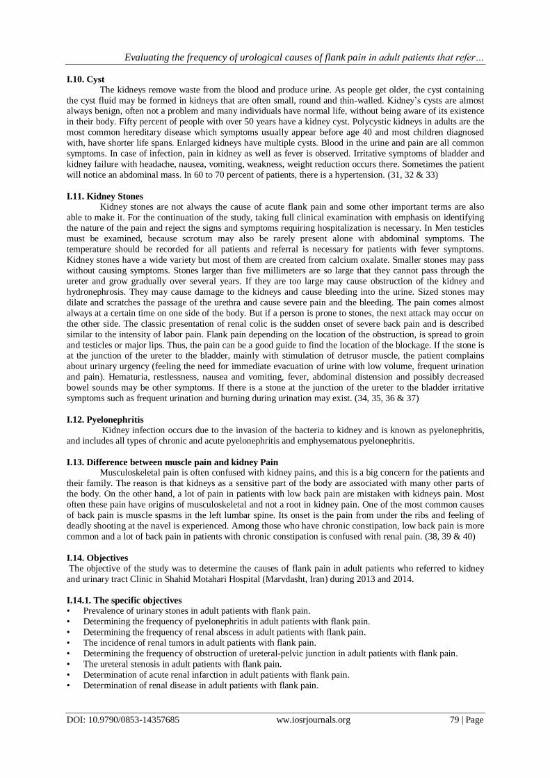
III. Results Figure (1): The frequency of patients with stones and hydronephrosis
The above diagram is related to the number of patients with stones and hydronephrosis observed in 350
adult patients referred to the kidneys and urinary tract clinic. 126 patients had renal stones with hydronephrosis.
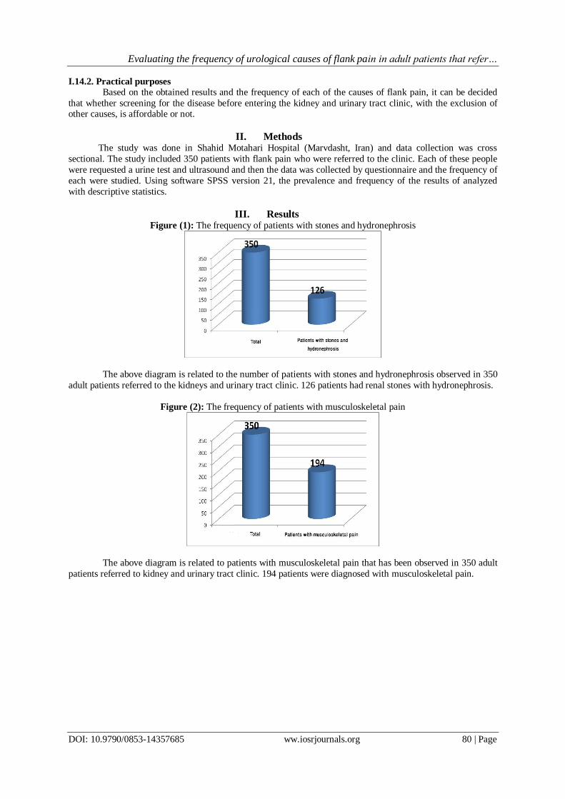
Figure (2): The frequency of patients with musculoskeletal pain
The above diagram is related to patients with musculoskeletal pain that has been observed in 350 adult
patients referred to kidney and urinary tract clinic. 194 patients were diagnosed with musculoskeletal pain.
Evaluating the frequency of urological causes of flank pain in adult patients that refer…
DOI: 10.9790/0853-14357685 ww.iosrjournals.org 81 | Page
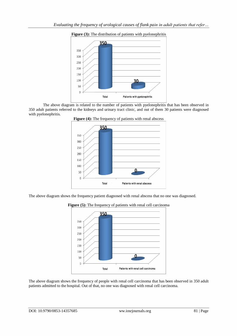
Figure (3): The distribution of patients with pyelonephritis
The above diagram is related to the number of patients with pyelonephritis that has been observed in
350 adult patients referred to the kidneys and urinary tract clinic, and out of them 30 patients were diagnosed
with pyelonephritis.
Figure (4): The frequency of patients with renal abscess
The above diagram shows the frequency patient diagnosed with renal abscess that no one was diagnosed.
Figure (5): The frequency of patients with renal cell carcinoma
The above diagram shows the frequency of people with renal cell carcinoma that has been observed in 350 adult
patients admitted to the hospital. Out of that, no one was diagnosed with renal cell carcinoma.

Evaluating the frequency of urological causes of flank pain in adult patients that refer…
DOI: 10.9790/0853-14357685 ww.iosrjournals.org 82 | Page
Figure (6): The frequency of patients with obstruction of ureteral-pelvic junction
The above diagram shows the frequency of people with obstruction of ureteral-pelvic junction that has been
observed in 350 adult patients admitted to the hospital. Out of them, no one was diagnosed with obstruction of
ureteral-pelvic junction.
Figure (7): The frequency of patients with ureteral stenosis.
The above diagram is related to the number of patients with ureteral stenosis that has been observed in
350 adult patients referred to the kidneys and urinary tract clinic, and out of them, no one was diagnosed with ureteral stenosis.
Figure 8: The frequency of patients with acute renal infarction
The above diagram is related to the number of patients with acute renal infarction that has been
observed in 350 adult patients referred to the kidneys and urinary tract clinic, and out of them, no one was
diagnosed with acute renal infarction.
Evaluating the frequency of urological causes of flank pain in adult patients that refer…
DOI: 10.9790/0853-14357685 ww.iosrjournals.org 83 | Page
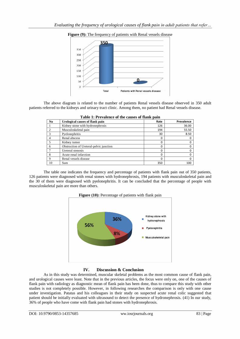
Figure (9): The frequency of patients with Renal vessels disease
The above diagram is related to the number of patients Renal vessels disease observed in 350 adult
patients referred to the kidneys and urinary tract clinic. Among them, no patient had Renal vessels disease.
Table 1: Prevalence of the causes of flank pain No Urological causes of flank pain Rate Prevalence
1 Kidney stone with hydronephrosis 126 00.36
2 Musculoskeletal pain 194 55.50
3 Pyelonephritis 30 8.50
4 Renal abscess 0 0
5 Kidney tumor 0 0
6 Obstruction of Ureteral-pelvic junction 0 0
7 Ureteral stenosis 0 0
8 Acute renal infarction 0 0
9 Renal vessels disease 0 0
10 Sum 350 100
The table one indicates the frequency and percentage of patients with flank pain out of 350 patients, 126 patients were diagnosed with renal stones with hydronephrosis, 194 patients with musculoskeletal pain and
the 30 of them were diagnosed with pyelonephritis. It can be concluded that the percentage of people with
musculoskeletal pain are more than others.
Figure (10): Percentage of patients with flank pain
IV. Discussion & Conclusion As in this study was determined, muscular skeletal problems as the most common cause of flank pain,
and urological causes were least. Note that in the previous articles, the focus were only on, one of the causes of flank pain with radiology as diagnostic mean of flank pain has been done, thus to compare this study with other
studies is not completely possible. However, in following researches the comparison is only with one cause
under investigation. Patatas and his colleagues in their study on suspected acute renal colic suggested that
patient should be initially evaluated with ultrasound to detect the presence of hydronephrosis. (41) In our study,
36% of people who have come with flank pain had stones with hydronephrosis.
Evaluating the frequency of urological causes of flank pain in adult patients that refer…
DOI: 10.9790/0853-14357685 ww.iosrjournals.org 84 | Page
In a study conducted by Chowdhury and his colleagues in 2006, the 500 people who were referred with
flank pain were enrolled. They evaluated a new imaging pathway for the investigation of patients presenting
with suspected acute renal colic. They found out the frequencies of stone were 44% and 12 percent of patients had a problem other than stones. In this study, the frequency of each of the other reasons not stated separately
and causes of flank pain is mostly due to non urologic. (42)
In another study that was conducted from 1995 to 1997, CT scanning revealed a stone in 92/281
patients (32.7%) and no stone in 189/281 patients (67.3%). Of the 189 patients, 60/189 patients (32%) had
another positive finding as a cause for flank pain. Eighty-one of 92 patients with a stone on CT (88%) had
confirmation of stone disease by radiologic or surgical intervention. Eleven of 92 patients (12%) did not have
confirmation of their diagnosis because of resolution of symptoms or refusal of further intervention. It seems
that the results obtained in this study of flank pain are consistent with results obtained in our study. (43)
In a study of Eshed and his colleagues, they found that, in 112 (26.3%) CT scans, the cause for the
patient's acute flank pain could not be explained. Forty-three (10.1%) alternative significant diagnoses that
explain the patient's complaints were found. Nineteen (44%) were findings related to the urinary tract, and 24 (56%) were not related to the urinary tract. (44)
Seen in this study, 350 adult patients referred to the kidney and urinary tract clinic in Shahid Motahari
Hospital (Marvdasht, Iran), 126 (36 %) patients had urinary stones with hydronephrosis, 194 (55.50%) people
with musculoskeletal pain, and 30 (8.50%) had pyelonephritis. So it can be concluded that screening all patients
before entering the kidney and urinary tract clinic, in order to discard other causes, can be very necessary and
affordable.
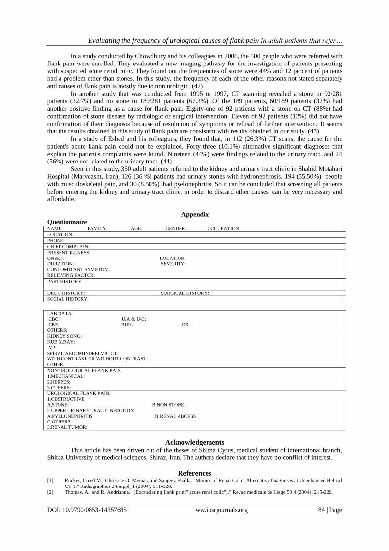
Appendix
Questionnaire NAME: FAMILY: AGE: GENDER: OCCUPATION:
LOCATION:
PHONE:
CHIEF COMPLAIN:
PRESENT ILLNESS
ONSET: LOCATION:
DURATION: SEVERITY:
CONCOMITANT SYMPTOM:
RELIEVING FACTOR:
PAST HISTORY:
DRUG HISTORY: SURGICAL HISTORY:
SOCIAL HISTORY:
LAB DATA: CBC: U/A & U/C:
CRP: BUN: CR:
OTHERS:
KIDNEY SONO:
KUB X.RAY:
IVP:
SPIRAL ABDOMINOPELVIC CT
WITH CONTRAST OR WITHOUT CONTRAST:
OTHER:
NON UROLOGICAL FLANK PAIN:
1.MECHANICAL:
2.HERPES:
3.OTHERS:
UROLOGICAL FLANK PAIN:
1.OBSTRUCTIVE
A,STONE: B,NON STONE :
2.UPPER URINARY TRACT INFECTION
A,PYELONEPHRITIS B,RENAL ABCESS
C,OTHERS: 3.RENAL TUMOR:
Acknowledgements This article has been driven out of the theses of Shima Cyrus, medical student of international branch,
Shiraz University of medical sciences, Shiraz, Iran. The authors declare that they have no conflict of interest.
References [1]. Rucker, Creed M., Christine O. Menias, and Sanjeev Bhalla. "Mimics of Renal Colic: Alternative Diagnoses at Unenhanced Helica l
CT 1." Radiographics 24.suppl_1 (2004): S11-S28.
[2]. Thomas, A., and R. Andrianne. "[Excruciating flank pain:" acute renal colic"]." Revue medicale de Liege 59.4 (2004): 215-220.
Evaluating the frequency of urological causes of flank pain in adult patients that refer…
DOI: 10.9790/0853-14357685 ww.iosrjournals.org 85 | Page
[3]. Heidenreich, Axel, F. Desgrandschamps, and F. Terrier. "Modern approach of diagnosis and management of acute flank pain:
review of all imaging modalities." European urology 41.4 (2002): 351-362.
[4]. Anderson, Kevin R., and Robert C. Smith. "CT for the evaluation of flank pain." Journal of endourology 15.1 (2001): 25-29.
[5]. Dalrymple, Neal C., et al. "Pearls and Pitfalls in the Diagnosis of Ureterolithiasis with Unenhanced Helical CT 1: (CME available in
print version and on RSNA Link)." Radiographics 20.2 (2000): 439-447.
[6]. Talner, L., and M. Vaughan. "Nonobstructive renal causes of flank pain: findings on noncontrast helical CT (CT KUB)."
Abdominal imaging 28.2 (2003): 0210-0216.
[7]. Krinsky, G. "Unenhanced helical CT in patients with acute flank pain and renal infarction: the need for contrast material in selected
cases." AJR. American journal of roentgenology 167.1 (1996): 282-283.
[8]. Dalla Palma, L., R. Pozzi-Mucelli, and F. Stacul. "Present-day imaging of patients with renal colic." European radiology 11.1
(2001): 4-17.
[9]. Katz, Douglas S., et al. "Alternative or additional diagnoses on unenhanced helical computed tomography for suspected renal colic:
experience with 1000 consecutive examinations." Urology 56.1 (2000): 53-57.
[10]. Smith, Robert C., et al. "Acute flank pain: comparison of non-contrast-enhanced CT and intravenous urography." Radiology 194.3
(1995): 789-794.
[11]. Babaian, R. J., and D. E. Johnson. "Primary carcinoma of the ureter." The Journal of urology 123.3 (1980): 357-359.
[12]. Sanseverino, E. "Benign tumors of the ureter; primary, benign and fibromatous neoplasms of the inferior extremity of the ureter,
extending in the bladder." Giornale italiano di chirurgia 12.6 (1956): 313.
[13]. Twinem, Francis P. "Primary tumors of the ureter." Journal of the American Medical Association 163.10 (1957): 808-813.
[14]. Jung, Dae Chul, et al. "Renal Papillary Necrosis: Review and Comparison of Findings at Multi–Detector Row CT and Intravenous
Urography 1." Radiographics 26.6 (2006): 1827-1836.
[15]. Simforoosh, N., and A. Nouralizadeh. "Iranian text book of Urology." Tehran: Behineh (2007): 211-212.
[16]. Smith, Robert C., et al. "Acute flank pain: comparison of non-contrast-enhanced CT and intravenous urography." Radiology 194.3
(1995): 789-794.
[17]. Yilmaz, Sindel, et al. "Renal colic: comparison of spiral CT, US and IVU in the detection of ureteral calculi." European radiology
8.2 (1998): 212-217.
[18]. Kermani, Tanaz A., et al. "Idiopathic retroperitoneal fibrosis: a retrospective review of clinical presentation, treatment, and
outcomes." Mayo Clinic Proceedings. Vol. 86. No. 4. Elsevier, 2011.
[19]. Scheel, Paul J., and Nancy Feeley. "Retroperitoneal fibrosis." Rheumatic Disease Clinics of North America 39.2 (2013): 365-381.
[20]. Dufour, M., C. Payet, and A. Gillet. "Bilateral renal infarction and ecstasy." Journal des maladies vasculaires 39.4 (2014): 285.
[21]. Tanagho, Emil A., and Jack W. McAninch, eds. Smith's general urology. Vol. 16. New York: McGraw-Hill Medical, 2008.
chapter13, P.202.
[22]. Simforoosh, N., and A. Nouralizadeh. "Iranian text book of Urology." Tehran: Behineh (2007): 87-88.
[23]. Meads, Albert M. "Perinephric Abscess Secondary to Renal Infection." California medicine 68.2 (1948): 83.
[24]. Jou, Yeong-Chin, et al. "Acute scrotal pain: An uncommon manifestation of renal vein thrombosis." Journal of the Chinese
Medical Association 77.3 (2014): 160-162.
[25]. Nana, Gael R., et al. "Left renal vein thrombosis: a rare cause of acute scrotal pain." BMJ case reports 2014 (2014): bcr2013202237.
[26]. Tonneijck, L., et al. "A male with extensive renal vein thrombosis." The Netherlands journal of medicine 71.9 (2013): 480-484.
[27]. Sandhu, G., et al. "Bilateral renal vein thrombosis can cause nephrotic range proteinuria." QJM: monthly journal of the Association
of Physicians 107.9 (2014): 763.
[28]. Tanagho, Emil A., and Jack W. McAninch, eds. Smith's general urology. Vol. 16. New York: McGraw-Hill Medical, 2008. Chapter
20, P. 311.
[29]. Simforoosh, N., and A. Nouralizadeh. "Iranian text book of Urology." Tehran: Behineh (2007): 354-355.
[30]. Mal'kov, G. A. "New approaches to the classification, grading, and prognosis of renal cell carcinoma." (2014): 76(2):60-70
[31]. Garozzo, M., A. Granata, and G. G. Battaglia. "[Renal cysts: current indications for ultrasound study]." Giornale italiano di
nefrologia: organo ufficiale della Societa italiana di nefrologia 31.1 (2013).
[32]. Smith, R. C., et al. "Acute ureteral obstruction: value of secondary signs of helical unenhanced CT." AJR. American journal of
roentgenology 167.5 (1996): 1109-1113.
[33]. Tonolini, Massimo, et al. "Complications of Sporadic, Hereditary, and Acquired Renal Cysts: Cross-Sectional Imaging Findings."
Current problems in diagnostic radiology 43.2 (2014): 80-90.
[34]. Kumar, K. H., et al. "Multiple cysts in kidneys: A case report." Saudi Journal of Kidney Diseases and Transplantation 25.1 (2014):
126.
[35]. Tanagho, Emil A., and Jack W. McAninch, eds. Smith's general urology. Vol. 16. New York: McGraw-Hill Medical, 2008. Chapter
20, P. 254-257.
[36]. Simforoosh, N., and A. Nouralizadeh. "Iranian text book of Urology." Tehran: Behineh (2007): 293-294.
[37]. Jung, Helene, and Palle JS Osther. "Acute management of stones: When to treat or not to treat?." World journal of urology (2014):
1-9.
[38]. Vehof, Jelle, et al. "Shared genetic factors underlie chronic pain syndromes." PAIN® 155.8 (2014): 1562-1568.
[39]. Hartvigsen, J., et al. "Patterns of musculoskeletal pain in the population: A latent class analysis using a nationally representative
interviewer‐based survey of 4817 Danes." European Journal of Pain 17.3 (2013): 452-460.
[40]. Urwin, Michelle, et al. "Estimating the burden of musculoskeletal disorders in the community: the comparative prevalence of
symptoms at different anatomical sites, and the relation to social deprivation." Annals of the rheumatic diseases 57.11 (1998): 649-
655.
[41]. Patatas, K., et al. "Emergency department imaging protocol for suspected acute renal colic: re-evaluating our service." Emergency
(2014): 85.101.
[42]. Chowdhury, F. U., et al. "Unenhanced multidetector CT (CT KUB) in the initial imaging of suspected acute renal colic: evaluating a
new service." Clinical radiology 62.10 (2007): 970-977.
[43]. Nachmann, Marcella M., et al. "Helical CT scanning: the primary imaging modality for acute flank pain." The American journal of
emergency medicine 18.6 (2000): 649-652.
[44]. Eshed, I., et al. "Unenhanced spiral CT for the assessment of renal colic. How does limiting the referral base affect the discovery of
additional findings not related to urinary tract calculi?." European journal of radiology 41.1 (2002): 60-64.
Related Documents