Anatomy , test and disorders of Eustachian Tube BY DR.VIJAYASUNDARAM ASSOCIATE PROFESSOR 1

Eustachian tube, anatomy, test and disorders, dr.vijaya sundarm, 20.03.17
Apr 21, 2017
Welcome message from author
This document is posted to help you gain knowledge. Please leave a comment to let me know what you think about it! Share it to your friends and learn new things together.
Transcript
1
Anatomy , test and disorders of Eustachian Tube
BYDR.VIJAYASUNDARAM
ASSOCIATE PROFESSOR
2
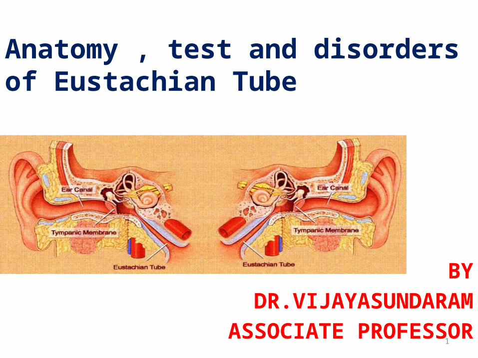
History
• Bartolomeus Eustachius first described it
as pharyngo-tympanic tube in 1562.
• Antonio Valsalva named it Eustachian
tube.

3
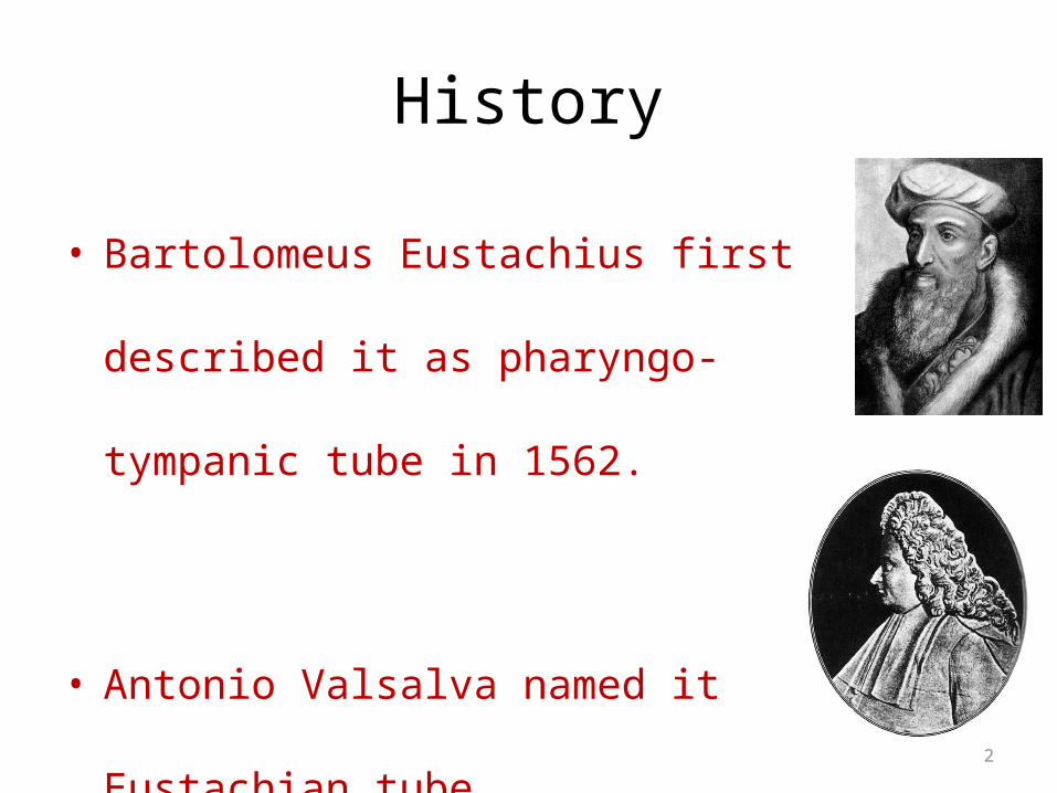
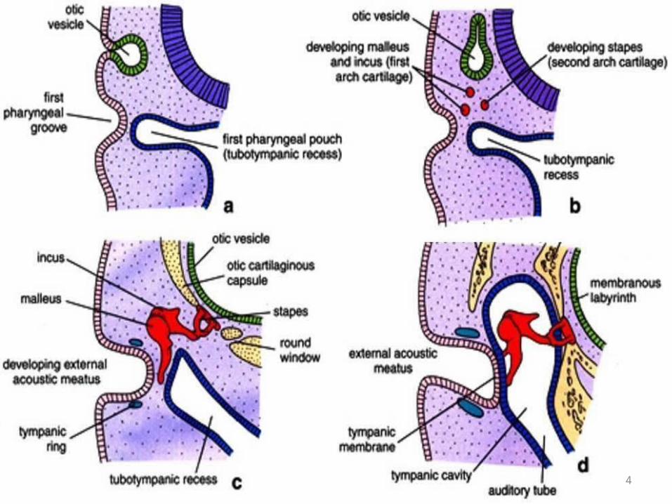
Embryology
• Develops from tubo-tympanic recess, derived from endoderm of 1st pharyngeal pouch.
• the distal portion of the pouch expands and forms middle ear cavity
• Proximal portion forms the Eustachian tube
• cartilage and muscles develop from surrounding mesoderm
4

5
Anatomy

6
Anatomy
• 36 mm long in adults.
• Directed anteriorly, inferiorly & medially from anterior wall of
M.E., forming angle of 450 with horizontal
• Enters naso-pharynx 1.25 cm behind posterior end of inferior
turbinate.
• Channel connecting tympanic cavity and nasopharynx
• lumen of the Eustachian tube is roughly triangular, measuring
2-3 mm vertically and 3-4 mm horizontally.
• Lined by respiratory epithelium with goblet cells and mucous
glands

7
Angulation

8
Pharyngeal opening
9
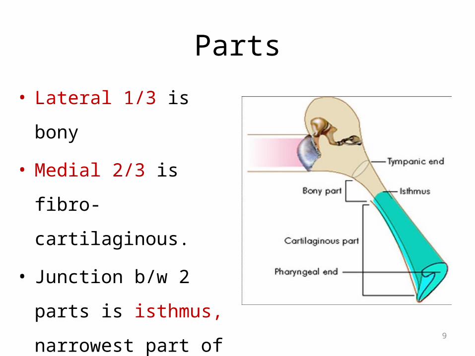
Parts
• Lateral 1/3 is bony
• Medial 2/3 is fibro-
cartilaginous.
• Junction b/w 2 parts is
isthmus, narrowest part
of Eustachian Tube.
10
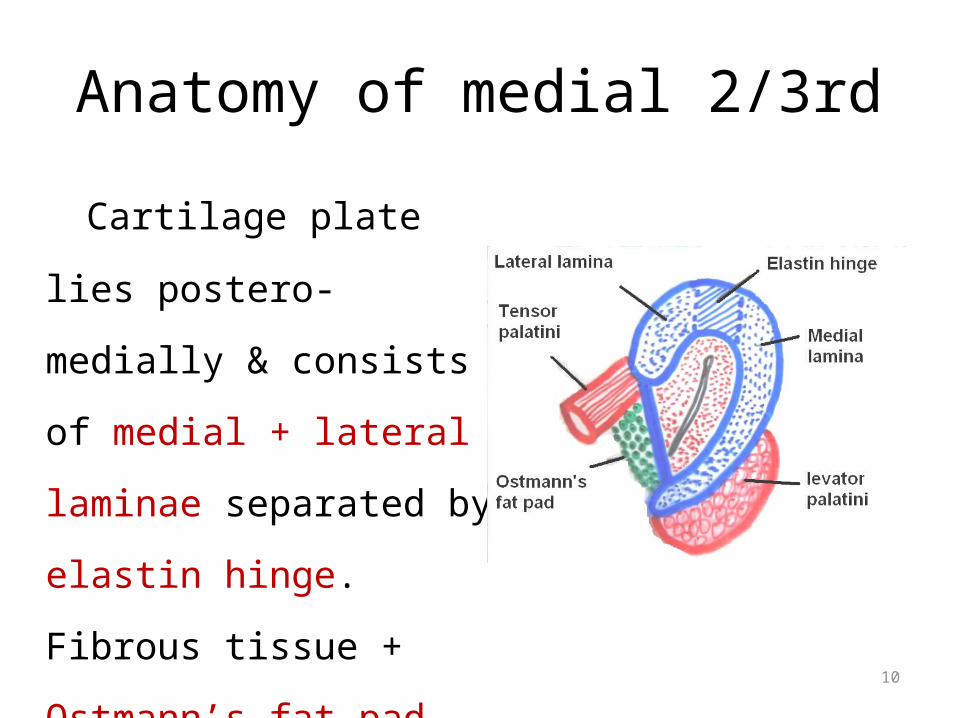
Anatomy of medial 2/3rd
Cartilage plate lies postero-
medially & consists of medial
+ lateral laminae separated
by elastin hinge. Fibrous
tissue + Ostmann’s fat pad lie
antero-laterally.
11
• Bony part : • 12mm long • widest at tympanic end• gradually narrows towards isthmus (2mm)• Thin plate of separating from tensor
tympani superiorly• Plate of bone separating from internal
carotid medially

12
• Cartilaginous part• 24mm long• Cartilage forms posteromedial wall and a small portion
anterolaterally • sits in a groove between petrous temporal bone and
greater wing of sphenoid• nasopharyngeal opening surrounded by tubal
elevation above and behind• Fossa of Rosen Muller lying behind this tubal elevation

13
Anatomy
• Lining epithelium: pseudo stratified ciliated columnar
• Arterial supply: ascending pharyngeal &
middle meningeal arteries
• Venous drainage: pharyngeal & pterygoid
venous plexus
• Lymphatic drainage: retropharyngeal node

14
Anatomy Muscle attachments:
Muscles attached to ET
Levaor palati – lower surface of petrous bone and cartilage and fascia of upper carotid sheath
Tensor palati- bony wall of scaphoid and whole length of short cartilaginous flange
Salphingo pharyngeus – inferior part of cartilage near its pharyngeal end
Tensor tympani – cartilage of ET, surrounding bony canal and greater wing of sphenoid

15
Nerve supply
• Tubal mucosa – tympanic branch of cranial
nerve IX
• Tensor veli palatini - Mandibular branch of
trigeminal • Levator veli palatini Pharyngeal plexus• Salpingo pharygeus

16
Endoscopic Anatomy
• Medial end forms tubal
elevation / torus tubarius
• Lymphoid collection over
torus is called Gerlach’s tubal
tonsil.
• Postero-superior to torus is
fossa of Rosenmüller.

17
Adult vs. Child (< 7 yr)

18
Adult vs INFANTADULT INFANT
Length 36 mm 18 mm
Angle with horizontal 45 0 10 0
Lumen Narrower Wider
Angulation at isthmus Present Absent
Cartilage Rigid Flaccid
Elastic recoil Effective Ineffective
Ostmann’s fat More Less

19
Infant E. tube • wider shorter and more horizontal
So secretions even milk can regurgitate from nasopharynx to middle ear if infant not fed in head up position
20
Physiology• Bony part is always open.
• Fibro-cartilaginous part is closed at rest.
• Opens on:
1. swallowing
2. yawning
3. sneezing
4. forceful inflation
21
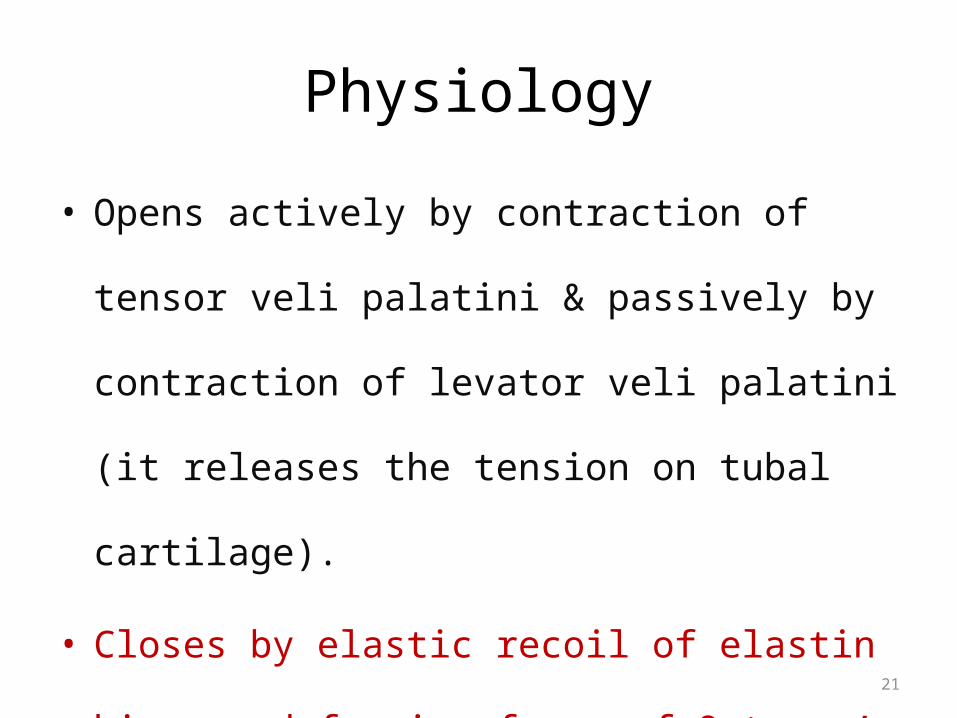
Physiology
• Opens actively by contraction of tensor veli palatini &
passively by contraction of levator veli palatini (it
releases the tension on tubal cartilage).
• Closes by elastic recoil of elastin hinge + deforming
force of Ostmann’s fat pad.

22
E.T. opening
23
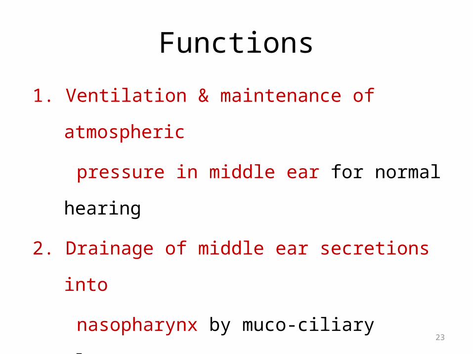
Functions
1. Ventilation & maintenance of atmospheric
pressure in middle ear for normal hearing
2. Drainage of middle ear secretions into
nasopharynx by muco-ciliary clearance,
pumping action of Eustachian tube &
presence of intra-luminal surface tension

24
Functions
3. Protection of middle ear from:
– Ascending nasopharyngeal secretions due to
narrow isthmus & angulation between 2 parts of
E.T. at isthmus
– Pressure fluctuations
– Loud sound coming through pharynx

25
Functions

26
Conditions of Dysfunction

27
EVALUATION OF ET• History• Fullness of ears
• Pain and discomfort
• Hearing loss
• Tinnitus
• Dizziness

28
EVALUATION OF ET
Physical examination• Retracted TM• Middle ear effusion• Pneumatic otoscopy• Postnasal examination• Endoscopic examination• Valsalva maneuver• Tonybee test• Politzer test• Sonotubometry

29
Tests for E.T. function

30
ET Function Tests• VALSALVA TEST– Principle: positive pressure in the nasopharynx causes air
to enter the Eustachian tube

31
– Tympanic membrane perforation- a hissing sound– Discharge in the middle ear- cracking sound– Only 65% of persons can do this test.– Contraindications:• Atrophic scar of tympanic membrane which can
rupture• Infection of nose & nasopharynx

32
• Politzer test– Done in children who are unable to perform valsalva
test.
– Olive shaped tip of the politzer’s bag is introduced into the patient’s nostril on the side of which the tubal function is desired to be tested
– Other nostril closed & the bag compressed while at the same time the patient swallows or says “ik,ik,ik”
33
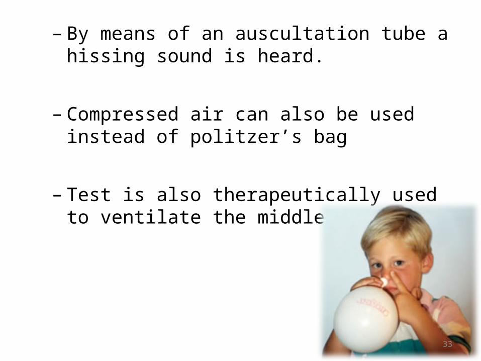
– By means of an auscultation tube a hissing sound is heard.
– Compressed air can also be used instead of politzer’s bag
– Test is also therapeutically used to ventilate the middle ear.
34
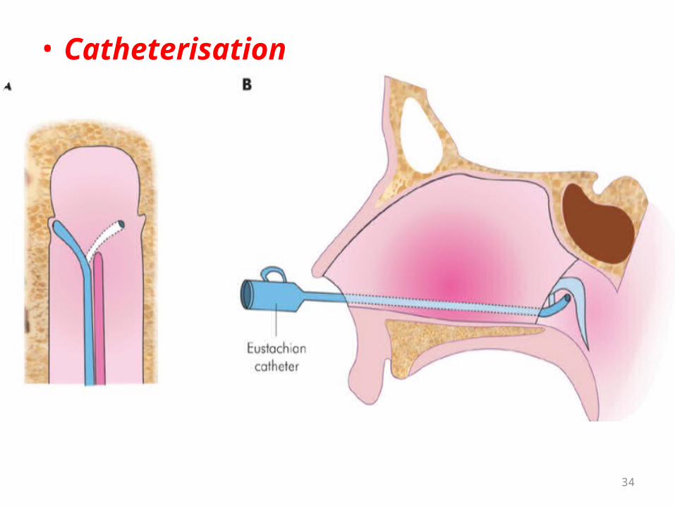
• Catheterisation

35
• Procedure for Catheterisation
•Nose is anaesthetised•E Tube catheter passed along the floor of nose till it reaches naso pharynx•Rotated 90deg medially•Pulled back till posterior border of nasal septum engaged•Rotated 180 deg laterally – tip lies against tubular opening• Politzer’s bag connected • Air insufflated • Entry of air to middle ear verified (lateral bulging of t.m)

36
6. E.T. catheterization Air pushed into E.T. catheter by squeezing Politzer bag.
Examiner hears by Toynbee auscultation tube put in
pt's ear.
Blowing sound = normal E.T. patency
Bubbling sound = middle ear fluid
Whistling sound = partial E.T. obstruction
No sound = complete obstruction of E.T.
37
– Complications:• Injury to Eustachian tube opening • Bleeding from nose• Transmission of nasal & nasopharyngeal infection into
middle ear• Rupture of atrophic area of tympanic membrane

38
• Toynbee’s test– Uses negative pressure– Ask the patient to swallow while nose is pinched– Draws air from middle ear to nasopharynx – inward
movement of t.m.

39
• Tympanometry (inflation-deflation test)– +Ve & -ve pressures are created in the external ear
and the patient swallows repeatedly– in patients with perforated or intact tympanic
membrane• Radiological Test• Saccharine/ Methylene blue Test– Saccharine solution– Methylene blue dye– Ear drops into ear with TM perforation
• Sonotubometry

40
Disorders of ET
41
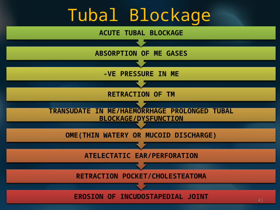
Tubal Blockage
EROSION OF INCUDOSTAPEDIAL JOINT
RETRACTION POCKET/CHOLESTEATOMA
ATELECTATIC EAR/PERFORATION
OME(THIN WATERY OR MUCOID DISCHARGE)
TRANSUDATE IN ME/HAEMORRHAGE PROLONGED TUBAL BLOCKAGE/DYSFUNCTION
RETRACTION OF TM
-VE PRESSURE IN ME
ABSORPTION OF ME GASES
ACUTE TUBAL BLOCKAGE
42
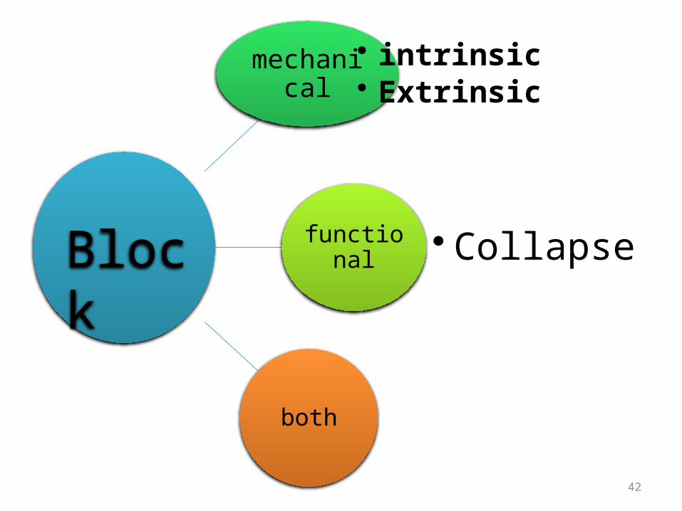
mechanical• intrinsic• Extrinsic
functional • Collapse
both
Block
43
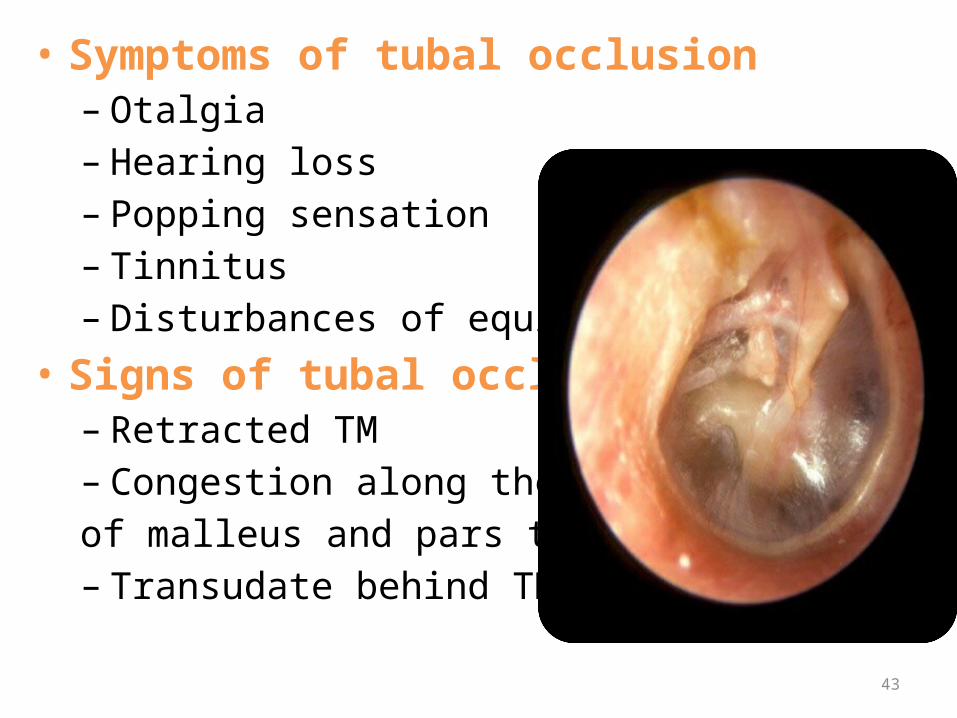
• Symptoms of tubal occlusion– Otalgia– Hearing loss– Popping sensation– Tinnitus– Disturbances of equilibrium
• Signs of tubal occlusion– Retracted TM– Congestion along the handle of malleus and pars tensa– Transudate behind TM

44
• Clinical causes of ET obstruction
– Upper respiratory tract infection– Allergy– Sinusitis– Nasal polypi– DNS– Hypertrophic adenoids– Nasopharyngeal tumour/ mass– Cleft palate– Submucous cleft palate– Down’s syndrome
45
Adenoids• Adenoids cause tubal dysfunction by:– Mechanical obstruction of the tubal opening– Acting as reservoir for pathogenic organisms– Inflammatory mediators in allergy cause tubal
blockage• Adenoids can cause otitis media with effusion or
recurrent acute otitis media• Adenoidectomy

46

47
large adenoid blocking left et

48
Cleft palate • Tubal dysfunction due to:– Abnormalities of torus tubaris– Tensor veli palatini doe not insert into the torus
tubaris• Otitis media with effusion is common in these
patients
49
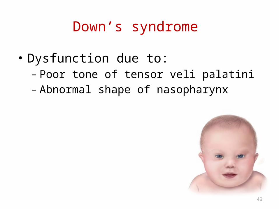
Down’s syndrome
• Dysfunction due to:– Poor tone of tensor veli palatini– Abnormal shape of nasopharynx

50
Barotrauma
• Non suppurative condition resulting from failure of E Tube to maintain M Ear pressure at ambient atmospheric level
• Cause:– Rapid descent during air flight– Under water diving– Compression in pressure chamber
• When atm pressure > M E pressure by critical pressure of 90mm Hg E T gets locked – Negative pressure in ME
• T M retraction - transudation/ h’ge

51
Retraction Pockets & ET
52
• Any obstruction in the ventilation pathway retraction pockets or atelectasis of tympanic membrane– Obstruction of Eustachian tube total atelectasis of tm
– Obstruction at additus cholesterol granuloma & collection of mucoid discharge in mastoid air cells

53
• Other changes – Thin atrophic TM– Cholesteatoma– Ossicular necrosis– Tympanosclerotic changes
• Management– Repair of irreversible pathologic processes– Establishment of ventilation
54
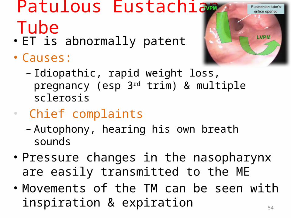
Patulous Eustachian Tube• ET is abnormally patent• Causes:– Idiopathic, rapid weight loss, pregnancy (esp 3rd trim)
& multiple sclerosis• Chief complaints– Autophony, hearing his own breath sounds
• Pressure changes in the nasopharynx are easily transmitted to the ME
• Movements of the TM can be seen with inspiration & expiration
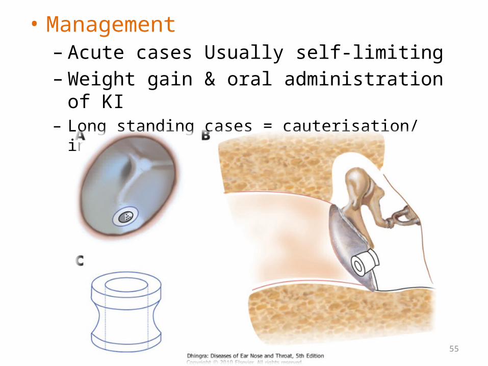
55
• Management– Acute cases Usually self-limiting– Weight gain & oral administration of KI – Long standing cases = cauterisation/ insertion of grommet

56
EXAMINATION OF EUSTACHIAN TUBEPharyngeal end of eustachian tube :posterior
rhinoscopy, rigid nasal endoscope or flexible nasopharyngoscope
Tympanic end :microscope or endoscope
Simple examination of TM may reveal retraction pockets or fluid in the me
Movements of TM with respiration point to patulous eustachian tube

57
• Aetiologic causes of eustachian tube dysfunction assessed through:– Nasal examination– Endoscopy– Tests of allergy– CT scan of temporal bones– MRI to exclude multiple sclerosis

58
Related Documents