A COMPARISON OF THE EFFICACY OF THREE ANTHELMINTIC DRUGS AGAINST MIXED NATURAL GASTROINTESTINAL NEMATODE INFECTIONS IN CAMEL! (Camelus dromedarius) IN KENYA ' iCo, *r , r< f „ EUSEBIUS JUMA MUKHWANA (B.V.M., UNIVERSITY OF NAIROBI) jilS Till-.-- • •inE D F A '‘ \NU a copy 0 N1 VEUS1 TY JET MAY ACCEPTED KOI* W^frrT............. — BE PLACED itf XM LIBRARY* A THESIS SUBMITTED IN PARTIAL FULFILMENT FOR THE DEGREE OF MASTER OF SCIENCE IN THE DEPARTMENT OF PUBLIC HEALTH, PHARMACOLOGY AND TOXICOLOGY, UNIVERSITY OF NAIROBI, KENYA. o*T 1993

Welcome message from author
This document is posted to help you gain knowledge. Please leave a comment to let me know what you think about it! Share it to your friends and learn new things together.
Transcript

A COMPARISON OF THE EFFICACY OF
THREE ANTHELMINTIC DRUGS AGAINST MIXED
NATURAL GASTROINTESTINAL NEMATODE
INFECTIONS IN CAMEL!
(Camelus dromedarius) IN KENYA '
iC o,* r , r< f „
EUSEBIUS JUMA MUKHWANA(B.V.M., UNIVERSITY OF NAIROBI)
jilS Till-.-- •• in E D F A ' ‘
\NU a c o py
0 N1VEUS1TY
JETMAY
ACCEPTED KOI*
W^frrT.............—BE PLACED itf XM
LIBRARY*
A THESIS SUBMITTED IN PARTIAL FULFILMENT FOR THE
DEGREE OF MASTER OF SCIENCE IN THE DEPARTMENT OF
PUBLIC HEALTH, PHARMACOLOGY AND TOXICOLOGY,
UNIVERSITY OF NAIROBI, KENYA.
o *T
1993
( 11
DF.CLARATION
This llvsis is my original work ami has not boon presented for a
degree in any otlior University
l)r. Fiisebius Juma Mukhwana, II V M.
This thesis has been submitted for examination with our approval
as University Supervisors.
Date: ..........
Prof. F.ric S. Miloma, B.V.M., M.S., Ph D.
Prof. Timothy Mnitho, B.V.M., M.Sc., Ph.D
Dr. Moses N. Kyule, B.V.M., M.So., MPVM., Ph D.

Il l
DEDICATION
This work is dedicated to my parents, Mr. Thomas G. Mukhwana
and Mrs. Anne Namarome Mukhwana for their foresightedness in
education success and achievements, a thing that has been a source
of great inspiration to their children over the years.

I V
ACKNOWLEDGEMENT
I wish to express my indebtedness to my supervisors; Professor
Eric S. Mitema, Professor Timothy E. Maitho and Dr. Moses N.
Kyule, all of the Department of Public Health, Pharmacology and
Toxicology (PHPT), University of Nairobi for their invaluable
advice, suggestions, criticisms and guidance throughout this study.
My appreciation also goes to Messrs. Ezekiel Weda and Daniel
Muriuki, technicians in the departments of Veterinary Microbiology
and Pathology and PHPT respectively, for their unfailing technical
assistance in the analysis of faecal egg counts and identification of
recovered larvae both in the field and in the laboratory.
This work was financially supported by a scholarship from Food
and Agricultural Research Management-Africa (FARM-Africa) to
whom I extend my sincere appreciation. I am particularly grateful to
the project leader, Dr. Christopher R. Field for his personal interest
and guidance throughout the entire study. My special thanks also go
to all the staff members of FARM-Africa , especially Messrs. Chris
Morris (Project Administrator), Francis Guturo, Mohamed Wario,
Reuben Lemunyete and M /s Dolly Njeru for their co-operation and
support. My gratitudes also go to the pastoralists, especially those of
the Kisima Camel Improvement group (CIG) for allowing me to
work with their camels. Special thanks go to Mr. J.O Evans of Ol
Maisor Ranch for his willingness to share his long experience of
working with camels with me and Brother J.W. Smit of Utrecht,
Netherlands for his encouraging letters that enabled me to endure
this part of my life.

V
Special devotion goes to all members of the Mukhwana family
and those of Bidii Women group for their initial efforts that enabled
me to start this work without a scholarship. Special thanks in this
regard go to Mr. and Mrs. Protus W. Masinde and Messrs Fred W.
Wamalwa, Henry W. Ngichabe and Mr. James W. Kundu who
sacrificed so much during this time of uncertainty.
My parents, Mr. and Mrs. Thomas G. Mukhwana are thanked for
their consistently devoted energies, will power and love that has
contributed significantly to the furtherance of their children's
welfare.I am much obliged to thank my late grandmother Paulina K.
Murunga who passed away just when I was about to complete this
work for her constant constructive advice throughout my life. She
was a teacher in deed.
Deep appreciation is expressed to Cosmos (K) Ltd, Kenya-Swiss
Chemical Co. Ltd and the organizers of the third Maralal
International Camel Derby (MICD) for their drug donations that
enabled me to get maximum co-operation from the farmers. In a
special way, I owe many thanks to my wife, Lucy Jemutai for her
love, patience and constant encouragement. Sincere appreciation
and gratitude goes to Mrs. Dorcas Nduati and Mr. Crispin Matere for
assisting with the data analysis. Lastly I would like to thank Miss
Hellen N. Muthui for typing the thesis.
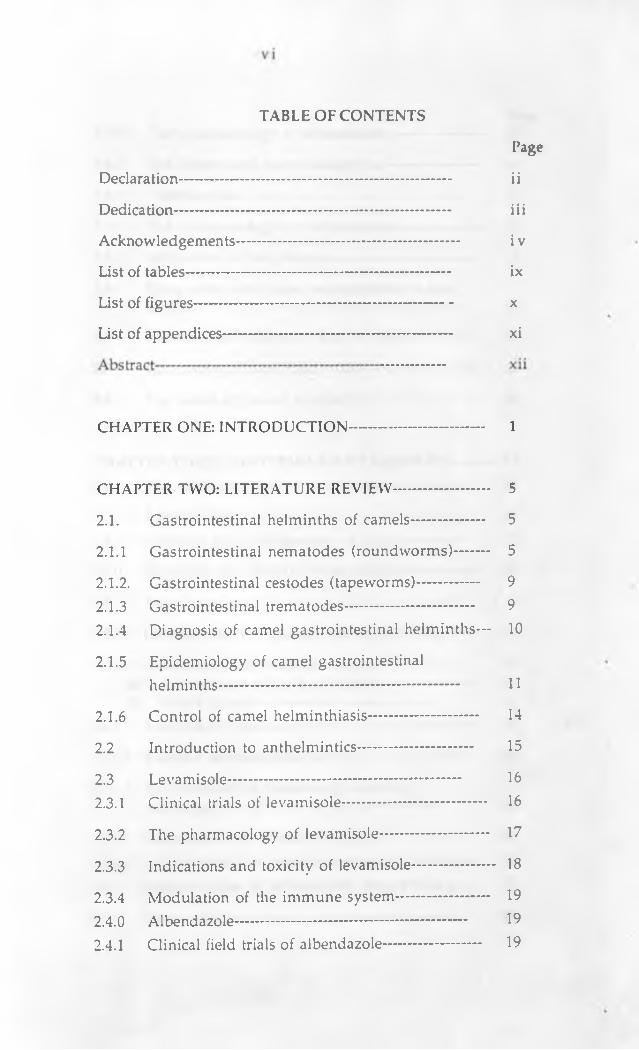
TABLE OF CONTENTS
Declaration-------------------------------------------------------- ii
Dedication--------------------------------------------------------- iii
Acknowledgements---------------------------------------------- i v
List of tables------------------------------------------------------ ix
List of figures----------------------------------------------------- x
List of appendices----------------------------------------------- xi
CHAPTER ONE: INTRODUCTION---------------------------- 1
CHAPTER TWO: LITERATURE REVIEW-------------------- 5
2.1. Gastrointestinal helminths of camels— ................ 5
2.1.1 Gastrointestinal nematodes (roundworms)------- 5
2.1.2. Gastrointestinal cestodes (tapeworms)-------------- 92.1.3 Gastrointestinal trematodes-------------------------- 92.1.4 Diagnosis of camel gastrointestinal helminths— 10
2.1.5 Epidemiology of camel gastrointestinalhelminths------------------------------------------------- 11
2.1.6 Control of camel helminthiasis---------------------- 14
2.2 Introduction to anthelmintics............................... 15
2.3 Levamisole------------------------------------------------ 162.3.1 Clinical trials of levamisole....................................... 16
2.3.2 The pharmacology of levamisole.............................. 17
2.3.3 Indications and toxicity of levamisole....................... 18
2.3.4 Modulation of the immune system -....................... 192.4.0 Albendazole------------------------------------------------ 192.4.1 Clinical field trials of albendazole-------------------- 19
Page
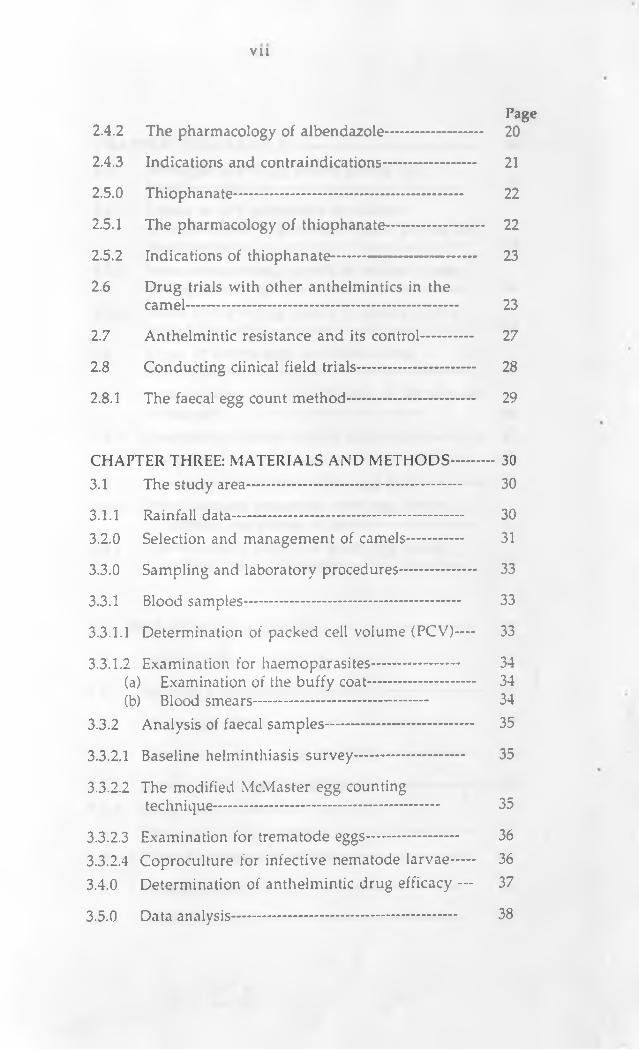
2.4.2 The pharmacology of albendazole-------------------- 20
2.4.3 Indications and contraindications------------------- 21
2.5.0 Thiophanate----------------------------------------------- 22
2.5.1 The pharmacology of thiophanate-------------------- 22
2.5.2 Indications of thiophanate---------------- ------------- 23
2.6 Drug trials with other anthelmintics in thecamel-------------------------------------------------------- 23
2.7 Anthelmintic resistance and its control---------- 27
2.8 Conducting clinical field trials------------------------ 28
2.8.1 The faecal egg count method-------------------------- 29
CHAPTER THREE: MATERIALS AND METHODS---------- 303.1 The study area-------------------------------------------- 30
3.1.1 Rainfall data----------------------------------------------- 303.2.0 Selection and management of camels----------- 31
3.3.0 Sampling and laboratory procedures..................... 33
3.3.1 Blood samples-------------------------------------------- 33
3.3.1.1 Determination of packed cell volume (PCV)— 33
3.3.1.2 Examination for haemoparasites....................... 34(a) Examination of the buffy coat---------------------- 34(b) Blood smears------------------------------------ 34
3.3.2 Analysis of faecal samples------------------------------ 35
3.3.2.1 Baseline helminthiasis survey............................. 35
3.3.2.2 The modified McMaster egg countingtechnique---------------------------------------------- 35
3.3.2.3 Examination for trematode eggs........................ 363.3.2.4 Coproculture for infective nematode larvae..... 363.4.0 Determination of anthelmintic drug efficacy — 37
3.5.0 Data analysis---------------------------------------------- 38
V l l
Page

vm
CHAPTER FOUR: RESULTS----------------------------------- 394.1 Strongyle worm egg counts during the
baseline survey------------------------------------------ 394.1.1 Levels of GIT nematodes in relation
to total rainfall in Lorroki Division during the study period---------------------------------------------- 39
4.1.2 Mean strongyle egg counts in relation to ageduring the survey---------------------------------------- 40
4.1.3 Mean strongyle egg counts in relation to sexduring the survey---------------------------------------- 44
4.2 Types of worms eggs identified duringthe survey-------------------------------------------------- 45
4.2.3 Larval culture and identification of the variousnematodes------------------------------------------ 46
4.4.0 Comparative efficacy of the anthelmintics---------- 474.4.1 The packed cell volume------------------- ------------- 474.4.2 Overall post treatment worm egg counts---------- 484.4.3 Post treatment nematode worm egg counts
in different age groups of camels--------------------- 514.4.4 Post treatment nematode worm egg counts
in different sexes of camels--------------------------- 55
Page
CHAPTER FIVE: DISCUSSION-------------------------------- 59
5.1 Introduction----------------------------------------------- 59
5.2 Baseline helminthiasis survey.................................. 59
5.2.1 Worm egg counts---------------------------------------- 59
5.2.2 The types of worm eggs identified------------------- 61
5.2.3 Larvae culture and identification............................ 625.3.0 Drug trials------------------------------- 635.3.1 The packed cell volume---------------------------------- 635.3.2 Overall anthelmintic efficacy................................... 63
6.0 REFERENCES-------------------------------------------- 66
7.0 APPENDICES--------------------------------------------

IX
Table 1: Common gastrointestinal helminths of camels— 8
Table 2: Age structure camels used in theanthelmintic drug trials----------------------------- 33
Table 3: Mean monthly strongyle egg counts of camelsin relation to rainfall in lorroki division of Samburu district-------------------------------------- 39
Table 4: Mean monthly strongyle egg counts of camelsin relation to age-------------------------------------- 42
Table 5: Mean strongyle egg counts for the differentsexes of camels during the baseline survey---- 44
Table 6: Percentage of different worm eggs identifiedduring the survey period--------------------------- 46
Table 7: Nematode larvae recovered during the surveyperiod as a percentage of the total------------------- 47
Table 8: PCV values for camels in different treatmentgroups before and after treatment---------------- 48
Table 9: A two way table of treatments and mean wormegg counts per gramme of faeces before and after treatment--------------------------------------- 49
LIST OF TABLES
Page

X
Figure 1: The structure of levamisole------------------- 17
Figure 2: The structure of albendazole------------------ 20
Figure 3: The structure of thiophanate----------------- 22
Figure 4 Mean strongyle egg counts of camels in relationto total rainfall (in mm) in Lorroki division------- 40
Figure 5: Mean strongyle egg counts for the differentage groups during the survey period--------------- 43
Figure 6: Mean strongyle egg counts in relation to sexof camels during the survey period----------------- 45
Figure 7: Mean e.p.g counts in different treatmentgroups following administration of the drugs---- 50
Figure 8: Mean nematode egg counts in different age groupsafter treatment with albendazole............................ 51
Figure 9: Mean nematode egg counts in different agegroups of camels treated with levamisole............ 52
Figure 10: Mean nematode egg counts in camels ofdifferent age groups treated with thiophanate-— 53
Figure 11 Mean nematode egg counts in camels ofdifferent age groups that received a placebo (control)---------------------------------------------------- 55
Figure 12: Mean nematode egg counts in male and femalecamelstreated with albendazole......................... 56
Figure 13: Mean nematode egg counts in male and femalecamels treated with thiophanate........................ 57
Figure 14: Mean nematode egg counts in different sexesof camels treated with Thiophanate........................ 58
LIST OF FIGURES
Page

XI
Appendix 1. Worm egg counts in November 1992---------- 77
Appendix 2. Worm egg counts in December 1992---------- 79
Appendix 3. Worm egg counts in January 1993------------- 81
Appendix 4. Worm egg counts in February 1993------------ 82
Appendix 5: Worm egg counts in March 1993--------------- 83
Appendix 6: Nematode worm egg counts for camelstreated with Albendazole over the 28 days period------------------------------------------------ 84
Appendix 7: Nematode worm egg counts for camelstreated with levamisole over the 28 days period------------------------------------------------ 85
Appendix 8: Nematode worm egg counts for camelstreated with thiophanate over the 28 day period-------------------------------------- 86
Appendix 9: Nematode worm egg counts for camels in thecontrol group over the 28 day period—........ 87
LIST OF APPENDICES
Page
XU
ABSTRACT
This study was undertaken to identify the types of helminth
parasites in camels, their prevalence rates in different seasons, the
effects of age and sex of camels on helminth infestation rates and to
compare the efficacy of three anthelmintics, namely albendazole,
levamisole and thiophanate in the treatment of gastrointestinal
nematodes in camels (Camelus dromedarius) owned by the local
community in Lorroki Division, Samburu District, Kenya.
During the survey, 255 camels had their faecal samples taken once
over a period of five months. These included 59 camels in
November 1992, 66 in December 1992, 47 in January 1993, 46 in
February 1993 and 37 in March 1993. The faecal samples were
subjected to the McMaster egg counting technique and coproculture.
The worm eggs and recovered nematode larvae were identified
using standard parasitological techniques.
Blood was collected in heparinized capillary tubes for
determination of the packed cell volume (PCV) which was used as
an indicator of the anemia status. Examination of the buffy coat and
blood smears was done to rule out the presence of haemoparasites.
Out of the 255 camels examined as previously described, 76
clinically healthy camels but which had moderate to heavy worm
egg counts (EPG of more than 400) were selected and used in the
anthelmintic drug study- These camels which included both males
and females comprised all age groups. PCV values for all the
animals was determined once before and one month after
treatment. The selected camels were randomly distributed (n=19) by

XU1
The survey on helminthiasis showed that peak strongyle worm
egg counts in this area occur during and soon after the rains. Calves
and adults had higher worm egg counts than immatures. When
assessing the effects of sex on worm egg burdens, it was found that
female camels had higher (p < 0.05) worm egg counts than males.
The data showed that 80% of all eggs that were identified were
those of strongyle nematodes. Other parasite eggs identified included
those of tapeworms (especially Moniezia spp), Strongyloides spp,
Trichuris spp. and Fasciola spp. Larval culture and identification
showed that Haemonchus spp and Trichostrongylus spp were the
most common and probably the most pathogenic gastrointestinal
helminths of camels in this area. Other nematode parasites
id e n tified included C oop eria spp, B u n ostom u m sp p ,
Oesophagostomum spp, Strongyloides spp and Ostertagia spp.
When assessing the efficacy of the three drugs studied, it was
found that the mean PCV values in all the treated camels were
significantly higher (p < 0.05) than those of the untreated controls
one month after treatment.
The present study indicates that thiophanate at a dose of 60 mg/kg
body weight was the best drug as shown by the significant reduction
in the post-treatment nematode worm egg counts. Albendazole at a
dose of 10 m g/kg and levamisole (at a dose of 10 mg/kg) came next
in that order with levamisole being the least effective.
This study reports, for the first time, the presence of Fasciola spp
in camels in Kenya. It also indicates that peak worm intestations
age, sex, EPG counts and household into three treatment and one
control group.
XI V
occur mostly during the rain season and that Haemonchus spp is the
most common GIT parasite in camels.The study also showed that
thiophanate and albendazole promise to be highly effective, safe and
fast acting drugs for use in treating nematode infections in camels of
all ages.

1
CHAPTER ONE
INTRODUCTION
Camels continue to be an integral component of an ecosystem
in which the vegetation of the marginal lands can be converted to
human food. This is because, all over the world, camels have been
found to be superbly adapted to their respective environments.
Inspite of this, camels are susceptible to a number of viral, bacterial,
mycotic, protozoal and parasitic diseases (Richard, 1984). Among all
these diseases, helminthiasis is ranked as the second major cause of
economic loss in camel production (Richard, 1976). Economic losses
result from impairment of physiological functions with a
consequential decrease in weight gain, milk production, working
capacity and reproductive performance.
It is generally believed that of the internal parasites of the
camel, gastrointestinal nematodes are of the most serious economic
consequence. This is based on the overall numbers of worms,
numbers of genera and species present, general level of
pathogenicity and widespread distribution.
The most common genera of nematodes reported in camels
include: H aem onchus, Trichuris, N em atodirus, Strongyloides,
Bunostomum and Oesophagostomum (Rutagwenda, 1985; Wilson,
1988). Of these, H aem onchus longistipcs and T rich ostron g y lu s
probolurus have been recognized as being the most pathogenic and
economically important parasites of camels in many countries
(Steward, 1950; Malek, 1959; Altaif, 1974; El Bihari and Kawasmeh,
1980; Abdul-Salam and Farah, 1988; Onvali and Onw'uliri, 1989).

2
The use of anthelmintics drugs forms the main link in the
chain of any systems of helminthiasis control in domestic animals.
They play the important roles of destroying and eliminating
intestinal parasites and reducing contamination of pastures.
Therefore, it is imperative that the relative efficacies of the available
anthelmintics is known with reasonable accuracy to enable effective
parasite control (Reinecke et al., 1962). Several methods are in use
for determining the efficacy of an anthelm intic drug or a
combination of drugs. These include the faecal egg count method in
the live animal (Gordon,1950), the critical techniques of Hall and
Forster (1918) and the controlled test of Moskey and Harwood (1941).
The easiest and most commonly used technique is the faecal egg
count method.
In general, systematic studies of the disease conditions caused
by helminths and their management in camels are scanty and hence,
in most developing countries parasite control programmes are based
on haphazard and random use of anthelmintics and usually
extrapolated from experience in cattle and other domestic animals.
While these procedures could be affording some protection against
diseases and even mortality, they are frequently not elfective in
preventing the exposure of the animals to high levels of infestation
(Brundson, 1980). Consequently, production losses still occur as a
result of reinfection in the interval between treatment. This
negligence has been attributed partly to the devalued economic
worth of the camel.

3
This situation is worsened by the fact that many farmers hardly
attempt deworming their camels. This fact, coupled by sharing of
grazing fields and watering points tremendously increase the chances
of re-infection for those who deworm their camels.
The challenge of camel helminthiasis calls for the introduction of
cost-effective strategic control programmes that minimize the effects
of worms in camels. To achieve this, one must combine information
regarding the efficacy of different anthelm intics under field
conditions with epidemiological data developed for each specific
geographic area. In Kenya, such data has not been documented.
Reported anthelmintic drug trials in camels include those of
ivermectin (Frolka and Rostinska, 1984; Jones, 1987), thiabendazole
(Graber, 1966; Chandrasekharan et al., 1970; Kapur and Sharma, 1972),
parbendazole (Chandrasekharan et al., 1971; Frolka and Rostinska,
1984; Frolka, 1988), Oxfendazole (Michael et al., 1980) , levamisole
(Walley, 1966; Lodha et al., 1977) and fenbendazole (Rutagwenda and
Munvua, 1983). No anthelmintic drug trials have reported the efficacy
of albendazole and thiophanate in treating camel helmithiasis.
Some workers have recommended that camels can be treated with
the same drugs as other large domestic animals. However, Wilson
(1988) warns that this must be done with caution especially when new
drugs are tried in camels. This is because camels have been shown to
be idiosyncratic in their reactions to drugs. Graber (1966) reported
toxicity signs in camels following use of tetramisole. He however
showed that thiabendazole is a good and safe anthelmintic drug for
use in camels.
4
The objectives of this study were:
1) To determine the genera of gastrointestinal helminths present
in camels in Lorroki Division, Samburu District, Kenya.
2) To examine the seasonal abundance of gastrointestinal
nematodes in different age groups and sexes of camels during
different seasons as an indicator of periods of transmission.
3) To compare the efficacy of albendazole, levamisole and
thiophanate in the treatment of gastrointestinal nematode
infections of camels of both sexes and all age groups using the
faecal egg count method and the packed cell volume .

5
CHAPTER TWO
LITERATURE REVIEW
2.1.: GASTROINTESTINAL HELMINTHS OF CAMELS
The camel is a creature of the arid and semi-arid areas, a habitat
generally considered not to be conducive to the development and
transmission of helminth parasites. However, several researchers
have found a surprisingly large and diverse fauna of helminths
comprising representatives of all classes of these metazoan parasites
(EL Bihari, 1985; Wilson, 1988).
In camels, helminthiasis, is a chronic problem which occurs
with an infection rate as high as 90% in natural conditions (Richard,
1984). However, some cases of mixed nematode infections have been
reported to precipitate acute conditions. (Arzoun et al., 1984a).
2.1.1: Gastrointestinal nematodes (roundworms)
Nematodes are the most important internal parasites of camels
(Steward, 1950, Malek, 1959, Graber et al., 1967; Wilson, 1988).
Nematodiasis in camels is characterized by diarrhoea, general
debility, reduced growth rates and milk yields, increased calving
intervals, innappetance, anaemia, and consumption of large
amounts of sand (pica) (Arzoun et al., 1984b). Wilson (1988) has
reported that common camel nematodes belong to the following
genera; Trichuris, Neinatodirus, Strongyloides, Haeitwnchus and
Trichostrongylus.. Camels are infected with these parasites when
they graze on infested pasture. But, Strongyloides spp is reported to
infect camels by skin penetration.

6
Several surveys indicate that camel nematodiasis occurs with
varying prevalences in different countries and even within
countries. Richard (1976) found that 92% of all camels examined in
Ethiopia had internal parasites of which 80% were Strongyles, 10%
Strongyloides spp and 16% Trichuris spp. Wilson et al. (1984)
reported a similar level of infestation in Kenya. They revealed that
in Kenya Haemonchus contortus, the stomach worm of sheep was
the most common strongyle nematode in adult camels and that
Strongyloides spp was common in all ages and Ascaris spp was
uncommon.
Reports from most camel keeping areas however, indicate that
H. longistipes is the commonest and most pathogenic internal
parasite of the camel (Steward, 1950; Malek, 1959; Graber et al., 1967;
EL Bihari and Kawasmeh, 1980; Arzoun et al., 1984a; Tager-Kagan,
1984; Onyali and Onwuliri, 1989; Tembely et al. 1992). According to
several researchers, H. longistipes usually occurs as a mixed
nematode infection mostly with Trichostrongylus spp. However,
Arzoun et al. (1984a) found on post mortem examination that apart
from ruminal amphistomes, H. longistipes was the only helminth
found in the gastrointestinal tracts of the camels examined.
H. longistipes is reported to be a serious blood sucker and
causes high mortality rates in tropical Africa (Onyali and Onwuliri,
1989). This parasite is responsible for 72% of all deaths caused by
helminths in Chad (Onyali and Onwuliri, 1989). Rutagwenda (1985)
and Wosene (1991) respectively found a high prevalence of
Haemonchus spp in Kenya and Ethiopia. Other parasites that have
been reported in Kenyan camels include Trichostrongylus spp and

7
While working with camels in Iraq and Kuwait (Altaif, 1974;
Abdul-Salam and Farah, 1988), it was found that Trichostrongylus
probolurus was the most prevalent helminth parasite present in all
camels that they examined. They further demonstrated that the
parasite was more common in calves and was associated with
emaciation and diarrhoea. This parasite has been reported to cause
considerable pathogenicity in camels (Steward, 1950; Tembely et al.,
1992).
Faecal examinations from a herd of ten bactarian camels by
Frolka (1988) revealed infections with nine nematode genera and
Eimeria spp. The most frequent and deleterious nematode was
Trichuris spp and the only camel that died of massive nematodiasis
yielded Trichuris ovis, Chabertia ovina, Trichostrongylus spp,
Ostertagia spp, Nematodirus spp and Capillaria sp.
Other common gastrointestinal nematodes reported in the
camel include Coopcria spp, C am elostron gy lu s m en tu latu s ,
Parabronema skrjabitii (Lodha et al., 1977), Oesophagostomum spp
and Impalaia spp (Tager-Kagan, 1984; Tembely et al., 1992). These
parasites and others are however considered to be of little
importance in the camel. Table 1 shows the major gastrointestinal
Oesophagostomum spp. Trichuris spp. has been reported to be
common among Turkana camels (Njanja, 1991) and camels in the
Ogaden (Ethiopia) (Wosene, 1991).
helminths of the camel.

8
Tablet: Common gastrointestinal helminths of camels(Modified from EL Bihari, 1985)
Parasite Location
1 - Haemonchus. longistipes Abomasum
2 - Camelostrongylus mentulatus Abomasum
3 - Trichostrongylus probolurus Duodenum
4 - Trichostrongylus colubriform is Duodenum & abomasum
5 - Trichostrongylus vitrinus Intestines & abomasum
6 - Trichuris ovis Caecum & colon
7 - Trichuris globulosa Caecum & colon
8 - Trichuris cameli Caecum & colon
9 - Strongyloides papillosus Duodenum
1 0 - Oesophagostomum spp Large intestine
1 1 - Bunostomum spp Small intestine
1 2 - Nematodirus spp Small intestine
1 3 - Haemonchus contortus Abomasum
1 4 - Ostertagia spp Abomasum
1 5 - Cooper ia spp Small intestine
1 6 - Moniezia expansa Small intestine
1 7 - Stilezia vittata Small intestine
1 8 - Fasciola hepatica )1 9 - Fasciola gigantica )
Bile d u cts, rarely ectopic in lungs
9
2.1.2: Gastrointestinal cestodes (tapeworms)
According to Altaif (1974) and Abdulrahman and Bornstein
(1991) intestinal tapeworms are universally present in camels.
Camels are reported to be susceptible to infections of both the adult
and larval stages of cestodes. Gastrointestinal cestpdes reported to
occur in camels include Moniezia expansa, Stilezia vittata and
Avitellina spp (Richard, 1976; Tager-Kagan, 1984; Wilson, 1988).
S. vittata is very common in the intestines especially of the
Arabian camels, although no pathogenic effects have so far been
attributed to it (EL Bihari, 1985). M. expansa is said to be fairly
common and its presence is usually detected at postmortem or when
segments are passed out in the faeces. Its occurrence has been
reported in Ethiopian camels by Wosene (1991) and in Somali
camels by Abdulrahman and Bornstein (1991). In Kenya, this
tapeworm is common (Wilson et al., 1984) although it is not known
to be pathogenic (Rutagwenda, 1985). However, the parasite may
obstruct the gastrointestinal tract and cause death in young animals
(Blood and Radostitis, 1989; Soulsby, 1986).
2.1.3.: Gastrointestinal trematodes
Although the environment in which camels live does not
seem to favour high prevalences of liver tlukes, they seem to occur
in a fair proportion of camels. Magzoub and Kassim (19/8) reported
infestations of Fasciola gigantic a and F. hepatica in camels ot Saudi
Arabia. Al-Khalidi et al. (1990) on examining faecal samples from
283 camels in Iraq using the sedimentation method, tound a high
infection rate of Fasciola spp especially during the summer period.

10
Fascioliasis is generally associated with high rainfall and
irrigation schemes that provide conducive environments in which
land snails, the intermediate hosts survive and transmit infections
to camels. Thus, in Saudi Arabia, camels from the East of the
country on the Persian Gulf have a higher incidence of fascioliasis
than those from other areas. It has also been noted in the Sudan that
camels around the River Nile and its major tributaries (where
irrigation schemes are common) have a higher incidence of
fascioliasis. Fascioliasis has not been reported in camels in Kenya.
The only pathological change which has been noted in camel
fascioliasis is the thickening of the bile ducts which may result in
partial or total condemnation of the affected livers at meat
inspection (EL Bihari, 1985).
2.1.4: Diagnosis of camel gastrointestinal helminths
Arzoun et al. (1984b) enumerated and described the clinical
signs of helminthiasis in experimentally infected camels. However,
these signs are seldom seen under natural conditions, and hence a
definitive diagnosis is required as it forms an integral part in the
camel helm inthiasis control programme. This involves taking
faecal samples from suspected camels and determining the number
of eggs per gramme of faeces (EPG) (Soulsby, 1986). This is a
quantitative index that is used to score the intensity of infection in
animals. Five hundred eggs per gram of faeces is normally taken to
be the pathogenic threshold in camels (Rutagwenda, 1985). In
addition, direct microscopic examination of faeces is useful as it may
reveal whole worms and proglottids of tapeworms.
1 1
Nematodirus spp, when present in large numbers are passed
out attached on the outside of faecal droppings and being held by
strands of mucus. In most mixed infections, mere detection of eggs is
not enough and larval culture and identification should always be
attempted. Diagnosis of Fasciola spp and whipworms infections
should be carried out using techniques established for sheep and
cattle (Anon, 1986).
2.1.5 Epidemiology of camel gastrointestinal helminths
The epidemiological picture of camel helminthiasis is probably
similar to that of the better studied helm inthiasis of other
ruminants. Although the conditions in which the camels are
usually kept throughout the world are not favourable for helminth
parasite transmission, more than 60 different species of helminths
are known to occur in these areas (EL Bihari, 1985).
The reasons for the occurrence of economically significant
helminthiases in camels may be multiple and interactive. Many
factors such as stocking density, immune status of hosts,
environmental temperature, humidity, soil structure, vegetation
type, drainage, nutritional status of hosts, concurrent diseases,
mineral deficiencies, age and sex of hosts which may singly or in
association with others determine or influence the occurrence of
helminthiasis (Brundson, 1980).
Depending on the tvpe of management, it has been found that
there is some degree of interchange of helminth parasites between
camels, sheep, goats and probably wild animals. This is of particular
relevance to transhumant communities whose camels are usually

Onyali and Onwuliri (1989) attributed the high prevalence of
camel T richostron gy lu s co lu brifo rm is, C ooperia p ectin ata ,
O esophagostom um colum bianum and Strongyloides papillosus
which are common nematodes of sheep, cattle and goats in Nigeria
to transmission from these animals to camels. This finding was
reinforced by the observation that camels occasionally grazed
alongside the other animals in the areas of study.
Experimentally, H. longistipes has been successively adapted to
goats and less successfully to sheep (Arzoun et al., 1983). In both,
cases, overt infections were reported and adult worms recovered.
Baitursinov and Berkinbaev (1989) in an ecological study of camel
parasites in South Eastern Kazakh (USSR) found out that there was
inter-transimission of helminth parasites between camels and
sheep. They also recorded five species of camel parasites for the first
time in this area. These included Moniezia benedeni, Chabertici
ovina, Nemcitodirus drom edarii, Nematodirus oiratianum and
Nenmtodirella longissimespiculata. Out of the 32 parasites that they
isolated, 22 were nematodes, 3 Eimeria, 4 treniatodes and 3 cestodes.
The low stocking rates of the camel in its traditional habitat and
the long intervals between waterings reduce the frequency of close
contact with other animals. This in turn minimizes the occurrence
of several helminth parasites which are shared between camels and
other animals. This reduced inter-transfer ot helminthiases is
further augmented by the fact that camels usually graze and browse
in a radius of 50 km around the watering point while cattle, sheep
herded together with goats and sheep and are often kept in the same
enclosures ("bomas") at night (EL Bihari, 1985).

and goats graze within 20 km from the nearest water point
(Bremaud, 1969, cited by Richard, 1984).
A one year study of trichostrongyloid egg output in camels in
Saudi Arabia (EL Bihari and Kawasmeh, 1980) found that egg
production peaked at the start of the short winter rains. This period
also concided with peak infection of camels. These researchers
suggested that routine dosing with anthelmintics may be done just
before the start of the short rains. In Kenya (Njanja, 1991)
demonstrated that high EPG. levels in camels occurred during the
wet and early dry seasons. The EPG values decreased progressively
during the late dry season only to begin rising again at the onset of
the rains.
Hypobiosis, a process whereby there is inhibition of larval
development has been reported to occur in camels. Retardation of
growth by H. longistipes in the abomasum of camels during the dry
season (Arzoun et a i, 1984a) has been observed.
The high prevalence of tapeworm infections in camels is
thought to be due to lack of toilets among most pastoral
communities while fascioliasis is more common in areas with high
amounts of rainfall, near irrigation schemes, rivers and dams
(Magzoub and Kassim, 1978).
Camel owners in Kenya, hardly ever attempt deworming,
although they know that helminthiasis is a problem. The later
coupled with communal use of grazing fields and watering points in
traditional camel keeping areas increases the chances and rate ot re
infection even when deworming is done by some tew farmers.

In the arid and semi-arid environment in which camels are
kept in Kenya, there is a complex interaction between parasitism and
nutritional stress, the two are often difficult to separate
(Njanja,1991).
2.1.6 Control of camel helminthiasis
Eradication of most helminth infections is not practical and
most regimes aim at controlling parasites to levels compatible with
economic production. In sub-saharan Africa control strategies are
often "protective" in nature and are based on haphazard and
random use of anthelmintics. Effective parasite control programmes
can only be achieved by integrating grazing management, use of
anthelmintics and dependence on acquisition of immunity.
However, interactions of many factors in the arid and semi-arid
areas limit the successful application of these three approaches. This
is because an integrated control programme requires an
understanding of the inter-relationships that exist between the
various sources of pasture contamination, the availability ot
infective larvae and the knowledge of seasonal fluctuations oi
helminthiasis. (Brundson, 1980).
It is difficult to recommend a universal regime for
administration of anthelmintics. This is because the value ot any
anthelmintic in a helminth control programme is determined alter
one has understood the management system (of animals) in
question, clim atic conditions, economics of production,
susceptibility of animals after infestation and other epidemiological
data. Because of the ever escalating costs oi anthelmintics, it has

1 5
become necessary for one to strategically use the most cost-effective
treatment.
2.2.0: Introduction to anthelmintics
Anthelmintics are drugs that act against helminth parasites
that inhabit the alimentary tract, the lungs, the liver and the
circulatory system and other parts of the body. Currently, there is a
wide range of anthelmintics in the market manufactured by
different companies. An ideal anthelmintic, however, should fulfil
the following characteristics (Brander et a i, 1991; Edward,1982).
1. Efficacy: The drug must have a high level of antiparasitic
action when used under natural conditions. That is, it must be
able to eliminate at least 95% of all the gastrointestinal
nematodes when used. The percent efficacy of the drug against
immature, larval and adult worms must be accurately known.
An efficacy of 100% is undesirable as it totally eliminates the
source of antigenic stimulation and hence may weaken the
animals acquired resistance to the parasite.
2. Wide therapeutic index: This is the ratio of the toxic dose
to the therapeutic dose. The drug should be toxic to the
parasite but have a good margin of safety for the host. Drugs
are usually much safer for the host when their mode of action
involves biochemical pathways that are not shared by the
parasite and the host.
3. It should be affordable.
1 6
4. It should have a wide spectrum of activity.
5. Its activity should be against both mature and immature stages
of the worms.
6. The drug should not require any alteration of the normal day
to day activities of the animal after or before treatment. It
should not impaire development of the treated animal nor its
offsprings.
7. The drug should be easy to administer.
8. It should have a short residue period in tissues so that
withdrawal periods for milk and meat are shortened.
2.3.0: LEVAMISOLE.
2.3.1 Clinical trials of levamisole
Levamisole is a major anthelmintic used in food producing
anim als belonging to the group of anthelm intics called
imidazothiazoles. It has been widely studied all over the world in
it's original form of tetramisole, and has been found to be very
effective against mature nematodes and somehow less effective
against immature forms (YValley, 1966). The combined activity ot
levamisole and bithionol sulfoxide (W orm icid^ plus, Cosmos) has
been studied in Kenva by Maribei (1985) in both sheep and cattle and
was found to be very effective against major adult nematodes.
Extensive field and laboratory trials of the effects ot levamisole
against nematodes has proved the high and consistent efficacy of the
drug.

Studies carried out in the camel showed that levamisole
hydrochloride was effective in treating helminthiasis although its
action was inconsistent (Lodha et a i , 1977). However, the drug was
found to be ineffective in treating Trichuris spp. in sheep (Walley,
1966).
2.3.2.: The pharmacology of levamisole
Levamisole, whose chemical name is (l-2:3:5;6 -tetrahydro-6-
phenyl-imidazo (2,1-6) thiazole hydrochloride, is the L-isomer of
tetramisole. It is a white crystalline compound which is highly
soluble in water. It is given either by injection or using the oral
route (Brander et a i , 1991). Figure 1 shows the structure of
levamisole hydrochloride.
Figure 1:. Levamisole hydrochloride
Levamisole causes sustained muscle contractions that lead to
paralysis of the nematodes. The drug acts as a ganglionic stimulant
(cholinomimetic) and at high concentrations it inhibits the tumarate
reductase system ( Van Neuten, 1972; Prichard, 1973) just like the
benzimidazoles. Following treatment, most nematodes are expelled
within 24 hours.
Absorption and excretion of levamisole is rapid following oral
administration of the radioactive labelled drug to rats at a dose ot 15
mg/kg. At least 40% of the drug is excreted in urine within 12 hours
(Brander et a l , 1991). The rest of the drug is excreted over a period of
8 days through various routes. Tissue residues of the drug are not
appreciable and levamisole is not detected in most organs of the
body 7 days after therapy. The identified metabolites are said to be
less toxic (Edward, 1982) than the parent compound.
2.3.3: Indications and toxicity of levamisole
Levamisole is a broad spectrum anthelmintic which is active
against adult stages of H aem on chu s, Ostertagia, Trichostrongylus,
C o o p e r ia , N e m a to d ir u s , B u n o stom u m , O esophagostom um ,
M etas tron g y lu s , A sc a r is , H y o stro n g y lu s and T rich u r is in
ruminants. In addition, it is active against benzimidazole resistant
H. contortus and T richostrogylus collubriform is. It’s efficacy for
ruminant gastrointestinal nematodes compares favourably with that
of thiabendazole, although the latter is reported to be more effective
against Strongyloides.
Larval and immature stages of the gastrointestinal parasites of
ruminants are not as effectively removed bv levamisole as the
adults. In the camel, levamisole has been found to be less effective
in treating infections of Trichuris globulosa when compared with
methyridine and morantel tartrate (Lodha et al., 1977). The action of
levamisole was said to be inconsistent, when given at the
recommended dose of 15m g/kg. However the drug is effective in
treating infection of camels with N ematodirus, Strongyloides and H.
longistipes (Lodha et a l, 1977).

Levamisole is tolerated well at the recommended dose rate.
When an animal is overdosed, both muscarinic and nicotinic effects
are exerted and hence in levam isole intoxication signs of salivation,
defecation, respiratory distress, increase in m otility of the GIT,
slowing of the heart rate and a rise in blood pressure are noticed.
2.3.4.: M odulation of the im m une system
Treatm ent of animals with levam isole has been found to
enhance the immune response especially in old and chronically ill
animals. The drug stimulates a cell mediated immune reaction by
p o ten tia tin g th e rate of T -ly m p h o cy te d ifferen tia tio n ,
responsiveness to antigens and mitogens and activity of the effector
lymphocytes.
2.4.0: ALBENDAZOLE
2.4.1.: Clinical field trials of albendazole
No work has been p u blished on the efficacy of the
benzimidazole anthelmintic, albendazole whose chemical name is
(methyl 5-(propylth io)-lH -benzim idazo-2-yl) carbam ate, against
gastrointestinal nematodes of camels. However, a large amount of
literature is available outlining the compound's effectiveness in
treating nem atode, cestode and trematode infections in other
domestic animals including cattle, sheep, goats, horses and pigs.
Comparative trials against camel helminths with methyridine,
morantel tartrate, tetramisole hydrochloride and thiabendazole at 90
m g/kg showed that thiabendazole was the least effective of the four
anthelmintics tested (Lodha et al. 1977).

20
H
Figure 2:. Albendazole
2.4.2.: The pharmacology of albendazole
The drug is very stable, white and odourless. It is insoluble in
water and only slightly soluble in most organic solvents. It was
discovered and developed by scientists at the Apple brook Research
Center, U.S.A., through a modification of the structure of
thiabendazole. It is metabolized and excreted much more slowly
than thiabendazole and has a much greater activity at lower doses
(Georgi et a i, 1991). Figure 2 shows the structure of albendazole.
Absorption of benzimidazoles from the GIT is generally limited
probablv due to their insolubility in water. However, albendazole is
absorbed to a much greater degree than most other drugs in this
group and 47% of the administered dose is recovered in urine over a
7 day period. The majority of the albendazole dose excreted has been
identified as three m etabolites; sulfoxide, sulfone and 2-
aminosulfone (Delatour et a/., 1989). It is generally thought that
albendazole sulfoxide is the active substance in the blood and tissues

2 1
of the treated animals. The peak serum concentration of albendazole
administered orally to five camels has been recorded to be 20 hours,
for the sulfone metabolite and 30 hours for the sulphoxide
metabolite (Delatour et a l , 1989). Both metabolites declined below
limits of detection after 48 hours using high perfomance liquid
chromatography. The metabolism and disposition of the drug in the
camel was found to be similar to that of sheep.
Benzimidazoles affect the cellular integrity, and although the
basis of their anthelmintic activity is not absolutely clear, it appears
that their ability to bind to tubulin and inhibit it's polymerization
into microtubules is their primary mode of action (Behm and
Byrant, 1985; Waller, 1986). The drugs inhibit the fumarate reductase
system thereby interfering with the energy generating metabolism of
the parasite.
2.4.3: Indications and contraindications of albendazole.
Albendazole is used at a dose of 10 mg/kg for the removal of
the adult and larval forms of Haemonchus spp, Ostertagia spp,
including the fourth stage inhibited larvae of Trichostrongylus axei,
T r ic h o s tr o n g y lu s c o lu b r i fo r in is , N em atod iru s s p n th ig e r ,
Nematodirus helvetianus, Cooperia punctata, Cooper in oncophora,
Bunostum um p h leb o tom u m , O esophagostom u m r a d ia t io n ,
M oniezia expansa, Moniezia benedeni and Fasciola hcpatica.
Albendazole is well tolerated by domestic and wild animals. It
has been demonstrated to be free of side effects at therapeutic doses
even when administered to young, sick and debilitated animals.

22
Embryotoxic and teratogenic effects have been associated with
administration of albendazole to sheep and cattle at a single dose of
lOmg/kg during early pregnancy (Delatour et al., 1989). Hence, the
drug is contraindicated in these two species of animals during the
first 45 days of pregnancy.
2.5.0: THIOPHANATE
Thiophanate is sometimes classified as a benzimidazole. This is
because, in the body of animals it is converted by cyclisation into
benzimidazole carbamates.
2.5.1: The pharmacology of thiophanate
The chemical name of thiophanate is diethyl 4,4'0-phenylene
bis (3-thioallophanate), or alternatively 1,2-bis (3-ethoxvcarbonyl-2-
thioureido)-benzene. The structure of thiophanate is shown in
Figure 3.
NHCSNHCOOC H 2 5
n h c s n h c o o c 2 h 5
•Figure 3:. Thiophanate

23
The drug is stable, pale yellowish-brown crystalline solid that is
slightly soluble in water, methanol, elthyl acetate and acetone. It is
very soluble in cyclohexanone.
Thiophanate is absorbed rapidly and distributed to all parts of
the body. Peak plasma levels have been recorded to occur within 8
hours of administration. Most of the drug is excreted from the body
in 72 hours mostly through faeces and urine.
2.5.2.: Indications of thiophanate
Thiophanate is a broadspectrum anthelmintic that is extremely
effective against adult and larval forms of the main GIT nematodes
of cattle, sheep and goats. These include Haemonchus contortus,
Trichostrongylus axei, Ostertcigia spp, other Trichostrongylus spp,
Nernatodirus spp , Bunostomum spp, Oesophagostominn spp and
Chabertia ovina. Clinical trials of this drug have not been reported
in the camel.
In Kenya the drug is available as a 20% w/v suspension for
drenching. In cattle a dose of 15 ml per 50 kg of body weight is used.
This is the dosage that has been adopted for camels. Following
treatment, animals should not be slaughtered for meat within 7 days
and milk from such animals is not consumed until after 3 days.
2.6.: Drug trials with other anthelmintics in the camel.
Treatment of worm conditions in the camel depends on the
levels of infestation and the species of parasites involved. Several
anthelmintic drugs have been tried in the camel with mixed results.
Jones (1987) reported successful use of Ivermectin (Ivomec, MSD) in
24
the treatment of helminth parasites . This drug, at a subcutaneous
dose of 200mcg/kg was found to be active against H. longistipes,
Trichostrongylus spp, Impalaia spp, and sarcoptes.In India,
Ivermectin has been tried using the same dose and route of
administration and shown to be effective in treating camels infected
with Haemonchus longistipes, Trichuris spp and Nematodirella
dromedarii apart from having a spectacular therapeutic effect against
mites and improving the status of anaemia and other
haematological factors. Frolka and Rostinska (1984) found
Ivermectin at a dose of 200mcg/kg to be ineffective in treating a
mixed nematode infection with Nematodirus and Trichuris, the two
being the predominant genera at the Lesna Zoological Gardens ,
(Zechoslovakia) in Bactarian camels.
In a study in Niger, Tager-Kagan (1984) demonstrated that H.
longistipes was the most important intestinal parasite. Other
parasites recorded included Stilezia spp, Impalaia nudicollis,
0 e sop ha g o s t o m u m Co l umb i a n u m , Tr i c h ur i s g l o b u l o s a ,
Trichostrongylus spp and Globidiinn camch. It was recommended
in this area that mass treatment with Morantel at a dose of 7.5
mg/kg mav be useful.
Chandrasekharan et a/.(1970) claimed that thiabendazole at a
dose of 50 m g /kg bodyweight was effective in treating
gastrointestinal nematodiasis in two camels in India. Kapur and
Sharma (1972), also in India, treated each of the 14 camels infected
with H a e m o n c h u s , O s t e r t a g i a , T r i c h o s t r o n g y l u s ,
Ocsophagostonium, Nematodirus and Strongyloidcs spp with 40
mg/kg bodyweight of thiabendazole orally twice at an interval ot
25
From a study in Chad, Graber (1966) recommended a dose of
100-150 m g/kg of thiabendazole for treatment of camels infected
with S tron g y lo id es pap illosu s, T richostron gy lu s v itrinu s,
Trichostrongylus probolurus and Impalaia nudicollis which were
found to be particularly dangerous for the camels in the area of
study. A dose of 300 mg/kg was however recommended for camels
infected with Haemonchus longistipes and O esophagostom um
co lu m bian u m .
Thiabendazole is said to be safe in camels, except when they are
chronically infected with other diseases such as trypanosomiasis,
multiple abscesses or pneumonia. (Graber, 1966) in which case half
the recommended dose should be administered. It has been
established that following treatment with this drug, the health status
of treated camels improves quickly especially when pasture is
available.
In comparing the efficacies of four anthelmintics in racing
camels in Qatar against natural nematode infections, Sharma (1991)
recorded 100% efficacy in camels treated with fenbendazole,
oxfendazole and ivermectin by day 7 after treatment. Complete cure
(100% efficacy) with thiabendazole was not noted until day 30 after
treatmetnt. In this studv, faecal samples trom experimental camels
was examined for worm eggs on days 7, 15 and 30 at ter treatment.
Chandrasekharan et al. (1971) dosed a camel with parbendazole
at 20 m g/kg and reported that the drug was completely effective
against Trichostrongylus spp but inelfective against Moniczia spp.
two weeks. The first dose reduced egg counts by 51-75% and the
second dose cleared most animals of all the parasites.

2 6
In another trial with benzimidazole anthelmintics, Frolka and
Rostinska (1984) found mebendazole at a dose of 15 m g/kg ( on two
consecutive days) to be ineffective in treating camels infected
predom inantly with Tr i chur i s and N em atodirus spp. But,
mebendazole (10 mg\kg on days 1-3 and 24) was found to be the
most effective drug in treating camels which had Trichuris spp as
the most deleterious nematode (Frolka, 1988).
A single dose of mebendazole, orally at 10 mg\kg body weight
in Bactarian (two humped) camels suffering from lungworm
infection produced cessation of excretion of the lungworm larvae by
4 weeks but after 10 weeks, five of the 10 camels treated were found
to be passing many larvae again. Forstner et al. (1977) cited by
Michael et al. (1980) administered mebendazole at a dose of 10
mg\kg in feed daily for 14 days to various zoo ruminants including
camels and observed a satisfactory reduction in egg count.
Michael et al (1980) administered oxfendazole orally at a dose of
4.5 m g/kg body weight in adult camels in poor condition with a
natural infection of nematodes and cestodes of the genera
Ha e m on ch us, Os t e r t a g i a , B u n o s t o m u m , C h a b e r t i a ,
Oesophagostomum, Trichuris and Moniezia reduced taecal egg
counts from 82-99% when compared with the control animals. They
found that the few nematode eggs still present in the faeces of
treated animals were non-viable on culture by the 10th day. This
indicated a prolonged ovicidal activity in camels.

27
2.7.: Anthelmintic resistance and its control
Resistance to anthelmintics is said to be present when there is a
greater frequency of parasites within a population that are able to
tolerate therapeutic doses of an anthelmintic than in a normal
proportion of the same species. Anthelmintic drug resistance has
emerged as the most important problem confronting the successful
control of nematode parasites world wide (Waller, 1987). Resistance
seems to occur in the most important nematode parasites and the
problem has reached alarming proportions in areas where the
abomasal parasite, Haemonchus contortus exists. The greatest
resistance problem is associated with the benzimidazole group of
anthelmintics.
Although the significance of the problem varies between and
within countries and farming systems, there is little likelihood that
it will disappear on it's own accord. Currently, progress is being
made in the use of non-therapeutic methods of helminth control
(Nansen, 1993; Gronvold et al. 1993), although these are unlikely to
provide anv practical alternatives in the foreseeable future. Nor, can
the pharmaceutical industrv be expected to solve the problem
because of the long period and the exceedingly high costs involved
in developing a completely new class of drugs (Waller, 1987).
Consequently, the answer must lie in carefully husbanding the
currently available anthelmintics by providing farmers with
programmes that give good levels of parasite control while at the
same time maintaining high productivity in animals with few
anthelmintic treatments. This is important because despite recent
advances in non-chemotherapeutic control, anthelmintics will
28
continue to dominate roundworm control programmes for a long
time to come.
2.8.:. CONDUCTING CLINICAL FIELD TRIALS
Clinical trials are conducted primarily to evaluate further the
efficacy of the product as used by the consumer in the field and to
extend experience on the safety of the drug when it is applied in
different clinical conditions. It is also useful in extending the use of
old drugs in animals in which they are not normally used.
The study should take care of the effects of different climatic
conditions, strain variation (of the parasite), drug resistance and
performance under different feeding and management practices
(Powers et a i , 1982).
Severely infected animals indigenous to the locale should
always be included in field drug trials. The drug to be used should be
in its final formulation and should be given at the recommended
dose using the routes indicated by the manufacturer.
Three methods are generally used in determining the efficacy
of an anthelmintic or a combination ot them. These include, the
faecal egg count method (Gordon, 1950), the critical techniques as
described by Hall and Forster (1918) and the controlled test (Moskey
and Harwood, 1941). The most commonly used technique is the
faecal egg count reduction method. Two faecal egg counts should be
performed on each animal before drugs are administered. A
minimum of three EPG readings should be pertormed on each
animal following treatment (Reinecke, 1980). An adequate number
of control animals should be used in the study and with the

2 9
exception of treatment, control animals should be handled like
those on treatment. A minimum of 6 animals per treatment group
is recommended.
2.8.1.1: The faecal eeg count method
The test provides an estimate of the anthelmintic efficacy by
comparing faecal worm egg counts of groups of animals before and
after treatment. The test does not require highly trained personnel,
expensive resources, sophisticated equipment or facilities. One of the
shortcomings of this procedure is that anthelmintic treatment may
cause a temporary suppression in worm egg output without any
worm loss. Failure of an anthelmintic considerably to reduce egg
counts indicates resistance, or ineffectiveness but most natural
infections are with a mixture of species and only one species may be
resistant. Hence, in addition to faecal egg counts, infective larvae
derived from pre-and post treatment faecal cultures should be
identified. If egg counts are low, this method may fail to detect
resistance. Furthermore, egg counts cannot detect the presence of
immature parasites that mav survive treatment and develop into
adult parasites and contribute to the post-treatment egg counts.
However, the faecal egg count method is the best initial screening
procedure for assessing anthelmintic etficacv (or resistance) in the
field because it allows all anthelmintics to be tested at the same time.

3 0
CHAPTER THREE
MATERIALS AND METHODS
3.1.0 THE STUDY AREA
The study was carried out in Lorroki Division of Samburu
District, Kenya. The area is situated on the lower side of Samburu
District bordering Turkana, Baringo and Laikipia districts. The
division has hilly dissected (undulating) plains with occasional arid
stony areas. Most of the division is made up of plains interupted
with hills. The mean annual rainfall (1992 figures) is 798.75 mm.
The majority of the farms have large numbers of livestock
including sheep, goats, cattle and few camels. The average number
of camels per household in this area is about 16 (Simpkin, Personal
Communication, 1991). Animals graze communally in this area and
often converge at the few watering points that are available in the
division to take water. The watering points mostly take the form of
stagnant water that collects during the rain season with some areas
having wells. In selecting the farms for this study, preference was
given to those that were accessible and convinient.
3.1.1: Rainfall data.
The amount of rainfall received in the area during the study
period was recorded at the Divisional headquarters in Suguta
Marmar by the Ministry of Agriculture staff. The prevalence ot GIT
nematodes during different rain seasons was compared among
different age groups and sexes of camels.
3.2.0:. SELECTION AND MANAGEMENT OF CAMELS.
Two hundred and fifty five camels belonging to 27 farmers in
Lorroki Division, Samburu District were used for the initial screening for
the different types of gastrointestinal helminths. Most of the animals
sampled had not received any veterinary input including deworming in
the past one year.
Fifty nine of these camels were examined during the dry period in
November 1992; 66 in December 1992; 47 in January 1993; 46 in February
1993 and 37 in March 1993. Thirty three camels that had PCV values of
less than 24% were examined for trypanosomiasis and helminthiasis
utilizing the techniques of faecal egg counts, thin blood smears and
examination of the buffy coat. Faecal samples from anaemic camels were
cultured to identify the nematodes responsible for the anaemia.
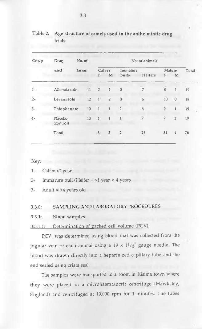
Seventy six camels that were found to have moderate to heavy
worm egg counts were selected for the anthelmintic efficacy study.
These came from 13 different manyattas (households). Table 2 shows
the age structure of the camels used in this study. The camels were
randomly distributed into three treatment groups and one control
group of 19 animals each. The drugs used in this clinical eflicacy
study included albendazole (V albazen^, Ciba Geigy), levamisole
(N ilv e rm (R) Cooper) and thiophanate (Nemafax(^ , Rhone
Poulenc). Control camels received dilute orange juice. The ellicacv of
the various drugs was compared between the different sexes and age
groups of camels although the number of immature camels used
was low. The animals used for clinical drug trials had their weights
and ages estimated as described by Wilson (1988).

3 2
All the animals used in this study were under the care of their
respective owners or their herdsmen. They were herded together
with other camels and other domestic animals. The animals were
penned at night in separate enclosures (bomas) at the various
owners' homesteads.The management was generally traditional
with the herdsmen deciding where to graze and when and where to
water the camels. On average the animals were watered once
weekly. Because of the drought that occurred during the most part of
1992, most animals from the other drier parts of the district were
brought into the division. Hence, during most of the study period
(November, 1992 to May 1993) most parts of the division were
severely overgrazed and there was a lot of overcrowding around the
few available watering points. There was no supplementation of the
study animals.
3 3
Table 2. Age structure of camels used in the anthelmintic drug trials
Group Drug No. of No. of animals
used farms Calves F M
ImmatureBulls Heifers
Mature F M
Total
1- Albendazole 11 2 1 0 7 8 1 19
2- Levamisole 12 1 2 0 6 10 0 19
3- Thiophanate 10 1 1 1 6 9 1 19
4- Placebo(control)
10 1 1 1 7 7 2 19
Total 5 5 2 26 34 4 76
Key:
1- Calf = <1 year
2- Immature bull/Heifer = >1 year < 4 years
3- Adult = >4 years old
3.3.0: SAMPLING AND LABORATORY PROCEDURES
3.3.1:. Blood samples
3.3.1.1: Determination of packed cell volume (PCV).
PCV. was determined using blood that was collected from the
jugular vein of each animal using a 19 x U /2 gauge needle. The
blood was drawn directly into a heparinized capillary tube and the
end sealed using crista seal.
The samples were transported to a room in Kisinia town where
they were placed in a microhaematocrit centrifuge (Hawksley,
England) and centrifuged at 10,000 rpm for 3 minutes. The tubes

3 4
capillary.
The PCV was used as an indicator of anaemia. PCV values were
determ ined once before treatm ent and one month after
administration of drugs for animals used in the drug trials. During
the survey, 160 camels in poor condition had their PCV values
determined. An animal with a PCV value of less than 24% was
taken to have anaemia (Higgins and Kock, 1984).
3.3.I.2.: Examination for haemoparasites.
(a) Examination of the buffv coat
After reading the PCV., the capillary tubes were broken 1 mm
below and 3 cm above the leucocyte layer using a diamond pencil.
The isolated segment contains 5 microlitres of erythrocytes,
leucocytes and 15 microlitres of serum. These were expelled onto a
slide and covered with a 22 X 22 mm cover slip. The slides were
examined under the microscope at X40 objective for trypanosome
parasites (without staining). All camels used in the drug trials were
subjected to this test before drugs were administered.
(b) Dlood smears
This was used to further rule out the presence of
haemoparaistes both at the start and during the course of the drug
trials. Blood smears were prepared from a small drop of blood placed
on a clean slide 1 cm from the edge. The edge of another slide was
were then placed in a microhaematocrit reader (Hawksley, England)
and the PCV. (expressed as a percentage) was read as the volume of
the red blood cells to the total volume of the whole blood in the
3 5
placed on the first, at an angle of 30-45 degrees. The blood was
allowed to spread by capillary action along the angle formed by the
two slides (Murray et a i ,1983). The angled slide was moved along
the first one with a steady movement drawing the blood behind it to
spread the drop evenly on the first slide.
The blood was immediately dried in the air and stained using
dilute giemsa (1:10), after fixation in methyl alcohol for 2-5 minutes.
The prepared slides were allowed to stand for 30-60 minutes in the
dilute Giemsa. After this the stain was washed off using neutral
water and drip dried in a vertical position. The slides were examined
at X I00 objective using oil emersion.
3.3.2:. Analysis of faecal samples3.3.2.1.: Baseline helminthiasis survey:
Faecal samples were collected from the rectum into plastic
faecal pots. This was done once for each of the 255 camels, during the
months of November,1992 to March, 1993. Nematode and cestode
eggs were concentrated by floatation while those of trematodes were
concentrated by sedimentation. Floatation involved the use ot the
modified McMaster egg counting technique (Anon, 1979).
3.3.2.2: The modified McMaster egg counting technique
Glass vials that had two marks at 28 ml and 30 ml levels were
used. A saturated magnesium sulphate solution was poured into the
vial up to the 28 ml mark. By displacement, 2 grams of faeces were
added until the level rose to the upper mark of 30 ml. The contents
were mixed thoroughly and passed through a cotfee strainer. The
filtrate was stirred with a dropper and, while stirring a dropper lull
3 6
of the mixture was withdrawn and used to fill the counting chamber
of the McMaster slide. The slide was left for 10 minutes to allow the
eggs to rise to the top of the slide. The slide was then examined
under low power (xlO objective) of the microscope and all the eggs
in the centimetre square of the slide were counted and identified
using standard parasitological keys (Soulsby, 1986). The count
obtained was multiplied by 100 to get the total number of eggs per
gram of faeces (EPG).
3.3.2.3.: Examination for trematode eggs
Three grammes of faeces were homogenized with water and
the suspension passed through a coarse mesh sieve (about 250
microns). The material retained on the screen was thoroughly
washed using a fine water jet and the debri discarded.
The filtrate was transfered to a conical flask and allowed to
stand for 2 minutes. Thereafter the supernatant was removed and
the remainder transfered to a flat bottomed tube. After
sedimentation for a further 2 minutes, the supernatant was again
drawn off and a few drops of 5% methylene blue added and the
sediment examined under the microscope using low power
objective (XlO). Trematode eggs( yellow), when present were readily
visible against the pale blue background.
33.2.4: Coproculture for infective nematode larvae
Fresh samples from few' animals that showed high EPG. values
(>1000) and anaemia were cultured using the established technique
3 7
(Anon, 1986). The cultures were done per household, and were mostly
from animals that were later used for the anthelmintic drug trials.
Procedure:
About 20 g of faeces was crushed, a little water added just to wet
them and placed in a jar with a tightly fitting lid. The faeces were
incubated for 7 days at room temperature and on the 8th day, the jar
was taken out, filled with water and inverted on a petri dish on
which some drops of water were put. The preparation was left for 24
hours after which the larvae were harvested by pipetting the
contents on the petri dish and transfering them to a second petri
dish. The larvae were killed by adding lugols' iodine and identified
under the microscope by standard methods (Soulsby, 1986).
3.4.0:_____Determination of anthelmintic efficacy
The 76 selected camels were randomly distributed according to
age, sex, farm/household and EPG. values into three treatment and
one control group (Table 2). Animals in the control group were
given a placebo that consisted of dilute orange juice .The dosages
used were those recommended by the manufacturers as follows:-
a) Group 1: Albendazole (ValbazenR, Ciba Geigy) :lOmg/kg body weight
b) Group 2: Levamisole (Nil verm R, Cooper) :7.5mg/kg body weight
c) Group 3: Thiophanate (NemafaxR, RhonePoulenc): 15ml/50kg body weight
d) Group 4: placebo (control).

3 8
The only nematode eggs counted are those of parasites that are
known to cause major economic losses in camels.
Pre-treatment EPG was done twice one week apart before
treatment of the camels with anthelmintics started and the average
of the 2 nematode worm egg counts was used as the EPG reading on
day 0. All faecal samples were collected between 10 am and 12 pm to
avoid diurnal variations in worm egg counts (Anon, 1986).
Following treatment faecal sam ples were collected for
determination of nematode worm egg counts on days 1, 2, 3, 14, 21
and 28. The anim als were also closely m onitored for
haemoparasitosis and other diseases over the 4 week period. Those
found to be sick were promptly treated. The animals were also
closely observed for any reactions to the anthelmintics used in this
study.
3.5.0: Data analysis
Data were subjected to analysis of variance (ANOVA) (Wayne,
1987) using IBM computer with the the SAS (SAS Institute Inc.,
Cary, NC, USA) statistical package. Tukey’s Highest Significant
difference (HSD) test was used to determine if there was a significant
difference in the group means at 5% level of signiticance.

3 9
CHAPTER FOUR
RESULTS
4.1.0: STRONGYLE WORM EGG COUNTS DURING THEBASELINESURVEY
4.1.1: Levels of GIT nematodes in relation to total rainfall inLorroki Division during the study period
The mean monthly strongyle egg counts for the camels used in the
baseline helminth survey and the total rainfall figures in the study area
during the months of November 1992 to March 1993 are presented in Table
3. The results show that mean strongyle egg counts increased from 379.7
EPG in November 1992 to 961.7 in January 1993. They then dropped to 747.8
in February 1993 and finally to 391.9 at the end of the survey period, in
March 1993. Figure 4 shows the mean strongyle egg counts of the camels in
relation to total rainfall recorded in mm.
Table 3: Mean monthly Strongyle egg counts of camels inrelation to rainfall in Lorroki division of Samburu district.
Month Total rainfall (mm)
Mean ± SD Strongyle egg counts (Number of samples in brackets)
November 1992 51.3 379.67 ± 432.23 (59)
December 1992 77.4 683.33 ± 1084.50 (66)
January 1993 105.4 961.70 ± 1934.0 (47)
February 1993 18.0 747.83 ± 1183.70(46)
March 1993 1.1 391.891530.92 (37)

4 0
Figure 4: Mean strongyle egg counts of camels in relation to total rainfall ( mm) in Lorroki division
4.1.2: Mean strongyle egg counts in relation to age during the survey
Table 4 shows the mean monthly strongyle egg counts in different age
groups of camels used in this study. Data from this study shows that calves
had lower strongyle egg counts than adults over the five months study
period except in November 1992 and January 1993. Although the sample
size for the immature bulls was generally low, the data suggest that their
worm egg counts were mostly lower than those ot calves except in
Rai
nfal
l in
mm

November 1992 and January 1993, when only one camel in this category was
sampled. The same trend applies to heifers which nonetheless had
generally higher egg counts than immature bulls. Figure 5 shows the mean
strongyle egg counts for the different age groups over the five months
period.

TABLE 4:
Mean ± SD Strongyle egg counts for the different age groups of camels during the survey (Number of samples in brackets)
Month Calves Immatures (> 1 year <4 years) Adults(< 1 year old) Bulls Heifers (>4 years)
1Csl
November 1992 454.58 - 663.87(11) 460.0 ±572.71(5) 400.0 ±362.53(15) 325.0 ± 338.43(28)
1 December 1992 388.89 ±261.94 (9) 175.0 ±95.74(4) 277.78 ± 315.35(9) 872.73 ± 73 ± 73.0(44)
January 1993 1458.30 ±2363.90(12) 100.0 ± 173.21(3) 408.33 ±334.28(12) 1125.0 ±2293.0(20)
February 1993 522.22 ±940.45(9) 100.0 ± 100.0(3) 916.671 110.70(12) 836.36 ± 1384.10(22)
March 1993 316.671483.39(6) 600.0 ±0(1) 485.71 ±985.61(7) 373.91 ±369.54(23)

2000
a - - Calves ♦ — Imm. bulls -fl— Heifers
Figure 5 Mean strongyle egg counts for the different age groups of camels over the study period
During the wettest month of the study period (January 1993) calves had
the highest worm egg counts followed by adults, while the immatures (both ̂
bulls and heifers had the highest egg counts during the dry month of March
1993 (and in November 1992 for the immature bulls).

44
4.1.3: Mean strongyle egg counts in relation to sex during thebaseline survey
Table 5 shows the mean strongyle egg counts in male and female camels
during the study period. According to this study, female animals had
significantly (p < 0.05) higher worm egg counts than male ones throughout
the study period (Figure 6). The highest worm egg counts in both male and
female camels was recorded during the heavy rains in January, 1993.
Table 5: Mean strongyle egg counts for the different sexes of all camelsstudied during the baseline survey (number of samples analysed is shown in brackets)
Month
November 1992
December 1992
January 1993
February 1993
March 1993
Mean±SD Strongyle
Males
362.50 ±422.49 (16)
577.42 ± 841.31 (31)
947.62 ±1867 (21)
511.11 ±945.31 (9)
387.50 ±491.17 (19)
counts
Females
386.05 ± 440.55 (43)
777.14 ± 1266.80 (35)
973.08 ± 2023.20 (26)
805.41 ± 1239.20 (37)
393.10 ±549.63 (29)

Figure 6: Mean strongyle egg counts in relation to sex of camels during the survey period.
4.2.0: Types of worm eggs identified during the baseline survey.
Although this study gave more emphasis to strongyle eggs because of the
recognized health and production constraints imposed on the camel by the
strongyle nematodes, several other types of eggs were recognized during the
study. Table 6 shows the percentages of the different eggs identified during
the survey.

4 6
Table 6: Percentage of different worm eggs identified during
the survey period.
Percentage of eggs
Month
Strongyle Cestodes Strongyloides Fasciola Trichuris
November 1992 79.15 6.48 5.20 1.98 7.19
December 1992 87.70 5.36 1.61 1.38 3.95
January 1993 88.22 6.43 1.50 3.00 0.85
February 1993 82.52 6.64 6.00 1.12 3.72
March 1993 93.95 5.0 0.00 0.30 0.74
Data from Table 6 shows that throughout the study period there was a
significantly higher (p<0.05) number of strongyle eggs observed than all the other
eggs combined. During all the months of the study at least 80% of all eggs were
those of strongyle nematodes. The second most commonly recognized eggs were
those of tapeworms. Tapeworm eggs, mostly those of Moniezia spp were more
common in calves and adult females. Segments of this tapeworm were visible in
the faeces of these animals especially during the rain season. Eggs of
Strongyloides spp and Trichuris spp were identified in a fair proportion ot the
animals sampled. Eggs of Fasciola spp occurred at low levels throughout the
study period.
4.3.0: Larval culture and identification of the various nematodes
The different types of infective nematode larvae isolated on culture and
identification are shown in Table 7.

Table 7: Nematode larvae recovered during the survey periodas a percentage of the total
P a r a s ite N o. of farms w here it was id en tified
% of the total p arasites identified
H aem onchus spp 1 4 4 8 .6 2
Trichostrongylus spp 1 0 3 2 .1 4
Cooperia spp 7 9 .7 8
Bunostom um spp 5 5 .2 9
O esophagostom um spp 3 2 .6 1
Strongyloides spp 3 0 .9 1
Ostertagia spp 1 0 .6 5
The results show that Haemonchus spp was by far the most common strongyle
nematode present. Trichostrongylus spp was also very common while Cooperia
spp and Bunostomum spp occurred at relatively low levels in a fair proportion of
the herds. Oesophagostomum spp. Strongyloides spp and Ostertagia spp were the
least encountered.
4.4.0: COMPARATIVE EFFICACY OF THE ANTHELMINTICS
4.4.1: The packed cell volume (PCV).
Mean PCV values of camels in different treatment groups before and
after treatment are shown in Table 8. Before treatment, all the groups had
mean PCV values above the critical minimum (24%).

4 8
Table 8: PCV values for camels in different treatment groups (n=19)before and after treatment
Group Drug Mean ± SD pre-treatmentPCV (%)
Mean ± SDpost-treatmentPCV(%)
1 Albendazole 24.89 ± 3.59 24.95 ± 3.24
2 Levamisole 24.16 ±3.93 24.16 ±2.77
3 Thiophanate 24.00 ± 4.29 25.16 ±3.42
4 Control 24.21 ± 4.02 23.32 ± 3.97
The data shows that all treatment groups had significantly (P < 0.05)
higher PCV values than the control group at the end of the experiment.
Hence treatment alleviated anaemia and it was noticed that more camels in
the treatment groups maintained good body conditions during and after the
study period when compared with the non-treated ones. Age and sex had
no significant effect (P > 0.05) on the etficacv of the different drugs used.
When exam ined individually, thiophanate caused the biggest
improvement in PCV followed bv albendazole. Levamisole did not cause
any change while the anaemia status in the control camels worsened.
4.4.2: Overall post treatment worm egg counts
The pre-and post treatment mean egg counts in the treatment and control
groups are shown in table 9. There was no significant difference (P > 0.05) in the
levels to which the three drugs reduced the nematode worm egg counts on the
first day after treatment. On this day, the data indicates a less than 50 & fall in
mean nematode egg counts except for thiophanate which caused a 66.19 /c fall in
worm egg counts. Figure 7 illustrates the results of this anthelmintic drug study.
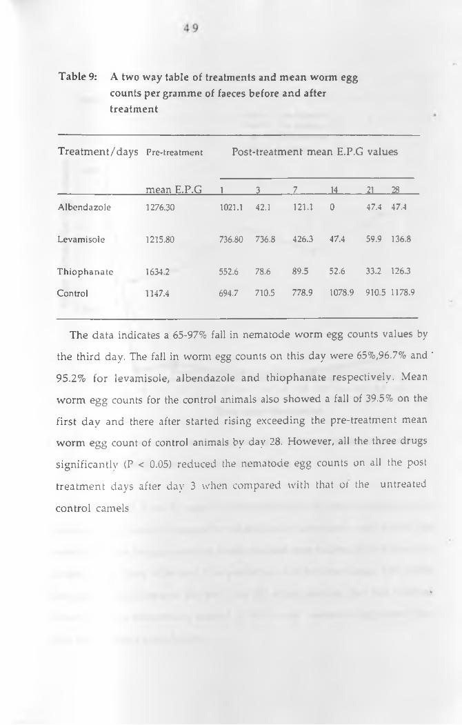
Table 9: A two way table of treatments and mean worm eggcounts per gramme of faeces before and after treatment
Treatment/days Pre-treatment Post-treatment mean E.P.G values
mean E.P.G 1 3 7 14 21 28
Albendazole 1276.30 1021.1 42.1 121.1 0 47.4 47.4
Levamisole 1215.80 736.80 736.8 426.3 47.4 59.9 136.8
Thiophanate 1634.2 552.6 78.6 89.5 52.6 33.2 126.3
Control 1147.4 694.7 710.5 778.9 1078.9 910.5 1178.9
The data indicates a 65-97% fall in nematode worm egg counts values by
the third day. The fall in worm egg counts on this day were 65%,96.7% and '
95.2% for levamisole, albendazole and thiophanate respectively. Mean
worm egg counts for the control animals also showed a fall of 39.5% on the
first day and there after started rising exceeding the pre-treatment mean
worm egg count of control animals by day 28. However, all the three drugs
significantly (P < 0.05) reduced the nematode egg counts on all the post
treatment days after day 3 when compared with that ot the untreated
control camels

5 0
D a y s a f t e r t r e a t m e n t
Figure 7.: Mean nematode worm egg counts in different treatment groups
following administration of the drugs.
From figure 7 it can be seen that thiophanate caused the most rapid fall in
nematode egg counts followed by albendazole. Levamisole took almost one
week to reduce the egg counts to levels that had been reached by the other two
drugs by day three. After dav 7 the performance of the three drugs was almost
the same except towards the end (day 28) when animals that had received
levamisole and thiophanate seemed to void more nematode eggs than those
that had received albendazole.
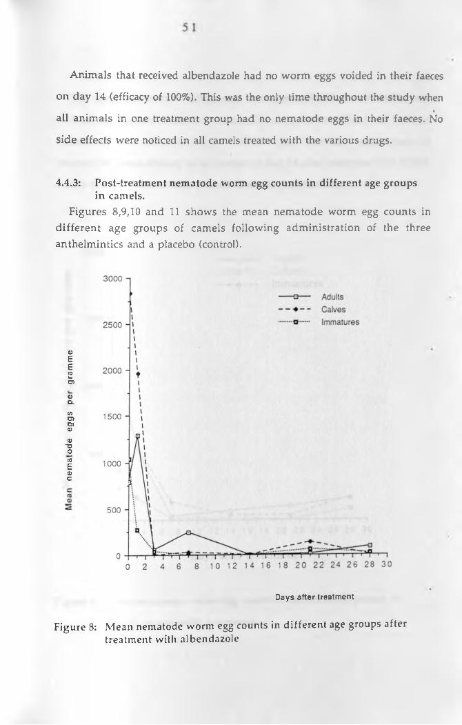
Animals that received albendazole had no worm eggs voided in their faeces
on day 14 (efficacy of 100%). This was the only time throughout the study when%
all animals in one treatment group had no nematode eggs in their faeces. No
side effects were noticed in all camels treated with the various drugs.
4.4.3: Post-treatment nematode worm egg counts in different age groupsin camels.
Figures 8,9,10 and 11 shows the mean nematode worm egg counts in different age groups of camels following administration of the three anthelmintics and a placebo (control).
0)EE03v.CD
oCL
V> CD CD <U
<UDOO03
E<uc
Days after treatment
Figure 8: Mean nematode worm egg counts in different age groups after treatment with albendazole
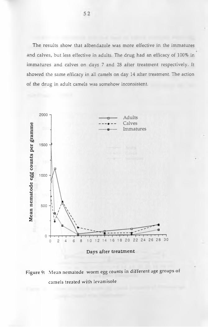
The results show that albendazole was more effective in the immatures
and calves, but less effective in adults. The drug had an efficacy of 100% in
immatures and calves on days 7 and 28 after treatment respectively. It
showed the same efficacy in all camels on day 14 after treatment. The action
of the drug in adult camels was somehow inconsistent.
D a y s a f t e r t r e a t m e n t
Figure 9: Mean nematode worm egg counts in different age groups of
camels treated with levamisole
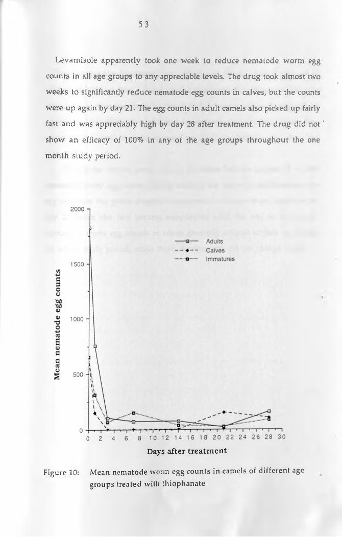
5 3
Levamisole apparently took one week to reduce nematode worm egg
counts in all age groups to any appreciable levels. The drug took almost two
weeks to significantly reduce nematode egg counts in calves, but the counts
were up again by day 21. The egg counts in adult camels also picked up fairly
fast and was appreciably high by day 28 after treatment. The drug did not *
show an efficacy of 100% in any of the age groups throughout the one
month study period.
D a y s a f t e r t r e a t m e n t
Figure 10: Mean nematode worm egg counts in camels of different agegroups treated with thiophanate
5 4
Thiophanate was apparently very effective in calves where it showed an
efficacy of 100% on days 3, 7 and 14 after treatment. The efficacy of this drug
was good in all age groups by day 3 after treatment although egg counts
remained fairly high in adults and immatures until day 14. There after
calves and adults seemed to pass out more nematode eggs than the
immatures.
Within the control group (figure 10) calves had the highest (P < 0.05)
nematode worm egg counts during much of the study period.Howrever, the
egg counts in this group dropped to the same level as those of immatures by
day 21 when the two became comparable until the end of the study.
Nematode worm egg counts in adults generally seemed to pick up during
the whole study period, while those of immatures did not change much.

Days after treatment
Figure 11: Mean nematode worm egg counts in camels of differentage groups that received a placebo (Control).
4.4.4: Post-treatment nematode worm egg counts in different sexes of
camels.
Figures 12,13 and 14 shows the mean nematode worm egg counts in male
and female camels following administration of the three drugs and a
placebo (control).

5 6
D a y s a f t e r t r e a t m e n t
Figure 12: Mean nematode egg counts in male and female of camels
treated with albendazole
Albendazole had a very good efficacy in male and female camels used in
this study as illustrated by figure 12. Nematode worm egg counts increased
slightly in female camels after day 3 but they were down again by day 14
after treatment. At the end of one month, male camels had more nematode
worm e££ counts than females.

Me
an
ne
ma
tod
e e
gg
co
un
ts
57
— fl— Males
D a y s a f t e r t r e a t m e n t
Figure 13: Mean nematode egg counts in male and female camels treated
with levamisole

5 8
D a y s a f t e r t r e a t m e n t
Figure 14:. Mean nematode egg counts in different sexes of camels treated
with Thiophanate.
The action of levamisole in both male and female camels was comparable
and followed the same pattern seen for age groups. Thiophanate was
apparently more effective in males than in female camels during much ot
the experimental period. In the control group males had higher nematode
worm egg counts than females during most of the study period. The egg
counts among the males had a very high variation during the study.

5 9
CHAPTER FIVE
DISCUSSION
5.1 INTRODUCTION
The management system of camels in the study area was
distinctly different from those found in traditional camel keeping
communities such as the Turkana, Gabra, Rendile and Somali. But,
because of the highly mobile nature (nomadism) of the later tribes, it
would have been very difficult to conduct a study of this nature in
such communities. Because of the recent introduction of camels in
the study area, the herd structure was different from what would be
found in the traditional camel keeping areas. In this area most
camels tended to be adult females and heifers with few males.
Camels in this area also tended to graze more than they browsed and
since the area was generally overgrazed, there could have been
higher chances of re-infection following treatment.
5.2: BASELINE HELiYIINTHIASIS SURVEY
5.2.1: Worm egg counts
It was observed that peak strongvle egg production in untreated
camels was recorded during the high unexpected rains of January
1993. The high amount rainfall could have favoured the survival of
infective larvae on the pasture. This has been reported previously in
Kenya (Rutagwenda, 1985; Njanja 1991), Ethiopia (Richard, 1984) and
Saudi Arabia (EL Bihari and Kawasmeh, 1980).
Despite the fact that there was very little rain in this area
throughout much of 1992, the worm egg counts in November 1992
were fairly high. The same was observed after the heavy rains in
6 0
March 1993 when despite recording just a trace amount of rainfall
(1.1 mm), the worm egg counts were high. This has been reported in
N igeria and Kenya (Rutagwenda, 1985) and was attributed to
inhibited immature stages of the worms that resume development
as a result of a decline in the immune status of the hosts during the
dry seasons when pasture is scarce and camels have to walk for long
distances in search of water and pasture. Arzoun et al. (1984a)
reported a similar phenomena (hypobiosis) in camels infected with
H. longistipes.
Generally, the results of strongyle worm egg counts showed
that adult camels had higher worm burdens than the immatures
and calves. However, the calves seemed to have higher worm egg
counts than the immatures and the worm egg counts for the
immature bulls continued decreasing even during the time when
rains were heaviest. Although, the study period was short, the
findings agree with what has been reported in cattle, sheep and goats
(Soulsby, 1986). The mature animals, especially the females are
mostly under the stress of pregnancy and lactation. This could have
reduced the immune status and could have been responsible for the
high worm counts among adult females. The undeveloped
immunity in calves could haave been responsible for the the high
worm egg counts. The results also indicate that the females had
higher worm egg counts than males throughout the study period.
This could still be due to stress in females that is generally lacking in
the males.
5.2.2.: The types of worm eggs identified
Throughout the study period, eggs of strongyle nematodes were
the most common. This agrees with the findings of earlier workers
(Richard, 1976, Lodha et al., 1977; Wilson et al., 1984; Arzoun et al.,
1984b) that nematodes are the most common and most pathogenic
parasites of the camel. In this study, a fairly high number of cestode
eggs was recorded. This confirms the findings of Alfaif (1974),
Richard (1976), Wilson (1988) and Abdulrahman and Bornstein
(1991). The low prevalence of Strongyloides spp and Trichuris spp
reported in this study was lower than what has been found
previously (Wilson et al 1984; Wilson, 1988; Njanja, 1991, Wosene,
1991)
Eggs of Fasciola spp occurred at low levels throughout this
study. This is the first time that fascioliasis is being reported in
camels in Kenya. However, this parasite has been reported in camels
in Saudi Arabia by Magzoub and Kassim (1978) and in the Sudan
(Malek, 1959). The presence of Fasciola spp in this area could be due
to the fairly high rainfall that occurs here compared to the further
North of Kenya where most research on camels has been done. High
amounts of rainfall has been known to provide a conducive
environment in which the snails, the intermediate hosts survive
and pass on infection to animals. The camels could also be getting
the fasciola infections from the large number of cattle and small
ruminants that are present in this area. Interchange of helminth
parasites between camels and other domestic animals has been
reported (Arzoun et al., 1983; El Bihari, 1985; Baitursinov and
Berkinbaev, 1989; Onvali and Onwuliri, 1989)
6 2
5.2.3: Larval culture and identification
The results indicate a high incidence of Haemonchus spp in
camels. Although this parasite was not characterized fully upto the
species level, probably the majority could have been Haem onchus
longistipes and a few Haemonchus contortus. The high incidence of
Haemonchus spp in Kenya has been reported before by Wilson et al.
(1984) and Rutagwenda (1985). However, Haemonchus longistipes is
recognized as the most common and most pathogenic internal
parasite of camels (Steward, 1950, Malek; 1959; Graber et al 1967, EL
Bihari and Kawasmeh, 1980, Arzoun et al. 1984a, Tager-Kagan, 1984,
Onyali and Onwuliri, 1989).
The high incidence of Trichostrongylus spp that was recorded
in this study has also been reported by other workers (Wosene 1991).
Altaif (1974) and Abdul-Salam and Farah (1988) respectively found
that in Iraq and Kuwait, Trichostrongylus probolurus was the most
common helminth parasite of camels and could have been the most
pathogenic.
The other parasites that were recorded in this study have been
reported with different prevalence rates in different countries
Oesophagostomum spp has been reported in the camel by Tager-
Kagan (1984) and Kapur and Sharma (1972), Ostertagia spp by Kapur
and Sharma (1972) and Michael et al. (1980), Strongyloides spp by
Graber (1966) and Kapur and Sharma (1972), Bunostomum spp by
Michael et al. (1980) and Cooperia spp by Frolka (1988).

6 3
5.3.0: DRUG TRIALS
5.3.1: The packed cell volume
The study showed significant differences (P<0.05) in PCV
values between the treated and untreated control camels one month
after the drugs were administered. The treated camels had higher
PCV values than the controls, showing that they probably had a
better health status. Since the presence of trypanosomiasis had been
ruled out in these animals, it seems the drugs were effective in
reducing the worm loads to levels where the anaemia status was
eliminated.
Hence the three drugs, administered at the recommended
dosage rates had positive effects on PCV values and possibly on
productivity. This action of albendazole has been reported for
thiabendazole (also a benzimidazole) in camels by Njanja (1991). No
reports exist on the actions of levamisole, albendazole and
thiophanate in improving PCV values in the camel.
5.3.2:_____Overall anthelmintic efficacy
The levels to which the three anthelmintics reduced the
nematode worm egg counts on day one after treatment was
comparable to the nematode worm egg counts of the control
animals.
Thiophanate was apparently more effective than levamisole
and albendazole upto the third day after treatment as it caused a
greater reduction in post-treatment nematode egg counts. This
action of thiophanate in camels has not been reported, but the

6 4
efficacy is comparable with what has been found for this drug in
cattle, sheep and goats (Brander et a i, 1991).
Although the rate at which albendazole reduced the worm egg
counts in the first three days, was slower than that by thiophanate it
had the highest efficacy by day three after treatment. This is also the
only drug that showed a 100% efficacy by day 14 following treatment.
Levamisole apparently took almost one week after treatment to
reduce the nematode egg counts to levels that had been reached by
albendazole and thiophanate on post-treatment day three. This slow
and inconsistent action of levamisole in camels had been reported
by Wallev (1966) and Lodha et a i , (1977) . However, the efficacy of
this drug in cattle and sheep in Kenya, in its combined form with
bithionol sulphoxide (WormicidR-plus, Cosmos) was found to be
very high (Maribei, 1985). Several other workers (Walley, 1966) have
proved the high and consistent efficacy of levamisole in treating
nematodes of cattle, sheep and goats.
After post-treatment day 7, the performance of the three
anthelmintics was apparently the same until after one month when
camels that had received levamisole and thiophanate seemed to
void more worm eggs than those that had received albendazole.
This could probably be due to the prolonged ovicidal action of
albendazole which has been reported in camels tor oxfendazole (also
a benzimidazole) by Michael et a i , (1980). Although thiophanate has
also been found to be ovicidal (unlike levamisole) to worm eggs in
cattle and other animals, it’s action in this study was not comparable
to that of albendazole. This action of thiophanate could be due to it’s

6 5
very early excretion (within 72 hours) from the body of treated
animals (Brander et a i , 1991).
The rapid and high efficacy of albendazole that was recorded in this
study has been reported for other benzimidazoles. Thiabendazole has
been found to be effective in treating camel helminthiasis (Graber 1966;
Chandrasekharan et a i , 1970; Kapur and Sharma, 1972). However, Lodha
et a i (1977) found in a comparative study that thiabendazole was the
least effective in treating camel helminthiasis when compared with
m ethyridine, m orantel tartrate and tetram isole (levam isole)
hydrochloride.
Other benzimidazoles that have been found to be effective in
treating camel helminthiasis include mebendazole (Forstner et a i, 1977,
cited by Michael et a i , 1980) and oxfendazole (Michael et a i , 1980).
However mebendazole is reported to be ineffective in treating
lungworm infections. No studies have been done to compare the
efficacy of anthelmintics among different age groups and sexes of camels.
The following conclusions and findings can be made from this study:.
1- The study confirms that peak worm egg counts in camels occur during and soon after the heavy rains.
2- The study confirms that Hacmonchus spp is the commonest and could be the most pathogenic helminth of the camel.
3- The study indicates that thiophanate was the best drug as shown by the significant reduction in the post-treatment nematode worm egg counts and improvement in the anaemia status. Albendazole and levamisole came next in that order
with the later being the least effective.
4- The study indicated that the age and sex of camels had no ettect
on the efficacy of the various drugs used.
/
6 6
REFERENCES
ABDUL-SALAM, J.M . and FARAH, M.A. (1988): Seasonal
fluctuations of Gastrointestinal helminths of Camels in
Kuwait. Veterinary Parasitology 28: 93-102.
ABDULRAHMAN, O.S. and BORNSTEIN, S. (1991): Diseases of
Camels (Camelus dromedarius) in Somalia and prospects
for better Health. Nomadic Peoples 29: 104-112.
AL-KHALIDI, N.W., HASSAN, M.A and AL-TAEE, A.F. (1990):
Faecal incidence of Fasciola spp and E u r y t r e m a
pancreaticum eggs in camels (Camelus drom edarius) in
Iraq. Journal of Veterinary parasitology 4(1): 75-76.
ALTAIF, K.I. (1974): Helminths in Camels in Iraq. Tropical Animal
Health and Production 6: 55-57.
ANON. (1986): Manual of Veterinary parasitolgical Techniques.
Technical Bulletin No' 18. Ministry of Agriculture,
Fisheries and Food, Agricultural Development and
Advisory Service. HMSO, London.
ARZOUN, I.H., HUSSEIN, H.S. and HUSSEIN, M.F. (1983): The
pathogenesis of Experimental Haemonchus longistipes
infection in goats. Journal of Comparative Pathology 4:
619-628.
ARZOUN, I.H., HUSSEIN, H.S. and HUSSEIN, M.F. (1984a): The
prevalence and pathogenesis of naturally occuring
Haemonchus longistipes infection in Sudanese Camels.
Journal of Comparative Pathology 94: 169-174.
ARZOUN, I.H., HUSSEIN,H.S. and HUSSEIN, M.F. (1984b): The
pathogenesis of experimental Haemonchus longistipes
infection in camels. Veterinary Parasitology 14: 43-53.
BAITURSINOV, K.K. and BERKINBAEV, O. (1989): The study of the
ecology of camel parasites in the Southeastern Kazakh
(USSR), lzv Akad Nauk Kaz Ssr ser Bio 0 (3): 34-37.
BEHM, C.A. and BYRANT, C. (1985): Modes of action of
a n th e lm in tic s . In: R esistance in Nematodes to
anthelmintic drugs. N. Anderson and P.J Waller (eds),
pp.57-68. CSRO, Melbourne, Australia.
BLOOD, D.C. and RADOSTITIS, O.M (1989): Clinical examination
and making a diagnosis. In: Veterinary medicine. A
textbook of diseases of cattle, sheep, goats, pigs and horses.
7th Edition, pp.3-31. Bailliere Tindall, Oxford, U.K
68
BRANDER, G.C., PUGH, DM., BYWATER, R.J. and JENKINS W.L.
(1991): A nthelm intics. In: V eter in a ry A p p lied
Pharmacolgy and Therapeutics (5th edition) pp. 470-502..
Bailliere Tindall, East Sussex, U.K.
BREMAUD, O. (1969): Notes sur L'elevage camelin dans les districts
nord de la Republique du Kenya. I.E.M.V.T., Maisons-
Alfort, 105 Pagestann exes.
BRUNDSON, R.V. (1980): Principles of Helminth control.
Veterinary Parasitology 6: 185-215.
CHANDRASEKHARAN, K., NAIR, K.P.D., SUNDARAM, R.K. and
PETER, C.I. (1970): On the use of Thiabendazole against
Trichostrongylus and T rich u ris infections in camels
(Camelus dromedarius). Kerala Journal of Veterinary
science 1: 129-132.
CHANDRASEKHARAN, K., NAIR, K.P.D., SUNDARAM, R.K. and
PETER, C.I. (1971): Anthelmintic activity of Parbendazole
(Helmatac premix) in camel (Camelus dromedarius) and
Nilgiri Tahr (Hemitragus hylocrius). Kerala Journal of
Veterinary Science 2:135-138.

69
DELATOUR, P., OUHSINE, A. and BENOIT, E. (1989): Comparative
pharmacokinetics of netobimin and albendazole in the
one humped camel (Cam elus drom edarius). British
Veterinary Journal 145: 478-482.
EDWARD, E.R (1982): Antinematodal drugs. In: N.H Booth and E.L
M acd onald (eds). V eter in ary pharm acology and
th e ra p eu tic s (5th edition) pp. 803-851. Iowa state
University press, Ames, Iowa.
EL BIHARI,S. (1985): Helminths of the Camel. A Review. British
Veterinary Journal 141: 315-326.
EL BIHARI,S. and KAWASMEH, F.A. (1980): Occurrence and
seasonal variation of some gastrointestinal helminths of
d ro m e d a ry , Camelus drom edarius in Saudi Arabia.
Proceedings of the Saudi Biological Society 4: 297-304.
FORSTNER, M.J., KOPP, H. and WIESNER, H. (1977): Berliner und
Muchener tierarzliche Wochen Schrift 90 (9): 180.
FROLKA, J. (1988): Erkankungen der zootiere. Ver handlungs bercht
des 30. Internationalen sym posium s uber die
Erkrankungen der zoound wildtiere, Sofia 1988.
70
FROLKA, J. and ROSTINSKA, J. (1984): Efficacy of Ivermectin MSD
(Ivomec, Eqvalan) against Sarcoptes scabiei and nematodes
in zoo animals. Internationalen symposiums Uber die
Erkrankungen der Zootiere Vom 2. Mai bis 6 1984 in Brno.
R. Schroder (ed) 26: 455-462.
GEORGI, J.R., GEORGI, M. and THEODORIDES, U. (1991): Diagnostic
parasitology. In: J. Dyson (ed). P arasito logy fo r
V eterinarians. (5th edition). W.B Saunders Company,
U.S.A.
GORDON, H McL (1950): Some aspects of parasitic gastro-enteritis of
sheep. Australian veterinary Journal 26: 65-72.
GRABER, M. (1966): A study of the parasiticide action of
Thiabendazole on some helminths of domestic animals
under some African conditions. II. Dromedary. Tome XIX
(Nouvelle Serie), Vigot Freres, Editeurs, Paris, France, pp.
527-542.
GRABER, M., TABO, H. and SERVICE, J. (1967): Enqutes sur les
helm inthes du drom edaire tchadien. Etude des
strongyloses gastrointestinales et de L'haemoncoce a'
Haemonchus longistipes. Revue D'elevage et de medecine
Veterinaire des pays Tropicaux 20 (2): 227-254.

GRONVOLD, J WOLSTRUP, J., NANSEN, P., HENRIKSEN, A.,
LARSEN, M. and BRESCIANI, J. (1993): Biological
control of nematode parasites in cattle with
nematode-trapping fungi: A survey of Danish
studies. Veterinary Parasitology 48 : 31 1-325.
HALL,M .C and FOSTER, W.D (1918): Efficacy of some
anthelmintics. Journal of African Research 12: 397-447.
HIGGINS, A.J. and KOCK, R.A. (1984): A guide to the examination,
chemical restraint and medication of the camel. British
Veterinary Journal 140: 485-504.
JONES, P.G.H (1987): A Review of the successful use of Ivermectin
against arthropod and helminth parasites of the camel.
Journal of the Egyptian Veterinary Medical Association 47:
347-364.
KAPUR, I.S. and SHARMA, R.K. (1972): A study on effectiveness of
Thiabendazole in treatment of pica in camels. American
Journal of Veterinary Research 2: 55-59.
LODHA, K.R., RASINGHANI, P.M. and KARWASRA, R.S. (1977):
Chemotherapeutic trials of some anthelmintics against
helminth parasites in camels. Indian Journal ot Animal
Science 47: 677-682.

7 2
MAGZOUB, M. and KASSIM, A.A. (1978): The prevalence of
Fascioliasis in Saudi Arabia. Tropical Animal Health and
Production 10: 205-206.
MALEK, E.A. (1959): Helminth parasites of the camel (C am elus
dromedarius) in the Sudan. Journal of Parasitology 45 (no'
4). sec.2 Suppl. (73) 38-39.
MARIBEI, J.M (1985): The efficacy of Wormicid^-plus- A new
broadspectruni anthelmintic, under field conditions in
Kenya. The Kenya Veterinarian 9 (1): 11-13.
MICHAEL, S.A., EL REFAII, A.H. and HIGGINS, A.J. (1980):
Evaluation of oxfendazole against natural infections of
gastrointestinal nematodes and cestodes in Egyptian
camels. British Veterinary Journal 136: 84-87.
MOSKEY, H.E. and HARWOOD, P.D. (1941): Methods of evaluating
the efficacy of anthelmintics. American Journal of
Veterinary Research 2: 55-59.
MURRAY, M., TRAIL, M.C., TURNER, D.A. and WISSOCQ, Y.
(1983): Livestock productivity and trypanotolcrance.
Network training manual pp.5, I.C.P.E.

7 3
NANSEN, P. (1993): Current and future prospects for control of
ostertagiasis in Northern Europe-Exam ples from
Denmark. Veterinary Parsitology 46:3-21.
NJANJA, J.C. (1991): Livestock ecology in Central Turkana.
UNESCO-Turkana Resource Evaluation Monitoring Unit
(TREMU). TREMU Technical report no' E-l.
ONYALI, I.O. and ONWULIRI, C.O.E. (1989): Gastrointestinal
helminths of camels in Nigeria. Tropical Animal Health
and Production 21: 145-146.
POWERS, K.G., WOOD, I.B., ECKERT, J., GIBSON, T. and SMITH,
H.J. (1982): World Association for the Advancement of
Veterinary Parasitology (W.A.A.V.P) Guidelines for
evaluating the efficacy of anthelmintics in ruminants
(bovine and ovine). Veterinary Parasitology 10: 265-284.
PRICHARD, R. K. (1973):-The fumarate reductase reaction of
Haemonchus contortus and the mode of action of some
anthelmintics. Int. J. Parasitol. 3:407-417.
REINECKE, R.K. (1980): Chemotherapy in the control of
helminthiasis. Veterinary Parasitology 6: 255-292.

REINECKE, R.K., SNIGDERS, A.J. and HORAK, I.C. (1962): A
modification of standard procedures for evaluating the
relative efficacy of anthelmintics. Onderstepoort Journal of
Veterinary Research 29: 241-257.
RICHARD, D. (1976): The diseases of the dromedary (C am elus
drom edarius) in Ethiopia. Ethiopian Veterinary Bulletin
21: 46-47.
.RICHARD, D. (1984): Dromedary Pathology. In the Proceedings of
the Khartoum Workshop on Camels. Volume 1. W.R.
Cockril (ed) pp 481.
RUTAGWENDA, T. and MUNYUA, W.K. (1983): The effects of
panacur (fenbendazole) on nematode parasites of camels
under nomadic conditions in Northern Kenya. Bull.
Anim. Hlth. prod.. Afr. 33:63-67.
RUTAGWENDA, T. (1985): The control of important camel diseases
in the IPAL study area. In Camel diseases and productivity
in the arid lands of Northern Kenya pp.9-75. Integrated
project in arid lands (IPAL) . Technical report no' E-7.
SHARMA, L.K (1991): Efficacy of some anthelmintics against
gastrointestinal nem atodes in camels { C a m e l u s
dromedarius). Indian Veterinanv Journal 68(11): 1069-1072.

SOULSBY, E.J.L. (1986): Helminths, Arthropods and Protozoa o f
domesticated animals. Bailliere Tindall and cassel Ltd ,
London.
STEWARD, J.S. (1950): Notes on some parasites of Camels in the
Sudan. Veterinary Record 62: 835.
TAGER-KAGAN, P. (1984): Resultas d'enguets sur les helmintiases
du dromadaire dans le department de Zinder (Rep. du
Niger); Leur evolution dans L'annee-Moyens de lutte.
Revue-d'Elevage-et de-medecine-Veterinaire-des-pays-
Tropicaux 37(1): 19-25.
TEMBELY, S., DIARRA, P.A., WAIGALO, Y., KOUMARE, A. and
VASSILIADES, G. (1992): Preliminary observation on
helminth parasite populations of the dromedary in
Northern Mali. Veterinary Parasitology 44: 339-342.
VAN NEUTEN J.M. (1972): Pharmacological aspects of tetramisole.
In: Comparative Biochemistry of parasites. Van Den
Bosche, H. (ed). Academic Press, New York and London
PP.101-115.
WALLER, P.J. (1986): Anthelmintic resistance in Nematode parasites
of sheep. In: G.E. Russel (ed). A gricultural Zoology
Reviews, pp. 333-373 ,Ponteland Newcastle Upon Tyne.
7 6
WALLER. P.J. (1987): Anthelmintic resistance and the future for
roundworm control. Veterinary Parasitology 25: 177-191.
WALLEY, J.K. (1966): Tetramisole (dLZM3M6,5-Tetrahodro-6-
Phenyl-Imidazo(2-/l-b)Thiazole hydrochlolride-Nilverm.)
in the treatment of Gastrointestinal and lungworms in
domestic animals. I. Sheep and goats. Veterinary Record
78: 406-414.
WAYNE, D.W. (1987): Analysis of variance. In: Biostatistics: A
foundation for analysis in the health sciences pp. 273-366.
John Wiley and Sons, Canada.
WILSON, A.J., DOLAN, R., SCHWARTZ, H.J. and FIELD, C.R.
(1984): Diseases of Camels in Kenya. In Proceedings of the
Khartoum Workshop on Camels. Volume 1. W.R. Cockril
(ed). pp 519.
WILSON, R.T. (1988): Management for health and productivity:
Diseases and parasites. In:. The Camel. 2nd edition, pp.
118-133. Longman, London.
WOSENE, A. (1991): Traditional husbandry practices and major
health problems of camel in the Ogaden (Ethiopia).
Nomadic Peoples 29: 21-30.

77
APPENDICES
Key t o th e a p p e n d ic e s
1- Sex 0 = Male1 = Female
2= Trichu= Trichuris eggs
3= Strloides = Strongyloides eggs
4= Cesto = Cestode eggs
5= Trem = Trematode eggs.
6= Total = Total number of worm eggs.
7= Age C = 1 = Calf A = 4 = Adult H = 2 = Heifer 1 = 3 = Immature
S-Post 1-6 = Post-treatment e.p.g readings on days 1,3 ,7 ,14.21 and 28.
9-Avpre = Average e.p.g readings before treatment.

78
A ppendix 1 : Worm egg c o u n ts in N ovem ber, 1 9 9 2 .
FarmAnimal Number o f eggs recordedNumber Sex Age Strongyles T r ic h u S trlo id e s Cesto Trem TOTAL
18 1 0 C 300 0 100 0 0 40018 2 1 c 600 100 0 0 0 70018 3 0 I 1400 0 0 100 0 150018 4 1 H 100 0 0 0 100 20018 5 1 H 100 0 0 0 0 10018 6 0 I 600 0 0 0 0 60018 7 0 A 400 0 0 0 0 40018 8 1 A 200 0 0 0 0 20018 9 1 A 100 0 0 0 0 10018 10 1 A 500 0 0 0 0 50018 11 1 A 0 100 0 0 0 100IS 12 1 A 400 0 0 0 0 40019 1 0 C 300 0 0 0 0 30019 2 0 C 400 0 0 0 0 40019 3 1 C 0 100 0 0 0 10019 4 1 C 200 0 0 0 0 20019 5 0 C 0 0 100 0 0 10019 6 1 H 500 0 0 0 0 50019 n 1 H 300 0 0 0 0 30019 8 0 I 200 0 0 0 0 20019 9 0 I 100 0 0 100 0 20019 10 0 A 0 0 0 0 0 019 11 1 A 100 0 0 0 0 10019 12 1 A 500 100 0 0 0 60019 13 1 A 0 0 0 100 0 10019 14 1 A 200 0 0 0 0 20019 15 1 A 200 0 0 0 0 20020 1 1 H 100 0 0 0 0 10020 2 1 H 900 0 0 100 0 100020 3 1 H 500 100 0 0 0 60020 4 1 H 700 0 0 0 100 80020 5 1 H 100 0 0 0 0 10020 6 0 A 1300 0 0 100 0 140021 1 0 C 0 100 0 0 0 10021 2 1 F 200 0 0 0 0 20021 3 1 H 0 0 0 0 0 021 4 0 A 300 0 0 0 0 30021 5 1 A 200 0 0 100 0 30021 6 1 A 0 0 200 0 0 200
21 n 1 A 800 0 0 100 0 900
21 8 1 A 900 0 0 0 100 1000
21 9 1 A 800 0 0 0 0 800
21 10 1 A 0 0 0 100 0 100'>'■ > 1 1 C 800 0 0 0 0 800i ' ) 2 1 C 2300 0 0 0 0 2300
3 1 C 100 0 100 0 100 300O ') 4 1 H 900 0 0 0 0 900

79
A pp endix 1 c o n t in u e d
22 5 1 H 50022 6 1 H 110022 7 1 H 022 8 0 I 022 9 0 A 20022 10 0 A 30022 11 1 A 022 12 1 A 022 13 1 A 60022 14 1 A 10022 15 1 A 20022 16 1 A 800
0 100 0 0 6000 0 0 0 11000 0 0 0 00 0 0 0 00 0 0 0 2000 0 100 0 4000 0 0 0 00 0 0 0 00 0 0 0 6000 0 0 0 100
100 0 0 0 3000 0 0 0 800

8 0
A ppendix 2 : Worm eg g c o u n ts in D ecem b er, 1 9 9 2 .
No. sei Age StroNumber
Tricbi■ Pf.eggs_
Stroidesre c o rd e d
Cesto Trei Total Pin1 0 C C 6 0 0 100 0 0 7 0 0 12 1 C 0 0 0 0 0 0 13 1 H 100 0 0 100 0 20 0 1i 1 H 200 100 0 0 9 3 0 0 l5 ! fl 100 0 0 0 9 10 0 16 0 A 0 0 0 0 0 0 17 1 A 5 0 0 0 0 0 0 5 0 0 !8 0 A ! 00 0 190 0 9 200 19 0 A 100 0 0 0 0 10 0 1!0 9 A 6 0 9 100 0 9 190 8 0 0 2! 0 C 100 0 0 0 0 10 0 12 fl H 6 0 9 9 0 100 0 7 0 0 23 9 A 0 0 0 0 9 0 2i 9 A 0 0 0 0 9 9 2A 0 A 1 7 9 0 0 9 9 0 1 7 0 0 16 0 A 5 0 0 9 0 9 0 5 0 0 31 1 C 200 0 0 0 0 200 1•) 9 c 6 0 9 9 0 9 9 6 0 9 \
3 ! A 5 0 0 190 9 9 9 6 0 0 3i ! A 7 0 9 9 9 9 0 TOO 3y 9 A 9 0 9 0 100 0 10 0 0 3
6 ! A 0 0 9 0 0 9 3- 0 A *00 0 109 0 0 5 0 0 t? 9 A 1 0 9 0 o j 0 f. ! 0 0 n 30 9 A 7 0 0 2 9 0 9 9 0 0 0 ft 3
! 0 0 A 9 0 0 | 9 9 0 9 o o o o 11 1 0 A •nr. 0 0 9 0 *•00 3m 9 A 9JL0 o 0 0 0 8 0 0 }u 0 A ) 4 0 0 0 0 0 n l i n ot 9 fl < 9 0 1 0 0 9 9 A 6 0 0 <
• 0 fl 0 0 9 1 9 9 :■ inn
i j 2 0 9 0 9 0 • ? A 3 9 0 a
9 A i n n 9 0 n A Tftfi
> i A 9 o A 0 9 % A| A 8 9 0 0 0 i n n ; U 8 Oft l
- i A n o a ! 9 0 n 1 0 9 9 2 5 0 0 L
a ! A MOO 9 0 190 tftft 1 3 0 0 1i A 1 9 9 9 0 ; n o 0 MOO 5
• ! r ’ n o n 9 9 M ?ftf. 59 i * 0 0 n 9 0 3 0 0 {
• 1 fl 9 0 0 0 o 0 flftft ?l ! fl 1 9 9 9 0 0 fl • 0 0 tC 0 A t o o 0 0 0 9 1 0 0 •K 9 A ! 0 0 9 9 9 ! 9 0 A
] A 1 0 0 0 0 0 1 9 0 %5 ! A m o 0 9 0 fl 4 2 0 9 (
Q I A 2 9 9 0 0 r 2 0 0 c

81
Appendi i 2 (coit i med)AniialHo. Sei Age Stro Tricko
10 1 A 2)00 100
11 1 A 300 012 1 A 0 01 0 C 600 02 0 C 500 1003 1 C 700 0i 1 H 0 05 0 I 100 06 0 I 100 2007 0 A 0 08 0 A 700 09 0 A *500 10010 1 A 600 0
1! 1 A 200 0
12 1 A 900 013 ! A 100 0
1 * 1 A 1300 10015 1 A 300 o
16 ! A 6200 0
Cesto Trei Totii Firi
0 0 2 4 0 0 5
0 0 3 0 0 5
0 0 0 60 0 6 0 0 6100 0 7 0 0 60 0 7 0 0 60 0 0 60 0 10 0 60 0 3 0 0 60 0 0 60 0 7 0 0 60 0 4 6 0 0 60 100 8 0 0 60 0 200 60 (1 9 0 0 60 0 10 0 60 0 1 4 0 0 6A A 3 0 0 6100 0 6 3 0 0 6
Strloides
000000000000! 0 0000000
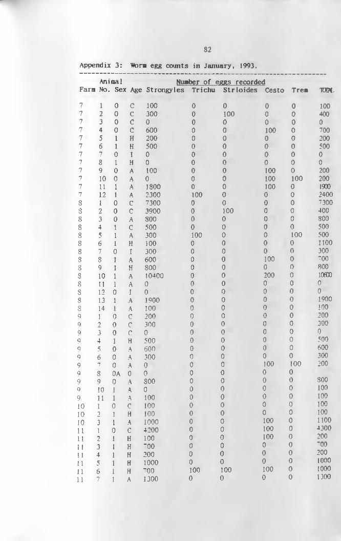
82
A ppendix 3 : Worm egg c o u n ts in J a n u a r y , 1993 .
Animal Number of eggs recordedFarmi No. Sex Age Strongyles Trichu Strloid es Cesto Trea TOM.
7 1 0 c 100 0 0 0 0 1007 2 0 c 300 0 100 0 0 4007 3 0 c 0 0 0 0 0 07 4 0 c 600 0 0 100 0 7007 5 1 H 200 0 0 0 0 2007 6 1 H 500 0 0 0 0 5007 7 0 I 0 0 0 0 0 07 8 1 H 0 0 0 0 0 07 9 0 A 100 0 0 100 0 2007 10 0 A 0 0 0 100 100 2007 11 1 A 1800 0 0 100 0 19007 12 1 A 2300 100 0 0 0 24008 1 0 C 7300 0 0 0 0 73008 2 0 C 3900 0 100 0 0 4008 3 0 A 800 0 0 0 0 8008 4 1 C 500 0 0 0 0 5008 5 1 A 300 100 0 0 100 5008 6 1 H 100 0 0 0 0 11008 7 0 I 300 0 0 0 0 3008 8 1 A 600 0 0 100 0 7008 9 1 H 800 0 0 0 0 8008 10 1 A 10400 0 0 200 0 lOfflO8 11 1 A 0 0 0 0 0 0S 12 0 I 0 0 0 0 0 08 13 1 A 1900 0 0 0 0 19008 14 1 A 100 0 0 0 0 1009 1 0 C 200 0 0 0 0 2009 2 0 C 300 0 0 0 0 3009 3 0 C 0 0 0 0 0 09 4 1 H 500 0 0 0 0 5009 5 0 A 600 0 0 0 0 6009 6 0 A 300 0 0 0 0 3009 7 0 A 0 0 0 100 100 2009 8 0A 0 0 0 0 0 09 9 0 A 800 0 0 0 0 3009 10 1 A 0 0 0 0 0 1009 11 1 A 100 0 0 0 0 10010 1 0 C 100 0 0 0 0 10010 2 1 H 100 0 0 0 0 10010 3 1 A 1000 0 0 100 0 110011 1 0 C 4200 0 0 100 0 430011 2 1 H 100 0 0 100 0 20011 3 1 H 700 0 0 0 0 "0011 4 1 H 200 0 0 0 0 20011 5 1 H 1000 0 0 0 0 100011 6 1 H 700 100 100 100 0 100011 7 1 A 1300 0 0 0 0 1300

83
A ppendix 4 : Worm eg g c o u n ts in F e b r u a r y , 1 993 .
Animal Number of eggs recordedFarm No. Sex Age Strongyles Trichu Strloid es Cesto Trem TOM.12 1 1 c 100 0 0 0 0 10012 2 1 c 200 0 0 0 0 20012 3 0 c 100 0 0 100 0 20012 4 1 H 700 0 0 0 0 70012 5 1 H 200 0 100 0 0 30012 6 1 H 200 0 0 200 0 40012 7 0 I 100 300 0 0 0 40012 8 0 I 0 0 100 0 0 10012 9 1 H 1100 0 0 0 0 110012 10 1 A 1800 0 0 0 0 180012 11 1 A 1900 0 0 0 0 190012 12 1 A 200 100 0 0 0 30012 13 1 A 300 0 0 0 0 30013 1 1 H 600 0 0 0 0 60013 2 0 I 200 0 0 100 0 30013 3 1 H 300 0 0 0 0 30013 4 1 A 200 0 0 0 0 20013 4 1 A 200 0 0 0 0 20013 5 1 A 2000 0 0 0 0 200014 1 1 H 1400 100 0 0 0 150014 2 1 H 1100 0 0 100 0 120015 1 1 C 200 0 0 0 0 20015 2 0 C 500 0 100 0 0 60015 3 1 C 300 0 100 100 0 50015 4 1 H 200 0 0 0 0 20015 5 1 A 1300 0 0 100 0 140015 6 1 A 200 0 0 0 0 20015 7 l A 600 0 0 100 0 70016 1 1 C 0 0 100 0 0 10016 0 0 C 300 200 0 0 0 50016 3 0 C 3000 0 0 0 0 300016 4 0 A 300 0 0 0 0 30016 5 0 A 100 0 0 0 0 10016 5 1 A 6400 0 0 0 0 640016 6 1 A 200 0 0 0 0 20016 n 1 A 500 0 0 100 0 60016 8 1 A 100 0 0 200 0 30016 9 1 A 300 0 0 0 0 30017 1 1 H 500 0 0 0 0 50017 2 1 H 500 0 0 0 0 50017 3 1 H 4200 0 0 0 0 *1200l 7 4 1 A 700 0 0 0 0 70017 5 1 A 400 0 0 100 0 50017 6 1 A 0 0 0 0 0 0P n 1 A 100 0 0 0 100 20017 8 1 A 600 100 0 100 0 800

84
A ppendix 5 : Worm eg g c o u n ts in M arch , 1 9 9 2 .
A n i m a l N u m b e r o f e g g s r e c o r d e d
Farm N o . S e x A g e S t r o n g y l e s T r i c h u S t r l o i d e s C e s t o T r e m T O W ,
23 1 1 A 800 0 0 0 80023 2 1 A 200 0 0 0 0 20024 1 0 C 100 0 0 0 0 10024 2 1 H 300 0 0 0 0 30024 3 1 A 0 0 0 0 0 024 4 1 A 800 0 0 0 0 80025 1 0 C 1300 200 0 0 0 150025 7L 1 H 0 0 0 0 0 025 3 1 H 2700 0 0 0 0 270025 4 0 I 600 0 0 0 0 60025 5 0 A 900 0 0 0 0 90025 6 1 A 1300 0 0 0 0 130025 7 1 A 100 0 0 0 0 10025 8 1 A 200 0 0 100 0 30025 9 1 A 200 0 0 0 0 20025 10 1 A 600 0 0 0 0 60026 1 0 C 100 0 0 0 0 10026 2 1 H 300 0 0 0 0 30026 3 1 H 100 0 0 0 0 10026 4 0 A 0 0 0 0 0 026 5 1 A 900 100 0 0 100 110026 6 1 A 100 0 0 0 0 10026 7 1 A 600 0 0 0 0 60026 8 1 A 100 0 0 0 0 10026 9 1 A 100 0 0 0 0 10026 10 1 A 0 0 0 100 0 100
1 0 C 100 0 0 0 0 1007 7 7 1 c 200 0 0 0 0 2007 7 3 1 c 100 0 0 0 0 1007 '*’ 4 1 H 0 0 0 0 0 07 " 5 1 H 0 0 0 0 0 07 ~ 6 0 A 0 0 0 0 0 07 “ 1 A 300 0 0 0 0 3007 7 3 1 A 400 0 0 0 0 4 0 0
7 7 9 1 A 500 0 0 100 0 6007 7 10 1 A 0 0 0 0 0 07 “ 11 1 A 500 0 0 0 0 500

8 5
A p p en d ix 6 : N em atode worm eg g c o u n ts f o r c a m e ls t r e a t e d w ith
a lb e n d a z o le o v e r t h e 2 8 day p e r io d .
Camel No,. of eggs per gramme of faeces
no. Age Sex Avpre P ostl Post2 Post3 Post4 Post5 Post6
1 2 0 900 0 0 0 0 0 0
2 1 0 800 1100 0 100 0 100 0
3 4 0 700 500 0 100 0 0 0
4 4 0 1100 3100 100 0 0 0 200
5 4 0 200 200 0 200 0 100 0
6 4 0 500 300 0 0 0 0 100
7 4 1 400 100 0 0 0 0 200
8 2 0 900 500 0 0 0 0 0
9 2 0 1100 300 0 0 0 0 0
10 4 0 2100 2800 0 0 0 0 100
11 4 0 200 2600 0 1900 0 0 100
12 2 0 1300 0 300 0 0 0 100
13 2 0 1350 200 0 0 0 300 0
14 4 0 800 800 400 0 0 0 100
15 2 0 800 900 0 0 0 100 0
16 1 0 1200 600 0 0 0 100 0
17 2 0 900 0 0 0 0 0 0
18 1 1 6500 4200 0 0 0 200 0
19 4 0 1100 1200 0 0 0 0 0

86
A p p en d ix 7 : N em atode worm eg g c o u n ts f o r c a m e ls t r e a t e d w ith
L e v a m is o le o v e r t h e 2 8 day p e r io d .
camel
no. Age Sex Avpre
20 1 0 900
21 4 0 600
22 4 0 900
23 4 0 800
24 1 1 450
25 4 0 500
26 4 0 100
27 4 0 1300
28 1 1 600
29 2 0 1000
30 2 0 900
31 4 0 1300
32 4 0 1100
33 2 0 200
34 2 0 4500
35 2 0 1600
36 4 0 2000
37 2 0 900
38 4 0 300
No. of eggs per
P ostl Post2 Post3
400 700 300
4000 2800 100
1000 0 0
1000 900 0
200 0 100
100 400 0
2800 500 100
500 300 100
100 1000 0
1000 300 0
0 600 0
0 0 0
1000 500 0
0 0 0
300 0 100
700 0 0
0 0 0
200 100 0
700 0 0
me of faeces
Post4 Post5 Post 6
0 0 100
100 0 300
200 0 0
0 400 100
0 0 0
100 0 200
200 200 400
0 100 0
100 100 400
0 0 0
0 0 100
0 200 100
0 0 100
o o o o o o
100 0 200
100 0 200
0 0 200
0 100 200

8 7
A p p en d ix 8 : N em atode worm eg g c o u n ts f o r c a m e ls t r e a t e d w ith
T h io p h a n a te o v e r t h e 28 d ay p e r io d .
camel No. of eqqs per gramne of faeces
no. Age Sex Avpre P ostl Post2 Post3 Post4 Post5 Post6
39 2 0 900 100 200 500 0 0 0
40 4 0 4000 900 100 0 100 0 100
41 4 0 600 1200 0 0 300 0 0
42 4 0 300 300 0 200 0 0 100
43 4 0 2000 0 0 0 0 0 400
44 4 0 400 100 100 0 300 100 400
45 1 1 1100 200 0 0 0 0 0
46 2 0 400 100 0 0 100 30 0
47 2 0 1300 1300 0 0 100 0 200
48 4 0 1100 100 100 200 0 100 100
49 2 0 500 0 0 100 0 0 100
50 4 0 300 300 0 0 100 0 200
51 2 0 300 400 100 100 0 0 200
52 4 1 800 700 400 0 0 0 200
53 4 0 1000 1400 0 200 0 0 100
54 4 1 4000 2000 100 0 0 0 0
55 1 0 200 100 0 0 0 300 200
56 2 0 500 0 100 200 0 100 0
57 4 0 5600 1300 300 200 o 0
1
100
Related Documents