Note: Within nine months of the publication of the mention of the grant of the European patent in the European Patent Bulletin, any person may give notice to the European Patent Office of opposition to that patent, in accordance with the Implementing Regulations. Notice of opposition shall not be deemed to have been filed until the opposition fee has been paid. (Art. 99(1) European Patent Convention). Printed by Jouve, 75001 PARIS (FR) (19) EP 3 117 011 B1 *EP003117011B1* (11) EP 3 117 011 B1 (12) EUROPEAN PATENT SPECIFICATION (45) Date of publication and mention of the grant of the patent: 06.05.2020 Bulletin 2020/19 (21) Application number: 15714069.0 (22) Date of filing: 12.03.2015 (51) Int Cl.: C12Q 1/68 (2018.01) (86) International application number: PCT/US2015/020250 (87) International publication number: WO 2015/138774 (17.09.2015 Gazette 2015/37) (54) METHODS AND PROCESSES FOR NON-INVASIVE ASSESSMENT OF GENETIC VARIATIONS VERFAHREN UND PROZESSE ZUR NICHT-INVASIVEN BEURTEILUNG GENETISCHER VARIATIONEN MÉTHODES ET PROCÉDÉS D’ÉVALUATION NON INVASIVE DE VARIATIONS GÉNÉTIQUES (84) Designated Contracting States: AL AT BE BG CH CY CZ DE DK EE ES FI FR GB GR HR HU IE IS IT LI LT LU LV MC MK MT NL NO PL PT RO RS SE SI SK SM TR (30) Priority: 13.03.2014 US 201461952135 P (43) Date of publication of application: 18.01.2017 Bulletin 2017/03 (73) Proprietor: Sequenom, Inc. San Diego, CA 92121 (US) (72) Inventors: • JENSEN, Taylor, Jacob San Diego, CA 92131 (US) • GEIS, Jennifer San Diego, CA 92108 (US) • KIM, Sung, Kyun Glendale, CA 91206 (US) • DECIU, Cosmin San Diego, CA 92121 (US) • EHRICH, Mathias San Diego, CA 92109 (US) (74) Representative: Vossius & Partner Patentanwälte Rechtsanwälte mbB Siebertstrasse 3 81675 München (DE) (56) References cited: WO-A1-03/062441 WO-A1-2011/034631 WO-A1-2014/011928 WO-A2-2007/132167 WO-A2-2011/092592 US-A1- 2012 065 076

Welcome message from author
This document is posted to help you gain knowledge. Please leave a comment to let me know what you think about it! Share it to your friends and learn new things together.
Transcript
Note: Within nine months of the publication of the mention of the grant of the European patent in the European PatentBulletin, any person may give notice to the European Patent Office of opposition to that patent, in accordance with theImplementing Regulations. Notice of opposition shall not be deemed to have been filed until the opposition fee has beenpaid. (Art. 99(1) European Patent Convention).
Printed by Jouve, 75001 PARIS (FR)
(19)EP
3 11
7 01
1B
1*EP003117011B1*
(11) EP 3 117 011 B1(12) EUROPEAN PATENT SPECIFICATION
(45) Date of publication and mention of the grant of the patent: 06.05.2020 Bulletin 2020/19
(21) Application number: 15714069.0
(22) Date of filing: 12.03.2015
(51) Int Cl.:C12Q 1/68 (2018.01)
(86) International application number: PCT/US2015/020250
(87) International publication number: WO 2015/138774 (17.09.2015 Gazette 2015/37)
(54) METHODS AND PROCESSES FOR NON-INVASIVE ASSESSMENT OF GENETIC VARIATIONS
VERFAHREN UND PROZESSE ZUR NICHT-INVASIVEN BEURTEILUNG GENETISCHER VARIATIONEN
MÉTHODES ET PROCÉDÉS D’ÉVALUATION NON INVASIVE DE VARIATIONS GÉNÉTIQUES
(84) Designated Contracting States: AL AT BE BG CH CY CZ DE DK EE ES FI FR GB GR HR HU IE IS IT LI LT LU LV MC MK MT NL NO PL PT RO RS SE SI SK SM TR
(30) Priority: 13.03.2014 US 201461952135 P
(43) Date of publication of application: 18.01.2017 Bulletin 2017/03
(73) Proprietor: Sequenom, Inc.San Diego, CA 92121 (US)
(72) Inventors: • JENSEN, Taylor, Jacob
San Diego, CA 92131 (US)• GEIS, Jennifer
San Diego, CA 92108 (US)
• KIM, Sung, KyunGlendale, CA 91206 (US)
• DECIU, CosminSan Diego, CA 92121 (US)
• EHRICH, MathiasSan Diego, CA 92109 (US)
(74) Representative: Vossius & Partner Patentanwälte Rechtsanwälte mbBSiebertstrasse 381675 München (DE)
(56) References cited: WO-A1-03/062441 WO-A1-2011/034631WO-A1-2014/011928 WO-A2-2007/132167WO-A2-2011/092592 US-A1- 2012 065 076
EP 3 117 011 B1
2
5
10
15
20
25
30
35
40
45
50
55
Description
Field
[0001] The present invention is defined by the claims. Technology provided herein generally relates in part to methods,processes, systems and apparatuses for non-invasive assessment of genetic variations.
Background
[0002] Genetic information of living organisms (e.g., animals, plants and microorganisms) and other forms of replicatinggenetic information (e.g., viruses) is encoded in deoxyribonucleic acid (DNA) or ribonucleic acid (RNA). Genetic infor-mation is a succession of nucleotides or modified nucleotides representing the primary structure of chemical or hypo-thetical nucleic acids. In humans, the complete genome contains about 30,000 genes located on twenty-four (24) chro-mosomes (see The Human Genome, T. Strachan, BIOS Scientific Publishers, 1992). Each gene encodes a specificprotein, which after expression via transcription and translation fulfills a specific biochemical function within a living cell.[0003] Many medical conditions are caused by one or more genetic variations. Certain genetic variations cause medicalconditions that include, for example, hemophilia, thalassemia, Duchenne Muscular Dystrophy (DMD), Huntington’sDisease (HD), Alzheimer’s Disease and Cystic Fibrosis (CF) (Human Genome Mutations, D. N. Cooper and M. Krawczak,BIOS Publishers, 1993). Such genetic diseases can result from an addition, substitution, or deletion of a single nucleotidein DNA of a particular gene. Certain birth defects are caused by a chromosomal abnormality, also referred to as ananeuploidy, such as Trisomy 21 (Down’s Syndrome), Trisomy 13 (Patau Syndrome), Trisomy 18 (Edward’s Syndrome),Monosomy X (Turner’s Syndrome) and certain sex chromosome aneuploidies such as Klinefelter’s Syndrome (XXY),for example. Another genetic variation is fetal gender, which can often be determined based on sex chromosomes Xand Y. Some genetic variations may predispose an individual to, or cause, any of a number of diseases such as, forexample, diabetes, arteriosclerosis, obesity, various autoimmune diseases and cancer (e.g., colorectal, breast, ovarian,lung).[0004] Identifying one or more genetic variations or variances can lead to diagnosis of, or determining predispositionto, a particular medical condition. Identifying a genetic variance can result in facilitating a medical decision and/oremploying a helpful medical procedure. Identification of one or more genetic variations or variances sometimes involvesthe analysis of cell-free DNA.[0005] Cell-free DNA (CF-DNA) is composed of DNA fragments that originate from cell death and circulate in peripheralblood. High concentrations of CF-DNA can be indicative of certain clinical conditions such as cancer, trauma, burns,myocardial infarction, stroke, sepsis, infection, and other illnesses. Additionally, cell-free fetal DNA (CFF-DNA) can bedetected in the maternal bloodstream and used for various noninvasive diagnostics (e.g., prenatal diagnostics).[0006] The presence of fetal nucleic acid in maternal plasma allows for non-invasive prenatal diagnosis through theanalysis of a maternal blood sample. For example, quantitative abnormalities of fetal DNA in maternal plasma can beassociated with a number of pregnancy-associated disorders, including preeclampsia, preterm labor, antepartum hem-orrhage, invasive placentation, fetal Down syndrome, and other fetal chromosomal aneuploidies. Hence, fetal nucleicacid analysis in maternal plasma can be a useful mechanism for the monitoring of fetomaternal well-being.[0007] WO2011034631 discloses a method for determining the presence or absence of a fetal aneuploidy using fetalnucleic acid from a maternal sample and measuring differential methylation in specific genomic regions.
Summary
[0008] The present invention is defined by the claims. Accordingly, the present invention relates to a method fordetecting one, two, three or four copies of a fetal chromosome 13, chromosome 18 and chromosome 21 or portionsthereof in a sample, comprising: (a) contacting a sample comprising circulating cell-free nucleic acid from a humanpregnant female bearing a fetus with a methylation sensitive restriction enzyme, thereby generating cleaved nucleic acidand non-cleaved nucleic acid; (b) determining in a single multiplex reaction amounts of each target polynucleotide inchromosome 13, chromosome 18 and chromosome 21 in the non-cleaved nucleic acid of (a), wherein the target poly-nucleotides are: chromosome 13 polynucleotides of SEQ ID NOs: 193-198, 200-204, 206, 208-210, and 212-215; chro-mosome 18 polynucleotides of SEQ ID NOs: 216-218, 220-230, and 232; and chromosome 21 polynucleotides of SEQID NOs: 234, 236, 238-240, 242-246, 248-253, 255, and 256, which single multiplex reaction comprises contacting thenucleic acid of (a) with (i) a collection of primer pairs specifically hybridizing to the target polynucleotides; and (ii) knownamounts of competitor polynucleotides, wherein the competitor oligonucleotides are polynucleotides that comprise anucleic acid sequence that is identical to its corresponding target polynucleotide apart from a single nucleotide basesubstitution within the competitor oligonucleotides that differentiates the competitor oligonucleotides from its correspond-ing target polynucleotide, under amplification conditions, thereby generating amplicons, which amplicons are further
EP 3 117 011 B1
3
5
10
15
20
25
30
35
40
45
50
55
processed by contacting them with extension oligonucleotides under conditions in which the extension oligonucleotidesanneal to the amplicons and are extended by one or more nucleotides, thereby generating extension products; and (c)quantifying, from the amounts of extension products the amounts of target polynucleotides, one, two, three or four copiesof chromosome 13, chromosome 18, chromosome 21, or portions thereof, in the fetus.[0009] Also provided herein in certain aspects is a method for detecting one, two, three or four copies of a fetalchromosome, or portion thereof, in a sample, comprising (a) determining amounts of three or more target polynucleotidesin each of chromosome 13, chromosome 18 and chromosome 21 in circulating cell-free nucleic acid from a sample froma human pregnant female bearing a fetus, where the three or more target polynucleotides in chromosome 13 are inchromosome 13 polynucleotides comprising chromosome 13 polynucleotides of SEQ ID NOs: 209, 211 and 214, orcomplement thereof, the three or more target polynucleotides in chromosome 18 are in chromosome 18 polynucleotidescomprising chromosome 18 polynucleotides of SEQ ID NOs: 232, 222 and 231, or complement thereof, and the threeor more target polynucleotides in chromosome 21 are in chromosome 21 polynucleotides comprising chromosome 21polynucleotides of SEQ ID NOs: 256, 253 and 252, or complement thereof, and (b) quantifying, from the amounts, one,two, three or four copies of one or more of chromosome 13, chromosome 18, chromosome 21, or portion thereof, in thefetus. In certain aspects the method comprises prior to (a), contacting nucleic acid from the sample with a cleavageagent under cleavage conditions, thereby generating cleaved nucleic acid and non-cleaved nucleic acid. In some aspectsthe cleavage agent is a methylation sensitive restriction enzyme. In certain aspects the restriction enzyme preferentiallycleaves nucleic acid comprising one or more non-methylated recognition sequences and the nucleic acid in (a) fromwhich the amounts of the three or more target polynucleotides are determined is substantially the non-cleaved nucleicacid. In certain aspects the amounts of the three or more target polynucleotides are determined by a process comprisingmass spectrometry. In certain aspects the amounts of the three or more target polynucleotides are determined by aprocess comprising sequencing.[0010] Also provided herein, in certain aspects, is a method for detecting one, two, three or four copies of a fetalchromosome or portion thereof in a sample, comprising (a) determining amounts of target polynucleotides in each ofchromosome 13, chromosome 18 and chromosome 21 in circulating cell-free nucleic acid from a sample from a humanpregnant female bearing a fetus, where the target polynucleotides are in chromosome 13 polynucleotides of SEQ IDNOs: 193-198, 200-204, 206, 208-210, 212-215, or complement thereof, chromosome 18 polynucleotides of SEQ IDNOs: 216-218, 220-230, 232, or complement thereof, and chromosome 21 polynucleotides of SEQ ID NOs: 234, 236,238-240, 242-246, 248-253, 255, 256, or complement thereof, and (b) quantifying, from the amounts, one, two, threeor four copies of one or more of chromosome 13, chromosome 18, chromosome 21, or portion thereof, in the fetus. Incertain aspects, the method comprising determining in (a) the amounts of target polynucleotides in chromosome 13polynucleotides of SEQ ID NOs: 199, 205, 207, 211 or complement thereof, chromosome 18 polynucleotides of SEQID NOs: 219, 231, or complement thereof, and chromosome 21 polynucleotides of SEQ ID NOs: 233, 235, 237, 241,247, 254 or complement thereof.[0011] Also provided herein, in certain aspects, is a method of amplifying one or more target polynucleotides in asample comprising (a) contacting a sample comprising circulating cell-free nucleic acid from a human pregnant femalebearing a fetus with a collection of primers under amplification conditions, where the primers specifically hybridize tonucleotide sequences located within three or more target polynucleotides in each of chromosome 13, chromosome 18and chromosome 21 under specific hybridization conditions, where the three or more target polynucleotides in chromo-some 13 are in chromosome 13 polynucleotides comprising chromosome 13 polynucleotides of SEQ ID NOs: 209, 211and 214, or complement thereof, the three or more target polynucleotides in chromosome 18 are in chromosome 18polynucleotides comprising chromosome 18 polynucleotides of SEQ ID NOs: 232, 222 and 231, or complement thereofand the three or more target polynucleotides in chromosome 21 are in chromosome 21 polynucleotides comprisingchromosome 21 polynucleotides of SEQ ID NOs: 256, 253 and 252, or complement thereof, thereby providing target-specific amplicons.[0012] Also provided herein, in certain aspects, is a method of amplifying one or more target polynucleotides in asample comprising (a) contacting a sample comprising circulating cell-free nucleic acid from a human pregnant femalebearing a fetus with a collection of primers under amplification conditions, where the primer pairs specifically hybridizeto nucleotide sequences located within three or more target polynucleotides in each of chromosome 13, chromosome18 and chromosome 21 under specific hybridization conditions, where the target polynucleotides are in chromosome13 polynucleotides of SEQ ID NOs: 193-198, 200-204, 206, 208-210, 212-215, or complement thereof, chromosome18 polynucleotides of SEQ ID NOs: 216-218, 220-230, 232, or complement thereof, and chromosome 21 polynucleotidesof SEQ ID NOs: 234, 236, 238-240, 242-246, 248-253, 255, 256, or complement thereof, thereby providing target-specificamplicons.[0013] Provided also in certain aspects is a method for detecting one, two, three or four copies of a fetal chromosome,or portion thereof, in a sample, comprising (a) determining amounts of three or more target polynucleotides chromosome13, chromosome 18 or chromosome 21 in circulating cell-free nucleic acid from a sample from a human pregnant femalebearing a fetus, where the three or more target polynucleotides in chromosome 13 are in chromosome 13 polynucleotides
EP 3 117 011 B1
4
5
10
15
20
25
30
35
40
45
50
55
comprising chromosome 13 polynucleotides of SEQ ID NOs: 209, 211 and 214, or complement thereof, the three ormore target polynucleotides in chromosome 18 are in chromosome 18 polynucleotides comprising chromosome 18polynucleotides of SEQ ID NOs: 232, 222 and 231, or complement thereof, or the three or more target polynucleotidesin chromosome 21 are in chromosome 21 polynucleotides comprising chromosome 21 polynucleotides of SEQ ID NOs:256, 253 and 252, or complement thereof, and (b) quantifying, from the amounts, one, two, three or four copies ofchromosome 13, chromosome 18 or chromosome 21, or portion thereof, in the fetus.[0014] Also provided herein, in certain aspects, is a method for detecting one, two, three or four copies of a fetalchromosome or portion thereof in a sample, comprising (a) determining amounts of target polynucleotides in chromosome13, chromosome 18 or chromosome 21 in circulating cell-free nucleic acid from a sample from a human pregnant femalebearing a fetus, where the target polynucleotides are in chromosome 13 polynucleotides of SEQ ID NOs: 193-198,200-204, 206, 208-210, 212-215, or complement thereof, chromosome 18 polynucleotides of SEQ ID NOs: 216-218,220-230, 232, or complement thereof, or chromosome 21 polynucleotides of SEQ ID NOs: 234, 236, 238-240, 242-246,248-253, 255, 256, or complement thereof, and (b) quantifying, from the amounts, one, two, three or four copies ofchromosome 13, chromosome 18 or chromosome 21, or portion thereof, in the fetus. In certain aspects, the methodcomprising determining in (a) the amounts of target polynucleotides in chromosome 13 polynucleotides of SEQ ID NOs:199, 205, 207, 211 or complement thereof, chromosome 18 polynucleotides of SEQ ID NOs: 219, 231, or complementthereof, or chromosome 21 polynucleotides of SEQ ID NOs: 233, 235, 237, 241, 247, 254 or complement thereof.[0015] Provided also herein, in certain aspects, is a method of amplifying one or more target polynucleotides in asample comprising (a) contacting a sample comprising circulating cell-free nucleic acid from a human pregnant femalebearing a fetus with a collection of primers under amplification conditions, where the primers specifically hybridize tonucleotide sequences located within target polynucleotides in chromosome 13, chromosome 18 or chromosome 21under specific hybridization conditions, where the three or more target polynucleotides in chromosome 13 are in chro-mosome 13 polynucleotides comprising chromosome 13 polynucleotides of SEQ ID NOs: 209, 211 and 214, or com-plement thereof, the three or more target polynucleotides in chromosome 18 are in chromosome 18 polynucleotidescomprising chromosome 18 polynucleotides of SEQ ID NOs: 232, 222 and 231, or complement thereof, or the three ormore target polynucleotides in chromosome 21 are in chromosome 21 polynucleotides comprising chromosome 21polynucleotides of SEQ ID NOs: 256, 253 and 252, or complement thereof, thereby providing target-specific amplicons.[0016] Also provided herein, in certain aspects, is a method of amplifying one or more target polynucleotides in asample comprising (a) contacting a sample comprising circulating cell-free nucleic acid from a human pregnant femalebearing a fetus with a collection of primers under amplification conditions, where the primer pairs specifically hybridizeto nucleotide sequences located within three or more target polynucleotides in chromosome 13, chromosome 18 orchromosome 21 under specific hybridization conditions, where the target polynucleotides are in chromosome 13 poly-nucleotides of SEQ ID NOs: 193-198, 200-204, 206, 208-210, 212-215, or complement thereof, chromosome 18 poly-nucleotides of SEQ ID NOs: 216-218, 220-230, 232, or complement thereof, or chromosome 21 polynucleotides of SEQID NOs: 234, 236, 238-240, 242-246, 248-253, 255, 256, or complement thereof, thereby providing target-specificamplicons. In certain aspects, the target polynucleotides are in chromosome 13 polynucleotides of SEQ ID NOs: 199,205, 207, 211 or complement thereof, chromosome 18 polynucleotides of SEQ ID NOs: 219, 231, or complement thereofand chromosome 21 polynucleotides of SEQ ID NOs: 233, 235, 237, 241, 247, 254 or complement thereof.[0017] Also provided herein, in certain aspects, is a kit for detecting one, two, three or four copies of a fetal chromosome,or portion thereof, in circulating cell-free nucleic acid from a sample from a human pregnant female bearing a fetus,comprisinga collection of oligonucleotide primer pairs where each primer pair is configured for amplifying three or moretarget polynucleotides in each of chromosome 13, chromosome 18 and chromosome 21, where the three or more targetpolynucleotides in chromosome 13 are in chromosome 13 polynucleotides comprising chromosome 13 polynucleotidesof SEQ ID NOs: 209, 211 and 214, or complement thereof, the three or more target polynucleotides in chromosome 18are in chromosome 18 polynucleotides comprising chromosome 18 polynucleotides of SEQ ID NOs: 232, 222 and 231,or complement thereof and the three or more target polynucleotides in chromosome 21 are in chromosome 21 polynu-cleotides comprising chromosome 21 polynucleotides of SEQ ID NOs: 256, 253 and 252, or complement thereof.[0018] Provided also herein, in certain aspects, is a kit for detecting one, two, three or four copies of a fetal chromosome,or portion thereof, in circulating cell-free nucleic acid from a sample from a human pregnant female bearing a fetus,comprisinga collection of oligonucleotide primer pairs where each primer pair is configured for amplifying three or moretarget polynucleotides in chromosome 13, chromosome 18 or chromosome 21, where the three or more target polynu-cleotides in chromosome 13 are in chromosome 13 polynucleotides comprising chromosome 13 polynucleotides of SEQID NOs: 209, 211 and 214, or complement thereof, the three or more target polynucleotides in chromosome 18 are inchromosome 18 polynucleotides comprising chromosome 18 polynucleotides of SEQ ID NOs: 232, 222 and 231, orcomplement thereof, or the three or more target polynucleotides in chromosome 21 are in chromosome 21 polynucleotidescomprising chromosome 21 polynucleotides of SEQ ID NOs: 256, 253 and 252, or complement thereof.[0019] Also provided herein is a kit for detecting one, two, three or four copies of a fetal chromosome, or portion thereof,in circulating cell-free nucleic acid from a sample from a human pregnant female bearing a fetus, comprising a collection
EP 3 117 011 B1
5
5
10
15
20
25
30
35
40
45
50
55
of oligonucleotide primer pairs where each primer pair is configured for amplifying three or more target polynucleotidesin each of chromosome 13, chromosome 18 and chromosome 21 , where the target polynucleotides are in chromosome13 polynucleotides of SEQ ID NOs: 193-198, 200-204, 206, 208-210, 212-215, or complement thereof, chromosome18 polynucleotides of SEQ ID NOs: 216-218, 220-230, 232, or complement thereof, and chromosome 21 polynucleotidesof SEQ ID NOs: 234, 236, 238-240, 242-246, 248-253, 255, 256, or complement thereof. In certain aspects, the targetpolynucleotides are in chromosome 13 polynucleotides of SEQ ID NOs: 199, 205, 207, 211 or complement thereof,chromosome 18 polynucleotides of SEQ ID NOs: 219, 231, or complement thereof and chromosome 21 polynucleotidesof SEQ ID NOs: 233, 235, 237, 241, 247, 254 or complement thereof.[0020] Provided also herein is a kit for detecting one, two, three or four copies of a fetal chromosome, or portion thereof,in circulating cell-free nucleic acid from a sample from a human pregnant female bearing a fetus, comprising a collectionof oligonucleotide primer pairs where each primer pair is configured for amplifying three or more target polynucleotidesin chromosome 13, chromosome 18 or chromosome 21, where the target polynucleotides are in chromosome 13 poly-nucleotides of SEQ ID NOs: 193-198, 200-204, 206, 208-210, 212-215, or complement thereof, chromosome 18 poly-nucleotides of SEQ ID NOs: 216-218, 220-230, 232, or complement thereof, or chromosome 21 polynucleotides of SEQID NOs: 234, 236, 238-240, 242-246, 248-253, 255, 256, or complement thereof. In some aspects of the kit the targetpolynucleotides are in chromosome 13 polynucleotides of SEQ ID NOs: 199, 205, 207, 211 or complement thereof,chromosome 18 polynucleotides of SEQ ID NOs: 219, 231, or complement thereof and chromosome 21 polynucleotidesof SEQ ID NOs: 233, 235, 237, 241, 247, 254 or complement thereof.[0021] In certain aspects a kit comprises one or more methylation sensitive restriction enzymes. In some aspects akit comprises one or more competitor oligonucleotides and/or one or more extension primers presented in Table 1.[0022] Certain aspects of the technology are described further in the following description, examples, drawings andclaims.
Brief Description of the Drawings
[0023] The drawings illustrate aspects of the technology and are not limiting. For clarity and ease of illustration, thedrawings are not made to scale and, in some instances, various aspects may be shown exaggerated or enlarged tofacilitate an understanding of particular aspects. FIG. 1 shows weighted probability from Sequencing (y-axis) vs Mas-sARRAY (x-axis) for chromosome 21 (FIG. 1A), chromosome 13 (FIG. 1B) and chromosome 18 (FIG. 1C). Euploids areindicated by a square, trisomy 13 (T13) is indicated by a cross, trisomy 18 (T18) is indicated by a filled-in circle, andtrisomy 21 (T21) is indicated by a triangle. For each marker classification, markers indicating the presence of threecopies of a chromosome are shown within the lower left quadrant.
Detailed Description
[0024] Cell free nucleic acid sometimes comprises a mixture of nucleic acids from different sources (e.g., fetal versusmaternal tissue) and nucleic acid from different sources are sometimes differentially methylated. Such differential meth-ylation of certain subpopulations of cell free nucleic acid can be useful for analyzing fetal nucleic acid. Provided also arenon-invasive methods, processes and apparatuses useful for identifying a genetic variation in a fetus. Also providedherein, in some aspects are methods, systems, kits and machines for detecting and/or quantifying one, two, three orfour copies of a fetal chromosome, or portion thereof, in a test sample, where the test sample comprises circulating cellfree nucleic acid obtained from a pregnant female. In some aspects methods herein comprise detecting and/or quantifyingone, two, three or four copies of chromosome 13, 18 and/or 21, or a portion thereof in a fetus.[0025] In some aspects, determining a copy number of a chromosome or portion thereof in a fetus comprises detectingand/or quantifying specific target polynucleotides in polynucleotides of chromosomes 13, 18 and/or 21 shown in Tables1A and 1B. Polynucleotides in Tables 1A and 1B were empirically chosen according to, in part, differential methylationbetween fetus and mother. In some aspects methylation sensitive restriction endonucleases are used to digest portionsof specific polynucleotides in chromosomes 13, 18 and 21 that are present in circulating cell free nucleic acid. In someaspects undigested fragments remain after digestion that comprise fetal nucleic acid and these fragments can be amplifiedusing specific primer pairs that flank or are within methylation sensitive restriction sites. In certain aspects presentedherein are methods and systems for analyzing and quantifying polynucleotide specific amplicons, for example by useof nucleic acid sequencing and/or mass spectrometry, to determine the presence of one, two, three or four copies offetal chromosomes.[0026] In some aspects, identifying a genetic variation by a method described herein can lead to a diagnosis of, ordetermining a predisposition to, a particular medical condition. Identifying a genetic variance can result in facilitating amedical decision and/or employing a helpful medical procedure.
EP 3 117 011 B1
6
5
10
15
20
25
30
35
40
45
50
55
Samples
[0027] Provided herein are methods and compositions for analyzing nucleic acid. In some aspects, nucleic acid frag-ments in a mixture of nucleic acid fragments are analyzed. A mixture of nucleic acids can comprise two or more nucleicacid fragment species having different nucleotide sequences, different fragment lengths, different origins (e.g., genomicorigins, fetal vs. maternal origins, cell or tissue origins, sample origins, subject origins, and the like), or combinationsthereof.[0028] Nucleic acid or a nucleic acid mixture utilized in methods and apparatuses described herein often is isolatedfrom a sample obtained from a subject (e.g., a test subject). A test subject can be any living or non-living organism,including but not limited to a human (e.g., including a human embryo, fetus, or unborn human child), a non-humananimal, a plant, a bacterium, a fungus or a protist. Non-limiting examples of a non-human animal include a mammal,reptile, avian, amphibian, fish, ungulate, ruminant, bovine (e.g., cattle), equine (e.g., horse), caprine and ovine (e.g.,sheep, goat), swine (e.g., pig), camelid (e.g., camel, llama, alpaca), monkey, ape (e.g., gorilla, chimpanzee), ursid (e.g.,bear), poultry, dog, cat, rodent (e.g., mouse, rat, and the like), fish, dolphin, whale and shark. A test subject may be amale or female (e.g., woman). In some aspects a test subject is a pregnant human female.[0029] Nucleic acid may be isolated from any type of suitable test subject, biological specimen or sample (e.g., a testsample). A sample or test sample can be any specimen that is isolated or obtained from a test subject. Non-limitingexamples of specimens include fluid or tissue from a subject, including, without limitation, umbilical cord blood, chorionicvilli, amniotic fluid, cerebrospinal fluid, spinal fluid, lavage fluid (e.g., bronchoalveolar, gastric, peritoneal, ductal, ear,arthroscopic), biopsy sample (e.g., from pre-implantation embryo), celocentesis sample, fetal nucleated cells or fetalcellular remnants, washings of female reproductive tract, urine, feces, sputum, saliva, nasal mucous, prostate fluid,lavage, semen, lymphatic fluid, bile, tears, sweat, breast milk, breast fluid, embryonic cells and fetal cells (e.g. placentalcells). In some aspects, a biological sample is a cervical swab from a subject. In some aspects, a biological sample maybe blood and sometimes plasma or serum. As used herein, the term "blood" encompasses whole blood or any fractionsof blood, such as serum and plasma as conventionally defined, for example. Blood or fractions thereof often comprisenucleosomes (e.g., maternal and/or fetal nucleosomes). Nucleosomes comprise nucleic acids and are sometimes cell-free or intracellular. Blood also comprises buffy coats. Buffy coats are sometimes isolated by utilizing a ficoll gradient.Buffy coats can comprise blood cells (e.g., white blood cells, e.g., leukocytes, T-cells, B-cells, platelets, and the like). Incertain instances, buffy coats comprise maternal and/or fetal cells and maternal and/or fetal nucleic acid. Blood plasmarefers to the fraction of whole blood resulting from centrifugation of blood treated with anticoagulants. Blood serum refersto the watery portion of fluid remaining after a blood sample has coagulated. Fluid or tissue samples often are collectedin accordance with standard protocols that hospitals or clinics generally follow. For blood, an appropriate amount ofperipheral blood (e.g., between 3-40 milliliters) often is collected and can be stored according to standard proceduresprior to or after preparation. A fluid or tissue sample from which nucleic acid is extracted may be acellular (e.g., cell-free). In some aspects, a fluid or tissue sample may contain cellular elements or cellular remnants. In some aspectsfetal cells or cancer cells may be included in a sample.[0030] A sample often is heterogeneous, by which is meant that more than one type of nucleic acid species is presentin the sample. For example, heterogeneous nucleic acid can include, but is not limited to, (i) fetal derived and maternalderived nucleic acid, (ii) cancer and non-cancer nucleic acid, (iii) pathogen and host nucleic acid, and more generally,(iv) mutated and wild-type nucleic acid. A sample may be heterogeneous because more than one cell type is present,such as a fetal cell and a maternal cell, a cancer and non-cancer cell, or a pathogenic and host cell.[0031] For prenatal applications of a technology described herein, a fluid or tissue sample (e.g., a test sample) maybe collected from a female (e.g., a pregnant female) at a gestational age suitable for testing, or from a female who isbeing tested for possible pregnancy. A suitable gestational age may vary depending on the prenatal test being performed.In certain aspects, a pregnant female subject sometimes is in the first trimester of pregnancy, at times in the secondtrimester of pregnancy, or sometimes in the third trimester of pregnancy. In certain aspects, a fluid or tissue is collectedfrom a pregnant female between about 1 to about 45 weeks of fetal gestation (e.g., at 1-4, 4-8, 8-12, 12-16, 16-20, 20-24,24-28, 28-32, 32-36, 36-40 or 40-44 weeks of fetal gestation), and sometimes between about 5 to about 28 weeks offetal gestation (e.g., at 6, 7, 8, 9,10, 11, 12, 13, 14, 15, 16, 17, 18, 19, 20, 21, 22, 23, 24, 25, 26 or 27 weeks of fetalgestation). In some aspects, a fluid or tissue sample is collected from a pregnant female during or just after (e.g., 0 to72 hours after) giving birth (e.g., vaginal or non-vaginal birth (e.g., surgical delivery)).
Nucleic Acid Isolation and Processing
[0032] Nucleic acid may be derived from one or more sources (e.g., cells, serum, plasma, buffy coat, lymphatic fluid,skin, soil, and the like) by methods known in the art. Cell lysis procedures and reagents are known in the art and maygenerally be performed by chemical (e.g., detergent, hypotonic solutions, enzymatic procedures, and the like, or com-bination thereof), physical (e.g., French press, sonication, and the like), or electrolytic lysis methods. Any suitable lysis
EP 3 117 011 B1
7
5
10
15
20
25
30
35
40
45
50
55
procedure can be utilized. For example, chemical methods generally employ lysing agents to disrupt cells and extractthe nucleic acids from the cells, followed by treatment with chaotropic salts. Physical methods such as freeze/thawfollowed by grinding, the use of cell presses and the like also are useful. High salt lysis procedures also are commonlyused. For example, an alkaline lysis procedure may be utilized. The latter procedure traditionally incorporates the useof phenol-chloroform solutions, and an alternative phenol-chloroform-free procedure involving three solutions can beutilized. In the latter procedures, one solution can contain 15mM Tris, pH 8.0; 10mM EDTA and 100 ug/ml Rnase A; asecond solution can contain 0.2N NaOH and 1% SDS; and a third solution can contain 3M KOAc, pH 5.5. Theseprocedures can be found in Current Protocols in Molecular Biology, John Wiley & Sons, N.Y., 6.3.1-6.3.6 (1989).[0033] The terms "nucleic acid" and "nucleic acid molecule" are used interchangeably. The terms refer to nucleic acidsof any composition form, such as deoxyribonucleic acid (DNA, e.g., complementary DNA (cDNA), genomic DNA (gDNA)and the like), ribonucleic acid (RNA, e.g., message RNA (mRNA), short inhibitory RNA (siRNA), ribosomal RNA (rRNA),transfer RNA (tRNA), microRNA, RNA highly expressed by the fetus or placenta, and the like), and/or DNA or RNAanalogs (e.g., containing base analogs, sugar analogs and/or a non-native backbone and the like), RNA/DNA hybridsand polyamide nucleic acids (PNAs), all of which can be in single- or double-stranded form. Unless otherwise limited,a nucleic acid can comprise known analogs of natural nucleotides, some of which can function in a similar manner asnaturally occurring nucleotides. A nucleic acid can be in any form useful for conducting processes herein (e.g., linear,circular, supercoiled, single-stranded, double-stranded and the like). A polynucleotide can be a nucleic acid and/or anucleic acid fragment. A nucleic acid may be, or may be from, a plasmid, phage, autonomously replicating sequence(ARS), centromere, artificial chromosome, chromosome (chr), or other nucleic acid able to replicate or be replicated invitro or in a host cell, a cell, a cell nucleus or cytoplasm of a cell in certain aspects. A nucleic acid in some aspects canbe from a single chromosome or fragment thereof (e.g., a nucleic acid sample may be from one chromosome of a sampleobtained from a diploid organism). Nucleic acids sometimes comprise nucleosomes, fragments or parts of nucleosomesor nucleosome-like structures. Nucleic acids sometimes comprise protein (e.g., histones, DNA binding proteins, and thelike). Nucleic acids analyzed by processes described herein sometimes are substantially isolated and are not substantiallyassociated with protein or other molecules. Nucleic acids also include derivatives, variants and analogs of RNA or DNAsynthesized, replicated or amplified from single-stranded ("sense" or "antisense", "plus" strand or "minus" strand, "for-ward" reading frame or "reverse" reading frame) and double-stranded polynucleotides. Deoxyribonucleotides includedeoxyadenosine, deoxycytidine, deoxyguanosine and deoxythymidine. For RNA, the base cytosine is replaced withuracil and the sugar 2’ position includes a hydroxyl moiety. A nucleic acid may be prepared using a nucleic acid obtainedfrom a subject as a template.[0034] Nucleic acid may be isolated at a different time point as compared to another nucleic acid, where each of thesamples is from the same or a different source. A nucleic acid may be from a nucleic acid library, such as a cDNA orRNA library, for example. A nucleic acid may be a result of nucleic acid purification or isolation and/or amplification ofnucleic acid molecules from the sample. Nucleic acid provided for processes described herein may contain nucleic acidfrom one sample or from two or more samples (e.g., from 1 or more, 2 or more, 3 or more, 4 or more, 5 or more, 6 ormore, 7 or more, 8 or more, 9 or more, 10 or more, 11 or more, 12 or more, 13 or more, 14 or more, 15 or more, 16 ormore, 17 or more, 18 or more, 19 or more, or 20 or more samples).[0035] Nucleic acids can include extracellular nucleic acid in certain aspects. The term "extracellular nucleic acid" asused herein can refer to nucleic acid isolated from a source having substantially no cells and also is referred to as "cell-free" nucleic acid and/or "cell-free circulating" nucleic acid. Extracellular nucleic acid can be present in and obtainedfrom blood (e.g., from the blood of a pregnant female). Extracellular nucleic acid often includes no detectable cells andmay contain cellular elements or cellular remnants. Non-limiting examples of acellular sources for extracellular nucleicacid are blood, blood plasma, blood serum and urine. As used herein, the term "obtain cell-free circulating sample nucleicacid" includes obtaining a sample directly (e.g., collecting a sample, e.g., a test sample) or obtaining a sample fromanother who has collected a sample. Without being limited by theory, extracellular nucleic acid may be a product of cellapoptosis and cell breakdown, which provides basis for extracellular nucleic acid often having a series of lengths acrossa spectrum (e.g., a "ladder").Extracellular nucleic acid can include different nucleic acid species, and therefore is referred to herein as "heterogeneous"in certain aspects. For example, blood serum or plasma from a person having cancer can include nucleic acid fromcancer cells and nucleic acid from non-cancer cells. In another example, blood serum or plasma from a pregnant femalecan include maternal nucleic acid and fetal nucleic acid. In some instances, fetal nucleic acid sometimes is about 5%to about 50% of the overall nucleic acid (e.g., about 4, 5, 6, 7, 8, 9, 10, 11, 12, 13, 14, 15, 16, 17, 18, 19, 20, 21, 22, 23,24, 25, 26, 27, 28, 29, 30, 31, 32, 33, 34, 35, 36, 37, 38, 39, 40, 41, 42, 43, 44, 45, 46, 47, 48, or 49% of the total nucleicacid is fetal nucleic acid). In some aspects, the majority of fetal nucleic acid in nucleic acid is of a length of about 500base pairs or less (e.g., about 80, 85, 90, 91, 92, 93, 94, 95, 96, 97, 98, 99 or 100% of fetal nucleic acid is of a lengthof about 500 base pairs or less). In some aspects, the majority of fetal nucleic acid in nucleic acid is of a length of about250 base pairs or less (e.g., about 80, 85, 90, 91, 92, 93, 94, 95, 96, 97, 98, 99 or 100% of fetal nucleic acid is of alength of about 250 base pairs or less). In some aspects, the majority of fetal nucleic acid in nucleic acid is of a length
EP 3 117 011 B1
8
5
10
15
20
25
30
35
40
45
50
55
of about 200 base pairs or less (e.g., about 80, 85, 90, 91, 92, 93, 94, 95, 96, 97, 98, 99 or 100% of fetal nucleic acid isof a length of about 200 base pairs or less). In some aspects, the majority of fetal nucleic acid in nucleic acid is of alength of about 150 base pairs or less (e.g., about 80, 85, 90, 91, 92, 93, 94, 95, 96, 97, 98, 99 or 100% of fetal nucleicacid is of a length of about 150 base pairs or less). In some aspects, the majority of fetal nucleic acid in nucleic acid isof a length of about 100 base pairs or less (e.g., about 80, 85, 90, 91, 92, 93, 94, 95, 96, 97, 98, 99 or 100% of fetalnucleic acid is of a length of about 100 base pairs or less). In some aspects, the majority of fetal nucleic acid in nucleicacid is of a length of about 50 base pairs or less (e.g., about 80, 85, 90, 91, 92, 93, 94, 95, 96, 97, 98, 99 or 100% offetal nucleic acid is of a length of about 50 base pairs or less). In some aspects, the majority of fetal nucleic acid innucleic acid is of a length of about 25 base pairs or less (e.g., about 80, 85, 90, 91, 92, 93, 94, 95, 96, 97, 98, 99 or100% of fetal nucleic acid is of a length of about 25 base pairs or less). The term "fetal nucleic acid" as referred to hereinmeans any nucleic acid (e.g., polynucleotide) derived from a tissue, cell or fluid originating from a human embryo, fetus,or unborn human child. Non-limiting examples of fetal tissue include umbilical cord, portions of the placenta, fetal organs,fetal skin, fetal hair, fetal blood (e.g., fetal plasma, fetal blood cells), fetal lymphatic fluid, amniotic fluid, the like orcombinations thereof).[0036] A nucleic acid sample obtained from blood, serum, plasma or urine often comprises circulating cell free (ccf)DNA (e.g., circulating cell free nucleic acids). Circulating cell free DNA from a pregnant female often comprise fetalnucleic acid and maternal nucleic acid. In some aspects ccf DNA isolated from a test subject comprises a nucleic acidderived from one or more tumors and nucleic acid derived from normal healthy (e.g., non-cancerous) tissues or cells.Circulating cell free DNA often comprises nucleic acid fragments ranging from about 1000 nucleotides in length or less.In some aspects the mean, average, median, mode or absolute size of ccf fragments is about 700 nucleotides (nt) orless, 600 nt or less, 500 nt or less, 400 nt or less, 350 nt or less, 300 nt or less, 250 nt or less, 200 nt or less, 190 nt orless, 180 nt or less, 170 nt or less, 160 nt or less, 150 nt or less, 140 nt or less, 130 nt or less, 120 nt or less, 110 nt orless or 100 nt or less. In some aspects the mean, average, median, mode or absolute size of ccf fragments is associatedwith a methylation status. For example, in some aspects ccf fragments of about 250 nt or less, 225 nt or less, 200 nt orless, 190 nt or less, 180 nt or less, 170 nt or less, 160 nt or less, 150 nt or less, 140 nt or less, 130 nt or less, 120 nt orless, 110 nt or less or 100 nt or less in length are derived from a locus that is hypermethylated. In some aspects ccffragments of about 150 nt or more, 160 nt or more, 170 nt or more, 180 nt or more, 190 nt or more, 200 nt or more, 250nt or more, or 300 nt or more are derived from a locus that is hypermethylated.[0037] Nucleic acid may be provided for conducting methods described herein without processing of the sample(s)containing the nucleic acid, in certain aspects. In some aspects, nucleic acid is provided for conducting methods describedherein after processing of the sample(s) containing the nucleic acid. For example, a nucleic acid can be extracted,isolated, purified, partially purified or amplified from the sample(s). The term "isolated" as used herein refers to nucleicacid removed from its original environment (e.g., the natural environment if it is naturally occurring, or a host cell ifexpressed exogenously), and thus is altered by human intervention (e.g., "by the hand of man") from its original envi-ronment. The term "isolated nucleic acid" as used herein can refer to a nucleic acid removed from a subject (e.g., ahuman subject). An isolated nucleic acid can be provided with fewer non-nucleic acid components (e.g., protein, lipid)than the amount of components present in a source sample. A composition comprising isolated nucleic acid can beabout 50% to greater than 99% free of non-nucleic acid components. A composition comprising isolated nucleic acidcan be about 90%, 91%, 92%, 93%, 94%, 95%, 96%, 97%, 98%, 99% or greater than 99% free of non-nucleic acidcomponents. The term "purified" as used herein can refer to a nucleic acid provided that contains fewer non-nucleic acidcomponents (e.g., protein, lipid, carbohydrate) than the amount of non-nucleic acid components present prior to subjectingthe nucleic acid to a purification procedure. A composition comprising purified nucleic acid may be about 80%, 81%,82%, 83%, 84%, 85%, 86%, 87%, 88%, 89%, 90%, 91%, 92%, 93%, 94%, 95%, 96%, 97%, 98%, 99% or greater than99% free of other non-nucleic acid components. The term "purified" as used herein can refer to a nucleic acid providedthat contains fewer nucleic acid species than in the sample source from which the nucleic acid is derived. A compositioncomprising purified nucleic acid may be about 90%, 91%, 92%, 93%, 94%, 95%, 96%, 97%, 98%, 99% or greater than99% free of other nucleic acid species. For example, fetal nucleic acid can be purified from a mixture comprising maternaland fetal nucleic acid. In certain examples, nucleosomes comprising small fragments of fetal nucleic acid can be purifiedfrom a mixture of larger nucleosome complexes comprising larger fragments of maternal nucleic acid.[0038] Nucleic acid also may be exposed to a process that modifies certain nucleotides in the nucleic acid beforeproviding nucleic acid for a method described herein. A process that selectively modifies nucleic acid based upon themethylation status of nucleotides therein can be applied to nucleic acid, for example. In addition, conditions such ashigh temperature, ultraviolet radiation, x-radiation, can induce changes in the sequence of a nucleic acid molecule.Nucleic acid may be provided in any form useful for conducting a sequence analysis or manufacture process describedherein, such as solid or liquid form, for example. In certain aspects, nucleic acid may be provided in a liquid form optionallycomprising one or more other components, including without limitation one or more buffers or salts.[0039] Nucleic acid may be single or double stranded. Single stranded DNA, for example, can be generated by de-naturing double stranded DNA by heating or by treatment with alkali, for example. Nucleic acid sometimes is in a D-loop
EP 3 117 011 B1
9
5
10
15
20
25
30
35
40
45
50
55
structure, formed by strand invasion of a duplex DNA molecule by an oligonucleotide or a DNA-like molecule such aspeptide nucleic acid (PNA). D loop formation can be facilitated by addition of E. Coli RecA protein and/or by alterationof salt concentration, for example, using methods known in the art.[0040] The term "polynucleotide" as used herein refers to all or a portion of a nucleic acid. The term "polynucleotide"as used herein can refer to a portion or all of a genome, chromosome, gene or locus. A polynucleotide is sometimes anucleic acid fragment (e.g., a fragment of nucleic acid produced from shearing or an enzymatic reaction, a ccf nucleicacid fragment, an amplicon, an extension product, or the like). A polynucleotide can be single or double stranded.
Methylation-sensitive cleavage
[0041] As used herein, "cleavage" refers to a procedure or conditions in which a nucleic acid molecule, such as anucleic acid template gene molecule or amplified product thereof, may be severed into two or more smaller nucleic acidmolecules. Cleavage of a nucleic acid often takes place when a nucleic acid comprising a specific restriction enzymerecognition sequence in contacted, under cleavage conditions, with a restriction enzyme that cuts at that specific restrictionenzyme recognition sequence. Nucleic acid molecules resulting from a cleavage (of a nucleic acid, polynucleotide (e.g.,target polynucleotide), or amplified product thereof are referred to herein as "cleaved" (e.g., "cleavage products" or"cleaved products" or grammatical variants thereof). In some aspects a polynucleotide (e.g., target polynucleotide) iscontacted with a restriction enzyme under cleavage conditions and the polynucleotide is not cleaved. Polynucleotidesthat are not cleaved by a restriction enzyme are referred to herein as uncleaved (e.g., uncleaved products, uncleavednucleic acid, uncleaved polynucleotides). In some aspects target polynucleotides in a mixture are partially cleaved. Incertain aspects a restriction enzyme reaction is not 100% efficient and results in some target polynucleotides that arenot cleaved. For example, some (e.g., a small percentage of) target polynucleotides comprising an unmethylated Hpallrestriction site are not cleaved when contacted with Hpall under conditions favorable for digestion by Hpall. In someaspects target polynucleotides in a test sample are substantially cleaved by a cleavage agent. The term "substantiallycleaved" refers to cleavage of 80% or more, 85% or more, 90% or more, 95% or more, 96%, 97%, 98%, or about 99%of target polynucleotides, where each of the target polynucleotides comprises a restriction site capable of being cleavedmy a specific restriction enzyme. In certain aspects partially cleaved or substantially cleaved target polynucleotides areanalyzed. For example, in some aspects one, two, three or four copies of a fetal chromosome, or portion thereof, aredetected and/or quantified where the target polynucleotides are partially or substantially cleaved by a methylation sensitivecleavage agent. In certain aspects, nucleic acid may be treated with one or more specific cleavage agents (e.g., 1, 2,3, 4, 5, 6, 7, 8, 9, 10 or more specific cleavage agents) in one or more reaction vessels (e.g., nucleic acid is treated witheach specific cleavage agent in a separate vessel).[0042] Nucleic acid may be specifically cleaved by contacting the nucleic acid with one or more enzymatic cleavageagents (e.g., nucleases, restriction enzymes). The term "specific cleavage agent" as used herein refers to an agent,sometimes a chemical or an enzyme that can cleave a nucleic acid at one or more specific sites. Specific cleavageagents often cleave specifically according to a particular nucleotide sequence at a particular site.[0043] In some aspects, a test sample comprising nucleic acid (e.g., a test sample comprising maternal nucleic acids,fetal nucleic acids or a mixture thereof, (e.g., ccf DNA)) is digested with one or more methylation sensitive cleavageagents. Any suitable sample nucleic acid can be contacted with or digested with a methylation sensitive cleavage agent.Non-limiting examples of sample nucleic acid that can be contacted with or digested with a methylation sensitive cleavageagent include nucleic acid (e.g., polynucleotides, or portions thereof) isolated from the blood, serum, plasma or urine ofa test subject (e.g., a pregnant female, a cancer patient), nucleic acid enriched for fetal nucleic acid, maternal nucleicacid, or a sample enriched for unmethylated nucleic acid, methylated nucleic acid, the like or combinations thereof. Insome aspects sample nucleic acid is contacted with one or more methylation sensitive cleavage agents under suitableconditions (e.g., using a suitable buffer, enzyme concentration, DNA concentration, pH, temperature and/or incubationduration) which often results in digested nucleic acid fragments and/or undigested nucleic acid fragments. Digestednucleic acid fragments can comprise any suitable subset of nucleic acid fragments or target polynucleotides. In someaspects undigested nucleic acid fragments can comprise any suitable subset of nucleic acid fragments or target poly-nucleotides. Non-limiting examples of digested or undigested subsets of nucleic acid fragments include fetal nucleicacid, maternal nucleic acid, unmethylated nucleic acid, methylated nucleic acid, the like, fragments thereof or combina-tions thereof. Digested and/or undigested nucleic acid fragments are often enriched, separated and/or analyzed by amethod described herein.[0044] In some aspects, one or more methylation sensitive cleavage agents are methylation sensitive restrictionenzymes (e.g., methylation sensitive restriction endonucleases). Methylation sensitive cleavage agents and methylationsensitive restriction enzymes are agents that cleave nucleic acid depending on the methylation status of their recognitionsite. For example, methylation sensitive DNA restriction endonucleases are generally dependent on the methylationstatus of their DNA recognition site for activity. In some instances, certain methylation sensitive endonucleases cleaveor digest nucleic acid only if it is not methylated at their DNA recognition sequence. Some methylation sensitive endo-
EP 3 117 011 B1
10
5
10
15
20
25
30
35
40
45
50
55
nucleases cleave or digest nucleic acid only if it is methylated at their DNA recognition sequence. Some methylationsensitive endonucleases cleave or digest nucleic acid at their or near their recognition sequence. (i.e. digest at unmeth-ylated or hypomethylated sites). Some methylation sensitive endonucleases cleave or digest nucleic acid 5’ and/or 3’of their recognition sequence. Sometimes methylation sensitive endonucleases cleave or digest nucleic acids at randomdistances (e.g., 5, 10, 20, 50, 100, or 150 base pairs or more) at a site located 5’ and/or 3’ of their recognition sequences.In some aspects an unmethylated DNA fragment can be cut into smaller fragments compared to a methylated DNAfragment that is not digested. In some aspects a methylated DNA fragment can be cut into smaller fragments comparedto an unmethylated DNA fragment that is not digested. For example, the average, mean, median or nominal length ofcertain digested nucleic acid fragments can be about 20 bases to about 200 bases (e.g., about 30, 40, 50, 60, 70, 80,90, 100, 150 bases). In certain aspects nucleic acids in a sample (e.g., genomic DNA or ccf DNA) are digested with anenzyme to produce digested nucleic acid fragments with an average, mean, median or nominal length of about 1000bases or less, about 500 bases or less, about 250 bases or less, about 200 bases or less, about 150 bases or less orabout 100 bases (e.g., 100 base pairs) or less. In some aspects nucleic acids in a sample are digested to producenucleic acid fragments with an average, mean, median or nominal length between about 25 bases and about 500 bases,between about 25 bases and about 250 bases, between about 25 bases and about 200 bases, between about 25 basesand about 150 bases, between about 40 bases and about 100 bases, or between about 40 bases and about 80 bases.In some aspects nucleic acids in a sample are digested to produce nucleic acid fragments with an average, mean,median or nominal length between about 500 bases, about 450 bases, about 400 bases, about 350 bases, about 300bases, about 250 bases, about 200 bases, about 190 bases, about 180 bases, about 170 bases, about 160 bases, about150 bases, about 140 bases, about 130 bases, about 120 bases, about 110 bases or about 100 bases. The terms"cleave", "cut" and "digest" are used interchangeably herein.[0045] In some aspects the expected average fragment size of digested fragments for a given restriction enzyme canbe estimated based, in part, on the length of the recognition sequence of the restriction enzyme. For example, withoutbeing limited to theory, in a genome with 50% GC content and no dinucleotide bias, a four-cutter (e.g., an endonucleasehaving a 4 base recognition sequence) can be estimated to cut at about every 256 bases, a six-cutter (e.g., an endo-nuclease having a 6 base recognition sequence) can be expected to cut at about every 4,096 bases, and an eight-cutter(e.g., an endonuclease having a 8 base recognition sequence) should cut at about every 65,536 bases. The expectedaverage fragment size of digested fragments for a given enzyme reaction can be reduced (e.g., frequency of cutting canbe increased) by including additional restriction endonucleases in a digestion reaction where each restriction endonu-clease has a different recognitions sequence and/or specificity. Sometimes the expected average fragment size ofdigested fragments for a given restriction enzyme or for a given digestion can be determined empirically for a givensample or sample type (e.g., genomic DNA, ccf DNA). In some aspects nucleic acid is digested with one or morerestriction endonucleases comprising a recognition sequence of 16 bases pairs or less, 12 base pairs or less, 8 basepairs or less, 6 base pairs or less or 4 base pairs or less. In some aspects nucleic acid is digested with one or morerestriction endonucleases comprising a recognition sequence of 4 base pairs or less.[0046] Methylation sensitive restriction enzymes can include any suitable methylation sensitive restriction enzymedescribed herein or known in the art. For example, a methylation sensitive restriction enzyme can include any suitableType I, Type II, Type III, Type IV or Type V restriction endonuclease. Type I enzymes are generally complex, multi-subunit, combination restriction-and-modification enzymes that cut DNA at random sites far from their recognition se-quences. Type II enzymes generally cut DNA at defined positions close to or within their recognition sequences. TypeII enzymes generally recognize DNA sequences that are symmetric, because they often bind to DNA as homodimers,but a some recognize asymmetric DNA sequences, because they bind as heterodimers. Some Type II enzymes recognizecontinuous sequences in which the two half-sites of the recognition sequence are adjacent, while others recognizediscontinuous sequences in which the half-sites are separated. Type II enzymes generally leaves a 3’-hydroxyl on oneside of each cut and a 5’-phosphate on the other. Sometimes Type II enzymes (e.g., Type IIS) cleave outside of theirrecognition sequence to one side. These enzymes generally recognize sequences that are continuous and asymmetric.Some Type II enzymes (e.g., Type IIG) cleave outside of their recognition sequences, recognize continuous sequencesand cleave on just one side. Other Type II enzymes cleave outside of their recognition sequences, recognize discontinuoussequences and cleave on both sides releasing a small fragment containing the recognition sequence. Type III enzymesgenerally cleave outside of their recognition sequences and require two such sequences in opposite orientations withinthe same DNA molecule to accomplish cleavage. Type IV enzymes generally recognize modified, typically methylatedDNA and are generally exemplified by the McrBC and Mrr systems of E. coli. Non-limiting examples of restriction enzymesthat can be used for a method described herein include AatII, AccII, ACiI, AclI, AfeI, AgeI, AgeI-HF, Aor13HI, Aor51HI,AscI, AseI, BceAI, BmgBI, BsaAI, BsaHI, BsiEI, BspDI, BsrFI, BspT104I, BssHII, BstBI, BstUI, Cfr10I, ClaI, CpoI, EagI,Eco52I, FauI, FseI, FspI, DpnI, DpnII, HaeII, HaeIII, HapII, HfaI, HgaI, HhaI, HinP1I, HPAII, Hpy99I, HpyCH4IV, KasI,MaeII, McrBC, MluI, MspI, NaeI, NgoMIV, NotI, NotI-HF, NruI, NsbI, NtBsmAI, NtCviPII, PaeR7I, PIuTI, PmlI, PmaCI,Psp1406I, PvuI, RsrII, SacII, SaiI, SalI-HF, ScrFI, SfoI, SfrAI, SmaI, SnaBI, TspMI, Zral, the like, isoschizomers thereof,or combinations thereof. Non-limiting examples of enzymes that digest nucleic acid according to a non-methylated
EP 3 117 011 B1
11
5
10
15
20
25
30
35
40
45
50
55
recognition sequence include HpaII, HinP1I, HhaI, MaeII, BstUI and AciI. In some aspects, one or more of the restrictionenzymes are selected from HHAI, HinP1I and HPAII. In some aspects, an enzyme that can be used is Hpall that cutsonly the unmethylated sequence CCGG. In some aspects, an enzyme that can be used is Hhal that cuts only theunmethylated sequence GCGC. In some aspects, an enzyme that can be used is HinP1I that cuts only the unmethylatedsequence GCGC. Such enzymes are available from New England BioLabs®, Inc. and from other suitable sources. Insome aspects combinations of two or more methyl-sensitive enzymes can be used. In some aspects combinations oftwo or more methyl-sensitive enzymes that digest only unmethylated DNA also can be used. In some aspects combi-nations of two or more methyl-sensitive enzymes that digest only methylated DNA also can be used. Suitable enzymesthat digest only methylated DNA include, but are not limited to, Dpnl, which cuts at a recognition sequence GATC, andMcrBC, which belongs to the family of AAA+ proteins and cuts DNA containing modified cytosines and cuts at recognitionsite 5’ ... PumC(N40-3000) PumC ... 3’ (New England BioLabs®, Inc., Beverly, Mass.).[0047] In some aspects, one or more restriction enzymes are selected according to the overhangs (i.e., one or moreunpaired nucleotides) that result from digestion with a restriction endonuclease. An overhang is generally one or moreunpaired nucleotides at the end of a double stranded polynucleotide fragment. In some aspects, one or more unpairednucleotides of an overhang extend from the 3’ end or 5’ end of a polynucleotide strand. Such overhangs sometimes canbe referred to as "sticky ends" and can be used, for example, for ligating to an oligonucleotide, adaptor or other moleculeas described herein. In some aspects overhangs are utilizes for hybridization of a primer sequence or part thereof, oftenfor a subsequent amplification process. In some aspects, one or more restriction enzymes are selected that produceblunt ends (e.g., no overhang). Blunt ends can also be utilized for ligating an adaptor (i.e., adapter). In some aspects,a restriction enzyme digest produces digested fragments comprising sticky ends, blunt ends and/or a combination thereof.For example, sometimes a digested fragment includes an overhang at both ends, a blunt end at both ends, or an overhangand a blunt end. In some aspects an overhang can be produced as a result of a polymerase extension (e.g., as a resultof a PCR reaction).[0048] In some aspects a locus, polynucleotides, and/or target polynucleotide comprises one or more restriction en-donuclease recognition sequence(s) (restriction site(s)) where each restriction site can be cleaved in an unmethylatedstate, by a methylation sensitive restriction endonuclease. In certain aspects a polynucleotide comprising a restrictionendonuclease recognition sequence can be cleaved by a methylation sensitive restriction endonuclease in an unmeth-ylated state. A restriction endonuclease recognition sequence is often referred to herein as a restriction endonucleaserecognition site (e.g., a restriction site). A restriction site that can be specifically cleaved, either in a methylated state orunmethylated state, by a methylation sensitive restriction endonuclease is sometimes referred to herein as a "methylationsensitive restriction site". A polynucleotide can comprise one or more methylation sensitive restriction sites that arerecognized by one or more methylation sensitive restriction endonucleases. A target polynucleotide often comprises atleast one methylation sensitive restriction site.[0049] Nucleic acid in a sample or mixture can be treated with an agent that modifies a methylated nucleotide toanother moiety. In some aspects nucleic acid in a sample or mixture may be treated with an agent (e.g., a chemicalagent), and the resulting modified nucleic acid may be cleaved. In some aspects nucleic acid in a sample or mixturemay be treated with an agent (e.g., a chemical agent), and the resulting modified nucleic acid may be resistant to cleavageby a cleavage agent. In some aspects, target polynucleotides comprising methylation sites that are unmethylated canbe targeted for specific cleavage by chemical methods that involve the use of nucleic acid modifying agents. Non-limitingexamples of nucleic acid modifying agents include (i) alkylating agents such as methylnitrosourea that generate severalalkylated bases, including N3-methyladenine and N3-methylguanine, which are recognized and cleaved by alkyl purineDNA-glycosylase; (ii) sodium bisulfite (i.e., bisulfite), which causes deamination of cytosine residues in DNA to formuracil residues that can be cleaved by uracil N-glycosylase; and (iii) a chemical agent that converts guanine to its oxidizedform, 8-hydroxyguanine, which can be cleaved by formamidopyrimidine DNA N-glycosylase. Examples of chemicalcleavage processes include without limitation alkylation, (e.g., alkylation of phosphorothioate-modified nucleic acid);cleavage of acid lability of P3’-N5’-phosphoroamidate-containing nucleic acid; and osmium tetroxide and piperidinetreatment of nucleic acid.
Oligonucleotide ligation
[0050] Any suitable overhang or blunt end can be used to ligate an oligonucleotide or adaptor to one end or both endsof a nucleic acid fragment. In some aspects, digestion of nucleic acid (e.g., methylation sensitive digestion of hypometh-ylated nucleic acid) generates digested nucleic acid fragments having blunt ends and/or overhangs (i.e., one or moreunpaired nucleotides) at the 3’ and/or 5’ ends of the digested fragments. Such blunt ends and/or overhangs can beligated to an oligonucleotide, adaptor or other molecule having a complementary overhang sequence (e.g., ligationsequence). For example, a digested fragment having a 5’-CG-3’ overhang can be ligated (e.g., using a DNA ligase) toan oligonucleotide having a 3’-GC-5’ overhang. Oligonucleotides comprising an overhang used for ligation are oftendouble-stranded. In some aspects, the oligonucleotide can ligate to substantially all fragments produced by a particular
EP 3 117 011 B1
12
5
10
15
20
25
30
35
40
45
50
55
cleavage agent. For example, an oligonucleotide can ligate to at least 90%, 95%, 96%, 97%, 98%, 99%, 99.9% or 100%of the fragments produced by a particular cleavage agent in some aspects. In some aspects, different oligonucleotidesare used.[0051] In some aspects ligation is not required for amplification and/or enrichment of nucleic acids digested by amethylation sensitive restriction enzyme. Digested nucleic acid can be amplified by one or more primer sets, often addedin excess, comprising a 3’ end that is complementary to overhangs produced as a result of a restriction digest or extension.In some aspects digested nucleic acid can be amplified using target specific primer sets directed to hybridize to nucleicacid sequences (e.g., target polynucleotide sequences) of hypomethylated or hypermethylated loci. In some aspects,hypomethylated or hypermethylated nucleic acid can be enriched prior to or after restriction digest by a suitable sizeselection method (e.g., size selection by PEG precipitation, size selection by column chromatograph, size selection bybridge amplification, the like or combinations thereof). In some aspects, hypomethylated nucleic acid can be enrichedprior to, during or after amplification of restriction digested products by a suitable method (e.g., size selection by PEGprecipitation, size selection by column chromatograph, size selection by bridge amplification, the like or combinationsthereof).[0052] In some aspects an overhang is not required for enrichment and/or amplification of hypermethylated nucleicacids. For example, hypomethylated nucleic acid can be enriched by precipitation using a methyl-specific binding agent(e.g., an antibody, a methyl binding protein), or by another suitable method followed by digestion of the hypomethylatednucleic acid by a restriction enzyme that produces blunt-ends or overhang ends. In either aspect, oligonucleotides (e.g.,double stranded oligonucleotides) can be ligated to the digested fragments and the ligated sequences can be captured,enriched, amplified, and/or sequenced by using nucleic acid sequences, or a portion thereof, of the newly ligated oligo-nucleotides.[0053] In some aspects, an oligonucleotide comprises an element useful for enrichment and/or analysis of the digestednucleic acid fragments. Elements useful for enrichment and/or analysis of the digested nucleic acid fragments mayinclude, for example, binding agents, capture agents (e.g., binding pairs), affinity ligands, antibodies, antigens, primerhybridization sequences (e.g., a sequence configured for a primer to specifically anneal), a suitable predeterminedsequence that can be used for enrichment and/or capture (e.g., a sequence that can hybridize to a complementarynucleic acid comprising a binding agent, e.g., biotin), adaptor sequences, identifier sequences, detectable labels andthe like, some of which are described in further detail below. For example, an oligonucleotide may be biotinylated suchthat it can be captured onto a streptavidin-coated bead. In some aspects, an oligonucleotide comprises an elementuseful for a targeted enrichment and/or analysis of the digested nucleic acid fragments. For example, certain nucleotidesequences in a sample may be targeted for enrichment and/or analysis (e.g., using oligonucleotides comprising se-quence-specific amplification primers). In some aspects, an oligonucleotide comprises an element useful for global (i.e.,non-targeted) enrichment and/or analysis of the digested nucleic acid fragments. For example, certain oligonucleotidesmay comprise universal amplification hybridization sequences useful for global (e.g., non-target sequence dependent)enrichment and/or analysis of digested nucleotide sequence fragments. Oligonucleotides can be designed and synthe-sized using a suitable process, and may be of any length suitable for ligating to certain nucleic acid fragments (e.g.,digested nucleic acid fragments) and performing enrichment and/or analysis processes described herein. Oligonucle-otides may be designed based upon a nucleotide sequence of interest (e.g., target fragment sequence, target polynu-cleotides, reference fragment sequence) or may be non-sequence specific (e.g., for a global enrichment process de-scribed herein) and/or may be sample-specific (e.g., may comprise a sample-specific identifier as described below). Anoligonucleotide, in some aspects, may be about 10 to about 300 nucleotides, about 10 to about 100 nucleotides, about10 to about 70 nucleotides, about 10 to about 50 nucleotides, about 15 to about 30 nucleotides, or about 5, 6, 7, 8, 9,10, 11, 12, 13, 14, 15, 16, 17, 18, 19, 20, 25, 30, 35, 40, 45, 50, 55, 60, 65, 70, 75, 80, 85, 90, 95 or 100 nucleotides inlength. An oligonucleotide may be composed of naturally occurring and/or non-naturally occurring nucleotides (e.g.,labeled nucleotides), or a mixture thereof. Oligonucleotides suitable for use with aspects described herein, may besynthesized and labeled using known techniques. Oligonucleotides may be chemically synthesized according to thesolid phase phosphoramidite triester method first described by Beaucage and Caruthers (1981) Tetrahedron Letts.22:1859-1862, using an automated synthesizer, and/or as described in Needham-VanDevanter et al. (1984) NucleicAcids Res. 12:6159-6168. Purification of oligonucleotides can be effected by native acrylamide gel electrophoresis orby anion-exchange high-performance liquid chromatography (HPLC), for example, as described in Pearson and Regnier(1983) J. Chrom. 255:137-149.
Primers
[0054] A primer is often a strand of nucleic acid (e.g., an oligonucleotide, an oligonucleotide primer) that serves as astarting point for nucleic acid synthesis. The terms "primer" and "oligonucleotide primer" are used interchangeably herein.A primer is often used for nucleic acid sequencing, amplification, fill-in reactions and extension reactions. A portion ofa primer is often complementary to, and can hybridize to, a portion of a nucleic acid template (e.g., a target polynucleotide).
EP 3 117 011 B1
13
5
10
15
20
25
30
35
40
45
50
55
A portion of a primer that is complimentary to a portion of a target sequence which the primer pair is configured to amplifyis sometimes referred to herein as a hybridization sequence. In some aspects, an oligonucleotide primer comprises ahybridization sequence (e.g., a sequence complementary to a portion of a target sequence or template nucleic acid).All or a portion of a primer hybridization sequence can be complementary to a portion of a target polynucleotide ortemplate nucleic acid. In some aspects a primer, or portion thereof is complementary to an adaptor that was previouslyligated to a target polynucleotide or template nucleic acid. In some aspects a primer, or portion thereof is complementaryto an overhang generated by a restriction enzyme cleavage reaction. In some aspects, a primer is useful for amplification(unidirectional amplification, bi-directional amplification) of certain nucleic acid fragments (e.g., digested nucleic acidfragments). In some aspects, oligonucleotides comprise hybridization sequences that are specific for certain genomictarget sequences (e.g., target polynucleotides). An oligonucleotide primer, primer pair or nucleic acid that is specific fora target polynucleotide often hybridized specifically to the target polynucleotide or a portion thereof under suitablehybridization conditions. In some aspects, oligonucleotides comprise primer hybridization sequences that are not specificfor certain genomic target sequences (e.g., universal primer hybridization sequences configured to anneal to a universaladaptor or linker that is ligated or attached to one or more target polynucleotides). Universal primer hybridization se-quences may be useful for global (i.e., non-targeted) amplification of certain nucleic acid fragments (e.g., digested nucleicacid fragments). The term "primer" as used herein refers to a nucleic acid that includes a nucleotide sequence capableof hybridizing or annealing to a target polynucleotide, at or near (e.g., adjacent to) a specific region of interest or universalprimer site (e.g., a ligated adaptor, an overhang). Primers can allow for specific determination of a target polynucleotidenucleotide sequence or detection of the target polynucleotide (e.g., presence or absence of a sequence or copy numberof a sequence), or feature thereof, for example. A primer may be naturally occurring or synthetic. The term "specific" or"specificity", as used herein, refers to the binding or hybridization of one molecule to another molecule, such as a primerfor a target polynucleotide or universal primer for a universal primer hybridization sequence. That is, "specific" or "spe-cificity" refers to the recognition, contact, and formation of a stable complex between two molecules, as compared tosubstantially less recognition, contact, or complex formation of either of those two molecules with other molecules. Asused herein, the term "anneal" refers to the formation of a stable complex between two molecules. The terms "primer","oligo", or "oligonucleotide" may be used interchangeably throughout the document, when referring to primers.[0055] A primer or primer pair can be designed and synthesized using suitable processes, and may be of any lengthsuitable for hybridizing to a nucleotide sequence of interest (e.g., where the nucleic acid is in liquid phase or bound toa solid support) and performing analysis processes described herein. Primers may be designed based upon a targetnucleotide sequence. A primer in some aspects may be about 10 to about 100 nucleotides, about 10 to about 70nucleotides, about 10 to about 50 nucleotides, about 15 to about 30 nucleotides, or about 5, 6, 7, 8, 9, 10, 11, 12, 13,14, 15, 16, 17, 18, 19, 20, 25, 30, 35, 40, 45, 50, 55, 60, 65, 70, 75, 80, 85, 90, 95 or 100 nucleotides in length. A primermay be composed of naturally occurring and/or non-naturally occurring nucleotides (e.g., labeled nucleotides), or amixture thereof. Primers suitable for use with aspects described herein, may be synthesized and labeled using knowntechniques. Primers may be chemically synthesized according to the solid phase phosphoramidite triester method firstdescribed by Beaucage and Caruthers, Tetrahedron Letts., 22:1859-1862, 1981, using an automated synthesizer, asdescribed in Needham-VanDevanter et al., Nucleic Acids Res. 12:6159-6168, 1984. Purification of primers can beeffected by native acrylamide gel electrophoresis or by anion-exchange high-performance liquid chromatography (HPLC),for example, as described in Pearson and Regnier, J. Chrom., 255:137-149, 1983.[0056] A primer pair refers to a pair of two oligonucleotide primers, oriented in opposite directions and configured foramplifying (e.g., by PCR) a nucleic acid template (e.g., a specific target polynucleotide). A nucleic acid template (e.g.,target polynucleotide) can be single and/or double stranded. A primer pair or a collection of primer pairs can be designedby a suitable method that often optimizes or matches various features of each primer of a primer pair. In some aspectswhere a collection of primer pairs is used in an amplification reaction, various features of each primer pair in a collectionare optimized. Algorithms and methods for designing and optimizing primer pairs, as well as collections of primer pairsfor an amplification (e.g., an amplification reaction) are well known. Any suitable method of designing and optimizingprimer pairs or collections of primer pairs can be used to design primer pairs or collections of primer pairs for amplificationof target polynucleotides. Non-limiting examples of features of oligonucleotide primers that are often used for designand optimization of primer pairs include primer length, GC content and Tm. Primers of a primer pair often comprise asimilar Tm. In some aspects a primer pair is optimized for amplification of a specific target polynucleotide.[0057] All or a portion of a primer nucleic acid sequence (e.g., where a primer comprises naturally occurring, syntheticor modified nucleotides, and/or an identifier) may be substantially complementary to a target polynucleotide, or to anadaptor or linker of a target polynucleotide, in some aspects. As referred to herein, "substantially complementary" withrespect to sequences, refers to nucleotide sequences that will hybridize with each other. The stringency of the hybridi-zation conditions can be altered to tolerate varying amounts of sequence mismatch. Included are target and primerhybridization sequences that are 55% or more, 56% or more, 57% or more, 58% or more, 59% or more, 60% or more,61% or more, 62% or more, 63% or more, 64% or more, 65% or more, 66% or more, 67% or more, 68% or more, 69%or more, 70% or more, 71% or more, 72% or more, 73% or more, 74% or more, 75% or more, 76% or more, 77% or
EP 3 117 011 B1
14
5
10
15
20
25
30
35
40
45
50
55
more, 78% or more, 79% or more, 80% or more, 81% or more, 82% or more, 83% or more, 84% or more, 85% or more,86% or more, 87% or more, 88% or more, 89% or more, 90% or more, 91% or more, 92% or more, 93% or more, 94%or more, 95% or more, 96% or more, 97% or more, 98% or more or 99% or more complementary to each other.[0058] Primers that are substantially complementary to a target polynucleotide sequence or portion thereof (e.g., linkeror adaptor thereof) are also substantially identical to the complement of a target polynucleotide sequence or portionthereof. That is, sometimes primers are substantially identical to the anti-sense strand of a target polynucleotide. Asreferred to herein, "substantially identical" with respect to sequences refers to nucleotide sequences that are 55% ormore, 56% or more, 57% or more, 58% or more, 59% or more, 60% or more, 61% or more, 62% or more, 63% or more,64% or more, 65% or more, 66% or more, 67% or more, 68% or more, 69% or more, 70% or more, 71% or more, 72%or more, 73% or more, 74% or more, 75% or more, 76% or more, 77% or more, 78% or more, 79% or more, 80% ormore, 81% or more, 82% or more, 83% or more, 84% or more, 85% or more, 86% or more, 87% or more, 88% or more,89% or more, 90% or more, 91% or more, 92% or more, 93% or more, 94% or more, 95% or more, 96% or more, 97%or more, 98% or more or 99% or more identical to each other. One test for determining whether two nucleotide sequencesare substantially identical is to determine the percent of identical nucleotide sequences shared.[0059] Primer hybridization sequences and lengths thereof may affect hybridization of a primer to a target polynucleotidesequence, or portion thereof. Depending on the degree of mismatch between the primer and target polynucleotide, low,medium or high stringency conditions may be used to effect primer/target annealing. As used herein, the term "stringentconditions" refers to conditions for hybridization and washing. Methods for hybridization reaction temperature conditionoptimization are known to those of skill in the art, and may be found in Current Protocols in Molecular Biology, JohnWiley & Sons, N.Y. , 6.3.1-6.3.6 (1989) or in chapter 11 of Sambrook et al., MOLECULAR CLONING: A LABORATORYMANUAL, second edition, Cold Spring Harbor Laboratory Press, New York (1990). Aqueous and non-aqueous methodsare described in that reference and either can be used. Non-limiting examples of stringent hybridization conditions arehybridization in 6X sodium chloride/sodium citrate (SSC) at about 45°C, followed by one or more washes in 0.2X SSC,0.1% SDS at 50°C. Another example of stringent hybridization conditions are hybridization in 6X sodium chloride/sodiumcitrate (SSC) at about 45°C, followed by one or more washes in 0.2X SSC, 0.1% SDS at 55°C. A further example ofstringent hybridization conditions is hybridization in 6X sodium chloride/sodium citrate (SSC) at about 45°C, followed byone or more washes in 0.2X SSC, 0.1% SDS at 60°C. Often, stringent hybridization conditions are hybridization in 6Xsodium chloride/sodium citrate (SSC) at about 45°C, followed by one or more washes in 0.2X SSC, 0.1% SDS at 65°C.More often, stringency conditions are 0.5M sodium phosphate, 7% SDS at 65°C, followed by one or more washes at0.2X SSC, 1% SDS at 65°C. Stringent hybridization temperatures can also be altered (i.e. lowered) with the addition ofcertain organic solvents, formamide for example. Organic solvents, like formamide, reduce the thermal stability of double-stranded polynucleotides, so that hybridization can be performed at lower temperatures, while still maintaining stringentconditions and extending the useful life of nucleic acids that may be heat labile.[0060] As used herein, the phrase "hybridizing" or grammatical variations thereof, refers to binding of a first nucleicacid molecule to a second nucleic acid molecule under low, medium or high stringency conditions, or under nucleic acidsynthesis conditions. Hybridizing can include instances where a first nucleic acid molecule binds to a second nucleicacid molecule, where the first and second nucleic acid molecules are complementary. As used herein, "specificallyhybridizes" refers to preferential hybridization under nucleic acid synthesis conditions of a primer, to a nucleic acidmolecule having a sequence complementary to the primer compared to hybridization to a nucleic acid molecule nothaving a complementary sequence. For example, specific hybridization includes the hybridization of a primer to a targetpolynucleotide sequence that is complementary to the primer.[0061] A primer, in certain aspects, may contain a modification such as one or more inosines, abasic sites, lockednucleic acids, minor groove binders, duplex stabilizers (e.g., acridine, spermidine), Tm modifiers or any modifier thatchanges the binding properties of the primers. A primer, in certain aspects, may contain a detectable molecule or entity(e.g., a fluorophore, radioisotope, colorimetric agent, particle, enzyme and the like).[0062] A primer also may refer to a polynucleotide sequence that hybridizes to a subsequence of a target polynucleotideor another primer and facilitates the detection of a primer, a target polynucleotide or both, as with molecular beacons,for example. The term "molecular beacon" as used herein refers to detectable molecule, where the detectable propertyof the molecule is detectable only under certain specific conditions, thereby enabling it to function as a specific andinformative signal. Non-limiting examples of detectable properties are, optical properties, electrical properties, magneticproperties, chemical properties and time or speed through an opening of known size.[0063] A primer often comprises one or more non-native elements. A non-native element can be any feature of anoligonucleotide primer that is made by the hand of a person. A non-native element associated with an oligonucleotideis often not associated with an oligonucleotide (e.g., DNA or RNA) in nature (e.g., not found in nature). In some aspects,a non-native element comprises an identifier. Non-limiting examples of an identifier include sequence tags, labels (e.g.,a radiolabel (e.g., an isotope), a metallic label, a fluorescent label, a fluorophore, a chemiluminescent label, an electro-chemiluminescent label (e.g., Origen™), a phosphorescent label, a light scattering molecule, a quencher (e.g., a fluor-ophore quencher), a fluorescence resonance energy transfer (FRET) pair (e.g., donor and acceptor), a dye, a protein
EP 3 117 011 B1
15
5
10
15
20
25
30
35
40
45
50
55
(e.g., an enzyme (e.g., alkaline phosphatase and horseradish peroxidase), an antibody (e.g., a suitable binding agent)or part thereof, a linker, a member of a binding pair), an enzyme substrate (e.g., any moiety capable of participating inan enzyme reaction), a small molecule (e.g., biotin, avidin), a mass tag, quantum dots, nanoparticles, the like or com-binations thereof), an amino acid, protein, carbohydrate, fatty acid, lipid, a modified nucleotide (e.g., a non-native nu-cleotide, e.g., a nucleotide comprising an additional element (e.g., an element of the periodic table of elements), molecule,or a secondary group not found associated with a nucleotide of a DNA or RNA oligonucleotide found in nature), the like,or a combination thereof. For aspects in which the identifier is a detectable label, the identifier often is a molecule thatemits a detectable signal having an intensity different than the intensity of a signal emitted by a naturally occurringnucleotide base under the same conditions (e.g., at the same emission wavelength for a fluorophore). In some aspectsa non-native element comprises or consists of a heterologous nucleotide sequence. A heterologous nucleotide sequencecan be any suitable sequence of nucleotides not from the same type of organism (e.g., not from the same species orstrain) from which a polynucleotide in the primer is from. A heterologous nucleotide sequence sometimes is syntheticand sometime originates from a type of organism (e.g., a non-human organism or non-mammalian organism) differentthan the organism from which a sample is derived from. A primer sometimes is a chimeric molecule comprising ahybridization sequence and a heterologous polynucleotide (e.g., heterologous to the hybridization sequence) made bythe hand of a person or by a machine and not found in nature. A non-native element can be attached or associated witha primer by any suitable method. In some aspects a non-native element is attached to a primer by a covalent bond. Insome aspects a non-native element is associated or bound to a primer by a non-covalent bond.
Adaptors
[0064] In some aspects, an oligonucleotide comprises an adaptor sequence and/or complement thereof. Adaptorsequences often are useful for certain sequencing methods such as, for example, a sequencing-by-synthesis processdescribed herein. Adaptors sometimes are referred to as sequencing adaptors or adaptor oligonucleotides. Adaptorsequences typically include one or more sites useful for attachment to a solid support (e.g., flow cell). In some aspects,adaptors comprise one or more binding and/or capture agents. Adaptors also may include sequencing primer hybridizationsites (e.g., sequences complementary to primers used in a sequencing reaction) and identifiers (e.g., indices) as describedbelow. Adaptor sequences can be located at the 5’ and/or 3’ end of a nucleic acid and sometimes can be located withina larger nucleic acid sequence. Adaptors can be any length and any sequence, and may be selected based on standardmethods in the art for adaptor design.[0065] One or more adaptor sequences may be incorporated into a nucleic acid (e.g. oligonucleotide) by any methodsuitable for incorporating adaptor sequences into a nucleic acid. For example, PCR primers used for generating PCRamplicons (i.e., amplification products) may comprise adaptor sequences or complements thereof. Thus, PCR ampliconsthat comprise one or more adaptor sequences can be generated during an amplification process. In some instances,one or more adaptor sequences can be ligated to a nucleic acid by any ligation method suitable for attaching adaptorsequences to a nucleic acid. In some aspects an adaptor, or portion thereof, is ligated to one or both ends of a nucleicacid fragment. Sometimes one or more adaptors are ligated to one or more unpaired nucleotides at the 5’ and 3’ end ofa digested nucleic acid fragment. In some aspects the sequence of an adaptor ligated to one end of a nucleic acidfragment is different that the sequence of an adaptor ligated at the other end of a nucleic acid fragment. In some aspectsa portion of an adaptor is complementary to a sticky end that remains after digestion of a nucleic acid by a restrictionendonuclease. Adaptors used for ligation are often initially double stranded. Sometimes after ligation an unligated strandof an adaptor is removed, discarded or displaced leaving a single strand of the adaptor ligated to its target. Ligationprocesses may include, for example, blunt-end ligations, ligations that exploit 3’ adenine (A) overhangs generated byTaq polymerase during an amplification process and ligate adaptors having 3’ thymine (T) overhangs, and other "sticky-end" ligations. Ligation processes can be optimized such that adaptor sequences hybridize to each end of a nucleic acidand not to each other.[0066] The term "modified variant" as used herein refers to a nucleic acid (e.g., a digested nucleic acid fragment)comprising any suitable modification or combination of modifications. Non-limiting examples of suitable modifications ofnucleic acids include chemically modified residues, enzymatically modified residues, cleaved fragments of a nucleicacid, a nucleic acid comprising one or more ligated adaptors or linkers, a nucleic acid comprising an identifier, bindingagent or capture agent, amplicons or extension products of a nucleic acid or a modified variant thereof, amplicons orextension products comprising a portion of a nucleic acid, amplicons or extension products comprising additional nu-cleotides and/or modified sequences (e.g., additions, deletions, and/or mutations), the like or combinations thereof.
Identifiers
[0067] In some aspects, a nucleic acid (e.g., an oligonucleotide), protein or binding agent comprises an identifier. Anidentifier can be any feature that can identify a particular origin or aspect of a nucleic acid fragment (e.g., digested nucleic
EP 3 117 011 B1
16
5
10
15
20
25
30
35
40
45
50
55
acid fragment), protein and/or binding agent. An identifier may be referred to herein as a tag, label, index, barcode,identification tag, sequence tag, index primer, and the like. An identifier can be a suitable detectable label or sequencetag incorporated into or attached to a nucleic acid (e.g., a polynucleotide) that allows detection, identification and/orquantitation of nucleic acids and/or nucleic acid targets that comprise the identifier. In some aspects an identifier allowsdetection, identification and/or quantitation of nucleic acids and/or nucleic acid targets that are associated with anidentifier. For example, in some aspects a first nucleic acid (e.g., a target) is associated with a second nucleic acidcomprising an identifier, the first nucleic acid can hybridized to the second nucleic acid and the first nucleic can beidentified, quantitated or characterized according to the identifier on the second nucleic acid. An identifier (e.g., a sampleidentifier) can identify the sample from which a particular fragment originated. For example, an identifier (e.g., a samplealiquot identifier) can identify the sample aliquot from which a particular fragment originated. In another example, anidentifier (e.g., chromosome identifier) can identify the chromosome from which a particular fragment originated. A nucleicacid comprising an identifier is sometimes referred to herein as "labeled" (e.g., for a nucleic acid comprising a suitablelabel) or "tagged" (e.g., for a nucleic acid comprising a sequence tag). In some aspects an identifier is distinguishablefrom another identifier. A "distinguishable identifier" as used herein means that a signal from one identifier can bedistinguished and/or differentiated from the signal from another identifier. A "signal" as referred to herein can be a suitabledetectable read-out and/or change thereof, non-limiting example of which include nucleotide sequence, mass, anydetectable electromagnetic radiation (e.g., visible light (e.g., fluorescence, phosphorescence, chemiluminescence), in-frared, ultraviolet, radiation (e.g., X-rays, gamma, beta or alpha), anions and ions (e.g., ionization, pH), the like orcombinations thereof. In some aspects a presence, absence or change in a signal can be detected and/or quantitated.For example, a change in wavelength or a change in the intensity (e.g., a loss or a gain) of a wavelength of electromagneticradiation may be a detectable and/or quantifiable read-out. In some aspects of nucleic acid sequencing, a signal maycomprise the detection and/or quantitation of a collection of signals.[0068] Non-limiting examples of detectable labels include a radiolabel (e.g., an isotope), a metallic label, a fluorescentlabel, a fluorophore, a chemiluminescent label, an electrochemiluminescent label (e.g., Origen™), a phosphorescentlabel, a light scattering molecule, a quencher (e.g., a fluorophore quencher), a fluorescence resonance energy transfer(FRET) pair (e.g., donor and acceptor), a dye, a protein (e.g., an enzyme (e.g., alkaline phosphatase and horseradishperoxidase), an antibody or part thereof, a linker, a member of a binding pair), an enzyme substrate (e.g., any moietycapable of participating in an enzyme reaction), a small molecule (e.g., biotin, avidin), a mass tag, quantum dots,nanoparticles, the like or combinations thereof.[0069] An identifier may be a unique sequence of nucleotides (e.g., sequence-based identifiers) and/or a particularlength of polynucleotide (e.g., length-based identifiers; size-based identifiers, a stuffer sequence). Identifiers for a col-lection of samples or plurality of chromosomes, for example, may each comprise a unique sequence of nucleotides (e.g.,a sequence tag). As used herein, the term "sequence tag" or "tag" refers to any suitable sequence of nucleotides in anucleic acid (e.g., a polynucleotide, a nucleic acid fragment). A sequence tag is sometimes a polynucleotide label. Asequence tag sometimes comprises a heterologous or artificial nucleotide sequence. A sequence tag may comprise anucleic acid index, barcode and/or one or more nucleotide analogues. A nucleic acid sequence of a sequence tag isoften known. In some aspects a "sequence tag" is a known and/or identifiable sequence of nucleotides and/or nucleotideanalogues. In some aspects a "sequence tag" is a unique sequence. A unique sequence may be a nucleotide sequence(e.g., a "sequence tag"), or reverse complement thereof, that is not present in a sample of nucleic acids where thesequence tag is used. In some aspects a unique sequence does not hybridize directly, under hybridization conditions,to sample nucleic acids or target polynucleotides.[0070] In some aspects a sequence tag is configured to hybridize to a target sequence (e.g., a sequence complementaryto a sequence tag). In some aspects a sequence tag is a probe. A probe is often a nucleic acid comprising one or moreidentifiers that is configured to hybridize to a specific sequence of a target polynucleotide. In some aspects a sequencetag is a primer or portion thereof. In some aspects a primer comprises a sequence tag. A primer is often a polynucleotideconfigured to bind in a sequence-specific manner to a target polynucleotide where the primer is configured for extensionby a polymerase while using a portion of the target as a template. In some aspects a target polynucleotide comprisesa sequence tag.[0071] A sequence tag sometimes is incorporated into a target polynucleotide species using a method known in theart. In some aspects, a sequence tag is incorporated into a target polynucleotide species as part of library preparation.In some aspects, a sequence tag is native to sample nucleic acid, is predetermined and/or pre-exists within a targetpolynucleotide. In some aspects target specific oligonucleotides are designed to hybridize near or adjacent to a prede-termined and/or pre-existing sequence tag. For example, a predetermined sequence tag may be a suitable four nucleotidesequence (e.g., ATGC) where the location of the sequence tag within a target polynucleotide (e.g., a chromosome) isknown. In certain aspects one or more target specific oligonucleotides are designed to hybridize to one or more locationson a target polynucleotide (e.g., a chromosome) adjacent to a predetermined and/or pre-existing sequence tag (e.g.,ATGC). In such aspects, the sequence tag (e.g., ATGC) is detected and/or quantitated by using the target specificoligonucleotides as a primer and by sequencing the next four nucleotides (e.g., ATGC). In certain aspects, complementary
EP 3 117 011 B1
17
5
10
15
20
25
30
35
40
45
50
55
nucleotides (e.g., or nucleotide analogues, labeled nucleotides) are added by a suitable polymerase. In some aspects,sequence tags may be detected directly or indirectly by a mass spectrometry method (e.g., using MALDI-TOF). In aspectswhere a 3 nucleotide sequence tag is used, 9 potential target polynucleotides may be detected by a suitable DNAsequencing method. Likewise, for example, a 4 nucleotide sequence tag may permit detection of 16 targets, a 5 nucleotidesequence tag may permit detection of 25 targets and so on.[0072] A sequence tag identifier (e.g., sequence-based identifiers, length-based identifiers) may be of any lengthsuitable to distinguish certain nucleic acid fragments from other nucleic acid fragments. In some aspects, identifiers maybe from about one to about 100 nucleotides in length. A sequence tag may comprise 1 or more, 2 or more, 3 or more,4 or more, 5 or more, 6 or more, 7 or more, 8 or more, 9 or more, 10 or more, 20 or more, 30 or more or 50 or morecontiguous nucleotides. In some aspects a sequence tag comprises about 1 to about 50, about 2 to about 30, about 2to about 20 or about 2 to about 10 contiguous nucleotides. For example, sequence tag identifiers independently maybe about 1, 2, 3, 4, 5, 6, 7, 8, 9, 10, 11, 12, 13, 14, 15, 20, 30, 40, 50, 60, 70, 80, 90 or 100 nucleotides (e.g., contiguousnucleotides) in length. In some aspects, an identifier contains a sequence of six nucleotides. In some instances, anidentifier is part of an adaptor sequence for a sequencing process, such as, for example, a sequencing-by-synthesisprocess described in further detail herein. In some instances, an identifier may be a repeated sequence of a singlenucleotide (e.g., poly-A, poly-T, poly-G, poly-C). Such sequence tag identifiers may be detected and distinguished fromeach other by any suitable method, for example, by using a suitable sequencing method, mass spectrometry, a nanoporetechnology, the like or combinations thereof.[0073] An identifier may be directly attached (e.g., by a covalent bond, e.g., by a phosphodiester linkage) or indirectlyattached and/or associated with a nucleic acid. Indirect attachment may comprise use of one or more binding pairs (e.g.,antibody/antigen, biotin/avidin, the like). Indirect attachment may comprise hybridization (e.g., sequence-specific, non-covalent, base-pairing interactions). An identifier may be covalently bound or non-covalently bound to a nucleic acid.An identifier may be permanently or reversibly attached. In some aspects an identifier is incorporated into or attachedto a nucleic acid during a sequencing method (e.g., by a polymerase). In some aspects, an identifier is located withinor adjacent to an adaptor sequence. In some aspects, an identifier is located within a portion of one or more primerhybridization sequences. A identifier may permit the detection, identification, quantitation and/or tracing of (i) polynucle-otides to which the identifier is attached or incorporated (e.g., a labeled or tagged oligonucleotide, a labeled or taggedprimer or extension product thereof), (ii) a polynucleotide to which a labeled or tagged polynucleotide hybridizes, and/or(iii) a polynucleotide to which a labeled or tagged polynucleotide is ligated to.[0074] Any suitable type and/or number of identifiers can be used (e.g., for multiplexing). In some aspects 1 or more,2 or more, 3 or more, 4 or more, 5 or more, 6 or more, 7 or more, 8 or more, 9 or more, 10 or more, 20 or more, 30 ormore or 50 or more different (e.g., distinguishable) identifiers are utilized in a method described herein (e.g., a nucleicacid detection, quantitation and/or sequencing method). In some aspects, one, two, three or more identifiers are asso-ciated with a nucleic acid or a subset of nucleic acids.[0075] In some aspects identifiers (e.g., sequence tags, labels) are chromosome-specific, locus specific, or genespecific. In some aspects a locus-specific identifier is used to analyze (e.g., identify, quantitate, or the like) a suitablelocus (e.g., hypomethylated region, hypomethylated nucleotides, SNPs, the like or a combination thereof) or a collectionof loci that are the same or different. For example, a locus-specific sequence tag sometimes is a sequence of nucleicacids that is configured to selectively identify one specific target locus. In some aspects a locus-specific identifier isconfigured to selectively identify two or more specific target loci.[0076] In some aspects, an analysis comprises analyzing (e.g., detecting, counting, sequencing, quantitating, process-ing counts, the like or combinations thereof) one or more identifiers. In some aspects, a detection process includesdetecting an identifier and sometimes not detecting other features (e.g., sequences) of a nucleic acid. In some aspects,a counting process includes counting each identifier. In some aspects, an identifier is the only feature of a nucleic acidthat is detected, analyzed and/or counted.
Binding/capture agents
[0077] In some aspects a method described herein involves the use of a binding agent and/or a capture agent (e.g.,a binding pair). The term "binding agent" as used herein refers to any molecule (e.g., nucleic acid, protein, carbohydrate,lipid, the like or combination thereof) that specifically binds another molecule (e.g., a target molecule (e.g., an antigen),a binding partner). An binding agent "specifically binds" to a corresponding binding partner where the binding agentoften has less than about 30%, 20%, 10%, 5% or 1% cross-reactivity with another agent. A binding agent and it’scorresponding binding partner are often referred to collectively herein as a binding pair. In some aspects a capture agentcomprises a binding agent. In some aspects a capture agent comprises a binding agent immobilized on a solid supportor a binding agent configured to bind a solid support. In some aspects a capture agent comprises a member of a bindingpair immobilized on a solid support or a member of a binding pair configured to bind a solid support. In some aspects abinding agent binds to a capture agent. In certain aspects a binding agent is covalently linked to a capture agent or a
EP 3 117 011 B1
18
5
10
15
20
25
30
35
40
45
50
55
member of a binding pair. For example, a binding agent may comprise an antibody covalently linked to biotin and acapture agent can comprise avidin immobilized on a solid support where the binding agent is configured to bind to thesolid support. Non-limiting examples of binding pairs include, without limitation: avidin/biotin; an antibody/antigen; anti-body/epitope; antibody/hapten; operator/repressor; nuclease/nucleotide; lectin/polysaccharide; steroid/steroid-bindingprotein; ligand/receptor; enzyme/substrate; Ig/protein A; Fc/protein A; Ig/protein G; Fc/protein G; Histidine polymers(e.g., a His tag) and heavy metals; a polynucleotide and its corresponding complement; the like or combinations thereof.[0078] A binding agent and/or corresponding partners can be directly or indirectly coupled to a substrate or solidsupport. In some aspects, a substrate or solid support is used to separate certain nucleic acid fragments (e.g., speciesof nucleic acid fragments, digested nucleic acid fragments) in a sample. Some methods involve binding partners whereone partner is associated with an oligonucleotide and the other partner is associated with a solid support. In someinstances, a single binding agent can be employed for the enrichment of certain nucleic acid fragments (e.g., digestednucleic acid fragments). In some instances, a combination of different binding agents may be employed for the enrichmentof certain nucleic acid fragments (e.g., digested nucleic acid fragments). For example, at least 2, 3, 4, 5, 6, 7, 8, 9, 10,11, 12, 13, 14, 15, 16, 17, 18, 19, 20, 25, 50, 75 or 100 different binding agents may be used for the enrichment of certainnucleic acid fragments (e.g., digested nucleic acid fragments).[0079] Methods of separation are known in the art. Any suitable method of separation can be used. Non-limitingexamples of separation methods include adsorption, centrifugation, chromatography (e.g., affinity chromatography, flowcytometry, various fluid separation methods (e.g., chip based separation), molecular size exclusion, the like or combi-nations thereof), crystallization, decantation, drying, electrophoresis, flotation, flocculation, filtration, dialysis, magneticseparation, precipitation (e.g., nucleic acid precipitation, immuno-precipitation, solid phase or solid support precipitation,or the like), sedimentation, gravity separation, sieving, the like or combinations thereof. A sample is often subjected toa separation process resulting in one or more separation products. In some aspects two or more nucleic acid species(e.g., nucleic acid species fragments) are separated by an enrichment process. Non-limiting examples of a separationproduct comprises an isolated product, a purified or partially purified product, a fractionated product (e.g., an elutionfraction, a flow though fraction), an immobilized product, an enriched product, the like or a combination thereof.[0080] In some aspects, a binding/capture agent is an antibody or a portion thereof, naturally occurring or synthetic(e.g., genetically engineered). Antibodies can be immunoglobulin molecules or immunologically active portions (e.g.,binding fragments) of immunoglobulin molecules (e.g., molecules that contain an antigen binding site that specificallybinds an antigen). Antibodies, portions thereof (e.g., binding portions), mutants or chimeras thereof can be expressedand/or isolated from any suitable biological organism or source. Non-limiting examples of binding/capture agents includemonoclonal antibodies, polyclonal antibodies, Fabs, Fab’, single chain antibodies, synthetic antibodies, DNA, RNA,aptamers (DNA/RNA), peptoids, zDNA, peptide nucleic acids (PNAs), locked nucleic acids (LNAs), lectins, synthetic ornaturally occurring chemical compounds (including but not limited to drugs, labeling reagents), dendrimers, peptides,polypeptides, biotin, streptavidin, or combinations thereof. A variety of antibodies and antibody fragments can be gen-erated for use as a specific binding agent. Antibodies sometimes are IgG, IgM, IgA, IgE, or an isotype thereof (e.g.,IgG1, IgG2a, IgG2b or IgG3), sometimes are polyclonal or monoclonal, and sometimes are chimeric, humanized orbispecific versions of such antibodies. In some aspects a binding/capture agent used herein is an antibody, or fragmentthereof that specifically binds 5-methylcytosine. Polyclonal antibodies, monoclonal antibodies, fragments thereof, andvariants thereof that bind specific antigens are commercially available, and methods for generating such antibodies areknown.[0081] A binding agent also can be a polypeptide or peptide. A polypeptide may include a sequence of amino acids,amino acid analogs, or peptidomimetics, typically linked by peptide bonds. The polypeptides may be naturally occurring,processed forms of naturally occurring polypeptides (such as by enzymatic digestion), chemically synthesized, or re-combinant expressed. The polypeptides for use in a method herein may be chemically synthesized using standardtechniques. Polypeptides may comprise D-amino acids (which are resistant to L-amino acid-specific proteases), a com-bination of D- and L-amino acids, beta amino acids, or various other designer or non-naturally occurring amino acids(e.g., beta-methyl amino acids, C alpha-methyl amino acids, N alpha-methyl amino acids, and the like) to convey specialproperties. Synthetic amino acids may include ornithine for lysine, and norleucine for leucine or isoleucine. In someinstances, polypeptides can have peptidomimetic bonds, such as ester bonds, to prepare polypeptides with novel prop-erties. Polypeptides also may include peptoids (N-substituted glycines), in which the side chains are appended to nitrogenatoms along the molecule’s backbone, rather than to the alpha-carbons, as in amino acids.[0082] In some aspects a binding agent is a methyl-specific binding agent. In some aspects a methyl-specific bindingagent selectively and/or specifically (e.g., with high affinity) binds a methylated nucleotide (e.g., 5-methyl cytosine). Insome aspects a methyl-specific binding agent selectively and/or specifically binds a methylation site or locus that isunmethylated (e.g., unmethylated cytosine, unmethylated CpG). In some aspects a methyl-specific binding agent is anantibody or portion thereof (e.g., a binding fragment thereof). In some aspects a methyl-specific binding agent comprisesa portion of an antibody (e.g., an Fc portion of an immunoglobulin). A methyl-specific binding agent can be an antibodythat specifically binds a methylation site or locus that is methylated. A methyl-specific binding agent can be an antibody
EP 3 117 011 B1
19
5
10
15
20
25
30
35
40
45
50
55
that specifically binds a hypermethylated locus. Non-limiting examples of antibodies that specifically bind methylatednucleic acid, hypermethylated nucleic acid and/or hypermethylated loci include anti-5-methylcytosine antibody, clone33D3; anti-5-hydroxymethylcytosine (5hmC) antibody, clone HMC-MA01; anti-5-hydroxymethylcytosine antibody, cloneAB3/63.3; anti-5-hydroxymethylcytosine (5hmC) antibody, clone HMC 31, the like or a combination thereof. In certainaspects, a methyl-specific binding agent can be an antibody that specifically binds a methylation site that is not methylated(e.g., an unmethylated CpG). Often, a methyl-specific binding agent that specifically binds a methylation site that isunmethylated does not substantially bind to a methylation site that is methylated. In some aspects a methyl-specificbinding agent is not an antibody or binding fragment thereof. In some aspects a methyl-specific binding agent comprisesa methyl-specific binding protein (e.g., a methyl-binding domain protein) or a portion thereof. Any suitable methyl-specificbinding protein, or portion thereof, can be used for a method described herein. Non-limiting examples of methyl-specificbinding proteins include methyl CpG Binding Protein 2 (Rett Syndrome)(MECP2), Methyl-CpG-binding domain protein1 (MBD1), Methyl-CpG-binding domain protein 2 (MBD2), Methyl-CpG-binding domain protein 4 (MBD4) and Methyl-CpG-binding domain proteins 5-12. Methyl-CpG-binding domain proteins that specifically bind methylated CpG can beisolated, purified or cloned and expressed from a suitable plant, animal, insect, yeast or prokaryote.
Solid Support
[0083] In some aspects, a binding/capture agent can be linked directly or indirectly to a solid support (e.g., a substrate).In some aspects, nucleic acid fragments are associated with a solid support, such as the solid supports described below,by one or more binding agents, such as the binding agents described herein. A solid support or substrate can be anyphysically separable solid to which a nucleic acid, protein, carbohydrate or binding agent can be directly or indirectlyattached.[0084] A solid support can be any shape (e.g., flat, concave, convex, a groove, a channel, a cylinder, a tube, a sphere(e.g., a bead)) or size, and can exist as a separate entity or as an integral part of an apparatus or machine (e.g., acollection of beads (e.g., beads in a column), membrane, microwell, matrix, cuvette, plate, vessel, plate, centrifuge tube,slide, chip, wafer, flow cell, the like, or combinations thereof. In some aspects a solid support comprises a suitablesurface, for example as provided by a suitable substrate (e.g., a microarray substrate, a chip). In some aspects a solidsupport is a flow cell configured for use in a DNA sequencer. In some aspects a solid support is configured for a massivelyparallel sequencing (MPS) method or configured for use in a massively parallel sequencing (MPS) apparatus, machineor device.[0085] A solid support can comprise a suitable material, non-limiting examples of which include glass, borosilicateglass, silica, quartz, fused quartz, mica, silicon (Si), carbon (e.g., diamond) modified silicon, a suitable metal (e.g., gold,titanium, silver, brass, aluminum and the like), steel (e.g., a steel alloy), ceramic, germanium, graphite, plastic, dextran,semiconductor fabrics, high refractive index dielectrics, crystals, a suitable polymer such as (poly)tetrafluoroethylene,(poly)vinylidenedifluoride, polymethacrylate (PMA), polymethyl methacrylate (PMMA), polydimethylsiloxane (PDMS),polystyrene, polycarbonate, polyacrylamide, nylon, latex, cellulose (e.g., activated cellulose), the like or combinationsthereof. In some aspects a solid support comprises particles such as beads (e.g., paramagnetic beads, magnetic beads,microbeads, nanobeads), microparticles, and nanoparticles. Solid supports also can include, for example, chips, columns,optical fibers, wipes, filters (e.g., flat surface filters), one or more capillaries, glass and modified or functionalized glass(e.g., controlled-pore glass (CPG)), quartz, mica, diazotized membranes (paper or nylon), polyformaldehyde, cellulose,cellulose acetate, paper, ceramics, metals, metalloids, semiconductive materials, quantum dots, coated beads or par-ticles, other suitable chromatographic materials, magnetic particles; plastics (including acrylics, polystyrene, copolymersof styrene or other materials, polybutylene, polyurethanes, TEFLON™, polyethylene, polypropylene, polyamide, poly-ester, polyvinylidene difluoride (PVDF), and the like), polysaccharides, nylon or nitrocellulose, resins, silica or silica-based materials including silicon, silica gel, and modified silicon, Sephadex®, Sepharose®, agarose, carbon, metals(e.g., steel, gold, silver, aluminum, silicon and copper), inorganic glasses, conducting polymers (including polymers suchas polypyrole and polyindole); micro or nanostructured surfaces such as nucleic acid tiling arrays, nanotube, nanowire,or nanoparticulate decorated surfaces; or porous surfaces or gels such as methacrylates, acrylamides, sugar polymers,cellulose, silicates, other fibrous or stranded polymers, the like or combinations thereof. In some aspects a solid supportis a collection of particles. In some instances, the solid support or substrate may be coated using passive or chemically-derivatized coatings with any number of materials, including polymers, such as dextrans, acrylamides, gelatins or agarose.Beads and/or particles may be free or in connection with one another (e.g., sintered). In some aspects, the solid phasecan be a collection of particles. In certain aspects, the particles can comprise silica, and the silica may comprise silicadioxide. In some aspects the silica can be porous, and in certain aspects the silica can be non-porous. In some aspects,the particles further comprise an agent that confers a paramagnetic property to the particles. In certain aspects, theagent comprises a metal, and in certain aspects the agent is a metal oxide, (e.g., iron or iron oxides, where the ironoxide contains a mixture of Fe2+ and Fe3+).[0086] In some aspects a solid support is configured to immobilize a nucleic acid, protein, carbohydrate, a nucleic acid
EP 3 117 011 B1
20
5
10
15
20
25
30
35
40
45
50
55
library, a reagent, binding agent, analyte, the like, combination thereof or portion thereof. In some aspects a solid supportcomprises a plurality of molecules (e.g., proteins, nucleic acids, functional groups, binding agents, one or members ofa binding pair, reactive chemical moieties, the like or combinations thereof). In certain aspects a solid support comprisesa plurality of oligonucleotides (e.g., primers) configured to capture a nucleic acid library or part thereof. In certain aspectsoligonucleotides are attached to a solid support at their 5’ ends or at their 3’ends. In some aspects attachment of anoligonucleotide to a solid support is reversible (e.g., by cleavage with a nuclease or restriction endonuclease). In someaspects, a plurality of primers are attached or immobilized to a support at their 5’ ends. In some aspects, the 5’ end ofone or more primers immobilized on a support comprise a single stranded region of about 5 nucleotides to about 30nucleotides.In some aspects a solid support comprises discrete locations (e.g., addresses, mapped locations) where target polynu-cleotide species are disposed. For example, in some aspects a solid support may comprises target-specific oligonucle-otides immobilized at discrete locations where the target-specific oligonucleotides are configured to capture and/oramplify specific target sequences (e.g., target polynucleotides). In some aspects target polynucleotides may be amplifiedat discrete locations on a solid support and the location of the specific amplicons is known (e.g., mapped, e.g., identifiablewith a suitable imaging device). In some aspects amplifying target polynucleotides on a solid support generates clusterof amplified target polynucleotide species at discrete locations on the solid phase.[0087] In some aspects a nucleic acid library, or portion thereof is immobilized to a suitable solid support. The term"immobilized" as used herein means direct or indirect attachment to a solid support. In some aspects the term "capture"as used herein refers to immobilization of a nucleic acid, protein, carbohydrate, analyte or reagent. Immobilization canbe covalent or non-covalent. Immobilization can be permanent or reversible. In some aspects immobilization compriseshybridization of complementary nucleic acid sequences. In some aspects a plurality of oligonucleotides is complementaryto one or more universal sequences or sequence tags integrated into a library of nucleic acids. In some aspects a pluralityof nucleic acids comprises specific nucleic acid sequences configured to hybridize, immobilize and/or capture nucleicacids comprising one or more specific loci (e.g., a hyper or hypo methylated locus). In some aspects nucleic acids areimmobilized by use of one or more binding agents (e.g., a binding protein or antibody) that bind specifically to a nucleicacid sequence, protein, carbohydrate, reagent, analyte or portion thereof. For example, a binding agent can specificallybind to and/or immobilize (e.g., capture) polynucleotides comprising specific nucleic acid sequences. In some aspectsa binding agent can specifically bind to and/or immobilize (e.g., capture) polynucleotides comprising specific nucleicacid sequences (e.g., CpG) with a specific methylation status (e.g., a methylated, unmethylated or partially methylatedsequence).
Methylated nucleotides and polynucleotides
[0088] A methylated nucleotide or a methylated nucleotide base refers to the presence of a methyl moiety (e.g., amethyl group) on a nucleotide base, where the methyl moiety is not normally present in the nucleotide base. For example,cytosine can comprise a methyl moiety at position 5 of its pyrimidine ring and can be referred to herein as methylatedor as methyl cytosine. Cytosine, in the absence of a 5-methyl group is not a methylated nucleotide and can be referredto herein as unmethylated. In another example, thymine contains a methyl moiety at position 5 of its pyrimidine ring,however, for purposes herein, thymine is not considered a methylated nucleotide. A "methylation site" as used hereinrefers to a location of a nucleotide (e.g., a cytosine) within a nucleic acid where the nucleotide is methylated or has thepossibility of being methylated. For example the nucleic acid sequence CpG is a methylation site where the cytosinemay or may not be methylated. Cytosine methylation may also occur at the methylation sites CHG and/or CHH (e.g.,where H=A, T or C). Where the particular methylated or unmethylated nucleotide is not specified, "methylation status"(e.g., unmethylated, methylated, hypomethylated, hypermethylated) often refers to cytosine methylation.[0089] In some aspects a polynucleotide in a chromosome (e.g., a locus, a target polynucleotide) comprises one ormore methylation sites. A polynucleotide (e.g., locus) of a chromosome often refers to a defined segment of a chromo-some. In some aspects a polynucleotide of a chromosome is defined by two flanking markers (e.g., chromosome locatorsor positions) or by distinct flanking nucleic acid sequences. In some aspects a polynucleotide of a chromosome comprisesa defined nucleic acid sequence (e.g., a SEQ ID Number). A polynucleotide of a chromosome can be any suitable lengththat is less than the total length of the chromosome. In some aspects a polynucleotide of a chromosome comprises atleast 1, at least 2, at least 3, at least 4, at least 5, at least 6, at least 7, at least 8, at least 9, at least 10, at least 11, atleast 12, at least 13, at least 14, at least 15, at least 16, at least 17, at least 18, at least 19, or at least 20 CpG methylationsites.[0090] The term "methylation status," are used herein refers to the state of methylation (e.g., methylated, unmethylated,percent methylated, or the like) of one or more methylation sites on a polynucleotide (e.g., a nucleic acid, a targetpolynucleotide), a nucleic acid species or subset, or a genetic locus (e.g., a defined region on a chromosome). Amethylation status can refer to a frequency of methylation, relative methylation, differential methylation, absolute meth-ylation, a ratio or percentage of methylation, the like or a combination thereof. A genetic locus comprising one or more
EP 3 117 011 B1
21
5
10
15
20
25
30
35
40
45
50
55
methylation sites is sometimes referred to herein as a methylation locus or loci. The term "methylation status" oftenrefers to the amount or relative amount of methylated or unmethylated methylation sites on a polynucleotide, a nucleicacid species or subset, or locus. A "methylation status" sometimes refers to a relative state of methylation for a polynu-cleotide (e.g., a locus) between two nucleic acid subsets or samples (e.g., fetal nucleic acid compared to maternal nucleicacid). For example, a polynucleotide can be relatively more methylated in fetal than in maternal nucleic acid. The term"amount" as used herein can refer to a mean, average, median, mode or absolute amount (e.g., quantity, number, count,total, aggregate, sum, quota, group, size, mass, weight, volume, bulk, lot, quantum, moles, concentration, percentage,or the like).A methylation status of a methylation site can be referred to as unmethylated or methylated, for example. Methylationstatus can be determined by any suitable method. A methylation site comprising a methylated nucleotide is referred toherein as methylated. A methylation site comprising an unmethylated nucleotide is referred to herein as unmethylated.Methylation status of a methylation site is often provided as a percent or ratio. In some aspects a methylation status ofa methylation site in a sample is a ratio of the quantity of the methylation sites that are methylated to either the amountof methylation sites that are unmethylated, or to the total number of sites. In some aspects a methylation status of amethylation site in a sample is expressed as a percentage derived from the amount of methylation sites that are methylatedto the total amount of methylation sites present in a sample or population of nucleic acid. For example, for a given sample,the methylation status for a methylation site can be 0.7 (e.g., 70%) indicating that 70% of polynucleotides containing themethylation site are methylated at the methylation site.[0091] The methylation status of a polynucleotide can be referred to as unmethylated, methylated, hypomethylated(e.g., less methylated), hypermethylated (e.g., more methylated), or differentially methylated, for example. A polynucle-otide comprising one or more methylated nucleotides can be referred to herein as methylated. For example methylatedpolynucleotides often comprise one or more methylated nucleotides. A polynucleotide that does not contain any meth-ylated nucleotides is referred to herein as unmethylated. The methylation status of a polynucleotide can be determinedby any suitable method. In some aspects the methylation status of a polynucleotide is determined as an average, meanor median of the methylation status of all methylation sites within the polynucleotide for a given sample or population ofnucleic acid. For example, the mean methylation status of a polynucleotide containing three methylation sites is 0.4(40%) where the methylation status is 0.3 (30%) for the first site, 0.4 (40%) for the second site, and 0.45 (45%) for thethird site.[0092] In some aspects a methylation site, polynucleotide (e.g., target polynucleotide) or locus (e.g., region) is differ-entially methylated between two or more samples (e.g., sources) or subsets of nucleic acids. A differentially methylatedsite or locus (e.g., a differentially methylated region (e.g., DMR)), sometimes refers to a difference in the methylationstatus of a methylation site, region or locus between two or more samples or subsets of nucleic acids (e.g., fetal derivedccf DNA verse maternal derived ccf DNA). In some aspects a methylation status of a locus is determined as an average,mean or median of the methylation status of a locus obtained from multiple test subjects (e.g., multiple samples) derivedfrom the same source (e.g., enriched fetal nucleic acid). For example a methylation status for a methylation locus canbe determined as an average, mean or median of the methylation status of a locus of a first sample, second sampleand third sample where all three samples were derived from a different test subject and all three samples were derivedfrom the same source (e.g., enriched fetal nucleic acid). In the foregoing example the presence or absence of a differ-entially methylated locus can be determined by comparing the methylation status of the first methylation locus derivedfrom multiple samples of a first source (e.g., multiple samples of enriched fetal nucleic acid) to the methylation status ofthe same methylation locus derived from multiple samples of a second source (e.g., maternal nucleic acid). In someaspects methylation sites or loci are determined as differentially methylated or not differentially methylated by a suitablestatistical method.
Detecting and quantifying chromosomes, polynucleotides, and portions thereof
[0093] The presence or absence of one or more fetal chromosomes (e.g., a number of copies of fetal chromosomes)can be detected by methods presented herein. Fetal chromosomes can be detected and/or quantified non-invasivelyby analyzing a test sample from a test subject using methods described herein. In certain aspects some or all fetalchromosomes of a fetus are detected and/or quantified. In some aspects the number of copies of one or more chromo-somes, or copies of portions of one or more chromosomes in a fetus are determined and/or quantified. In some aspects,one, two, three or four copies of a fetal chromosome, or portion thereof, are detected and/or quantified from a test sample(e.g., from ccf nucleic acid in a test sample). The term "quantify" or "quantifying" as used herein refers to a processcomprising determining the amount of a certain chromosome, polynucleotide, target polynucleotide, amplicon, competitoroligonucleotide, extension product, read, primer, or any suitable nucleic acid or identifier.[0094] A method of detecting and/or quantifying the amount of copies of a fetal chromosome often comprises deter-mining the amount of one or more target polynucleotides. Target polynucleotides are polynucleotides (e.g., nucleic acids)that are located within a portion of a chromosome (e.g., a polynucleotide of a chromosome, e.g., a loci of a chromosome).
EP 3 117 011 B1
22
5
10
15
20
25
30
35
40
45
50
55
In some aspects a target polynucleotide is located within a selected portion (e.g., a selected polynucleotide, DMR,selected loci) of a chromosome. Target polynucleotides can be in any suitable polynucleotide (e.g., polynucleotide portionof a chromosome, e.g., a locus) in a chromosome. The meaning of a target polynucleotides being "in" a particularpolynucleotide in a chromosome is that a target polynucleotide is generally a subsequence of a particular polynucleotidein a chromosome. Target polynucleotides can be selected from any suitable portion of a polynucleotide in a chromosome(e.g., a DMR listed in Table 1A and Table 1B). Target polynucleotides can be selected by a suitable method, softwareor algorithm. In some aspects target polynucleotides are suitable regions in a polynucleotide that can be amplified by aselected primer pair. In some aspects target polynucleotides are selected using available software, that, when providedwith a specified polynucleotide sequence in a chromosome, generate primer pairs that are configured to amplify a pluralityof suitable target polynucleotides located in the specified polynucleotide sequence.[0095] One or more target polynucleotides can be in a polynucleotide in chromosome. Two or more target polynucle-otides in a polynucleotide of a chromosome may overlap, may be adjacent (e.g., end to end), or may be separated byone or more nucleotides bases. Target polynucleotides can be single stranded or double stranded. Either strand of anucleic acid (e.g. polynucleotide in a chromosome) can be targeted for detecting the amount of a polynucleotide. Onestrand sometimes is targeted for a subset of regions and the other strand sometimes is targeted for the remaining regionsin an assay, and sometimes one strand is targeted for all regions in an assay. In some aspects, a target polynucleotidesequence is known. In some aspects, a target polynucleotide sequence, or portion thereof, is unknown. Non-limitingexamples of target polynucleotides and/or amplicon sequences generated for quantification of target polynucleotidesare provided in Table 1A and Table 1B according to the start (Amp Start) and stop (Amp Stop) positions for primerswithin the human chromosomes indicated in Table 1A for each assay. In some aspects target polynucleotides are inpolynucleotides in chromosomes 13, 18 and 21.[0096] Detecting the number of copies of a fetal chromosome, or portions thereof, in some aspects , comprises de-termining the amount of one or more target polynucleotides in a polynucleotide (e.g. DMR, e.g., a DMR listed in Table1A) in a chromosome. In some aspects the amount of 1 or more, 2 or more, 3 or more, 4 or more, 5 or more, 6 or more,7 or more, 8 or more, 9 or more, 10 or more, 11 or more, 12 or more, 13 or more, 14 or more, 15 or more, 16 or more,17 or more, 18 or more, 19 or more, 20 or more, 21 or more, 22 or more, 23 or more, 24 or more, 25 or more, 26 ormore, 27 or more, 28 or more, 29 or more, 30 or more, 32 or more, 34 or more, 36 or more, 38 or more, 40 or more, 42or more, 44 or more, 46 or more, 48 or more, or 50 or more target polynucleotides within a particular chromosome aredetermined. In certain aspects the amount of 1 to 50, 1 to 40, 1 to 30, 1 to 20, 1 to 15, 1 to 10, or 1 to 5 target polynucleotidesare determined. In some aspects, the amount of 20, 19, 18, 17, 16, 15, 14, 13, 12, 11, 10, 9, 8, 7, 6, 5, 4, 3, or 2 targetpolynucleotides are determined in a particular chromosome. In some aspects the amount of one or more target polynu-cleotides in 2 or more, 3 or more, 4 or more, 5 or more, 6 or more, 7 or more, 8 or more, 9 or more, 10 or more, 11 ormore, 12 or more, 13 or more, 14 or more, 15 or more, 16 or more, 17 or more, 18 or more, 19 or more, 20 or more, 21or more, 22 or more, 23 or more, 24 or more, 25 or more, 26 or more, 27 or more, 28 or more, 29 or more, 30 or more,32 or more, 34 or more, 36 or more, 38 or more, 40 or more, 42 or more, 44 or more, 46 or more, 48 or more, or 50 ormore chromosome polynucleotides within a particular chromosome are determined. In certain aspects the amount ofone or more target polynucleotides in 1 to 50, 1 to 40, 1 to 30, 1 to 20, 1 to 15, 1 to 10, or 1 to 5 chromosome polynucleotidesin a particular chromosome are determined. In some aspects, the amount of one or more target polynucleotides in 20,19, 18, 17, 16, 15, 14, 13, 12, 11, 10, 9, 8, 7, 6, 5, 4, 3, or 2 chromosome polynucleotides are determined in a particularchromosome. The amount of one target polynucleotide per chromosome polynucleotide often is determined, and forcertain chromosome polynucleotides, two or more target polynucleotides per chromosome polynucleotide sometimesis determined (e.g., 2, 3, 4, 5, 6, 7, 8, 9 or 10 target polynucleotides in a chromosome polynucleotide). Certain non-limiting examples of chromosome polynucleotides within chromosomes 13, 18 and 21 are described in Table 1A (DMRcolumn) and Table 1B (DMR start and DMR end columns).[0097] A chromosome polynucleotide in a chromosome often represents a selected portion of a chromosome, whichsometimes is a differentially methylated region (DMR) described herein. The meaning of a chromosome polynucleotidebeing "in" a particular chromosome is that a chromosome polynucleotide generally is a subsequence within a particularchromosome. Chromosome polynucleotides presented herein (e.g., Table 1A and 1B) often are predetermined accordingto one or more features, non-limiting examples of which include methylation status in fetal and maternal nucleic acid(e.g., differentially methylated loci), length, number, frequency and/or spacing of one or more methylation sensitiverestriction endonuclease sites, number, frequency and/or spacing of methylation sites, presence or absence of poly-morphisms, number and/or density of genes and/or certain empirical data (e.g., ability to reproducibly generate ampli-cons). Selected chromosome polynucleotides (e.g., DMRs) in chromosomes 13, 18 and 21 are provided in Table 1A. Atarget polynucleotide generally is a subsequence of a chromosome polynucleotide, which often includes or is flankedby specific amplification primers that can specifically hybridize to a chromosome polynucleotide or complement thereof,or to a sequence that flanks a chromosome polynucleotide or complement thereof. A chromosome polynucleotide in achromosome and/or a target polynucleotide can be single stranded or double stranded. A chromosome polynucleotidein a chromosome and/or a target polynucleotide can be the sense and/or anti-sense strand of a chromosome. A chro-
EP 3 117 011 B1
23
5
10
15
20
25
30
35
40
45
50
55
mosome polynucleotide in a chromosome and/or a target polynucleotide can be a reverse complement of a sequenceprovided by Table 1A or Table 1B.[0098] Detecting the number of copies of one or more fetal chromosomes, or portions thereof, in some aspects,comprises determining the amount of one or more target polynucleotides in a polynucleotide in chromosome 13. Detectingthe number of copies of one or more fetal chromosomes, or portions thereof, in some aspects, comprises determiningthe amount of one or more target polynucleotides in a polynucleotide in chromosome 13 where the polynucleotides inchromosome 13 are chosen from SEQ ID NOs: 193-215 or a complement thereof. In some aspects, the polynucleotidesin chromosome 13 consist of SEQ ID NOs: 193-215 or a complement thereof. In some aspects, the polynucleotides inchromosome 13 are selected from SEQ ID NOs: 193-198, 200-204, 206, 208-210, 212-215 or a complement thereof.[0099] Detecting the number of copies of one or more fetal chromosomes, or portions thereof, in some aspects,comprises determining the amount of one or more target polynucleotides in a polynucleotide in chromosome 18. Detectingthe number of copies of one or more fetal chromosomes, or portions thereof, in some aspects, comprises determiningthe amount of one or more target polynucleotides in a polynucleotide in chromosome 18 where the polynucleotides inchromosome 18 are chosen from SEQ ID NOs: 216-232 or a complement thereof. In some aspects, the polynucleotidesin chromosome 18 consist of SEQ ID NOs: 216-232 or a complement thereof. In some aspects, the polynucleotides inchromosome 18 are selected from SEQ ID NOs: 216-218, 220-230, 232 or a complement thereof.[0100] Detecting the number of copies of one or more fetal chromosomes, or portions thereof, in some aspects,comprises determining the amount of one or more target polynucleotides in a polynucleotide in chromosome 21. Detectingthe number of copies of one or more fetal chromosomes, or portions thereof, in some aspects, comprises determiningthe amount of one or more target polynucleotides in a polynucleotide in chromosome 21 where the polynucleotides inchromosome 21 are chosen from SEQ ID NOs: 233-256 or a complement thereof. In some aspects, the polynucleotidesin chromosome 21 consist of SEQ ID NOs: 233-256 or a complement thereof. In some aspects, the polynucleotides inchromosome 21 are selected from SEQ ID NOs: 234, 236, 238-240, 242-246, 248-253, 255, 256 or a complement thereof.[0101] Target polynucleotides are often detected and/or quantitated by a process comprising nucleic acid amplification.Target polynucleotides can be amplified by contacting target polynucleotides with one or more primers or primer pairsunder amplification conditions. Any suitable primer pair can be used to amplify a target polynucleotide. Examples ofprimers are provided in Table 1 for amplifying specific target polynucleotides. A primer in Table 1 can be substitutedwith an oligonucleotide comprising a nucleotide sequence that is substantially identical to the primer in Table 1. Asubstantially identical primer can be used effectively for a method or kit provided herein. Substantially identical nucleotidesequences generally comprise about 75%, 76%, 77%, 78%, 79%, 80%, 81%, 82%, 83%, 84%, 85%, 86%, 87%, 88%,89%, 90%, 91%, 92%, 93%, 94%, 95%, 96%, 97%, 98%, 99% or greater than 99% sequence identity. Primers used formethods and kits here can also comprise sequence tags, sequence identifiers, universal hybridization sequences, oneor more identifiers or binding agents, or mismatched nucleotide bases that generally are not configured to hybridize toa target polynucleotide.[0102] In some aspects target polynucleotides are amplified in a reaction comprising a competitor oligonucleotide. Acompetitor oligonucleotide (e.g., a competitor) is a polynucleotide that comprises a nucleic acid sequence that is identicalor substantially identical to a target polynucleotide. A competitor oligonucleotide often comprises at least one distin-guishing feature that is different than a target polynucleotide to which the competitor is identical to, or substantiallyidentical to. A distinguishing feature can be a suitable identifier. In some aspects, a distinguishing feature is one or morenucleotide base substitutions within the competitor that differentiates the competitor from its corresponding target poly-nucleotide. In certain aspects, a distinguishing feature is a single nucleotide base substitution. Target polynucleotidesand their corresponding competitors can often be distinguished by a suitable detection method (e.g., by sequencing,mass spectrometry, fluorescence spectrometry, flow cytometry, the like or combinations thereof).[0103] In certain aspects target polynucleotide and competitor oligonucleotides are amplified together in the samereaction mixture. In some aspects, target polynucleotides and competitor oligonucleotides are amplified using the sameamplification conditions. A competitor and its corresponding target polynucleotide are often amplified using the sameprimers or primer set. In some aspects the amount of a competitor polynucleotide is known. In certain aspects a knownamount of one or more competitors is contacted (e.g., mixed) with target polynucleotides and the amount of eachcompetitor in the mixture (e.g., in an amplification reaction) is known. In some aspects a known amount of one or morecompetitor oligonucleotides are contacted with a test sample comprising nucleic acid (e.g., ccf nucleic acid, e.g., targetpolynucleotides) and primers, under amplification conditions. In certain aspects a known amount of one or more competitoroligonucleotides are contacted with primers and a test sample comprising cleaved and uncleaved target polynucleotides,under amplification conditions, where target specific and competitor specific amplicons are generated. Target specificamplicons are often generated from uncleaved target polynucleotides.[0104] Target polynucleotides and/or competitors can be amplified using primer sets provided herein thereby providingamplicons (target specific amplicons, competitor specific amplicons). Amplicons of target polynucleotides and competitorscan be detected and/or quantitated using a suitable method. In some aspects a ratio of target specific amplicons tocompetitor specific amplicons is determined for a certain target polynucleotide and its corresponding competitor oligo-
EP 3 117 011 B1
24
5
10
15
20
25
30
35
40
45
50
55
nucleotide. The amount of target polynucleotide in a mixture can often be determined according to a ratio of targetspecific amplicons to competitor specific amplicons when the amount of counterpart is known. For example, once a ratiovalue is determined, the amount of target polynucleotide in a sample can be ascertained, for example by multiplying theknown amount of a competitor by the ratio value.[0105] In some aspects, target specific and competitor specific amplicons are further processed after an amplificationreaction. Target specific and/or competitor specific amplicons can be contacted with one or more extension oligonucle-otides (e.g., extension primers) under conditions in which the extension oligonucleotides anneal to the amplicons andare extended by one or more nucleotides, sometimes referred to herein as extension conditions. Any suitable extensioncondition can be used to perform an extension resulting in the production of extension products (e.g., polynucleotidesresulting from an extension reaction). In some aspects a sample comprising target polynucleotides is contacted withone or more target specific extension primers under extension conditions where target specific extension products aregenerated. Extension conditions often comprise a mixture of nucleic acid templates (e.g., amplicons), one or moreextension oligonucleotides, a suitable polymerase and a suitable extension reaction mixture. Extension conditions cancomprise a single extension primer species, or two or more target specific extension primers configured to hybridize toand extend from different target specific and/or competitor specific amplicons. An extension reaction (e.g., extensionconditions) comprising a plurality of target specific extension primers configured to hybridize to and extend from differenttarget specific and/or competitor specific amplicons is sometimes referred to herein as a multiplex reaction. Extensionreaction mixtures can comprise a suitable buffer, suitable salts, nucleotide triphosphates and sometimes additionalcomponents, chemicals and co-factors necessary for polymerase activity. Components of an extension reaction mixtureare often recommended by a manufacturer or supplier of a polymerase. In some aspects, non-naturally occurring nu-cleotides or nucleotide analogs, such as analogs containing a detectable label (e.g., fluorescent or colorimetric label),may be used for example. Polymerases can be selected by a skilled reader and can include any suitable polymeraseused for thermocycle amplification and/or DNA modification (e.g., Klenow fragments, T4 polymerase, T7 polymerase,Pol I, Pol II, Pol III, or any other suitable natural or recombinant polymerase, combination or portion thereof).An extension oligonucleotide is often configured to hybridize to a portion or all of an amplicon (e.g., a target specificand/or competitor specific amplicon). An extension oligonucleotide often comprise a portion that specifically hybridizesto a target specific and/or competitor specific amplicon (e.g., a hybridization sequence). A portion of an extensionoligonucleotide (e.g., a hybridization sequence) is often identical to or substantially identical to the reverse complementof a portion of a target specific and/or competitor specific amplicon. An extension oligonucleotide can sometimes hybridizeto a portion of a target specific amplicon and/or to an identical or substantially identical portion of a correspondingcompetitor specific amplicon. In some aspects an extension primer, or a portion thereof, can comprise additional com-ponents, nucleotides or nucleic acid sequence that does not hybridize to an amplicon (e.g., an amplicon for which theextension primer is configured to hybridize to). For example an extension primer can comprise of an identifier, label,binding agent, the like or combinations thereof. Any suitable extension primer can be used to generate an extensionproduct from a specific amplicon. Extension primer can be designed by a skilled read and/or can be designed by asuitable software or algorithm for a give amplicon template.[0106] Examples of extension oligonucleotides that are suitable for generating extension products for target specificand competitor specific amplicons are provided in Table 1. Any extension primer in Table 1 can be substituted with asuitable oligonucleotide comprising a nucleotide sequence that is substantially identical to an extension primer in Table1 for use in the methods or kits described herein.[0107] Extension products of an extension reaction can be detected and/or quantitated by a suitable method. In someaspects extension products are detected and/or quantitated by a process comprising nucleic acid sequencing. In someaspects extension products are detected and/or quantitated by a process comprising mass spectrometry (e.g., MALDImass spectrometry). Extension products often comprise an extended portion (e.g., a nucleic acid portion that is extendedby a polymerase) that is identical or substantially identical to the reverse complement of its corresponding template (e.g.,target specific amplicon or target specific competitor). Extension products generated from a target specific amplicon andits corresponding (e.g., substantially identical) target specific competitor are often different and distinguishable. A targetspecific amplicon and its corresponding (e.g., substantially identical) target specific competitor can often be differentiatedaccording to a distinguishing feature that originates in the nucleic acid sequence of a competitor. For example a specificcompetitor may comprise a single nucleotide substitution which is replicated in a competitor specific amplicon, and whichis subsequently replicated in the extension product generated from the competitor amplicon. Therefore extension productsderived from a target specific amplicon and its corresponding (e.g., substantially identical) target specific competitor canbe independently detected and quantitated using a method described herein.[0108] In some aspects the amount of one or more extension products is quantified and the amount, or relative amount,of each extension product is used to determine the amount of a target polynucleotide. In some aspects the amount ofan extension product is used to determine the amount of an amplicon from which the extension product was generated.In some aspects a ratio of two extension products is used to determine the amount of a target polynucleotide, for examplewhere a first extension product is derived from a target specific amplicon and a second extension product is derived
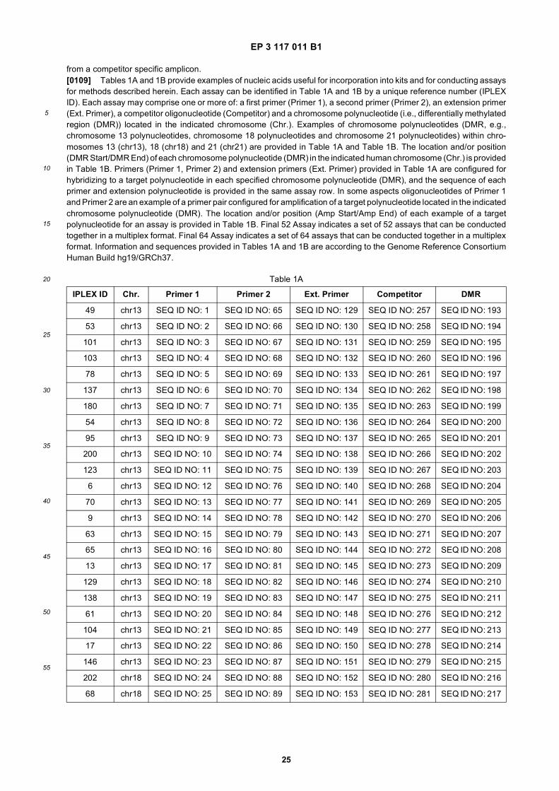
EP 3 117 011 B1
25
5
10
15
20
25
30
35
40
45
50
55
from a competitor specific amplicon.[0109] Tables 1A and 1B provide examples of nucleic acids useful for incorporation into kits and for conducting assaysfor methods described herein. Each assay can be identified in Table 1A and 1B by a unique reference number (IPLEXID). Each assay may comprise one or more of: a first primer (Primer 1), a second primer (Primer 2), an extension primer(Ext. Primer), a competitor oligonucleotide (Competitor) and a chromosome polynucleotide (i.e., differentially methylatedregion (DMR)) located in the indicated chromosome (Chr.). Examples of chromosome polynucleotides (DMR, e.g.,chromosome 13 polynucleotides, chromosome 18 polynucleotides and chromosome 21 polynucleotides) within chro-mosomes 13 (chr13), 18 (chr18) and 21 (chr21) are provided in Table 1A and Table 1B. The location and/or position(DMR Start/DMR End) of each chromosome polynucleotide (DMR) in the indicated human chromosome (Chr.) is providedin Table 1B. Primers (Primer 1, Primer 2) and extension primers (Ext. Primer) provided in Table 1A are configured forhybridizing to a target polynucleotide in each specified chromosome polynucleotide (DMR), and the sequence of eachprimer and extension polynucleotide is provided in the same assay row. In some aspects oligonucleotides of Primer 1and Primer 2 are an example of a primer pair configured for amplification of a target polynucleotide located in the indicatedchromosome polynucleotide (DMR). The location and/or position (Amp Start/Amp End) of each example of a targetpolynucleotide for an assay is provided in Table 1B. Final 52 Assay indicates a set of 52 assays that can be conductedtogether in a multiplex format. Final 64 Assay indicates a set of 64 assays that can be conducted together in a multiplexformat. Information and sequences provided in Tables 1A and 1B are according to the Genome Reference ConsortiumHuman Build hg19/GRCh37.
Table 1A
IPLEX ID Chr. Primer 1 Primer 2 Ext. Primer Competitor DMR
49 chr13 SEQ ID NO: 1 SEQ ID NO: 65 SEQ ID NO: 129 SEQ ID NO: 257 SEQ ID NO: 193
53 chr13 SEQ ID NO: 2 SEQ ID NO: 66 SEQ ID NO: 130 SEQ ID NO: 258 SEQ ID NO: 194
101 chr13 SEQ ID NO: 3 SEQ ID NO: 67 SEQ ID NO: 131 SEQ ID NO: 259 SEQ ID NO: 195
103 chr13 SEQ ID NO: 4 SEQ ID NO: 68 SEQ ID NO: 132 SEQ ID NO: 260 SEQ ID NO: 196
78 chr13 SEQ ID NO: 5 SEQ ID NO: 69 SEQ ID NO: 133 SEQ ID NO: 261 SEQ ID NO: 197
137 chr13 SEQ ID NO: 6 SEQ ID NO: 70 SEQ ID NO: 134 SEQ ID NO: 262 SEQ ID NO: 198
180 chr13 SEQ ID NO: 7 SEQ ID NO: 71 SEQ ID NO: 135 SEQ ID NO: 263 SEQ ID NO: 199
54 chr13 SEQ ID NO: 8 SEQ ID NO: 72 SEQ ID NO: 136 SEQ ID NO: 264 SEQ ID NO: 200
95 chr13 SEQ ID NO: 9 SEQ ID NO: 73 SEQ ID NO: 137 SEQ ID NO: 265 SEQ ID NO: 201
200 chr13 SEQ ID NO: 10 SEQ ID NO: 74 SEQ ID NO: 138 SEQ ID NO: 266 SEQ ID NO: 202
123 chr13 SEQ ID NO: 11 SEQ ID NO: 75 SEQ ID NO: 139 SEQ ID NO: 267 SEQ ID NO: 203
6 chr13 SEQ ID NO: 12 SEQ ID NO: 76 SEQ ID NO: 140 SEQ ID NO: 268 SEQ ID NO: 204
70 chr13 SEQ ID NO: 13 SEQ ID NO: 77 SEQ ID NO: 141 SEQ ID NO: 269 SEQ ID NO: 205
9 chr13 SEQ ID NO: 14 SEQ ID NO: 78 SEQ ID NO: 142 SEQ ID NO: 270 SEQ ID NO: 206
63 chr13 SEQ ID NO: 15 SEQ ID NO: 79 SEQ ID NO: 143 SEQ ID NO: 271 SEQ ID NO: 207
65 chr13 SEQ ID NO: 16 SEQ ID NO: 80 SEQ ID NO: 144 SEQ ID NO: 272 SEQ ID NO: 208
13 chr13 SEQ ID NO: 17 SEQ ID NO: 81 SEQ ID NO: 145 SEQ ID NO: 273 SEQ ID NO: 209
129 chr13 SEQ ID NO: 18 SEQ ID NO: 82 SEQ ID NO: 146 SEQ ID NO: 274 SEQ ID NO: 210
138 chr13 SEQ ID NO: 19 SEQ ID NO: 83 SEQ ID NO: 147 SEQ ID NO: 275 SEQ ID NO: 211
61 chr13 SEQ ID NO: 20 SEQ ID NO: 84 SEQ ID NO: 148 SEQ ID NO: 276 SEQ ID NO: 212
104 chr13 SEQ ID NO: 21 SEQ ID NO: 85 SEQ ID NO: 149 SEQ ID NO: 277 SEQ ID NO: 213
17 chr13 SEQ ID NO: 22 SEQ ID NO: 86 SEQ ID NO: 150 SEQ ID NO: 278 SEQ ID NO: 214
146 chr13 SEQ ID NO: 23 SEQ ID NO: 87 SEQ ID NO: 151 SEQ ID NO: 279 SEQ ID NO: 215
202 chr18 SEQ ID NO: 24 SEQ ID NO: 88 SEQ ID NO: 152 SEQ ID NO: 280 SEQ ID NO: 216
68 chr18 SEQ ID NO: 25 SEQ ID NO: 89 SEQ ID NO: 153 SEQ ID NO: 281 SEQ ID NO: 217
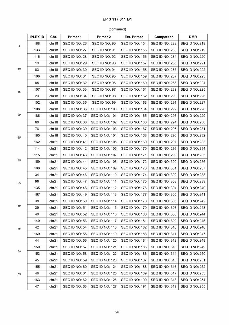
EP 3 117 011 B1
26
5
10
15
20
25
30
35
40
45
50
55
(continued)
IPLEX ID Chr. Primer 1 Primer 2 Ext. Primer Competitor DMR
188 chr18 SEQ ID NO: 26 SEQ ID NO: 90 SEQ ID NO: 154 SEQ ID NO: 282 SEQ ID NO: 218
133 chr18 SEQ ID NO: 27 SEQ ID NO: 91 SEQ ID NO: 155 SEQ ID NO: 283 SEQ ID NO: 219
116 chr18 SEQ ID NO: 28 SEQ ID NO: 92 SEQ ID NO: 156 SEQ ID NO: 284 SEQ ID NO: 220
19 chr18 SEQ ID NO: 29 SEQ ID NO: 93 SEQ ID NO: 157 SEQ ID NO: 285 SEQ ID NO: 221
83 chr18 SEQ ID NO: 30 SEQ ID NO: 94 SEQ ID NO: 158 SEQ ID NO: 286 SEQ ID NO: 222
106 chr18 SEQ ID NO: 31 SEQ ID NO: 95 SEQ ID NO: 159 SEQ ID NO: 287 SEQ ID NO: 223
85 chr18 SEQ ID NO: 32 SEQ ID NO: 96 SEQ ID NO: 160 SEQ ID NO: 288 SEQ ID NO: 224
107 chr18 SEQ ID NO: 33 SEQ ID NO: 97 SEQ ID NO: 161 SEQ ID NO: 289 SEQ ID NO: 225
23 chr18 SEQ ID NO: 34 SEQ ID NO: 98 SEQ ID NO: 162 SEQ ID NO: 290 SEQ ID NO: 226
102 chr18 SEQ ID NO: 35 SEQ ID NO: 99 SEQ ID NO: 163 SEQ ID NO: 291 SEQ ID NO: 227
108 chr18 SEQ ID NO: 36 SEQ ID NO: 100 SEQ ID NO: 164 SEQ ID NO: 292 SEQ ID NO: 228
186 chr18 SEQ ID NO: 37 SEQ ID NO: 101 SEQ ID NO: 165 SEQ ID NO: 293 SEQ ID NO: 229
60 chr18 SEQ ID NO: 38 SEQ ID NO: 102 SEQ ID NO: 166 SEQ ID NO: 294 SEQ ID NO: 230
76 chr18 SEQ ID NO: 39 SEQ ID NO: 103 SEQ ID NO: 167 SEQ ID NO: 295 SEQ ID NO: 231
185 chr18 SEQ ID NO: 40 SEQ ID NO: 104 SEQ ID NO: 168 SEQ ID NO: 296 SEQ ID NO: 232
162 chr21 SEQ ID NO: 41 SEQ ID NO: 105 SEQ ID NO: 169 SEQ ID NO: 297 SEQ ID NO: 233
114 chr21 SEQ ID NO: 42 SEQ ID NO: 106 SEQ ID NO: 170 SEQ ID NO: 298 SEQ ID NO: 234
115 chr21 SEQ ID NO: 43 SEQ ID NO: 107 SEQ ID NO: 171 SEQ ID NO: 299 SEQ ID NO: 235
159 chr21 SEQ ID NO: 44 SEQ ID NO: 108 SEQ ID NO: 172 SEQ ID NO: 300 SEQ ID NO: 236
160 chr21 SEQ ID NO: 45 SEQ ID NO: 109 SEQ ID NO: 173 SEQ ID NO: 301 SEQ ID NO: 237
34 chr21 SEQ ID NO: 46 SEQ ID NO: 110 SEQ ID NO: 174 SEQ ID NO: 302 SEQ ID NO: 238
96 chr21 SEQ ID NO: 47 SEQ ID NO: 111 SEQ ID NO: 175 SEQ ID NO: 303 SEQ ID NO: 239
135 chr21 SEQ ID NO: 48 SEQ ID NO: 112 SEQ ID NO: 176 SEQ ID NO: 304 SEQ ID NO: 240
167 chr21 SEQ ID NO: 49 SEQ ID NO: 113 SEQ ID NO: 177 SEQ ID NO: 305 SEQ ID NO: 241
38 chr21 SEQ ID NO: 50 SEQ ID NO: 114 SEQ ID NO: 178 SEQ ID NO: 306 SEQ ID NO: 242
39 chr21 SEQ ID NO: 51 SEQ ID NO: 115 SEQ ID NO: 179 SEQ ID NO: 307 SEQ ID NO: 243
40 chr21 SEQ ID NO: 52 SEQ ID NO: 116 SEQ ID NO: 180 SEQ ID NO: 308 SEQ ID NO: 244
140 chr21 SEQ ID NO: 53 SEQ ID NO: 117 SEQ ID NO: 181 SEQ ID NO: 309 SEQ ID NO: 245
42 chr21 SEQ ID NO: 54 SEQ ID NO: 118 SEQ ID NO: 182 SEQ ID NO: 310 SEQ ID NO: 246
169 chr21 SEQ ID NO: 55 SEQ ID NO: 119 SEQ ID NO: 183 SEQ ID NO: 311 SEQ ID NO: 247
44 chr21 SEQ ID NO: 56 SEQ ID NO: 120 SEQ ID NO: 184 SEQ ID NO: 312 SEQ ID NO: 248
150 chr21 SEQ ID NO: 57 SEQ ID NO: 121 SEQ ID NO: 185 SEQ ID NO: 313 SEQ ID NO: 249
153 chr21 SEQ ID NO: 58 SEQ ID NO: 122 SEQ ID NO: 186 SEQ ID NO: 314 SEQ ID NO: 250
45 chr21 SEQ ID NO: 59 SEQ ID NO: 123 SEQ ID NO: 187 SEQ ID NO: 315 SEQ ID NO: 251
155 chr21 SEQ ID NO: 60 SEQ ID NO: 124 SEQ ID NO: 188 SEQ ID NO: 316 SEQ ID NO: 252
46 chr21 SEQ ID NO: 61 SEQ ID NO: 125 SEQ ID NO: 189 SEQ ID NO: 317 SEQ ID NO: 253
163 chr21 SEQ ID NO: 62 SEQ ID NO: 126 SEQ ID NO: 190 SEQ ID NO: 318 SEQ ID NO: 254
47 chr21 SEQ ID NO: 63 SEQ ID NO: 127 SEQ ID NO: 191 SEQ ID NO: 319 SEQ ID NO: 255
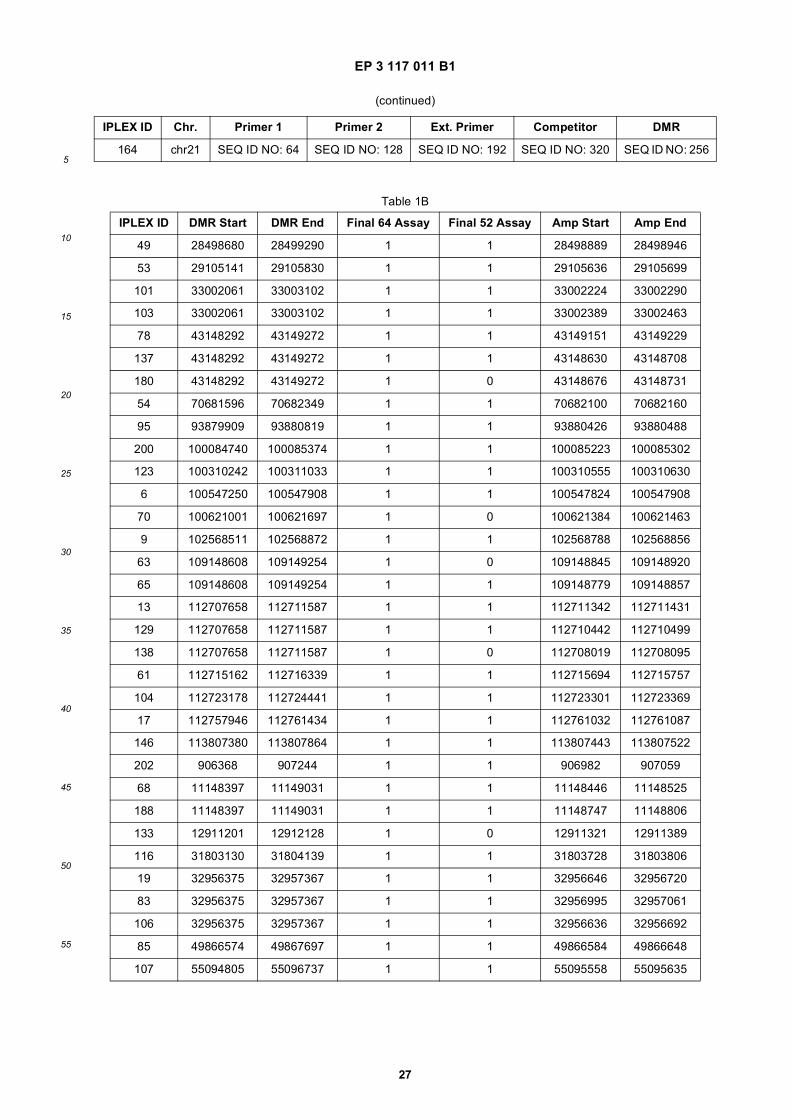
EP 3 117 011 B1
27
5
10
15
20
25
30
35
40
45
50
55
(continued)
IPLEX ID Chr. Primer 1 Primer 2 Ext. Primer Competitor DMR
164 chr21 SEQ ID NO: 64 SEQ ID NO: 128 SEQ ID NO: 192 SEQ ID NO: 320 SEQ ID NO: 256
Table 1B
IPLEX ID DMR Start DMR End Final 64 Assay Final 52 Assay Amp Start Amp End
49 28498680 28499290 1 1 28498889 28498946
53 29105141 29105830 1 1 29105636 29105699
101 33002061 33003102 1 1 33002224 33002290
103 33002061 33003102 1 1 33002389 33002463
78 43148292 43149272 1 1 43149151 43149229
137 43148292 43149272 1 1 43148630 43148708
180 43148292 43149272 1 0 43148676 43148731
54 70681596 70682349 1 1 70682100 70682160
95 93879909 93880819 1 1 93880426 93880488
200 100084740 100085374 1 1 100085223 100085302
123 100310242 100311033 1 1 100310555 100310630
6 100547250 100547908 1 1 100547824 100547908
70 100621001 100621697 1 0 100621384 100621463
9 102568511 102568872 1 1 102568788 102568856
63 109148608 109149254 1 0 109148845 109148920
65 109148608 109149254 1 1 109148779 109148857
13 112707658 112711587 1 1 112711342 112711431
129 112707658 112711587 1 1 112710442 112710499
138 112707658 112711587 1 0 112708019 112708095
61 112715162 112716339 1 1 112715694 112715757
104 112723178 112724441 1 1 112723301 112723369
17 112757946 112761434 1 1 112761032 112761087
146 113807380 113807864 1 1 113807443 113807522
202 906368 907244 1 1 906982 907059
68 11148397 11149031 1 1 11148446 11148525
188 11148397 11149031 1 1 11148747 11148806
133 12911201 12912128 1 0 12911321 12911389
116 31803130 31804139 1 1 31803728 31803806
19 32956375 32957367 1 1 32956646 32956720
83 32956375 32957367 1 1 32956995 32957061
106 32956375 32957367 1 1 32956636 32956692
85 49866574 49867697 1 1 49866584 49866648
107 55094805 55096737 1 1 55095558 55095635
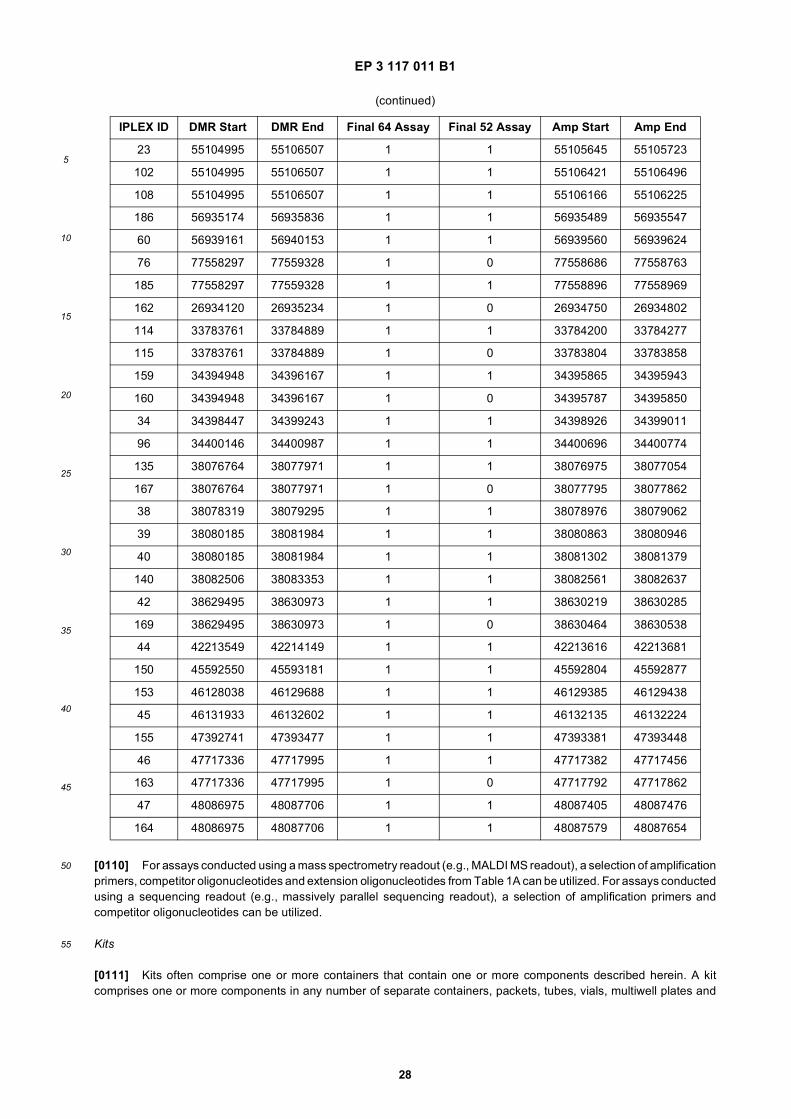
EP 3 117 011 B1
28
5
10
15
20
25
30
35
40
45
50
55
[0110] For assays conducted using a mass spectrometry readout (e.g., MALDI MS readout), a selection of amplificationprimers, competitor oligonucleotides and extension oligonucleotides from Table 1A can be utilized. For assays conductedusing a sequencing readout (e.g., massively parallel sequencing readout), a selection of amplification primers andcompetitor oligonucleotides can be utilized.
Kits
[0111] Kits often comprise one or more containers that contain one or more components described herein. A kitcomprises one or more components in any number of separate containers, packets, tubes, vials, multiwell plates and
(continued)
IPLEX ID DMR Start DMR End Final 64 Assay Final 52 Assay Amp Start Amp End
23 55104995 55106507 1 1 55105645 55105723
102 55104995 55106507 1 1 55106421 55106496
108 55104995 55106507 1 1 55106166 55106225
186 56935174 56935836 1 1 56935489 56935547
60 56939161 56940153 1 1 56939560 56939624
76 77558297 77559328 1 0 77558686 77558763
185 77558297 77559328 1 1 77558896 77558969
162 26934120 26935234 1 0 26934750 26934802
114 33783761 33784889 1 1 33784200 33784277
115 33783761 33784889 1 0 33783804 33783858
159 34394948 34396167 1 1 34395865 34395943
160 34394948 34396167 1 0 34395787 34395850
34 34398447 34399243 1 1 34398926 34399011
96 34400146 34400987 1 1 34400696 34400774
135 38076764 38077971 1 1 38076975 38077054
167 38076764 38077971 1 0 38077795 38077862
38 38078319 38079295 1 1 38078976 38079062
39 38080185 38081984 1 1 38080863 38080946
40 38080185 38081984 1 1 38081302 38081379
140 38082506 38083353 1 1 38082561 38082637
42 38629495 38630973 1 1 38630219 38630285
169 38629495 38630973 1 0 38630464 38630538
44 42213549 42214149 1 1 42213616 42213681
150 45592550 45593181 1 1 45592804 45592877
153 46128038 46129688 1 1 46129385 46129438
45 46131933 46132602 1 1 46132135 46132224
155 47392741 47393477 1 1 47393381 47393448
46 47717336 47717995 1 1 47717382 47717456
163 47717336 47717995 1 0 47717792 47717862
47 48086975 48087706 1 1 48087405 48087476
164 48086975 48087706 1 1 48087579 48087654
EP 3 117 011 B1
29
5
10
15
20
25
30
35
40
45
50
55
the like, or components may be combined in various combinations in such containers. One or more of the followingcomponents, for example, may be included in a kit: (i) one or more amplification primers for amplifying a nucleotidesequence species of a set, (ii) one or more extension primers for discriminating between amplified nucleic acid speciesor nucleotide sequence species of each set, (iii) a solid support for multiplex detection of amplified nucleic acid speciesor nucleotide sequence species of each set (e.g., a solid support that includes matrix for matrix-assisted laser desorptionionization (MALDI) mass spectrometry; (iv) reagents for detecting amplified nucleic acid species or nucleotide sequencespecies of each set; (vi) a detector for detecting the amplified nucleic acid species or nucleotide sequence species ofeach set (e.g., mass spectrometer); (vii) reagents and/or equipment for quantifying fetal nucleic acid in extracellularnucleic acid from a pregnant female; (viii) reagents and/or equipment for enriching fetal nucleic acid from extracellularnucleic acid from a pregnant female; (ix) software and/or a machine for analyzing signals resulting from a process fordetecting the amplified nucleic acid species or nucleotide sequence species of the sets; (x) information for identifyingpresence or absence of a chromosome abnormality (e.g., a table or file that converts signal information or amounts intooutcomes), (xi) equipment for drawing blood); (xii) equipment for generating cell-free blood; (xiii) reagents for isolatingnucleic acid (e.g., DNA, RNA) from plasma, serum or urine; (xiv) reagents for stabilizing serum, plasma, urine or nucleicacid for shipment and/or processing.[0112] A kit sometimes is utilized in conjunction with a process, and can include instructions for performing one ormore processes and/or a description of one or more compositions. A kit may be utilized to carry out a process (e.g.,using a solid support) described herein. Instructions and/or descriptions may be in tangible form (e.g., paper and thelike) or electronic form (e.g., computer readable file on a tangle medium (e.g., compact disc) and the like) and may beincluded in a kit insert. A kit also may include a written description of an internet location that provides such instructionsor descriptions (e.g., a URL for the World-Wide Web).[0113] In some aspects provided herein are one or more kits configured to conduct one or more assays describedherein. In some aspects an assay is configured for generating target specific and competitor specific amplicons from atarget polynucleotide in a polynucleotide in a chromosome. In certain aspects an assay is configured for generatingextension products. One or more assays (e.g., a collection of assays) are often configured to detect and/or quantify one,two, three or four copies of a fetal chromosome, or portion thereof, in a test sample. Sometimes at least 100, at least90, at least 80, at least 70, at least 65, at least 60, at least 55, at least 50, at least 40, at least 30 or at least 20 assaysare configured to detect and/or quantify one, two, three or four copies of a fetal chromosome, or portion thereof, in atest sample. In some aspects 70, 69, 68, 67, 66, 65, 64, 63, 62, 61, 59, 58, 57, 56, 55, 54, 53, 52, 51 or 50 assays areconfigured to detect and/or quantify one, two, three or four copies of a fetal chromosome, or portion thereof, in a testsample.[0114] Provided herein, in some e aspects is a kit, comprising components needed to conduct one or more methodsor assays described herein. A kit can comprise one or more primers configured for amplifying target polynucleotides. Insome aspects a kit comprises a collection of oligonucleotide primer pairs where each primer pair is configured foramplifying three or more target polynucleotides in each of chromosome 13, chromosome 18 and chromosome 21. Incertain aspects a kit comprises competitor oligonucleotides. In certain aspects a kit comprises extension primers. Insome aspects a kit comprises competitor oligonucleotides and a collection of oligonucleotide primer pairs configured foramplifying three or more target polynucleotides in each of chromosome 13, chromosome 18 and chromosome 21 andtheir corresponding competitor oligonucleotides, and extension primers configured for generating extension productsfrom target specific and competitor specific amplicons. Kits provided herein can optionally comprise one or more of thefollowing: buffers (e.g., comprising salts and components required for polymerase activity), restriction enzymes, dNTPs,polymerases, instructions, tubes, trays, the like or combinations thereof. In some aspects a kit comprises nucleic acidcontrols (e.g., ccf DNA obtained from a euploid test subject). In some aspects a kit comprises components suitable forconducting 10 or more, 20 or more, 30 or more, 40 or more, 50 or more or 60 or more assays. In some aspects a kitcomprises components suitable for conducting 1-60, 1-50, 1-40, 1-30, 1-25, 1-20, 1-15, 1-10 or 1-5 assay. In someaspects a kit comprises components suitable for conducting about 70, 69, 68, 67, 66, 65, 64, 63, 62, 61, 59, 58, 57, 56,55, 54, 53, 52, 51 or 50 assays. Amplification primers, extension oligonucleotides and competitor oligonucleotidessuitable for performing assays for 1-64, 1-60, 1-52, 1-50, 1-40, 1-30, 1-25, 1-20, 1-15, 1-10 or 1-5 assays are providedin Table 1A.
Determining Fetal Nucleic Acid Content
[0115] In some aspects an analysis (e.g., an analysis of nucleic acids) comprises determining an amount of fetalnucleic acid in a nucleic acid sample. An amount of fetal nucleic acid (e.g., concentration, relative amount, ratio, absoluteamount, copy number, and the like) in nucleic acid (e.g., a nucleic acid sample or mixture) is determined in some aspects.In some aspects, the amount of fetal nucleic acid in a sample is referred to as "fetal fraction". In some aspects, "fetalfraction" refers to the fraction of fetal nucleic acid in circulating cell-free nucleic acid in a sample (e.g., a blood sample,a serum sample, a plasma sample) obtained from a pregnant female. In some aspects determining an amount of fetal
EP 3 117 011 B1
30
5
10
15
20
25
30
35
40
45
50
55
nucleic acid comprises determining a ratio (e.g., percentage, a percent representation) of fetal nucleic acid to a totalamount of nucleic acid in a sample. In some aspects determining an amount of fetal nucleic acid comprises determininga ratio (e.g., percentage) of the amount of fetal nucleic acid to the amount of maternal nucleic acid in a sample. In someaspects, a method in which a genetic variation is determined also can comprise determining fetal fraction. Determiningfetal fraction can be performed in a suitable manner, non-limiting examples of which include methods described below.[0116] In some aspects, the amount of fetal nucleic acid is determined according to markers specific to a male fetus(e.g., Y-chromosome STR markers (e.g., DYS 19, DYS 385, DYS 392 markers); RhD marker in RhD-negative females),allelic ratios of polymorphic sequences, or according to one or more markers specific to fetal nucleic acid and not maternalnucleic acid (e.g., differential epigenetic biomarkers (e.g., methylation; described in further detail below) between motherand fetus, or fetal RNA markers in maternal blood plasma (see e.g., Lo, 2005, Journal of Histochemistry and Cytochemistry53 (3): 293-296)).[0117] Determination of fetal nucleic acid content (e.g., fetal fraction) sometimes is performed using a fetal quantifierassay (FQA) as described, for example, in U.S. Patent Application Publication No. 2010/0105049. This type of assayallows for the detection and quantification of fetal nucleic acid in a maternal sample based on the methylation status ofthe nucleic acid in the sample. The amount of fetal nucleic acid from a maternal sample sometimes can be determinedrelative to the total amount of nucleic acid present, thereby providing the percentage of fetal nucleic acid in the sample.The copy number of fetal nucleic acid sometimes can be determined in a maternal sample. The amount of fetal nucleicacid sometimes can be determined in a sequence-specific (or locus-specific) manner and sometimes with sufficientsensitivity to allow for accurate chromosomal dosage analysis (for example, to detect the presence or absence of a fetalaneuploidy or other genetic variation).[0118] A fetal quantifier assay (FQA) can be performed in conjunction with any method described herein. Such anassay can be performed by any method known in the art and/or described in U.S. Patent Application Publication No.2010/0105049, such as, for example, by a method that can distinguish between maternal and fetal DNA based ondifferential methylation status, and quantify (i.e. determine the amount of) the fetal DNA. Methods for differentiatingnucleic acid based on methylation status include, but are not limited to, methylation sensitive capture, for example, usinga MBD2-Fc fragment in which the methyl binding domain of MBD2 is fused to the Fc fragment of an antibody (MBD-FC)(Gebhard et al. (2006) Cancer Res. 66(12):6118-28); methylation specific antibodies; bisulfite conversion methods, forexample, MSP (methylation-sensitive PCR), COBRA, methylation-sensitive single nucleotide primer extension (Ms-SNuPE) or Sequenom MassCLEAVE™ technology; and the use of methylation sensitive restriction enzymes (e.g.,digestion of maternal DNA in a maternal sample using one or more methylation sensitive restriction enzymes therebyenriching for fetal DNA). Methyl-sensitive enzymes also can be used to differentiate nucleic acid based on methylationstatus, which, for example, can preferentially or substantially cleave or digest at their DNA recognition sequence if thelatter is non-methylated. Thus, an unmethylated DNA sample will be cut into smaller fragments than a methylated DNAsample and a hypermethylated DNA sample will not be cleaved. Except where explicitly stated, any method for differ-entiating nucleic acid based on methylation status can be used with the compositions and methods of the technologyherein. The amount of fetal DNA can be determined, for example, by introducing one or more competitors at knownconcentrations during an amplification reaction. Determining the amount of fetal DNA also can be done, for example,by RT-PCR, primer extension, sequencing and/or counting. In certain instances, the amount of nucleic acid can bedetermined using BEAMing technology as described in U.S. Patent Application Publication No. 2007/0065823. In someaspects, the restriction efficiency can be determined and the efficiency rate is used to further determine the amount offetal DNA.[0119] A fetal quantifier assay (FQA) sometimes can be used to determine the concentration of fetal DNA in a maternalsample, for example, by the following method: a) determine the total amount of DNA present in a maternal sample; b)selectively digest the maternal DNA in a maternal sample using one or more methylation sensitive restriction enzymesthereby enriching the fetal DNA; c) determine the amount of fetal DNA from step b); and d) compare the amount of fetalDNA from step c) to the total amount of DNA from step a), thereby determining the concentration of fetal DNA in thematernal sample. The absolute copy number of fetal nucleic acid in a maternal sample sometimes can be determined,for example, using mass spectrometry and/or a system that uses a competitive PCR approach for absolute copy numbermeasurements. See for example, Ding and Cantor (2003) Proc.Natl.Acad.Sci. USA 100:3059-3064, and U.S. PatentApplication Publication No. 2004/0081993.[0120] Fetal fraction sometimes can be determined based on allelic ratios of polymorphic sequences (e.g., singlenucleotide polymorphisms (SNPs)), such as, for example, using a method described in U.S. Patent Application PublicationNo. 2011/0224087. In such a method, nucleotide sequence reads are obtained for a maternal sample and fetal fractionis determined by comparing the total number of nucleotide sequence reads that map to a first allele and the total numberof nucleotide sequence reads that map to a second allele at an informative polymorphic site (e.g., SNP) in a referencegenome. Fetal alleles can be identified, for example, by their relative minor contribution to the mixture of fetal andmaternal nucleic acids in the sample when compared to the major contribution to the mixture by the maternal nucleicacids. Accordingly, the relative abundance of fetal nucleic acid in a maternal sample can be determined as a parameter
EP 3 117 011 B1
31
5
10
15
20
25
30
35
40
45
50
55
of the total number of unique sequence reads mapped to a target polynucleotide sequence on a reference genome foreach of the two alleles of a polymorphic site.[0121] The amount of fetal nucleic acid in extracellular nucleic acid can be quantified and used in conjunction with amethod provided herein. Thus, in certain aspects, methods of the technology described herein comprise an additionalstep of determining the amount of fetal nucleic acid. The amount of fetal nucleic acid can be determined in a nucleicacid sample from a subject before or after processing to prepare sample nucleic acid. In certain aspects, the amount offetal nucleic acid is determined in a sample after sample nucleic acid is processed and prepared, which amount is utilizedfor further assessment. In some aspects, an outcome comprises factoring the fraction of fetal nucleic acid in the samplenucleic acid (e.g., adjusting counts, removing samples, making a call or not making a call).[0122] The determination step can be performed before, during, at any one point in a method described herein, orafter certain (e.g., aneuploidy detection) methods described herein. For example, to achieve an aneuploidy determinationmethod with a given sensitivity or specificity, a fetal nucleic acid quantification method may be implemented prior to,during or after aneuploidy determination to identify those samples with greater than about 2%, 3%, 4%, 5%, 6%, 7%,8%, 9%, 10%, 11%, 12%, 13%, 14%,15%,16%, 17%, 18%, 19%, 20%, 21%, 22%, 23%, 24%, 25% or more fetal nucleicacid. In some aspects, samples determined as having a certain threshold amount of fetal nucleic acid (e.g., about 15%or more fetal nucleic acid; about 4% or more fetal nucleic acid) are further analyzed for the presence or absence ofaneuploidy or genetic variation, for example. In certain aspects, determinations of, for example, the presence or absenceof aneuploidy are selected (e.g., selected and communicated to a patient) only for samples having a certain thresholdamount of fetal nucleic acid (e.g., about 15% or more fetal nucleic acid; about 4% or more fetal nucleic acid).[0123] In some aspects, the determination of fetal fraction or determining the amount of fetal nucleic acid is not requiredor necessary for identifying the presence or absence of a chromosome aneuploidy. In some aspects, identifying thepresence or absence of a chromosome aneuploidy does not require the sequence differentiation of fetal versus maternalDNA. This is because the summed contribution of both maternal and fetal sequences in a particular chromosome,chromosome portion or segment thereof is analyzed, in some aspects. In some aspects, identifying the presence orabsence of a chromosome aneuploidy does not rely on a priori sequence information that would distinguish fetal DNAfrom maternal DNA.
Nucleic acid amplification
[0124] In many instances, it is desirable to amplify a target polynucleotide sequence or a subset of a polynucleotidesequence herein using any of several nucleic acid amplification procedures which are well known in the art, some ofwhich are listed or described herein. Specifically, nucleic acid amplification is the enzymatic synthesis of nucleic acidamplicons (copies) which contain a sequence that is complementary to a nucleic acid sequence being amplified. In someaspects amplification comprises ligating one or more adaptors to a nucleic acid target or target subset of nucleic acids(e.g., digested nucleic acid, enriched nucleic acid, separated nucleic acid). Nucleic acid amplification is especially ben-eficial when the amount of target sequence present in a sample is very low. By amplifying the target sequences anddetecting the amplicon synthesized, the sensitivity of an assay can be vastly improved, since fewer target sequencesare needed at the beginning of the assay to better ensure detection of nucleic acid in the sample belonging to theorganism or virus of interest. One or more nucleic acids can be amplified in solution or while immobilized on a solidphase. One or more nucleic acids can be amplified prior to and/or after immobilization on a solid support (e.g., a solidsupport in a flow cell). In some aspects one or more nucleic acids can be amplified after release from a solid phase.[0125] A variety of polynucleotide amplification methods are well established and frequently used in research. Forinstance, the general methods of polymerase chain reaction (PCR) for polynucleotide sequence amplification are wellknown in the art and are thus not described in detail herein. For a review of PCR methods, protocols, and principles indesigning primers, see, e.g., Innis, et al., PCR Protocols: A Guide to Methods and Applications, Academic Press, Inc.N.Y., 1990. PCR reagents and protocols are also available from commercial vendors, such as Roche Molecular Systems.[0126] PCR is most usually carried out as an automated process with a thermostable enzyme. In this process, thetemperature of the reaction mixture is cycled through a denaturing region, a primer annealing region, and an extensionreaction region automatically. Machines specifically adapted for this purpose are commercially available.[0127] Although PCR amplification of a polynucleotide sequence (e.g., a target polynucleotide) is typically used inpracticing the present technology, one of skill in the art will recognize that the amplification of a genomic sequence foundin a maternal blood sample may be accomplished by a known method, such as ligase chain reaction (LCR), transcription-mediated amplification, and self-sustained sequence replication or nucleic acid sequence-based amplification (NASBA),each of which provides sufficient amplification. More recently developed branched-DNA technology may also be usedto qualitatively demonstrate the presence of a particular genomic sequence of the technology herein, which representsa particular methylation pattern, or to quantitatively determine the amount of this particular genomic sequence in thematernal blood. For a review of branched-DNA signal amplification for direct quantitation of nucleic acid sequences inclinical samples, see Nolte, Adv. Clin. Chem. 33:201-235, 1998.
EP 3 117 011 B1
32
5
10
15
20
25
30
35
40
45
50
55
[0128] The compositions and processes of the technology herein are also particularly useful when practiced with digitalPCR. Digital PCR was first developed by Kalinina and colleagues (Kalinina et al., "Nanoliter scale PCR with TaqMandetection." Nucleic Acids Research. 25; 1999-2004, (1997)) and further developed by Vogelstein and Kinzler (DigitalPCR. Proc Natl Acad Sci U S A. 96; 9236-41, (1999)). The application of digital PCR for use with fetal diagnostics wasfirst described by Cantor et al. (PCT Patent Publication No. WO05023091A2) and subsequently described by Quake etal. (US Patent Publication No. US 20070202525). Digital PCR takes advantage of nucleic acid (DNA, cDNA or RNA)amplification on a single molecule level, and offers a highly sensitive method for quantifying low copy number nucleicacid. Fluidigm® Corporation offers systems for the digital analysis of nucleic acids.[0129] The term "amplified" as used herein refers to subjecting a target polynucleotide in a sample to a process thatlinearly or exponentially generates amplicon nucleic acids having the same or substantially the same nucleotide sequenceas a target polynucleotide, or segment thereof. A target polynucleotide is sometimes represented in a sample as apolynucleotide fragment. In some aspects a nucleotide sequence, or portion thereof, of a target polynucleotide is known.The term "amplified" as used herein can refer to subjecting a target polynucleotide (e.g., in a sample comprising othernucleic acids) to a process that selectively and linearly or exponentially generates amplicon nucleic acids having thesame or substantially the same nucleotide sequence as the target polynucleotide, or segment thereof. Amplicons thatare generated from, and have the same or substantially the same nucleotide sequence as a target polynucleotide, arereferred to herein as target specific amplicons. The term "amplified" as used herein can refer to subjecting a populationof nucleic acids to a process that non-selectively and linearly or exponentially generates amplicon nucleic acids havingthe same or substantially the same nucleotide sequence as nucleic acids, or portions thereof, that were present in thesample prior to amplification. In some aspects, the term "amplified" refers to a method that comprises a polymerasechain reaction (PCR). The terms "amplify", "amplification", "selective amplification", "amplification reaction", or "ampli-fying" refer to any in vitro process for multiplying the copies of a nucleic acid.[0130] Any suitable amplification technique can be utilized. Amplification of polynucleotides include, but are not limitedto, polymerase chain reaction (PCR); ligation amplification (or ligase chain reaction (LCR)); amplification methods basedon the use of Q-beta replicase or template-dependent polymerase (see US Patent Publication Number US20050287592);helicase-dependent isothermal amplification (Vincent et al., "Helicase-dependent isothermal DNA amplification". EMBOreports 5 (8): 795-800 (2004)); strand displacement amplification (SDA); thermophilic SDA nucleic acid sequence basedamplification (3SR or NASBA) and transcription-associated amplification (TAA). Non-limiting examples of PCR amplifi-cation methods include standard PCR, AFLP-PCR, Allele-specific PCR, Alu-PCR, Asymmetric PCR, Colony PCR, Hotstart PCR, Inverse PCR (IPCR), In situ PCR (ISH), Intersequence-specific PCR (ISSR-PCR), Long PCR, Multiplex PCR,Nested PCR, Quantitative PCR, Reverse Transcriptase PCR (RT-PCR), Real Time PCR, Single cell PCR, Solid phasePCR, digital PCR, combinations thereof, and the like. For example, amplification can be accomplished using digital PCR,in certain aspects (see e.g. Kalinina et al., "Nanoliter scale PCR with TaqMan detection." Nucleic Acids Research. 25;1999-2004, (1997); Vogelstein and Kinzler (Digital PCR. Proc Natl Acad Sci U S A. 96; 9236-41, (1999); PCT PatentPublication No. WO05023091A2; US Patent Publication No. US 20070202525). Digital PCR takes advantage of nucleicacid (DNA, cDNA or RNA) amplification on a single molecule level, and offers a highly sensitive method for quantifyinglow copy number nucleic acid. Systems for digital amplification and analysis of nucleic acids are available (e.g., Fluidigm®Corporation). Reagents and hardware for conducting PCR are commercially available.[0131] A generalized description of a selective amplification process is presented herein. Primers (e.g., a primer pair,a collection of primer pairs) and nucleic acid (e.g., target polynucleotides) are contacted under suitable hybridizationconditions, and complementary sequences anneal to one another, for example. Primers can anneal to a nucleic acid,at or near (e.g., adjacent to, abutting, flanking, and the like) a sequence of interest (e.g., a target polynucleotide). Insome aspects, a primer pair hybridizes within about 10 to 30 nucleotides from a nucleic acid sequence of interest and,under amplification conditions can produce amplified products (e.g., amplicons). In some aspects, primers hybridizewithin a nucleic acid sequence of interest (e.g., a target polynucleotide). Any suitable amplification conditions can beused to perform an amplification resulting in the production of amplicons. In some aspects a sample comprising targetpolynucleotides is contacted with one or more target specific primer pairs (e.g., a collection of primers) under amplificationconditions where target specific amplicons are generated. Amplification conditions often comprise a reaction mixturecontaining a polymerase, at least one primer (e.g., a primer pair), at least one target polynucleotide and additionalcomponents (e.g., buffers, salts and nucleotide triphosphates) necessary for polymerase activity. Non-limiting examplesof components of an amplification reaction may include, but are not limited to, e.g., primers (e.g., individual primers,primer pairs, a collection of primer pairs and the like) a polynucleotide template, polymerase, nucleotides, dNTPs andthe like. In some aspects, non-naturally occurring nucleotides or nucleotide analogs, such as analogs containing adetectable label (e.g., fluorescent or colorimetric label), may be used for example. Polymerases can be selected by aperson of ordinary skill and include polymerases for thermocycle amplification (e.g., Taq DNA Polymerase; Q-Bio™ TaqDNA Polymerase (recombinant truncated form of Taq DNA Polymerase lacking 5’-3’exo activity); SurePrime™ Polymer-ase (chemically modified Taq DNA polymerase for "hot start" PCR); Arrow™ Taq DNA Polymerase (high sensitivity andlong template amplification)) and polymerases for thermostable amplification (e.g., RNA polymerase for transcription-
EP 3 117 011 B1
33
5
10
15
20
25
30
35
40
45
50
55
mediated amplification (TMA) described at World Wide Web URL "gen-probe.com/pdfs/tma_whiteppr.pdf"). Other en-zyme components can be added, such as reverse transcriptase for transcription mediated amplification (TMA) reactions,for example.[0132] Amplification conditions can be dependent upon primer sequences (e.g., primer hybridization sequences),abundance of nucleic acid, and the desired amount of amplification, and therefore, one of skill in the art may choosefrom a number of PCR protocols available (see, e.g., U.S. Pat. Nos. 4,683,195 and 4,683,202; and PCR Protocols: AGuide to Methods and Applications, Innis et al., eds, 1990. Digital PCR is also known in the art; see, e.g., United StatesPatent Application Publication no. 20070202525, filed February 2, 2007). Amplification conditions often comprise aplurality of suitable temperature changes (e.g., temperature cycles) and incubation times (e.g., an incubation time forannealing, melting and extension). Amplification is typically carried out as an automated process, often in a thermocyclerwith a thermostable enzyme. In this process, the temperature of the reaction mixture is cycled multiple times through adenaturing step, a primer-annealing step, and an extension reaction step automatically. Some amplification protocolsalso include an activation step and a final extension step. Machines specifically adapted for this purpose are commerciallyavailable. A non-limiting example of a amplification protocol that may be suitable for aspects described herein is, treatingthe sample at 95 °C for 5 minutes; repeating thirty-five cycles of 95 °C for 45 seconds and 68 °C for 30 seconds; andthen treating the sample at 72 °C for 3 minutes. A completed amplification reaction can optionally be kept at 4 °C untilfurther action is desired. Multiple cycles frequently are performed using a commercially available thermal cycler. Suitableisothermal amplification processes known and selected by the person of ordinary skill in the art also may be applied, incertain aspects.[0133] In some aspects, an amplification product (e.g., an amplicon) may include naturally occurring nucleotides, non-naturally occurring nucleotides, nucleotide analogs and the like and combinations of the foregoing. An amplicon oftenhas a nucleotide sequence that is identical to or substantially identical to a nucleic acid sequence herein, or complementthereof. A "substantially identical" nucleotide sequence in an amplification product will generally have a high degree ofsequence identity to the nucleotide sequence species being amplified or complement thereof (e.g., about 75%, 76%,77%, 78%, 79%, 80%, 81%, 82%, 83%, 84%, 85%, 86%, 87%, 88%, 89%, 90%, 91%, 92%, 93%, 94%, 95%, 96%,97%, 98%, 99% or greater than 99% sequence identity), and variations sometimes are a result of infidelity of thepolymerase used for extension and/or amplification, or additional nucleotide sequence(s) added to the primers used foramplification.[0134] Nucleic acids in a sample can be enriched by an amplification method described herein. An amplification product(e.g., amplicons) can be generated before, during or after any step of a method described herein. An amplificationproduct can be generated before, during or after a digestion or cleavage reaction. An amplification product can begenerated before, during or after a modification of nucleic acids in a sample. An amplification product can be generatedbefore, during or after an enrichment method. An amplification product can be generated before, during or after aseparation or purifications step. An amplification product can be generated before, during or after a process comprisingnucleic acid sequencing. In some aspects digested nucleic acids or undigested nucleic acids are enriched by an ampli-fication. In some aspects enriched and/or separated nucleic acid are further enriched by an amplification. In some aspectsenriched and/or separated methylated, hypermethylated and/or hypomethylated nucleic acid are further enriched by anamplification.
Nucleic Acid Analysis
[0135] In some aspects, nucleic acid fragments (e.g., digested nucleic acid fragments) may be amplified and/or sub-jected to an analysis and/or detection process (e.g., sequence-based analysis, mass spectrometry). In some aspects,nucleic acid fragments are (e.g., digested nucleic acid fragments) subjected to a detection process (e.g., sequencing)without amplification.[0136] Nucleic acid fragments (e.g., digested nucleic acid fragments),, polynucleotides or amplicon sequences, ordetectable products prepared from the foregoing, can be analyzed (e.g., detected, identified, quantitated, compared) bya suitable process. Non-limiting examples of methods of analysis include mass detection and analysis (e.g., matrix-assisted laser desorption ionization (MALDI) mass spectrometry and electrospray (ES) mass spectrometry), primerextension methods (e.g., iPLEX™; Sequenom, Inc.), direct DNA sequencing, Molecular Inversion Probe (MIP) technologyfrom Affymetrix, restriction fragment length polymorphism (RFLP analysis), allele specific oligonucleotide (ASO) analysis,methyl-specific PCR (MSPCR), pyrosequencing analysis, acycloprime analysis, reverse dot blot, GeneChip microarrays,Dynamic allele-specific hybridization (DASH), Peptide nucleic acid (PNA) and locked nucleic acids (LNA) probes, Taq-Man, Molecular Beacons, Intercalating dye, FRET primers, AlphaScreen, SNPstream, genetic bit analysis (GBA), Mul-tiplex minisequencing, SNaPshot, GOOD assay, Microarray miniseq, arrayed primer extension (APEX), Microarrayprimer extension, Tag arrays, Coded microspheres, Template-directed incorporation (TDI), fluorescence polarization,Colorimetric oligonucleotide ligation assay (OLA), Sequence-coded OLA, Microarray ligation, Ligase chain reaction,Padlock probes, Invader assay, hybridization using at least one probe, cloning and sequencing, electrophoresis, the
EP 3 117 011 B1
34
5
10
15
20
25
30
35
40
45
50
55
use of hybridization probes and quantitative real time polymerase chain reaction (QRT-PCR), digital PCR, nanoporesequencing, chips, the like and combinations thereof. In some aspects the amount of a polynucleotide or nucleic acidspecies is determined by mass spectrometry, primer extension, sequencing (e.g., any suitable method, for examplenanopore or pyrosequencing), Quantitative PCR (Q-PCR or QRT-PCR), digital PCR, combinations thereof, and the like.Nucleic acid detection and/or quantification also may include, for example, solid support array based detection of fluo-rescently labeled nucleic acid with fluorescent labels incorporated during or after PCR, single molecule detection offluorescently labeled molecules in solution or captured on a solid phase, or other sequencing technologies such as, forexample, sequencing using ION TORRENT or MISEQ platforms or single molecule sequencing technologies usinginstrumentation such as, for example, PACBIO sequencers, HELICOS sequencer, or nanopore sequencing technologies.[0137] An analysis can be a target-based analysis (e.g., targeted analysis) or a non-target-based analysis (e.g., non-targeted). A target-based analysis generally comprises analysis (e.g., sequencing, quantitation) of selected nucleic acidsor a selected subset of nucleic acids (e.g., a subpopulation of nucleic acids). In some aspects a selective nucleic acidsubset comprises selected genes, selected loci (e.g., hypomethylated loci, hypermethylated loci), selected alleles (e.g.,selected polymorphisms), nucleic acids derived from one or more selected chromosomes, selected fetal nucleic acids,the like or combinations thereof. In some aspects a target-bases analysis comprises a suitable target specific amplificationor sequencing method. A target-based analysis generally comprises use of one or more sequence-specific oligonucle-otides (e.g., primers or capture agents) that hybridize to specific selected nucleic acid sequences that are expectedand/or known to exist in a test sample (e.g., an unmanipulated sample isolated from a test subject). A non-target-basedanalysis generally does not comprise a sequence-specific selection process or utilizes oligonucleotides that hybridizeto specific selected nucleic acid sequences that are expected and/or known to exist in a test sample. In some aspectsa non-target-based analysis utilizes adaptors and/or adaptor specific primers to amplify and/or sequence nucleic acidsor a subset of nucleic acids in a test sample. For example, a non-target-based analysis sometimes comprises ligationof adaptors and/or hybridization of primers to sticky ends that results from restriction enzyme cleavage followed by asuitable capture, primer extension, amplification and/or sequencing method.
Sequencing
[0138] In some aspects, nucleic acids (e.g., nucleic acid fragments, sample nucleic acid, cell-free nucleic acid) aresequenced. In certain embodiments, a full or substantially full sequence is obtained and sometimes a partial sequenceis obtained. Sequencing, mapping and related analytical methods are known in the art (e.g., United States PatentApplication Publication US2009/0029377). Certain aspects of such processes are described hereafter.In some aspects some or all nucleic acids in a sample are enriched and/or amplified (e.g., non-specifically, e.g., by aPCR based method) prior to or during sequencing. In certain aspects specific nucleic acid portions or subsets in a sampleare enriched and/or amplified prior to or during sequencing. In some aspects, a portion or subset of a pre-selected poolof nucleic acids is sequenced randomly. In some aspects, nucleic acids in a sample are not enriched and/or amplifiedprior to or during sequencing.[0139] As used herein, "reads" (i.e., "a read", "a sequence read") are short nucleotide sequences produced by anysequencing process described herein or known in the art. Reads can be generated from one end of nucleic acid fragments("single-end reads"), and sometimes are generated from both ends of nucleic acids (e.g., paired-end reads, double-endreads).[0140] The length of a sequence read is often associated with the particular sequencing technology. High-throughputmethods, for example, provide sequence reads that can vary in size from tens to hundreds of base pairs (bp). Nanoporesequencing, for example, can provide sequence reads that can vary in size from tens to hundreds to thousands of basepairs. In some aspects, sequence reads are of a mean, median, average or absolute length of about 15 bp to about 900bp long. In certain aspects sequence reads are of a mean, median, average or absolute length about 1000 bp or more.[0141] In some aspects the nominal, average, mean or absolute length of single-end reads sometimes is about 15contiguous nucleotides to about 50 or more contiguous nucleotides, about 15 contiguous nucleotides to about 40 ormore contiguous nucleotides, and sometimes about 15 contiguous nucleotides or about 36 or more contiguous nucle-otides. In certain aspects the nominal, average, mean or absolute length of single-end reads is about 20 to about 30bases, or about 24 to about 28 bases in length. In certain aspects the nominal, average, mean or absolute length ofsingle-end reads is about 1, 2, 3, 4, 5, 6, 7, 8, 9, 10, 11, 12, 13,14, 15, 16, 17, 18, 19, 21, 22, 23, 24, 25, 26, 27, 28 orabout 29 bases or more in length.[0142] In certain aspects, the nominal, average, mean or absolute length of the paired-end reads sometimes is about10 contiguous nucleotides to about 25 contiguous nucleotides or more (e.g., about 10, 11, 12, 13, 14, 15, 16, 17, 18,19, 20, 21, 22, 23, 24 or 25 nucleotides in length or more), about 15 contiguous nucleotides to about 20 contiguousnucleotides or more, and sometimes is about 17 contiguous nucleotides or about 18 contiguous nucleotides.[0143] Reads generally are representations of nucleotide sequences in a physical nucleic acid. For example, in a readcontaining an ATGC depiction of a sequence, "A" represents an adenine nucleotide, "T" represents a thymine nucleotide,
EP 3 117 011 B1
35
5
10
15
20
25
30
35
40
45
50
55
"G" represents a guanine nucleotide and "C" represents a cytosine nucleotide, in a physical nucleic acid. Sequencereads obtained from the blood of a pregnant female can be reads from a mixture of fetal and maternal nucleic acid. Amixture of relatively short reads can be transformed by processes described herein into a representation of a genomicnucleic acid present in the pregnant female and/or in the fetus. A mixture of relatively short reads can be transformedinto a representation of a copy number variation (e.g., a maternal and/or fetal copy number variation), genetic variationor an aneuploidy, for example. Reads of a mixture of maternal and fetal nucleic acid can be transformed into a repre-sentation of a composite chromosome or a segment thereof comprising features of one or both maternal and fetalchromosomes. In certain aspects, "obtaining" nucleic acid sequence reads of a sample from a subject and/or "obtaining"nucleic acid sequence reads of a biological specimen from one or more reference persons can involve directly sequencingnucleic acid to obtain the sequence information. In some aspects, "obtaining" can involve receiving sequence informationobtained directly from a nucleic acid by another.[0144] In some aspects, a representative fraction of a genome is sequenced and is sometimes referred to as "coverage"or "fold coverage". For example, a 1-fold coverage indicates that roughly 100% of the nucleotide sequences of thegenome are represented by reads. In some aspects "fold coverage" is a relative term referring to a prior sequencing runas a reference. For example, a second sequencing run may have 2-fold less coverage than a first sequencing run. Insome aspects a genome is sequenced with redundancy, where a given region of the genome can be covered by two ormore reads or overlapping reads (e.g., a "fold coverage" greater than 1, e.g., a 2-fold coverage).[0145] In some aspects, one nucleic acid sample from one individual is sequenced. In certain aspects, nucleic acidsfrom each of two or more samples are sequenced, where samples are from one individual or from different individuals.In certain aspects, nucleic acid samples from two or more biological samples are pooled, where each biological sampleis from one individual or two or more individuals, and the pool is sequenced. In the latter embodiments, a nucleic acidsample from each biological sample often is identified by one or more unique identifiers.[0146] In some aspects a sequencing method utilizes identifiers that allow multiplexing of sequence reactions in asequencing process. The greater the number of unique identifiers, the greater the number of samples and/or chromo-somes for detection, for example, that can be multiplexed in a sequencing process. A sequencing process can beperformed using any suitable number of unique identifiers (e.g., 4, 8, 12, 24, 48, 96, or more).[0147] A sequencing process sometimes makes use of a solid phase, and sometimes the solid phase comprises aflow cell on which nucleic acid from a library can be attached and reagents can be flowed and contacted with the attachednucleic acid. A flow cell sometimes includes flow cell lanes, and use of identifiers can facilitate analyzing a number ofsamples in each lane. A flow cell often is a solid support that can be configured to retain and/or allow the orderly passageof reagent solutions over bound analytes. Flow cells frequently are planar in shape, optically transparent, generally inthe millimeter or sub-millimeter scale, and often have channels or lanes in which the analyte/reagent interaction occurs.In some aspects the number of samples analyzed in a given flow cell lane are dependent on the number of uniqueidentifiers utilized during library preparation and/or probe design. single flow cell lane. Multiplexing using 12 identifiers,for example, allows simultaneous analysis of 96 samples (e.g., equal to the number of wells in a 96 well microwell plate)in an 8 lane flow cell. Similarly, multiplexing using 48 identifiers, for example, allows simultaneous analysis of 384 samples(e.g., equal to the number of wells in a 384 well microwell plate) in an 8 lane flow cell. Non-limiting examples of commerciallyavailable multiplex sequencing kits include Illumina’s multiplexing sample preparation oligonucleotide kit and multiplexingsequencing primers and PhiX control kit (e.g., Illumina’s catalog numbers PE-400-1001 and PE-400-1002, respectively).[0148] Any suitable method of sequencing nucleic acids can be used, non-limiting examples of which include Maxim& Gilbert, chain-termination methods, sequencing by synthesis, sequencing by ligation, sequencing by mass spectrom-etry, microscopy-based techniques, the like or combinations thereof. In some aspects, a first generation technology,such as, for example, Sanger sequencing methods including automated Sanger sequencing methods, including micro-fluidic Sanger sequencing, can be used in a method provided herein. In some aspects sequencing technologies thatinclude the use of nucleic acid imaging technologies (e.g. transmission electron microscopy (TEM) and atomic forcemicroscopy (AFM)), can be used. In some aspects, a high-throughput sequencing method is used. High-throughputsequencing methods generally involve clonally amplified DNA templates or single DNA molecules that are sequencedin a massively parallel fashion, sometimes within a flow cell. Next generation (e.g., 2nd and 3rd generation) sequencingtechniques capable of sequencing DNA in a massively parallel fashion can be used for methods described herein andare collectively referred to herein as "massively parallel sequencing" (MPS). In some aspects MPS sequencing methodsutilize a targeted approach, where specific chromosomes, genes or regions of interest are sequences. In certain aspectsa non-targeted approach is used where most or all nucleic acids in a sample are sequenced, amplified and/or capturedrandomly.[0149] In some aspects a targeted enrichment, amplification and/or sequencing approach is used. A targeted approachoften isolates, selects and/or enriches a subset of nucleic acids in a sample for further processing by use of sequence-specific oligonucleotides. In some aspects a library of sequence-specific oligonucleotides are utilized to target (e.g.,hybridize to) one or more sets of nucleic acids in a sample. Sequence-specific oligonucleotides and/or primers are oftenselective for particular sequences (e.g., unique nucleic acid sequences) present in one or more chromosomes, genes,
EP 3 117 011 B1
36
5
10
15
20
25
30
35
40
45
50
55
exons, introns, and/or regulatory regions of interest. Any suitable method or combination of methods can be used forenrichment, amplification and/or sequencing of one or more subsets of targeted nucleic acids. In some aspects targetedsequences are isolated and/or enriched by capture to a solid phase (e.g., a flow cell, a bead) using one or more sequence-specific anchors. In some aspects targeted sequences are enriched and/or amplified by a polymerase-based method(e.g., a PCR-based method, by any suitable polymerase based extension) using sequence-specific primers and/or primersets. Sequence specific anchors often can be used as sequence-specific primers.[0150] MPS sequencing sometimes makes use of sequencing by synthesis and certain imaging processes. A nucleicacid sequencing technology that may be used in a method described herein is sequencing-by-synthesis and reversibleterminator-based sequencing (e.g. Illumina’s Genome Analyzer; Genome Analyzer II; HISEQ 2000; HISEQ 2500 (Illu-mina, San Diego CA)). With this technology, millions of nucleic acid (e.g. DNA) fragments can be sequenced in parallel.In one example of this type of sequencing technology, a flow cell is used which contains an optically transparent slidewith 8 individual lanes on the surfaces of which are bound oligonucleotide anchors (e.g., adaptor primers). A flow celloften is a solid support that can be configured to retain and/or allow the orderly passage of reagent solutions over boundanalytes. Flow cells frequently are planar in shape, optically transparent, generally in the millimeter or sub-millimeterscale, and often have channels or lanes in which the analyte/reagent interaction occurs.[0151] Sequencing by synthesis, in some aspects, comprises iteratively adding (e.g., by covalent addition) a nucleotideto a primer or preexisting nucleic acid strand in a template directed manner. Each iterative addition of a nucleotide isdetected and the process is repeated multiple times until a sequence of a nucleic acid strand is obtained. The length ofa sequence obtained depends, in part, on the number of addition and detection steps that are performed. In someaspects of sequencing by synthesis, one, two, three or more nucleotides of the same type (e.g., A, G, C or T) are addedand detected in a round of nucleotide addition. Nucleotides can be added by any suitable method (e.g., enzymaticallyor chemically). For example, in some aspects a polymerase or a ligase adds a nucleotide to a primer or to a preexistingnucleic acid strand in a template directed manner. In some aspects of sequencing by synthesis, different types ofnucleotides, nucleotide analogues and/or identifiers are used. In some aspects reversible terminators and/or removable(e.g., cleavable) identifiers are used. In some aspects fluorescent labeled nucleotides and/or nucleotide analogues areused. In certain aspects sequencing by synthesis comprises a cleavage (e.g., cleavage and removal of an identifier)and/or a washing step. In some aspects the addition of one or more nucleotides is detected by a suitable methoddescribed herein or known in the art, non-limiting examples of which include any suitable imaging apparatus, a suitablecamera, a digital camera, a CCD (Charge Couple Device) based imaging apparatus (e.g., a CCD camera), a CMOS(Complementary Metal Oxide Silicon) based imaging apparatus (e.g., a CMOS camera), a photo diode (e.g., a photo-multiplier tube), electron microscopy, a field-effect transistor (e.g., a DNA field-effect transistor), an ISFET ion sensor(e.g., a CHEMFET sensor), the like or combinations thereof. Other sequencing methods that may be used to conductmethods herein include digital PCR and sequencing by hybridization.[0152] Other sequencing methods that may be used to conduct methods herein include digital PCR and sequencingby hybridization. Digital polymerase chain reaction (digital PCR or dPCR) can be used to directly identify and quantifynucleic acids in a sample. Digital PCR can be performed in an emulsion, in some aspects. For example, individual nucleicacids are separated, e.g., in a microfluidic chamber device, and each nucleic acid is individually amplified by PCR.Nucleic acids can be separated such that there is no more than one nucleic acid per well. In some aspects, differentprobes can be used to distinguish various alleles (e.g. fetal alleles and maternal alleles). Alleles can be enumerated todetermine copy number.[0153] In certain aspects, sequencing by hybridization can be used. The method involves contacting a plurality ofpolynucleotide sequences with a plurality of polynucleotide probes, where each of the plurality of polynucleotide probescan be optionally tethered to a substrate. The substrate can be a flat surface with an array of known nucleotide sequences,in some aspects. The pattern of hybridization to the array can be used to determine the polynucleotide sequencespresent in the sample. In some aspects, each probe is tethered to a bead, e.g., a magnetic bead or the like. Hybridizationto the beads can be identified and used to identify the plurality of polynucleotide sequences within the sample.[0154] In some aspects, nanopore sequencing can be used in a method described herein. Nanopore sequencing isa single-molecule sequencing technology whereby a single nucleic acid molecule (e.g. DNA) is sequenced directly asit passes through a nanopore.[0155] A suitable MPS method, system or technology platform for conducting methods described herein can be usedto obtain nucleic acid sequencing reads. Non-limiting examples of MPS platforms include Illumina/Solex/HiSeq (e.g.,Illumina’s Genome Analyzer; Genome Analyzer II; HISEQ 2000; HISEQ), SOLiD, Roche/454, PACBIO and/or SMRT,Helicos True Single Molecule Sequencing, Ion Torrent and Ion semiconductor-based sequencing (e.g., as developedby Life Technologies), WildFire, 5500, 5500xl W and/or 5500xl W Genetic Analyzer based technologies (e.g., as devel-oped and sold by Life Technologies, US patent publication no. US20130012399); Polony sequencing, Pyrosequencing,Massively Parallel Signature Sequencing (MPSS), RNA polymerase (RNAP) sequencing, LaserGen systems andmethods , Nanopore-based platforms, chemical-sensitive field effect transistor (CHEMFET) array, electron microscopy-based sequencing (e.g., as developed by ZS Genetics, Halcyon Molecular), nanoball sequencing,
EP 3 117 011 B1
37
5
10
15
20
25
30
35
40
45
50
55
[0156] In some aspects, chromosome-specific sequencing is performed. In some aspects, chromosome-specific se-quencing is performed utilizing DANSR (digital analysis of selected regions). Digital analysis of selected regions enablessimultaneous quantification of hundreds of loci by cfDNA-dependent catenation of two locus-specific oligonucleotidesvia an intervening ’bridge’ oligonucleotide to form a PCR template. In some aspects, chromosome-specific sequencingis performed by generating a library enriched in chromosome-specific sequences. In some aspects, sequence readsare obtained only for a selected set of chromosomes. In some aspects, sequence reads are obtained only for chromo-somes 21, 18 and 13.
Mapping reads
[0157] Sequence reads can be mapped and the number of reads mapping to a specified nucleic acid region (e.g., achromosome, portion or segment thereof) are referred to as counts. Any suitable mapping method (e.g., process, algo-rithm, program, software, module, the like or combination thereof) can be used. Certain aspects of mapping processesare described hereafter.[0158] Mapping nucleotide sequence reads (i.e., sequence information from a fragment whose physical genomicposition is unknown) can be performed in a number of ways, and often comprises alignment of the obtained sequencereads with a matching sequence in a reference genome. In such alignments, sequence reads generally are aligned toa reference sequence and those that align are designated as being "mapped", "a mapped sequence read" or a "mappedread". In certain aspects, a mapped sequence read is referred to as a "hit" or "count". In some aspects, mapped sequencereads are grouped together according to various parameters and assigned to particular portions of a genome, whichare discussed in further detail below.[0159] As used herein, the terms "aligned", "alignment", or "aligning" refer to two or more nucleic acid sequences thatcan be identified as a match (e.g., 100% identity) or partial match. Alignments can be done manually or by a computer(e.g., a software, program, module, or algorithm), non-limiting examples of which include the Efficient Local Alignmentof Nucleotide Data (ELAND) computer program distributed as part of the Illumina Genomics Analysis pipeline. Alignmentof a sequence read can be a 100% sequence match. In some cases, an alignment is less than a 100% sequence match(i.e., non-perfect match, partial match, partial alignment). In some aspects an alignment is about a 99%, 98%, 97%,96%, 95%, 94%, 93%, 92%, 91%, 90%, 89%, 88%, 87%, 86%, 85%, 84%, 83%, 82%, 81%, 80%, 79%, 78%, 77%, 76%or 75% match. In some aspects , an alignment comprises a mismatch. In some aspects, an alignment comprises 1, 2,3, 4 or 5 mismatches. Two or more sequences can be aligned using either strand. In certain embodiments a nucleicacid sequence is aligned with the reverse complement of another nucleic acid sequence.[0160] Various computational methods can be used to map sequence reads to specific or general genomic positions.Non-limiting examples of computer algorithms that can be used to align sequences include, without limitation, BLAST,BLITZ, FASTA, BOWTIE 1, BOWTIE 2, ELAND, MAQ, PROBEMATCH, SOAP or SEQMAP, or variations thereof orcombinations thereof. In some aspects, sequence reads can be aligned with sequences in a reference genome. In someaspects, the sequence reads can be found and/or aligned with sequences in nucleic acid databases known in the artincluding, for example, GenBank, dbEST, dbSTS, EMBL (European Molecular Biology Laboratory) and DDBJ (DNADatabank of Japan). BLAST or similar tools can be used to search the identified sequences against a sequence database.Search hits can then be used to map, identify, quantitate, and/or sort the identified sequences according to their appro-priate genomic positions (e.g., genomic sections, genomic locations and/or positions, chromosomes, chromosome seg-ments, chromosome locations and/or positions, pre-determined loci, polynucleotides of a chromosome, specific targetpolynucleotides, and the like), for example.[0161] In some aspects, a read may uniquely or non-uniquely map to portions in the reference genome. A read isconsidered to be "uniquely mapped" if it aligns with a single sequence in the reference genome. A read is consideredas "non-uniquely mapped" if it aligns with two or more sequences in the reference genome. In some aspects, non-uniquely mapped reads are eliminated from further analysis (e.g. quantification). A certain, small degree of mismatch(0-1) may be allowed to account for single nucleotide polymorphisms that may exist between the reference genome andthe reads from individual samples being mapped, in certain aspects. In some aspects, no degree of mismatch is allowedfor a read mapped to a reference sequence.[0162] As used herein, the term "reference genome" can refer to any particular known, sequenced or characterizedgenome, whether partial or complete, of any organism or virus which may be used to reference identified sequencesfrom a subject. For example, a reference genome used for human subjects as well as many other organisms can befound at the National Center for Biotechnology Information at www.ncbi.nlm.nih.gov. A "genome" refers to the completegenetic information of an organism or virus, expressed in nucleic acid sequences. As used herein, a reference sequenceor reference genome often is an assembled or partially assembled genomic sequence from an individual or multipleindividuals. In some aspects, a reference genome is an assembled or partially assembled genomic sequence from oneor more human individuals. In some aspects, a reference genome comprises sequences assigned to chromosomes.
EP 3 117 011 B1
38
5
10
15
20
25
30
35
40
45
50
55
Counting
[0163] Sequence reads that have been mapped on a selected feature or variable can be quantified to determine thenumber of reads that were mapped to a portion of a genome (e.g., polynucleotide of a chromosome, genomic segmentand the like), in some aspects. In certain aspects, reads of amplicons and/or extension products are mapped, analyzed,compared and/or counted.[0164] Quantifying or counting sequence reads can be done in any suitable manner including but not limited to manualcounting methods and automated counting methods. In some aspects, an automated counting method can be embodiedin software that determines or counts the number of sequence reads mapping to a chromosome and/or one or moreselected polynucleotides of a chromosome. As used herein, software refers to computer readable program instructionsthat, when executed by a computer, perform computer operations.[0165] In certain aspects, data or data sets can be organized into a matrix having two or more dimensions based onone or more features of variables. Data organized into matrices can be stratified using any suitable features or variables.A non-limiting example of data organized into a matrix includes data that is stratified by maternal age, maternal ploidy,genomic location or position, fetal fraction, the like or combinations thereof.
Mass spectrometry
[0166] In some aspects a mass spectrometer is used to analyze nucleic acids and/or enriched nucleic acids (e.g.,enriched fetal or maternal nucleic acids). Analysis of nucleic acids by a mass spectrometer can be a target-based analysisor a non-target based analysis. In some aspects a mass spectrometer is used quantitate nucleic acids and/or specificsubsets (e.g., subpopulations) of nucleic acids. In some aspects a mass spectrometer is used to detect, measure and/orquantitate an identifier (e.g., a sequence tag, a label, a mass tag) associated with a selected subset or subpopulationof nucleic acids or associated with a specific target polynucleotide. In some aspects detection, identification and/orquantitation of a target polynucleotide (e.g., a specific polynucleotide, a target comprising a tag) is determined by massspectrometry (e.g., by a target-based analysis). In certain aspects, a sequence of an oligonucleotide or polynucleotideis determined by a mass spectrometer. Mass spectrometry methods typically are used to determine the mass of amolecule, such as a nucleic acid fragment, sequence tag or an identifier. In some aspects, the length and/or the sequenceof a nucleic acid fragment (e.g., a sequence tag) can be extrapolated from the mass of a fragment, tag or a fragmentcomprising a tag. In some aspects, the length and/or the sequence of a first nucleic acid fragment and/or a first sequencetag can be extrapolated from the mass of a second nucleic acid fragment that hybridizes to the first fragment or tag. Insome aspects, presence of a target and/or reference nucleic acid of a given length and/or sequence can be verified bycomparing the mass of the detected signal with the expected mass of the target and/or a reference fragment. The relativesignal strength, e.g., mass peak on a spectra, for a particular nucleic acid fragment and/or fragment length sometimescan indicate the relative population of the fragment species amongst other nucleic acids in a sample (see e.g., Jurinkeet al. (2004) Mol. Biotechnol. 26, 147-164).[0167] Mass spectrometry generally works by ionizing chemical compounds to generate charged molecules or moleculefragments and measuring their mass-to-charge ratios. A typical mass spectrometry procedure involves several steps,including (1) loading a sample onto the mass spectrometry instrument followed by vaporization, (2) ionization of thesample components by any one of a variety of methods (e.g., impacting with an electron beam), resulting in chargedparticles (ions), (3) separation of ions according to their mass-to-charge ratio in an analyzer by electromagnetic fields,(4) detection of ions (e.g., by a quantitative method), and (5) processing of the ion signal into mass spectra.[0168] Mass spectrometry methods are well known in the art (see, e.g., Burlingame et al. Anal. Chem. 70:647R-716R(1998)), and include, for example, quadrupole mass spectrometry, ion trap mass spectrometry, time-of-flight massspectrometry, gas chromatography mass spectrometry and tandem mass spectrometry can be used with the methodsdescribed herein. The basic processes associated with a mass spectrometry method are the generation of gas-phaseions derived from the sample, and the measurement of their mass. The movement of gas-phase ions can be preciselycontrolled using electromagnetic fields generated in the mass spectrometer. The movement of ions in these electro-magnetic fields is proportional to the m/z (mass to charge ratio) of the ion and this forms the basis of measuring the m/zand therefore the mass of a sample. The movement of ions in these electromagnetic fields allows for the containmentand focusing of the ions which accounts for the high sensitivity of mass spectrometry. During the course of m/z meas-urement, ions are transmitted with high efficiency to particle detectors that record the arrival of these ions. The quantityof ions at each m/z is demonstrated by peaks on a graph where the x axis is m/z and the y axis is relative abundance.Different mass spectrometers have different levels of resolution, that is, the ability to resolve peaks between ions closelyrelated in mass. The resolution is defined as R=m/delta m, where m is the ion mass and delta m is the difference inmass between two peaks in a mass spectrum. For example, a mass spectrometer with a resolution of 1000 can resolvean ion with a m/z of 100.0 from an ion with a m/z of 100.1.Certain mass spectrometry methods can utilize various combinations of ion sources and mass analyzers which allows
EP 3 117 011 B1
39
5
10
15
20
25
30
35
40
45
50
55
for flexibility in designing customized detection protocols. In some aspects, mass spectrometers can be programmed totransmit all ions from the ion source into the mass spectrometer either sequentially or at the same time. In some aspects,a mass spectrometer can be programmed to select ions of a particular mass for transmission into the mass spectrometerwhile blocking other ions.[0169] Several types of mass spectrometers are available or can be produced with various configurations. In general,a mass spectrometer has the following major components: a sample inlet, an ion source, a mass analyzer, a detector,a vacuum system, and instrument-control system, and a data system. Difference in the sample inlet, ion source, andmass analyzer generally define the type of instrument and its capabilities. For example, an inlet can be a capillary-columnliquid chromatography source or can be a direct probe or stage such as used in matrix-assisted laser desorption. Commonion sources are, for example, electrospray, including nanospray and microspray or matrix-assisted laser desorption.Mass analyzers include, for example, a quadrupole mass filter, ion trap mass analyzer and time-of-flight mass analyzer.[0170] The ion formation process is a starting point for mass spectrum analysis. Several ionization methods areavailable and the choice of ionization method depends on the sample used for analysis. For example, for the analysisof polypeptides a relatively gentle ionization procedure such as electrospray ionization (ESI) can be desirable. For ESI,a solution containing the sample is passed through a fine needle at high potential which creates a strong electrical fieldresulting in a fine spray of highly charged droplets that is directed into the mass spectrometer. Other ionization proceduresinclude, for example, fast-atom bombardment (FAB) which uses a high-energy beam of neutral atoms to strike a solidsample causing desorption and ionization. Matrix-assisted laser desorption ionization (MALDI) is a method in which alaser pulse is used to strike a sample that has been crystallized in an UV-absorbing compound matrix (e.g., 2,5-dihy-droxybenzoic acid, alpha-cyano-4-hydroxycinammic acid, 3-hydroxypicolinic acid (3-HPA), di-ammoniumcitrate (DAC)and combinations thereof). Other ionization procedures known in the art include, for example, plasma and glow discharge,plasma desorption ionization, resonance ionization, and secondary ionization.[0171] A variety of mass analyzers are available that can be paired with different ion sources. Different mass analyzershave different advantages as known in the art and as described herein. The mass spectrometer and methods chosenfor detection depends on the particular assay, for example, a more sensitive mass analyzer can be used when a smallamount of ions are generated for detection. Several types of mass analyzers and mass spectrometry methods aredescribed below.[0172] Ion mobility mass (IM) spectrometry is a gas-phase separation method. IM separates gas-phase ions basedon their collision cross-section and can be coupled with time-of-flight (TOF) mass spectrometry. IM-MS is discussed inmore detail by Verbeck et al. in the Journal of Biomolecular Techniques (Vol 13, Issue 2, 56-61).[0173] Quadrupole mass spectrometry utilizes a quadrupole mass filter or analyzer. This type of mass analyzer iscomposed of four rods arranged as two sets of two electrically connected rods. A combination of rf and dc voltages areapplied to each pair of rods which produces fields that cause an oscillating movement of the ions as they move from thebeginning of the mass filter to the end. The result of these fields is the production of a high-pass mass filter in one pairof rods and a low-pass filter in the other pair of rods. Overlap between the high-pass and low-pass filter leaves a definedm/z that can pass both filters and traverse the length of the quadrupole. This m/z is selected and remains stable in thequadrupole mass filter while all other m/z have unstable trajectories and do not remain in the mass filter. A mass spectrumresults by ramping the applied fields such that an increasing m/z is selected to pass through the mass filter and reachthe detector. In addition, quadrupoles can also be set up to contain and transmit ions of all m/z by applying a rf-onlyfield. This allows quadrupoles to function as a lens or focusing system in regions of the mass spectrometer where iontransmission is needed without mass filtering.[0174] A quadrupole mass analyzer, as well as the other mass analyzers described herein, can be programmed toanalyze a defined m/z or mass range. Since the desired mass range of nucleic acid fragment is known, in some instances,a mass spectrometer can be programmed to transmit ions of the projected correct mass range while excluding ions ofa higher or lower mass range. The ability to select a mass range can decrease the background noise in the assay andthus increase the signal-to-noise ratio. Thus, in some instances, a mass spectrometer can accomplish a separation stepas well as detection and identification of certain mass-distinguishable nucleic acid fragments.[0175] Ion trap mass spectrometry utilizes an ion trap mass analyzer. Typically, fields are applied such that ions of allm/z are initially trapped and oscillate in the mass analyzer. Ions enter the ion trap from the ion source through a focusingdevice such as an octapole lens system. Ion trapping takes place in the trapping region before excitation and ejectionthrough an electrode to the detector. Mass analysis can be accomplished by sequentially applying voltages that increasethe amplitude of the oscillations in a way that ejects ions of increasing m/z out of the trap and into the detector. In contrastto quadrupole mass spectrometry, all ions are retained in the fields of the mass analyzer except those with the selectedm/z. Control of the number of ions can be accomplished by varying the time over which ions are injected into the trap.[0176] Time-of-flight mass spectrometry utilizes a time-of-flight mass analyzer. Typically, an ion is first given a fixedamount of kinetic energy by acceleration in an electric field (generated by high voltage). Following acceleration, the ionenters a field-free or "drift" region where it travels at a velocity that is inversely proportional to its m/z. Therefore, ionswith low m/z travel more rapidly than ions with high m/z. The time required for ions to travel the length of the field-free
EP 3 117 011 B1
40
5
10
15
20
25
30
35
40
45
50
55
region is measured and used to calculate the m/z of the ion.[0177] Gas chromatography mass spectrometry often can analyze a target in real-time. The gas chromatography (GC)portion of the system separates the chemical mixture into pulses of analyte and the mass spectrometer (MS) identifiesand quantifies the analyte.Tandem mass spectrometry can utilize combinations of the mass analyzers described above. Tandem mass spectrom-eters can use a first mass analyzer to separate ions according to their m/z in order to isolate an ion of interest for furtheranalysis. The isolated ion of interest is then broken into fragment ions (called collisionally activated dissociation orcollisionally induced dissociation) and the fragment ions are analyzed by the second mass analyzer. These types oftandem mass spectrometer systems are called tandem in space systems because the two mass analyzers are separatedin space, usually by a collision cell. Tandem mass spectrometer systems also include tandem in time systems whereone mass analyzer is used, however the mass analyzer is used sequentially to isolate an ion, induce fragmentation, andthen perform mass analysis.[0178] Mass spectrometers in the tandem in space category have more than one mass analyzer. For example, atandem quadrupole mass spectrometer system can have a first quadrupole mass filter, followed by a collision cell,followed by a second quadrupole mass filter and then the detector. Another arrangement is to use a quadrupole massfilter for the first mass analyzer and a time-of-flight mass analyzer for the second mass analyzer with a collision cellseparating the two mass analyzers. Other tandem systems are known in the art including reflectron-time-of-flight, tandemsector and sector-quadrupole mass spectrometry.[0179] Mass spectrometers in the tandem in time category have one mass analyzer that performs different functionsat different times. For example, an ion trap mass spectrometer can be used to trap ions of all m/z. A series of rf scanfunctions are applied which ejects ions of all m/z from the trap except the m/z of ions of interest. After the m/z of interesthas been isolated, an rf pulse is applied to produce collisions with gas molecules in the trap to induce fragmentation ofthe ions. Then the m/z values of the fragmented ions are measured by the mass analyzer. Ion cyclotron resonanceinstruments, also known as Fourier transform mass spectrometers, are an example of tandem-in-time systems.[0180] Several types of tandem mass spectrometry experiments can be performed by controlling the ions that areselected in each stage of the experiment. The different types of experiments utilize different modes of operation, some-times called "scans," of the mass analyzers. In a first example, called a mass spectrum scan, the first mass analyzerand the collision cell transmit all ions for mass analysis into the second mass analyzer. In a second example, called aproduct ion scan, the ions of interest are mass-selected in the first mass analyzer and then fragmented in the collisioncell. The ions formed are then mass analyzed by scanning the second mass analyzer. In a third example, called aprecursor ion scan, the first mass analyzer is scanned to sequentially transmit the mass analyzed ions into the collisioncell for fragmentation. The second mass analyzer mass-selects the product ion of interest for transmission to the detector.Therefore, the detector signal is the result of all precursor ions that can be fragmented into a common product ion. Otherexperimental formats include neutral loss scans where a constant mass difference is accounted for in the mass scans.[0181] Any suitable mass spectrometer, mass spectrometer format, configuration or technology described herein orknown can be used to perform a method described herein, non-limiting examples of which include Matrix-Assisted LaserDesorption/Ionization Time-of-Flight (MALDI-TOF) Mass Spectrometry (MS), Laser Desorption Mass Spectrometry(LDMS), Electrospray (ES) MS, Ion Cyclotron Resonance (ICR) MS, Fourier Transform MS, inductively coupled plasma-mass spectrometry (ICP-MS), accelerator mass spectrometry (AMS), thermal ionization-mass spectrometry (TIMS),spark source mass spectrometry (SSMS), as described in international patent application number PCT/US2012/038710(i.e., International Publication No. WO 2012/159089), the like and combinations thereof.[0182] For quantification, controls may be used which can provide a signal in relation to the amount of the nucleic acidfragment, for example, that is present or is introduced. A control to allow conversion of relative mass signals into absolutequantities can be accomplished by addition of a known quantity of a mass tag or mass label to each sample beforedetection of the nucleic acid fragments. See for example, Ding and Cantor (2003) PNAS USA Mar 18;100(6):3059-64.Any mass tag that does not interfere with detection of the fragments can be used for normalizing the mass signal. Suchstandards typically have separation properties that are different from those of any of the molecular tags in the sample,and could have the same or different mass signatures.[0183] A separation step sometimes can be used to remove salts, enzymes, or other buffer components from thenucleic acid sample. Several methods well known in the art, such as chromatography, gel electrophoresis, or precipitation,can be used to clean up the sample. For example, size exclusion chromatography or affinity chromatography can beused to remove salt from a sample. The choice of separation method can depend on the amount of a sample. Forexample, when small amounts of sample are available or a miniaturized apparatus is used, a micro-affinity chromatog-raphy separation step can be used. In addition, whether a separation step is desired, and the choice of separationmethod, can depend on the detection method used. Salts sometimes can absorb energy from the laser in matrix-assistedlaser desorption/ionization and result in lower ionization efficiency. Thus, the efficiency of matrix-assisted laser desorp-tion/ionization and electrospray ionization sometimes can be improved by removing salts from a sample.
EP 3 117 011 B1
41
5
10
15
20
25
30
35
40
45
50
55
Data processing
[0184] In some aspects, data and/or results in a data set can be processed (e.g., mathematically and/or statisticallymanipulated) and/or displayed to facilitate providing an outcome. In certain aspects, data sets, including larger datasets, may benefit from processing to facilitate further analysis. Processing of data sets sometimes involves removal ofredundant and/or uninformative data, and/or over represented or under-represented data. Without being limited bytheory, data processing and/or preprocessing may (i) remove noisy data (e.g., data with a high level of uncertainty, highstandard deviation), (ii) remove uninformative data, (iii) remove redundant data, (iv) reduce the complexity of larger datasets, and/or (v) facilitate transformation of the data from one form into one or more other forms. Processing can renderdata more amenable to further analysis, and can generate an outcome in some aspects.[0185] A cutoff threshold value or range of values often is calculated by mathematically and/or statistically manipulatingsequence read data (e.g., from a reference and/or subject), in some aspects, and in certain aspects, sequence readdata manipulated to generate a threshold cutoff value or range of values is sequence read data (e.g., from a referenceand/or subject). In some aspects, a threshold cutoff value is obtained by calculating the standard deviation and/or medianabsolute deviation (e.g., MAD) of a raw or normalized count profile and multiplying the standard deviation for the profileby a constant representing the number of standard deviations chosen as a cutoff threshold (e.g., multiply by 3 for 3standard deviations), whereby a value for an uncertainty is generated.[0186] Any suitable procedure can be utilized for processing data sets described herein. Non-limiting examples ofprocedures suitable for use for processing data sets include filtering, normalizing, weighting, monitoring peak heights,monitoring peak areas, monitoring peak edges, determining area ratios, mathematical processing of data, statisticalprocessing of data, application of statistical algorithms, analysis with fixed variables, analysis with optimized variables,plotting data to identify patterns or trends for additional processing, the like and combinations of the foregoing. In someaspects, data sets are processed based on various features (e.g., GC content, redundant mapped reads, centromereregions, telomere regions, the like and combinations thereof) and/or variables (e.g., fetal gender, maternal age, maternalploidy, percent contribution of fetal nucleic acid, the like or combinations thereof). In certain aspects, processing datasets as described herein can reduce the complexity and/or dimensionality of large and/or complex data sets. A non-limiting example of a complex data set includes sequence read data generated from one or more test subjects and aplurality of reference subjects of different ages and ethnic backgrounds. In some aspects, data sets can include fromthousands to millions of sequence reads for each test and/or reference subject.[0187] Data processing can be performed in any number of steps, in certain aspects. For example, data may beprocessed using only a single processing procedure in some aspects, and in certain aspects data may be processedusing 1 or more, 5 or more, 10 or more or 20 or more processing steps (e.g., 1 or more processing steps, 2 or moreprocessing steps, 3 or more processing steps, 4 or more processing steps, 5 or more processing steps, 6 or moreprocessing steps, 7 or more processing steps, 8 or more processing steps, 9 or more processing steps, 10 or moreprocessing steps, 11 or more processing steps, 12 or more processing steps, 13 or more processing steps, 14 or moreprocessing steps, 15 or more processing steps, 16 or more processing steps, 17 or more processing steps, 18 or moreprocessing steps, 19 or more processing steps, or 20 or more processing steps). In some aspects, processing stepsmay be the same step repeated two or more times (e.g., filtering two or more times, normalizing two or more times),and in certain aspects, processing steps may be two or more different processing steps (e.g., filtering, normalizing;normalizing, monitoring peak heights and edges; filtering, normalizing, normalizing to a reference, statistical manipulationto determine p-values, and the like), carried out simultaneously or sequentially. In some aspects, any suitable numberand/or combination of the same or different processing steps can be utilized to process sequence read data to facilitateproviding an outcome. In certain aspects, processing data sets by the criteria described herein may reduce the complexityand/or dimensionality of a data set.In some aspects, one or more processing steps can comprise one or more filtering steps.[0188] In some aspects , one or more processing steps can comprise one or more normalization steps. The term"normalization" as used herein refers to division of one or more data sets by a predetermined variable. Any suitablenumber of normalizations can be used. In some aspects, data sets can be normalized 1 or more, 5 or more, 10 or moreor even 20 or more times. Data sets can be normalized to values (e.g., normalizing value) representative of any suitablefeature or variable (e.g., sample data, reference data, or both). Normalizing a data set sometimes has the effect ofisolating statistical error, depending on the feature or property selected as the predetermined normalization variable.Normalizing a data set sometimes also allows comparison of data characteristics of data having different scales, bybringing the data to a common scale (e.g., predetermined normalization variable). In some aspects, one or more nor-malizations to a statistically derived value can be utilized to minimize data differences and diminish the importance ofoutlying data.[0189] In certain aspects, a processing step can comprise one or more mathematical and/or statistical manipulations.Any suitable mathematical and/or statistical manipulation, alone or in combination, may be used to analyze and/ormanipulate a data set described herein. Any suitable number of mathematical and/or statistical manipulations can be
EP 3 117 011 B1
42
5
10
15
20
25
30
35
40
45
50
55
used. In some aspects, a data set can be mathematically and/or statistically manipulated 1 or more, 5 or more, 10 ormore or 20 or more times. Non-limiting examples of mathematical and statistical manipulations that can be used includeaddition, subtraction, multiplication, division, algebraic functions, least squares estimators, curve fitting, differential equa-tions, rational polynomials, double polynomials, orthogonal polynomials, z-scores, p-values, chi values, phi values,analysis of peak elevations, determination of peak edge locations, calculation of peak area ratios, analysis of medianchromosomal elevation, calculation of mean absolute deviation, sum of squared residuals, mean, standard deviation,standard error, the like or combinations thereof. A mathematical and/or statistical manipulation can be performed on allor a portion of a data set, or processed products thereof.[0190] In some aspects, a processing step can include the use of one or more statistical algorithms. Any suitablestatistical algorithm, alone or in combination, may be used to analyze and/or manipulate a data set described herein.Any suitable number of statistical algorithms can be used. In some aspects, a data set can be analyzed using 1 or more,5 or more, 10 or more or 20 or more statistical algorithms. Non-limiting examples of statistical algorithms suitable foruse with methods described herein include decision trees, counternulls, multiple comparisons, omnibus test, Behrens-Fisher problem, bootstrapping, Fisher’s method for combining independent tests of significance, null hypothesis, type Ierror, type II error, exact test, one-sample Z test, two-sample Z test, one-sample t-test, paired t-test, two-sample pooledt-test having equal variances, two-sample unpooled t-test having unequal variances, one-proportion z-test, two-proportionz-test pooled, two-proportion z-test unpooled, one-sample chi-square test, two-sample F test for equality of variances,confidence interval, credible interval, significance, meta-analysis, simple linear regression, robust linear regression, thelike or combinations of the foregoing. Non-limiting examples of data set variables or features that can be analyzed usingstatistical algorithms include raw counts, filtered counts, normalized counts, P-values, relative representation of nucleicacid species, the like or combinations thereof.[0191] In certain aspects, a data set can be analyzed by utilizing multiple (e.g., 2 or more) statistical algorithms (e.g.,least squares regression, principle component analysis, linear discriminant analysis, quadratic discriminant analysis,bagging, neural networks, support vector machine models, random forests, classification tree models, K-nearest neigh-bors, logistic regression and/or loss smoothing) and/or mathematical and/or statistical manipulations (e.g., referred toherein as manipulations). The use of multiple manipulations can generate an N-dimensional space that can be used toprovide an outcome, in some aspects. In certain aspects, analysis of a data set by utilizing multiple manipulations canreduce the complexity and/or dimensionality of the data set. For example, the use of multiple manipulations on a referencedata set can generate an N-dimensional space (e.g., probability plot) that can be used to represent the presence orabsence of a genetic variation, depending on the genetic status of the reference samples (e.g., positive or negative fora selected genetic variation). Analysis of test samples using a substantially similar set of manipulations can be used togenerate an N-dimensional point for each of the test samples. The complexity and/or dimensionality of a test subjectdata set sometimes is reduced to a single value or N-dimensional point that can be readily compared to the N-dimensionalspace generated from the reference data. Test sample data that fall within the N-dimensional space populated by thereference subject data are indicative of a genetic status substantially similar to that of the reference subjects. Test sampledata that fall outside of the N-dimensional space populated by the reference subject data are indicative of a geneticstatus substantially dissimilar to that of the reference subjects. In some aspects, references are euploid or do not otherwisehave a genetic variation or medical condition.[0192] In some aspects, analysis and processing of data can include the use of one or more assumptions. Any suitablenumber or type of assumptions can be utilized to analyze or process a data set. Non-limiting examples of assumptionsthat can be used for data processing and/or analysis include maternal ploidy, fetal contribution, prevalence of certainsequences in a reference population, ethnic background, prevalence of a selected medical condition in related familymembers, parallelism between raw count profiles from different patients and/or runs after GC-normalization and repeatmasking (e.g., GCRM), identical matches represent PCR artifacts (e.g., identical base position), assumptions inherentin a fetal quantifier assay (e.g., FQA), assumptions regarding twins (e.g., if 2 twins and only 1 is affected the effectivefetal fraction is only 50% of the total measured fetal fraction (similarly for triplets, quadruplets and the like)), fetal cellfree DNA (e.g., cfDNA) uniformly covers the entire genome, the like and combinations thereof.[0193] As noted above, data sometimes is transformed from one form into another form. The terms "transformed","transformation", and grammatical derivations or equivalents thereof, as used herein refer to an alteration of data froma physical starting material (e.g., test subject and/or reference subject sample nucleic acid) into a digital representationof the physical starting material (e.g., sequence read data), and in some aspects includes a further transformation intoone or more numerical values or graphical representations of the digital representation that can be utilized to providean outcome. In certain aspects, the one or more numerical values and/or graphical representations of digitally representeddata can be utilized to represent the appearance of a test subject’s physical genome (e.g., virtually represent or visuallyrepresent the presence or absence of a genomic insertion or genomic deletion; represent the presence or absence ofa variation in the physical amount of a sequence associated with medical conditions). A virtual representation sometimesis further transformed into one or more numerical values or graphical representations of the digital representation of thestarting material. These procedures can transform physical starting material into a numerical value or graphical repre-
EP 3 117 011 B1
43
5
10
15
20
25
30
35
40
45
50
55
sentation, or a representation of the physical appearance of a test subject’s genome.[0194] In some aspects, transformation of a data set facilitates providing an outcome by reducing data complexityand/or data dimensionality. Data set complexity sometimes is reduced during the process of transforming a physicalstarting material into a virtual representation of the starting material (e.g., sequence reads representative of physicalstarting material). Any suitable feature or variable can be utilized to reduce data set complexity and/or dimensionality.Non-limiting examples of features that can be chosen for use as a target feature for data processing include GC content,fetal gender prediction, identification of chromosomal aneuploidy, identification of particular genes or proteins, identifi-cation of cancer, diseases, inherited genes/traits, chromosomal abnormalities, a biological category, a chemical category,a biochemical category, a category of genes or proteins, a gene ontology, a protein ontology, co-regulated genes, cellsignaling genes, cell cycle genes, proteins pertaining to the foregoing genes, gene variants, protein variants, co-regulatedgenes, co-regulated proteins, amino acid sequence, nucleotide sequence, protein structure data and the like, and com-binations of the foregoing. Non-limiting examples of data set complexity and/or dimensionality reduction include; reductionof a plurality of sequence reads to profile plots, reduction of a plurality of sequence reads to numerical values (e.g.,normalized values, Z-scores, p-values); reduction of multiple analysis methods to probability plots or single points;principle component analysis of derived quantities; and the like or combinations thereof.
Machines, software and interfaces
[0195] Apparatuses, software and interfaces may be used to conduct methods described herein. Using apparatuses,software and interfaces, a user may enter, request, query or determine options for using particular information, programsor processes (e.g., mapping sequence reads, processing mapped data and/or providing an outcome), which can involveimplementing statistical analysis algorithms, statistical significance algorithms, statistical algorithms, iterative steps,validation algorithms, and graphical representations, for example. In some aspects, a data set may be entered by a useras input information, a user may download one or more data sets by any suitable hardware media (e.g., flash drive),and/or a user may send a data set from one system to another for subsequent processing and/or providing an outcome(e.g., send sequence read data from a sequencer to a computer system for sequence read mapping; send mappedsequence data to a computer system for processing and yielding an outcome and/or report).[0196] A user may, for example, place a query to software which then may acquire a data set via internet access, andin certain aspects, a programmable processor may be prompted to acquire a suitable data set based on given parameters.A programmable processor also may prompt a user to select one or more data set options selected by the processorbased on given parameters. A programmable processor may prompt a user to select one or more data set optionsselected by the processor based on information found via the internet, other internal or external information, or the like.Options may be chosen for selecting one or more data feature selections, one or more statistical algorithms, one ormore statistical analysis algorithms, one or more statistical significance algorithms, iterative steps, one or more validationalgorithms, and one or more graphical representations of methods, apparatuses, or computer programs.[0197] Systems addressed herein may comprise general components of computer systems, such as, for example,network servers, laptop systems, desktop systems, handheld systems, personal digital assistants, computing kiosks,and the like. A computer system may comprise one or more input means such as a keyboard, touch screen, mouse,voice recognition or other means to allow the user to enter data into the system. A system may further comprise one ormore outputs, including, but not limited to, a display screen (e.g., CRT or LCD), speaker, FAX machine, printer (e.g.,laser, ink jet, impact, black and white or color printer), or other output useful for providing visual, auditory and/or hardcopyoutput of information (e.g., outcome and/or report).[0198] In a system, input and output means may be connected to a central processing unit which may comprise amongother components, a microprocessor for executing program instructions and memory for storing program code and data.In some aspects, processes may be implemented as a single user system located in a single geographical site. In certainaspects, processes may be implemented as a multi-user system. In the case of a multi-user implementation, multiplecentral processing units may be connected by means of a network. The network may be local, encompassing a singledepartment in one portion of a building, an entire building, span multiple buildings, span a region, span an entire countryor be worldwide. The network may be private, being owned and controlled by a provider, or it may be implemented asan internet based service where the user accesses a web page to enter and retrieve information. Accordingly, in certainaspects, a system includes one or more machines, which may be local or remote with respect to a user. More than onemachine in one location or multiple locations may be accessed by a user, and data may be mapped and/or processedin series and/or in parallel. Thus, any suitable configuration and control may be utilized for mapping and/or processingdata using multiple machines, such as in local network, remote network and/or "cloud" computing platforms.[0199] A system can include a communications interface in some aspects. A communications interface allows fortransfer of software and data between a computer system and one or more external devices. Non-limiting examples ofcommunications interfaces include a modem, a network interface (such as an Ethernet card), a communications port,a PCMCIA slot and card, and the like. Software and data transferred via a communications interface generally are in
EP 3 117 011 B1
44
5
10
15
20
25
30
35
40
45
50
55
the form of signals, which can be electronic, electromagnetic, optical and/or other signals capable of being received bya communications interface. Signals often are provided to a communications interface via a channel. A channel oftencarries signals and can be implemented using wire or cable, fiber optics, a phone line, a cellular phone link, an RF linkand/or other communications channels. Thus, in an example, a communications interface may be used to receive signalinformation that can be detected by a signal detection module.[0200] Data may be input by any suitable device and/or method, including, but not limited to, manual input devices ordirect data entry devices (DDEs). Non-limiting examples of manual devices include keyboards, concept keyboards,touch sensitive screens, light pens, mouse, tracker balls, joysticks, graphic tablets, scanners, digital cameras, videodigitizers and voice recognition devices. Non-limiting examples of DDEs include bar code readers, magnetic strip codes,smart cards, magnetic ink character recognition, optical character recognition, optical mark recognition, and turnarounddocuments.[0201] In some aspects, output from a sequencing apparatus may serve as data that can be input via an input device.In certain aspects, mapped sequence reads may serve as data that can be input via an input device. In certain aspects,simulated data is generated by an in silico process and the simulated data serves as data that can be input via an inputdevice. The term "in silico" refers to research and experiments performed using a computer. In silico processes include,but are not limited to, mapping sequence reads and processing mapped sequence reads according to processes de-scribed herein.[0202] A system may include software useful for performing a process described herein, and software can includeone or more modules for performing such processes (e.g., data acquisition module, data processing module, data displaymodule). The term "software" refers to computer readable program instructions that, when executed by a computer,perform computer operations. The term "module" refers to a self-contained functional unit that can be used in a largersoftware system. For example, a software module is a part of a program that performs a particular process or task.Software often is provided on a program product containing program instructions recorded on a computer readablemedium, including, but not limited to, magnetic media including floppy disks, hard disks, and magnetic tape; and opticalmedia including CD-ROM discs, DVD discs, magnetooptical discs, flash drives, RAM, floppy discs, the like, and othersuch media on which the program instructions can be recorded. In online implementation, a server and web site maintainedby an organization can be configured to provide software downloads to remote users, or remote users may access aremote system maintained by an organization to remotely access software.[0203] Software may obtain or receive input information. Software may include a module that specifically obtains orreceives data (e.g., a data receiving module that receives sequence read data and/or mapped read data) and mayinclude a module that specifically processes the data (e.g., a processing module that processes received data (e.g.,filters, normalizes, provides an outcome and/or report). The terms "obtaining" and "receiving" input information refersto receiving data (e.g., sequence reads, mapped reads) by computer communication means from a local, or remote site,human data entry, or any other method of receiving data. The input information may be generated in the same locationat which it is received, or it may be generated in a different location and transmitted to the receiving location. In someaspects, input information is modified before it is processed (e.g., placed into a format amenable to processing (e.g.,tabulated)).[0204] In some aspects, provided are computer program products, such as, for example, a computer program productcomprising a computer usable medium (e.g., a non-transitory storage medium) having a computer readable programcode embodied therein, the computer readable program code adapted to be executed to implement a method comprising:(a) obtaining nucleotide sequence reads from a sample comprising circulating, cell-free nucleic acid from a pregnantfemale, where the sample has been enriched for fetal nucleic acid, (b) mapping the nucleotide sequence reads toreference genome sections, (c) counting the number of nucleotide sequence reads mapped to each reference genomesection, (d) comparing the number of counts of the nucleotide sequence reads mapped in (c), or derivative thereof, toa reference, or portion thereof, thereby making a comparison, and (e) determining the presence or absence of a fetalaneuploidy based on the comparison.[0205] Software can include one or more algorithms in certain aspects. An algorithm may be used for processing dataand/or providing an outcome or report according to a finite sequence of instructions. An algorithm often is a list of definedinstructions for completing a task. Starting from an initial state, the instructions may describe a computation that proceedsthrough a defined series of successive states, eventually terminating in a final ending state. The transition from one stateto the next is not necessarily deterministic (e.g., some algorithms incorporate randomness). By way of example, andwithout limitation, an algorithm can be a search algorithm, sorting algorithm, merge algorithm, numerical algorithm, graphalgorithm, string algorithm, modeling algorithm, computational genometric algorithm, combinatorial algorithm, machinelearning algorithm, cryptography algorithm, data compression algorithm, parsing algorithm and the like. An algorithmcan include one algorithm or two or more algorithms working in combination. An algorithm can be of any suitablecomplexity class and/or parameterized complexity. An algorithm can be used for calculation and/or data processing,and in some aspects, can be used in a deterministic or probabilistic/predictive approach. An algorithm can be implementedin a computing environment by use of a suitable programming language, non-limiting examples of which are C, C++,
EP 3 117 011 B1
45
5
10
15
20
25
30
35
40
45
50
55
Java, Perl, Python, Fortran, and the like. In some aspects , an algorithm can be configured or modified to include marginof errors, statistical analysis, statistical significance, and/or comparison to other information or data sets (e.g., applicablewhen using a neural net or clustering algorithm).[0206] In certain aspects, several algorithms may be implemented for use in software. These algorithms can be trainedwith raw data in some aspects. For each new raw data sample, the trained algorithms may produce a representativeprocessed data set or outcome. A processed data set sometimes is of reduced complexity compared to the parent dataset that was processed. Based on a processed set, the performance of a trained algorithm may be assessed based onsensitivity and specificity, in some embodiments. An algorithm with the highest sensitivity and/or specificity may beidentified and utilized, in certain embodiments.[0207] In certain aspects, simulated (or simulation) data can aid data processing, for example, by training an algorithmor testing an algorithm. In some aspects, simulated data includes hypothetical various samplings of different groupingsof sequence reads. Simulated data may be based on what might be expected from a real population or may be skewedto test an algorithm and/or to assign a correct classification. Simulated data also is referred to herein as "virtual" data.Simulations can be performed by a computer program in certain aspects. One possible step in using a simulated dataset is to evaluate the confidence of an identified results, e.g., how well a random sampling matches or best representsthe original data. One approach is to calculate a probability value (p-value), which estimates the probability of a randomsample having better score than the selected samples. In some aspects, an empirical model may be assessed, in whichit is assumed that at least one sample matches a reference sample (with or without resolved variations). In some aspects,another distribution, such as a Poisson distribution for example, can be used to define the probability distribution.[0208] A system may include one or more processors in certain aspects. A processor can be connected to a commu-nication bus. A computer system may include a main memory, often random access memory (RAM), and can alsoinclude a secondary memory. Secondary memory can include, for example, a hard disk drive and/or a removable storagedrive, representing a floppy disk drive, a magnetic tape drive, an optical disk drive, memory card and the like. A removablestorage drive often reads from and/or writes to a removable storage unit. Non-limiting examples of removable storageunits include a floppy disk, magnetic tape, optical disk, and the like, which can be read by and written to by, for example,a removable storage drive. A removable storage unit can include a computer-usable storage medium having storedtherein computer software and/or data.[0209] A processor may implement software in a system. In some aspects, a processor may be programmed toautomatically perform a task described herein that a user could perform. Accordingly, a processor, or algorithm conductedby such a processor, can require little to no supervision or input from a user (e.g., software may be programmed toimplement a function automatically). In some aspects, the complexity of a process is so large that a single person orgroup of persons could not perform the process in a timeframe short enough for providing an outcome determinative ofthe presence or absence of a genetic variation.[0210] In some aspects, secondary memory may include other similar means for allowing computer programs or otherinstructions to be loaded into a computer system. For example, a system can include a removable storage unit and aninterface device. Non-limiting examples of such systems include a program cartridge and cartridge interface (such asthat found in video game devices), a removable memory chip (such as an EPROM, or PROM) and associated socket,and other removable storage units and interfaces that allow software and data to be transferred from the removablestorage unit to a computer system.
Genetic variations and medical conditions
[0211] Some genetic variations are associated with medical conditions. Genetic variations often include a gain, a loss,and/or alteration (e.g., duplication, deletion, substitution, reorganization, fusion, insertion, mutation, reorganization, sub-stitution or aberrant methylation) of genetic information (e.g., chromosomes, portions of chromosomes, polymorphicregions, translocated regions, altered nucleotide sequence, the like or combinations of the foregoing) that result in achange (e.g., a detectable change) in a genome or genetic information of a test subject with respect to a referencesubject that is substantially free of a genetic variation. The presence or absence of a genetic variation can be determinedby analyzing and/or manipulating nucleic acids. In some aspects, the presence or absence of a genetic variation canbe determined by analyzing and/or manipulating amplicons, polymerase extension products, and/or sequence reads asdescribed herein.[0212] The presence or absence of a genetic variance can be determined using a method or apparatus describedherein. In certain aspects, the presence or absence of one or more genetic variations is determined according to anoutcome provided by methods and apparatuses described herein. A genetic variation generally is a particular geneticphenotype present in certain individuals, and often a genetic variation is present in a statistically significant sub-populationof individuals. In some aspects, a genetic variation is a chromosome abnormality (e.g., aneuploidy), partial chromosomeabnormality or mosaicism, each of which is described in greater detail herein. Non-limiting examples of genetic variationsinclude one or more deletions (e.g., micro-deletions), duplications (e.g., micro-duplications), insertions, mutations, pol-
EP 3 117 011 B1
46
5
10
15
20
25
30
35
40
45
50
55
ymorphisms (e.g., single-nucleotide polymorphisms (SNPs)), fusions, repeats (e.g., short tandem repeats), distinct meth-ylation sites, distinct methylation patterns, the like and combinations thereof. An insertion, repeat, deletion, duplication,mutation or polymorphism can be of any length, and in some aspects, is about 1 base or base pair (bp) to about 250megabases (Mb) in length. In some aspects, an insertion, repeat, deletion, duplication, mutation or polymorphism isabout 1 base or base pair (bp) to about 1,000 kilobases (kb) in length (e.g., about 10 bp, 50 bp, 100 bp, 500 bp, 1kb, 5kb, 10kb, 50 kb, 100 kb, 500 kb, or 1000 kb in length).[0213] A genetic variation is sometime a deletion. In some aspects, a deletion is a mutation (e.g., a genetic aberration)in which a part of a chromosome or a sequence of DNA is missing. A deletion is often the loss of genetic material. Anynumber of nucleotides can be deleted. A deletion can comprise the deletion of one or more entire chromosomes, asegment of a chromosome, an allele, a gene, an intron, an exon, any non-coding region, any coding region, a segmentthereof or combination thereof. A deletion can comprise a microdeletion. A deletion can comprise the deletion of a singlebase.[0214] A genetic variation is sometimes a genetic duplication. In some aspects, a duplication is a mutation (e.g., agenetic aberration) in which a part of a chromosome or a sequence of DNA is copied and inserted back into the genome.In some aspects, a genetic duplication (i.e. duplication) is any duplication of a region of DNA. In some aspects a duplicationis a nucleic acid sequence that is repeated, often in tandem, within a genome or chromosome. In some aspects aduplication can comprise a copy of one or more entire chromosomes, a segment of a chromosome, an allele, a gene,an intron, an exon, any non-coding region, any coding region, segment thereof or combination thereof. A duplicationcan comprise a microduplication. A duplication sometimes comprises one or more copies of a duplicated nucleic acid.A duplication sometimes is characterized as a genetic region repeated one or more times (e.g., repeated 1, 2, 3, 4, 5,6, 7, 8, 9 or 10 times). Duplications can range from small regions (thousands of base pairs) to whole chromosomes insome instances. Duplications frequently occur as the result of an error in homologous recombination or due to a retro-transposon event. Duplications have been associated with certain types of proliferative diseases. Duplications can becharacterized using genomic microarrays or comparative genetic hybridization (CGH).[0215] A genetic variation is sometimes an insertion. An insertion is sometimes the addition of one or more nucleotidebase pairs into a nucleic acid sequence. An insertion is sometimes a microinsertion. In some aspects, an insertioncomprises the addition of a segment of a chromosome into a genome, chromosome, or segment thereof. In someaspects, an insertion comprises the addition of an allele, a gene, an intron, an exon, any non-coding region, any codingregion, segment thereof or combination thereof into a genome or segment thereof. In some aspects, an insertion com-prises the addition (i.e., insertion) of nucleic acid of unknown origin into a genome, chromosome, or segment thereof.In some aspects, an insertion comprises the addition (i.e. insertion) of a single base.[0216] As used herein a "copy number variation" generally is a class or type of genetic variation or chromosomalaberration. A copy number variation can be a deletion (e.g. micro-deletion), duplication (e.g., a micro-duplication) orinsertion (e.g., a micro-insertion). Often, the prefix "micro" as used herein sometimes is a segment of nucleic acid lessthan 5 Mb in length. A copy number variation can include one or more deletions (e.g. micro-deletion), duplications and/orinsertions (e.g., a micro-duplication, micro-insertion) of a segment of a chromosome. In some aspects, a duplicationcomprises an insertion. In some aspects, an insertion is a duplication. In some aspects, an insertion is not a duplication.[0217] In some aspects a copy number variation is a fetal copy number variation. Often, a fetal copy number variationis a copy number variation in the genome of a fetus. In some aspects a copy number variation is a maternal copy numbervariation. In some aspects, a maternal and/or fetal copy number variation is a copy number variation within the genomeof a pregnant female (e.g., a female subject bearing a fetus), a female subject that gave birth or a female capable ofbearing a fetus. A copy number variation can be a heterozygous copy number variation where the variation (e.g., aduplication or deletion) is present on one allele of a genome. A copy number variation can be a homozygous copynumber variation where the variation is present on both alleles of a genome. In some aspects a copy number variationis a heterozygous or homozygous fetal copy number variation. In some aspects a copy number variation is a heterozygousor homozygous maternal and/or fetal copy number variation. A copy number variation sometimes is present in a maternalgenome and a fetal genome, a maternal genome and not a fetal genome, or a fetal genome and not a maternal genome.[0218] "Ploidy" refers to the number of chromosomes present in a fetus or mother. In some aspects, "Ploidy" is thesame as "chromosome ploidy". In humans, for example, autosomal chromosomes are often present in pairs. For example,in the absence of a genetic variation, most humans have two of each autosomal chromosome (e.g., chromosomes 1-22).The presence of the normal complement of 2 autosomal chromosomes in a human is often referred to as euploid.
Outcomes
[0219] A determination of the presence or absence of a genetic variation (e.g., fetal aneuploidy) can be generated fora sample (e.g., for an enriched nucleic acid), thereby providing an outcome (e.g., thereby providing an outcome deter-minative of the presence or absence of a genetic variation (e.g., fetal aneuploidy)) by a suitable method or by a methoddescribed here.
EP 3 117 011 B1
47
5
10
15
20
25
30
35
40
45
50
55
[0220] Methods described herein sometimes determine presence or absence of a fetal aneuploidy (e.g., full chromo-some aneuploidy (e.g., monosomy, trisomy, partial chromosome aneuploidy or segmental chromosomal aberration (e.g.,mosaicism, deletion and/or insertion)) for a test sample from a pregnant female bearing a fetus. Sometimes methodsdescribed herein detect euploidy or lack of euploidy (non-euploidy) for a sample from a pregnant female bearing a fetus.Methods described herein sometimes detect trisomy for one or more chromosomes (e.g., chromosome 13, chromosome18, chromosome 21 or combination thereof) or segment thereof.[0221] A suitable reference (e.g., reference sample, reference polynucleotides, control) can be used to compareamounts of polynucleotides of a chromosome, amplicons of target polynucleotides, extension products and/or sequencereads (e.g., counts) for determining the presence or absence of a genetic variation (e.g., one, two, three or four copiesof a fetal chromosome) in a test subject. In some aspects, a fetal fraction determination can be factored with methodsdescribed herein to determine the presence or absence of a genetic variation. A suitable process for quantifying fetalfraction can be utilized, non-limiting examples of which include a mass spectrometric process, sequencing process orcombination thereof.[0222] Laboratory personnel (e.g., a laboratory manager) can analyze data (e.g., qualitative or quantitative results ofan analysis) underlying a determination of the presence or absence of a genetic variation (e.g., determination of euploidor non-euploid for a test sample). For calls pertaining to presence or absence of a genetic variation that are close orquestionable, laboratory personnel can re-order the same test, and/or order a different test (e.g., karyotyping and/oramniocentesis in the case of fetal aneuploidy determinations), that makes use of the same or different sample nucleicacid from a test subject.[0223] A genetic variation sometimes is associated with medical condition. An outcome determinative of a geneticvariation is sometimes an outcome determinative of the presence or absence of a condition (e.g., a medical condition),disease, syndrome or abnormality, or includes, detection of a condition, disease, syndrome or abnormality (e.g., non-limiting examples listed in Table 2). In some cases a diagnosis comprises assessment of an outcome. An outcomedeterminative of the presence or absence of a condition (e.g., a medical condition), disease, syndrome or abnormalityby methods described herein can sometimes be independently verified by further testing (e.g., by karyotyping and/oramniocentesis).[0224] Analysis and processing of data can provide one or more outcomes. In some aspects an analysis (e.g., ananalysis of nucleic acids) comprises determining an outcome. The term "outcome" as used herein refers to a result ofdata processing that facilitates determining whether a subject was, or is at risk of having, a genetic variation. An outcomeoften comprises one or more numerical values generated using a processing method described herein in the context ofone or more considerations of probability. A consideration of probability includes but is not limited to: measure of variability,confidence level, sensitivity, specificity, standard deviation, coefficient of variation (CV) and/or confidence level, Z-scores,Chi values, Phi values, ploidy values, fitted fetal fraction, area ratios, median elevation, the like or combinations thereof.A consideration of probability can also facilitate determining whether a subject is at risk of having, or has, a geneticvariation, and an outcome determinative of a presence or absence of a genetic disorder often includes such a consid-eration. An outcome or call can also be determined according to a statistical difference. A statistical difference is oftendetermined by comparing two or more data values or sets of data. A statistical difference (e.g., a significant difference)can be determined by a suitable statistical method. Non-limiting examples of suitable statistical tests or methods thatcan compare two or more data values or data sets and/or determine a statistical difference include a t-test (e.g., a mean,median, or absolute t-statistic), a student’s t-test, a Z-test, an F-test, Chi-squared test, Wilcox test, ANOVA, MANOVA,MANCOVA, logistic regression, maximum likelihood, p-values, the like, combinations thereof or variations thereof. De-termining the presence of a statistical difference can also facilitate determining whether a subject is at risk of having, orhas, a genetic variation, and/or an outcome determinative of a presence or absence of a genetic disorder.[0225] An outcome often is a phenotype with an associated level of confidence (e.g., fetus is positive for trisomy 21with a confidence level of 99%, test subject is negative for a cancer associated with a genetic variation at a confidencelevel of 95%). Different methods of generating outcome values sometimes can produce different types of results. Gen-erally, there are four types of possible scores or calls that can be made based on outcome values generated usingmethods described herein: true positive, false positive, true negative and false negative. The terms "score", "scores","call" and "calls" as used herein refer to calculating the probability that a particular genetic variation is present or absentin a subject/sample. The value of a score may be used to determine, for example, a variation, difference, or ratio ofmapped sequence reads that may correspond to a genetic variation. For example, calculating a positive score for aselected genetic variation from a data set, with respect to a reference genome can lead to an identification of the presenceor absence of a genetic variation, which genetic variation sometimes is associated with a medical condition (e.g., cancer,preeclampsia, trisomy, monosomy, and the like). In some aspects, an outcome comprises a profile. In those aspects inwhich an outcome comprises a profile, any suitable profile or combination of profiles can be used for an outcome. Non-limiting examples of profiles that can be used for an outcome include z-score profiles, p-value profiles, chi value profiles,phi value profiles, the like, and combinations thereof[0226] An outcome generated for determining the presence or absence of a genetic variation sometimes includes a
EP 3 117 011 B1
48
5
10
15
20
25
30
35
40
45
50
55
null result (e.g., a data point between two clusters, a numerical value with a standard deviation that encompasses valuesfor both the presence and absence of a genetic variation, a data set with a profile plot that is not similar to profile plotsfor subjects having or free from the genetic variation being investigated). In some aspects, an outcome indicative of anull result still is a determinative result, and the determination can include the need for additional information and/or arepeat of the data generation and/or analysis for determining the presence or absence of a genetic variation.[0227] An outcome can be generated after performing one or more processing steps described herein, in some aspects.In certain aspects, an outcome is generated as a result of one of the processing steps described herein, and in someaspects, an outcome can be generated after each statistical and/or mathematical manipulation of a data set is performed.An outcome pertaining to the determination of the presence or absence of a genetic variation can be expressed in anysuitable form, which form comprises without limitation, a probability (e.g., odds ratio, p-value), likelihood, value in or outof a cluster, value over or under a threshold value, value with a measure of variance or confidence, or risk factor,associated with the presence or absence of a genetic variation for a subject or sample. In certain aspects, comparisonbetween samples allows confirmation of sample identity (e.g., allows identification of repeated samples and/or samplesthat have been mixed up (e.g., mislabeled, combined, and the like)).[0228] In some aspects, an outcome comprises a value above or below a predetermined threshold or cutoff value(e.g., greater than 1, less than 1), and an uncertainty or confidence level associated with the value. An outcome alsocan describe any assumptions used in data processing. In certain aspects, an outcome comprises a value that fallswithin or outside a predetermined range of values and the associated uncertainty or confidence level for that value beinginside or outside the range. In some aspects, an outcome comprises a value that is equal to a predetermined value (e.g.,equal to 1, equal to zero), or is equal to a value within a predetermined value range, and its associated uncertainty orconfidence level for that value being equal or within or outside a range. An outcome sometimes is graphically representedas a plot (e.g., profile plot).[0229] As noted above, an outcome can be characterized as a true positive, true negative, false positive or falsenegative. The term "true positive" as used herein refers to a subject correctly diagnosed as having a genetic variation.The term "false positive" as used herein refers to a subject wrongly identified as having a genetic variation. The term"true negative" as used herein refers to a subject correctly identified as not having a genetic variation. The term "falsenegative" as used herein refers to a subject wrongly identified as not having a genetic variation. Two measures ofperformance for any given method can be calculated based on the ratios of these occurrences: (i) a sensitivity value,which generally is the fraction of predicted positives that are correctly identified as being positives; and (ii) a specificityvalue, which generally is the fraction of predicted negatives correctly identified as being negative. The term "sensitivity"as used herein refers to the number of true positives divided by the number of true positives plus the number of falsenegatives, where sensitivity (sens) may be within the range of 0 ≤ sens ≤ 1. Ideally, the number of false negatives equalzero or close to zero, so that no subject is wrongly identified as not having at least one genetic variation when theyindeed have at least one genetic variation. Conversely, an assessment often is made of the ability of a prediction algorithmto classify negatives correctly, a complementary measurement to sensitivity. The term "specificity" as used herein refersto the number of true negatives divided by the number of true negatives plus the number of false positives, wheresensitivity (spec) may be within the range of 0 ≤ spec ≤ 1. Ideally, the number of false positives equal zero or close tozero, so that no subject is wrongly identified as having at least one genetic variation when they do not have the geneticvariation being assessed.[0230] In certain aspects, one or more of sensitivity, specificity and/or confidence level are expressed as a percentage.In some aspects, the percentage, independently for each variable, is greater than about 90% (e.g., about 90, 91, 92,93, 94, 95, 96, 97, 98 or 99%, or greater than 99% (e.g., about 99.5%, or greater, about 99.9% or greater, about 99.95%or greater, about 99.99% or greater)). Coefficient of variation (CV) in some aspects is expressed as a percentage, andsometimes the percentage is about 10% or less (e.g., about 10, 9, 8, 7, 6, 5, 4, 3, 2 or 1%, or less than 1% (e.g., about0.5% or less, about 0.1% or less, about 0.05% or less, about 0.01% or less)). A probability (e.g., that a particular outcomeis not due to chance) in certain embodiments is expressed as a Z-score, a p-value, or the results of a t-test. In someaspects, a measured variance, confidence interval, sensitivity, specificity and the like (e.g., referred to collectively asconfidence parameters) for an outcome can be generated using one or more data processing manipulations describedherein.[0231] A method that has sensitivity and specificity equaling one, or 100%, or near one (e.g., between about 90% toabout 99%) sometimes is selected. In some aspects, a method having a sensitivity equaling 1, or 100% is selected, andin certain aspects, a method having a sensitivity near 1 is selected (e.g., a sensitivity of about 90%, a sensitivity of about91%, a sensitivity of about 92%, a sensitivity of about 93%, a sensitivity of about 94%, a sensitivity of about 95%, asensitivity of about 96%, a sensitivity of about 97%, a sensitivity of about 98%, or a sensitivity of about 99%). In someaspects, a method having a specificity equaling 1, or 100% is selected, and in certain aspects, a method having aspecificity near 1 is selected (e.g., a specificity of about 90%, a specificity of about 91%, a specificity of about 92%, aspecificity of about 93%, a specificity of about 94%, a specificity of about 95%, a specificity of about 96%, a specificityof about 97%, a specificity of about 98%, or a specificity of about 99%).

EP 3 117 011 B1
49
5
10
15
20
25
30
35
40
45
50
55
[0232] After one or more outcomes have been generated, an outcome often is used to provide a determination of thepresence or absence of a genetic variation and/or associated medical condition. An outcome typically is provided to ahealth care professional (e.g., laboratory technician or manager; physician or assistant). In some aspects, an outcomedeterminative of the presence or absence of a genetic variation is provided to a healthcare professional in the form ofa report, and in certain aspects the report comprises a display of an outcome value and an associated confidenceparameter. Generally, an outcome can be displayed in any suitable format that facilitates determination of the presenceor absence of a genetic variation and/or medical condition. Non-limiting examples of formats suitable for use for reportingand/or displaying data sets or reporting an outcome include digital data, a graph, a 2D graph, a 3D graph, and 4D graph,a picture, a pictograph, a chart, a bar graph, a pie graph, a diagram, a flow chart, a scatter plot, a map, a histogram, adensity chart, a function graph, a circuit diagram, a block diagram, a bubble map, a constellation diagram, a contourdiagram, a cartogram, spider chart, Venn diagram, nomogram, and the like, and combination of the foregoing.[0233] In some aspects, presence or absence of a genetic variation (e.g., chromosome aneuploidy) is determined fora fetus. In such aspects, presence or absence of a fetal genetic variation (e.g., fetal chromosome aneuploidy) is deter-mined. In some aspects an analysis (e.g., an analysis of nucleic acids) comprises determining the presence or absenceof one or more genetic variations (e.g., in a fetus). In some aspects an analysis comprises determining the presence orabsence of one or more chromosome aneuploidies (e.g., a fetal aneuploidy). In some aspects a fetal aneuploidy is atrisomy. In some aspects a fetal trisomy is a trisomy of chromosome 13, 18, and/or 21.[0234] In certain aspects, presence or absence of a genetic variation (e.g., chromosome aneuploidy) is determinedfor a test sample. In such aspects, presence or absence of a genetic variation in test sample nucleic acid (e.g., chro-mosome aneuploidy) is determined. In some aspects, a variation detected or not detected resides in sample nucleicacid from one source but not in sample nucleic acid from another source. Non-limiting examples of sources includeplacental nucleic acid, fetal nucleic acid, maternal nucleic acid, cancer cell nucleic acid, non-cancer cell nucleic acid,the like and combinations thereof. In non-limiting examples, a particular genetic variation detected or not detected (i)resides in placental nucleic acid but not in fetal nucleic acid and not in maternal nucleic acid; (ii) resides in fetal nucleicacid but not maternal nucleic acid; or (iii) resides in maternal nucleic acid but not fetal nucleic acid.
Use of outcomes
[0235] A health care professional, or other qualified individual, receiving a report comprising one or more outcomesdeterminative of the presence or absence of a genetic variation can use the displayed data in the report to make a callregarding the status of the test subject or patient. The healthcare professional can make a recommendation based onthe provided outcome, in some aspects. A health care professional or qualified individual can provide a test subject orpatient with a call or score with regards to the presence or absence of the genetic variation based on the outcome valueor values and associated confidence parameters provided in a report, in some aspects. In certain aspects, a score orcall is made manually by a healthcare professional or qualified individual, using visual observation of the provided report.In certain aspects, a score or call is made by an automated routine, sometimes embedded in software, and reviewedby a healthcare professional or qualified individual for accuracy prior to providing information to a test subject or patient.The term "receiving a report" as used herein refers to obtaining, by any communication means, a written and/or graphicalrepresentation comprising an outcome, which upon review allows a healthcare professional or other qualified individualto make a determination as to the presence or absence of a genetic variation in a test subject or patient. The report maybe generated by a computer or by human data entry, and can be communicated using electronic means (e.g., over theinternet, via computer, via fax, from one network location to another location at the same or different physical sites), orby any other method of sending or receiving data (e.g., mail service, courier service and the like). In some aspects theoutcome is transmitted to a health care professional in a suitable medium, including, without limitation, in verbal, document,or file form.[0236] The file may be, for example, but not limited to, an auditory file, a computer readable file, a paper file, a laboratoryfile or a medical record file.[0237] The term "providing an outcome" and grammatical equivalents thereof, as used herein also can refer to anymethod for obtaining such information, including, without limitation, obtaining the information from a laboratory file. Alaboratory file can be generated by a laboratory that carried out one or more assays or one or more data processingsteps to determine the presence or absence of the medical condition. The laboratory may be in the same location ordifferent location (e.g., in another country) as the personnel identifying the presence or absence of the medical conditionfrom the laboratory file. For example, the laboratory file can be generated in one location and transmitted to anotherlocation in which the information therein will be transmitted to the pregnant female subject. The laboratory file may bein tangible form or electronic form (e.g., computer readable form), in certain aspects.[0238] A healthcare professional or qualified individual, can provide any suitable recommendation based on the out-come or outcomes provided in the report. Non-limiting examples of recommendations that can be provided based onthe provided outcome report includes, surgery, radiation therapy, chemotherapy, genetic counseling, after birth treatment
EP 3 117 011 B1
50
5
10
15
20
25
30
35
40
45
50
55
solutions (e.g., life planning, long term assisted care, medicaments, symptomatic treatments), pregnancy termination,organ transplant, blood transfusion, the like or combinations of the foregoing. In some aspects the recommendation isdependent on the outcome based classification provided (e.g., Down’s syndrome, Turner syndrome, medical conditionsassociated with genetic variations in T13, medical conditions associated with genetic variations in T18).[0239] Software can be used to perform one or more steps in the process described herein, including but not limitedto; counting, data processing, generating an outcome, and/or providing one or more recommendations based on gen-erated outcomes.
Fetal gender
[0240] In some aspects, the prediction of a fetal gender or gender related disorder (e.g., sex chromosome aneuploidy)can be determined by a method or apparatus described herein. In some aspects, a method in which fetal gender isdetermined can also comprise determining fetal fraction and/or presence or absence of a fetal genetic variation (e.g.,fetal chromosome aneuploidy). Determining presence or absence of a fetal genetic variation can be performed in asuitable manner, non-limiting examples of which include karyotype analysis, amniocentesis, circulating cell-free nucleicacid analysis, cell-free fetal DNA analysis, nucleotide sequence analysis, sequence read quantification, targeted ap-proaches, amplification-based approaches, mass spectrometry-based approaches, differential methylation-based ap-proaches, differential digestion-based approaches, polymorphism-based approaches, hybridization-based approaches(e.g., using probes), and the like.[0241] Gender determination generally is based on a sex chromosome. In humans, there are two sex chromosomes,the X and Y chromosomes. The Y chromosome contains a gene, SRY, which triggers embryonic development as amale. The Y chromosomes of humans and other mammals also contain other genes needed for normal sperm production.Individuals with XX are female and XY are male and non-limiting variations, often referred to as sex chromosomeaneuploidies, include X0, XYY, XXX and XXY. In some instances, males have two X chromosomes and one Y chromo-some (XXY; Klinefelter’s Syndrome), or one X chromosome and two Y chromosomes (XYY syndrome; Jacobs Syndrome),and some females have three X chromosomes (XXX; Triple X Syndrome) or a single X chromosome instead of two (X0;Turner Syndrome). In some instances, only a portion of cells in an individual are affected by a sex chromosome aneuploidywhich may be referred to as a mosaicism (e.g., Turner mosaicism). Other cases include those where SRY is damaged(leading to an XY female), or copied to the X (leading to an XX male).[0242] In certain cases, it can be beneficial to determine the gender of a fetus in utero. For example, a patient (e.g.,pregnant female) with a family history of one or more sex-linked disorders may wish to determine the gender of the fetusshe is carrying to help assess the risk of the fetus inheriting such a disorder. Sex-linked disorders include, withoutlimitation, X-linked and Y-linked disorders. X-linked disorders include X-linked recessive and X-linked dominant disorders.Examples of X-linked recessive disorders include, without limitation, immune disorders (e.g., chronic granulomatousdisease (CYBB), Wiskott-Aldrich syndrome, X-linked severe combined immunodeficiency, X-linked agammaglobuline-mia, hyper-IgM syndrome type 1, IPEX, X-linked lymphoproliferative disease, Properdin deficiency), hematologic disor-ders (e.g., Hemophilia A, Hemophilia B, X-linked sideroblastic anemia), endocrine disorders (e.g., androgen insensitivitysyndrome/Kennedy disease, KAL1 Kallmann syndrome, X-linked adrenal hypoplasia congenital), metabolic disorders(e.g., ornithine transcarbamylase deficiency, oculocerebrorenal syndrome, adrenoleukodystrophy, glucose-6-phosphatedehydrogenase deficiency, pyruvate dehydrogenase deficiency, Danon disease/glycogen storage disease Type IIb,Fabry’s disease, Hunter syndrome, Lesch-Nyhan syndrome, Menkes disease/occipital horn syndrome), nervous systemdisorders (e.g., Coffin-Lowry syndrome, MASA syndrome, X-linked alpha thalassemia mental retardation syndrome,Siderius X-linked mental retardation syndrome, color blindness, ocular albinism, Norrie disease, choroideremia, Charcot-Marie-Tooth disease (CMTX2-3), Pelizaeus-Merzbacher disease, SMAX2), skin and related tissue disorders (e.g., dys-keratosis congenital, hypohidrotic ectodermal dysplasia (EDA), X-linked ichthyosis, X-linked endothelial corneal dystro-phy), neuromuscular disorders (e.g., Becker’s muscular dystrophy/Duchenne, centronuclear myopathy (MTM1), Conradi-Hünermann syndrome, Emery-Dreifuss muscular dystrophy 1), urologic disorders (e.g., Alport syndrome, Dent’s disease,X-linked nephrogenic diabetes insipidus), bone/tooth disorders (e.g., AMELX Amelogenesis imperfecta), and other dis-orders (e.g., Barth syndrome, McLeod syndrome, Smith-Fineman-Myers syndrome, Simpson-Golabi-Behmel syndrome,Mohr-Tranebjærg syndrome, Nasodigitoacoustic syndrome). Examples of X-linked dominant disorders include, withoutlimitation, X-linked hypophosphatemia, Focal dermal hypoplasia, Fragile X syndrome, Aicardi syndrome, Incontinentiapigmenti, Rett syndrome, CHILD syndrome, Lujan-Fryns syndrome, and Orofaciodigital syndrome 1. Examples of Y-linked disorders include, without limitation, male infertility, retinits pigmentosa, and azoospermia.
Chromosome abnormalities
[0243] In some aspects, the presence or absence of a fetal chromosome abnormality can be determined by using amethod or apparatus described herein. Chromosome abnormalities include, without limitation, a gain or loss of an entire
EP 3 117 011 B1
51
5
10
15
20
25
30
35
40
45
50
55
chromosome or a region of a chromosome comprising one or more genes. Chromosome abnormalities include mono-somies, trisomies, polysomies, loss of heterozygosity, deletions and/or duplications of one or more nucleotide sequences(e.g., one or more genes), including deletions and duplications caused by unbalanced translocations. The terms "ane-uploidy" and "aneuploid" as used herein refer to an abnormal number of chromosomes in cells of an organism. Asdifferent organisms have widely varying chromosome complements, the term "aneuploidy" does not refer to a particularnumber of chromosomes, but rather to the situation in which the chromosome content within a given cell or cells of anorganism is abnormal. In some aspects, the term "aneuploidy" herein refers to an imbalance of genetic material causedby a loss or gain of a whole chromosome, or part of a chromosome. An "aneuploidy" can refer to one or more deletionsand/or insertions of a segment of a chromosome.[0244] The term "monosomy" as used herein refers to lack of one chromosome of the normal complement. Partialmonosomy can occur in unbalanced translocations or deletions, in which only a segment of the chromosome is presentin a single copy. Monosomy of sex chromosomes (45, X) causes Turner syndrome, for example.[0245] The term "disomy" refers to the presence of two copies of a chromosome. For organisms such as humans thathave two copies of each chromosome (those that are diploid or "euploid"), disomy is the normal condition. For organismsthat normally have three or more copies of each chromosome (those that are triploid or above), disomy is an aneuploidchromosome state. In uniparental disomy, both copies of a chromosome come from the same parent (with no contributionfrom the other parent).[0246] The term "euploid", in some aspects, refers a normal complement of chromosomes.[0247] The term "trisomy" as used herein refers to the presence of three copies, instead of two copies, of a particularchromosome. The presence of an extra chromosome 21, which is found in human Down syndrome, is referred to as"Trisomy 21." Trisomy 18 and Trisomy 13 are two other human autosomal trisomies. Trisomy of sex chromosomes canbe seen in females (e.g., 47, XXX in Triple X Syndrome) or males (e.g., 47, XXY in Klinefelter’s Syndrome; or 47, XYYin Jacobs Syndrome).[0248] The terms "tetrasomy" and "pentasomy" as used herein refer to the presence of four or five copies of a chro-mosome, respectively. Although rarely seen with autosomes, sex chromosome tetrasomy and pentasomy have beenreported in humans, including XXXX, XXXY, XXYY, XYYY, XXXXX, XXXXY, XXXYY, XXYYY and XYYYY.[0249] Chromosome abnormalities can be caused by a variety of mechanisms. Mechanisms include, but are not limitedto (i) nondisjunction occurring as the result of a weakened mitotic checkpoint, (ii) inactive mitotic checkpoints causingnon-disjunction at multiple chromosomes, (iii) merotelic attachment occurring when one kinetochore is attached to bothmitotic spindle poles, (iv) a multipolar spindle forming when more than two spindle poles form, (v) a monopolar spindleforming when only a single spindle pole forms, and (vi) a tetraploid intermediate occurring as an end result of themonopolar spindle mechanism.[0250] The terms "partial monosomy" and "partial trisomy" as used herein refer to an imbalance of genetic materialcaused by loss or gain of part of a chromosome. A partial monosomy or partial trisomy can result from an unbalancedtranslocation, where an individual carries a derivative chromosome formed through the breakage and fusion of twodifferent chromosomes. In this situation, the individual would have three copies of part of one chromosome (two normalcopies and the segment that exists on the derivative chromosome) and only one copy of part of the other chromosomeinvolved in the derivative chromosome.[0251] The term "mosaicism" as used herein refers to aneuploidy in some cells, but not all cells, of an organism. Certainchromosome abnormalities can exist as mosaic and non-mosaic chromosome abnormalities. For example, certain tri-somy 21 individuals have mosaic Down syndrome and some have non-mosaic Down syndrome. Different mechanismscan lead to mosaicism. For example, (i) an initial zygote may have three 21st chromosomes, which normally would resultin simple trisomy 21, but during the course of cell division one or more cell lines lost one of the 21st chromosomes; and(ii) an initial zygote may have two 21st chromosomes, but during the course of cell division one of the 21st chromosomeswere duplicated. Somatic mosaicism likely occurs through mechanisms distinct from those typically associated withgenetic syndromes involving complete or mosaic aneuploidy. Somatic mosaicism has been identified in certain types ofcancers and in neurons, for example. In certain instances, trisomy 12 has been identified in chronic lymphocytic leukemia(CLL) and trisomy 8 has been identified in acute myeloid leukemia (AML). Also, genetic syndromes in which an individualis predisposed to breakage of chromosomes (chromosome instability syndromes) are frequently associated with in-creased risk for various types of cancer, thus highlighting the role of somatic aneuploidy in carcinogenesis. Methodsand protocols described herein can identify presence or absence of non-mosaic and mosaic chromosome abnormalities.[0252] Tables 2A and 2B present a non-limiting list of disease associations (e.g., medical conditions, chromosomeconditions, syndromes, genetic variations and/or abnormalities) that can be potentially identified by methods, systems,machines and apparatus described herein. Table 2B is from the DECIPHER database as of October 6, 2011 (e.g.,version 5.1, based on positions mapped to GRCh37; available at uniform resource locator (URL) dechipher.sanger.ac.uk).
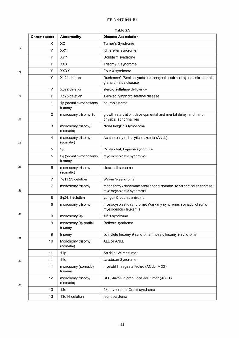
EP 3 117 011 B1
52
5
10
15
20
25
30
35
40
45
50
55
Table 2A
Chromosome Abnormality Disease Association
X XO Turner’s Syndrome
Y XXY Klinefelter syndrome
Y XYY Double Y syndrome
Y XXX Trisomy X syndrome
Y XXXX Four X syndrome
Y Xp21 deletion Duchenne’s/Becker syndrome, congenital adrenal hypoplasia, chronic granulomatus disease
Y Xp22 deletion steroid sulfatase deficiency
Y Xq26 deletion X-linked lymphproliferative disease
1 1p (somatic) monosomy trisomy
neuroblastoma
2 monosomy trisomy 2q growth retardation, developmental and mental delay, and minor physical abnormalities
3 monosomy trisomy (somatic)
Non-Hodgkin’s lymphoma
4 monosomy trisomy (somatic)
Acute non lymphocytic leukemia (ANLL)
5 5p Cri du chat; Lejeune syndrome
5 5q (somatic) monosomy trisomy
myelodysplastic syndrome
6 monosomy trisomy (somatic)
clear-cell sarcoma
7 7q11.23 deletion William’s syndrome
7 monosomy trisomy monosomy 7 syndrome of childhood; somatic: renal cortical adenomas; myelodysplastic syndrome
8 8q24.1 deletion Langer-Giedon syndrome
8 monosomy trisomy myelodysplastic syndrome; Warkany syndrome; somatic: chronic myelogenous leukemia
9 monosomy 9p Alfi’s syndrome
9 monosomy 9p partial trisomy
Rethore syndrome
9 trisomy complete trisomy 9 syndrome; mosaic trisomy 9 syndrome
10 Monosomy trisomy (somatic)
ALL or ANLL
11 11p- Aniridia; Wilms tumor
11 11q- Jacobson Syndrome
11 monosomy (somatic) trisomy
myeloid lineages affected (ANLL, MDS)
12 monosomy trisomy (somatic)
CLL, Juvenile granulosa cell tumor (JGCT)
13 13q- 13q-syndrome; Orbeli syndrome
13 13q14 deletion retinoblastoma
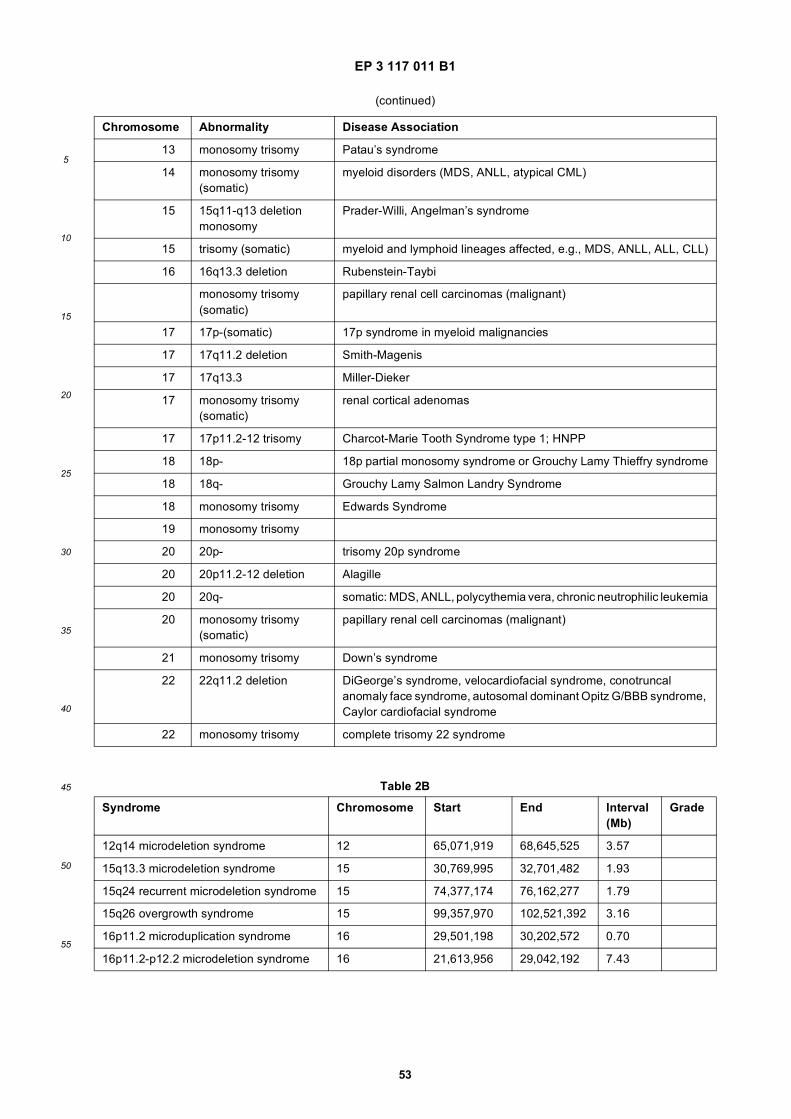
EP 3 117 011 B1
53
5
10
15
20
25
30
35
40
45
50
55
(continued)
Chromosome Abnormality Disease Association
13 monosomy trisomy Patau’s syndrome
14 monosomy trisomy (somatic)
myeloid disorders (MDS, ANLL, atypical CML)
15 15q11-q13 deletion monosomy
Prader-Willi, Angelman’s syndrome
15 trisomy (somatic) myeloid and lymphoid lineages affected, e.g., MDS, ANLL, ALL, CLL)
16 16q13.3 deletion Rubenstein-Taybi
monosomy trisomy (somatic)
papillary renal cell carcinomas (malignant)
17 17p-(somatic) 17p syndrome in myeloid malignancies
17 17q11.2 deletion Smith-Magenis
17 17q13.3 Miller-Dieker
17 monosomy trisomy (somatic)
renal cortical adenomas
17 17p11.2-12 trisomy Charcot-Marie Tooth Syndrome type 1; HNPP
18 18p- 18p partial monosomy syndrome or Grouchy Lamy Thieffry syndrome
18 18q- Grouchy Lamy Salmon Landry Syndrome
18 monosomy trisomy Edwards Syndrome
19 monosomy trisomy
20 20p- trisomy 20p syndrome
20 20p11.2-12 deletion Alagille
20 20q- somatic: MDS, ANLL, polycythemia vera, chronic neutrophilic leukemia
20 monosomy trisomy (somatic)
papillary renal cell carcinomas (malignant)
21 monosomy trisomy Down’s syndrome
22 22q11.2 deletion DiGeorge’s syndrome, velocardiofacial syndrome, conotruncal anomaly face syndrome, autosomal dominant Opitz G/BBB syndrome, Caylor cardiofacial syndrome
22 monosomy trisomy complete trisomy 22 syndrome
Table 2B
Syndrome Chromosome Start End Interval (Mb)
Grade
12q14 microdeletion syndrome 12 65,071,919 68,645,525 3.57
15q13.3 microdeletion syndrome 15 30,769,995 32,701,482 1.93
15q24 recurrent microdeletion syndrome 15 74,377,174 76,162,277 1.79
15q26 overgrowth syndrome 15 99,357,970 102,521,392 3.16
16p11.2 microduplication syndrome 16 29,501,198 30,202,572 0.70
16p11.2-p12.2 microdeletion syndrome 16 21,613,956 29,042,192 7.43
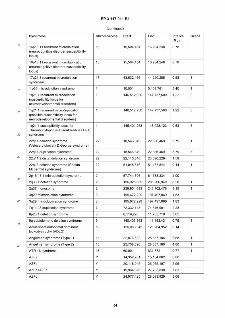
EP 3 117 011 B1
54
5
10
15
20
25
30
35
40
45
50
55
(continued)
Syndrome Chromosome Start End Interval (Mb)
Grade
16p13.11 recurrent microdeletion (neurocognitive disorder susceptibility locus)
16 15,504,454 16,284,248 0.78
16p13.11 recurrent microduplication (neurocognitive disorder susceptibility locus)
16 15,504,454 16,284,248 0.78
17q21.3 recurrent microdeletion syndrome
17 43,632,466 44,210,205 0.58 1
1 p36 microdeletion syndrome 1 10,001 5,408,761 5.40 1
1q21.1 recurrent microdeletion (susceptibility locus for neurodevelopmental disorders)
1 146,512,930 147,737,500 1.22 3
1q21.1 recurrent microduplication (possible susceptibility locus for neurodevelopmental disorders)
1 146,512,930 147,737,500 1.22 3
1q21.1 susceptibility locus for Thrombocytopenia-Absent Radius (TAR) syndrome
1 145,401,253 145,928,123 0.53 3
22q11 deletion syndrome (Velocardiofacial / DiGeorqe syndrome)
22 18,546,349 22,336,469 3.79 1
22q11 duplication syndrome 22 18,546,349 22,336,469 3.79 3
22q11.2 distal deletion syndrome 22 22,115,848 23,696,229 1.58
22q13 deletion syndrome (Phelan-Mcdermid syndrome)
22 51,045,516 51,187,844 0.14 1
2p15-16.1 microdeletion syndrome 2 57,741,796 61,738,334 4.00
2q33.1 deletion syndrome 2 196,925,089 205,206,940 8.28 1
2q37 monosomy 2 239,954,693 243,102,476 3.15 1
3q29 microdeletion syndrome 3 195,672,229 197,497,869 1.83
3q29 microduplication syndrome 3 195,672,229 197,497,869 1.83
7q11.23 duplication syndrome 7 72,332,743 74,616,901 2.28
8p23.1 deletion syndrome 8 8,119,295 11,765,719 3.65
9q subtelomeric deletion syndrome 9 140,403,363 141,153,431 0.75 1
Adult-onset autosomal dominant leukodystrophy (ADLD)
5 126,063,045 126,204,952 0.14
Angelman syndrome (Type 1) 15 22,876,632 28,557,186 5.68 1
Angelman syndrome (Tvpe 2) 15 23,758,390 28,557,186 4.80 1
ATR-16 syndrome 16 60,001 834,372 0.77 1
AZFa Y 14,352,761 15,154,862 0.80
AZFb Y 20,118,045 26,065,197 5.95
AZFb+AZFc Y 19,964,826 27,793,830 7.83
AZFc Y 24,977,425 28,033,929 3.06
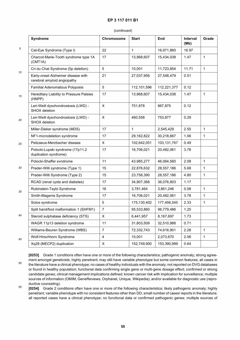
EP 3 117 011 B1
55
5
10
15
20
25
30
35
40
45
50
55
[0253] Grade 1 conditions often have one or more of the following characteristics; pathogenic anomaly; strong agree-ment amongst geneticists; highly penetrant; may still have variable phenotype but some common features; all cases inthe literature have a clinical phenotype; no cases of healthy individuals with the anomaly; not reported on DVG databasesor found in healthy population; functional data confirming single gene or multi-gene dosage effect; confirmed or strongcandidate genes; clinical management implications defined; known cancer risk with implication for surveillance; multiplesources of information (OMIM, GeneReviews, Orphanet, Unique, Wikipedia); and/or available for diagnostic use (repro-ductive counseling).[0254] Grade 2 conditions often have one or more of the following characteristics; likely pathogenic anomaly; highlypenetrant; variable phenotype with no consistent features other than DD; small number of cases/ reports in the literature;all reported cases have a clinical phenotype; no functional data or confirmed pathogenic genes; multiple sources of
(continued)
Syndrome Chromosome Start End Interval (Mb)
Grade
Cat-Eye Syndrome (Type I) 22 1 16,971,860 16.97
Charcot-Marie-Tooth syndrome type 1A (CMT1A)
17 13,968,607 15,434,038 1.47 1
Cri du Chat Syndrome (5p deletion) 5 10,001 11,723,854 11.71 1
Early-onset Alzheimer disease with cerebral amyloid angiopathy
21 27,037,956 27,548,479 0.51
Familial Adenomatous Polyposis 5 112,101,596 112,221,377 0.12
Hereditary Liability to Pressure Palsies (HNPP)
17 13,968,607 15,434,038 1.47 1
Leri-Weill dyschondrostosis (LWD) - SHOX deletion
X 751,878 867,875 0.12
Leri-Weill dyschondrostosis (LWD) - SHOX deletion
X 460,558 753,877 0.29
Miller-Dieker syndrome (MDS) 17 1 2,545,429 2.55 1
NF1-microdeletion syndrome 17 29,162,822 30,218,667 1.06 1
Pelizaeus-Merzbacher disease X 102,642,051 103,131,767 0.49
Potocki-Lupski syndrome (17p11.2 duplication syndrome)
17 16,706,021 20,482,061 3.78
Potocki-Shaffer svndrome 11 43,985,277 46,064,560 2.08 1
Prader-Willi syndrome (Type 1) 15 22,876,632 28,557,186 5.68 1
Prader-Willi Syndrome (Type 2) 15 23,758,390 28,557,186 4.80 1
RCAD (renal cysts and diabetes) 17 34,907,366 36,076,803 1.17
Rubinstein-Taybi Syndrome 16 3,781,464 3,861,246 0.08 1
Smith-Magenis Syndrome 17 16,706,021 20,482,061 3.78 1
Sotos syndrome 5 175,130,402 177,456,545 2.33 1
Split hand/foot malformation 1 (SHFM1) 7 95,533,860 96,779,486 1.25
Steroid sulphatase deficiency (STS) X 6,441,957 8,167,697 1.73
WAGR 11p13 deletion syndrome 11 31,803,509 32,510,988 0.71
Williams-Beuren Syndrome (WBS) 7 72,332,743 74,616,901 2.28 1
Wolf-Hirschhorn Syndrome 4 10,001 2,073,670 2.06 1
Xq28 (MECP2) duplication X 152,749,900 153,390,999 0.64
EP 3 117 011 B1
56
5
10
15
20
25
30
35
40
45
50
55
information (OMIM, Genereviews, Orphanet, Unique, Wikipedia); and/or may be used for diagnostic purposes andreproductive counseling.[0255] Grade 3 conditions often have one or more of the following characteristics; susceptibility locus; healthy indi-viduals or unaffected parents of a proband described; present in control populations; non penetrant; phenotype mild andnot specific; features less consistent; no functional data or confirmed pathogenic genes; more limited sources of data;possibility of second diagnosis remains a possibility for cases deviating from the majority or if novel clinical findingpresent; and/or caution when using for diagnostic purposes and guarded advice for reproductive counseling.
Examples
[0256] The examples set forth below illustrate certain embodiments and do not limit the technology.[0257] Example 1: Assays to determine the presence or absence of a chromosome aneuploidy in Chromosomes 13,18 and/or 21.
Assay design
[0258] PCR primers and MALDI-TOF MS TyperPLEX extension primers were designed using Assay Design Suite1.0,online tool hosted at www.MYSequenom.com (Design Suite 1.0 [online], [retrieved on 2014-03-04], retrieved from theWorld Wide Web internet URL mysequenom.com).[0259] All DNA sequences were retrieved from UCSC genome browser (http://genome.ucsc.edu/). DNA sequenceswith more than one restriction enzyme recognition site for the methylation sensitive restriction enzymes Hhal and Hpallwere loaded to Assay Design Suite 1.0. The target regions (i.e., portions of DMRs which spanned at least one restrictionsite) were loaded into the Assay Design Suite. The parameters of design were as follows: amplicon length was 60-100bp, optimum (amplicon length) was 80 bp, mass range was 4300 - 9000 Da, overall amplicons design confidence scorecutoff was 0.3, extension primer false primer potential was 0.2, hairpin/dimer extension potential was 0.2, overall con-fidence score cutoff was 0.2, minimum multiplex level was 5, and multiplex confidence score cutoff was 0.4. All additionalparameters were set as default values. All designed assays were unique to chromosome 13 (chr13), chromosome 18(chr18) and chromosome 21 (chr21), had at least one methylation sensitive restriction enzyme site, and did not containany known SNPs within PCR primers, extension primers, or restriction enzyme sites.[0260] A competitor oligonucleotide was designed for each assay. Each competitor oligonucleotide matched the se-quence of the target sequence of each assay in all positions with the exception a single nucleotide base mismatch,sometimes referred to as an artificial SNP. To ensure successful annealing of PCR primers to the competitor oligonu-cleotide, all competitor oligonucleotides were expanded to include genomic sequences 8-10 base pairs (bp) upstreamand downstream of each amplicon.
Assay selection
[0261] For a region to be considered validated and thus move forward to inclusion in the initial evaluation of a methylationbased assay, the data must show less than 3% methylation in non-pregnant plasma (as determined by the ratio of copynumber of digested non-pregnant ccf DNA/undigested non-pregnant ccf DNA) and a Pearson correlation coefficient ofgreater than 0.8 when using a model system consisting of an increasing ratio of placenta to peripheral blood mononuclearcells. Differentially methylated regions (DMRs) described in Tables 1A and 1B were selected, and 64 assays weredesigned to quantify target polynucleotides in these DMRs. A subset of 52 assays also was tested. Assays designatedin Tables 1A and 1B were designed with suitable amplification primers and competitor oligonucleotides (e.g., for MALDIMS and MPS assays) and suitable extension oligonucleotides (e.g., for MADLI MS assays).
Methylation assay
[0262] Circulating cell free (ccf) DNA was extracted from 4 mL of plasma using the QIAamp circulating nucleic acidkit (Qiagen) and eluted in 55mL buffer EB (Qiagen). A majority portion of the eluted ccf DNA (40mL) from each extractedsample was used for the methylation based assay; 10mL were reserved for use for an assay to determine fetal fractionand input copy number quantification (SeqFQA).[0263] DNA samples were treated with 20 units (U) Exol (NEB), 20 U Hhal (NEB), 50 U Hpall (NEB), 1x PCR buffer(Sequenom), and 2 mM MgCl2 in a 66 microliter (mL) reaction volume and incubated at 41°C for 60 minutes. Afterdigestion, enzymes were inactivated by heating at 98°C for 10 minutes. After enzyme digestion, DNA was amplified bythe addition of 22 mL PCR mixture, the final PCR reaction containing 2 mM of MgCl2, 0.5 millimolar (mM) dNTPs, 60micromolar (mM) forward PCR primer, 180 mM reverse PCR primer, 5 U of Fast Start polymerase (Roche), and competitoroligonucleotides at a known concentration. The PCR was initiated by a 5 minute incubation at 98°C, followed by 45
EP 3 117 011 B1
57
5
10
15
20
25
30
35
40
45
50
55
cycles (30 seconds (sec.) at 95°C, 30 sec. at 64°C, and 30 sec. at 72°C), and 72°C for 3 minutes. After PCR, 1 milliAnsonunit (mAU) of protease (Qiagen) was added into the PCR reaction and incubated at 55°C for 30 minutes to degradepeptides present in DNA extracted from plasma.[0264] A portion (20 mL) of purified PCR product from each sample was split into 4 parallel reactions of 5 mL each.Subsequently, 2 mL of a phosphatase reaction cocktail containing 0.5 U shrimp alkaline phosphatase (SAP), 5 U RNaself(NEB) and 0.85 x SAP buffer (Sequenom) was added. After inactivation of SAP and RNaself at 85°C for 5 minutes, anextension primer cocktail was added. The extension cocktail consisted of a mixture of extension primers, 0.2x iPLEXbuffer (Sequenom), 0.2x TypePLEX Termination Mix (Sequenom), and 0.04 U TypePLEX enzyme (Sequenom). Theextension program was: 94°C for 30 sec., 40 cycles (94°C for 5 sec. followed by 5 repetitions of 52°C for 5 sec. and80°C for 5 sec.), and 72°C for 3 minutes. After desalting the mixture by adding 6 milligrams (mg) Clean Resin (Sequenom),12 nanoliters (nL) from each reaction was transferred to SpectronCHIP II-G384 and mass spectra recorded using aMassARRAY MALDI-TOF mass spectrometer. Spectra were acquired by using Sequenom SpectroAcquire software.The software parameters were set to acquire 20 shots from each of 9 raster positions, using best 5 shots for signalcalculation.
Assay requirements and data modeling
[0265] In addition to the experimental validation of differentiation methylation, a power analysis was performed toapproximate the number of assays that would be needed to differentiate a euploid and aneuploid sample. For this poweranalysis, each population was modeled as a normal distribution. For an assay with 1000 input genomic copies, a fetalfraction of 4%, an observed average of maternal and placenta methylation levels, and an observed technical variance,it was estimated that a minimum of 7 assays per chromosome can differentiate euploid and trisomy samples (e.g., witha sensitivity equal to about 99%) when the analysis is conducted using mass spectrometry. When an assay analysis isconducted using DNA sequencing, it was estimated that a minimum of 4 assays per chromosome (e.g., analysis of atleast 4 target polynucleotides per chromosome) can differentiate euploid and trisomy samples (e.g., with a sensitivityequal to about 99%). While the level of methylation for each of the loci evaluated in this assay are unlikely to vary greatlymoving forward, assay optimization resulting in reduced technical variance could reduce the number of required assaysfurther.
Sequencing-based fetal quantifier assay (SeqFQA)
[0266] SeqFQA Loci PCR with competitor oligo addition for copy number determination. The SeqFQA loci PCR wasmodified to incorporate a synthetic competitor oligonucleotide that is included at a known copy number and can beconcurrently amplified with SeqFQA target SNPs. Methods and applications of SeqFQA are described inWO2014/011928. The competitor oligonucleotide is a known genomic DNA sequence with a single base change differ-ence from the genomic DNA reference - an artificial SNP not present in any population. Combined with knowledge oftotal competitor copy number included in the reaction, the ratio of sequencing reads from competitor oligonucleotide tothe number of wild type genomic locus reads can be used to determine the genomic DNA copy number in the startingmaterial (e.g., ccf nucleic acid).Competitor and primer sequences. The synthetic competitor oligo was designed as a double stranded plasmid containingamplicon sequences corresponding to a portion of Albumin, RNaseP and ApoE gene loci. The plasmid (pIDTSmart-Kan) was obtained from IDT and has a Mlul restriction site at each end of the gene insert region. A Hindlll restriction site(AAGCTT) was added between each of the 3 individual competitor sequences flanked by the 5’ linker, ATA. Table listsindividual competitor sequences, their position in the gene region of the dsDNA plasmid and primer sequences designedto amplify the competitor and their genomic DNA counterpart. The assay process included two steps: Loci PCR andUniversal PCR. The Loci PCR step is a multiplex PCR reaction which targets and amplifies target regions within thegenome or from competitor oligonucleotides. The Universal PCR step was used to add sample specific index barcodesequences and enable the hybridization of the amplified products to a fixed surface, including, but not limited to, amassively parallel sequencing flowcell (Illumina). Loci PCR primer sequences were designed to contain tags to allowsequencing adapters to be incorporated via a Universal PCR using Assay Designer v4.0. The nucleotide sequence ofthe forward primer tag (e.g., Tag 1, SEQ ID NO: 324) and the reverse primer tag (e.g., Tag 2, SEQ ID NO: 325) areprovided in Table 3. Competitor sequences were used as input for Assay Designer but constricted so that the forwardprimer had to hybridize within 35 bp of the artificial SNP site in the competitor oligonucleotide so that the site can beidentified with a 36 bp sequencing read. Primer sequences meeting acceptability criteria for amplification were only forRNaseP and ApoE.
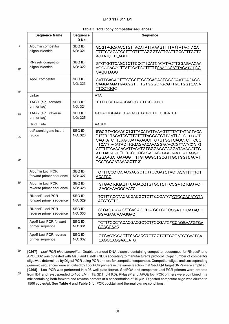
EP 3 117 011 B1
58
5
10
15
20
25
30
35
40
45
50
55
[0267] Loci PCR plus competitor. Double stranded DNA plasmid containing competitor sequences for RNaseP andAPOE302 was digested with Msul and Hindlll (NEB) according to manufacturer’s protocol. Copy number of competitoroligo was determined by Digital PCR using PCR primers for competitor sequences. Competitor oligos and correspondinggenomic sequences were amplified by Loci PCR primers in the same reaction that SeqFQA target SNPs were amplified.[0268] Loci PCR was performed in a 96-well plate format. SeqFQA and competitor Loci PCR primers were orderedfrom IDT and re-suspended to 100 mM in TE (IDT, pH 8.0). RNaseP and APOE loci PCR primers were combined in amix containing both forward and reverse primers at a concentration of 10 mM. Digested competitor oligo was diluted to1500 copies/ml. See Table 4 and Table 5 for PCR cocktail and thermal cycling conditions.
Table 3. Total copy competitor sequences.
Sequence Name Sequence ID No.
Sequence
Albumin competitor oligonucleotide
SEQ ID NO: 321
RNaseP competitor oligonucleotide
SEQ ID NO: 322
ApoE competitor SEQ ID NO: 323
Linker ATA
TAG 1 (e.g., forward primer tag)
SEQ ID NO: 324
TCTTTCCCTACACGACGCTCTTCCGATCT
TAG 2 (e.g., reverse primer tag)
SEQ ID NO: 325
GTGACTGGAGTTCAGACGTGTGCTCTTCCGATCT
HindIII site AAGCTT
dsPlasmid gene insert region
SEQ ID NO: 326
Albumin Loci PCR forward primer sequence
SEQ ID NO: 327
Albumin Loci PCR reverse primer sequence
SEQ ID NO: 328
RNaseP Loci PCR forward primer sequence
SEQ ID NO: 329
RNaseP Loci PCR reverse primer sequence
SEQ ID NO: 330
ApoE Loci PCR forward primer sequence
SEQ ID NO: 331
ApoE Loci PCR reverse primer sequence
SEQ ID NO: 332
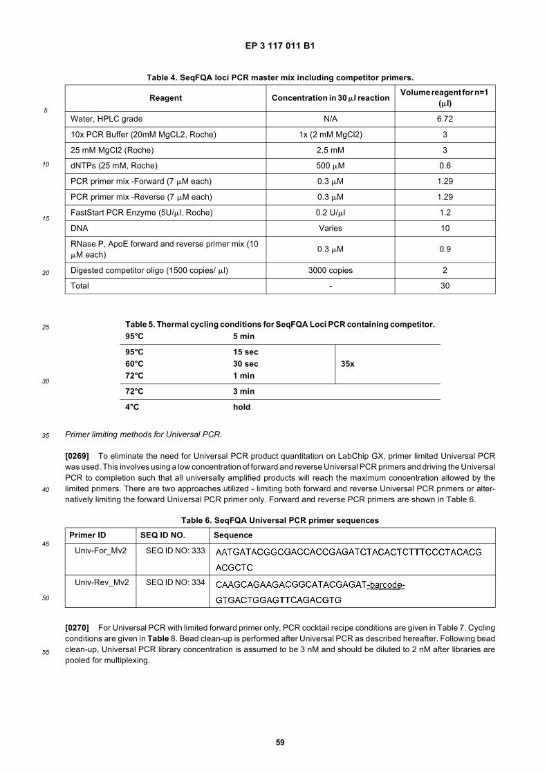
EP 3 117 011 B1
59
5
10
15
20
25
30
35
40
45
50
55
Primer limiting methods for Universal PCR.
[0269] To eliminate the need for Universal PCR product quantitation on LabChip GX, primer limited Universal PCRwas used. This involves using a low concentration of forward and reverse Universal PCR primers and driving the UniversalPCR to completion such that all universally amplified products will reach the maximum concentration allowed by thelimited primers. There are two approaches utilized - limiting both forward and reverse Universal PCR primers or alter-natively limiting the forward Universal PCR primer only. Forward and reverse PCR primers are shown in Table 6.
[0270] For Universal PCR with limited forward primer only, PCR cocktail recipe conditions are given in Table 7. Cyclingconditions are given in Table 8. Bead clean-up is performed after Universal PCR as described hereafter. Following beadclean-up, Universal PCR library concentration is assumed to be 3 nM and should be diluted to 2 nM after libraries arepooled for multiplexing.
Table 4. SeqFQA loci PCR master mix including competitor primers.
Reagent Concentration in 30 ml reaction Volume reagent for n=1 (ml)
Water, HPLC grade N/A 6.72
10x PCR Buffer (20mM MgCL2, Roche) 1x (2 mM MgCl2) 3
25 mM MgCl2 (Roche) 2.5 mM 3
dNTPs (25 mM, Roche) 500 mM 0.6
PCR primer mix -Forward (7 mM each) 0.3 mM 1.29
PCR primer mix -Reverse (7 mM each) 0.3 mM 1.29
FastStart PCR Enzyme (5U/ml, Roche) 0.2 U/ml 1.2
DNA Varies 10
RNase P, ApoE forward and reverse primer mix (10 mM each)
0.3 mM 0.9
Digested competitor oligo (1500 copies/ ml) 3000 copies 2
Total - 30
Table 5. Thermal cycling conditions for SeqFQA Loci PCR containing competitor.95°C 5 min
95°C 15 sec60°C 30 sec 35x72°C 1 min
72°C 3 min
4°C hold
Table 6. SeqFQA Universal PCR primer sequences
Primer ID SEQ ID NO. Sequence
Univ-For_Mv2 SEQ ID NO: 333
Univ-Rev_Mv2 SEQ ID NO: 334
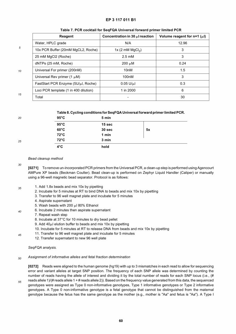
EP 3 117 011 B1
60
5
10
15
20
25
30
35
40
45
50
55
Bead cleanup method
[0271] To remove un-incorporated PCR primers from the Universal PCR, a clean-up step is performed using AgencourtAMPure XP beads (Beckman Coulter). Bead clean-up is performed on Zephyr Liquid Handler (Caliper) or manuallyusing a 96-well magnetic bead separator. Protocol is as follows:
1. Add 1.8x beads and mix 10x by pipetting2. Incubate for 5 minutes at RT to bind DNA to beads and mix 10x by pipetting3. Transfer to 96 well magnet plate and incubate for 5 minutes4. Aspirate supernatant5. Wash beads with 200 ml 80% Ethanol6. Incubate 2 minutes then aspirate supernatant7. Repeat wash step8. Incubate at 37°C for 10 minutes to dry bead pellet9. Add 40ml elution buffer to beads and mix 10x by pipetting10. Incubate for 5 minutes at RT to release DNA from beads and mix 10x by pipetting11. Transfer to 96 well magnet plate and incubate for 5 minutes12. Transfer supernatant to new 96 well plate
SeqFQA analysis.
Assignment of informative alleles and fetal fraction determination
[0272] Reads were aligned to the human genome (hg19) with up to 3 mismatches in each read to allow for sequencingerror and variant alleles at target SNP position. The frequency of each SNP allele was determined by counting thenumber of reads having the allele of interest and dividing it by the total number of reads for each SNP locus (i.e., (#reads allele 1)/(# reads allele 1 + # reads allele 2)). Based on the frequency value generated from this data, the sequencedgenotypes were assigned as Type 0 non-informative genotypes, Type 1 informative genotypes or Type 2 informativegenotypes. A Type 0 non-informative genotype is a fetal genotype that cannot be distinguished from the maternalgenotype because the fetus has the same genotype as the mother (e.g., mother is "Aa" and fetus is "Aa"). A Type I
Table 7. PCR cocktail for SeqFQA Universal forward primer limited PCR
Reagent Concentration in 30 ml reaction Volume reagent for n=1 (ml)
Water, HPLC grade N/A 12.96
10x PCR Buffer (20mM MgCL2, Roche) 1x (2 mM MgCl2) 3
25 mM MgCl2 (Roche) 2.5 mM 3
dNTPs (25 mM, Roche) 200 mM 0.24
Universal For primer (200nM) 10nM 1.5
Universal Rev primer (1 mM) 100nM 3
FastStart PCR Enzyme (5U/ml, Roche) 0.05 U/ml 0.3
Loci PCR template (1 in 400 dilution) 1 in 2000 6
Total - 30
Table 8. Cycling conditions for SeqFQA Universal forward primer limited PCR.95°C 5 min
95°C 15 sec60°C 30 sec 5x72°C 1 min72°C 3 min
4°C hold
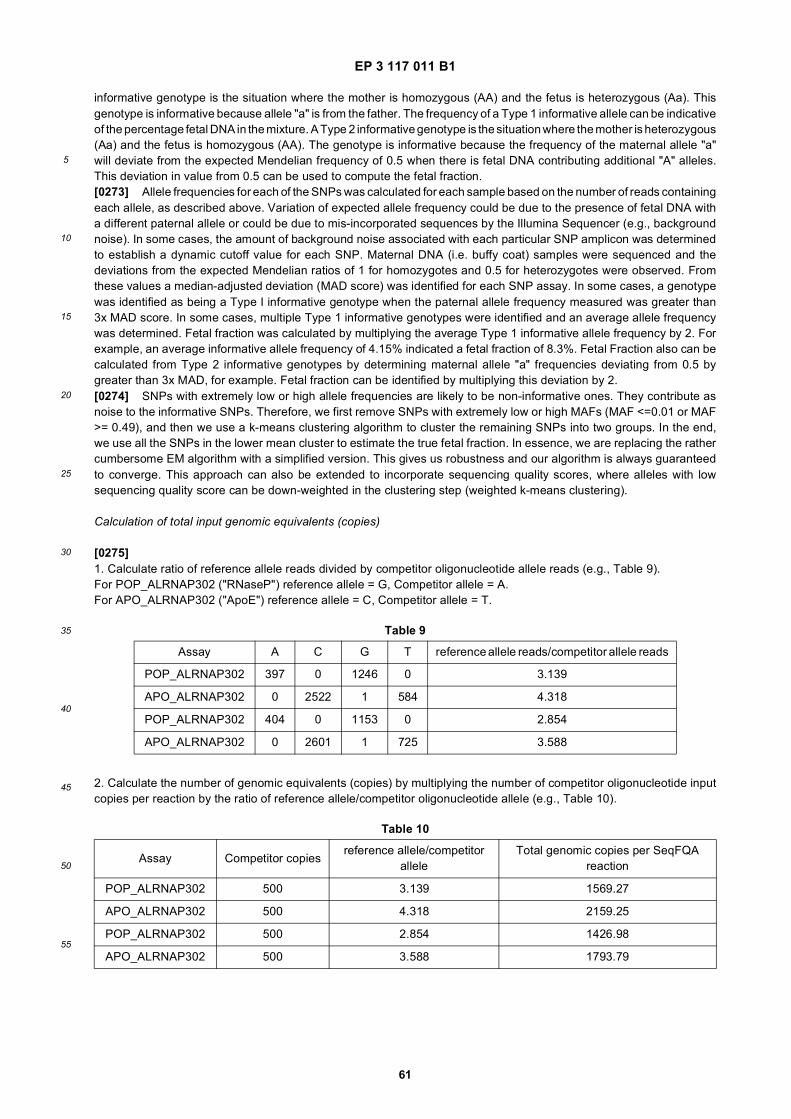
EP 3 117 011 B1
61
5
10
15
20
25
30
35
40
45
50
55
informative genotype is the situation where the mother is homozygous (AA) and the fetus is heterozygous (Aa). Thisgenotype is informative because allele "a" is from the father. The frequency of a Type 1 informative allele can be indicativeof the percentage fetal DNA in the mixture. A Type 2 informative genotype is the situation where the mother is heterozygous(Aa) and the fetus is homozygous (AA). The genotype is informative because the frequency of the maternal allele "a"will deviate from the expected Mendelian frequency of 0.5 when there is fetal DNA contributing additional "A" alleles.This deviation in value from 0.5 can be used to compute the fetal fraction.[0273] Allele frequencies for each of the SNPs was calculated for each sample based on the number of reads containingeach allele, as described above. Variation of expected allele frequency could be due to the presence of fetal DNA witha different paternal allele or could be due to mis-incorporated sequences by the Illumina Sequencer (e.g., backgroundnoise). In some cases, the amount of background noise associated with each particular SNP amplicon was determinedto establish a dynamic cutoff value for each SNP. Maternal DNA (i.e. buffy coat) samples were sequenced and thedeviations from the expected Mendelian ratios of 1 for homozygotes and 0.5 for heterozygotes were observed. Fromthese values a median-adjusted deviation (MAD score) was identified for each SNP assay. In some cases, a genotypewas identified as being a Type I informative genotype when the paternal allele frequency measured was greater than3x MAD score. In some cases, multiple Type 1 informative genotypes were identified and an average allele frequencywas determined. Fetal fraction was calculated by multiplying the average Type 1 informative allele frequency by 2. Forexample, an average informative allele frequency of 4.15% indicated a fetal fraction of 8.3%. Fetal Fraction also can becalculated from Type 2 informative genotypes by determining maternal allele "a" frequencies deviating from 0.5 bygreater than 3x MAD, for example. Fetal fraction can be identified by multiplying this deviation by 2.[0274] SNPs with extremely low or high allele frequencies are likely to be non-informative ones. They contribute asnoise to the informative SNPs. Therefore, we first remove SNPs with extremely low or high MAFs (MAF <=0.01 or MAF>= 0.49), and then we use a k-means clustering algorithm to cluster the remaining SNPs into two groups. In the end,we use all the SNPs in the lower mean cluster to estimate the true fetal fraction. In essence, we are replacing the rathercumbersome EM algorithm with a simplified version. This gives us robustness and our algorithm is always guaranteedto converge. This approach can also be extended to incorporate sequencing quality scores, where alleles with lowsequencing quality score can be down-weighted in the clustering step (weighted k-means clustering).
Calculation of total input genomic equivalents (copies)
[0275]1. Calculate ratio of reference allele reads divided by competitor oligonucleotide allele reads (e.g., Table 9).For POP_ALRNAP302 ("RNaseP") reference allele = G, Competitor allele = A.For APO_ALRNAP302 ("ApoE") reference allele = C, Competitor allele = T.
2. Calculate the number of genomic equivalents (copies) by multiplying the number of competitor oligonucleotide inputcopies per reaction by the ratio of reference allele/competitor oligonucleotide allele (e.g., Table 10).
Table 9
Assay A C G T reference allele reads/competitor allele reads
POP_ALRNAP302 397 0 1246 0 3.139
APO_ALRNAP302 0 2522 1 584 4.318
POP_ALRNAP302 404 0 1153 0 2.854
APO_ALRNAP302 0 2601 1 725 3.588
Table 10
Assay Competitor copiesreference allele/competitor
alleleTotal genomic copies per SeqFQA
reaction
POP_ALRNAP302 500 3.139 1569.27
APO_ALRNAP302 500 4.318 2159.25
POP_ALRNAP302 500 2.854 1426.98
APO_ALRNAP302 500 3.588 1793.79
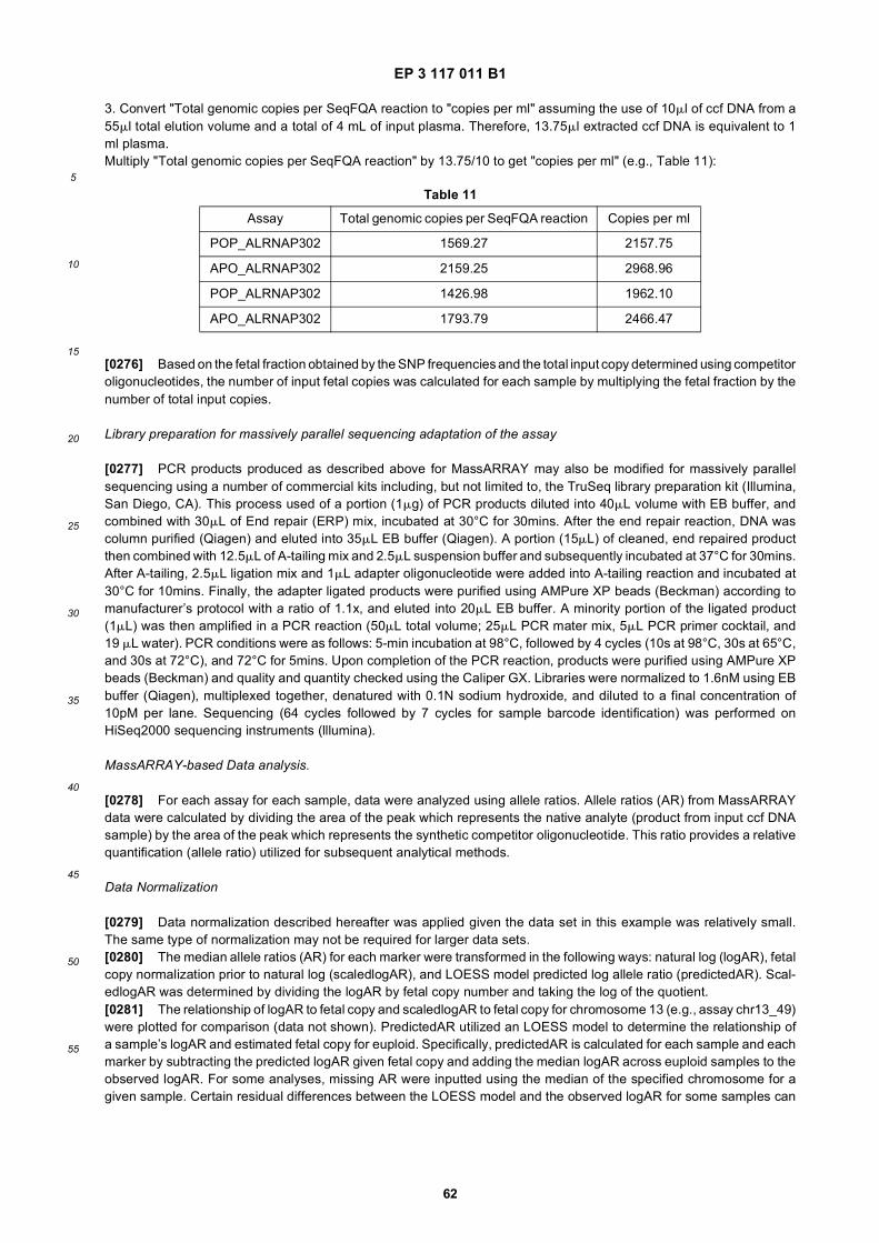
EP 3 117 011 B1
62
5
10
15
20
25
30
35
40
45
50
55
3. Convert "Total genomic copies per SeqFQA reaction to "copies per ml" assuming the use of 10ml of ccf DNA from a55ml total elution volume and a total of 4 mL of input plasma. Therefore, 13.75ml extracted ccf DNA is equivalent to 1ml plasma.Multiply "Total genomic copies per SeqFQA reaction" by 13.75/10 to get "copies per ml" (e.g., Table 11):
[0276] Based on the fetal fraction obtained by the SNP frequencies and the total input copy determined using competitoroligonucleotides, the number of input fetal copies was calculated for each sample by multiplying the fetal fraction by thenumber of total input copies.
Library preparation for massively parallel sequencing adaptation of the assay
[0277] PCR products produced as described above for MassARRAY may also be modified for massively parallelsequencing using a number of commercial kits including, but not limited to, the TruSeq library preparation kit (Illumina,San Diego, CA). This process used of a portion (1mg) of PCR products diluted into 40mL volume with EB buffer, andcombined with 30mL of End repair (ERP) mix, incubated at 30°C for 30mins. After the end repair reaction, DNA wascolumn purified (Qiagen) and eluted into 35mL EB buffer (Qiagen). A portion (15mL) of cleaned, end repaired productthen combined with 12.5mL of A-tailing mix and 2.5mL suspension buffer and subsequently incubated at 37°C for 30mins.After A-tailing, 2.5mL ligation mix and 1mL adapter oligonucleotide were added into A-tailing reaction and incubated at30°C for 10mins. Finally, the adapter ligated products were purified using AMPure XP beads (Beckman) according tomanufacturer’s protocol with a ratio of 1.1x, and eluted into 20mL EB buffer. A minority portion of the ligated product(1mL) was then amplified in a PCR reaction (50mL total volume; 25mL PCR mater mix, 5mL PCR primer cocktail, and19 mL water). PCR conditions were as follows: 5-min incubation at 98°C, followed by 4 cycles (10s at 98°C, 30s at 65°C,and 30s at 72°C), and 72°C for 5mins. Upon completion of the PCR reaction, products were purified using AMPure XPbeads (Beckman) and quality and quantity checked using the Caliper GX. Libraries were normalized to 1.6nM using EBbuffer (Qiagen), multiplexed together, denatured with 0.1N sodium hydroxide, and diluted to a final concentration of10pM per lane. Sequencing (64 cycles followed by 7 cycles for sample barcode identification) was performed onHiSeq2000 sequencing instruments (lllumina).
MassARRAY-based Data analysis.
[0278] For each assay for each sample, data were analyzed using allele ratios. Allele ratios (AR) from MassARRAYdata were calculated by dividing the area of the peak which represents the native analyte (product from input ccf DNAsample) by the area of the peak which represents the synthetic competitor oligonucleotide. This ratio provides a relativequantification (allele ratio) utilized for subsequent analytical methods.
Data Normalization
[0279] Data normalization described hereafter was applied given the data set in this example was relatively small.The same type of normalization may not be required for larger data sets.[0280] The median allele ratios (AR) for each marker were transformed in the following ways: natural log (logAR), fetalcopy normalization prior to natural log (scaledlogAR), and LOESS model predicted log allele ratio (predictedAR). Scal-edlogAR was determined by dividing the logAR by fetal copy number and taking the log of the quotient.[0281] The relationship of logAR to fetal copy and scaledlogAR to fetal copy for chromosome 13 (e.g., assay chr13_49)were plotted for comparison (data not shown). PredictedAR utilized an LOESS model to determine the relationship ofa sample’s logAR and estimated fetal copy for euploid. Specifically, predictedAR is calculated for each sample and eachmarker by subtracting the predicted logAR given fetal copy and adding the median logAR across euploid samples to theobserved logAR. For some analyses, missing AR were inputted using the median of the specified chromosome for agiven sample. Certain residual differences between the LOESS model and the observed logAR for some samples can
Table 11
Assay Total genomic copies per SeqFQA reaction Copies per ml
POP_ALRNAP302 1569.27 2157.75
APO_ALRNAP302 2159.25 2968.96
POP_ALRNAP302 1426.98 1962.10
APO_ALRNAP302 1793.79 2466.47
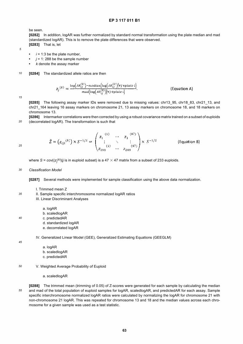
EP 3 117 011 B1
63
5
10
15
20
25
30
35
40
45
50
55
be seen.[0282] In addition, logAR was further normalized by standard normal transformation using the plate median and mad(standardized logAR). This is to remove the plate differences that were observed.[0283] That is, let
• i = 1:3 be the plate number,• j = 1: 288 be the sample number• k denote the assay marker
[0284] The standardized allele ratios are then
[0285] The following assay marker IDs were removed due to missing values: chr13_95, chr18_83, chr21_13, andchr21_164 leaving 16 assay markers on chromosome 21, 13 assay markers on chromosome 18, and 18 markers onchromosome 13.[0286] Intermarker correlations were then corrected by using a robust covariance matrix trained on a subset of euploids(decorrelated logAR). The transformation is such that
where S = cov((zj(k))|j is in euploid subset) is a 47 3 47 matrix from a subset of 233 euploids.
Classification Model
[0287] Several methods were implemented for sample classification using the above data normalization.
I. Trimmed mean ZII. Sample specific interchromosome normalized logAR ratiosIII. Linear Discriminant Analyses
a. logARb. scaledlogARc. predictedARd. standardized logARe. decorrelated logAR
IV. Generalized Linear Model (GEE), Generalized Estimating Equations (GEEGLM)
a. logARb. scaledlogARc. predictedAR
V. Weighted Average Probability of Euploid
a. scaledlogAR
[0288] The trimmed mean (trimming of 0.05) of Z-scores were generated for each sample by calculating the medianand mad of the total population of euploid samples for logAR, scaledlogAR, and predictedAR for each assay. Samplespecific interchromosome normalized logAR ratios were calculated by normalizing the logAR for chromosome 21 withnon-chromosome 21 logAR. This was repeated for chromosome 13 and 18 and the median values across each chro-mosome for a given sample was used as a test statistic.
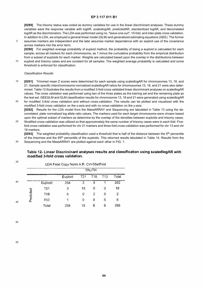
EP 3 117 011 B1
64
5
10
15
20
25
30
35
40
45
50
55
[0289] The trisomy status was coded as dummy variables for use in the linear discriminant analyses. These dummyvariables were the response variable with logAR, scaledlogAR, predictedAR, standardized logAR, and decorrelatedlogAR as the discriminators. The LDA was performed using no, "leave-one-out", 10-fold, and inter-plate cross validation.In addition to LDA, we employed a general linear model (GLM) and generalized estimating equations (GEE). The formerassumes markers are independent and the later assumes marker dependence with an explicit use of the covarianceacross markers into the error term.[0290] For weighted average probability of euploid method, the probability of being a euploid is calculated for eachsample, across all markers for each chromosome, as 1 minus the cumulative probability from the empirical distributionfrom a subset of euploids for each marker. Weights are calculated based upon the overlap in the distributions betweeneuploid and trisomy cases and are constant for all samples. The weighted average probability is calculated and somethreshold is enforced for classification.
Classification Results
[0291] Trimmed mean Z scores were determined for each sample using scaledlogAR for chromosomes 13, 18, and21. Sample specific interchromosome normalized scaledlogAR ratios for chromosomes 13, 18, and 21 were also deter-mined. Table 12 illustrates the results from a modified 3-fold cross validated linear discriminant analyses on scaledlogARvalues. The cross validation was performed using two of the three plates as the training set and the remaining plate asthe test set. GEEGLM and GLM classification results for chromosome 13, 18 and 21 were generated using scaledlogARfor modified 3-fold cross validation and without cross-validation. The results can be plotted and visualized with themodified 3-fold cross validation on the x-axis and with no cross-validation on the y-axis.[0292] Results for the LDA model from the MassARRAY and Sequencing are tabulated in Table 13 using the de-correlated, plate normalized log allele ratio values. The markers used for each target chromosome were chosen basedupon the optimal subset of markers as determine by the overlap of the densities between euploids and trisomy cases.Stratified cross-validation was utilized so that approximately the same number of trisomy cases were in each fold. Five-fold cross-validation was performed for chr 21 markers and three-fold cross-validation was performed for chr 13 and chr18 markers.[0293] The weighted probability classification used a threshold that is half of the distance between the 5th percentileof the trisomies and the 95th percentile of the euploids. This returned results tabulated in Table 14. Results from theSequencing and the MassARRAY are plotted against each other in FIG. 1.
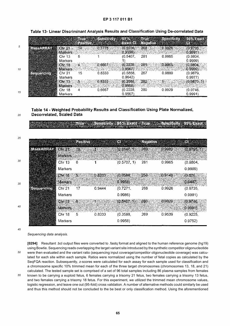
EP 3 117 011 B1
65
5
10
15
20
25
30
35
40
45
50
55
Sequencing data analysis.
[0294] Resultant .bcl output files were converted to .fastq format and aligned to the human reference genome (hg19)using Bowtie. Sequencing reads overlapping the target variant site introduced by the synthetic competitor oligonucleotidewere then evaluated and the variant ratio (sequencing input coverage/competitor oligonucleotide coverage) was calcu-lated for each site within each sample. Ratios were normalized using the number of fetal copies as calculated by theSeqFQA reaction. Subsequently, z-scores were calculated for each assay for each sample used for classification anda chromosome specific 10% trimmed mean for each of the three target chromosomes (chromosomes 13, 18, and 21)calculated. The tested sample set is comprised of a set of 96 total samples including 86 plasma samples from femalesknown to be carrying a euploid fetus, 6 females carrying a trisomy 21 fetus, two females carrying a trisomy 13 fetus,and two females carrying a trisomy 18 fetus. For this experiment, we utilized the trimmed mean chromosome values,logistic regression, and leave one out (95-fold) cross validation. A number of alternative methods could similarly be usedand thus this method should not be concluded to the be best or only classification method. Using the aforementioned
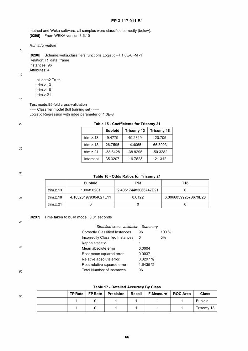
EP 3 117 011 B1
66
5
10
15
20
25
30
35
40
45
50
55
method and Weka software, all samples were classified correctly (below).[0295] From WEKA version 3.6.10
Run information
[0296] Scheme:weka.classifiers.functions.Logistic -R 1.0E-8 -M -1Relation: R_data_frameInstances: 96Attributes: 4
all.data2.Truthtrim.z.13trim.z.18trim.z.21
Test mode:95-fold cross-validation=== Classifier model (full training set) ===Logistic Regression with ridge parameter of 1.0E-8
[0297] Time taken to build model: 0.01 seconds
Table 15 - Coefficients for Trisomy 21
Euploid Trisomy 13 Trisomy 18
trim.z.13 9.4779 49.2319 -20.705
trim.z.18 26.7595 -4.4065 66.3903
trim.z.21 -38.5428 -38.9295 -50.3282
Intercept 35.3207 -16.7623 -21.312
Table 16 - Odds Ratios for Trisomy 21
Euploid T13 T18
trim.z.13 13068.0281 2.405174483066747E21 0
trim.z.18 4.183251979304027E11 0.0122 6.806603992573679E28
trim.z.21 0 0 0
Stratified cross-validation - SummaryCorrectly Classified Instances 96 100 %Incorrectly Classified Instances 0 0%Kappa statistic 1Mean absolute error 0.0004Root mean squared error 0.0037Relative absolute error 0.3297 %Root relative squared error 1.6435 %Total Number of Instances 96
Table 17 - Detailed Accuracy By Class
TP Rate FP Rate Precision Recall F-Measure ROC Area Class
1 0 1 1 1 1 Euploid
1 0 1 1 1 1 Trisomy 13
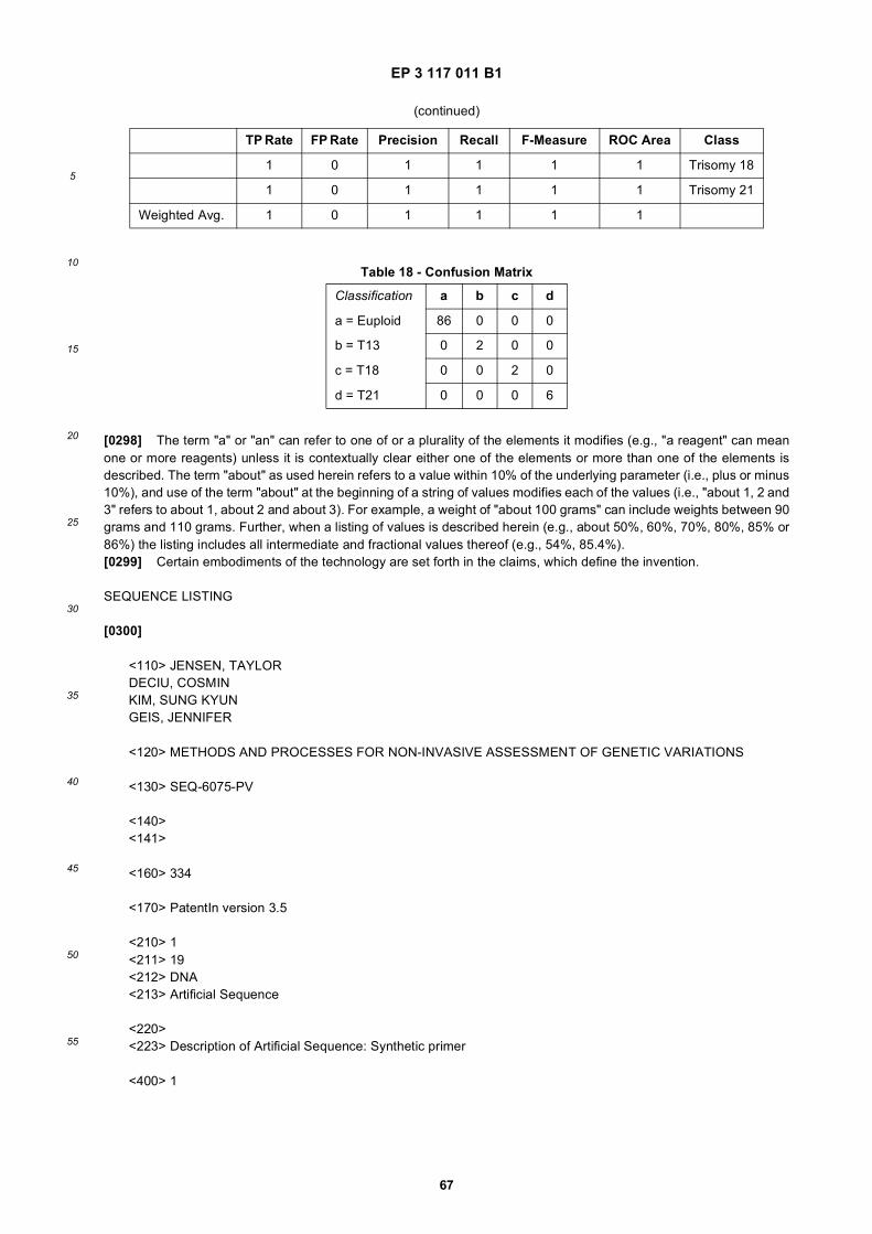
EP 3 117 011 B1
67
5
10
15
20
25
30
35
40
45
50
55
[0298] The term "a" or "an" can refer to one of or a plurality of the elements it modifies (e.g., "a reagent" can meanone or more reagents) unless it is contextually clear either one of the elements or more than one of the elements isdescribed. The term "about" as used herein refers to a value within 10% of the underlying parameter (i.e., plus or minus10%), and use of the term "about" at the beginning of a string of values modifies each of the values (i.e., "about 1, 2 and3" refers to about 1, about 2 and about 3). For example, a weight of "about 100 grams" can include weights between 90grams and 110 grams. Further, when a listing of values is described herein (e.g., about 50%, 60%, 70%, 80%, 85% or86%) the listing includes all intermediate and fractional values thereof (e.g., 54%, 85.4%).[0299] Certain embodiments of the technology are set forth in the claims, which define the invention.
SEQUENCE LISTING
[0300]
<110> JENSEN, TAYLORDECIU, COSMINKIM, SUNG KYUNGEIS, JENNIFER
<120> METHODS AND PROCESSES FOR NON-INVASIVE ASSESSMENT OF GENETIC VARIATIONS
<130> SEQ-6075-PV
<140><141>
<160> 334
<170> PatentIn version 3.5
<210> 1<211> 19<212> DNA<213> Artificial Sequence
<220><223> Description of Artificial Sequence: Synthetic primer
<400> 1
(continued)
TP Rate FP Rate Precision Recall F-Measure ROC Area Class
1 0 1 1 1 1 Trisomy 18
1 0 1 1 1 1 Trisomy 21
Weighted Avg. 1 0 1 1 1 1
Table 18 - Confusion Matrix
Classification a b c d
a = Euploid 86 0 0 0
b = T13 0 2 0 0
c = T18 0 0 2 0
d = T21 0 0 0 6
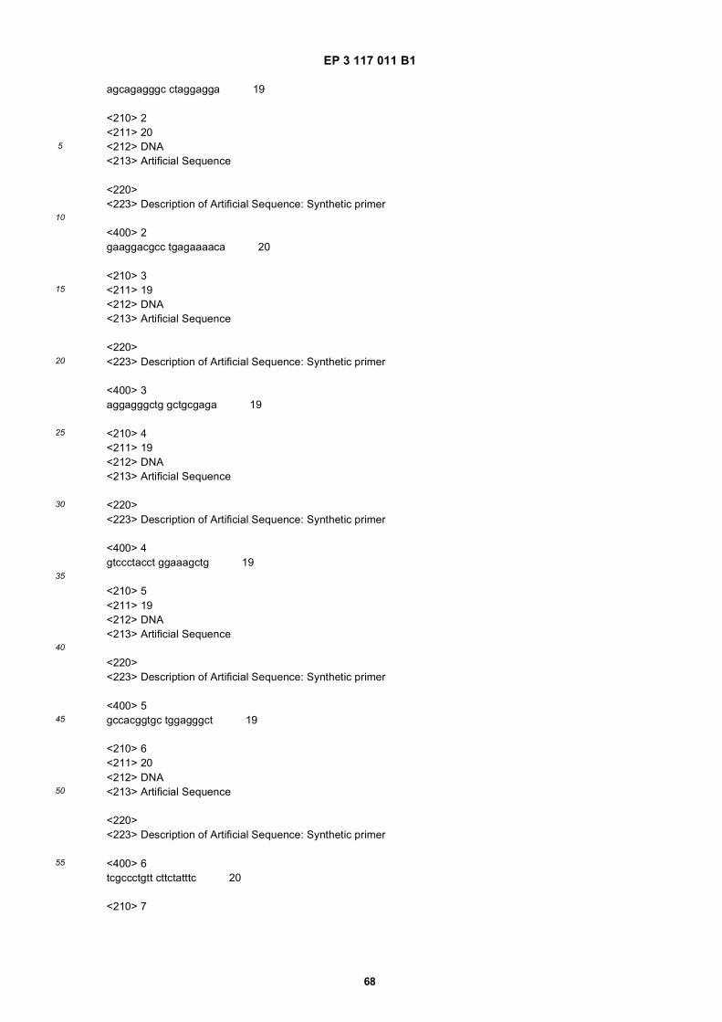
EP 3 117 011 B1
68
5
10
15
20
25
30
35
40
45
50
55
agcagagggc ctaggagga 19
<210> 2<211> 20<212> DNA<213> Artificial Sequence
<220><223> Description of Artificial Sequence: Synthetic primer
<400> 2gaaggacgcc tgagaaaaca 20
<210> 3<211> 19<212> DNA<213> Artificial Sequence
<220><223> Description of Artificial Sequence: Synthetic primer
<400> 3aggagggctg gctgcgaga 19
<210> 4<211> 19<212> DNA<213> Artificial Sequence
<220><223> Description of Artificial Sequence: Synthetic primer
<400> 4gtccctacct ggaaagctg 19
<210> 5<211> 19<212> DNA<213> Artificial Sequence
<220><223> Description of Artificial Sequence: Synthetic primer
<400> 5gccacggtgc tggagggct 19
<210> 6<211> 20<212> DNA<213> Artificial Sequence
<220><223> Description of Artificial Sequence: Synthetic primer
<400> 6tcgccctgtt cttctatttc 20
<210> 7
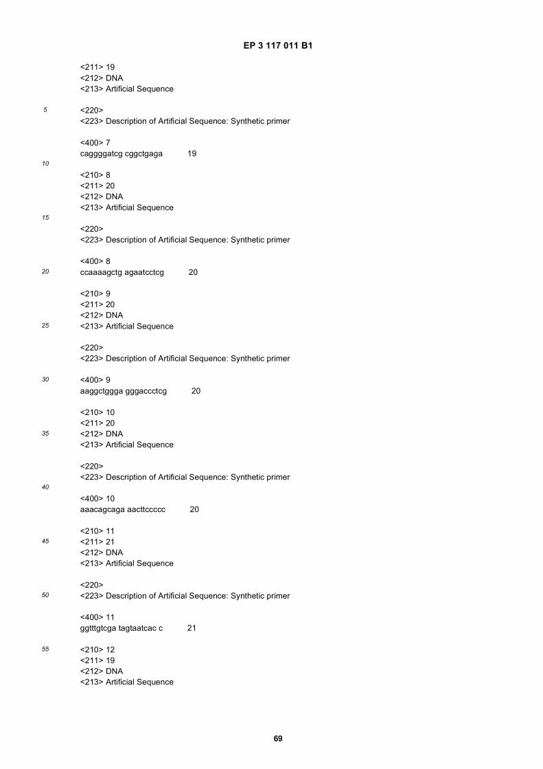
EP 3 117 011 B1
69
5
10
15
20
25
30
35
40
45
50
55
<211> 19<212> DNA<213> Artificial Sequence
<220><223> Description of Artificial Sequence: Synthetic primer
<400> 7caggggatcg cggctgaga 19
<210> 8<211> 20<212> DNA<213> Artificial Sequence
<220><223> Description of Artificial Sequence: Synthetic primer
<400> 8ccaaaagctg agaatcctcg 20
<210> 9<211> 20<212> DNA<213> Artificial Sequence
<220><223> Description of Artificial Sequence: Synthetic primer
<400> 9aaggctggga gggaccctcg 20
<210> 10<211> 20<212> DNA<213> Artificial Sequence
<220><223> Description of Artificial Sequence: Synthetic primer
<400> 10aaacagcaga aacttccccc 20
<210> 11<211> 21<212> DNA<213> Artificial Sequence
<220><223> Description of Artificial Sequence: Synthetic primer
<400> 11ggtttgtcga tagtaatcac c 21
<210> 12<211> 19<212> DNA<213> Artificial Sequence
EP 3 117 011 B1
70
5
10
15
20
25
30
35
40
45
50
55
<220><223> Description of Artificial Sequence: Synthetic primer
<400> 12aaaaccagcc cgcgcagag 19
<210> 13<211> 19<212> DNA<213> Artificial Sequence
<220><223> Description of Artificial Sequence: Synthetic primer
<400> 13acccacagga gccatccac 19
<210> 14<211> 19<212> DNA<213> Artificial Sequence
<220><223> Description of Artificial Sequence: Synthetic primer
<400> 14atgaccccca cagaccttg 19
<210> 15<211> 20<212> DNA<213> Artificial Sequence
<220><223> Description of Artificial Sequence: Synthetic primer
<400> 15agctccattg tctcagaggc 20
<210> 16<211> 19<212> DNA<213> Artificial Sequence
<220><223> Description of Artificial Sequence: Synthetic primer
<400> 16tccccagctg ttgcctctt 19
<210> 17<211> 20<212> DNA<213> Artificial Sequence
<220><223> Description of Artificial Sequence: Synthetic primer

EP 3 117 011 B1
71
5
10
15
20
25
30
35
40
45
50
55
<400> 17aagtgtgtct cgaacttgcg 20
<210> 18<211> 19<212> DNA<213> Artificial Sequence
<220><223> Description of Artificial Sequence: Synthetic primer
<400> 18ggtgacatcc tcccacccg 19
<210> 19<211> 20<212> DNA<213> Artificial Sequence
<220><223> Description of Artificial Sequence: Synthetic primer
<400> 19aagaagacgg tcttctgagc 20
<210> 20<211> 20<212> DNA<213> Artificial Sequence
<220><223> Description of Artificial Sequence: Synthetic primer
<400> 20tccaggcctc taggcaatag 20
<210> 21<211> 19<212> DNA<213> Artificial Sequence
<220><223> Description of Artificial Sequence: Synthetic primer
<400> 21cgtgtccccc actcacctt 19
<210> 22<211> 19<212> DNA<213> Artificial Sequence
<220><223> Description of Artificial Sequence: Synthetic primer
<400> 22gaacactgcg cccattgtc 19
EP 3 117 011 B1
72
5
10
15
20
25
30
35
40
45
50
55
<210> 23<211> 20<212> DNA<213> Artificial Sequence
<220><223> Description of Artificial Sequence: Synthetic primer
<400> 23ttagggagga cctacctggc 20
<210> 24<211> 20<212> DNA<213> Artificial Sequence
<220><223> Description of Artificial Sequence: Synthetic primer
<400> 24cactttctcc gtgcgagaag 20
<210> 25<211> 20<212> DNA<213> Artificial Sequence
<220><223> Description of Artificial Sequence: Synthetic primer
<400> 25tcactttgtt aagaagtccc 20
<210> 26<211> 19<212> DNA<213> Artificial Sequence
<220><223> Description of Artificial Sequence: Synthetic primer
<400> 26gcacgctccc ggggctctt 19
<210> 27<211> 19<212> DNA<213> Artificial Sequence
<220><223> Description of Artificial Sequence: Synthetic primer
<400> 27gtggtcatta gggcagtgg 19
<210> 28<211> 20<212> DNA
EP 3 117 011 B1
73
5
10
15
20
25
30
35
40
45
50
55
<213> Artificial Sequence
<220><223> Description of Artificial Sequence: Synthetic primer
<400> 28tgcgcctctc ccgaaagcag 20
<210> 29<211> 20<212> DNA<213> Artificial Sequence
<220><223> Description of Artificial Sequence: Synthetic primer
<400> 29ctaagggata tatgagggac 20
<210> 30<211> 20<212> DNA<213> Artificial Sequence
<220><223> Description of Artificial Sequence: Synthetic primer
<400> 30ttcacctggc agggcaggcg 20
<210> 31<211> 20<212> DNA<213> Artificial Sequence
<220><223> Description of Artificial Sequence: Synthetic primer
<400> 31gtcaggtacc ctaagggata 20
<210> 32<211> 20<212> DNA<213> Artificial Sequence
<220><223> Description of Artificial Sequence: Synthetic primer
<400> 32accccagtaa caagtgagag 20
<210> 33<211> 19<212> DNA<213> Artificial Sequence
<220>
EP 3 117 011 B1
74
5
10
15
20
25
30
35
40
45
50
55
<223> Description of Artificial Sequence: Synthetic primer
<400> 33ggagagcgga caggcctcg 19
<210> 34<211> 19<212> DNA<213> Artificial Sequence
<220><223> Description of Artificial Sequence: Synthetic primer
<400> 34agcctcgcac agctcgctt 19
<210> 35<211> 20<212> DNA<213> Artificial Sequence
<220><223> Description of Artificial Sequence: Synthetic primer
<400> 35caagcgttgc tgagatgaga 20
<210> 36<211> 20<212> DNA<213> Artificial Sequence
<220><223> Description of Artificial Sequence: Synthetic primer
<400> 36ctggtgggtt ttagtacccg 20
<210> 37<211> 20<212> DNA<213> Artificial Sequence
<220><223> Description of Artificial Sequence: Synthetic primer
<400> 37atctgagcct tagccccatc 20
<210> 38<211> 20<212> DNA<213> Artificial Sequence
<220><223> Description of Artificial Sequence: Synthetic primer
<400> 38
EP 3 117 011 B1
75
5
10
15
20
25
30
35
40
45
50
55
ccagatccca gttcctcaac 20
<210> 39<211> 18<212> DNA<213> Artificial Sequence
<220><223> Description of Artificial Sequence: Synthetic primer
<400> 39ccatgcgccc cgtcctcc 18
<210> 40<211> 20<212> DNA<213> Artificial Sequence
<220><223> Description of Artificial Sequence: Synthetic primer
<400> 40ttctgcgatt tcggagtgac 20
<210> 41<211> 19<212> DNA<213> Artificial Sequence
<220><223> Description of Artificial Sequence: Synthetic primer
<400> 41gactcagtgg tgtcgcctc 19
<210> 42<211> 19<212> DNA<213> Artificial Sequence
<220><223> Description of Artificial Sequence: Synthetic primer
<400> 42actggctggg cgtccgaac 19
<210> 43<211> 20<212> DNA<213> Artificial Sequence
<220><223> Description of Artificial Sequence: Synthetic primer
<400> 43ggtatctaat gccatccacc 20
<210> 44
EP 3 117 011 B1
76
5
10
15
20
25
30
35
40
45
50
55
<211> 20<212> DNA<213> Artificial Sequence
<220><223> Description of Artificial Sequence: Synthetic primer
<400> 44cacagagctt ccgactttgc 20
<210> 45<211> 19<212> DNA<213> Artificial Sequence
<220><223> Description of Artificial Sequence: Synthetic primer
<400> 45tccccgacct cccaagcta 19
<210> 46<211> 20<212> DNA<213> Artificial Sequence
<220><223> Description of Artificial Sequence: Synthetic primer
<400> 46aaaaggaggg ccagagatag 20
<210> 47<211> 20<212> DNA<213> Artificial Sequence
<220><223> Description of Artificial Sequence: Synthetic primer
<400> 47cacaagttag ttggaagccg 20
<210> 48<211> 19<212> DNA<213> Artificial Sequence
<220><223> Description of Artificial Sequence: Synthetic primer
<400> 48gcgtccgaac tccccaggt 19
<210> 49<211> 20<212> DNA<213> Artificial Sequence
EP 3 117 011 B1
77
5
10
15
20
25
30
35
40
45
50
55
<220><223> Description of Artificial Sequence: Synthetic primer
<400> 49gaggcacaaa gtgatggcag 20
<210> 50<211> 20<212> DNA<213> Artificial Sequence
<220><223> Description of Artificial Sequence: Synthetic primer
<400> 50aaaaaacctc gcccgcattg 20
<210> 51<211> 20<212> DNA<213> Artificial Sequence
<220><223> Description of Artificial Sequence: Synthetic primer
<400> 51aggtttcgga ttggaaggag 20
<210> 52<211> 19<212> DNA<213> Artificial Sequence
<220><223> Description of Artificial Sequence: Synthetic primer
<400> 52acggcgagag cacagatgg 19
<210> 53<211> 20<212> DNA<213> Artificial Sequence
<220><223> Description of Artificial Sequence: Synthetic primer
<400> 53aggcctccgg gctcttgctt 20
<210> 54<211> 19<212> DNA<213> Artificial Sequence
<220><223> Description of Artificial Sequence: Synthetic primer
EP 3 117 011 B1
78
5
10
15
20
25
30
35
40
45
50
55
<400> 54tgaagcacca tgcagcgcc 19
<210> 55<211> 20<212> DNA<213> Artificial Sequence
<220><223> Description of Artificial Sequence: Synthetic primer
<400> 55ttcagtgagt gccactgacc 20
<210> 56<211> 20<212> DNA<213> Artificial Sequence
<220><223> Description of Artificial Sequence: Synthetic primer
<400> 56cattggaagg tcagccaatc 20
<210> 57<211> 19<212> DNA<213> Artificial Sequence
<220><223> Description of Artificial Sequence: Synthetic primer
<400> 57aggaagtgag cagctcctg 19
<210> 58<211> 20<212> DNA<213> Artificial Sequence
<220><223> Description of Artificial Sequence: Synthetic primer
<400> 58tagaaagcga gagacgagag 20
<210> 59<211> 18<212> DNA<213> Artificial Sequence
<220><223> Description of Artificial Sequence: Synthetic primer
<400> 59gtctaggcgg caggtccc 18
EP 3 117 011 B1
79
5
10
15
20
25
30
35
40
45
50
55
<210> 60<211> 19<212> DNA<213> Artificial Sequence
<220><223> Description of Artificial Sequence: Synthetic primer
<400> 60tcctcaggag gcggtgcca 19
<210> 61<211> 20<212> DNA<213> Artificial Sequence
<220><223> Description of Artificial Sequence: Synthetic primer
<400> 61aggaggcgac cccagagagt 20
<210> 62<211> 19<212> DNA<213> Artificial Sequence
<220><223> Description of Artificial Sequence: Synthetic primer
<400> 62ccttcaaggg ggtccctgc 19
<210> 63<211> 20<212> DNA<213> Artificial Sequence
<220><223> Description of Artificial Sequence: Synthetic primer
<400> 63ttcagggagc gtccggtgag 20
<210> 64<211> 20<212> DNA<213> Artificial Sequence
<220><223> Description of Artificial Sequence: Synthetic primer
<400> 64tttccttatc cgcaggtgtg 20
<210> 65<211> 18<212> DNA
EP 3 117 011 B1
80
5
10
15
20
25
30
35
40
45
50
55
<213> Artificial Sequence
<220><223> Description of Artificial Sequence: Synthetic primer
<400> 65ccccattcaa ctgccagg 18
<210> 66<211> 20<212> DNA<213> Artificial Sequence
<220><223> Description of Artificial Sequence: Synthetic primer
<400> 66cctatgggga aggatcaaac 20
<210> 67<211> 20<212> DNA<213> Artificial Sequence
<220><223> Description of Artificial Sequence: Synthetic primer
<400> 67acaacagctt tggccaagac 20
<210> 68<211> 20<212> DNA<213> Artificial Sequence
<220><223> Description of Artificial Sequence: Synthetic primer
<400> 68aggtgggagt gggtgcgtca 20
<210> 69<211> 20<212> DNA<213> Artificial Sequence
<220><223> Description of Artificial Sequence: Synthetic primer
<400> 69gtcattctgc ccacccattg 20
<210> 70<211> 20<212> DNA<213> Artificial Sequence
<220>

EP 3 117 011 B1
81
5
10
15
20
25
30
35
40
45
50
55
<223> Description of Artificial Sequence: Synthetic primer
<400> 70aaggagatgg gcgctctcag 20
<210> 71<211> 20<212> DNA<213> Artificial Sequence
<220><223> Description of Artificial Sequence: Synthetic primer
<400> 71gactcagttt ccaagtgcgg 20
<210> 72<211> 20<212> DNA<213> Artificial Sequence
<220><223> Description of Artificial Sequence: Synthetic primer
<400> 72ttgtcccacc tcgccataac 20
<210> 73<211> 19<212> DNA<213> Artificial Sequence
<220><223> Description of Artificial Sequence: Synthetic primer
<400> 73aaaccagcct tgaaacgcc 19
<210> 74<211> 19<212> DNA<213> Artificial Sequence
<220><223> Description of Artificial Sequence: Synthetic primer
<400> 74gaatccccgg ccgcttctg 19
<210> 75<211> 20<212> DNA<213> Artificial Sequence
<220><223> Description of Artificial Sequence: Synthetic primer
<400> 75
EP 3 117 011 B1
82
5
10
15
20
25
30
35
40
45
50
55
gaagacttgt cacccagtcc 20
<210> 76<211> 19<212> DNA<213> Artificial Sequence
<220><223> Description of Artificial Sequence: Synthetic primer
<400> 76ggtccccacg gaggatcag 19
<210> 77<211> 19<212> DNA<213> Artificial Sequence
<220><223> Description of Artificial Sequence: Synthetic primer
<400> 77acctcaagcc ctgcctctg 19
<210> 78<211> 20<212> DNA<213> Artificial Sequence
<220><223> Description of Artificial Sequence: Synthetic primer
<400> 78tcttctccaa agtgcgcatc 20
<210> 79<211> 19<212> DNA<213> Artificial Sequence
<220><223> Description of Artificial Sequence: Synthetic primer
<400> 79cgatgcgctg aggctcgac 19
<210> 80<211> 20<212> DNA<213> Artificial Sequence
<220><223> Description of Artificial Sequence: Synthetic primer
<400> 80agacaatgga gctgacactg 20
<210> 81
EP 3 117 011 B1
83
5
10
15
20
25
30
35
40
45
50
55
<211> 18<212> DNA<213> Artificial Sequence
<220><223> Description of Artificial Sequence: Synthetic primer
<400> 81agacaccgcg cccctctc 18
<210> 82<211> 20<212> DNA<213> Artificial Sequence
<220><223> Description of Artificial Sequence: Synthetic primer
<400> 82ttttcgcctg gaactaccac 20
<210> 83<211> 20<212> DNA<213> Artificial Sequence
<220><223> Description of Artificial Sequence: Synthetic primer
<400> 83aagaggaagt ggcccaggag 20
<210> 84<211> 20<212> DNA<213> Artificial Sequence
<220><223> Description of Artificial Sequence: Synthetic primer
<400> 84actgcggggt cggcgggaaa 20
<210> 85<211> 20<212> DNA<213> Artificial Sequence
<220><223> Description of Artificial Sequence: Synthetic primer
<400> 85taaaacccaa gtctggtgtc 20
<210> 86<211> 19<212> DNA<213> Artificial Sequence
EP 3 117 011 B1
84
5
10
15
20
25
30
35
40
45
50
55
<220><223> Description of Artificial Sequence: Synthetic primer
<400> 86aaagtctgcg cagctgctc 19
<210> 87<211> 20<212> DNA<213> Artificial Sequence
<220><223> Description of Artificial Sequence: Synthetic primer
<400> 87tgcatctgca cccgcccgta 20
<210> 88<211> 20<212> DNA<213> Artificial Sequence
<220><223> Description of Artificial Sequence: Synthetic primer
<400> 88cctaaactta gccagttcgg 20
<210> 89<211> 20<212> DNA<213> Artificial Sequence
<220><223> Description of Artificial Sequence: Synthetic primer
<400> 89aggtgagtct ggtccgcttt 20
<210> 90<211> 19<212> DNA<213> Artificial Sequence
<220><223> Description of Artificial Sequence: Synthetic primer
<400> 90tggtgagggg ctacccaga 19
<210> 91<211> 20<212> DNA<213> Artificial Sequence
<220><223> Description of Artificial Sequence: Synthetic primer
EP 3 117 011 B1
85
5
10
15
20
25
30
35
40
45
50
55
<400> 91acagctgagc gcagggttct 20
<210> 92<211> 20<212> DNA<213> Artificial Sequence
<220><223> Description of Artificial Sequence: Synthetic primer
<400> 92aggaaggaga gatccgggac 20
<210> 93<211> 19<212> DNA<213> Artificial Sequence
<220><223> Description of Artificial Sequence: Synthetic primer
<400> 93tggtgctgaa gtgacaggg 19
<210> 94<211> 19<212> DNA<213> Artificial Sequence
<220><223> Description of Artificial Sequence: Synthetic primer
<400> 94tgagtccgcg caccccttc 19
<210> 95<211> 20<212> DNA<213> Artificial Sequence
<220><223> Description of Artificial Sequence: Synthetic primer
<400> 95tcttccagag gctgctttag 20
<210> 96<211> 19<212> DNA<213> Artificial Sequence
<220><223> Description of Artificial Sequence: Synthetic primer
<400> 96aggggaccca gggaggaga 19

EP 3 117 011 B1
86
5
10
15
20
25
30
35
40
45
50
55
<210> 97<211> 20<212> DNA<213> Artificial Sequence
<220><223> Description of Artificial Sequence: Synthetic primer
<400> 97ttccccctac actcggaatg 20
<210> 98<211> 19<212> DNA<213> Artificial Sequence
<220><223> Description of Artificial Sequence: Synthetic primer
<400> 98ctcagcggtg cgaggaaag 19
<210> 99<211> 19<212> DNA<213> Artificial Sequence
<220><223> Description of Artificial Sequence: Synthetic primer
<400> 99tgcagacttg caccatggg 19
<210> 100<211> 19<212> DNA<213> Artificial Sequence
<220><223> Description of Artificial Sequence: Synthetic primer
<400> 100ctgccaagtt ccttctggt 19
<210> 101<211> 19<212> DNA<213> Artificial Sequence
<220><223> Description of Artificial Sequence: Synthetic primer
<400> 101cttccctgga gccggcgtg 19
<210> 102<211> 20<212> DNA
EP 3 117 011 B1
87
5
10
15
20
25
30
35
40
45
50
55
<213> Artificial Sequence
<220><223> Description of Artificial Sequence: Synthetic primer
<400> 102gcaaggtcaa cctaccagag 20
<210> 103<211> 19<212> DNA<213> Artificial Sequence
<220><223> Description of Artificial Sequence: Synthetic primer
<400> 103agtgaccaca gtccggacc 19
<210> 104<211> 19<212> DNA<213> Artificial Sequence
<220><223> Description of Artificial Sequence: Synthetic primer
<400> 104ccctgctgtg tggggagac 19
<210> 105<211> 20<212> DNA<213> Artificial Sequence
<220><223> Description of Artificial Sequence: Synthetic primer
<400> 105gggaatccat ccagcgcaat 20
<210> 106<211> 20<212> DNA<213> Artificial Sequence
<220><223> Description of Artificial Sequence: Synthetic primer
<400> 106gttttaaagt gggccacagc 20
<210> 107<211> 18<212> DNA<213> Artificial Sequence
<220>

EP 3 117 011 B1
88
5
10
15
20
25
30
35
40
45
50
55
<223> Description of Artificial Sequence: Synthetic primer
<400> 107acccactcag aggtgggc 18
<210> 108<211> 20<212> DNA<213> Artificial Sequence
<220><223> Description of Artificial Sequence: Synthetic primer
<400> 108agtcaggggt gaaagtcgag 20
<210> 109<211> 20<212> DNA<213> Artificial Sequence
<220><223> Description of Artificial Sequence: Synthetic primer
<400> 109ttcttcacct ggcaggcttc 20
<210> 110<211> 19<212> DNA<213> Artificial Sequence
<220><223> Description of Artificial Sequence: Synthetic primer
<400> 110tgcaccgcag taccacagc 19
<210> 111<211> 20<212> DNA<213> Artificial Sequence
<220><223> Description of Artificial Sequence: Synthetic primer
<400> 111gtggattgac ccagatattg 20
<210> 112<211> 20<212> DNA<213> Artificial Sequence
<220><223> Description of Artificial Sequence: Synthetic primer
<400> 112

EP 3 117 011 B1
89
5
10
15
20
25
30
35
40
45
50
55
cgggtatcct acgtattctg 20
<210> 113<211> 19<212> DNA<213> Artificial Sequence
<220><223> Description of Artificial Sequence: Synthetic primer
<400> 113tgggattaaa cgagcgcgg 19
<210> 114<211> 19<212> DNA<213> Artificial Sequence
<220><223> Description of Artificial Sequence: Synthetic primer
<400> 114ccaagcaccc ctgcctcta 19
<210> 115<211> 20<212> DNA<213> Artificial Sequence
<220><223> Description of Artificial Sequence: Synthetic primer
<400> 115aaaaaccccc tacacgctac 20
<210> 116<211> 20<212> DNA<213> Artificial Sequence
<220><223> Description of Artificial Sequence: Synthetic primer
<400> 116tgccagccct ccccacgagt 20
<210> 117<211> 19<212> DNA<213> Artificial Sequence
<220><223> Description of Artificial Sequence: Synthetic primer
<400> 117agctcctgca gggtgaatg 19
<210> 118
EP 3 117 011 B1
90
5
10
15
20
25
30
35
40
45
50
55
<211> 19<212> DNA<213> Artificial Sequence
<220><223> Description of Artificial Sequence: Synthetic primer
<400> 118ggtaccatgt gcagcgcag 19
<210> 119<211> 20<212> DNA<213> Artificial Sequence
<220><223> Description of Artificial Sequence: Synthetic primer
<400> 119aaggtgcctc ctgcgcaatc 20
<210> 120<211> 19<212> DNA<213> Artificial Sequence
<220><223> Description of Artificial Sequence: Synthetic primer
<400> 120tggcgcgcgc ctcggaggt 19
<210> 121<211> 18<212> DNA<213> Artificial Sequence
<220><223> Description of Artificial Sequence: Synthetic primer
<400> 121ctcttggcag ggagggct 18
<210> 122<211> 20<212> DNA<213> Artificial Sequence
<220><223> Description of Artificial Sequence: Synthetic primer
<400> 122ggggaacctg ctttgatttt 20
<210> 123<211> 21<212> DNA<213> Artificial Sequence
EP 3 117 011 B1
91
5
10
15
20
25
30
35
40
45
50
55
<220><223> Description of Artificial Sequence: Synthetic primer
<400> 123aaagccctcc gagggagccc a 21
<210> 124<211> 19<212> DNA<213> Artificial Sequence
<220><223> Description of Artificial Sequence: Synthetic primer
<400> 124gctgcccagc gctggcaac 19
<210> 125<211> 20<212> DNA<213> Artificial Sequence
<220><223> Description of Artificial Sequence: Synthetic primer
<400> 125acatctggaa ggaaggaagc 20
<210> 126<211> 19<212> DNA<213> Artificial Sequence
<220><223> Description of Artificial Sequence: Synthetic primer
<400> 126tgcggcaccc aagcccctc 19
<210> 127<211> 20<212> DNA<213> Artificial Sequence
<220><223> Description of Artificial Sequence: Synthetic primer
<400> 127tacctaagtg agaggcagac 20
<210> 128<211> 20<212> DNA<213> Artificial Sequence
<220><223> Description of Artificial Sequence: Synthetic primer
EP 3 117 011 B1
92
5
10
15
20
25
30
35
40
45
50
55
<400> 128acgcacgggc tttccgaatt 20
<210> 129<211> 23<212> DNA<213> Artificial Sequence
<220><223> Description of Artificial Sequence: Synthetic primer
<400> 129gcctaggagg accccgggcg tgg 23
<210> 130<211> 28<212> DNA<213> Artificial Sequence
<220><223> Description of Artificial Sequence: Synthetic primer
<400> 130gcctgagaaa acagaccccg gccccgtg 28
<210> 131<211> 19<212> DNA<213> Artificial Sequence
<220><223> Description of Artificial Sequence: Synthetic primer
<400> 131agagccccgg gttcccttt 19
<210> 132<211> 26<212> DNA<213> Artificial Sequence
<220><223> Description of Artificial Sequence: Synthetic primer
<400> 132gggacgctgg gagactcaca gcccgg 26
<210> 133<211> 19<212> DNA<213> Artificial Sequence
<220><223> Description of Artificial Sequence: Synthetic primer
<400> 133gtgctggagg gctgcgcgg 19
EP 3 117 011 B1
93
5
10
15
20
25
30
35
40
45
50
55
<210> 134<211> 26<212> DNA<213> Artificial Sequence
<220><223> Description of Artificial Sequence: Synthetic primer
<400> 134cgccctgttc ttctatttca gagcgc 26
<210> 135<211> 22<212> DNA<213> Artificial Sequence
<220><223> Description of Artificial Sequence: Synthetic primer
<400> 135gggatcgcgg ctgagagcgc cc 22
<210> 136<211> 26<212> DNA<213> Artificial Sequence
<220><223> Description of Artificial Sequence: Synthetic primer
<400> 136agctgagaat cctcgatgcc cgcgcg 26
<210> 137<211> 22<212> DNA<213> Artificial Sequence
<220><223> Description of Artificial Sequence: Synthetic primer
<400> 137ctgggaggga ccctcgccgg gg 22
<210> 138<211> 22<212> DNA<213> Artificial Sequence
<220><223> Description of Artificial Sequence: Synthetic primer
<400> 138cagaaacttc ccccggctgt gg 22
<210> 139<211> 25<212> DNA
EP 3 117 011 B1
94
5
10
15
20
25
30
35
40
45
50
55
<213> Artificial Sequence
<220><223> Description of Artificial Sequence: Synthetic primer
<400> 139tttgtcgata gtaatcacca ccttc 25
<210> 140<211> 22<212> DNA<213> Artificial Sequence
<220><223> Description of Artificial Sequence: Synthetic primer
<400> 140gggctcgccc gcggggtctg gg 22
<210> 141<211> 17<212> DNA<213> Artificial Sequence
<220><223> Description of Artificial Sequence: Synthetic primer
<400> 141ccgcagccgc cctcctc 17
<210> 142<211> 21<212> DNA<213> Artificial Sequence
<220><223> Description of Artificial Sequence: Synthetic primer
<400> 142ccccacagac cttggcgccg c 21
<210> 143<211> 25<212> DNA<213> Artificial Sequence
<220><223> Description of Artificial Sequence: Synthetic primer
<400> 143ggcggccaag tgagggacca gtctc 25
<210> 144<211> 23<212> DNA<213> Artificial Sequence
<220>
EP 3 117 011 B1
95
5
10
15
20
25
30
35
40
45
50
55
<223> Description of Artificial Sequence: Synthetic primer
<400> 144cccagctgtt gcctcttgcg cgg 23
<210> 145<211> 21<212> DNA<213> Artificial Sequence
<220><223> Description of Artificial Sequence: Synthetic primer
<400> 145ggggctgcgg agctggccgg t 21
<210> 146<211> 17<212> DNA<213> Artificial Sequence
<220><223> Description of Artificial Sequence: Synthetic primer
<400> 146cctcccaccc gggggtt 17
<210> 147<211> 27<212> DNA<213> Artificial Sequence
<220><223> Description of Artificial Sequence: Synthetic primer
<400> 147gcgtccgccc tccccgcctc gccgcgc 27
<210> 148<211> 20<212> DNA<213> Artificial Sequence
<220><223> Description of Artificial Sequence: Synthetic primer
<400> 148ataggcccgg cgccccctcc 20
<210> 149<211> 18<212> DNA<213> Artificial Sequence
<220><223> Description of Artificial Sequence: Synthetic primer
<400> 149
EP 3 117 011 B1
96
5
10
15
20
25
30
35
40
45
50
55
cccactcacc ttccccgg 18
<210> 150<211> 22<212> DNA<213> Artificial Sequence
<220><223> Description of Artificial Sequence: Synthetic primer
<400> 150gcgcccattg tcccgggcgc tc 22
<210> 151<211> 18<212> DNA<213> Artificial Sequence
<220><223> Description of Artificial Sequence: Synthetic primer
<400> 151cctggctgtg ccggtctg 18
<210> 152<211> 23<212> DNA<213> Artificial Sequence
<220><223> Description of Artificial Sequence: Synthetic primer
<400> 152tttctccgtg cgagaagcac cgg 23
<210> 153<211> 23<212> DNA<213> Artificial Sequence
<220><223> Description of Artificial Sequence: Synthetic primer
<400> 153ctttgttaag aagtccccca ccc 23
<210> 154<211> 23<212> DNA<213> Artificial Sequence
<220><223> Description of Artificial Sequence: Synthetic primer
<400> 154gggctcttgg ccgcccctcg ccc 23
<210> 155
EP 3 117 011 B1
97
5
10
15
20
25
30
35
40
45
50
55
<211> 22<212> DNA<213> Artificial Sequence
<220><223> Description of Artificial Sequence: Synthetic primer
<400> 155gtcattaggg cagtggaccc gg 22
<210> 156<211> 17<212> DNA<213> Artificial Sequence
<220><223> Description of Artificial Sequence: Synthetic primer
<400> 156aaagcagccg cccgccc 17
<210> 157<211> 24<212> DNA<213> Artificial Sequence
<220><223> Description of Artificial Sequence: Synthetic primer
<400> 157ggatatatga gggaccccgg ggct 24
<210> 158<211> 19<212> DNA<213> Artificial Sequence
<220><223> Description of Artificial Sequence: Synthetic primer
<400> 158cgcccggggt ccgccgggg 19
<210> 159<211> 20<212> DNA<213> Artificial Sequence
<220><223> Description of Artificial Sequence: Synthetic primer
<400> 159ccctaaggga tatatgaggg 20
<210> 160<211> 26<212> DNA<213> Artificial Sequence
EP 3 117 011 B1
98
5
10
15
20
25
30
35
40
45
50
55
<220><223> Description of Artificial Sequence: Synthetic primer
<400> 160accccagtaa caagtgagag cgctcc 26
<210> 161<211> 28<212> DNA<213> Artificial Sequence
<220><223> Description of Artificial Sequence: Synthetic primer
<400> 161gagagcggac aggcctcgcc ctgcgccg 28
<210> 162<211> 25<212> DNA<213> Artificial Sequence
<220><223> Description of Artificial Sequence: Synthetic primer
<400> 162agctcgcttc cggagctgcg agctc 25
<210> 163<211> 27<212> DNA<213> Artificial Sequence
<220><223> Description of Artificial Sequence: Synthetic primer
<400> 163agcgttgctg agatgagaaa gcgtggc 27
<210> 164<211> 21<212> DNA<213> Artificial Sequence
<220><223> Description of Artificial Sequence: Synthetic primer
<400> 164gtacccgccg gcttcttggg c 21
<210> 165<211> 22<212> DNA<213> Artificial Sequence
<220><223> Description of Artificial Sequence: Synthetic primer
EP 3 117 011 B1
99
5
10
15
20
25
30
35
40
45
50
55
<400> 165gccttagccc catccactcc gg 22
<210> 166<211> 21<212> DNA<213> Artificial Sequence
<220><223> Description of Artificial Sequence: Synthetic primer
<400> 166caacctgggc gctttacctg g 21
<210> 167<211> 21<212> DNA<213> Artificial Sequence
<220><223> Description of Artificial Sequence: Synthetic primer
<400> 167cctccggccc ggggagctcc c 21
<210> 168<211> 20<212> DNA<213> Artificial Sequence
<220><223> Description of Artificial Sequence: Synthetic primer
<400> 168gatttcggag tgaccggtgg 20
<210> 169<211> 20<212> DNA<213> Artificial Sequence
<220><223> Description of Artificial Sequence: Synthetic primer
<400> 169gtggtgtcgc ctcgcccggg 20
<210> 170<211> 17<212> DNA<213> Artificial Sequence
<220><223> Description of Artificial Sequence: Synthetic primer
<400> 170gcacagtcta gaggttc 17
EP 3 117 011 B1
100
5
10
15
20
25
30
35
40
45
50
55
<210> 171<211> 19<212> DNA<213> Artificial Sequence
<220><223> Description of Artificial Sequence: Synthetic primer
<400> 171tgccatccac ccttccggg 19
<210> 172<211> 24<212> DNA<213> Artificial Sequence
<220><223> Description of Artificial Sequence: Synthetic primer
<400> 172ccttccaggc tctagactcg cgcc 24
<210> 173<211> 21<212> DNA<213> Artificial Sequence
<220><223> Description of Artificial Sequence: Synthetic primer
<400> 173acctcccaag ctactccggc g 21
<210> 174<211> 17<212> DNA<213> Artificial Sequence
<220><223> Description of Artificial Sequence: Synthetic primer
<400> 174gggccacctg tgccggg 17
<210> 175<211> 19<212> DNA<213> Artificial Sequence
<220><223> Description of Artificial Sequence: Synthetic primer
<400> 175gccggcgttc ggtatcaga 19
<210> 176<211> 18<212> DNA

EP 3 117 011 B1
101
5
10
15
20
25
30
35
40
45
50
55
<213> Artificial Sequence
<220><223> Description of Artificial Sequence: Synthetic primer
<400> 176aactccccag gtctgcgc 18
<210> 177<211> 19<212> DNA<213> Artificial Sequence
<220><223> Description of Artificial Sequence: Synthetic primer
<400> 177aaagtgatgg cagcccggc 19
<210> 178<211> 20<212> DNA<213> Artificial Sequence
<220><223> Description of Artificial Sequence: Synthetic primer
<400> 178atggaacagc tggcagcgcc 20
<210> 179<211> 26<212> DNA<213> Artificial Sequence
<220><223> Description of Artificial Sequence: Synthetic primer
<400> 179aggaccgcgc tcgtggggcg cctgtg 26
<210> 180<211> 24<212> DNA<213> Artificial Sequence
<220><223> Description of Artificial Sequence: Synthetic primer
<400> 180cagatggcgg ctgcggagcc gggg 24
<210> 181<211> 17<212> DNA<213> Artificial Sequence
<220>
EP 3 117 011 B1
102
5
10
15
20
25
30
35
40
45
50
55
<223> Description of Artificial Sequence: Synthetic primer
<400> 181gctcttgctt ggtttgg 17
<210> 182<211> 26<212> DNA<213> Artificial Sequence
<220><223> Description of Artificial Sequence: Synthetic primer
<400> 182gcgcccacca gccggcagcg cccacc 26
<210> 183<211> 19<212> DNA<213> Artificial Sequence
<220><223> Description of Artificial Sequence: Synthetic primer
<400> 183ctccgccccg gcgtccggc 19
<210> 184<211> 19<212> DNA<213> Artificial Sequence
<220><223> Description of Artificial Sequence: Synthetic primer
<400> 184ggcgcggagc tgctcccgg 19
<210> 185<211> 25<212> DNA<213> Artificial Sequence
<220><223> Description of Artificial Sequence: Synthetic primer
<400> 185cctgcggaac ctctggctgg cacag 25
<210> 186<211> 17<212> DNA<213> Artificial Sequence
<220><223> Description of Artificial Sequence: Synthetic primer
<400> 186
EP 3 117 011 B1
103
5
10
15
20
25
30
35
40
45
50
55
gagacgagag gggaatg 17
<210> 187<211> 23<212> DNA<213> Artificial Sequence
<220><223> Description of Artificial Sequence: Synthetic primer
<400> 187ctaggcggca ggtcccgggc tct 23
<210> 188<211> 17<212> DNA<213> Artificial Sequence
<220><223> Description of Artificial Sequence: Synthetic primer
<400> 188cggtgccagc ccccggc 17
<210> 189<211> 17<212> DNA<213> Artificial Sequence
<220><223> Description of Artificial Sequence: Synthetic primer
<400> 189cagagagtgg ggcgcgg 17
<210> 190<211> 17<212> DNA<213> Artificial Sequence
<220><223> Description of Artificial Sequence: Synthetic primer
<400> 190ctgcgccccc ggagagg 17
<210> 191<211> 22<212> DNA<213> Artificial Sequence
<220><223> Description of Artificial Sequence: Synthetic primer
<400> 191gggagcgtcc ggtgagccta ag 22
<210> 192
EP 3 117 011 B1
104
5
10
15
20
25
30
35
40
45
50
55
<211> 19<212> DNA<213> Artificial Sequence
<220><223> Description of Artificial Sequence: Synthetic primer
<400> 192aggtgtgcgc ggcgctcgc 19
<210> 193<211> 610<212> DNA<213> Homo sapiens
<400> 193
<210> 194<211> 689<212> DNA<213> Homo sapiens
<400> 194
EP 3 117 011 B1
105
5
10
15
20
25
30
35
40
45
50
55
<210> 195<211> 1041<212> DNA<213> Homo sapiens
<400> 195
EP 3 117 011 B1
106
5
10
15
20
25
30
35
40
45
50
55
<210> 196<211> 1041<212> DNA<213> Homo sapiens
<400> 196
EP 3 117 011 B1
107
5
10
15
20
25
30
35
40
45
50
55
<210> 197<211> 980<212> DNA<213> Homo sapiens
<400> 197
EP 3 117 011 B1
108
5
10
15
20
25
30
35
40
45
50
55
<210> 198<211> 980<212> DNA<213> Homo sapiens
<400> 198
EP 3 117 011 B1
109
5
10
15
20
25
30
35
40
45
50
55
<210> 199<211> 980<212> DNA<213> Homo sapiens
<400> 199
EP 3 117 011 B1
110
5
10
15
20
25
30
35
40
45
50
55
<210> 200<211> 753<212> DNA<213> Homo sapiens
<400> 200
EP 3 117 011 B1
111
5
10
15
20
25
30
35
40
45
50
55
<210> 201<211> 910<212> DNA<213> Homo sapiens
<400> 201
EP 3 117 011 B1
112
5
10
15
20
25
30
35
40
45
50
55
<210> 202<211> 634<212> DNA<213> Homo sapiens
<400> 202
<210> 203<211> 791<212> DNA<213> Homo sapiens
<400> 203
EP 3 117 011 B1
113
5
10
15
20
25
30
35
40
45
50
55
<210> 204<211> 658<212> DNA<213> Homo sapiens
<400> 204
<210> 205<211> 696<212> DNA
EP 3 117 011 B1
114
5
10
15
20
25
30
35
40
45
50
55
<213> Homo sapiens
<400> 205
<210> 206<211> 361<212> DNA<213> Homo sapiens
<400> 206
<210> 207<211> 646<212> DNA<213> Homo sapiens
<400> 207
EP 3 117 011 B1
115
5
10
15
20
25
30
35
40
45
50
55
<210> 208<211> 646<212> DNA<213> Homo sapiens
<400> 208
<210> 209<211> 3929<212> DNA<213> Homo sapiens
<400> 209
EP 3 117 011 B1
116
5
10
15
20
25
30
35
40
45
50
55
EP 3 117 011 B1
117
5
10
15
20
25
30
35
40
45
50
55
EP 3 117 011 B1
118
5
10
15
20
25
30
35
40
45
50
55
<210> 210<211> 3929<212> DNA<213> Homo sapiens
<400> 210

EP 3 117 011 B1
119
5
10
15
20
25
30
35
40
45
50
55
EP 3 117 011 B1
120
5
10
15
20
25
30
35
40
45
50
55
EP 3 117 011 B1
121
5
10
15
20
25
30
35
40
45
50
55
<210> 211

EP 3 117 011 B1
122
5
10
15
20
25
30
35
40
45
50
55
<211> 3929<212> DNA<213> Homo sapiens
<400> 211
EP 3 117 011 B1
123
5
10
15
20
25
30
35
40
45
50
55
EP 3 117 011 B1
124
5
10
15
20
25
30
35
40
45
50
55
EP 3 117 011 B1
125
5
10
15
20
25
30
35
40
45
50
55
<210> 212<211> 1177<212> DNA<213> Homo sapiens
<400> 212
<210> 213<211> 1263<212> DNA
EP 3 117 011 B1
126
5
10
15
20
25
30
35
40
45
50
55
<213> Homo sapiens
<400> 213
<210> 214<211> 3488<212> DNA<213> Homo sapiens
<400> 214
EP 3 117 011 B1
127
5
10
15
20
25
30
35
40
45
50
55
EP 3 117 011 B1
128
5
10
15
20
25
30
35
40
45
50
55
EP 3 117 011 B1
129
5
10
15
20
25
30
35
40
45
50
55
<210> 215<211> 484<212> DNA<213> Homo sapiens
<400> 215
EP 3 117 011 B1
130
5
10
15
20
25
30
35
40
45
50
55
<210> 216<211> 876<212> DNA<213> Homo sapiens
<400> 216
<210> 217<211> 634<212> DNA<213> Homo sapiens
EP 3 117 011 B1
131
5
10
15
20
25
30
35
40
45
50
55
<400> 217
<210> 218<211> 634<212> DNA<213> Homo sapiens
<400> 218
<210> 219<211> 927<212> DNA<213> Homo sapiens
<400> 219
EP 3 117 011 B1
132
5
10
15
20
25
30
35
40
45
50
55
<210> 220<211> 1009<212> DNA<213> Homo sapiens
<400> 220
EP 3 117 011 B1
133
5
10
15
20
25
30
35
40
45
50
55
<210> 221<211> 992<212> DNA<213> Homo sapiens
<400> 221
EP 3 117 011 B1
134
5
10
15
20
25
30
35
40
45
50
55
<210> 222<211> 992<212> DNA<213> Homo sapiens
<400> 222
EP 3 117 011 B1
135
5
10
15
20
25
30
35
40
45
50
55
<210> 223<211> 992<212> DNA<213> Homo sapiens
<400> 223
EP 3 117 011 B1
136
5
10
15
20
25
30
35
40
45
50
55
<210> 224<211> 1123<212> DNA<213> Homo sapiens
<400> 224
EP 3 117 011 B1
137
5
10
15
20
25
30
35
40
45
50
55
<210> 225<211> 1932<212> DNA<213> Homo sapiens
<400> 225
EP 3 117 011 B1
138
5
10
15
20
25
30
35
40
45
50
55
EP 3 117 011 B1
139
5
10
15
20
25
30
35
40
45
50
55
<210> 226<211> 1512<212> DNA<213> Homo sapiens
<400> 226
EP 3 117 011 B1
140
5
10
15
20
25
30
35
40
45
50
55
<210> 227<211> 1512<212> DNA<213> Homo sapiens
<400> 227
EP 3 117 011 B1
141
5
10
15
20
25
30
35
40
45
50
55
<210> 228<211> 1512<212> DNA<213> Homo sapiens
<400> 228
EP 3 117 011 B1
142
5
10
15
20
25
30
35
40
45
50
55
<210> 229<211> 662<212> DNA<213> Homo sapiens
<400> 229
EP 3 117 011 B1
143
5
10
15
20
25
30
35
40
45
50
55
<210> 230<211> 992<212> DNA<213> Homo sapiens
<400> 230
EP 3 117 011 B1
144
5
10
15
20
25
30
35
40
45
50
55
<210> 231<211> 1031<212> DNA<213> Homo sapiens
<400> 231
EP 3 117 011 B1
145
5
10
15
20
25
30
35
40
45
50
55
<210> 232<211> 1031<212> DNA<213> Homo sapiens
<400> 232
EP 3 117 011 B1
146
5
10
15
20
25
30
35
40
45
50
55
<210> 233<211> 1114<212> DNA<213> Homo sapiens
<400> 233

EP 3 117 011 B1
147
5
10
15
20
25
30
35
40
45
50
55
<210> 234<211> 1128<212> DNA<213> Homo sapiens
<400> 234
EP 3 117 011 B1
148
5
10
15
20
25
30
35
40
45
50
55
<210> 235<211> 1128<212> DNA<213> Homo sapiens
<400> 235
EP 3 117 011 B1
149
5
10
15
20
25
30
35
40
45
50
55
<210> 236<211> 1219<212> DNA<213> Homo sapiens
<400> 236
EP 3 117 011 B1
150
5
10
15
20
25
30
35
40
45
50
55
<210> 237<211> 1219<212> DNA<213> Homo sapiens
<400> 237
EP 3 117 011 B1
151
5
10
15
20
25
30
35
40
45
50
55
<210> 238<211> 796<212> DNA<213> Homo sapiens
<400> 238
EP 3 117 011 B1
152
5
10
15
20
25
30
35
40
45
50
55
<210> 239<211> 841<212> DNA<213> Homo sapiens
<400> 239
<210> 240<211> 1207<212> DNA<213> Homo sapiens
<400> 240
EP 3 117 011 B1
153
5
10
15
20
25
30
35
40
45
50
55
<210> 241<211> 1207<212> DNA<213> Homo sapiens
<400> 241
EP 3 117 011 B1
154
5
10
15
20
25
30
35
40
45
50
55
<210> 242<211> 976<212> DNA<213> Homo sapiens
<400> 242
EP 3 117 011 B1
155
5
10
15
20
25
30
35
40
45
50
55
<210> 243<211> 1799<212> DNA<213> Homo sapiens
<400> 243
EP 3 117 011 B1
156
5
10
15
20
25
30
35
40
45
50
55
<210> 244
EP 3 117 011 B1
157
5
10
15
20
25
30
35
40
45
50
55
<211> 1799<212> DNA<213> Homo sapiens
<400> 244
EP 3 117 011 B1
158
5
10
15
20
25
30
35
40
45
50
55
<210> 245<211> 847<212> DNA<213> Homo sapiens
<400> 245
<210> 246<211> 1478<212> DNA<213> Homo sapiens
<400> 246
EP 3 117 011 B1
159
5
10
15
20
25
30
35
40
45
50
55
<210> 247<211> 1478<212> DNA<213> Homo sapiens
<400> 247
EP 3 117 011 B1
160
5
10
15
20
25
30
35
40
45
50
55
<210> 248<211> 600<212> DNA<213> Homo sapiens
<400> 248
EP 3 117 011 B1
161
5
10
15
20
25
30
35
40
45
50
55
<210> 249<211> 631<212> DNA<213> Homo sapiens
<400> 249
<210> 250<211> 1650<212> DNA<213> Homo sapiens
<400> 250
EP 3 117 011 B1
162
5
10
15
20
25
30
35
40
45
50
55 <210> 251<211> 669<212> DNA<213> Homo sapiens
EP 3 117 011 B1
163
5
10
15
20
25
30
35
40
45
50
55
<400> 251
<210> 252<211> 736<212> DNA<213> Homo sapiens
<400> 252
<210> 253
EP 3 117 011 B1
164
5
10
15
20
25
30
35
40
45
50
55
<211> 659<212> DNA<213> Homo sapiens
<400> 253
<210> 254<211> 659<212> DNA<213> Homo sapiens
<400> 254
<210> 255<211> 731<212> DNA
EP 3 117 011 B1
165
5
10
15
20
25
30
35
40
45
50
55
<213> Homo sapiens
<400> 255
<210> 256<211> 731<212> DNA<213> Homo sapiens
<400> 256
EP 3 117 011 B1
166
5
10
15
20
25
30
35
40
45
50
55
<210> 257<211> 79<212> DNA<213> Artificial Sequence
<220><223> Description of Artificial Sequence: Synthetic oligonucleotide
<400> 257
<210> 258<211> 85<212> DNA<213> Artificial Sequence
<220><223> Description of Artificial Sequence: Synthetic oligonucleotide
<400> 258
<210> 259<211> 88<212> DNA
EP 3 117 011 B1
167
5
10
15
20
25
30
35
40
45
50
55
<213> Artificial Sequence
<220><223> Description of Artificial Sequence: Synthetic oligonucleotide
<400> 259
<210> 260<211> 96<212> DNA<213> Artificial Sequence
<220><223> Description of Artificial Sequence: Synthetic oligonucleotide
<400> 260
<210> 261<211> 100<212> DNA<213> Artificial Sequence
<220><223> Description of Artificial Sequence: Synthetic polynucleotide
<400> 261
<210> 262<211> 100<212> DNA<213> Artificial Sequence
<220><223> Description of Artificial Sequence: Synthetic polynucleotide
<400> 262
<210> 263
EP 3 117 011 B1
168
5
10
15
20
25
30
35
40
45
50
55
<211> 75<212> DNA<213> Artificial Sequence
<220><223> Description of Artificial Sequence: Synthetic oligonucleotide
<400> 263
<210> 264<211> 82<212> DNA<213> Artificial Sequence
<220><223> Description of Artificial Sequence: Synthetic oligonucleotide
<400> 264
<210> 265<211> 84<212> DNA<213> Artificial Sequence
<220><223> Description of Artificial Sequence: Synthetic oligonucleotide
<400> 265
<210> 266<211> 99<212> DNA<213> Artificial Sequence
<220><223> Description of Artificial Sequence: Synthetic oligonucleotide
<400> 266
<210> 267
EP 3 117 011 B1
169
5
10
15
20
25
30
35
40
45
50
55
<211> 97<212> DNA<213> Artificial Sequence
<220><223> Description of Artificial Sequence: Synthetic oligonucleotide
<400> 267
<210> 268<211> 109<212> DNA<213> Artificial Sequence
<220><223> Description of Artificial Sequence: Synthetic polynucleotide
<400> 268
<210> 269<211> 101<212> DNA<213> Artificial Sequence
<220><223> Description of Artificial Sequence: Synthetic polynucleotide
<400> 269
<210> 270<211> 109<212> DNA<213> Artificial Sequence
<220><223> Description of Artificial Sequence: Synthetic polynucleotide
<400> 270
EP 3 117 011 B1
170
5
10
15
20
25
30
35
40
45
50
55
<210> 271<211> 97<212> DNA<213> Artificial Sequence
<220><223> Description of Artificial Sequence: Synthetic oligonucleotide
<400> 271
<210> 272<211> 100<212> DNA<213> Artificial Sequence
<220><223> Description of Artificial Sequence: Synthetic polynucleotide
<400> 272
<210> 273<211> 110<212> DNA<213> Artificial Sequence
<220><223> Description of Artificial Sequence: Synthetic polynucleotide
<400> 273
<210> 274<211> 79<212> DNA<213> Artificial Sequence
<220><223> Description of Artificial Sequence: Synthetic oligonucleotide
<400> 274
EP 3 117 011 B1
171
5
10
15
20
25
30
35
40
45
50
55
<210> 275<211> 98<212> DNA<213> Artificial Sequence
<220><223> Description of Artificial Sequence: Synthetic oligonucleotide
<400> 275
<210> 276<211> 85<212> DNA<213> Artificial Sequence
<220><223> Description of Artificial Sequence: Synthetic oligonucleotide
<400> 276
<210> 277<211> 90<212> DNA<213> Artificial Sequence
<220><223> Description of Artificial Sequence: Synthetic oligonucleotide
<400> 277
<210> 278<211> 76<212> DNA<213> Artificial Sequence
<220><223> Description of Artificial Sequence: Synthetic oligonucleotide
<400> 278
EP 3 117 011 B1
172
5
10
15
20
25
30
35
40
45
50
55
<210> 279<211> 101<212> DNA<213> Artificial Sequence
<220><223> Description of Artificial Sequence: Synthetic polynucleotide
<400> 279
<210> 280<211> 97<212> DNA<213> Artificial Sequence
<220><223> Description of Artificial Sequence: Synthetic oligonucleotide
<400> 280
<210> 281<211> 101<212> DNA<213> Artificial Sequence
<220><223> Description of Artificial Sequence: Synthetic polynucleotide
<400> 281
<210> 282<211> 79<212> DNA<213> Artificial Sequence
<220><223> Description of Artificial Sequence: Synthetic oligonucleotide
EP 3 117 011 B1
173
5
10
15
20
25
30
35
40
45
50
55
<400> 282
<210> 283<211> 90<212> DNA<213> Artificial Sequence
<220><223> Description of Artificial Sequence: Synthetic oligonucleotide
<400> 283
<210> 284<211> 100<212> DNA<213> Artificial Sequence
<220><223> Description of Artificial Sequence: Synthetic polynucleotide
<400> 284
<210> 285<211> 95<212> DNA<213> Artificial Sequence
<220><223> Description of Artificial Sequence: Synthetic oligonucleotide
<400> 285
<210> 286<211> 88<212> DNA<213> Artificial Sequence
<220>
EP 3 117 011 B1
174
5
10
15
20
25
30
35
40
45
50
55
<223> Description of Artificial Sequence: Synthetic oligonucleotide
<400> 286
<210> 287<211> 78<212> DNA<213> Artificial Sequence
<220><223> Description of Artificial Sequence: Synthetic oligonucleotide
<400> 287
<210> 288<211> 86<212> DNA<213> Artificial Sequence
<220><223> Description of Artificial Sequence: Synthetic oligonucleotide
<400> 288
<210> 289<211> 99<212> DNA<213> Artificial Sequence
<220><223> Description of Artificial Sequence: Synthetic oligonucleotide
<400> 289
<210> 290<211> 99<212> DNA<213> Artificial Sequence
EP 3 117 011 B1
175
5
10
15
20
25
30
35
40
45
50
55
<220><223> Description of Artificial Sequence: Synthetic oligonucleotide
<400> 290
<210> 291<211> 97<212> DNA<213> Artificial Sequence
<220><223> Description of Artificial Sequence: Synthetic oligonucleotide
<400> 291
<210> 292<211> 81<212> DNA<213> Artificial Sequence
<220><223> Description of Artificial Sequence: Synthetic oligonucleotide
<400> 292
<210> 293<211> 78<212> DNA<213> Artificial Sequence
<220><223> Description of Artificial Sequence: Synthetic oligonucleotide
<400> 293
<210> 294<211> 86
EP 3 117 011 B1
176
5
10
15
20
25
30
35
40
45
50
55
<212> DNA<213> Artificial Sequence
<220><223> Description of Artificial Sequence: Synthetic oligonucleotide
<400> 294
<210> 295<211> 99<212> DNA<213> Artificial Sequence
<220><223> Description of Artificial Sequence: Synthetic oligonucleotide
<400> 295
<210> 296<211> 93<212> DNA<213> Artificial Sequence
<220><223> Description of Artificial Sequence: Synthetic oligonucleotide
<400> 296
<210> 297<211> 72<212> DNA<213> Artificial Sequence
<220><223> Description of Artificial Sequence: Synthetic oligonucleotide
<400> 297
EP 3 117 011 B1
177
5
10
15
20
25
30
35
40
45
50
55
<210> 298<211> 99<212> DNA<213> Artificial Sequence
<220><223> Description of Artificial Sequence: Synthetic oligonucleotide
<400> 298
<210> 299<211> 76<212> DNA<213> Artificial Sequence
<220><223> Description of Artificial Sequence: Synthetic oligonucleotide
<400> 299
<210> 300<211> 98<212> DNA<213> Artificial Sequence
<220><223> Description of Artificial Sequence: Synthetic oligonucleotide
<400> 300
<210> 301<211> 83<212> DNA<213> Artificial Sequence
<220><223> Description of Artificial Sequence: Synthetic oligonucleotide
<400> 301
EP 3 117 011 B1
178
5
10
15
20
25
30
35
40
45
50
55
<210> 302<211> 106<212> DNA<213> Artificial Sequence
<220><223> Description of Artificial Sequence: Synthetic polynucleotide
<400> 302
<210> 303<211> 100<212> DNA<213> Artificial Sequence
<220><223> Description of Artificial Sequence: Synthetic polynucleotide
<400> 303
<210> 304<211> 101<212> DNA<213> Artificial Sequence
<220><223> Description of Artificial Sequence: Synthetic polynucleotide
<400> 304
<210> 305<211> 87<212> DNA<213> Artificial Sequence
<220><223> Description of Artificial Sequence: Synthetic oligonucleotide
EP 3 117 011 B1
179
5
10
15
20
25
30
35
40
45
50
55
<400> 305
<210> 306<211> 107<212> DNA<213> Artificial Sequence
<220><223> Description of Artificial Sequence: Synthetic polynucleotide
<400> 306
<210> 307<211> 102<212> DNA<213> Artificial Sequence
<220><223> Description of Artificial Sequence: Synthetic polynucleotide
<400> 307
<210> 308<211> 98<212> DNA<213> Artificial Sequence
<220><223> Description of Artificial Sequence: Synthetic oligonucleotide
<400> 308
<210> 309<211> 98<212> DNA<213> Artificial Sequence
<220>
EP 3 117 011 B1
180
5
10
15
20
25
30
35
40
45
50
55
<223> Description of Artificial Sequence: Synthetic oligonucleotide
<400> 309
<210> 310<211> 87<212> DNA<213> Artificial Sequence
<220><223> Description of Artificial Sequence: Synthetic oligonucleotide
<400> 310
<210> 311<211> 94<212> DNA<213> Artificial Sequence
<220><223> Description of Artificial Sequence: Synthetic oligonucleotide
<400> 311
<210> 312<211> 100<212> DNA<213> Artificial Sequence
<220><223> Description of Artificial Sequence: Synthetic polynucleotide
<400> 312
<210> 313<211> 98<212> DNA<213> Artificial Sequence
EP 3 117 011 B1
181
5
10
15
20
25
30
35
40
45
50
55
<220><223> Description of Artificial Sequence: Synthetic oligonucleotide
<400> 313
<210> 314<211> 75<212> DNA<213> Artificial Sequence
<220><223> Description of Artificial Sequence: Synthetic oligonucleotide
<400> 314
<210> 315<211> 111<212> DNA<213> Artificial Sequence
<220><223> Description of Artificial Sequence: Synthetic polynucleotide
<400> 315
<210> 316<211> 91<212> DNA<213> Artificial Sequence
<220><223> Description of Artificial Sequence: Synthetic oligonucleotide
<400> 316
<210> 317<211> 95
EP 3 117 011 B1
182
5
10
15
20
25
30
35
40
45
50
55
<212> DNA<213> Artificial Sequence
<220><223> Description of Artificial Sequence: Synthetic oligonucleotide
<400> 317
<210> 318<211> 90<212> DNA<213> Artificial Sequence
<220><223> Description of Artificial Sequence: Synthetic oligonucleotide
<400> 318
<210> 319<211> 92<212> DNA<213> Artificial Sequence
<220><223> Description of Artificial Sequence: Synthetic oligonucleotide
<400> 319
<210> 320<211> 95<212> DNA<213> Artificial Sequence
<220><223> Description of Artificial Sequence: Synthetic oligonucleotide
<400> 320
<210> 321
EP 3 117 011 B1
183
5
10
15
20
25
30
35
40
45
50
55
<211> 97<212> DNA<213> Artificial Sequence
<220><223> Description of Artificial Sequence: Synthetic oligonucleotide
<400> 321
<210> 322<211> 90<212> DNA<213> Artificial Sequence
<220><223> Description of Artificial Sequence: Synthetic oligonucleotide
<400> 322
<210> 323<211> 88<212> DNA<213> Artificial Sequence
<220><223> Description of Artificial Sequence: Synthetic oligonucleotide
<400> 323
<210> 324<211> 29<212> DNA<213> Artificial Sequence
<220><223> Description of Artificial Sequence: Synthetic primer
<400> 324tctttcccta cacgacgctc ttccgatct 29
<210> 325<211> 34<212> DNA
EP 3 117 011 B1
184
5
10
15
20
25
30
35
40
45
50
55
<213> Artificial Sequence
<220><223> Description of Artificial Sequence: Synthetic primer
<400> 325gtgactggag ttcagacgtg tgctcttccg atct 34
<210> 326<211> 302<212> DNA<213> Artificial Sequence
<220><223> Description of Artificial Sequence: Synthetic polynucleotide
<400> 326
<210> 327<211> 48<212> DNA<213> Artificial Sequence
<220><223> Description of Artificial Sequence: Synthetic primer
<400> 327tctttcccta cacgacgctc ttccgatcta ctacattttt ctacatcc 48
<210> 328<211> 54<212> DNA<213> Artificial Sequence
<220><223> Description of Artificial Sequence: Synthetic primer
<400> 328gtgactggag ttcagacgtg tgctcttccg atctgatact gagcaaaggc aatc 54
<210> 329<211> 49<212> DNA<213> Artificial Sequence
<220>
EP 3 117 011 B1
185
5
10
15
20
25
30
35
40
45
50
55
<223> Description of Artificial Sequence: Synthetic primer
<400> 329tctttcccta cacgacgctc ttccgatctc tcccacatgt aatgtgttg 49
<210> 330<211> 55<212> DNA<213> Artificial Sequence
<220><223> Description of Artificial Sequence: Synthetic primer
<400> 330gtgactggag ttcagacgtg tgctcttccg atctcatact tggagaacaa aggac 55
<210> 331<211> 49<212> DNA<213> Artificial Sequence
<220><223> Description of Artificial Sequence: Synthetic primer
<400> 331tctttcccta cacgacgctc ttccgatctc caggaatgtg accagcaac 49
<210> 332<211> 54<212> DNA<213> Artificial Sequence
<220><223> Description of Artificial Sequence: Synthetic primer
<400> 332gtgactggag ttcagacgtg tgctcttccg atctcaatca caggcaggaa gatg 54
<210> 333<211> 49<212> DNA<213> Artificial Sequence
<220><223> Description of Artificial Sequence: Synthetic primer
<400> 333aatgatacgg cgaccaccga gatctacact ctttccctac acgacgctc 49
<210> 334<211> 44<212> DNA<213> Artificial Sequence
<220><223> Description of Artificial Sequence: Synthetic primer
<220>
EP 3 117 011 B1
186
5
10
15
20
25
30
35
40
45
50
55
<221> misc_feature<222> (24) .. (25)<223> Nucleotides at these positions are separated by an undefined barcode sequence
<400> 334caagcagaag acggcatacg agatgtgact ggagttcaga cgtg 44
Claims
1. A method for detecting one, two, three or four copies of a fetal chromosome 13, chromosome 18 and chromosome21 or portions thereof in a sample, comprising:
(a) contacting a sample comprising circulating cell-free nucleic acid from a human pregnant female bearing afetus with a methylation sensitive restriction enzyme, thereby generating cleaved nucleic acid and non-cleavednucleic acid;(b) determining in a single multiplex reaction amounts of each target polynucleotide in chromosome 13, chro-mosome 18 and chromosome 21 in the non-cleaved nucleic acid of (a), wherein the target polynucleotides are :chromosome 13 polynucleotides of SEQ ID NOs: 193-198, 200-204, 206, 208-210, and 212-215;chromosome 18 polynucleotides of SEQ ID NOs: 216-218, 220-230, and 232; andchromosome 21 polynucleotides of SEQ ID NOs: 234, 236, 238-240, 242-246, 248-253, 255, and 256,which single multiplex reaction comprises contacting the nucleic acid of (a) with
(i) a collection of primer pairs specifically hybridizing to the target polynucleotides; and(ii) known amounts of competitor polynucleotides, wherein the competitor oligonucleotides are polynucle-otides that comprise a nucleic acid sequence that is identical to its corresponding target polynucleotideapart from a single nucleotide base substitution within the competitor oligonucleotides that differentiatesthe competitor oligonucleotides from its corresponding target polynucleotide,
under amplification conditions, thereby generating amplicons,which amplicons are further processed by contacting them with extension oligonucleotides under conditions inwhich the extension oligonucleotides anneal to the amplicons and are extended by one or more nucleotides,thereby generating extension products; and(c) quantifying, from the amounts of extension products the amounts of target polynucleotides, one, two, threeor four copies of chromosome 13, chromosome 18, chromosome 21, or portions thereof, in the fetus.
2. The method of claim 1, wherein the amounts of the target polynucleotides are determined by a process comprisingmass spectrometry.
3. The method of claim 2, wherein the mass spectrometry is matrix-assisted laser desorption ionization (MALDI) massspectrometry.
4. The method of claim 1, wherein the amounts of the target polynucleotides are determined by a process comprisingsequencing.
5. The method of claim 1, wherein the competitor oligonucleotides each comprise a polynucleotide chosen from SEQID NOs 257, 258, 259, 260, 261, 262, 264, 265, 266, 267, 268, 270, 272, 273, 274, 276, 277, 278, 279, 280, 281,282, 284, 285, 286, 287, 288, 289, 290, 291, 292, 293, 294, 296, 298, 300, 302, 303, 304, 306, 307, 308, 309, 310,312, 313, 314, 315, 316, 317, 319, 320, or complement thereof.
6. The method of claim 5, wherein the competitor oligonucleotides each comprise a polynucleotide chosen from SEQID NOs 263, 269, 271, 275, 283, 295, 297, 299, 301, 305, 311, 318, or complement thereof.
7. The method of claim 1, wherein the primers comprise polynucleotides of SEQ ID NOs: 1, 65, 2, 66, 3, 67, 4, 68, 5,69, 6, 70, 8, 72, 9, 73, 10, 74, 11, 75, 12, 76, 14, 78, 16, 80, 17, 81, 18, 82, 20, 84, 21, 85, 22, 86, 23, 87, 24, 88,25, 89, 26, 90, 28, 92, 29, 93, 30, 94, 31, 95, 32, 96, 33, 97, 34, 98, 35, 99, 36, 100, 37, 101, 38, 102, 40, 104, 42,106, 44, 108, 46, 110, 47, 111, 48, 112, 50, 114, 51, 115, 52, 116, 53, 117, 54, 118, 56, 120, 57, 121, 58, 122, 59,123, 60, 124, 61, 125, 63, 127, 64, 128, or complement thereof.
EP 3 117 011 B1
187
5
10
15
20
25
30
35
40
45
50
55
8. The method of claim 7, wherein the primers comprise polynucleotides of SEQ ID NOs: 7, 71, 13, 77, 15, 79, 19, 83,27, 91, 39, 103, 41, 105, 43, 107, 45, 109, 49, 113, 55, 119, 62, 126 or complement thereof.
9. The method of claim 1, wherein each of the extension oligonucleotides comprises a polynucleotide chosen fromSEQ ID NOs: 257, 258, 259, 260, 261, 262, 264, 265, 266, 267, 268, 270, 272, 273, 274, 276, 277, 278, 279, 280,281, 282, 284, 285, 286, 287, 288, 289, 290, 291, 292, 293, 294, 296, 298, 300, 302, 303, 304, 306, 307, 308, 309,310, 312, 313, 314, 315, 316, 317, 319, 320, or complement thereof.
10. The method of claim 9, wherein each of the extension oligonucleotides comprises a polynucleotide chosen fromSEQ ID NOs: 263, 269, 271, 275, 283, 295, 297, 299, 301, 305, 311, 318, or complement thereof.
Patentansprüche
1. Verfahren zum Nachweis von einer, zwei, drei oder vier Kopie(n) eines fötalen Chromosoms 13, Chromosoms 18und Chromosoms 21 oder Teilen davon in einer Probe, umfassend:
(a) Kontaktieren einer Probe, die zirkulierende zellfreie Nukleinsäure einer menschlichen schwangeren Frauumfasst, die einen Fötus trägt, mit einem methylierungssensitiven Restriktionsenzym, wodurch gespalteneNukleinsäure und nicht gespaltene Nukleinsäure generiert wird;(b) Bestimmung der Mengen eines jeden Zielpolynukleotids in Chromosom 13, Chromosom 18 und Chromosom21 in der nicht gespaltenen Nukleinsäure nach (a) in einer einzigen Multiplexreaktion, wobei die Zielpolynukle-otide die Chromosom 13-Polynukleotide der SEQ ID Nrn. 193-198, 200-204, 206, 208-210 und 212-215 sind;die Chromosom 18-Polynukleotide der SEQ ID Nrn. 216-218, 220-230 und 232 sind; unddie Chromosom 21-Polynukleotide der SEQ ID Nrn. 234, 236, 238-240, 242-246, 248-253, 255 und 256 sind,wobei die einzige Multiplexreaktion das Kontaktieren der Nukleinsäure nach (a) umfasst mit
(i) einer Sammlung von Primerpaaren, die spezifisch an die Zielpolynukleotide hybridisieren; und(ii) bekannten Mengen an Konkurrenzpolynukleotiden, wobei die Konkurrenzoligonukleotide Polynukleotidesind, die eine Nukleinsäuresequenz umfassen, die mit der ihrer korrespondierenden Zielnukleinsäure über-einstimmt, abgesehen von einer einzigen Nukleotidbasensubstitution in den Konkurrenzpoligonukleotiden,die die Konkurrenzoligonukleotide von den korrespondierenden Zielnukleinsäuren unterscheidet
unter Amplifikationsbedingungen, wodurch Amplikone generiert werden,wobei die Amplikone weiter prozessiert werden, indem sie mit Verlängerungsoligonukleotiden unter Bedingun-gen kontaktiert werden, unter denen die Verlängerungsoligonukleotide sich an die Amplikone anlagern unddurch ein oder mehrere Nukleotid(e) verlängert werden, wodurch Verlängerungsprodukte generiert werden; und(c) Quantifizierung, ausgehend von den Mengen der Verlängerungsprodukte, der Mengen der Zielpolynukleo-tide, einer, zwei, drei oder vier Kopie(n) des Chromosoms 13, Chromosoms 18 und Chromosoms 21 oder Teilendavon in dem Fötus.
2. Verfahren nach Anspruch 1, wobei die Mengen der Zielpolynukleotide mit einem Verfahren bestimmt werden, dasMassenspektrometrie umfasst.
3. Verfahren nach Anspruch 2, wobei die Massenspektrometrie eine Matrix-Assistierte Laser-Desorption-Ionisierung(MALDI)-Massenspektrometrie ist.
4. Verfahren nach Anspruch 1, wobei die Mengen der Zielpolynukleotide mit einem Verfahren bestimmt werden, dasSequenzierung umfasst.
5. Verfahren nach Anspruch 1, wobei jedes Konkurrenzoligonukleotid ein Polynukleotid umfasst, ausgewählt aus denSEQ ID Nrn. 257, 258, 259, 260, 261, 262, 264, 265, 266, 267, 268, 270, 272, 273, 274, 276, 277, 278, 279, 280,281, 282, 284, 285, 286, 287, 288, 289, 290, 291, 292, 293, 294, 296, 298, 300, 302, 303, 304, 306, 307, 308, 309,310, 312, 313, 314, 315, 316, 317, 319, 320 oder Komplementären davon.
6. Verfahren nach Anspruch 5, wobei jedes Konkurrenzoligonukleotid ein Polynukleotid umfasst, ausgewählt aus denSEQ ID Nrn. 263, 269, 271, 275, 283, 295, 297, 299, 301, 305, 311, 318 oder Komplementären davon.
EP 3 117 011 B1
188
5
10
15
20
25
30
35
40
45
50
55
7. Verfahren nach Anspruch 1, wobei die Primer Polynukleotide der SEQ ID Nrn. 1, 65, 2, 66, 3, 67, 4, 68, 5, 69, 6,70, 8, 72, 9, 73, 10, 74, 11, 75, 12, 76, 14, 78, 16, 80, 17, 81, 18, 82, 20, 84, 21, 85, 22, 86, 23, 87, 24, 88, 25, 89,26, 90, 28, 92, 29, 93, 30, 94, 31, 95, 32, 96, 33, 97, 34, 98, 35, 99, 36, 100, 37, 101, 38, 102, 40, 104, 42, 106, 44,108, 46, 110, 47, 111, 48, 112, 50, 114, 51, 115, 52, 116, 53, 117, 54, 118, 56, 120, 57, 121, 58, 122, 59, 123, 60,124, 61, 125, 63, 127, 64, 128 oder Komplementäre davon umfassen.
8. Verfahren nach Anspruch 7, wobei die Primer Polynukleotide der SEQ ID Nrn. 7, 71, 13, 77, 15, 79, 19, 83, 27, 91,39, 103, 41, 105, 43, 107, 45, 109, 49, 113, 55, 119, 62, 126 oder Komplementäre davon umfassen.
9. Verfahren nach Anspruch 1, wobei jedes der Verlängerungsoligonukleotide ein Polynukleotid umfasst, ausgewähltaus den SEQ ID Nrn. 257, 258, 259, 260, 261, 262, 264, 265, 266, 267, 268, 270, 272, 273, 274, 276, 277, 278,279, 280, 281, 282, 284, 285, 286, 287, 288, 289, 290, 291, 292, 293, 294, 296, 298, 300, 302, 303, 304, 306, 307,308, 309, 310, 312, 313, 314, 315, 316, 317, 319, 320 oder Komplementäre davon.
10. Verfahren nach Anspruch 9, wobei jedes der Verlängerungsoligonukleotide ein Polynukleotid umfasst, ausgewähltaus den SEQ ID Nrn. 263, 269, 271, 275, 283, 295, 297, 299, 301, 305, 311, 318 oder Komplementäre davon.
Revendications
1. Méthode pour détecter une, deux, trois ou quatre copies d’un chromosome 13, d’un chromosome 18 et d’un chro-mosome 21 fœtaux ou de portions de ceux-ci dans un échantillon, comprenant :
(a) la mise en contact d’un échantillon comprenant un acide nucléique acellulaire en circulation provenant d’unefemme humaine enceinte portant un fœtus avec une enzyme de restriction sensible à la méthylation, ce quigénère ainsi un acide nucléique coupé et un acide nucléique non coupé ;(b) la détermination, dans une réaction multiplex unique, des quantités de chaque polynucléotide cible dans lechromosome 13, le chromosome 18 et le chromosome 21 dans l’acide nucléique non coupé de (a), dans lesquelsles polynucléotides cibles sont :
les polynucléotides du chromosome 13 des SEQ ID NO : 193-198, 200-204, 206, 208-210, et 212-215 ;les polynucléotides du chromosome 18 des SEQ ID NO : 216-218, 220-230, et 232 ; etles polynucléotides du chromosome 21 des SEQ ID NO : 234, 236, 238-240, 242-246, 248-253, 255, et 256,laquelle réaction multiplex unique comprend la mise en contact de l’acide nucléique de (a) avec
(i) une collection de paires d’amorces s’hybridant spécifiquement aux polynucléotides cibles ; et(ii) des quantités connues de polynucléotides compétiteurs, parmi lesquels les oligonucléotides com-pétiteurs sont des polynucléotides qui comprennent une séquence d’acide nucléique qui est identiqueà son polynucléotide cible correspondant à part une seule substitution de base nucléotidique dans lesoligonucléotides compétiteurs, qui différencie les oligonucléotides compétiteurs de leur polynucléotidecible correspondant,
dans des conditions d’amplification, ce qui génère ainsi des amplicons,lesquels amplicons sont en outre transformés par mise en contact de ceux-ci avec des oligonucléotidesd’extension dans des conditions dans lesquelles les oligonucléotides d’extension s’hybrident aux ampliconset sont étendus d’un ouplusieurs nucléotides, ce qui génère ainsi des produits d’extension ; et
(c) la quantification, à partir des quantités de produits d’extension, des quantités de polynucléotides cibles,d’une, deux, trois ou quatre copies du chromosome 13, du chromosome 18, du chromosome 21, ou de partiesde celles-ci, dans le fœtus.
2. Méthode selon la revendication 1, dans laquelle les quantités des polynucléotides cibles sont déterminées par unprocédé comprenant une spectrométrie de masse.
3. Méthode selon la revendication 2, dans laquelle la spectrométrie de masse est une spectrométrie de masse àdésorption-ionisation laser assistée par matrice (MALDI).
EP 3 117 011 B1
189
5
10
15
20
25
30
35
40
45
50
55
4. Méthode selon la revendication 1, dans laquelle les quantités des polynucléotides cibles sont déterminées par unprocédé comprenant un séquençage.
5. Méthode selon la revendication 1, dans laquelle les oligonucléotides compétiteurs comprennent chacun un polynu-cléotide choisi parmi les SEQ ID NO : 257, 258, 259, 260, 261, 262, 264, 265, 266, 267, 268, 270, 272, 273, 274,276, 277, 278, 279, 280, 281, 282, 284, 285, 286, 287, 288, 289, 290, 291, 292, 293, 294, 296, 298, 300, 302, 303,304, 306, 307, 308, 309, 310, 312, 313, 314, 315, 316, 317, 319, 320, ou leur complément.
6. Méthode selon la revendication 5, dans laquelle les oligonucléotides compétiteurs comprennent chacun un polynu-cléotide choisi parmi les SEQ ID NO: 263, 269, 271, 275, 283, 295, 297, 299, 301, 305, 311, 318, ou leur complément.
7. Méthode selon la revendication 1, dans laquelle les amorces comprennent des polynucléotides des SEQ ID NO :1, 65, 2, 66, 3, 67, 4, 68, 5, 69, 6, 70, 8, 72, 9, 73, 10, 74, 11, 75, 12, 76, 14, 78, 16, 80, 17, 81, 18, 82, 20, 84, 21,85, 22, 86, 23, 87, 24, 88, 25, 89, 26, 90, 28, 92, 29, 93, 30, 94, 31, 95, 32, 96, 33, 97, 34, 98, 35, 99, 36, 100, 37,101, 38, 102, 40, 104, 42, 106, 44, 108, 46, 110, 47, 111, 48, 112, 50, 114, 51, 115, 52, 116, 53, 117, 54, 118, 56,120, 57, 121, 58, 122, 59, 123, 60, 124, 61, 125, 63, 127, 64, 128, ou leur complément.
8. Méthode selon la revendication 7, dans laquelle les amorces comprennent des polynucléotides des SEQ ID NO :7, 71, 13, 77, 15, 79, 19, 83, 27, 91, 39, 103, 41, 105, 43, 107, 45, 109, 49, 113, 55, 119, 62, 126, ou leur complément.
9. Méthode selon la revendication 1, dans laquelle chacun des oligonucléotides d’extension comprend un polynucléo-tide choisi parmi les SEQ ID NO : 257, 258, 259, 260, 261, 262, 264, 265, 266, 267, 268, 270, 272, 273, 274, 276,277, 278, 279, 280, 281, 282, 284, 285, 286, 287, 288, 289, 290, 291, 292, 293, 294, 296, 298, 300, 302, 303, 304,306, 307, 308, 309, 310, 312, 313, 314, 315, 316, 317, 319, 320, ou leur complément.
10. Méthode selon la revendication 9, dans laquelle chacun des oligonucléotides d’extension comprend un polynucléo-tide choisi parmi les SEQ ID NO : 263, 269, 271, 275, 283, 295, 297, 299, 301, 305, 311, 318, ou leur complément.
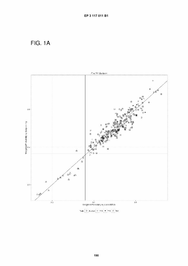
EP 3 117 011 B1
190
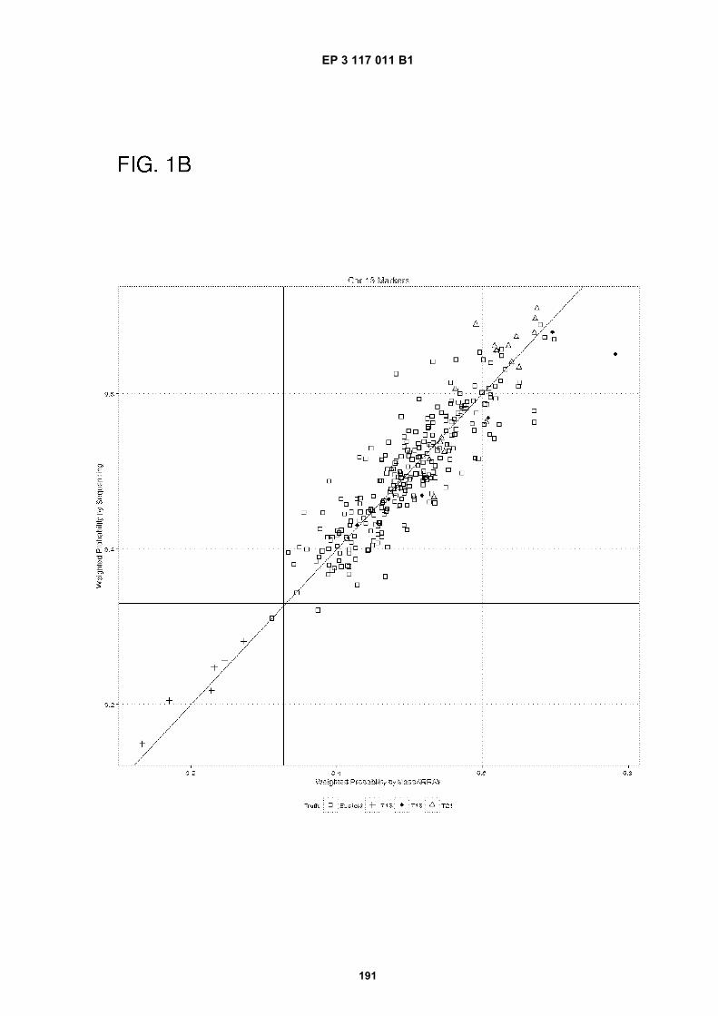
EP 3 117 011 B1
191
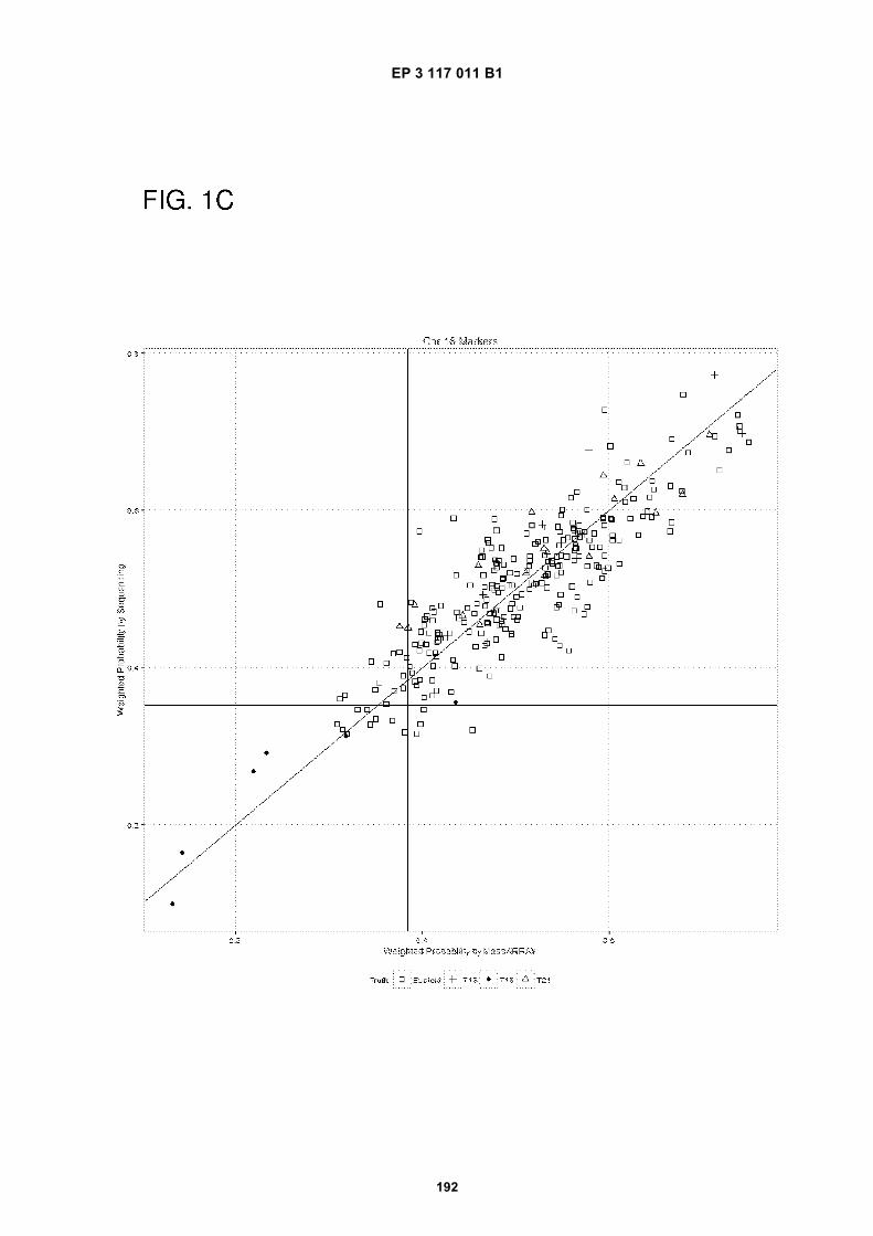
EP 3 117 011 B1
192
EP 3 117 011 B1
193
REFERENCES CITED IN THE DESCRIPTION
This list of references cited by the applicant is for the reader’s convenience only. It does not form part of the Europeanpatent document. Even though great care has been taken in compiling the references, errors or omissions cannot beexcluded and the EPO disclaims all liability in this regard.
Patent documents cited in the description
• WO 2011034631 A [0007]• US 20100105049 [0117] [0118]• US 20070065823 [0118]• US 20040081993 [0119]• US 20110224087 [0120]• WO 05023091 A2, Cantor [0128] [0130]• US 20070202525 A [0128] [0130] [0132]• US 20050287592 A [0130]
• US 4683195 A [0132]• US 4683202 A [0132]• US 20090029377 A [0138]• US 20130012399 A [0155]• US 2012038710 W [0181]• WO 2012159089 A [0181]• WO 2014011928 A [0266]
Non-patent literature cited in the description
• T. STRACHAN. The Human Genome. BIOS Scien-tific Publishers, 1992 [0002]
• D. N. COOPER ; M. KRAWCZAK. Human GenomeMutations. BIOS Publishers, 1993 [0003]
• Current Protocols in Molecular Biology. John Wiley& Sons, 1989 [0032]
• BEAUCAGE ; CARUTHERS. Tetrahedron Letts,1981, vol. 22, 1859-1862 [0053]
• NEEDHAM-VANDEVANTER et al. Nucleic AcidsRes., 1984, vol. 12, 6159-6168 [0053] [0055]
• PEARSON ; REGNIER. J. Chrom., 1983, vol. 255,137-149 [0053] [0055]
• BEAUCAGE ; CARUTHERS. Tetrahedron Letts.,1981, vol. 22, 1859-1862 [0055]
• Current Protocols in Molecular Biology. John Wiley& Sons, 1989, 6.3.1-6.3.6 [0059]
• SAMBROOK et al. MOLECULAR CLONING: A LAB-ORATORY MANUAL. Cold Spring Harbor Laborato-ry Press, 1990 [0059]
• LO. Journal of Histochemistry and Cytochemistry,2005, vol. 53 (3), 293-296 [0116]
• GEBHARD et al. Cancer Res., 2006, vol. 66 (12),6118-28 [0118]
• DING ; CANTOR. Proc.Natl.Acad.Sci. USA, 2003,vol. 100, 3059-3064 [0119]
• INNIS et al. PCR Protocols: A Guide to Methods andApplications. Academic Press, Inc, 1990 [0125]
• NOLTE. Adv. Clin. Chem., 1998, vol. 33, 201-235[0127]
• KALININA et al. Nanoliter scale PCR with TaqMandetection. Nucleic Acids Research, 1997, vol. 25,1999-2004 [0128] [0130]
• Digital PCR. Proc Natl Acad Sci U S A., 1999, vol.96, 9236-41 [0128]
• VINCENT et al. Helicase-dependent isothermal DNAamplification. EMBO reports, 2004, vol. 5 (8),795-800 [0130]
• VOGELSTEIN ; KINZLER. Digital PCR. Proc NatlAcad Sci U S A., 1999, vol. 96, 9236-41 [0130]
• Digital PCR is also known in the art. PCR Protocols:A Guide to Methods and Applications. 1990 [0132]
• JURINKE et al. Mol. Biotechnol., 2004, vol. 26,147-164 [0166]
• BURLINGAME et al. Anal. Chem., 1998, vol. 70,647R-716R [0168]
• VERBECK et al. the Journal of Biomolecular Tech-niques, vol. 13 (2), 56-61 [0172]
• DING ; CANTOR. PNAS USA, 18 March 2003, vol.100 (6), 3059-64 [0182]
Related Documents