Citation: Hytiroglou, P.; Bioulac-Sage, P.; Theise, N.D.; Sempoux, C. Etiology, Pathogenesis, Diagnosis, and Practical Implications of Hepatocellular Neoplasms. Cancers 2022, 14, 3670. https:// doi.org/10.3390/cancers14153670 Academic Editor: Alessandro Vitale Received: 10 June 2022 Accepted: 25 July 2022 Published: 28 July 2022 Publisher’s Note: MDPI stays neutral with regard to jurisdictional claims in published maps and institutional affil- iations. Copyright: © 2022 by the authors. Licensee MDPI, Basel, Switzerland. This article is an open access article distributed under the terms and conditions of the Creative Commons Attribution (CC BY) license (https:// creativecommons.org/licenses/by/ 4.0/). cancers Review Etiology, Pathogenesis, Diagnosis, and Practical Implications of Hepatocellular Neoplasms Prodromos Hytiroglou 1, * , Paulette Bioulac-Sage 2 , Neil D. Theise 3 and Christine Sempoux 4 1 Department of Pathology, Aristotle University School of Medicine, 54124 Thessaloniki, Greece 2 INSERM, BRIC, U1312, University Bordeaux, F-33000 Bordeaux, France; [email protected] 3 Department of Pathology, New York University Grossman School of Medicine, New York, NY 10016, USA; [email protected] 4 Service of Clinical Pathology, Institute of Pathology, Lausanne University Hospital, University of Lausanne, CH-1007 Lausanne, Switzerland; [email protected] * Correspondence: [email protected]; Tel.: +30-2310-999-218 Simple Summary: In recent years, significant progress has been made in elucidating the mechanisms via which hepatocellular neoplasms, i.e., hepatocellular adenoma and hepatocellular carcinoma, arise. Hepatocellular carcinoma usually occurs in livers with chronic disease, due to deregulation of impor- tant intracellular pathways of signal transmission. Recent studies suggest that subclassification of hepatocellular carcinoma is practically useful. On the other hand, subclassification of hepatocellular adenomas has been well established through correlation of molecular alterations with morphology and protein expression. Advances in hepatic imaging have resulted in a new approach for diagnostic assessment of lesions arising in advanced chronic liver disease. Histologic examination, aided by immunohistochemistry, is the gold standard for the diagnosis and subclassification of hepatocellular neoplasms, while clinicopathologic correlation is essential for best patient management. We summa- rize the etiology and pathogenesis of hepatocellular neoplasms, provide practical information for their histologic diagnosis, and address various frequently asked questions regarding their diagnosis and practical implications. Abstract: Hepatocellular carcinoma (HCC), a major global contributor of cancer death, usually arises in a background of chronic liver disease, as a result of molecular changes that deregulate important signal transduction pathways. Recent studies have shown that certain molecular changes of hepatocarcinogenesis are associated with clinicopathologic features and prognosis, suggesting that subclassification of HCC is practically useful. On the other hand, subclassification of hepatocellular adenomas (HCAs), a heterogenous group of neoplasms, has been well established on the basis of genotype–phenotype correlations. Histologic examination, aided by immunohistochemistry, is the gold standard for the diagnosis and subclassification of HCA and HCC, while clinicopathologic correlation is essential for best patient management. Advances in clinico-radio-pathologic correlation have introduced a new approach for the diagnostic assessment of lesions arising in advanced chronic liver disease by imaging (LI-RADS). The rapid expansion of knowledge concerning the molecular pathogenesis of HCC is now starting to produce new therapeutic approaches through precision oncology. This review summarizes the etiology and pathogenesis of HCA and HCC, provides practical information for their histologic diagnosis (including an algorithmic approach), and addresses a variety of frequently asked questions regarding the diagnosis and practical implications of these neoplasms. Keywords: hepatocellular adenoma; hepatocellular carcinoma; molecular pathology; histologic diagnosis; diagnostic algorithm; LI-RADS; frequently asked questions 1. Introduction In the past decade, application of novel methodologies of molecular medicine in hepa- tocellular neoplasms has significantly improved our understanding of the pathogenesis of Cancers 2022, 14, 3670. https://doi.org/10.3390/cancers14153670 https://www.mdpi.com/journal/cancers

Welcome message from author
This document is posted to help you gain knowledge. Please leave a comment to let me know what you think about it! Share it to your friends and learn new things together.
Transcript

Citation: Hytiroglou, P.;
Bioulac-Sage, P.; Theise, N.D.;
Sempoux, C. Etiology, Pathogenesis,
Diagnosis, and Practical Implications
of Hepatocellular Neoplasms.
Cancers 2022, 14, 3670. https://
doi.org/10.3390/cancers14153670
Academic Editor: Alessandro Vitale
Received: 10 June 2022
Accepted: 25 July 2022
Published: 28 July 2022
Publisher’s Note: MDPI stays neutral
with regard to jurisdictional claims in
published maps and institutional affil-
iations.
Copyright: © 2022 by the authors.
Licensee MDPI, Basel, Switzerland.
This article is an open access article
distributed under the terms and
conditions of the Creative Commons
Attribution (CC BY) license (https://
creativecommons.org/licenses/by/
4.0/).
cancers
Review
Etiology, Pathogenesis, Diagnosis, and Practical Implications ofHepatocellular NeoplasmsProdromos Hytiroglou 1,* , Paulette Bioulac-Sage 2, Neil D. Theise 3 and Christine Sempoux 4
1 Department of Pathology, Aristotle University School of Medicine, 54124 Thessaloniki, Greece2 INSERM, BRIC, U1312, University Bordeaux, F-33000 Bordeaux, France; [email protected] Department of Pathology, New York University Grossman School of Medicine, New York, NY 10016, USA;
[email protected] Service of Clinical Pathology, Institute of Pathology, Lausanne University Hospital, University of Lausanne,
CH-1007 Lausanne, Switzerland; [email protected]* Correspondence: [email protected]; Tel.: +30-2310-999-218
Simple Summary: In recent years, significant progress has been made in elucidating the mechanismsvia which hepatocellular neoplasms, i.e., hepatocellular adenoma and hepatocellular carcinoma, arise.Hepatocellular carcinoma usually occurs in livers with chronic disease, due to deregulation of impor-tant intracellular pathways of signal transmission. Recent studies suggest that subclassification ofhepatocellular carcinoma is practically useful. On the other hand, subclassification of hepatocellularadenomas has been well established through correlation of molecular alterations with morphologyand protein expression. Advances in hepatic imaging have resulted in a new approach for diagnosticassessment of lesions arising in advanced chronic liver disease. Histologic examination, aided byimmunohistochemistry, is the gold standard for the diagnosis and subclassification of hepatocellularneoplasms, while clinicopathologic correlation is essential for best patient management. We summa-rize the etiology and pathogenesis of hepatocellular neoplasms, provide practical information fortheir histologic diagnosis, and address various frequently asked questions regarding their diagnosisand practical implications.
Abstract: Hepatocellular carcinoma (HCC), a major global contributor of cancer death, usuallyarises in a background of chronic liver disease, as a result of molecular changes that deregulateimportant signal transduction pathways. Recent studies have shown that certain molecular changesof hepatocarcinogenesis are associated with clinicopathologic features and prognosis, suggesting thatsubclassification of HCC is practically useful. On the other hand, subclassification of hepatocellularadenomas (HCAs), a heterogenous group of neoplasms, has been well established on the basis ofgenotype–phenotype correlations. Histologic examination, aided by immunohistochemistry, is thegold standard for the diagnosis and subclassification of HCA and HCC, while clinicopathologiccorrelation is essential for best patient management. Advances in clinico-radio-pathologic correlationhave introduced a new approach for the diagnostic assessment of lesions arising in advanced chronicliver disease by imaging (LI-RADS). The rapid expansion of knowledge concerning the molecularpathogenesis of HCC is now starting to produce new therapeutic approaches through precisiononcology. This review summarizes the etiology and pathogenesis of HCA and HCC, provides practicalinformation for their histologic diagnosis (including an algorithmic approach), and addresses a varietyof frequently asked questions regarding the diagnosis and practical implications of these neoplasms.
Keywords: hepatocellular adenoma; hepatocellular carcinoma; molecular pathology; histologicdiagnosis; diagnostic algorithm; LI-RADS; frequently asked questions
1. Introduction
In the past decade, application of novel methodologies of molecular medicine in hepa-tocellular neoplasms has significantly improved our understanding of the pathogenesis of
Cancers 2022, 14, 3670. https://doi.org/10.3390/cancers14153670 https://www.mdpi.com/journal/cancers
Cancers 2022, 14, 3670 2 of 28
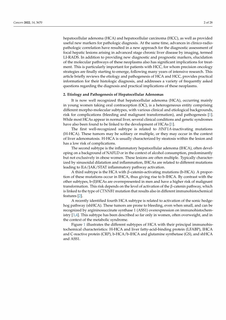
hepatocellular adenoma (HCA) and hepatocellular carcinoma (HCC), as well as provideduseful new markers for pathologic diagnosis. At the same time, advances in clinico-radio-pathologic correlation have resulted in a new approach for the diagnostic assessment offocal hepatic lesions arising in advanced stage chronic liver disease by imaging, termedLI-RADS. In addition to providing new diagnostic and prognostic markers, elucidationof the molecular pathways of these neoplasms also has significant implications for treat-ment. This is particularly important for patients with HCC, for whom precision oncologystrategies are finally starting to emerge, following many years of intensive research. Thisarticle briefly reviews the etiology and pathogenesis of HCA and HCC, provides practicalinformation for their histologic diagnosis, and addresses a variety of frequently askedquestions regarding the diagnosis and practical implications of these neoplasms.
2. Etiology and Pathogenesis of Hepatocellular Adenomas
It is now well recognized that hepatocellular adenoma (HCA), occurring mainlyin young women taking oral contraception (OC), is a heterogeneous entity comprisingdifferent morpho-molecular subtypes, with various clinical and etiological backgrounds,risk for complications (bleeding and malignant transformation), and pathogenesis [1].While most HCAs appear in normal liver, several clinical conditions and genetic syndromeshave also been found to be linked to the development of HCAs [1].
The first well-recognized subtype is related to HNF1A-inactivating mutations(H-HCA). These tumors may be solitary or multiple, or they may occur in the contextof liver adenomatosis. H-HCA is usually characterized by steatosis within the lesion andhas a low risk of complications.
The second subtype is the inflammatory hepatocellular adenoma (IHCA), often devel-oping on a background of NAFLD or in the context of alcohol consumption, predominantlybut not exclusively in obese women. These lesions are often multiple. Typically character-ized by sinusoidal dilatation and inflammation, IHCAs are related to different mutationsleading to IL6/JAK/STAT inflammatory pathway activation.
A third subtype is the HCA with β-catenin-activating mutations (b-HCA). A propor-tion of these mutations occur in IHCA, thus giving rise to b-IHCA. By contrast with theother subtypes, b-(I)HCAs are overrepresented in men and have a higher risk of malignanttransformation. This risk depends on the level of activation of the β-catenin pathway, whichis linked to the type of CTNNB1 mutation that results also in different immunohistochemicalfeatures [2].
A recently identified fourth HCA subtype is related to activation of the sonic hedge-hog pathway (shHCA). These tumors are prone to bleeding, even when small, and can berecognized by argininosuccinate synthase 1 (ASS1) overexpression on immunohistochem-istry [3,4]. This subtype has been described so far only in women, often overweight, and inthe context of the metabolic syndrome.
Figure 1 illustrates the different subtypes of HCA with their principal immunohis-tochemical characteristics: H-HCA and liver fatty-acid-binding protein (LFABP), IHCAand C-reactive protein (CRP), b-HCA/b-IHCA and glutamine synthetase (GS), and shHCAand ASS1.
Cancers 2022, 14, 3670 3 of 28Cancers 2022, 14, x FOR PEER REVIEW 3 of 29
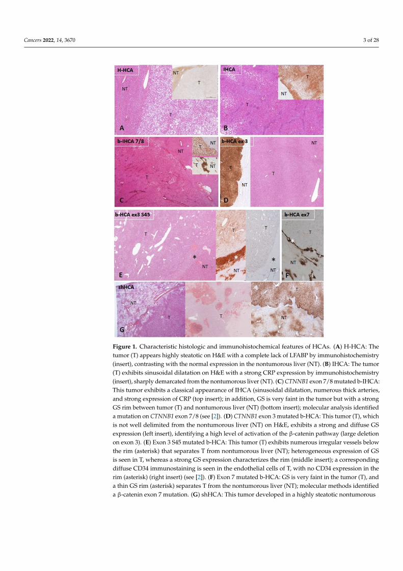
Figure 1. Characteristic histologic and immunohistochemical features of HCAs. (A) H-HCA: The tumor (T) appears highly steatotic on H&E with a complete lack of LFABP by immunohistochemis-try (insert), contrasting with the normal expression in the nontumorous liver (NT). (B) IHCA: The tumor (T) exhibits sinusoidal dilatation on H&E with a strong CRP expression by immunohisto-chemistry (insert), sharply demarcated from the nontumorous liver (NT). (C) CTNNB1 exon 7/8 mu-tated b-IHCA: This tumor exhibits a classical appearance of IHCA (sinusoidal dilatation, numerous thick arteries, and strong expression of CRP (top insert); in addition, GS is very faint in the tumor but with a strong GS rim between tumor (T) and nontumorous liver (NT) (bottom insert); molecular analysis identified a mutation on CTNNB1 exon 7/8 (see [2]). (D) CTNNB1 exon 3 mutated b-HCA: This tumor (T), which is not well delimited from the nontumorous liver (NT) on H&E, exhibits a strong and diffuse GS expression (left insert), identifying a high level of activation of the β-catenin pathway (large deletion on exon 3). (E) Exon 3 S45 mutated b-HCA: This tumor (T) exhibits numer-ous irregular vessels below the rim (asterisk) that separates T from nontumorous liver (NT); heter-ogeneous expression of GS is seen in T, whereas a strong GS expression characterizes the rim (mid-dle insert); a corresponding diffuse CD34 immunostaining is seen in the endothelial cells of T, with no CD34 expression in the rim (asterisk) (right insert) (see [2]). (F) Exon 7 mutated b-HCA: GS is very faint in the tumor (T), and a thin GS rim (asterisk) separates T from the nontumorous liver (NT); molecular methods identified a β-catenin exon 7 mutation. (G) shHCA: This tumor developed in a highly steatotic nontumorous liver (NT, left picture) and exhibits focally large hemorrhagic foci
Figure 1. Characteristic histologic and immunohistochemical features of HCAs. (A) H-HCA: Thetumor (T) appears highly steatotic on H&E with a complete lack of LFABP by immunohistochemistry(insert), contrasting with the normal expression in the nontumorous liver (NT). (B) IHCA: The tumor(T) exhibits sinusoidal dilatation on H&E with a strong CRP expression by immunohistochemistry(insert), sharply demarcated from the nontumorous liver (NT). (C) CTNNB1 exon 7/8 mutated b-IHCA:This tumor exhibits a classical appearance of IHCA (sinusoidal dilatation, numerous thick arteries,and strong expression of CRP (top insert); in addition, GS is very faint in the tumor but with a strongGS rim between tumor (T) and nontumorous liver (NT) (bottom insert); molecular analysis identifieda mutation on CTNNB1 exon 7/8 (see [2]). (D) CTNNB1 exon 3 mutated b-HCA: This tumor (T), whichis not well delimited from the nontumorous liver (NT) on H&E, exhibits a strong and diffuse GSexpression (left insert), identifying a high level of activation of the β-catenin pathway (large deletionon exon 3). (E) Exon 3 S45 mutated b-HCA: This tumor (T) exhibits numerous irregular vessels belowthe rim (asterisk) that separates T from nontumorous liver (NT); heterogeneous expression of GSis seen in T, whereas a strong GS expression characterizes the rim (middle insert); a correspondingdiffuse CD34 immunostaining is seen in the endothelial cells of T, with no CD34 expression in therim (asterisk) (right insert) (see [2]). (F) Exon 7 mutated b-HCA: GS is very faint in the tumor (T), anda thin GS rim (asterisk) separates T from the nontumorous liver (NT); molecular methods identifieda β-catenin exon 7 mutation. (G) shHCA: This tumor developed in a highly steatotic nontumorous

Cancers 2022, 14, 3670 4 of 28
liver (NT, left picture) and exhibits focally large hemorrhagic foci (middle picture); ASS1 immunohis-tochemistry shows an overexpression in the tumor (T), in comparison with the nontumorous liver(NT), in which its expression is restricted to the periportal/septal zones (right picture). Abbreviations:H-HCA, HNF1A-mutated hepatocellular adenoma; IHCA, inflammatory HCA; b-IHCA, β-catenin-mutated inflammatory HCA; b-HCA, β-catenin-mutated HCA; shHCA, sonic hedgehog-activatedHCA; LFABP, liver fatty-acid-binding protein; CRP, C reactive protein; GS, glutamine synthetase.
3. Diagnosis and Subtyping of Hepatocellular Adenomas
The histologic diagnosis of HCA requires careful assessment of representative hema-toxylin and eosin (H&E)-stained sections. HCAs are characterized by a benign hepatocellu-lar proliferation, devoid of portal tracts. “Unpaired” arteries (i.e., arteries unaccompaniedby veins or bile ducts) are present among the neoplastic cells. Other characteristic featuresinclude steatosis, inflammation, sinusoidal dilatation, and/or areas of hemorrhage. AfterH&E assessment, immunohistochemical evaluation follows with specific antibodies recog-nizing the targets identified by the genotype–phenotype studies [1,2]. An algorithm for thediagnosis is proposed in Figure 2.
Cancers 2022, 14, x FOR PEER REVIEW 4 of 29
(middle picture); ASS1 immunohistochemistry shows an overexpression in the tumor (T), in com-parison with the nontumorous liver (NT), in which its expression is restricted to the periportal/sep-tal zones (right picture). Abbreviations: H-HCA, HNF1A-mutated hepatocellular adenoma; IHCA, inflammatory HCA; b-IHCA, β-catenin-mutated inflammatory HCA; b-HCA, β-catenin-mutated HCA; shHCA, sonic hedgehog-activated HCA; LFABP, liver fatty-acid-binding protein; CRP, C re-active protein; GS, glutamine synthetase.
3. Diagnosis and Subtyping of Hepatocellular Adenomas The histologic diagnosis of HCA requires careful assessment of representative hema-
toxylin and eosin (H&E)-stained sections. HCAs are characterized by a benign hepatocel-lular proliferation, devoid of portal tracts. “Unpaired” arteries (i.e., arteries unaccompa-nied by veins or bile ducts) are present among the neoplastic cells. Other characteristic features include steatosis, inflammation, sinusoidal dilatation, and/or areas of hemor-rhage. After H&E assessment, immunohistochemical evaluation follows with specific an-tibodies recognizing the targets identified by the genotype–phenotype studies [1,2]. An algorithm for the diagnosis is proposed in Figure 2.
Figure 2. Diagnostic algorithm for HCAs. From a practical point of view, most of the cases are easily recognized as benign or malignant, but some are not. In the situation of an obvious HCA, if there is steatosis, with LFABP (−) and GS (−), there is no need to perform further IHC staining; it can be concluded that the tumor is an H-HCA. If an HCA shows sinusoidal dilatation and inflammation, with LFABP (+) and GS (−), it is mandatory to perform CRP and/or SAA immunostaining in order to diagnose an IHCA. GS immunostaining is mandatory in all IHCAs in order to diagnose a b-IHCA. Different patterns of GS staining exist, linked to the type of underlying mutations (see [2]). If LFABP is positive and all other markers are negative, then an overexpression of ASS1 will lead to the iden-tification of a shHCA, whereas, if it is not overexpressed, it is an UHCA. * Importantly, the GS(+)/CD34(−) rim can be irregular or discontinuous and is usually better represented in b-HCA
Figure 2. Diagnostic algorithm for HCAs. From a practical point of view, most of the cases are easilyrecognized as benign or malignant, but some are not. In the situation of an obvious HCA, if thereis steatosis, with LFABP (−) and GS (−), there is no need to perform further IHC staining; it can beconcluded that the tumor is an H-HCA. If an HCA shows sinusoidal dilatation and inflammation,with LFABP (+) and GS (−), it is mandatory to perform CRP and/or SAA immunostaining in orderto diagnose an IHCA. GS immunostaining is mandatory in all IHCAs in order to diagnose a b-IHCA.
Cancers 2022, 14, 3670 5 of 28
Different patterns of GS staining exist, linked to the type of underlying mutations (see [2]). If LFABP ispositive and all other markers are negative, then an overexpression of ASS1 will lead to the identifica-tion of a shHCA, whereas, if it is not overexpressed, it is an UHCA. * Importantly, the GS(+)/CD34(−)rim can be irregular or discontinuous and is usually better represented in b-HCA than in b-IHCA. Itsrecognition on biopsies can be challenging (see [2]). In case of an uncertain diagnosis, HCA versusHCC or HCA versus FNH, additional histochemical and immunohistochemical stains are needed.The differential diagnosis of HCA versus HCC is discussed in FAQ 1. Reticulin stain might help to rec-ognize alterations of the framework, although it is not a strict feature. Cytokeratin 7 and cytokeratin19 stains help to recognize ductular reaction, and GS has a specific map-like pattern in FNH. Abbrevia-tions: HCA, hepatocellular adenoma; H-HCA, HNF1A-mutated HCA; IHCA, inflammatory HCA;b-HCA, β-catenin-activated HCA; b-IHCA, β-catenin-activated and inflammatory HCA; shHCA,sonic hedgehog-activated HCA; UHCA, unclassified HCA; HCC, hepatocellular carcinoma; FNH,focal nodular hyperplasia; LFABP, liver fatty-acid-binding protein; CRP, C reactive protein; SAA,serum amyloid A; GS, glutamine synthetase; ASS1, argininosuccinate synthase; CK7, cytokeratin 7;CK19, cytokeratin 19; DR, ductular reaction.
4. Etiology and Pathogenesis of Hepatocellular Carcinoma
Hepatocellular carcinoma (HCC) usually arises in livers with chronic disease, and it ismost often discovered when disease has reached an advanced stage, traditionally knownas cirrhosis. The most common chronic diseases that are associated with HCC are chronichepatitis B, chronic hepatitis C, and alcoholic liver disease, accounting together for 84%of the cases occurring globally in 2015 [5]. In the meanwhile, nonalcoholic steatohepatitis(NASH), associated with the metabolic syndrome, is emerging as a major risk factor forHCC [6]. Other risk factors include hereditary metabolic disorders (such as hemochro-matosis, α1-antitrypsin deficiency, and tyrosinemia), aflatoxin B1 exposure (in individualschronically infected with HBV), and tobacco smoking. Chronic liver diseases other thanthose mentioned above (e.g., autoimmune hepatitis, primary biliary cholangitis, primarysclerosing cholangitis, and Wilson disease) are uncommonly associated with developmentof HCC.
HCC arising in noncirrhotic livers is often caused by HBV, which is a virus withknown carcinogenic effects. HBV DNA insertion in the host genome can deregulate genesinvolved in cell signaling and replication (such as TERT, PDGFR, MLL4, and CCNE1),while the HBV X protein transactivates genes involved in signal transduction pathwaysand inhibits TP53 expression [7–9]. NASH and hereditary hemochromatosis are alsoincreasingly recognized as causes of HCC arising in noncirrhotic livers [6,10]. However,HCC can also arise in apparently normal liver. Some of these cases may represent evolutionof HCA (mostly b-HCA and b-IHCA) to HCC (discussed in the previous sections), whileothers, usually occurring in older individuals, remain unexplained. A special HCC subtypearising in normal livers of young individuals is fibrolamellar carcinoma, which is associatedwith a characteristic somatic gene fusion, DNAJB1–PRKACA, resulting from deletions inchromosome 19 and activating protein kinase A [11].
In chronic liver diseases, continuous cell loss results in cell proliferation occurring in anoxious microenvironment, characterized by oxidative stress due to chronic inflammation,overexpression of growth factors, and epigenetic changes due to derangements of DNAmethyltransferases [12–14]. Thus, the possibility of mutations that initiate or promotecarcinogenesis is increased, while mutations providing survival benefits to hepatocytesfavor clonal expansion. This process is accelerated in the advanced stages of chronic liverdiseases when vascular changes, including intrahepatic vein thrombosis and vascularreorganization, result in extensive cell loss. In that setting, hepatic regeneration largelydepends on progenitor cell proliferation due to senescence of hepatocytes. Therefore,critical mutations in progenitor cells have the potential to produce large numbers of clonallyexpanding hepatocytes with increased likelihood to progress to precancerous lesions andthen to HCC.

Cancers 2022, 14, 3670 6 of 28
The diverse molecular changes that are associated with HCC have been recently re-viewed [15]. Whole-exome and whole-genome sequencing studies have revealed 40–60somatic coding mutations per HCC, including 4–6 driver mutations [16]. The most frequentmutations in HCC are those involving the promoter of telomerase reverse transcriptase(TERT), occurring in 60% of cases [17]. In an additional 30% of HCCs, TERT is deregulatedby other molecular mechanisms, such as viral insertion [18]. TERT promoter mutationshave also been detected in precancerous nodules and are considered an early event inhepatocarcinogenesis [19]. Other frequently mutated genes in HCC include CTNNB1,TP53, RB1, ARID1A, ARID2, AXIN1, albumin, and apolipoprotein B [20–22]. The muta-tions occurring in hepatocarcinogenesis can disrupt various signal transduction pathways,such as telomere maintenance (TERT), cell-cycle control (TP53, CDKN2A), Wnt/β-catenin(CTNNB1, AXIN1), epigenetic (ARID1A, ARID2, MLL2), and oxidative stress (NFE2L2,KEAP1) [17,23,24]. “Druggable” genetic alterations are under intense investigation because,at the present time, targeted therapeutic agents for HCC are limited to a small number ofmultikinase inhibitors. On the other hand, understanding the interaction between neo-plastic cells and their microenvironment will be crucial for identifying biomarkers anddeveloping new therapies based on immune checkpoint inhibition [25]
Recent studies have shown that certain molecular changes in HCC are associatedwith specific clinicopathologic features and prognosis, suggesting the possibility of amolecular classification for the future [26–29]. This active research has resulted in therecognition of several HCC subtypes (also called “variants”) that hold promise for a morepersonalized treatment of HCC patients. Eight HCC subtypes, considered to representdistinct clinicopathological/molecular entities and accounting together for up to 35% ofHCCs, have been included in the latest edition of the WHO classification of liver tumors [30].The characteristic features of these subtypes are briefly presented in Section 5. It shouldbe kept in mind that subclassification of HCC is a work in progress that will achievesignificantly more importance if it becomes useful from a therapeutic point of view.
5. Diagnosis of Hepatocellular Carcinoma
Diagnosis of HCC is traditionally made by histologic examination of biopsy, surgical,or autopsy specimens, and it is based on the recognition of two basic attributes in the histo-logic material: (i) hepatocellular differentiation, and (ii) malignancy. Features suggestinghepatocellular differentiation include resemblance of neoplastic cells to hepatocytes, bileproduction by neoplastic cells, positive immunostaining of neoplastic cells for “hepatocytic”markers, such as arginase-1 and carbamoyl phosphate synthetase-1 (recognized by theantibody HepPar1), and detection of albumin mRNA by in situ hybridization. Exceptfor bile production by neoplastic cells, none of the other features mentioned above isentirely specific for HCC. On the other hand, features indicating malignancy include stro-mal invasion, vascular invasion, metastatic spread, trabeculae thicker than three cells, andimmunopositivity of neoplastic cells for oncofetal antigens α-fetoprotein and/or glypican-3.
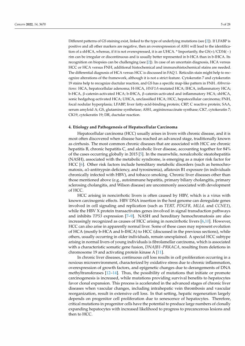
In addition to the most common trabecular growth pattern, HCCs often displaysolid (compact), pseudoglandular, and macrotrabecular patterns of growth, includingcombinations thereof. Similar to hepatocytes, the neoplastic cells may contain fat, glycogen(resulting in clear cell change), hyaline bodies, Mallory–Denk bodies, or pale bodies.Scattered arteries unaccompanied by veins or bile ducts (i.e., “unpaired” arteries) are acharacteristic histologic finding. Portal tracts are not a feature of classic HCC, except inthe invasive front of some tumors. Similar to other carcinomas, HCC is also histologicallyclassified as well, moderately and poorly differentiated [30] (Figure 3). Histologic diagnosisof poorly differentiated HCC is often difficult and requires immunohistochemical stains insupport of the diagnosis (arginase-1, HepPar1, α-fetoprotein, and glypican-3), as well asappropriate markers for other tumors that are included in the differential diagnosis, on acase-per-case basis.
Cancers 2022, 14, 3670 7 of 28Cancers 2022, 14, x FOR PEER REVIEW 7 of 29
Figure 3. Degrees of differentiation in HCC: (a) This well-differentiated HCC consists of neoplastic cells resembling hepatocytes, which are arranged in trabeculae and pseudoglandular structures. (b) As compared to (a), this moderately differentiated HCC displays an increased nuclear–cytoplasmic ratio, larger nuclei with prominent nucleoli, and increased cytoplasmic basophilia. (c) This poorly differentiated HCC is characterized by marked tumor cell pleomorphism, including multinucleated cells; the architecture is trabecular and compact. (d) Bile production by neoplastic cells, often in pseudoglandular structures, as illustrated here, is a diagnostic feature of HCC.
On the other hand, some HCCs are difficult to recognize histologically, especially in biopsy material, because of well-differentiated features. Absence of portal tracts and pres-ence of unpaired arteries in the biopsy material are features suggesting hepatocellular ne-oplasm, but do not allow distinction between HCA and well-differentiated HCC, while thin cell plates (<3 cells) do not exclude HCC. This difficult differential diagnosis is dis-cussed below (see FAQ 1). It is emphasized that correlation of clinical, radiologic, and pathologic findings is essential for correct classification of difficult cases. This is particu-larly true in the interpretation of biopsy material from small (<2 cm) nodular lesions in cirrhotic livers, where the differential diagnosis includes large regenerative nodule, dys-plastic nodule (low or high grade), early HCC, and classic HCC (see Section 6). This inter-pretation is facilitated when biopsy material from the hepatic parenchyma away from the lesion is available for comparison.
Early HCC (eHCC) has recently been recognized as a distinct step in hepatocarcino-genesis, characterized by ability for stromal invasion, but not for vascular invasion or met-astatic spread [31]. By definition, eHCC is a well-differentiated, early-stage tumor that measures less than 2 cm in diameter. On gross examination, eHCC often appears vaguely nodular, without distinct pushing boundaries or pseudocapsule, whereas small HCC of the classic (also called “progressed”) type usually has distinct boundaries marked by a pseudocapsule comprising compressed portal tracts or disease-associated scars [32]. Small classic HCCs tend to be better differentiated than larger ones, but have similar histologic features. On the other hand, many histologic features of eHCCs are reminiscent of those seen in high-grade dysplastic nodules. Early HCCs are usually composed of crowded, relatively small neoplastic cells, arranged in thin trabeculae and occasional small pseudo-glandular structures. High cellularity (more than twice that of the surrounding paren-chyma) and indistinct borders are characteristic features on low-power microscopic ex-amination. Unpaired arteries are usually sparse and small, as compared to those of classic
Figure 3. Degrees of differentiation in HCC: (a) This well-differentiated HCC consists of neoplasticcells resembling hepatocytes, which are arranged in trabeculae and pseudoglandular structures.(b) As compared to (a), this moderately differentiated HCC displays an increased nuclear–cytoplasmicratio, larger nuclei with prominent nucleoli, and increased cytoplasmic basophilia. (c) This poorlydifferentiated HCC is characterized by marked tumor cell pleomorphism, including multinucleatedcells; the architecture is trabecular and compact. (d) Bile production by neoplastic cells, often inpseudoglandular structures, as illustrated here, is a diagnostic feature of HCC.
On the other hand, some HCCs are difficult to recognize histologically, especiallyin biopsy material, because of well-differentiated features. Absence of portal tracts andpresence of unpaired arteries in the biopsy material are features suggesting hepatocellu-lar neoplasm, but do not allow distinction between HCA and well-differentiated HCC,while thin cell plates (<3 cells) do not exclude HCC. This difficult differential diagnosis isdiscussed below (see FAQ 1). It is emphasized that correlation of clinical, radiologic, andpathologic findings is essential for correct classification of difficult cases. This is particularlytrue in the interpretation of biopsy material from small (<2 cm) nodular lesions in cirrhoticlivers, where the differential diagnosis includes large regenerative nodule, dysplastic nod-ule (low or high grade), early HCC, and classic HCC (see Section 6). This interpretationis facilitated when biopsy material from the hepatic parenchyma away from the lesion isavailable for comparison.
Early HCC (eHCC) has recently been recognized as a distinct step in hepatocarcino-genesis, characterized by ability for stromal invasion, but not for vascular invasion ormetastatic spread [31]. By definition, eHCC is a well-differentiated, early-stage tumor thatmeasures less than 2 cm in diameter. On gross examination, eHCC often appears vaguelynodular, without distinct pushing boundaries or pseudocapsule, whereas small HCC ofthe classic (also called “progressed”) type usually has distinct boundaries marked by apseudocapsule comprising compressed portal tracts or disease-associated scars [32]. Smallclassic HCCs tend to be better differentiated than larger ones, but have similar histologicfeatures. On the other hand, many histologic features of eHCCs are reminiscent of thoseseen in high-grade dysplastic nodules. Early HCCs are usually composed of crowded,relatively small neoplastic cells, arranged in thin trabeculae and occasional small pseudog-landular structures. High cellularity (more than twice that of the surrounding parenchyma)and indistinct borders are characteristic features on low-power microscopic examination.Unpaired arteries are usually sparse and small, as compared to those of classic HCC. “En-

Cancers 2022, 14, 3670 8 of 28
trapped” portal tracts may be present in eHCC, especially in peripheral regions of thelesion. Steatosis is also often seen in eHCC, and it has been attributed to reduced oxygensupply compared to surrounding parenchyma [32]. On occasion, histologic examinationof hepatic nodules may reveal classic HCC arising within eHCC (Figure 4). The vascularsupply of eHCC (portal tract vessels and poorly developed unpaired arteries) significantlyoverlaps with that of dysplastic nodules; therefore, distinction between these lesions withimaging methods is difficult to impossible. The histologic features distinguishing eHCCfrom high-grade dysplastic nodules are discussed below (see Section 6).
Cancers 2022, 14, x FOR PEER REVIEW 8 of 29
HCC. “Entrapped” portal tracts may be present in eHCC, especially in peripheral regions of the lesion. Steatosis is also often seen in eHCC, and it has been attributed to reduced oxygen supply compared to surrounding parenchyma [32]. On occasion, histologic exam-ination of hepatic nodules may reveal classic HCC arising within eHCC (Figure 4). The vascular supply of eHCC (portal tract vessels and poorly developed unpaired arteries) significantly overlaps with that of dysplastic nodules; therefore, distinction between these lesions with imaging methods is difficult to impossible. The histologic features distin-guishing eHCC from high-grade dysplastic nodules are discussed below (see Section 6).
Figure 4. Classic HCC arising within early HCC (right and lower parts of the picture). Note the small unpaired arteries (right middle and lower part of the picture).
Table 1 provides a comparison of the etiology, pathogenesis, and diagnostically use-ful histopathologic features of HCA and HCC.
Table 1. Comparison of etiology, pathogenesis, and diagnostically useful histopathologic features of hepatocellular neoplasms.
Hepatocellular Adenoma Hepatocellular Carcinoma Etiology and Pathogenesis
Chronic liver disease Usually absent Usually present Molecular changes Four specific morpho-molecular Large variety of mutations
subtypes, including the following: affecting a number of signal - H-HCA: HNF1A-inactivating mutations transduction pathways; - IHCA: mutations activating IL6/JAK/STAT most frequent mutations
- b-HCA, b-IHCA: CTNNB1-activating mutations Involve TERT promoter
- shHCA: INHBE–GLI1 gene fusion Tumor architecture
Thickness of cell plates 1–2 cells Variable Pseudoglandular structures Absent or few Absent or present
Reticulin fibers Preserved or focally disorganized Decreased, disorganized Invasive growth in stroma or
vessels Absent Present
Figure 4. Classic HCC arising within early HCC (right and lower parts of the picture). Note the smallunpaired arteries (right middle and lower part of the picture).
Table 1 provides a comparison of the etiology, pathogenesis, and diagnostically usefulhistopathologic features of HCA and HCC.
HCC Subtypes
The steatohepatitic subtype of HCC occurs usually, but not exclusively, in patients withmetabolic syndrome or alcohol use and is characterized by histologic features similarto those of steatohepatitis occurring in nontumorous liver, i.e., macrovesicular steato-sis, inflammation, ballooned cells, Mallory–Denk bodies, and pericellular fibrosis [33,34](Figure 5a). This subtype was found to be associated with frequent IL6/JAK/STAT path-way activation, without CTNNB1, TERT, and TP53 alterations [29]. At this point in time,steatohepatitic HCC does not seem to prognostically differ from average classic HCC.
The clear cell subtype owes its appearance to glycogen accumulation in tumor cells, thussimulating clear-cell carcinoma of the kidney and other organs (Figure 5b). No characteristicmolecular alterations have been found in this subtype, which appears to be associated witha better-than-average prognosis [35]. Distinction from metastatic renal cell carcinoma mayrequire immunohistochemical stains for hepatocytic markers (arginase-1, HepPar1) andrenal transcription factor PAX-8.
The macrotrabecular massive subtype is histologically characterized by thick trabecu-lae, although the exact thickness (>6 cells vs. ≥10 cells thick) differs among authors [36](Figure 5c). This subtype is associated with high serum α-fetoprotein and poor progno-sis [29]. TP53 mutations and FGF19 amplifications are common in these tumors.
The scirrhous subtype is characterized by diffuse fibrosis, and it has been associatedwith TSC1/TSC2 mutations [29] (Figure 5d). The prognosis of this subtype does not appear

Cancers 2022, 14, 3670 9 of 28
to differ from the average classic HCC. On histologic examination, this subtype should bedistinguished from cholangiocarcinoma. Immunohistochemistry for hepatocytic markersarginase-1 and HepaPar1 is useful in this regard, whereas cytokeratin 7 is positive in mostscirrhous HCCs and almost all cholangiocarcinomas.
The chromophobe subtype is characterized by light staining cytoplasm of the neoplasticcells, mostly bland nuclei, as well as scattered cells with large atypical nuclei. Anothercharacteristic feature is the presence of scattered cystic spaces, filled with serum-likematerial. On a molecular basis, this subtype is characterized by alternative lengthening oftelomeres, a mechanism for telomere preservation without TERT promoter mutation [37].The prognosis of this subtype does not appear to differ from the average classic HCC.
Table 1. Comparison of etiology, pathogenesis, and diagnostically useful histopathologic features ofhepatocellular neoplasms.
Hepatocellular Adenoma Hepatocellular Carcinoma
Etiology and Pathogenesis
Chronic liver disease Usually absent Usually present
Molecular changes Four specific morpho-molecular Large variety of mutations
subtypes, including the following: affecting a number of signal
- H-HCA: HNF1A-inactivating mutations transduction pathways;- IHCA: mutations activating IL6/JAK/STAT most frequent mutations- b-HCA, b-IHCA: CTNNB1-activating mutations Involve TERT promoter
- shHCA: INHBE–GLI1 gene fusion
Tumor architecture
Thickness of cell plates 1–2 cells Variable
Pseudoglandular structures Absent or few Absent or present
Reticulin fibers Preserved or focally disorganized Decreased, disorganized
Invasive growth in stroma or vessels Absent Present
Cytologic features
Small cell size Uncommon Sometimes present
Nuclear hyperchromasia Uncommon Commonly present
Nuclear contour irregularities Uncommon Commonly present
Nuclear pleomorphism Uncommon Commonly present
Nuclear–cytoplasmic ratio Usually normal Often increased
Cytoplasmic basophilia Usually absent Commonly present
Mitotic figures Absent or rare Often present
Nonlesional hepatic parenchyma
Evidence of cirrhosis Absent (rarely present in IHCA) Present or absent
Positive immunohistochemical staining
Alpha-fetoprotein Absent Present or absent
Glypican-3 Absent Present or absent
The fibrolamellar subtype has long been considered a distinctive HCC variant occurringin young individuals (median age: 25 years) without liver disease. These tumors are welldifferentiated and consist of groups and trabeculae of large polygonal cells, separated bybands of lamellar fibrosis. The neoplastic cells have abundant eosinophilic cytoplasm,often displaying pale bodies, as well as large nuclei with prominent nucleoli (Figure 5e).In contrast to most other HCCs, those of the fibrolamellar subtype are positive for cytok-eratin 7 and CD68. Almost all fibrolamellar HCCs have the characteristic somatic genefusion DNAJB1–PRKACA, the detection of which can aid diagnosis [11]. The prognosis

Cancers 2022, 14, 3670 10 of 28
of fibrolamellar HCC is similar to that of classic well-differentiated HCC occurring innoncirrhotic liver.
Cancers 2022, 14, x FOR PEER REVIEW 10 of 29
cytokeratin 7 and CD68. Almost all fibrolamellar HCCs have the characteristic somatic gene fusion DNAJB1–PRKACA, the detection of which can aid diagnosis [11]. The prog-nosis of fibrolamellar HCC is similar to that of classic well-differentiated HCC occurring in noncirrhotic liver.
The neutrophil-rich subtype is characterized by abundant intratumoral neutrophils, due to granulocyte colony-stimulating factor (G-CSF) produced by neoplastic cells. Most tumors are poorly differentiated and may have sarcomatoid areas. The patients have ele-vated peripheral white blood cell counts, serum IL-6 levels, and often serum C-reactive protein. The prognosis of this subtype is worse than the average classic HCC [30].
The lymphocyte-rich subtype is characterized by abundant intratumoral lymphocytes. Cases tested for Epstein–Barr virus (EBV) were found to be negative. No prognostic sig-nificance has been attributed to this subtype. The lymphocyte-rich subtype should be dis-tinguished from lymphoepithelioma-like HCC, a rare, poorly differentiated carcinoma, com-posed of tumor cells growing in poorly defined groups within a dense lymphoplasmacytic infiltrate [36,38]. Most cases of this neoplasm, which has similar histologic features to na-sopharyngeal carcinoma and lymphoepithelioma-like carcinomas arising in other organs, have also been found to be negative for EBV.
Figure 5. Examples of HCC subtypes: (a) steatohepatitic; (b) clear cell; (c) macrotrabecular; (d) scir-rhous; (e) fibrolamellar; (f) sarcomatoid. Figure 5. Examples of HCC subtypes: (a) steatohepatitic; (b) clear cell; (c) macrotrabecular; (d) scir-rhous; (e) fibrolamellar; (f) sarcomatoid.
The neutrophil-rich subtype is characterized by abundant intratumoral neutrophils, dueto granulocyte colony-stimulating factor (G-CSF) produced by neoplastic cells. Most tumorsare poorly differentiated and may have sarcomatoid areas. The patients have elevatedperipheral white blood cell counts, serum IL-6 levels, and often serum C-reactive protein.The prognosis of this subtype is worse than the average classic HCC [30].
The lymphocyte-rich subtype is characterized by abundant intratumoral lymphocytes.Cases tested for Epstein–Barr virus (EBV) were found to be negative. No prognostic sig-nificance has been attributed to this subtype. The lymphocyte-rich subtype should bedistinguished from lymphoepithelioma-like HCC, a rare, poorly differentiated carcinoma,composed of tumor cells growing in poorly defined groups within a dense lymphoplasma-cytic infiltrate [36,38]. Most cases of this neoplasm, which has similar histologic features tonasopharyngeal carcinoma and lymphoepithelioma-like carcinomas arising in other organs,have also been found to be negative for EBV.

Cancers 2022, 14, 3670 11 of 28
In addition to lymphoepithelioma-like HCC, sarcomatoid HCC is another poorly dif-ferentiated variant that has not been recognized as a separate subtype in the latest editionof the WHO classification of liver tumors [30]. However, sarcomatoid HCC merits spe-cific mention because it has a poor prognosis, as well as a spindle cell morphology thatmimics various sarcomas [39] (Figure 5f). Extensive sampling may be required to revealareas of typical HCC in these tumors, while immunohistochemical stains demonstratingexpression of epithelial and hepatocytic markers can be useful, especially in cases withlimited histologic material. Heterologous differentiation may be found in these rare tumors,in which case the term carcinosarcoma is appropriately used. It should be kept in mindthat sarcomatoid change may develop in HCC following chemotherapy or transarterialchemoembolization [40].
The characteristic histologic and molecular findings of hepatocellular carcinoma sub-types are summarized in Table 2.
Table 2. Characteristic histologic and molecular findings of hepatocellular carcinoma subtypes.
Subtype Characteristic Histologic Findings Characteristic Molecular Findings
Steatohepatitic
Features simulating steatohepatitis(macrovesicular steatosis, inflammation,ballooned cells, Mallory–Denk bodies,
and pericellular fibrosis)
IL6/JAK/STAT pathway activation
Clear cell Glycogen accumulation in tumor cells None to date
Macrotrabecular massive Thick trabeculae (>6 cells thick) TP53 mutations, FGF19 amplifications
Scirrhous Diffuse fibrosis TSC1/TSC2 mutations
ChromophobeLight staining cytoplasm, mostly blandnuclei, occasional large atypical nuclei;cystic spaces with serum-like material
Alternative lengthening of telomeres
Fibrolamellar
Large polygonal cells with abundanteosinophilic cytoplasm, large nuclei andprominent nucleoli; pale bodies; lamellar
fibrosis; immunopositivity forcytokeratin 7
and CD68
DNAJB1–PRKACA gene fusion
Neutrophil-rich Abundant intratumoral neutrophils G-CSF production by neoplastic cells
Lymphocyte-rich Abundant intratumoral lymphocytes None to date
Sarcomatoid Spindle cell morphology None to date
6. Precancerous Lesions in Hepatocarcinogenesis
Clonal populations of hepatocytes bearing molecular alterations of the early stepsof carcinogenesis may be morphologically recognized in chronically diseased livers asprecancerous lesions. These include the following [41]:
(i) dysplastic foci (DFs), which are incidentally detected on microscopic examination andmeasure less than 1 mm in diameter;
(ii) dysplastic nodules (DNs), which are larger than dysplastic foci, occasionally mea-suring over 1 cm in diameter, and may be detected on imaging studies and grossexamination
The diagnosis of both DFs and DNs is made by histologic examination. Detection ofsuch lesions is associated with an increased risk of HCC.
DFs are most commonly composed of hepatocytes with small cell change forminga roundish area with increased proliferative activity, as compared to the surroundingparenchyma. Small cell change is characterized by small cell size, increased nuclear–cytoplasmic ratio, mild nuclear pleomorphism and hyperchromasia, and cytoplasmic ba-sophilia [42]. Small cell change of hepatocytes cytologically resembles early HCC. In livers

Cancers 2022, 14, 3670 12 of 28
with hereditary hemochromatosis, DFs are characterized by resistance to iron accumulation(“iron-free foci”) [43].
DNs are grossly defined on the basis of comparisons to surrounding liver tissue as“distinctive nodules”. They are most typically distinctive in terms of size, being larger thansurrounding cirrhotic nodules [31,44]. However, they may also differ in terms of color(yellow if steatotic, tan-white if fibrotic, dark brown or black if iron-retentive, and green ifcholestatic). These lesions are not distinguishable from small HCCs on gross examination.Confirmation that a distinctive nodule is a DN rather than HCC depends on histologicexamination. DNs may display cytologic and architectural atypia, but to a degree that isinsufficient for a diagnosis of HCC. Most consistently, DNs contain portal tracts, sometimesin a virtually normal distribution, while small, classic HCCs will have destroyed these orpushed them out of the way as they expand. Small classic HCCs will also often display allthe histologic features of larger HCCs, such as overt cytologic atypia and thick trabeculae.Distinction between DNs and eHCC is more difficult; this is why eHCC was internationallyrecognized as an entity only in 2009 [31]. The histologic and immunohistochemical featuresthat are useful for this distinction are discussed below. Sometimes, there are subnoduleswith features histologically suggestive of HCC within a DN; this is evidence of the DN’spremalignant nature and is also further discussed below.
6.1. Low-Grade vs. High-Grade Dysplastic Nodules
DNs are subclassified in two categories, low-grade (LGDNs) and high-grade(HGDNs) [41]. LGDNs are lacking cellular atypia or architectural atypia that wouldbe suspicious for HCC, although they may have large cell change. HGDNs are defined ashaving cytologic atypia (increased nuclear–cytoplasmic ratio, mild nuclear contour irregu-larities and hyperchromasia, cytoplasmic basophilia, and small cell change), or architecturalatypia (thickened—but less than three cells thick—trabeculae, occasional pseudoglandularstructures), which are reminiscent of an emerging HCC but insufficiently extensive toconfidently denote a fully progressed HCC. HGDNs may display nodule-in-nodule type ofgrowth, with a distinctive subnodule showing more atypical features. Sometimes the subn-odule will merely be more expansile than the surrounding DN parenchyma with increasedproliferation producing a “pushing border” at its edges. On occasion, the subnodule willbe an overt HCC, displaying stromal invasion into portal tracts or fibrous septa containedwithin the surrounding DN (Figure 6) [45].
DNs are now understood to represent clonal neoplastic expansions of cells that oftendevelop long before advanced stage liver disease is established [44,46]. They are generallylesions with low proliferation compared to surrounding, hyperplastic cirrhotic nodules [47].(Figure 7). DNs are able to spread, however, because they are also resistant to apoptosis.This resistance gives them a slight survival advantage compared to non-neoplastic hepato-cytes in adjacent parenchyma which, in response to the underlying chronic liver disease,have increased turnover [44]. The measure of how slight this advantage must be is thatthey may take many years to achieve sizes of up to 1.5 cm. DNs’ resistance to the diseaseaffecting the liver as a whole is also evidenced by diminished activation of hepatic stellatecells (HSCs) leading to an absence of scar within the DN or at least diminished scarringcompared to the rest of the liver (Figure 7) [48].
6.2. Low-Grade Dysplastic Nodules vs. Large Regenerative Nodules
In early studies of DNs in sequential cirrhotic explants, the primary criterion foridentifying DNs was a size cutoff (either 0.8 or 1.0 cm, depending on the study). Themajority of livers containing DNs have a small number, rarely over 10; however, a subsetof liver explants in patients with “macronodular cirrhosis” following either autoimmunehepatitis or hepatitis B had “uncountable” numbers of DNs by this criterion [49]. None ofthese were HGDN and none of the livers had HCC. Thus, it was clear that sometimes largeregenerative nodules (LRNs) can mimic LGDNs.

Cancers 2022, 14, 3670 13 of 28
Cancers 2022, 14, x FOR PEER REVIEW 12 of 29
with hereditary hemochromatosis, DFs are characterized by resistance to iron accumula-tion (“iron-free foci”) [43].
DNs are grossly defined on the basis of comparisons to surrounding liver tissue as “distinctive nodules”. They are most typically distinctive in terms of size, being larger than surrounding cirrhotic nodules [31,44]. However, they may also differ in terms of color (yellow if steatotic, tan-white if fibrotic, dark brown or black if iron-retentive, and green if cholestatic). These lesions are not distinguishable from small HCCs on gross ex-amination. Confirmation that a distinctive nodule is a DN rather than HCC depends on histologic examination. DNs may display cytologic and architectural atypia, but to a de-gree that is insufficient for a diagnosis of HCC. Most consistently, DNs contain portal tracts, sometimes in a virtually normal distribution, while small, classic HCCs will have destroyed these or pushed them out of the way as they expand. Small classic HCCs will also often display all the histologic features of larger HCCs, such as overt cytologic atypia and thick trabeculae. Distinction between DNs and eHCC is more difficult; this is why eHCC was internationally recognized as an entity only in 2009 [31]. The histologic and immunohistochemical features that are useful for this distinction are discussed below. Sometimes, there are subnodules with features histologically suggestive of HCC within a DN; this is evidence of the DN’s premalignant nature and is also further discussed below.
6.1. Low-Grade vs. High-Grade Dysplastic Nodules DNs are subclassified in two categories, low-grade (LGDNs) and high-grade
(HGDNs) [41]. LGDNs are lacking cellular atypia or architectural atypia that would be suspicious for HCC, although they may have large cell change. HGDNs are defined as having cytologic atypia (increased nuclear–cytoplasmic ratio, mild nuclear contour irreg-ularities and hyperchromasia, cytoplasmic basophilia, and small cell change), or architec-tural atypia (thickened—but less than three cells thick—trabeculae, occasional pseudo-glandular structures), which are reminiscent of an emerging HCC but insufficiently ex-tensive to confidently denote a fully progressed HCC. HGDNs may display nodule-in-nodule type of growth, with a distinctive subnodule showing more atypical features. Sometimes the subnodule will merely be more expansile than the surrounding DN paren-chyma with increased proliferation producing a “pushing border” at its edges. On occa-sion, the subnodule will be an overt HCC, displaying stromal invasion into portal tracts or fibrous septa contained within the surrounding DN (Figure 6) [45].
Figure 6. HCC (central and right part of the picture) arising in dysplastic nodule (left part). Thetumor has features of early HCC (central part) and classic HCC with steatosis (right part).
In resection specimens, histologic distinctions between LGDN and LRN can be coun-terintuitive. LGDNs are more likely to show relatively preserved, even “normal appearing”parenchymal architecture, while LRNs may show significant disturbances of organizationand function, such as variably regenerative or atrophic hepatocytes, large cell change, andhepatocyte injury such as ballooning or cholestasis. Thus, paradoxically, the neoplasticlesions, LGDNs, will appear more like normal liver, while the hyperplastic LRNs willappear reactive and, therefore, abnormal.
Cancers 2022, 14, x FOR PEER REVIEW 13 of 29
Figure 6. HCC (central and right part of the picture) arising in dysplastic nodule (left part). The tumor has features of early HCC (central part) and classic HCC with steatosis (right part).
DNs are now understood to represent clonal neoplastic expansions of cells that often develop long before advanced stage liver disease is established [44,46]. They are generally lesions with low proliferation compared to surrounding, hyperplastic cirrhotic nodules [47]. (Figure 7). DNs are able to spread, however, because they are also resistant to apop-tosis. This resistance gives them a slight survival advantage compared to non-neoplastic hepatocytes in adjacent parenchyma which, in response to the underlying chronic liver disease, have increased turnover [44]. The measure of how slight this advantage must be is that they may take many years to achieve sizes of up to 1.5 cm. DNs’ resistance to the disease affecting the liver as a whole is also evidenced by diminished activation of hepatic stellate cells (HSCs) leading to an absence of scar within the DN or at least diminished scarring compared to the rest of the liver (Figure 7) [48].
Figure 7. Important histologic features in hepatocellular nodules emerging in chronically diseased livers, and their LIRADS correlation. Abbreviations: LRN, large regenerative nodule; LGDN, low-grade dysplastic nodule; HGDN, high-grade dysplastic nodule; eHCC, early hepatocellular carci-noma; HCC, classic (progressed) hepatocellular carcinoma; HSC, hepatic stellate cell.
6.2. Low-Grade Dysplastic Nodules vs. Large Regenerative Nodules In early studies of DNs in sequential cirrhotic explants, the primary criterion for iden-
tifying DNs was a size cutoff (either 0.8 or 1.0 cm, depending on the study). The majority of livers containing DNs have a small number, rarely over 10; however, a subset of liver explants in patients with “macronodular cirrhosis” following either autoimmune hepatitis or hepatitis B had “uncountable” numbers of DNs by this criterion [49]. None of these were HGDN and none of the livers had HCC. Thus, it was clear that sometimes large regenerative nodules (LRNs) can mimic LGDNs.
In resection specimens, histologic distinctions between LGDN and LRN can be coun-terintuitive. LGDNs are more likely to show relatively preserved, even “normal appear-ing” parenchymal architecture, while LRNs may show significant disturbances of organ-ization and function, such as variably regenerative or atrophic hepatocytes, large cell change, and hepatocyte injury such as ballooning or cholestasis. Thus, paradoxically, the neoplastic lesions, LGDNs, will appear more like normal liver, while the hyperplastic LRNs will appear reactive and, therefore, abnormal.
Figure 7. Important histologic features in hepatocellular nodules emerging in chronically diseasedlivers, and their LIRADS correlation. Abbreviations: LRN, large regenerative nodule; LGDN, low-gradedysplastic nodule; HGDN, high-grade dysplastic nodule; eHCC, early hepatocellular carcinoma;HCC, classic (progressed) hepatocellular carcinoma; HSC, hepatic stellate cell.

Cancers 2022, 14, 3670 14 of 28
If the nodule has some distinctive features that might suggest clonality, this wouldsupport a diagnosis of LGDN over LRN. Such changes include diffuse iron or copperaccumulation not seen in the surrounding liver or diffuse steatosis, with or without steato-hepatitis, in the absence of background fatty liver disease. These findings favor the nodulebeing a true neoplasm. If one wishes to be more certain, one could do further studies toexamine hepatocyte proliferation rates and HSC activation (both low in LGDN and highin LRN) (Figure 7) [47,48]. Moreover, LRNs lack unpaired arteries indicating neoplasia-associated angiogenesis, while LGDNs often have many such vessels (Figure 7) [50,51].However, in many instances, the distinction between LGDN and LRN may be impossible,particularly in biopsy samples, but even when the whole nodule is present in a resection orautopsy specimen [31].
6.3. High-Grade Dysplastic Nodules vs. Hepatocellular Carcinoma
Distinguishing HGDN from well-differentiated HCC can be challenging, especiallyon needle biopsy material. Recognition of invasive properties, in the stroma or vessels,a hallmark of malignancy (Figure 8), is obviously of paramount importance, but is oftendifficult to detect. Stromal invasion is the feature distinguishing eHCC from HGDN,and it is suspected when hepatocytes, even some without significant atypia, are presentwithin the stroma of a portal tract or a septum in a large nodule. In such cases, absenceof a ductular reaction, confirmed by immunohistochemical stains for cytokeratins 7 or 19,will support the presence of stromal invasion and, therefore, the diagnosis of HCC [45].Immunohistochemistry can also be useful in biopsy material from nodules where HCC issuspected despite the lack of any evidence of invasion. Immunopositivity of lesional cellsfor two out of three markers, including glypican-3, glutamine synthetase, and HSP70, isconsidered diagnostic for HCC (either early or classic), whereas positivity for one or nomarker does not resolve the issue of differential diagnosis between HGDN and HCC [52,53].
Cancers 2022, 14, x FOR PEER REVIEW 14 of 29
If the nodule has some distinctive features that might suggest clonality, this would support a diagnosis of LGDN over LRN. Such changes include diffuse iron or copper ac-cumulation not seen in the surrounding liver or diffuse steatosis, with or without steato-hepatitis, in the absence of background fatty liver disease. These findings favor the nodule being a true neoplasm. If one wishes to be more certain, one could do further studies to examine hepatocyte proliferation rates and HSC activation (both low in LGDN and high in LRN) (Figure 7) [47,48]. Moreover, LRNs lack unpaired arteries indicating neoplasia-associated angiogenesis, while LGDNs often have many such vessels (Figure 7) [50,51]. However, in many instances, the distinction between LGDN and LRN may be impossible, particularly in biopsy samples, but even when the whole nodule is present in a resection or autopsy specimen [31].
6.3. High-Grade Dysplastic Nodules vs. Hepatocellular Carcinoma Distinguishing HGDN from well-differentiated HCC can be challenging, especially
on needle biopsy material. Recognition of invasive properties, in the stroma or vessels, a hallmark of malignancy (Figure 8), is obviously of paramount importance, but is often difficult to detect. Stromal invasion is the feature distinguishing eHCC from HGDN, and it is suspected when hepatocytes, even some without significant atypia, are present within the stroma of a portal tract or a septum in a large nodule. In such cases, absence of a duct-ular reaction, confirmed by immunohistochemical stains for cytokeratins 7 or 19, will sup-port the presence of stromal invasion and, therefore, the diagnosis of HCC [45]. Immuno-histochemistry can also be useful in biopsy material from nodules where HCC is sus-pected despite the lack of any evidence of invasion. Immunopositivity of lesional cells for two out of three markers, including glypican-3, glutamine synthetase, and HSP70, is con-sidered diagnostic for HCC (either early or classic), whereas positivity for one or no marker does not resolve the issue of differential diagnosis between HGDN and HCC [52,53].
Figure 8. Well-differentiated HCC invading portal tract and fibrous septum in liver with advanced stage chronic hepatitis C. Note the absence of ductular reaction.
Figure 8. Well-differentiated HCC invading portal tract and fibrous septum in liver with advancedstage chronic hepatitis C. Note the absence of ductular reaction.

Cancers 2022, 14, 3670 15 of 28
7. Frequently Asked Questions
FAQ 1—Can all hepatocellular neoplasms be definitely classified as either benignor malignant?
Recognizing a hepatocellular proliferation as benign is usually relatively easy, butcan be difficult or even impossible in some cases. In livers with advanced chronic dis-ease, the differential diagnosis is basically between high-grade dysplastic nodule andwell-differentiated HCC (early or classic). An algorithmic approach to this differentialdiagnosis has recently been proposed [54]. In livers without chronic disease the difficultiesin distinguishing HCA from well-differentiated HCC have long been recognized by experi-enced liver pathologists and are variably termed in the literature as “atypical hepatocellularadenoma/neoplasm”, “HCA with borderline features”, and “hepatocellular neoplasm withuncertain malignant potential” [1,55]. The worrisome features for the pathologist includearchitectural abnormalities, such as thickening of liver cell plates, presence of more thanoccasional pseudoglandular structures, and reticulin disorganization or disappearance,as well as cytological atypia, including presence of small cells, nuclear hyperchromasia,nuclear contour irregularities, nuclear pleomorphism, increased nuclear–cytoplasmic ratio,cytoplasmic basophilia, and presence of more than rare mitotic figures (see Table 1). Insuch cases, a careful search for features that allow a definite diagnosis of HCC (such asstromal or vascular invasion, trabeculae thicker than three cells, or immunopositivity forthe oncofetal proteins α-fetoprotein and glypican-3) is warranted. However, despite carefulhistopathologic assessment, this differential diagnosis may occasionally remain unresolved.Detection of TERT promoter mutation, a marker of approximately 60% of HCCs [17], wouldbe an argument for malignancy in such borderline lesions and holds promise as a diagnostictool for the future. From a practical point of view, it is currently recommended to indicatethis diagnostic difficulty in the report, especially when dealing with a biopsy specimen, inorder to trigger appropriate clinical management and/or surveillance.
FAQ 2—Some HCCs arise in completely normal liver. Do these HCCs arise from HCAs?HCAs are monoclonal neoplasms carrying a risk of malignant transformation reported
to be in the range of 4–10%, depending on the series [1]. This percentage is obviously biasedbecause (a) some lesions do not get a biopsy, and (b) many HCAs measuring more than5 cm are surgically resected or ablated before expressing any potential to evolve to HCC.Since the majority of HCAs arise in normal livers, HCCs arising from and replacing HCAswill also be surrounded by normal hepatic parenchyma, except when an adenomatousrim will still be present at the periphery of the HCC. On the other hand, a minority ofHCCs are discovered in normal livers, raising the possibility of a preexisting HCA thatcannot be morphologically recognized. None of the immunohistochemical or moleculartools used to diagnose the different subtypes of HCA are useful at this point, because theirexpression can be modified in malignant lesions; LFABP can be decreased in HCC [56],CRP can be expressed by some HCCs [57], and CTNNB1 mutations are commonly found inHCC. Therefore, none of these markers can be used for an argument to prove that an HCCarose from an HCA [1]. It is important for the pathologist to check the past medical andimaging history in order to identify clues of a preexisting HCA.
FAQ 3—Do HCAs arise in cirrhotic livers?Theoretically, the definition of cirrhosis (i.e., a stage in the evolution of chronic liver
diseases characterized by scarring and diffuse development of nodules) should not excludethe possibility of HCA of any subtype occurring in cirrhotic livers. However, the clinicalcontext of HCA development is different from chronic liver disease, and pathologists arehesitant to make a diagnosis of HCA in cirrhotic livers. To date, the only HCA subtype thathas been reported in livers with cirrhosis is IHCA. Rare IHCAs have been well documentedin advanced-stage fatty liver disease, associated with alcohol or metabolic syndrome,with characteristic pathologic, immunohistochemical (overexpression of SAA/CRP), andmolecular (different somatic mutations leading to IL6/JAK/STAT pathway activation)features [58,59]. In this context, one must be very cautious and not assert the diagnosis ofIHCA only on the basis of immunohistochemical features, since cirrhotic nodules, large

Cancers 2022, 14, 3670 16 of 28
regenerative nodules, and dysplastic nodules can overexpress SAA or CRP [58]. Therefore,it is necessary to confirm the presence of a specific IHCA mutation by molecular analysisbefore reaching a diagnosis of IHCA developing in cirrhotic liver. A fortiori, it is notadvisable to affirm this diagnosis on a needle biopsy. As mentioned above, HCC canexpress CRP, independently from the development in a preexisting IHCA [29].
FAQ 4—Are there any minimum requirements for the use of immunohistochemistryin the diagnosis of HCA?
After confirming that a tumor is an HCA on the basis of H&E-stained sections, it isimportant to define the subtype, which will determine further patient management. Thechoice of immunohistochemical stains depends on the pathological features, as demon-strated in Figure 2. If the tumor is highly steatotic, LFABP is mandatory to assert thediagnosis of H-HCA, provided nontumoral liver with normal expression of LFABP isavailable for comparison. If the tumor exhibits inflammatory features, sinusoidal dilatation,thick arteries, and pseudoportal tracts, CRP and/or SAA is first requested and will lead tothe diagnosis of IHCA, if overexpressed. Of note, some H-HCA can be devoid of steatosisand some IHCA can show very little inflammation or show steatosis, which makes bothimmunostains (LFABP and CRP) useful for the right diagnosis in such cases. On the otherhand, in case of a completely characteristic H-HCA, with steatosis and loss of LFABP expres-sion, one can easily conclude that this is the diagnosis. However, in routine practice, even ifa step-by-step approach seems to be logical, most of the time, LFABP, CRP, and glutaminesynthetase (GS) are determined from the beginning in order to save time and materials. GSis mandatory for three reasons: (1) this marker is very useful to recognize and differentiatethe tumoral area from the non-tumoral liver, something not always easy, particularly onbiopsy specimens (GS in nontumoral liver is expressed only in a few rows of hepatocytesaround the central veins); (2) GS helps to rule out focal nodular hyperplasia (FNH) in caseof doubt (absence of classical map-like staining pattern in HCA); (3) GS is the major tool todiagnose CTNNB1-mutated HCA with or without associated inflammation allowing thediagnosis of b-HCA and b-IHCA. If GS is strong and diffuse, it means that there is a highlevel of activation of the β-catenin pathway (most likely due to exon 3 non-S45 mutation).Lower levels of activation of this pathway exist [60], and the pattern of GS expressionis a good reflection of this phenomenon, with different immunohistochemical featuressuggesting different underlying molecular abnormalities, such as at the hotspot S45 ofexon 3 or in exon 7/8, resulting in a moderate or low level of β-catenin pathway activation,respectively; in these latter cases, the diffuse CD34 staining in the tumor endothelial cells,except at the peripheral rim, is a good additional argument for the diagnosis [1,2].
It is emphasized that GS is mandatory in all IHCAs in order to reach a diagnosis ofb-IHCA, which has the same risk of developing malignant transformation as b-HCA in thecase of high-level β-catenin pathway activation. GS immunohistochemistry is much morereliable than β-catenin immunohistochemistry, which is not sensitive enough to identifyCTNNB1-mutated HCAs. Indeed, this is positive only when GS is strongly expressed and,most of the times, positivity is focal, in a few nuclei. Therefore, there is no need to performβ-catenin immunostaining in HCA subtypes other than b-HCA or b-IHCA.
If LFABP is normally expressed, and stains for CRP and GS are negative, ASS1 is auseful new marker allowing to diagnose shHCA [3,4]. While ASS1 is normally expressed innontumor liver with a periportal/periseptal pattern (“honeycomb pattern”), overexpressionin tumor cells, as compared to nontumor is a requirement in order to make the diagnosis ofshHCA. It is important to recognize shHCAs because of their high risk of bleeding. Thealgorithm (Figure 2) summarizes how to proceed in daily practice.
FAQ 5—Do molecular studies provide any benefit in terms of diagnosis or prognosisof HCA, as compared to standard immunohistochemical stains?
In routine diagnosis, standard immunohistochemical stains (i.e., LFABP, CRP, andGS) are sufficient, most of the time, for the diagnosis of H-HCA, IHCA, b-HCA, and b-IHCA, together representing more than 90% of HCA cases. There is no further benefit toidentify inactivation of the HNF1A gene by molecular analysis or to search which mutation

Cancers 2022, 14, 3670 17 of 28
leads to IL6/JAK/STAT pathway activation, in order to reach a diagnosis of H-HCA orIHCA, respectively.
Concerning the β-catenin pathway, if GS immunostaining is strong and diffuse, itrepresents evidence that the activation level is high, which means a probable mutationin exon 3, not at the S45 hotspot. In this situation, there is no added value to searchwhich hotspot of exon 3 is mutated for patient management decisions. Indeed, it is wellknown that these b-HCA/b-IHCA cases have to be resected since they have a high risk ofmalignant transformation. When the GS immunostaining is heterogeneous or very faint,when the GS-positive peripheral rim is not obvious, particularly in biopsy specimens, orwhen there are technical problems with immunohistochemistry, molecular methods areuseful to search for mutations in exon 3 S45 or exon 7/8, the latter having a very lowpotential of malignant transformation but a high risk of bleeding, which makes recognitionon biopsy material important for further patient management.
Many molecular analyses, such as those concerning CTNNB1 mutations, can be per-formed today on formalin-fixed, paraffin-embedded tissue (FFPET), which is easier toobtain than frozen tissue. However, DNA of FFPET may be degraded and, therefore,without value for molecular analysis.
Regarding prognosis of HCA, it has been proposed to search for TERT promotermutations (this is feasible on FFPET) as evidence of malignancy. This would be particularlyuseful in cases of b-HCA and b-IHCA, when atypical features are present.
In summary, apart from research protocols in referral centers, molecular studies indaily practice add value in terms of subtype diagnosis in b-HCA and b-IHCA, but are notnecessary to determine prognosis when resection is mandatory (i.e., men and malignanttransformation).
FAQ 6—Should there be different guidelines for the treatment of different types of HCA?So far, the literature and the existing guidelines [61] indicate that (1) CTNNB1-mutated
HCAs must be surgically resected or ablated, even if they measure less than 5 cm, (2) HCAsoccurring in men also have to be resected or ablated, (3) any HCA measuring more than 5cm should be resected or ablated, and (4) any HCA that is causing symptoms should beresected or ablated. Emerging evidence from the recent literature suggests that managementshould be adapted to the subtype more than to the size of the tumors [62]. In cases ofadenomatosis, most residual HCAs after resection stabilize or regress, if steatohepatitisand obesity are corrected and/or the OC is discontinued; however, this evolution can takesome time [63].
H-HCAs are usually indolent, even if they are large, and they can remain for yearswithout regression and without giving rise to complications, except if they occur in specificclinical contexts, such as vascular liver diseases [64]. Not all b-HCAs and b-IHCAs are atrisk of malignant transformation; the risk depends on the type of mutation, with those ofexon 3 having the highest risk. On the other hand, shHCAs have a high risk of bleeding,which is clinically significant, even if they are smaller than 5 cm. It is probable that thesespecificities will guide the establishment of the future guidelines for the management forHCAs. With the aim of building guidelines in mind, it is important to collect standardizedclinical and imaging data that led to the clinical management decision in each case [65].
FAQ 7—When should we conclude that an HCA is “unclassified”?An HCA should be considered unclassified (UHCA) when all other HCA subtypes
have been ruled out by currently recommended immunomarkers. Therefore, UHCA shouldbe LFABP-positive, CRP-negative, and SAA-negative, with no abnormal staining of GS andwith no abnormal expression of ASS1 (in comparison with the nontumoral liver; see above).
It is recommended, particularly for biopsy specimens, to be cautious with the interpre-tation because (1) some cases with very light GS staining could be a b-HCA with exon 7/8mutation and not UHCA, and (2) ASS1 overexpression may be difficult to appreciate incomparison with nontumor liver. In both such situations, it might be advisable to repeatand interpret immunohistochemical stains at referral centers.

Cancers 2022, 14, 3670 18 of 28
FAQ 8—Can we recognize an IHCA when the nontumorous liver is positive for CRPon immunohistochemistry?
It is not rare that nontumorous liver surrounding an IHCA or b-IHCA is CRP-positive,for instance, after portal or arterial embolization, or when there is a severe general inflam-matory syndrome with a high level of blood CRP. In such cases, before concluding thata tumor is an IHCA, it is important to be sure that CRP immunopositivity is stronger inthe tumor than in nontumorous liver, and to also perform SAA staining for comparison;otherwise, the staining may not be interpretable.
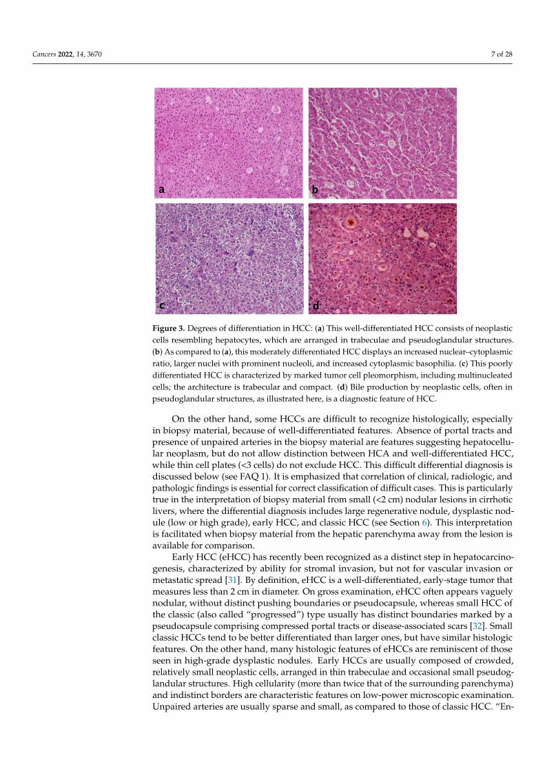
FAQ 9—Is a specialized liver center needed for the management of HCAs?The clinical management of HCA relies on hepatologists, surgeons, radiologists, and
pathologists, sharing their expertise in tumor board meetings. Imaging techniques arereliable to identify most cases of the H-HCA and IHCA subtypes, provided the radiologisthas some experience and uses specific techniques. By contrast, specific recognition ofb-HCA, b-IHCA, and shHCA by imaging is still under investigation. In their routinepractice, with the help of immunohistochemistry, pathologists can also provide a diagnosisof H-HCA, IHCA, and b-(I)HCA (in case of a strong diffuse GS staining). b-(I)HCAwith other patterns of GS staining and shHCA are less well known, and interpretation ofimmunohistochemistry can be difficult requiring confirmation by molecular methods. Inclinical centers where HCAs are rare, referring patients to specialized centers will improvediagnosis and decision making and, for the rarer subtypes, will also help to foster cutting-edge guidelines for patient management.
FAQ 10—Should the HCC grade (i.e., degree of differentiation) be reported in biopsyand in surgical specimens? What is the best grading system?
Similar to other carcinomas, HCCs are graded as well, moderately, or poorly differen-tiated. The grade is a marker of prognosis, and has been found to predict patient survivaland disease-free survival after both surgical resection and liver transplantation [66–68].Therefore, HCC grade represents useful information that should be included in pathologyreports. However, HCC often displays variable differentiation in different parts of thetumor. While prognosis would be expected to be primarily related to the least differentiatedcomponent of the neoplasm, knowledge of the existence of other components may beuseful for the assessment of additional specimens, such as those obtained at later datesfrom metastatic sites. On the other hand, biopsy specimens may not be representative ofthe entire range of differentiation present in any given HCC, due to sampling error. Never-theless, a study has found significant correlation between grade assessment of the biopsyand subsequent surgical specimen of HCC arising in cirrhotic patients [69]. Therefore, HCCgrade should be reported in biopsy specimens, with the understanding that it may notalways be entirely representative.
Various grading systems for HCC have been devised over the years. What is importantfor practical purposes is reproducibility of grading and clinical usefulness of the system.While the four-tiered Edmondson–Steiner system has been widely used in clinical studies,a three-tiered system is currently favored for daily practice [30]. It is hoped that adequatedescription of the characteristic features of each grade will result in high reproducibilityamong pathologists.
FAQ 11—Which is the best immunohistochemical panel to assure hepatocellular ori-gin of a malignancy?
The most commonly used immunohistochemical markers of HCC are those mentionedin Section 5, i.e., arginase-1, HepPar1, glypican-3, and α-fetoprotein. Arginase-1 is the mostsensitive and specific marker for HCC, staining over 90% of cases, while nonhepatocellulartumors are rarely positive for this marker [70]. HepPar1 stains most well differentiatedHCCs, but less than 50% of poorly differentiated ones. Furthermore, various adenocarcino-mas may occasionally be positive for HepPar1, particularly those from the small intestine,the normal enterocytes of which also uniformly express the antigen [71]. Glypican-3 ispositive in 65–80% of HCCs, more often in poorly differentiated than well differentiatedtumors; however, glypican-3 may also be positive in a variety of other malignancies, such

Cancers 2022, 14, 3670 19 of 28
as carcinomas from other sites, melanoma, and germ cell tumors [70]. α-Fetoprotein haslow sensitivity for HCC (<50%) and may be expressed in germ cell tumors and rare othermalignant neoplasms. In addition to these markers, HCCs arising in patients with chronichepatitis B may occasionally be positive for HBsAg, an uncommon but most specific finding.Lastly, in situ hybridization for albumin mRNA can be very useful in distinguishing HCCfrom other malignancies but is available in a limited number of institutions.
Immunohistochemical stains for carcinoembryonic antigen utilizing polyclonal an-tibodies (pCEA) often provide a canalicular pattern of staining that is useful for HCCdiagnosis. However, poorly differentiated HCCs often lack this pattern and may, instead,display membranous or even cytoplasmic staining, similar to that of adenocarcinomas. Im-munohistochemical stains for CD10 often demonstrate in HCC a similar canalicular patternof staining as that seen with pCEA. Again, this is usually absent in poorly differentiatedHCC. An example of establishing the diagnosis of HCC with the aid of immunohistochemi-cal stains is shown in Figure 9.
However, demonstrating the hepatocellular nature of a poorly differentiated carci-noma may be difficult in some cases. This is especially true in biopsy specimens withlimited material, taking into account that immunopositivity of tumor cells for the markersmentioned above may be focal, resulting in false-negative findings. Clinicopathologiccorrelation taking into account all the clinical, imaging, and pathologic findings will beessential in such cases. Appropriate additional markers for other tumors should also beincluded, as per the differential diagnosis in each particular case.
On the other hand, if a well-differentiated carcinoma consists of what appear tobe hepatocytes, then an appropriate panel would include arginase-1, HepPar1, pCEA (orCD10), and glypican-3, as α-fetoprotein has very low yield in well-differentiated HCC. If thelesion is truly HCC, any one marker may be positive, or a pair or more may show staining.However, stains for mimics of well differentiated HCC—particularly renal cell carcinoma,adrenal cortical carcinoma, neuroendocrine tumors, and follicular thyroid carcinomas—should also be considered [72]. Again, clinicopathologic correlation is essential.
FAQ 12—Are there any HCC subtypes that need to be specified on histologic diagnosis?The importance of HCC subtyping is related to differences in clinical correlations,
prognosis and treatment among subtypes. A subtype that is important to identify isthe fibrolamellar HCC, which is characterized by young patient age, lack of underlyingliver disease and tendency to metastasize to hilar lymph nodes. Therefore, the mainstayof therapy is surgery, including regional lymphadenectomy. The recent discovery of acharacteristic somatic gene fusion in fibrolamellar HCC (see Section 4) allows optimismfor future development of targeted therapies for nonresectable tumors. Other subtypes areworth identifying because of prognostic differences, as compared to average; for example,the macrotrabecular massive subtype and the neutrophil-rich subtype are associated withworse prognosis, while the clear cell subtype with better prognosis.
It is emphasized that HCC subtyping is a work in progress. With the exception of thefibrolamellar subtype, HCC had been regarded until recently as a tumor of bleak prognosis;therefore, there was little interest in subtyping. This view is now changing because of theavailability of surveillance programs and the hopes for new therapies based on molecularprofiling. Therefore, correlation of the clinical, pathologic, and molecular features in largeseries of patients with the various subtypes of HCC may allow a more individualizedapproach for treatment. As more data become available, the importance of subtyping willlikely increase. Histologic examination is the basis for HCC subtyping; therefore, criteria ofeach subtype should be clear and reproducible.
Cancers 2022, 14, 3670 20 of 28
Cancers 2022, 14, x FOR PEER REVIEW 20 of 29
molecular profiling. Therefore, correlation of the clinical, pathologic, and molecular fea-tures in large series of patients with the various subtypes of HCC may allow a more indi-vidualized approach for treatment. As more data become available, the importance of subtyping will likely increase. Histologic examination is the basis for HCC subtyping; therefore, criteria of each subtype should be clear and reproducible.
Figure 9. Abdominal MRI (a) and guided liver biopsy specimen (b–f) from a 53 year old man with breathing difficulty and history of metabolic syndrome, including obesity, type 2 diabetes mellitus, and hyperlipidemia. (a) There was marked hepatomegaly with innumerable, scattered nodules, measuring up to 3 cm, suggesting metastatic disease. However, needle biopsy revealed HCC. In this limited biopsy material, polygonal tumor cells appeared to be arranged in a compact sheet (b, left side); nevertheless, immunohistochemical stain for CD34 (c), highlighting the endothelial cells, demonstrated trabecular architecture. Further stains showed positivity of tumor cells for arginase-1 (d) and HepPar1 (e). Adjacent hepatocytes (d,e, right side) are also positive for these markers, serving as “internal controls”. Tumor cells also displayed a canalicular pattern of staining with pCEA (f), as well as positivity for glypican-3 (not shown).
FAQ 13—Do immunohistochemical stains or molecular studies provide any action-able items for HCC? Do molecular studies provide any added value in terms of diagnosis or prognosis of HCC, as compared to standard immunohistochemical stains?
Figure 9. Abdominal MRI (a) and guided liver biopsy specimen (b–f) from a 53 year old man withbreathing difficulty and history of metabolic syndrome, including obesity, type 2 diabetes mellitus,and hyperlipidemia. (a) There was marked hepatomegaly with innumerable, scattered nodules,measuring up to 3 cm, suggesting metastatic disease. However, needle biopsy revealed HCC. Inthis limited biopsy material, polygonal tumor cells appeared to be arranged in a compact sheet(b, left side); nevertheless, immunohistochemical stain for CD34 (c), highlighting the endothelialcells, demonstrated trabecular architecture. Further stains showed positivity of tumor cells forarginase-1 (d) and HepPar1 (e). Adjacent hepatocytes (d,e, right side) are also positive for thesemarkers, serving as “internal controls”. Tumor cells also displayed a canalicular pattern of stainingwith pCEA (f), as well as positivity for glypican-3 (not shown).
FAQ 13—Do immunohistochemical stains or molecular studies provide any action-able items for HCC? Do molecular studies provide any added value in terms of diagnosisor prognosis of HCC, as compared to standard immunohistochemical stains?
The main use of immunohistochemical stains in cases of suspected HCC is to confirmthe diagnosis and rule out other neoplasms. However, there is also a stain that has beenfound to be of prognostic significance in HCC; cytokeratin 19-positive HCCs have higherrecurrence rates than usual, as well as higher resistance to locoregional therapies [73–75].It should be kept in mind that cytokeratin 19 is positive in a variety of adenocarcinomas,

Cancers 2022, 14, 3670 21 of 28
including cholangiocarcinoma; therefore, it is used as a prognostic, but not as a diagnosticmarker for HCC.
Molecular methods have been used extensively in recent years to identify the molecu-lar changes occurring in hepatocarcinogenesis, including potential targets for treatment.Substantial molecular data have been accumulated, and several molecular classificationshave been proposed on the basis of a correlation of clinical, pathologic, and moleculardata [22,26–29]. However, these classifications have not yet found their way to clinical prac-tice. As a result of these studies, most HCCs can now be grouped into two classes [25,76]:(i) the proliferation class, which is etiologically related to HBV infection, and displaysmolecular and histologic features associated with aggressive clinical behavior; (ii) thenonproliferation class, which is etiologically related to HCV infection or alcohol, and dis-plays features associated with better clinical outcome. HCCs of the proliferation classare characterized by TP53 mutations, chromosomal instability, and activation of variousoncogenic pathways, tend to be poorly differentiated, and are associated with high serumα-fetoprotein. On the other hand, HCCs of the nonproliferation class often have CTNNB1mutations and a gene expression profile resembling that of normal hepatocytes. Thesetumors tend to be better differentiated and with lower incidence of vascular invasion thanthose of the proliferation class.
The characteristic molecular changes of the various HCC subtypes are summarized inthe section on diagnosis of hepatocellular carcinoma (see Table 2). Although not routinelyused in daily diagnosis, detection of these changes can be used in support of the diagnosis.For instance, detection of the gene fusion DNAJB1–PRKACA can confirm the diagnosis offibrolamellar HCC.
FAQ 14—What should pathologists know and do about combined hepatocellular-cholangiocarcinomas (cHCC–CCA)?
FAQ14.1. Tissue Diagnosis of cHCC–CCA
A tissue diagnosis of a primary cHCC–CCA is straightforwardly made by routinehematoxylin–eosin stains; immunostains for markers of hepatocyte or cholangiocyte differ-entiation are merely confirmatory [30,77]. The presence of stainable hepatocyte markers inglandular epithelium (e.g., arginase-1, HepPar1, α-fetoprotein, glypican-3, and albuminmRNA) or, conversely, of cholangiocyte markers in HCC (e.g., keratins 7 and 19, andEpCAM) are not proof of cHCC–CCA given the possibilities of aberrant gene expressionin any malignancy [77]. Differentiated components of cHCC–CCA may be located indistinct areas of a tumor, or they may be intimately intermingled throughout the lesion.Boundaries between the components may be sharply defined or indistinct. There are, asyet, no definitive cutoffs for a percentage requirement for the presence of the component. Aminute component of intrahepatic CCA (iCCA) within an otherwise clear HCC is sufficientto call it cHCC–CCA and vice versa.
A common pitfall of diagnosis is when cHCC–CCA is suspected on radiographicgrounds, but only one element is present in the biopsy specimen. In this case, the pathologistmust be careful to comment on the limitations of biopsy. Small biopsy specimens maysample only one component of such a heterogeneous tumor; the absence of the othercomponent does not exclude cHCC–CCA and a formal statement to that effect in thepathology report is important. On the other hand, metastatic lesions associated with aprimary cHCC–CCA may comprise either component alone or mimic the cHCC–CCAappearance of the primary tumor. Biopsy specimens from metastatic lesions must becautiously interpreted in this light [78].
Another pitfall for diagnosis can occur when there appear to be two separate masslesions in the liver that are merging together. An HCC and a separate, but simultaneousCCA may grow into each other forming a “collision tumor”, particularly in chronic liverdiseases that predispose to both malignancies. Each of these tumors should be assessedpathologically as independent entities.
Lastly, a very rare variant of “intermediate cell type” of cHCC–CCA notably breaks allthe rules; its tumor cells appear morphologically intermediate between hepatocytes and

Cancers 2022, 14, 3670 22 of 28
cholangiocytes and do not show typical growth patterns of either HCC (e.g., trabeculaeand pseudoglandular structures) or iCCA (e.g., mucin-producing glands, tubules, andsignet ring cells), often appearing homogeneous throughout. Dual differentiation in thesetumors is, indeed, at the cellular level, with each cell showing combined hepatocyte andcholangiocyte marker expression [30].
It should also be noted that, while cHCC–CCA may present, de novo, as a primaryhepatic malignancy, it has also been seen to emerge from HCCs that have undergoneloco-regional treatments [79]. It has been suggested that hypoxia of surviving tumor cellsafter transarterial chemoembolization leads to expression of proteins, such as EpCAMand cytokeratin 19 [80]. Such adaptive changes might explain emergence of cHCC-CCAfrom a treated HCC, although the possibility that there was a previously undetected minorcomponent of CCA originally is difficult to exclude. In any case, while this occurrenceseems uncommon, it should be considered when tumor recurs post treatment, particularlyif imaging features no longer show classic features of HCC, alone.
Subpopulations of tumor cells in cHCC–CCA may have what has been described asa “stem-cell appearance”, i.e., small cells with high nuclear–cytoplasmic ratio, sometimesarrayed with larger hepatobiliary cells in what appear to be lineage relationships likethose seen in ductular reactions in diseased or injured liver. While subclasses of “stem-celltumors” were characterized in the 2010 edition of the WHO “Blue Book” [81], the morerecent edition [82] has eliminated the term as a diagnostic category in all primary livercancers. Nonetheless, the question of its importance remains uncertain, and it has beenrecommended that the presence of “stem-cell features” be noted in the pathology report oftumors that contain them [83].
FAQ14.2. Pathology–Radiology Collaboration for cHCC–CCA
A biphasic radiographic appearance of a lesion may help a pathologist avoid missingthe opportunity for including cHCC–CCA in the differential diagnosis when only onecomponent is sampled in a biopsy specimen [84]. However, in multidisciplinary conferencesfor liver malignancies, clinicians and radiologists may miss clues to cHCC–CCA given theirrarity, but the attentive pathologist can help guide radiologists toward how to best samplea lesion for successful complete diagnosis.
In early-stage liver disease or in sporadic tumors in which there is no predisposinghepatic disease, cHCC–CCA may appear biphasic, with separate areas showing typicalfeatures of HCC or iCCA, or they may merely be atypical, without imaging characteristicsspecific for either, thus being more suggestive of metastasis [84]. In advanced-stage liverdisease, in which the Liver Imaging Reporting and Data System (LI-RADS) classification isapplicable, radiologists may label a lesion LIRADS-M because unusual features suggest ametastasis, but the lesion may actually be a cHCC–CCA [85,86]. Alternatively, they mayrecognize that one or more parts of the lesion have an LIRADS-5 (diagnostic for HCC)appearance and, thus, label the whole lesion with that designation, even though some areasappear distinct [84,85]. In both these settings, a pathologist who is attentive to radiographicdescriptions that might hint at cHCC–CCA may save the day.
FAQ14.3. Molecular Pathology and Treatment Implications of the Diagnosis of cHCC–CCA
Molecular studies support that these tumors may sometimes derive from a malig-nantly transformed hepatobiliary stem/progenitor cell or from de-/redifferentiation ofmalignantly transformed hepatocytes or cholangiocytes, and that they may be more likeiCCA, more like HCC, or intermediate between them. All of these data confirm that theyare certainly, at least to some degree, heterogeneous in origin and in behavior [87]. On theother hand, comparison of clinicopathological characteristics of cHCC–CCA with regard tothe newest WHO classification [82] supports its relevance and that cHCC–CCA has inter-mediate survival between HCC and iCCA, if not actually tilting toward the dire outcomesfor iCCA [87,88]. Given the propensity for early and distant spread of CCA componentsalong lymphatic and perineural pathways that are typical of iCCA itself, it is no surprisethat clinical outcomes after resection are, overall, worse than for HCC. On the other hand,

Cancers 2022, 14, 3670 23 of 28
if transplanted within the Milan criteria, cHCC–CCA showed similar overall survival toHCC after transplantation [89].
Unfortunately, the rarity of these tumors has interfered with the performance ofrandomized clinical trials. There is a paucity of data regarding immunotherapies [90],although studies suggest that at least some cHCC–CCA should be responsive to these typesof treatment [91,92]. Broad genomic profiling of malignancies for actionable mutationsspecific to each case is currently the most likely path to any possible clinical benefit [93].
FAQ 15—I do not work in a transplant center and am so unlikely to ever see a dys-plastic nodule specimen. Do I need to know about them? If so, why?
Yes! One needs to know about them! Currently, screening for emergence of malignancyin chronic liver disease depends largely on radiographic criteria defined by the LiverImaging Reporting and Data System (LI-RADS) classification [94]. Pathologists may findthemselves involved in liver tumor multidisciplinary conferences in which radiologists willreport distinctive nodules that are subclassified into LIRADS-1 through 5, with a higherscore indicating the higher confidence that a lesion is an actual HCC (Figure 7) [95]. Therepetition of the phrase “distinctive nodule” is not a coincidence; LI-RADS was formulatedto reflect the pathologic understanding of DNs as neoplastic and often premalignant lesions.
A classification of LIRADS-5 is so specific for HCC that, in most medical centers, it issufficient for diagnosis without confirmatory biopsy. LIRADS-1 lesions are considered likelyto be benign, probably merely large regenerative nodules. LIRADS-2 through 4 probablyreflect LGDN through HGDN (although direct pathology–radiology correlations for thesehave not been reported) [94,95].
It is not necessary for the pathologist to know the full and subtle criteria for theLIRADS classifications, but it is vital to know what lesions each designation may reflect,thus enabling the pathologist to carefully guide the clinicians and radiologists in terms offollow-up screening or treatment of the patient. It is worth knowing, however, that theincreasing stages of the LI-RADS classification probably reflect the changes in vascularsupply. Regenerative nodules and LGDNs have mostly intact portal vein blood flow withlittle increase in arterialization. However, the increasing ratio between enlarging arterialblood flow (angiogenesis inside truly neoplastic LGDN and HGDN) and the diminishingblood supply (as portal tracts are degraded or pushed to the sides) result in the characteristicLIRADS features (Figure 7).
Examples are provided below.
• LIRADS-1: The pathologist may advise the clinical team that, while this lesion isprobably just a regenerative nodule, the possibility that it is a DN, possibly even anHGDN, is not excluded. Repeat screening at a shorter time interval may be warranted.If the lesion disappears, it was probably large regenerative nodule that underwentinvolution or further scarring that eliminated its distinctive appearance on imaging. Ifit does indeed disappear, return to normal surveillance screening is reasonable.
• LIRADS-2 or -3: These lesions are more likely to be DNs, either LGDN or HGDN.Repeat imaging should be performed more frequently. If the nodule disappears, it wasprobably regenerative. If it persists, then it may be LGDN or HGDN and the patient isconsidered at higher risk for HCC and should return more frequently for imaging. Ifthe lesion progresses upward in LIRADS score, it is probably a DN giving rise to anHCC. Continued imaging or ablation may be considered depending on the clinicalcircumstances.
• LIRADS-4: These lesions are probably an HGDN, possibly with an emerging focusof HCC, or possibly a small HCC. Continued imaging or ablation may be considereddepending on clinical circumstances.
• LIRADS-5: This feature is diagnostic for HCC. While a biopsy is not necessary fordiagnosis, oncologists are increasingly requesting a pre-ablation biopsy for molecularstudies to inform future treatments if the lesion is resistant to ablation or if there ispost-treatment (i.e., ablation, resection, or transplant) recurrence.
Cancers 2022, 14, 3670 24 of 28
• LIRADS-M: These are lesions without typical imaging features of HCC, but highly sus-picious for malignancy. The pathologists may aver that the lesion could be metastatic(M), but they could also be iCCA or cHCC–CCA. Such lesions probably require biopsyfor diagnosis.
With regard to cHCC–CCA, it behooves the pathologist to watch for lesions describedby the radiologist as “complex” or as having an isolated part with typical LIRADS-5 featureswhile the other parts of the lesion are atypical for HCC [84–86]. Because cHCC–CCA mayhave distinct regions of the tumor that are either HCC or CCA, in such tumors, someregions will show LIRADS-5 changes, while others will not. This situation is one in whichthe pathologist can make a decisive difference, recommending targeted biopsies of boththe LIRADS-5 and the atypical areas. iCCA’s worse prognosis and different treatmentimplications make it important to diagnose as early as possible in the treatment course.The pathologist may be the only person in the room sensitive to this uncommon cancer.Alerting the clinical team to make sure that cHCC–CCA has been completely evaluatedmay be crucial for saving the life of this patient or preventing an inappropriate transplantfor an incurable malignancy.
FAQ 16—When should tumors with imaging features of HCC be biopsied?It is currently believed that the findings which define LIRADS-5, i.e., those lesions with
all imaging features of HCC by imaging, do not require confirmatory biopsy. However,ablative treatment of these lesions is then likely, and tissue for molecular analysis fordetermining possible targeted therapies will not be available. For this reason, there maybe a shift in clinical practice toward biopsy of LIRADS-5 HCCs in the near future, not fordiagnosis or prognosis, but for determination of suitable targeted therapies in the eventof recurrence. Subtyping of HCC might also be found to be relevant in the near future toguide treatment decisions, also with the support of artificial intelligence [96].
As noted above, if part of a lesion displays typical HCC imaging features, but otherparts do not, the possibility of a cHCC–CCA cannot be excluded. In addition, even rarerHCC variants could appear in combination with classic HCC, such as the sarcomatoid one,which might have nontypical imaging features. In such cases, biopsy of both the classicHCC component and of the nontypical component is warranted.
Author Contributions: Conceptualization, P.H., P.B.-S., N.D.T. and C.S.; methodology, P.H., P.B.-S.,N.D.T. and C.S.; validation, P.H., P.B.-S., N.D.T. and C.S.; formal analysis, P.H., P.B.-S., N.D.T. and C.S.;investigation, P.H., P.B.-S., N.D.T. and C.S.; resources, P.H., P.B.-S., N.D.T. and C.S.; data curation, P.H.,P.B.-S., N.D.T. and C.S.; writing—original draft preparation, P.H., P.B.-S., N.D.T. and C.S.; writing—review and editing, P.H., P.B.-S., N.D.T. and C.S.; visualization, P.H., P.B.-S., N.D.T. and C.S.; projectadministration, P.H. All authors have read and agreed to the published version of the manuscript.
Funding: This research received no external funding.
Acknowledgments: The authors wish to acknowledge the members of the International Liver Pathol-ogy Study Group (the “Elves”), past and present, whose research and clinical wisdom inform all ofthe concepts in this manuscript. In particular, the authors are grateful to Venancio Alves, AlbertoQuaglia, and Charles Balabaud, for their suggested FAQs.
Conflicts of Interest: The authors declare no conflict of interest.
References1. Bioulac-Sage, P.; Gouw, A.S.; Balabaud, C.; Sempoux, C. Hepatocellular adenoma: What we know, what we do not know, and
why it matters. Histopathology 2022, 80, 878–897. [CrossRef] [PubMed]2. Sempoux, C.; Gouw, A.S.; Dunet, V.; Paradis, V.; Balabaud, C.; Bioulac-Sage, P. Predictive Patterns of Glutamine Synthetase
Immunohistochemical Staining in CTNNB1-mutated Hepatocellular Adenomas. Am. J. Surg. Pathol. 2021, 45, 477–487. [CrossRef][PubMed]
3. Henriet, E.; Hammoud, A.A.; Dupuy, J.-W.; Dartigues, B.; Ezzoukry, Z.; Dugot-Senant, N.; Leste-Lasserre, T.; Pallares-Lupon, N.;Nikolski, M.; Le Bail, B.; et al. Argininosuccinate synthase 1 (ASS1): A marker of unclassified hepatocellular adenoma and highbleeding risk. Hepatology 2017, 66, 2016–2028. [CrossRef]

Cancers 2022, 14, 3670 25 of 28
4. Sala, M.; Gonzales, D.; Leste-Lasserre, T.; Dugot-Senant, N.; Paradis, V.; Di Tommaso, S.; Dupuy, J.; Pitard, V.; Dourthe, C.;Sciarra, A.; et al. ASS1 Overexpression: A Hallmark of Sonic Hedgehog Hepatocellular Adenomas; Recommendations for ClinicalPractice. Hepatol. Commun. 2020, 4, 809–824. [CrossRef] [PubMed]
5. Global Burden of Disease Liver Cancer Collaboration. The Burden of Primary Liver Cancer and Underlying Etiologies From 1990to 2015 at the Global, Regional, and National Level: Results from the Global Burden of Disease Study 2015. JAMA Oncol. 2017, 3,1683–1691. [CrossRef]
6. Ioannou, G.N. Epidemiology and risk-stratification of NAFLD-associated HCC. J. Hepatol. 2021, 75, 1476–1484. [CrossRef][PubMed]
7. Paterlini-Bréchot, P.; Saigo, K.; Murakami, Y.; Chami, M.; Gozuacik, D.; Mugnier, C.; Lagorce, D.; Bréchot, C. Hepatitis Bvirus-related insertional mutagenesis occurs frequently in human liver cancers and recurrently targets human telomerase gene.Oncogene 2003, 22, 3911–3916. [CrossRef]
8. Murakami, Y.; Saigo, K.; Takashima, H.; Minami, M.; Okanoue, T.; Bréchot, C.; Brechot, P.P. Large scaled analysis of hepatitis Bvirus (HBV) DNA integration in HBV related hepatocellular carcinomas. Gut 2005, 54, 1162–1168. [CrossRef]
9. Sung, W.-K.; Zheng, H.; Li, S.; Chen, R.; Liu, X.; Li, Y.; Lee, N.P.; Lee, W.H.; Ariyaratne, P.N.; Tennakoon, C.; et al. Genome-widesurvey of recurrent HBV integration in hepatocellular carcinoma. Nat. Genet. 2012, 44, 765–769. [CrossRef]
10. Goh, J.; Callagy, G.; McEntee, G.; O’Keane, J.C.; Bomford, A.; Crowe, J. Hepatocellular carcinoma arising in the absence ofcirrhosis in genetic haemochromatosis: Three case reports and review of literature. Eur. J. Gastroenterol. Hepatol. 1999, 11, 915–919.[CrossRef] [PubMed]
11. Honeyman, J.N.; Simon, E.P.; Robine, N.; Chiaroni-Clarke, R.; Darcy, D.G.; Lim, I.I.P.; Gleason, C.E.; Murphy, J.M.; Rosenberg,B.R.; Teegan, L.; et al. Detection of a Recurrent DNAJB1-PRKACA Chimeric Transcript in Fibrolamellar Hepatocellular Carcinoma.Science 2014, 343, 1010–1014. [CrossRef] [PubMed]
12. Thorgeirsson, S.S.; Grisham, J.W. Molecular pathogenesis of human hepatocellular carcinoma. Nat. Genet. 2002, 31, 339–346.[CrossRef]
13. Hatziapostolou, M.; Polytarchou, C.; Aggelidou, E.; Drakaki, A.; Poultsides, G.A.; Jaeger, S.A.; Ogata, H.; Karin, M.; Struhl, K.;Hadzopoulou-Cladaras, M.; et al. An HNF4α-miRNA inflammatory feedback circuit regulates hepatocellular oncogenesis. Cell2011, 147, 1233–1247. [CrossRef]
14. Brody, R.I.; Theise, N.D. An inflammatory proposal for hepatocarcinogenesis. Hepatology 2012, 56, 382–384. [CrossRef]15. Hytiroglou, P.; Bioulac-Sage, P. Molecular Pathogenesis and Diagnostics of Hepatocellular Tumors. In Odze and Goldblum Surgical
Pathology of the GI Tract, Liver, Biliary Tract, and Pancreas, 4th ed.; Odze, R.D., Goldblum, J.R., Eds.; Elsevier: Philadelphia, PA, USA,2023; pp. 1369–1383.
16. Gerbes, A.; Zoulim, F.; Tilg, H.; Dufour, J.-F.; Bruix, J.; Paradis, V.; Salem, R.; Peck-Radosavljevic, M.; Galle, P.R.; Greten, T.F.; et al.Gut roundtable meeting paper: Selected recent advances in hepatocellular carcinoma. Gut 2018, 67, 380–388. [CrossRef]
17. Nault, J.C.; Mallet, M.; Pilati, C.; Calderaro, J.; Bioulac-Sage, P.; Laurent, C.; Laurent, A.; Cherqui, D.; Balabaud, C.;Zucman-Rossi, J. High frequency of telomerase reverse-transcriptase promoter somatic mutations in hepatocellular carcinomaand preneoplastic lesions. Nat. Commun. 2013, 4, 2218. [CrossRef]
18. Nault, J.-C.; Ningarhari, M.; Rebouissou, S.; Zucman-Rossi, J. The role of telomeres and telomerase in cirrhosis and liver cancer.Nat. Rev. Gastroenterol. Hepatol. 2019, 16, 544–558. [CrossRef] [PubMed]
19. Nault, J.C.; Calderaro, J.; Di Tommaso, L.; Balabaud, C.; Zafrani, E.S.; Bioulac-Sage, P.; Roncalli, M.; Zucman-Rossi, J. Telomerasereverse transcriptase promoter mutation is an early somatic genetic alteration in the transformation of premalignant nodules inhepatocellular carcinoma on cirrhosis. Hepatology 2014, 60, 1983–1992. [CrossRef] [PubMed]
20. The Cancer Genome Atlas Research Network. Comprehensive and integrative genomic characterization of hepatocellularcarcinoma. Cell 2017, 169, 1327–1341. [CrossRef]
21. Dhanasekaran, R.; Nault, J.-C.; Roberts, L.R.; Zucman-Rossi, J. Genomic Medicine and Implications for Hepatocellular CarcinomaPrevention and Therapy. Gastroenterology 2019, 156, 492–509. [CrossRef]
22. Nault, J.; Martin, Y.; Caruso, S.; Hirsch, T.; Bayard, Q.; Calderaro, J.; Charpy, C.; Copie-Bergman, C.; Ziol, M.; Bioulac-Sage, P.; et al.Clinical Impact of Genomic Diversity From Early to Advanced Hepatocellular Carcinoma. Hepatology 2020, 71, 164–182. [CrossRef]
23. Totoki, Y.; Tatsuno, K.; Covington, K.R.; Ueda, H.; Creighton, C.J.; Kato, M.; Tsuji, S.; Donehower, L.A.; Slagle, B.L.;Nakamura, H.; et al. Trans-ancestry mutational landscape of hepatocellular carcinoma genomes. Nat. Genet. 2014, 46, 1267–1273.[CrossRef] [PubMed]
24. Schulze, K.; Imbeaud, S.; Letouzé, E.; Alexandrov, L.B.; Calderaro, J.; Rebouissou, S.; Couchy, G.; Meiller, C.; Shinde, J.;Soysouvanh, F.; et al. Exome sequencing of hepatocellular carcinomas identifies new mutational signatures and potentialtherapeutic targets. Nat. Genet. 2015, 47, 505–511. [CrossRef] [PubMed]
25. Villanueva, A. Hepatocellular carcinoma. N. Engl. J. Med. 2019, 380, 1450–1462. [CrossRef] [PubMed]26. Boyault, S.; Rickman, D.S.; Bioulac-Sage, P.; De Reyniès, A.; Balabaud, C.; Rebouissou, S.; Jeannot, E.; Hérault, A.; Saric, J.;
Belghiti, J.; et al. Transcriptome classification of HCC is related to gene alterations and to new therapeutic targets. Hepatology2007, 45, 42–52. [CrossRef] [PubMed]
27. Hoshida, Y.; Nijman, S.M.B.; Kobayashi, M.; Chan, J.A.; Brunet, J.-P.; Chiang, D.Y.; Villanueva, A.; Newell, P.; Ikeda, K.;Hashimoto, M.; et al. Integrative Transcriptome Analysis Reveals Common Molecular Subclasses of Human HepatocellularCarcinoma. Cancer Res. 2009, 69, 7385–7392. [CrossRef] [PubMed]
Cancers 2022, 14, 3670 26 of 28
28. Nault, J.; De Reyniès, A.; Villanueva, A.; Calderaro, J.; Rebouissou, S.; Couchy, G.; Decaens, T.; Franco, D.; Imbeaud, S.; Rousseau,F.; et al. A Hepatocellular Carcinoma 5-Gene Score Associated With Survival of Patients After Liver Resection. Gastroenterology2013, 145, 176–187. [CrossRef]
29. Calderaro, J.; Couchy, G.; Imbeaud, S.; Amaddeo, G.; Letouzé, E.; Blanc, J.-F.; Laurent, C.; Hajji, Y.; Azoulay, D.;Bioulac-Sage, P.; et al. Histological subtypes of hepatocellular carcinoma are related to gene mutations and moleculartumour classification. J. Hepatol. 2017, 67, 727–738. [CrossRef] [PubMed]
30. Torbenson, M.S.; Ng, I.O.L.; Park, Y.N.; Roncalli, M.; Sakamoto, M. Hepatocellular carcinoma. In Digestive System Tumours, 5th ed.;WHO Classification of Tumours Editorial Board; WHO Classification of Tumours Series; International Agency for Research onCancer: Lyon, France, 2019; Volume 1, pp. 229–239.
31. International Consensus Group for Hepatocellular Neoplasia. Pathologic diagnosis of early hepatocellular carcinoma: A report ofthe International Consensus Group for Hepatocellular Neoplasia. Hepatology 2009, 49, 658–664. [CrossRef]
32. Kojiro, M. Pathology of Hepatocellular Carcinoma; Blackwell: Malden, MA, USA, 2006.33. Salomao, M.; Woojin, M.Y.; Brown, R.S., Jr.; Emond, J.C.; Lefkowitch, J.H. Steatohepatitic hepatocellular carcinoma (SH-HCC): A
distinctive histological variant of HCC in hepatitis C virus-related cirrhosis with associated NAFLD/NASH. Am. J. Surg. Pathol.2010, 34, 1630–1636. [CrossRef]
34. Yeh, M.M.; Liu, Y.; Torbenson, M. Steatohepatitic variant of hepatocellular carcinoma in the absence of metabolic syndrome orbackground steatosis: A clinical, pathological, and genetic study. Hum. Pathol. 2015, 46, 1769–1775. [CrossRef]
35. Li, T.; Fan, J.; Qin, L.-X.; Zhou, J.; Sun, H.-C.; Qiu, S.-J.; Ye, Q.-H.; Wang, L.; Tang, Z.-Y. Risk Factors, Prognosis, and Managementof Early and Late Intrahepatic Recurrence After Resection of Primary Clear Cell Carcinoma of the Liver. Ann. Surg. Oncol. 2011,18, 1955–1963. [CrossRef]
36. Torbenson, M.S. Hepatocellular carcinoma: Making sense of morphological heterogeneity, growth patterns, and subtypes. Hum.Pathol. 2021, 112, 86–101. [CrossRef]
37. Wood, L.D.; Heaphy, C.M.; Daniel, H.D.-J.; Naini, B.V.; Lassman, C.R.; Arroyo, M.R.; Kamel, I.R.; Cosgrove, D.P.; Boitnott, J.K.;Meeker, A.K.; et al. Chromophobe hepatocellular carcinoma with abrupt anaplasia: A proposal for a new subtype of hepatocellularcarcinoma with unique morphological and molecular features. Mod. Pathol. 2013, 26, 1586–1593. [CrossRef]
38. Torbenson, M.S. Morphologic Subtypes of Hepatocellular Carcinoma. Gastroenterol. Clin. N. Am. 2017, 46, 365–391. [CrossRef][PubMed]
39. Maeda, T.; Adachi, E.; Kajiyama, K.; Takenaka, K.; Sugimachi, K.; Tsuneyoshi, M. Spindle cell hepatocellular carcinoma: Aclinicopathologic and immunohistochemical analysis of 15 cases. Cancer 1996, 77, 51–57. [CrossRef]
40. Kojiro, M.; Sugihara, S.; Kakizoe, S.; Nakashima, O.; Kiyomatsu, K. Hepatocellular carcinoma with sarcomatous change: A specialreference to the relationship with anticancer therapy. Cancer Chemother. Pharmacol. 1989, 23, S4–S8. [CrossRef]
41. International Working Party. Terminology of nodular hepatocellular lesions. Hepatology 1995, 22, 983–993. [CrossRef]42. Watanabe, S.; Okita, K.; Harada, T.; Kodama, T.; Numa, Y.; Takemoto, T.; Takahashi, T. Morphologic studies of the liver cell
dysplasia. Cancer 1983, 51, 2197–2205. [CrossRef]43. Deugnier, Y.M.; Charalambous, P.; Le Quilleuc, D.; Turlin, B.; Searle, J.; Brissot, P.; Powell, L.W.; Halliday, J.W. Preneoplastic
significance of hepatic iron-free foci in genetic hemochromatosis: A study of 185 patients. Hepatology 1993, 18, 1363–1369.44. Hytiroglou, P.; Park, Y.N.; Krinsky, G.; Theise, N.D. Hepatic Precancerous Lesions and Small Hepatocellular Carcinoma.
Gastroenterol. Clin. N. Am. 2007, 36, 867–887. [CrossRef]45. Park, Y.N.; Kojiro, M.; Di Tommaso, L.; Dhillon, A.P.; Kondo, F.; Nakano, M.; Sakamoto, M.; Theise, N.D.; Roncalli, M. Ductular
reaction is helpful in defining early stromal invasion, small hepatocellular carcinomas, and dysplastic nodules. Cancer 2007, 109,915–923. [CrossRef]
46. Hytiroglou, P.; Theise, N.D. The differential diagnosis of liver nodules. Sem. Diag. Pathol. 1998, 15, 285–299.47. Theise, N.D.; Marcelin, K.; Goldfischer, M.; Hytiroglou, P.; Ferrell, L.; Thung, S.N. Low proliferative activity in macroregenerative
nodules: Evidence for an alternate hypothesis concerning human hepatocarcinogenesis. Liver 1996, 16, 134–139. [CrossRef]48. Park, Y.N.; Yang, C.-P.; Cubukcu, O.; Thung, S.N.; Theise, N.D. Hepatic stellate cell activation in dysplastic nodules: Evidence for
an alternate hypothesis concerning human hepatocarcinogenesis. Liver Int. 2008, 17, 271–274. [CrossRef] [PubMed]49. Hytiroglou, P.; Theise, N.D.; Schwartz, M.; Mor, E.; Miller, C.; Thung, S.N. Macroregenerative nodules in a series of adult cirrhotic
liver explants: Issues of classification and nomenclature. Hepatology 1995, 21, 703–708. [PubMed]50. Park, Y.N.; Yang, C.-P.; Fernandez, G.J.; Cubukcu, O.; Thung, S.N.; Theise, N.D. Neoangiogenesis and Sinusoidal “Capillarization”
in Dysplastic Nodules of the Liver. Am. J. Surg. Pathol. 1998, 22, 656–662. [CrossRef] [PubMed]51. Theise, N.D.; Park, Y.N.; Thung, S.N. “Vascular profiles” of regenerative and dysplastic nodules. Hepatology 2000, 31, 1380–1381.
[CrossRef] [PubMed]52. Di Tommaso, L.; Franchi, G.; Park, Y.N.; Fiamengo, B.; Destro, A.; Morenghi, E.; Montorsi, M.; Torzilli, G.; Tommasini, M.;
Terracciano, L.; et al. Diagnostic value of HSP70, glypican 3, and glutamine synthetase in hepatocellular nodules in cirrhosis.Hepatology 2007, 45, 725–734.
53. Di Tommaso, L.; Destro, A.; Seok, J.Y.; Balladore, E.; Terracciano, L.; Sangiovanni, A.; Iavarone, M.; Colombo, M.G.; Jang, J.J.;Yu, E.; et al. The application of markers (HSP70 GPC3 and GS) in liver biopsies is useful for detection of hepatocellular carcinoma.J. Hepatol. 2009, 50, 746–754.

Cancers 2022, 14, 3670 27 of 28
54. Hytiroglou, P. Well-differentiated hepatocellular nodule: Making a diagnosis on biopsy and resection specimens of patients withadvanced stage chronic liver disease. Semin. Diagn. Pathol. 2017, 34, 138–145. [CrossRef]
55. Bioulac-Sage, P.; Kakar, S.; Nault, J.C. Hepatocellular adenoma. In Digestive System Tumours, 5th ed.; WHO Classification ofTumours Editorial Board; WHO Classification of Tumours Series; International Agency for Research on Cancer: Lyon, France,2019; Volume 1, pp. 224–228.
56. Joseph, N.M.; Blank, A.; Shain, A.H.; Gill, R.M.; Umetsu, S.E.; Shafizadeh, N.; Torbenson, M.S.; Kakar, S. Hepatocellular neoplasmswith loss of liver fatty acid binding protein: Clinicopathologic features and molecular profiling. Hum. Pathol. 2022, 122, 60–71.[CrossRef]
57. Julien, C.; Le Bail, B.; Chiche, L.; Balabaud, C.; Bioulac-Sage, P. Malignant transformation of hepatocellular adenoma. JHEP Rep.2022, 4, 100430. [CrossRef] [PubMed]
58. Calderaro, J.; Nault, J.C.; Balabaud, C.; Couchy, G.; Saint-Paul, M.-C.; Azoulay, D.; Mehdaoui, D.; Luciani, A.; Zafrani, E.S.;Bioulac-Sage, P.; et al. Inflammatory hepatocellular adenomas developed in the setting of chronic liver disease and cirrhosis. Mod.Pathol. 2016, 29, 43–50. [CrossRef]
59. Sasaki, M.; Yoneda, N.; Sawai, Y.; Imai, Y.; Kondo, F.; Fukusato, T.; Yoshikawa, S.; Kobayashi, S.; Sato, Y.; Matsui, O.; et al.Clinicopathological characteristics of serum amyloid A-positive hepatocellular neoplasms/nodules arising in alcoholic cirrhosis.Histopathology 2015, 66, 836–845. [CrossRef] [PubMed]
60. Rebouissou, S.; Franconi, A.; Calderaro, J.; Letouzé, E.; Imbeaud, S.; Pilati, C.; Nault, J.-C.; Couchy, G.; Laurent, A.;Balabaud, C.; et al. Genotype-phenotype correlation of CTNNB1 mutations reveals different β-catenin activity associated withliver tumor progression. Hepatology 2016, 64, 2047–2061. [CrossRef]
61. European Association for the Study of the Liver (EASL). EASL Clinical Practice Guidelines on the Management of BenignLiver Tumours. Available online: https://www.journal-of-hepatology.eu/article/S0168-8278(16)30101-5/pdf (accessed on31 January 2022).
62. Julien, C.; Le-Bail, B.; Touhami, K.O.; Frulio, N.; Blanc, J.; Adam, J.; Laurent, C.; Balabaud, C.; Bioulac-Sage, P.; Chiche, L.Hepatocellular adenoma risk factors of hemorrhage: Size is not the only concern! Single center retrospective experience of 261patients. Ann. Surg. 2021, 274, 843–850. [CrossRef]
63. Klompenhouwer, A.J.; Rosmalen, B.V.; Haring, M.P.D.; Thomeer, M.G.J.; Doukas, M.; Verheij, J.; Meijer, V.E.; Gulik, T.M.;Takkenberg, R.B.; Kazemier, G.; et al. A multicentre retrospective analysis on growth of residual hepatocellular adenoma afterresection. Liver Int. 2020, 40, 2272–2278. [CrossRef]
64. Putra, J.; Ferrell, L.D.; Gouw, A.S.H.; Paradis, V.; Rishi, A.; Sempoux, C.; Balabaud, C.; Thung, S.N.; Bioulac-Sage, P. Malignanttransformation of liver fatty acid binding protein-deficient hepatocellular adenomas: Histopathologic spectrum of a rarephenomenon. Mod. Pathol. 2020, 33, 665–675. [CrossRef] [PubMed]
65. Haring, M.P.D.; Cuperus, F.J.C.; Duiker, E.W.; de Haas, R.J.; de Meijer, V.E. Scoping review of clinical practice guidelines on themanagement of benign liver tumors. BMJ Open Gastroenterol. 2021, 8, e000592.
66. Lang, H.; Sotiropoulos, G.C.; Brokalaki, E.I.; Schmitz, K.J.; Bertona, C.; Meyer, G.; Frilling, A.; Paul, A.; Malagó, M.; Broelsch, C.E.Survival and Recurrence Rates after Resection for Hepatocellular Carcinoma in Noncirrhotic Livers. J. Am. Coll. Surg. 2007, 205,27–36. [CrossRef] [PubMed]
67. Zhou, L.; Rui, J.-A.; Wang, S.-B.; Chen, S.-G.; Qu, Q.; Chi, T.-Y.; Wei, X.; Han, K.; Zhang, N.; Zhao, H.-T. Outcomes and prognosticfactors of cirrhotic patients with hepatocellular carcinoma after radical major hepatectomy. World J. Surg. 2007, 31, 1782–1787.[CrossRef] [PubMed]
68. Jonas, S.; Bechstein, W.O.; Steinmüller, T.; Herrmann, M.; Radke, C.; Berg, T.; Settmacher, U.; Neuhaus, P. Vascular invasion andhistopathologic grading determine outcome after liver transplantation for hepatocellular carcinoma in cirrhosis. Hepatology 2001,33, 1080–1086. [CrossRef] [PubMed]
69. Colecchia, A.; Scaioli, E.; Montrone, L.; Vestito, A.; Di Biase, A.R.; Pieri, M.; D’Errico-Grigioni, A.; Bacchi-Reggiani, M.L.;Ravaioli, M.; Grazi, G.L.; et al. Pre-operative liver biopsy in cirrhotic patients with early hepatocellular carcinoma represents asafe and accurate diagnostic tool for tumour grading assessment. J. Hepatol. 2011, 54, 300–305. [CrossRef]
70. Ferrell, L.D.; Kakar, S.; Terraciano, M.; Wee, A. Tumours and Tumour-like lesions of the liver. In MacSween’s Pathology of the Liver,7th ed.; Burt, A.D., Ferrell, L.D., Huebscher, S.G., Eds.; Elsevier: Phladelphia, PA, USA, 2018; pp. 780–879.
71. Lagana, S.; Hsiao, S.; Bao, F.; Sepulveda, A.; Moreira, R.; Lefkowitch, J.; Remotti, H. HepPar-1 and Arginase-1 Immunohis-tochemistry in Adenocarcinoma of the Small Intestine and Ampullary Region. Arch. Pathol. Lab. Med. 2015, 139, 791–795.[CrossRef]
72. Saxena, R.; Albores-Saavedra, J.; Bioulac-Sage, P.; Hytiroglou, P.; Sakamoto, M.; Theise, N.D.; Sui, W.M.S. Diagnostic algorithmsfor tumours of the liver. In WHO Classification of Tumours of the Digestive System, 4th ed.; Bosman, F.T., Carneiro, F., Hruban, R.H.,Theise, N.D., Eds.; IARC: Lyon, France, 2010.
73. Kim, H.; Choi, G.H.; Na, D.C.; Ahn, E.Y.; Kim, G.I.; Lee, J.E.; Cho, J.Y.; Yoo, J.E.; Choi, J.S.; Park, Y.N. Human hepatocellularcarcinomas with “Stemness”-related marker expression: Keratin 19 expression and a poor prognosis. Hepatology 2011, 54,1707–1717. [CrossRef]
74. Tsuchiya, K.; Komuta, M.; Yasui, Y.; Tamaki, N.; Hosokawa, T.; Ueda, K.; Kuzuya, T.; Itakura, J.; Nakanishi, H.; Takahashi, Y.; et al.Expression of Keratin 19 Is Related to High Recurrence of Hepatocellular Carcinoma after Radiofrequency Ablation. Oncology2011, 80, 278–288. [CrossRef]

Cancers 2022, 14, 3670 28 of 28
75. Rhee, H.; Nahm, J.H.; Kim, H.; Choi, G.H.; Yoo, J.E.; Lee, H.S.; Koh, M.J.; Park, Y.N. Poor outcome of hepatocellular carcinomawith stemness marker under hypoxia: Resistance to transarterial chemoembolization. Mod. Pathol. 2016, 29, 1038–1049. [CrossRef]
76. Llovet, J.M.; Pinyol, R.; Kelley, R.K.; El-Khoueiry, A.; Reeves, H.L.; Wang, X.W.; Gores, G.J.; Villanueva, A. Molecular pathogenesisand systemic therapies for hepatocellular carcinoma. Nat. Cancer 2022, 3, 386–401. [CrossRef] [PubMed]
77. Brunt, E.; Aishima, S.; Clavien, P.-A.; Fowler, K.; Goodman, Z.; Gores, G.; Gouw, A.; Kagen, A.; Klimstra, D.; Komuta, M.; et al.cHCC-CCA: Consensus terminology for primary liver carcinomas with both hepatocytic and cholangiocytic differentation.Hepatology 2018, 68, 113–126. [CrossRef] [PubMed]
78. De Vito, C.; Sarker, D.; Ross, P.; Heaton, N.; Quaglia, A. Histological heterogeneity in primary and metastatic classic combinedhepatocellular-cholangiocarcinoma: A case series. Virchows Arch. 2017, 471, 619–629. [CrossRef]
79. Zen, C.; Zen, Y.; Mitry, R.R.; Corbeil, D.; Karbanova, J.; O’Grady, J.; Karani, J.; Kane, P.; Heaton, N.; Portmann, B.C.; et al. Mixedphenotype hepatocellular carcinoma after transarterial chemoembolization and liver transplantation. Liver Transplant. 2011, 17,943–954. [CrossRef]
80. Lai, J.-P.; Conley, A.; Knudsen, B.S.; Guindi, M. Hypoxia after transarterial chemoembolization may trigger a progenitor cellphenotype in hepatocellular carcinoma. Histopathology 2015, 67, 442–450. [CrossRef] [PubMed]
81. Theise, N.D.; Nakashima, O.; Park, Y.N.; Nakanuma, Y. Combined hepatocellular-cholangiocarcinoma. In WHO Classification ofTumours of the Digestive System, 4th ed.; Bosman, F.T., Carneiro, F., Hruban, R.H., Theise, N.D., Eds.; IARC: Lyon, France, 2010.
82. Sempoux, C.; Kakar, S.; Kondo, F. Combined hepatocellular-cholangiocarcinoma and undifferentiated primary liver carcinoma.In Digestive System Tumours, 5th ed.; WHO Classification of Tumours Editorial Board; WHO Classification of Tumours Series;International Agency for Research on Cancer: Lyon, France, 2019; Volume 1, pp. 260–262.
83. Kim, M.; Hwang, S.; Ahn, C.-S.; Kim, K.-H.; Moon, D.-B.; Song, G.-W.; Jung, D.-H.; Hong, S.-M. Post-resection prognosis ofcombined hepatocellular carcinoma-cholangiocarcinoma cannot be predicted by the 2019 World Health Organization classification.Asian J. Surg. 2021, 44, 1389–1395. [CrossRef]
84. Gigante, E.; Ronot, M.; Bertin, C.; Ciolina, M.; Bouattour, M.; Dondero, F.; Cauchy, F.; Soubrane, O.; Vilgrain, V.; Paradis, V.Combining imaging and tumour biopsy improves the diagnosis of combined hepatocellular-cholangiocarcinoma. Liver Int. 2019,39, 2386–2396. [CrossRef] [PubMed]
85. Jeon, S.K.; Joo, I.; Lee, D.H.; Lee, S.M.; Kang, H.-J.; Lee, K.-B.; Lee, J.M. Combined hepatocellular cholangiocarcinoma: LI-RADSv2017 categorisation for differential diagnosis and prognostication on gadoxetic acid-enhanced MR imaging. Eur. Radiol. 2019, 29,373–382. [CrossRef]
86. Zou, X.; Luo, Y.; Morelli, J.N.; Hu, X.; Shen, Y.; Hu, D. Differentiation of hepatocellular carcinoma from intrahepatic cholangio-carcinoma and combined hepatocellular-cholangiocarcinoma in high-risk patients matched to MR field strength: Diagnosticperformance of LI-RADS version 2018. Abdom. Radiol. 2021, 46, 3168–3178. [CrossRef]
87. Beaufrère, A.; Calderaro, J.; Paradis, V. Combined hepatocellular-cholangiocarcinoma: An update. J. Hepatol. 2021, 74, 1212–1224.[CrossRef] [PubMed]
88. Yen, C.-C.; Yen, C.-J.; Shan, Y.-S.; Lin, Y.J.; Liu, I.-T.; Huang, H.-Y.; Yeh, M.M.; Chan, S.-H.; Tsai, H.-W. Comparing the clinicopatho-logical characteristics of combined hepatocellular-cholangiocarcinoma with those of other primary liver cancers by use of theupdated World Health Organization classification. Histopathology 2021, 79, 556–572. [CrossRef]
89. Dageforde, L.A.; Vachharajani, N.; Tabrizian, P.; Agopian, V.; Halazun, K.; Maynard, E.; Croome, K.; Nagorney, D.; Hong, J.C.;Lee, D.; et al. Multi-Center Analysis of Liver Transplantation for Combined Hepatocellular Carcinoma-CholangiocarcinomaLiver Tumors. J. Am. Coll. Surg. 2021, 232, 361–371. [CrossRef]
90. Rizell, M.; Åberg, F.; Perman, M.; Ny, L.; Stén, L.; Hashimi, F.; Svanvik, J.; Lindnér, P. Checkpoint Inhibition Causing CompleteRemission of Metastatic Combined Hepatocellular-Cholangiocarcinoma after Hepatic Resection. Case Rep. Oncol. 2020, 13,478–484. [CrossRef] [PubMed]
91. Nguyen, C.T.; Caruso, S.; Maille, P.; Beaufrère, A.; Augustin, J.; Favre, L.; Pujals, A.; Boulagnon-Rombi, C.; Rhaiem, R.; Amaddeo,G.; et al. Immune Profiling of Combined Hepatocellular- Cholangiocarcinoma Reveals Distinct Subtypes and Activation of GeneSignatures Predictive of Response to Immunotherapy. Clin. Cancer Res. 2022, 28, 540–551. [CrossRef]
92. Yagi, N.; Suzuki, T.; Mizuno, S.; Kojima, M.; Kudo, M.; Sugimoto, M.; Kobayashi, S.; Gotohda, N.; Ishii, G.; Nakatsura, T.Component with abundant immune-related cells in combined hepatocellular cholangiocarcinoma identified by cluster analysis.Cancer Sci. 2022, 113, 1564–1574. [CrossRef]
93. Murugesan, K.; Sharaf, R.; Montesion, M.; Moore, J.A.; Pao, J.; Pavlick, D.C.; Frampton, G.M.; Upadhyay, V.A.; Alexander, B.M.;Miller, V.A.; et al. Genomic Profiling of Combined Hepatocellular Cholangiocarcinoma Reveals Genomics Similar to EitherHepatocellular Carcinoma or Cholangiocarcinoma. JCO Precis. Oncol. 2021, 5, 1285–1296. [CrossRef]
94. Fowler, K.J.; Burgoyne, A.; Fraum, T.J.; Hosseini, M.; Ichikawa, S.; Kim, S.; Kitao, A.; Lee, J.M.; Paradis, V.; Taouli, B.; et al.Pathologic, Molecular, and Prognostic Radiologic Features of Hepatocellular Carcinoma. RadioGraphics 2021, 41, 1611–1631.[CrossRef]
95. Fung, A.; Shambhogue, K.; Tafel, M.; Theise, N.D. Imaging of hepatocarcinogenesis with clinical and pathological correlation.Magn. Reson. Imaging Clin. N. Am. 2021, 29, 359–374. [CrossRef] [PubMed]
96. Zeng, Q.; Klein, C.; Caruso, S.; Maille, P.; Laleh, N.G.; Sommacale, D.; Laurent, A.; Amaddeo, G.; Gentien, D.; Rapinat, A.; et al.Artificial intelligence predicts immune and inflammatory gene signatures directly from hepatocellular carcinoma histology. J.Hepatol. 2022, 77, 116–127. [CrossRef] [PubMed]
Related Documents