Estrogens and aspects of prostate disease Domenico Prezioso, Louis J Denis, Helmut Klocker, Alexandria Sciarra, Mario Reis, Kurt Naber, Bernard Lobel, Dalibor Pacik and Keith Griffiths Oncology Center, Antwerpen, Belgium Abstract: Estrogens have long been associated with the processes involved in prostate carcinogenesis, particularly in cancer suppression. However, the synergistic influence of low concentrations of estrogens, together with androgens, in promoting aberrant growth of the gland has also been recognized. As new insights into the complex molecular events implicated in growth regulation of the prostate are revealed, the role of the estrogens has become clearer. The present review considers this role in relation to the pathogenesis of prostate cancer and the potential cancer-repressive influence of the dietary estrogens. Key words: chemoprevention, estrogens, phyto-estrogens, prostate. Introduction Estrogens were probably the first compounds to be implicated in car- cinogenesis and were also known to suppress cancer growth. Reflecting on attitudes of 50 years ago, 1,2 it was considered that estrogen-related cancer was due, not to a sudden ‘over-abundance’ of estradiol-17b, but to a long-term supply of relatively small amounts. Males were consid- ered ‘more reactive’ to estrogens than females, with low concentrations enhancing the effects of androgens in the prostate and inducing aber- rant growth. Noteworthy, in 1935, before Doisy had announced the isolation and identification of estradiol-17b, there was speculation 2 that the benign enlargement of the aging human prostate (benign prostatic hyperplasia; BPH) was the consequence of a relative excess of estrogen. Since then, the role of estradiol-17b in the prostate has been some- thing of an enigma. Once Huggins 3 had established a scientific basis for the treatment of advanced cancer of the prostate by surgical castration, attention was primarily directed either to restraining the intraprostatic action of 5a-dihydrotestosterone (DHT), or alternatively, on lowering plasma testosterone levels. Androgens were recognized as the predomi- nant growth promoting influence on the gland. Today, despite modern biotechnology that has provided a greater understanding of the molecu- lar events implicated in prostatic disease, both BPH and prostate cancer continue to impose worldwide healthcare problems. 4 Nevertheless, progress can be recognized and with molecular endo- crinology well to the foremost, the past decade has revealed an exciting insight into the regulatory processes within the prostate, with its complex intracellular signaling pathways that control growth and func- tion. 5,6 New therapeutic approaches have been conceived 5 and much has been learnt about the clinical management of prostate cancer from a wide range of clinical trials. 7 Important, however, is the continual reassessment of concepts that govern our understanding of treatment strategies and few would deny the controversy 8 throughout this time, on all aspects of this disease, ranging from its natural history, the value of screening for early cancer, 9 to the current interest in chemoprevention, 6 certain aspects of which center on the role of estrogens within the prostate. Prostate cancer can probably be restrained in its latent, indo- lent form that is so prevalent in men of all ethnic groups, worldwide and the chemopreventive potential of the dietary phyto-estrogens now dominates the media. The role of estrogens in relation to prostate growth is now seen as more important than hitherto believed, and this review is based on reflections from an International Prostate Health Council (IPHC) Study Group directed to this topic and the clinical value of antiestrogen therapy. Discussion centered on whether phyto-estrogens can restrain prostate cancer progression through their agonistic or antagonistic estrogenic properties, or by some other of the associated imposing range of biological effects (Fig. 1). Benefit may result from their capac- ity to act as effective antioxidants, as tyrosine kinase inhibitors, or as aromatase inhibitors of estrogen biosynthesis. Estrogens and the prostate Although the male or female phenotype is determined to a large extent by differences in the serum concentrations of the sex steroid hormones, there is no sexual specificity with regard to any particular hormone and there are substantial levels of estrogens in the human male, although markedly less than those of testosterone (Fig. 2). The influence of estrogens on the developing embryonic or neonatal prostate, the impact of the changing hormone balance at puberty and the endocrine status of a young man entering his 20s, together with the more intrusive effects of estrogens on the ‘mid-life’ prostate, all impinge on growth regulatory mechanisms within the gland. Early estrogen-mediated gene imprinting that can subsequently influence the insulin-like growth factor (IGF)- network, or another of the estradiol-related molecular events in later life, thereby increasing the propensity for prostatic dysfunction, demands fuller investigation. Substantive evidence for an estrogen receptor (ER) in the canine prostate 10 and the classical studies of Walsh and Coffey 11 which showed that estradiol-17b, together with DHT, or a 5a-androstanediol, induced a fourfold increase in its weight and DNA content, focused atten- tion on their synergistic effects on the gland. Testosterone, together with estradiol-17b, failed to enhance prostate weight. The 5a- androstanediols are weak estrogens 12 that associate with ER. Although there are many recorded effects of estrogen on the prostate, 13 it is the new, elegant type of studies, such as those of Klocker and his col- leagues, 14 which will most effectively impact on our understanding the regulatory role of estrogens. Correspondence: Louis J Denis, MD, Oncology Centre Antwerp, Lange Gast- huisstraat 35–37, 2000 Antwerpen, Belgium. Email: [email protected] Received 27 February 2006; accepted 19 April 2006. International Journal of Urology (2007) 14, 1–16 doi: 10.1111/j.1442-2042.2006.01476.x © 2007 The Japanese Urological Association 1

Welcome message from author
This document is posted to help you gain knowledge. Please leave a comment to let me know what you think about it! Share it to your friends and learn new things together.
Transcript
Estrogens and aspects of prostate diseaseDomenico Prezioso, Louis J Denis, Helmut Klocker, Alexandria Sciarra, Mario Reis, Kurt Naber, Bernard Lobel,Dalibor Pacik and Keith Griffiths
Oncology Center, Antwerpen, Belgium
Abstract: Estrogens have long been associated with the processes involved in prostate carcinogenesis, particularly in cancer suppression.However, the synergistic influence of low concentrations of estrogens, together with androgens, in promoting aberrant growth of the gland hasalso been recognized. As new insights into the complex molecular events implicated in growth regulation of the prostate are revealed, the roleof the estrogens has become clearer. The present review considers this role in relation to the pathogenesis of prostate cancer and the potentialcancer-repressive influence of the dietary estrogens.
Key words: chemoprevention, estrogens, phyto-estrogens, prostate.
Introduction
Estrogens were probably the first compounds to be implicated in car-cinogenesis and were also known to suppress cancer growth. Reflectingon attitudes of 50 years ago,1,2 it was considered that estrogen-relatedcancer was due, not to a sudden ‘over-abundance’ of estradiol-17b, butto a long-term supply of relatively small amounts. Males were consid-ered ‘more reactive’ to estrogens than females, with low concentrationsenhancing the effects of androgens in the prostate and inducing aber-rant growth. Noteworthy, in 1935, before Doisy had announced theisolation and identification of estradiol-17b, there was speculation2 thatthe benign enlargement of the aging human prostate (benign prostatichyperplasia; BPH) was the consequence of a relative excess ofestrogen.
Since then, the role of estradiol-17b in the prostate has been some-thing of an enigma. Once Huggins3 had established a scientific basis forthe treatment of advanced cancer of the prostate by surgical castration,attention was primarily directed either to restraining the intraprostaticaction of 5a-dihydrotestosterone (DHT), or alternatively, on loweringplasma testosterone levels. Androgens were recognized as the predomi-nant growth promoting influence on the gland. Today, despite modernbiotechnology that has provided a greater understanding of the molecu-lar events implicated in prostatic disease, both BPH and prostate cancercontinue to impose worldwide healthcare problems.4
Nevertheless, progress can be recognized and with molecular endo-crinology well to the foremost, the past decade has revealed an excitinginsight into the regulatory processes within the prostate, with itscomplex intracellular signaling pathways that control growth and func-tion.5,6 New therapeutic approaches have been conceived5 and much hasbeen learnt about the clinical management of prostate cancer from awide range of clinical trials.7 Important, however, is the continualreassessment of concepts that govern our understanding of treatmentstrategies and few would deny the controversy8 throughout this time, onall aspects of this disease, ranging from its natural history, the value ofscreening for early cancer,9 to the current interest in chemoprevention,6
certain aspects of which center on the role of estrogens within theprostate. Prostate cancer can probably be restrained in its latent, indo-
lent form that is so prevalent in men of all ethnic groups, worldwide andthe chemopreventive potential of the dietary phyto-estrogens nowdominates the media.
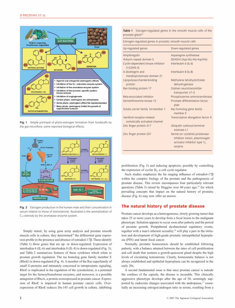
The role of estrogens in relation to prostate growth is now seen asmore important than hitherto believed , and this review is based onreflections from an International Prostate Health Council (IPHC) StudyGroup directed to this topic and the clinical value of antiestrogentherapy. Discussion centered on whether phyto-estrogens can restrainprostate cancer progression through their agonistic or antagonisticestrogenic properties, or by some other of the associated imposingrange of biological effects (Fig. 1). Benefit may result from their capac-ity to act as effective antioxidants, as tyrosine kinase inhibitors, or asaromatase inhibitors of estrogen biosynthesis.
Estrogens and the prostate
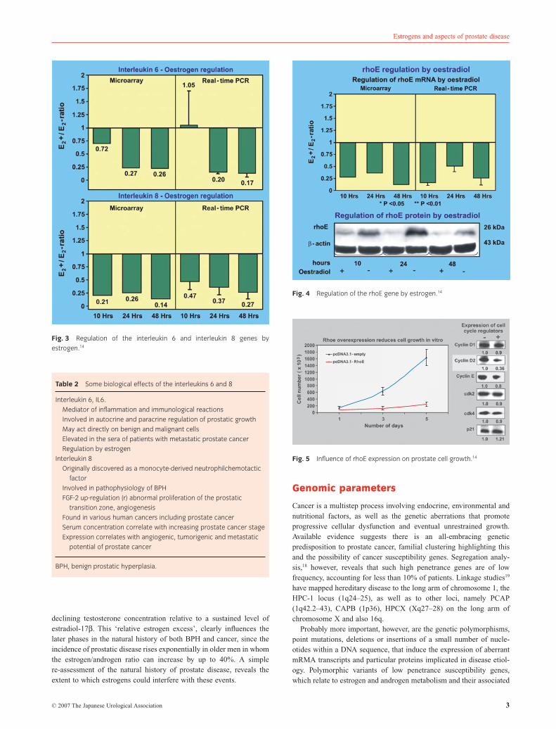
Although the male or female phenotype is determined to a large extentby differences in the serum concentrations of the sex steroid hormones,there is no sexual specificity with regard to any particular hormone andthere are substantial levels of estrogens in the human male, althoughmarkedly less than those of testosterone (Fig. 2). The influence ofestrogens on the developing embryonic or neonatal prostate, the impactof the changing hormone balance at puberty and the endocrine status ofa young man entering his 20s, together with the more intrusive effectsof estrogens on the ‘mid-life’ prostate, all impinge on growth regulatorymechanisms within the gland. Early estrogen-mediated gene imprintingthat can subsequently influence the insulin-like growth factor (IGF)-network, or another of the estradiol-related molecular events in laterlife, thereby increasing the propensity for prostatic dysfunction,demands fuller investigation.
Substantive evidence for an estrogen receptor (ER) in the canineprostate10 and the classical studies of Walsh and Coffey11 which showedthat estradiol-17b, together with DHT, or a 5a-androstanediol, induceda fourfold increase in its weight and DNA content, focused atten-tion on their synergistic effects on the gland. Testosterone, togetherwith estradiol-17b, failed to enhance prostate weight. The 5a-androstanediols are weak estrogens12 that associate with ER. Althoughthere are many recorded effects of estrogen on the prostate,13 it is thenew, elegant type of studies, such as those of Klocker and his col-leagues,14 which will most effectively impact on our understanding theregulatory role of estrogens.
Correspondence: Louis J Denis, MD, Oncology Centre Antwerp, Lange Gast-huisstraat 35–37, 2000 Antwerpen, Belgium. Email: [email protected]
Received 27 February 2006; accepted 19 April 2006.
International Journal of Urology (2007) 14, 1–16 doi: 10.1111/j.1442-2042.2006.01476.x
© 2007 The Japanese Urological Association 1
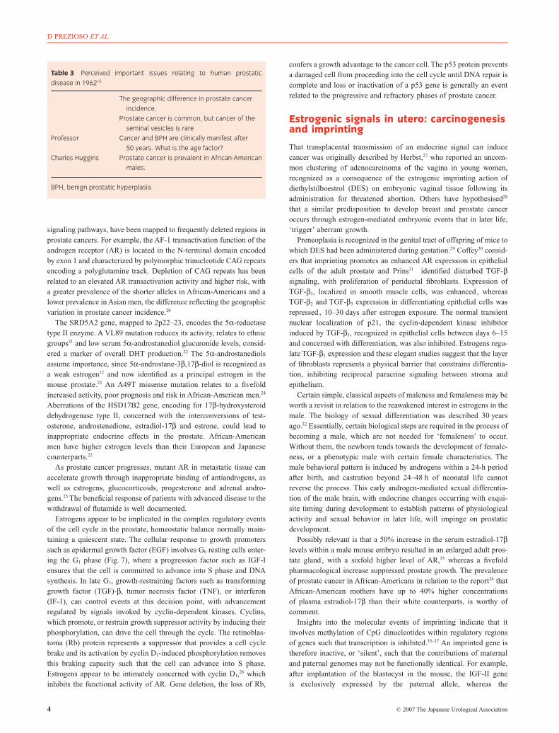
Simply stated , by using gene array analysis and prostate smoothmuscle cells in culture, they determined14 the differential gene expres-sion profile in the presence and absence of estradiol-17b. These identify(Table 1) those genes that are up- or down-regulated. Expression ofinterleukin 6 (IL-6) and interleukin 8 (IL-8) is down-regulated (Fig. 3),and Table 2 summarizes features of these cytokines which relate toprostate growth regulation. The ras homolog gene family member E(RhoE) is down-regulated (Fig. 4). A member of the Ras superfamily ofsmall G-proteins and intimately concerned in intraprostatic signaling,RhoE is implicated in the regulation of the cytoskeleton, is a potentialtarget for the farnesyltransferase enzymes, and moreover, is a possibleantagonist of RhoA, a protein overexpressed in prostate cancer. Expres-sion of RhoE is impaired in human prostate cancer cells. Over-expression of RhoE reduces Du-145 cell growth in culture, inhibiting
proliferation (Fig. 5) and inducing apoptosis, possibly by controllingthe expression of cyclin D2, a cell cycle regulator.
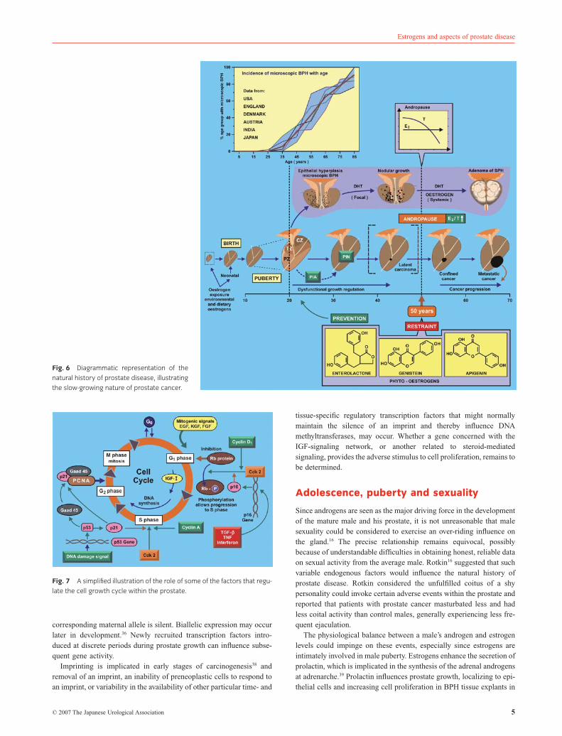
Such studies emphasize the far ranging influence of estradiol-17bwithin the complex biology of the prostate and the pathogenesis ofprostate disease. This review encompasses four particularly relevantquestions (Table 3) raised by Huggins over 40 years ago,15 for whichprevailing concepts that impact on the natural history of prostaticdisease (Fig. 6) may now offer an answer.
The natural history of prostate disease
Prostate cancer develops as a heterogeneous, slowly growing tumor thattakes 25 or more years to develop from a focal lesion to the malignantphenotype. Initiation appears to occur soon after puberty and the periodof prostate growth. Postpubertal dysfunctional regulatory events,together with a man’s inherent sexuality,16 will play a part in the initia-tion and development of high-grade prostatic intraepithelial hyperpla-sia (PIN) and latent focal cancer.
Normally, prostatic homeostasis should be established followingpuberty, with a balance attained between the rates of cell proliferationand cell death that sustains a growth-quiescent gland despite the highlevels of circulating testosterone. Clearly, homeostatic balance is notalways established and epithelial hyperplasia can be recognized in theearly 20s.
A second fundamental issue is that once prostate cancer is outsidethe confines of the capsule, the disease is incurable. This clinicallyaggressive phenotype develops after the age of 50, seemingly sup-ported by endocrine changes associated with the andropause,17 essen-tially an increasing estrogen/androgen ratio in serum, resulting from a
Fig. 1 Simple portrayal of phyto-estrogen formation from foodstuffs by
the gut microflora: some reported biological effects.
Fig. 2 Estrogen production in the human male and their concentration in
serum relative to those of testosterone. Illustrated is the aromatization of
C19-steroids by the aromatase enzyme system.
Table 1 Estrogen-regulated genes in the smooth muscle cells of the
prostate gland14
Estrogen regulated genes in prostatic smooth muscle cells
Up-regulated genes Down-regulated genes
Amphiregulin Asparagine synthetase
Ankyrin repeat domain 5 DEAD/H (Asp-Glu-Ala-Asp/His)
Cyclin-dependent kinase inhibitor
3 (CDKN 3)
Interleukin 6 (IL-6)
A disintegrin and
metalloproteinase domain 21
Interleukin 8 (IL-8)
Lipopolysaccharide-binding
protein
Methylene tetrahydrofolate
dehydrogenase
Ran binding protein 17 Orphan neurotransmitter
transporter v7–3
Rela-associated inhibitor Phosphoserine aminotransferase
Serine/threonine kinase 15 Prostate differentiation factor,
plab
Solute carrier family 16-member 7 Ras homolog gene family
member E
Vanilloid receptor-related
osmotically activated channel
Transcription elongation factor A
Zinc finger protein 217 Ubiquitin carboxyl-terminal
esterase L1
Zinc finger protein 237 Serine (or cysteine) proteinase
inhibitor (nexin, plasminogen
activator inhibitor type 1),
serpine
D PREZIOSO ET AL.
2 © 2007 The Japanese Urological Association
declining testosterone concentration relative to a sustained level ofestradiol-17b. This ‘relative estrogen excess’, clearly influences thelater phases in the natural history of both BPH and cancer, since theincidence of prostatic disease rises exponentially in older men in whomthe estrogen/androgen ratio can increase by up to 40%. A simplere-assessment of the natural history of prostate disease, reveals theextent to which estrogens could interfere with these events.
Genomic parameters
Cancer is a multistep process involving endocrine, environmental andnutritional factors, as well as the genetic aberrations that promoteprogressive cellular dysfunction and eventual unrestrained growth.Available evidence suggests there is an all-embracing geneticpredisposition to prostate cancer, familial clustering highlighting thisand the possibility of cancer susceptibility genes. Segregation analy-sis,18 however, reveals that such high penetrance genes are of lowfrequency, accounting for less than 10% of patients. Linkage studies19
have mapped hereditary disease to the long arm of chromosome 1, theHPC-1 locus (1q24–25), as well as to other loci, namely PCAP(1q42.2–43), CAPB (1p36), HPCX (Xq27–28) on the long arm ofchromosome X and also 16q.
Probably more important, however, are the genetic polymorphisms,point mutations, deletions or insertions of a small number of nucle-otides within a DNA sequence, that induce the expression of aberrantmRMA transcripts and particular proteins implicated in disease etiol-ogy. Polymorphic variants of low penetrance susceptibility genes,which relate to estrogen and androgen metabolism and their associated
Fig. 3 Regulation of the interleukin 6 and interleukin 8 genes by
estrogen.14
Table 2 Some biological effects of the interleukins 6 and 8
Interleukin 6, IL6.
Mediator of inflammation and immunological reactions
Involved in autocrine and paracrine regulation of prostatic growth
May act directly on benign and malignant cells
Elevated in the sera of patients with metastatic prostate cancer
Regulation by estrogen
Interleukin 8
Originally discovered as a monocyte-derived neutrophilchemotactic
factor
Involved in pathophysiology of BPH
FGF-2 up-regulation (r) abnormal proliferation of the prostatic
transition zone, angiogenesis
Found in various human cancers including prostate cancer
Serum concentration correlate with increasing prostate cancer stage
Expression correlates with angiogenic, tumorigenic and metastatic
potential of prostate cancer
BPH, benign prostatic hyperplasia.
Fig. 4 Regulation of the rhoE gene by estrogen.14
Fig. 5 Influence of rhoE expression on prostate cell growth.14
Estrogens and aspects of prostate disease
© 2007 The Japanese Urological Association 3
signaling pathways, have been mapped to frequently deleted regions inprostate cancers. For example, the AF-1 transactivation function of theandrogen receptor (AR) is located in the N-terminal domain encodedby exon 1 and characterized by polymorphic trinucleotide CAG repeatsencoding a polyglutamine track. Depletion of CAG repeats has beenrelated to an elevated AR transactivation activity and higher risk, witha greater prevalence of the shorter alleles in African-Americans and alower prevalence in Asian men, the difference reflecting the geographicvariation in prostate cancer incidence.20
The SRD5A2 gene, mapped to 2p22–23, encodes the 5a-reductasetype II enzyme. A VL89 mutation reduces its activity, relates to ethnicgroups21 and low serum 5a-androstanediol glucuronide levels, consid-ered a marker of overall DHT production.22 The 5a-androstanediolsassume importance, since 5a-androstane-3b,17b-diol is recognized asa weak estrogen12 and now identified as a principal estrogen in themouse prostate.23 An A49T missense mutation relates to a fivefoldincreased activity, poor prognosis and risk in African-American men.24
Aberrations of the HSD17B2 gene, encoding for 17b-hydroxysteroiddehydrogenase type II, concerned with the interconversions of test-osterone, androstenedione, estradiol-17b and estrone, could lead toinappropriate endocrine effects in the prostate. African-Americanmen have higher estrogen levels than their European and Japanesecounterparts.22
As prostate cancer progresses, mutant AR in metastatic tissue canaccelerate growth through inappropriate binding of antiandrogens, aswell as estrogens, glucocorticoids, progesterone and adrenal andro-gens.25 The beneficial response of patients with advanced disease to thewithdrawal of flutamide is well documented.
Estrogens appear to be implicated in the complex regulatory eventsof the cell cycle in the prostate, homeostatic balance normally main-taining a quiescent state. The cellular response to growth promoterssuch as epidermal growth factor (EGF) involves G0 resting cells enter-ing the G1 phase (Fig. 7), where a progression factor such as IGF-Iensures that the cell is committed to advance into S phase and DNAsynthesis. In late G1, growth-restraining factors such as transforminggrowth factor (TGF)-b, tumor necrosis factor (TNF), or interferon(IF-1), can control events at this decision point, with advancementregulated by signals invoked by cyclin-dependent kinases. Cyclins,which promote, or restrain growth suppressor activity by inducing theirphosphorylation, can drive the cell through the cycle. The retinoblas-toma (Rb) protein represents a suppressor that provides a cell cyclebrake and its activation by cyclin D1-induced phosphorylation removesthis braking capacity such that the cell can advance into S phase.Estrogens appear to be intimately concerned with cyclin D1,26 whichinhibits the functional activity of AR. Gene deletion, the loss of Rb,
confers a growth advantage to the cancer cell. The p53 protein preventsa damaged cell from proceeding into the cell cycle until DNA repair iscomplete and loss or inactivation of a p53 gene is generally an eventrelated to the progressive and refractory phases of prostate cancer.
Estrogenic signals in utero: carcinogenesisand imprinting
That transplacental transmission of an endocrine signal can inducecancer was originally described by Herbst,27 who reported an uncom-mon clustering of adenocarcinoma of the vagina in young women,recognized as a consequence of the estrogenic imprinting action ofdiethylstilboestrol (DES) on embryonic vaginal tissue following itsadministration for threatened abortion. Others have hypothesised28
that a similar predisposition to develop breast and prostate canceroccurs through estrogen-mediated embryonic events that in later life,‘trigger’ aberrant growth.
Preneoplasia is recognized in the genital tract of offspring of mice towhich DES had been administered during gestation.29 Coffey30 consid-ers that imprinting promotes an enhanced AR expression in epithelialcells of the adult prostate and Prins31 identified disturbed TGF-bsignaling, with proliferation of periductal fibroblasts. Expression ofTGF-b1, localized in smooth muscle cells, was enhanced , whereasTGF-b2 and TGF-b3 expression in differentiating epithelial cells wasrepressed , 10–30 days after estrogen exposure. The normal transientnuclear localization of p21, the cyclin-dependent kinase inhibitorinduced by TGF-b1, recognized in epithelial cells between days 6–15and concerned with differentiation, was also inhibited. Estrogens regu-late TGF-b1 expression and these elegant studies suggest that the layerof fibroblasts represents a physical barrier that constrains differentia-tion, inhibiting reciprocal paracrine signaling between stroma andepithelium.
Certain simple, classical aspects of maleness and femaleness may beworth a revisit in relation to the reawakened interest in estrogens in themale. The biology of sexual differentiation was described 30 yearsago.32 Essentially, certain biological steps are required in the process ofbecoming a male, which are not needed for ‘femaleness’ to occur.Without them, the newborn tends towards the development of female-ness, or a phenotypic male with certain female characteristics. Themale behavioral pattern is induced by androgens within a 24-h periodafter birth, and castration beyond 24–48 h of neonatal life cannotreverse the process. This early androgen-mediated sexual differentia-tion of the male brain, with endocrine changes occurring with exqui-site timing during development to establish patterns of physiologicalactivity and sexual behavior in later life, will impinge on prostaticdevelopment.
Possibly relevant is that a 50% increase in the serum estradiol-17blevels within a male mouse embryo resulted in an enlarged adult pros-tate gland , with a sixfold higher level of AR,33 whereas a fivefoldpharmacological increase suppressed prostate growth. The prevalenceof prostate cancer in African-Americans in relation to the report34 thatAfrican-American mothers have up to 40% higher concentrationsof plasma estradiol-17b than their white counterparts, is worthy ofcomment.
Insights into the molecular events of imprinting indicate that itinvolves methylation of CpG dinucleotides within regulatory regionsof genes such that transcription is inhibited.35–37 An imprinted gene istherefore inactive, or ‘silent’, such that the contributions of maternaland paternal genomes may not be functionally identical. For example,after implantation of the blastocyst in the mouse, the IGF-II geneis exclusively expressed by the paternal allele, whereas the
Table 3 Perceived important issues relating to human prostatic
disease in 196215
The geographic difference in prostate cancer
incidence.
Prostate cancer is common, but cancer of the
seminal vesicles is rare
Professor Cancer and BPH are clinically manifest after
50 years. What is the age factor?
Charles Huggins Prostate cancer is prevalent in African-American
males.
BPH, benign prostatic hyperplasia.
D PREZIOSO ET AL.
4 © 2007 The Japanese Urological Association
corresponding maternal allele is silent. Biallelic expression may occurlater in development.36 Newly recruited transcription factors intro-duced at discrete periods during prostate growth can influence subse-quent gene activity.
Imprinting is implicated in early stages of carcinogenesis38 andremoval of an imprint, an inability of preneoplastic cells to respond toan imprint, or variability in the availability of other particular time- and
tissue-specific regulatory transcription factors that might normallymaintain the silence of an imprint and thereby influence DNAmethyltransferases, may occur. Whether a gene concerned with theIGF-signaling network, or another related to steroid-mediatedsignaling, provides the adverse stimulus to cell proliferation, remains tobe determined.
Adolescence, puberty and sexuality
Since androgens are seen as the major driving force in the developmentof the mature male and his prostate, it is not unreasonable that malesexuality could be considered to exercise an over-riding influence onthe gland.16 The precise relationship remains equivocal, possiblybecause of understandable difficulties in obtaining honest, reliable dataon sexual activity from the average male. Rotkin16 suggested that suchvariable endogenous factors would influence the natural history ofprostate disease. Rotkin considered the unfulfilled coitus of a shypersonality could invoke certain adverse events within the prostate andreported that patients with prostate cancer masturbated less and hadless coital activity than control males, generally experiencing less fre-quent ejaculation.
The physiological balance between a male’s androgen and estrogenlevels could impinge on these events, especially since estrogens areintimately involved in male puberty. Estrogens enhance the secretion ofprolactin, which is implicated in the synthesis of the adrenal androgensat adrenarche.39 Prolactin influences prostate growth, localizing to epi-thelial cells and increasing cell proliferation in BPH tissue explants in
Fig. 6 Diagrammatic representation of the
natural history of prostate disease, illustrating
the slow-growing nature of prostate cancer.
Fig. 7 A simplified illustration of the role of some of the factors that regu-
late the cell growth cycle within the prostate.
Estrogens and aspects of prostate disease
© 2007 The Japanese Urological Association 5
culture.40 Transgenic mice over-expressing the rat PLN gene develop adramatic prostate enlargement that resembles BPH.41 A high fat intakeelevates serum prolactin and the reported suppression of the normallyelevated nocturnal levels of plasma prolactin in those who change froman omnivorous to a vegetarian diet,39 emphasizes the close relationshipbetween estrogens, prolactin and diet.
Growth hormone (GH) and consequently, the IGF-network, are alsointimately involved in adolescence and puberty, with GH and IGF-Isecretion promoted by estrogens. IGF-I mediated signaling inducesepithelial42 and stromal cell43 proliferation, and systemic administrationof IGF-I, but not EGF, promoted growth of the rat prostate.44 Moreover,serum IGF-I levels increase during early puberty,45 promoting LH-RHrelease from the hypothalamus. IGF-I administration will advancethe stages of puberty. Enhanced expression of the IGF-I gene wasidentified46 in the liver of rats during puberty, with a corresponding risein serum IGF-I and gonadotrophin levels, an associated enhancementof IGF-IR in the median eminence, release of LH-RH and activation ofthe pituitary–testicular–prostatic axis.
The role of IGF-I in prostate growth regulation assumes greaterimportance.47 An incompletely developed prostate was identified inIGF-I deficient transgenic mice48 and elevated levels of IGF-I appearto predict prostate cancer risk, with the ratio of IGF-I/IGFBP-3 inserum particularly significant.49 Interestingly, in China where the inci-dence of prostate cancer is low, but the prevalence of BPH similar tothat in the West, a case–control study showed that BPH is positivelyassociated with plasma IGF-I levels and inversely with IGFBP-3.50
Serum IGF-1 levels in subjects with evidence of PIN, as well as thosewith prostate cancer were higher than controls.51 Estradiol-17benhances the expression of GH-receptor in the liver and this relates toIGF-I expression.52 Patients with acromegaly have a high prevalenceof prostate enlargement that is associated with elevated plasma levelsof GH and IGF-I and reduced circulating androgen levels.53 Severeinsulin resistance is also characteristic of acromegalics, who have apropensity to develop cancer, particularly of breast.53,54 Possibly rel-evant, is that tamoxifen decreases levels of serum IGF-I in patientswith breast cancer55 and influences the IGF-I/IGFBP-3 ratio to favorthe growth inhibitory actions of IGFBP-3. The phyto-estrogensgenistein and daidzein may well beneficially influence male puberty. AWestern diet lowers the age of menarche – there are marked differ-ences in the onset of hormone secretion and ovulatory cycles betweengirls in Britain and Thailand56,57 – and may influence the LH-RH pulsegenerator in males.
The foregoing highlights particular aspects of puberty that involveestrogens. Classically, it is considered58 that the onset of puberty is‘physiologically gated’, governed by the energy resources of the body,especially in the female. The discovery of leptin, the antiobesityprotein, provided another important insight into the ‘endocrine puzzle’of puberty.59 The obesity (Ob) gene is exclusively expressed by adipo-cytes and the action of leptin on the neuroendocrine–reproductive axisthat induces the onset of puberty, probably exercises a permissive roleas a ‘metabolic gate’. Leptin now assumes an important neuroendo-crine role and as Professor Robert Steiner stated ,60 ‘The story of leptinhas all the earmarks of a Dostoyevsky novel: you know it is bound toget more complicated before it ends with some great Truth’. Studies onleptin have centered on the female and emerging results demonstrateracial differences in levels of plasma leptin and in its role in controllingresting energy expenditure between black and white women, differ-ences that may be concerned in the prevalence of obesity in the African-American population. A relationship to adverse risks with regardto prostatic cancer will undoubtedly invoke controversy. The conse-quences of Ob gene mutations, or in the leptin receptor (Ob-R), with
ineffective signaling in the hypothalamus that delays or impairs thecomplex processes of puberty, require consideration.
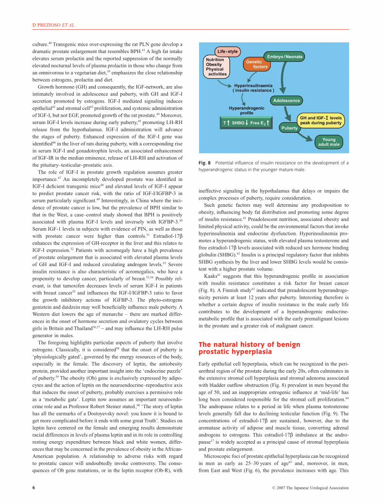
Such genetic factors may well determine any predisposition toobesity, influencing body fat distribution and promoting some degreeof insulin resistance.61 Preadolescent nutrition, associated obesity andlimited physical activity, could be the environmental factors that invokehyperinsulinemia and endocrine dysfunction. Hyperinsulinemia pro-motes a hyperandrogenic status, with elevated plasma testosterone andfree estradiol-17b levels associated with reduced sex hormone bindingglobulin (SHBG).62 Insulin is a principal regulatory factor that inhibitsSHBG synthesis by the liver and lower SHBG levels would be consis-tent with a higher prostate volume.
Kaaks61 suggests that this hyperandrogenic profile in associationwith insulin resistance constitutes a risk factor for breast cancer(Fig. 8). A Finnish study63 indicated that preadolescent hyperandroge-nicity persists at least 12 years after puberty. Interesting therefore iswhether a certain degree of insulin resistance in the male early lifecontributes to the development of a hyperandrogenic endocrine-metabolic profile that is associated with the early premalignant lesionsin the prostate and a greater risk of malignant cancer.
The natural history of benignprostatic hyperplasia
Early epithelial cell hyperplasia, which can be recognized in the peri-urethral region of the prostate during the early 20s, often culminates inthe extensive stromal cell hyperplasia and stromal adenoma associatedwith bladder outflow obstruction (Fig. 8) prevalent in men beyond theage of 50, and an inappropriate estrogenic influence at ‘mid-life’ haslong been considered responsible for the stromal cell proliferation.64
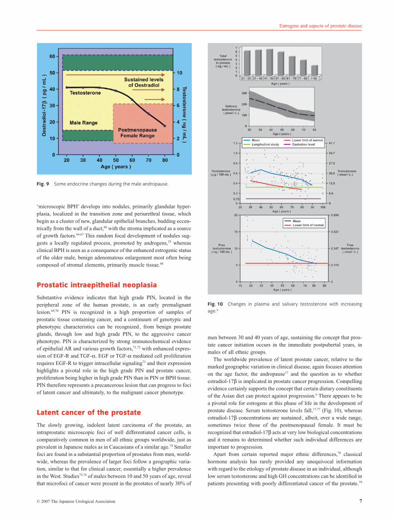
The andropause relates to a period in life when plasma testosteronelevels generally fall due to declining testicular function (Fig. 9). Theconcentrations of estradiol-17b are sustained , however, due to thearomatase activity of adipose and muscle tissue, converting adrenalandrogens to estrogens. This estradiol-17b imbalance at the andro-pause17 is widely accepted as a principal cause of stromal hyperplasiaand prostate enlargement.
Microscopic foci of prostate epithelial hyperplasia can be recognizedin men as early as 25–30 years of age65 and , moreover, in men,from East and West (Fig. 6), the prevalence increases with age. This
Fig. 8 Potential influence of insulin resistance on the development of a
hyperandrogenic status in the younger mature male.
D PREZIOSO ET AL.
6 © 2007 The Japanese Urological Association
‘microscopic BPH’ develops into nodules, primarily glandular hyper-plasia, localized in the transition zone and periurethral tissue, whichbegin as a cluster of new, glandular epithelial branches, budding eccen-trically from the wall of a duct,66 with the stroma implicated as a sourceof growth factors.64,67 This random focal development of nodules sug-gests a locally regulated process, promoted by androgens,25 whereasclinical BPH is seen as a consequence of the enhanced estrogenic statusof the older male, benign adenomatous enlargement most often beingcomposed of stromal elements, primarily muscle tissue.68
Prostatic intraepithelial neoplasia
Substantive evidence indicates that high grade PIN, located in theperipheral zone of the human prostate, is an early premalignantlesion.69,70 PIN is recognized in a high proportion of samples ofprostatic tissue containing cancer, and a continuum of genotypic andphenotypic characteristics can be recognized , from benign prostateglands, through low and high grade PIN, to the aggressive cancerphenotype. PIN is characterized by strong immunochemical evidenceof epithelial AR and various growth factors,71,72 with enhanced expres-sion of EGF-R and TGF-a. EGF or TGF-a mediated cell proliferationrequires EGF-R to trigger intracellular signaling73 and their expressionhighlights a pivotal role in the high grade PIN and prostate cancer,proliferation being higher in high grade PIN than in PIN or BPH tissue.PIN therefore represents a precancerous lesion that can progress to fociof latent cancer and ultimately, to the malignant cancer phenotype.
Latent cancer of the prostate
The slowly growing, indolent latent carcinoma of the prostate, anintraprostatic microscopic foci of well differentiated cancer cells, iscomparatively common in men of all ethnic groups worldwide, just asprevalent in Japanese males as in Caucasians of a similar age.74 Smallerfoci are found in a substantial proportion of prostates from men, world-wide, whereas the prevalence of larger foci follow a geographic varia-tion, similar to that for clinical cancer, essentially a higher prevalencein the West. Studies75,76 of males between 10 and 50 years of age, revealthat microfoci of cancer were present in the prostates of nearly 30% of
men between 30 and 40 years of age, sustaining the concept that pros-tate cancer initiation occurs in the immediate postpubertal years, inmales of all ethnic groups.
The worldwide prevalence of latent prostate cancer, relative to themarked geographic variation in clinical disease, again focuses attentionon the age factor, the andropause17 and the question as to whetherestradiol-17b is implicated in prostate cancer progression. Compellingevidence certainly supports the concept that certain dietary constituentsof the Asian diet can protect against progression.6 There appears to bea pivotal role for estrogens at this phase of life in the development ofprostate disease. Serum testosterone levels fall,17,77 (Fig. 10), whereasestradiol-17b concentrations are sustained , albeit, over a wide range,sometimes twice those of the postmenopausal female. It must berecognized that estradiol-17b acts at very low biological concentrationsand it remains to determined whether such individual differences areimportant to progression.
Apart from certain reported major ethnic differences,78 classicalhormone analysis has rarely provided any unequivocal informationwith regard to the etiology of prostate disease in an individual, althoughlow serum testosterone and high GH concentrations can be identified inpatients presenting with poorly differentiated cancer of the prostate.79
Fig. 9 Some endocrine changes during the male andropause.
Fig. 10 Changes in plasma and salivary testosterone with increasing
age.6
Estrogens and aspects of prostate disease
© 2007 The Japanese Urological Association 7
With regard to familial clustering of cancer of the prostate,80 patientsand their first-degree relatives had lower plasma levels of testosteronethan matched controls and a greater capacity to synthesize estrogens.There are certain trends, with lower concentrations of plasma testoster-one and estradiol-17b found in Japanese men, compared to theirwestern counterparts.78
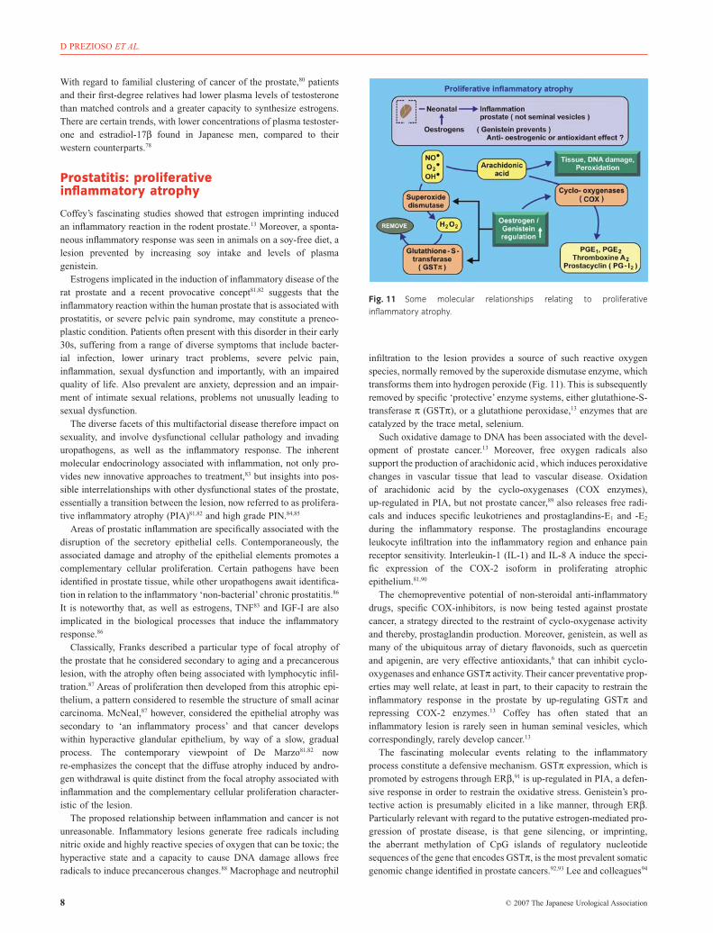
Prostatitis: proliferativeinflammatory atrophy
Coffey’s fascinating studies showed that estrogen imprinting inducedan inflammatory reaction in the rodent prostate.13 Moreover, a sponta-neous inflammatory response was seen in animals on a soy-free diet, alesion prevented by increasing soy intake and levels of plasmagenistein.
Estrogens implicated in the induction of inflammatory disease of therat prostate and a recent provocative concept81,82 suggests that theinflammatory reaction within the human prostate that is associated withprostatitis, or severe pelvic pain syndrome, may constitute a preneo-plastic condition. Patients often present with this disorder in their early30s, suffering from a range of diverse symptoms that include bacter-ial infection, lower urinary tract problems, severe pelvic pain,inflammation, sexual dysfunction and importantly, with an impairedquality of life. Also prevalent are anxiety, depression and an impair-ment of intimate sexual relations, problems not unusually leading tosexual dysfunction.
The diverse facets of this multifactorial disease therefore impact onsexuality, and involve dysfunctional cellular pathology and invadinguropathogens, as well as the inflammatory response. The inherentmolecular endocrinology associated with inflammation, not only pro-vides new innovative approaches to treatment,83 but insights into pos-sible interrelationships with other dysfunctional states of the prostate,essentially a transition between the lesion, now referred to as prolifera-tive inflammatory atrophy (PIA)81,82 and high grade PIN.84,85
Areas of prostatic inflammation are specifically associated with thedisruption of the secretory epithelial cells. Contemporaneously, theassociated damage and atrophy of the epithelial elements promotes acomplementary cellular proliferation. Certain pathogens have beenidentified in prostate tissue, while other uropathogens await identifica-tion in relation to the inflammatory ‘non-bacterial’ chronic prostatitis.86
It is noteworthy that, as well as estrogens, TNF83 and IGF-I are alsoimplicated in the biological processes that induce the inflammatoryresponse.86
Classically, Franks described a particular type of focal atrophy ofthe prostate that he considered secondary to aging and a precancerouslesion, with the atrophy often being associated with lymphocytic infil-tration.87 Areas of proliferation then developed from this atrophic epi-thelium, a pattern considered to resemble the structure of small acinarcarcinoma. McNeal,87 however, considered the epithelial atrophy wassecondary to ‘an inflammatory process’ and that cancer developswithin hyperactive glandular epithelium, by way of a slow, gradualprocess. The contemporary viewpoint of De Marzo81,82 nowre-emphasizes the concept that the diffuse atrophy induced by andro-gen withdrawal is quite distinct from the focal atrophy associated withinflammation and the complementary cellular proliferation character-istic of the lesion.
The proposed relationship between inflammation and cancer is notunreasonable. Inflammatory lesions generate free radicals includingnitric oxide and highly reactive species of oxygen that can be toxic; thehyperactive state and a capacity to cause DNA damage allows freeradicals to induce precancerous changes.88 Macrophage and neutrophil
infiltration to the lesion provides a source of such reactive oxygenspecies, normally removed by the superoxide dismutase enzyme, whichtransforms them into hydrogen peroxide (Fig. 11). This is subsequentlyremoved by specific ‘protective’ enzyme systems, either glutathione-S-transferase p (GSTp), or a glutathione peroxidase,13 enzymes that arecatalyzed by the trace metal, selenium.
Such oxidative damage to DNA has been associated with the devel-opment of prostate cancer.13 Moreover, free oxygen radicals alsosupport the production of arachidonic acid , which induces peroxidativechanges in vascular tissue that lead to vascular disease. Oxidationof arachidonic acid by the cyclo-oxygenases (COX enzymes),up-regulated in PIA, but not prostate cancer,89 also releases free radi-cals and induces specific leukotrienes and prostaglandins-E1 and -E2
during the inflammatory response. The prostaglandins encourageleukocyte infiltration into the inflammatory region and enhance painreceptor sensitivity. Interleukin-1 (IL-1) and IL-8 A induce the speci-fic expression of the COX-2 isoform in proliferating atrophicepithelium.81,90
The chemopreventive potential of non-steroidal anti-inflammatorydrugs, specific COX-inhibitors, is now being tested against prostatecancer, a strategy directed to the restraint of cyclo-oxygenase activityand thereby, prostaglandin production. Moreover, genistein, as well asmany of the ubiquitous array of dietary flavonoids, such as quercetinand apigenin, are very effective antioxidants,6 that can inhibit cyclo-oxygenases and enhance GSTp activity. Their cancer preventative prop-erties may well relate, at least in part, to their capacity to restrain theinflammatory response in the prostate by up-regulating GSTp andrepressing COX-2 enzymes.13 Coffey has often stated that aninflammatory lesion is rarely seen in human seminal vesicles, whichcorrespondingly, rarely develop cancer.13
The fascinating molecular events relating to the inflammatoryprocess constitute a defensive mechanism. GSTp expression, which ispromoted by estrogens through ERb,91 is up-regulated in PIA, a defen-sive response in order to restrain the oxidative stress. Genistein’s pro-tective action is presumably elicited in a like manner, through ERb.Particularly relevant with regard to the putative estrogen-mediated pro-gression of prostate disease, is that gene silencing, or imprinting,the aberrant methylation of CpG islands of regulatory nucleotidesequences of the gene that encodes GSTp, is the most prevalent somaticgenomic change identified in prostate cancers.92,93 Lee and colleagues94
Fig. 11 Some molecular relationships relating to proliferative
inflammatory atrophy.
D PREZIOSO ET AL.
8 © 2007 The Japanese Urological Association
identified GSTp methylation in nearly 70% of PIN lesions and in morethan 90% of carcinomas, but in neither BPH, nor normal prostatetissue.
This common epigenetic event, the loss of the protective GSTpenzyme, could well be implicated in the proposed transition from PIAto PIN, providing not only a specific growth advantage, but the possi-bility that estrogens could play a significant role in this transitional stepin prostate carcinogenesis. Dysfunctional gene methylation is becom-ing a common feature of prostate cancer, with aberrant methylation ofthe A isoform of the ras association domain family protein 1(RASSF1A) gene, also prevalent in prostatic cancer.95 The silencing ofeither of the ERa and ERb genes, also a common characteristic ofprostate cancer96,97 will influence disease progression. Although ERb isexpressed together with ERa in the normal prostate, it is rarely iden-tified in prostate cancer, emphasizing the possible importance of itsregulatory growth-restraining role. Furthermore, the expression of adeletion ER-variants in prostate cancer also highlights the pivotal roleof dysfunctional ER-signaling in the progression of the disease to themalignant phenotype.
Stromal–epithelial interactions
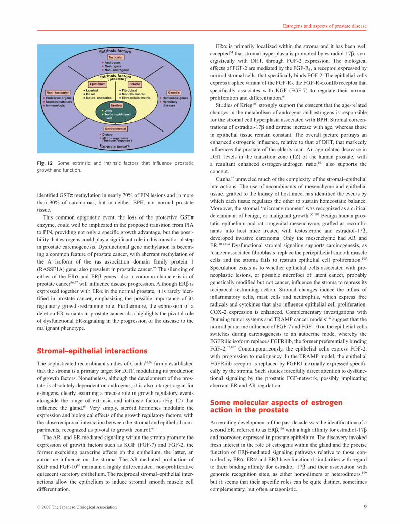
The sophisticated recombinant studies of Cunha67,98 firmly establishedthat the stroma is a primary target for DHT, modulating its productionof growth factors. Nonetheless, although the development of the pros-tate is absolutely dependent on androgens, it is also a target organ forestrogens, clearly assuming a precise role in growth regulatory eventsalongside the range of extrinsic and intrinsic factors (Fig. 12) thatinfluence the gland.64 Very simply, steroid hormones modulate theexpression and biological effects of the growth regulatory factors, withthe close reciprocal interaction between the stromal and epithelial com-partments, recognized as pivotal to growth control.64
The AR- and ER-mediated signaling within the stroma promote theexpression of growth factors such as KGF (FGF-7) and FGF-2, theformer exercising paracrine effects on the epithelium, the latter, anautocrine influence on the stroma. The AR-mediated production ofKGF and FGF-1099 maintain a highly differentiated , non-proliferativequiescent secretory epithelium. The reciprocal stromal–epithelial inter-actions allow the epithelium to induce stromal smooth muscle celldifferentiation.
ERa is primarily localized within the stroma and it has been wellaccepted64 that stromal hyperplasia is promoted by estradiol-17b, syn-ergistically with DHT, through FGF-2 expression. The biologicaleffects of FGF-2 are mediated by the FGF-R1, a receptor, expressed bynormal stromal cells, that specifically binds FGF-2. The epithelial cellsexpress a splice variant of the FGF-R2, the FGF-R2exonIIb receptor thatspecifically associates with KGF (FGF-7) to regulate their normalproliferation and differentiation.64
Studies of Krieg100 strongly support the concept that the age-relatedchanges in the metabolism of androgens and estrogens is responsiblefor the stromal cell hyperplasia associated with BPH. Stromal concen-trations of estradiol-17b and estrone increase with age, whereas thosein epithelial tissue remain constant. The overall picture portrays anenhanced estrogenic influence, relative to that of DHT, that markedlyinfluences the prostate of the elderly man. An age-related decrease inDHT levels in the transition zone (TZ) of the human prostate, witha resultant enhanced estrogen/androgen ratio,101 also supports theconcept.
Cunha67 unraveled much of the complexity of the stromal–epithelialinteractions. The use of recombinants of mesenchyme and epithelialtissue, grafted to the kidney of host mice, has identified the events bywhich each tissue regulates the other to sustain homeostatic balance.Moreover, the stromal ‘microenvironment’ was recognized as a criticaldeterminant of benign, or malignant growth.67,102 Benign human pros-tatic epithelium and rat urogenital mesenchyme, grafted as recombi-nants into host mice treated with testosterone and estradiol-17b,developed invasive carcinoma. Only the mesenchyme had AR andER.103,104 Dysfunctional stromal signaling supports carcinogenesis, as‘cancer associated fibroblasts’ replace the periepithelial smooth musclecells and the stroma fails to restrain epithelial cell proliferation.105
Speculation exists as to whether epithelial cells associated with pre-neoplastic lesions, or possible microfoci of latent cancer, probablygenetically modified but not cancer, influence the stroma to repress itsreciprocal restraining action. Stromal changes induce the influx ofinflammatory cells, mast cells and neutrophils, which express freeradicals and cytokines that also influence epithelial cell proliferation.COX-2 expression is enhanced. Complementary investigations withDunning tumor systems and TRAMP cancer models106 suggest that thenormal paracrine influence of FGF-7 and FGF-10 on the epithelial cellsswitches during carcinogenesis to an autocrine mode, whereby theFGFRiiic isoform replaces FGFRiiib, the former preferentially bindingFGF-2.67,107 Contemporaneously, the epithelial cells express FGF-2,with progression to malignancy. In the TRAMP model, the epithelialFGFRiiib receptor is replaced by FGFR1 normally expressed specifi-cally by the stroma. Such studies forcefully direct attention to dysfunc-tional signaling by the prostatic FGF-network, possibly implicatingaberrant ER and AR regulation.
Some molecular aspects of estrogenaction in the prostate
An exciting development of the past decade was the identification of asecond ER, referred to as ERb,108 with a high affinity for estradiol-17band moreover, expressed in prostate epithelium. The discovery invokedfresh interest in the role of estrogens within the gland and the precisefunction of ERb-mediated signaling pathways relative to those con-trolled by ERa. ERa and ERb have functional similarities with regardto their binding affinity for estradiol-17b and their association withgenomic recognition sites, as either homodimers or heterodimers,109
but it seems that their specific roles can be quite distinct, sometimescomplementary, but often antagonistic.
Fig. 12 Some extrinsic and intrinsic factors that influence prostatic
growth and function.
Estrogens and aspects of prostate disease
© 2007 The Japanese Urological Association 9
In the mouse ventral prostate, molecular events mediated by ERb areclearly important, the majority of epithelial nuclei express the receptorand in bERKO mice, all these cells are in the cell cycle, not in G0. ARlevels are elevated and the gland contains areas of hyperplasia,110 withmost epithelial cells expressing Ki-67. Moreover, prostatic hyperplasiaprogressively develops as BERKO mice age, with PIN lesions identi-fied in later life.
In the human prostate, epithelial elements would seem a target ofERb-mediated estrogen signaling and loss of the receptor in PINlesions supports its regulatory, role in repressing cellular proliferation.The absence of the ERb gene allows the accumulation of cells normallyprogrammed to die. ERb regulates cellular proliferation by suppressingestrogen-mediated ERa-signaling pathways that promote AR synthe-sis.111 ERb preferentially binds genistein, which has also been shown tosuppress AR expression.112 Such studies provide a new insight into theestrogen conundrum. Estradiol-17b can therefore exercise divergenteffects, in part through AR expression, depending on the cellularcontent of ERa and ERb. Genistein, presumably through ERb, simi-larly induces G2M phase cell cycle arrest and cellular apoptosis, aneffect associated with p53-independent up-regulation of p21 expressionand the down-regulation of cyclin B1.113
The antiestrogen tamoxifen also induces p21 expression and S-phasecell cycle arrest in Du145 and PC3 prostate cancer cells.114 Also note-worthy is that 5a-androstane-3b,17b-diol elicits an estrogenic responsein the aorta, but not the pituitary, and associates with ERb to decreaseAR content in the wild-type prostate, but not in BERKO mice.23
The estrogen conundrum remains complex, however, as evidenceemerges of various splice variants of ERb, now recognized as ERb1.ERb2 has a 1000-fold lower affinity for estradiol-17b, ERbcx has noaffinity and ERbs 3–5, all ER-subtypes, can form heterodimer com-plexes and thereby influence ER-signaling. The interrelationships ofthe ER-subtypes to the various coregulators that control transcription,activators, repressors and integrators,115,116 that form bridges betweenreceptors and ligands on the genome, remain to be determined.
Estradiol-17b mediated signaling through either ERa or ERb, canelicit differential activation at AP-1 sites and thereby, opposing biologi-cal actions. Although the estrogen and DNA binding domains of ERaand ERb are similar, such that they bind to the same genomic ERE, theN-terminal A/B region of the receptor and its associated transcriptionalactivation function (AF-1) are different. ERa and ERb can thereforeexercise differential gene expression through receptor protein–proteininteraction with other transcription factors associated with the DNA.Tamoxifen can therefore positively promote transcription through ERb-mediated signaling, an action that would tend to explain its estrogenagonistic effect on the uterus and through ERa and AP-1 sites, itsantagonistic influence on breast cancer.
Important then, is whether estradiol-17b mediated intraprostatic sig-naling assumes greater importance, if the activity of the DHT signalingpathway is impaired by declining plasma testosterone levels at theandropause, an effect that could impact on a prostate gland harboring alatent cancer. If differential expression of ERs is implicated in diseaseprogression, therapeutic strategies to oppose these events could offer aninnovative new approach to patient management.
Antiestrogen action: the potential ofselective estrogen receptor modulators
Discussion now centers on strategies directed to the clinical efficacy ofantiestrogen therapy. The potential of selective estrogen receptor modu-lators (SERMS) is evident and these, together with aromatase inhibitorsand the innovative Mepartricin, the former inhibiting aromatization in
adipose tissue, the latter lowering serum estrogen levels by interferingwith steroid enterohepatic recirculation, could offer different optionsfor the management of prostatic disease.
The steroid-binding domain of ERa consists of an antiparallela-helical sandwich containing 12 a-helices and a ‘protected’ steroidbinding pocket.117,118 With estradiol-17b bound into a pocket within theestradiol-ER-complex, a short amphipathic a-helix in the C-terminalregion of the steroid binding domain, controls AF-2 activity. This helix12 locks the cavity and folds the domain into a transcriptionally com-petent AF-2 configuration. As such, the complex can then interact withthe specific coactivators, corepressors and integrators necessary foreffective gene activation and expression. The binding of tamoxifeninduces a different configuration in relation to helix 12, such that theAF-2 is transcriptionally incompetent and coactivators cannot berecruited , thereby inhibiting ER-mediated transactivation.
Various growth factor signaling pathways that induce downstreamMAP-kinase phosphorylation, can also induce transcriptional activa-tion of ER in the absence of steroid ligand.119–121 Briefly, ER-mediatedsignaling by receptor transactivation, can be promoted by growthfactors such as EGF, TGF-a or IGF-I in the absence of estrogen.Particularly important, however, is the role and recruitment of the ARcoregulators in the growth regulatory processes of the prostate. Theinterrelationship between receptors and coregulators is complex, withcoactivators, corepressors and intergrators influencing gene transcrip-tion by acting as a ‘bridge’ between the steroid receptor and the tran-scriptional factor complex.122 Important, with regard to the putativeinfluence of estradiol-17b on progression, is the interaction between theERs and the cell-specific coregulators on the genome of prostate cancercells, an interaction that could be different not only for ERa relative toERb, but also for the various ER subtypes.
Seemingly exciting with regard to estrogen action in the prostate isthe AR coregulator, ARA70, a relatively specific coactivator of AR.123
ARA70 enhanced transcriptional activity of mutant AR in the presenceof DHT, but also important is that it induced AR transcriptional activitymore than 30-fold in the presence of estradiol-17b, but not DES. Theeffect was reported to be estradiol-17b specific and dose-dependent atphysiological levels. Estradiol-17b may therefore have a more directrole in AR-mediated signaling. Although mutant AR offer an explana-tion as to how estrogens could sustain prostate cancer progression andprostate-specific antigen (PSA) secretion, it appears that estradiol-17bcan also activate AR target genes such as the PSA gene, in the presenceof AR and its coactivator, ARA70. If estradiol-17b is implicated inprostate cancer progression, then the observed effect of DES on theinteractions of these ‘tripartite receptor complexes’,122 provides supportfor the use of DES, or possibly antiestrogens, as a therapeutic option forprogressive advanced cancer of the prostate.
Selective estrogen receptor modulators elicit antagonist and agonisteffects, depending on specific tissue characteristics and the interactionbetween the SERM and the available ER subtypes that are expressed.The possibility that specifically designed SERMS could be synthesizedwith specific antagonistic effects through ERb mediated signaling, willundoubtedly constitute a new and exciting approach to therapy.124
Essentially and pragmatically, an effective SERM will do the work thatgenistein may well do and has done for the Asian people through theyears.
Antiestrogen therapy: the potentialof mepartricin
Fiber intake has long been considered a health benefit, the fiber pro-foundly affecting the enterohepatic circulation of estrogens, a larger
D PREZIOSO ET AL.
10 © 2007 The Japanese Urological Association
fiber intake with greater fecal output increases125 the fecal excretion ofestrogens. This is associated with a decreased urinary excretion ofestrogens, an increased fecal estrogen excretion and a lower concentra-tion of estrogens in plasma. Similarly, high fiber intake lowers plasmalevels of estradiol-17b and testosterone.126
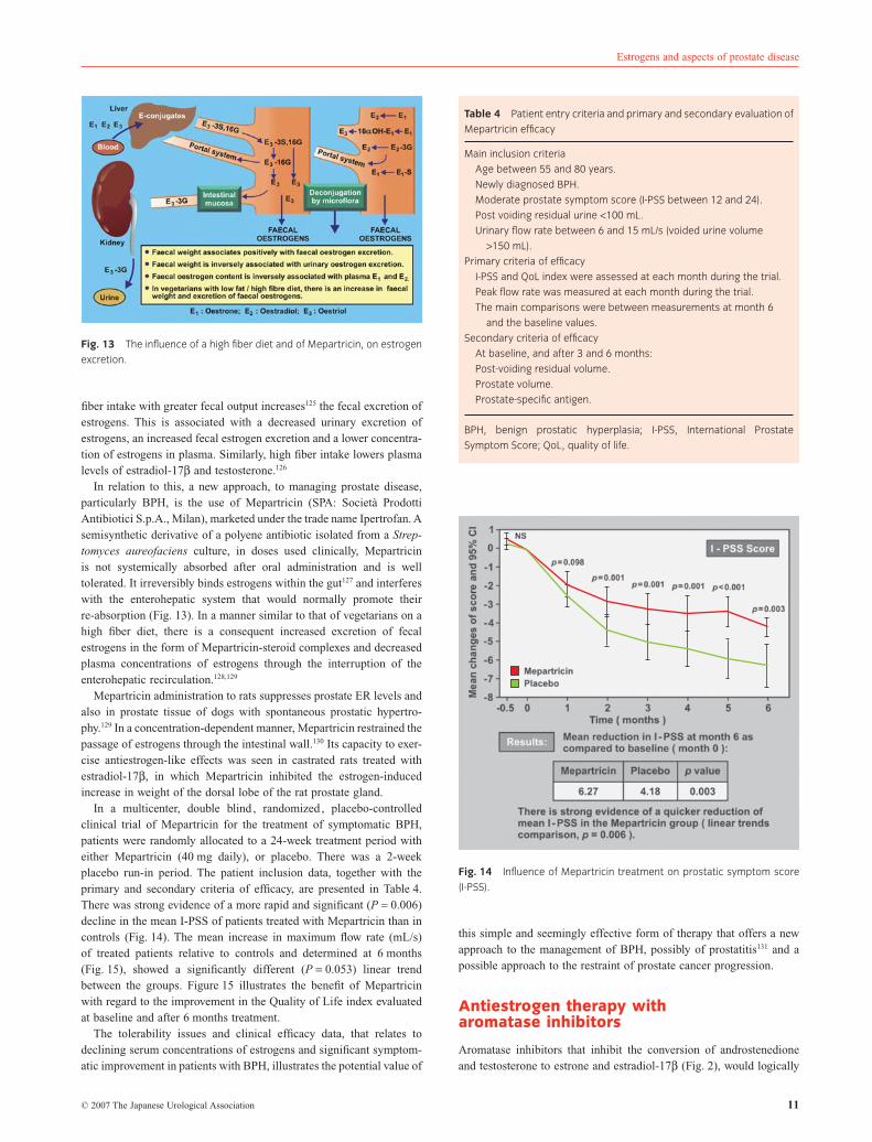
In relation to this, a new approach, to managing prostate disease,particularly BPH, is the use of Mepartricin (SPA: Società ProdottiAntibiotici S.p.A., Milan), marketed under the trade name Ipertrofan. Asemisynthetic derivative of a polyene antibiotic isolated from a Strep-tomyces aureofaciens culture, in doses used clinically, Mepartricinis not systemically absorbed after oral administration and is welltolerated. It irreversibly binds estrogens within the gut127 and interfereswith the enterohepatic system that would normally promote theirre-absorption (Fig. 13). In a manner similar to that of vegetarians on ahigh fiber diet, there is a consequent increased excretion of fecalestrogens in the form of Mepartricin-steroid complexes and decreasedplasma concentrations of estrogens through the interruption of theenterohepatic recirculation.128,129
Mepartricin administration to rats suppresses prostate ER levels andalso in prostate tissue of dogs with spontaneous prostatic hypertro-phy.129 In a concentration-dependent manner, Mepartricin restrained thepassage of estrogens through the intestinal wall.130 Its capacity to exer-cise antiestrogen-like effects was seen in castrated rats treated withestradiol-17b, in which Mepartricin inhibited the estrogen-inducedincrease in weight of the dorsal lobe of the rat prostate gland.
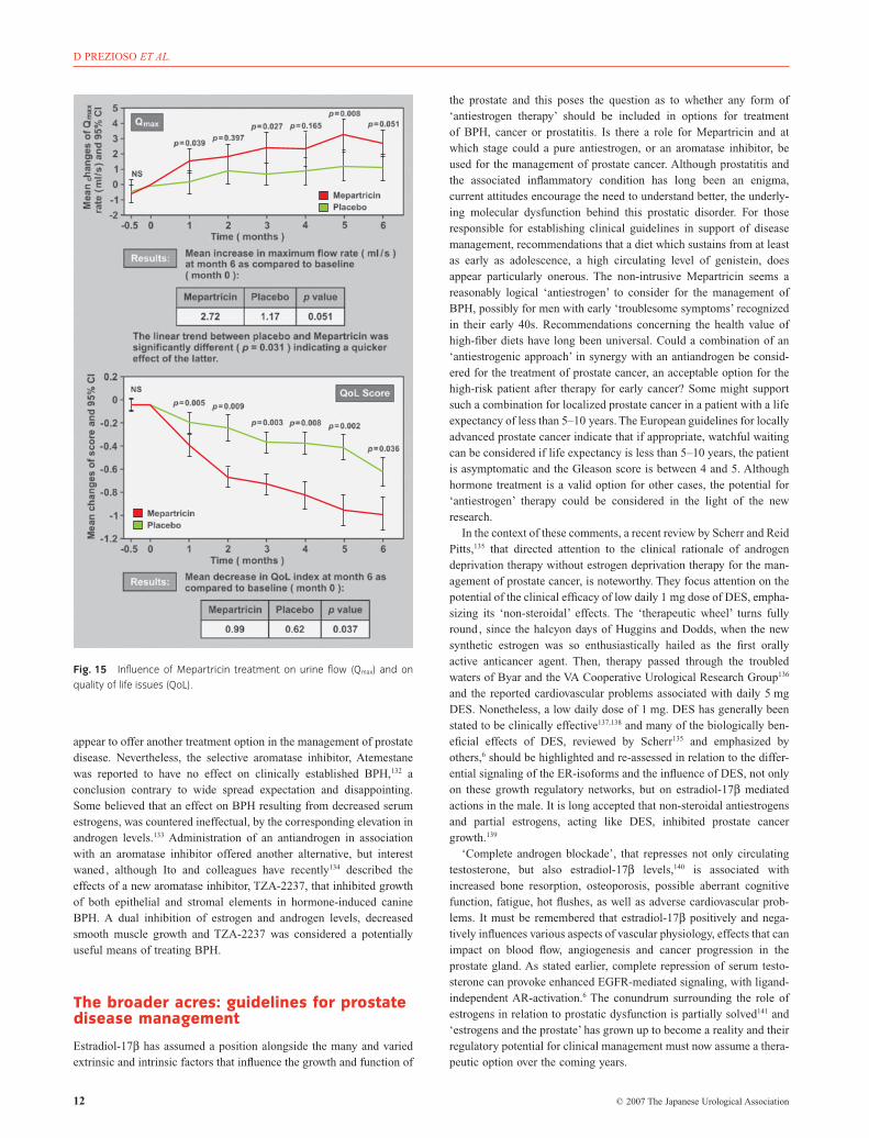
In a multicenter, double blind , randomized , placebo-controlledclinical trial of Mepartricin for the treatment of symptomatic BPH,patients were randomly allocated to a 24-week treatment period witheither Mepartricin (40 mg daily), or placebo. There was a 2-weekplacebo run-in period. The patient inclusion data, together with theprimary and secondary criteria of efficacy, are presented in Table 4.There was strong evidence of a more rapid and significant (P = 0.006)decline in the mean I-PSS of patients treated with Mepartricin than incontrols (Fig. 14). The mean increase in maximum flow rate (mL/s)of treated patients relative to controls and determined at 6 months(Fig. 15), showed a significantly different (P = 0.053) linear trendbetween the groups. Figure 15 illustrates the benefit of Mepartricinwith regard to the improvement in the Quality of Life index evaluatedat baseline and after 6 months treatment.
The tolerability issues and clinical efficacy data, that relates todeclining serum concentrations of estrogens and significant symptom-atic improvement in patients with BPH, illustrates the potential value of
this simple and seemingly effective form of therapy that offers a newapproach to the management of BPH, possibly of prostatitis131 and apossible approach to the restraint of prostate cancer progression.
Antiestrogen therapy witharomatase inhibitors
Aromatase inhibitors that inhibit the conversion of androstenedioneand testosterone to estrone and estradiol-17b (Fig. 2), would logically
Fig. 13 The influence of a high fiber diet and of Mepartricin, on estrogen
excretion.
Table 4 Patient entry criteria and primary and secondary evaluation of
Mepartricin efficacy
Main inclusion criteria
Age between 55 and 80 years.
Newly diagnosed BPH.
Moderate prostate symptom score (I-PSS between 12 and 24).
Post voiding residual urine <100 mL.
Urinary flow rate between 6 and 15 mL/s (voided urine volume
>150 mL).
Primary criteria of efficacy
I-PSS and QoL index were assessed at each month during the trial.
Peak flow rate was measured at each month during the trial.
The main comparisons were between measurements at month 6
and the baseline values.
Secondary criteria of efficacy
At baseline, and after 3 and 6 months:
Post-voiding residual volume.
Prostate volume.
Prostate-specific antigen.
BPH, benign prostatic hyperplasia; I-PSS, International Prostate
Symptom Score; QoL, quality of life.
Fig. 14 Influence of Mepartricin treatment on prostatic symptom score
(I-PSS).
Estrogens and aspects of prostate disease
© 2007 The Japanese Urological Association 11
appear to offer another treatment option in the management of prostatedisease. Nevertheless, the selective aromatase inhibitor, Atemestanewas reported to have no effect on clinically established BPH,132 aconclusion contrary to wide spread expectation and disappointing.Some believed that an effect on BPH resulting from decreased serumestrogens, was countered ineffectual, by the corresponding elevation inandrogen levels.133 Administration of an antiandrogen in associationwith an aromatase inhibitor offered another alternative, but interestwaned , although Ito and colleagues have recently134 described theeffects of a new aromatase inhibitor, TZA-2237, that inhibited growthof both epithelial and stromal elements in hormone-induced canineBPH. A dual inhibition of estrogen and androgen levels, decreasedsmooth muscle growth and TZA-2237 was considered a potentiallyuseful means of treating BPH.
The broader acres: guidelines for prostatedisease management
Estradiol-17b has assumed a position alongside the many and variedextrinsic and intrinsic factors that influence the growth and function of
the prostate and this poses the question as to whether any form of‘antiestrogen therapy’ should be included in options for treatmentof BPH, cancer or prostatitis. Is there a role for Mepartricin and atwhich stage could a pure antiestrogen, or an aromatase inhibitor, beused for the management of prostate cancer. Although prostatitis andthe associated inflammatory condition has long been an enigma,current attitudes encourage the need to understand better, the underly-ing molecular dysfunction behind this prostatic disorder. For thoseresponsible for establishing clinical guidelines in support of diseasemanagement, recommendations that a diet which sustains from at leastas early as adolescence, a high circulating level of genistein, doesappear particularly onerous. The non-intrusive Mepartricin seems areasonably logical ‘antiestrogen’ to consider for the management ofBPH, possibly for men with early ‘troublesome symptoms’ recognizedin their early 40s. Recommendations concerning the health value ofhigh-fiber diets have long been universal. Could a combination of an‘antiestrogenic approach’ in synergy with an antiandrogen be consid-ered for the treatment of prostate cancer, an acceptable option for thehigh-risk patient after therapy for early cancer? Some might supportsuch a combination for localized prostate cancer in a patient with a lifeexpectancy of less than 5–10 years. The European guidelines for locallyadvanced prostate cancer indicate that if appropriate, watchful waitingcan be considered if life expectancy is less than 5–10 years, the patientis asymptomatic and the Gleason score is between 4 and 5. Althoughhormone treatment is a valid option for other cases, the potential for‘antiestrogen’ therapy could be considered in the light of the newresearch.
In the context of these comments, a recent review by Scherr and ReidPitts,135 that directed attention to the clinical rationale of androgendeprivation therapy without estrogen deprivation therapy for the man-agement of prostate cancer, is noteworthy. They focus attention on thepotential of the clinical efficacy of low daily 1 mg dose of DES, empha-sizing its ‘non-steroidal’ effects. The ‘therapeutic wheel’ turns fullyround , since the halcyon days of Huggins and Dodds, when the newsynthetic estrogen was so enthusiastically hailed as the first orallyactive anticancer agent. Then, therapy passed through the troubledwaters of Byar and the VA Cooperative Urological Research Group136
and the reported cardiovascular problems associated with daily 5 mgDES. Nonetheless, a low daily dose of 1 mg. DES has generally beenstated to be clinically effective137,138 and many of the biologically ben-eficial effects of DES, reviewed by Scherr135 and emphasized byothers,6 should be highlighted and re-assessed in relation to the differ-ential signaling of the ER-isoforms and the influence of DES, not onlyon these growth regulatory networks, but on estradiol-17b mediatedactions in the male. It is long accepted that non-steroidal antiestrogensand partial estrogens, acting like DES, inhibited prostate cancergrowth.139
‘Complete androgen blockade’, that represses not only circulatingtestosterone, but also estradiol-17b levels,140 is associated withincreased bone resorption, osteoporosis, possible aberrant cognitivefunction, fatigue, hot flushes, as well as adverse cardiovascular prob-lems. It must be remembered that estradiol-17b positively and nega-tively influences various aspects of vascular physiology, effects that canimpact on blood flow, angiogenesis and cancer progression in theprostate gland. As stated earlier, complete repression of serum testo-sterone can provoke enhanced EGFR-mediated signaling, with ligand-independent AR-activation.6 The conundrum surrounding the role ofestrogens in relation to prostatic dysfunction is partially solved141 and‘estrogens and the prostate’ has grown up to become a reality and theirregulatory potential for clinical management must now assume a thera-peutic option over the coming years.
Fig. 15 Influence of Mepartricin treatment on urine flow (Qmax) and on
quality of life issues (QoL).
D PREZIOSO ET AL.
12 © 2007 The Japanese Urological Association
References
1 Burrows H. The localisation of response to oestrogenic compounds inthe organs of male mice. J. Pathol. 1935; 51: 423–9.
2 De Jongh SE. Der Einflass von Geschlechtshormonen auf die Prostataund ihr Umgebung bei der Maus. Acta Brev. Neeri Physiol.Pharmacol. Microbiol. 1935; 5: 28–32.
3 Huggins C, Stevens RE, Hodges CV. Studies on prostatic cancer 2: theeffects of castration on advanced carcinoma of the prostate gland.Arch. Surg. 1941; 43: 209–23.
4 Holtgrewe HL, Ackermann R, Bay-Nielsen H et al. The economics ofBPH. In: Cockett ATK, Khoury S, Aso Y et al. (eds). The ThirdInternational Consultation on Benign Prostatic Hyperplasia (BPH).SCI, Paris, 1996; 53–70.
5 Bartsch G, Klocker H, Ackermann R et al. Translational research areasand new treatment modalities. In: Murphy G, Khoury S, Partin A,Denis L. (eds). Second International Consultation on Prostate Cancer.Health Publication Ltd, Plymouth, 2000; 59–136.
6 Griffiths K, Denis LJ, Turkes A. Oestrogens, Phyto-Oestrogens and thePathogenesis of Prostatic Disease. Martin Dunitz, London, 2002.
7 Denis LJ, Murphy GP. Overview of phase III trials on combinedandrogen treatment in patients with metastatic prostate cancer. Cancer1993; 72: 3888–95.
8 Denis LJ. Prostate cancer: a continuum of controversy. Eur. J. Cancer1995; 31A: 839–40.
9 Denis LJ, Murphy GP, Schroeder FH. Report of the consensusworkshop on screening and global strategy for prostate cancer. Cancer1995; 75: 1187–207.
10 Chaisari N, Pierrepoint CG. Examination of the distribution ofoestrogen receptor between the stromal and epithelial compartments ofthe canine prostate. Prostate 1980; 1: 357–66.
11 Coffey DS, Walsh PC. Clinical experimental studies of benignprostatic hyperplasia. Urol. Clin. North Am. 1990; 17: 461–75.
12 Nicholson RI, Davies P, Griffiths K. Interaction of androgens withoestradiol-17b receptor proteins in DMBA-induced mammary tumours:a possible oncolytic mechanism. Eur. J. Cancer 1978; 14: 439–45.
13 Coffey DS. Similarities of prostate and breast cancer: evolution, dietand estrogens. Urology 2001; 57 (Suppl.): 31–8.
14 Bektic J, Wrulich OA, Dobler G et al. Identification of genes involvedin estrogenic action in the human prostate using microarray analysis.Genomics 2004; 83: 34–44.
15 Huggins C. Introduction. In: Vollmer EP, Kauffmann G (eds). Biologyof the Prostate and Related Tissues. Monogr. 12, Natl Cancer Inst. USDepartment Health, Education & Welfare, Bethesda, 1963; xi–xii.
16 Rotkin ID. Epidemiologic clues to increased risk of prostae cancer. In:Spring-Mills E, Hafez ESE (eds). Male Accessory Sex Glands: Biologyand Pathology. Elsevier/North-Holland Medical Press, Amsterdam,1980; 289–311.
17 Griffiths K and the Internatonal Prostate Health Council Study Group.Estrogens and prostatic disease, (Review). Prostate 2000; 45: 87–100.
18 Carter BS, Beaty TH, Steinberg GD, Childs B, Walsh PC. Mendelianinheritance of familial prostate cancer. Proc Natl Acad. Sci. USA 1992;89: 3367–71.
19 Gonzalgo ML, Isaacs WB. Molecular pathways to prostate cancer.J. Urol. 2003; 170: 2444–52.
20 Giovannucci E, Stampfer MJ, Krithivas K et al. The CAG repeatwithin the androgen receptor gene and its relationship to prostatecancer. Proc. Natl Acad. Sci. USA 1997; 94: 3320–3.
21 Makridakis N, Poss RK, Pike MC et al. A prevalent mis-sensesubstitution that modulates activity of prostatic steroid 5alphareductase. Cancer Res. 1997; 57: 1020–2.
22 Reichardt JKV, Makridakis N, Henderson BE, Yu MC, Pike MC, RossRK. Genetic variability of the human SRD5A2 gene: implications forprostate cancer risk. Cancer Res. 1995; 55: 3973–5.
23 Weihua Z, Makela S, Andersson LC et al. A role for estrogen receptorbeta in the regulation of growth of the ventral prostate. Proc. NatlAcad. Sci. USA 2001; 98: 6330–5.
24 Makridakis NM, Ross RK, Pike MC et al. Association of mis-sensesubstitution in SRD5A2 gene with prostate cancer in African-Americanand Hispanic men in Los Angeles. Lancet 1999; 354: 975–8.
25 Culig Z, Klocker H, Bartsch G, Hobisch A. Androgen receptors inprostate cancer. Endocr Relat Cancer 2002; 9: 155–70.
26 Matthews J, Gustafsson J-A. Estrogen signaling: a subtle balancebetween ER alpha and ER beta (Review). Mol. Interv. 2003; 3:281–92.
27 Herbst AL, Ulfeloer H, Poskanzer DC. Adenocarcinoma of the vagina:association of maternal stilboestrol therapy with tumor appearance inyoung women. N. Engl. J. Med. 1971; 284: 878–81.
28 Trichopoulos D. Hypothesis: does breast cancer originate in utero?Lancet 1990; 21: 939–40.
29 Newbold RR, McLachlan JA. Diethylstilboestrol-associated defects inmurine genital tract development. In: McLauchlan JA (ed.). Estrogensin the Environment: Influences on Development. Elsevier,North-Holland, New York, 1985; 288–318.
30 Naslund M, Coffey D. The differential effects of neonatal androgen,estrogen and progesterone on adult rat prostate growth. J. Urol. 1986;136: 1136–40.
31 Chang WY, Birch L, Woodham C, Gold LI, Prins GS. Neonatalestrogen exposure alters the transforming growth factor-beta signalingsystem in the developing rat prostate and blocks the transient p21(cip1/waf1) expression associated with epithelial differentiation.Endocrinology 1999; 140: 2801–13.
32 Levine S. Sexual differentiation: the development of maleness andfemaleness. Calif. Med. 1971; 114: 12–17.
33 Vom Saal FS, Timms BG, Montano MM et al. Prostate enlargement inmice due to fetal exposure to low doses of estradiol ordiethylstilbestrol and opposite effects at high doses. Proc. Natl Acad.Sci. USA 1997; 94: 2056–61.
34 Henderson BE, Bernstein L, Ross RK, Depue RH, Judd HI. The earlyin utero oestrogen and testosterone environment of black and whites:potential effects on male offspring. Br. J. Cancer 1988; 57: 216–18.
35 Solter D. Differential imprinting and expression of maternal andpaternal genomes. Annu. Rev. Genet. 1988; 22: 127–46.
36 DeChiara TM, Robertson EJ, Efstratiadis A. Parental imprinting of themouse insulin-like growth factor II gene. Cell 1991; 64: 849–59.
37 Jaenisch R, Beard C, Li E. DNA methylation and mammaliandevelopment. In: Ohlsson R, Hall K, Ritzen M (eds). GenomicImprinting: Causes and Consequences. Cambridge University Press,Cambridge, 1995; 118–26.
38 Wilkins RJ. Genomic imprinting and carcinogenesis. Lancet 1988; 1:329–31.
39 Adams JB. Control of secretion and the function of C19-delta5-steroidsof the human adrenal gland. Mol. Cell. Endocr. 1985; 41: 1–17.
40 Syms AJ, Harper ME, Griffiths K. The effect of prolactin on humanBPH epithelial cell proliferation. Prostate 1985; 6: 145–53.
41 Dillner K, Kindblom J, Flores-Morles A et al. Molecularcharacterization of prostate hyperplasia in polactin-transgenic mice byusing cDNA representational difference analysis. Prostate 2002; 52:139–49.
42 Cohen P, Peehl DM, Lamson G, Rosenfeld RG. Insulin-like growthfactors (IGFs), IGF receptors and IGF binding proteins in primarycultures of prostate epithelial cells. J. Clin. Endocrinol. Metab. 1991;73: 401–7.
43 Grant ES, Ross MB, Ballard S, Naylor A, Habib F. The insulin-likegrowth factor type I receptor stimulates growth and suppressesapoptosis in prostate stromal cells. J. Clin. Endocrinol. Metab. 1998;83: 3252–7.
44 Torring N, Vinter-Jensen L, Pedersen SB, Sorensen FB, Flyvbjerg A,Nexo E. Systemic administration of insulin-like growth factor (IGF-I)causes growth of the rat prostate. J. Urol. 1998; 158: 222–7.
45 Anders J, Bang P, Hertel NT et al. Serum insulin-like growth factor-Iin 1030 healthy children, adolescents and adults: relation to age, sex,stage of puberty, testicular size and body mass index. J. Clin.Endocrinol. Metab. 1994; 78: 744–52.
Estrogens and aspects of prostate disease
© 2007 The Japanese Urological Association 13
46 Hiney JK, Srivastava V, Nyberg CL, Ojeda SR, Dess WL. Insulin-likegrowth factor-I of peripheral origin acts centrally to accelerate theinitiation of female puberty. Endocrinology 1996; 137:3717–28.
47 Baserga R, Perluzzi F, Reiss K. The IGF-I receptor in cancer biology.Int. J. Cancer 2003; 107: 873–7.
48 Ruan W, Powell-Braxton L, Kopchick JJ, Kleinberg DL. Evidence thatinsulin-like growth factor I and growth hormone are required forprostate gland development. Endocrinology 1999; 140: 1984–9.
49 Chan JM, Stampfer MJ, Giovannucci E et al. Plasma insulin-likegrowth factor-I and prostate cancer risk: a prospective study. Science1998; 279: 563–56.
50 Chokkalingam AP, Gao Y-T, Deng J et al. Insulin-like growth factorsand risk of benign prostatic hyperplasia. Prostate 2002; 52: 98–105.
51 Turkes A, Peeling WB, Griffiths K. Serum IGF-1 determination inrelation to prostate cancer screening: possible differential diagnosis inrelation to PSA assays. Prostate Cancer Prostatic Dis. 2000; 3:173–5.
52 Robinson ICAF, Lightman SL. Differential regulation of the growthhormone receptor gene: effects of dexamethasone and estradiol.Endocrinology 1996; 137: 3891–6.
53 Colao AM, Marzullo O, Spiezia S et al. Effect of growth hormone(GH) and insulin-like growth factor-I on prostatic diseases: anultrasonographic and endocrine study in acromegaly, GH deficiencyand healthy subjects. J. Clin. Endocrinol. Metab. 1999; 84: 1986–91.
54 Orme SM, McNally R, Staines A, Cartwright RA, Belchetz PE.Mortality and cancer incidence in acromegaly: a retrospective cohortstudy. J. Clin. Endocrinol. Metab. 1998; 83: 2730–4.
55 Pollak M, Costantino J, Polychronakos C et al. Effect of tamoxifen onserum insulin-like growth factor-I levels in stage 1 breast cancerpatients. J. Natl Cancer Inst. 1990; 82: 1693–7.
56 Danutra V, Turkes A, Read GF et al. Progesterone concentrations insamples of saliva from adolescent girls living in Britain and Thailand ,two countries where women are at widely differing risk of breastcancer. J. Endocrinol. 1988; 121: 375–81.
57 Wilson DW, Turkes A, Jones R, Danutra V, Read GR, Griffiths K.A comparison of menstrual cycle profiles of salivary progesterone inBritish and Thai adolescent girls. Eur. J. Cancer 1992; 28A:1162–7.
58 Cheung CC, Thornton JE, Kuijper JL, Weigle DS, Clifton DK, SteinerRA. Leptin is a metabolic gate for the onset of puberty in the femalerat. Endocrinology 1997; 138: 855–8.
59 Bray GA, York DA. Leptin and clinical medicine: a new piece in thepuzzle of obesity. J. Clin. Endocrinol. Metab. 1997; 82: 2771–6.
60 Steiner RA. Editorial: Lords and ladies leapin’ on leptin.Endocrinology 1996; 137: 4533–4.
61 Kaaks R. Nutrition, hormones and breast cancer: is insulin the missinglink? Cancer Causes Control 1996; 7: 605–25.
62 Toscano V, Balducci P, Bianchi P, Mangiantini A, Sciarra F. Steroidaland nonsteroidal factors in plsma sex hormone binding globulinregulation. J. Steroid Biochem. Mol. Biol. 1992; 43: 431–7.
63 Apter D, Vihko R. Endocrine determinants of fertility: serum androgenconcentrations during follow-up of adolescents into the third decade oflife. J. Clin. Endocrinol. Metab. 1990; 71: 970–4.
64 Griffiths K, Coffey DS, Cockett ATK et al. The regulation of prostaticgrowth. In: The Third International Consultation on Benign ProstaticHyperplasia (BPH). Cockett ATK, Khoury S, Aso Y et al. (eds). SCI,Paris, 1996; 73–115.
65 Isaacs JT, Coffey DS. Etiology and disease process of benign prostatichyperplasia. Prostate 1989; 15 (Suppl. 2): 33–50.
66 McNeal JE. Anatomy of the prostate and morphogenesis of BPH. In:Kimball FA, Buhl AE, Carter DB (eds). New Approaches to the Studyof Benign Prostatic Hyperplasia. Alan R. Liss, New York, 1984;27–53.
67 Cunha GR, Hayward SW, Wang YZ, Ricke WA. Role of the stromalmicroenvironment in carcinogenesis of the prostate. Int. J. Cancer2003; 107: 1–10.
68 Bruengger A, Bartsche G, Hollinger BE, Holly B, Rohr HP. Smoothmuscle cell of the canine prostate in spontaneous benign hyperplasia,steroid induced hyperplasia and estrogen or tamoxifen treated dogs.J. Urol. 1983; 130: 1208–10.
69 Sakr WA. High-grade prostatic intraepithelial neoplasia: additionallinks to a potentially more aggressive prostate cancer? J. Natl CancerInst. 1998; 90: 486–7.
70 Sakr WA. Prostatic intraepithelial neoplasia: a marker for high-riskgroups and a potential target for chemoprevention. Eur. Urol. 1999;35: 474–8.
71 Harper ME, Goddard L, Glynne-Jones E, Peeling WB, Gffiths K.Epidermal growth factor expression by northern analysis andimmunohistochemistry in benign and malignant prostatic tumours. Eur.J. Cancer 1995; 31: 1492–7.
72 Harper ME, Glynne-Jones E, Goddard L, Mathews P, Nicholson RI.Expression of androgen receptor and growth factors in premalignantlesions of the prostate. J. Pathol. 1998; 186: 169–77.
73 Bonaccorsi L, Carloni V, Muratori M et al. EGF receptor (EGFR)signalling promoting invasion is disrupted in androgen-sensitiveprostate cancer cells by an interaction between EGFR and androgenreceptor. Int. J. Cancer 2004; 112: 78–86.
74 Boyle P, Napalkov P, Barry MJ et al. Epidemiology and naturalhistory of prostate cancer. In: Murphy GP, Denis L, Chatelain C,Griffiths K, Khoury S, Cockett ATK (eds). First InternationalConsultation on Prostate Cancer. SCI, Paris, 1997; 1–29.
75 Sakr WA, Haas GP, Cassin BF, Pontes JE, Crissman JD. Thefrequency of carcinoma and intraepithelial neoplasia of the prostate inyoung male patients. J. Urol. 1993; 150: 379–85.
76 Sakr WA, Grignon DJ, Haas GP. Pathology of premalignant lesionsand carcinoma of the prostate in African-American men. Semin. Urol.Oncol. 1998; 16: 214–20.
77 Vermeulen A, Giagulli VA. Andropause. In: Studd J (ed.). TheManagement of the Menopause. Carnforth Parthenon Publishing,Carnforth, 1998; 279–96.
78 Ross R, Bernstein L, Judd H, Hanisch R, Pike M, Henderson B. Serumtestosterone levels in healthy young black and white men. J. NatlCancer Inst. 1986; 76: 45–8.
79 Wilson DW, Harper ME, Jensen HM et al. A prognostic index for theclinical management of patients with advanced prostatic cancer: aBritish Prostate Study Group investigation. Prostate 1985; 7:131–41.
80 Meikle AW, Smith JR, West DW. Familial factors affecting prostaticcancer risk and plasma sex-steroid levels. Prostate 1985; 6:121–8.
81 De Marzo AM, Nelson WG, Isaacs WB, Epstein JI. Pathologicaland molecular aspects of prostate cancer. Lancet 2003; 361: 955–64.
82 De Marzo AM, Marchi VL, Epstein JI, Nelson WG. Proliferativeinflammatory atrophy of the prostate: implications for prostaticcarcinogenesis. Am. J. Pathol. 1999; 155: 1985–92.
83 Steed PM, Tansey MG, Zalevsky J et al. Inactivation of TNF signallingby rationally designed dominant-negative TNF variants. Science 2003;301: 1895–8.
84 Dennis LK, Lynch CF, Torner JC. Epidemiologic association betweenprostatitis and prostate cancer. Urology 2002; 60: 78–83.
85 Putzi MJ, De Marzo AM. Morphologic transitions betweenproliferative inflammatory atrophy and high-grade prostaticintraepithelial neoplasia. Urology 2000; 56: 828–32.
86 Prezioso D, Naber KG, Lobel B et al. Some historical perspectives onthe pathogenesis of prostatitis. Acad. Med. Sci. In press.
87 Franks LM, McNeal JE. Discussion, Third Tenovus Workshop on ‘SomeAspects of the Aetiology and Biochemistry of Prostate Cancer’.Griffiths K, Pierrepoint CG (eds). Alpha Publishing, Cardiff 1970;39–46.
88 Oberley TD, Zhong W, Szweda LI, Oberley LW. Localization ofantioxidant enzymes and oxidative damage products in normal andmalignant prostate epithelium. Prostate 2000; 44: 144–55.
D PREZIOSO ET AL.
14 © 2007 The Japanese Urological Association
89 Zha S, Gage WR, Sauvageot J et al. Cyclooxygenase-2 is up-regulatedin proliferative inflammatory atrophy of the prostate, but not inprostate cancinoma. Cancer Res. 2001; 61: 8617–23.
90 Deutsch E, Maggiorella L, Eschwege P, Bourhis J, Soria JC,Abdulkarim B. Environmental, genetic and molecular features ofprostate cancer. Lancet Oncol. 2004; 5: 303–13.
91 Montano MM, Jaiswal AK, Katzenellenbogan BS. Transcriptionalregulation of the human quinone reductase gene by anti-estrogenliganded estrogen receptor-alpha and estrogen receptor-beta. J. Biol.Chem. 1998; 273: 25443–9, i.
92 Jeronimo C, Usadel H, Henrique R et al. Quantitation of GSTP1methylation in non-neoplastic prostatic tissue and organ-confinedprostate adenocarcinoma. J. Natl Cancer Inst. 2001; 93:1747–52.
93 Maruyama R, Toyooka S, Toyooka KO et al. Aberrant promotermethylation profile of prostate cancers and its relationship toclinicopathological features. Clin. Cancer Res. 2002; 8: 514–19.
94 Lee WH, Morton RA, Epstein JI et al. Cytidine methylation ofregulatory sequences near the pi-class glutathione S-transferase geneaccompanies human prostaic carcinogenesis. Proc. Natl Acad. Sci. USA1994; 91: 11 733–7.
95 Kuzmin I, Gillespie JW, Protopopov A et al. The RASSF1A tumoursuppressor gene is inactivated in prostate tumors and suppressesgrowth of prostate carcinoma cells. Cancer Res. 2002; 62: 3498–502.
96 Sasaki M, Tanaka Y, Perinchery G et al. Methylation and inactivationof estrogen, progesterone and androgen receptors in prostate cancer.J. Natl Cancer Inst. 2002; 94: 384–90.
97 Pasquali D, Rossi V, Esposito D et al. Loss of estrogen receptor betaexpression in malignant human prostate cells in primary cultures andin prostate cancer tissues. J. Clin. Endocrinol. Metab. 2001; 86:2051–5.
98 Cunha GR, Donjacour AA, Cooke PS et al. The endocrinology anddevelopment biology of the prostate. Endocr. Rev. 1987; 8:338–62.
99 McKeehan WL, Wang F, Kan M. The heparan sulfate-fibroblastgrowth factor family: diversity of structure and function. Prog. NucleicAcid Res. Mol. Biol. 1998; 59: 135–76.
100 Krieg M, Nass R, Tunn S. Effect of ageing on endogenous level of5a-dihydrotestosterone, testosterone, estradiol and estrone inepithelium and stroma of normal and hyperplastic human prostate.J. Clin. Endocrinol. Metab. 1993; 77: 375–81.
101 Shibata Y, Ito K, Suzuki K et al. Changes in the endocrineenvironment of the human prostate transition zone with ageing:simultaneous quantitative analysis of prostatic sex steroids andcomparison with human prostatic histological composition. Prostate2000; 42: 45–55.
102 Risbridger G, Wang H, Frydenberg M, Cunha GR. The metplasticeffects of estrogen on prostate epithelium proliferation of cells withbasal cell phenotype. Endocrinology 2001; 142: 2443–50.
103 Wang YZ, Wong YC. Sex hormone-induced prostatic carcinogenesis inthe noble rat: the role of insulin-like growth factor-I (IGF-I) andvascular endothelial growth factor (VEGF) in the development ofprostate cancer. Prostate 1998; 35: 165–77.
104 Wang YZ, Hayward SW, Cao M, Young P, Cardiff R, Cunha GR. Roleof estrogen signalling in prostatic hormonal carcinogenesis. J. Urol.2001; 165: 1320.
105 Olumi AF, Grossfeld GD, Hayward SW, Carroll PR, Tisty TD, CunhaGR. Carcinoma-associated fibroblasts direct tumor progression ofinitiated human prostatic epithelium. Cancer Res. 1999; 59: 5002–11.
106 van Kempen LCL, Rhee J-S, Dehne K, Lee J, Edwards DR, CoussensLM. Epithelial carcinogenesis: dynamic interplay between neoplasticcells and their microenvironment. Differentiation 2002; 60: 610–23.
107 Foster BA, Evangelou A, Gingrich JR, Kaplan PJ, DeMayo F,Greenberg NM. Enforced expression of FGF-7 promotes epithelialhyperplasia whereas a dominant negative FGFR2iiib provides theemergence of neuroendocrine phenotype in prostate glands oftransgenic mice. Differentiation 2002; 60: 624–32.
108 Kuiper GG, Enmark E, Pelto-Huikko M, Nilsson S, Gustafsson JA.Cloning of a novel receptor expressed in rat prostate and ovary. Proc.Natl Acad. Sci. USA 1996; 93: 5925–30.
109 Kuiper GG, Carlsson B, Grandien K et al. Comparison of the ligandbinding specificity and transcript tissue distribution of estrogenreceptors alpha and beta. Endocrinology 1997; 138: 863–70.
110 Krege JH, Hodgin JB, Couse JF et al. Generation and reproductivephenotypes of mice lacking estrogen receptor beta. Proc. Natl Acad.Sci. USA 1998; 95: 15 677–82.
111 Paech K, Webb P, Kuiper GGJM et al. Differential ligand activation ofestrogen receptors ERalpha and ERbeta at AP-1 sites. Science 1997;277: 1508–10.
112 Bektic J, Berger AP, Pfeil K, Dobler G, Bartsch G, Klocker H.Androgen receptor regulation by physiological concentrations of theisoflavnoid genistein in androgen-dependent LNCaP cells is mediatedby estrogen receptor b. Eur. Urol. 2004; 45: 245–51.
113 Hedlund TE, Johannes WU, Miller GI. Soy isoflavonoid equolmodulates the growth of benign and malignant prostatic epithelial cellsin vitro. Prostate 2003; 54: 68–78.
114 Rohlff C, Blagosklonny MV, Kyle E et al. Prostatic cancer cell growthinhibition by tamoxifen is associated with an inhibition of proteinkinase C and induction of p21 (waf/cip1). Prostate 1998; 37:51–9.
115 Horwitz KB, Jackson TA, Bain DL, Richer JK, Takimoto GS, Tung L.Nuclear receptor coactivators and corepressorrs. Mol. Endocrinol.1996; 10: 1167–77.
116 Kalkhoven E, Valentine JE, Heery DM, Parker MG. Isoforms ofsteroid receptor co-activator I differ in their ability to potentiatetranscription by the estrogen receptor. EMBO J. 1998; 17: 232–43.
117 Brzozowski AM, Pike ACW, Dauter Z et al. Molecular basis ofagonism in the estrogen receptor. Nature 1997; 389: 753–8.
118 Hihi AK, Wahli W. Structure and function of the estrogen receptor.In: Oettel M, Schillinger M (eds). Handbook of ExperimentalPharmacology, Vol. 135/I, Estrogens and Antiestrogens.Springer-Verlag, Berlin, 1999; 111–26.
119 Aronica SM, Katzenellenbogen BS. Stimulation of estrogen mediatedtranscription and alteration in the phosphorylation state of the ratuterine estrogen receptor by estrogen, cyclic adenosine monophosphateand insulin-like growth factor-I. Mol. Endocrinol. 1993; 7:743–52.
120 Cho H, Katzenellenbogen BS. Synergistic activation of estrogenreceptor mediated transcription by estradiol and protein kinaseactivators. Mol. Endocrinol. 1993; 7: 441–52.
121 Bunone G, Briand PA, Miksicek RJ, Picard D. Activation of theunliganded estrogen receptor by ECF involves the MAP-kinasepathway and direct phosphorylation. EMBO J. 1996; 15:2174–83.
122 Katzenellenbogen JA, O’Malley BW, Katzenellenbogen BS. Tripartitesteroid hormone receptor pharmacology: interaction with multipleeffector sites as a base for the cell- and promoter-specific action ofthese hormones. Mol. Endocrinol. 1996; 10: 119–31.
123 Yeh S, Chang C. Cloning and characterization of a specificco-activator, ARA70, for the androgen receptor in human prostate cells.Proc. Natl Acad. Sci. USA 1996; 93: 5517–21.
124 Jordan VC. Designer Estrogens. Sci. Am. 1998; 279: 60–7.125 Goldin BR, Adlercreutz H, Gorbach SL et al. Estrogen excretion
patterns and plasma levels in vegetarian and omnivorous women.N. Engl. J. Med. 1982; 307: 1542–7.
126 Hamalainen E, Adlercreutz H, Puska P, Pietinen P. Diet and serumhormones in healthy men. J. Steroid Biochem. 1984; 20: 459–64.
127 Barone D, Peroglio F, Toso E, Bruzzese T. Binding of Mepartricin tosex hormones: a key factor of its activity on benign prostatichyperplasia. Arzneimittel-Forsch. 2001; 51: 984–90.
128 Re G, Badino R, Odore R et al. Effects of Mepartricin on estradiol andtestosterone serum levels and on prostatic estrogen, androgen andadrenergic receptor concentrations in adult rats. Pharmacol. Res. 2001;44: 141–7.
Estrogens and aspects of prostate disease
© 2007 The Japanese Urological Association 15
129 Yoshinaka Y, Kobayashi H, Kirihara J, Sato F, Shakutou S, YamanakaH. Effects of mepartricin (S-160) on spontaneous canine benignprostate hyperplasia. Eur. Urol. 2000; 37: 428–35.
130 Boenm S, Nirnberger G, Ferrari P. Estrogen suppression as apharmacotherapeutic strategy in the medical treatment of beingnprostatic hyperplasia: evidence for its efficacy from studies withmepartricin. Wien. Klin. Wochenschr. 1998; 110: 817–23.
131 De Rose AF, Gallo F, Giglio M, Carmignani G. Role of Mepartricin incategory III chronic nonbacterial prostatitis/chronic pelvic painsyndrome: a randomised prospective placbo-controlled trial. Urology2004; 63: 13–16.
132 Radlmaier A, Eickenberg HU, Fletcher MS et al. Estrogen reductionby aromatase inhibition for benign prostatic hyperplasia: results of adouble-blind , placebo-controlled , randomized clinical trial using twodoses of the aromatase-inhibitor atamestane. Atamestane Study Group.Prostate 1996; 29: 199–208.
133 Gingell JC, Knonagel H, Kurth KH, Tunn UW. Placebo controlleddouble-blind study to test the efficacy of the aromatase inhibitoratamestane in patients with benign prostatic hyperplasia not requiringoperation. The Schering 90.062 Study Group. J. Urol. 1995; 154:399–401.
134 Ito K, Fukabori Y, Shibata Y et al. Effects of a new steroidalaromatase inhibitor, TZA-2237, and/or chlormadinone acetate on
hormone-induced and spontaneous canine benign prostatic hyperplasia.Eur. J. Endocrinol. 2000; 143: 543–54.
135 Schherr DS, Reid Pitts W. The nonsteroidal effects ofdiethylstilboestrol: the rationale for androgen deprivation therapywithout estrogen deprivation in the treatment of prostate cancer.J. Urol. 2003; 170: 1703–8.
136 Byar DP, Corle DK. Hormone therapy for prostate cancer: results ofthe Veterans Administration Cooperative Urological Research Groupstudies. NCI Monogr. 1988; 7: 165–70.
137 Smith DC, Redman BG, Flaherty LE, Li L, Strawderman M, PientaKJ. A phase II trial of oral stilboestrol as a second-line hormonal agentin advanced prostate cancer. Urology 1998; 52: 257.
138 Cox RL, Crawford ED. Estrogens in the treatment of prostate cancer.J. Urol. 1995; 154: 1991–8.
139 Schneider MR, Hartmann RW, Sinowatz F, Amselgruber W.Nonsteroidal antiestrogens and partial estrogens wih prostate tumourinhibiting activity. J. Cancer Res. Clin. Oncol. 1986; 112: 258–65.
140 Kirby R, Robertson C, Turkes A et al. Finasteride in association witheither flutamide or goserelin as combination hormonal therapy inpatients with stage M1 carcinoma of the prostate gland. Prostate 1999;40: 105–14.
141 Gustafsson J-A. Estrogen receptor b – getting in on the action. Nat.
Med. 1997; 3: 493–4.
D PREZIOSO ET AL.
16 © 2007 The Japanese Urological Association
Related Documents