Estrogen Sulfotransferase/SULT1E1 Promotes Human Adipogenesis Chibueze A. Ihunnah, a,b Taira Wada, a,b Brian J. Philips, c Sudheer K. Ravuri, c Robert B. Gibbs, b Levent Kirisci, b J. Peter Rubin, c Kacey G. Marra, c Wen Xie a,b,d Center for Pharmacogenetics a and Department of Pharmaceutical Sciences, b University of Pittsburgh, Pittsburgh, Pennsylvania, USA; Department of Plastic Surgery and McGowan Institute for Regenerative Medicine, University of Pittsburgh, Pittsburgh, Pennsylvania, USA c ; Department of Pharmacology and Chemical Biology, University of Pittsburgh, Pittsburgh, Pennsylvania, USA d Estrogen sulfotransferase (EST/SULT1E1) is known to catalyze the sulfoconjugation and deactivation of estrogens. The goal of this study is to determine whether and how EST plays a role in human adipogenesis. By using human primary adi- pose-derived stem cells (ASCs) and whole-fat tissues from the abdominal subcutaneous fat of obese and nonobese subjects, we showed that the expression of EST was low in preadipocytes but increased upon differentiation. Overexpression and knockdown of EST in ASCs promoted and inhibited differentiation, respectively. The proadipogenic activity of EST in hu- mans was opposite to the antiadipogenic effect of the same enzyme in rodents. Mechanistically, EST promoted adipogene- sis by deactivating estrogens. The proadipogenic effect of EST can be recapitulated by using an estrogen receptor (ER) an- tagonist or ER knockdown. In contrast, activation of ER in ASCs inhibited adipogenesis by decreasing the recruitment of the adipogenic peroxisome proliferator-activated receptor (PPAR) onto its target gene promoters, whereas ER antago- nism increased the recruitment of PPAR to its target gene promoters. Linear regression analysis revealed a positive corre- lation between the expression of EST and body mass index (BMI), as well as a negative correlation between ER expression and BMI. We conclude that EST is a proadipogenic factor which may serve as a druggable target to inhibit the turnover and accumulation of adipocytes in obese patients. O besity is a major health concern with high prevalence. Obe- sity is defined as a body mass index (BMI) equal to or greater than 30, whereas a BMI equal to or greater than 25 is considered overweight. An estimated 400 million people worldwide are obese. In the United States alone, obesity-related medical complications contribute to an average of 300,000 deaths annually (1). The obesity epidemic has generated much research attention toward understanding the biochemical regulation of adipose tis- sue and the development of adipocytes, known as adipogenesis. Adipogenesis is a multifaceted process that is regulated by tempo- ral and spatial expression of a battery of adipogenic genes. When the preadipocytes located in various visceral and subcutaneous fat depots are stimulated by specific mitogenic and adipogenic cues, they begin the differentiation process until maturation is reached (2). This process is accompanied by a dramatic increase in the expression of adipogenic genes, such as the genes for lipoprotein lipase (LPL), fatty acid binding protein 4/adipocyte protein 2 (FABP4/aP2), and the CCAATT enhancer binding proteins , , and (C/EBP,-, and -)(3). Peroxisome proliferator-activated receptor (PPAR) is a nuclear receptor known as the master regulator of adipogenesis. Activation of PPAR is required for the induction of several of the aforementioned adipogenic enzymes and transcription factors (3). Differentiation also leads to mor- phological and biochemical changes in preadipocytes that allow them to store lipids and secrete adipokines (4). The sex hormones are known to have a marked impact on adipose tissue development, accumulation, and distribution in humans. Men tend to have a more central abdominal accumula- tion of fat, while women tend to accumulate adipose tissue around the gluteal and femoral area (5). Animal models and human epi- demiological studies have shown that, in general, loss of estrogen signaling facilitates increased adipose tissue accumulation. This has been described in ER and aromatase knockout mice and documented in postmenopausal women (6–9). In contrast, estro- gen replacement therapy in older women resulted in reductions in central subcutaneous and visceral adipose tissue (10, 11). The homeostasis of estrogens is tightly regulated by balanced synthesis and deactivation. Estrogen sulfotransferase (EST, or SULT1E1) is a key enzyme known to catalyze the sulfation of estrogens, leading to their inactivation because of the inability of estrogen sulfates to bind to the estrogen receptor (ER) (12). We recently described a novel role for Est in murine adipogenesis, in which Est functions as a negative regulator of adipogenesis. Est is highly expressed in mouse preadipocytes, and differentiation at- tenuates the expression of Est (13). Moreover, Est overexpression and ablation inhibited and promoted murine adipogenesis, re- spectively (13). It is unclear whether the role of EST in adipogen- esis is conserved in humans. In this study, we found that the effect of EST on adipogenesis is highly species specific. EST promoted human adipogenesis by de- activating estrogens. The proadipogenic effect of EST was recapit- ulated in preadipocytes whose ER was pharmacologically or ge- netically inhibited. In contrast, pharmacological activation of ER inhibited adipogenesis. We propose EST as a druggable target whose inhibition may be used to inhibit the turnover of adipocytes in obese patients. Received 4 September 2013 Returned for modification 4 October 2013 Accepted 19 February 2014 Published ahead of print 24 February 2014 Address correspondence to Wen Xie, [email protected]. Copyright © 2014, American Society for Microbiology. All Rights Reserved. doi:10.1128/MCB.01147-13 1682 mcb.asm.org Molecular and Cellular Biology p. 1682–1694 May 2014 Volume 34 Number 9 on July 2, 2016 by guest http://mcb.asm.org/ Downloaded from

Welcome message from author
This document is posted to help you gain knowledge. Please leave a comment to let me know what you think about it! Share it to your friends and learn new things together.
Transcript
Estrogen Sulfotransferase/SULT1E1 Promotes Human Adipogenesis
Chibueze A. Ihunnah,a,b Taira Wada,a,b Brian J. Philips,c Sudheer K. Ravuri,c Robert B. Gibbs,b Levent Kirisci,b J. Peter Rubin,c
Kacey G. Marra,c Wen Xiea,b,d
Center for Pharmacogeneticsa and Department of Pharmaceutical Sciences,b University of Pittsburgh, Pittsburgh, Pennsylvania, USA; Department of Plastic Surgery andMcGowan Institute for Regenerative Medicine, University of Pittsburgh, Pittsburgh, Pennsylvania, USAc; Department of Pharmacology and Chemical Biology, University ofPittsburgh, Pittsburgh, Pennsylvania, USAd
Estrogen sulfotransferase (EST/SULT1E1) is known to catalyze the sulfoconjugation and deactivation of estrogens. Thegoal of this study is to determine whether and how EST plays a role in human adipogenesis. By using human primary adi-pose-derived stem cells (ASCs) and whole-fat tissues from the abdominal subcutaneous fat of obese and nonobese subjects,we showed that the expression of EST was low in preadipocytes but increased upon differentiation. Overexpression andknockdown of EST in ASCs promoted and inhibited differentiation, respectively. The proadipogenic activity of EST in hu-mans was opposite to the antiadipogenic effect of the same enzyme in rodents. Mechanistically, EST promoted adipogene-sis by deactivating estrogens. The proadipogenic effect of EST can be recapitulated by using an estrogen receptor (ER) an-tagonist or ER� knockdown. In contrast, activation of ER in ASCs inhibited adipogenesis by decreasing the recruitment ofthe adipogenic peroxisome proliferator-activated receptor � (PPAR�) onto its target gene promoters, whereas ER antago-nism increased the recruitment of PPAR� to its target gene promoters. Linear regression analysis revealed a positive corre-lation between the expression of EST and body mass index (BMI), as well as a negative correlation between ER� expressionand BMI. We conclude that EST is a proadipogenic factor which may serve as a druggable target to inhibit the turnover andaccumulation of adipocytes in obese patients.
Obesity is a major health concern with high prevalence. Obe-sity is defined as a body mass index (BMI) equal to or greater
than 30, whereas a BMI equal to or greater than 25 is consideredoverweight. An estimated 400 million people worldwide are obese.In the United States alone, obesity-related medical complicationscontribute to an average of 300,000 deaths annually (1).
The obesity epidemic has generated much research attentiontoward understanding the biochemical regulation of adipose tis-sue and the development of adipocytes, known as adipogenesis.Adipogenesis is a multifaceted process that is regulated by tempo-ral and spatial expression of a battery of adipogenic genes. Whenthe preadipocytes located in various visceral and subcutaneous fatdepots are stimulated by specific mitogenic and adipogenic cues,they begin the differentiation process until maturation is reached(2). This process is accompanied by a dramatic increase in theexpression of adipogenic genes, such as the genes for lipoproteinlipase (LPL), fatty acid binding protein 4/adipocyte protein 2(FABP4/aP2), and the CCAATT enhancer binding proteins �, �,and � (C/EBP�, -�, and -�) (3). Peroxisome proliferator-activatedreceptor � (PPAR�) is a nuclear receptor known as the masterregulator of adipogenesis. Activation of PPAR� is required for theinduction of several of the aforementioned adipogenic enzymesand transcription factors (3). Differentiation also leads to mor-phological and biochemical changes in preadipocytes that allowthem to store lipids and secrete adipokines (4).
The sex hormones are known to have a marked impact onadipose tissue development, accumulation, and distribution inhumans. Men tend to have a more central abdominal accumula-tion of fat, while women tend to accumulate adipose tissue aroundthe gluteal and femoral area (5). Animal models and human epi-demiological studies have shown that, in general, loss of estrogensignaling facilitates increased adipose tissue accumulation. Thishas been described in ER� and aromatase knockout mice anddocumented in postmenopausal women (6–9). In contrast, estro-
gen replacement therapy in older women resulted in reductions incentral subcutaneous and visceral adipose tissue (10, 11).
The homeostasis of estrogens is tightly regulated by balancedsynthesis and deactivation. Estrogen sulfotransferase (EST, orSULT1E1) is a key enzyme known to catalyze the sulfation ofestrogens, leading to their inactivation because of the inability ofestrogen sulfates to bind to the estrogen receptor (ER) (12). Werecently described a novel role for Est in murine adipogenesis, inwhich Est functions as a negative regulator of adipogenesis. Est ishighly expressed in mouse preadipocytes, and differentiation at-tenuates the expression of Est (13). Moreover, Est overexpressionand ablation inhibited and promoted murine adipogenesis, re-spectively (13). It is unclear whether the role of EST in adipogen-esis is conserved in humans.
In this study, we found that the effect of EST on adipogenesis ishighly species specific. EST promoted human adipogenesis by de-activating estrogens. The proadipogenic effect of EST was recapit-ulated in preadipocytes whose ER was pharmacologically or ge-netically inhibited. In contrast, pharmacological activation of ERinhibited adipogenesis. We propose EST as a druggable targetwhose inhibition may be used to inhibit the turnover of adipocytesin obese patients.
Received 4 September 2013 Returned for modification 4 October 2013Accepted 19 February 2014
Published ahead of print 24 February 2014
Address correspondence to Wen Xie, [email protected].
Copyright © 2014, American Society for Microbiology. All Rights Reserved.
doi:10.1128/MCB.01147-13
1682 mcb.asm.org Molecular and Cellular Biology p. 1682–1694 May 2014 Volume 34 Number 9
on July 2, 2016 by guesthttp://m
cb.asm.org/
Dow
nloaded from
MATERIALS AND METHODSASC and adipose tissue collection. Human adipose-derived stem cells(ASCs; primary preadipocytes) and whole fat/lipoaspirate were obtainedthrough the Adipose Stem Cell Center, Department of Plastic Surgery,University of Pittsburgh. Preadipocytes were isolated from the abdominalsubcutaneous fat of 15 obese patients and 3 nonobese (lean) patients whohad undergone liposuction or whole-fat removal surgery. All experimentswere performed on cells from the obese patients except as otherwise spec-ified. The names of the patients were kept anonymous, and all patientsused for preadipocyte differentiation experiments were female, nondia-betic nonsmokers and ranged in age from 25 to 56 years. The demographicinformation of the 18 patients is available upon request. The whole fats orliposuction aspirates were collected from the abdominal subcutaneous fatof 16 female patients who were nondiabetic nonsmokers and ranged in agefrom 32 to 59 years. The demographic information of these 16 patients isavailable upon request. The cells were cultured under standard conditions asreported previously (14). Briefly, cells were cultured in Dulbecco’s modifiedEagle’s medium (DMEM)–F-12 medium containing 10% standard fetal bo-vine serum (FBS) and 1% penicillin-streptomycin (Pen-Strep). Each cell linewas cultured for no more than four passages. Patient sample and data collec-tion were performed in accordance with the University of Pittsburgh Institu-tional Review Board Protocol PRO12050016.
Lentivirus generation and production. To generate lentivirus ex-pressing EST and mutant EST AAK (EST with a change from GXXGXXKto AXXAXXK in the 3=-phosphoadenosine 5=-phosphosulfate [PAPS]-binding domain), wild-type (WT) EST was cloned by reverse transcrip-tion-PCR (RT-PCR) using cDNA from differentiated human adipocytes.WT EST or AAK EST cDNA was cloned into a lentiviral expression plas-mid (pWPI) via the Pme1 restriction sites. Lentiviral particles were gen-erated using a second-generation system that contained three plasmids:the transgene expression plasmid (pWPI-EST), a packaging plasmid(psPAX2), and an envelope plasmid (pMDG.2). All three plasmids weretransfected simultaneously into 293T cells for viral particle packaging,assembly, and amplification using Trans-IT transfection reagent fromMirus (Madison, WI). Viral lysates were collected every 24 h after trans-fection, filtered with 0.45-�m-pore-size Millex GV syringe filter unitsfrom Millipore (Billerica, MA), pooled, and concentrated with a Lenti-X-Concentrator from Clontech (Mountain View, CA). Titer concentrationswere assessed with Lenti-X-Stix from Clontech and by fluorescent exam-ination of the green fluorescence protein that was engineered in the len-tiviral vector. Viral lysates were aliquoted and stored at �80°C until use.The cDNA for the EST AAK mutant was cloned by overlap extension PCRmutagenesis (15). Preadipocytes were infected at a multiplicity of infec-tion (MOI) of 3 before puromycin selection. Based on our observations,there was no noticeable effect of lentiviral infection and puromycin selec-tion alone on the differentiation.
To generate lentivirus expressing short hairpin RNAs (shRNAs)against EST and ER� (shEST and shER�, respectively), expression plas-mids containing shEST and shER� were purchased from Open Biosys-tems (Pittsburgh, PA). For each knockdown, five sequences were pur-chased and tested in transient-transfection assays, and the sequence withthe most efficient knockdown was chosen for lentiviral production. Thetargeting sequences for EST and ER� are ATGAGTCTTCACAATTCTAGG (product TRCN0000035880) and TTCCAGAGACTTCAGGGTGCT (product TRCN0000003299), respectively. A scrambled shRNA(shSCR) plasmid was purchased to serve as a control.
Adipocyte differentiation. In all experiments except those in whichexogenous estrogen (E2) was added, differentiation medium 1 (DM1)consisted of DMEM–F-12 medium, 10% standard FBS, 1% Pen-Strep, 33�M biotin, 100 nM insulin, 17 �M pantothenic acid, 0.5 mM methyl-isobutylxanthine, 1 �M dexamethasone, and 1 �M rosiglitazone. Differ-entiation medium 2 (DM2) consisted of DMEM–F-12 medium, 10% FBS,1% Pen-Strep, 1 �M dexamethasone, and 100 nM insulin. Confluentpreadipocytes were cultured in DM1 for 3 days before being switched toDM2 for 2 weeks to reach terminal differentiation. Culture medium was
changed every other day. For differentiation experiments in which E2 wasexogenously added, the cells were cultured in phenol red-free DMEM–F-12 medium and dextran-coated charcoal (DCC)-stripped FBS beforethe addition of E2 at a final concentration of 10 nM.
Quantitative real-time reverse transcription-PCR (RT-PCR). ThecDNA was synthesized from 1.0 �g of total RNA by Iscript from Bio-Rad(Hercules, CA). Aliquots of cDNA were amplified on an ABI 7300 real-time PCR system from Applied Biosystems (Foster City, CA) using theSYBR green PCR master mix. mRNA expression was normalized againstthe expression of cyclophilin or glyceraldehyde-3-phosphate dehydroge-nase (GAPDH).
Oil red O staining and quantification. Differentiated six-well culturedishes were washed twice with cold phosphate-buffered saline (PBS) andthen prefixed for 1 h with 10% formaldehyde in PBS. After 1 h, fresh 10%formaldehyde was added, and the cells were incubated overnight at roomtemperature. The next day, the cells were washed twice with PBS and thenincubated in 60% isopropanol for 5 min, followed by drying at roomtemperature (RT). Cells were incubated with oil red O working solutionfor 1 min and then washed five times with deionized water. Images wereacquired microscopically. For quantification of oil red O staining, cellswere differentiated in triplicate in 24-well plates, stained with oil red O,and eluted with 100% isopropanol, and 100 �l of elute was loaded onto96-well plates. Absorbance was measured at 500 nm using a PerkinElmerplate reader.
Western blot analysis. Cells were lysed with NP-40 lysis buffer con-taining protease inhibitors and then quantified for protein concentrationsby a bicinchoninic acid (BCA) assay kit from Pierce (Rockford, IL). Pro-tein samples were resolved by electrophoresis on 10% SDS-polyacryl-amide gels. For the detection of insulin receptor substrate 1 (IRS1) and itsphosphorylation, cell lysates were immunoprecipitated with an IRS1 an-tibody before being subjected to Western blotting using an IRS1 antibodyand phosphotyrosine antibody. After transfer of proteins to polyvi-nylidene difluoride (PVDF) membranes, the membranes were probedwith antibodies against total extracellular signal-regulated kinases 1 and 2(ERK1/2) (catalog no. sc94; Santa Cruz), phospho-ERK1/2 (catalog no.sc7383; Santa Cruz), total AKT (catalog no. 9272; Cell Signaling), phos-pho-AKT (catalog no. 9215; Cell Signaling), phospho-CREB (catalog no.87G3; Cell Signaling), total CREB (catalog no. 48H2; Cell Signaling), hu-man EST (catalog no. SAB1400267; Sigma), ER� (catalog no. sc7207;Santa Cruz), IRS1 (catalog no. 2382S; Cell Signaling), and phosphoty-rosine (catalog no. ab10321; Abcam). Detection was achieved by using anECL system from Amersham (Piscataway, NJ). Quantification was per-formed using the NIH ImageJ software.
MTT cell proliferation assay. An MTT [3-(4,5-dimethyl-2-thiazolyl)-2,5-diphenyl-2H-tetrazolium bromide] assay was performed using an as-say kit from ATCC (Manassas, VA). Briefly, preadipocytes were grown in150-cm dishes and treated with lentivirus expressing either EST or vectorat an MOI of 3. Five days later, cells were plated in triplicate at a density of3 � 103 cells per well in 96-well plates for 1, 3, and 5 days. On the appro-priate day, 10 �l of MTT reagent was added to each well, and the cells wereincubated for 3 h at 37°C. Subsequently, 100 �l of detergent was added,and the cells were incubated overnight at RT before a colorimetric assess-ment was performed using a PerkinElmer plate reader at 570 nm.
Transient-transfection and luciferase assays. HepG2 cells or 293Tcells were plated at a density of 2 � 106 cells per 48-well plate and incu-bated overnight. Transfection was performed using Trans-IT reagentfrom Mirus. Plasmids that were used in triplicate at an amount of 300 ngincluded pCMX-EST, pCMX-EST AAK, pCMX-ER�, pCMX-PPAR�,and pCMX. The triplicate plasmid amounts for pCMX-CBP, pCMX–�-Gal (where �-Gal is �-galactosidase), and tk-ERE-Luc (where ERE is es-trogen response element Luc is luciferase) or tk-PPRE-Luc (where PPREis peroxisome proliferator response element) were 50 ng, 200 ng, and 600ng, respectively. Cells were transfected and incubated for 24 h. Trans-fected cells were treated with the appropriate ligand using DMEM withoutphenol red and DCC FBS, followed by lysis and assays for luciferase and
Estrogen Sulfotransferase Promotes Human Adipogenesis
May 2014 Volume 34 Number 9 mcb.asm.org 1683
on July 2, 2016 by guesthttp://m
cb.asm.org/
Dow
nloaded from
�-Gal activities. The luciferase activities were normalized to �-Gal activ-ities.
ChIP assay. Chromatin immunoprecipitation (ChIP) was performedaccording to a standard protocol (16). In brief, preadipocytes were platedin duplicate six-well plates, infected with either the vector or EST-express-ing virus (here, EST virus), grown to confluence, and differentiated.Cross-linking was performed by the addition of formaldehyde, followedby sonication to shear the DNA. Immunoprecipitation was performedusing an anti-PPAR� antibody (catalog no. ab45036) from Abcam, fol-lowed by elution using protein A magnetic beads (catalog no. S1425S)from NEB (Ipswich, MA). Duplicate eluates and 2% of the input DNAwere amplified by PCR, and the PCR products were resolved on a 1%agarose gel. Quantification was performed by using the NIH ImageJ soft-ware. Fold enrichment was calculated as precipitated DNA versus inputDNA.
Statistical analysis. When applicable, results are presented asmeans standard deviations (SD). The Student t test was used tocalculate P values. P values of less than 0.05 are considered to besignificant. Linear regression analysis was performed using the Graph-Pad Prism software.
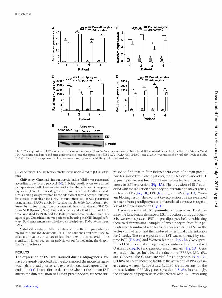
RESULTSThe expression of EST was induced during adipogenesis. Wehave previously reported that the expression of the mouse Est genewas high in preadipocytes, and expression decreased upon differ-entiation (13). In an effort to determine whether the human ESTaffects the differentiation of human preadipocytes, we were sur-
prised to find that in four independent cases of human preadi-pocytes isolated from obese patients, the mRNA expression of ESTin preadipocytes was low, and differentiation led to a marked in-crease in EST expression (Fig. 1A). The induction of EST coin-cided with the induction of adipocyte differentiation maker genes,such as PPAR� (Fig. 1B), LPL (Fig. 1C), and aP2 (Fig. 1D). West-ern blotting results showed that the expression of ER� remainedconstant from preadipocytes to differentiated adipocytes regard-less of EST overexpression (Fig. 1E).
Overexpression of EST promoted adipogenesis. To deter-mine the functional relevance of EST induction during adipogen-esis, we overexpressed EST in preadipocytes before subjectingthem to differentiation. Samples of preadipocytes from four pa-tients were transduced with lentivirus overexpressing EST or thevector control virus and then induced to terminal differentiationfor 2 weeks. The overexpression of EST was confirmed by real-time PCR (Fig. 2A) and Western blotting (Fig. 2B). Overexpres-sion of EST promoted adipogenesis, as confirmed by both oil redO staining (Fig. 2C) and gene expression analysis (Fig. 2D). Geneexpression changes included the induction of PPAR�, LPL, aP2,and C/EBP�. The C/EBPs are vital for adipogenesis (3, 4, 17).C/EBP� has been shown to facilitate the activation of PPAR� tar-get genes, whereas C/EBP� and C/EBP� are important for thetransactivation of PPAR� gene expression (18–23). Interestingly,the enhanced adipogenesis in cells infected with EST-expressing
FIG 1 The expression of EST was induced during adipogenesis. (A to D) Preadipocytes were cultured and differentiated in standard medium for 14 days. TotalRNA was extracted before and after differentiation, and the expression of EST (A), PPAR� (B), LPL (C), and aP2 (D) was measured by real-time PCR analysis.*, P 0.05. (E) The expression of ER� was measured by Western blotting. NT, nontransfected.
Ihunnah et al.
1684 mcb.asm.org Molecular and Cellular Biology
on July 2, 2016 by guesthttp://m
cb.asm.org/
Dow
nloaded from
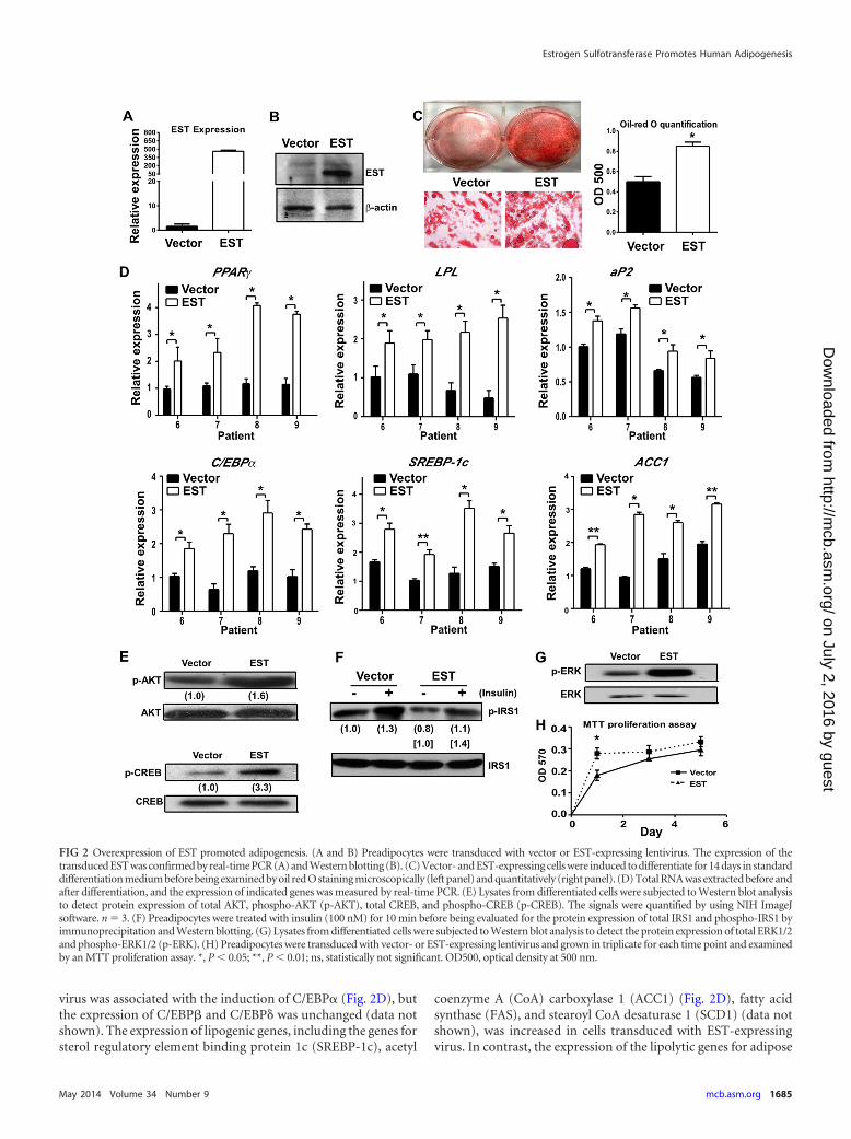
virus was associated with the induction of C/EBP� (Fig. 2D), butthe expression of C/EBP� and C/EBP� was unchanged (data notshown). The expression of lipogenic genes, including the genes forsterol regulatory element binding protein 1c (SREBP-1c), acetyl
coenzyme A (CoA) carboxylase 1 (ACC1) (Fig. 2D), fatty acidsynthase (FAS), and stearoyl CoA desaturase 1 (SCD1) (data notshown), was increased in cells transduced with EST-expressingvirus. In contrast, the expression of the lipolytic genes for adipose
FIG 2 Overexpression of EST promoted adipogenesis. (A and B) Preadipocytes were transduced with vector or EST-expressing lentivirus. The expression of thetransduced EST was confirmed by real-time PCR (A) and Western blotting (B). (C) Vector- and EST-expressing cells were induced to differentiate for 14 days in standarddifferentiation medium before being examined by oil red O staining microscopically (left panel) and quantitatively (right panel). (D) Total RNA was extracted before andafter differentiation, and the expression of indicated genes was measured by real-time PCR. (E) Lysates from differentiated cells were subjected to Western blot analysisto detect protein expression of total AKT, phospho-AKT (p-AKT), total CREB, and phospho-CREB (p-CREB). The signals were quantified by using NIH ImageJsoftware. n � 3. (F) Preadipocytes were treated with insulin (100 nM) for 10 min before being evaluated for the protein expression of total IRS1 and phospho-IRS1 byimmunoprecipitation and Western blotting. (G) Lysates from differentiated cells were subjected to Western blot analysis to detect the protein expression of total ERK1/2and phospho-ERK1/2 (p-ERK). (H) Preadipocytes were transduced with vector- or EST-expressing lentivirus and grown in triplicate for each time point and examinedby an MTT proliferation assay. *, P 0.05; **, P 0.01; ns, statistically not significant. OD500, optical density at 500 nm.
Estrogen Sulfotransferase Promotes Human Adipogenesis
May 2014 Volume 34 Number 9 mcb.asm.org 1685
on July 2, 2016 by guesthttp://m
cb.asm.org/
Dow
nloaded from
triglyceride lipase (ATGL) and hormone-sensitive lipase (HSL)was not affected by EST overexpression (data not shown).
The enhancement of differentiation in cells infected with EST-expressing virus was associated with increased phosphorylation ofAKT and CREB in terminally differentiated cells (Fig. 2E), whichwas suggestive of increased insulin signaling. However, when theacute insulin response was evaluated in preadipocytes treated withinsulin for 10 min, we found that the phosphorylation of the in-sulin receptor substrate 1 (IRS1) was similarly increased in vector-infected and EST virus-infected cells (Fig. 2F). These results sug-gested that overexpression of EST alone did not confer increasedacute insulin sensitivity. The increased AKT and CREB phosphor-ylation in terminally differentiated cells might have been second-ary to the enhanced differentiation when EST was overexpressed.The phosphorylation of ERK1/2 was also increased in EST virus-infected cells (Fig. 2G). ERK1/2 activation is often linked to cellproliferation. During adipocyte differentiation, ERK1/2 activa-tion was required during the phase of clonal expansion, whereaspersistent activation of ERK1/2 may inhibit adipocyte differenti-ation (24, 25). Consistent with the increased ERK1/2 phosphory-lation, the proliferation of cells infected with EST-expressing viruswas increased compared to that of vector-infected cells in the earlyphase of growth, but the difference became insignificant after 3days of culture when the cells were more confluent (Fig. 2H).Addition of the ERK1/2 inhibitor PD98059 had little effect on thedifferentiation of either the vector- or EST virus-infected cells(data not shown), suggesting that the difference in proliferationrate in the early phase might not be the key factor responsible forthe difference in differentiation.
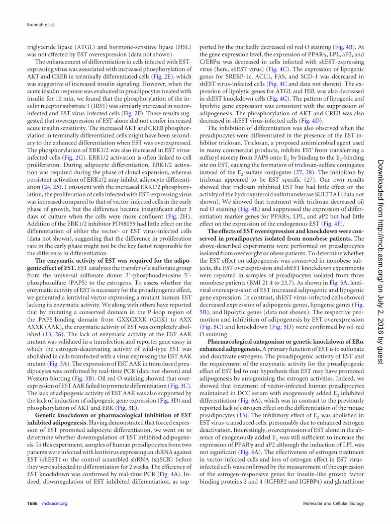
The enzymatic activity of EST was required for the adipo-genic effect of EST. EST catalyzes the transfer of a sulfonate groupfrom the universal sulfonate donor 3=-phosphoadenosine 5=-phosphosulfate (PAPS) to the estrogens. To assess whether theenzymatic activity of EST is necessary for the proadipogenic effect,we generated a lentiviral vector expressing a mutant human ESTlacking its enzymatic activity. We along with others have reportedthat by mutating a conserved domain in the P-loop region ofthe PAPS-binding domain from GXXGXXK (GGK) to AXXAXXK (AAK), the enzymatic activity of EST was completely abol-ished (13, 26). The lack of enzymatic activity of the EST AAKmutant was validated in a transfection and reporter gene assay inwhich the estrogen-deactivating activity of wild-type EST wasabolished in cells transfected with a virus expressing the EST AAKmutant (Fig. 3A). The expression of EST AAK in transduced prea-dipocytes was confirmed by real-time PCR (data not shown) andWestern blotting (Fig. 3B). Oil red O staining showed that over-expression of EST AAK failed to promote differentiation (Fig. 3C).The lack of adipogenic activity of EST AAK was also supported bythe lack of induction of adipogenic gene expression (Fig. 3D) andphosphorylation of AKT and ERK (Fig. 3E).
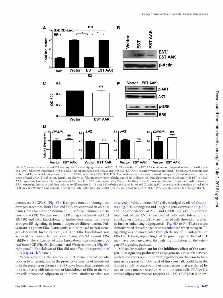
Genetic knockdown or pharmacological inhibition of ESTinhibited adipogenesis. Having demonstrated that forced expres-sion of EST promoted adipocyte differentiation, we went on todetermine whether downregulation of EST inhibited adipogene-sis. In this experiment, samples of human preadipocytes from twopatients were infected with lentivirus expressing an shRNA againstEST (shEST) or the control scrambled shRNA (shSCR) beforethey were subjected to differentiation for 2 weeks. The efficiency ofEST knockdown was confirmed by real-time PCR (Fig. 4A). In-deed, downregulation of EST inhibited differentiation, as sup-
ported by the markedly decreased oil red O staining (Fig. 4B). Atthe gene expression level, the expression of PPAR�, LPL, aP2, andC/EBP� was decreased in cells infected with shEST-expressingvirus (here, shEST virus) (Fig. 4C). The expression of lipogenicgenes for SREBP-1c, ACC1, FAS, and SCD-1 was decreased inshEST virus-infected cells (Fig. 4C and data not shown). The ex-pression of lipolytic genes for ATGL and HSL was also decreasedin shEST knockdown cells (Fig. 4C). The pattern of lipogenic andlipolytic gene expression was consistent with the suppression ofadipogenesis. The phosphorylation of AKT and CREB was alsodecreased in shEST virus-infected cells (Fig. 4D).
The inhibition of differentiation was also observed when thepreadipocytes were differentiated in the presence of the EST in-hibitor triclosan. Triclosan, a proposed antimicrobial agent usedin many commercial products, inhibits EST from transferring asulfuryl moiety from PAPS onto E2 by binding to the E2-bindingsite on EST, causing the formation of triclosan-sulfate conjugatesinstead of the E2-sulfate conjugates (27, 28). The inhibition bytriclosan appeared to be EST specific (27). Our own resultsshowed that triclosan inhibited EST but had little effect on theactivity of the hydroxysteroid sulfotransferase SULT2A1 (data notshown). We showed that treatment with triclosan decreased oilred O staining (Fig. 4E) and suppressed the expression of differ-entiation marker genes for PPAR�, LPL, and aP2 but had littleeffect on the expression of the endogenous EST (Fig. 4F).
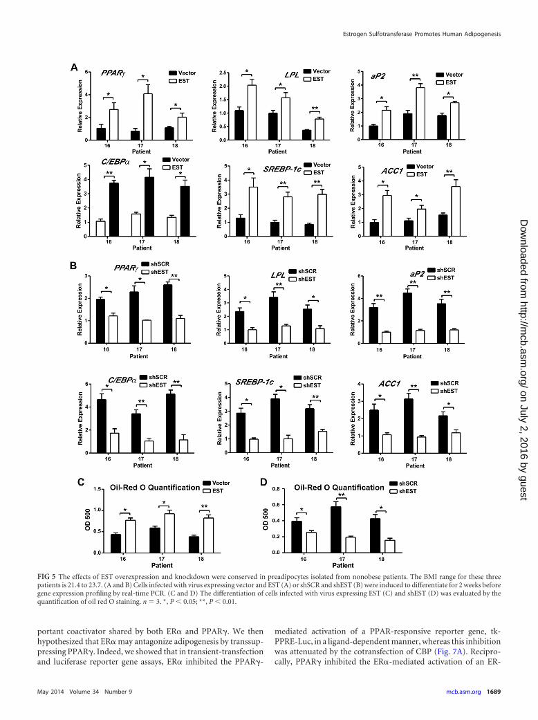
The effects of EST overexpression and knockdown were con-served in preadipocytes isolated from nonobese patients. Theabove-described experiments were performed on preadipocytesisolated from overweight or obese patients. To determine whetherthe EST effect on adipogenesis was conserved in nonobese sub-jects, the EST overexpression and shEST knockdown experimentswere repeated in samples of preadipocytes isolated from threenonobese patients (BMI 21.4 to 23.7). As shown in Fig. 5A, lenti-viral overexpression of EST increased adipogenic and lipogenicgene expression. In contrast, shEST virus-infected cells showeddecreased expression of adipogenic genes, lipogenic genes (Fig.5B), and lipolytic genes (data not shown). The respective pro-motion and inhibition of adipogenesis by EST overexpression(Fig. 5C) and knockdown (Fig. 5D) were confirmed by oil redO staining.
Pharmacological antagonism or genetic knockdown of ER�enhanced adipogenesis. A primary function of EST is to sulfonateand deactivate estrogens. The proadipogenic activity of EST andthe requirement of the enzymatic activity for the proadipogeniceffect of EST led to our hypothesis that EST may have promotedadipogenesis by antagonizing the estrogen activities. Indeed, weshowed that treatment of vector-infected human preadipocytesmaintained in DCC serum with exogenously added E2 inhibiteddifferentiation (Fig. 6A), which was in contrast to the previouslyreported lack of estrogen effect on the differentiation of the mousepreadipocytes (13). The inhibitory effect of E2 was abolished inEST virus-transduced cells, presumably due to enhanced estrogendeactivation. Interestingly, overexpression of EST alone in the ab-sence of exogenously added E2 was still sufficient to increase theexpression of PPAR� and aP2 although the induction of LPL wasnot significant (Fig. 6A). The effectiveness of estrogen treatmentin vector-infected cells and loss of estrogen effect in EST virus-infected cells was confirmed by the measurement of the expressionof the estrogen-responsive genes for insulin-like growth factorbinding proteins 2 and 4 (IGFBP2 and IGFBP4) and glutathione
Ihunnah et al.
1686 mcb.asm.org Molecular and Cellular Biology
on July 2, 2016 by guesthttp://m
cb.asm.org/
Dow
nloaded from
peroxidase 3 (GPX3) (Fig. 6B). Estrogens function through theestrogen receptors. Both ER� and ER� are expressed in adiposetissues, but ER� is the predominant ER isoform in human subcu-taneous fat (29). We then used the ER antagonist fulvestrant (ICI182780) and ER� knockdown to further determine the role ofestrogen-ER signaling in human adipocyte differentiation. Ful-vestrant is a potent ER�/� antagonist clinically used to treat estro-gen-dependent breast cancer (30). The ER� knockdown wasachieved by using a lentivirus expressing shRNA against ER�(shER�). The efficiency of ER� knockdown was confirmed byreal-time PCR (Fig. 6C, left panel) and Western blotting (Fig. 6C,right panel). Knockdown of ER� did not affect the expression ofER� (Fig. 6C, left panel).
When subjecting the vector- or EST virus-infected preadi-pocytes to differentiation in the presence or absence of fulvestrantor in the presence or absence of shER�, we found that treatment ofthe vector cells with fulvestrant or knockdown of ER� in the vec-tor cells promoted adipogenesis to a level similar to what was
observed in vehicle-treated EST cells, as judged by oil red O stain-ing (Fig. 6D), adipogenic and lipogenic gene expression (Fig. 6E),and phosphorylation of AKT and CREB (Fig. 6F). In contrast,treatment of the EST virus-infected cells with fulvestrant orknockdown of ER� in EST virus-infected cells showed little effectin further enhancing adipogenesis (Fig. 6D to F). These resultsdemonstrated that adipogenesis was enhanced when estrogen-ERsignaling was downregulated through the use of ER antagonist orER� knockdown, supporting that the proadipogenic effect of ESTmay have been mediated through the inhibition of the estro-gen-ER signaling pathway.
Molecular mechanism for the inhibitory effect of the estro-gen-ER� signaling pathway on adipogenesis. Cross talk betweennuclear receptors is an important regulatory mechanism to fine-tune gene expression. The heart of this cross talk could lie in thelimited supply of coactivators or corepressors that are shared bytwo or more nuclear receptors within the same cells. PPAR� is acritical adipogenic nuclear receptor (31, 32). CBP/p300 is an im-
FIG 3 The enzymatic activity of EST was required for the adipogenic effect of EST. (A) The activity of the EST AAK mutant was compared to that of the wild-typeEST. 293T cells were transfected with a tk-ERE-Luc reporter gene and ER�, along with EST, EST AAK, or empty vector as indicated. The cells were either treatedwith 1 nM E2 or vehicle in phenol red-free DMEM containing 10% DCC FBS. The luciferase activities are normalized against �-Gal activities from thecotransfected CMX-�-Gal vector. Results are shown as fold induction over vehicle, treated in triplicate. (B) Preadipocytes were infected with EST- or ESTAAK-expressing lentivirus. The expression of EST and EST AAK was measured by Western blotting. (C to E) Preadipocytes were transduced with vector- orAAK-expressing lentivirus and then induced to differentiate for 14 days before being evaluated for oil red O staining (C), gene expression analysis by real-timePCR (D), and Western blot analysis to detect total AKT, phospho-AKT, total ERK1/2, and phospho-ERK1/2 (E). *, P 0.05; ns, statistically not significant.
Estrogen Sulfotransferase Promotes Human Adipogenesis
May 2014 Volume 34 Number 9 mcb.asm.org 1687
on July 2, 2016 by guesthttp://m
cb.asm.org/
Dow
nloaded from
FIG 4 Genetic knockdown or pharmacological inhibition of EST inhibited adipogenesis. (A to D) Preadipocytes were transduced with lentivirus expressingeither shEST or a scrambled shRNA (shSCR) and then induced to differentiate for 2 weeks before being evaluated for the efficiency of EST knockdown byreal-time PCR analysis (A), oil red O staining and quantification (B), gene expression analysis by real-time PCR (C), and Western blot analysis to detect theexpression of total AKT, phospho-AKT, total CREB, and phospho-CREB (D). (E and F) Preadipocytes were induced to differentiate for 2 weeks in the presenceof 10 �M triclosan or vehicle (Veh) before being evaluated for oil red O staining (E) and the expression of adipogenic genes and EST (F). *, P 0.05; **, P 0.01;ns, statistically not significant.
Ihunnah et al.
1688 mcb.asm.org Molecular and Cellular Biology
on July 2, 2016 by guesthttp://m
cb.asm.org/
Dow
nloaded from
portant coactivator shared by both ER� and PPAR�. We thenhypothesized that ER� may antagonize adipogenesis by transsup-pressing PPAR�. Indeed, we showed that in transient-transfectionand luciferase reporter gene assays, ER� inhibited the PPAR�-
mediated activation of a PPAR-responsive reporter gene, tk-PPRE-Luc, in a ligand-dependent manner, whereas this inhibitionwas attenuated by the cotransfection of CBP (Fig. 7A). Recipro-cally, PPAR� inhibited the ER�-mediated activation of an ER-
FIG 5 The effects of EST overexpression and knockdown were conserved in preadipocytes isolated from nonobese patients. The BMI range for these threepatients is 21.4 to 23.7. (A and B) Cells infected with virus expressing vector and EST (A) or shSCR and shEST (B) were induced to differentiate for 2 weeks beforegene expression profiling by real-time PCR. (C and D) The differentiation of cells infected with virus expressing EST (C) and shEST (D) was evaluated by thequantification of oil red O staining. n � 3. *, P 0.05; **, P 0.01.
Estrogen Sulfotransferase Promotes Human Adipogenesis
May 2014 Volume 34 Number 9 mcb.asm.org 1689
on July 2, 2016 by guesthttp://m
cb.asm.org/
Dow
nloaded from
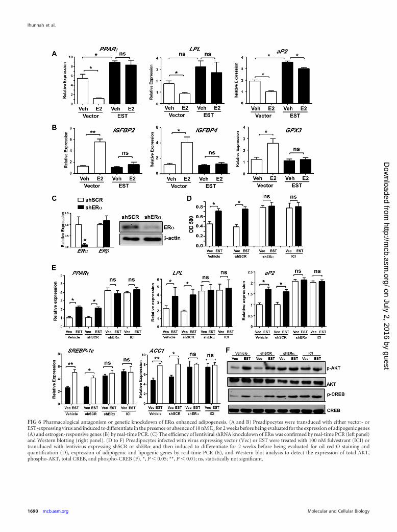
FIG 6 Pharmacological antagonism or genetic knockdown of ER� enhanced adipogenesis. (A and B) Preadipocytes were transduced with either vector- orEST-expressing virus and induced to differentiate in the presence or absence of 10 nM E2 for 2 weeks before being evaluated for the expression of adipogenic genes(A) and estrogen-responsive genes (B) by real-time PCR. (C) The efficiency of lentiviral shRNA knockdown of ER� was confirmed by real-time PCR (left panel)and Western blotting (right panel). (D to F) Preadipocytes infected with virus expressing vector (Vec) or EST were treated with 100 nM fulvestrant (ICI) ortransduced with lentivirus expressing shSCR or shER� and then induced to differentiate for 2 weeks before being evaluated for oil red O staining andquantification (D), expression of adipogenic and lipogenic genes by real-time PCR (E), and Western blot analysis to detect the expression of total AKT,phospho-AKT, total CREB, and phospho-CREB (F). *, P 0.05; **, P 0.01; ns, statistically not significant.
Ihunnah et al.
1690 mcb.asm.org Molecular and Cellular Biology
on July 2, 2016 by guesthttp://m
cb.asm.org/
Dow
nloaded from
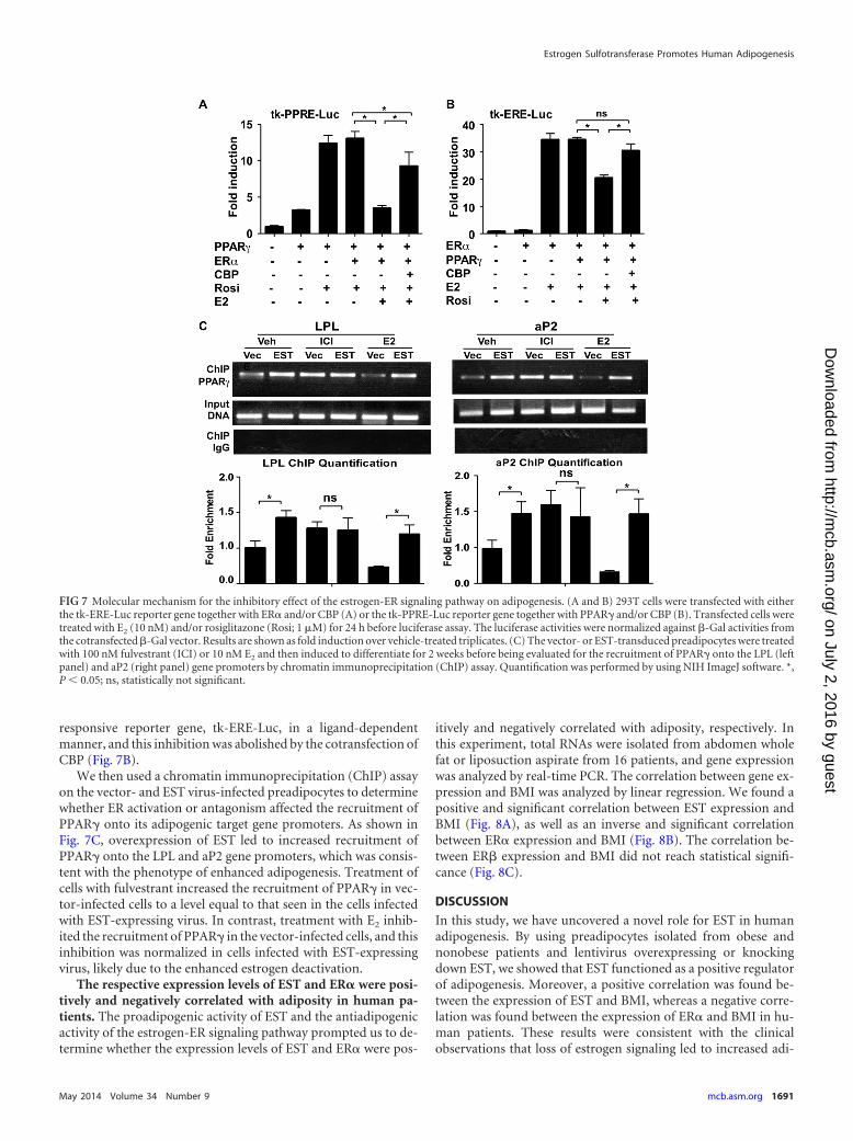
responsive reporter gene, tk-ERE-Luc, in a ligand-dependentmanner, and this inhibition was abolished by the cotransfection ofCBP (Fig. 7B).
We then used a chromatin immunoprecipitation (ChIP) assayon the vector- and EST virus-infected preadipocytes to determinewhether ER activation or antagonism affected the recruitment ofPPAR� onto its adipogenic target gene promoters. As shown inFig. 7C, overexpression of EST led to increased recruitment ofPPAR� onto the LPL and aP2 gene promoters, which was consis-tent with the phenotype of enhanced adipogenesis. Treatment ofcells with fulvestrant increased the recruitment of PPAR� in vec-tor-infected cells to a level equal to that seen in the cells infectedwith EST-expressing virus. In contrast, treatment with E2 inhib-ited the recruitment of PPAR� in the vector-infected cells, and thisinhibition was normalized in cells infected with EST-expressingvirus, likely due to the enhanced estrogen deactivation.
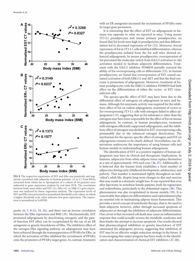
The respective expression levels of EST and ER� were posi-tively and negatively correlated with adiposity in human pa-tients. The proadipogenic activity of EST and the antiadipogenicactivity of the estrogen-ER signaling pathway prompted us to de-termine whether the expression levels of EST and ER� were pos-
itively and negatively correlated with adiposity, respectively. Inthis experiment, total RNAs were isolated from abdomen wholefat or liposuction aspirate from 16 patients, and gene expressionwas analyzed by real-time PCR. The correlation between gene ex-pression and BMI was analyzed by linear regression. We found apositive and significant correlation between EST expression andBMI (Fig. 8A), as well as an inverse and significant correlationbetween ER� expression and BMI (Fig. 8B). The correlation be-tween ER� expression and BMI did not reach statistical signifi-cance (Fig. 8C).
DISCUSSION
In this study, we have uncovered a novel role for EST in humanadipogenesis. By using preadipocytes isolated from obese andnonobese patients and lentivirus overexpressing or knockingdown EST, we showed that EST functioned as a positive regulatorof adipogenesis. Moreover, a positive correlation was found be-tween the expression of EST and BMI, whereas a negative corre-lation was found between the expression of ER� and BMI in hu-man patients. These results were consistent with the clinicalobservations that loss of estrogen signaling led to increased adi-
FIG 7 Molecular mechanism for the inhibitory effect of the estrogen-ER signaling pathway on adipogenesis. (A and B) 293T cells were transfected with eitherthe tk-ERE-Luc reporter gene together with ER� and/or CBP (A) or the tk-PPRE-Luc reporter gene together with PPAR� and/or CBP (B). Transfected cells weretreated with E2 (10 nM) and/or rosiglitazone (Rosi; 1 �M) for 24 h before luciferase assay. The luciferase activities were normalized against �-Gal activities fromthe cotransfected �-Gal vector. Results are shown as fold induction over vehicle-treated triplicates. (C) The vector- or EST-transduced preadipocytes were treatedwith 100 nM fulvestrant (ICI) or 10 nM E2 and then induced to differentiate for 2 weeks before being evaluated for the recruitment of PPAR� onto the LPL (leftpanel) and aP2 (right panel) gene promoters by chromatin immunoprecipitation (ChIP) assay. Quantification was performed by using NIH ImageJ software. *,P 0.05; ns, statistically not significant.
Estrogen Sulfotransferase Promotes Human Adipogenesis
May 2014 Volume 34 Number 9 mcb.asm.org 1691
on July 2, 2016 by guesthttp://m
cb.asm.org/
Dow
nloaded from
posity (6, 7, 9–11, 33, 34), and there was an inverse correlationbetween the ER� expression and BMI (35). Mechanistically, ESTpromoted adipogenesis by deactivating estrogens, and the gain-of-function EST effect can be recapitulated by the use of an ERantagonist or genetic knockdown of ER�. The inhibitory effect ofthe estrogen-ER� signaling pathway on adipogenesis may havebeen achieved through the transsuppression of PPAR� by ER�, inwhich the activation of ER� inhibited the recruitment of PPAR�onto the promoters of PPAR� target genes. In contrast, treatment
with an ER antagonist increased the recruitment of PPAR� ontoits target gene promoters.
It is interesting that the effect of EST on adipogenesis in hu-mans was opposite to what we reported in mice. Using mouse3T3-L1 preadipocytes and mouse primary preadipocytes, wefound that Est levels were high in preadipocytes and that differen-tiation led to decreased expression of Est (13). Moreover, forcedexpression of Est in 3T3-L1 cells inhibited differentiation, whereasthe preadipocytes isolated from the Est null mice showed en-hanced adipogenesis. In mouse preadipocytes, overexpression ofEst prevented the molecular switch from Erk1/2 activation to Aktactivation needed to facilitate adipocyte differentiation. Treat-ment with the Erk1/2 inhibitor PD98059 partially restored theability of Est-overexpressing cells to differentiate (13). In humanpreadipocytes, we found that overexpression of EST caused sus-tained activation of both ERK1/2 and AKT and that the final out-come is promotion of adipogenesis. Moreover, treatment of hu-man preadipocytes with the ERK1/2 inhibitor PD98059 had littleeffect on the differentiation of either the vector- or EST virus-infected cells.
The species-specific effect of EST may have been due to thedifferential effect of estrogens on adipogenesis in mice and hu-mans. Although Est enzymatic activity was required for the inhib-itory effect of Est on rodent adipogenesis, treatment of vector orEst-overexpressing 3T3-L1 cells with estrogens failed to affect ad-ipogenesis (13), suggesting that an Est substrate(s) other than theestrogens may have been responsible for the effect of Est on mouseadipogenesis. In contrast, in human preadipocytes, treatmentwith estrogens efficiently suppressed adipogenesis, and the inhib-itory effect of estrogens was abolished in EST-overexpressing cells,presumably due to the enhanced estrogen deactivation. Themechanism for the species-specific effect of estrogens and EST onadipogenesis remains to be clearly defined. Nevertheless, our ob-servations underscore the importance of using human cells andhuman models in understanding human adipogenesis.
The identification of EST as a positive regulator of human ad-ipogenesis may have its clinical and therapeutic implications. Inhumans, adipocytes from white adipose tissue replace themselvesat a rate of approximately 10% each year (36, 37). Additionally, itis believed that the human body establishes a fixed number ofadipocytes during early childhood development, adolescence, andpuberty. That number is maintained tightly throughout an indi-vidual’s adult life, despite long-term changes in diet and exercisethat may result in a dramatic weight loss. It was reported that evenafter lipectomy in nonobese female patients, body fat regeneratesand redistributes, particularly to the abdominal region (38). Thisphenomenon was also recapitulated in mouse models (39). It isconceivable that adipocyte differentiation and replenishment playan essential role in maintaining adipose tissue homeostasis. Thisprovides a novel concept of antiobesity therapy, that is, the need tolimit adipocyte turnover, either through increased apoptosis ofadipocytes or through the inhibition of adipocyte differentiation.One caveat is that increased cell death may cause an inflammatoryresponse that could actually worsen the metabolic syndrome andthus hinder the attempt to increase apoptosis. Our results showedthat pharmacological inhibition or genetic knockdown of ESTattenuated the adipogenic process, suggesting that inhibition ofEST may be an effective weight reduction strategy in the future. Itis encouraging that major progress has been made in the identifi-cation and characterization of chemical EST inhibitors (27, 40).
FIG 8 The respective expression of EST and ER� was positively and neg-atively correlated with adiposity in human patients. (A to C) Total RNAsextracted from whole fat or lipoaspirate of a cohort of 16 patients weresubjected to gene expression analysis by real-time PCR. The correlationbetween body mass index and EST (A), ER� (A), or ER� (C) gene expres-sion was analyzed by linear regression analysis. The expression levels ofEST and ERs were presented as normalized threshold cycle values, in whicha higher threshold cycle value indicates low gene expression. The expres-sion is normalized to GAPDH.
Ihunnah et al.
1692 mcb.asm.org Molecular and Cellular Biology
on July 2, 2016 by guesthttp://m
cb.asm.org/
Dow
nloaded from
Among limitations, it is noted that the age range of our humansubjects was quite varied and that all samples were from abdomi-nal subcutaneous fat. Although the effects of EST overexpressionand knockdown were observed in preadipocytes isolated fromboth the obese and nonobese patients, we cannot exclude thepossibility that age, menopausal status, steroid hormone level,obesity, and origin of the fat depot affect the phenotypic exhibi-tion. In addition, since overexpression of EST under the estrogen-free DCC cell culture condition can still increase the expression ofcertain adipogenic marker genes (Fig. 6A), we cannot exclude thepossibility of an off-target effect due to the overexpression of EST,as well as the existence of additional EST substrates that may alsohave an effect on adipogenesis.
In summary, we have established that EST is an importantpositive regulator of adipogenesis in humans. We propose thatEST is a druggable target whose inhibition can be used to inhibitthe turnover of adipocytes in obese patients.
ACKNOWLEDGMENTS
We thank Donald DeFranco (University of Pittsburgh) for his insightfulcomments on this study and Jiang Li (University of Pittsburgh) for someof the data analysis.
This work was supported in part by NIH grants DK083953 andHD073070 (to W.X.), and CA114246 (to J.P.R.). C.A.I was supported bythe NIH Ruth L. Kirschstein National Research Service Awards for Indi-vidual Predoctoral Fellowships to Promote Diversity in Health-RelatedResearch (F31-DK-095589). W.X. is the Joseph Koslow Endowed Chair inPharmaceutical Sciences at the University of Pittsburgh School of Phar-macy.
REFERENCES1. Zammit C, Liddicoat H, Moonsie I, Makker H. 2010. Obesity and
respiratory diseases. Int. J. Gen. Med. 3:335–343. http://dx.doi.org/10.2147/IJGM.S11926.
2. Rayalam S, Della-Fera MA, Baile CA. 2008. Phytochemicals and regula-tion of the adipocyte life cycle. J. Nutr. Biochem. 19:717–726. http://dx.doi.org/10.1016/j.jnutbio.2007.12.007.
3. Gregoire FM, Smas CM, Sul HS. 1998. Understanding adipocyte differ-entiation. Physiol. Rev. 78:783– 809.
4. Gregoire FM. 2001. Adipocyte differentiation: from fibroblast to endo-crine cell. Exp. Biol. Med. (Maywood) 226:997–1002.
5. Mayes JS, Watson GH. 2004. Direct effects of sex steroid hormones onadipose tissues and obesity. Obes. Rev. 5:197–216. http://dx.doi.org/10.1111/j.1467-789X.2004.00152.x.
6. Mueller SO, Korach KS. 2001. Estrogen receptors and endocrine diseases:lessons from estrogen receptor knockout mice. Curr. Opin. Pharmacol.1:613– 619. http://dx.doi.org/10.1016/S1471-4892(01)00105-9.
7. Couse JF, Korach KS. 1999. Estrogen receptor null mice: what have welearned and where will they lead us? Endocr. Rev. 20:358 – 417. http://dx.doi.org/10.1210/edrv.20.3.0370.
8. Jones ME, Thorburn AW, Britt KL, Hewitt KN, Wreford NG, ProiettoJ, Oz OK, Leury BJ, Robertson KM, Yao S, Simpson ER. 2000. Aroma-tase-deficient (ArKO) mice have a phenotype of increased adiposity. Proc.Natl. Acad. Sci. U. S. A. 97:12735–12740. http://dx.doi.org/10.1073/pnas.97.23.12735.
9. D’Eon TM, Souza SC, Aronovitz M, Obin MS, Fried SK, Greenberg AS.2005. Estrogen regulation of adiposity and fuel partitioning. Evidence ofgenomic and non-genomic regulation of lipogenic and oxidative path-ways. J. Biol. Chem. 280:35983–35991. http://dx.doi.org/10.1074/jbc.M507339200.
10. Salpeter SR, Walsh JME, Ormiston TM, Greyber E, Buckley NS, Sal-peter EE. 2006. Meta-analysis: effect of hormone-replacement therapy oncomponents of the metabolic syndrome in postmenopausal women. Dia-betes Obes. Metab. 8:538 –554. http://dx.doi.org/10.1111/j.1463-1326.2005.00545.x.
11. Davis SR, Castelo-Branco C, Chedraui P, Lumsden MA, Nappi RE,Shah D, Villaseca P, Writing Group of the International MenopauseSociety for World Menopause Day 2012. 2012. Understanding weight
gain at menopause. Climacteric 15:419 – 429. http://dx.doi.org/10.3109/13697137.2012.707385.
12. Song WC. 2001. Biochemistry and reproductive endocrinology of estro-gen sulfotransferase. Ann. N. Y. Acad. Sci. 948:43–50. http://dx.doi.org/10.1111/j.1749-6632.2001.tb03985.x.
13. Wada T, Ihunnah CA, Gao J, Chai X, Zeng S, Philips BJ, Rubin JP,Marra KG, Xie W. 2011. Estrogen sulfotransferase inhibits adipocytedifferentiation. Mol. Endocrinol. 25:1612–1623. http://dx.doi.org/10.1210/me.2011-1089.
14. Gerlach JC, Lin YC, Brayfield CA, Minteer DM, Li H, Rubin JP, MarraKG. 2012. Adipogenesis of human adipose-derived stem cells withinthree-dimensional hollow fiber-based bioreactors. Tissue Eng. Part CMethods 18:54 – 61. http://dx.doi.org/10.1089/ten.tec.2011.0216.
15. Ho SN, Hunt HD, Horton RM, Pullen JK, Pease LR. 1989. Site-directedmutagenesis by overlap extension using the polymerase chain reaction.Gene 77:51–59. http://dx.doi.org/10.1016/0378-1119(89)90358-2.
16. Nelson JD, Denisenko O, Bomsztyk K. 2006. Protocol for the fast chro-matin immunoprecipitation (ChIP) method. Nat. Protoc. 1:179 –185.http://dx.doi.org/10.1038/nprot.2006.27.
17. Rangwala SM, Lazar MA. 2000. Transcriptional control of adipogenesis.Annu. Rev. Nutr. 20:535–559. http://dx.doi.org/10.1146/annurev.nutr.20.1.535.
18. Cao Z, Umek RM, McKnight SL. 1991. Regulated expression of threeC/EBP isoforms during adipose conversion of 3T3-L1 cells. Genes Dev.5:1538 –1552. http://dx.doi.org/10.1101/gad.5.9.1538.
19. Hwang CS, Mandrup S, MacDougald OA, Geiman DE, Lane MD. 1996.Transcriptional activation of the mouse obese (ob) gene by CCAAT/enhancer binding protein alpha. Proc. Natl. Acad. Sci. U. S. A. 93:873–877. http://dx.doi.org/10.1073/pnas.93.2.873.
20. Ross SR, Graves RA, Greenstein A, Platt KA, Shyu HL, Mellovitz B,Spiegelman BM. 1990. A fat-specific enhancer is the primary determinantof gene expression for adipocyte P2 in vivo. Proc. Natl. Acad. Sci. U. S. A.87:9590 –9594. http://dx.doi.org/10.1073/pnas.87.24.9590.
21. Wu Z, Bucher NL, Farmer SR. 1996. Induction of peroxisome prolifera-tor-activated receptor gamma during the conversion of 3T3 fibroblastsinto adipocytes is mediated by C/EBP�, C/EBP�, and glucocorticoids.Mol. Cell. Biol. 16:4128 – 4136.
22. Wu Z, Xie Y, Bucher NL, Farmer SR. 1995. Conditional ectopic expres-sion of C/EBP beta in NIH-3T3 cells induces PPAR gamma and stimulatesadipogenesis. Genes Dev. 9:2350 –2363. http://dx.doi.org/10.1101/gad.9.19.2350.
23. Miki H, Yamauchi T, Suzuki R, Komeda K, Tsuchida A, Kubota N,Terauchi Y, Kamon J, Kaburagi Y, Matsui J, Akanuma Y, Nagai R,Kimura S, Tobe K, Kadowaki T. 2001. Essential role of insulin receptorsubstrate 1 (IRS-1) and IRS-2 in adipocyte differentiation. Mol. Cell. Biol.21:2521–2532. http://dx.doi.org/10.1128/MCB.21.7.2521-2532.2001.
24. Font de Mora J, Porras A, Ahn N, Santos E. 1997. Mitogen-activatedprotein kinase activation is not necessary for, but antagonizes, 3T3-L1adipocytic differentiation. Mol. Cell. Biol. 17:6068 – 6075.
25. Sale EM, Atkinson PG, Sale GJ. 1995. Requirement of MAP kinase fordifferentiation of fibroblasts to adipocytes, for insulin activation of p90 S6kinase and for insulin or serum stimulation of DNA synthesis. EMBO J.14:674 – 684.
26. Komatsu K, Driscoll WJ, Koh YC, Strott CA. 1994. A P-loop relatedmotif (GxxGxxK) highly conserved in sulfotransferases is required forbinding the activated sulfate donor. Biochem. Biophys. Res. Commun.204:1178 –1185. http://dx.doi.org/10.1006/bbrc.1994.2587.
27. Wang LQ, Falany CN, James MO. 2004. Triclosan as a substrate andinhibitor of 3=-phosphoadenosine 5=-phosphosulfate-sulfotransferaseand UDP-glucuronosyl transferase in human liver fractions. Drug Metab.Dispos. 32:1162–1169. http://dx.doi.org/10.1124/dmd.104.000273.
28. Zhang H, Varlamova O, Vargas FM, Falany CN, Leyh TS. 1998. Sulfuryltransfer: the catalytic mechanism of human estrogen sulfotransferase. J. Biol.Chem. 273:10888–10892. http://dx.doi.org/10.1074/jbc.273.18.10888.
29. Joyner JM, Hutley LJ, Cameron DP. 2001. Estrogen receptors in humanpreadipocytes. Endocrine 15:225–230. http://dx.doi.org/10.1385/ENDO:15:2:225.
30. Cardoso F, Bischoff J, Brain E, Zotano AG, Luck H-J, Tjan-Heijnen VC,Tanner M, Aapro M. 2013. A review of the treatment of endocrine re-sponsive metastatic breast cancer in postmenopausal women. CancerTreat. Rev. 39:457– 465. http://dx.doi.org/10.1016/j.ctrv.2012.06.011.
Estrogen Sulfotransferase Promotes Human Adipogenesis
May 2014 Volume 34 Number 9 mcb.asm.org 1693
on July 2, 2016 by guesthttp://m
cb.asm.org/
Dow
nloaded from

31. Anghel SI, Wahli W. 2007. Fat poetry: a kingdom for PPAR gamma. CellRes. 17:486 –511. http://dx.doi.org/10.1038/cr.2007.48.
32. Spiegelman BM. 1998. PPAR-gamma: adipogenic regulator and thiazoli-dinedione receptor. Diabetes 47:507–514. http://dx.doi.org/10.2337/diabetes.47.4.507.
33. Cooke PS, Naaz A. 2005. Effects of estrogens and the phytoestrogengenistein on adipogenesis and lipogenesis in males and females. Birth De-fects Res. A Clin. Mol. Teratol. 73:472– 473. http://dx.doi.org/10.1002/bdra.20142.
34. Heine PA, Taylor JA, Iwamoto GA, Lubahn DB, Cooke PS. 2000.Increased adipose tissue in male and female estrogen receptor-alphaknockout mice. Proc. Natl. Acad. Sci. U. S. A. 97:12729 –12734. http://dx.doi.org/10.1073/pnas.97.23.12729.
35. Lundholm L, Zang H, Hirschberg AL, Gustafsson J-A, Arner P, Dahl-man-Wright K. 2008. Key lipogenic gene expression can be decreased byestrogen in human adipose tissue. Fertil. Steril. 90:44 – 48. http://dx.doi.org/10.1016/j.fertnstert.2007.06.011.
36. Arner P, Spalding KL. 2010. Fat cell turnover in humans. Biochem.
Biophys. Res. Commun. 396:101–104. http://dx.doi.org/10.1016/j.bbrc.2010.02.165.
37. Spalding KL, Arner E, Westermark PO, Bernard S, Buchholz BA,Bergmann O, Blomqvist L, Hoffstedt J, Naslund E, Britton T, ConchaH, Hassan M, Ryden M, Frisen J, Arner P. 2008. Dynamics of fat cellturnover in humans. Nature 453:783–787. http://dx.doi.org/10.1038/nature06902.
38. Hernandez TL, Kittelson JM, Law CK, Ketch LL, Stob NR, LindstromRC, Scherzinger A, Stamm ER, Eckel RH. 2011. Fat redistributionfollowing suction lipectomy: defense of body fat and patterns of restora-tion. Obesity 19:1388 –1395. http://dx.doi.org/10.1038/oby.2011.64.
39. Rigamonti A, Brennand K, Lau F, Cowan CA. 2011. Rapid cellularturnover in adipose tissue. PLoS One 6:e17637. http://dx.doi.org/10.1371/journal.pone.0017637.
40. James MO, Li W, Summerlot DP, Rowland-Faux L, Wood CE. 2010.Triclosan is a potent inhibitor of estradiol and estrone sulfonation in sheepplacenta. Environ. Int. 36:942–949. http://dx.doi.org/10.1016/j.envint.2009.02.004.
Ihunnah et al.
1694 mcb.asm.org Molecular and Cellular Biology
on July 2, 2016 by guesthttp://m
cb.asm.org/
Dow
nloaded from
Related Documents


![Amine N-Sulfotransferase* · 1999. 2. 4. · 10040 Amine N-Sulfotransferase total volume of 50 pl, was incubated in 0.2 M potassium bicine at pH 8.0, 2 mM amine, and 1 mM [%]PAPS](https://static.cupdf.com/doc/110x72/5fc69085f7afdb4e584dc1ba/amine-n-sulfotransferase-1999-2-4-10040-amine-n-sulfotransferase-total-volume.jpg)