ESTIMATION OF GENDER BY COSTOCHONDRAL CALCIFICATION MODEL OBTAINED FROM COMPUTED TOMOGRAPHY IMAGES 2021 MASTER THESIS ANATOMY Albaraa AL-SAMANEE Thesis Advisor Assoc. Prof. Dr. Zülal ÖNER

Welcome message from author
This document is posted to help you gain knowledge. Please leave a comment to let me know what you think about it! Share it to your friends and learn new things together.
Transcript

ESTIMATION OF GENDER BY COSTOCHONDRAL CALCIFICATION MODEL
OBTAINED FROM COMPUTED TOMOGRAPHY IMAGES
2021 MASTER THESIS
ANATOMY
Albaraa AL-SAMANEE
Thesis Advisor Assoc. Prof. Dr. Zülal ÖNER

ESTIMATION OF GENDER BY COSTOCHONDRAL CALCIFICATION
MODEL OBTAINED FROM COMPUTED TOMOGRAPHY IMAGES
Albaraa AL-SAMANEE
T.C.
Karabuk University
Institute of Graduate Programs
Department of Anatomy
Prepared as
Master Thesis
Thesis Advisor
Assoc. Prof. Dr. Zülal ÖNER
KARABUK
June 2021

ii
I certify that in my opinion the thesis submitted by Albaraa AL-SAMANEE titled
“ESTIMATION OF GENDER BY COSTOCHONDRAL CALCIFICATION
MODEL OBTAINED FROM COMPUTED TOMOGRAPHY IMAGES” is fully
adequate in scope and in quality as a thesis for the degree of Master of Anatomy.
APPROVAL
Assoc. Prof. Dr. Zülal ÖNER ..........................
Thesis Advisor, Department of Anatomy
This thesis is accepted by the examining committee with a unanimous vote in the
Department of Anatomy as a Master of Anatomy thesis. 18.06.2021
Examining Committee Members (Institutions) Signature
Chairman : Prof. Dr. Bünyamin ŞAHİN (TOGÜ) ..........................
Member : Assoc. Prof. Dr. Zülal ÖNER (KBÜ) ..........................
Member : Assist. Prof. Dr. Şeyma TOY (KBÜ) ..........................
The degree of Master of Anatomy by the thesis submitted is approved by the
Administrative Board of the Institute of Graduate Programs, Karabuk University.
Prof. Dr. Hasan SOLMAZ ..........................
Director of the Institute of Graduate Programs

iii
“I declare that all the information within this thesis has been gathered and presented
in accordance with academic regulations and ethical principles and I have
according to the requirements of these regulations and principles cited all those
which do not originate in this work as well.”
Albaraa AL-SAMANEE

iv
ABSTRACT
Master Thesis
ESTIMATION OF GENDER BY COSTOCHONDRAL CALCIFICATION
MODEL OBTAINED FROM COMPUTED TOMOGRAPHY IMAGES
Albaraa AL-SAMANEE
Karabuk University
Institute of Graduate Programs
The Department of Anatomy
Thesis Advisor:
Assoc. Prof. Dr. Zülal ÖNER
June 2021, 32 pages
Gender estimation plays a key role in human identification. Between the various
measurement methods of gender estimation from skeletal remains, the use of the
calcification patterns of costal cartilages is highly suggested especially when the
skull and pelvic bones are not available. The purpose of our study is to estimate
gender according to the difference in the patterns of costal cartilage calcifications.
Our study was performed by using the Computed Tomography (CT) images of 200
individuals (100 female, 100 male) in the 20-60 age group who applied to Karabuk
University Karabuk Training and Research Hospital and had no pathology or
orthopedic surgery history. CT images were obtained from the hospital Picture
Archiving and Communication System (PACS) and were recorded in Digital
Imaging and Communications in Medicine (DICOM) format. In our study used the
method of Rejtarova et al. (2004) for classification the patterns of costal cartilage
calcifications and calculate the number and percentage of each pattern in male and

v
female to estimate the gender. The results showed 193 (96.5%) individuals with
calcification in the costal cartilages and 7 (3.5%) individuals without calcification in
their costal cartilages. Peripheral pattern (Type I) shown male gender estimation with
100% and central pattern (Type II) shown female gender estimation with 92.3%. As
a result of this study, it has been reached that gender can be predicted from the costal
cartilage calcification models by using the method of Rejtarova et al. (2004).
Keywords : Computed Tomography, Costal Cartilage, Calcification, Gender
Estimation
Science Code : 1005

vi
ÖZET
Yüksek Lisans Tezi
BİLGİSAYARLI TOMOGRAFİ GÖRÜNTÜLERİ ÜZERİNDEN ELDE
EDİLEN KOSTOKONDRAL KALSİFİKASYON MODELİ İLE CİNSİYET
TAHMİNİ
Albaraa AL-SAMANEE
Karabük Üniversitesi
Lisansüstü Eğitim Enstitüsü
Anatomi Anabilim Dalı
Tez Danışmanı:
Assoc. Prof. Dr. Zülal ÖNER
Haziran 2021, 32 sayfa
Cinsiyet tahmini, insan kimlik tespitinde önemli bir rol oynar. İskelet kalıntılarından
cinsiyeti tahmin etmek için çeşitli ölçüm yöntemlerinden biri olan kostal
kıkırdakların kalsifikasyon modellerinin kullanımı, özellikle kafatası ve pelvis
kemikleri bulunmadığında kullanılması şiddetle tavsiye edilir. Çalışmamızın amacı,
kostal kıkırdak kalsifikasyon modellerindeki farklılığa göre cinsiyeti tahmin
etmektir. Çalışmamız, Karabük Üniversitesi Karabük Eğitim ve Araştırma
Hastanesi'ne başvuran, patolojisi veya ortopedik ameliyat öyküsü olmayan 20-60 yaş
grubundaki 200 kişinin (100 kadın, 100 erkek) Bilgisayarlı Tomografi (BT)
görüntüleri kullanılarak yapıldı. BT görüntüleri hastanenin Görüntü Arşivleme ve
İletişim Sisteminden (PACS) elde edildi ve Tıpta Dijital Görüntüleme ve İletişim
(DICOM) formatında kaydedildi. Çalışmada, cinsiyeti tahmin edebilmek için kostal
kıkırdak kalsifikasyon modellerinden olan Rejtarova et al. (2004) yöntemi kullanıldı.

vii
Kostal kıkırdaklarda kireçlenme olan 193 birey (%96,5), kostal kıkırdaklarında hiç
kireçlenme olmayan 7 bireyin (%3,5) olduğu tespit edildi. Periferik modelin (Tip I)
erkekleri tahmin etme oranı %100, merkezi modelin (Tip II) kadınları tahmin etme
oranı ise %92,3 olduğu görüldü. Bu çalışmanın sonucunda kostal kıkırdak
kalsifikasyon modellerinden Rejtarova et al. (2004) yöntemiyle cinsiyetin tahmin
edilebileceğine ulaşılmıştır.
Anahtar Kelimeler : Bilgisayarlı Tomografi, Cartilago Costalis, Kalsifikasyon,
Cinsiyet Tahmini.
Bilim Kodu : 1005
viii
ACKNOWLEDGMENT
I would like to express my deep and sincere gratitude to my supervisor, Assoc. Prof.
Dr. Zülal ÖNER, who not only shared her profound scientific knowledge with me
but also taught me great lessons of life. Her support, suggestions and encouragement
gave me the drive and will to complete this work.
I would also like to extend my deepest gratitude to Assoc. Prof. Dr. Serkan ÖNER
and Assist. Prof. Dr. Muhammed Kamil TURAN for their knowledge, motivation,
kindness and support throughout the study.
I wish to express my warm and sincere thanks to Research Assistants Yusuf
SEÇGİN, Rukiye Sümeyye BAKICI and Necati Emre ŞAHİN for their collaboration
to this research.
Finally, I am grateful to my family for their love and support.

ix
CONTENTS
Page
APPROVAL ................................................................................................................. ii
ABSTRACT ................................................................................................................ iv
ÖZET........................................................................................................................... vi
ACKNOWLEDGMENT ........................................................................................... viii
CONTENTS ................................................................................................................ ix
LIST OF FIGURES .................................................................................................... xi
LIST OF TABLES ..................................................................................................... xii
SYMBOLS AND ABBREVIATIONS INDEX........................................................ xiii
PART 1 ........................................................................................................................ 1
INTRODUCTION AND PURPOSE ........................................................................... 1
PART 2 ........................................................................................................................ 3
GENERAL INFORMATIONS .................................................................................... 3
2.1. ABOUT ANTHROPOLOGY .......................................................................... 3
2.2. FORENSIC ANTHROPOLOGY ..................................................................... 3
2.3. IDENTIFICATION .......................................................................................... 4
2.4. THE IMPORTANCE OF GENDER DETERMINATION IN
IDENTIFICATION .......................................................................................... 4
2.5. COSTOCHONDRAL CALCIFICATION ....................................................... 5
2.6. ANATOMY OF STERNUM AND RIBS ........................................................ 6
2.7. ANATOMY OF COSTAL CARTILAGES ..................................................... 7
2.8. ARTICULSATIONS OF COSTAL CARTILAGES ....................................... 8
PART 3 ...................................................................................................................... 10
MATERIALS AND METHODS ............................................................................... 10

x
Page
3.1. IMAGE ANALYSIS METHOD .................................................................... 10
PART 4 ...................................................................................................................... 16
RESULTS .................................................................................................................. 16
PART 5 ...................................................................................................................... 21
DISCUSSION ............................................................................................................ 21
PART 6 ...................................................................................................................... 24
CONCLUSION AND RECOMMENDATIONS ....................................................... 24
REFERENCES ........................................................................................................... 25
APPENDIX ................................................................................................................ 30
RESUME ................................................................................................................... 32

xi
LIST OF FIGURES
Page
Figure 2.1. Thoracic Cage (Modified from Sobotta Anatomy Atlas) .......................... 7
Figure 2.2. Costal Cartilages (Modified from Sobotta Anatomy Atlas) ...................... 8
Figure 2.3. Articulations of Costal Cartilages (Modified from Grays Atlas of
Anatomy) .................................................................................................. 9
Figure 3.1. Peripheral pattern of costal cartilage calcification (Type I). ................... 12
Figure 3.2. Central globular pattern of costal cartilage calcification (Type IIb). ...... 12
Figure 3.3. Central lingual and globular pattern of costal cartilage calcification
(Type IIc). ............................................................................................... 13
Figure 3.4. Mixed (peripheral and central pattern) Type III. ..................................... 13
Figure 3.5. Indifferent pattern of costal cartilage calcification (Type IV). ............... 14
Figure 4.1. Percentage of individuals with CCC (Positive: with calcification,
Negative: without calcification). ............................................................. 17

xii
LIST OF TABLES
Page
Table 4.1. The mean age of female and male. ........................................................... 16
Table 4.2. Number of individuals with CCC in female and male. ............................. 17
Table 4.3. Number of of each CCC pattern in female and male. ............................... 18
Table 4.4. Number of each central pattern subtype in female and male .................... 20

xiii
SYMBOLS AND ABBREVIATIONS INDEX
BT : Bilgisayarlı Tomografi
C : Central pattern
CCC : Costal cartilage calcification
Cg : Central globular pattern
Cl : Central lingual pattern
Clg : Central lingual and globular pattern
CT : Computed Tomography
DICOM : Digital Imaging and Communications in Medicine
DVI : Disaster victim identification
Ind : Indifferent pattern
Mix : Mixed pattern
P : Peripheral pattern
PACS : Picture Archiving and Communication System

1
PART 1
INTRODUCTION AND PURPOSE
Human identification is the main topic in forensic anthropology which identifies the
skeleton remains of the human body based on the assessment of age, gender, stature,
and individual traits (Reppien et al., 2006). Over a century forensic anthropological
knowledge has been used in human identification, but until 1970 Thomas Dale
Stewart confirmed the important role of forensic anthropology in the process of
identification (Hans et al., 2019).
Nowadays, the importance of human identification still one of the most challenging
after a natural disaster, war crimes, terrorist attacks (Giurazza et al., 2013). The
identification process is not only important for the deceased but also for surviving
family and friends or maybe required legally, for example, to aid criminal
proceedings, facilitate settlement of estate and inheritance, or the right of the
remaining partner to re-marry (Blau and Hill, 2009).
The new techniques in human identification that are using anthropological data like
race, age, stature, and gender are increasing with time because it's more available
than DNA technologies which is more expensive and less available (Baraybar, 2008).
Radiology applications have been used in forensic medicine for human identification,
especially in cases where the body is fragmented, decomposed, or burned, and the
cranium is considered the most helpful region of the body for comparison
radiologically (Sidhu et al., 2014).
The determination of age and gender according to the costal cartilage calcification
patterns is well documented (Stewart and McCormick, 1984). In addition to that is
useful in forensic anthropology, especially when the pelvic bones and skull are not
available (Scheuer, 2002).

2
The difference between male and female in the patterns of costal cartilage
calcification was first described by Fischer in 1955 ( Fischer, 1955). After that, many
authors described the difference in their studies (Nishino, 1969; Verma, et a., 1980;
Elkeles, 1996).
The calcification patterns of costal cartilage are separated according to the
radiographic appearance into three general categories: a peripheral type, a central
type, and a mixed type (Nishino, 1969; Navani et al., 1970; Gupta and Mathur,
1978). Rejtarova et al., classified the calcification patterns into four groups: a
peripheral pattern, a central pattern, a mixed pattern, and indifferent pattern. After
subdivided the central type into three further subgroups: central lingual pattern,
central globular pattern, and central lingual and globular pattern (Rejtarova et al.,
2004).
Our study aims to estimate gender according to the differences between the
calcification patterns in the costal cartilages.

3
PART 2
GENERAL INFORMATIONS
2.1. ABOUT ANTHROPOLOGY
The term anthropology is a produced compound of Greek, the Greek (Anthropos)
means human and (logos) means science, so Anthropology means the science of
human. Anthropology got momentum in the seventeenth and the eighteenth centuries
in France and Germany and appeared with different names (like ethnology,
Volkskunde, Volkerkunde, etc.). In 1805, the anthropology word first appeared in the
English language, while the major development mainly happened between the
nineteenth and the twentieth centuries. Langness defines anthropology as the
scientific study of human beings in prehistoric, ancient, and modern including male,
female, all colors, and shapes. While defined as the study of humankind in all times
and places by Haviland, Prins, Walrath, and McBride. Anthropology was divided
into four main branches: physical anthropology, cultural anthropology, linguistic
anthropology, and archaeology (Nurazzura et al., 2014). The application of physical
anthropology in the service of criminal cases and justice is called Forensic
anthropology (Lynnerup et al., 1990).
2.2. FORENSIC ANTHROPOLOGY
Forensic anthropology is the use of anthropological researches and techniques for
resolving the problems in medicolegal topics (Cattaneo and Baccino, 2002). Forensic
anthropology depends on two basic things which are the examination and evaluation
of human remains and the identification of the living, forensic anthropology is
considered a multidisciplinary field. It can be represented as a meeting of the
disciplines of forensic sciences, medicine, and anthropology. In other words the
forensic anthropology work in a symmetric way with other disciplines like anatomy,

4
pathology, biology, osteology, odontology, botany, entomology, taphonomy and
archaeology (Neha et al., 2019).
Forensic anthropology origins trace back to the late 1800s with rudimentary forensic
skeletal research conducted by anatomists and physical anthropologists to estimate
gender and age from bone (Komar and Buikstra, 2008).
During recent years, forensic anthropology has had a significant role in legal
processes like in forensic investigations of recent mass graves and exhumations in
Kosovo (Rainio et al., 2001), Iraq (Stover et al., 2003), Croatia (Slaus et al., 2007).
2.3. IDENTIFICATION
The term “identify” refers to distinguishing a person from other people through all
traits, and when these characteristics are revealed to the living and dead person they
are called identification (Zeyfeoğlu and Hancı, 2001).
Forensic anthropology used the identification process of humans by applying the
knowledge of the development, morphology, and variation of the human body
depending on four essential criteria that represent the primary identification: age,
gender, living stature, and racial affiliation (Klepinger, 2006).
2.4. THE IMPORTANCE OF GENDER DETERMINATION IN
IDENTIFICATION
Gender determination play an important role in forensic medicine because it
represents a classic procedure and an important step in constructing a post-mortem
profile (Saccucci and Cipriani et al., 2015). Gender determination is considered one
of the chief parameters that used in any forensic investigation because when it is
possible to determine gender the human identification became more easy and simple
(Richardson and Malhotra, 1975). Between different variables within the biological
profile of missing persons, the estimation gender from skeletal remains plays an
important role in human identification and reduces the possible matches by 50%

5
(Wen et al., 2019). Gender can be determined from primary anatomical parts by
looking for distinctive sexual dimorphisms, the best skeletal parts are the pelvic
bones, particularly in adult individuals. If the pelvic bones are not available, forensic
anthropologists must be aware of other areas on the skeleton such as skull bone,
mandible, clavicle, femur, and many other parts of the human skeleton show gender
difference (Afrianty et al., 2013). When the whole skeleton is available, gender
determination becomes more easy and accurate as 95% accurate with the pelvis and
90% accurate with skull bones (More, et al., 2017).
2.5. COSTOCHONDRAL CALCIFICATION
Calcification is defined as the accumulation of calcium salts in the body tissue,
which can be normal like in the formation of bone and abnormal like the
accumulation that occurs in soft tissue and lead to hardening (Bertazzo et al., 2013).
The abnormal accumulation may occur due to damage in tissue, in hypercalcemic or
hyperparathyroid states (Zaidi et al., 2005). While the accumulation may occur
normally as a standard part of the aging process like the calcification that occurs in
the pineal gland (Zimmerman, 1982).
The calcification can occur in the costal cartilages with advancing age and this
calcification may be found within or around the cartilage segment (Lau et al., 2011).
Costal cartilages are prone to calcification after the age of adolescence (Vastine et
al., 1948). After that increase with age from about 6% in the 3rd decade of life to
45% in the 9th decade of life (Teale et al., 1989). Generally, costochondral
calcification is not clear radiographically until after the age of 30 years (Ontell et al.,
1997). Forensic medicine used the shape and onset of the calcification that appear in
costal cartilages in the determination of the gender and the age of unknown bodies
(Elkeles, 1966; Rao and Pai, 1988; Inoi, 1997).
Previous studies have attempted to explain the correlation between the costochondral
calcification and many pathological states, for example, arteriosclerosis, nutritional
state, metabolic or endocrine changes (Horner, 1949; King, 1939). While some

6
authors consider even extensive costochondral calcifications as a variant or normal
finding (Freyschmidt et al., 2001).
The calcification that occurs in the first costal cartilage especially in men tends to be
complete and more extensive than the other costal cartilages and considered as
physiological and age-related changes and not associated with degenerative
processes. In contrast the sexual differences in the patterns of costal cartilage
calcification have not been found in the first costal cartilage. For this reason, it was
ignored in gender estimation by costochondral calcification patterns (Kampen et al.,
1995).
The calcification is usually seen in the sixth, seventh, eighth costal cartilage and
appear much earlier than in the upper costal cartilages. In addition to that, the costal
cartilages are sufficiently resistant to the decay making them the best method used in
radiological studies for many months after death (McCormick, 1980).
2.6. ANATOMY OF STERNUM AND RIBS
The thoracic cage be formed by the sternum and twelve ribs pairs. The sternum is a
bone appear in flat shap and be made up by the manubrium, the body, and the
xiphoid process. The sternum is longer and thicker in males. The manubrium of the
sternum articulates with the clavicle on each side and articulates with the first rib
cartilage and upper half of the second rib. Manubrium and the body of the sternum
articulate with each other by the manubriosternal joint. The body of the sternum
articulates with the second to seventh costal cartilages and articulates with the
xiphoid process at the xiphisternal joint. The xiphoid process of the sternum is a
cartilaginous process at birth that slowly harden and after middle age unites with the
body of the sternum. The ribs form the main part of the thoracic cage and consist of
12 pairs of bones, extending from the vertebrae toward the sternum. The twelve pairs
of ribs classify into three types according to their connection to the sternum. The first
seven ribs make a direct connection by their costal cartilages to the sternum, so these
ribs are called true ribs, while the false ribs referred to the ribs eighth to tenth which
are connected by the costal cartilages immediately above them to the sternum. The

7
last two ribs connected only with vertebrae and not connected to the sternum, so
these ribs are called floating ribs (Figure 2.1) (Kyung Won Chung, 2005).
Figure 2.1. Thoracic Cage (Modified from Sobotta Anatomy Atlas), (Sobotta, 2006).
2.7. ANATOMY OF COSTAL CARTILAGES
Costal cartilages are twelve bars of hyaline cartilage that make a connection between
the ribs and the sternum. The upper seven costal cartilages link the upper seven ribs
directly to the sternum while the 8th, 9th and 10th ribs united through the cartilage
immediately above to the sternum, The cartilages of the 11th and 12th ribs join the
tapered extremities of these ribs and end in the abdominal musculature (Figure 2.2).
The costal cartilages contribute to the flexibility and mobility of the walls of the
thorax and protect the sternum and ribs from the most common fracture. In people
with old age, the costal cartilages tend to lose some of their flexibility because they
are undergoing progressive ossification with age and become radio-opaque in chest
radiograph (Harold and Vishy, 2013). Each costal cartilage has anterior and posterior
surfaces, superior and inferior borders, and lateral and medial extremities. The
anterior surface is convex while the posterior surface concave, the superior border is
concave while the inferior border convex (Adam et al., 2020).

8
Figure 2.2. Costal Cartilages (Modified from Sobotta Anatomy Atlas), (Sobotta,
2006).
2.8. ARTICULSATIONS OF COSTAL CARTILAGES
The costal cartilages articulate laterally with the ribs by the costochondral joints,
which are cartilaginous make the movement is impossible, while the costal cartilages
articulate medially with sternum by sternocostal joints, the first costal cartilages
connect with the manubrium, by cartilaginous joints that allow no movement, the
second to seventh costal cartilages articulate by synovial joints with the lateral border
of the sternum which permit a movement during breathing. Also, the ninth, sixth,
seventh, eighth, and tenth costal cartilages articulate with each other through their
borders by small synovial joints named interchondral joints. The cartilages of the
11th and 12th ribs are inserted in the abdominal musculature (Figure 2.3) (Snell,
2012).
The costochondral joints are consist of two articular surfaces, the lateral end of the
costal cartilage is circular and the anterior end of the rib is cup-shaped. These joints
without joint capsule, ligaments, or cavity. The costochondral articulations are fixed

9
joints that do not allow movement. However, the costal cartilages give a flexible
addition for the anterior ends of the ribs to the sternum and may undergo small
bending and warpping movements that enable the widening of the thoracic diameters
during breathing (Moore et al., 2014).
Interchondral joints take place between the adjacent ribs costal cartilages, chiefly
between the seventh costal cartilages to the tenth, but may include the costal
cartilages of the sixth ribs and fifth. Interchondral joints give indirect berth to the
sternum and contribute to the consistence of a smooth inferior costal margin. They
are synovial, and the thin fibrous capsules are reinforced by interchondral ligaments
(Adam et al., 2020).
Figure 2.3. Articulations of Costal Cartilages (Modified from Grays Atlas of
Anatomy) (Richard et al., 2015).

10
PART 3
MATERIALS AND METHODS
This retrospective study chose 200 individuals (100 female, 100 male) between the
ages of 20-60 who were determined to be healthy and had no costal cartilage fracture
or any pathology. These individuals previously applied to Karabük University
Karabük Training and Research Hospital with various health problems.
Before the research, approval was obtained from the non-interventional clinical
research ethics committee of Karabük University with decision number 321 dated
31.08.2020.
3.1. IMAGE ANALYSIS METHOD
Computed Tomography (CT) images of 200 individuals (100 female, 100 male)
without costal cartilage fracture and any pathology were selected from the Picture
Archiving and Communication System (PACS) of Karabük University Karabük
Training and Research Hospital.
In this study, the collected CT images of 200 individuals are noted with details of
name, age, gender on each CT image and examined with the help of a CT image
viewer (RadiAnt DICOM Viewer, Version 2020, Poland). Each CT image was
studied for looking to at evidence of any calcification in the costal cartilages by
seeing the anterior view of the chest and then carefully studied for the patterns of
calcification in the costal cartilages.
The CT image of the chest viewed by using the radiant DICOM viewer then used the
tool of multiplanar reconstructions and selected the 3D MPR button, then adjust the
view by using the tools of thickness, color, and zoom to show the best image of the
sternum, costal cartilages, and ribs. After that, the images are saved and labeled with
11
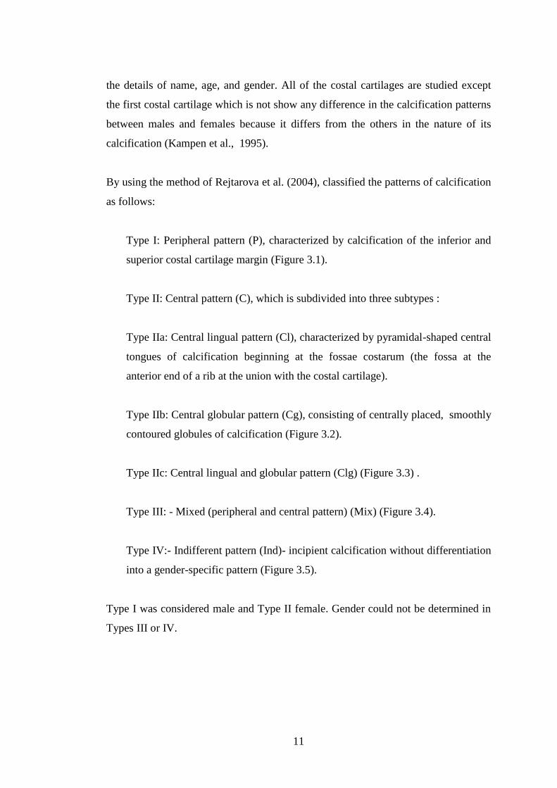
the details of name, age, and gender. All of the costal cartilages are studied except
the first costal cartilage which is not show any difference in the calcification patterns
between males and females because it differs from the others in the nature of its
calcification (Kampen et al., 1995).
By using the method of Rejtarova et al. (2004), classified the patterns of calcification
as follows:
Type I: Peripheral pattern (P), characterized by calcification of the inferior and
superior costal cartilage margin (Figure 3.1).
Type II: Central pattern (C), which is subdivided into three subtypes :
Type IIa: Central lingual pattern (Cl), characterized by pyramidal-shaped central
tongues of calcification beginning at the fossae costarum (the fossa at the
anterior end of a rib at the union with the costal cartilage).
Type IIb: Central globular pattern (Cg), consisting of centrally placed, smoothly
contoured globules of calcification (Figure 3.2).
Type IIc: Central lingual and globular pattern (Clg) (Figure 3.3) .
Type III: - Mixed (peripheral and central pattern) (Mix) (Figure 3.4).
Type IV:- Indifferent pattern (Ind)- incipient calcification without differentiation
into a gender-specific pattern (Figure 3.5).
Type I was considered male and Type II female. Gender could not be determined in
Types III or IV.

12
Figure 3.1. Peripheral pattern of costal cartilage calcification (Type I).
Figure 3.2. Central globular pattern of costal cartilage calcification (Type IIb).

13
Figure 3.3. Central lingual and globular pattern of costal cartilage calcification (Type
IIc).
Figure 3.4. Mixed (peripheral and central pattern) Type III.

14
Figure 3.5. Indifferent pattern of costal cartilage calcification (Type IV).
Microsoft Excel (2010) was used for data analysis, which included making tables,
distributing the numbers of cases according to the previously mentioned calcification
patterns, and extracting the percentages for them.

15

16
PART 4
RESULTS
Our study applied to 200 individuals (100 female, 100 male) in the 20-60 age group
in order to estimate the gender by calculating the number and percentage of the
individuals with each costal cartilage calcification pattern. The mean age of the
female was 41.12 ± 10.60, and the mean age of the male was 35.39 ± 9.22. Ages of
male and female individuals were tested with the Anderson Darling test, which is a
normality test, and it was found that they did not show normal distribution. With the
Mann-Whitney U test, a significant difference was found between men and women
according to age (p≤0.05),(Table 4.1).
Table 4.1. The mean age of female and male.
Gender Median Minimum Maximum P Value*
Female 39.00 20 60 0.0001
Male 34.50 20 60
*Mann-Whitney U Test
The results showed the number of individuals with costal cartilage calcification
(CCC) was 193, in which 97 females and 96 males, while found only 7 individuals
without any calcification in their costal cartilages, in which 3 females and 4 males
(Table 4.2).

17
Table 4.2. Number of individuals with CCC in female and male.
Gender Negative Positive Total
Female 3 97 100
Male 4 96 100
Total 7 193 200
Positive: with calcification, Negative: without calcification
The percentage of individuals with costal cartilage calcification was 96.5%, while
found 3.5% of individuals without calcification (Figure 4.1).
Figure 4.1. Percentage of individuals with CCC (Positive: with calcification,
Negative: without calcification).
By using the method of Rejtarova et al. (2004), the patterns of costal cartilage
calcification classified into four main types: peripheral pattern (P) (Type I) which
characterized by calcification of the superior and inferior margin of costal cartilage,
central pattern (C) (Type II) which characterized by a lingual or globular pattern of

18
calcification, mixed pattern (Mix) (Type III) which represent peripheral and central
pattern, and Indifferent pattern (Ind) (Type IV) incipient calcification without
differentiation into a gender-specific pattern.
The results showed the number of individuals with peripheral pattern (P) (Type I)
was 6 males, while not found the peripheral pattern in females. The number of
individuals with central pattern (C) (Type II) was 39, in which 36 females and 3
males. The number of individuals with mixed pattern (Mix) (Type III) was 129, in
which 46 females and 83 males. The number of individuals with indifferent pattern
(Ind) (Type IV) was 19, in which 15 females and 4 males (Table 4.3).
Table 4.3. Number of of each CCC pattern in female and male.
Gender Ind P C Mix Total
Female 15 0 36 46 97
Male 4 6 3 83 96
Total 19 6 39 129 193
P: Peripheral pattern, C: Central pattern, Mix: Mixed pattern,
Ind: Indifferent pattern.
The percentage of individuals with peripheral pattern (P) (Type I) was 100% in
males and 0% in females. The percentage of individuals with central pattern (C)
(Type II) was 92.3% in females and 7.7% in males. The percentage of individuals
with mixed pattern (Mix) (Type III) was 35.7% in females and 64.3% in males. The
percentage of individuals with indifferent pattern (Ind) (Type IV) was 78.9% in
females and 21.1% in males (Figure 4.2).

19
Figure 4.2. Percentage of each CCC pattern in female and male (P: Peripheral
pattern, C: Central pattern, Mix: Mixed pattern, Ind: Indifferent
pattern).
By using the method of Rejtarova et al. (2004), the central pattern (Type II) is
subdivided into three subtypes: central lingual pattern (Cl) (Type IIa), central
globular pattern (Cg) (Type IIb), and central lingual and globular pattern (Clg) (Type
IIc).
The results showed the number of individuals with central globular pattern (Cg)
(Type IIb) was 14, in which 13 females and one male. The number of individuals
with central lingual and globular pattern (Clg) (Type IIc) was 25, in which 23
females and two males. While not found the central lingual pattern (Cl) (Type IIa) in
males and females (Table 4.4).
0
20
40
60
80
100
Ind P C Mix
Female
Male

20
Table 4.4. Number of each central pattern subtype in female and male
Gender Cl Cg Clg Total
Female 0 13 23 36
Male 0 1 2 3
Total 0 14 25 39
Cl: Central lingual pattern, Cg: Central globular pattern, Clg: Central lingual
and globular pattern.
The percentage of individuals with central globular pattern (Cg) (Type IIb) was
92.9% in females and 7.1% in males. The percentage of individuals with central
lingual and globular pattern (Clg) (Type IIc) was 92% in females and 8% in males.
While the percentage of central lingual pattern (Cl) (Type IIa) was 0% in males and
females (Figure 4.3).
Figure 4.3. Percentage of each central pattern subtype in female and male (Cl:
Central lingual pattern, Cg: Central globular pattern, Clg: Central
lingual and globular pattern).
.
0
20
40
60
80
100
Cl Cg Clg
Female
Male

21
PART 5
DISCUSSION
The importance of human identification processes in forensic medicine is constantly
growing with time. Because there is increasing in the occurrence of natural disasters
like earthquakes, floods, and fires, and also due to mass disasters like accidents of a
plane, train and maritime, wars, explosions and acts of terror, and can be also due to
the heightened importance of security measures (Demir, 2014).
The most accurate and reliable technique for human identification is DNA analysis
which is commonly used for Disaster Victim Identification (DVI) or in the
identification of human remains found in mass graves. But this technique needs
forensic laboratories and high cost, for this reasons have limited their diffusion on a
large scale and preferred the traditional techniques more than it (Baraybar, 2008).
Gender estimation represents one of the most important components of the biological
identity that used in the traditional techniques for human identification from skeletal
remains (Vacca and Vella, 2012).
Between various methods for estimation gender, the radiological study of costal
cartilage calcification pattern is a simple, rapid, and inexpensive method, requiring
little expertise. It could best method for estimation the gender in all cases requiring
gender establishment in the living or in the dead, provided the thoracic cage is intact
(Nageshkumar and Lakshman, 1988).
Previous studies have used the difference in the calcification patterns that appeared
in the costal cartilage for gender estimation, like the study that applied to the Czech
population in (2004) and take 1044 chest and abdominal radiograms from the
Department of Radiology, Charles University Hospital in Hradec Králové in the

22
period (1995–2003), with age ranging from 10 to 95 years and included 537 male
and 507 female. In this study, the first costal cartilage was ignored and used a
method that classifies the patterns of calcification into main four groups (peripheral
pattern, central pattern, mixed pattern, and indifferent pattern), and subdivided the
central group into three further subgroups (central lingual pattern, central globular
pattern, central lingual and globular pattern). The result of this study found
calcification of the costal cartilage in 528 (51%) individuals and not found any
calcification in 516 (49%) individuals. All the types of patterns were found in the
individuals with costal cartilage calcification as the following: peripheral pattern
found in 249 individuals of males and found in one individual of females, central
pattern found in 201 individuals of females and not found any central pattern in
males, mixed pattern found in 23 individuals of females and found in 4 individuals of
males, indifferent pattern found in 35 individuals of females and found in 15
individuals of males. All the subtypes of central pattern found in this study as the
following: central lingual pattern found in 114 (57%) individuals, central globular
pattern found in 47 (23%) individuals, central lingual and globular pattern found in
40 (20%) individuals. At the end of the results, considered that the central pattern for
female with 100% probability and peripheral pattern for male with 99.6%
probability, while considered the gender could not be determined in Types III or IV.
In comparison with our study, the estimation of gender from the patterns of costal
cartilage calcification was applied to CT images of 200 individuals (100 female and
100 male) that obtained from the Picture Archiving and Communication System
(PACS) of Karabük University Karabük Training and Research Hospital, with age
ranging from 20 to 60 years. By using the method of Rejtarova et al. (2004), the
result showed 193 (96.5%) individuals with calcification in the costal cartilages and
7 (3.5%) individuals without calcification in their costal cartilages. All types of
calcification patterns are found in our study. Peripheral pattern (Type I) was found in
6 males, while not found the peripheral pattern in females. Central pattern (Type II)
found in 39 individuals, in which 36 females and 3 males. Mixed pattern (Type III)
was found in 129 individuals, in which 46 females and 83 males. Indifferent pattern
(Type IV) was found in 19 individuals, in which 15 females and 4 males. Not all the
subtypes of central pattern found in our study. Central globular pattern (Type IIb)

23
was found in 14 individuals, in which 13 females and one male. Central lingual and
globular pattern (Type IIc) was found in 25 individuals, in which 23 females and two
males. While not found the central lingual pattern (Cl) (Type IIa) in males and
females. At the end of our results, considered that the central pattern for female with
92.3% probability and peripheral pattern for male with 100% probability, while
considered the gender could not be determined in Types III or IV. On other hand, we
can accurately estimate the gender from the method of Rejtarova et al. (2004), and
the little difference in the percentage of accuracy of Type II female pattern between
our study and the previous study may be due to many reasons like the difference in
population samples that are studied, genetic and environmental factors that have a
strong influence on skeletal development, differences in age sample, or differences in
the radiological techniques that are used.
The other study that applied to the Scottish population and took 41 chest radiographs
from the Laboratory of Human Anatomy, University of Glasgow, between 2006 and
2010, included 19 females and 22 males, with ages ranging from 67–91 years for
female and 57–91 years for male. This study used the method of Rejtarova et al.
(2004), to classify the calcification patterns of costal cartilages and found 22
individuals (53.7%) in which the gender indeterminate and 19 individuals wherein
theory gender could be determined, 11 (57.9%) were correctly identified as female
while 8 (42.1%) were males wrongly identified as females. In comparison with our
study that applied to CT images of 200 individuals (100 female and 100 male) that
obtained from the Picture Archiving and Communication System (PACS) of
Karabük University Karabük Training and Research Hospital, with age ranging from
20 to 60 years. We found 148 (76.7%) individuals in which the gender indeterminate
and 45 (23.3%) individuals wherein theory gender could be determined, 6 were
correctly identified as male and 36 correctly identified as female, while 3 were males
wrongly identified as females. In the other words, our results were more accurate in
the estimation of gender than the previous study because from the total number of
individuals (45) wherein theory gender could be determined, 42 individuals were
correctly identified, 36 individuals as female and 6 individuals as male. While in the
previous study from the total number of individuals (19) wherein theory gender
could be determined, just 11 individuals were correctly identified as female.

24
PART 6
CONCLUSION AND RECOMMENDATIONS
After natural disasters, mass disasters, wars, explosions, and acts of terror, we need
to find simple, easy, accurate, and inexpensive technique for human identification.
the estimation of gender is one of the most important parameters for human
identification, so in our study suggested the estimation of gender from the patterns of
calcification that occur in costal cartilage by using the method of Rejtarova et al.
(2004). CT images of 200 individuals (100 female and 100 male) with age 20-60
were examined. The result showed 193 (96.5%) individuals with calcification in the
costal cartilages and 7 (3.5%) individuals without calcification in their costal
cartilages. Peripheral pattern (Type I) shown male gender estimation with 100% and
central pattern (Type II) shown female gender estimation with 92.3%, while
considered the gender could not be determined in Types III or IV.
We suggest using machine learning algorithms method for estimating gender from
the calcification patterns of costal cartilages.

25
REFERENCES
1. Adam, W. M., Richard, L., and Wayne Vogl, A., " Gray's anatomy for students
fourth ed.", Churchill Livingstone, an imprint of Elsevier Inc., Philadelphia,
127-151 (2020).
2. Afrianty, I., Nasien, D., Mohammed, R. A., and Haron, H., "Determination of
Gender from Pelvic Bones and Patella in Forensic Anthropology: A Comparison
of Classification Techniques," 1st International Conference on Artificial
Intelligence, Modelling and Simulation, Malaysia, 3-7 (2013).
3. Baraybar, J. P., "When DNA is Not Available, Can We Still Identify People?
Recommendations for Best Practice", Journal of Forensic Science
International, 53: 533-540 (2008).
4. Bertazzo, S., Gentleman, E., Cloyd, K. L., Chester, A. H., Yacoub, M. H.,
Stevens, and M. M., "Nano-analytical electron microscopy reveals fundamental
insights into human cardiovascular tissue calcification", Nature Materials, 12
(6): 576-583 (2013).
5. Blau, S., and Hill, T., "Disaster victim identification: a review", Minerva
Medicolegale, 129: 35-46 (2009).
6. Cattaneo, C., and Baccino, E., "A call for forensic anthropology in Europe",
International Journal of Legal Medicine, 116: 1-2 (2002).
7. Demir, S., "Klivus ve foramen magnum ölçüleri ile posterior kranial fossa
hacminin temporal BT görüntüleme aracılığıyla cinsiyet tahmininde
kullanılması ",Uzmanlık Tezi, Pamukkale Üniversitesi Tıp Fakültesi Adli Tıp
Anabilim Dalı,Denizli, 9-10 (2014).
8. Elkeles, A., "Sex differences in the calcification of the costal cartilages",
Journal of the American Geriatrics Society, 14 (5): 456-462 (1966).
9. Fischer, E., "Verkalkungsformen der Rippenknorpel", Fortschr der Rontgenstr
u Nuklearmed, 82: 479-481 (1955).
10. Freyschmidt, J., Brossmann, J., and Czerny, C., "Schultergurtel und Brustkorb",
Grenzen des Normalen und Anfange des Pathologischen in der Radiologie des
Skeletts, 3: 352 (2001).

26
11. Giurazza, F., Schena, E., Del Vescovo, R., Cazzato, R. L., Mortato, L.,
Saccomandi, P., and Zobel, B., "Sex determination from scapular length
measurements by CTscans images in a Caucasian population", Conference
proceedings IEEE Engineering in Medicine and Biology Society, 35: 1632-
1635 (2013).
12. Gupta, D. K., and Mathur, A. S., "Influence of sex on patterns of costal cartilage
calcification", Indian Journal of Chest Diseases and Allied Sciences, 20: 130-
134 (1978).
13. Hans, H., Soren, B., Tania, D., and Lucina, H., "The role of forensic
anthropology in disaster victim identification (DVI): recent developments and
futureprospects", Forensic Sciences Research, 4: 303-315 (2019).
14. Harold E., and Vishy M., "Clinical Anatomy Applied Anatomy for Students and
Junior Doctors 3rd ed.", John Wiley & Sons, United kingdom, 10-11 (2013).
15. Horner, J. L., "Premature calcification on the costal cartilage, its frequent
association with symptoms of non organic origin", The American Journal of
the Medical Sciences, 218: 186-93 (1949).
16. Inoi, T., "Estimation of sex and age by calcification pattern of costal cartilage in
Japanese", Nihon Hoigaku Zasshi, 51)2(: 89-94 (1997).
17. Kampen, W. U., Claassen, H., and Kirsch, T., "Mineralization and osteogenesis
in human first rib cartilage", Annals of Anatomy journal, 177: 177-77 (1995).
18. King, J. B., "Calcification of the costal cartilage", The British Journal of
Radiology, 12: 2-12 (1939).
19. Klepinger, L. L., "Fundamentals of Forensic Anthropology", John Wiley &
Sons, (2006).
20. Komar, D. A., and Buikstra, J. E., "Forensic Anthropology: Contemporary
Theory and Practice", Oxford University Press (2008).
21. Kyung Won Chung, PH. D., "Gross Anatomy 5th ed. ", Lippincott Williams &
Wilkins, United States of America, 141-143 (2005).
22. Lau, A., Kindig, M., and Kent, R., "Morphology, distribution, mineral density
and volume fraction of human calcified costal cartilage", Acta Biomaterialia
journal, 7(3): 1202‐ 1209 (2011).
23. Lynnerup, N., Thomsen, I., and Frøhlich, B., "A Non-invasive Method for Age at
Death Determination", Medicine, Science and the Law journal, 30(4): 317-20
(1990).

27
24. McCormick, W. F., "Mieralisation of Costa1 cartilage as an indicator of Age:
Preliminary observations", Journal of Forensic Science International, 25: 736
- 741 (1980).
25. Moore, K. L., Dalley, A. F., and Agur, A. M. R., "Clinically Oriented Anatomy
7th ed.", Lippincott Williams and Wilkins, Philadelphia (2014).
26. More, C. B., Vijayvargiya, R., and Saha, N., "Morphometric analysis of
mandibular ramus for sex determination on digital orthopantomogram", Journal
of Forensic Dental Sciences, 9, 1-5 (2017).
27. Nageshkumar, G. R., and Lakshman, M. P., " Costal cartilage calcification
pattern - a clue forestablishing sex identity", Forensic Science International,
38: 193-292 (1988).
28. Navani, S., Shah, J. R., and Levy, P. S., "Determination of sex by costal cartilage
calcification", The American Journal of Roentgenology Radium, 108: 771–774
(1970).
29. Neha, B., Kewal, K., and Tanuj, K., "The development and status of forensic
anthropology in India: A review of the literature and future directions",
Medicine, Science and the Law, 59(1): 61–69 (2019).
30. Nishino, K., "Studies on the human rib cartilage", Kekkaku, 44: 131–37 (1969).
31. Nurazzura, M. D., Dewan, M. H., Sohela, M., and Noor, S. R., " An Overview of
the Anthropological Theories", International Journal of Humanities and
Social Science, 10(1): 155-164 (2014).
32. Ontell, F. K., Moore E. H., Shepard J. A., and Shelton D. K., "The costal
cartilages in health and disease", Radiographics, 17(3): 571–577 (1997).
33. Rainio, J., Hedman,M., Karkola, K., Lalu, K., Peltola, P., Ranta, H. Sajantila, A.,
Söderholm, N., and Penttilä, A., "Forensic osteological investigations in
Kosovo", Journal of Forensic Science International, 121: 166–173 (2001).
34. Rao, N. G., and Pai, L. M., "Costal cartilage calcification pattern - a clue for
establishing sex identity", Journal of Forensic Science International, 38(3-4):
193–202 (1988).
35. Rejtarova, O., Slizova, D., Smoranc, P., Rejtar, P., and Bukac, J., "Costal
cartilages - A clue for determination of sex", Biomedical papers of the Medical
Faculty of the University Palacky, Olomouc, Czech Republic, 148: 241–243
(2004).
36. Reppien, K., Sejrsen, B., and Lynnerup, N., "Evaluation of post-mortem
estimated dental age versus real age: A retrospective 21-year survey", Journal
of Forensic Science International, 159: 84-88 (2006).

28
37. Richard, L., Wayne Vogl, A., and Adam, W. M., "Gray’s Atlas Of Anatomy 2nd
ed.", Churchill Livingstone, an imprint of Elsevier Inc., Philadelphia, 67
(2015).
38. Richardson, E. R., and Malhotra, S. K., "Mesiodistal crown dimension of the
permanent dentition of American Negroes", American Journal of Orthodontics
and Dentofacial Orthopedics, 68: 157-164 (1975).
39. Saccucci, M., and Cipriani, F. et al., "Gender assessment through three-
dimensional analysis of maxillary sinuses by means of Cone Beam Computed
Tomography", European Review for Medical and Pharmacological Sciences,
19: 185-193 (2015).
40. Scheuer, L., "Application of osteology to forensic medicine", Journal of Clinical
Anatomy, 15: 297–312 (2002).
41. Sidhu, R., Chandra, S., Devi, P., Taneja, N., Sah, K., and Kaur, N., "Forensic
importance of maxillary sinus in gender determination", European Journal of
General Dentistry, 3(1): 53-56 (2014).
42. Slaus, M., Strinoviˇc, D., Pe´cina- ˘ Slaus, N., Brki´c, H., Baliˇcevi´c, D.,
Petroveˇcki, V., and Pe´cina, T.C., "Identification and analysis of human
remains recovered from wells from the 1991 War in Croatia", Journal of
Forensic Science International, 171: 37–43 (2007).
43. Snell, Richard S., "Clinical Anatomy By Regions", Ninth edition, Lippincott
Williams & Wilkins, USA (2012).
44. Sobotta J., “İnsan anatomisi atlası: Baş, boyun, üst ekstremite: türkçesini
hazırlayan: A. Elhan. Cilt 1”, Beta Basım Yayım Dağıtım, (2006).
45. Stewart, J. H., and McCormick, W. F., "A sex‐ and age‐ limited ossification
pattern in human costal cartilages", American Journal of Clinical Pathology,
81(6): 765‐ 769 (1984).
46. Stover, E., Haglund, W. D., and Samuels, M., "Exhumation of mass graves in
Iraq: Considerations for forensic investigations, humanitarian needs, and the
demands of justice", Journal of the American Medical Association, 290: 663–
666 (2003).
47. Teale, C., Romaniuk, C., and Mulley, G., "Calcification on chest radiographs:
The Association with age", Oxford Journals – Medicine Age and Ageing,
18(5): 333- 336 (1989).
48. Vacca, E., and Vella, G. Di., "Metric characterization of the human coxal bone
on a recent Italian sample and multivariate discriminant analysis to determine
sex", Journal of Forensic Science International, 222(1-3): 401-409 (2012).

29
49. Vastine, J. H., Vastine, M. F., and Arango, O., "Genetic influence on osseous
development with particular reference to the deposition of calcium in costal
cartilages", American Journal of Roentgenology, 59: 213–217 (1948).
50. Verma, G. L., Agerwal, G. R., and Hiran, S., "Sex determination by costal
cartilage calcification", International Journal of Radiation Research, 34: 22–
25 (1980).
51. Wen, Y., Xiaoning, L., Kegang, W., Jiabei, H., Guohua, G., Jun, F., "Sex
Determination of Three-Dimensional Skull Based on Improved Backpropagation
Neural Network", Computational and Mathematical Methods in Medicine, vol.
2019: 1-8 (2019).
52. Zaidi, A. N., Ceneviva, G. D., Phipps, L. M., Dettorre, M. D., Mart, C. R., and
Thomas, N. J., "Myocardial calcification caused by secondary
hyperparathyroidism due to dietary deficiency of calcium and vitamin D",
Journal of Pediatric Cardiology, 26: 460–463 (2005).
53. Zeyfeoğlu, Y., and Hancı, İ. H., "İnsanlarda Kimlik Tespiti", Sürekli Tıp Eğitim
Dergisi, 10(10): 375-377 (2001).
54. Zimmerman, R. A., "Age-Related Incidence of Pineal Calcification Detected by
Computed Tomography", Radiological Society of North America, 142 (3): 659-
62 (1982).

30
APPENDIX

31

32
RESUME
Albaraa AL-SAMANEE, he completed his primary and secondary education in
Mosul. In 2007, he entered Ninevah College of Medicine. In 2014, he started
working as a general doctor in Ibn-alatheer hospital. He worked in the burn center
and plastic surgery in al-jumhory hospital.
Related Documents