221 e Chest Respiratory Physiology Preoperative Assessment Pulmonary Function Tests Perioperative Etiology and Epidemiology Upper Respiratory Tract Infection Lower Airway Disease Cystic Fibrosis Sickle Cell Disease Summary Essentials of Pulmonology Paul G. Firth and Kenan E. Haver MAINTENANCE OF ADEQUATE GAS exchange and delivery is a fundamental goal of anesthesia. Although the lungs play an important role in acid-base balance, temperature regulation, metabolism, and endocrine signaling, the preservation of oxygen and carbon dioxide (CO 2 ) equilibrium is the principal pulmo- nary function of immediate concern to the anesthesiologist. Respiratory problems are common in children and are fre- quently encountered by anesthesiologists during perioperative consultations, intraoperatively, or in intensive care units. Prob- lems range from mild acute respiratory tract infections to chronic lung disease with end-stage respiratory failure. In this chapter we discuss the basics of respiratory physiology, assess- ment of pulmonary function, and practical anesthetic manage- ment of specific pulmonary problems. Airway and thoracic aspects pertinent to ventilation are discussed in Chapters 12 and 13, whereas pulmonary issues specific to neonates, intensive care, or other disease states are addressed in the relevant chapters. Respiratory Physiology e morphologic development of the lung begins several weeks into the embryonic period and continues into the first decade and beyond postnatal life. 1 Intrauterine gas exchange occurs via the placenta, but the respiratory system develops in preparation for extrauterine life when gas exchange transfers abruptly to the lungs. e respiratory system is an outgrowth of the ventral wall of the foregut. During the embryonic period of development of the first few postconceptual weeks, lung buds form as a projec- tion of the endodermal tissue. During the pseudoglandular period, extending to the 17th week of life, rapid lung growth is accompanied by formation of the bronchi and branching of the airways down to the terminal bronchioli. Further development of bronchioli and vascularization of the airways occurs during the canalicular stage of the second trimester. e saccular stage begins at approximately 24 weeks, when terminal air sacs begin to form. Proliferation of capillary networks surrounding these air spaces become sufficient for pulmonary gas exchange by 26 to 28 weeks, when extrauterine survival of premature neonates becomes possible. Formation of alveoli occurs by lengthening of the saccules and thinning of the saccular walls and has begun by the 36th postconceptual week in most human fetuses (Fig. 11-1). e vast majority of alveolar formation occurs after birth, however, typically continuing to as late as 8 to 10 years postna- tally. e neonatal lung at birth commonly has 10 to 20 million terminal air sacs (many of which are saccules rather than alveoli), which is one tenth of the number in the mature adult lung. Growth of the lungs after birth is primarily due to an increase in the number of respiratory bronchioles and alveoli and not due to an increase in the size of the alveoli. Respiratory rhythmogenesis, as seen by rhythmic thoracic movement, begins well before birth and may be necessary for normal anatomic and physiologic lung development. At birth, interruption of umbilical blood flow initiates rhythmic breath- ing. Amniotic fluid is expelled from the lungs via the upper airways with the first few breaths, with residual fluid draining through the lymphatic and pulmonary channels in the first days of life. Changes in Po 2 , Pco 2 , and pH cause an acute decrease in pulmonary vascular resistance and a consequent increase in pulmonary blood flow. Increased left atrial and decreased right atrial pressure reverse the pressure gradient across the foramen ovale, causing functional closure of this left-to-right one-way flap valve. Expansion of the lungs combined with increased pulmonary blood flow initiates the abrupt transition to SECTION III CHAPTER 11

Welcome message from author
This document is posted to help you gain knowledge. Please leave a comment to let me know what you think about it! Share it to your friends and learn new things together.
Transcript

221
Th e Chest
Respiratory Physiology
Preoperative Assessment
Pulmonary Function Tests
Perioperative Etiology and Epidemiology
Upper Respiratory Tract Infection
Lower Airway Disease
Cystic Fibrosis
Sickle Cell Disease
Summary
Essentials of PulmonologyPaul G. Firth and Kenan E. Haver
MAINTENANCE OF ADEQUATE GAS exchange and delivery is a
fundamental goal of anesthesia. Although the lungs play an
important role in acid-base balance, temperature regulation,
metabolism, and endocrine signaling, the preservation of oxygen
and carbon dioxide (CO2) equilibrium is the principal pulmo-
nary function of immediate concern to the anesthesiologist.
Respiratory problems are common in children and are fre-
quently encountered by anesthesiologists during perioperative
consultations, intraoperatively, or in intensive care units. Prob-
lems range from mild acute respiratory tract infections to
chronic lung disease with end-stage respiratory failure. In this
chapter we discuss the basics of respiratory physiology, assess-
ment of pulmonary function, and practical anesthetic manage-
ment of specifi c pulmonary problems. Airway and thoracic
aspects pertinent to ventilation are discussed in Chapters 12 and
13, whereas pulmonary issues specifi c to neonates, intensive
care, or other disease states are addressed in the relevant
chapters.
Respiratory PhysiologyTh e morphologic development of the lung begins several weeks
into the embryonic period and continues into the fi rst decade
and beyond postnatal life.1 Intrauterine gas exchange occurs via
the placenta, but the respiratory system develops in preparation
for extrauterine life when gas exchange transfers abruptly to the
lungs. Th e respiratory system is an outgrowth of the ventral wall
of the foregut. During the embryonic period of development of
the fi rst few postconceptual weeks, lung buds form as a projec-
tion of the endodermal tissue. During the pseudoglandular
period, extending to the 17th week of life, rapid lung growth is
accompanied by formation of the bronchi and branching of the
airways down to the terminal bronchioli. Further development
of bronchioli and vascularization of the airways occurs during
the canalicular stage of the second trimester. Th e saccular stage
begins at approximately 24 weeks, when terminal air sacs begin
to form. Proliferation of capillary networks surrounding these
air spaces become suffi cient for pulmonary gas exchange by 26
to 28 weeks, when extrauterine survival of premature neonates
becomes possible. Formation of alveoli occurs by lengthening of
the saccules and thinning of the saccular walls and has begun
by the 36th postconceptual week in most human fetuses (Fig.
11-1). Th e vast majority of alveolar formation occurs after birth,
however, typically continuing to as late as 8 to 10 years postna-
tally. Th e neonatal lung at birth commonly has 10 to 20 million
terminal air sacs (many of which are saccules rather than alveoli),
which is one tenth of the number in the mature adult lung.
Growth of the lungs after birth is primarily due to an increase
in the number of respiratory bronchioles and alveoli and not
due to an increase in the size of the alveoli.
Respiratory rhythmogenesis, as seen by rhythmic thoracic
movement, begins well before birth and may be necessary for
normal anatomic and physiologic lung development. At birth,
interruption of umbilical blood fl ow initiates rhythmic breath-
ing. Amniotic fl uid is expelled from the lungs via the upper
airways with the fi rst few breaths, with residual fl uid draining
through the lymphatic and pulmonary channels in the fi rst days
of life. Changes in Po2, Pco2, and pH cause an acute decrease
in pulmonary vascular resistance and a consequent increase in
pulmonary blood fl ow. Increased left atrial and decreased right
atrial pressure reverse the pressure gradient across the foramen
ovale, causing functional closure of this left-to-right one-way
fl ap valve. Expansion of the lungs combined with increased
pulmonary blood fl ow initiates the abrupt transition to
SECTION IIICHAPTER 11

A Practice of Anesthesia for Infants and Children
222
extrauterine gas exchange. Continuous increased arterial oxygen
levels following birth, in comparison with intrauterine levels,
augment and maintain ventilatory rhythm.
Breathing is controlled by a complex interaction of input
from sensors, integration by a central control system, and sub-
sequent output to eff ector muscles.2 Aff erent signaling is pro-
vided by peripheral arterial chemoreceptors, central brainstem
chemoreceptors, upper airway and intrapulmonary receptors,
and chest wall and muscle mechanoreceptors. Th e peripheral
arterial chemoreceptors consist of the carotid and aortic bodies,
with the carotid bodies playing the greater role in arterial chemi-
cal sensing in humans. Th e carotid bodies sense both Pao2 and
hydrogen ion concentration. Th e central chemoreceptors are
responsive to Paco2 and hydrogen ion concentration and are
thought to be located at or near the ventral surface of the
medulla. Th e nose, pharynx, and larynx have a wide variety of
pressure, chemical, temperature, and fl ow receptors that can
cause apnea, coughing, or changes in ventilatory pattern. Pul-
monary receptors lie in the airways and lung parenchyma. Th e
airway receptors are subdivided into the slowly adapting recep-
tors, or pulmonary stretch receptors, and the rapidly adapting
receptors. Th e stretch receptors, found in the airway smooth
muscle, are thought to be involved in the balance of inspiration
and expiration. Th e rapidly adapting receptors lie between the
airway epithelial cells and are triggered by noxious stimuli such
as smoke, dust, and histamine. Parenchymal receptors, or jux-
tacapillary receptors, are located adjacent to the alveolar blood
vessels; they respond to hyperinfl ation of the lungs, to various
chemical stimuli in the pulmonary circulation, and possibly to
interstitial congestion. Chest wall receptors include mechano-
receptors and joint proprioreceptors. Mechanoreceptors in the
muscle spindle endings and tendons of respiratory muscles
sense changes in length, tension, and movement.
Central control of respiration is maintained by the brainstem
(involuntary) and cortical (voluntary) centers. Although the
precise mechanism of the neural ventilatory rhythmogenesis is
unknown, the pre-Bötzinger complex and retrotrapezoid
nucleus/parafacial respiratory group, neural circuits in the ven-
trolateral medulla, are thought to be the respiratory rhythm
generators.3 Th ese neuron groups fi re in an oscillating pattern,
an inherent rhythm that is moderated by inputs from other
respiratory centers. Involuntary integration of sensory input
occurs in various respiratory nuclei and neural complexes in the
pons and medulla, which modify the baseline pacemaker fi ring
of the respiratory rhythm generators. Th e cerebral cortex also
aff ects breathing rhythm and infl uences or overrides involun-
tary rhythm generation in response to conscious or subcon-
scious activity, such as emotion, arousal, pain, speech, breath
holding, and other activities.2
Th e eff ectors of ventilation include the neural eff erent path-
ways, the muscles of respiration, the bones and cartilage of the
chest wall and airway, and elastic connective tissue. Upper
airway patency is maintained by connective tissue and by sus-
tained and cyclical contraction of the pharyngeal dilator muscles.
Th e diaphragm produces the majority of tidal volume during
quiet inspiration, with the intercostal, abdominal, and accessory
muscles (sternocleidomastoid and neck muscles) providing
additional negative pressure. Th e elastic recoil of the lungs and
thorax produces expiration, with inspiration an active, and expi-
ration a passive action in normal lungs during quiet breathing.
During vigorous breathing or with airway obstruction, both
inspiration and expiration become active processes.
FertilizationWeeks Months
YearsAge
Birth
10 20 30 3 6 9 1 2 3 4 5 6 7
Lung growthLung development
Embryonic period
Pseudoglandular stage
Canalicular stage
Saccular stage
Alveolar stage
Microvascularmaturation
Normal growth period
Figure 11-1. Timetable for lung development. (Reproduced and modifi ed with permission from Guttentag S, Ballard PL: Lung development: embryology, growth, maturation, and developmental biology. In Tausch HW, Ballard RA, Gleason CA [eds]: Avery’s Diseases of the Newborn, 8th ed. Philadelphia, WB Saunders, 2004, p 602.)

Essentials of Pulmonology
223
11Preoperative AssessmentTh e preoperative assessment of the respiratory system in a child
is based on history, physical examination, and evaluation of vital
signs. Further investigations, such as laboratory, radiographic,
and pulmonary function studies, may be indicated if there is
doubt as to the diagnosis or severity of the pulmonary disease.
Because ventilation is a complex process involving many systems
beyond the lung, preoperative pulmonary appraisal must include
airway, musculoskeletal, and neurologic assessment that might
impact gas exchange under anesthesia or in the postoperative
period. Th e potential impact of esophageal refl ux, cardiac,
hepatic, renal, or hematologic disease on gas exchange and pul-
monary function should also be considered. Th e child’s reaction
to the presence and approach of medical staff may deter-
mine the order and even the position of the history and
examination.
Th e history should establish the current respiratory status,
the presence of chronic and present status of pulmonary disease.
Because children may be unwilling or unable to give a reliable
history, parents or caregivers are often the sole or an important
supplemental source of information. Respiratory issues espe-
cially pertinent to the pediatric population include upper respi-
ratory tract infections (URIs), reactive airway disease/asthma,
ventilatory problems relating to prematurity, and congenital dis-
eases. Viral URIs are common in children, and the time, fre-
quency, and severity of infection should be established. Reactive
airway disease is also widespread in the pediatric population,
and the precipitants, frequency, severity, and relieving factors
should be determined. Chronic pulmonary diseases often have
a variable clinical course, and a history of acute exacerbations
of chronic problems should be elicited. Th e conceptual age at
birth, the current postconceptual age, neonatal respiratory dif-
fi culties, and prolonged intubation in the neonatal period are
particularly important to ascertain in the younger child because
subglottic stenosis and/or tracheomalacia are common sequelae.
Whereas congenital lesions may manifest at birth, symptoms of
airway obstruction may only become evident later in life.
Physical examination begins when you enter the room. Par-
ticularly with young children, your best opportunity to observe
them before they react to your presence is from across the room,
and inspection from a distance can provide useful information.
Respiratory rate is a sensitive marker of pulmonary problems,
and scrutiny before a young child becomes agitated and hyper-
ventilates is an important means of assessment. Pulse oximetry,
the “fi fth vital sign,” is a useful baseline indicator of oxygenation.
Nasal fl aring, intercostal retractions, and the marked use of
accessory respiratory muscles are all signs of respiratory dis-
tress. General appearance is also important. Apathy, anxiety,
agitation, or persistent adoption of a fi xed posture may indicate
profound respiratory or airway diffi culties, whereas intense cya-
nosis can also be detected from a distance. Weight may relate
to pulmonary function: patients with chronic severe pulmonary
disease are often underweight owing to retarded growth or
malnourishment, whereas severe obesity can produce airway
obstruction and sleep apnea. Inspection of the chest contour
may reveal hyperinfl ation or thoracic wall deformities.
Closer physical examination adds further information. Aus-
cultation may reveal wheezes, rales, fi ne or coarse crepitus,
transmitted breath sounds from the upper airway, altered breath
sounds, or cardiac murmurs. Chest percussion can provide an
estimate of the position of the diaphragm and serve as a useful
marker of hyperinfl ation. Patience, a gentle approach, and warm
hands will improve diagnostic yield and patient satisfaction.
Pulmonary Function TestsFurther investigations of pulmonary function include chest radi-
ography, measurement of hematocrit, arterial blood gas analysis,
pulmonary function tests, and sleep studies. Special investiga-
tions are not routinely indicated preoperatively and should be
reserved for times when the diagnosis is unclear, the progression
or treatment of a disease needs to be established, or the severity
of impairment is not evident. In most cases, a comprehensive
history and careful physical examination will be adequate to
establish an appropriate anesthetic plan. Before requesting a
new investigation, the clinician should have a clear idea of what
question is being asked and how the answer will modify anes-
thetic management and outcome.
Pulmonary function tests enable clinicians to (1) establish
mechanical dysfunction in children with respiratory symptoms,
(2) quantify the degree of dysfunction, and (3) defi ne the nature
of the dysfunction as obstructive, restrictive, or mixed obstruc-
tive and restrictive.4 Figure 11-2 illustrates a normal pulmonary
function test (normal fl ow-volume loop and spirometry param-
eters). Pulmonary function studies include dynamic studies,
measurement of static lung volumes, and diff using capacity. Th e
109876543210
–1–2–3–4–5–6–7–8–9
–10
Inha
latio
n
Flo
w (
L/se
c)
Volume (L)
ObservedPredicted
Exh
alat
ion
1 2 3 4 5 6 7 8 9 10
SpirometryParameterFVCFEV1FEV1/FVC
UnitsLiterLiterPercent
PredictedValue4.864.1986
ObservedPre5.284.6889
Percent Pred109112103
Figure 11-2. Normal pulmonary function test (broken line, predicted curve, solid line, measured curve). The normal fl ow-volume curve obtained during forced expiration rapidly ascends to the peak expiratory fl ow. Shortly after reaching the peak expiratory fl ow (highest point on curve), the curve descends with decreasing volume following a reproducible shape that is independent of effort. In this normal fl ow-volume curve, the FEV1/FVC, FEV1, and FVC are all within the normal range for this child’s age, height, gender and race. The shapes of both the inspiratory and expiratory limbs are normal as well. Pre, prebronchodilator; Pred, predicted.

A Practice of Anesthesia for Infants and Children
224
dynamic studies, which are the most commonly used, include
spirometry, fl ow-volume loops, and measurement of peak expi-
ratory fl ow. A reliable study requires patient eff ort and cooper-
ation, accurate testing equipment, as well as a skilled and
knowledgeable staff . Although the maneuvers needed to obtain
measurements appear straightforward, studies are valid only if
they refl ect maximal eff ort and are reproducible. Children older
than the age of 6 years can often perform adequately, although
children as young as age 3 years have been shown to be able to
perform spirometry with coaching. Th ere must be three accept-
able maneuvers and two studies need to have values for both
FVC and FEV1 that are within 0.15 L of each other before the
study can be accepted. Table 11-1 presents common indications
for pulmonary function testing in children.
An obstructive process is characterized by decreased velocity
of airfl ow through the airways (Fig. 11-3), whereas a restrictive
defect produces decreased lung volumes (Fig. 11-4). Examining
the ratio of airfl ow to lung volume assists in diff erentiating these
components of lung disease. Spirometry measures the volume
of air inspired and expired as a function of time and is by far the
most frequently performed test of pulmonary function in chil-
dren. With a forced maneuver, the volume exhaled from full
inhalation in the fi rst second is referred to as the forced expira-
tory volume in the fi rst second (FEV1). Th e maneuver is com-
pleted when the subject has completed exhaling as fast as
possible after a maximal inhalation. Th is is referred to as the
forced vital capacity (FVC). Normally, a child should be able to
exhale more than 80% of the total lung volume in the fi rst
Table 11-1. Uses of Pulmonary Function Studies in Children
• To establish pulmonary mechanical abnormality in children with respiratory symptoms
• To quantify the degree of dysfunction
• To defi ne the nature of pulmonary dysfunction (obstructive, restrictive, or mixed obstructive and restrictive)
• To aid in defi ning the site of airway obstruction as central or peripheral
• To differentiate fi xed from variable and intrathoracic from extrathoracic central airway obstruction
• To follow the course of pulmonary disease processes
• To assess the effect of therapeutic interventions and guide changes in therapy
• To detect increased airway reactivity
• To evaluate the risk of diagnostic and therapeutic procedures
• To monitor for pulmonary side effects of chemotherapy or radiation therapy
• To aid in prediction of the prognosis and quantitate pulmonary disability
• To investigate the effect of acute and chronic disease processes on lung growth
Modifi ed with permission from Castile R: Pulmonary function testing in children. In Chernick V, Boat TF, Wilmott RW, Bush A (eds): Kendig’s Disorders of the Respiratory Tract in Children, 7th ed. Philadelphia, Elsevier, 2006, p 168.
Figure 11-3. This fl ow-volume curve demonstrates a reversible obstructive defect. The forced expiratory volume over 1 second (FEV1) as a percentage of forced vital capacity (FVC), or total volume exhaled, is decreased in patients with airway obstruction. The pre-bronchodilator curve shape (blue) is scooped. After administration of a short-acting bronchodilator, the curve shape (brown) appears normal and there is an increase in both the FEV1/FVC and FEV1. This child has asthma and demonstrates a marked (40%) increase in FEV1 after treatment with a short-acting bronchodilator. Reversible airfl ow obstruction is one of the hallmarks of asthma. Post, postbronchodilator; Pre, prebronchodila-tor; Pred, predicted.
6
5
4
3
2
1
0
–1
–2
–3
–4
–5
–6
Inha
latio
nE
xhal
atio
nF
low
(L/
sec)
1 2 3 4 5 6
PrebronchodilatorPostbronchodilatorPredicted
SpirometryParameterFVCFEV1FEV1/FVC
UnitsLiterLiterPercent
PredictedValue1.941.7190
Pre2.151.3060
Observed PercentChange
04042
Post2.141.8285
Percent Pred11010694
Percent Pred1117667
Observed

Essentials of Pulmonology
225
11second of exhalation. Children with obstructive lung disease
have decreased air fl ow in relation to lung volume. If the volume
exhaled in the fi rst second divided by the volume of full exhala-
tion (FEV1/FVC) is less than 80%, then airway obstruction is
present (Table 11-2, see Fig. 11-3). Th e FEV1 needs to be inter-
preted in the context of the FVC. A low FEV1 itself is not suffi -
cient to make the diagnosis of airfl ow obstruction. Th ose with
restrictive lung disease have a decreased fl ow rate and reduced
total exhaled volume. Restrictive lung disease is associated with
a loss of lung tissue or a decrease in the lung’s ability to expand.
A restrictive defect is diagnosed when the FVC is less than 80%
of normal, with either a normal or an increased FEV1/FVC (see
Table 11-2 and Fig. 11-4).
Most children with respiratory problems have an obstructive
pattern, whereas isolated restrictive diseases are far less common.
Asthma is the most common obstructive pulmonary disease in
children. Rare causes of obstruction include airway lesions, con-
genital subglottic webs, or vocal cord dysfunction. Although the
diseases arise from specifi c isolated genetic disorders, children
with cystic fi brosis and sickle cell disease can have very variable
pulmonary pathologic processes, with both obstructive and
restrictive components of lung disease. Bronchopulmonary dys-
plasia may also have both obstructive and restrictive pathology.
Restrictive lung disease can arise from limitations to chest wall
movement such as chest wall deformities, scoliosis, or pleural
eff usions or from space-occupying intrathoracic processes such
as large bullae or congenital cysts. Alveolar fi lling defects, such
as lobar pneumonia, also reduce lung volume and can be con-
sidered as restrictive processes.
Additional uses of pulmonary function tests are as aids to
diff erentiate fi xed from variable obstruction and to locate the
obstruction as above or below the thoracic inlet (Figs. 11-5 to
11-7 [see website for Fig. 11-7B]). Th is information can be
gleaned from distinctive changes in the confi guration of fl ow-
volume loops, a graphic representation of inspiratory and expi-
ratory fl ow volumes plotted against time. Th e fl ow-volume loop
in Figure 11-2 is normal. A fi xed central airway obstruction,
such as a tumor or stenosis, may obstruct both inspiration and
expiration, fl attening the fl ow-volume curve both on inspiration
and expiration. Th e child with tracheal stenosis, for example,
has fl attening of both inhalation and exhalation curves (see Fig.
11-6). A variable obstruction will tend to aff ect one part of the
ventilatory cycle. When inhaling, the chest expands and draws
the airways open. On exhalation, as the chest collapses, the
intrathoracic airways collapse. Variable extrathoracic lesions
tend to obstruct on inhalation more than exhalation, whereas
intrathoracic lesions will have a more pronounced eff ect on
exhalation. Th is produces characteristic fl ow-volume patterns.
Spirometry cannot, however, provide data about absolute
lung volumes, because it measures the amount of air entering
or leaving the lung rather than the amount of air in the lung.
Th us, information about functional residual capacity (FRC) and
lung volumes calculated from FRC, such as total lung capacity
and residual volume, must be computed via diff erent means.
Th ese include gas dilution and body plethysmography. Gas dilu-
tion is based on measuring the dilution of nitrogen or helium in
a circuit in closed connection to the lungs, whereas body pleth-
ysmography calculates lung gas volumes based on changes in
thoracic pressures.
In addition to diagnostic uses, spirometry is used to assess
the indication for, and effi cacy of, treatment. For example, the
obstruction in those with asthma is usually reversible either
gradually over time without intervention or much more rapidly
after treatment with a short-acting bronchodilator. An improve-
ment in FEV1 of 12% and 200 mL is considered a positive
response. In addition to confi rming the diagnosis of asthma, the
degree of airfl ow obstruction, as indicated by the FEV1, is one
indication of asthma control. A low FEV1 or an acute decrease
from baseline may refl ect a child whose asthma is not under
good control and therefore who potentially is at greater risk for
a perioperative exacerbation (see Fig. 11-3).
Perioperative Etiology and EpidemiologyRespiratory problems account for the majority of perioperative
morbidity in children.5,6 Th e triggers of these problems include
Figure 11-4. Flow-volume curve demonstrating a restrictive defect (predicted, broken line; measured, solid blue line). The fl ow-volume curves in patients with restrictive defects appear to be near normal in confi guration but smaller in all dimensions. The FEV1/FVC ratio is normal, but both the FEV1 and FVC are reduced. The curve shape appears normal. This child has interstitial lung disease. Pred, predicted.
Table 11-2. Characteristics of Obstructive and Restrictive Patterns of Lung Disease
Measurement
Disease Category
Obstructive Restrictive
FVC Normal/decreased Decreased
FEV1 Decreased Decreased
FEV1/FVC Decreased Normal
FEV1, forced expiratory volume over 1 second; FVC, forced vital capacity.
SpirometryParameterFVCFEV1FEV1/FVC
UnitsLiterLiterPercent
Actual2.592.1483
Predicted3.343.0092
Percent pred777190
8
7
6
5
4
3
2
1
0
–1
–2
–3
–4
–5
–6
–7
–8
Inha
latio
nE
xhal
atio
n
Flo
w (
L/se
c)
1 2 3 4 5 6 7 8
ObservedPredicted
Volume (L)

A Practice of Anesthesia for Infants and Children
226
airway manipulation, the alteration of airway refl exes by anes-
thetic drugs, the surgical insult, and the depression of breathing
by anesthetic and analgesic medications. Adverse events
comprise laryngospasm, airway obstruction, bronchospasm,
hemoglobin oxygen desaturation, prolonged coughing,
atelectasis, pneumonia, and respiratory failure.7,8 Th e incidence
of respiratory problems in one study of 755 children was 21%
intraoperatively and 13% in the postanesthetic care unit.7
Various disease states can further aff ect the frequency of respi-
ratory complications in pediatric anesthesia.
Younger age of the child has consistently been found to be a
risk factor for respiratory morbidity.5,6,9,10 Th e neonate is particu-
larly sensitive to respiratory problems for multiple reasons.
Although the FRC approaches adult levels within days, a persis-
tently large closing capacity increases the likelihood of alveolar
collapse and intrapulmonary shunt. Residual patency of the
ductus arteriosus can contribute to shunting. Th e greater meta-
bolic rate increases oxygen requirements and decreases the time
to arterial desaturation after an interruption to ventilation and
gas exchange. Th e work of breathing is greater, consequently,
because of high-resistance, small-caliber airways, increased
chest wall compliance, and reduced lung compliance.
Upper Respiratory Tract InfectionUpper respiratory tract infections are a common problem
among young children, typically occurring as often as six to
eight times a year, with possibly an even greater incidence
among children in day care. Viruses cause the majority of URIs,
with rhinoviruses constituting approximately one third to one
Figure 11-5. Pulmonary function test demonstrating a nonreversible obstructive defect. The FEV1/FVC is decreased, as is the FEV1. After administration of a short-acting bronchodilator there is no signifi cant improvement in the FEV1, in contrast to the pattern in Figure 11-3. This child has cystic fi brosis with a nonreversible obstructive defect. Post, postbronchodilator; Pre, prebronchodilator; Pred, predicted.
Figure 11-6. Pulmonary function test showing an extrathoracic airway obstruction; both the inspiratory and expiratory limbs of the fl ow-volume curve are fl attened. This child has subglottic stenosis that developed at the site of her tracheotomy 2 years after the tracheostomy had been removed. Pred, predicted.
PrebronchodilatorPostbronchodilatorPredicted
8
7
6
5
4
3
2
1
0
–1
–2
–3
–4
–5
–6
–7
–8
Inha
latio
nE
xhal
atio
n
1 2 3 4 5 6 7 8
SpirometryParameterFVCFEV1FEV1/FVC
UnitsLiterLiterPercent
PredictedValue3.162.8291
ObservedPre2.501.5662
Percent Pred795568
Observed PercentChange
–2 0 3
Post2.451.5664
Percent Pred785570
4
3
2
1
0
–1
–2
–3
–4Volume (L)
1 432
SpirometryParameterFVCFEV1FEV1/FVC
UnitsLiterLiter
Percent
PredictedValue1.411.2291
ObservedPre1.190.8370
Percent Pred846877
Inha
latio
n
Flo
w (
L/se
c)
Exh
alat
ion
ObservedPredicted

Essentials of Pulmonology
227
11
Figure 11-7. A, Pulmonary function test from a child with an intrathoracic airway obstruction (vascular ring). The fl ow-volume curve shapes suggest a fi xed expiratory obstruction. The shape of the inspiratory link is normal; the expiratory fl ow limb is fl attened on both the pre-bronchodilator (brown) and post-bronchodilator (blue) fl ow-volume curves. B, (see website) A magnetic resonance angiogram accompanies the fl ow loop. C, Slit-like tracheal compression before repair. D, Note the marked improvement in the tracheal lumen after division of the vascular ring. (B courtesy of Brian O’Sullivan, MD; C and D courtesy of Christopher Hartnick, MD.) Post, postbronchodilator; Pre, prebron-chodilator; Pred, predicted.
half of etiologic species.11 Other common causative viruses
include adenovirus and coronavirus. Although most URIs are
short-lived, self-limiting infections, and by defi nition limited to
the upper airway, they may increase airway sensitivity to noxious
stimuli or secretions for several weeks after the infection. Th e
mechanisms probably involve a combination of mucosal inva-
sion, chemical mediators, and altered neurogenic refl exes.11
URIs may also impair pulmonary function by decreasing FVC,
FEV1, peak expiratory fl ow, and diff usion capacity.12,13 Th ese data
suggest that children with recent URIs may have an increased
incidence of perioperative pulmonary complications and, con-
sequently, may benefi t from a postponement of anesthesia.
Despite this suggestive evidence, the practical clinical conse-
quences are less well defi ned.14 In 1979, a case series of 11 chil-
dren noted signifi cant perioperative complications, including
atelectasis; all but 1 of the children had a reported respiratory
tract infection in the preceding month.15 Two postoperative
deaths in children with URIs have been reported16,17: in one case,
premature extubation and inadequate monitoring may have
been implicated,18 but, in the other, postmortem examination
suggested viral myocarditis.16 Larger studies, however, failed to
detect a high risk of major complications.19,20 Compared with
children without URIs, children with recent URIs desaturate
more quickly, but this typically responds rapidly to oxygen
administration and alveolar recruitment.21 Other specifi c com-
plications include bronchospasm and laryngospasm, breath
holding, arterial oxygen desaturations (Spo2 below 90%), and
severe coughing (Table 11-3).10,19,20,22 Although there appears to
6.0
0.0
–6.0
6.0Flo
w (
L/se
c)
Inha
latio
nE
xhal
atio
n
A
SpirometryParameterFVCFEV1FEV1/FVC
UnitsLiterLiterPercent
ObservedPrebron-chodilator
3.532.6374.7
Percent Pred10289
Observed PercentChange
3 0–3
Postbron-chodilator
3.632.6572.8
Percent Pred10690
C
D
Table 11-3 Incidence of Common Upper Respiratory Tract Infection–Associated Perioperative Adverse Events
Study
Laryngospasm (%) Bronchospasm (%) SpO2 (%)
URI No URI URI No URI URI No URI
Tait and Knight, 198787 1.3 1.2
Tait et al., 199888 7.3 12.2 [<90] 17.1
Tait et al., 200120 4.2 3.9 5.7 3.3 [<90] 15.7 7.8*
Cohen and Cameron, 19909 2.2 1.7
Levy et al., 199289 [<93] 63.6 59.0
DeSoto et al., 198890 [<95] 20.0 0*
Rolf and Coté, 199222 5.9 3.3 13.3 0.6* [<85] 13.3 10.5
*P < .05 versus corresponding URI group.Reproduced from Tait AR: Anesthetic management of the child with an upper respiratory tract infection. Curr Opin Anaesthesiol 2005; 18:603-607.

A Practice of Anesthesia for Infants and Children
228
be an increased risk of adverse perioperative events, there
is little residual morbidity; and most complications can be
predicted and successfully managed without long-term
sequelae.11,23
An approach to the problem of the child with a URI is to
detect the pathologic process and associated comorbidity, estab-
lish the acuity and severity of the URI, and then decide whether
to modify anesthetic technique or postpone surgery (Table 11-4,
Fig. 11-8). Th e presence of a current or recent URI should be
established, because this will alert the anesthesiologist to an
increased risk of perioperative problems. Th e basis of diagnosis
is a careful history and physical examination, with further inves-
tigations in limited situations. Because they are usually familiar
with their child’s state of health, the parents or caregivers can
provide helpful insight into the presence and severity of a URI.
Th e child should be evaluated for fever (defi ned as >38.5º C
[101.3º F]), change in demeanor or behavior, dyspnea, produc-
tive cough, sputum production, nasal congestion, rales, rhonchi,
and wheezing. A chest radiograph may be considered if the
pulmonary examination is questionable, but because the radio-
graphic changes lag behind clinical symptoms, it is typically of
limited value. Although laboratory tests may confi rm the diag-
nosis of a viral or bacterial URI, these are not cost eff ective or
practical in a busy surgical setting.
For children with symptoms of an uncomplicated URI who
are afebrile with clear secretions and who are otherwise healthy,
anesthesia may proceed as planned, because the problems
encountered are generally transient and easily managed.22 Elec-
tive surgery should be postponed for children with more severe
symptoms, such as mucopurulent secretions, productive cough,
pyrexia greater than 38º C (100.4º F), or pulmonary involvement.
Th ere are no defi nitive data to establish the exact duration of
postponement, but 3 to 4 weeks is suggested to be a prudent
timeframe, although airway reactivity may last for 6 to 8 weeks.11
Th is refl ects a balance between the time to diminish airway
hyperreactivity and perioperative risk and the incidence of URI
recurrence and need to perform the procedure. If bacterial
infection is suspected, appropriate antibiotics should be
prescribed.
Judgment about the suitability of a child for surgery becomes
more diffi cult when the symptoms of the URI lie somewhere
between the extremes of mild and severe. In these instances,
other considerations play a greater role in assessment of the
risk/benefi t ratio. Th ese include the presence of comorbid con-
ditions, such as asthma or cardiac disease, the type and urgency
of surgery, the age of the child and history of prematurity, and
the frequency of URIs. Th e comfort level and experience of the
anesthesiologist may also be important in the decision to
proceed with or postpone surgery. Th e need for postoperative
admission of a child after anesthetic complications or exacerba-
tion of the URI may expose other patients to a contagious illness.
Consideration must also be given to the complexity and antici-
pated duration of the surgery and implantation of foreign bodies
such as pins, rods, or other implants (see Chapter 4).
If the decision is to proceed with general anesthesia, manage-
ment is aimed at minimizing secretions and avoiding stimula-
tion of a potentially sensitized and infl amed airway. Use of an
endotracheal tube (ETT) should be avoided, if possible, because
it increases the risk of complications, especially in younger chil-
dren.20 Although airway management with a face mask is associ-
ated with the least incidence of complications, it may be
inappropriate for certain cases. Similarly, a laryngeal mask
Table 11-4. Risk Factors for Perioperative Adverse Events in Children with URIs
Study URI? Factors RR/OR
Parnis et al., 200119 URI and non-URI ETTChild has a “cold”Child snoresPassive smokerAnesthetic agentSputum productionAnticholinesterase givenNasal congestion
Tait et al., 200120 URI Copious secretions 3.9ETT in child <5 yr 1.9Prematurity (<37 wk) 2.3Nasal congestion 1.4Passive smoker 1.6Reactive airway disease 1.8Surgery of airway 1.8
Bordet et al., 200210 URI and non-URI Age <8 yr 1.8LMA 2.3Respiratory infections 3.7
Mamie et al., 20047 Non-URI Nonpediatric anesthesiologist 1.7ENT procedure 1.8ETT without relaxants 1.2
ENT, ear, nose, and throat; ETT, endotracheal tube; LMA, laryngeal mask airway; OR, odds ratio; RR, relative risk; URI, upper respiratory tract infection.Reproduced from Tait AR: Anesthetic management of the child with an upper respiratory tract infection. Curr Opin Anaesthesiol 2005; 18:603-607.

Essentials of Pulmonology
229
11
airway (LMA) is associated with fewer episodes of respiratory
events than an ETT, but its use may be contraindicated by the
type of surgical procedure and the need to protect the airway
from pulmonary aspiration of gastric contents. Whatever the
choice of airway management, it is essential that the depth of
anesthesia is adequate to obtund airway refl exes, particularly
during placement of an ETT or an LMA. Th e data on optimal
depth of anesthesia for removal of airway devices are equivocal.
Some studies in children with20 or without24 URIs found no dif-
ference in emergence complications between awake versus deep
extubation, whereas others found a greater incidence of arterial
oxygen desaturation or coughing on awake removal of ETT or
LMA in children with URIs.25,26
Because viral infections aff ect the nature and quantity of
secretions, and because copious secretions are identifi ed as risk
factors, the airway should be suctioned under deep anesthesia
to attempt to decrease the risk of airway irritation or mucus
plugging of a bronchus or ETT. For longer cases, adequate intra-
venous hydration and humidifi cation of inspired gases may
minimize inspissation and plugging by secretions. Th e use of
anticholinergics such as glycopyrrolate or atropine may decrease
secretions and blunt vagally mediated airway hyperreactivity,
but defi nitive data on effi cacy are lacking. Bronchodilators may
be of potential benefi t, but one study found no eff ect on URI-
related complications.
Lower Airway DiseaseAsthma is one of the most common chronic diseases of child-
hood, aff ecting an estimated 6.2 million children in the United
States.27 Asthma may be associated with an increased risk of
perioperative bronchospasm and, less commonly, anaphylaxis,
adrenal crisis, and ventilatory barotrauma, such as pneumotho-
rax or pneumomediastinum. An anesthetic approach to chil-
dren with asthma should include a basic understanding of the
disease, an assessment of the child’s current state of health,
the modifi cation of anesthetic technique as appropriate, and the
recognition and treatment of complications if they occur.
Asthma is diffi cult to defi ne precisely because the exact
pathophysiology remains unclear. Th e word “asthma” derives
from the Greek aazein, which translates as “to breathe with
open mouth or to pant.”28 A working defi nition of asthma is a
common chronic disorder of the airways that is complex and
characterized by variable and recurring symptoms, airway
obstruction, infl ammation, and hyperresponsiveness of the
airways.27 Clinical expressions of asthma include wheezing, per-
sistent dry cough, chest tightness or discomfort, and dyspnea
on exertion. Severe respiratory distress can occur during acute
exacerbations and may be characterized by chest wall retraction,
use of accessory muscles, prolonged expiration, pneumothorax,
and progression to respiratory failure and death. In some chil-
dren, the development of chronic infl ammation may be associ-
ated with permanent airway changes—referred to as airway
remodeling—that are not prevented or fully responsive to
current available treatments. Th ere is a strong association
between asthma and atopy, or immunoglobin E (IgE)-mediated
hypersensitivity.27 Many aspects of asthma were reviewed in
depth by a panel of experts; this review of the world’s literature
is available elsewhere but serves as the basis for much of our
discussion.27
Th e diagnosis of asthma may be challenging because wheez-
ing and bronchospasm may arise from many disease processes.
Asthma itself is unlikely to be a single disease entity, with the
disease process markedly modifi ed by various genetic and envi-
ronmental factors.27,28 Many young children wheeze, and there
is no defi nitive confi rmatory blood, histologic, or radiographic
diagnostic test. Given the diffi culty with diagnosis, the name
“preschool wheezers” may be a more appropriate description for
young children with reversible airway obstruction than a diag-
nosis of “asthma.”28 Th e Tucson birth cohort study is the largest
U.S.-based longitudinal study to attempt to diff erentiate wheez-
ing or asthma phenotypes in children who did not subsequently
develop asthma.29-31 Th is study examined 826 children at ages 3
Child with URIsymptoms
Surgery urgent?
Proceed Infectiousetiology?
Severesymptoms?
Yes No
Proceed
Yes No
Risk factors?• Hx of asthma• Use of an ETT• Copious secretions• Nasal congestion• Parental smoking• Surgery of airway• Hx of prematurity
Other factors?• Need for expedience• Parents traveled far• Surgery canceled previously• Comfort anesthetizing child with URI
Risk/benefit?
Proceed
Good Poor
Postpone 4 wks
Management• Avoid ETT• Consider LMA• Pulse oximetry• Hydration• Humidification?• Anticholinergics?
Postpone 4 wks Generalanesthesia?
No orrecent URI
Yes
Proceed
NoYes
Figure 11-8. Suggested algorithm for the assessment and management of the child with an upper respiratory tract infection. ETT, endotracheal tube; Hx, history; LMA, laryngeal mask airway; URI, upper respiratory tract infection. (Reproduced from Tait AR, Malviya S: Anesthesia for the child with an upper respiratory tract infection: still a dilemma? Anesth Analg 2005; 100:59-65.)

A Practice of Anesthesia for Infants and Children
230
and 6 years from a cohort of 1246 neonates. By the age of 6
years, 48.5% of the children had experienced at least one docu-
mented episode of wheezing and were categorized into three
groups. “Transient wheezers” are children who wheeze only in
response to viral infections, typically during the fi rst 3 years of
life. “Non-atopic wheezers” are children who wheeze beyond the
fi rst few years of life, often in response to viral infections, but
who are less likely to persistently wheeze in later childhood.
“Atopy-associated wheezers” are children with a reversible
wheeze together with a tendency toward IgE-mediated hyper-
sensitivity; they have the greatest risk of persistent symptoms
into late childhood and adulthood.29
Th e development of asthma is a complex process that prob-
ably involves the interaction of two crucial elements: host factors
(specifi cally genetic modifi ers of infl ammation) and environ-
mental exposures (e.g., viral infections, environmental allergens,
and pollution) that occur during a crucial time in the develop-
ment of the immune system.27 Th e population of young children
who wheeze, therefore, includes a spectrum of disorders rather
than one specifi c pathologic process.
Asthma must be diff erentiated from other distinct causes that
produce similar symptoms (Table 11-5). Tracheomalacia or
bronchomalacia may produce wheezing, but this tends to be
present from birth, which is unusual for asthma. Th e wheezing
is commonly of a single pitch and is heard loudest in the central
airways, as opposed to asthma, which typically produces poly-
phonic sounds from the lung periphery. Breathing diffi culties
due to chronic aspiration are often related to feeding times.
Unremitting wheeze or stridor is often due to a fi xed obstruc-
tion or foreign body. Chronic cough is the most common mani-
festation of asthma in children. Many children who cough may
never be heard to wheeze and still have asthma. A cough with
or without wheeze may be due to a viral infection, whereas a
persistent productive cough may suggest suppurative lung
disease such as cystic fi brosis. Th e response of the cough to
asthma medication suggests the diagnosis of asthma.
Th e exact incidence of perioperative complications in the
pediatric asthma population is diffi cult to ascertain, owing to
variations in the defi nition of asthma, the defi nition and detec-
tion of complications, the presence of coexisting diseases,
overlap with adult populations, and changing anesthetic man-
agement techniques. A retrospective review of 706 adult and
pediatric patients with a rigorous defi nition of asthma found an
incidence of documented bronchospasm of 1.7% and no pneu-
monia, pneumothorax, or death.32 Of 211 children younger than
age 12 years, none developed bronchospasm at the time of
surgery. A retrospective review of over 136,000 computer-based
anesthetic records found an incidence of 0.8% of bronchospasm
in patients with asthma.33 By contrast, older studies from the
1960s noted incidences of wheezing of 7% to 8% in asthmatic
patients.34,35 A blinded, prospective study of 59 asthmatic
patients detected transient wheezing after intubation in 25% of
cases; however, most events were brief and self-limited.36 An
editorial review of the subject of asthma and anesthesia con-
cluded that, although the true incidence of major complications
is low, severe adverse outcomes do result from bronchospasm
and patients with asthma are at heightened risk for severe
morbidity.37
Both the severity and the control of asthma must be estab-
lished preoperatively. Th e two aspects of the current disease
state should be clearly diff erentiated.38 For example, severe
asthma may be well controlled, whereas mild asthma may be
poorly controlled, but both situations may have heightened
potential for perioperative complications because even the child
with intermittent asthma can have a severe exacerbation. Sever-
ity and control may be assessed by the frequency of symptoms,
medication use, emergency department attendance, hospitaliza-
tions, and need for ventilatory support. Maintenance treatment
of asthma is based on a stepwise approach; the type of therapy
is, therefore, often an indication of severity. An approach to
assessment of severity and control in children aged 5 to 11 years
is outlined in Tables 11-6 and 11-7 (see website). Short-acting
inhaled β agonists are fi rst-line therapy, with inhaled corticoste-
roids for those with persistent symptoms poorly managed by
bronchodilators as the preferred second step. Alternative treat-
ments at this step include a leukotriene receptor antagonist, a
mast cell stabilizer such as cromolyn sodium or nedocromil, and
a methylxanthine bronchodilator such as theophylline. Th e third
step in therapy involves increasing the dose of inhaled cortico-
steroid or the addition of an alternative treatment to a lower
dose of corticosteroid; a long-acting β agonist, a leukotriene-
receptor antagonist, or theophylline may be considered. Step 4
involves a medium dose of corticosteroid together with a long-
acting β agonist. Th e fi nal steps of therapy include a high dose
of inhaled corticosteroid or commencing an oral corticosteroid
(Fig. 11-9, see website).
Most children with asthma have disease that is intermittent
or persistent but mild and will be treated with inhaled short-
acting β agonists on an as-needed basis, alone or in combination
with low-dose inhaled corticosteroids or an adjunctive therapy.
Poor control may relate to poor compliance with medication,
inadequate inhaler technique, or incorrect diagnosis. Severe
asthma is diagnosed when symptom control is poor despite high
doses of corticosteroids (steps 5 or 6 in Fig. 11-9 [see website]).
A small group of children have “brittle asthma” that is diffi -
cult to control despite optimal therapy and may lead to life-
threatening respiratory compromise. A history of severe attacks
or admission to intensive care is particularly ominous.
Special investigations are not routinely indicated but may be
useful in specifi c circumstances. A chest radiograph is not
Table 11-5. Causes of Wheezing in Children
Acute
Asthma Pneumothorax
Foreign body Endobronchial intubation
Bronchiolitis Herniated ETT cuff
Inhalation injury
Recurrent or Persistent
Asthma Mediastinal mass
Foreign body Tracheomalacia/bronchomalacia
Bronchiolitis Vascular ring
Cardiac failure Tracheal web/stenosis
Cystic fi brosis Bronchial stenosis
Sickle cell disease Roundworm infestation
Recurrent aspiration
Essentials of Pulmonology
231
11usually helpful to assess the severity of asthma but can help
diagnose a superimposed infection, pneumothorax, or pneumo-
mediastinum during an acute exacerbation. Pulmonary function
tests are important in following long-term response to therapy
but are of little use in the immediate routine preoperative
workup of cases at a stable clinical baseline. Th e measurements
of nitric oxide and various infl ammatory markers are primarily
of use as research tools at present but their role in asthma
management is evolving.
Although an assessment of disease severity is essential, an
important caveat is that many asthma deaths in the community
setting occur not in those with severe disease but in those with
what was thought to be mild or moderate disease. Asthma is
often undertreated,38 so the sensitivity of medication prescrip-
tion as a marker of disease activity must be viewed with some
caution. Some studies of asthma have found a poor correlation
between assessment of disease sensitivity and the occurrence of
perioperative bronchospasm. Disease activity, as noted by recent
asthma symptoms, use of medications for symptom treatment,
and recent therapy in a medical facility for asthma, was signifi -
cantly associated with perioperative bronchospasm in one
study.32
Children should continue their regular medications before
anesthesia. Midazolam has been reported to be a safe premedi-
cation for asthmatics.39 Corticosteroids may help prevent peri-
operative bronchospasm, although controlled clinical data to
substantiate this practice are lacking.40 Inhaled β agonists before
or shortly after induction of anesthesia attenuate the increases
in airway resistance associated with tracheal intubation.41,42 Ket-
amine is the traditional choice of intravenous induction agent
in patients with severe asthma, although this has not been sub-
stantiated in clinical trials.43,44 Propofol is typically preferred
over thiopentone because it causes less bronchoconstriction.36,45
Both halothane and sevofl urane are used extensively as inhala-
tion induction agents.
Airway manipulation is a potent stimulus for bronchospasm.
In children with URIs, when the airways may be acutely hyper-
active, the avoidance of intubation is associated with a reduced
incidence of pulmonary complications.19 Th ere are inadequate
clinical outcome data on the perioperative management of
asthma to make defi nitive recommendations about airway man-
agement. Nevertheless, avoidance of airway stimulation when
possible seems a sensible approach. For short cases, a face mask
may be adequate; a laryngeal mask airway similarly is less of an
irritant than an ETT. If endotracheal intubation is mandatory, a
deep plane of anesthesia blunts airway hyperreactivity. Similarly,
unless contraindicated by other factors, deep extubation may be
preferable for the same reason. Surgical stimulation is another
trigger of bronchospasm, and anesthetic depth and analgesia
should be adequate to prevent this response.
Intraoperative bronchospasm is characterized variously by
polyphonic expiratory wheeze, prolonged expiration, active
expiration with increased respiratory eff ort, increased airway
pressures, a slow upslope on the end-tidal CO2 monitor, raised
end-tidal CO2, and hypoxemia (see also Fig. 37-9A-B). Other
causes of wheezing must be excluded, such as partial ETT
obstruction (secretions or herniation of the cuff causing obstruc-
tion), mainstem intubation (deep endobronchial intubation),
aspiration, pneumothorax, or pulmonary edema. Mechanical
obstruction of the circuit or ETT must also be excluded. First-
line responses to bronchospasm involve removing the triggering
stimulus if possible, deepening anesthesia, increasing Fio2 if
appropriate, and increasing expiratory time to minimize alveo-
lar air trapping. In severe status asthmaticus, ventilation strategy
should focus primarily on achieving adequate oxygenation,
rather than attempting to normalize Paco2 at the potential cost
of inducing pulmonary barotrauma. Inhaled β agonists can be
delivered by nebulizer or by a metered-dose inhaler down the
airway device with specially designed adaptors (see also Fig.
37-10). Alternatively, a 60-mL syringe can be used to deliver
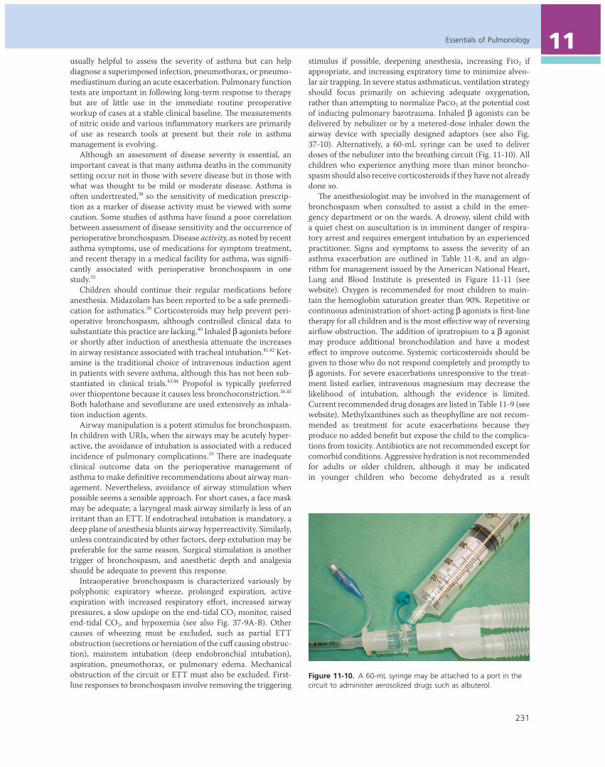
doses of the nebulizer into the breathing circuit (Fig. 11-10). All
children who experience anything more than minor broncho-
spasm should also receive corticosteroids if they have not already
done so.
Th e anesthesiologist may be involved in the management of
bronchospasm when consulted to assist a child in the emer-
gency department or on the wards. A drowsy, silent child with
a quiet chest on auscultation is in imminent danger of respira-
tory arrest and requires emergent intubation by an experienced
practitioner. Signs and symptoms to assess the severity of an
asthma exacerbation are outlined in Table 11-8, and an algo-
rithm for management issued by the American National Heart,
Lung and Blood Institute is presented in Figure 11-11 (see
website). Oxygen is recommended for most children to main-
tain the hemoglobin saturation greater than 90%. Repetitive or
continuous administration of short-acting β agonists is fi rst-line
therapy for all children and is the most eff ective way of reversing
airfl ow obstruction. Th e addition of ipratropium to a β agonist
may produce additional bronchodilation and have a modest
eff ect to improve outcome. Systemic corticosteroids should be
given to those who do not respond completely and promptly to
β agonists. For severe exacerbations unresponsive to the treat-
ment listed earlier, intravenous magnesium may decrease the
likelihood of intubation, although the evidence is limited.
Current recommended drug dosages are listed in Table 11-9 (see
website). Methylxanthines such as theophylline are not recom-
mended as treatment for acute exacerbations because they
produce no added benefi t but expose the child to the complica-
tions from toxicity. Antibiotics are not recommended except for
comorbid conditions. Aggressive hydration is not recommended
for adults or older children, although it may be indicated
in younger children who become dehydrated as a result
Figure 11-10. A 60-mL syringe may be attached to a port in the circuit to administer aerosolized drugs such as albuterol.

A Practice of Anesthesia for Infants and Children
232
Table 11-8. Formal Evaluation of Asthma Exacerbation Severity
Mild Moderate SevereSubset: Respiratory Arrest Imminent
Symptoms
Breathlessness While walkingCan lie down
While at rest (infant—softer, shorter cry, diffi culty feeding)Prefers sitting
While at rest (infant—stops feeding)Sits upright
Talks in Sentences Phrases Words
Alertness May be agitated Usually agitated Usually agitated Drowsy or confused
Signs
Respiratory rate Increased Increased Increased
Guide to rates of breathing in awake children:
Age<2 months2-12 months1-5 years6-8 years
Normal rate<60/min<50/min<40/min<30/min
Use of accessory muscles; suprastemal retractions
Usually not Commonly Usually Paradoxical thoracoabdominal movement
Wheeze Moderate, often only end expiratory
Loud; throughout exhalation
Usually loud; throughout inhalation and exhalation
Absence of wheeze
Pulse/minute Slightly increased Increased Tachycardia Bradycardia
Guide to normal pulse rates in children:
Age2-12 months1-2 years2-8 years
Normal rate<160/min<120/min<110/min
Pulsus paradoxus Absent <10 mm Hg May be present 10-25 mm Hg
Often present >25 mm Hg (adult)20-40 mm Hg (child)
Absence suggests respiratory muscle fatigue
Functional Assessment
PEFPercent predicted or Percent personal best
≥70% Approx. 40-69% or response lasts <2 hours
<40% <25%Note: PEF testing may not be needed in very severe attacks
PaO2 (on air)and/orPCO2
Normal (test not usually necessary)
≥60 mm Hg (test not usually necessary)
<60 mm Hg: possible cyanosis
<42 mm Hg (test not usually necessary)
<42 mm Hg (test not usually necessary)
>42 mm Hg: possible respiratory failure (see text)
SaO2% (on air) at sea level
>95% (test not usually necessary)
90-95% (test not usually necessary)
<90%
Hypercapnia (hypoventilation) develops more readily in young children than in adults and adolescents.
PEF, peak expiratory fl ow; SaO2, oxygen saturation.Medifi ed from National Asthma Education and Prevention Program: Full Report of the Expert Panel: Guidelines for the Diagnosis and Management of Asthma (EPR-3). Bethesda, MD, National Heart, Lung, and Blood Institute, National Institutes of Health, 2007.

Essentials of Pulmonology
233
11of decreased oral intake and increased respiratory rate.
Chest physical therapy and mucolytics are not generally
recommended.
Children with severe atopy-associated asthma are possibly at
greater risk of developing anaphylaxis in response to neuromus-
cular blocking drugs, antibiotics, or latex.40 Bronchospasm due
to asthma is diff erentiated from that due to anaphylaxis by addi-
tional systemic signs such as angioedema, fl ushing, urticaria,
and cardiovascular collapse. Adrenal crisis is another potential
complication associated with severe asthma, owing to iatrogenic
suppression of the hypothalamic-pituitary-adrenal (HPA) axis.
Th is manifests as hypotension, hypoglycemia, or seizures. HPA
suppression should be assumed in any child on signifi cant doses
of corticosteroids for a prolonged period. Short courses of pred-
nisolone used to treat acute fl ares of asthma may aff ect function
for up to 10 days, but prolonged dysfunction is unlikely. High
doses, prolonged therapy for more than a few weeks, and
evening dosing will all cause HPA suppression that may persist
for up to a year. Prophylactic corticosteroid cover is indicated
for those recently requiring systemic corticosteroids and should
be considered for those on high-dose inhaled corticosteroids,
when their corticosteroid regimen is interrupted by the surgical
schedule (see Chapter 24).
Cystic FibrosisCystic fi brosis (CF) is an autosomal recessive disorder. Th e inci-
dence of cystic fi brosis is approximately 1 in 2000 white births,
making it the most common fatal inherited disease of this popu-
lation group. In 1989, a mutation that causes CF was localized
on the long arm of chromosome 7. Th e disease syndrome usually
arises from one of several mutations in the gene that codes for
CF transmembrane conductance regulator, a membrane glyco-
protein chlorine channel that contributes to regulation of ion
fl ux at various epithelial surfaces.46 Th is disruption of electrolyte
transport in epithelial cells in the sweat ducts, airway, pancreatic
duct, intestine, biliary tree, and vas deferens can variously cause
elevated sweat chloride concentrations, viscous mucus produc-
tion, lung disease, intestinal obstruction, pancreatic insuffi -
ciency, biliary cirrhosis, and congenital absence of the vas
deferens. Th e clinical outcome is widely variable, even among
children with identical mutations at the CF locus, suggesting
that genetic, environmental, and therapeutic factors aff ect
expression. Modifi er genes may include genes encoding trans-
forming growth factor and an anti-infl ammatory mediator, mac-
rophage inhibitory factor.47
Lung disease is the main cause of morbidity and mortality in
CF and, consequently, is the focus of anesthetic concern. Th e
pathophysiology involves mucus plugging, chronic infection,
infl ammation, and epithelial injury.47 Mucus clearance defends
the lung against inhaled bacteria. Th e mucociliary transport
system requires two fully functioning layers to be eff ective. Th e
base is a layer of ciliary epithelia bathed in a watery liquid (sol),
overlaid by more viscous gel (mucus) that is responsible for
transporting particles along the tips of the cilia. Normally,
mucus is moved at the speed of about 10 mm/min, thus expel-
ling foreign particles and pathogens from the lungs. Th e effi cacy
of clearance is dependent on adequate hydration of the mucus.48
Lack of regulation of sodium absorption and chloride secretion
causes decreased liquid on the airway luminal surfaces, slows
mucus clearance, and promotes the formation of adherent plugs
to the airway.49 Increased secretions, viscous mucus, and
impaired ciliary clearance contribute to airway impaction, pro-
viding the nidus for infection. At birth, the lung is normal, or
nearly so.46 However, chronic and recurrent bacterial infections
occur early in life, assisted by the pooling of secretions and
impaired neutrophil bacterial killing on airway surfaces.47,50
Repeated and persistent infections stimulate a chronic neutro-
philic infl ammatory response, ultimately destroying the airway
walls. Early pathogens include Staphylococcus aureus and Hae-mophilus infl uenzae. Pseudomonas aeruginosa typically invades
later in life, acquires a mucoid phenotype, and forms a biofi lm
in the lung, an event associated with accelerated decline in
pulmonary function. Th e invasion of the lung by antibiotic-
resistant pathogens such as certain strains of Burkholderia cepacia is often devastating, markedly increasing death rates
from lung disease. Chronic lung damage progresses to bronchi-
ectasis and moderate emphysema, ventilation/perfusion mis-
matching, and hypoxemia. Growth of blood vessels with
advancing bronchiectasis predisposes to hemoptysis. Bronchial
hyperreactivity and increased airway resistance are common,
whereas bullae formation can lead to pneumothorax. Pulmo-
nary function abnormalities are commonly obstructive in nature:
increased FRC, decreased FEV1, decreased peak expiratory fl ow
rate, and decreased vital capacity (see Fig. 11-5). Compensatory
hyperventilation typically produces a lowered Paco2, although
hypercapnia may supersede in end-stage pulmonary pathology.
End-stage cor pulmonale may lead to cardiomegaly, fl uid reten-
tion, and hepatomegaly.
Malnutrition is a common problem in CF, consequent on
pancreatic insuffi ciency, failure of enzyme secretion, impaired
gastrointestinal motility, abnormal enterohepatic circulation of
bile, increased caloric demand due to severe lung disease, and
anorexia of chronic disease.46 Low weight and body mass index
are closely associated with, and can predict, poor lung function.
CF-related diabetes arises from progressive pancreatic disease
and scarring that compromises the pancreatic islets. More than
12% of teenagers older than age 13 years have insulin-dependent
diabetes, and the incidence increases with age. Evidence is accu-
mulating that diabetes contributes to the lung disease and worse
outcome.47 In addition, classic diabetic complications are being
reported in CF patients. Hepatic dysfunction results in decreased
plasma cholinesterase and clotting factors II, VII, IX, and X,
whereas malabsorption of vitamin K may also contribute to
coagulation issues.
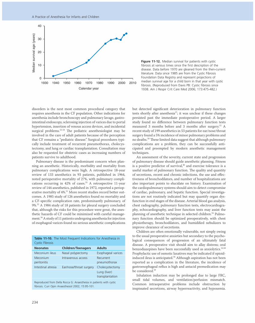
When CF was fi rst distinguished from celiac disease in 1938,
life expectancy was approximately 6 months. Since then, sub-
stantial advances in aggressive supportive treatment have
improved mean median survival to over 30 years (Fig. 11-12).46
More than 35% of patients with CF are now older than 18 years
of age. Th e pillars of treatment include nutritional repletion,
relief of airway obstruction, and antibiotic therapy for lung
infection. Suppression of infl ammation has been a more recent
focus of therapy. Organ transplantation, in particular, lung
transplantation, can also extend life when end-stage organ
failure supersedes (see Chapter 29).46
Th e multisystem nature of the disease and changing demo-
graphics mean children present for a wide variety of surgical
procedures. Th e most common indications for anesthesia in
children are nasal polypectomy and ear/nose/throat surgery,
consequent on the frequency of upper airway pathologic
processes such as chronic sinusitis and nasal polyps (Table
11-10).51,52 Th e investigation or correction of gastrointestinal
A Practice of Anesthesia for Infants and Children
234
disorders is the next most common procedural category that
requires anesthesia in the CF population. Other indications for
anesthesia include bronchoscopy and pulmonary lavage, gastro-
intestinal endoscopy, sclerosing injection of varices due to portal
hypertension, insertion of venous access devices, and incidental
surgical problems.52-54 Th e pediatric anesthesiologist may be
involved in the care of adult patients because of the perception
that CF remains a “pediatric disease.” Surgical procedures typi-
cally include treatment of recurrent pneumothorax, cholecys-
tectomy, and lung or cardiac transplantation. Consultation may
also be requested for obstetric cases as increasing numbers of
patients survive to adulthood.
Pulmonary disease is the predominant concern when plan-
ning an anesthetic. Historically, morbidity and mortality from
pulmonary complications were high. A retrospective 18-year
review of 133 anesthetics in 93 patients, published in 1964,
noted perioperative mortality of 27%, with pulmonary compli-
cations occurring in 42% of cases.55 A retrospective 11-year
review of 144 anesthetics, published in 1972, reported a periop-
erative mortality of 4%.51 More recent studies record better out-
comes. A 1985 study of 126 anesthetics found no mortality and
a CF-specifi c complication rate, predominantly pulmonary, of
9%.52 A 1984 study of 18 patients for pleural surgery concluded
that, although the risks for this procedure were great, the anes-
thetic hazards of CF could be minimized with careful manage-
ment.56 A study of 11 patients undergoing anesthesia for injection
of esophageal varices found no serious anesthetic complications
but detected signifi cant deterioration in pulmonary function
tests shortly after anesthesia57; it was unclear if these changes
persisted past the immediate postoperative period. A larger
study found no diff erence between pulmonary function tests
measured 3 months before and 3 months after surgery.52 A
recent study of 199 anesthetics in 53 patients for ear/nose/throat
surgery found a 5% incidence of minor pulmonary problems and
no deaths.58 Th ese limited data suggest that although pulmonary
complications are a problem, they can be successfully anti-
cipated and preempted by modern anesthetic management
techniques.
An assessment of the severity, current state and progression
of pulmonary disease should guide anesthetic planning. Fitness
is a positive predictor of survival,46 and exercise tolerance is a
useful marker of pulmonary function. Th e quality and quantity
of secretions, recent and chronic infections, the use and eff ec-
tiveness of bronchodilators, and number of hospitalizations are
also important points to elucidate on history. Examination of
the cardiopulmonary systems should aim to detect compromise
of cardiac, pulmonary, and hepatic function. Special investiga-
tions are not routinely indicated but may quantify organ dys-
function in end stages of the disease. Arterial blood gas analysis,
chest radiography, pulmonary function tests, electrocardiogra-
phy, echocardiography, and liver function tests may assist the
planning of anesthetic technique in selected children.54 Pulmo-
nary function should be optimized preoperatively, with chest
physiotherapy, bronchodilators, and humidifi ed nebulizers to
improve clearance of secretions.
Children are often emotionally vulnerable, not simply owing
to the usual preoperative anxieties but secondary to the psycho-
logical consequences of progression of an ultimately fatal
disease. A preoperative visit should aim to allay distress; oral
benzodiazepines have been successfully used as anxiolytics.52,58
Prophylactic use of osmotic laxatives may be indicated if opioid-
induced ileus is anticipated.54 Although aspiration has not been
reported as a complication in the literature, the incidence of
gastroesophageal refl ux is high and antacid premedication may
be considered.59
Inhalation induction may be prolonged due to large FRC,
small tidal volumes, and ventilation/perfusion mismatch.
Common intraoperative problems include obstruction by
inspissated secretions, airway hyperreactivity, and hypoxemia.
Figure 11-12. Median survival for patients with cystic fi brosis at various times since the fi rst description of the disease. Data before 1970 are gleaned from the then-current literature. Data since 1985 are from the Cystic Fibrosis Foundation Data Registry and represent projections of median survival age for a child born in that year with cystic fi brosis. (Reproduced from Davis PB: Cystic fi brosis since 1938. Am J Respir Crit Care Med 2006; 173:475-482.)
Table 11-10. The Most Frequent Indications for Anesthesia in Cystic Fibrosis
Neonates Children/Teenagers Adults
Meconium ileus Nasal polypectomy Esophageal varices
Meconium peritonitis
Intravenous access Recurrent pneumothorax
Intestinal atresia Ear/nose/throat surgery Cholecystectomy
Lung (liver) transplantation
Reproduced from Della Rocca G: Anaesthesia in patients with cystic fi brosis. Curr Opin Anaesthesiol 2002; 15:95-101.
Calendar year
1930 1940 1950 1960 1970 1980 1990 2000 2010
40
30
20
10
0
Med
ian
surv
ival
age
(ye
ars)

Essentials of Pulmonology
235
11Pneumothorax, consequent on high ventilatory pressures and
rupture of bullae, is a less common complication. Postoperative
tribulations include impaired clearance of secretions, atelecta-
sis, pneumonia, and respiratory failure. Anesthesia should be
deep enough to prevent bronchospasm or increased shunting
during surgical stimulation or airway suctioning but ideally
should not compromise postoperative respiratory function.
Regional anesthesia is an ideal option if not contraindicated by
coagulopathy or patient refusal. For general anesthesia, short-
acting agents are preferred to minimize postoperative respira-
tory compromise. A cuff ed ETT is often required, owing to the
great airway pressures needed for adequate ventilation.
Because dehydration of secretions is a central pulmonary
issue in CF, general anesthesia poses specifi c problems. During
spontaneous ventilation under normal conditions, inspired
gases are warmed to body temperature and saturated with
water vapor, reaching this state at a point just below the
carina.60 Th is region is known as the isothermic saturation
point and ensures that the lower airways are kept moist
and warm.61 Th e alveolar environment in optimal circumstances
has a saturated water vapor pressure of 47.1 mm Hg and an
absolute humidity of 43.4 g/m−3 at 37º C. Th e inspiration of cold,
desiccated anesthetic gases and vapors can impair the warming
and humidifi cation of the airways. Th e use of any airway
device—oropharyngeal airway, laryngeal mask, or ETT—
bypasses the nasal and oropharyngeal passages and delivers
cold, dry gas to a varying extent farther down the airway.62 Th is
shifts the isothermic saturation point distally, forcing bronchi
that normally function in optimal conditions to take part in heat
and gas exchange.61 Th ese parts of the airway are less adapted
to moisture exchange and tend to dehydrate more rapidly,
thereby impairing the mucociliary escalator and predisposing to
impaction of secretions.63,64 Direct impairment of mucociliary
motion by anesthetic medications, as well as blunting of the
cough response and ventilatory drive, can contribute further to
the problem. Particular attention should, therefore, be directed
to hydrating the airway in the perioperative period, to minimize
the exacerbation of the primary pathologic pulmonary process
of CF. Inhalation of hypertonic saline (7% sodium chloride)
accelerates mucus clearance and improves lung function65-67 and
is now part of the routine maintenance management of
CF. Nebulized saline treatments should be continued up to the
start of anesthesia and recommenced after the procedure is
complete. Inhaled gases should be humidifi ed or an artifi cial
“nose” inserted into the circuit to conserve airway moisture and
minimize inspissation of secretions. Bronchial washing and
suction can be used to clear secretions for more prolonged
procedures, under a depth of anesthesia adequate to prevent
bronchospasm.
At the conclusion of surgery, complete reversal of neuromus-
cular blockade should be confi rmed. Whenever possible, the
child should be extubated and encouraged to breathe spontane-
ously. A 30- to 40-degree head-up position assists movement of
the diaphragm and ventilation. Postoperatively, physiotherapy,
airway humidifi cation, close attention to analgesia, and early
mobilization should aim to enhance clearance of secretions and
minimize atelectasis. Th e use of regional or local anesthesia,
plus non-opioid analgesics, is useful to avoid respiratory
depression. Ambulatory surgery is optimal, if feasible, because
it minimizes disruption to the patient’s schedule and decreases
exposure to nosocomial infection.
Sickle Cell DiseaseSickle cell disease (SCD) is an inherited hemoglobinopathy
resulting from a point mutation on chromosome 11. Th e mutant
gene codes for the production of hemoglobin S, a mutant variant
of the normal hemoglobin A. Th is leads to widespread and
progressive vascular damage.68,69 Clinical features of the disease
include acute episodes of pain, acute and chronic pulmonary
disease, hemorrhagic and occlusive stroke, renal insuffi ciency,
and splenic infarction, with mean life expectancy shortened to
just over 3 decades.70 Perioperative problems and management
are covered in more detail in Chapter 9, and this discussion will
be limited to a brief review of the pulmonary pathology of
SCD.
Th e acute chest syndrome (ACS) is an acute lung injury
caused by SCD. Diagnostic criteria include a new pulmonary
infi ltrate involving at least one lung segment on the radiograph
(excluding atelectasis), combined with one or more symptoms
or signs of chest pain, pyrexia greater than 38.5º C (101.3º F),
tachypnea, wheezing, and cough.71-73 Precipitants include infec-
tion, fat embolism after bone marrow infarction, pulmonary
infarction, and surgical procedures.73-75 Potential risk factors for
the development and severity of perioperative ACS include a
history of lung disease, recent clustering of acute pulmonary
complications, pregnancy, increased age, and the invasiveness
of the surgical procedure.68 However, a study of 60 laparoscopic
surgeries noted an association between younger age and ACS,
which the authors suggested may be related to reduced tempera-
tures and greater relative blood loss as a proportion of total
blood volume in smaller children.76
Minor procedures such as inguinal hernia repair or distal
extremity surgery have a low risk of pulmonary complications
(none to 5%), whereas intra-abdominal or major joint surgery
has an ACS rate of 10% to 15%.75,77,78 Although overall periopera-
tive mortality specifi cally from SCD appears to be quite small,
slightly under 1%,75 ACS can lead to prolonged postoperative
hospitalization, respiratory failure, and death. One study of 604
patients noted that ACS typically developed 3 days postopera-
tively and persisted for 8 days; 2 patients died in approximately
60 episodes.74
SCD also causes chronic lung damage, known as sickle cell
lung disease (SCLD).79 Because lung function has not yet been
assessed longitudinally in a cohort from early childhood to
adulthood, the precise pathology of and relationship between
the obstructive and restrictive patterns of lung disease is
unclear.80 Children appear to have a predominantly obstructive
pattern,81 whereas adults have more restrictive pulmonary fi nd-
ings.79,82,83 Th e later stages of lung damage involve decreased
vital and total lung capacities, impaired gas diff usion, pulmo-
nary fi brosis, pulmonary artery hypertension, right-sided car-
diomyopathy, and progressive hypoxemia.79,83 Th e development
of pulmonary artery hypertension, which can precede clinically
apparent lung damage, is a particularly ominous sign of disease
progression and is associated with heightened risk of sudden
death.82 Recurrent ACS is an independent risk factor for the
development of end-stage SCLD, but subtle evidence of paren-
chymal and vascular damage commonly precedes clustered epi-
sodes of ACS.79
Assessment of lung function should include a history of the
occurrence, frequency, severity, and known precipitants of ACS
and a search for progression of chronic lung damage. A recent
chest radiograph will serve as a baseline for comparison if post-

236
operative radiographs are needed and can also delineate lung
pathology. Early features of lung damage include decreased
distal pulmonary vascularity and diff use interstitial fi brosis,
whereas later stages are characterized by pulmonary fi brosis and
right ventricular hypertrophy.79 Pulmonary function testing can
reveal the need for bronchodilators and the presence of obstruc-
tive or restrictive lung disease.
Th e effi cacy of preoperative or intraoperative management
techniques beyond basic standards of care has not been clearly
demonstrated, and well-delivered anesthetic and postoperative
care may be the best guarantors of good outcome.68,69 Because
the eff ect of perioperative red blood cell transfusion versus no
transfusion in preventing ACS or other sickle cell complications
has not been tested by an adequately controlled study, the effi -
cacy of prophylactic erythrocyte transfusion is controversial.
One recent guideline suggests the avoidance of transfusion in
low-risk situations, while considering transfusion only for cases
assessed as greater risk.68 If transfusion is undertaken, exchange
transfusion aiming to decrease the concentration of hemoglobin
S to 30% is no more effi cacious than correction of anemia to a
hematocrit of 30% in preventing SCD exacerbations but results
in more transfusion-related complications.74 Consequently, if a
decision is made to transfuse in the hope of preventing ACS,
the target should be a hematocrit of 30% rather than a specifi c
dilution of hemoglobin S.
Sickle cell patients frequently develop postoperative
atelectasis. It is unclear if this relates to underlying sickle
lung disease, diffi culty with analgesia, other causes, or a
combination of factors. Pain management can be diffi cult.
Postoperative pain and opioid consumption is often great in
SCD patients, and opioid use may lead to respiratory blunting
and atelectasis.84 ACS tends to involve the lower segments of
the lung,73 suggesting an association between atelectasis
and ACS. Incentive spirometry can prevent the development
of atelectasis and pulmonary infi ltrates associated with
ACS.85 Regional analgesia, supplemental nonopioid analgesics,
prophylactic incentive spirometry, early mobilization, and good
pulmonary toilet may decrease the incidence of atelectasis and
ACS.
Treatment of ACS is focused on supporting gas exchange.
Supplemental oxygen, noninvasive ventilatory support such as
continuous positive airway pressure, or intubation and mechan-
ical ventilation are indicated by the degree of dysfunction. Bron-
chodilators, incentive spirometry, and chest physiotherapy may
be useful in preventing progression of the disease. In the pres-
ence of a signifi cant ventilation/perfusion mismatch, correction
of anemia can improve arterial oxygenation. Erythrocyte trans-
fusion increases oxygen-carrying capacity, decreases fractional
peripheral tissue extraction, and increases returning venous
oxygen levels. Because the mean arterial oxygen content in the
presence of a shunt is signifi cantly aff ected by the oxygenation
of blood returning from nonventilated parts of the lung, increas-
ing venous oxygen levels can improve arterial oxygen content.
Whereas transfusion has not been clearly shown to improve
outcome, both exchange and simple transfusion can improve
oxygenation.73
SummaryPulmonary complications are a major cause of perioperative
morbidity in the pediatric population. Although preexisting
pulmonary pathologic processes in children can present
signifi cant challenges to anesthetic delivery, a thorough
assessment of the problem combined with intelligent anesthetic
management allows most children to undergo surgical
interventions without long-term adverse sequelae. Consultation
with a pediatric pulmonologist is indicated when appropriate
for specifi c problems as outlined in this chapter; a team
approach may markedly improve operative and postoperative
outcomes.
Annotated ReferencesAccurso FJ: Update in cystic fi brosis 2005. Am J Respir Crit Care Med
2006; 173:944-947
A current review of the pathophysiology of cystic fi brosisBishop MJ, Cheney FW: Anesthesia for patients with asthma: low risk
but not no risk. Anesthesiology 1996; 85:455-456
A thoughtful editorial on the implications, dangers, and practical impli-cations of asthma
Davis PB: Cystic fi brosis since 1938. Am J Respir Crit Care Med 2006;
173:475-482
A succinct discourse on the evolution of management of cystic fi brosisDoherty GM, Chisakuta A, Crean P, Shields MD: Anesthesia and the
child with asthma. Paediatr Anaesth 2005; 15:446-454
A recent review of the perioperative management of asthma
Firth PG, Head CA: Sickle cell disease and anesthesia. Anesthesiology
2004; 101:766-785
A comprehensive review of anesthetic management of sickle cell diseaseNational Asthma Education and Prevention Program: Full Report of the
Expert Panel: Guidelines for the Diagnosis and Management of
Asthma (EPR-3). Bethesda, MD, National Heart, Lung, and Blood
Institute, National Institutes of Health, 2007
An extensive review of current evidence on the pathophysiology, diagno-sis, and management of asthma
Tait AR, Malviya S: Anesthesia for the child with an upper respiratory
tract infection: still a dilemma? Anesth Analg 2005; 100:59-65
A broad review of the data on perioperative upper respiratory tract infec-tions and suggested approaches to management
A Practice of Anesthesia for Infants and Children
ReferencesPlease see www.expertconsult.com
Related Documents


