Anesthetic Considerations in Patients with Inherited Coagulation Defects Essay Submitted for Partial Fulfillment of the Master Degree in Anesthesiology By Radwa khairy Abd El Mohsen M.B., B.Ch. Faculty of medicine- Ain Shams University Under supervision of Prof. Dr. Mervat Mohamed Marzok Professor of Anesthesiology, Intensive care and pain Management Faculty of Medicine- Ain Shams University Dr. Dalia Abd El Hameed Mohamed Assistant professor of Anesthesiology, Intensive care and Pain Management Faculty of Medicine- Ain Shams University Dr. Mahmoud Ahmed Abd El Hakim Lecturer of Anesthesiology, Intensive care and Pain Management Faculty of Medicine, Ain Shams University Faculty of Medicine Ain Shams University 2017

Welcome message from author
This document is posted to help you gain knowledge. Please leave a comment to let me know what you think about it! Share it to your friends and learn new things together.
Transcript
Anesthetic Considerations in Patients
with Inherited Coagulation Defects
Essay
Submitted for Partial Fulfillment of the Master
Degree in Anesthesiology
By
Radwa khairy Abd El Mohsen
M.B., B.Ch.
Faculty of medicine- Ain Shams University
Under supervision of
Prof. Dr. Mervat Mohamed Marzok Professor of Anesthesiology, Intensive care and pain Management
Faculty of Medicine- Ain Shams University
Dr. Dalia Abd El Hameed Mohamed Assistant professor of Anesthesiology, Intensive care and Pain
Management
Faculty of Medicine- Ain Shams University
Dr. Mahmoud Ahmed Abd El Hakim Lecturer of Anesthesiology, Intensive care and Pain Management
Faculty of Medicine, Ain Shams University
Faculty of Medicine
Ain Shams University
2017

Acknowledgment
All praise is to Allah and all thanks. He has guided and
enabled me by his mercy to fulfill this essay, which I hope
to be beneficial for people.
I would like to express my deepest gratitude and sincere
appreciation to Prof. Dr. Mervat Mohamed Marzok,
Professor of Anesthesiology, Intensive care and pain
management, Faculty of Medicine, Ain Shams
University, for her encouragement, his kind support and
appreciated suggestions that guided me to accomplish this
work.
I would like to express my deepest gratitude and sincere
appreciation to Dr. Dalia Abd El Hameed Mohamed,
Assistant professor of Anesthesiology, Intensive care and
Pain management, Faculty of Medicine, Ain Shams
University for she encouragement, She kind support and
appreciated suggestions that guided me to accomplish this
work.
I am also grateful to, Dr. Mahmoud Ahmed Abd El
Hakim, Lecturer of Anesthesiology, Intensive care and
Pain management, Faculty of Medicine- Ain Shams
University for her encouragement, his kind support and
appreciated suggestions that guided me to accomplish this
work
Radwa khairy Abd El Mohsen
Contents
Contents
Subjects Page
List of Abbreviations ............................................................................................. I
List of Tables ............................................................................................................. IV
List of Figures ............................................................................................................ V
Introduction .................................................................................................................... 1
Aim of the Essay ...................................................................................................... 4
Chapter 1: Physiology of coagulation and
hemostasis ........................................................................... 1
Chapter 2: Inherited coagulation disease's
pathophysiology, diagnosis and
treatment ............................................................................. 22
Chapter 3: Anesthetic management .................................. 52
Summary ...................................................................................................................... 80
References ................................................................................................................... 82
Arabic summary
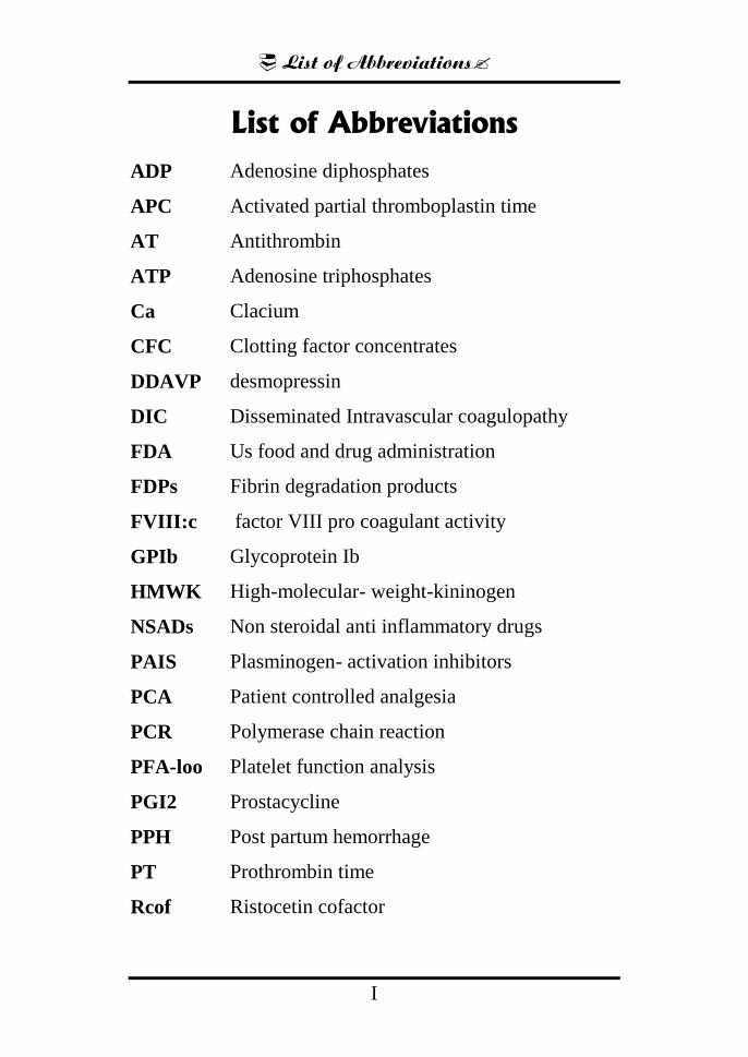
List of Abbreviations
I
List of Abbreviations
ADP Adenosine diphosphates
APC Activated partial thromboplastin time
AT Antithrombin
ATP Adenosine triphosphates
Ca Clacium
CFC Clotting factor concentrates
DDAVP desmopressin
DIC Disseminated Intravascular coagulopathy
FDA Us food and drug administration
FDPs Fibrin degradation products
FVIII:c factor VIII pro coagulant activity
GPIb Glycoprotein Ib
HMWK High-molecular- weight-kininogen
NSADs Non steroidal anti inflammatory drugs
PAIS Plasminogen- activation inhibitors
PCA Patient controlled analgesia
PCR Polymerase chain reaction
PFA-loo Platelet function analysis
PGI2 Prostacycline
PPH Post partum hemorrhage
PT Prothrombin time
Rcof Ristocetin cofactor
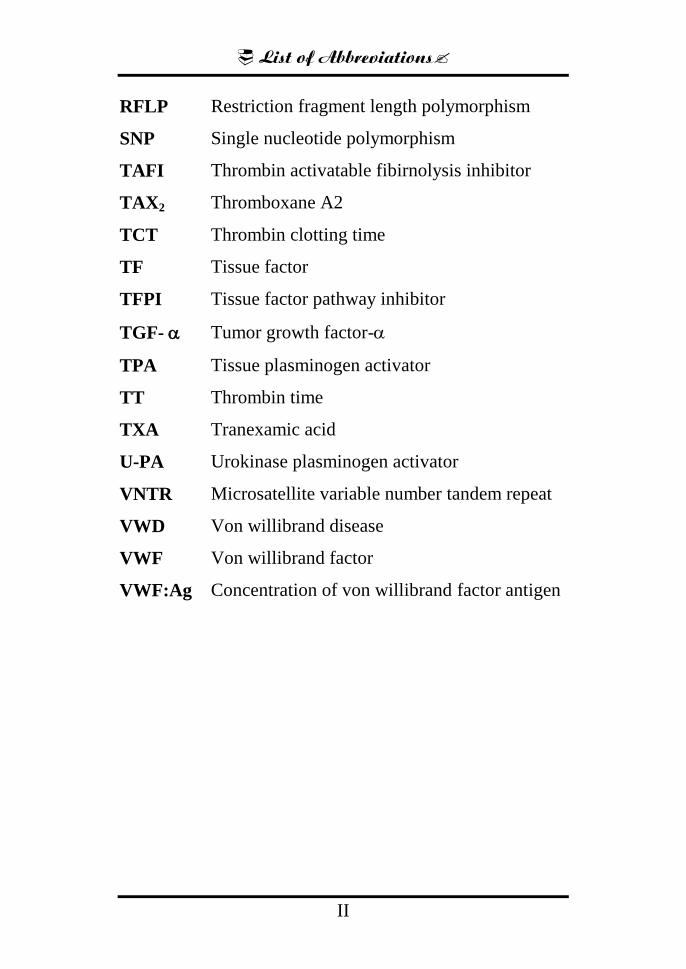
List of Abbreviations
II
RFLP Restriction fragment length polymorphism
SNP Single nucleotide polymorphism
TAFI Thrombin activatable fibirnolysis inhibitor
TAX2 Thromboxane A2
TCT Thrombin clotting time
TF Tissue factor
TFPI Tissue factor pathway inhibitor
TGF- Tumor growth factor-
TPA Tissue plasminogen activator
TT Thrombin time
TXA Tranexamic acid
U-PA Urokinase plasminogen activator
VNTR Microsatellite variable number tandem repeat
VWD Von willibrand disease
VWF Von willibrand factor
VWF:Ag Concentration of von willibrand factor antigen

List of Tables
III
List of Tables
Table No. Title Page
No.
Table (1) Clotting factors in blood and their
synonyms
5
Table (2) Disorders of Primary Hemostasis 23
Table (3) Disorders of Secondary Hemostasis 24
Table (4) Phenotypic classification of hemophilia
and its clinical features
28
Table (5) Guidelines for neuraxial techniques in
hemophilia and VWD
61
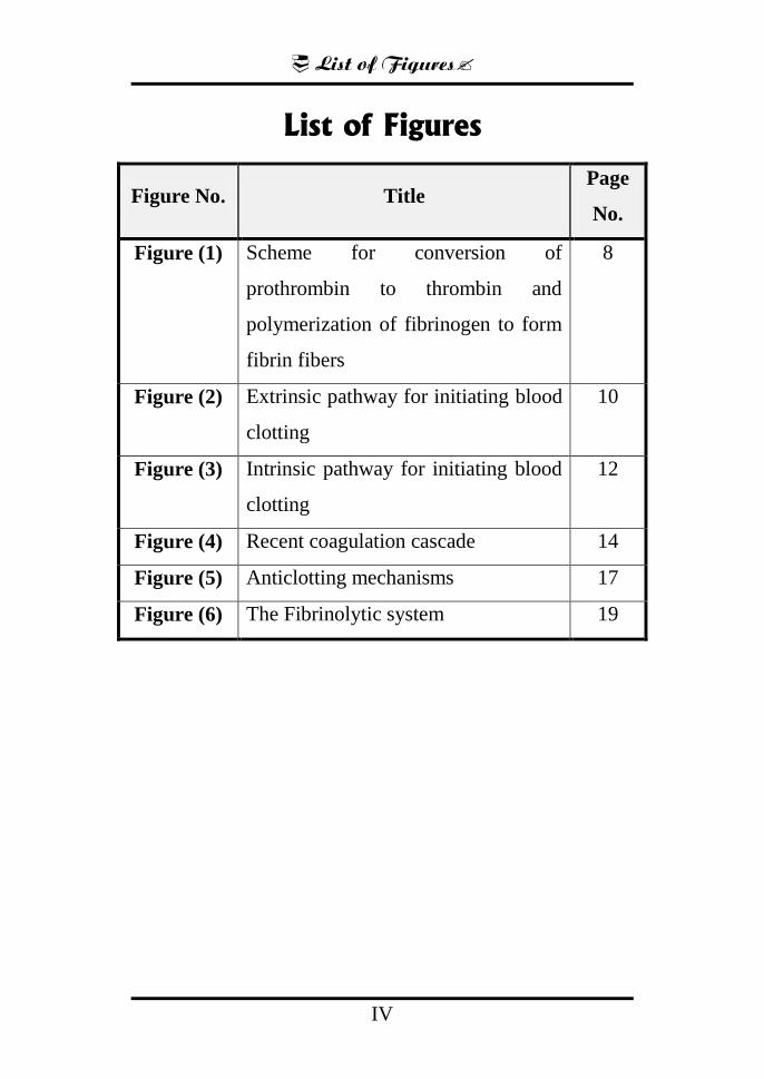
List of Figures
IV
List of Figures
Page
No. Title Figure No.
8 Scheme for conversion of
prothrombin to thrombin and
polymerization of fibrinogen to form
fibrin fibers
Figure (1)
10 Extrinsic pathway for initiating blood
clotting
Figure (2)
12 Intrinsic pathway for initiating blood
clotting
Figure (3)
14 Recent coagulation cascade Figure (4)
17 Anticlotting mechanisms Figure (5)
19 The Fibrinolytic system Figure (6)
Introduction
- 1 -
Introduction
Hereditary bleeding disorders are a diverse group of
diseases that include abnormalities of primary and
secondary hemostasis. The most common congenital
bleeding disorders include:- Von Willebrand disease,
Hemophilia A (factor VIII deficiency), Hemophilia B
(factor IX deficiency) Less common congenital bleeding
disorders include:- Factor I (fibrinogen) deficiency, Factor
II (prothrombin) deficiency, Factor V deficiency, Factor
VII deficiency, Factor X deficiency, Factor XI deficiency,
Factor XIII deficiency, Platelet disorders. (Kendall
Crookston et al., 2010)
Primary hemostasis involves formation of the platelet
plug which involves platelets, the blood vessel wall and
von Willebrand factor abnormalities can include problems
in platelet number, adhesion or aggregation.
Secondary hemostasis involves the formation of fibrin
through the humoral coagulation cascade; abnormalities
include deficiencies of coagulation factors or contact
factors, deficiencies or abnormalities of fibrinogen or
connective tissue diseases. Mutations can be inherited in an
autosomal dominant, recessive or X-linked pattern.
(Kendall Crookston et al., 2010)
Introduction
- 2 -
Hemophilia A and B are the most frequent inherited
bleeding disorders. Together with Von Will brand disease
these X- linked disorders include 95% to 97% of all the
inherited deficiencies of coagulation factors. (Mannucci
and Tuddenham, 2003)
The remaining defects, generally transmitted as
autosomal recessive traits in both sexes, are rare, with
prevalences of the presumably homozygous forms in the
general population ranging from approximately 1 in 2
million for factor II (prothrombin) and factor XIII (FXIII)
deficiency to 1 in500 000 for factor VII (FVII) deficiency.
Exceptions to these low prevalences are countries with
large Jewish communities (where FXI deficiency is much
more prevalent), Middle Eastern countries, and Southern
India. In the last 2 regions, consanguineous marriages are
relatively common, so that autosomal recessive traits occur
more frequently in homozygosity. (Peyvandi et al., 2002)
Preoperative assessment ideally must be done by a
team of hematologist, surgeon, and anesthetist, so that a
tailored individual plan is formulated and discussed with
the patient. Detailed history about the type of hemophilia
and VWD (von will brand disease) and its severity must be
obtained. Prior information about response to DDAVP
Introduction
- 3 -
(desmopressin), use of recombinant factors VIII and IX,
and previous transfusion of blood will be useful. A
complete blood count, coagulation profile and fibrinogen
level, and specific factor assays must be done if indicated.
All patients must be evaluated for the presence of
transfusion-related infections such as HIV and hepatitis B
and C. Examination for the presence of joint deformities,
contractures, and a thorough airway assessment. (Martlew,
2000)
There are case series and reports of central neuraxial
blocks with catheters in hemophiliac patients undergoing
lower limb orthopedic surgeries after correction of factor
levels, but there are no randomized controlled trials.
Therefore, the risk–benefit ratio must be assessed
individually on a case-to-case basis. (Choi and Brull,
2009)
Aim of the Essay
Aim of the Essay
Is to present the significant challenges in the
perioperative period in patients with inherited coagulation
defects and the different anesthetic management in such
cases.
Physiology of Coagulation System &Hemostasis
1
Physiology of Coagulation System and
Hemostasis
The concept of blood coagulation dates back to the
1960's when Davie, Rat off and Macfarlane described the
―waterfall‖ and ―cascade‖ theories showing the
fundamental principle of a cascade of proenzymes leading
to the activation of some enzymes. Usually, the process of
coagulation is controlled by several inhibitors that limit the
clot formation and prevents more propagation of the
thrombus. This thrombo-hemorhagic balance is maintained
in the body by some complicated interactions between the
coagulation and the fibrinolytic system as well as the
platelets and the blood vessel wall. (Achneck et al., 2010)
Hemostasis is defined as the arrest of bleeding. It
comes from Greek, where, haeme means blood and stasis
means to stop. When a vessel is injured or ruptured,
hemostasis occurs by several mechanisms:- (1) vascular
constriction, (2) formation of a platelet plug, (3) formation
of a blood clot as a result of blood coagulation, and (4) the
growth of fibrous tissue into the blood clot which closes the
hole in the vessel permanently. (Anderson et al., 2006)
Physiology of Coagulation System &Hemostasis
2
Steps of coagulation:-
1. Vascular Constriction:-
After a blood vessel has been cut or ruptured, the
trauma to the vessel wall itself causes the smooth muscle in
the wall to contract immediately; this causes instant
reduction of the flow of blood from the ruptured vessel.
The contraction results from (1) local myogenic
spasm, (2) local autacoid factors released from the
traumatized tissues and platelets, and (3) nervous reflexes.
Initiation of the nervous reflexes is by pain nerve impulses
or sensory impulses that originate from the traumatized
blood vessel or the tissues nearby. (Anderson et al., 2006)
In the resting state, blood is actively maintained in a
liquid form by endothelial cells and circulating plasma
protein inhibitors. When the vascular integrity is disrupted
or the endothelium becomes inflamed, the thrombotic
activity of the endothelial cells is triggered. This occurs
through secreting platelet-activating factor, a substance that
induces platelet aggregation and formation of Von
Willebrand factor (VWF), which is a cofactor for adherence
of platelets to the subendothelium. The endothelium is also
able to secrete plasminogen activator inhibitor which
inhibits the fibrinolytic system. (Dittman and Majerus,
2001)
Physiology of Coagulation System &Hemostasis
3
Damage to the endothelium exposes blood to a highly
thrombogenic subendothelial connective tissue which
initiates the clot formation. This connective tissue consists
of various types of compounds including fibrillar collagen,
which is a potent stimulus for the activation and adhesion
of platelets. Simultaneously, subendothelial components
convert inactive coagulation factors into powerful enzymes,
initiating an intrinsic stimulation of the plasma coagulation
system. (Edward and Juan, 2000)
2. Formation of the platelet plug:-
Platelets have an over-expanding role in hemostasis.
Beside their role when vascular integrity is disturbed, they
also maintain the integrity of normal endothelium. This is
why patients with platelet deficiencies have a tendency to
develop purpuric bleeding. (Edward and Juan, 2000)
Platelets respond through three steps:-
1. Adhesion:-
When platelets become in contact with a damaged
blood vessel, along with the collagen fibres present in the
vascular wall, the platelets begin to swell, forming irregular
shapes with multiple radiating pseudopods protruding from
their surfaces, following that their contractile proteins
contract forcefully and release granules that contain
Related Documents