ESE Clinical Update on Acromegaly 2021 A series of webinars held on 13−15 September 2021

Welcome message from author
This document is posted to help you gain knowledge. Please leave a comment to let me know what you think about it! Share it to your friends and learn new things together.
Transcript
ESE Clinical Update on Acromegaly 2021A series of webinars held on 13−15 September 2021
02
Welcome
WelcomeAcromegaly continues to present challenges to patients and endocrinologists alike. This report presents the content of three dynamic webinars in September 2021 that addressed the latest issues in the diagnosis, management and complexities associated with this debilitating disease..The European Society of Endocrinology (ESE) was delighted to welcome experts in the field, who shared their knowledge and experience over the course of three, 2-hour webinar sessions that examined:
• aspects relating to diagnosis, which spanned hormone assays, sex differences, and pseudoacromegaly
• the management and impact of the disease, including sleep apnoea, optimal use of radiotherapy, and psychosocial consequences of the disorder
• challenges and the future landscape, which considered discrepancies between clinical outcomes and biochemistry, discordant GH and IGF-1 results, and difficulties encountered on a case-by-case basis.
These areas of discussion were expanded upon by carefully selected case presentations, which not only supported the main presentations but enabled the webinars to incorporate a range of other topics relevant to the field.
We are grateful to all who took part, including the attendees, who contributed important experience and many pertinent and relevant questions.
The content of the webinars is available to attendees at www.eseondemand.org.
Niki Karavitaki, Marinella Tzanela, Nienke Biermasz and Philippe Chanson Scientific Programme Committee
Kindly supported by an independent medical education grant from Pfizer Inc.
03
Contents
ContentsWelcome
Chairs and Speakers
Webinar 1: Diagnosis in acromegaly
• Hormone assays in acromegaly • Rachel Webster (UK)
• Sex differences at diagnosis of acromegaly • Mónica Marazuela (Spain)
• Rare syndromes resembling acromegaly: pseudoacromegaly • Márta Korbonits (UK)
Case presentations
• Acromegaly with normal IGF-1 levels • Diana Borges Duarte (Portugal)
• Acromegaly, goitre and menopause • Puja Thadani (UK)
• Acromegaly with normal IGF-1 and suppressed GH during OGTT • Peter Wolf (France/Austria)
Webinar 2: Management and burden of disease
• Sleep apnoea in acromegaly • Romana Netea-Maier (The Netherlands)
• Radiotherapy: what is the best modality per case? • Giuseppe Minniti (Italy)
• Acromegaly: psychosocial consequences • Nienke Biermasz (The Netherlands)
Case presentations
• Management of acromegaly and McCune−Albright syndrome • Mariana Aveiro Lavrador (Portugal)
• Optimal management in acromegaly with McCune−Albright syndrome • Eirini Papadimitriou (Greece)
• Gigantism • Neha Malhotra (UK)
Webinar 3: Challenges and future landscape
• Discrepancies between clinical outcomes and biochemistry: new tools • Pietro Maffei (Italy)
• Discordant GH and IGF-1 results: to treat or not to treat? • Stephan Petersenn (Germany)
• Troubles in managing acromegaly: case by case • Jens-Otto Jørgensen (Denmark)
Case presentations
• Post-operative management in pituitary GH-secreting microadenoma • Judit Tőke (Hungary)
• Radiotherapy and hepatitis B on antiviral medication in a male with acromegaly • Katerina Lavrentaki (Greece)
• PANCH tumour causing acromegaly • Amy Coulden (UK)
0204
05050607
0808
009
10101112
131314
15151617
181819

04
Chairs and Speakers
ESE thanks all faculty members for their valuable contributions to the ESE Clinical Update on Acromegaly 2021. Chairs and Speakers
Albert Beckers Liège, Belgium
Philippe Chanson Paris, France
Mariana Aveiro Lavrador Coimbra, Portugal
Nienke Biermasz Leiden, The Netherlands
Diana Borges Duarte Porto, Portugal
Jens-Otto Jørgensen Aarhus, Denmark
Katerina Lavrentaki Athens, Greece
Amy Coulden Birmingham, UK
Niki Karavitaki Birmingham, UK
Márta Korbonits London, UK
Neha Malhotra London, UK
Romana Netea-Maier Nijmegen, The Netherlands
Pietro Maffei Padua, Italy
Mónica Marazuela Madrid, Spain
Giuseppe Minniti Rome/Siena, Italy
Stephan Petersenn Hamburg, Germany
Judit Tőke Budapest, Hungary
Eirini Papadimitriou Athens, Greece
Marija Pfeifer Ljubljana, Slovenia
Puja Thadani Coventry, UK
Rachel Webster Birmingham, UK
Marinella TzanelaAthens, Greece
Peter Wolf Vienna, AustriaParis, France
05
WEBINAR 1 Diagnosis of acromegaly
Hormone assays in acromegaly Rachel Webster Birmingham, UK
The analytes of interest in acromegaly are growth hormone (GH) and insulin-like growth factor-1 (IGF-1). GH levels change throughout the day, while IGF-1 is a more stable marker. An oral glucose tolerance test (OGTT) can be used to measure these analytes in response to glucose ingestion.
During an OGTT, normal subjects will exhibit suppression of GH to undetectable levels. In around 8% of people with acromegaly, fasting GH may be normal at the beginning of the test, but won't suppress to undetectable values during the test.1 This may be due to chronic renal failure, liver failure, active hepatitis, anorexia, malnutrition, hypothyroidism, diabetes or adolescence.2 Specific assay-dependent cut-offs are needed for interpretation.
Both GH and IGF-1 are measured in laboratories by immunoassay. Immunoassay has been the cornerstone of hormone analysis since the 1950s, with a sensitivity of pmol to µmol. Hormones are measured using an antibody–antigen interaction.
Types of immunoassayImmunoassays can be non-competitive or competitive. A non-competitive assay measures the sites occupied by the analyte in the sample (i.e. the patient’s hormone). This is typically found in enzyme-linked immunosorbent assays (ELISAs). The signal is proportional to the analyte concentration.
A competitive immunoassay measures unoccupied sites. In this case, a labelled
analyte is added and measured instead of the analyte from the patient’s sample. Because the labelled analyte competes with the patient’s hormone, the signal is inversely proportional. These are typically used in radioimmunoassay and small molecule assays.
Competitive and non-competitive assays can be homogeneous or heterogeneous. Homogeneous assays are fast, with just one step. However, they can be susceptible to the ‘hook effect’, where swamping of antibodies causes falsely low results. This can be avoided using a heterogeneous assay, which includes a wash step to eliminate background noise. This takes slightly longer but offers increased specificity and sensitivity. Glucose does not interfere with hormone assays, so repeated analyses are possible.
Historically, manufacturers have used polyclonal antibodies for these assays. These bind to more than one epitope on the antigen, which can reduce specificity. They are also raised in animals, which means the assay must change over time, leading to different results. By contrast, monoclonal antibodies are raised to a specific single antigen, which increases specificity. Because they are produced in immortal cell lines, the assay remains unchanged, offering a more stable and sustainable service.
Challenges with immunoassayThere are some problems with immunoassay, which should always be considered when interpreting results.
Immunoassays of the same analyte are not always comparable. All laboratories should be enrolled in an external quality assurance scheme. Different platforms measuring IGF-1 can have different reference ranges, which can introduce varying levels of bias. You must know the reference ranges for each individual platform to accurately interpret the results. Immunoassays are complex reactions, so there are differences in results. These differences are smaller with GH than with IGF-1, because there is an agreed international standard; there is no controlled analyte for IGF-1.
In addition to this lack of standardisation, manufacturers use different detection and capture antibodies with varying specificity, which means their assays are looking at different parts of the molecule, and analytes are not recognised consistently by assays. With IGF-1, many binding proteins need to be
split away to avoid interference. Differences in antibody binding strength can also cause variation in assay sensitivity.
A lack of antibody specificity for the molecule of interest can cause falsely elevated concentrations of analyte. A monoclonal antibody can minimise cross-reactivity, but will be more expensive, so there is a trade-off between specificity and cost.
When to suspect interferenceImmunoassay interference may be suspected when the results do not fit with the clinical condition. Laboratories do not always have access to clinical histories, so clinicians are encouraged to notify the lab if the results do not fit the patient. Similarly, there may be interference if there is an inconsistent ‘delta’ check between current and previous results, or a lack of fit with biochemical variables. Implausible results, non-linear dilution or markedly different results from different analysers all suggest possible interference.
Effective liaison between clinician and laboratory is vital to help identify and avoid possible interference.
Reasons to use immunoassayGiven these challenges, there are still good reasons to use immunoassay. Primarily, there is a lack of valid alternatives. Laboratory workload is high, and immunoassay is easier to automate than alternatives such as mass spectrometry. Interference only affects a small number of samples, which means these assays are adequate for the vast majority.
The key is to remain vigilant for interference. Clinicians are encouraged to contact their clinical biochemistry laboratory, understand their methods and protocols, and communicate promptly to discuss unexpected results, as samples may not be retained in the long term. Odd results can be confirmed using alternative methods, to ascertain the presence of interference.
Diagnosis of acromegalyChairs: Philippe Chanson (France) & Nienke Biermasz (The Netherlands)
REFERENCES1. Brockmeier et al. 1992 Hormone & Metabolic
Research 24 392−400.2. Freda 2009 Clinical Endocrinology 71 166−170.
06
WEBINAR 1 Diagnosis of acromegaly
Sex differences at diagnosis of acromegaly Mónica Marazuela Madrid, Spain
Research suggests that acromegaly probably affects more women than men: a review of gender distribution and prevalence found that 54% of 5955 patients with acromegaly were female.1 Another recent meta-analysis of 33 studies, including more than 25 000 patients, found that 53% of acromegaly diagnoses were in women.2 Incidence data corroborate prevalence data, with higher female incidence in some but not all studies.
Most studies report that men are diagnosed earlier than women, by 3−5 years on average. There are several possible explanations for this difference. Women may be likely to visit more doctors, thus taking longer to reach a diagnosis; there may be under-reporting in older male patients, resulting in under-diagnosis; and some early symptoms of acromegaly in women can be misattributed to menopause, delaying diagnosis.
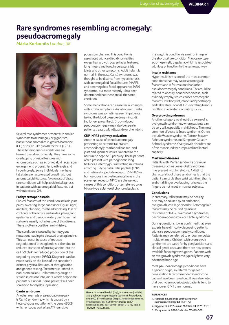
Facial analysis and genderFacial analysis has long been used in the diagnosis of acromegaly, with 2D photography allowing clinicians to monitor changes in facial features over time. Now, 3D photography provides more detailed lateral, anterolateral and anterior views of the patient, and analysis of new indices, such as nose length and depth, upper and lower vermilion height, and face length and breadth.
A study of 39 acromegalic patients using 3D photography found that facial changes characteristic of the disease are not the same in men and women (see Figure, right).3 Face breadth, nose length and width, nasofrontal angle, and columella−labial angle differ between men and women with acromegaly.
3D cephalometry by computed tomography scan can quantify facial anatomical features of both soft tissues and bone. This has also shown that substantive facial changes in acromegalic patients are different in men and women.
New methods are being developed to automatically detect acromegaly through 2D photography, using machine learning to estimate probability and severity of disease. A study using machine learning to measure 58 parameters found more significant facial changes in male patients than in female patients.4 Interestingly, only a few changes were found to play a vital role in disease prediction, and the combination necessary for such a prediction is not the same in men and women. This means that methods using machine learning for facial analysis from both 2D and 3D photographs will need to be different for men and women.
IGF-1 concentrationsWomen have consistently lower levels of insulin-like growth factor-1 (IGF-1) than men for a given growth hormone (GH) concentration. This is most probably because of hormone behaviour: testosterone in normal subjects enhances GH secretion and responsiveness, while oestrogen reduces GH pulsatile secretion, causing a reduction in IGF-1 in premenopausal women. During menopause, loss of oestradiol reduces GH secretion and increases adrenal and ovarian androgens, which leads to higher levels of IGF-1. Both IGF-1 and the IGF-1/GH ratio are around 15% lower in premenopausal women, a difference that disappears in women aged over 50 years.
Tumour sizeSeveral studies have found that women have larger and more invasive tumours than men.5 Oestrogen modulation of the GH/IGF-1 axis in premenopausal women is one possible explanation. That said, other studies have found larger tumours in men or found no differences at all.
Quality of lifeData on gender differences in quality of life (QoL) are inconclusive. Most studies have found evidence of poorer QoL in women, though others have found no differences or varied results.
Among patients in remission, men report more persistent joint pain, while women report higher persistence of hypertension. Women also experience more negative effects in relation to emotional well-being and perceived health over time.
Acromegaly complications and mortalitySimilarly mixed findings emerge when looking at acromegaly complications. Hypertension is more frequent in women and sleep apnoea more frequent in men, with no clear differences in diabetes and cancer.
Improved disease control has led to a gradual reduction in mortality in patients with acromegaly. Most studies have found greater mortality in women. Cerebrovascular disease is the major cause of death for women, while cardiovascular disease and cancer are more common causes of death for men.
During questions, it was suggested that facial analysis software would need to be sensitive to differences in ethnic origins as well as gender. It was agreed that different models would be needed for ethnic origin and possibly also age, though these need further study.
REFERENCES1. Lenders et al. 2020 European Journal of Endocrinology
182 R67−R82.2. Dal et al. 2016 European Journal of Endocrinology 175
181−190.3. Guo et al. 2018 Frontiers in Endocrinology 9 722.4. Meng et al. 2020 Frontiers in Endocrinology 11 492.5. Park et al. 2018 Journal of Clinical Endocrinology &
Metabolism 103 909−906.
3D images of acromegaly patients and healthy controls, showing differences in facial analysis for men and women. Reproduced under CC BY 4.0 licence (https://creativecommons.org/licenses/by/4.0) from Guo et al.3 https://doi.org/10.3389/fendo.2018.00722 ©2018 The Authors.
07
WEBINAR 1 Diagnosis of acromegaly
REFERENCES1. Marques & Korbonits 2019 Frontiers in
Neuroendocrinology 52 113−143.2. Diggle et al. 2012 Human Mutation 33 1175−1181.3. Marques et al. 2020 Endocrine 67 499–500.
Rare syndromes resembling acromegaly: pseudoacromegalyMárta Korbonits London, UK
Several rare syndromes present with similar symptoms to acromegaly or gigantism, but without anomalies in growth hormone (GH) or insulin-like growth factor-1 (IGF1).1 These heterogeneous conditions are termed pseudoacromegaly. They have some overlapping physical features with acromegaly, such as acromegaloid faces, acral enlargement, prognathism, arthralgia and hyperhidrosis. Some individuals may have tall stature or accelerated growth without acromegaloid features. Awareness of these rare conditions will help avoid misdiagnosis in patients with acromegaloid features, but without excess GH.
PachydermoperiostosisClinical features of this condition include joint pains, sweating, large hands (see Figure, right) and feet, clubbing, forehead wrinkling, loss of contours of the wrists and ankles, ptosis, long eyelashes and periodic watery diarrhoea.2 Tall stature is usually not a feature of this disease. There is often a positive family history.
The condition is caused by homozygous mutations leading to elevated prostaglandins. This can occur because of reduced degradation of prostaglandins, either due to reduced transport of prostaglandins into the cell (SLCO2A1) or reduced production of the degrading enzyme (HPGD). Diagnosis can be made easily on the basis of the condition’s distinct physical features, or through urine and genetic testing. Treatment is limited to non-steroidal anti-inflammatory drugs or steroid injections into joints, which help some patients but not all. Some patients will need screening for myelosuppression.
Cantú syndromeAnother example of pseudoacromegaly is Cantú syndrome, which is caused by a heterozygous mutation of the gene ABCC9, which encodes part of an ATP-sensitive
potassium channel. This condition is associated with cardiac abnormalities, excess hair growth, coarse facial features, long fingers and toes, hyperextensible joints and other symptoms. Adult height is normal. In the past, Cantú syndrome was thought to be distinct from hypertrichosis with acromegaloid facial features (HAFF), and acromegaloid facial appearance (AFA) syndrome, but more recently it has been determined that these are all the same condition.
Some medications can cause facial changes with similar symptoms. An iatrogenic Cantú syndrome was sometimes seen in patients taking the blood pressure drug minoxidil (no longer prescribed). Drug-induced pseudoacromegaly may also be seen in patients treated with diazoxide or phenytoin.
CNP-NPR2 pathway activationAnother cause of pseudoacromegaly presenting as extreme tall stature, arachnodactyly, marfanoid habitus, and joint and ligament issues is related to the natriuretic peptide C pathway. These patients often present with pathognomic long halluces. Heterozygous activating mutations affecting C-type natriuretic peptide (CNP) and natriuretic peptide receptor 2 (NPR2) or homozygous inactivating mutations in the scavenger receptor NPR3 are the genetic causes of this condition, often referred to as Miura-type epiphyseal chondrodysplasia.
In a way, this condition is a mirror image of the short stature condition Maroteaux type acromesomelic dysplasia, which is associated with loss of function in the same pathway.
Insulin resistanceHyperinsulinism is one of the most common conditions that may cause acromegalic features and is far less rare than other pseudoacromegaly conditions. This could be related to obesity, or another disease, such as lipodystrophy, which causes acromegalic features, low body fat, muscular hypertrophy and tall stature, or an IGF-1-secreting tumour resulting in elevated circulating IGF-2.
Overgrowth syndromesAnother category we should be aware of is overgrowth syndromes, where patients can be very tall, especially in childhood. The most common of these is Sotos syndrome. Others include Weaver syndrome, Tatton−Brown−Rahman syndrome and Simpson−Golabi−Behmel syndrome. Overgrowth disorders are often associated with impaired intellectual capacity.
Marfanoid diseasesPatients with Marfan syndrome or similar diseases, such as Loeys-Dietz syndrome, may present with tall stature. A distinct characteristic of these syndromes is that the patient can circle their wrist with their thumb and small finger overlapping, whereas the fingers do not meet in normal subjects.
ConclusionsIn summary, tall stature may be healthy, or it may be caused by an endocrine, overgrowth, cartilage disorder. Acromegaloid features may be caused by insulin resistance or IGF-2, overgrowth syndromes, pachydermoperiostosis or Cantú syndrome.
During questions, it was confirmed that even experts have difficulty diagnosing patients with rare pseudoacromegaly conditions. Patients may be referred to endocrinologists multiple times. Children with overgrowth syndromes are cared for by paediatricians and clinical geneticists, and there are now panels available for overgrowth genes. Patients with an overgrowth syndrome typically have very advanced bone age.
Most pseudoacromegaly conditions have a genetic origin, so referral for genetic consultation is recommended if endocrine causes have been ruled out. It was also noted that pachydermoperiostosis patients tend to have lower IGF-1 than normal.
Hands in normal health (top), acromegaly (middle) and pachydermoperiostosis (bottom). Reproduced under CC BY 4.0 licence (https://creativecommons.org/licenses/by/4.0) from Marques et al.3
https://doi.org/10.1007/s12020-019-02168-5 ©2020 The Authors.
08
WEBINAR 1 Diagnosis of acromegaly
Case 1. Acromegaly with normal IGF-1 levels Diana Borges Duarte Porto, Portugal
Case 2. Acromegaly, goitre and menopause Puja Thadani Coventry, UK
Diagnosis and management of acromegaly involve the measurement of growth hormone (GH) and insulin-like growth factor-1 (IGF-1). This case is an example of an unusual clinical situation, where those measurements did not follow the expected path.
In 1995, a 34-year-old man presented with blurry vision, an increase in shoe size, swollen hands and coarse facial features. He had a body mass index of 22kg/m2. Investigation showed a random GH level of 2ng/ml, a GH nadir of 16ng/ml on oral glucose tolerance test (OGTT) and significantly elevated levels
of IGF-1. No evidence of co-morbidities was found. A magnetic resonance imaging (MRI) report showed a 23-mm pituitary adenoma with suprasellar extension and compression of the optic chiasm. A visual field test suggested right homonymous hemianopsia.
He was sent for a craniotomy that revealed a densely granulated somatotroph adenoma. In the immediate post-operative period, his random GH was 2ng/ml and IGF-1 was normal. He was kept on active surveillance for the next 20 years and, although he didn’t reach the criteria for cure on OGTT, IGF-1 remained normal and no residual lesions were found, so he was considered cured.
The patient was lost to follow-up until late 2019, when he complained of temporary vision loss and headaches. Again, random GH was 2ng/ml with normal IGF-1 levels, but the GH nadir on OGTT was not reached. MRI showed a 4-cm pituitary adenoma with suprasellar extension. He had a second craniotomy in early 2020, with similar pathological findings. In July 2020, he began taking cabergoline. Recent MRI scans show no signs of residual disease.
After almost 25 years of follow-up, this patient presented with a recurrence of acromegaly while maintaining normal IGF-1 values. IGF-1 and GH offer different information: quantification of GH can provide a correlate of pituitary secretion, while IGF-1 provides a
marker of peripheral response. They can each be modified independently. Discrepancies in both can also be expected in specific clinical conditions. For example, normal levels of IGF-1 are associated with chronic illness and poorly managed diabetes mellitus, and those levels may increase when the clinical condition is treated. Since both parameters are used together to define disease activity, such discrepancies can make the diagnosis and monitoring of acromegaly challenging.1
However, in this case, there was no evidence of underlying disease to explain the surprising results. Similarly, while immunoassay is now more accurate, no changes in measurements have been found that could explain the situation. This case is an example of how there can always be exceptions to the rules.
The treatment decision was for close clinical follow-up, with more frequent MRI scans than in standard acromegaly cases. Consideration has been given to somatostatin analogues and use of GH as a surrogate marker to monitor therapy.
This case is an atypical presentation of acromegaly alongside multinodular goitre. It also shows that symptoms of acromegaly can be confounded by postmenopausal symptoms. Without a high index of suspicion, diagnosis of acromegaly can be delayed.
A 58-year-old woman presented with a right-sided neck lump of 6 weeks’ duration. She was clinically and biochemically euthyroid. A sub-total thyroidectomy had been performed 23 years earlier. She had been prescribed clonidine for postmenopausal symptoms 18 months previously.
Both thyroid lobes and isthmus were visible in a thyroid ultrasound scan. The scan showed a dominant 6.3-cm nodule in the right lobe and a hypoechoic avascular 1.2-cm nodule in the left lobe. There was no evidence of lymphadenopathy. Fine needle aspiration cytology was non-diagnostic.
Case presentations
REFERENCE1. Schilbach et al. 2017 Pituitary 20 33–45.
A total thyroidectomy was planned, but the patient developed obstructive symptoms while awaiting surgery. A computerised tomography scan of the head, neck and thorax showed a multinodular goitre with retrosternal extension, severe tracheal stenosis, displacement of the superior vena cava and pituitary enlargement. Histopathology revealed multinodular goitre with papillary thyroid microcarcinoma (2-mm).
The patient reported swelling of her hands and feet and increased sweating over the previous 10 years, and a change in shoe size and intermittent headaches over the previous 2 years. She had no visual symptoms.
Investigations showed raised fasting growth hormone (GH) levels, which were not suppressed following an oral glucose
09
WEBINAR 1 Diagnosis of acromegaly
tolerance test. Insulin-like growth factor-1 (IGF-1) levels at baseline were three to four times the upper limit of normal. Additional investigation showed normal results for cortisol, thyroid function and prolactin, while gonadotrophin levels were in keeping with her menopausal state.
Magnetic resonance imaging of the pituitary showed a macroadenoma of 1.4x1.9cm with sellar expansion, but no suprasellar extension or optic chiasm compression. She was diagnosed with acromegaly and began monthly somatostatin analogue injections. She is awaiting transsphenoidal surgery.
Acromegaly and nodular goitreAcromegaly is associated with an increase in thyroid volume and nodularity.1 Disease duration correlates with the number of nodules on palpation. Nodular goitre has been reported in 43−75.6% of acromegaly patients
in ultrasound-based studies, with a pooled prevalence of 59.2%.2
In individuals with acromegaly, thyroid cell proliferation occurs due to direct action of hepatic IGF-1 on receptors in thyroid cells, and through autocrine secretion of IGF-1 by thyrocytes in response to GH stimulation. GH and IGF-1 indirectly promote thyroid growth by increasing the effect of thyroid-stimulating hormone.
Acromegaly and thyroid cancerIn 2014, a meta-analysis showed an increased risk for thyroid cancer in people with acromegaly compared with healthy controls and those with other pituitary tumours, though the rate of malignancy was not significantly higher.3 Overall, the link between increased incidence of thyroid cancer and acromegaly remains debatable.
Recurrence of goitre with rapid enlargement is rare, and secondary causes for visceromegaly should be considered during evaluation. In this case, enlargement of both lobes despite the hemi-thyroidectomy 23 years earlier suggested something was driving the growth of the gland: possibly GH or IGF-1.
Based on current guidelines, routine surveillance for thyroid disorders is not recommended, but routine clinical examination of the thyroid should be performed to identify enlargement.
Case 3. Acromegaly with normal IGF-1 and suppressed GH during oral glucose tolerance test Peter Wolf Vienna, Austria and Paris, France
The case demonstrates a rare but significant challenge in diagnosing acromegaly: when biochemical test results do not uphold patient testimony and clinical suspicion.
A 36-year-old woman was referred for a second opinion, believing she had symptoms of acromegaly. She reported soft tissue swelling and joint pain with the subjective impression of growing hands and feet. Her shoe size and ring size had not changed and she reported no headaches, sweating or paraesthesia. Her height, weight and blood pressure were all within normal ranges, and she had no acromegaly-associated co-morbidities.
Her previous biochemical evaluation had shown growth hormone (GH) suppressed from
27ng/ml to 0.5ng/ml during oral glucose tolerance test (OGTT). Insulin-like growth factor-1 (IGF-1) was 389ng/ml, which was around the upper limit of the normal range for her age and sex. The second evaluation, performed 3 months later, showed GH suppression from 8ng/ml to 0.2ng/ml during OGTT, and an IGF-1 level of 380ng/ml. Therefore, it was not possible to confirm an acromegaly diagnosis.
The patient organised a pituitary magnetic resonance imaging scan, which revealed a 4-mm pituitary adenoma. A third OGTT, performed 6 months after the second, showed GH suppression from 7ng/ml to 0.16ng/ml. Her GH profile during the day showed concentrations below 0.5ng/ml. This time, she had an IGF-1 of 525ng/ml, which was outside the normal range and higher than before. She also brought photographs of herself from 2 years earlier, which showed facial changes.
She underwent transsphenoidal surgery of the microadenoma. Biochemical evaluation performed 3 months post-operatively showed GH suppression from 0.4ng/ml to 0.17ng/ml during OGTT and an IGF-1 of 198ng/ml, which was now at the lower limit of the normal range.
The problem with cut-off pointsThis patient had a very low GH level at the time of diagnosis, which happens in around 5−10% of patients, according to the Liège Acromegaly Survey.1 A study assessing the clinical utility of OGTT in acromegaly patients with mild GH output found that OGTT has only limited diagnostic value in such cases.2
Another study has shown that patients with acromegaly may have individual IGF-1 ranges and can experience significant changes while remaining within a normal range for the general population.3 It is also important to note that reference values for IGF-1 are assay-specific. This is clinically relevant in patients with borderline IGF-1 elevation. Therefore, whenever there is clinical hesitation about non-diagnostic results, further testing and monitoring are needed.
During questions, it was suggested that oral contraceptives may have led to a higher GH set point. This patient was not taking oral contraceptives at the time of assessment. However, one study has shown significant differences between the individual GH ranges of premenopausal women who are taking oral contraceptives and those who are not.4
REFERENCES1. Dogan et al. 2014 Endocrine 45 114−121.2. Gadelha et al. 2019 Endocrine Reviews 40 268−332.3. Wolinski et al. 2014 PLoS One 9 e88787.
REFERENCES 1. Petrossians et al. 2017 Endocrine-Related Cancer
24 505–518. 2. Ribeiro-Oliveira et al. 2011 European Journal of
Endocrinology 164 17–22.3. Borofsky et al. 2002 Clinical Chemistry
48 2248–2251.4. Schilbach et al. 2019 European Journal of
Endocrinology 181 55–67.
10
WEBINAR 2 Management and burden of disease
Sleep apnoea in acromegalyRomana Netea-Maier Nijmegen, The Netherlands
Obstructive sleep apnoea syndrome (OSAS) is the most common form of sleep apnoea syndrome (SAS), where the patient stops breathing for repeated short periods as they sleep. During OSAS episodes, the upper airway collapses as the patient breathes in. Partial obstruction causes snoring, while full obstruction causes intermittent hypopnoea and apnoea, and decreased oxygen saturation in the blood.
Apnoea is quantifiable and well-defined: an event lasts at least 10 seconds with a drop in airflow of at least 90% for at least 90% of the duration. An hypopnoea event lasts at least 10 seconds with a drop in airflow of at least 30%, accompanied by oxygen desaturation of at least 4%, for at least 90% of the duration.
Airflow, thoracic and abdominal excursions and oxygen saturation can all be measured using polysomnography. A respiratory disturbance index (RDI) is calculated based on the number of instances of apnoea, hypopnoea and respiratory event-related arousal per hour of sleep. SAS will be diagnosed if the RDI is above five, and either the patient is symptomatic or their daytime fatigue levels reach a certain threshold using the Epworth Sleepiness Scale (ESS).1
Clinical manifestations and consequencesPatients with OSAS may experience a variety of effects, including snoring, choking, nycturia, insomnia, fatigue, headaches, cognitive dysfunction and low mood.2 Over the long term, it is associated with increased risk of cardiovascular disease and impaired glucose tolerance, and is independently associated with an increased risk of mortality.
Treatment of OSASTreatment goals for patients with mild OSAS are likely to focus on snoring and weight reduction. Active treatment, such as continuous positive airway pressure (CPAP), will be recommended for patients with severe OSAS, and those with moderate OSAS plus cardiovascular symptoms.
CPAP can be very effective, reversing symptoms within days or weeks. Neurocognitive functions can improve within 3−6 months. CPAP has a positive effect on hypertension and cardiovascular health, though the effect on insulin resistance and lipid profiles is unclear.3 Because the
equipment is uncomfortable to wear, compliance can be an issue.
OSAS in acromegalyThe craniofacial and oropharyngeal changes that come with acromegaly can put patients at increased risk of obstructive apnoea. SAS is prevalent in 44−87.5% of patients with active acromegaly, and in 35−58% of patients with controlled acromegaly. OSAS is most common.
Because of the link with anatomical changes, SAS can improve when acromegaly is controlled, though studies investigating this have been very small to date. In some patients, there seems to be a deterioration in SAS status following treatment of acromegaly, which cannot be explained.
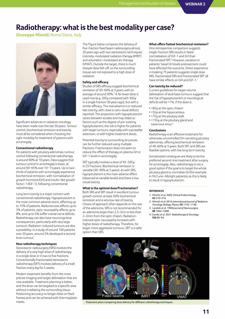
A study of 27 treatment-naïve acromegaly patients was carried out to investigate this relationship.4 Polysomnography, ESS and insulin-like growth factor-1 (IGF-1) measurements were taken before treatment, and again at 1 year and 2.5 years after baseline. SAS was present in 74.1% of patients at baseline, reducing to 54.2% at 1 year and again to 21.7% at 2.5 years.
The number of patients with severe OSAS decreased from 33.3% at baseline to 8.3% at 1 year, with no patients experiencing severe OSAS after 2.5 years (see Figure, left). SAS severity and sleep parameters also correlated with IGF-1 levels: normal IGF-1 levels were seen in 70.8% of patients after 1 year, and in 100% of patients after 2.5 years. No other clinical parameters appeared to change.
RecommendationsGiven these findings, patients should be screened for SAS when diagnosed with acromegaly, and those at risk should undergo polysomnography. Given the substantial decrease in prevalence and severity of OSAS following acromegaly treatment, polysomnography should be repeated within 1−2 years of treatment.
SAS screening can be based on questionnaires using the ESS. Polysomnography costs more, but is more reliable. Given that nearly three-quarters of patients are affected, reliable measurement is important.
Management and burden of diseaseChairs: Marinella Tzanela (Greece) & Marija Pfeifer (Slovenia)
REFERENCES1. Epstein et al. 2009 Journal of Clinical Sleep Medicine
5 263−276.2. Spicuzza et al. 2015 Therapeutic Advances in Chronic
Disease 6 273–285.3. Gadelha et al. 2019 Endocrine Reviews 40 268−332.4. Wolters et al. 2020 Journal of Clinical Endocrinology &
Metabolism 105 290–304.
Changes in prevalence of OSAS at different levels of severity following acromegaly treatment (mild OSAS, RDI 5−14; moderate OSAS, RDI 15−29; severe OSAS, RDI >29). The difference between T0 and T1 was significant (P<0.0001). Reproduced under CC BY 4.0 licence (https://creativecommons.org/licenses/by/4.0) from Wolters et al.4 https://doi.org/10.1210/clinem/dgz050 ©2019 The Authors.
T0: Baseline (n=27) T1: 1 year (n=24) T2: 2.5 years (n=23)
11
WEBINAR 2Management and burden of disease
Management and burden of diseaseChairs: Marinella Tzanela (Greece) & Marija Pfeifer (Slovenia)
Radiotherapy: what is the best modality per case?Giuseppe Minniti Rome/Siena, Italy
Significant advances in radiation oncology have been made over the last 50 years. Tumour control, biochemical remission and toxicity must all be considered when choosing the right modality for treatment of patients with acromegaly.
Conventional radiotherapyFor patients with pituitary adenomas, tumour control following conventional radiotherapy is around 90% at 10 years. Data suggest that tumour control in acromegaly is lower, at around 50−65% over 10−15 years. Up to two-thirds of patients with acromegaly experience biochemical remission, with normalisation of growth hormone (GH) and insulin-like growth factor-1 (IGF-1), following conventional radiotherapy.
Long term toxicity is a major concern with conventional radiotherapy. Hypopituitarism is the most common adverse event, affecting up to 75% of patients. Radionecrosis affects up to 3% of patients, optic neuropathy affects up to 8%, and up to 5% suffer cranial nerve deficits. Radiotherapy can also have neurocognitive consequences, particularly with very large tumours. Radiation-induced tumours are also a possibility: in a study of around 100 patients over 20 years, around 2% developed a second brain tumour.1
New radiotherapy techniquesStereotactic radiosurgery (SRS) involves the delivery of a very high dose of radiotherapy in a single dose or in two to five fractions. Conventionally fractionated stereotactic radiotherapy (SRT) involves delivery of a small fraction every day for 5 weeks.
Modern treatment benefits from the more precise imaging and target delineation that are now available. Treatment planning is better, and the dose can be targeted to a specific area without irradiating the surrounding tissue. Positioning accuracy no longer relies on fixed frames and can be achieved with thermoplastic masks.
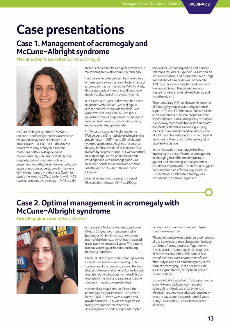
The Figure below compares the delivery of five-fraction fixed beam radiosurgery almost 20 years ago with two stereotactic techniques: intensity-modulated radiation therapy (IMRT) and volumetric-modulated arc therapy (VMAT). Outside the target, there is much sharper dose fall-off, so the surrounding tissues are not exposed to a high dose of radiation.
Safety and efficacyStudies of SRS efficacy suggest biochemical remission of 30−60% at 5 years, with an average of around 50%.2 A far lower dose is used now (e.g. 20Gy compared with 30Gy in a single fraction 30 years ago), but with a similar efficacy. The real advance is in reduced late toxicity, with close to zero visual defects reported. The proportion with hypopituitarism varies between studies and may relate to factors such as the degree of pre-existing hypopituitarism; the risk is higher for patients with larger tumours, especially with suprasellar extension, or with higher treatment doses.
Potential toxicity to surrounding structures can be further reduced using multiple fractions. Fractionation does not seem to reduce the effect of therapy on plasma GH or IGF-1 levels in acromegaly.
SRT typically involves a dose of 45−50Gy in 25 fractions. Biochemical remission is variable (30−90% at 5 years). As with SRS, hypopituitarism is the main adverse effect (observed at variable levels) and there is low visual toxicity.
What is the optimal dose/fractionation?Both SRS and SRT result in excellent tumour growth control, at least 50% biochemical remission and a very low rate of toxicity. Choice of approach often depends on the size of the adenoma. SRS is not recommended for an adenoma larger than 2.5–3cm or less than 2–3mm from the optic chiasm. Radiation-induced optic neuropathy increases with higher doses of radiotherapy. Therefore, for larger, more aggressive tumours, SRT is a safer option than SRS.
What offers fastest biochemical remission?One retrospective comparison suggests single-fraction SRS results in faster normalisation of IGF-1 and GH than fractionated SRT.3 However, variation in patients’ basal GH levels pretreatment could have affected the outcome. Direct experience in treating 75 patients suggests single dose SRS, fractionated SRS and fractionated SRT all have similar effects on GH and IGF-1.1
Can toxicity be reduced?Current guidelines for target volume delineation of skull base tumours suggest that the risk of hypopituitarism or neurological deficits will be <1%, if the dose is:
• <8Gy at the optic chiasm• <5Gy at the hippocampus• <7Gy at the pituitary stalk• <15Gy at the pituitary gland and
cavernous sinus.4
ConclusionsRadiotherapy is an effective treatment for otherwise uncontrolled GH-secreting pituitary adenomas, offering biochemical remission of 40–60% at 5 years. Both SRT and SRS are feasible options, with low long term toxicity.
Somatostatin analogues are likely to be the preferred second-line treatment after surgery for acromegaly. Also, radiotherapy is not a good option if the goal is to target the whole pituitary gland to normalise GH (for example, in McCune−Albright patients), as this is likely to result in hypopituitarism.
REFERENCES1. Minniti et al. 2005 Clinical Endocrinology
62 210–216.2. Minniti et al. 2016 International Journal of Radiation
Oncology, Biology, Physics 95 1142–1148.3. Landolt et al. 1998 Journal of Neurosurgery
88 1002–1008.4. Combs et al. 2021 Radiotherapy & Oncology
156 80–94.
Treatment plans comparing dose delivery for different radiotherapy techniques.
12
WEBINAR 2 Management and burden of disease
Acromegaly: psychosocial consequences Nienke Biermasz Leiden, The Netherlands
An acromegaly diagnosis can have a huge physical and psychosocial impact on patients. However, wide variability in presentation, timelines and treatment paths means that it can be challenging to discuss the likely impact with patients. Evaluation of treatment goals and results must include both clinical perspectives and patient-reported outcome measures (PROMs).
Sometimes acromegaly patients will have active disease while their hormone measurements remain within the normal range. In these cases, PROMs covering mental, physical, cognitive, social and sexual well-being may give a fuller readout of the patient’s clinical state.
The key is to remember that quality of life (QoL) is defined as what the patient perceives it to be. It may refer to the gap between the patient’s expectations and their actual present experience of the impact of their condition.1 Measuring QoL is complex, multidimensional and multidisciplinary, and usually involves a general questionnaire or a disease-specific questionnaire. The standard Wilson–Cleary health-related QoL model is a useful starting point.2 It incorporates biological and physiological aspects of disease, symptoms, functional status, perceptions of general health, and QoL.
Patient perspective on psychosocial impactIn a recorded interview, an acromegaly patient reflected on how his diagnosis affected his QoL. His initial concern had been about survival, so he was reassured to learn that the disease is not life-threatening. His condition progressed slowly over 20 years, so he had not realised what was happening. Looking back, he said he now realises that he had experienced cognitive challenges at work and in his personal life.
Because acromegaly is such a rare disease, there is no clear care path for the psychosocial consequences. The patient said he saw
no need for psychosocial support in the aftermath of his treatment because it had gone well. However, several years on, he reflected that the impact on his QoL was much greater than expected. He felt acromegaly had affected his personality and his QoL was reduced. He found his reactions, ways of working, emotions (especially positive feelings) and concentration had all changed or deteriorated. He experienced a lack of drive and motivation. The realisation of being a different person had been hard to accept and explain to other people. He felt angry and frustrated about what had happened to him.
He felt that it would be very helpful for patients to be informed in advance about how the disease may affect their personality and cognitive capacity, and that they may need psychological help later on. He agreed that specialised help from a psychologist would be a good addition to treatment protocols.
Reflections from the clinicLiterature shows that acromegaly has a more significant impact on QoL than other pituitary diseases. Active disease is usually associated with poor QoL and, although surgery and multimodality treatment improve QoL, well-being does not normalise in the long term. Therefore, QoL considerations may add to the interpretation of optimal endocrine treatment, recognising that these will vary from patient to patient.
QoL measures need to become available in individual patient care with feedback and clinical action, though choosing the right questionnaire remains challenging, as many have limitations.
Leiden Bother and Need QuestionnaireTo understand how best to incorporate psychosocial factors in treatment pathways, a series of patient focus groups was undertaken. Data from these focus groups confirm that pituitary disease has multidimensional long term consequences
for patients. Patients reported biological consequences, including cognitive problems, sexual dysfunction and physical complaints. They listed psychological consequences including personality issues, negative feelings and emotional challenges. Social consequences included difficulty with communication, work-related problems and limitations on leisure activities (see Table below).
Based on these findings, the Leiden Bother and Need Questionnaire (LBNQ) has been developed for all pituitary patients, including those with acromegaly. It records the issues that bother patients most and their greatest needs for support on the following subscales: mood problems, negative illness perceptions, issues in sexual functioning, physical and cognitive complaints, and issues in social functioning, as well as a total score.3 It can be used with tools such as Acromegaly Quality of Life (AcroQoL) and Patient-assessed Acromegaly Symptom Questionnaire (PASQ) scores. The aim is to make consideration of QoL a daily practice and offer pituitary patients multidisciplinary support. The measures could be monitored over time to see how acromegaly and other pituitary diseases affect patients’ QoL.
Strategies to improve QoL include:• treating the disease• treating co-morbidities including depression• listening to the patient and addressing
issues raised in the LBNQ through information and rehabilitation
•improving self-management.
A core clinical outcome set that includes patient-reported outcomes for pituitary disease care paths is needed, to evaluate care and facilitate precision-based medicine and value-based healthcare.
REFERENCES1. Calman 1984 Journal of Medical Ethics 10 124–127.2. Wilson & Cleary 1995 JAMA 273 59–65. 3. Andela et al. 2016 Pituitary 19 293–302.
Top 10 highest ‘bothers’ and ‘needs for support’.3
Highest bothered by n (%) Highest needs for support n (%)
Fatigue 63 (17) Fatigue 84 (25)
Difficulties in performing work 42 (12) Afraid that pituitary tumour will recur 68 (20)
Problems concentrating 37 (11) Worried about physical symptoms 65 (19)
More sensitive to stressful situations 35 (10) Problems concentrating 62 (18)
Pain 35 (10) Less interested in sex 55 (16)
Going beyond own limits 34 (10) Mood swings 55 (16)
Less interested in sex 34 (10) Memory problems 54 (16)
Physical problems during sex 34 (10) Difficulties in performing work 52 (15)
Sleeping problems 34 (10) More sensitive to stressful situations 51 (15)
Difficulties letting go of certain thoughts 33 (10) Sleeping problems 50 (15)
Reproduced under CC BY 4.0 licence (https://creativecommons.org/licenses/by/4.0) from Andela et al.3 https://doi.org/10.1007/s11102-016-0707-4 ©2016 The Authors.
13
WEBINAR 2Management and burden of disease
Case 1. Management of acromegaly and McCune−Albright syndrome Mariana Aveiro Lavrador Coimbra, Portugal
Case 2. Optimal management in acromegaly with McCune−Albright syndromeEirini Papadimitriou Athens, Greece
McCune−Albright syndrome (MAS) is a rare, non-heritable genetic disease with an estimated prevalence of between 1 in 100 000 and 1 in 1 000 000. The disease results from early embryonic somatic mutations of the GNAS gene and is characterised by poly-/monostotic fibrous dysplasia, café-au-lait skin spots and polyendocrinopathy. Polyendocrinopathy can cause precocious puberty, growth hormone (GH) excess, hyperthyroidism and Cushing’s syndrome. Around 20% of patients with MAS have acromegaly. Acromegaly in MAS usually
presents earlier and has a higher prevalence in males compared with sporadic acromegaly.
Diagnosis of acromegaly can be challenging in these cases, since the craniofacial effects of acromegaly may be masked by MAS. Similarly, fibrous dysplasia of the sphenoid bone may impair visualisation of the pituitary gland.
In this case, a 22-year-old woman had been diagnosed with MAS at 2 years of age. A detailed clinical history was available, with symptoms including café-au-lait spots, polyostotic fibrous dysplasia of the sphenoid bone, vaginal bleeding, precocious puberty and an accelerated growth rate.
At 10 years of age, her height was in the 97th percentile. She had elevated insulin-like growth factor-1 (IGF-1) and GH levels, and hyperprolactinaemia. Magnetic resonance imaging (MRI) showed the sella turcica fully involved by dysplastic bone, but with a normal pituitary image. At this point, the patient was diagnosed with acromegaly and was prescribed Sandostatin and bromocriptine until the age of 16, when she was lost to follow-up.
When she returned to care at the age of 18, evaluation showed IGF-1 at 590μg/l
and a nadir GH reading during oral glucose tolerance test of 4.83µg/l. She was started on lanreotide (90mg) and bromocriptine (2.5mg) immediately. Lanreotide was increased to 120mg after 2 years. Biochemical remission was not achieved. The patient was also treated for central adrenal insufficiency and hypothyroidism.
Recent pituitary MRI has shown the presence of pituitary hyperplasia and a hypointense signal in T1 and T2. This could indicate either a microadenoma or fibrous dysplasia of the sphenoid bone. A multidisciplinary discussion is underway to consider the best therapeutic approach, with options including surgery, medical therapy (including the introduction of a GH receptor antagonist or more frequent injections of the somatostatin analogue) or pituitary irradiation.
In the discussion, it was suggested that increasing the dose of somatostatin agonist, or changing to a different somatostatin agonist and combining with pegvisomant, could be a way forward. The literature suggests pegvisomant is an effective way to reduce GH secretion. Combination therapy was considered the optimal approach.
In this case of McCune−Albright syndrome (MAS), a 39-year-old man presented in September 2018 with an extensive bone lesion of the forehead, which had increased in size over the previous 3 years. The patient also had acromegalic features, including increasing hand size.
A facial skull computerised tomography scan showed the bone lesion extending to the frontal area of the head and around the right orbit, plus frontal and sphenoid bone fibrous dysplasia. Bone scintigraphy showed fibrous dysplasia of the skull and sacrum, and bone metabolism markers were elevated.
Hormonal investigations confirmed the acromegaly diagnosis: insulin-like growth factor-1 (IGF-1) levels were elevated and growth hormone (GH) was not suppressed during oral glucose tolerance test. Elevated prolactin and hypogonadotrophic
Case presentations
hypogonadism were also evident. Thyroid function was normal.
The patient underwent partial surgical removal of the bone lesion, and subsequent histology confirmed fibrous dysplasia. Together with the diagnosis of acromegaly, the diagnosis of MAS was established. This patient had two of the three classic symptoms of MAS: fibrous dysplasia and endocrinopathy in the form of acromegaly. He did not have café-au-lait pigmentation, or any heart or liver co-morbidities.
He was initially treated with 120mg lanreotide every 4 weeks, with pegvisomant and cabergoline introduced after 6 months. Medical therapies were adjusted repeatedly over the subsequent approximately 2 years, though biochemical remission was never achieved.
16 Hypoparathyroidism14
WEBINAR 2 Management and burden of disease
Case 3. Gigantism Neha Malhotra London, UK
Treatment considerationsAround 10−20% of patients with MAS develop acromegaly. These patients almost always have skull base fibrous dysplasia, which means pituitary surgery is not the best option. Dysplastic bones make it technically difficult, and risk of haemorrhage is high.
Patients are usually treated medically, with somatostatin analogues, pegvisomant, dopamine agonists, or a combination. Pituitary irradiation carries an increased risk of osteosarcoma and is therefore not recommended.
Discussion of treatment options in the literature is limited. A review of 112 cases1 made the following comparisons:
• only 3 out of 25 cases treated with pituitary surgery experienced disease remission
This was a rare case of a teenager with symptoms of gigantism and a pituitary macroadenoma, with early but inconsistent biochemical evidence of acromegaly.
The patient was referred to Great Ormond Street Hospital (London, UK) at the age of 12, due to concerns about his height. He was 194.2cm and weighed 139.15kg. He was already at the end of pubertal development and had been diagnosed with mild autism. He had no specific facial features to suggest overgrowth syndrome and no café-au-lait spots, though he did sweat a lot and reported headaches with screen time.
Magnetic resonance imaging (MRI) showed a macroadenoma in the left anterior pituitary gland, with no cavernous sinus invasion or impingement on the optic chiasm. Cerebral parenchymal appearances were normal. He had an advanced bone age of 14 years and 9 months.
• 4 out of 29 patients treated with radiotherapy were in remission, while 3 out of 6 who had pituitary radiation had developed sarcoma
• 12.5% of the 32 cases treated with dopamine agonists experienced remission
• around 32% of 56 cases treated with somatostatin analogues experienced remission, increasing to 46% in cases of microadenoma, and decreasing to 14% in cases of macroadenoma
• pegvisomant gave a remission rate of 77% in a sample of 13 cases.
In this case, the patient is currently receiving 100mg pegvisomant weekly, 120mg lanreotide every 4 weeks, and 0.5mg cabergoline twice weekly. Although biochemical remission has not been achieved, there are no acromegalic co-morbidities
Baseline insulin-like growth factor-1 (IGF-1) was within the upper limit of the normal reference range and other baseline pituitary blood markers were within normal ranges. An oral glucose tolerance test (OGTT) showed that growth hormone (GH) failed to suppress. Given his age and development, the plan at this point was to continue surveillance using MRI scans and baseline pituitary blood tests.
At age 14, an MRI scan showed a well-defined pituitary lesion, measuring around 7mm by 6mm, again with no suprasellar or cavernous extension. The patient reported no new symptoms, and his GH and IGF-1 levels were fairly stable. Again, the plan was to continue surveillance with MRI scans and pituitary function tests.
A clinical review when the patient was 15 years of age determined that height velocity was stable at 2cm per year. It was considered unlikely that he had pituitary gigantism as his final height was only just above the 99.6th percentile. MRI showed no significant change in size, morphology, signal or enhancement of the pituitary lesion.
However, while GH was never undetectable, it was consistently in the 1–2ng/ml range, which supports a gigantism diagnosis. The plan was to repeat the OGTT and full baseline pituitary function tests and clarify whether the patient had symptoms of acromegaly.
The next OGTT suggested insulin resistance, and so the patient was started on metformin. The daytime GH profile was between 0.9μg/l and 1.9µg/l. The genetic report did not give any confirmed diagnosis for his pituitary adenoma.
and magnetic resonance imaging shows a decrease in lesion size. Options now include increasing the dose of pegvisomant or cabergoline, or shortening the interval of lanreotide administration. Given the patient has no co-morbidities, a final option is simply to monitor the patient.
The adenoma is visible in this case. In the discussion, it was suggested that, since the adenoma has already shrunk, the combination of somatostatin analogues and dopamine agonists seems to be working and should continue, potentially with a gradual increase in dose. Surgery and irradiation would not be advised.
The patient is now 17 years old. His baseline pituitary function tests have shown persistently high IGF-1 levels, though they are not extreme. GH has never suppressed and has never been undetectable. The question now is whether to watch and wait, or to proceed with transsphenoidal surgery followed by somatostatin receptor ligands, given that he is asymptomatic with stable IGF-1 levels.
In the discussion, it was agreed that the goal should be to normalise IGF-1 and GH, though it was suggested that even though GH levels are not high, the fact that they don’t suppress supports an acromegaly diagnosis.
Knowing when to proceed with treatment is challenging with this age group. Earlier transsphenoidal surgery could have led to hypopituitarism, which would have then required replacement medication. This had been ruled out given the patient’s learning difficulties and social situation. It was agreed that it was sensible to deal with the disease post-puberty, though waiting too long could increase the risk of acromegalic co-morbidities or diabetes.
REFERENCE1. Salenave et al. 2014 Journal of Clinical Endocrinology &
Metabolism 99 1955–1969.
Hypoparathyroidism 17 15
Challenges and future landscape
Case 3. Gigantism Neha Malhotra London, UK
WEBINAR 3
Discrepancies between clinical outcomes and biochemistry: new toolsPietro Maffei Padua, Italy
Treatment for acromegaly aims to ameliorate symptoms, reduce morbidity and mortality, and control hormonal hypersecretion and tumour growth. Clinical features and biochemical profiles can sometimes give conflicting information in patients with suspected or confirmed acromegaly. This can hamper the diagnostic and decision-making process. Practical tools to assist the holistic assessment and monitoring of disease stage and progression are needed.
Patient-reported outcome toolsThese tools allow patients to give their perspectives on their symptoms and quality of life (QoL). The Patient-assessed Acromegaly Symptom Questionnaire (PASQ) is disease-specific; the patient scores headache, excessive sweating, joint pain, fatigue, soft tissue swelling and numbness of the extremities on a scale of 0–8, and their overall health status on a scale of 0–10. Higher scores indicate more severe disease.
Others include the Acromegaly Quality of Life (AcroQoL) questionnaire, the Acromegaly Treatment Satisfaction Questionnaire (Acro-TSQ), the enlargement of the extremities questionnaire, and the acromegaly co-morbidities and complaints questionnaire. These can be used alongside clinician-reported tools, such as SAGIT and the Acromegaly Disease Activity Tool (ACRODAT).
Clinician-reported outcome toolsSAGIT is designed to assist clinicians in staging acromegaly, assessing the response to treatment and adapting patient management in their everyday practice. It is an acronym,
with each letter representing one of five specific disease parameters:
• Signs and symptoms, including sweating, headaches, joint symptoms and swelling
• Associated co-morbidities, such as diabetes, hypertension, sleep apnoea, heart disease, hypopituitarism and active malignant tumour
• Growth hormone (GH) measurements• Insulin-like growth factor-1 (IGF-1) levels• Tumour visibility.1
Global SAGIT scores may range from 1 to 22; higher scores indicate a greater severity of disease. A small pilot study in 2016 found that endocrinologists scored it highly for assessing response to treatment and aiding decision-making, but less so for screening symptoms and side effects.
In a validation study, baseline data suggested that 40% of cases where active disease was not yet controlled did not progress to different or intensified treatment. It confirmed that SAGIT could effectively help clinicians evaluate severity of disease and make better-informed treatment decisions.2 At baseline, the SAGIT subscores for symptoms, GH, IGF-1 and tumour visibility were all found to be significantly lower in patients with controlled acromegaly than in uncontrolled disease. After 2 years, the subscores for GH and IGF-1 showed the most pronounced, significant improvement in patients from the uncontrolled group. A classification and regression tree analysis confirmed that SAGIT could be used to correctly classify disease control status in 93% of controlled cases and 87% of uncontrolled cases, and therefore can be considered a useful tool for disease staging, and was found to be consistent with therapeutic decision-making.3
ACRODAT is a software tool to help clinicians measure disease activity. It examines five parameters that are objective, as well as patient-reported, indicators of disease severity. These are:
• IGF-1 level• tumour status• presence of co-morbidities (cardiovascular
disease, diabetes, sleep apnoea)• signs and symptoms• health-related QoL.
Each is scored on a scale of 1−3:• level 1: patient adequately controlled• level 2: mild disease activity (further
evaluation needed)• level 3: significant disease activity (clinical
action required).In a 2017 validation study,4 practising endocrinologists were asked to judge the disease status of patients in the 243 possible scenarios generated by the five parameters and three different levels within each parameter. The study’s aim was to use these scenarios to assess the degree of agreement of disease status between practising endocrinologists and to develop a model that could predict endocrinologists’ judgement of disease activity status in acromegaly patients. The findings were used to develop an online tool to help clinicians calculate a disease level score for each patient.
Assessing discrepanciesThe Acromegaly Consensus Group strongly recommends GH and IGF-1 as cornerstone biochemical targets for acromegaly treatment, although biochemical control does not necessarily correlate with clinical well-being.5 The Group also strongly recommends that a patient-centred approach, accounting for biochemical parameters, co-morbidities, treatment complications and QoL measures, should be considered in treatment decisions.
They suggest that SAGIT, ACRODAT and AcroQoL can be helpful in identifying specific factors for follow-up. When GH and IGF-1 levels are inconsistent, or when patients are only partially responsive to treatment, clinical factors, such as disease-related symptoms, should be used to guide treatment decisions.
In conclusionThese new tools capture clinical features pertinent to assessment, monitoring and therapeutic decision-making that cannot be easily predicted solely on biochemical evaluation. They may provide a precise classification of acromegaly severity for use in daily management, and improve and standardise the application of guidelines.
REFERENCES1. Giustina et al. 2016 Pituitary 19 39–49.2. Giustina et al. 2019 Pituitary 22 476-487.3. Giustina et al. 2021 Journal of Clinical Endocrinology & Metabolism 27 dgab536.4. van der Lely et al. 2017 Pituitary 20 692–701.5. Giustina et al. 2020 Journal of Clinical Endocrinology & Metabolism 105 dgz096.
Challenges and future landscapeChairs: Niki Karavitaki (UK) & Albert Beckers (Belgium)
16
WEBINAR 3 Challenges and future landscape
Discordant GH and IGF-1 results: to treat or not to treat?Stephan Petersenn Hamburg, Germany
If GH assays use different calibrators, focus on specific GH isoforms, or are affected by the presence of GH-binding protein, they will generate different results. Cut-off points also vary by assay.1 This variation has been reflected in guidelines, where different cut-off levels for GH after OGTT have been recommended, ranging from 0.3μg/l to 1µg/l.
GH and IGF-1 cut-offs are not designed to demonstrate normality: their purpose is to balance sufficient sensitivity to detect disease and sufficient specificity to exclude healthy subjects. Cut-off points may vary between clinical scenarios, such as whether the assay is carried out during diagnosis or follow-up.
A 2007 study compared six assays for IGF-1 with different reference ranges, and noted that, across 23 centres in the UK, 70% found a specific patient’s results to be consistent with acromegaly, while 30% would exclude that same patient.2 The problem with reference ranges has reduced as more data have emerged in recent years for patients of different ages.
Co-morbidities can also affect GH and IGF-1 levels and therefore explain inconsistencies. IGF-1 is falsely decreased in malnutrition, hepatic or renal failure, and in catabolic states. GH can be falsely increased in cases of diabetes mellitus, liver or renal disease, and anorexia nervosa (see the Table above). Interestingly, many of the factors that increase GH also reduce IGF-1.3
How often are GH and IGF-1 discordant?A meta-analysis of 39 studies covering more than 7000 cases found that 25.7% of patients presented with discordant biochemical levels.4 Most had elevated IGF-1 levels but normal GH levels, while fewer had increased GH but normal IGF-1 levels. Studies using ultrasensitive assays found a slightly higher level of discordance than those using less sensitive assays. Those with strict cut-offs showed a higher discordance rate than those with conventional cut-offs. The discordance rate was also higher when a mean or random GH test was used, compared with measuring the nadir. Patients treated with somatostatin analogues also had a higher level of discordance.
Other studies have found a lower rate of discordance among patients taking dopamine agonists but higher with radiotherapy. A study of patients with deletion of exon 3 of the GH receptor showed they had higher discordance than those without the deletion.
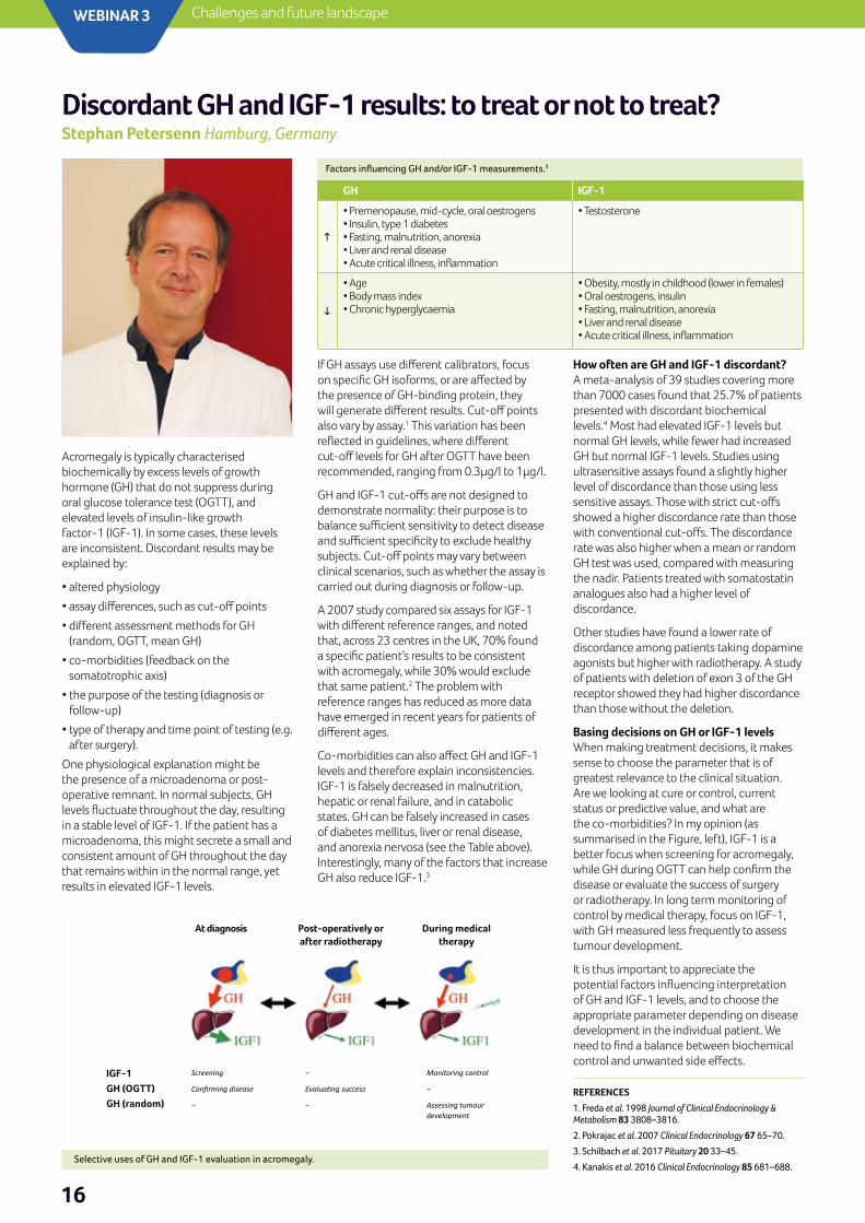
Basing decisions on GH or IGF-1 levelsWhen making treatment decisions, it makes sense to choose the parameter that is of greatest relevance to the clinical situation. Are we looking at cure or control, current status or predictive value, and what are the co-morbidities? In my opinion (as summarised in the Figure, left), IGF-1 is a better focus when screening for acromegaly, while GH during OGTT can help confirm the disease or evaluate the success of surgery or radiotherapy. In long term monitoring of control by medical therapy, focus on IGF-1, with GH measured less frequently to assess tumour development.
It is thus important to appreciate the potential factors influencing interpretation of GH and IGF-1 levels, and to choose the appropriate parameter depending on disease development in the individual patient. We need to find a balance between biochemical control and unwanted side effects.
Acromegaly is typically characterised biochemically by excess levels of growth hormone (GH) that do not suppress during oral glucose tolerance test (OGTT), and elevated levels of insulin-like growth factor-1 (IGF-1). In some cases, these levels are inconsistent. Discordant results may be explained by:
• altered physiology• assay differences, such as cut-off points• different assessment methods for GH
(random, OGTT, mean GH)• co-morbidities (feedback on the
somatotrophic axis)• the purpose of the testing (diagnosis or
follow-up)• type of therapy and time point of testing (e.g.
after surgery).One physiological explanation might be the presence of a microadenoma or post-operative remnant. In normal subjects, GH levels fluctuate throughout the day, resulting in a stable level of IGF-1. If the patient has a microadenoma, this might secrete a small and consistent amount of GH throughout the day that remains within in the normal range, yet results in elevated IGF-1 levels.
REFERENCES1. Freda et al. 1998 Journal of Clinical Endocrinology & Metabolism 83 3808–3816.2. Pokrajac et al. 2007 Clinical Endocrinology 67 65–70.3. Schilbach et al. 2017 Pituitary 20 33–45.4. Kanakis et al. 2016 Clinical Endocrinology 85 681–688.
At diagnosis Post-operatively or after radiotherapy
During medicaltherapy
Screening − Monitoring control
Confirming disease Evalua7ng success −
IGF-1 GH (OGTT) GH (random) − − Assessing tumour
development
Selective uses of GH and IGF-1 evaluation in acromegaly.
GH IGF-1
• Premenopause, mid-cycle, oral oestrogens• Insulin, type 1 diabetes• Fasting, malnutrition, anorexia• Liver and renal disease• Acute critical illness, inflammation
• Testosterone
• Age• Body mass index• Chronic hyperglycaemia
• Obesity, mostly in childhood (lower in females)• Oral oestrogens, insulin• Fasting, malnutrition, anorexia• Liver and renal disease• Acute critical illness, inflammation
Factors influencing GH and/or IGF-1 measurements.3
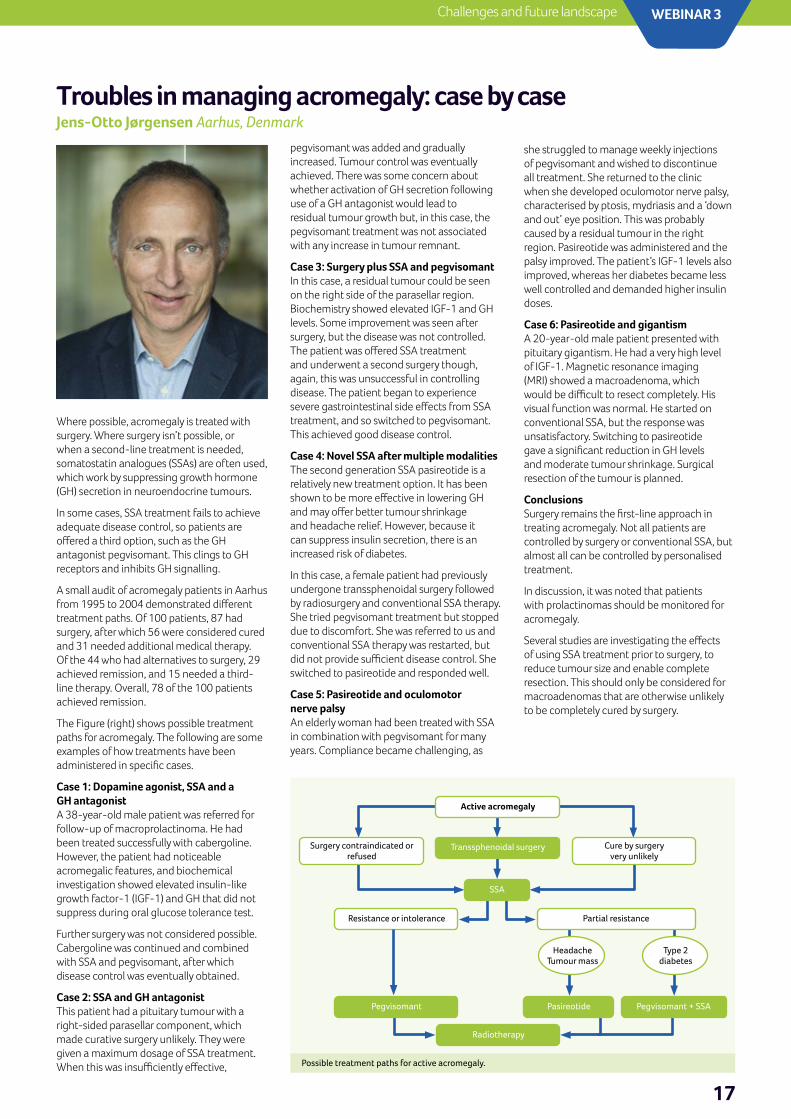
Resistance or intolerance
Radiotherapy
Active acromegaly
Transsphenoidal surgerySurgery contraindicated or refused
Cure by surgery very unlikely
SSA
Type 2diabetes
HeadacheTumour mass
Pegvisomant Pasireotide Pegvisomant + SSA
17
Troubles in managing acromegaly: case by caseJens-Otto Jørgensen Aarhus, Denmark
pegvisomant was added and gradually increased. Tumour control was eventually achieved. There was some concern about whether activation of GH secretion following use of a GH antagonist would lead to residual tumour growth but, in this case, the pegvisomant treatment was not associated with any increase in tumour remnant.
Case 3: Surgery plus SSA and pegvisomantIn this case, a residual tumour could be seen on the right side of the parasellar region. Biochemistry showed elevated IGF-1 and GH levels. Some improvement was seen after surgery, but the disease was not controlled. The patient was offered SSA treatment and underwent a second surgery though, again, this was unsuccessful in controlling disease. The patient began to experience severe gastrointestinal side effects from SSA treatment, and so switched to pegvisomant. This achieved good disease control.
Case 4: Novel SSA after multiple modalitiesThe second generation SSA pasireotide is a relatively new treatment option. It has been shown to be more effective in lowering GH and may offer better tumour shrinkage and headache relief. However, because it can suppress insulin secretion, there is an increased risk of diabetes.
In this case, a female patient had previously undergone transsphenoidal surgery followed by radiosurgery and conventional SSA therapy. She tried pegvisomant treatment but stopped due to discomfort. She was referred to us and conventional SSA therapy was restarted, but did not provide sufficient disease control. She switched to pasireotide and responded well.
Case 5: Pasireotide and oculomotor nerve palsyAn elderly woman had been treated with SSA in combination with pegvisomant for many years. Compliance became challenging, as
she struggled to manage weekly injections of pegvisomant and wished to discontinue all treatment. She returned to the clinic when she developed oculomotor nerve palsy, characterised by ptosis, mydriasis and a ‘down and out’ eye position. This was probably caused by a residual tumour in the right region. Pasireotide was administered and the palsy improved. The patient’s IGF-1 levels also improved, whereas her diabetes became less well controlled and demanded higher insulin doses.
Case 6: Pasireotide and gigantismA 20-year-old male patient presented with pituitary gigantism. He had a very high level of IGF-1. Magnetic resonance imaging (MRI) showed a macroadenoma, which would be difficult to resect completely. His visual function was normal. He started on conventional SSA, but the response was unsatisfactory. Switching to pasireotide gave a significant reduction in GH levels and moderate tumour shrinkage. Surgical resection of the tumour is planned.
Conclusions Surgery remains the first-line approach in treating acromegaly. Not all patients are controlled by surgery or conventional SSA, but almost all can be controlled by personalised treatment.
In discussion, it was noted that patients with prolactinomas should be monitored for acromegaly.
Several studies are investigating the effects of using SSA treatment prior to surgery, to reduce tumour size and enable complete resection. This should only be considered for macroadenomas that are otherwise unlikely to be completely cured by surgery.
Where possible, acromegaly is treated with surgery. Where surgery isn’t possible, or when a second-line treatment is needed, somatostatin analogues (SSAs) are often used, which work by suppressing growth hormone (GH) secretion in neuroendocrine tumours.
In some cases, SSA treatment fails to achieve adequate disease control, so patients are offered a third option, such as the GH antagonist pegvisomant. This clings to GH receptors and inhibits GH signalling.
A small audit of acromegaly patients in Aarhus from 1995 to 2004 demonstrated different treatment paths. Of 100 patients, 87 had surgery, after which 56 were considered cured and 31 needed additional medical therapy. Of the 44 who had alternatives to surgery, 29 achieved remission, and 15 needed a third-line therapy. Overall, 78 of the 100 patients achieved remission.
The Figure (right) shows possible treatment paths for acromegaly. The following are some examples of how treatments have been administered in specific cases.
Case 1: Dopamine agonist, SSA and a GH antagonistA 38-year-old male patient was referred for follow-up of macroprolactinoma. He had been treated successfully with cabergoline. However, the patient had noticeable acromegalic features, and biochemical investigation showed elevated insulin-like growth factor-1 (IGF-1) and GH that did not suppress during oral glucose tolerance test.
Further surgery was not considered possible. Cabergoline was continued and combined with SSA and pegvisomant, after which disease control was eventually obtained.
Case 2: SSA and GH antagonistThis patient had a pituitary tumour with a right-sided parasellar component, which made curative surgery unlikely. They were given a maximum dosage of SSA treatment. When this was insufficiently effective,
WEBINAR 3Challenges and future landscape
Possible treatment paths for active acromegaly.
Partial resistance
18
WEBINAR 3 Challenges and future landscape
Case 1. Post-operative management in pituitary GH-secreting microadenomaJudit Tőke Budapest, Hungary
Case 2. Radiotherapy and hepatitis B on antiviral medication in a male with acromegalyKaterina Lavrentaki Athens, Greece
Acromegaly can be a challenging condition to treat. In this case, the source of the patient’s active acromegaly was never determined, and biochemical remission was not achieved, despite attempts to control the disease with different medical therapies.
A male patient presented 10 years ago with fatigue and headache, at the age of 30. Magnetic resonance imaging (MRI) revealed a double pituitary microadenoma, measuring 5mm and 8mm in the left and right intrasellar regions respectively. Lab tests confirmed an acromegaly diagnosis, and the patient was referred for pituitary surgery, which was
performed in 2012. Post-operative histology confirmed a pituitary adenoma, with a slightly elevated Ki67 index of 3–5% and diffuse positive immunostaining for growth hormone (GH). Recent re-evaluation of the 2011 MRI scan suggests that, rather than a double pituitary microadenoma, the patient may have had a single, crescent-shaped tumour, which was totally removed during surgery.
After surgery, the patient had consistently elevated insulin-like growth factor-1 (IGF-1), with a normal GH profile. He reported concerns about fatigue and swelling of his hands and feet. In 10 years, normal IGF-1 levels have never been achieved. He was initially treated with lanreotide (90mg/month), but switched to octreotide (40mg/month) after 3 years because of diarrhoea. Just before switching to octreotide in early 2015, the patient suffered acute necrotising biliary pancreatitis, and underwent a cholecystectomy. The decision was made to continue somatostatin analogues (SSA) throughout this time. In 2020, the patient was started on pegvisomant at a low dose (10mg daily), but suffered drug-induced hepatopathy 11 weeks later, so the treatment was stopped.
The pathological source of the acromegaly remains unknown. His most recent MRI scan showed an adenohypophysis at the base of the sella turcica. No recurrent or residual tumour was visible. In 2021, a 11C-methionine
positron emission tomography-computerised tomography scan also showed no visible tumour.
Next stepsTotal biochemical control of acromegaly has never been achieved (IGF-1 levels at 1.5−2.0 times the upper limit of normal), despite various pharmacologic treatments. There is no doubt that the acromegaly diagnosis is correct: the patient has had consistently high IGF-1 levels, he developed diabetes mellitus, and post-operative histology clearly showed a GH-secreting microadenoma.
Options under consideration include cabergoline therapy, or possible pituitary irradiation. The patient’s marginally elevated QTcF interval precluded his involvement in a clinical trial of pasireotide, but it is uncertain whether this a true contraindication for use of this drug in a patient who might be a good candidate for a good biochemical response.
In discussion, the use of cabergoline was considered a useful option, in preference to radiotherapy.
Another possible explanation is that the patient may have a very mild pituitary carcinoma, with some metastasis in the spinal area or liver. Regular abdominal ultrasound suggests the patient’s liver is intact, but the team may consider lumbar/spinal imaging to check for spinal metastasis.
This case demonstrates the progression of acromegaly over 15 years, in a patient with hepatitis B.
In 2004, a 40-year-old male with hypertension and palpitations was referred to the endocrine department by his cardiologist. Acromegaly was suspected because of acral enlargement, coarsening facial features, frontal bossing, enlarged nose, macroglossia, a bulky handshake, deep voice and snoring.
He had a very high basal growth hormone (GH) level and his insulin-like growth factor-1 (IGF-1) level was twice the upper normal limit. There was no GH suppression during oral glucose tolerance test (OGTT). Anterior pituitary hormones were normal, and there was no evidence of hypercalciuria, hyperparathyroidism or renal stones.
Case presentations
Co-morbidities at the time included cardiac enlargement, colonic polyp, nodular goitre and osteopenia.
Magnetic resonance imaging (MRI) showed a macroadenoma of 3.5cm with suprasellar extension causing pressure on the optic chiasm, and extension into the left cavernous sinus. There was some loss of vision on the left side.
The patient’s medical history included gastritis, bile duct polyps (for which a cholecystectomy was recommended) and chronic prostatitis. He was also diagnosed with hepatitis B, with positive core antibodies and a high viral load. A liver biopsy showed stage VI inflammation and stage III fibrosis, but no cirrhosis or liver failure.
19
WEBINAR 3Challenges and future landscape
Case 3. PANCH tumour causing acromegaly Amy Coulden Birmingham, UK
Acromegaly is most often caused by somatotroph adenoma, but tumours with a neuronal component can trigger the condition too, as shown in this case.
A 36-year-old female presented in August 2017 with a history of acromegalic symptoms over the previous decade or more. Symptoms included rapid weight gain, hirsutism, increasing shoe size, headache, joint pain, carpal tunnel syndrome, menstrual issues and fatigue. In 2014, magnetic resonance imaging (MRI) to investigate trigeminal neuralgia showed a ‘bulky pituitary’. In 2016, she had dental surgery to reset her jaw due to macroglossia and malocclusion. There was a significant diagnostic delay between onset of symptoms and first referral to endocrinology.
On review, there were very clear signs of acromegaly: frontal bossing, prominent facial features, prognathism, tongue and gingival hypertrophy, widening of interdental spaces, facial hirsutism and soft tissue expansion in the hands and feet. Biochemical evaluation
showed very high levels of insulin-like growth factor-1 (IGF-1) and growth hormone (GH) that failed to suppress during oral glucose tolerance test. MRI showed a 16-mm solid pituitary macroadenoma, with cavernous sinus invasion and no chiasm involvement.
She started treatment in September 2017 with a somatostatin analogue, followed by a dopamine agonist introduced in April 2018. Biochemical disease persisted, so transsphenoidal surgery was carried out in July 2018.
Post-operative histology showed a sparsely granulated mixed lacto–somatotroph adenoma, containing focal ganglion-like cells suggestive of pituitary adenoma−neuronal choristoma (PANCH tumour).
There was a significant clinical and biochemical improvement following surgery, and GH and IGF-1 are now within the normal range. No residual tumour was seen in repeat annual imaging. She was offered treatment for several hormone deficiencies (adrenocorticotrophin, gonadotrophin and thyrotrophin), and remains under clinical, biochemical and radiological follow-up.
PANCH tumoursPANCH tumours are rare mixed pituitary adenomas and gangliocytomas, accounting for around 0.5% of sella turcica lesions. They mostly affect females aged 30–60 and are usually associated with excess GH secretion. A 1994 study of 15 patients with PANCH tumours found that 11 had clinical acromegaly and all 15 had positive GH cells in the adenoma.1 PANCH tumours may present less frequently with other hormones in excess, such as prolactin or cortisol. Almost all are diagnosed as macroadenomas prior to surgery.
The proportion of adenomatous and neuronal components of PANCH tumours varies considerably from patient to patient. The vast majority have sparsely granulated somatotroph adenoma, with many plurihormonal tumours (with prolactin as the most common secondary hormone, as in this case), and fewer non-secreting tumours.
Immunohistochemically, they are positive for neuronal markers, anterior hormonal markers and neuroendocrine pituitary markers. Literature shows that often the neuronal part can stain for hormones (not seen here) and the adenomatous components can stain for neuronal markers, and different hormones can appear in both. Pathogenesis remains unclear.
In the limited cases where medical treatment was tried before surgery, somatostatin analogues and dopamine agonists have both been unsuccessful. Surgery remains the main treatment option, with follow-up monitoring through imaging, biochemistry and clinical assessment. Recurrence has been reported.
ConclusionsEven now, there are significant diagnostic delays for acromegaly caused by PANCH tumours, emphasising the importance of having a high index of suspicion. Because of the lack of long term follow-up data, the effect of the neuronal component of the tumour and the likelihood of recurrence are both unclear.
The patient’s initial treatment for acromegaly began with 90mg lanreotide per month in May 2004, increasing to 120mg per month in December 2004. There was some clinical improvement, though IGF-1 remained elevated. He also started antiviral treatment and began on perindopril for hypertension and carvedilol for palpitations.
Transsphenoidal surgery was carried out in February 2005. Biochemical evaluation 3 months after surgery showed persistent disease, with elevated IGF-1 levels and no suppression of GH during OGTT. MRI showed residual tumour.
Various pharmacological treatments were tried, Cabergoline was prescribed in addition to lanreotide, but stopped due to lack of improvement. Pegvisomant led to some improvement, and was stopped after IGF-1
levels returned to within normal limits following conventional radiation therapy. When his IGF-1 level increased again in August 2007, he restarted pegvisomant and responded well.
The patient was lost to follow up between 2012 and 2014, and discontinued both acromegaly and antiviral treatments. When he returned, his IGF-1 level was very high. After restarting lanreotide, his IGF-1 reduced, though not to within normal limits. MRI showed no changes.
Now, the patients’ liver function is deteriorating. His liver enzymes are twice the normal level, though he doesn’t have cirrhosis or liver failure. The residual pituitary tumour mass remains troubling.
How should treatment proceed?The patient responded partially to somatostatin analogues, but GH did not suppress to normal levels despite higher doses. Dopamine agonists were unsatisfactory. A GH receptor antagonist was effective, but the team are concerned about using it again due to the potential impact on the patient’s liver function.
In discussion, it was suggested that lanreotide with a small, gradually increasing dose of pegvisomant may be effective. Another option is to retry the dopamine agonist. There were mixed views about how either option might interact with the patient’s liver damage.
A final option is to wait: clinicians may find this challenging because the disease is not biochemically controlled, but the patient is in good health, so urgent action is not required.
REFERENCE1. Hovarth et al. 1994 Ultrastructural Pathology
18 565–574.

Follow us:ESEndocrinology
EuropeanSocietyofEndocrinology
esehormones
European Society of Endocrinology
ESE Office (UK) Starling House1600 Bristol Parkway NorthBristol BS34 8YU UK
T: +44 (0)1454 642247E: [email protected]
ESE Office (Belgium) c/o InterofficesAvenue des Arts 561000 BruxellesBelgium
www.ese-hormones.org The European Society of Endocrinology is a Company Limited by Guarantee, registered in England and Wales, company registration number: 5540866. Registered charity number: 1123492. Registered office: Redwood House, Brotherswood Court, Great Park Road, Almondsbury Business Park, Bristol BS32 4QW, UK. ©2021 European Society of Endocrinology.Medical Writer: Louise Shanahan; Managing Editor: Caroline Brewser; Design: Qube Design Associates.
Related Documents