NRC Publications Archive (NPArC) Archives des publications du CNRC (NPArC) Publisher’s version / la version de l'éditeur: Protein Expression and Purification, 64, 2, 2009 Escherichia coli expression and refolding of E/K-coil-tagged EGF generates fully bioactive EGF for diverse applications Le, Phuong Mai; Lenferink, Anne; Pinard, M.; Baardsnes, Jason; Massie, Bernard; O'Connor-McCourt, Maureen Contact us / Contactez nous: [email protected]. http://nparc.cisti-icist.nrc-cnrc.gc.ca/npsi/jsp/nparc_cp.jsp?lang=fr L’accès à ce site Web et l’utilisation de son contenu sont assujettis aux conditions présentées dans le site Web page / page Web http://dx.doi.org/10.1016/j.pep.2008.11.005 http://nparc.cisti-icist.nrc-cnrc.gc.ca/npsi/ctrl?action=rtdoc&an=12919053&lang=en http://nparc.cisti-icist.nrc-cnrc.gc.ca/npsi/ctrl?action=rtdoc&an=12919053&lang=fr LISEZ CES CONDITIONS ATTENTIVEMENT AVANT D’UTILISER CE SITE WEB. READ THESE TERMS AND CONDITIONS CAREFULLY BEFORE USING THIS WEBSITE. Access and use of this website and the material on it are subject to the Terms and Conditions set forth at http://nparc.cisti-icist.nrc-cnrc.gc.ca/npsi/jsp/nparc_cp.jsp?lang=en

Welcome message from author
This document is posted to help you gain knowledge. Please leave a comment to let me know what you think about it! Share it to your friends and learn new things together.
Transcript
NRC Publications Archive (NPArC)Archives des publications du CNRC (NPArC)
Publisher’s version / la version de l'éditeur: Protein Expression and Purification, 64, 2, 2009
Escherichia coli expression and refolding of E/K-coil-tagged EGF generates fully bioactive EGF for diverse applicationsLe, Phuong Mai; Lenferink, Anne; Pinard, M.; Baardsnes, Jason; Massie, Bernard; O'Connor-McCourt, Maureen
Contact us / Contactez nous: [email protected].
http://nparc.cisti-icist.nrc-cnrc.gc.ca/npsi/jsp/nparc_cp.jsp?lang=frL’accès à ce site Web et l’utilisation de son contenu sont assujettis aux conditions présentées dans le site
Web page / page Webhttp://dx.doi.org/10.1016/j.pep.2008.11.005http://nparc.cisti-icist.nrc-cnrc.gc.ca/npsi/ctrl?action=rtdoc&an=12919053&lang=enhttp://nparc.cisti-icist.nrc-cnrc.gc.ca/npsi/ctrl?action=rtdoc&an=12919053&lang=fr
LISEZ CES CONDITIONS ATTENTIVEMENT AVANT D’UTILISER CE SITE WEB.
READ THESE TERMS AND CONDITIONS CAREFULLY BEFORE USING THIS WEBSITE.
Access and use of this website and the material on it are subject to the Terms and Conditions set forth athttp://nparc.cisti-icist.nrc-cnrc.gc.ca/npsi/jsp/nparc_cp.jsp?lang=en
Escherichia coli expression and refolding of E/K-coil-tagged EGF generates fully
bioactive EGF for diverse applications
Phuong U. Le a, Anne E.G. Lenferink a,*, Maxime Pinard a,c, Jason Baardsnes a,Bernard Massie a,c, Maureen D. O’Connor-McCourt a,b
aBiotechnology Research Institute, National Research Council of Canada, 6100 Royalmount Avenue, Montréal, Québec, Canada H4P 2R2bDepartment of Biochemistry, McGill University, Montréal, Québec, Canada H3G 1Y6cDepartment of Microbiology and Immunology, Université de Montréal, Québec, Canada H3T 1J4
a r t i c l e i n f o
Article history:
Received 13 May 2008
and in revised form 3 November 2008
Available online 25 November 2008
Keywords:
EGF
E/K-coil
Refolding
Inclusion bodies
Tissue engineering
a b s t r a c t
Heterodimerizing peptides, such as the de novo designed E5/K5 peptide pair, have several applications
including as tags for protein purification or immobilization. Recently, we demonstrated that E5-tagged
epidermal growth factor (EGF), when bound to a K4 expressing adenovirus, promotes retargeting of
the adenovirus to EGFR expressing target cells. In this study, we present the Escherichia coli expression,
refolding and purification of human EGF fused with the E5-coil (E5-coil-EGF) or with the K5-coil (K5-
coil-EGF). EGF receptor phosphorylation and cell proliferation assays demonstrated that the biological
activity of the coil-tagged EGF versions was comparable to that of non-tagged EGF. Additionally, analysis
of the binding of E5/K5-coil-EGF to cell surface EGFR or to soluble EGFR ectodomain, as measured by cell-
based binding competition assays and by SPR-based biosensor experiments, indicated that the coil-
tagged EGF versions bound to EGFR with affinities similar to that of non-tagged EGF. Finally, we show
that E-coil-tagged EGF, but not non-tagged EGF, can retarget a K-coil containing adenovirus to EGF recep-
tor expressing glioblastoma tumor cells. Overall these results indicate that E. coli expression offers a prac-
tical platform for the reproducible production of fully biologically active E5/K5-coil-tagged EGF, and
support applications of heterodimerizing coil-tagged ligands, e.g. the targeting of viruses or other entities
such as nanoparticles to tumor cells, or growth factor immobilization on cell culture scaffolds for tissue
engineering.
Crown copyright � 2008 Published by Elsevier Inc. All rights reserved.
Epidermal growth factor (EGF)1 is a 53 amino acid long polypep-
tide growth factor, which contains a conserved six-cysteine residue
motif that is characteristic of the EGF-domain. Many reports have
shown that EGF plays a key role in the regulation of cell proliferation,
differentiation and migration [1]. EGF binds and activates the epider-
mal growth factor receptor (EGFR), a single transmembrane domain
glycoprotein, which has frequently been implicated in various types
of cancer. There are various mechanisms by which the EGFR can be-
come oncogenic, e.g. by the presence of auto/paracrine ligand loops,
mutations that render the receptor active, or the failure to attenuate
signaling through receptor down regulation (e.g. through heterodi-
merization of EGFR with the orphan ErbB2 receptor). In most cancer
types, it is the overexpression and/or amplification of the EGFR gene
that prevails.
Because of its role in tumor development, the EGFR has been
studied intensively as a therapeutic target. Currently, the two main
types of therapeutics that are being applied are antibodies that
bind to the EGFR extracellular domain, and small-molecule inhib-
itors that target its intracellular tyrosine kinase domain [2]. While
efficient inhibition of EGFR with these drugs can be observed, evi-
dence of resistance to these drugs has been described [3]. There-
fore, the development of other EGFR targeting strategies is
required. Targeted adenovirus-based gene therapy can potentially
be used as an effective treatment for cancer. The application of
adenovirus in cancer treatment has however been limited due to
the wide tropism of the adenovirus. This is caused by the ubiqui-
tous cell surface expression pattern of the CAR and integrin recep-
tors, which then leads to undesired virus uptake and gene
expression in non-targeted tissues. To address this viral tropism
problem, several research groups have introduced modifications
and adaptor targeting-molecules to the virus fiber, which allows
for the retargeting of the adenovirus to tumor cells [4–6].
1046-5928/$ - see front matter Crown copyright � 2008 Published by Elsevier Inc. All rights reserved.
doi:10.1016/j.pep.2008.11.005
* Corresponding author. Fax: +1 514 496 5143.
E-mail address: [email protected] (A.E.G. Lenferink).1 Abbreviations used: EGF, epidermal growth factor; EGFR, EGF receptor; IBs,
inclusion bodies; SPR, surface plasmon resonance; CBB, Coomassie Brilliant Blue;
HPLC, high performance liquid chromatography; EDC, 1-ethyl-3-(3-dimethylamino-
propyl) carbodiimide hydrochloride; NHS, N-hydroxysuccinimide; HEPES:
N-(2-hydroxyethyl) piperazineethanesulfonic acid; PDEA, 2-(2-pyridinyldi-
thio)ethaneamine; Guanidine–HCl, guanidine hydrochloride; EDTA, disodium ethyl-
enediaminetetraacetate; RU, resonance unit; CH3CN, Acetonitril; TFA, trifluoroacetic
acid; PMSF, phenylmethylsulfonyl fluoride.
Protein Expression and Purification 64 (2009) 108–117
Contents lists available at ScienceDirect
Protein Expression and Purification
journal homepage: www.elsevier .com/locate /yprep
The de novo designed E/K heterodimerizing coiled-coil system
as previously described [7] is composed of two distinct peptides,
designated E- and K-coil. Each peptide has five repeats of a seven
amino acid sequence that contains both charged and hydrophobic
residues. Individually, the E5- and K5-coil are highly soluble due to
the large number of charged residues. When interacting, the E5-
and K5-coil form a high affinity coiled-coil dimer [7,8] that has
been proven to be useful for several applications, e.g. as a tag for
purification purposes or for induced protein–protein interaction
studies. We have shown previously using SPR-based biosensor
studies that E5 or K5-coil-tagged receptor ectodomains (from the
transforming growth factor (TGF)-b Types II and III receptors) bind
TGF-b similarly to untagged receptor ectodomains, and that the
coil tags can be used for receptor capture on the biosensor surface
[9,10]. More recently, we used the E/K coiled-coil dimerization sys-
tem to establish a virus retargeting scheme in which E5-coil-EGF
was utilized to retarget K4-coil-expressing adenovirus to EGFR
expressing cells [11].
Bacterial expression systems are frequently used to produce fu-
sion proteins because of their simplicity in genetic manipulation
and low production costs. However, protein refolding is often re-
quired after such productions. This can be a difficult and compli-
cated process that requires many steps and often results in a
very low recovery of the refolded protein. Here, we describe a
straightforward method for the refolding and recovery of pure ac-
tive E5/K5-coil-EGF fusion proteins using anEscherichia coli expres-
sion system. The biological activity of the E5/K5-coil-EGF fusion
proteins was determined by their capacity to induce EGF receptor
phosphorylation, and to stimulate the cell growth. The results ob-
tained from both assays indicate that the biological activities of the
E5/K5-coil-EGF fusion proteins are similar to those of native un-
tagged murine submaxillary gland (m)EGF, and recombinant hu-
man (rh)EGF. In addition, analysis of binding using a cell-based
competition assay, as well as the EGFR ectodomain on a SPR-based
biosensor indicates that the EGFR binding characteristics of the E5/
K5-coil-EGF versions are similar to that of non-tagged rhEGF.
In conclusion, our results show that E. coli offers a robust plat-
form for the expression of E/K-coil-tagged proteins, such as E5/
K5-coil-EGF, and that refolding as described here results in the
recovery of a highly biologically active preparation of fusion pro-
tein. Due to the versatile and high affinity nature of the coiled-coil
system, proteins tagged with such coils can be used in diverse
applications. As shown here, the fusion of one coil to EGF enables
it to act as a bifunctional adaptor molecule that can promote the
retargeting of an adenovirus, which expresses the partner coil,
through the EGF receptor. Additionally, immobilization of one of
the coils on a biocompatible scaffold or nanoparticle would allow
for the oriented and stable capture (grafting) of a protein fused
to the partner coil, e.g. a growth factor such as EGF, thereby gener-
ating a homogenous surface that can be used for tissue regenera-
tion purposes or nanoparticle targeting.
Materials and methods
The pGEMT vector was purchased from Promega Corporation
(CA, USA), and the pET32c expression vector and recombinant
enterokinase were from Novagen (CA, USA). QIAquick purification
kit, the Miniprep kit and Ni–NTA superflow resin were purchased
from Qiagen (ON, Canada). Isopropyl-b-D-1-thiogalactopyranoside
(IPTG), ampicillin, guanidine HCl, L-cysteine were from Sigma
(ON, Canada). PCR primers were purchased from IDT (ON, Canada)
and restriction enzymes were from NEB (ON, Canada). Murine EGF
(submaxillary glands) was purchased from Calbiochem (CA, USA)
and recombinant human EGF was from Austral Biological
(CA, USA). Sheep anti-human EGF antibody was a gift from
Dr. H. Gregory and anti-phosphotyrosine monoclonal antibody
was purchased from Upstate Biotechnology Inc. (NY, USA). All
HRP-conjugated secondary antibodies were purchased from Jack-
son ImmunoResearch Laboratories Inc. (PA, USA).
Cell culture
Human lung carcinoma cells (A549) and human glioblastoma
cells (U87MG) were obtained from American Type Culture Collec-
tion and grown in Roswell Park Memorial Institute (RPMI-1640)
media supplemented with 5% fetal bovine serum (FBS) or Dul-
becco’s Modified Eagle’s Medium (DMEM) supplemented with
10% FBS and 2 mM L-glutamine, respectively. Mouse mammary tu-
mor cell line BRI-JM01 (as previously characterized [12]) was
grown in DMEM/F12 media supplemented with 5% fetal bovine
serum. All cell lines were cultured at 37 �C in a humidified incuba-
tor containing 5% CO2.
Construction of the E5/K5-coil-EGF expression vector
Human EGF cDNA was amplified by PCR using the pEZZ18-EGF
construct (gift from Dr. E.J.J. van Zoelen, University of Nijmegen,
The Netherlands) with the following primers, EGFforward: 50-GATC
GAATTCAACAGC GACTCTGAATGCCCG-30 (EcoRI restriction site
underlined) and EGFreverse: 50-GATCAAGCTTTTATTAACGGAGCTCC
CACCA-30 (HindIII restriction site underlined). E5-coil and K5-coil
cDNA was amplified by PCR using the pcDNA3-E5-coil or pcDNA3-
K5-coil construct (gift fromMrs.MyriamBanville, BiotechnologyRe-
search Institute,Montreal,Que., Canada). A15-meraminoacid linker
(Gly4; Ser1)3was addedC-terminally of the E5- andK5-coil using the
following primers, E5-coilforward: 50-GATCCCATGGAGGTATCCGCTT
TAGAGAAAGAA-30 (NcoI restriction site underlined), E5-coilreverse:
50-GATCGAATTCAGAGCCACCGCCACCGCTGCCACCGCCACCAGAGCCA
CCGCCACCCTTCTCAAGTGCTGATACTTCCTT-30 (EcoRI restriction site
underlined, linker in bold italic). K5-coilfor: 50-GATCCCATGG
GAAAGGTATCCGCTTTAAAGGAGAAA-30 (NcoI restriction site under-
lined) and K5-coilrev: 50-GATCGAATTCAGAGCCACCGCCACCGC
TGCCACCGCCACCAGAGCCACCGCCACCCTCTTTAAGTGCTGAAACCTT
TTC-30 (EcoRI restriction site underlined, linker in bold italic). These
fragments were then directly cloned into the pGEMT vector, thus
generating the pGEMT-EGF, pGEMT-E5-coil and pGEMT-K5-coil
constructs. Human EGF cDNA, obtained by an EcoRI/NotI digest of
the pGEMT-EGF construct, was ligated to pGEMT-E5-coil linker or
pGEMT-K5-coil linker construct digested with the same enzymes,
generating the pGEMT-E5-coil-EGF and pGEMT-K5-coil-EGF con-
structs. E5-coil-EGF and K5-coil-EGF fragments were obtained by
digesting these constructs with NcoI and HindIII, after which they
were inserted into the pET32c construct (in frame with the thiore-
doxin). The resulting gene expression plasmid used for the thiore-
doxin-E5-coil-EGF and thioredoxin-K5-coil-EGF fusion protein
production was named pET-E5-coil-EGF and pET-K5-coil-EGF. The
correct sequence of recombinant E5/K5-coil-EGF was verified by
DNA sequence analysis.
Expression and purification of recombinant His-tagged E5/K5-coil-EGF
Escherichia coli BL21 (DE3) cells were transformed (by heat
shock) with pET-E5-coil-EGF or pET-K5-coil-EGF and grown in LB
medium containing ampicillin (100 lg/mL) at 37 �C. At an optical
density of 0.7–1.0 (550 nm), IPTG was added to a final concentra-
tion of 1 mM to induce protein expression. Cultures were grown
for another 4 h at 37 �C, harvested by centrifugation at 5000g
(10 min) and stored overnight at �20 �C. The next day, cell pellets
were washed once with Buffer A (50 mM Tris–HCl, 150 mM NaCl,
5 mM EDTA, and 1 mM PMSF, pH 8.0), resuspended in lysis buffer
(Buffer A + 2% Triton-X100, and 0.4 mg/mL lysozyme) and
incubated on ice (30 min) before being disrupted by sonication.
P.U. Le et al. / Protein Expression and Purification 64 (2009) 108–117 109
Cell lysates were centrifuged at 10,000g (4 �C, 30 min) to recover
the His-E5/K5-coil-EGF inclusion bodies (IBs). IBs were washed
twice with Buffer B (Buffer A + 2% Triton-X100) and twice with
Buffer C (20 mM Tris, 150 mM NaCl, 1% Triton-X100; pH 8.0) with
a centrifugation step in between each wash (4 �C, 10,000g, 10 min),
after which the IBs were solubilized overnight (22 �C) in Buffer C
containing 6 M Guanidine–HCl. The next day, solubilized IBs were
centrifuged at 4 �C (10,000g, 60 min) and the coil-tagged EGF pres-
ent in the supernatant was recovered and further purified by Ni–
NTA affinity chromatography. Bound recombinant protein was
eluted with 40 mL of elution buffer (20 mM Tris–HCl, 150 mM
NaCl, 200 mM imidazole pH 8.0) and the His-E5/K5-coil-EGF frac-
tions were analyzed by SDS–PAGE followed by Coomassie Brilliant
Blue (CBB) staining. Finally, recombinant protein fractions were
pooled, concentrated, subjected to a buffer exchange (20 mM
Tris–HCl buffer; pH 8.0) using an Amicon Ultra device (cut-off:
10 kDa), and the protein concentration was determined by Bicinch-
oninic Acid (BCA) Protein Assay.
Denaturation and refolding
His-E5/K5-coil-EGF was refolded essentially as previously de-
scribed [13]. Briefly, His-E5/K5-coil-EGF was diluted to 0.5 mg/
mL and denatured at 22 �C (90 min) in 0.1 M Tris–HCl buffer (pH
8.4) containing 6 M guanidine hydrochloride and 5 mM L-cysteine.
To initiate folding the denaturant was removed by passing the
sample through a PD-10 column (Amersham Pharmacia Biotech)
equilibrated in 0.1 M Tris–HCl (pH 8.4). Reduced and denatured re-
combinant protein was recovered in a volume of 3.5 mL, which was
immediately diluted in the same Tris–HCl buffer supplemented
with 2.5 mM L-cysteine to a final protein concentration of
0.5 mg/mL for oxidative refolding (24 h, 22 �C). Trifluoroacetic acid
(TFA) was then added, at a final concentration of 1%, to the refolded
His-E5/K5-coil-EGF before purification by high performance liquid
chromatography (HPLC).
HPLC separation
Refolded His-E5/K5-coil-EGF was further purified by HPLC
(Waters, ON, Canada) on a 4.6 � 250 mm Vydac C18 column using
a linear gradient of CH3CN in 0.1% (v/v) TFA at a flow rate of
1 mL/min. Fractions that, at a concentration of 100 ng/mL, were
able to induced EGF receptor phosphorylation were pooled and
freeze-dried. Recombinant enterokinase (1 unit per 50 lg of coil-
tagged-EGF for 16 h at 22 �C) was used to cleave the His-tag from
E5/K5-coil-EGF according to the manufacturer’s instructions. After
cleavage, 0.1 M PMSF was added to neutralize the enterokinase;
the E5/K5-coil-EGF was separated from its His-tag by HPLC using
the conditions described above, prior to lyophilization. Refolded
E5/K5-coil-EGF protein was finally characterized by SDS–PAGE
and western blot analysis using a specific anti-human EGF anti-
body. Immunoreactive bands were detected by a goat anti-sheep
antibody linked to horseradish peroxidase (Jackson), and visual-
ized by chemiluminescence (ECL, Roche Molecular Biochemicals).
Stimulation of receptor tyrosine phosphorylation
A549 were seeded in 24-well plates (80,000 cells/well) and
incubated overnight under the growth conditions described. At
90% confluency, various amounts of rhEGF, mEGF, E5-coil-EGF or
K5-coil-EGF were added and cells were incubated for 10 min at
37 �C. Subsequently, cells were washed once with tissue culture
media and once with PBS (4 �C) and then immediately lysed in
100 ll of hot SDS (2%). Cell lysates were collected, mixed with
reducing sample buffer and boiled for 5 min. Twenty microliters
of these cell lysates were then analyzed on a 7% SDS–PAGE fol-
lowed by western blotting with anti-phosphotyrosine monoclonal
antibody. Immunoreactive bands were detected by an anti-mouse
antibody linked to horseradish peroxidase (Jackson), and visualized
by chemiluminescence (ECL, Roche Molecular Biochemicals).
Experiments were repeated at least three times.
Mitogenic assays
BRI-JM01 cells (40,000 cells/well) were plated in 24-well plates
and incubated overnight under the growth conditions described.
The medium was then replaced with 1 mL serum-free DMEM/F12
and cells were incubated at 37 �C for another 4 h. Various amounts
of mEGF, E5-coil- and K5-coil-EGF were added in 50 ll DMEM/F12
serum-free medium. After 24 h at 37 �C (5% CO2), 100 ll serum-
free media containing 0.5 lCi [3H]-thymidine (Amersham, Piscata-
way, NJ, USA) was added per well. Cells were incubated for 3 h at
37 �C (5% CO2), washed 3 times with PBS (RT), harvested in
250 ll of 0.05% trypsin–EDTA (37 �C, 30 min), and directly trans-
ferred to 5 mL scintillation fluid (Universol; ICN Biochemicals
Inc.). Incorporated [3H]-thymidine was determined using a b coun-
ter (LKB-Wallac). Experiments were performed in triplicates and
repeated at least three times.
[125I]-rhEGF binding competition assay
A549 cells (2.0 � 105 cells/well) were plated in 24-well plates
and incubated overnight under the growth conditions described.
The next day, cells were washed once with ice-cold tissue culture
medium prior to incubation with 175 ll of medium containing
1 nM [125I-rhEGF] and various concentrations of rhEGF or cleaved
E5/K5-coil-EGF. Cells were washed twice with PBS, lysed by adding
250 ll 1% Triton-X100 in PBS per well followed by 1 h incubation
at room temperature. Lysates were evaluated for their [125I-rhEGF]
levels using a c-counter (Wallac Wizard, Perkin Elmer). Experi-
ments were performed in triplicates and repeated at least three
times.
SPR experiments
SPR analysis of the extracellular domain of the EGFR (EGFR-ED)
and E5/K5-coil-EGF interaction was measured using a Biacore 3000
instrument (GE Healthcare Bio-Sciences Corporation). E5-coil and
K5-coil surfaces used to capture E5/K5-coil-EGF were prepared
on Biacore research grade CM-5 sensor chips using the thiol cou-
pling method. The CM-5 surface was activated using a mixture of
0.2 M EDC and 0.05 M NHS in water followed by 80 mM PDEA in
0.1 M sodium borate (pH 8.5). Approximately 2 mg of E5-coil or
K5-coil (containing a N-terminal cysteine) was dissolved in
200 ll peptide buffer (100 mM HEPES, 1.0 M NaCl; pH 7.5) and
flown over the PDEA-activated surface using the MANUAL INJECT
command until approximately 200 RUs of peptide were coupled
to the surface. Remaining activated non-peptide bound thiol
groups were quenched by injecting 50 mM cysteine and 1.0 M NaCl
in 0.1 M sodium formate pH 4.0 followed by 5.0 M guanidine
hydrochloride to remove any non-specifically bound peptide. Con-
trol surfaces were prepared in the same manner without ligand. All
procedures were carried out at a flow-rate of 5 ll/min. E5-coil-EGF
and K5-coil-EGF was diluted to approximately 1 lg/mL in HBS buf-
fer (10 mM HEPES, 3.4 lM EDTA, 150 mM NaCl, 0.005% Tween 20)
and injected over the K5- or E5-coil surface, respectively, using the
MANUAL INJECT command until 70 RUs were captured. This sur-
face remained extremely stable and was used without regenerat-
ing for the duration of the EGFR analysis.
To determine the E5/K5-coil-EGF affinity for EGFR-ED, EGFR-ED
dilution series between 500 nM and 3.9 nM were injected using
the KINJECT command. Every single injection was preceded by
110 P.U. Le et al. / Protein Expression and Purification 64 (2009) 108–117
one PRIME and five buffer injections and followed by an EXTRA-
CLEAN command before the next injection cycle. Three indepen-
dent EGFR-ED dilution series were analyzed over each of the
EGF-coil surfaces. Generated sensorgrams were zero-ed to the
injection start, blank surface subtracted, and double referenced
using a buffer injection. These resulting binding sensorgrams were
analyzed using BiaEvaluation version 4.1 (GE Healthcare Bio-Sci-
ences Corporation) and the affinity of E5/K5-coil-EGF for EGFR-
ED was determined by steady state analysis using the sensorgram’s
plateau values determined by the AVERAGE general fit command.
Generation of Adenovirus vectors
Adenovirus vectors (AdVs) expressing Green Fluorescent Pro-
tein (GFP) and wild-type fiber (Ad5FiberWt/GFP), or GFP and K5-
coil-tagged fiber (Ad5FiberHIK5cDm/GFP), were produced by
cleaving plasmids encoding the Adeno genome with PacI followed
by transfection of 293E (cell line described in [11]) using polyethy-
lenimine (PEI) [14]. After 21 days, when cytopathic effects were
observed, cells were harvested by three cycles of freeze-thawing
and the AdVs were propagated using standard methods. The infec-
tious titer of the AdV was determined by measuring GFP expres-
sion in 293E cells by flow cytometry using a Cytomics FC500
MPL flow Cytometer as described previously [15], and is expressed
in Transducing units ([TU]/ml). Stocks of AdV were purified
through two consecutive centrifugation steps on CsCl gradients
using standard procedures [16]. Titers were determined (OD at
260 nm) and expressed as physical particles/ml. Finally, purified
AdV stocks were resuspended in buffer (20 mM Tris–HCl (pH
8.0), 2.5% glycerol, 25 mM NaCl) and stored at �80 �C.
Targeting assays
About 3 � 105 infectious viral particles of CsCl purified
Ad5FiberHIK5cDm/GFP or Ad5FiberWt/GFP vectors in DMEM sup-
plemented with 5% serum were incubated for 24 h at 37 �C in the
absence or presence of 10 ng of rhEGF or 30 ng of purified E5-
coil-EGF in a final volume of 50 ll. The AdVs were then used to in-
fect U87MG cells at a MOI of 3. Four days later, cells were collected
using Trypsin–EDTA (Sigma–Aldrich, St. Louis, MO), the enzymatic
reaction was terminated by adding formaldehyde (Polysciences
Inc., Washington, DC) to a final concentration of 2%, and cells were
analyzed for GFP expression using a Cytomics FC500 MPL flow
Cytometer (Beckman–Coulter, Miami, Fl) equipped with a 25 mW
488 nm argon ion laser as excitation source.
Viable cell populations were selected using forward and side
scattering in combination with a 488 nm dichroic long pass filter.
The GFP emission was detected using a 550 nm dichroic filter
and a 525 nm band pass filter set.
Results
Construction, expression, and purification of coil-tagged human EGF
fusion proteins
In this study, we produced plasmid constructs encoding for hu-
man EGF (hEGF) fusion proteins that are N-terminally tagged with
either the E5-coil peptide or the K5-coil peptide (E5-coil-EGF or
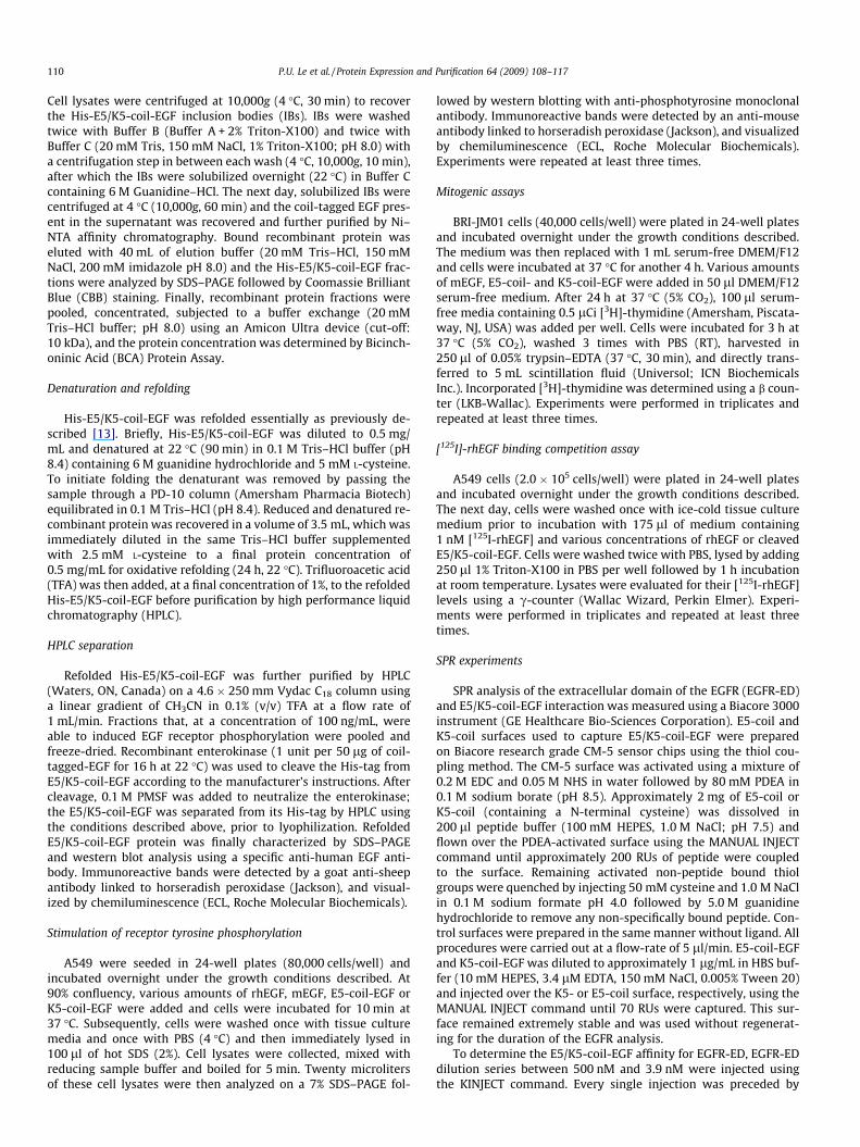
K5-coil-EGF). These EGF fusion proteins (Fig. 1), which have an
apparent molecular weight of approximately 28 kDa, were engi-
neered to include a series of functional domains: a His tag (for
purification purposes), an enterokinase cleavage site (for removal
of all the tags after the purification and refolding), and the E5-coil
or K5-coil domain linked to hEGF by the 15 amino acid linker
((Gly4; Ser1)3).
Using IPTG, E. coli cultures were induced to express high levels
of His-E5-coil- or His-K5-coil-EGF, which accumulated in insoluble
inclusion bodies (IBs). Approximately 30–50 mg of fusion protein
was recovered from 6 M guanidine–HCl-dissolved IBs derived from
500 mL of bacterial culture (Table 1). His-E5/K5-coil-EGF fusion
proteins were purified by Ni–NTA chromatography under reducing
conditions and eluted using 250 mM imidazole. CBB staining of the
SDS–PAGE showed that His-E5-coil-EGF migrated as a single band
of approximately 37 kDa (Fig. 2A), while His-K5-coil-EGF migrated
as a single band of about 30 kDa (Fig. 2B). The discrepancy in MW
between the His-E5-coil-EGF and the His-K5-coil-EGF band is
likely due to the opposite charges present on the E5- and K5-coils.
Purification of the fusion proteins under reducing conditions im-
proved binding to the Ni–NTA resin, and therefore prevented loss
of protein during the wash steps (Fig. 2B). On average a 500 mL
bacterial culture resulted in approximately 25 mg of His-E5-coil-
EGF or 13 mg of His-K5-coil-EGF following Ni–NTA purification
(Table 1).
Refolding and HPLC purification of coil-tagged EGF
Because of the reducing environment of the bacterial cyto-
plasm, proteins often accumulate in inclusion bodies in an inactive
state [17]. Coil-tagged-EGF therefore requires refolding to recover
its biological activities. We refolded His-E5/K5-coil-EGF essentially
as previously described [13]. To avoid protein aggregation the
E
His Tag
Thioredoxin Tag
Enterokinase
T7 promoter
coil-tagged-EGF
Ecoil/Kcoil EGFlinker
NcoI EcoRI HindIII
pET32c-Ecoil-EGF or
pET32c-Kcoil-EGF
S-Tag
Thrombin
Fig. 1. Schematic structure of the gene and construct used for the expression of E5/
K5-coil-tagged hEGF. Human EGF tagged with an E5- or K5-coil through a 15 a.a
linker was cloned into the pET32c expression vector. This vector adds several
additional tags to the fusion protein, which are used for purification (His tag), and
cleavage (enterokinase) purposes.
Table 1
Typical yield of E5/K5-coil-EGF at the different purification and refolding steps.
E5-coil-
EGF
K5-coil-
EGF
Bacteria pellet (wet weight) from 500 mL culture 2.79 g 3.61 g
Solubilized IBs 50 mg 32 mg
After Ni–NTA purification and buffer exchange 25 mg 13 mg
After Refolding and the first HPLC purification 5 mg 2.7 mg
After the protein cleavage and the second HPLC
purification
3 mg 1.5 mg
P.U. Le et al. / Protein Expression and Purification 64 (2009) 108–117 111
refolding was done immediately after protein purification by Ni–
NTA chromatography. Imidazole was removed from Ni–NTA puri-
fied protein by buffer exchange, and recombinant E5/K5-coil-EGF
was diluted to 0.5 mg/mL in Tris buffer (0.1 M, pH 8.4) containing
6 M guanidine hydrochloride and 5 mM cysteine to promote dena-
turation and reduction. Then, guanidine–HCl was rapidly removed
by passing the samples through a PD-10 column. The refolding pro-
cess was completed by incubating the coil-tagged-EGF in Tris buf-
fer (0.1 M, pH 8.4) containing 2 mM cysteine for 24 h (RT).
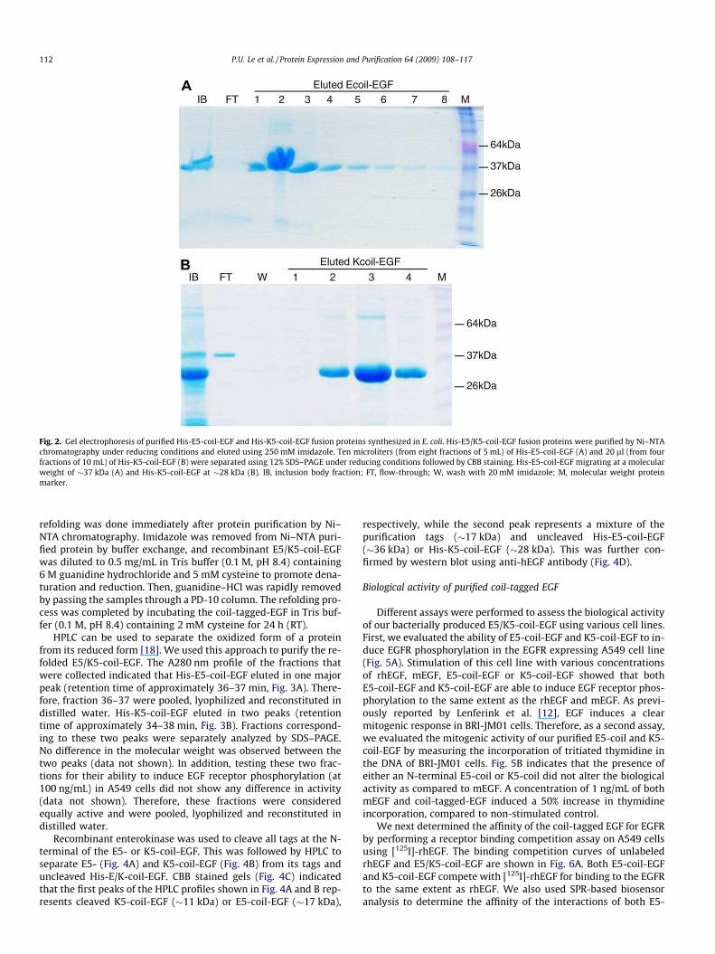
HPLC can be used to separate the oxidized form of a protein
from its reduced form [18]. We used this approach to purify the re-
folded E5/K5-coil-EGF. The A280 nm profile of the fractions that
were collected indicated that His-E5-coil-EGF eluted in one major
peak (retention time of approximately 36–37 min, Fig. 3A). There-
fore, fraction 36–37 were pooled, lyophilized and reconstituted in
distilled water. His-K5-coil-EGF eluted in two peaks (retention
time of approximately 34–38 min, Fig. 3B). Fractions correspond-
ing to these two peaks were separately analyzed by SDS–PAGE.
No difference in the molecular weight was observed between the
two peaks (data not shown). In addition, testing these two frac-
tions for their ability to induce EGF receptor phosphorylation (at
100 ng/mL) in A549 cells did not show any difference in activity
(data not shown). Therefore, these fractions were considered
equally active and were pooled, lyophilized and reconstituted in
distilled water.
Recombinant enterokinase was used to cleave all tags at the N-
terminal of the E5- or K5-coil-EGF. This was followed by HPLC to
separate E5- (Fig. 4A) and K5-coil-EGF (Fig. 4B) from its tags and
uncleaved His-E/K-coil-EGF. CBB stained gels (Fig. 4C) indicated
that the first peaks of the HPLC profiles shown in Fig. 4A and B rep-
resents cleaved K5-coil-EGF (�11 kDa) or E5-coil-EGF (�17 kDa),
respectively, while the second peak represents a mixture of the
purification tags (�17 kDa) and uncleaved His-E5-coil-EGF
(�36 kDa) or His-K5-coil-EGF (�28 kDa). This was further con-
firmed by western blot using anti-hEGF antibody (Fig. 4D).
Biological activity of purified coil-tagged EGF
Different assays were performed to assess the biological activity
of our bacterially produced E5/K5-coil-EGF using various cell lines.
First, we evaluated the ability of E5-coil-EGF and K5-coil-EGF to in-
duce EGFR phosphorylation in the EGFR expressing A549 cell line
(Fig. 5A). Stimulation of this cell line with various concentrations
of rhEGF, mEGF, E5-coil-EGF or K5-coil-EGF showed that both
E5-coil-EGF and K5-coil-EGF are able to induce EGF receptor phos-
phorylation to the same extent as the rhEGF and mEGF. As previ-
ously reported by Lenferink et al. [12], EGF induces a clear
mitogenic response in BRI-JM01 cells. Therefore, as a second assay,
we evaluated the mitogenic activity of our purified E5-coil and K5-
coil-EGF by measuring the incorporation of tritiated thymidine in
the DNA of BRI-JM01 cells. Fig. 5B indicates that the presence of
either an N-terminal E5-coil or K5-coil did not alter the biological
activity as compared to mEGF. A concentration of 1 ng/mL of both
mEGF and coil-tagged-EGF induced a 50% increase in thymidine
incorporation, compared to non-stimulated control.
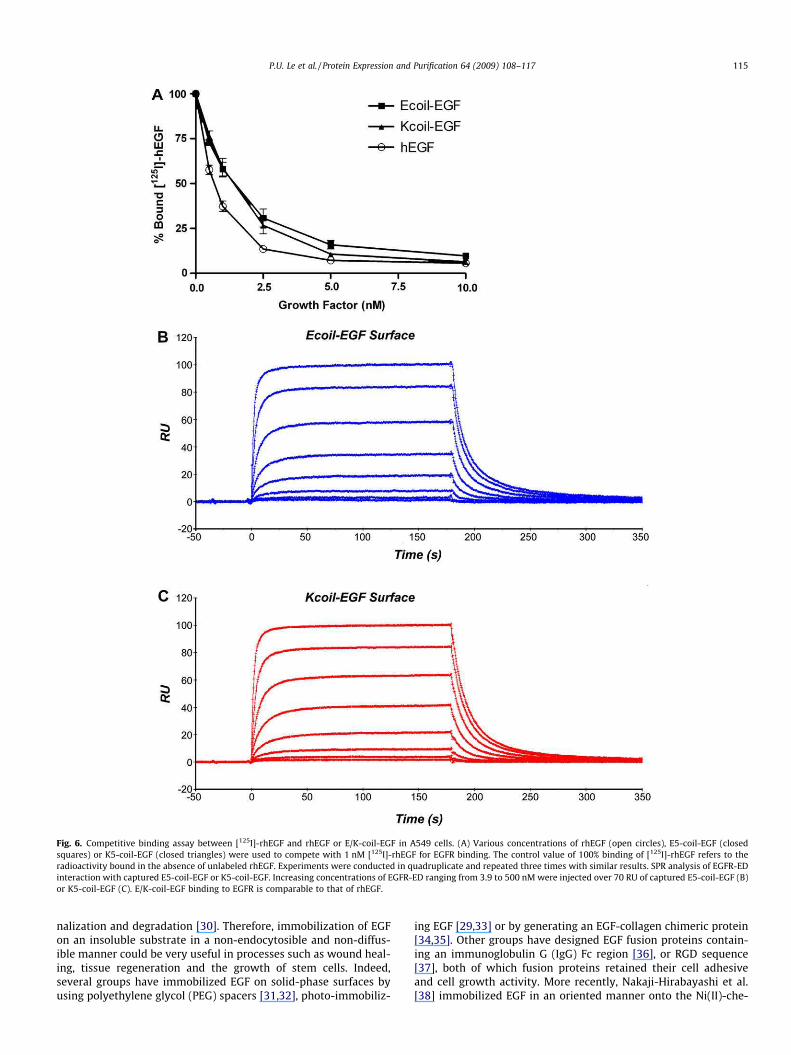
We next determined the affinity of the coil-tagged EGF for EGFR
by performing a receptor binding competition assay on A549 cells
using [125I]-rhEGF. The binding competition curves of unlabeled
rhEGF and E5/K5-coil-EGF are shown in Fig. 6A. Both E5-coil-EGF
and K5-coil-EGF compete with [125I]-rhEGF for binding to the EGFR
to the same extent as rhEGF. We also used SPR-based biosensor
analysis to determine the affinity of the interactions of both E5-
Fig. 2. Gel electrophoresis of purified His-E5-coil-EGF and His-K5-coil-EGF fusion proteins synthesized in E. coli. His-E5/K5-coil-EGF fusion proteins were purified by Ni–NTA
chromatography under reducing conditions and eluted using 250 mM imidazole. Ten microliters (from eight fractions of 5 mL) of His-E5-coil-EGF (A) and 20 ll (from four
fractions of 10 mL) of His-K5-coil-EGF (B) were separated using 12% SDS–PAGE under reducing conditions followed by CBB staining. His-E5-coil-EGF migrating at a molecular
weight of �37 kDa (A) and His-K5-coil-EGF at �28 kDa (B). IB, inclusion body fraction; FT, flow-through; W, wash with 20 mM imidazole; M, molecular weight protein
marker.
112 P.U. Le et al. / Protein Expression and Purification 64 (2009) 108–117
coil-EGF and K5-coil-EGF with the extracellular domain of the
EGFR-ED. An E- or K-coil surface was used to capture the K5-coil
and E5-coil-tagged EGF, respectively, after which various dilutions
of the extracellular domain of EGFR-ED were injected. Steady state
analysis demonstrated that E5-coil-EGF and K5-coil EGF exhibited
similar average kDs (183 ± 12.1 and 146 ± 6.11 nM, respectively).
These affinities are comparable to the values reported for mEGF
and EGFR-ED (200 nM) using the SPR technology [19]. It can
therefore be concluded from these experiments that the biological
activities of rhEGF are not affected by the presence of an N-termi-
nal E5- or K5-coil. Taken together, these results demonstrate that
biologically active E5/K5-coil-EGF fusion proteins can be recovered
from transformed bacteria by oxidative refolding.
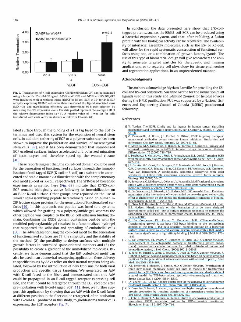
E-coil-tagged EGF retargets K-coil AdVs to EGFR expressing U87MG
cells
In order to demonstrate a functional advantage for coil-
tagged EGF, as compared to untagged EGF, we compared the
abilities of E-coil-EGF and rhEGF to retarget K-coil expressing
adenovirus through the EGFR. Because EGFR is present on sev-
eral cancer cell types, such as U87MG glioblastoma cells, EGFR
is thought to be a good target for improving the tumor tropism
of AdVs. We incubated an AdV that has the K5-coil fused in its
fiber knob (Ad5FiberHIK5cDm/GFP), as well as an AdV that
expresses wild type fiber (Ad5FiberWt/GFP), with rhEGF or the
E-coil-EGF purified in this study. We then evaluated retargeting
competency in U87MG cells. As shown in Fig. 7, Ad5FiberWt/GFP
(white bars) and Ad5FiberHIK5cDm/GFP (black bars), alone or in
the presence of rhEGF, did not efficiently transduce these cells.
Notably, pre-incubation of E-coil-EGF with Ad5FiberHIK5cDm/
GFP, which expresses the complementary K-coil, resulted in
efficient transduction of the EGFR expressing U87MG cells. In
contrast, pre-incubation of E-coil-EGF with the Ad5FiberWt/GFP
control virus did not affect transduction efficiency. Together,
these results indicate that the coiled-coil dependant interaction
between E-coil-EGF and Ad5FiberHIK5cDm/GFP enables E-coil-
EGF to act as a bispecific adaptor that promotes the retargeting
of this AdV through the EGF receptor.
Discussion
In themanuscript presented here we describe the engineering of
novel recombinant EGF fusion proteins that are N-terminally tagged
with an E5-coil or a K5-coil peptide (E5/K5-coil-EGF), and their
expression and isolation from E. coli inclusion bodies (IBs). The IBs,
which contain the recombinant protein in a highly enriched form
low in contaminants, can be easily recovered by cell lysis followed
by centrifugation. Native EGF contains three disulfide bonds that
are vital for its biological activity. Since the reducing environment
of the bacterial cytosol prevents the formation of these disulfide
bonds, it promotes the manifestation of protein aggregates in these
IBs [20]. Therefore, the EGF recovered from these IBs requires refold-
ing in order to regain its biological function.
It has been reported that, during refolding, approximately
43% of EGF becomes kinetically trapped in a single stable 2-
disulfide form (EGFII). When performing refolding in the absence
of redox agents, this form is unable to convert to the native EGF
form. Furthermore, conversion of EGFII to the native EGF struc-
ture requires the crossing of an energy barrier that demands
extensive unfolding of the EGFII [13]. In agreement with these
reports, we found that refolding of E5/K5-coil-EGF in the pres-
ence of different concentrations of reduced/oxidized glutathione,
a commonly used redox agent in protein refolding, resulted in
less than 1% biologically active E5/K5-coil-EGF (data not shown).
The addition of arginine which suppresses aggregation [21,22],
glycerol or sucrose which enhance protein stability in solution
[23,24], also did not significantly increase the yields of biologi-
cally active E5/K5-coil-EGF after refolding (data not shown). It
has been reported that cysteine has a higher redox potential
than glutathione [25,26]. Results published by Chang et al. [27]
demonstrated that the use of cysteine increased not only the
rate at which hEGF was refolded but also its yield [13,27].
Refolding of our recombinant E5/K5-coil-EGF in the presence of
cysteine resulted in an approximately 20% yield after purification
(Table 1), confirming that cysteine improves refolding of coil-
tagged EGF, as well as untagged EGF. The yield of E5/K5-coil-
EGF after the two other purification steps (from inclusion bodies
and after cleavage) was approximately 50% (Table 1). This indi-
cates that refolding remains a limiting step, even in the presence
of cysteine. It has been reported by Lu et al. [28] that, when EGF
is mutated to contain an extra cysteine, this residue is able to
act as a ‘‘built-in” cysteine thiol catalyst that potently promotes
disulfide scrambling and refolding. The use of such a mutant in
the context of coil-tagged EGF could potentially increase yield.
Self-assembling oligopeptides, such as the coiled-coil motif,
have been demonstrated to be useful in numerous applications,
e.g. the generation of mini-antibodies [41,42], the assembly of
receptor ectodomain subunits [9,19,43–45], and as an affinity tag
system for purification and biosensor applications [8–10,46–48].
Immobilized EGF has been shown to be more mitogenic for CHO
cells than soluble EGF [29], perhaps due to decreased ligand inter-
Fig. 3. HPLC profile of (A) His-E5-coil-EGF and (B) His-K5-coil-EGF. His-E5-coil-EGF
eluted as one peak (retention time �36–37 min), whereas His-K5-coil-EGF eluted in
two peaks �34–38 min after injection.
P.U. Le et al. / Protein Expression and Purification 64 (2009) 108–117 113
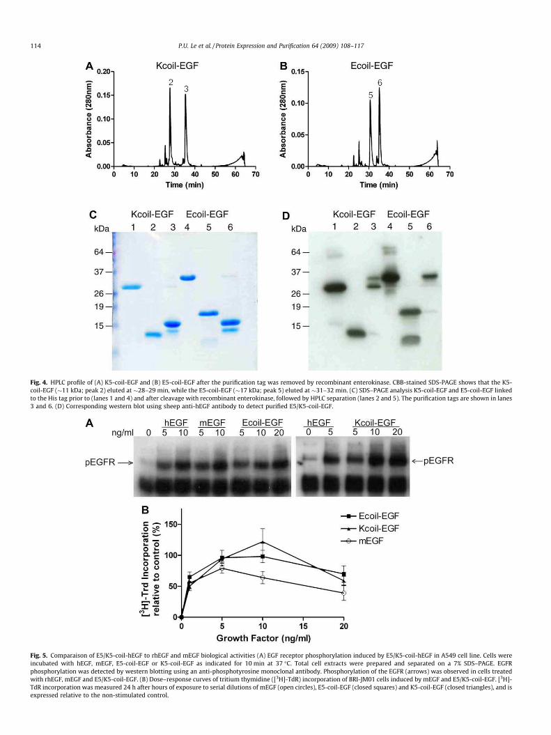
Fig. 4. HPLC profile of (A) K5-coil-EGF and (B) E5-coil-EGF after the purification tag was removed by recombinant enterokinase. CBB-stained SDS-PAGE shows that the K5-
coil-EGF (�11 kDa; peak 2) eluted at �28–29 min, while the E5-coil-EGF (�17 kDa; peak 5) eluted at �31–32 min. (C) SDS–PAGE analysis K5-coil-EGF and E5-coil-EGF linked
to the His tag prior to (lanes 1 and 4) and after cleavage with recombinant enterokinase, followed by HPLC separation (lanes 2 and 5). The purification tags are shown in lanes
3 and 6. (D) Corresponding western blot using sheep anti-hEGF antibody to detect purified E5/K5-coil-EGF.
Fig. 5. Comparaison of E5/K5-coil-hEGF to rhEGF and mEGF biological activities (A) EGF receptor phosphorylation induced by E5/K5-coil-hEGF in A549 cell line. Cells were
incubated with hEGF, mEGF, E5-coil-EGF or K5-coil-EGF as indicated for 10 min at 37 �C. Total cell extracts were prepared and separated on a 7% SDS–PAGE. EGFR
phosphorylation was detected by western blotting using an anti-phosphotyrosine monoclonal antibody. Phosphorylation of the EGFR (arrows) was observed in cells treated
with rhEGF, mEGF and E5/K5-coil-EGF. (B) Dose–response curves of tritium thymidine ([3H]-TdR) incorporation of BRI-JM01 cells induced by mEGF and E5/K5-coil-EGF. [3H]-
TdR incorporation was measured 24 h after hours of exposure to serial dilutions of mEGF (open circles), E5-coil-EGF (closed squares) and K5-coil-EGF (closed triangles), and is
expressed relative to the non-stimulated control.
114 P.U. Le et al. / Protein Expression and Purification 64 (2009) 108–117
nalization and degradation [30]. Therefore, immobilization of EGF
on an insoluble substrate in a non-endocytosible and non-diffus-
ible manner could be very useful in processes such as wound heal-
ing, tissue regeneration and the growth of stem cells. Indeed,
several groups have immobilized EGF on solid-phase surfaces by
using polyethylene glycol (PEG) spacers [31,32], photo-immobiliz-
ing EGF [29,33] or by generating an EGF-collagen chimeric protein
[34,35]. Other groups have designed EGF fusion proteins contain-
ing an immunoglobulin G (IgG) Fc region [36], or RGD sequence
[37], both of which fusion proteins retained their cell adhesive
and cell growth activity. More recently, Nakaji-Hirabayashi et al.
[38] immobilized EGF in an oriented manner onto the Ni(II)-che-
Fig. 6. Competitive binding assay between [125I]-rhEGF and rhEGF or E/K-coil-EGF in A549 cells. (A) Various concentrations of rhEGF (open circles), E5-coil-EGF (closed
squares) or K5-coil-EGF (closed triangles) were used to compete with 1 nM [125I]-rhEGF for EGFR binding. The control value of 100% binding of [125I]-rhEGF refers to the
radioactivity bound in the absence of unlabeled rhEGF. Experiments were conducted in quadruplicate and repeated three times with similar results. SPR analysis of EGFR-ED
interaction with captured E5-coil-EGF or K5-coil-EGF. Increasing concentrations of EGFR-ED ranging from 3.9 to 500 nM were injected over 70 RU of captured E5-coil-EGF (B)
or K5-coil-EGF (C). E/K-coil-EGF binding to EGFR is comparable to that of rhEGF.
P.U. Le et al. / Protein Expression and Purification 64 (2009) 108–117 115
lated surface through the binding of a His tag fused to the EGF C-
terminus and used this system for the expansion of neural stem
cells. In addition, tethering of EGF to a polymer substrate has been
shown to improve the proliferation and survival of mesenchymal
stem cells [39], and it has been demonstrated that immobilized
EGF gradient surfaces induce accelerated and polarized migration
of keratinocytes and therefore speed up the wound closure
[40].
These reports suggest that, the coiled-coil domain could be used
for the generation of functionalized surfaces through the immobi-
lization of coil-tagged EGF (K-coil or E-coil) on a substrate in an ori-
ented and stable manner via dimerization with the complementary
coil motif (E-coil or K-coil, respectively). The SPR-based biosensor
experiments presented here (Fig. 6B) indicate that E5/K5-coil-
EGF remains biologically active following its immobilization on
an E- or K-coil surface. Other groups have reported on the use of
similar self-assembling peptide heterodimers based on human B-
ZIP leucine zipper proteins for the generation of functionalized sur-
faces [49]. In this approach, one peptide was fused to a cysteine,
which allowed for grafting to a polyacrylamide gel, whereas the
other peptide was coupled to the RDGS cell adhesion binding do-
main. Combining the RGDS domain containing peptide with the
modified polyacrylamide gel resulted in a functionalized hydrogel
that supported the adhesion and spreading of endothelial cells
[50]. The advantages for using the coil–coil motif for the generation
of functionalized surfaces are (1) the simplicity and the stability of
the method, (2) the possibility to design surfaces with multiple
growth factors in controlled space-oriented manners and (3) the
possibility to create a gradient of the immobilized molecules. Re-
cently, we have demonstrated that the E/K coiled-coil motif can
also be used in an adenoviral retargeting application. Gene delivery
to specific tissues by AdVs relies on their natural tropism being ab-
lated, followed by the introduction of new tropisms for both viral
production and specific tissue targeting. We generated an AdV
with K-coil fused to the fiber, and demonstrated that this AdV
could be propagated in an E-coil-tagged receptor expressing cell
line, and that it could be retargeted through the EGF receptor after
pre-incubation with E-coil-tagged EGF [11]. Here, we further sup-
port this application by showing that an AdV with the K-coil fused
at different position in the fiber can be retargeted, after incubation
with E-coil-EGF produced in this study, to glioblastoma tumor cells
expressing the EGF receptor (Fig. 7).
In conclusion, the data presented here show that E/K-coil-
tagged proteins, such as the E5/K5-coil-EGF, can be produced using
a bacterial expression system, and that, after refolding, a fusion
protein with full biological activity can be recovered. The availabil-
ity of interfacial assembly molecules, such as the E5- or K5-coil,
will allow for the rapid systematic construction of functional sur-
faces using one, or a combination of, growth factors/ligands. The
use of this type of biomaterial design will give researchers the abil-
ity to generate targeted particles for therapeutic and imaging
applications, or to regulate cell physiology for tissue engineering
and regeneration applications, in an unprecedented manner.
Acknowledgments
The authors acknowledge Myriam Banville for providing the E5-
coil and K5-coil constructs, Suzanne Grothe for the iodination of all
ligands used in this study and Edmund Ziomek for helpful advices
during the HPLC purification. PUL was supported by a National Sci-
ences and Engineering Council of Canada (NSERC) postdoctoral
fellowship.
References
[1] Y. Yarden, The EGFR family and its ligands in human cancer signallingmechanisms and therapeutic opportunities, Eur. J. Cancer 37 (Suppl. 4) (2001)S3–S8.
[2] O. Dassonville, A. Bozec, J.L. Fischel, G. Milano, EGFR targeting therapies:monoclonal antibodies versus tyrosine kinase inhibitors. Similarities anddifferences, Crit. Rev. Oncol. Hematol. 62 (2007) 53–61.
[3] F. Morgillo, M.A. Bareschino, R. Bianco, G. Tortora, F. Ciardiello, Primary andacquired resistance to anti-EGFR targeted drugs in cancer therapy,Differentiation 75 (2007) 788–799.
[4] L. Pereboeva, S. Komarova, J. Roth, S. Ponnazhagan, D.T. Curiel, Targeting EGFRwith metabolically biotinylated fiber-mosaic adenovirus, Gene Ther. 14 (2007)627–637.
[5] J.E. Carette, H.C. Graat, F.H. Schagen, D.C. Mastenbroek, M.G. Rots, H.J. Haisma,G.M. Groothuis, G.R. Schaap, J. Bras, G.J. Kaspers, P.I. Wuisman, W.R. Gerritsen,V.W. van Beusechem, A conditionally replicating adenovirus with strictselectivity in killing cells expressing epidermal growth factor receptor,Virology 361 (2007) 56–67.
[6] N. Belousova, G. Mikheeva, J. Gelovani, V. Krasnykh, Modification of adenoviruscapsid with a designed protein ligand yields a gene vector targeted to a majormolecular marker of cancer, J. Virol. (2007) 630–637.
[7] G. De Crescenzo, J.R. Litowski, R.S. Hodges, M.D. O’Connor-McCourt, Real-timemonitoring of the interactions of two-stranded de novo designed coiled-coils:effect of chain length on the kinetic and thermodynamic constants of binding,Biochemistry 42 (2003) 1754–1763.
[8] H. Chao, M.E. Houston Jr., S. Grothe, C.M. Kay, M. O’Connor-McCourt, R.T. Irvin,R.S. Hodges, Kinetic study on the formation of a de novo designedheterodimeric coiled-coil: use of surface plasmon resonance to monitor theassociation and dissociation of polypeptide chains, Biochemistry 35 (1996)12175–12185.
[9] G. De Crescenzo, P.L. Pham, Y. Durocher, M.D. O’Connor-McCourt,Transforming growth factor-beta (TGF-beta) binding to the extracellulardomain of the type II TGF-beta receptor: receptor capture on a biosensorsurface using a new coiled-coil capture system demonstrates that aviditycontributes significantly to high affinity binding, J. Mol. Biol. 328 (2003) 1173–1183.
[10] G. De Crescenzo, P.L. Pham, Y. Durocher, H. Chao, M.D. O’Connor-McCourt,Enhancement of the antagonistic potency of transforming growth factor-{beta} receptor extracellular domains by coiled coil-induced homo- andheterodimerization, J. Biol. Chem. (2004) 26013–26018.
[11] Y. Zeng, M. Pinard, J. Jaime, L. Bourget, P. Uyen Le, M.D. O’Connor-McCourt, R.Gilbert, B. Massie, A ligand-pseudoreceptor system based on de novo designedpeptides for the generation of adenoviral vectors with altered tropism, J. GeneMed. 10 (2008) 355–367.
[12] A.E. Lenferink, J. Magoon, C. Cantin, M.D. O’Connor-McCourt, Investigation ofthree new mouse mammary tumor cell lines as models for transforminggrowth factor (TGF)-beta and Neu pathway signaling studies: identification ofa novel model for TGF-beta-induced epithelial-to-mesenchymal transition,Breast Cancer Res. 6 (2004) R514–R530.
[13] J.Y. Chang, L. Li, P.H. Lai, A major kinetic trap for the oxidative folding of humanepidermal growth factor, J. Biol. Chem. 276 (2001) 4845–4852.
[14] Y. Durocher, S. Perret, A. Kamen, High-level and high-throughput recombinantprotein production by transient transfection of suspension-growing human293-EBNA1 cells, Nucleic Acids Res. 30 (2002) E9.
[15] J. Cote, L. Bourget, A. Garnier, A. Kamen, Study of adenovirus production inserum-free 293SF suspension culture by GFP-expression monitoring,Biotechnol. Prog. 13 (1997) 709–714.
0.00
1.00
2.00
3.00
4.00
Virus Virus
+ hEGF
Virus
+ ECoil-EGF
Re
lative
T
ran
sd
uctio
n
Ad5FiberWt/GFP
Ad5FiberHIK5cDm/GFP
p=0.012
Fig. 7. Transduction of K-coil expressing Ad5FiberHIK5cDm/GFP can be increased
using a bispecific E5-coil-EGF ligand. Ad5FiberWt/GFP and Ad5FiberHIK5cDM/GFP
were incubated with or without ligand (rhEGF or E5-coil-EGF) at 37� for 24 h. EGF
receptor expressing U87MG cells were then transduced this ligand associated virus
(MOI = 3), and transduction efficiency was determined 96 h post-infection by
measuring the GFP expression levels. The data plotted represent the average ± SD of
the relative fluorescence index (n = 6). A relative value of 1 was set for cells
transduced with each vector in absence of rhEGF or E5-coil-EGF.
116 P.U. Le et al. / Protein Expression and Purification 64 (2009) 108–117
[16] B. Massie, F. Couture, L. Lamoureux, D.D. Mosser, C. Guilbault, P. Jolicoeur, F.Belanger, Y. Langelier, Inducible overexpression of a toxic protein by anadenovirus vector with a tetracycline-regulatable expression cassette, J. Virol.72 (1998) 2289–2296.
[17] K.W. Walker, H.F. Gilbert, Effect of redox environment on the in vitro andin vivo folding of RTEM-1 beta-lactamase and Escherichia coli alkalinephosphatase, J. Biol. Chem. 269 (1994) 28487–28493.
[18] J.Y. Lee, C.S. Yoon, I.Y. Chung, Y.S. Lee, E.K. Lee, Scale-up process for expressionand renaturation of recombinant human epidermal growth factor fromEscherichia coli inclusion bodies, Biotechnol. Appl. Biochem. 31 (Pt. 3) (2000)245–248.
[19] G. De Crescenzo, S. Grothe, R. Lortie, M.T. Debanne, M. O’Connor-McCourt,Real-time kinetic studies on the interaction of transforming growth factoralpha with the epidermal growth factor receptor extracellular domain reveal aconformational change model, Biochemistry 39 (2000) 9466–9476.
[20] H. Lilie, E. Schwarz, R. Rudolph, Advances in refolding of proteins produced inE. coli, Curr. Opin. Biotechnol. 9 (1998) 497–501.
[21] D. Arora, N. Khanna, Method for increasing the yield of properly foldedrecombinant human gamma interferon from inclusion bodies, J. Biotechnol. 52(1996) 127–133.
[22] M. Suenaga, H. Ohmae, S. Tsuji, T. Itoh, O. Nishimura, Renaturation ofrecombinant human neurotrophin-3 from inclusion bodies using asuppressor agent of aggregation, Biotechnol. Appl. Biochem. 28 (Pt. 2) (1998)119–124.
[23] E. De Bernardez Clark, E. Schwarz, R. Rudolph, Inhibition of aggregation sidereactions during in vitro protein folding, Methods Enzymol. 309 (1999) 217–236.
[24] K. Tsumoto, D. Ejima, I. Kumagai, T. Arakawa, Practical considerations inrefolding proteins from inclusion bodies, Protein Expr. Purif. 28 (2003) 1–8.
[25] J.Y. Chang, Controlling the speed of hirudin folding, Biochem. J. 300 (Pt. 3)(1994) 643–650.
[26] B. Raman, T. Ramakrishna, C.M. Rao, Refolding of denatured and denatured/reduced lysozyme at high concentrations, J. Biol. Chem. 271 (1996) 17067–17072.
[27] J.Y. Chang, P. Schindler, U. Ramseier, P.H. Lai, The disulfide folding pathway ofhuman epidermal growth factor, J. Biol. Chem. 270 (1995) 9207–9216.
[28] B.Y. Lu, C. Jiang, J.Y. Chang, Isomers of epidermal growth factor with Ser ? Cysmutation at the N-terminal sequence: isomerization, Stability, unfolding,refolding, and structure, Biochemistry 44 (2005) 15032–15041.
[29] G. Chen, Y. Ito, Y. Imanishi, Photo-immobilization of epidermal growth factorenhances its mitogenic effect by artificial juxtacrine signaling, Biochim.Biophys. Acta 1358 (1997) 200–208.
[30] H.S. Wiley, Trafficking of the ErbB receptors and its influence on signaling, Exp.Cell Res. 284 (2003) 78–88.
[31] P.R. Kuhl, L.G. Griffith-Cima, Tethered epidermal growth factor as a paradigmfor growth factor-induced stimulation from the solid phase, Nat. Med. 2 (1996)1022–1027.
[32] B.J. Klenkler, M. Griffith, C. Becerril, J.A. West-Mays, H. Sheardown, EGF-graftedPDMS surfaces in artificial cornea applications, Biomaterials 26 (2005) 7286–7296.
[33] Y. Ito, J.S. Li, T. Takahashi, Y. Imanishi, Y. Okabayashi, Y. Kido, M. Kasuga,Enhancement of the mitogenic effect by artificial juxtacrine stimulation usingimmobilized EGF, J. Biochem. 121 (1997) 514–520.
[34] F.L. Hall, A. Kaiser, L. Liu, Z.H. Chen, J. Hu, M.E. Nimni, R.W. Beart Jr., E.M.Gordon, Design, expression, and renaturation of a lesion-targeted
recombinant epidermal growth factor-von Willebrand factor fusionprotein: efficacy in an animal model of experimental colitis, Int. J. Mol.Med. 6 (2000) 635–643.
[35] M. Hayashi, M. Tomita, K. Yoshizato, Production of EGF-collagen chimericprotein which shows the mitogenic activity, Biochim. Biophys. Acta 1528(2001) 187–195.
[36] K. Ogiwara, M. Nagaoka, C.S. Cho, T. Akaike, Construction of a novelextracellular matrix using a new genetically engineered epidermal growthfactor fused to IgG-Fc, Biotechnol. Lett. 27 (2005) 1633–1637.
[37] I. Elloumi, R. Kobayashi, H. Funabashi, M. Mie, E. Kobatake, Construction ofepidermal growth factor fusion protein with cell adhesive activity,Biomaterials 27 (2006) 3451–3458.
[38] T. Nakaji-Hirabayashi, K. Kato, Y. Arima, H. Iwata, Oriented immobilization ofepidermal growth factor onto culture substrates for the selective expansion ofneural stem cells, Biomaterials 28 (2007) 3517–3529.
[39] V.H. Fan, K. Tamama, A. Au, R. Littrell, L.B. Richardson, J.W. Wright, A. Wells,L.G. Griffith, Tethered epidermal growth factor provides a survival advantageto mesenchymal stem cells, Stem cells 25 (2007) 1241–1251.
[40] T.J. Stefonek, K.S. Masters, Immobilized gradients of epidermal growth factorpromote accelerated and directed keratinocyte migration, Wound RepairRegen. 15 (2007) 847–855.
[41] P. Pack, A. Pluckthun, Miniantibodies: use of amphipathic helices to producefunctional, flexibly linked dimeric FV fragments with high avidity inEscherichia coli, Biochemistry 31 (1992) 1579–1584.
[42] P. Pack, K. Muller, R. Zahn, A. Pluckthun, Tetravalent miniantibodieswith high avidity assembling in Escherichia coli, J. Mol. Biol. 246 (1995)28–34.
[43] H.C. Chang, Z. Bao, Y. Yao, A.G. Tse, E.C. Goyarts, M. Madsen, E. Kawasaki, P.P.Brauer, J.C. Sacchettini, S.G. Nathenson, et al., A general method for facilitatingheterodimeric pairing between two proteins: application to expression ofalpha and beta T-cell receptor extracellular segments, Proc. Natl. Acad. Sci. USA91 (1994) 11408–11412.
[44] Z. Wu, S.F. Eaton, T.M. Laue, K.W. Johnson, T.R. Sana, T.L. Ciardelli, Coiled-coilmolecular recognition: directed solution assembly of receptor ectodomains,Protein Eng. 7 (1994) 1137–1144.
[45] Z. Wu, K.W. Johnson, B. Goldstein, Y. Choi, S.F. Eaton, T.M. Laue, T.L. Ciardelli,Solution assembly of a soluble, heteromeric, high affinity interleukin-2receptor complex, J. Biol. Chem. 270 (1995) 16039–16044.
[46] B. Tripet, L. Yu, D.L. Bautista, W.Y. Wong, R.T. Irvin, R.S. Hodges, Engineering ade novo-designed coiled-coil heterodimerization domain off the rapiddetection, purification and characterization of recombinantly expressedpeptides and proteins, Protein Eng. 9 (1996) 1029–1042.
[47] H. Chao, D.L. Bautista, J. Litowski, R.T. Irvin, R.S. Hodges, Use of a heterodimericcoiled-coil system for biosensor application and affinity purification, J.Chromatogr. B. Biomed. Sci. Appl. 715 (1998) 307–329.
[48] B. Catimel, M.C. Faux, M. Nerrie, J. Rothacker, L.J. Otvos, J.D. Wade, E.C. Nice,A.W. Burgess, The use of coiled-coil interactions for the analysis andmicropreparative isolation of adenomatous polyposis coli protein complexes,J. Pept. Res. 58 (2001) 493–503.
[49] J.R. Moll, S.B. Ruvinov, I. Pastan, C. Vinson, Designed heterodimerizing leucinezippers with a ranger of pIs and stabilities up to 10(-15) M, Protein Sci. 10(2001) 649–655.
[50] P.J. Willcox, C.A. Reinhart-King, S.J. Lahr, W.F. DeGrado, D.A. Hammer, Dynamicheterodimer-functionalized surfaces for endothelial cell adhesion,Biomaterials 26 (2005) 4757–4766.
P.U. Le et al. / Protein Expression and Purification 64 (2009) 108–117 117
Related Documents