Research Article Errors on the Trail Making Test Are Associated with Right Hemispheric Frontal Lobe Damage in Stroke Patients Bruno Kopp, 1,2 Nina Rösser, 1 Sandra Tabeling, 1,3 Hans Jörg Stürenburg, 3 Bianca de Haan, 4 Hans-Otto Karnath, 4,5 and Karl Wessel 1 1 Cognitive Neurology, Technische Universit¨ at Braunschweig and Department of Neurology, Braunschweig Hospital, Salzdahlumer Street 90, 38126 Braunschweig, Germany 2 Department of Neurology, Hannover Medical School, Carl-Neuberg Street 1, 30625 Hannover, Germany 3 Klinik Niedersachsen, Haupt Street 59, 31542 Bad Nenndorf, Germany 4 Division of Neuropsychology, Center of Neurology, Hertie Institute for Clinical Brain Research, University of T¨ ubingen, Hoppe-Seyler Street 3, 72076 T¨ ubingen, Germany 5 Department of Psychology, University of South Carolina, 1512 Pendleton Street, Columbia, SC 29208, USA Correspondence should be addressed to Karl Wessel; [email protected] Received 30 January 2015; Revised 20 March 2015; Accepted 15 April 2015 Academic Editor: Marjan Jahanshahi Copyright © 2015 Bruno Kopp et al. is is an open access article distributed under the Creative Commons Attribution License, which permits unrestricted use, distribution, and reproduction in any medium, provided the original work is properly cited. Measures of performance on the Trail Making Test (TMT) are among the most popular neuropsychological assessment techniques. Completion time on TMT-A is considered to provide a measure of processing speed, whereas completion time on TMT-B is considered to constitute a behavioral measure of the ability to shiſt between cognitive sets (cognitive flexibility), commonly attributed to the frontal lobes. However, empirical evidence linking performance on the TMT-B to localized frontal lesions is mostly lacking. Here, we examined the association of frontal lesions following stroke with TMT-B performance measures (i.e., completion time and completion accuracy measures) using voxel-based lesion-behavior mapping, with a focus on right hemispheric frontal lobe lesions. Our results suggest that the number of errors, but not completion time on the TMT-B, is associated with right hemispheric frontal lesions. is finding contradicts common clinical practice—the use of completion time on the TMT-B to measure cognitive flexibility, and it underscores the need for additional research on the association between cognitive flexibility and the frontal lobes. Further work in a larger sample, including leſt frontal lobe damage and with more power to detect effects of right posterior brain injury, is necessary to determine whether our observation is specific for right frontal lesions. 1. Introduction Trail making tasks are popular neuropsychological tests [1, 2], because of their ease of administration and the presumed utility as sensitive measures of brain dysfunction [3]. e most widespread trail making task is Trail Making Test, Parts A and B (TMT;[4]). On TMT-A, one connects 25 encircled numbers randomly arranged on a page in ascending order by drawing a pencil line (i.e., 1-2-3⋅⋅⋅ 25). It is the same on TMT-B, except that 25 encircled numbers and letters need to be connected in alternating order (i.e., 1-A-2⋅⋅⋅ 12-L-13). Trail making tasks were originally based on a test measur- ing divided attention, Partington’s Pathways [5]. Halstead [6] recognized its potential for his studies of the biological basis for intelligence. Many other trail making tasks are available meanwhile (e.g., [7]; see [3], for review). Perceptual/motor speed, speed of cognitive processing, (divided) attention, visual search, working memory, executive control, cogni- tive flexibility, and general intelligence contribute to TMT performance. However, there is no consensus about their exact nature and relative contributions (see [8] for review). ere exists a substantial correlation between TMT-A and TMT-B completion times [9], suggesting that they measure “... similar although somewhat different functions” [3, page 668]. Executive abilities (see [10], for discussion) play an important role in TMT-B performance. According to Kortte et al. [11], TMT-B performance correlates more closely with cognitive flexibility measures (i.e., perseveration errors on the Hindawi Publishing Corporation Behavioural Neurology Volume 2015, Article ID 309235, 10 pages http://dx.doi.org/10.1155/2015/309235

Welcome message from author
This document is posted to help you gain knowledge. Please leave a comment to let me know what you think about it! Share it to your friends and learn new things together.
Transcript
Research ArticleErrors on the Trail Making Test Are Associated with RightHemispheric Frontal Lobe Damage in Stroke Patients
Bruno Kopp,1,2 Nina Rösser,1 Sandra Tabeling,1,3 Hans Jörg Stürenburg,3 Bianca de Haan,4
Hans-Otto Karnath,4,5 and Karl Wessel1
1Cognitive Neurology, Technische Universitat Braunschweig and Department of Neurology, Braunschweig Hospital,Salzdahlumer Street 90, 38126 Braunschweig, Germany2Department of Neurology, Hannover Medical School, Carl-Neuberg Street 1, 30625 Hannover, Germany3Klinik Niedersachsen, Haupt Street 59, 31542 Bad Nenndorf, Germany4Division of Neuropsychology, Center of Neurology, Hertie Institute for Clinical Brain Research, University of Tubingen,Hoppe-Seyler Street 3, 72076 Tubingen, Germany5Department of Psychology, University of South Carolina, 1512 Pendleton Street, Columbia, SC 29208, USA
Correspondence should be addressed to Karl Wessel; [email protected]
Received 30 January 2015; Revised 20 March 2015; Accepted 15 April 2015
Academic Editor: Marjan Jahanshahi
Copyright © 2015 Bruno Kopp et al. This is an open access article distributed under the Creative Commons Attribution License,which permits unrestricted use, distribution, and reproduction in any medium, provided the original work is properly cited.
Measures of performance on the Trail Making Test (TMT) are among the most popular neuropsychological assessment techniques.Completion time on TMT-A is considered to provide a measure of processing speed, whereas completion time on TMT-B isconsidered to constitute a behavioral measure of the ability to shift between cognitive sets (cognitive flexibility), commonlyattributed to the frontal lobes. However, empirical evidence linking performance on the TMT-B to localized frontal lesions ismostlylacking. Here, we examined the association of frontal lesions following stroke with TMT-B performance measures (i.e., completiontime and completion accuracymeasures) using voxel-based lesion-behaviormapping, with a focus on right hemispheric frontal lobelesions. Our results suggest that the number of errors, but not completion time on the TMT-B, is associated with right hemisphericfrontal lesions.This finding contradicts common clinical practice—the use of completion time on the TMT-B to measure cognitiveflexibility, and it underscores the need for additional research on the association between cognitive flexibility and the frontal lobes.Further work in a larger sample, including left frontal lobe damage and with more power to detect effects of right posterior braininjury, is necessary to determine whether our observation is specific for right frontal lesions.
1. Introduction
Trail making tasks are popular neuropsychological tests [1, 2],because of their ease of administration and the presumedutility as sensitive measures of brain dysfunction [3]. Themost widespread trail making task is Trail Making Test, PartsA and B (TMT; [4]). On TMT-A, one connects 25 encirclednumbers randomly arranged on a page in ascending orderby drawing a pencil line (i.e., 1-2-3⋅ ⋅ ⋅ 25). It is the same onTMT-B, except that 25 encircled numbers and letters need tobe connected in alternating order (i.e., 1-A-2⋅ ⋅ ⋅ 12-L-13).
Trail making tasks were originally based on a test measur-ing divided attention, Partington’s Pathways [5]. Halstead [6]recognized its potential for his studies of the biological basis
for intelligence. Many other trail making tasks are availablemeanwhile (e.g., [7]; see [3], for review). Perceptual/motorspeed, speed of cognitive processing, (divided) attention,visual search, working memory, executive control, cogni-tive flexibility, and general intelligence contribute to TMTperformance. However, there is no consensus about theirexact nature and relative contributions (see [8] for review).There exists a substantial correlation between TMT-A andTMT-B completion times [9], suggesting that they measure“. . . similar although somewhat different functions” [3, page668]. Executive abilities (see [10], for discussion) play animportant role in TMT-B performance. According to Kortteet al. [11], TMT-B performance correlates more closely withcognitive flexibilitymeasures (i.e., perseveration errors on the
Hindawi Publishing CorporationBehavioural NeurologyVolume 2015, Article ID 309235, 10 pageshttp://dx.doi.org/10.1155/2015/309235

2 Behavioural Neurology
Wisconsin Card Sorting Test) (WCST [12]; see [13], for review)thanwithworkingmemorymeasures (i.e., failure tomaintainset score onWCST).The consistent finding of substantial cor-relations between TMT-B and WCST perseverative indicessuggests cognitive flexibility being among the key executiveabilities underlying performance on the TMT-B (e.g., [11]).
TMT completion time measures are sensitive to the pres-ence of various neurological and psychiatric disorders [14],but their diagnostic utility in differential diagnosis has repeat-edly been questioned [15–17]. The generally accepted linkagebetween cognitive flexibility and frontal lobe functioning(e.g., [18]) suggests that the TMT-B could be used to evaluatefrontal lobe (dys)function. Ricker et al. [19] found TMT-Bcompletion time being related to frontal lobe dysfunction.However, this finding must be treated cautiously givennegative results in studies comparing patients with frontaland posterior brain damage on TMT-B completion time[20, 21]. A meta-analysis by Demakis [22] found significantgroup differences only for completion time on TMT-A, butthe effect size was small, indicating little separation betweenfrontal and posterior groups and relatively poor TMT-Asensitivity and specificity. Bonilha et al. [23] investigatedthe relationship between prefrontal cortical atrophy andneuropsychological performance in schizophrenic patientsand found that decreased Brodmann’s area (BA9) grey mattervolume correlated with poorer task performance on WCSTerrors and TMT-B completion time.
Taken together, the association between TMT-B comple-tion time measures and frontal lobe dysfunction seems tobe relatively weak or even absent. Against this background,it is often thought that prolongation of TMT-B completiontime in the presence of normal TMT-A completion time doessuggest frontal dysfunction. Specifically, subtracting TMT-A from TMT-B completion time is a common method forpartialling out effects of general processing speed difficultiesthat patients might have [1]. Although this subtractionmethod is widely used in clinical practice, we deliberatelydecided to refrain from examining whether TMT-B minusTMT-A completion time data correlate with relevant brainregions. Our reluctance can be traced back to the fact that thedifference between two substantially correlated measures, asin the case ofTMT-A andTMT-B completion times, possessesunacceptable low levels of reliability, thereby precluding itspotential use in clinical practice (see [9], for the rationalebehind this recommendation).
Thus, there remains a need for additional researchon potential associations between cognitive flexibility, asassessed by the TMT, and frontal lobes. TMT-B completionaccuracy represents a promising candidate measure in thatregard since a former study suggested a relationship betweenTMT-B completion accuracy measures and frontal braindysfunctions [24]. Analysis of errors on TMT-B in Stuss etal.’s [24] study indicated that all patients who made two ormore than two errors had frontal lesions (but see [17], for afailure to replicate Stuss et al.’s [24] finding). Further, dividingthe frontal damaged patients into subgroups on the basis ofthe number of errors yielded specificity of brain-behaviorrelations within the frontal lobes: patients with damage indorsolateral frontal areas were most impaired, while those
with damage to themedial frontal lobes were not significantlyaffected on TMT-B completion accuracy.
Klusman et al. [25] distinguished betweenTMT-B shifting(e.g., connecting 1-2⋅ ⋅ ⋅ or A-B⋅ ⋅ ⋅ ) and sequencing errors(e.g., connecting 1-A-3⋅ ⋅ ⋅ or A-2-C⋅ ⋅ ⋅ ). Stuss et al. [24]found notable slowing of TMT-B in patients with frontal lobedamage, but they concluded error analysis providing a moreuseful method to differentiate between frontal and posteriorbrain damage because all patients committing two or moreTMT-B errors had frontal lesions. Notably, many patientswith lesions in the dorsolateral prefrontal cortex (DLPFC)committed two or more errors (irrespective of error type),whereas ventrolateral prefrontal and orbitofrontal lesions didnot affect TMT-B accuracy variables comparably. Stuss andLevine [18, page 415] concluded the following: “TMT-B errors(but not time), therefore, are a valid measure of DLPFCdysfunction.”
In the present study, we investigate the associationbetween TMT completion time and accuracy measures withfrontal lobe damage in stroke patients using voxel-basedlesion-behavior mapping [26–28]. In contrast to traditionaloverlap designs [26] in which the overlapping of lesionboundaries in individual patients fromdifferent groups limitsstudy validity (cf. [26]), voxel-based lesion-behavior analysisyields a statistical approach to uncover brain-behavior rela-tionships without any prior patient categorization. Moreover,previous research on behavioral effects of frontal braindamage often compared patient groups with heterogeneousetiological lesions (see [29]). These shortcomings notwith-standing and based on Stuss et al. [24], we hypothesizethat TMT-B completion accuracy measures, but not TMT-Bcompletion time measures, are sensitive to DLPFC damage.
2. Method
2.1. Subjects. Thirty acute, first-ever, and right-hemisphere-damaged stroke patients with damage involving the frontallobe in most patients participated in the study (see Table 1).These neuropsychological results show that our sample con-sisted of stroke patients without generalized cognitive deficitsas revealed by the Mini-Mental-State-Examination (MMSE,[32]) and without notable verbal disturbances as revealedby the Wortschatz-Test (Vocabulary Test) (WST, [35]) andby the Regensburger Wortflussigkeits-Test (RegensburgerWord Fluency Test) (RWT, [34]). Further, the Modified CardSorting Test (MCST, [33]) (a variant of the WCST) datasuggest that cognitive flexibility and working memory werenot severely disturbed in our patients.
The logic in restricting to right hemispheric strokeswas to exclude patients with paresis of the dominant righthand and/or apraxia, possibly distorting task performance.Further, left-hemisphere strokes might have hampered theunderstanding of task instructions, due to potential presenceof sensory aphasia.1 Patients with traumatic brain injury,brain tumours, subcortical arteriosclerotic encephalopathy,neurodegenerative disease, or gross neurological defects(pronounced pain reported by the patient, left homonymoushemianopia revealed by clinical examination, and hemispa-tial visual neglect) were excluded to ensure symptoms did
Behavioural Neurology 3
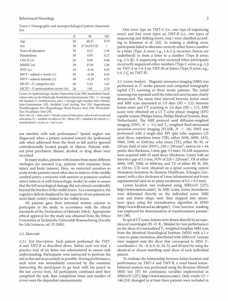
Table 1: Demographic and neuropsychological patient characteris-tics.
𝑁 M SDAge 30 60.17 9.70Sex 30 17 (m)/13 (f) /Years of education 30 12.17 2.16Handedness 30 0.93 0.27CES-D (𝑧) 26 0.10 0.86MMSE (rs) 30 27.50 2.18WST (𝑧) 28 −0.36 0.83RWT—subtest s-words (𝑧) 30 −0.38 0.65RWT—subtest animals (𝑧) 30 −0.29 0.51MCST—𝑁 categories (rs) 26 5.42 1.21MCST—𝑁 perseveration errors (rs) 26 1.81 2.33Center for Epidemiologic Studies Depression Scale [30]; handedness: hand-edness ratio on the Edinburgh Handedness Questionnaire [31]; −1 = stronglyleft-handed, 0 = ambidextrous, and 1 = strongly right-handed; Mini-Mental-State-Examination [32]; Modified Card Sorting Test [33]; RegensburgerWortflussigkeits-Test (Regensburger Word Fluency Test) [34]; Wortschatz-Test (Vocabulary Test) [35].Note. Sex: m =male and f = female; years of education: school and vocationaleducation; N = number of subjects; M =Mean; SD = standard deviation; 𝑧 =standardized 𝑧-score; rs = raw score.
not interfere with task performance.1 Spatial neglect wasdiagnosed when a patient oriented toward the ipsilesionalside when addressed from the front or left and/or ignoredcontralesionally located people or objects. Patients with-out prior psychiatric disease, alcohol, or drug abuse wererecruited.
Inmany studies, patientswith lesions frommanydifferentetiologies are assessed (e.g., patients with traumatic braininjury and brain tumors). Here, we restricted ourselves toacute stroke patients (most often due to infarcts of themiddlecerebral artery, a minority with anterior or posterior cerebralartery infarcts or with hemorrhagic stroke) in order to assurethat the full neurological damage did not extend considerablybeyond the borders of the visible lesion.As a consequence, thecognitive deficits displayed by themajority of our patients aremost likely (solely) related to the visible lesion.
All patients gave their informed written consent toparticipate in the study, in accordance with the ethicalstandards of the Declaration of Helsinki (1964). Appropriateethical approval for the study was obtained from the EthicsCommittee at Technische Universitat Braunschweig (Facultyfor Life Sciences; ref. 37-2010).
2.2. Materials
2.2.1. Test Description. Each patient performed the TMT-A and TMT-B as described above. Before each test trial, apractice trial of six items was administered to ensure taskunderstanding. Participants were instructed to perform thetest as fast and as accurately as possible. During performance,each error was immediately corrected by the examinerinstructing the participant to go back with his pencil tothe last correct item. All participants continued until theycompleted the task. Raw completion times and number oferrors were the dependent measurements.
One error type on TMT-A (i.e., one type of sequencingerror) and four error types on TMT-B (i.e., two types ofsequencing and shifting errors, resp.) were classified accord-ing to Klusman et al. [25]. In making a shifting error,participants failed to alternate correctly either from a numberto a letter (Type A error; e.g., 1-A-2-3; incorrect choices areunderlined) or from a letter to a number (Type B error;e.g., 1-A-B). A sequencing error occurred when participantsincorrectly sequenced either numbers (Type C error; e.g., 1-3on TMT-A or 1-A-3 on TMT-B) or letters (Type D error; e.g.,1-A-2-C on TMT-B).
2.3. Lesion Analysis. Magnetic resonance imaging (MRI) wasperformed in 27 stroke patients and computed tomography(spiral CT) scanning in three stroke patients. The initialscanning was repeated until the infarcted area became clearlydemarcated. The mean time interval between lesion onsetand MRI scan amounted to 3.9 days (SD = 3.2), betweenlesion onset and CT scanning to 2.6 days (SD = 3.7). MRIscans were obtained on a 1.5 T echo planar imaging (EPI)capable system (Philips Intera, PhilipsMedical Systems, Best,Netherlands). The MRI protocol used diffusion-weightedimaging (DWI, 𝑁 = 11) and T
2
-weighted fluid-attenuatedinversion-recovery imaging (FLAIR, 𝑁 = 16). DWI wasperformed with a single-shot EPI spin echo sequence (25axial slices; repetition times (TR), either 3690, 4000, 4452,5060, 5300, or 6360ms; echo times (TE), either 90, 95, or120ms; field of view (FOV), 230 × 230mm2; matrix 64 × 64pixels; slice thickness, 5mm; gap, 5.5mm). FLAIR sequenceswere acquired with 25 axial slices (thickness, 5mm) with aninterslice gap of 5.5mm, FOV of 220 × 220mm2, TR of either4000, 5397, 5500, or 6000ms, and TE of either 89, 91, 100,or 120ms. CTs were obtained on a spiral scanning system(Somatom Sensation 16, Siemens Healthcare, Erlangen, Ger-many) with a slice thickness of 3mm infratentorial and 6mmsupratentorial (and an in-plane resolution of 0.5 × 0.5mm).
Lesion location was evaluated using MRIcroN ([27];http://www.mricro.com/). In MRI scans, lesion boundarieswere delineated directly on the individual scans. MRIscan and lesion shape were then mapped into stereo-taxic space using the normalization algorithm in SPM5(http://www.fil.ion.ucl.ac.uk/spm/). Cost-function maskingwas employed for determination of transformation parame-ters [36].
In spiral CT scans, lesionswere drawndirectly by an expe-rienced neurologist (H.-O. K., blinded for test performance)on the slices of a normalized T
1
-weighted template MRI scanfrom the Montreal Neurological Institute (MNI) with a 1 ×1mm in-plane resolution, distributed withMRIcroN. Lesionswere mapped onto the slices that correspond to MNI 𝑍-coordinates (−16, −8, 0, 8, 16, 24, 32, and 40mm) by using theidentical or closest matching axial slices of each individualpatient.
To evaluate the relationship between lesion location andperformance on TMT-A and TMT-B, a voxel-based lesion-behavior analysis was performed using the Brunner-Munzel(BM) test [37] for continuous variables implemented inMRIcroN ([27]; http://www.mricro.com/). Only voxels (𝑁 =146.224) damaged in at least three patients were included in

4 Behavioural Neurology
Table 2: Neuropsychological results (number of subjects) and Brunner-Munzel test statistics (max. z, 𝑧crit) over various TMT error scores.
TMT error scores no errors 1 error ≥2 erorrs max. 𝑧 𝑧crit
TMT-A, sequencing errors (type C) 26 2 2 2.425 3.452TMT-B, total errors 9 8 13
+
3.972
∗ 3.501TMT-B, shifting errors (type A) 18 9 2 3.084 3.381TMT-B, shifting errors (type B) 20 6 4 4.163
∗ 3.285TMT-B, shifting errors (type A + B) 16 7 7 3.233
∗ 2.996TMT-B, sequencing errors (type C) 28 2 0 3.154 3.154TMT-B, sequencing errors (type D) 22 6 2 4.250
∗ 3.403TMT-B, sequencing errors (type C + D) 20 8 2 3.724
∗ 3.285Note: ∗𝑝 < .05. +All patients who made two or more than two errors of either type on the TMT-B (i.e., TMT-B total errors) had frontal lesions in Stuss et al.’s[24] study.
Table 3: Neuropsychological results and Brunner-Munzel test statistics (max. z, 𝑧crit) over various TMT time scores.
TMT time scores M SD Mdn IQR max. 𝑧 𝑧crit
TMT-A, completion time (sec) 82.95 122.15 48.10 61.02 2.549 3.121TMT-B, completion time (sec) 184.51 153.01 134.13 114.36 3.320 3.320Note: IQR = interquartile range (Q75–Q25).
the analysis.We controlled formultiple comparisons applyingpermutation-based thresholding [38] using 4000 iterations.All results presented survived a 5% permutation-based falsepositive probability threshold.
3. Results
3.1. Neuropsychological Results. Tables 2 and 3 summa-rize patients’ TMT performance. Average completion timesshowed the typical prolongation of TMT-B compared toTMT-A. Sequencing errors on TMT-A occurred relativelyinfrequently (6.7% of patients committed ≥ 2 errors). Totalerrors on TMT-B were much more common (43% of patientscommitted ≥ 2 errors). Type B (13.3% of patients committed≥ 2 errors) outnumbered Type A (6.7% of patients committed≥ 2 errors) shifting errors, indicating that the failure to shiftfrom a letter to a number was more prevalent than thefailure to shift from numbers to letters. Whereas incorrectlysequencing numbers in ascending order hardly ever occurredon TMT-B (0% of patients committed ≥ 2 errors), the failureto connect letters in correct alphabetical order happenedslightly more often (6.7% of patients committed ≥ 2 errors).Note that these data are merely descriptive and that they arenot backed up by statistical analyses.
3.2. Lesion Analysis. Figure 1 shows an overlay lesion plot ofall thirty patients in eight axial slices of a standard brain inMNI space. The maximum lesion overlap occurred in theright prefrontal cortex (PFC) where up to eleven patientsshowed overlapping lesions in single voxels.
Figure 2 demonstrates a lesion subtraction analysis forTMT-B total errors. Figure 2(a) shows the overlay lesion plotof patients committing two ormore errors on theTMT-B, andthe overlay lesion plot of patients committing less than twoerrors is presented in Figure 2(b). The results of subtractingthe group with less than two errors from the group with twoormore errors are shown in Figure 2(c).The right frontal lobe
was more frequently damaged in patients committing two ormore errors on the TMT-B.
A voxel-based lesion-behavior analysis revealed a signif-icant association between voxel damage in the right hemi-spheric DLPFC and TMT-B total errors (cf. Table 2). Thesignificant voxels’ locations are shown in Figure 3(a). Theanalysis revealed three regions: First, an area around MNIcoordinates𝑋 = 38,𝑌 = 2, and𝑍 = 24 in the frontal subgyralwhite matter underneath cortical area BA6. Second, an areaaround MNI coordinates 𝑋 = 34, 𝑌 = 5, and 𝑍 = 32 in thefrontal subgyral white matter underneath cortical area BA9.Third, an area around MNI coordinates 𝑋 = 37, 𝑌 = 17, and𝑍 = 32 within the right middle frontal gyrus (cortical areaBA9).
Figure 3(b) depicts the voxels’ location for which a voxel-based lesion-behavior analysis revealed a significant associ-ation between voxel damage and number of TMT-B shiftingerrors (cf. Table 2). The analysis revealed three regions: First,an area around MNI coordinates 𝑋 = 37, 𝑌 = 5, and 𝑍 = 24in the frontal subgyral white matter underneath cortical areaBA6. Second, an area aroundMNI coordinates𝑋 = 35,𝑌 = 6,and𝑍 = 32within the right inferior frontal gyrus underneathcortical area BA9. Third, an area around MNI coordinates𝑋 = 37, 𝑌 = 17, and 𝑍 = 32 within the right middle frontalgyrus (cortical area BA9).
Figure 3(c) depicts the voxels’ location for which thevoxel-based lesion-behavior analysis revealed a significantassociation between voxel damage and number of TMT-Bsequencing errors (cf. Table 2). The analysis revealed threeregions: First, an area around MNI coordinates 𝑋 = 38,𝑌 = 3, and 𝑍 = 24 in the frontal subgyral white matterunderneath cortical area BA6. Second, an area around MNIcoordinates 𝑋 = 34, 𝑌 = 5, and 𝑍 = 32 in the frontalsubgyral white matter underneath cortical area BA9. Third,an area around MNI coordinates 𝑋 = 37, 𝑌 = 17, and𝑍 = 32 within the right middle frontal gyrus (cortical areaBA9).

Behavioural Neurology 5
-16 -8 0 8 16 24 32 40L R0 8 16 24 32 40L R
0 30
−16 −8
Figure 1: Overlay lesion plots of all thirty patients inMNI space. Eight axial slices. The number of overlapping lesions is illustrated by color,from violet (𝑁 = 1) to red (𝑁 = 30). Maximum overlap occurred in the right frontal lobe. The area colored light blue indicates overlappinglesions in eleven patients (37% lesion overlap). Numbers indicate MINI coordinates.
-16 -8 0 8 16 24 32 40L R0 8 16 24 32 40L R
0 13
−16 −8
(a)
-16 -8 0 8 16 24 32 40L R0 8 16 24 32 40L R
0 17
−16 −8
(b)
-16 -8 0 8 16 24 32 40L R0 8 16 24 32 40L R−16 −8
−100 −80 −60 −40 −20 0 20 40 60 80 100
(c)
Figure 2: Anatomical results obtained from the lesion subtraction analysis on the number of TMT-B total errors. (a) Overlay lesion plots forthose patients who committed two or more total errors (Mdn = 1) on the TMT-B (𝑁 = 13). The number of overlapping lesions is illustratedby color, from violet (𝑁 = 1) to red (𝑁 = 13). (b) Overlay lesion plots for those patients who committed less than two total errors on theTMT-B (𝑁 = 17). The number of overlapping lesions is illustrated by color, from violet (𝑁 = 1) to red (𝑁 = 17). (c) Overlay plots of thesubtracted superimposed lesions of the patients with two or more total errors on the TMT-B minus the patients with less than two totalerrors on the TMT-B. Colors code increasing frequencies from dark-red (difference from 1% to 20%) to white-yellow (difference from 81% to100%), indicating regions damaged more frequently in patients who committed relatively many total errors on the TMT-B. The colors fromdark-blue (difference from −1 to −20%) to light-green (difference from −81 to −100%) indicate regions damaged more frequently in patientswho committed relatively few total errors on the TMT-B.

6 Behavioural Neurology
24 32L R24 32L R
0.0 0.9 1.8 2.7 3.6
(a)
24 32L R24 32L R
0.0 0.9 1.8 2.7 3.6
(b)
24 32L R24 32L R
0.0 0.9 1.8 2.7 3.6
(c)
Figure 3: Anatomical results obtained from the voxel-based lesion-behavior mapping (a) on the number of TMT-B total errors, (b) on thenumber of TMT-B shifting errors (error type A + type B), and (c) on the number of TMT-B sequencing errors (error type C + type D). Thelocation of voxels for which the voxel-based lesion-behavior mapping indicated that the observed Bz surpassed 𝑧crit is shown. See text fordetails. Numbers indicate MNI coordinates.
No significant association between voxel damage and thetotal amount of TMT-A errors was found (cf. Table 2).
Voxel-based lesion-behavior analyses revealed no signifi-cant association between voxel damage and TMT-A or TMT-B completion time (cf. Table 3).
4. Discussion
Our results are congruent with Stuss et al.’s [24], accord-ing to which TMT-B errors, but not speed measures, aresensitive to DLPFC damage. There are, however, notabledifferences between these two studies. First, Stuss et al. [24]relied on group analysis treating patients with damage indifferent brain parts as separate groups. In contrast, voxel-based lesion-behavior mapping analyses whether lesions inindividual brain voxels are reliable predictors of behavioral
impairments, without any a priori assumptions [26–28]. Thisimproves the anatomical precision for the analysis of lesionlocation. Second, the patient groups in Stuss et al.’s [24] studywere quite heterogeneous with regard to lesion etiology andchronicity, two factors which possibly interact in unknownways with the number of TMT-B errors. In our study, thepatient sample was relatively homogeneous, since solely acutestroke patients participated. Further empirical efforts arerequired to clarify whether the current findings generalize tosubacute and chronic stroke patients with prefrontal lesionsand to frontal lobe damage of other etiologies. Third, onlypatients with right hemispheric lesions were included in thecurrent study, whereas patients with lesion in the left or righthemisphere as well as with bilateral lesions participated inStuss et al.’s [24] study. As the number of TMT-B errorswas not affected by lesion laterality in this earlier study,
Behavioural Neurology 7
the authors concluded unilateral prefrontal lesions of eithercerebral hemisphere being associated with impaired TMT-Baccuracy.
However, this conclusion should be treated cautiouslygiven the available task switching studies. Generally, in taskswitching experiments, response times (RTs) are slower andresponse accuracy is often lower for trials requiring taskswitching compared to task repetition (see [39], for review).Neuropsychological studies of task switching are scarce andtheir results are contradictory (see [40], for review), possiblydue to usually small neurological patient samples. Aronet al. [41] applied a predictable task switching paradigmwithout explicit task cues (i.e., an endogenous paradigm).Patients with left in contrast to right lateral prefrontal lesionsshowed significantly larger RT switch costs, whereas theright compared to the left lateral prefrontal group showeddramatically elevated shifting errors on switch trials. Shalliceet al. [42] used a task switching paradigm with explicit taskcues (i.e., an exogenous paradigm). The authors reported aleft lateral prefrontal effect, but this time on errors, whereasthe major RT effect was a striking slowing on switch andrepeat trials for patients with superior medial prefrontallesions.
The TMT-B should be considered to be an endogenoustask switching paradigm. Given our, Stuss et al.’s [24], andAron et al.’s [41] results, it has been more consistentlydemonstrated that lateral prefrontal lesions in the rightcerebral hemisphere are associatedwith elevated error rates inendogenous task switching paradigms. However, this lateral-ization hypothesis (right/left hemispheric lesions are associ-ated with elevated error rates in endogenous/exogenous taskswitching paradigms) calls for further systematic inquiry.
Our results show that TMT-B errors, but not completiontimes, are associated with DLPFC lesions. Most patients whowere included in our study had prefrontal lesions, becausewe were mainly interested in precisely describing prefrontalareas involved in endogenous set shifting. Our study wasnot specifically designed to detect reliable brain-behavior-relationships of other, for example, posterior, brain regions.Thus, we do not claim that the documented associationbetweenTMT-B performance accuracy and prefrontal lesionsis exclusively related to this particular lesion location; rather,the specificity of the prefrontal brain-behavior-relationshiphas still to be studied. Nevertheless, the relationship betweenright frontal damage and errors on the TMT-B is of impor-tance against the background that there are few measuresavailable for assessing functional disability in right frontalpatients [10, 43, 44]. For example, while verbal fluency can beconsidered a test of left frontal function [45, 46], nonverbalanalogs of verbal fluency, such as design fluency, do not seemto provide comparably sensitive and specific indices of rightfrontal function [47].
We conclude that acute stroke lesions in the right hemi-spheric DLPFC (i.e., two regions within the right inferiorfrontal gyrus (BA9 and BA6)) are associated with enhancedTMT-B error proneness (i.e., increased TMT-B total andsequencing errors). Furthermore, a region within the rightmiddle frontal gyrus (BA9) predicted the occurrence ofTMT-B shifting errors. These anatomical data suggest caudal areas
within BA9 and rostral areaswithin BA6being frontal regionsassociated with shifting and sequencing errors on the TMT-B. In accordance with Stuss et al. [24], the observation ofmultiple TMT-B errors constitutes a behavioral corollary ofright hemisphere DLPFC dysfunction. This is remarkable ifone considers that there are not many neuropsychologicalmeasures possessing documented sensitivity to right hemi-spheric DLPFC dysfunction [18, 44].
Most of the regions found to be significant from voxel-based lesion-behavior analyses were white matter regions.The importance of white matter disconnection as a patho-genetic mechanism for specific neurological syndromes hasbeen appreciated since long times (e.g., [48]). Our resultsindicate that white matter disconnection is important forpoststroke behavioral deficits. The disconnection of whitematter leads to remote physiological effects in regions ofcortex that are structurally intact. The cortical distribution ofthese secondary cortical abnormalities, including theDLPFC,may partly account for the observed behavioral correlations.It is well-established that the DLPFC is implicated as akey node in a frontoparietal network mediating top-downexecutive control of attention [49]. Damage to the DLPFC,or of white matter beneath the DLPFC, might contribute tothe observed behavioral correlations in a variety of ways, asdiscussed below.
The empirical finding that associates right hemisphericDLPFC lesions with TMT-B errors needs explanation by acognitive theory. First, a major component of the TMT-B isshifting between two cognitive sets (numbers and letters).Any failure of cognitive set shifting should lead to a specificaccumulation of shifting errors. Second, the TMT-B requiresserial organization of behavior, under conditions of dividedattention and high working memory load. Impaired serialorganization of behavior should lead to an accumulationof sequencing errors. However, we observed both, moreshifting and more sequencing errors in patients with righthemispheric DLPFC lesions, suggesting that neither a failureof cognitive flexibility nor a failure of serial organization ofbehavior accounts for our findings. Alternatively, enhancedTMT-B error pronenessmight result fromamore general ten-dency towards stimulus-bound behavior [10]. From this per-spective, both error types reflect “false alarms” to distractorsvia impaired concentration, enhanced distractibility, and/ordisturbed response inhibition, that is, neuropsychologicaldysfunctions, which are often asserted to right hemisphericDLPFC lesions [50, 51].
5. Limitations and Conclusions
The data presented here need to be interpreted with cautionmainly for three reasons: First, the majority of the patients inthe current sample had right frontal lesions, and an extendedsample should include both patients with right and leftfrontal lobe damage in order to examine whether or not ourobservation is specific for right frontal lesions. Second, theextended sample should also include many more patientswith right posterior damage in order to examine whether ornot our observation is specific for frontal lesions.With regardto this issue it would be interesting to know to what extent

8 Behavioural Neurology
the increased TMT-B error rate in our patients is explainedpartly by concurrent posterior cortical lesions causing avisuoperceptual deficit. As noted by one of our reviewers, thearea of overlapping occipital damage amongst patients waslarger and more diffuse than that in prefrontal areas. Thus,our results were obtained in an analysis that was weightedtowards highlighting the more concentrated (prefrontal)area. However, damage to multiple sites across the occipitalcortex could lead to the same (or similar) visuoperceptualdeficit, yet not be highlighted by the analysis used as thelesion sites are more distributed spatially amongst patients.Third, exclusion of patients with visual field defects or withhemispatial neglect generally reduces the generalizability ofour claims.
Taken together, our findings suggest that aspects of TMTperformance, namely, the accuracy on TMT-B, are associatedwith right frontal lobe damage. However, all our patientssuffered from damage to the right frontal lobe and we canthus not compare the performance of patients with damageto the right frontal lobe to the performance of patients withdamage elsewhere. We can, as a consequence, not draw firmconclusions concerning the specificity of the relationshipbetween damage to the right frontal lobe and the number oferrors on TMT-B. Specifically, future work should examineTMT-B accuracy in patients with left frontal lesions and inpatients with posterior lesions.
On a pragmatic basis, our data imply that one shouldbe cautious about concluding the location of a lesion basedon completion times in trail making tasks. However, TMTranks fourth among the top ten executive functioninginstruments [1], and completion times, but not error scores,received standardization [3]. The subtraction method (TMT-B completion time minus TMT-A completion time) is widelyused in clinical practice; however, the subtraction methodshould not be pursued for psychometric reasons [9]. Thecurrent data suggest that multiple TMT-B errors might be amore sensitive indicator of DLPFC dysfunction, as originallyconjectured by Stuss and Levine [18]. The finding that righthemispheric DLPFC lesions were associated with enhancedshifting and sequencing errors alike suggests that rathernonspecific attentional impairments underlie enhanced errorproneness, as discussed above.
Abbreviations
BA: Brodmann’s areaBM: Brunner-Munzel testCES-D: Center for Epidemiologic Studies
Depression ScaleCT: Computed tomographyDLPFC: Dorsolateral prefrontal cortexDWI: Diffusion-weighted imagingEPI: Echo planar imagingFLAIR: Fluid-attenuated inversion-recovery
imagingFOV: Field of viewMCST: Modified Card Sorting TestMMSE: Mini-Mental-State-ExaminationMNI: Montreal Neurological Institute
MRI: Magnetic resonance imagingPFC: Prefrontal cortexRT: Response timeRWT: Regensburger Wortflussigkeits-Test
(Regensburger Word Fluency Test)TE: Echo timesTR: Repetition timeTMT: Trail Making TestWCST: Wisconsin Card Sorting TestWST: Wortschatz-Test (Vocabulary Test).
Conflict of Interests
The authors declare that there is no conflict of interestsregarding the publication of this paper.
Acknowledgments
This work was supported by the ZNS–Hannelore KohlStiftung, Bonn, Germany (Grant no. 2004007 to Bruno Koppand Karl Wessel), by the Erwin-Rover-Stiftung, Hannover,Germany (Grant no. 20082014 to Karl Wessel), and by theDeutsche Forschungsgemeinschaft (Grant nos. KA1258/15-1 and HA 58393/3-1 to Hans-Otto Karnath and Bianca deHaan).
Endnotes
1. A possible statistical solution to the problem would beto use the severity of hemiparesis, apraxia, aphasia, pain,hemianopia, neglect, and other neuropsychological dis-turbances as covariates. However, covariance analysispresupposes the separation of patients into meaningfulgroups of individuals, as in neuropsychological groupstudies, and it further requires a number of restrictiveconditions to be met, for example, that the slopes of theregression lines (which relate covariates and dependentvariables) fitted to the groups, to be parallel.
References
[1] E. Strauss, E. Sherman, and O. Spreen, A Compendium of Neu-ropsychological Tests: Administration, Norms, and Commentary,Oxford University Press, New York, NY, USA, 2006.
[2] M.N.Mitrushina, K. B. Boone, J. Razani, and L. F. D'Elia,Hand-book of Normative Data for Neuropsychological Assessment,OxfordUniversity Press,NewYork,NY,USA, 2nd edition, 2005.
[3] R. M. Reitan and D. Wolfson, “A selective and critical reviewof neuropsychological deficits and the frontal lobes,” Neuropsy-chology Review, vol. 4, no. 3, pp. 161–198, 1994.
[4] R. M. Reitan, “The relation of the Trail Making Test to organicbrain damage,” Journal of Consulting Psychology, vol. 19, no. 5,pp. 393–394, 1955.
[5] J. E. Partington and R. G. Leiter, “Partington’s pathways test,”The Psychological Services Center Bulletin, vol. 1, pp. 9–20, 1949.
[6] W. C. Halstead, Brain and Intelligence: A Quantitative Studyof the Frontal Lobes, University of Chicago Press, Chicago, Ill,USA, 1947.

Behavioural Neurology 9
[7] B. Kopp, N. Rosser, and K. Wessel, “Psychometric characteris-tics and practice effects of the Brunswick Trail Making Test,”Perceptual & Motor Skills, vol. 107, no. 3, pp. 707–733, 2008.
[8] I. Sanchez-Cubillo, J. A. Perianez, D. Adrover-Roig et al.,“Construct validity of the Trail Making Test: role of task-switching, working memory, inhibition/interference control,and visuomotor abilities,” Journal of the International Neuropsy-chological Society, vol. 15, no. 3, pp. 438–450, 2009.
[9] B. Kopp, “Neuropsychologists must keep their eyes on thereliability of difference measures,” Journal of the InternationalNeuropsychological Society, vol. 17, no. 3, pp. 562–563, 2011.
[10] M. D. Lezak, Neuropsychological Assessment, Oxford UniversityPress, New York, NY, USA, 3rd edition, 1995.
[11] K. B. Kortte, M. D. Horner, and W. K. Windham, “The trailmaking test, part B: cognitive flexibility or ability to maintainset?” Applied Neuropsychology, vol. 9, no. 2, pp. 106–109, 2002.
[12] R. K. Heaton, G. J. Chelune, J. L. Talley, G. G. Kay, and G.Curtiss, Wisconsin Card Sorting Test [Manual], PsychologicalAssessment Resources, Lutz, Fla, USA, 1993.
[13] G. J. Demakis, “A meta-analytic review of the sensitivity of theWisconsin Card Sorting Test to frontal and lateralized frontalbrain damage,” Neuropsychology, vol. 17, no. 2, pp. 255–264,2003.
[14] C. R. Bowie and P. D. Harvey, “Administration and interpreta-tion of the Trail Making Test,” Nature Protocols, vol. 1, no. 5, pp.2277–2281, 2006.
[15] G. L. Iverson, R. T. Lange, and M. D. Franzen, “Effects of mildtraumatic brain injury cannot be differentiated from substanceabuse,” Brain Injury, vol. 19, no. 1, pp. 11–18, 2005.
[16] E. Tamez, J. Myerson, L. Morris, D. A. White, C. Baum, andL. T. Connor, “Assessing executive abilities following acutestroke with the trail making test and digit span,” BehaviouralNeurology, vol. 24, no. 3, pp. 177–185, 2011.
[17] E. Chan, S. E. MacPherson, G. Robinson et al., “Limitationsof the trail making test part-B in assessing frontal executivedysfunction,” Journal of the International NeuropsychologicalSociety, vol. 21, no. 2, pp. 169–174, 2015.
[18] D. T. Stuss and B. Levine, “Adult clinical neuropsychology:lessons from studies of the frontal lobes,” Annual Review ofPsychology, vol. 53, pp. 401–433, 2002.
[19] J. H. Ricker, B. N. Axelrod, and B. D. Houtler, “Clinicalvalidation of the oral Trail Making Test,” Neuropsychiatry,Neuropsychology and Behavioral Neurology, vol. 9, no. 1, pp. 50–53, 1996.
[20] R. M. Reitan and D. Wolfson, “Category test and Trial MakingTest as measures of frontal lobe functions,” The Clinical Neu-ropsychologist, vol. 9, no. 1, pp. 50–56, 1995.
[21] D. T. Stuss, D. F. Benson, E. F. Kaplan, W. S. Weir, and C. DellaMalva, “Leucotomized and nonleucotomized schizophrenics:comparison on tests of attention,” Biological Psychiatry, vol. 16,no. 11, pp. 1085–1100, 1981.
[22] G. J. Demakis, “Frontal lobe damage and tests of executiveprocessing: a meta-analysis of the Category Test, Stroop Test,and Trail-Making Test,” Journal of Clinical and ExperimentalNeuropsychology, vol. 26, no. 3, pp. 441–450, 2004.
[23] L. Bonilha, C. Molnar, M. D. Horner et al., “Neurocogni-tive deficits and prefrontal cortical atrophy in patients withschizophrenia,” Schizophrenia Research, vol. 101, no. 1–3, pp.142–151, 2008.
[24] D. T. Stuss, S. M. Bisschop, M. P. Alexander, B. Levine, D. Katz,and D. Izukawa, “The trail making test: a study in focal lesion
patients,” Psychological Assessment, vol. 13, no. 2, pp. 230–239,2001.
[25] L. E. Klusman, L. I. Cripe, and C. B. Dodrill, “Analysis of errorson the Trail Making Test,” Perceptual &Motor Skills, vol. 68, no.3, pp. 1199–1204, 1989.
[26] C. Rorden and H.-O. Karnath, “Using human brain lesions toinfer function: a relic from a past era in the fMRI age?” NatureReviews Neuroscience, vol. 5, no. 10, pp. 813–819, 2004.
[27] C. Rorden, H.-O. Karnath, and L. Bonilha, “Improving lesion-symptom mapping,” Journal of Cognitive Neuroscience, vol. 19,no. 7, pp. 1081–1088, 2007.
[28] C. Rorden, J. Fridriksson, and H.-O. Karnath, “An evaluationof traditional and novel tools for lesion behavior mapping,”NeuroImage, vol. 44, no. 4, pp. 1355–1362, 2009.
[29] H.-O. Karnath and J. P. Steinbach, “Do brain tumoursallow valid conclusions on the localisation of human brainfunctions?—objections,” Cortex, vol. 47, no. 8, pp. 1004–1006,2011.
[30] L. S. Radloff, “The CES-D Scale: a self-report depression scalefor research in the general population,” Applied PsychologicalMeasurement, vol. 1, no. 3, pp. 385–401, 1977.
[31] R. C. Oldfield, “The assessment and analysis of handedness: theEdinburgh inventory,”Neuropsychologia, vol. 9, no. 1, pp. 97–113,1971.
[32] M. F. Folstein, S. E. Folstein, and P. R. McHugh, “‘MINI-MENTAL STATE’. A practical method for grading the cognitivestate of patients for the clinician,” Journal of Psychiatric Research,vol. 12, pp. 189–198, 1975.
[33] H. E. Nelson, “A modified card sorting test sensitive to frontallobe defects,” Cortex, vol. 12, no. 4, pp. 313–324, 1976.
[34] S. Aschenbrenner, O. Tucha, and K. W. Lange, RegensburgerWortflussigkeits-Test, Hogrefe, Gottingen, Germany, 2000.
[35] K.-H. Schmidt and P. Metzler,Wortschatztest [Vocabulary Test](WST), Beltz Test, Weinheim, Germany, 2008.
[36] M. Brett, A. P. Leff, C. Rorden, and J. Ashburner, “Spatialnormalization of brain images with focal lesions using costfunction masking,” NeuroImage, vol. 14, no. 2, pp. 486–500,2001.
[37] E. Brunner andU.Munzel, “The nonparametric Behrens-Fisherproblem: asymptotic theory and a small-sample approxima-tion,” Biometrical Journal, vol. 42, no. 1, pp. 17–25, 2000.
[38] D. Y. Kimberg, H. B. Coslett, and M. F. Schwartz, “Powerin voxel-based lesion-symptom mapping,” Journal of CognitiveNeuroscience, vol. 19, no. 7, pp. 1067–1080, 2007.
[39] A. Kiesel, M. Steinhauser, M. Wendt et al., “Control andinterference in task switching-a review,” Psychological Bulletin,vol. 136, no. 5, pp. 849–874, 2010.
[40] T. Shallice, D. T. Stuss, T. W. Picton, M. P. Alexander, and S.Gillingham, “Mapping task switching in frontal cortex throughneuropsychological group studies,” Frontiers in Neuroscience,vol. 2, pp. 79–85, 2008.
[41] A. R. Aron, S. Monsell, B. J. Sahakian, and T. W. Robbins, “Acomponential analysis of task-switching deficits associated withlesions of left and right frontal cortex,” Brain, vol. 127, no. 7, pp.1561–1573, 2004.
[42] T. Shallice, D. T. Stuss, T. W. Picton, M. P. Alexander, andS. Gillingham, “Multiple effects of prefrontal lesions on task-switching,” Frontiers in Human Neuroscience, vol. 1, article 2,2008.
[43] A. Vallesi, “Organisation of executive functions: hemisphericasymmetries,” Journal of Cognitive Psychology, vol. 24, no. 4, pp.367–386, 2012.

10 Behavioural Neurology
[44] B. Kopp, N. Rosser, S. Tabeling et al., “Disorganized behavioron Link’s cube test is sensitive to right hemispheric frontal lobedamage in stroke patients,” Frontiers in Human Neuroscience,vol. 8, article 79, 2014.
[45] J. D. Henry and J. R. Crawford, “A meta-analytic review ofverbal fluency performance following focal cortical lesions,”Neuropsychology, vol. 18, no. 2, pp. 284–295, 2004.
[46] J. V. Baldo, S. Schwartz, D. Wilkins, and N. F. Dronkers,“Role of frontal versus temporal cortex in verbal fluency asrevealed by voxel-based lesion symptom mapping,” Journal ofthe International Neuropsychological Society, vol. 12, no. 6, pp.896–900, 2006.
[47] J. V. Baldo, A. P. Shimamura, D. C. Delis, J. Kramer, and E.Kaplan, “Verbal and design fluency in patients with frontal lobelesions,” Journal of the International Neuropsychological Society,vol. 7, no. 5, pp. 586–596, 2001.
[48] N. Geschwind, “Disconnexion syndromes in animals andman,”Brain, vol. 88, no. 3, p. 585, 1965.
[49] D. T. Stuss and R. T. Knight, Principles of Frontal Lobe Function,OxfordUniversity Press, NewYork, NY, USA, 2nd edition, 2013.
[50] A. R. Aron, “The neural basis of inhibition in cognitive control,”Neuroscientist, vol. 13, no. 3, pp. 214–228, 2007.
[51] R. T. Knight and M. D’Esposito, “Lateral prefrontal syndrome:a disorder of executive control,” in Neurological Foundations ofCognitive Neuroscience, M. D’Esposito, Ed., pp. 259–279, MITPress, Cambridge, Mass, USA, 2003.

Submit your manuscripts athttp://www.hindawi.com
Stem CellsInternational
Hindawi Publishing Corporationhttp://www.hindawi.com Volume 2014
Hindawi Publishing Corporationhttp://www.hindawi.com Volume 2014
MEDIATORSINFLAMMATION
of
Hindawi Publishing Corporationhttp://www.hindawi.com Volume 2014
Behavioural Neurology
EndocrinologyInternational Journal of
Hindawi Publishing Corporationhttp://www.hindawi.com Volume 2014
Hindawi Publishing Corporationhttp://www.hindawi.com Volume 2014
Disease Markers
Hindawi Publishing Corporationhttp://www.hindawi.com Volume 2014
BioMed Research International
OncologyJournal of
Hindawi Publishing Corporationhttp://www.hindawi.com Volume 2014
Hindawi Publishing Corporationhttp://www.hindawi.com Volume 2014
Oxidative Medicine and Cellular Longevity
Hindawi Publishing Corporationhttp://www.hindawi.com Volume 2014
PPAR Research
The Scientific World JournalHindawi Publishing Corporation http://www.hindawi.com Volume 2014
Immunology ResearchHindawi Publishing Corporationhttp://www.hindawi.com Volume 2014
Journal of
ObesityJournal of
Hindawi Publishing Corporationhttp://www.hindawi.com Volume 2014
Hindawi Publishing Corporationhttp://www.hindawi.com Volume 2014
Computational and Mathematical Methods in Medicine
OphthalmologyJournal of
Hindawi Publishing Corporationhttp://www.hindawi.com Volume 2014
Diabetes ResearchJournal of
Hindawi Publishing Corporationhttp://www.hindawi.com Volume 2014
Hindawi Publishing Corporationhttp://www.hindawi.com Volume 2014
Research and TreatmentAIDS
Hindawi Publishing Corporationhttp://www.hindawi.com Volume 2014
Gastroenterology Research and Practice
Hindawi Publishing Corporationhttp://www.hindawi.com Volume 2014
Parkinson’s Disease
Evidence-Based Complementary and Alternative Medicine
Volume 2014Hindawi Publishing Corporationhttp://www.hindawi.com
Related Documents


