FORUM REVIEW ARTICLE ER Stress in Diabetic Peripheral Neuropathy: A New Therapeutic Target Phillipe D. O’Brien, 1 Lucy M. Hinder, 1 Stacey A. Sakowski, 2 and Eva L. Feldman 1 Abstract Significance: Diabetes and other diseases that comprise the metabolic syndrome have reached epidemic pro- portions. Diabetic peripheral neuropathy (DPN) is the most prevalent complication of diabetes, affecting *50% of diabetic patients. Characterized by chronic pain or loss of sensation, recurrent foot ulcerations, and risk for amputation, DPN is associated with significant morbidity and mortality. Mechanisms underlying DPN patho- genesis are complex and not well understood, and no effective treatments are available. Thus, an improved understanding of DPN pathogenesis is critical for the development of successful therapeutic options. Recent Advances: Recent research implicates endoplasmic reticulum (ER) stress as a novel mechanism in the onset and progression of DPN. ER stress activates the unfolded protein response (UPR), a well-orchestrated signaling cascade responsible for relieving stress and restoring normal ER function. Critical Issues: During times of extreme or chronic stress, such as that associated with diabetes, the UPR may be insufficient to alleviate ER stress, resulting in apoptosis. Here, we discuss the potential role of ER stress in DPN, as well as evidence demonstrating how ER stress intersects with pathways involved in DPN development and progression. An improved understanding of how ER stress contributes to peripheral nerve dysfunction in diabetes will provide important insight into DPN pathogenesis. Future Directions: Future studies aimed at gaining the necessary insight into ER stress in DPN pathogenesis will ultimately facilitate the development of novel therapies. Antioxid. Redox Signal. 21, 621–633. Introduction D iseases that comprise the metabolic syndrome, in- cluding obesity, atherosclerosis, and diabetes mellitus, have reached epidemic proportions. In 2012, the International Diabetes Federation reported that over 370 million people worldwide have diabetes (38). In the US, 1.9 million new diabetes cases were diagnosed in 2010, adding to the existing cases for a total of 25 million Americans, or over 8% of the population (12). A further 33% of the US population is af- fected by prediabetes, a condition characterized by elevated glucose levels and impaired glucose tolerance, and associated with a high risk of developing diabetes (15, 25). Diabetes is a complex metabolic disorder affecting car- bohydrate, lipid, protein, and electrolyte metabolism. Type 1 diabetes is due to impaired insulin signaling resulting from pancreatic islet cell death, while type 2 diabetes is due to insulin resistance in metabolically active tissues. Chronic hyperglycemia in diabetes invokes the onset of macro- vascular (heart disease, stroke, and peripheral arterial dis- ease) and microvascular (nephropathy, retinopathy, and neuropathy) complications. Diabetic peripheral neuropathy (DPN) is the most prevalent microvascular complication, affecting *50% of diabetic patients (19). The consequences of DPN include chronic pain or loss of sensation, recurrent foot ulcerations, and amputation (20). Mechanisms under- lying the pathogenesis of DPN are complex and despite over 30 years of intensive research, no mechanism-based treat- ment has proved effective in the treatment of DPN in man (20, 96). An improved understanding of DPN pathogenesis is critical for the development of successful therapeutic options. Recent research implicates endoplasmic reticulum (ER) stress as a novel mechanism in the onset and progression of DPN (56, 57). 1 Department of Neurology, University of Michigan, Ann Arbor, Michigan. 2 A. Alfred Taubman Medical Research Institute, University of Michigan, Ann Arbor, Michigan. ANTIOXIDANTS & REDOX SIGNALING Volume 21, Number 4, 2014 ª Mary Ann Liebert, Inc. DOI: 10.1089/ars.2013.5807 621

Welcome message from author
This document is posted to help you gain knowledge. Please leave a comment to let me know what you think about it! Share it to your friends and learn new things together.
Transcript
FORUM REVIEW ARTICLE
ER Stress in Diabetic Peripheral Neuropathy:A New Therapeutic Target
Phillipe D. O’Brien,1 Lucy M. Hinder,1 Stacey A. Sakowski,2 and Eva L. Feldman1
Abstract
Significance: Diabetes and other diseases that comprise the metabolic syndrome have reached epidemic pro-portions. Diabetic peripheral neuropathy (DPN) is the most prevalent complication of diabetes, affecting *50%of diabetic patients. Characterized by chronic pain or loss of sensation, recurrent foot ulcerations, and risk foramputation, DPN is associated with significant morbidity and mortality. Mechanisms underlying DPN patho-genesis are complex and not well understood, and no effective treatments are available. Thus, an improvedunderstanding of DPN pathogenesis is critical for the development of successful therapeutic options. RecentAdvances: Recent research implicates endoplasmic reticulum (ER) stress as a novel mechanism in the onset andprogression of DPN. ER stress activates the unfolded protein response (UPR), a well-orchestrated signalingcascade responsible for relieving stress and restoring normal ER function. Critical Issues: During times ofextreme or chronic stress, such as that associated with diabetes, the UPR may be insufficient to alleviate ERstress, resulting in apoptosis. Here, we discuss the potential role of ER stress in DPN, as well as evidencedemonstrating how ER stress intersects with pathways involved in DPN development and progression. Animproved understanding of how ER stress contributes to peripheral nerve dysfunction in diabetes will provideimportant insight into DPN pathogenesis. Future Directions: Future studies aimed at gaining the necessaryinsight into ER stress in DPN pathogenesis will ultimately facilitate the development of novel therapies.Antioxid. Redox Signal. 21, 621–633.
Introduction
D iseases that comprise the metabolic syndrome, in-cluding obesity, atherosclerosis, and diabetes mellitus,
have reached epidemic proportions. In 2012, the InternationalDiabetes Federation reported that over 370 million peopleworldwide have diabetes (38). In the US, 1.9 million newdiabetes cases were diagnosed in 2010, adding to the existingcases for a total of 25 million Americans, or over 8% of thepopulation (12). A further 33% of the US population is af-fected by prediabetes, a condition characterized by elevatedglucose levels and impaired glucose tolerance, and associatedwith a high risk of developing diabetes (15, 25).
Diabetes is a complex metabolic disorder affecting car-bohydrate, lipid, protein, and electrolyte metabolism. Type 1diabetes is due to impaired insulin signaling resulting frompancreatic islet cell death, while type 2 diabetes is due to
insulin resistance in metabolically active tissues. Chronichyperglycemia in diabetes invokes the onset of macro-vascular (heart disease, stroke, and peripheral arterial dis-ease) and microvascular (nephropathy, retinopathy, andneuropathy) complications. Diabetic peripheral neuropathy(DPN) is the most prevalent microvascular complication,affecting *50% of diabetic patients (19). The consequencesof DPN include chronic pain or loss of sensation, recurrentfoot ulcerations, and amputation (20). Mechanisms under-lying the pathogenesis of DPN are complex and despite over30 years of intensive research, no mechanism-based treat-ment has proved effective in the treatment of DPN in man(20, 96). An improved understanding of DPN pathogenesis iscritical for the development of successful therapeutic options.Recent research implicates endoplasmic reticulum (ER)stress as a novel mechanism in the onset and progression ofDPN (56, 57).
1Department of Neurology, University of Michigan, Ann Arbor, Michigan.2A. Alfred Taubman Medical Research Institute, University of Michigan, Ann Arbor, Michigan.
ANTIOXIDANTS & REDOX SIGNALINGVolume 21, Number 4, 2014ª Mary Ann Liebert, Inc.DOI: 10.1089/ars.2013.5807
621
ER stress is associated with the development of variousdiseases, including neurodegenerative disorders (53) andmore recently the metabolic syndrome (3, 68, 76, 80, 105). Inthis review, we summarize evidence supporting the potentialrole of ER stress in the pathogenesis of DPN. We first in-troduce the ER stress response and then present evidence ofER stress in DPN. Finally, we discuss the important inter-section of ER stress pathways with other established signal-ing mechanisms associated with peripheral nerve injury inDPN. Understanding how ER stress contributes to peripheralnerve dysfunction in diabetes will provide important insight intoDPN pathogenesis and may identify novel therapeutic targets.
The ER Stress Response
The ER is a membranous network that extends from thenuclear envelope toward the periphery of the cell. It is re-quired for protein packaging and lipid biosynthesis, and acts asan intracellular calcium store and as a sensor of cellular stress.All secreted and membrane proteins must undergo specificpost-translational modifications and appropriate protein fold-ing within the ER before they are fully functional and targetedto their final destination. The ER has an intricate qualitycontrol system to ensure accuracy of protein folding andpost-translational modifications, with the capacity to adapt tohomeostatic disturbances caused during periods of cellularstress or at times when increased demands on protein pro-duction occur. Pathological cellular stressors that disrupt ERhomeostasis include increases in unsaturated fatty acids orcholesterol, altered redox status, nutrient deprivation, elevatedglucose, and perturbation of calcium homeostasis. Thesestressors result in the accumulation of unfolded or misfoldedproteins within the luminal space of the ER, resulting inER stress and activation of the unfolded protein response(UPR), a well-orchestrated signaling cascade responsible forrelieving stress and restoring normal ER function (17). This isaccomplished by (i) attenuating protein translation, (ii) upre-gulating the synthesis of chaperones and enzymes that assist inprotein folding, and (iii) promoting protein degradation viaER-associated protein degradation (ERAD) (78).
Responding to stress: the UPR
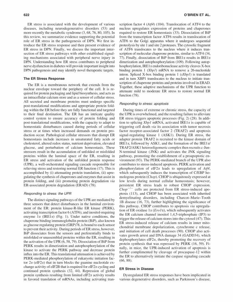
The distinct signaling pathways of the UPR are mediated bythree sensors that detect disturbances in the luminal environ-ment of the ER: protein kinase-R-like ER kinase (PERK),activating transcription factor 6 (ATF6), and inositol-requiringenzyme 1a (IRE1a) (Fig. 1). Under native conditions, thechaperone binding immunoglobulin protein (BiP), also knownas glucose regulating protein (GRP)78, is bound to each sensorto prevent their activity. During periods of ER stress, however,BiP dissociates from the sensors and preferentially binds tomisfolded or unassembled proteins within the ER, resulting inthe activation of the UPR (6, 58, 79). Dissociation of BiP fromPERK results in dimerization and autophosphorylation of thekinase to activate the PERK pathway and decrease proteininflux into the ER. This translational attenuation is achieved byPERK-mediated phosphorylation of eukaryotic initiation fac-tor 2a (eIF2a) that in turn blocks the guanine nucleotide ex-change activity of eIF2B that is required for eIF2a cycling andcontinued protein synthesis (32, 44). Repression of globalprotein synthesis resulting from limited eIF2a activity resultsin favored translation of mRNAs, including activating tran-
scription factor 4 (Atf4) (104). Translocation of ATF4 to thenucleus upregulates expression of proteins and chaperonesrequired to restore ER homeostasis (33). Dissociation of BiPfrom the transcription factor ATF6 results in translocation ofATF6 to the Golgi apparatus where it undergoes sequentialproteolysis by site 1 and site 2 proteases. The cytosolic fragmentof ATF6 translocates to the nucleus where it induces tran-scription of molecular chaperone proteins, similar to ATF4 (34,77). Finally, dissociation of BiP from IRE1a results in IRE1adimerization and autophosphorylation (109). Following autop-hosphorylation, IRE1a endoribonuclease activity cleaves X-boxbinding protein 1 (Xbp1) mRNA to remove a 26-nucleotideintron. Spliced X-box binding protein 1 (sXbp1) is translatedand in turn XBP1 translocates to the nucleus to initiate tran-scription of chaperone proteins and proteins involved in ERAD.Together, these adaptive mechanisms of the UPR function toattenuate mild to moderate ER stress to restore normal ERfunction (78).
Responding to stress: apoptosis
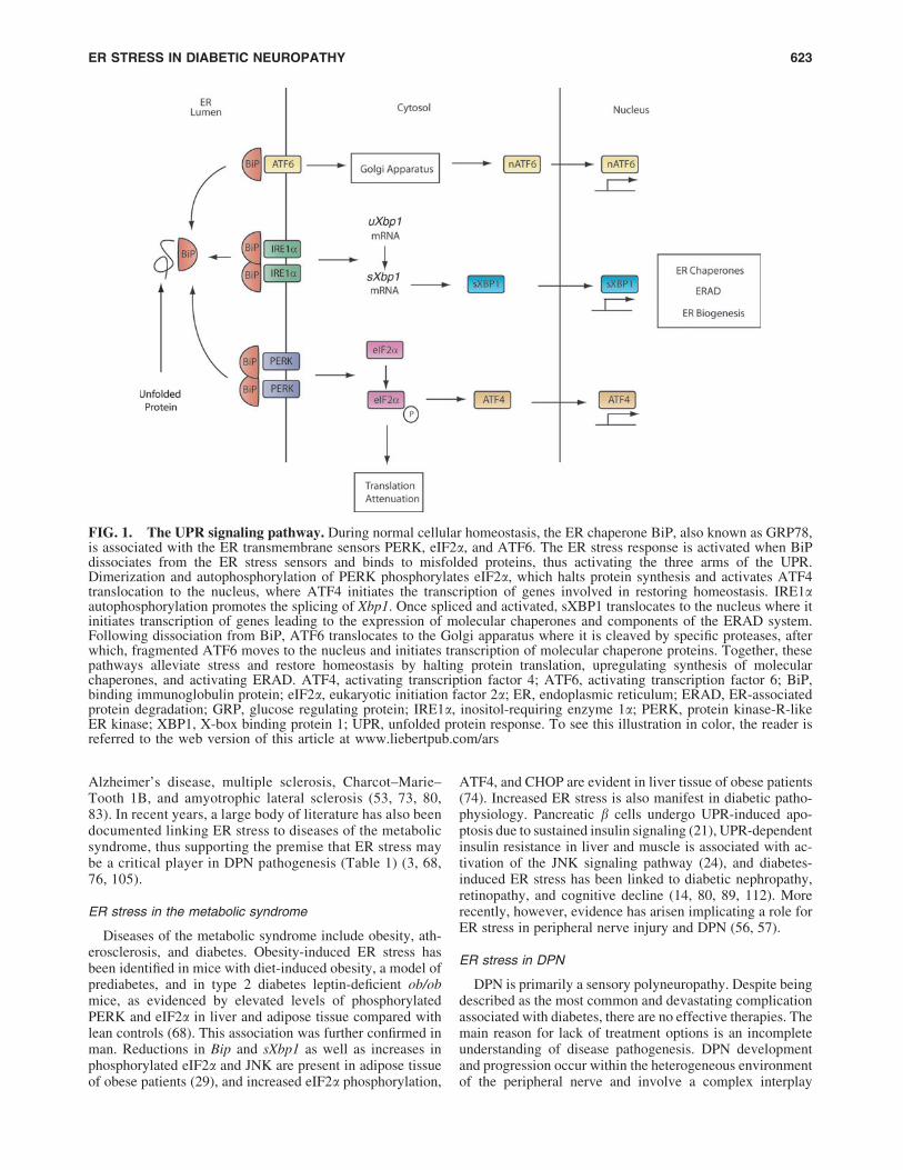
During times of extreme or chronic stress, the capacity ofthe UPR is overwhelmed, and the resulting failure to alleviateER stress triggers apoptotic processes (Fig. 2) (28). In addi-tion to splicing Xbp1 mRNA, activated IRE1a is capable oftriggering cell death via its association with tumor necrosisfactor receptor-associated factor 2 (TRAF2) and apoptosissignal-regulating kinase 1 (ASK1). During ER stress, theadapter protein TRAF2 is recruited to the kinase domain ofIRE1a, followed by ASK1, and the formation of the IRE1a/TRAF2/ASK1 heterooligomeric complex then recruits c-Jun-N-terminal kinase ( JNK) and activates the JNK signalingpathway, promoting the establishment of a proapoptotic en-vironment (93). The PERK-mediated branch of the UPR alsocontributes to stress-induced apoptosis. PERK activation andphosphorylation of eIF2a leads to upregulation of Atf4,which subsequently induces the transcription of C/EBP ho-mologous protein (Chop). CHOP is ubiquitously expressed atlow levels during normal cellular homeostasis; however,persistent ER stress leads to robust CHOP expression.Chop - / - cells are protected from ER stress-induced apo-ptosis (113), and CHOP has been associated with inheriteddemyelinating disorders, including Charcot–Marie–Tooth1B disease (16, 73), further highlighting the significance ofthis pathway. CHOP contributes to apoptosis via upregula-tion of ER oxidase 1a (Ero1a), which subsequently activatesthe ER calcium channel inositol 1,4,5-triphosphate (IP3) totrigger the release of calcium stores into the cytosol (47). ThisER stress-induced release of calcium results in inner mito-chondrial membrane depolarization, cytochrome c release,and initiation of cell death processes (90). CHOP also acti-vates growth arrest and DNA damage 34 (GADD34), whichdephosphorylates eIF2a, thereby promoting the recovery ofprotein synthesis that was repressed by PERK (16, 59). Fi-nally, in mice, the UPR-induced activation of apoptosis isfurther complimented by cleavage of procaspase-12 withinthe ER to alternatively initiate the caspase signaling cascade(66, 88).
ER Stress in Disease
Dysregulated ER stress responses have been implicated invarious degenerative disorders, such as Parkinson’s disease,
622 O’BRIEN ET AL.
Alzheimer’s disease, multiple sclerosis, Charcot–Marie–Tooth 1B, and amyotrophic lateral sclerosis (53, 73, 80,83). In recent years, a large body of literature has also beendocumented linking ER stress to diseases of the metabolicsyndrome, thus supporting the premise that ER stress maybe a critical player in DPN pathogenesis (Table 1) (3, 68,76, 105).
ER stress in the metabolic syndrome
Diseases of the metabolic syndrome include obesity, ath-erosclerosis, and diabetes. Obesity-induced ER stress hasbeen identified in mice with diet-induced obesity, a model ofprediabetes, and in type 2 diabetes leptin-deficient ob/obmice, as evidenced by elevated levels of phosphorylatedPERK and eIF2a in liver and adipose tissue compared withlean controls (68). This association was further confirmed inman. Reductions in Bip and sXbp1 as well as increases inphosphorylated eIF2a and JNK are present in adipose tissueof obese patients (29), and increased eIF2a phosphorylation,
ATF4, and CHOP are evident in liver tissue of obese patients(74). Increased ER stress is also manifest in diabetic patho-physiology. Pancreatic b cells undergo UPR-induced apo-ptosis due to sustained insulin signaling (21), UPR-dependentinsulin resistance in liver and muscle is associated with ac-tivation of the JNK signaling pathway (24), and diabetes-induced ER stress has been linked to diabetic nephropathy,retinopathy, and cognitive decline (14, 80, 89, 112). Morerecently, however, evidence has arisen implicating a role forER stress in peripheral nerve injury and DPN (56, 57).
ER stress in DPN
DPN is primarily a sensory polyneuropathy. Despite beingdescribed as the most common and devastating complicationassociated with diabetes, there are no effective therapies. Themain reason for lack of treatment options is an incompleteunderstanding of disease pathogenesis. DPN developmentand progression occur within the heterogeneous environmentof the peripheral nerve and involve a complex interplay
FIG. 1. The UPR signaling pathway. During normal cellular homeostasis, the ER chaperone BiP, also known as GRP78,is associated with the ER transmembrane sensors PERK, eIF2a, and ATF6. The ER stress response is activated when BiPdissociates from the ER stress sensors and binds to misfolded proteins, thus activating the three arms of the UPR.Dimerization and autophosphorylation of PERK phosphorylates eIF2a, which halts protein synthesis and activates ATF4translocation to the nucleus, where ATF4 initiates the transcription of genes involved in restoring homeostasis. IRE1aautophosphorylation promotes the splicing of Xbp1. Once spliced and activated, sXBP1 translocates to the nucleus where itinitiates transcription of genes leading to the expression of molecular chaperones and components of the ERAD system.Following dissociation from BiP, ATF6 translocates to the Golgi apparatus where it is cleaved by specific proteases, afterwhich, fragmented ATF6 moves to the nucleus and initiates transcription of molecular chaperone proteins. Together, thesepathways alleviate stress and restore homeostasis by halting protein translation, upregulating synthesis of molecularchaperones, and activating ERAD. ATF4, activating transcription factor 4; ATF6, activating transcription factor 6; BiP,binding immunoglobulin protein; eIF2a, eukaryotic initiation factor 2a; ER, endoplasmic reticulum; ERAD, ER-associatedprotein degradation; GRP, glucose regulating protein; IRE1a, inositol-requiring enzyme 1a; PERK, protein kinase-R-likeER kinase; XBP1, X-box binding protein 1; UPR, unfolded protein response. To see this illustration in color, the reader isreferred to the web version of this article at www.liebertpub.com/ars
ER STRESS IN DIABETIC NEUROPATHY 623
between the nerve and surrounding cells and tissues. A de-creased blood flow to the nerves (108), hyperglycemia (35,99, 101), dyslipidemia (98, 99), and a lack of insulin sig-naling (42) contribute to DPN pathogenesis. These factorsinduce multiple pathogenic processes, such as low-gradeinflammation, elevated sorbitol aldose reductase signaling,protein kinase C activation, advanced glycated end products,oxidative stress, and mitochondrial dysfunction (96), whichculminate in the physiologic and morphologic changes as-sociated with DPN.
A role of ER stress in DPN is being increasingly consid-ered based on in vivo evidence supporting ER stress in-volvement in the initiation and progression of DPN in bothtype 1 and type 2 diabetic rodent models. Furthermore,studies using ER chaperone proteins, which assist proteinfolding in the ER by preventing newly synthesized poly-peptide chains and assembled subunits from aggregatinginto nonfunctional structures, is an emerging therapeuticapproach aimed at restoring ER function (22). Oral ad-ministration of trimethylamine oxide (TMAO), a chemicalchaperone known to alleviate ER stress, to Zucker fatty (fa/fa) rats decreased protein expression of BiP/GRP78 in thesciatic nerve, and improved nerve conduction velocities andbehavioral responses to mechanical and thermal stimuli (56).C57B6 mice fed a high-fat diet (HFD) also displayed animproved neuronal phenotype when treated with salubrinal, acompound that enhances eIF2a phosphorylation (56). Neu-ropathy severity was also attenuated in streptozotocin (STZ)-
treated rats treated with TMAO (57). Finally, C57B6Chop - / - mice injected with STZ displayed improved nervefunction and increased expression of the folding proteins BiP/GRP78 and GRP94 in peripheral nerves compared with wild-type STZ-injected mice, further suggesting a role for ERsignaling in DPN development (57). Together, these datasupport a link between ER stress and DPN, as restoration ofER function by administration of chemical chaperones or ERstress inhibitors alleviates ER stress and improves peripheralnerve function (56, 57).
Although these studies confirm the role of ER stress inDPN, certain considerations must be made when evaluatingoutcomes following systemic administration of chemicalchaperones. As both hyperglycemia and dyslipidemia areknown contributors to DPN (10, 96, 98), improved neuronalphenotypes could be attributed to enhanced UPR function inpancreatic b cells rather than direct effects on the UPR in theperipheral nerve. Administration of the chemical chaperonephenyl butyric acid (PBA) or overexpression of oxygenregulated protein 150 (ORP150) improves pancreatic b cellfunction and enhances insulin secretion, which in turn im-proves the metabolic profile of the animal (67, 69). Similarly,insulin sensitivity in adipose and hepatic tissue may berestored following chaperone treatment (24). This must alsobe considered in DPN studies. TMAO treatment in Zuckerfa/fa rats improved blood glucose and triglyceride levels,and impaired glucose tolerance was attenuated in highfat-fed mice treated with salubrinal (56). Thus, systemic
FIG. 2. ER stress-associated apoptosis. Under conditions of unresolvable ER stress, apoptotic pathways are activated.IRE1a forms a multioligomer complex with TRAF2 and ASK1, and in turn recruits and activates JNK, which promotes celldeath by establishing a proapoptotic environment. The PERK/eIF2a pathway is also capable of activating apoptosis viaATF4-mediated upregulation of CHOP, which activates expression of proapoptotic genes and downregulates the expressionof antiapoptotic genes. CHOP also induces the expression of ERO1a, which activates the ER calcium release channel, IP3,resulting in increased cytosolic calcium that triggers mitochondrial-associated cell death. Alternatively, activation andcleavage of ER-bound caspase-12 can trigger apoptosis via UPR-independent mechanisms. ASK1, apoptosis signal-regulatingkinase 1; CHOP, C/EBP homologous protein; ERO1a = ER oxidase 1a; IP3, inositol 1,4,5-triphosphate; JNK, c-Jun-N-terminal kinase; TRAF2, tumor necrosis factor receptor-associated factor 2. To see this illustration in color, the reader isreferred to the web version of this article at www.liebertpub.com/ars
624 O’BRIEN ET AL.
administration or overexpression of chaperones may maskthe beneficial effects on the nerve by indirectly improving themetabolic profile. To circumvent these issues, direct neuronaldelivery of chaperones without impacting systemic glycemiamay be warranted to elucidate the role of ER stress on nerve
function in DPN. However, TMAO treatment in STZ-injected rats improved DPN phenotypes despite maintainedhyperglycemia (57), suggesting that therapies targeting ERstress responses may have direct efficacy in the peripheralnerve.
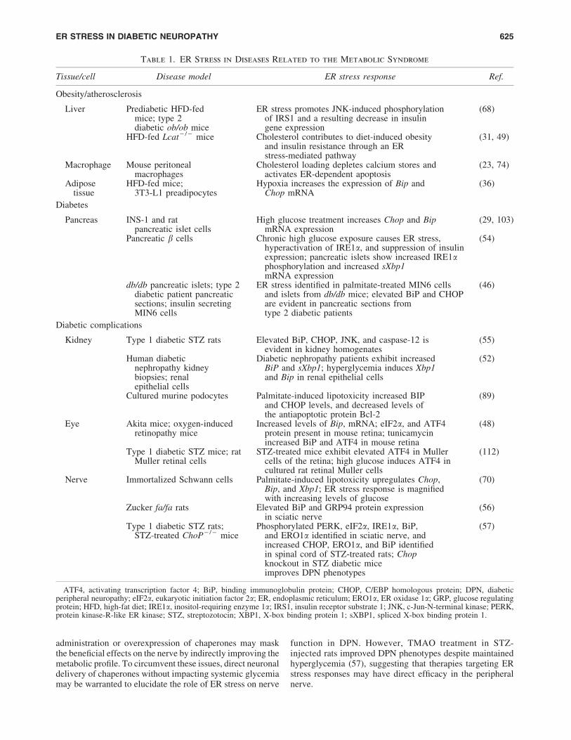
Table 1. ER Stress in Diseases Related to the Metabolic Syndrome
Tissue/cell Disease model ER stress response Ref.
Obesity/atherosclerosis
Liver Prediabetic HFD-fedmice; type 2diabetic ob/ob mice
ER stress promotes JNK-induced phosphorylationof IRS1 and a resulting decrease in insulingene expression
(68)
HFD-fed Lcat - / - mice Cholesterol contributes to diet-induced obesityand insulin resistance through an ERstress-mediated pathway
(31, 49)
Macrophage Mouse peritonealmacrophages
Cholesterol loading depletes calcium stores andactivates ER-dependent apoptosis
(23, 74)
Adiposetissue
HFD-fed mice;3T3-L1 preadipocytes
Hypoxia increases the expression of Bip andChop mRNA
(36)
Diabetes
Pancreas INS-1 and ratpancreatic islet cells
High glucose treatment increases Chop and BipmRNA expression
(29, 103)
Pancreatic b cells Chronic high glucose exposure causes ER stress,hyperactivation of IRE1a, and suppression of insulinexpression; pancreatic islets show increased IRE1aphosphorylation and increased sXbp1mRNA expression
(54)
db/db pancreatic islets; type 2diabetic patient pancreaticsections; insulin secretingMIN6 cells
ER stress identified in palmitate-treated MIN6 cellsand islets from db/db mice; elevated BiP and CHOPare evident in pancreatic sections fromtype 2 diabetic patients
(46)
Diabetic complications
Kidney Type 1 diabetic STZ rats Elevated BiP, CHOP, JNK, and caspase-12 isevident in kidney homogenates
(55)
Human diabeticnephropathy kidneybiopsies; renalepithelial cells
Diabetic nephropathy patients exhibit increasedBiP and sXbp1; hyperglycemia induces Xbp1and Bip in renal epithelial cells
(52)
Cultured murine podocytes Palmitate-induced lipotoxicity increased BIPand CHOP levels, and decreased levels ofthe antiapoptotic protein Bcl-2
(89)
Eye Akita mice; oxygen-inducedretinopathy mice
Increased levels of Bip, mRNA; eIF2a, and ATF4protein present in mouse retina; tunicamycinincreased BiP and ATF4 in mouse retina
(48)
Type 1 diabetic STZ mice; ratMuller retinal cells
STZ-treated mice exhibit elevated ATF4 in Mullercells of the retina; high glucose induces ATF4 incultured rat retinal Muller cells
(112)
Nerve Immortalized Schwann cells Palmitate-induced lipotoxicity upregulates Chop,Bip, and Xbp1; ER stress response is magnifiedwith increasing levels of glucose
(70)
Zucker fa/fa rats Elevated BiP and GRP94 protein expressionin sciatic nerve
(56)
Type 1 diabetic STZ rats;STZ-treated ChoP - / - mice
Phosphorylated PERK, eIF2a, IRE1a, BiP,and ERO1a identified in sciatic nerve, andincreased CHOP, ERO1a, and BiP identifiedin spinal cord of STZ-treated rats; Chopknockout in STZ diabetic miceimproves DPN phenotypes
(57)
ATF4, activating transcription factor 4; BiP, binding immunoglobulin protein; CHOP, C/EBP homologous protein; DPN, diabeticperipheral neuropathy; eIF2a, eukaryotic initiation factor 2a; ER, endoplasmic reticulum; ERO1a, ER oxidase 1a; GRP, glucose regulatingprotein; HFD, high-fat diet; IRE1a, inositol-requiring enzyme 1a; IRS1, insulin receptor substrate 1; JNK, c-Jun-N-terminal kinase; PERK,protein kinase-R-like ER kinase; STZ, streptozotocin; XBP1, X-box binding protein 1; sXBP1, spliced X-box binding protein 1.
ER STRESS IN DIABETIC NEUROPATHY 625
Molecular Mechanisms of ER Stress and DPN
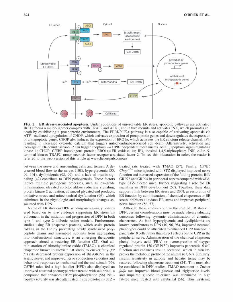
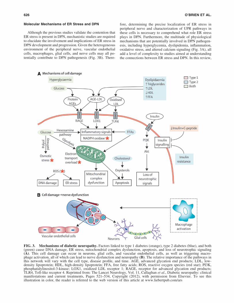
Although the previous studies validate the contention thatER stress is present in DPN, mechanistic studies are requiredto elucidate the involvement and implications of ER stress inDPN development and progression. Given the heterogeneousenvironment of the peripheral nerve, vascular endothelialcells, macrophages, glial cells, and nerve cells may all po-tentially contribute to DPN pathogenesis (Fig. 3B). There-
fore, determining the precise localization of ER stress inperipheral nerve and characterization of UPR pathways inthese cells is necessary to comprehend what role ER stressplays in DPN. Furthermore, the multitude of physiologicalmechanisms that are potentially involved in DPN pathogen-esis, including hyperglycemia, dyslipidemia, inflammation,oxidative stress, and altered calcium signaling (Fig. 3A), alladd a level of complexity to studies aimed at understandingthe connections between ER stress and DPN. In this review,
FIG. 3. Mechanisms of diabetic neuropathy. Factors linked to type 1 diabetes (orange), type 2 diabetes (blue), and both(green) cause DNA damage, ER stress, mitochondrial complex dysfunction, apoptosis, and loss of neurotrophic signaling(A). This cell damage can occur in neurons, glial cells, and vascular endothelial cells, as well as triggering macro-phage activation, all of which can lead to nerve dysfunction and neuropathy (B). The relative importance of the pathways inthis network will vary with the cell type, disease profile, and time. AGE, advanced glycation end products; LDL, low-density lipoprotein; HDL, high-density lipoprotein; FFA, free fatty acids; ROS, reactive oxygen species (red star); PI3K,phosphatidylinositol-3-kinase; LOX1, oxidized LDL receptor 1; RAGE, receptor for advanced glycation end products;TLR4, Toll-like receptor 4. Reprinted from: The Lancet Neurology, Vol. 11, Callaghan et al., Diabetic neuropathy: clinicalmanifestations and current treatments, Pages 521–534, Copyright (2012), with permission from Elsevier. To see thisillustration in color, the reader is referred to the web version of this article at www.liebertpub.com/ars
626 O’BRIEN ET AL.
we discuss evidence demonstrating how ER stress intersectswith these pathways to provide insight into the possible in-volvement of ER stress in DPN pathogenesis.
Hyperglycemia
Evidence from studies examining ER stress in diabeticcomplications-prone tissues supports the contention thathyperglycemia can impact ER stress and the integrated stressresponse (14, 45). Diabetic retinopathy studies demonstratethat high-glucose treatment of retinal endothelial cells resultsin increased levels of CHOP, ATF4, and phosphorylatedPERK and eIF2a, upregulation of inflammatory cytokines,including tumor necrosis factor a (TNFa), and increasedexpression of vascular endothelial growth factor (VEGF)(14). This link is further supported by studies demonstratingimprovements in retinal inflammation and vascular leakagewith Atf4 knockdown in STZ-injected mice (14). Markers ofUPR signaling are also significantly upregulated in diabeticcardiomyopathy. Increased expression of phosphorylatedPERK and eIF2a, ATF4, CHOP, and ATF6 is observed in themyocardium of spontaneous diabetic Torii rats, a model ofnonobese type 2 diabetes (45). Finally, lipotoxicity-inducedER stress responses, including decreases in ER calciumlevels and expression of CHOP, XBP1, and BiP/GRP78, areexacerbated in immortalized Schwann cells (iSCs) in thepresence of high glucose (70); however, glucose exposure inthe absence of lipotoxicity did not affect cell viability or ERcalcium levels. In addition, along these lines, diabetic pa-tients with DPN who receive intensive glucose therapy donot show favorable improvements in nerve function (10),suggesting that glucose-independent processes also contrib-ute to DPN.
Dyslipidemia
Dyslipidemia is strongly associated with DPN patho-physiology by both epidemiological and basic research (2,98, 99, 105), and elevated levels of fatty acid and cholesterol-induced ER stress have been implicated in metabolic diseases(37, 46, 50, 65, 89). Therefore, it is important to understandhow abnormal lipid content can provoke ER stress in DPN.Cell-based approaches have determined that elevated con-centrations of free fatty acids (FFA) can trigger the UPR insupporting cells of the peripheral nerve. Using iSCs, inves-tigators have demonstrated that lipotoxicity associated withthe saturated fatty acid palmitate (PA) promotes iSC dys-function and cell death via the UPR signaling pathway, asevidenced by increased expression of BiP, CHOP, and XBP1(70). These studies also demonstrated that activation of theUPR precedes the generation of reactive oxygen species(ROS), mitochondrial depolarization, and apoptosis, sug-gesting that the ER stress response occurs upstream of theseprocesses (70). Interestingly, oleate, a monounsaturated fattyacid, abolishes PA-induced ER stress and lipotoxicity inC2C12 myoblasts, indicating that the composition of fattyacid-derived phospholipids within the ER is an importantdeterminant of UPR activation (72).
Cholesterol has also been implicated in ER stress, as theER is incredibly sensitive to perturbations in free cholesterollevels given its particularly low native cholesterol content(8). In macrophages, cholesterol loading depletes ER luminalcalcium stores and triggers UPR activation, the expression of
CHOP, and caspase-3-mediated apoptosis (23). Moreover,unlike esterified cholesterol, the insertion of free cholesterolinto the lipid bilayer of macrophages can cause disturbancesin the physical properties of the membrane, which may inturn activate the UPR. Along these lines, Schwann cells playa significant role in myelination and are incredibly sensitiveto perturbation in membrane composition, suggesting thatdyslipidemia-induced ER stress in Schwann cells may play arole in DPN pathogenesis. ER stress is involved in multiplemyelin-related disorders (16, 51, 73, 83), and recent evidencehas identified abnormalities in the sciatic nerve of STZ-treated rats that include imbalances in myelin’s phospholipid,fatty acid, and cholesterol content (13). Thus, an altered lipidprofile brought on by disturbances in lipid homeostasis hasimportant consequences in Schwann cell biology by affectingmembrane fluidity and function (1, 13). This may be espe-cially important during remyelination following nerve injuryin diabetes if myelin generation is compromised due to ele-vated/abnormal lipid substrates. However, the role of lipidhomeostasis and ER stress in DPN remains relatively unex-plored and further studies addressing this relationship arewarranted.
Insulin signaling
Although hyperglycemia resulting from dysfunctional in-sulin signaling in pancreatic b cells and metabolically activetissues is a key contributor to nerve injury in DPN, recentevidence also suggests that impaired insulin signaling inperipheral neurons may also be an important factor in nervedegeneration. The insulin receptor (IR) and insulin receptorsubstrate 1 (IRS1) are widely expressed in cell bodies andaxons of peripheral neurons (30, 86, 87), where they are ac-tivated by insulin to provide vital neurotrophic support to thenerve (9, 91, 107). In STZ-injected rats, intrathecal low-doseinsulin treatment attenuated DPN-associated reductions innerve function and reversed atrophy of myelinated suralnerve sensory axons (9). Similarly, insulin resistance hasbeen observed in dorsal root ganglia from db/db type 2 dia-betic mice (42) and in hypothalamic neurons in response toPA-induced lipotoxicity (62). Whereas the exact mechanismslinking ER stress and insulin resistance in peripheral nervesmust still be elucidated, several mechanisms linking ERstress and hepatic and adipose insulin resistance have beenproposed (24). One theory suggests that UPR signaling di-rectly suppresses IR signaling through hyperactivation ofJNK and subsequent phosphorylation of IRS1 (39, 62, 93).Alternatively, induction of lipogenic and glyconeogenicgenes downstream of UPR signaling could result in abnormalactivation of these pathways and ultimately promote insulinresistance (24). Further studies are required to comprehendhow ER stress contributes to insulin resistance in DPN.
Inflammation
ER stress-induced UPR signaling is associated with theproduction of numerous proinflammatory cytokines (50). Inmacrophages, excess free cholesterol induces TNFa andinterleukin-6 (IL-6) production via activation of JNK/NFjBand CHOP, respectively (37, 110). Recent studies also pro-vide evidence that inflammation-mediated ER stress is im-portant in Schwann cell-based nerve injury. Studiesinvestigating spinal cord injury in rats demonstrate that
ER STRESS IN DIABETIC NEUROPATHY 627
upregulation of TNFa, following sciatic nerve crush injury,effectively triggers the UPR in Schwann cells (61). Alongwith increased BiP/GRP78 expression, TNFa induction ofCHOP expression led to Schwann cell apoptosis, demyelin-ation, and nerve degeneration (61), suggesting that inflam-matory mediators may play an important role in DPN diseaseprogression via ER stress pathways. Upregulation of low-grade inflammation (41, 81) and elevation of cytokines, suchas TNFa (26, 106), in patients with DPN further emphasizethe importance of additional studies focused on determiningwhether these inflammatory factors contribute to ER stress-induced nerve pathology.
Oxidative stress
Oxidative stress is a major mechanism of hyperglycemia-induced DPN in humans and rodents (97, 99, 102). Duringnormal cellular metabolism in mitochondria, ROS are formedas by-products of electron transfer to molecular oxygen and areimportant signaling molecules in biological processes such asautophagy, phagocytosis, and inflammation (95). However,increased ROS production and compromised endogenous an-tioxidant defenses in diabetes lead to oxidative stress, a pro-oxidant state resulting in injurious oxidation of proteins andlipids (97, 99, 102). Indeed, hyperglycemia leads to increasedROS and cellular apoptosis in cultured dorsal root ganglianeurons (102). Furthermore, the ER is both a source of in-creased ROS generation and is affected by increased cellularROS (4). Although it is less well characterized than in mito-chondria, the ER has its own electron transport system thattraffics reducing equivalents within the lumen of the ER andtransfers electrons to molecular oxygen, resulting in con-comitant ROS production (60). Thus, ROS generation is anatural by-product of ER oxidative protein folding and ac-counts for *25% of the total cellular ROS generation (92).Under times of increased protein load, ER ROS generation cansignificantly increase (60). Given the potential for high levelsof ROS in the cell and ER, oxidative stress may significantlyimpact the ER stress response in DPN.
During normal physiological conditions, endogenous an-tioxidant mechanisms are upregulated to scavenge free rad-icals and prevent cellular injury (4). During ER stress,activation of PERK signaling increases NF-E2-related factor2 (NRF2) activation and subsequent transcription of antiox-idant and detoxification enzyme genes. This response is ev-ident in cultured dorsal root ganglion neurons and inSchwann cells in response to acute hyperglycemic and oxi-dative (hydrogen peroxide) stress (100). Prolonged hyper-glycemia, on the other hand, attenuates dorsal root ganglionneuron antioxidant levels and activities (100), increasing thesignificance of ER ROS generation under disease conditions.Similarly, the glutathione antioxidant activity in the ER isimpacted by oxidative stress. During normal ER proteinfolding (4, 60) and during the UPR response to correct mis-folded proteins (60), glutathione is converted from its re-duced form (GSH) to an oxidized form (GSSG); however,excessive UPR activation can deplete the GSH:GSSG ratio,decreasing its antioxidant capacity. Chronic stress-inducedUPR-mediated CHOP elevation also depletes the glutathioneantioxidant capacity and increases ROS production, furtherescalating cellular oxidative stress levels (5, 63). Along theselines, decreasing ER stress and CHOP expression with
TMAO chaperone protein treatment for 12 weeks followingSTZ-induced diabetes in rats attenuates lipid and proteinoxidation in the sciatic nerve, and is coincident with im-provements in electrophysiological, behavioral, and struc-tural neuropathy parameters in treated diabetic animals (57).Further work from the same group using Chop knockoutmice confirmed that a CHOP-mediated ER stress response isinvolved in sciatic nerve oxidative stress and the develop-ment of DPN (57), and is consistent with reports that pan-creatic b cell CHOP deletion decreases oxidative damage in anumber of mouse models of diabetes (82).
In addition to normal protein folding and the UPR, sourcesof ROS outside the ER may also trigger the ER stress re-sponse (i.e., the mitochondrial respiratory chain, increasedNADPH oxidase activity, and inflammatory processes). Al-though these sources of ROS in diabetes and their contribu-tion to peripheral nerve dysfunction are relatively wellcharacterized, their interactions with the ER remain largelyunexplored (60) and require future attention.
Calcium signaling
The ER is a major store for intracellular calcium, and cal-cium levels are three- to four-times greater in the ER lumenthan in the cytosol (85); thus, factors that perturb calciumhomeostasis activate the UPR. Experimentally, calcium storesin the ER can be depleted using the calcium ionophore A23167or thapsigargin to block uptake of calcium from the cytosol (7,18). This depletion impairs calcium-dependent chaperones andfolding enzymes and results in the accumulation of misfoldedproteins and subsequent activation of the UPR (11, 27, 75, 84).Disruptions of ER calcium homeostasis are involved in variousforms of neuropathology (94), and evidence for disrupted ERcalcium homeostasis is evident in diabetic animal models.Dorsal root ganglia sensory neurons from type 1 diabetic STZ-injected rats exhibit reduced calcium levels and diminishedcalcium uptake by the ER (111). Similarly, comparison oflumbar and cervical dorsal root ganglia neurons from diabeticSTZ rats revealed that altered calcium dynamics are moreprominent in lumbar neurons, the ganglia that are affected firstin DPN (40, 43). Additional implications for a role of ERcalcium homeostasis in DPN stems from the recent microarrayanalysis of peripheral nerve tissue from diabetic mice. Dif-ferentially expressed genes (DEGs) related to Ca2 + transportare highly downregulated in 24-week db/db mice; however, nosignificant increase is observed in UPR-related DEGs despitelarge changes in calcium (71). Experimental validation is re-quired to confirm these data, but it is plausible that calcium-induced ER stress may induce UPR-independent cellularstress. Studies in human and mouse macrophages demonstratethat ER stress activates the NOD-like receptor family, pyrindomain containing 3 (NLRP3) inflammasome, and subsequentinflammatory responses independent of the UPR (64), andcalcium release from the ER can impact mitochondrialmembrane depolarization and increase oxidative stress, whichcould contribute to neuronal injury (27). Further examinationinto the relationship between calcium signaling in DPN isrequired.
Conclusion
Despite recent advances in our understanding of DPNpathophysiology, few therapies exist for the management of
628 O’BRIEN ET AL.
DPN. Given the recent association of ER stress with DPN indiabetic animal models, the association of ER stress withpathways involved in DPN pathogenesis and the insightgained from studies examining interventions that target ERstress pathways, the development of therapeutic approachesthat enhance ER function (i.e., increase chaperone avail-ability) and decrease UPR-activated cell death should beconsidered. The role of ER stress in DPN, however, remainslargely unexplored and several key questions must still beanswered. For instance, what induces ER stress in the diabeticmilieu of the peripheral nerve? What cells in the peripheralnerve are affected by ER stress, and are all affected cellsequally susceptible to ER stress? Furthermore, which UPRpathways are implicated in DPN? Finally, what is the inter-play between ER stress and other components of DPNpathogenesis, such as calcium storage and transport, oxida-tive stress, myelination and insulin signaling? As an abun-dance of information on other signaling pathways in DPNetiology exists, it is crucial to understand these interactionswith the ER and elucidate how they contribute to disease.With additional insight into cell-specific contributions of theUPR to DPN pathogenesis, we hope to ultimately discovermechanism-based therapies that can prevent this injury cas-cade and ameliorate the signs and symptoms of DPN.
Acknowledgments
The authors would like to thank Mrs. Judith Bentley forexcellent administrative support during the preparation ofthis article. Funding support is provided by the Program forNeurology Research & Discovery and the A. Alfred Taub-man Medical Research Institute. L.M.H. is funded by a fel-lowship from the Juvenile Diabetes Research Foundation.
References
1. Anitei M and Pfeiffer SE. Myelin biogenesis: sorting outprotein trafficking. Curr Biol 16: R418–R421, 2006.
2. Ansquer JC, Foucher C, Aubonnet P, and Le Malicot K.Fibrates and microvascular complications in diabetes—insight from the FIELD study. Curr Pharm Des 15: 537–552, 2009.
3. Back SH and Kaufman RJ. Endoplasmic reticulum stressand type 2 diabetes. Annu Rev Biochem 81: 767–793,2012.
4. Bashan N, Kovsan J, Kachko I, Ovadia H, and Rudich A.Positive and negative regulation of insulin signaling byreactive oxygen and nitrogen species. Physiol Rev 89: 27–71, 2009.
5. Bek MF, Bayer M, Muller B, Greiber S, Lang D, SchwabA, August C, Springer E, Rohrbach R, Huber TB, BenzingT, and Pavenstadt H. Expression and function of C/EBPhomology protein (GADD153) in podocytes. Am J Pathol168: 20–32, 2006.
6. Bertolotti A, Zhang Y, Hendershot LM, Harding HP, andRon D. Dynamic interaction of BiP and ER stress trans-ducers in the unfolded-protein response. Nat Cell Biol 2:326–332, 2000.
7. Booth C and Koch GL. Perturbation of cellular calciuminduces secretion of luminal ER proteins. Cell 59: 729–737, 1989.
8. Bretscher MS, and Munro S. Cholesterol and the Golgiapparatus. Science 261: 1280–1281, 1993.
9. Brussee V, Cunningham FA, and Zochodne DW. Directinsulin signaling of neurons reverses diabetic neuropathy.Diabetes 53: 1824–1830, 2004.
10. Callaghan BC, Little AA, Feldman EL, and Hughes RA.Enhanced glucose control for preventing and treatingdiabetic neuropathy. Cochrane Database Syst Rev 6:CD007543, 2012.
11. Cao SS and Kaufman RJ. Targeting endoplasmic reticu-lum stress in metabolic disease. Expert Opin Ther Targets17: 437–448, 2013.
12. Centers for Disease Control and Prevention. National di-abetes fact sheet: national estimates and general infor-mation on diabetes and prediabetes in the United States A.Georgia: U.S. Department of Health and Human Services,Centers for Disease Control and Prevention, 2011.
13. Cermenati G, Abbiati F, Cermenati S, Brioschi E, Vo-lonterio A, Cavaletti G, Saez E, De Fabiani E, Crestani M,Garcia-Segura LM, Melcangi RC, Caruso D, and Mitro N.Diabetes-induced myelin abnormalities are associatedwith an altered lipid pattern: protective effects of LXRactivation. J Lipid Res 53: 300–310, 2012.
14. Chen Y, Wang JJ, Li J, Hosoya KI, Ratan R, Townes T,and Zhang SX. Activating transcription factor 4 mediateshyperglycaemia-induced endothelial inflammation andretinal vascular leakage through activation of STAT3 in amouse model of type 1 diabetes. Diabetologia 55: 2533–2545, 2012.
15. Cowie CC, Rust KF, Ford ES, Eberhardt MS, Byrd-HoltDD, Li C, Williams DE, Gregg EW, Bainbridge KE,Saydah SH, and Geiss LS. Full accounting of diabetes andpre-diabetes in the U.S. population in 1988–1994 and2005–2006. Diabetes Care 32: 287–294, 2009.
16. D’Antonio M, Musner N, Scapin C, Ungaro D, Del CarroU, Ron D, Feltri ML, and Wrabetz L. Resetting transla-tional homeostasis restores myelination in Charcot-Marie-Tooth disease type 1B mice. J Exp Med 210: 821–838,2013.
17. Dorner AJ, Wasley LC, and Kaufman RJ. Increasedsynthesis of secreted proteins induces expression ofglucose-regulated proteins in butyrate-treated Chinesehamster ovary cells. J Biol Chem 264: 20602–20607,1989.
18. Drummond IA, Lee AS, Resendez E Jr., and SteinhardtRA. Depletion of intracellular calcium stores by calciumionophore A23187 induces the genes for glucose-regulatedproteins in hamster fibroblasts. J Biol Chem 262: 12801–12805, 1987.
19. Dyck PJ, Kratz KM, Karnes JL, Litchy WJ, Klein R, PachJM, Wilson DM, O’Brien PC, Melton LJ 3rd, and ServiceFJ. The prevalence by staged severity of various types ofdiabetic neuropathy, retinopathy, and nephropathy in apopulation-based cohort: the Rochester Diabetic Neuro-pathy Study. Neurology 43: 817–824, 1993.
20. Edwards JL, Vincent AM, Cheng HT, and Feldman EL.Diabetic neuropathy: mechanisms to management. Phar-macol Ther 120: 1–34, 2008.
21. Eizirik DL, Miani M, and Cardozo AK. Signalling danger:endoplasmic reticulum stress and the unfolded proteinresponse in pancreatic islet inflammation. Diabetologia56: 234–241, 2013.
22. Engin F and Hotamisligil GS. Restoring endoplasmic re-ticulum function by chemical chaperones: an emergingtherapeutic approach for metabolic diseases. DiabetesObes Metab 12 Suppl 2: 108–115, 2010.
ER STRESS IN DIABETIC NEUROPATHY 629
23. Feng B, Yao PM, Li Y, Devlin CM, Zhang D, HardingHP, Sweeney M, Rong JX, Kuriakose G, Fisher EA,Marks AR, Ron D, and Tabas I. The endoplasmic retic-ulum is the site of cholesterol-induced cytotoxicity inmacrophages. Nat Cell Biol 5: 781–792, 2003.
24. Flamment M, Hajduch E, Ferre P, and Foufelle F. Newinsights into ER stress-induced insulin resistance. TrendsEndocrinol Metab 23: 381–390, 2012.
25. Geiss LS, James C, Gregg EW, Albright A, WilliamsonDF, and Cowie CC. Diabetes risk reduction behaviorsamong U.S. adults with prediabetes. Am J Prev Med 38:403–409, 2010.
26. Gonzalez-Clemente JM, Mauricio D, Richart C, Broch M,Caixas A, Megia A, Gimenez-Palop O, Simon I, Martinez-Riquelme A, Gimenez-Perez G, and Vendrell J. Diabeticneuropathy is associated with activation of the TNF-alphasystem in subjects with type 1 diabetes mellitus. ClinEndocrinol (Oxf) 63: 525–529, 2005.
27. Gorlach A, Klappa P, and Kietzmann T. The endoplasmicreticulum: folding, calcium homeostasis, signaling, andredox control. Antioxid Redox Signal 8: 1391–1418, 2006.
28. Gorman AM, Healy SJ, Jager R, and Samali A. Stressmanagement at the ER: regulators of ER stress-inducedapoptosis. Pharmacol Ther 134: 306–316, 2012.
29. Gregor MF, Yang L, Fabbrini E, Mohammed BS, EagonJC, Hotamisligil GS, and Klein S. Endoplasmic reticulumstress is reduced in tissues of obese subjects after weightloss. Diabetes 58: 693–700, 2009.
30. Grote CW, Morris JK, Ryals JM, Geiger PC, and WrightDE. Insulin receptor substrate 2 expression and involve-ment in neuronal insulin resistance in diabetic neuropathy.Exp Diabetes Res 2011: 212571, 2011.
31. Hager L, Li L, Pun H, Liu L, Hossain MA, Maguire GF,Naples M, Baker C, Magomedova L, Tam J, Adeli K,Cummins CL, Connelly PW, and Ng DS. Lecithin:cholesterol acyltransferase deficiency protects againstcholesterol-induced hepatic endoplasmic reticulum stressin mice. J Biol Chem 287: 20755–20768, 2012.
32. Harding HP and Ron D. Endoplasmic reticulum stress andthe development of diabetes: a review. Diabetes 51 Suppl3: S455–S461, 2002.
33. Harding HP, Zhang Y, Bertolotti A, Zeng H, and Ron D.Perk is essential for translational regulation and cell sur-vival during the unfolded protein response. Mol Cell 5:897–904, 2000.
34. Harding HP, Zhang Y, Zeng H, Novoa I, Lu PD, CalfonM, Sadri N, Yun C, Popko B, Paules R, Stojdl DF, BellJC, Hettmann T, Leiden JM, and Ron D. An integratedstress response regulates amino acid metabolism and re-sistance to oxidative stress. Mol Cell 11: 619–633, 2003.
35. Hinder LM, Vincent AM, Burant CF, Pennathur S, andFeldman EL. Bioenergetics in diabetic neuropathy: what weneed to know. J Peripher Nerv Syst 17 Suppl 2: 10–14, 2012.
36. Hosogai N, Fukuhara A, Oshima K, Miyata Y, Tanaka S,Segawa K, Furukawa S, Tochino Y, Komuro R, MatsudaM, and Shimomura I. Adipose tissue hypoxia in obesityand its impact on adipocytokine dysregulation. Diabetes56: 901–911, 2007.
37. Hotamisligil GS. Endoplasmic reticulum stress and theinflammatory basis of metabolic disease. Cell 140: 900–917, 2010.
38. International Diabetes Federation. IDF Diabetes Atlas6th Edition. Brussels, Belgium: International DiabetesFederation, 2013. http://www.idf.org/diabetesatlas
39. Hu P, Han Z, Couvillon AD, Kaufman RJ, and Exton JH.Autocrine tumor necrosis factor alpha links endoplasmicreticulum stress to the membrane death receptor pathwaythrough IRE1alpha-mediated NF-kappaB activation anddown-regulation of TRAF2 expression. Mol Cell Biol 26:3071–3084, 2006.
40. Huang TJ, Sayers NM, Fernyhough P, and Verkhratsky A.Diabetes-induced alterations in calcium homeostasis in sen-sory neurones of streptozotocin-diabetic rats are restrictedto lumbar ganglia and are prevented by neurotrophin-3.Diabetologia 45: 560–570, 2002.
41. Hur J, Sullivan KA, Pande M, Hong Y, Sima AA, Ja-gadish HV, Kretzler M, and Feldman EL. The identifica-tion of gene expression profiles associated withprogression of human diabetic neuropathy. Brain 134:3222–3235, 2011.
42. Kim B, McLean LL, Philip SS, and Feldman EL. Hy-perinsulinemia induces insulin resistance in dorsal rootganglion neurons. Endocrinology 152: 3638–3647, 2011.
43. Kostyuk E, Voitenko N, Kruglikov I, Shmigol A, ShishkinV, Efimov A, and Kostyuk P. Diabetes-induced changes incalcium homeostasis and the effects of calcium channelblockers in rat and mice nociceptive neurons. Diabetolo-gia 44: 1302–1309, 2001.
44. Krishnamoorthy T, Pavitt GD, Zhang F, Dever TE, andHinnebusch AG. Tight binding of the phosphorylated al-pha subunit of initiation factor 2 (eIF2alpha) to the reg-ulatory subunits of guanine nucleotide exchange factoreIF2B is required for inhibition of translation initiation.Mol Cell Biol 21: 5018–5030, 2001.
45. Lakshmanan AP, Harima M, Suzuki K, Soetikno V, Na-gata M, Nakamura T, Takahashi T, Sone H, Kawachi H,and Watanabe K. The hyperglycemia stimulated myo-cardial endoplasmic reticulum (ER) stress contributes todiabetic cardiomyopathy in the transgenic non-obese type2 diabetic rats: a differential role of unfolded protein re-sponse (UPR) signaling proteins. Int J Biochem Cell Biol45: 438–447, 2013.
46. Laybutt DR, Preston AM, Akerfeldt MC, Kench JG,Busch AK, Biankin AV, and Biden TJ. Endoplasmic re-ticulum stress contributes to beta cell apoptosis in type 2diabetes. Diabetologia 50: 752–763, 2007.
47. Li G, Mongillo M, Chin KT, Harding H, Ron D, MarksAR, and Tabas I. Role of ERO1-alpha-mediated stimula-tion of inositol 1,4,5-triphosphate receptor activity in en-doplasmic reticulum stress-induced apoptosis. J Cell Biol186: 783–792, 2009.
48. Li J, Wang JJ, Yu Q, Wang M, and Zhang SX. En-doplasmic reticulum stress is implicated in retinal in-flammation and diabetic retinopathy. FEBS Lett 583:1521–1527, 2009.
49. Li L, Hossain MA, Sadat S, Hager L, Liu L, Tam L,Schroer S, Huogen L, Fantus IG, Connelly PW, Woo M,and Ng DS. Lecithin cholesterol acyltransferase null miceare protected from diet-induced obesity and insulin re-sistance in a gender-specific manner through multiplepathways. J Biol Chem 286: 17809–17820, 2011.
50. Li Y, Schwabe RF, DeVries-Seimon T, Yao PM, Gerbod-Giannone MC, Tall AR, Davis RJ, Flavell R, Brenner DA,and Tabas I. Free cholesterol-loaded macrophages arean abundant source of tumor necrosis factor-alphaand interleukin-6: model of NF-kappaB- and map kinase-dependent inflammation in advanced atherosclerosis.J Biol Chem 280: 21763–21772, 2005.
630 O’BRIEN ET AL.
51. Lin W and Popko B. Endoplasmic reticulum stress indisorders of myelinating cells. Nat Neurosci 12: 379–385,2009.
52. Lindenmeyer MT, Rastaldi MP, Ikehata M, Neusser MA,Kretzler M, Cohen CD, and Schlondorff D. Proteinuriaand hyperglycemia induce endoplasmic reticulum stress.J Am Soc Nephrol 19: 2225–2236, 2008.
53. Lindholm D, Wootz H, and Korhonen L. ER stress andneurodegenerative diseases. Cell Death Differ 13: 385–392, 2006.
54. Lipson KL, Fonseca SG, Ishigaki S, Nguyen LX, Foss E,Bortell R, Rossini AA, and Urano F. Regulation of insulinbiosynthesis in pancreatic beta cells by an endoplasmicreticulum-resident protein kinase IRE1. Cell Metab 4:245–254, 2006.
55. Liu G, Sun Y, Li Z, Song T, Wang H, Zhang Y, and Ge Z.Apoptosis induced by endoplasmic reticulum stress in-volved in diabetic kidney disease. Biochem Biophys ResCommun 370: 651–656, 2008.
56. Lupachyk S, Watcho P, Obrosov AA, Stavniichuk R, andObrosova IG. Endoplasmic reticulum stress contributes toprediabetic peripheral neuropathy. Exp Neurol 247: 342–348, 2013.
57. Lupachyk S, Watcho P, Stavniichuk R, Shevalye H, andObrosova IG. Endoplasmic reticulum stress plays a keyrole in the pathogenesis of diabetic peripheral neuropathy.Diabetes 62: 944–952, 2013.
58. Ma K, Vattem KM, and Wek RC. Dimerization and re-lease of molecular chaperone inhibition facilitate activa-tion of eukaryotic initiation factor-2 kinase in response toendoplasmic reticulum stress. J Biol Chem 277: 18728–18735, 2002.
59. Ma Y and Hendershot LM. Delineation of a negativefeedback regulatory loop that controls protein translationduring endoplasmic reticulum stress. J Biol Chem 278:34864–34873, 2003.
60. Malhotra JD and Kaufman RJ. Endoplasmic reticulumstress and oxidative stress: a vicious cycle or a double-edged sword? Antioxid Redox Signal 9: 2277–2293, 2007.
61. Mantuano E, Henry K, Yamauchi T, Hiramatsu N, Ya-mauchi K, Orita S, Takahashi K, Lin JH, Gonias SL, andCampana WM. The unfolded protein response is a majormechanism by which LRP1 regulates Schwann cell sur-vival after injury. J Neurosci 31: 13376–13385, 2011.
62. Mayer CM and Belsham DD. Palmitate attenuates insu-lin signaling and induces endoplasmic reticulum stressand apoptosis in hypothalamic neurons: rescue of resis-tance and apoptosis through adenosine 5¢ monophosphate-activated protein kinase activation. Endocrinology 151:576–585, 2010.
63. McCullough KD, Martindale JL, Klotz LO, Aw TY, andHolbrook NJ. Gadd153 sensitizes cells to endoplasmicreticulum stress by down-regulating Bcl2 and perturbingthe cellular redox state. Mol Cell Biol 21: 1249–1259,2001.
64. Menu P, Mayor A, Zhou R, Tardivel A, Ichijo H, Mori K,and Tschopp J. ER stress activates the NLRP3 in-flammasome via an UPR-independent pathway. CellDeath Dis 3: e261, 2012.
65. Musso G, Gambino R, and Cassader M. Cholesterol me-tabolism and the pathogenesis of non-alcoholic steatohe-patitis. Prog Lipid Res 52: 175–191, 2013.
66. Nakagawa T, Zhu H, Morishima N, Li E, Xu J, YanknerBA, and Yuan J. Caspase-12 mediates endoplasmic-
reticulum-specific apoptosis and cytotoxicity by amyloid-beta. Nature 403: 98–103, 2000.
67. Ozawa K, Miyazaki M, Matsuhisa M, Takano K, NakataniY, Hatazaki M, Tamatani T, Yamagata K, Miyagawa J,Kitao Y, Hori O, Yamasaki Y, and Ogawa S. The endo-plasmic reticulum chaperone improves insulin resistancein type 2 diabetes. Diabetes 54: 657–663, 2005.
68. Ozcan U, Cao Q, Yilmaz E, Lee AH, Iwakoshi NN, Oz-delen E, Tuncman G, Gorgun C, Glimcher LH, and Ho-tamisligil GS. Endoplasmic reticulum stress links obesity,insulin action, and type 2 diabetes. Science 306: 457–461,2004.
69. Ozcan U, Yilmaz E, Ozcan L, Furuhashi M, VaillancourtE, Smith RO, Gorgun CZ, and Hotamisligil GS. Chemicalchaperones reduce ER stress and restore glucose homeo-stasis in a mouse model of type 2 diabetes. Science 313:1137–1140, 2006.
70. Padilla A, Descorbeth M, Almeyda AL, Payne K, De andLeon M. Hyperglycemia magnifies Schwann cell dys-function and cell death triggered by PA-induced lipo-toxicity. Brain Res 1370: 64–79, 2011.
71. Pande M, Hur J, Hong Y, Backus C, Hayes JM, Oh SS,Kretzler M, and Feldman EL. Transcriptional profiling ofdiabetic neuropathy in the BKS db/db mouse: a model oftype 2 diabetes. Diabetes 60: 1981–1989, 2011.
72. Peng G, Li L, Liu Y, Pu J, Zhang S, Yu J, Zhao J, and LiuP. Oleate blocks palmitate-induced abnormal lipid distri-bution, endoplasmic reticulum expansion and stress, andinsulin resistance in skeletal muscle. Endocrinology 152:2206–2218, 2011.
73. Pennuto M, Tinelli E, Malaguti M, Del Carro U, D’An-tonio M, Ron D, Quattrini A, Feltri ML, and Wrabetz L.Ablation of the UPR-mediator CHOP restores motorfunction and reduces demyelination in Charcot-Marie-Tooth 1B mice. Neuron 57: 393–405, 2008.
74. Puri P, Mirshahi F, Cheung O, Natarajan R, Maher JW,Kellum JM, and Sanyal AJ. Activation and dysregulationof the unfolded protein response in nonalcoholic fatty li-ver disease. Gastroenterology 134: 568–576, 2008.
75. Sambrook JF. The involvement of calcium in transport ofsecretory proteins from the endoplasmic reticulum. Cell61: 197–199, 1990.
76. Scheuner D and Kaufman RJ. The unfolded protein re-sponse: a pathway that links insulin demand with beta-cellfailure and diabetes. Endocr Rev 29: 317–333, 2008.
77. Schindler AJ and Schekman R. In vitro reconstitution ofER-stress induced ATF6 transport in COPII vesicles. ProcNatl Acad Sci U S A 106: 17775–17780, 2009.
78. Schroder M and Kaufman RJ. ER stress and the unfoldedprotein response. Mutat Res 569: 29–63, 2005.
79. Shen J, Chen X, Hendershot L, and Prywes R. ER stressregulation of ATF6 localization by dissociation of BiP/GRP78 binding and unmasking of Golgi localizationsignals. Dev Cell 3: 99–111, 2002.
80. Sims-Robinson C, Zhao S, Hur J, and Feldman EL.Central nervous system endoplasmic reticulum stress in amurine model of type 2 diabetes. Diabetologia 55: 2276–2284, 2012.
81. Sjoholm A and Nystrom T. Inflammation and the etiologyof type 2 diabetes. Diabetes Metab Res Rev 22: 4–10,2006.
82. Song B, Scheuner D, Ron D, Pennathur S, and KaufmanRJ. Chop deletion reduces oxidative stress, improves betacell function, and promotes cell survival in multiple
ER STRESS IN DIABETIC NEUROPATHY 631
mouse models of diabetes. J Clin Invest 118: 3378–3389,2008.
83. Southwood CM, Garbern J, Jiang W, and Gow A. Theunfolded protein response modulates disease severity inPelizaeus-Merzbacher disease. Neuron 36: 585–596,2002.
84. Stevens FJ and Argon Y. Protein folding in the ER. SeminCell Dev Biol 10: 443–454, 1999.
85. Stutzmann GE, and Mattson MP. Endoplasmic reticulumCa(2 + ) handling in excitable cells in health and disease.Pharmacol Rev 63: 700–727, 2011.
86. Sugimoto K, Murakawa Y, and Sima AA. Expression andlocalization of insulin receptor in rat dorsal root gan-glion and spinal cord. J Peripher Nerv Syst 7: 44–53,2002.
87. Sugimoto K, Murakawa Y, Zhang W, Xu G, and SimaAA. Insulin receptor in rat peripheral nerve: its localiza-tion and alternatively spliced isoforms. Diabetes MetabRes Rev 16: 354–363, 2000.
88. Szegezdi E, Fitzgerald U, and Samali A. Caspase-12 andER-stress-mediated apoptosis: the story so far. Ann N YAcad Sci 1010: 186–194, 2003.
89. Tao JL, Wen YB, Shi BY, Zhang H, Ruan XZ, Li H, LiXM, Dong WJ, and Li XW. Endoplasmic reticulum stressis involved in podocyte apoptosis induced by saturatedfatty acid palmitate. Chin Med J (Engl) 125: 3137–3142,2012.
90. Timmins JM, Ozcan L, Seimon TA, Li G, Malagelada C,Backs J, Backs T, Bassel-Duby R, Olson EN, AndersonME, and Tabas I. Calcium/calmodulin-dependent pro-tein kinase II links ER stress with Fas and mitochondrialapoptosis pathways. J Clin Invest 119: 2925–2941, 2009.
91. Toth C, Brussee V, Martinez JA, McDonald D, Cun-ningham FA, and Zochodne DW. Rescue and regenerationof injured peripheral nerve axons by intrathecal insulin.Neuroscience 139: 429–449, 2006.
92. Tu BP and Weissman JS. Oxidative protein folding ineukaryotes: mechanisms and consequences. J Cell Biol164: 341–346, 2004.
93. Urano F, Wang X, Bertolotti A, Zhang Y, Chung P,Harding HP, and Ron D. Coupling of stress in the ER toactivation of JNK protein kinases by transmembraneprotein kinase IRE1. Science 287: 664–666, 2000.
94. Verkhratsky A. Physiology and pathophysiology of thecalcium store in the endoplasmic reticulum of neurons.Physiol Rev 85: 201–279, 2005.
95. Vernon PJ and Tang D. Eat-me: autophagy, phagocytosis,and reactive oxygen species signaling. Antioxid RedoxSignal 18: 677–691, 2013.
96. Vincent AM, Callaghan BC, Smith AL, and Feldman EL.Diabetic neuropathy: cellular mechanisms as therapeutictargets. Nat Rev Neurol 7: 573–583, 2011.
97. Vincent AM and Feldman EL. New insights into themechanisms of diabetic neuropathy. Rev Endocr MetabDisord 5: 227–236, 2004.
98. Vincent AM, Hayes JM, McLean LL, Vivekanandan-GiriA, Pennathur S, and Feldman EL. Dyslipidemia-inducedneuropathy in mice: the role of oxLDL/LOX-1. Diabetes58: 2376–2385, 2009.
99. Vincent AM, Hinder LM, Pop-Busui R, and Feldman EL.Hyperlipidemia: a new therapeutic target for diabeticneuropathy. J Peripher Nerv Syst 14: 257–267, 2009.
100. Vincent AM, Kato K, McLean LL, Soules ME, andFeldman EL. Sensory neurons and schwann cells respond
to oxidative stress by increasing antioxidant defensemechanisms. Antioxid Redox Signal 11: 425–438, 2009.
101. Vincent AM, McLean LL, Backus C, and Feldman EL.Short-term hyperglycemia produces oxidative damage andapoptosis in neurons. FASEB J 19: 638–640, 2005.
102. Vincent AM, Russell JW, Low P, and Feldman EL. Oxi-dative stress in the pathogenesis of diabetic neuropathy.Endocr Rev 25: 612–628, 2004.
103. Wang H, Kouri G, and Wollheim CB. ER stress andSREBP-1 activation are implicated in beta-cell glucoli-potoxicity. J Cell Sci 118: 3905–3915, 2005.
104. Wek RC and Cavener DR. Translational control and theunfolded protein response. Antioxid Redox Signal 9:2357–2371, 2007.
105. Wiggin TD, Sullivan KA, Pop-Busui R, Amato A, SimaAA, and Feldman EL. Elevated triglycerides correlatewith progression of diabetic neuropathy. Diabetes 58:1634–1640, 2009.
106. Wilson NM, and Wright DE. Inflammatory mediators indiabetic neuropathy. J Diabetes Metab S5: 004, 2011.
107. Xu QG, Li XQ, Kotecha SA, Cheng C, Sun HS, andZochodne DW. Insulin as an in vivo growth factor. ExpNeurol 188: 43–51, 2004.
108. Ylitalo KR, Sowers M, and Heeringa S. Peripheral vas-cular disease and peripheral neuropathy in individualswith cardiometabolic clustering and obesity: NationalHealth and Nutrition Examination Survey 2001–2004.Diabetes Care 34: 1642–1647, 2011.
109. Yoshida H, Matsui T, Yamamoto A, Okada T, and MoriK. XBP1 mRNA is induced by ATF6 and spliced by IRE1in response to ER stress to produce a highly active tran-scription factor. Cell 107: 881–891, 2001.
110. Zhang K and Kaufman RJ. From endoplasmic-reticulumstress to the inflammatory response. Nature 454: 455–462,2008.
111. Zherebitskaya E, Schapansky J, Akude E, Smith DR, Vander Ploeg R, Solovyova N, Verkhratsky A, and Ferny-hough P. Sensory neurons derived from diabetic rats havediminished internal Ca2 + stores linked to impaired re-uptake by the endoplasmic reticulum. ASN Neuro 4:e00072, 2012.
112. Zhong Y, Li J, Chen Y, Wang JJ, Ratan R, and Zhang SX.Activation of endoplasmic reticulum stress by hyperglyce-mia is essential for Muller cell-derived inflammatory cyto-kine production in diabetes. Diabetes 61: 492–504, 2012.
113. Zinszner H, Kuroda M, Wang X, Batchvarova N, Light-foot RT, Remotti H, Stevens JL, and Ron D. CHOP isimplicated in programmed cell death in response to im-paired function of the endoplasmic reticulum. Genes Dev12: 982–995, 1998.
Address correspondence to:Dr. Eva L. Feldman
Department of NeurologyUniversity of Michigan109 Zina Pitcher Place
5017 AAT-BSRBAnn Arbor, MI 48109
E-mail: [email protected]
Date of first submission to ARS Central, December 18, 2013;date of acceptance, January 1, 2014.
632 O’BRIEN ET AL.
Abbreviations Used
AGE¼ advanced glycation end productsASK1¼ apoptosis signal-regulating kinase 1ATF4¼ activating transcription factor 4ATF6¼ activating transcription factor 6
BiP¼ binding immunoglobulin proteinCHOP¼C/EBP homologous protein
DEG¼ differentially expressed geneDPN¼ diabetic peripheral neuropathy
eIF2a¼ eukaryotic initiation factor 2aER¼ endoplasmic reticulum
ERAD¼ER-associated protein degradationERO1a¼ER oxidase 1a
FFA¼ free fatty acidGRP¼ glucose regulating protein
GSH, GSSG¼ glutathione (reduced, oxidized)HDL¼ high-density lipoproteinHFD¼ high-fat dietIL-6¼ interleukin-6IP3¼ inositol 1,4,5-triphosphateIR¼ insulin receptor
IRE1a¼ inositol-requiring enzyme 1aIRS1¼ insulin receptor substrate 1iSCs¼ immortalized Schwann cells
JNK¼ c-Jun-N-terminal kinaseLCAT¼ lecithin-cholesterol acyltransferase
LDL¼ low-density lipoproteinLOX1¼ oxidized LDL receptor 1
NLRP3¼NOD-like receptor family, pyrindomain containing 3
NRF2¼NF-E2-related factor 2ORP150¼ oxygen regulated protein 150
PA¼ palmitatePBA¼ phenyl butyric acid
PERK¼ protein kinase-R-like ER kinasePI3K¼ phosphatidylinositol-3-kinase
RAGE¼ receptor for advanced glycationend products
ROS¼ reactive oxygen speciesSTZ¼ streptozotocin
sXBP1¼ spliced X-box binding protein 1TLR4¼Toll-like receptor 4
TMAO¼ trimethylamine oxideTNFa¼ tumor necrosis factor a
TRAF2¼ tumor necrosis factor receptor-associated factor 2
UPR¼ unfolded protein responseXBP1¼X-box binding protein 1
ER STRESS IN DIABETIC NEUROPATHY 633
Related Documents


