Epithelial polarity and tubulogenesis in vitro q Mirjam M.P. Zegers 1 , Lucy E. O’Brien 1 , Wei Yu 1 , Anirban Datta 1 and Keith E. Mostov 2 1 Department of Anatomy, Department of Biochemistry and Biophysics, and Cardiovascular Research Institute, University of California, San Francisco, CA 94143-0452, USA 2 Room HSE 201P, Department of Anatomy, 513 Parnassus Avenue, San Francisco, CA 94143-0452, USA The most fundamental type of organization of cells in metazoa is that of epithelia, which comprise sheets of adherent cells that divide the organism into topologi- cally and physiologically distinct spaces. Some epi- thelial cells cover the outside of the organism; these often form multiple layers, such as in skin. Other epi- thelial cells form monolayers that line internal organs, and yet others form tubes that infiltrate the whole organism, carrying liquids and gases containing nutri- ents, waste and other materials. These tubes can form elaborate networks in the lung, kidney, reproductive passages and vasculature tree, as well as the many glands branching from the digestive system such as the liver, pancreas and salivary glands. In vitro systems can be used to study tube formation and might help to define common principles underlying the formation of diverse types of tubular organ. Analysis of development in vivo has yielded a great deal of information, particularly on the growth factors, receptors, signaling pathways and transcription factors that specify the location, differentiation and patterning of tubular organs [1]. The use of genetically tractable systems has led to great advances, as exemplified by analyses of the formation of the trachea in Drosophila [2,3]. Many of the developmental pathways found to be important in flies and worms have been conserved throughout evolution, thereby facilitating analysis of the corresponding genes in ver- tebrate systems. During development, tubules can arise from cells in many different starting conditions and configurations, and can be induced by several growth factors. Our premise is that these diverse pathways converge on a smaller number of common downstream mechanisms that are directly responsible for organizing epithelial cells into tubules [3,4]. These mechanisms are still poorly understood and sit at the crossroads of developmental and cell biology. Indeed, tubulogenesis is an exciting new frontier in cell and developmental biology. Tubulogenesis involves many cellular processes, includ- ing differentiation, polarization, shape change, proteol- ysis, growth, mitosis, death, motility, adhesion, signaling, ion fluxes, cytoskeletal organization and membrane traffic. Although each of these processes has been well studied, we have a less complete picture of how they are coordinated in space and time, and especially how the behaviors of many individual cells are linked together to form tubules. Tubulogenesis is the most complex of several classically described alterations in epithelial tissue architecture, including convergent extension, epiboly, sheet closure and epithelial–mesenchymal transition, and may share some features with these other types of morphogenic movement [5–8]. A principal approach to studying cellular processes is to culture cells in vitro. For the most part this has involved plating cells on plastic or glass supports (Fig. 1 and Box 1). For the past 15–20 years, epithelial cells have been cultured frequently on permeable filter supports, which allows much better differentiation of monolayer epithelial cells [9] (Fig. 1b). For example, Madin–Darby canine kidney (MDCK) cells are typically about 3 – 5 mm tall when grown on solid supports, but about 10–15 mm tall when grown on filters. We refer to cells grown on solid or filter support as two-dimensional (2D) culture. Such 2D cultures on porous supports have been extremely useful for study- ing many aspects of epithelial cell biology, such as polarized membrane traffic [10]. Here we review the culture and use of epithelial cells in three-dimensional (3D) gels of extracellular matrix (ECM), such as collagen I or Matrigele (an extract of the copious ECM produced by Englebreth–Holm–Swarm tumors). Much more ‘in-vivo-like’ conditions are produced by 3D cultures than by 2D cultures (Fig. 1 and Box 2). Many epithelial cells, both primary cultures and established lines, form complex epithelial structures when grown in 3D ECM. Three-dimensional cultures have been used by a relatively few investigators for decades, notably in the pioneering work of Bissell and co-workers (reviewed in [11]). Their utility has been greatly increased by several advances, especially confocal microscopy, which permits the facile visualization of tubulogenesis and immunocytochemical localization. Culture in 3D ECM is rapidly becoming more popular, and we anticipate that this technique will have as great an impact on the field as the advent of filter supports has had over the past two decades. The 3D–MDCK–HGF system MDCK cells, which have properties of the kidney distal tubule and collecting duct, and many other epithelial lines q This article is the seventh review in our ‘Tube Morphogenesis’ series, which commenced in the August 2002 issue of TCB. Eds. Corresponding author: Keith E. Mostov ([email protected]). Review TRENDS in Cell Biology Vol.13 No.4 April 2003 169 http://ticb.trends.com 0962-8924/03/$ - see front matter q 2003 Elsevier Science Ltd. All rights reserved. doi:10.1016/S0962-8924(03)00036-9

Welcome message from author
This document is posted to help you gain knowledge. Please leave a comment to let me know what you think about it! Share it to your friends and learn new things together.
Transcript

Epithelial polarity and tubulogenesisin vitroq
Mirjam M.P. Zegers1, Lucy E. O’Brien1, Wei Yu1, Anirban Datta1 and Keith E. Mostov2
1Department of Anatomy, Department of Biochemistry and Biophysics, and Cardiovascular Research Institute,
University of California, San Francisco, CA 94143-0452, USA2Room HSE 201P, Department of Anatomy, 513 Parnassus Avenue, San Francisco, CA 94143-0452, USA
The most fundamental type of organization of cells in
metazoa is that of epithelia, which comprise sheets of
adherent cells that divide the organism into topologi-
cally and physiologically distinct spaces. Some epi-
thelial cells cover the outside of the organism; these
often form multiple layers, such as in skin. Other epi-
thelial cells form monolayers that line internal organs,
and yet others form tubes that infiltrate the whole
organism, carrying liquids and gases containing nutri-
ents, waste and other materials. These tubes can form
elaborate networks in the lung, kidney, reproductive
passages and vasculature tree, as well as the many
glands branching from the digestive system such as the
liver, pancreas and salivary glands. In vitro systems can
be used to study tube formation and might help to
define common principles underlying the formation of
diverse types of tubular organ.
Analysis of development in vivo has yielded a great deal ofinformation, particularly on the growth factors, receptors,signaling pathways and transcription factors that specifythe location, differentiation and patterning of tubularorgans [1]. The use of genetically tractable systems has ledto great advances, as exemplified by analyses of theformation of the trachea in Drosophila [2,3]. Many of thedevelopmental pathways found to be important in flies andworms have been conserved throughout evolution, therebyfacilitating analysis of the corresponding genes in ver-tebrate systems.
During development, tubules can arise from cells inmany different starting conditions and configurations, andcan be induced by several growth factors. Our premise isthat these diverse pathways converge on a smaller numberof common downstream mechanisms that are directlyresponsible for organizing epithelial cells into tubules[3,4]. These mechanisms are still poorly understood and sitat the crossroads of developmental and cell biology. Indeed,tubulogenesis is an exciting new frontier in cell anddevelopmental biology.
Tubulogenesis involves many cellular processes, includ-ing differentiation, polarization, shape change, proteol-ysis, growth, mitosis, death, motility, adhesion, signaling,ion fluxes, cytoskeletal organization and membrane traffic.
Although each of these processes has been well studied, wehave a less complete picture of how they are coordinated inspace and time, and especially how the behaviors of manyindividual cells are linked together to form tubules.Tubulogenesis is the most complex of several classicallydescribed alterations in epithelial tissue architecture,including convergent extension, epiboly, sheet closureand epithelial–mesenchymal transition, and may sharesome features with these other types of morphogenicmovement [5–8].
A principal approach to studying cellular processes is toculture cells in vitro. For the most part this has involvedplating cells on plastic or glass supports (Fig. 1 and Box 1).For the past 15–20 years, epithelial cells have beencultured frequently on permeable filter supports, whichallows much better differentiation of monolayer epithelialcells [9] (Fig. 1b). For example, Madin–Darby caninekidney (MDCK) cells are typically about 3–5 mm tall whengrown on solid supports, but about 10–15 mm tall whengrown on filters. We refer to cells grown on solid or filtersupport as two-dimensional (2D) culture. Such 2D cultureson porous supports have been extremely useful for study-ing many aspects of epithelial cell biology, such aspolarized membrane traffic [10].
Here we review the culture and use of epithelial cells inthree-dimensional (3D) gels of extracellular matrix (ECM),such as collagen I or Matrigele (an extract of the copiousECM produced by Englebreth–Holm–Swarm tumors).Much more ‘in-vivo-like’ conditions are produced by 3Dcultures than by 2D cultures (Fig. 1 and Box 2). Manyepithelial cells, both primary cultures and establishedlines, form complex epithelial structures when grown in3D ECM. Three-dimensional cultures have been used by arelatively few investigators for decades, notably in thepioneering work of Bissell and co-workers (reviewed in [11]).Theirutility has been greatly increased byseveral advances,especially confocal microscopy, which permits the facilevisualization of tubulogenesis and immunocytochemicallocalization. Culture in 3D ECM is rapidly becoming morepopular, and we anticipate that this technique will have asgreat an impact on the field as the advent of filter supportshas had over the past two decades.
The 3D–MDCK–HGF system
MDCK cells, which have properties of the kidney distaltubule and collecting duct, and many other epithelial lines
q This article is the seventh review in our ‘Tube Morphogenesis’ series, whichcommenced in the August 2002 issue of TCB. Eds.
Corresponding author: Keith E. Mostov ([email protected]).
Review TRENDS in Cell Biology Vol.13 No.4 April 2003 169
http://ticb.trends.com 0962-8924/03/$ - see front matter q 2003 Elsevier Science Ltd. All rights reserved. doi:10.1016/S0962-8924(03)00036-9

Fig. 1. Two-dimensional and three-dimensional culture systems. (a) Like most cultured cells, epithelial cells were classically grown mainly on solid supports, such as cover-
slips or tissue-culture-treated plastic. Epithelial cells usually obtain most of their nutrition from the basolateral surface (blue), which corresponds to the surface near the
blood supply; therefore, tight monolayers of epithelial cells cannot obtain proper nutrition when grown on solid supports. This forces the cells to become partially depolar-
ized and dedifferentiated. Consequentially, epithelial cells, such as MDCK cells, are usually rather flat (squamous) when grown on solid supports. In all panels, the unbroken
red and blue lines indicate the apical and basolateral surfaces, respectively. The red and blue shading of the cytoplasm indicates the apical–basolateral polarization of the
whole cell. Nuclei are violet. (b) The establishment of techniques to grow epithelial cells on porous filter supports (indicated by cross-hatching) was a major
advance [9,54,55]. When grown on porous supports, epithelial cells can obtain nutrients from the basolateral surface and become much better differentiated and polar-
ized as columnar monolayers. The medium in contact with the surfaces of the cell is indicated in yellow. (c) The best in-vivo-like conditions for culture of epithelial cells
involves growth in 3D gels of ECM. Under such conditions, many well-differentiated epithelial lines form hollow cysts that are lined by a monolayer of cells with the
apical surface (red) facing inwards. (d) A single confocal immunofluorescent image of a section through a cyst. The apical surface marker GP135 is stained red, the baso-
lateral marker b-catenin is stained green, and the nuclei are stained blue.
TRENDS in Cell Biology
Apical membraneTight junctionBasolateral membrane
(a) (b) (c) (d)
Box 1. Morphogenesis in 2D and 3D culture systems
(a) Cultured epithelial cells are generally passaged by using a
combination of protease (typically trypsin) and Ca2þ chelator
(typically EDTA) to produce a single-cell suspension. The cells can
be plated out as non-polarized cells (indicated by the uniform gray
shading of cytoplasm) at a subconfluent density in 2D culture on a
solid or permeable support.
(b) These cells can form cell–cell contacts [58]. The cells migrate and
often have a rather fibroblastic appearance and migratory polarity
(indicated by the green shading of cytoplasm).
(c) As they migrate and proliferate, the cells form islands. The cells in
the center of these islands show greater epithelial differentiation
(side view: apical in red, basolateral in blue) than do the cells at the
edges, which have still migratory polarity (green).
(d) The islands eventually merge to form a continuous monolayer.
Especially when grown on filters, the cells continue to proliferate
and become more columnar and more closely packed, although
eventually they reach a steady state in which proliferation is
balanced by cell death. It is important to note that the cells in the
final monolayer show ‘contact inhibition’, that is, they do not
continue to migrate or to proliferate excessively and do not pile up
on top of each other. But some experimental manipulations, such
as overexpression of a conditionally active mutant of Raf, cause
the cells to lose contact inhibition of motility and to depolarize
partially, forming a multilayer [59]. It is not clear whether steps
(a–d) have a clear in vivo correlate; a possibility is that events in
steps (a–d) resemble the recovery from diffuse epithelial injury
in vivo (e.g. in response to a widespread toxic insult that kills
many cells).
(e) A second, related type of morphogenesis relies on the manipu-
lation of extracellular calcium by the so-called ‘calcium switch’ [60].
If a confluent monolayer isplaced in mediumwith an extremely low
concentration of calcium (,5 mM), the E-cadherin-dependent cell–
cell junctions are disrupted and the cells depolarize. In a variation of
this approach, the cells can be initially plated at confluent density
but in low calcium medium, so that they never have an opportunity
to form cell–cell junctions. Raising the calcium concentration in the
medium to a normal level (1.8 mM) results in the rapid formation of
cell–cell junctions and the development of cell polarity. Although
this manipulation is not physiological, it does allow the synchro-
nous formation of cell–cell junctions, which is particularly useful
for biochemical analysis.
(f) A third type of 2D morphogenesis occurs when a confluent
monolayer is mechanically wounded. The cells at the edge of the
wound migrate into the wound and eventually meet cells coming
from the other side. Such cells normally show contact inhibition
and restore a confluent monolayer without multilayering or
excessive proliferation. The physiological correlate of this in vitro
assay is probably the healing of a simple mechanical wound. This
can be compared to the growth of islands of cells (c), where each
island can be considered as surrounded by a wound [61]. (g) In 3D
culture, single cells can be plated in an ECM gel, where the cells
proliferate. Cells that lack a basal surface will die (hatched cells),
whereas cells that lack an apical surface (cells at top right corner)
will create apical (red) lumens. Eventually, the cells will form a
hollow sphere that is lined by a monolayer of polarized epithelial
cells.
TRENDS in Cell Biology
Low Ca2+
Normal Ca2+
Top view
Side view
(a)
(b)
(c)
(d)
(e)
(f)
(g)
Review TRENDS in Cell Biology Vol.13 No.4 April 2003170
http://ticb.trends.com

self-organize into hollow spheres formed by a monolayer ofpolarized epithelial cells (Fig. 1c). These ‘cysts’ arereminiscent of the alveoli, acini and follicles found at theend of tubules in many epithelial organs, such as lung,pancreas, mammary and salivary gland. Treating thesecysts with hepatocyte growth factor (HGF) causes them toproduce branching tubules over a period of several days[12–15,70]. This branching tubulogenesis is reminiscentof the structures found in many epithelial organs; thus,cysts and tubules can be considered as the basic buildingblocks from which more complex epithelial organs areformed.
Of course, such reductionist culture systems are a grosssimplification of true in vivo development. The widely used3D-MDCK-HGF system is not perfectly representative ofany in vivo developmental process and has a chieflimitation: because MDCK cells are derived from thedistal nephron of adult and not embryonic kidney, thesystem might not be especially useful for determiningwhich growth factors are responsible for the very complexsteps in kidney morphogenesis [16]. Embryonic kidney celllines have been developed that are more suitable toanalyze these factors [17,18].
The 3D-MDCK-HGF system might be appropriate,however, for studying the growth factors and other signalsinvolved in regenerating adult kidney, such as after acutetubular necrosis, in which HGF has proved beneficial inanimal models [19,20]. Given the current interest in usingstem cells and other approaches to regenerate epithelialorgans, it is important to understand how such regener-ation occurs. In addition, the 3D-MDCK-HGF systemcould be useful for studying the development of tubules inadults, such as mammary and endometrial glands underhormonal control, or submucosal bronchial glands, whichproliferate in asthma and hypersecretory diseases of theairway.
Despite its limitations, the simplicity of the 3D-MDCK-HGF system provides advantages for studying the down-stream mechanisms underlying tubulogenesis and comp-lements genetic analyses of tubulogenesis in vivo. Forexample, although much in vitro and biochemical evidenceimplicates HGF in kidney development, HGF knockoutmice have normal kidneys, presumably owing to genetic
redundancy [21–23]. Generally, the mutation of geneswhose products carry out very basic cellular functions canbe lethal early in development, thereby limiting analysesof later events such as tubulogenesis. In such cases, the3D-MDCK-HGF system can be valuable by allowing, forexample, the conditional expression of toxic dominant-negative mutations.
As described below, the simplicity of 3D cultures alsoallows the removal of external factors that orientate cellpolarity, thereby facilitating a dissection of the orientationmechanism. In addition, in vitro culture (especially withMDCK cells) is particularly powerful for analyzingpolarized membrane traffic [10], which, as discussedbelow, is central to understanding epithelial and tubulemorphogenesis.
Concepts from in vitro tubulogenesis: monolayer and
lumen formation
We focus here on a few principal issues and concepts thatare emerging from studies of the cellular and molecularmechanisms of tubulogenesis, primarily from work withthe 3D-MDCK-HGF system. The most fundamentalfeature of internal epithelial structures, both sphericaland tubular in shape, is that they are generally lined by amonolayer of epithelial cells [4,24]. These cells are wellpolarized, with an apical surface facing the central lumen,a lateral surface contacting adjacent cells, and a basalsurface adhering to the underlying basement membrane –a specialized type of ECM. The basal and lateral surfacesare not separated by junctions or other diffusion barriersand thus are often considered together as ‘basolateral’(Fig. 1b). MDCK cells are by far the most widely usedculture system for studying epithelial polarity. They havebeen particularly useful for analyzing how polarity ismodulated during tubulogenesis, which is a main topic ofthis review.
MDCK and other epithelial cells form sphericalstructures (cysts) when grown in collagen gels (Fig. 1and Box 1), which indicates that the cells have the intrinsicability to coordinate their individual behaviors to form thisprecise organization. We have previously proposed asimple principle that can explain how cells achieve thisspherical arrangement [4]: as it differentiates, each
Box 2. Why 3D culture resembles the in vivo situation better than 2D culture
There are at least six reasons why 3D culture resembles the in vivo
situation more closely than 2D culture.
(1) Unlike 2D culture, cells in 3D culture do not have the powerful and
anisotropic external cue of the artificial support (solid or
permeable) and therefore must define their orientation in a
relatively isotropic environment. In particular, they must create an
apical surface de novo.
(2) In 3D culture, the cells are capable of migrating and
interacting with the ECM in three dimensions; by contrast,
in 2D culture the cells cannot migrate into the support and, if
they migrate away from the support, they are forced to crawl
on top of other cells without mechanical interaction with the
ECM.
(3) In 2D culture the artificial solid or porous support is far more rigid
than naturally occurring ECM. Cells can clearly respond to the
mechanical (e.g. viscoelastic) properties of their external
environment, and this artificial rigidity is likely to affect the cell’s
behavior considerably [62,63].
(4) The polarized location of certain proteins depends on whether
the cells are grown in 2D or 3D culture. For example, Galectin 3 is
secreted from the apical surface of 2D cultures [64] but from the
basolateral surface of 3D cultures [65]. Galectin 3 promotes the
differentiation of epithelial cells, and so its proper polarized
localization is likely to be important [66]. There are probably
many other proteins whose location changes between 2D and 3D
culture.
(5) Cells grown in 3D culture are far more resistant to apoptosis [67].
(6) Cells secrete many proteins, including ECM components (e.g.
laminin [41]), growth factors (e.g. transforming growth factor-b)
and enzymes (e.g. latent matrix metalloproteinases). These can
interact with and be organized by the surrounding ECM, and
thereby influence the behavior of the cells.
Review TRENDS in Cell Biology Vol.13 No.4 April 2003 171
http://ticb.trends.com

epithelial cell tries to establish the three types of surfacedomain characteristic of a polarized epithelial cell –apical, lateral and basal. We call this the ‘drive for threesurfaces’.
The absence of any of the three surfaces will inducecellular behaviors that will eventually create such asphere, which is the simplest geometry that can satisfythis ‘three surfaces’ rule (Box 1). Cells that lack a basalsurface – that is, cells that do not contact ECM directly –die [25–27]. (Cells in multilayer epithelia, which are foundon external surfaces and in a few internal locations, do notobey this rule.) Cell death is involved in creating cavities inotherwise solid balls of cells, such that the cavity iseventually lined by a monolayer [28]. In thinner cords thatare only 2–3 cells thick, however, cell death is notnecessary to create a lumen. Instead, cells in cords thatlack an apical surface will generate one by creating spacebetween adjoining cells. Cells that lack a lateral surface,and therefore do not touch other cells, can divide to createa neighbor.
Understanding the molecular mechanisms thatunderlie the formation of these three surfaces is funda-mental to our knowledge of epithelial morphogenesis.Formation of the basal and lateral surfaces is the bestunderstood and is largely based on studies of epithelialcells in 2D culture [24]. Epithelial cells express integrinand other receptors for ECM components, which help todefine the basal surface. Epithelial cells interact with theirneighbors through adhesion proteins (typically E-cad-herin) and specialized junctions, such as adherens junc-tions, tight junctions and desmosomes [29]. This has led tothe concept that cell–cell interaction leads to the assemblyof a ‘targeting patch’ [30]. Vesicles carrying proteins fromto the basolateral surface are targeted to this patch in aprocess that uses the exocyst (or Sec6/8 complex) – aneight-subunit complex that was first identified for its rolein exocytosis in yeast [31,32].
Formation of the apical surface and the junctions thatdivide it from the lateral surface is perhaps the mostimportant event that defines epithelial polarization and isunique to polarized epithelia [3,33,34]. Unfortunately,formation of the apical surface and especially formation ofthe lumen is not as well understood as formation of thebasolateral surface. Mainly this is because of the historicalreliance on cells in 2D culture for this kind of study. In 2Dexperimental conditions, the cells are forced from the onsetof culture to have one surface in contact with the artificialsupport (filter, plastic or coverslip) while the other surfaceis free and in contact with the overlying growth medium.Because the free surface seems to be interpreted by the cellas the apical surface, this system is not suitable forstudying how cells create an apical surface and lumende novo.
Work in Drosophila and other systems has shown thatthe formation of apical and basolateral surfaces isdependent on three conserved protein groups that act ina hierarchy [33,35,36]. The Par3/Par6/aPKC complex actsfirst to define the apical surface; this complex is alsoinvolved in polarization of non-epithelial cells and, indeed,Par3 and Par6 were initially discovered through theirinvolvement in polarization of the Caenorhabditis elegans
early embryo. Downstream of this complex, the Crumbs/Stardust/Discs lost complex promotes apical surfacedevelopment and acts antagonistically to the Scribble/Discs large complex and its associated Lethal giant larvaeprotein, which promote basolateral surface development.During development of the Drosophila salivary gland, thedelivery of membrane vesicles to the apical surfacedepends on Crumbs and Klarsicht, a regulator of adynein-like microtubule motor [37]. This result directlyties the establishment of apical polarity to polarizedmembrane traffic. Understanding the role of Crumbsand how it relates to the other known components of themembrane traffic machinery is an issue that we anticipatemight be approached productively through use of culturedcells, such as MDCK cells [38].
Orientation of polarity
The cells in the monolayer lining a cyst or tubule are allpolarized in the same orientation, with their apicalsurfaces facing the central lumen and their basal surfacesfacing the outside of the structure. In 2D culture, thiscoordination among cells is forced on them by the artificialsupport, which provides a powerful external cue forplacement of the basal surface. Recent work has illumi-nated how such coordination is achieved in the morephysiological context of 3D culture [39]. When MDCK cellsexpressing a dominant-negative variant of the smallGTPase Rac1 (DNRac) were grown in 3D collagen I gel,they formed solid cysts of cells that lacked lumens (Fig. 2).Notably, markers of the apical surface were found aroundthe periphery of the cysts, rather than towards the center,indicating that the orientation of apical polarity of theDNRac cysts was inverted relative to that of control cysts(see Fig. 2d,e). An important conclusion is that theestablishment of epithelial cell polarity can be separatedexperimentally from the orientation of that polarity [4].
The distinction between polarization per se and theorientation of polarization has long been appreciated instudies of a different kind of polarization, that of migratingcells such as neutrophils and Dictyostelium amoebae [40](Fig. 2a–c). But this issue had not been previouslyaddressed in epithelial cells, because in 2D cultures theorientation is usually defined by the overwhelming cuefrom the artificial support. Ordinarily, MDCK cells inculture secrete laminin and assemble a basement mem-brane at their basal surface. By contrast, the DNRac-expressing MDCK cells do not assemble their secretedlaminin correctly into a basement membrane [39].
When the cultures are supplemented with a highconcentration of exogenous laminin, which can spon-taneously assemble into a network, the DNRac-expressingcells form cysts with central lumens and the apical surfacefacing the lumen. This suggests that the pathway fororientating the apical surface involves Rac activity, whichpromotes the assembly of laminin. The assembled lamininthen causes the apical surface to be orientated at theopposite pole of the cell. We speculate that in the3D-MDCK system the assembled laminin functions in asignaling pathway involving an integrin receptor forlaminin. We propose that this signal acts upstream ofthe Par3/Par6/aPKC complex to determine the orientation
Review TRENDS in Cell Biology Vol.13 No.4 April 2003172
http://ticb.trends.com

of polarity. A possible intermediate between the integrinsignal and the Par3/Par6/aPKC complex is the smallGTPase Cdc42, which associates with the mammalianPar3/Par6/aPKC complex [41–44].
Although much work in Drosophila and other systemshas been done on the role of cell–cell contact [24,30] and onthe three protein groups involved in establishing polarity,these studies have largely not examined how the orien-tation of epithelial polarity is determined in vivo. Forexample, many studies in Drosophila have focused on theinitial epithelium that covers the embryo [33,35]. In thecovering epithelium the orientation of polarity is mostprobably determined by the relationship of the epithelialcells to both the underlying organism and the externalspace surrounding the organism. In this sense, studies ofcovering epithelium in Drosophila have a limitationsimilar to that of the 2D culture of epithelial cells. Polarityis inverted in Drosophila cystic structures resulting froman absence of Lethal giant larvae, Discs large or Scribbleactivity, but this is apparently due to an expansion of theapical surface rather than a block in the mechanism thatorientates polarity [36]. By contrast, in the 3D-MDCK-HGF system the external cues that orientate polarity canbe manipulated experimentally, facilitating analysis of theorientation pathway [39].
Remodeling to form tubes and branches
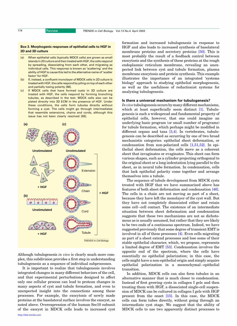
When MDCK cysts are treated with HGF, they undergo anordered sequence of events to generate a tubule [45–47](Boxes 2 and 3). First, some cells send out a large‘extension’ of their basolateral surface, although thesecells remain in the cyst wall and at this stage retain a smallapical surface. This large extension is similar to the largepseudopodia seen in fibroblasts and neurons moving in 3DECM [48,49]. Extension formation requires phosphatidyl-inositol 3-kinase and the lipid products of this kinase areconcentrated in the plasma membrane of the extension[47]. This is similar to the concentration of phosphatidyl-inositol 3-lipids in the leading edge of migrating neutro-phils and Dictyostelium amoebae [40]. Ordinarily, only afew cells in the cyst wall form extensions in response toHGF. Notably, inhibition of Rho kinase causes a tenfold
increase in the number of extensions formed, indicatingthat extension formation is normally limited by the RhoGTPase and its effector, Rho kinase [47].
Next, the cells divide and some migrate out of the wall ofthe cyst to form ‘chains’ of cells, which are typically 1–3cells in length. In the absence of HGF, the cells divide inthe plane of the monolayer, such that both daughter cellsremain in the cyst wall. On treatment with HGF, however,the orientation of cell division changes such that onedaughter leaves the cyst wall and initiates chain formation[47]. Cells in chains have lost their epithelial apical–basolateral polarity. Apical surface marker proteins areundetectable, and basolateral marker proteins such asE-cadherin surround the cell [45–47]. Cells at the end ofthe chain have a polarity and phenotype that resemblesthat of a migrating fibroblast or mesenchymal cell.
Finally, the cells divide further to form ‘cords’, which areabout 2–3 cells thick. At this stage, the cells begin toregain epithelial polarity and form small lumens betweenthemselves. Eventually the lumens enlarge, merge andbecome contiguous with the central lumen of the cyst toform mature tubules.
A key aspect of this sequence of events is that some ofthe cells undergo a transient loss of epithelial polarization,becoming similar to migrating fibroblasts [47]. We havepreviously suggested that this is essentially a transient,partial epithelial–mesenchymal transition (EMT) [4,6]and that the cells re-establish the polarized epithelialphenotype to form new tubes. Reversible EMTs occurnormally during many stages of development, whereasirreversible EMTs are characteristic of invasive cancersderived from epithelial cells. We suggest that the process ofre-establishment of polarity is similar to that used to formcysts and might even involve the same Rac and lamininassembly pathway. Partial EMT, followed by re-establish-ment of the epithelial phenotype, might be also used togenerate branches from an existing tubule [2].
This simple model separates the complex process oftubulogenesis into two chief subprocesses: partial loss of thepolarized phenotype to permit remodeling into tubules andmore complex shapes, and re-establishment of the polarizedphenotype on the basis of the drive for three surfaces.
Fig. 2. Polarization and orientation of cells. (a) Many migrating cells, such as neutrophils and Dictyostelium discoideum amoeba, can move by chemotaxis up a gradient of
an external attractant. These cells show a highly polarized morphology, typically with a leading edge in the direction of migration. In all panels, the green shading indicates
the polarization of the cell in the direction of migration. (b) When uniformly surrounded by a chemoattractant, however, cells sometimes can be polarized at random [56].
(c) Depending on the situation, cells can show migration and a polarized morphology in a random orientation, or even multiple leading edges suggestive of multipolar
polarization [57]. (d) Epithelial cells grown as cysts in 3D culture are polarized and the polarity is coordinated so that the apical surfaces of all the cells are oriented towards
the central lumen of the cyst (red). (e) Expression of a dominant-negative mutant of the small GTPase Rac1 causes a partial inversion of polarity. Markers for the apical sur-
faces of the cells (red) are now on the periphery of the cysts, which lack lumen. The internal organization of the cytoplasm (blue to red shading) is also inverted, such that
the Golgi lies beneath the apical surface at the periphery of the cyst.
Golgi
TRENDS in Cell Biology
(a) (b) (c) (d) (e)
Review TRENDS in Cell Biology Vol.13 No.4 April 2003 173
http://ticb.trends.com
Although tubulogenesis in vivo is clearly much more com-plex, this subdivision provides a first step in understandingtubulogenesis as a sequence of individual subprocesses.
It is important to realize that tubulogenesis involvesintegrated changes in many different behaviors of the cell,and that experimental perturbations designed to affectonly one cellular process can lead to protean changes inmany aspects of cyst and tubule formation, and even tounexpected insight into the connections among theseprocesses. For example, the exocytosis of newly madeproteins at the basolateral surface involves the exocyst, asnoted above. Overexpression of the human Sec10 subunitof the exocyst in MDCK cells leads to increased cyst
formation and increased tubulogenesis in response toHGF and also leads to increased synthesis of basolateralmembrane proteins and secretory proteins [50]. This ismost probably the result of a feedback control betweenexocytosis and the synthesis of these proteins at the roughendoplasmic reticulum membrane, revealing an unex-pected link between cyst and tubule formation, plasmamembrane exocytosis and protein synthesis. This exampleillustrates the importance of an integrated ‘systemsbiology’ approach to studying epithelial morphogenesis,as well as the usefulness of reductionist systems foranalyzing tubulogenesis.
Is there a universal mechanism for tubulogenesis?
In vivo tubulogenesis occurs by many different mechanisms,which at least superficially seem distinct [1]. Tubulo-genesis is such a widespread and fundamental property ofepithelial cells, however, that one could imagine anunderlying basic program (or small number of programs)for tubule formation, which perhaps might be modified indifferent organs and taxa [3,4]. In vertebrates, tubulo-genesis can be described as occurring by one of two broadmechanistic categories: epithelial sheet deformation, orcondensation from non-polarized cells [1,51,52]. In epi-thelial sheet deformation, the cells move as a coherentsheet that invaginates or evaginates. This sheet can formvarious shapes, such as a cylinder projecting orthogonal tothe original sheet or a long indentation lying parallel to thesheet, as in neural tube formation. In condensation, cellsthat lack epithelial polarity come together and arrangethemselves into a tubule.
The sequence of tubule development from MDCK cyststreated with HGF that we have summarized above hasfeatures of both sheet deformation and condensation [46].The cells in a chain are not moving as part of a sheet,because they have left the monolayer of the cyst wall. Butthey have not completely dissociated either and retainsome cell–cell contact. The existence of an intermediatesituation between sheet deformation and condensationsuggests that these two mechanisms are not as dichoto-mous as is usually assumed, but rather that they are likelyto be two ends of a continuous spectrum. Indeed, we havesuggested previously that some degree of transient EMT isinvolved in all of these processes [4]. Even cells migratingas part of a sheet extend processes and lose some of theirstable epithelial character, which, we propose, representsa limited degree of EMT [53]. Condensation involves theopposite end of the spectrum, where the cells haveessentially no epithelial polarization; in this case, thecells might have a non-epithelial origin and simply acquireepithelial polarization in a mesenchymal–epithelialtransition.
In addition, MDCK cells can also form tubules in analternative manner that is much closer to condensation.Instead of first growing cysts in collagen I gels and thentreating them with HGF, a dissociated single-cell suspen-sion of MDCK can be cultured in collagen I gels with HGFpresent from the onset [15]. In this case, the MDCKcells can form tubes directly, without going through anintermediate cyst stage. We suggest that the ability ofMDCK cells to use two apparently distinct processes to
Box 3. Morphogenic responses of epithelial cells to HGF in
2D and 3D culture
(a) When epithelial cells (typically MDCK cells) are grown as small
islands in2Dcultureand then treated withHGF, thecells respond
by spreading, dissociating from each other, and migrating as
individual cells. This response is known as ‘scattering’ and the
ability of HGF to cause this led to the alternative name of ‘scatter
factor’ for HGF.
(b) If, instead, a confluent monolayer of MDCK cells in 2D culture is
treated with HGF, thecells respond by piling on top of each other
and partially losing polarity [68].
(c) If MDCK cells that have formed cysts in 3D culture are
treated with HGF, the cells respond by forming branching
tubules, as described in the text. MDCK cells also can be
plated directly into 3D ECM in the presence of HGF. Under
these conditions, the cells form tubules directly without
forming a cyst. The cells might go through intermediates
that resemble extensions, chains and cords, although this
issue has not been clearly resolved [69].
TRENDS in Cell Biology
+ HGF
+ HGF
+ HGF
Unstimulated
Unstimulated
Unstimulated
Extension
Chain
Cord
Tubule
(a)
(b)
(c)
Review TRENDS in Cell Biology Vol.13 No.4 April 2003174
http://ticb.trends.com

form tubules supports the concept that there is anunderlying common tubulogenesis program, which canbe manifested in several different ways.
Concluding remarks
The potential value of any reductionist in vitro system isthe extent to which it can tell us about in vivo processes,such as tubulogenesis. The 3D-MDCK-HGF system is thebest characterized tubulogenesis system for cellularprocesses downstream of the inductive signals, and ithas already led to the formulation of several simpleconcepts. These include, first, the drive for three surfaces,which might underlie the formation of polarized mono-layers enclosing a lumen; second, the distinction betweenpolarization and orientation of polarization; and third, theuse of transient EMT to remodel structures into tubes andbranches. These simple concepts can provide a conceptualframework for future analyses of tubulogenesis in vivo.Tubulogenesis in animals is much more complex and muchmore diverse than in vitro, and it will be important to seehow the basic processes that have been identified throughin vitro work are implemented and modified in vivo.
AcknowledgementsWe apologize to the authors whose important work could not be citedowing to space limitations. We thank Paul Brakeman for comments on themanuscript and Valerie Weaver for pointing out the importance of theviscoelastic properties of ECM and other discussions. We thank DavidBilder, Mark Krasnow and Zena Werb for providing manuscripts inadvance of publication and for discussions. Work in our laboratory issupported by grants from the National Institutes of Health (AI25144,DK58061, AI36953, HL55980, AI053194). W.Y. is supported by anAmerican Heart Association Fellowship (0120084Y); A.D. is supportedby a Susan G. Komen Breast Cancer Foundation Fellowship(PDF0100766).
References
1 Hogan, B.L.M. and Kolodziej, P.A. (2002) Organogenesis: molecularmechanisms of tubulogenesis. Nat. Rev. Genet. 3, 513–523
2 Affolter, M. et al. (2003) Tube or not tube: remodeling epithelial tissuesby branching morphogenesis. Dev. Cell 4, 11–18
3 Lubarsky, B. and Krasnow, M.A. (2003) Tube morphogenesis: makingand shaping biological tubes. Cell 112, 19–28
4 O’Brien, L.E. et al. (2002) Building epithelial architecture: insightsfrom three-dimensional culture models. Nat. Rev. Mol. Cell Biol. 3,531–537
5 Jacinto, A. et al. (2002) Dynamic analysis of dorsal closure inDrosophila: from genetics to cell biology. Dev. Cell 3, 9–19
6 Thiery, J.P. (2002) Epithelial-mesenchymal transitions in tumourprogression. Nat. Rev. Cancer 2, 442–454
7 Van Aelst, L. and Symons, M. (2002) Role of Rho family GTPases inepithelial morphogenesis. Genes Dev. 16, 1032–1054
8 Wallingford, J.B. et al. (2002) Convergent extension: the molecularcontrol of polarized cell movement during embryonic development.Dev. Cell 2, 695–706
9 Simons, K. and Fuller, S.D. (1985) Cell surface polarity in epithelia.Annu. Rev. Cell Biol. 1, 243–288
10 Mostov, K.E. et al. (2000) Membrane traffic in polarized epithelial cells.Curr. Opin. Cell Biol. 12, 483–490
11 Bissell, M.J. and Radisky, D. (2001) Putting tumours in context. Nat.Rev. Cancer 1, 46–54
12 Birchmeier, C. and Gherardi, E. (1998) Developmental roles of HGF/SF and its receptor, the c-Met tyrosine kinase. Trends Cell Biol. 8,404–410
13 Brinkmann, V. et al. (1995) Hepatocyte growth factor/scatter factorinduces a variety of tissue-specific morphogenic programs in epithelialcells. J. Cell Biol. 131, 1573–1586
14 Montesano, R. et al. (1991) Identification of a fibroblast-derivedepithelial morphogen as hepatocyte growth factor. Cell 67, 901–908
15 Montesano, R. et al. (1991) Induction of epithelial tubular morpho-genesis in vitro by fibroblast-derived soluble factors. Cell 66, 697–711
16 Dressler, G. (2002) Tubulogenesis in the developing mammaliankidney. Trends Cell Biol. 12, 390–395
17 Pohl, M. et al. (2000) Branching morphogenesis during kidneydevelopment. Annu. Rev. Physiol. 62, 595–620
18 Sakurai, H. et al. (1997) An in vitro tubulogenesis system using celllines derived from the embryonic kidney shows dependence onmultiple soluble growth factors. Proc. Natl. Acad. Sci. U. S. A. 94,6279–6284
19 Liu, Y. (2002) Hepatocyte growth factor and the kidney. Curr. Opin.Nephrol. Hypertens. 11, 23–30
20 Nagano, T. et al. (2002) Ameliorative effect of hepatocyte growth factoron glycerol-induced acute renal failure with acute tubular necrosis.Nephron 91, 730–738
21 Santos, O.F. et al. (1994) Involvement of hepatocyte growth factor inkidney development. Dev. Biol. 163, 525–529
22 van Adelsberg, J. et al. (2001) Activation of hepatocyte growth factor(HGF) by endogenous HGF activator is required for metanephrickidney morphogenesis in vitro. J. Biol. Chem. 276, 15099–15106
23 Woolf, A.S. et al. (1995) Roles of hepatocyte growth factor/scatter factorand the met receptor in the early development of the metanephros.J. Cell Biol. 128, 171–184
24 Yeaman, C. et al. (1999) New perspectives on mechanisms involved ingenerating epithelial cell polarity. Physiol. Rev. 79, 73–98
25 Debnath, J. et al. (2002) The role of apoptosis in creating andmaintaining luminal space within normal and oncogene-expressingmammary acini. Cell 111, 29–40
26 Lin, H.H. et al. (1999) Bcl-2 overexpression prevents apoptosis-induced Madin–Darby canine kidney simple epithelial cyst formation.Kidney Int. 55, 168–178
27 Murray, P. and Edgar, D. (2000) Regulation of programmed cell deathby basement membranes in embryonic development. J. Cell Biol. 150,1215–1221
28 Coucouvanis, E. and Martin, G.R. (1995) Signals for death andsurvival: a two-step mechanism for cavitation in the vertebrateembryo. Cell 83, 279–287
29 Gumbiner, B.M. (1996) Cell adhesion: the molecular basis of tissuearchitecture and morphogenesis. Cell 84, 345–357
30 Drubin, D.G. and Nelson, W.J. (1996) Origins of cell polarity. Cell 84,335–344
31 Grindstaff, K.K. et al. (1998) Sec6/8 complex is recruited to cell–cellcontacts and specifies transport vesicle delivery to the basal-lateralmembrane in epithelial cells. Cell 93, 731–740
32 Lipschutz, J.H. and Mostov, K.E. (2002) Exocytosis: the many mastersof the exocyst. Curr. Biol. 12, R212–R214
33 Knust, E. and Bossinger, O. (2002) Composition and formation ofintercellular junctions in epithelial cells. Science 298, 1955–1959
34 Lecuit, T. and Wieschaus, E. (2002) Junctions as organizing centers inepithelial cells? A fly perspective. Traffic 3, 92–97
35 Bilder, D. et al. (2003) Integrated activity of PDZ protein complexesregulates epithelial polarity. Nat. Cell Biol. 5, 53–58
36 Tanentzapf, G. and Tepass, U. (2003) Interactions between the crumbs,lethal giant larvae and bazooka pathways in epithelial polarization.Nat. Cell Biol. 5, 46–52
37 Myat, M.M. and Andrew, D.J. (2002) Epithelial tube morphology isdetermined by the polarized growth and delivery of apical membrane.Cell 111, 879–891
38 Medina, E. et al. (2002) Crumbs interacts with moesin and b(heavy)-spectrin in the apical membrane skeleton of Drosophila. J. Cell Biol.158, 941–951
39 O’Brien, L.E. et al. (2001) Rac1 orientates epithelial apical polaritythrough effects on basolateral laminin assembly. Nat. Cell Biol. 3,831–838
40 Iijima, M. et al. (2002) Temporal and spatial regulation of chemotaxis.Dev. Cell 3, 469–478
41 Clark, E.A. et al. (1998) Integrin-mediated signals regulated bymembers of the Rho family of GTPases. J. Cell Biol. 142, 573–586
42 Joberty, G. et al. (2000) The cell-polarity protein Par6 links Par3 andatypical protein kinase C to Cdc42. Nat. Cell Biol. 2, 531–539
43 Lin, D. et al. (2000) A mammalian PAR-3–PAR-6 complex implicated
Review TRENDS in Cell Biology Vol.13 No.4 April 2003 175
http://ticb.trends.com
in Cdc42/Rac1 and aPKC signalling and cell polarity. Nat. Cell Biol. 2,540–547
44 Price, L.S. et al. (1998) Activation of Rac and Cdc42 by integrinsmediates cell spreading. Mol. Biol. Cell 9, 1863–1871
45 Pollack, A.L. et al. (1997) Dynamics of b-catenin interactions with APCprotein regulate epithelial tubulogenesis. J. Cell Biol. 137, 1651–1662
46 Pollack, A.L. et al. (1998) Morphogenetic mechanisms of epithelialtubulogenesis: MDCK cell polarity is transiently rearranged withoutloss of cell-cell contact during scatter factor/hepatocyte growth factor-induced tubulogenesis. Dev. Biol. 204, 64–79
47 Yu, W. et al. (2003) Hepatocyte growth factor switches orientation ofpolarity and mode of movement during morphogenesis of multicellularepithelial structures. Mol. Biol. Cell 14, 748–763
48 Knight, B. et al. (2000) Visualizing muscle cell migration in situ. Curr.Biol. 10, 576–585
49 Murase, S. and Horwitz, A.F. (2002) Deleted in colorectal carcinomaand differentially expressed integrins mediate the directionalmigration of neural precursors in the rostral migratory stream.J. Neurosci. 22, 3568–3579
50 Lipschutz, J.H. et al. (2000) Exocyst is involved in cystogenesis andtubulogenesis and acts by modulating synthesis and delivery ofbasolateral plasma membrane and secretory proteins. Mol. Biol. Cell11, 4259–4275
51 Locascio, A. and Nieto, M.A. (2001) Cell movements during vertebratedevelopment: integrated tissue behaviour versus individual cellmigration. Curr. Opin. Genet. Dev. 11, 464–469
52 Thiery, J.P. and Boyer, B. (1992) The junction between cytokines andcell adhesion. Curr. Opin. Cell Biol. 4, 782–792
53 Fenteany, G. et al. (2000) Signaling pathways and cell mechanicsinvolved in wound closure by epithelial cell sheets. Curr. Biol. 10,831–838
54 Lipschutz, J.H. et al. (2001) Analysis of membrane traffic in polarizedepithelial cells. Curr. Protocols Cell Biol., 1551–15515
55 Rodriguez-Boulan, E. and Powell, S.K. (1992) Polarity of epithelial andneuronal cells. Annu. Rev. Cell Biol. 8, 395–427
56 Weiner, O.D. (2002) Regulation of cell polarity during eukaryoticchemotaxis: the chemotactic compass. Curr. Opin. Cell Biol. 14,196–202
57 Funamoto, S. et al. (2002) Spatial and temporal regulation of 3-phosphoinositides by PI 3-kinase and PTEN mediates chemotaxis. Cell109, 611–623
58 Ehrlich, J. et al. (2002) Spatio-temporal regulation of Rac1 localizationand lamellipodia dynamics during epithelial cell–cell adhesion. Dev.Cell 3, 259–270
59 Hansen, S.H. et al. (2000) Induced expression of Rnd3 is associatedwith transformation of polarized epithelial cells by the Raf-MEK-extracellular signal-regulated kinase pathway. Mol. Cell. Biol. 20,9364–9375
60 Rodriguez-Boulan, E. and Nelson, W.J. (1989) Morphogenesis of thepolarized epithelial cell phenotype. Science 245, 718–725
61 Geiser, T.K. et al. (2001) Pseudomonas aeruginosa ExoT inhibits invitro lung epithelial wound repair. Cell. Microbiol. 3, 223–236
62 Flanagan, L.A. et al. (2002) Neurite branching on deformablesubstrates. NeuroReport 13, 2411–2415
63 Zahir, N. et al. (2002) Peering from the outside in: Viscoelasticproperties of the extracellular matrix dictate spatial organizationand apoptosis resistance in mammary epithelial cells. Proceedingsof the Second Joint meeting of the IEEE Engineering in Medicineand Biology Society and the Biomedical Engineering Society 1,430–431
64 Lindstedt, R. et al. (1993) Apical secretion of a cytosolic protein byMDCK cells: evidence for polarized release of an endogenous lectin by anon-classical secretory pathway. J. Biol. Chem. 268, 11750–11757
65 Bao, Q. and Hughes, R.C. (1999) Galectin-3 and polarized growthwithin collagen gels of wild-type and ricin-resistant MDCK renalepithelial cells. Glycobiology 9, 489–495
66 Hikita, C. et al. (2000) Induction of terminal differentiation inepithelial cells requires polymerization of hensin by galectin 3.J. Cell Biol. 151, 1235–1246
67 Weaver, V. et al. (2002) b4 integrin-dependent formation ofpolarized three-dimensional architecture confers resistance toapoptosis in normal and malignant mammary epithelium. CancerCell 2, 205–216
68 Balkovetz, D.F. et al. (1997) Hepatocyte growth factor alters thepolarity of Madin–Darby canine kidney cell monolayers. J. Biol.Chem. 272, 3471–3477
69 Chiu, S.J. et al. (2002) Hepatocyte growth factor upregulates a2b1integrin in Madin–Darby canine kidney cells: implications intubulogenesis. J. Biomed. Sci. 9, 261–272
70 Rosario, M. and Birchmeier, W. How to make tubes: signalling by thec-Met receptor tyrosine kinase. (2003) Trends Cell Biol. (in press)
Managing your references and BioMedNet ReviewsDid you know that you can now download selected search results from BioMedNet Reviews directly into your chosen reference-managing software? After performing a search, simply click to select the articles you are interested in, choose the format required(e.g. EndNote 3.1) and the bibliographic details, abstract and link to the full-text will download into your desktop reference managerdatabase.BioMedNet Reviews is available on institute-wide subscription. If you do not have access to the full-text articles in BioMedNetReviews, ask your librarian to contact [email protected]
Review TRENDS in Cell Biology Vol.13 No.4 April 2003176
http://ticb.trends.com
Related Documents