NEUROSYSTEMS Epilepsy-induced abnormal striatal plasticity in Bassoon mutant mice Veronica Ghiglieri, 1, * Barbara Picconi, 1, * Carmelo Sgobio, 1 Vincenza Bagetta, 1 Ilaria Barone, 1 Vincent Paille `, 1 Massimiliano Di Filippo, 1,2 Federica Polli, 3 Fabrizio Gardoni, 3 Wilko Altrock, 4 Eckart D. Gundelfinger, 4 Giovambattista De Sarro, 5 Giorgio Bernardi, 1 Martine Ammassari-Teule, 6 Monica Di Luca 3 and Paolo Calabresi 1,2 1 Laboratorio di Neurofisiologia, Fondazione Santa Lucia, IRCCS c ⁄ o CERC, Rome, Italy 2 Clinica Neurologica, Dip. Specialita ` Medico Chirurgiche e Sanita ` Pubblica, Universita ` di Perugia, Italy 3 Center of Excellence on Neurodegenerative Diseases and Department of Pharmacological Sciences, University of Milan, Milan, Italy 4 Leibniz Institute for Neurobiology, Magdeburg, Germany 5 Facolta ` di Medicina e Chirurgia, Universita ` degli Studi Magna Graecia di Catanzaro, Campus di Germaneto, Italy 6 Istituto di Neuroscienze del CNR, Sezione di Psicobiologia e Psicofarmacologia, Fondazione Santa Lucia, IRCCS, Rome, Italy Keywords: dorsolateral striatum, fast-spiking interneurons, postsynaptic density, scaffolding proteins, synaptic plasticity Abstract Recently, the striatum has been implicated in the spread of epileptic seizures. As the absence of functional scaffolding protein Bassoon in mutant mice is associated with the development of pronounced spontaneous seizures, we utilized this new genetic model of epilepsy to investigate seizure-induced changes in striatal synaptic plasticity. Mutant mice showed reduced long-term potentiation in striatal spiny neurons, associated with an altered N-methyl-d-aspartate (NMDA) receptor subunit distribution, whereas GABAergic fast-spiking (FS) interneurons showed NMDA-dependent short-term potentiation that was absent in wild-type animals. Alterations in the dendritic morphology of spiny neurons and in the number of FS interneurons were also observed. Early antiepileptic treatment with valproic acid reduced epileptic attacks and mortality, rescuing physiological striatal synaptic plasticity and NMDA receptor subunit composition. However, morphological alterations were not affected by antiepileptic treatment. Our results indicate that, in Bsn mutant mice, initial morphological alterations seem to reflect a more direct effect of the abnormal genotype, whereas plasticity changes are likely to be caused by the occurrence of repeated cortical seizures. Introduction Bassoon is an integral component of the presynaptic cytoskeleton that functionally interacts with other proteins to organize the highly specialized cytomatrix at active zones of regulated neurotransmitter release (tom Dieck et al., 1998; Winter et al., 1999). This regulatory activity can be exerted at both inhibitory and excitatory glutamatergic synapses and in several brain areas, including the cortex, hippocam- pus, and striatum (Richter et al., 1999; Garner et al., 2000; Schoch & Gundelfinger, 2006). Mice lacking the central domain of the Bassoon gene (Bsn) display alterations in neuronal activity associated with the occurrence of spontaneous cortical seizures, which often lead to death in homozygous mutants (Altrock et al., 2003). These features make this mutant a novel genetic model of epilepsy and a useful tool to explore the role of specific brain circuits in the generation, propagation and recurrence of epileptic seizures. Although the involvement of cortical and hippocampal circuits in the genesis of epileptic activity has been extensively characterized, the possible role of basal ganglia, and in particular of the striatum, in modulating seizures has only recently been postulated (Slaght et al., 2002; Bouilleret et al., 2008). Adaptive changes in striatal cell-type-specific synaptic plasticity might play a major role in the basal ganglia control of seizure activity. In fact, the striatum is the main input structure of the basal ganglia– thalamo-cortical loop, in which the functional interplay of converging excitatory and inhibitory inputs underlies the correct information processing needed to control motor learning and habit formation (Calabresi et al., 2007). This critical role of the striatum in basal ganglia relies on the interactions between medium spiny (MS) neurons and a subtype of GABAergic interneurons that express parvalbumin (PV), called fast-spiking (FS) interneurons on the basis of their electrophysiological characteristics (Kawaguchi, 1993). Striatal PV- containing FS interneurons receive powerful excitatory input from the cortex and exert strong feedforward inhibition of MS neurons, their predominant synaptic target (Kawaguchi, 1993; Koos & Tepper, 1999; Tepper et al., 2004; Mallet et al., 2005). Given the critical importance of FS interneurons in the balance of striatal MS neuron activity, which, in turn, shapes the activity of basal ganglia output nuclei, it is conceivable that the frequent spread of epileptic seizures Correspondence: Dr Paolo Calabresi, 2 Clinica Neurologica, as above. E-mail [email protected] *These authors contributed equally to this work. Received 29 December 2008, revised 18 February 2009, accepted 28 February 2009 European Journal of Neuroscience, Vol. 29, pp. 1979–1993, 2009 doi:10.1111/j.1460-9568.2009.06733.x ª The Authors (2009). Journal Compilation ª Federation of European Neuroscience Societies and Blackwell Publishing Ltd European Journal of Neuroscience

Welcome message from author
This document is posted to help you gain knowledge. Please leave a comment to let me know what you think about it! Share it to your friends and learn new things together.
Transcript

NEUROSYSTEMS
Epilepsy-induced abnormal striatal plasticity in Bassoonmutant mice
Veronica Ghiglieri,1,* Barbara Picconi,1,* Carmelo Sgobio,1 Vincenza Bagetta,1 Ilaria Barone,1 Vincent Paille,1
Massimiliano Di Filippo,1,2 Federica Polli,3 Fabrizio Gardoni,3 Wilko Altrock,4 Eckart D. Gundelfinger,4
Giovambattista De Sarro,5 Giorgio Bernardi,1 Martine Ammassari-Teule,6 Monica Di Luca3 and Paolo Calabresi1,2
1Laboratorio di Neurofisiologia, Fondazione Santa Lucia, IRCCS c ⁄ o CERC, Rome, Italy2Clinica Neurologica, Dip. Specialita Medico Chirurgiche e Sanita Pubblica, Universita di Perugia, Italy3Center of Excellence on Neurodegenerative Diseases and Department of Pharmacological Sciences, University of Milan, Milan,Italy4Leibniz Institute for Neurobiology, Magdeburg, Germany5Facolta di Medicina e Chirurgia, Universita degli Studi Magna Graecia di Catanzaro, Campus di Germaneto, Italy6Istituto di Neuroscienze del CNR, Sezione di Psicobiologia e Psicofarmacologia, Fondazione Santa Lucia, IRCCS, Rome, Italy
Keywords: dorsolateral striatum, fast-spiking interneurons, postsynaptic density, scaffolding proteins, synaptic plasticity
Abstract
Recently, the striatum has been implicated in the spread of epileptic seizures. As the absence of functional scaffolding proteinBassoon in mutant mice is associated with the development of pronounced spontaneous seizures, we utilized this new genetic modelof epilepsy to investigate seizure-induced changes in striatal synaptic plasticity. Mutant mice showed reduced long-term potentiationin striatal spiny neurons, associated with an altered N-methyl-d-aspartate (NMDA) receptor subunit distribution, whereas GABAergicfast-spiking (FS) interneurons showed NMDA-dependent short-term potentiation that was absent in wild-type animals. Alterations inthe dendritic morphology of spiny neurons and in the number of FS interneurons were also observed. Early antiepileptic treatmentwith valproic acid reduced epileptic attacks and mortality, rescuing physiological striatal synaptic plasticity and NMDA receptorsubunit composition. However, morphological alterations were not affected by antiepileptic treatment. Our results indicate that, in Bsnmutant mice, initial morphological alterations seem to reflect a more direct effect of the abnormal genotype, whereas plasticitychanges are likely to be caused by the occurrence of repeated cortical seizures.
Introduction
Bassoon is an integral component of the presynaptic cytoskeleton thatfunctionally interacts with other proteins to organize the highlyspecialized cytomatrix at active zones of regulated neurotransmitterrelease (tom Dieck et al., 1998; Winter et al., 1999). This regulatoryactivity can be exerted at both inhibitory and excitatory glutamatergicsynapses and in several brain areas, including the cortex, hippocam-pus, and striatum (Richter et al., 1999; Garner et al., 2000; Schoch &Gundelfinger, 2006). Mice lacking the central domain of the Bassoongene (Bsn) display alterations in neuronal activity associated with theoccurrence of spontaneous cortical seizures, which often lead to deathin homozygous mutants (Altrock et al., 2003). These features makethis mutant a novel genetic model of epilepsy and a useful tool toexplore the role of specific brain circuits in the generation, propagationand recurrence of epileptic seizures. Although the involvement ofcortical and hippocampal circuits in the genesis of epileptic activity
has been extensively characterized, the possible role of basal ganglia,and in particular of the striatum, in modulating seizures has onlyrecently been postulated (Slaght et al., 2002; Bouilleret et al., 2008).Adaptive changes in striatal cell-type-specific synaptic plasticity mightplay a major role in the basal ganglia control of seizure activity. Infact, the striatum is the main input structure of the basal ganglia–thalamo-cortical loop, in which the functional interplay of convergingexcitatory and inhibitory inputs underlies the correct informationprocessing needed to control motor learning and habit formation(Calabresi et al., 2007). This critical role of the striatum in basalganglia relies on the interactions between medium spiny (MS) neuronsand a subtype of GABAergic interneurons that express parvalbumin(PV), called fast-spiking (FS) interneurons on the basis of theirelectrophysiological characteristics (Kawaguchi, 1993). Striatal PV-containing FS interneurons receive powerful excitatory input from thecortex and exert strong feedforward inhibition of MS neurons, theirpredominant synaptic target (Kawaguchi, 1993; Koos & Tepper,1999; Tepper et al., 2004; Mallet et al., 2005). Given the criticalimportance of FS interneurons in the balance of striatal MS neuronactivity, which, in turn, shapes the activity of basal ganglia outputnuclei, it is conceivable that the frequent spread of epileptic seizures
Correspondence: Dr Paolo Calabresi, 2Clinica Neurologica, as above.
E-mail [email protected]
*These authors contributed equally to this work.
Received 29 December 2008, revised 18 February 2009, accepted 28 February 2009
European Journal of Neuroscience, Vol. 29, pp. 1979–1993, 2009 doi:10.1111/j.1460-9568.2009.06733.x
ª The Authors (2009). Journal Compilation ª Federation of European Neuroscience Societies and Blackwell Publishing Ltd
European Journal of Neuroscience

throughout the cortex could profoundly alter both short-term and long-term excitability in the striatal neuronal subtypes as an adaptivemechanism.Here, we tested the hypothesis that adaptive changes in the
synaptic plasticity of striatal MS neurons and FS interneurons mightoccur. To this end, we characterized the corticostriatal pathway ofBsn mutant mice by in vitro intracellular recordings from striatalMS neurons and FS interneurons, and analysed possible correlationswith molecular and morphological aspects. We also investigated thepossibility that early, chronic treatment with the antiepileptic drug(AED) valproic acid (VPA) could influence the clinical expressionof seizures as well as the molecular, electrophysiological andmorphological abnormalities observed in this new genetic model ofepilepsy.
Materials and methods
Animals
Mice lacking Bassoon (BsnDEx4 ⁄ 5) were generated as describedpreviously (Altrock et al., 2003), back-crossed into the C57 ⁄ bl6mouse strain, and then crossed with the SV129 strain, yielding micewith a mixed C57 ⁄ bl6–SV129 genetic background for mutants andwild types. Genotyping was performed by polymerase chain reaction,as described previously (Altrock et al., 2003); age-matched wild-typelittermates were used as controls. At the beginning of the experiment,animals were 2–3 months old, and their weights ranged from 23 to28 g. They were housed in a temperature-controlled room (22�C) witha light ⁄ dark cycle of 12 : 12 h (lights on from 07:00 to 19:00 h).Food and water were given ad libitum. All experiments were carriedout according to the guidelines on the ethical use of animals from theEuropean Communities Council Directive of 24 November 1986(86 ⁄ 609 ⁄ EEC) and from the National Ministry of Health (D.M.222 ⁄ 2008).
Behavioral characterization of seizure attacks
The behavior of 14-day-old, 20-day-old and 2-month-old mice wasvideo-recorded for 4 h every day for 5 days in order to monitorepileptic activity in Bsn mutants. The animals were placed in asoundproof room between 11:00 and 15:00 h with the light on, andrecorded through a video camera interfaced with a computer. Videoswere stored and analysed offline, and the frequency and duration ofattacks were scored.
Electrophysiological experiments
Adult mice (2–3 months old, weighting 23–28 g) were used for allexperiments. Animals were killed by cervical dislocation, in order toobtain coronal corticostriatal slices for electrophysiological record-ings (Calabresi et al., 1992, 2000). The slices (200–300 lm) wereprepared from tissue blocks of the brain with the use of a vibratome.A single slice was transferred to a recording chamber andsubmerged in continuously flowing Krebs solution (33�C, 2–3 mL ⁄ min) gassed with 95% O2 and 5% CO2. The compositionof the control solution was 126 mm NaCl, 2.5 mm KCl, 1.2 mm
MgCl2, 1.2 mm NaH2PO4, 2.4 mm CaCl2, 11 mm glucose, and25 mm NaHCO3. In all experiments, the intracellular recordingelectrodes were filled with 2 m KCl (30–60 MX). Signals wererecorded utilizing an Axoclamp 2B amplifier (Axon Instruments,Foster City, CA, USA), displayed on a separate oscilloscope, andstored and analysed on a digital system (pClamp 9; Axon
Instruments). For synaptic stimulation, bipolar electrodes were used.These stimulating electrodes were located in the white matterbetween the cortex and the striatum to activate corticostriatal fibers.For the long-term potentiation (LTP) protocol, at the beginning ofintracellular recordings, magnesium ions were omitted from themedium to increase the N-methyl-d-aspartate (NMDA)-mediatedcomponent of the excitatory postsynaptic potential (EPSP). Asconditioning tetanus [high-frequency stimulation (HFS)], we usedthree trains (duration, 3 s; frequency, 100 Hz; intervals, 20 s).During tetanic stimulation, the intensity was increased to supra-threshold levels. In a subset of experiments, variations in the EPSPamplitude in response to the increasing stimulus intensity werestudied. Input–output responses were plotted and fitted by asigmoidal function, and then compared by two-way anova forstatistical significance. Quantitative data on EPSP modificationsinduced by tetanic stimulation are expressed as a percentage of thecontrols, the latter representing the mean of responses recordedduring a stable period (15–20 min) before the tetanus. Values givenin the text and in the figures are mean ± SEM of changes in therespective cell populations. Two-way anova was used; this wasfollowed by Bonferroni post hoc test for comparisons betweengenotypes or Student’s t-test to compare the means between pre- andpost-tetanic stimulation.
Golgi–Cox impregnation of brain tissue
Mice were anesthetized with chloral hydrate (400 mg ⁄ kg) andperfused intracardially with 0.9% saline. The brains were dissected,and impregnated using a standard Golgi–Cox solution (1% potassiumdichromate, 1% mercuric chloride, and 0.8% potassium chromate),according to a previously described method (Glaser & Van der Loos,1981). The brains immersed in the Golgi–Cox solution were stored atroom temperature for 6 days, immersed in a sucrose solution (30%)for 2 days, and sectioned coronally (100 lm) using a vibratome.Sections were mounted on gelatinized slides, stained according to apreviously described method (Gibb & Kolb, 1998), and covered withPermount solution.
Spine density
Measurements were performed on MS neurons lying in the dorsolat-eral striatum. Three spiny neurons within each hemisphere wereselected. As no interhemispheric difference was detected, the datawere pooled together so that six neurons per striatum were consideredin each animal. Fully impregnated neurons were first identified underlow magnification [·20; numerical aperture (NA), 0.5]. Subsequently,spines were analysed ⁄ counted under a higher magnification (·63; NA,0.75). Measurements were performed on each neuron and for threedendrites; five 20-lm dendrite segments lying on the focal plane(Leuner et al., 2003) were randomly selected. Segments were sampled50 lm away from soma to exclude the spine-depleted zone arisingfrom the cell body. The average spine density (number of spines per10 lm of dendrite length) was estimated by focusing in and out withthe fine adjustment of the microscope (Leica DMLB, Wetzlar,Germany). All protrusions were counted as spines if they were indirect contact with the dendritic shaft. As this method has been provento give reliable results (Horner & Arbuthnott, 1991), no attempt wasmade to introduce a correction factor for hidden spines. Allmeasurements were performed by an experimenter blind to theexperimental condition of the animal under examination, and analysedby anova.
1980 V. Ghiglieri et al.
ª The Authors (2009). Journal Compilation ª Federation of European Neuroscience Societies and Blackwell Publishing LtdEuropean Journal of Neuroscience, 29, 1979–1993

Dendrite branching
For the branching analysis, three basal dendrites for every neuron wereclassified by using the centrifugal method (Uylings et al., 1986). Thebranches arising from the soma were numbered as branch order 1.Bifurcations on order 1 branches were numbered as branch order 2. Inour Golgi–Cox preparations, the maximal branch order reached 6. Thecomplexity of basal dendrite branching was estimated by counting thenumber of branches on each dendrite. Data were expressed as the meannumber of branches (order > 1) per dendrite, and analysed by anova.
Spine morphology
The morphology of up to 35 spines on a dendrite segment starting50 lm out from the soma was determined under high magnification(·1000; NA, 1.25). The spines were placed into five spine maturitycategories, according to Irwin et al. (2001): (i) filopodia (narrow neck,no head); (ii) thin spines (narrow neck, small head); (iii) mushroomspines (long neck, large head); (iv) stubby spines (short neck with asizeable head); and (v) multiple-head spines (short neck, multiplehead). The frequency of each spine morphology type encountered foreach animal was converted by means of a fixed angular correctionfactor, and compared across groups by anova analysis.
Immunohistochemistry and unbiased quantification
Forty-micrometer-thick coronal sections were cut on a microtome(Microm, Walldorf, Germany), and collected in sequence in 24-wellplates containing 1 mL of phosphate buffer (PB). Sections wereprocessed free-floating for PV immunohistochemistry: following threerinses in PB (pH 7.4), endogenous peroxidase activity was quenchedfor 30 min in 0.3% H2O2 ⁄ PB. After being rinsed four times in PB,sections were preincubated in 3% normal donkey serum (NDS) ⁄ 0.3%Triton X-100 ⁄ PB for 45 min, and transferred to a 1 : 500 dilution ofprimary anti-rat PV serum raised in rabbit (Swant, Bellinzona,Switzerland) in 1% NDS ⁄ 0.3% Triton X-100 ⁄ PB. Following over-night incubation at 4�C, sections were rinsed four times in PB, andincubated in a 1 : 500 dilution of biotinylated anti-rabbit IgG raised indonkey (Jackson Immunoresearch Laboratories, Inc., West Grove, PA,USA) in 1% NDS ⁄ 0.3% Triton X-100 ⁄ PB for 1.5 h, and rinsed twicein PB. The sections were then transferred to a Vectastain ABC kit(Vector, Peterborough, UK) in phosphate-buffered saline for 1 h. A3,3¢-diaminobenzidine kit (Vector) served as chromogen in thesubsequent visualization reaction. Sections were mounted on gelatin-coated slides, left to dry overnight, dehydrated in increasing alcoholconcentrations (75%, 95%, 100%), and immersed in xylene. Cover-slips were mounted with Eukitt (Labonord, Villeneuve d’Ascq,France). To obtain an unbiased estimate of the total number ofneurons, we used the dissector principle and random systematicsampling (Sterio, 1984; Coggeshall, 1992). Stereology analysissoftware (Mercator; Explora Nova, La Rochelle, France) was usedto perform unbiased stereological counts of PV-immunoreactive cellbodies in the striatum [for review, see Schmitz & Hof (2005)]. For theunbiased quantification, a line was drawn around the striatum area ofeach section (eight sections were counted). For analysis of the variousbrain sections, the observer was blinded to the mouse number. Cellswere counted with a ·60 objective (NA, 0.85) using a Nikon EclipseE600 microscope (Tokyo, Japan) with a motorized stage (x, y, and z).Random and systematic counting frames (50 · 50, spaced by 200 lm)were used; only neurons within the frame and focused in 50% ofthe thickness were counted on cryosections (40-lm serial sections,one every six sections) obtained from over eight mice through
the extent of the striatum. A PV-positive interneuron was defined as aPV-immunoreactive cell body with a clearly visible cell body.PV-positive cells in the striatum were expressed as a total number.The total number of neurons in each striatum was calculated using theformula Nt ¼ Vt=Vu � Nu; where Nt is the total number of PVinterneurons in the striatum, Vt is the total volume of the striatum, Vu isthe unit volume in which the number of neurons was counted, and Nu
is the number of neurons counted in the unit volume. The average ofthe total amount of cells for each group was analysed with a Mann–Whitney non-parametric test.Striatum sections were also processed free-floating for PV and
calbindin immunofluorescence. After being rinsed in PB, sectionswere preincubated in 3% NDS ⁄ 0.3% Triton X-100 ⁄ PB for 45 min,and transferred to a 1 : 500 dilution of primary anti-rat PV raised inrabbit (Chemicon, Temecula, CA, USA) and anti-rat calbindin raisedin mouse (1 : 500; Sigma Aldrich, Milan, Italy) in 1% NDS ⁄ 0.3%Triton X-100 ⁄ PB. After 2 h of incubation at 4�C, sections were rinsedin PB and incubated in a 1 : 200 dilution of anti-mouse IgG Cy2raised in donkey and anti-rabbit IgG Cy3 raised in donkey (JacksonImmunoresearch Laboratories) in 1% NDS ⁄ 0.3% Triton X-100 ⁄ PBfor 1.5 h, and rinsed in PB. Sections were then mounted in gelatin-coated slides, air-dried, and coverslipped with GEL ⁄ MOUNT (Bio-meda, Foster City, CA, USA). For each section, two pictures of thedorsolateral part of the striatum were taken using ·20 magnificationwith a CLSM confocal microscope (Zeiss LSM 510). For each mouse,PV-positive and calbindin-positive cells were counted at four differentrostrocaudal levels of the striatum. The number of cells was evaluatedin each picture acquired, using ImageJ software (http://rsb.info.nih.gov/ij). The ratio of the total amount of cells for each labeling wascalculated using the formula PV-positive cells ⁄ calbindin-positivecells, and compared using a one-way anova.
Western blotting analysis
Western blotting analysis was performed as described previously, withminor modifications (Gardoni et al., 2006). The Triton-insolublefraction (TIF) was purified from blind samples of a single striatum ofcontrol (n = 4) and Bsn mutant animals (n = 4), using a previouslyvalidated biochemical fractionating method (Gardoni et al., 2006) inthe presence of protease inhibitors (Complete�; Roche Diagnostics,Basel, Switzerland) and phosphatase inhibitors (Sigma, St Louis, MO,USA). Similar protein yields were obtained in TIFs purified from thestriatum (� 50 lg) of both groups. The protein composition of thispreparation was tested for the absence of the presynaptic synapticvesicle marker synaptophysin (Gardoni et al., 2001) and enrichment inthe postsynaptic density (PSD) proteins (Gardoni et al., 2006).Samples (3 lg) were subjected to sodium dodecylsulfate polyacryl-amide gel electrophoresis and electroblotted. Three independentwestern blotting experiments were run for each TIF preparation.After blocking of non-specific protein interactions with 10% albuminin Tris-buffered saline (TBS), the nitrocellulose papers were incubatedfor 2 h at room temperature with the primary antibodies in 3%albumin in TBS: NR1 (1 : 1000; Pharmingen, San Diego, CA, USA),NR2A (1 : 1000; Zymed, San Francisco, CA, USA), NR2B (1 : 1000;Zymed), GluR1 (1 : 1500; Chemicon), GluR2 (1 : 1500; Chemicon),PSD-95 (1 : 2000; Affinity BioReagents, Golden, CO, USA), SAP97(1 : 1000; StressGen, San Diego, CA, USA), aCaMKII (1 : 3000;Chemicon), p286-CaMKII (1 : 1000; Promega, San Luis Obispo, CA,USA), and actin (1 : 3000; Sigma Aldrich, Milan, Italy). Afterextensive rinsing in TBS ⁄ 0.1% Tween-20, the nitrocellulose paperswere incubated with horseradish peroxidase-conjugated secondary
Striatal plasticity and epilepsy 1981
ª The Authors (2009). Journal Compilation ª Federation of European Neuroscience Societies and Blackwell Publishing LtdEuropean Journal of Neuroscience, 29, 1979–1993

antibodies. Actin was used as loading control in both the homogenateand the TIF. Finally, the antigen–antibody complex was revealed byenhanced chemiluminescence (Amersham Biosciences, Little Chal-font, UK). Quantification was performed with the Quantity-One
computer-assisted imaging system (Bio-Rad, Hercules, CA, USA).Student’s t-test was used to compare western blotting results fromwild-type and Bsn mutant animals.
Drugs
Sodium valproate (Depakin, 200 mg ⁄ mL; Sanofi Aventis, Milan,Italy) was added to drinking water at a concentration of 100 mg ⁄100 mL in order to administer a dose of 400 mg ⁄ kg. Dizocilpine(MK-801; Tocris, Cookson, UK) was applied by dissolving it to thedesired final concentration in Krebs solution and by switching theperfusion from control to drug-containing solution.
Results
Behavioral characterization of seizure attacks in Bsn mutants
Video monitoring was performed on 14-day-old, 20-day-old and2-month-old mice to analyse the seizure phenotype in more detail.When mice were 14 days old, it was possible to observe tremor, briefepisodes of head–neck myoclonus, and, more rarely, forelimbmyoclonus (Supporting information, Video S1). Twenty-day-old miceshowed more characteristic epileptic behavior: tremor, head-bobbingand hypermotility preceded a series of epileptic episodes of forelimbmyoclonus associated with rearing, tail myoclonus and tonic extensionof forelimbs. When mice were 2 months old, they showed morecomplex and severe seizures, characterized by head–neck myoclonus,forelimb clonus, jumping, tail clonus, rearing barrel rolling followedby tonic limb extension (more evident for forelimbs), which ended ingeneralized tonic–clonic seizures (Supporting information, Video S2).Seizures appeared to be more behaviorally severe when observed atage 60 days. Behavioral classification and age of onset of habitualseizures in the mice are shown in Table 1.
Altered synaptic plasticity in MS neurons of Bsn mutant mice
Twenty-four MS neurons from Bsn mutant mice were discriminatedfrom FS interneurons on the basis of their electrophysiologicalcharacteristics, and compared with MS neurons obtained from wild-type littermates (n = 25). No significant changes in the input–outputcurve or the intrinsic membrane properties (membrane potential, inputresistance, and firing discharge) of MS neurons were found betweenthe two genotypes (Fig. 1A, supporting Fig. S1A, and Table 2). As
previously described (Calabresi et al., 1992, 2000), in the absence ofmagnesium ions, HFS (three trains: frequency, 100 Hz; duration, 3 s;interval, 20 s) induces stable LTP of corticostriatal synaptic transmis-
Table 1. Behavioral characterization of seizure attacks in Bsn mutant mice atdifferent ages
Age FrequencyAttacksper day Duration (s) Attack type
14 days 10 ⁄ 16 Up to 2 40.5 ± 2.0 Myoclonic seizures20 days 12 ⁄ 16 Up to 2 31.2 ± 4.0 Generalized
tonic–clonic seizures2 months 16 ⁄ 20 Up to 4 25.8 ± 4.0 Generalized
tonic–clonic seizures
Frequency of attacks was calculated as the ratio of the number of animals withseizures to the number of animals observed. Values are mean ± SEM of theduration of the seizure attacks.
Fig. 1. Electrophysiological characterization of synaptic plasticity in mediumspiny (MS) neurons of Bsnmutant mice. (A) Intrinsic and synaptic properties ofstriatal MS neurons from Bsn wild-type (WT) and mutant mice. The actionpotential discharge was induced by hyperpolarizing and depolarizing currentsteps in MS neurons from WT (upper trace) and Bsn mutant mice (lower trace).(B) In the absence of physiological concentrations of external magnesium,high-frequency stimulation (HFS) induced long-term potentiation (LTP) in MSneurons from both mutant and WT mice. Note that although the excitatorypostsynaptic potential (EPSP) amplitude during the post-tetanic potentiation(first time point at 2 min after HFS) was comparable between the twogenotypes, the amplitude of LTP induced in mutant mice was smaller than inWT mice (***P < 0.001). The arrows indicate when HFS was delivered. Thebottom parts of both A and B show examples of EPSPs recorded in MS neuronsfrom WT and mutant slices immediately before, 10 min after and 30 min afterHFS.
1982 V. Ghiglieri et al.
ª The Authors (2009). Journal Compilation ª Federation of European Neuroscience Societies and Blackwell Publishing LtdEuropean Journal of Neuroscience, 29, 1979–1993

sion. The HFS protocol induced LTP in MS neurons intracellularlyrecorded from both experimental groups. Interestingly, however, theamplitude of the early phase (5–25 min after the tetanus) of LTPobserved in MS neurons from mutant mice was lower than that inwild-type animals (mutant, n = 6; wild type, n = 5; P < 0.001)(Fig. 1B). Detailed analysis of responses to tetanic stimulation(depolarization area and amplitude; data not shown), and post-tetanicpotentiation (see first time point after the tetanus in Fig. 1B) showedthat there were no differences in the induction of post-tetanicpotentiation between groups. Moreover, bath application of theNMDA receptor antagonist MK-801 (10 lm) blocked LTP in bothmutant and wild-type mice (n = 3 for each group; data not shown).
Morphological analysis of MS neurons in mice lacking Bsn
To examine whether structural changes were involved in the markeddecrease in LTP at corticostriatal synapses found in Bsn mutants, weperformed a morphological analysis of Golgi-stained striatal MSneurons. Although the loss of Bsn was not found to affect spine density(data not shown), it was associated with changes in the proportion ofmature vs. immature spines, counted on apical dendrite segments,towards spines characterized by fewer multiple-head, mushroom-structured spines and more thin and elongated ones as compared withthe wild type (genotype · spine category interaction effect:F4,32 = 4.10; P < 0.01). In particular, post hoc comparison withineach category, revealed that the frequency of the thin spines category inBsnmutants was significantly higher than in wild-type mice (Newman–Keuls test; P < 0.05) (Fig. 2B, left). However, examination of thedendritic arbors revealed a higher number of branches per dendrite onMS neurons of Bsn mutant mice than on those of wild-type mice(anova: F1,46 = 4.14, P < 0.05) (Fig. 2B, right).
Altered distribution of NMDA receptor subunits in epilepticanimals
As LTP in the striatum, as well as in other brain areas, requires activationof NMDA receptors (Calabresi et al., 1992; Bortolotto & Collingridge,1998), the altered amplitude of LTP inBsnmutantmice is likely to reflectan alteration in NMDA receptor-associated signaling elements. Becausethe NMDA receptor complex is highly expressed in the PSD, we firstmeasured protein levels of NMDA receptor subunits and other PSD-associated signaling proteins in both striatal homogenates and purifiedTIFs from control and Bsn mutant mice by western blot analysis(Gardoni et al., 2001). Levels of the NMDA receptor subunit NR1,
MAGUK proteins (PSD-95 and SAP97), aCaMKII, p286-(autophosph-orylated)-aCaMKII and actin were not altered in striatal homogenatesand TIFs fromBsnmutantmice (P > 0.05) (Fig. 3A), suggesting that thegross composition of the TIF was not affected in Bsn mutant animals.Interestingly, the synaptic levels of NR2A and NR2B showed profoundchanges, with a significant increase in NR2A (P < 0.05) and aconcomitant decrease in NR2B (P < 0.01) in Bsn mutant striata(Fig. 3B). Significant decreases in NR2B levels (P < 0.05) were alsofound in the total homogenate from Bsn mutant animals.
Abnormal synaptic potentiation in FS interneurons of Bsnmutant mice
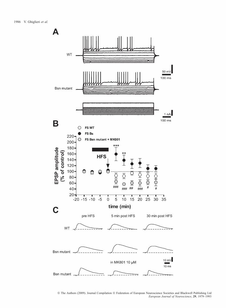
To study the synaptic plasticity of this neuronal subtype, wecharacterized FS interneurons recorded from the dorsolateral striatumand tested whether these cells respond to corticostriatal stimulation,which is known to induce synaptic changes in other neuronal subtypes.The intrinsic membrane properties of 20 FS interneurons from Bsnmutant mice were not changed as compared with wild-type mice(n = 19) (Fig. 4A; Table 2). Input–output relationships were alsosimilar between the two genotypes (P > 0.05; wild type, n = 9;mutant, n = 11) (supporting information Fig. S1B). Their actionpotentials showed typical fast, deep afterhyperpolarization, and thefiring pattern varied from tonic to interrupted (Kawaguchi, 1993;Kawaguchi et al., 1995; Koos & Tepper, 1999; Bracci et al., 2002).Consistent with previous studies (Kawaguchi, 1993; Bracci et al.,2002), their resting membrane potential was )76.91 ± 2.54 mV, andthe input resistance was 37.00 ± 4.37 MX. Moreover, FS interneuronsshowed a characteristic longer duration of evoked EPSP (EPSP decayslope: wild type, )0.14 ± 0.01, n = 14; mutant, )0.13 ± 0.01, n = 19)as compared with MS neurons (EPSP decay slope: wild type,)0.23 ± 0.02, n = 15, P < 0.001 for MS vs. FS; mutant,)0.22 ± 0.02, n = 23, P < 0.001 for MS vs. FS). Interestingly, in theabsence of magnesium ions and following HFS, FS interneurons fromBsn mutant mice showed a short-term increase in synaptic strength,whereas neurons from wild-type mice showed either no change or asmall transient synaptic depression (n = 6, mutant and wild type, 5 and10 min after the tetanus, P < 0.001 and P < 0.01, respectively, mutantvs. wild-type) (Fig. 4B and C). Pharmacological manipulations ofglutamatergic receptors with the NMDA antagonist MK-801 indicatedthat, similar to what was observed in MS neurons (Calabresi et al.,1992), the induction of the potentiation observed in FS interneuronswas NMDA-dependent. Interestingly, pharmacological antagonism ofNMDA receptors not only blocked synaptic potentiation, but evencaused a depression in FS interneurons from Bsn mutant mice (n = 5,P < 0.001, 5–10, 20 min after tetanus, P < 0.01, 15 min after tetanus,P < 0.05, 25–30 min after tetanus, mutant + MK-801 vs. mutant)(Fig. 4B and C).
Increased number of PV-positive cells in Bsn mutant mice
PV immunohistochemistry in the striatum was investigated in orderto quantify the amounts of striatal FS interneurons in wild-type andBsn mutant mice (Fig. 5A). Comparison of striatal PV-positive cellquantification between the two genotypes (Fig. 5B) showed ahigher number of FS interneurons in Bsn mutant mice than in wild-type mice (Mann–Whitney non-parametric test; mutant, n = 4; wildtype, n = 4; P < 0.001). Taken together, these findings suggest thatBsn mutation-induced epilepsy is associated with profound struc-tural and functional modifications in both the neuronal subtypesanalysed.
Table 2. Properties of striatal medium spiny (MS) neurons and fast-spiking(FS) interneurons
Wild type Bsn mutant
MS, n = 25 FS, n = 19 MS, n = 24 FS, n = 20
AP half-width(ms)
0.62 ± 0.02 0.57 ± 0.01 0.60 ± 0.01 0.55 ± 0.01
AHP amplitude(mV)
15.99 ± 0.98 21.62 ± 0.57 17.97 ± 0.59 22.02 ± 0.66
Rin (MX) 33.87 ± 3.89 37.00 ± 4.37 35.54 ± 1.86 39.64 ± 3.28RMP (mV) )86.24 ± 4.00 )76.91 ± 2.54 )85.05 ± 1.45 )75.38 ± 1.49
Values are mean ± SEM of the action potential (AP) half-width, the afterpo-larization (AHP) amplitude, the input resistance (Rin), and the resting mem-brane potential (RMP). No significant differences in these parameters werefound between Bsn mutant and wild-type mice for each cell type.
Striatal plasticity and epilepsy 1983
ª The Authors (2009). Journal Compilation ª Federation of European Neuroscience Societies and Blackwell Publishing LtdEuropean Journal of Neuroscience, 29, 1979–1993

Chronic VPA treatment reduced epileptic seizures andnormalized synaptic plasticity and NMDA subunit compositionin Bsn mutants
In order to distinguish between the contribution of Bsn mutation andthe effect exerted by chronic epileptic seizures on corticostriatalsynaptic plasticity and striatal PSD composition, VPA (400 mg ⁄ kg,
100 mg ⁄ 100 mL of water), a broad-spectrum AED, was given orallyto heterozygous females, in order to treat pups via maternal breastmilk, from postnatal day (P)0 to weaning (P21). Treatment wasextended into adulthood at the same dose previously used with thedams. Early exposure to VPA significantly reduced the postnatalmortality associated with the epileptic syndrome in mutants (Fig. 6A,left) (v2 = 6.47; P < 0.05). Behavioral monitoring of the surviving
Fig. 2. Increase in dendrite branching and shift in proportion of immature vs. mature spines in medium spiny (MS) neurons. (A) Examples of typical spinemorphologies and dendrite branching on Golgi-impregnated MS neurons from Bsn mutant mice (right) and wild-type (WT) mice (left). (B) The histogram on the leftshows the distribution of spine morphology types encountered on MS dendrites. Bsn mutant mice exhibited more longer and fewer shorter spines in the striatal MSneurons analysed, specifically within the thin spines category (*P < 0.05). The graph on the right shows the averaged number of branches encountered on each MSdendrite. MS neurons of Bsn mutant mice were characterized by increased branching as compared with those of WT mice (P < 0.05).
1984 V. Ghiglieri et al.
ª The Authors (2009). Journal Compilation ª Federation of European Neuroscience Societies and Blackwell Publishing LtdEuropean Journal of Neuroscience, 29, 1979–1993

Fig. 3. Characterization of the N-methyl-d-aspartate (NMDA) receptor complex in Bsn mutant mice striatum. (A) Histograms show the quantification of results ofwestern blotting (WB) performed in homogenates (HOMO) and Triton-insoluble fractions (TIF) [P > 0.05, PSD-95, SAP97, and aCaMKII (total and p286);*P < 0.05, NR2A; **P < 0.01, NR2B; *P < 0.05, NR2B (HOMO)]. (B) Striatal (str) homogenates and TIFs from wild-type (wt) and Bsn mutant (mut) mice striatawere analysed by western blot analysis with NMDA receptor subunits (NR1, NR2A, NR2B), MAGUK proteins (PSD-95, SAP97 and SAP102), aCaMKII (total andp286) and actin antibodies. The same amount of protein was loaded per lane, and actin was used as loading control in both homogenates and TIF fractions.
Striatal plasticity and epilepsy 1985
ª The Authors (2009). Journal Compilation ª Federation of European Neuroscience Societies and Blackwell Publishing LtdEuropean Journal of Neuroscience, 29, 1979–1993
1986 V. Ghiglieri et al.
ª The Authors (2009). Journal Compilation ª Federation of European Neuroscience Societies and Blackwell Publishing LtdEuropean Journal of Neuroscience, 29, 1979–1993

animals showed that VPAwas able to reduce the frequency of epilepticseizures by 58% (Fig. 6A, right) (t10 = 1.99; P < 0.05). Intracellularrecordings from corticostriatal slices showed that LTP at 25 min afterthe tetanus was rescued in MS neurons from treated mutant mice (Bsnmutant vs. Bsn mutant with VPA, P < 0.01), and no effect on treatedwild-type animals was observed (Fig. 6B, left). Moreover, striatal FSinterneurons from VPA-exposed epileptic mice failed to show thepostsynaptic potentiation previously observed in naıve mutants (Bsnmutant vs. Bsn mutant with VPA, P < 0.01) (Fig. 6B, right). Analysisof striatal PSD composition revealed that the abnormal ratio betweenthe NR2A and NR2B subunits was normalized by VPA treatment inhomozygous mutants (P < 0.01), whereas the other components wereunaffected by drug administration (Fig. 6C).
Chronic VPA failed to reverse morphological alterationsin Bsn mutants
The morphological pattern associated with Bsn deletion was analysedfollowing VPA administration. In Bsn mutants, neonatal exposure tothe AED was not able to prevent extensive branching (genotype maineffect, F1,12 = 5.93; P < 0.05; genotype · treatment interaction effect,F1,12 = 0.02; P > 0.05) and the spine immaturity of MS neurons(genotype · spine category interaction effect, F4,64 = 0.01; P < 0.05;treatment main factor and relative interactions, P > 0.05) observed innaıve animals (Fig. 7). Moreover, immunohistochemical analysis ofstriatal tissues showed that VPA failed to prevent or reverse the PVoverexpression associated with the epileptic condition. In fact, theamounts of FS interneurons and MS neurons were not affected byVPA treatment, as shown by the ratio between the numbers of the twoneuronal subtypes, which was found to be unchanged in bothgenotypes (genotype main effect, F1,8 = 42.23, treatment main factorand interaction, P > 0.05) (Fig. 8).
Discussion
The main findings of this work are that striatal neuronal subtypes aredifferentially sensitive to frequent seizures triggered in the brain ofBsn mutant mice, and that synaptic plasticity changes develop inresponse to early-onset epilepsy in these mice. Interestingly, functionalbut not structural alterations in Bsn mutants were prevented by chronicantiepileptic treatment with VPA, suggesting that the observedelectrophysiological and molecular changes are secondary to theseizure activity rather than being directly induced by the lack offunctional Bassoon. Conversely, as morphological alterations were notfound to be prevented by chronic antiepileptic treatment, it is possibleto hypothesize that they represent a direct effect of the abnormalgenotype of these mice.
In this model, early-onset epileptic seizures are associated with analtered LTP time course and a reorganization of spine morphology anddendrite connectivity in MS neurons. Along with the alterationsobserved in MS projecting neurons, we found that FS interneurons ofmutant mice express a short-term potentiation that is not present in FS
interneurons of wild-type mice. The expression of this form ofplasticity in FS cells, as well as the increased number of theseGABAergic interneurons, might reflect upregulation of the feedfor-ward inhibition within the striatum of the mutant mice, and it mightrepresent a compensatory mechanism aimed at limiting striatal damagefollowing repetitive seizures.We found that in Bsnmutant mice, epileptic attacks developed early,
around P14, as myoclonic seizures, and evolved into generalizedtonic–clonic seizures in adult mutants. This finding is consistent withthe epileptic phenotype observed in other rodent experimental models(Chapman et al., 1987; Engstrom & Woodbury, 1988; Musumeciet al., 2000; De Sarro et al., 2004).In Bsn mutant mice, as in other epileptic conditions, cortical
neurons engaged in paroxysmal activity might lead to alterations incorticostriatal pathway activity. Increasing attention has recently beenpaid to the changes occurring through the corticostriatal projectionsduring pathological and physiological activity originating from thecortex at the level of different neuronal subtypes (Bracci et al., 2004;Slaght et al., 2004; Mallet et al., 2005). It is known that dendriticspines represent the principal site of synaptic input and therefore playa key role in activity-dependent synaptic plasticity in MS neurons(Fiala et al., 2002; Tada & Sheng, 2006).In epileptic patients and in experimental in vivo and in vitro
models of epilepsy, there is a marked loss of dendritic spines in thebrain foci of seizures, and this is often associated with distortion ofthe shape of the remaining spines and degeneration (Fiala et al.,2002). In line with these observations, we have shown that theabsence of functional Bassoon is associated with a shift in theproportion of mature vs. immature spines counted on apical dendritesegments, as well as an increase in the branching of MS neurons ofmutant mice as compared with wild-type mice. Given the role ofBassoon in synapse maturation and neuronal connectivity, it is morereasonable to consider the functional deletion of this protein as thepossible cause of these morphological alterations rather than theepileptic activity itself. In particular, initial morphological alterationsmight lead to the establishment of a pathological substrate, which,in turn, might favor recurrence of seizures, triggering a viciouscircle.On this view, abnormal striatal plasticity could be interpreted as a
consequence of repeated seizures. It is possible to hypothesize thatthe impairment of LTP in MS neurons is the result of altered NMDAreceptor subunit composition. Indeed, activation, correct assemblyand localization of the NMDA receptors are essential for striatal LTP(Picconi et al., 2004). Interestingly, previous reports demonstratedthat the PSD, a synaptic compartment highly enriched in ionotropicglutamate receptors (Kim & Sheng, 2004), is altered in brain areaswhere the seizure activity is more pronounced (Wyneken et al.,2001, 2003; Finardi et al., 2006). It has been recently postulated thatregulation of the NR2A ⁄ NR2B ratio plays a pivotal role in thecontrol of cortical synaptic plasticity (Yashiro & Philpot, 2008).Moreover, two recent studies have shown that inactivation of theNR2B subunit is sufficient to selectively abolish hippocampal LTP(Bartlett et al., 2007), and that an appropriate synaptic localization of
Fig. 4. N-Methyl-d-aspartate (NMDA)-dependent short-term plasticity in fast-spiking (FS) interneurons of Bsn mutant mice. (A) Intrinsic and synaptic properties ofstriatal FS interneurons. Action potential discharges were induced by hyperpolarizing and depolarizing current steps in FS interneurons from wild-type (WT) (uppertrace) and Bsn mutant mice (lower trace). (B) High-frequency stimulation (HFS; arrow) induced a synaptic potentiation that lasted 15–20 min in FS interneurons ofBsn mutant mice. Conversely, no potentiation was observed in WT mice (n = 6, mutant and WT, 5 and 10 min after tetanus, ***P < 0.001 and **P < 0.01,respectively, mutant vs. WT). Note that incubation of the Bsn mutant slices with the NMDA receptor antagonist MK801 reversed potentiation into a synapticdepression (n = 5, ###P < 0.001, 5–10, 20 min after tetanus, ##P < 0.01, 15 min after tetanus, #P < 0.05, 25–30 min after tetanus, mutant + MK-801 vs. mutant).(C) Traces represent examples of excitatory postsynaptic potentials (EPSPs) from FS interneurons recorded intracellularly in WT (upper traces), Bsn mutant (middletraces) and Bsn mutant mouse slices with application of MK801 (lower traces).
Striatal plasticity and epilepsy 1987
ª The Authors (2009). Journal Compilation ª Federation of European Neuroscience Societies and Blackwell Publishing LtdEuropean Journal of Neuroscience, 29, 1979–1993

NR2B-containing receptor is critically required for LTP induction inhippocampal slices (Gardoni et al., 2009). Accordingly, in Bsnmutants, we observed altered NMDA composition in the striatal
PSD. In particular, the levels of NR2A and NR2B were modified,with a significant increase in NR2A ⁄ NR2B ratios in Bsn mutantstriata.
Fig. 5. Increased number of parvalbumin (PV)-positive cells in Bsn mutants. (A) PV immunohistochemical investigations were performed in the striatum in order toquantify the amounts of striatal fast-spiking (FS) interneurons in wild-type (WT) mice (right) and Bsn mutant mice (left). Upper panels correspond to ·60magnification; the squares are the dissectors (50 · 50 lm) used for the unbiased counting. Lower panels correspond to ·20 magnification; scale bar = 100 lm.(B) Comparison of striatal PV-positive cell quantification between the two genotypes showed a higher number of FS interneurons in Bsn mutant mice than in WTmice (**P < 0.01), suggesting a compensatory mechanism of FS interneurons in the striatum of these mutant animals.
1988 V. Ghiglieri et al.
ª The Authors (2009). Journal Compilation ª Federation of European Neuroscience Societies and Blackwell Publishing LtdEuropean Journal of Neuroscience, 29, 1979–1993

Another critical factor that may play a role in the reduction ofsynaptic plasticity in the MS neurons of Bsn mutant mice is theemergence of a new form of synaptic plasticity in the PV-positive
interneurons. Striatal PV-positive FS interneurons receive a criticalglutamatergic input from the cortex, and provide the major synapticinhibitory control within the striatum (Ramanathan et al., 2002). As in
Fig. 6. Valproic acid (VPA) reduced the number of seizures and rescued synaptic plasticity and N-methyl-d-aspartate (NMDA) receptor subunit composition.(A) The histograms show that chronic administration of VPA induced a significant increase in survival rate (left) (*P < 0.05) and reduced the number of epilepticattacks in mutant mice (right) (*P < 0.05). (B) In the medium spiny (MS) neurons of treated Bsn mice, long-term potentiation (LTP) amplitude at 25 min aftertetanus was significantly increased as compared with untreated epileptic mice (left) (**P < 0.01). Antiepileptic treatment was also able to block the short-termpotentiation observed in fast-spiking (FS) interneurons of mutants. The right panel shows the amplitude of excitatory postsynaptic potential (EPSPs) measured at5 min after tetanus (right) (**P < 0.01). (C) Chronic treatment with VPAwas also able to reverse the altered NR2A ⁄ NR2B NMDA receptor subunit ratio observedin the striata of Bsn mutant mice. WB, western blotting; WT, wild type.
Striatal plasticity and epilepsy 1989
ª The Authors (2009). Journal Compilation ª Federation of European Neuroscience Societies and Blackwell Publishing LtdEuropean Journal of Neuroscience, 29, 1979–1993

MS neurons recorded from animals lacking Bsn, the ability to enhancesynaptic strength was altered, FS interneurons showed a formof plasticity that was absent in normal conditions. The importanceof PV-positive neurons in epileptic models has already been hypoth-esized, and, notably, FS interneurons have been implicated in neuronal
survival, as a higher susceptibility to seizures is associated withhippocampal PV deficiency (Schwaller et al., 2004) and abnormalfiring of cortical FS interneurons (Lau et al., 2000). Moreover, in agenetic model of absence epilepsy, striatal FS interneurons have beenreported to modulate MS neuronal activity by changing the firing
Fig. 7. Chronic valproic acid (VPA) treatment did not prevent increases in branching and spine immaturity of medium spiny (MS) neurons. (A) The histogram showsthe distribution of spine morphology types encountered on MS dendrites of treated and untreated Bsn mutant and wild-type (WT) mice. (B) The branching of MSdendrites was also unaffected by the VPA treatment. Treated mice exhibited a morphological pattern that is similar to the one observed in untreated mice. *P < 0.05.
Fig. 8. Chronic valproic acid (VPA) treatment failed to prevent parvalbumin (PV) overexpression in the striatum of treated mutant mice. (A) Examples of calbindin(Calb; upper panels) and PV (lower panels) immunohistochemistry performed in the striatum of Bsn mutant mice (left) and Bsn mutant mice chronically treated withVPA (right). All panels correspond to ·20 magnification. (B) Comparisons of the ratio between striatal calbindin-positive and PV-positive cells in the two genotypeswith and without VPA treatment. The histogram shows that the numbers of fast-spiking interneurons and medium spiny neurons were not affected by VPA treatment(*P < 0.05). WT, wild type.
1990 V. Ghiglieri et al.
ª The Authors (2009). Journal Compilation ª Federation of European Neuroscience Societies and Blackwell Publishing LtdEuropean Journal of Neuroscience, 29, 1979–1993

pattern in response to cortical seizures (Slaght et al., 2004). Given thisacknowledged role of FS interneurons in epileptic conditions, wetested whether these cells respond to repetitive corticostriatal stimu-lation, which is known to induce synaptic changes in the MS neuronalsubtype. Despite the maintenance of intrinsic properties, FS interneu-rons of Bsn mutants showed an NMDA-dependent increase in synapticstrength following HFS of corticostriatal synapses, whereas neuronsrecorded from wild-type mice showed either no change or a smalltransient synaptic depression.
Besides the expression of this uncommon form of plasticity, we alsofound a higher number of FS interneurons in Bsn mutant mice. Thealteration in MS neuron plasticity was therefore paralleled by anenhancement of FS interneuronal activity, possibly leading to areorganization of GABAergic control on striatal output activity.However, there is no substantial evidence to support the notion thatMS neurons would receive more GABAergic inputs. In fact, we failedto obtain morphological data to demonstrate that PV interneuronsincrease their connections to MS neurons. In addition, inhibition ofGABAergic inputs to MS neurons by bicucullin application (data notshown) was not able to rescue MS LTP. Thus, it is unclear whetherenhancement of interneuronal activity makes a contribution to MSplasticity changes. Moreover, overall striatal modifications might notbe sufficient, in the end, to preserve the cortex from the recurrence ofthe seizures, which often lead to death in homozygous mutants.
In order to distinguish between the consequences of Bsn mutationper se and the effects of the seizures, and to clarify the causes of thesefunctional and structural alterations, we explored how the antiepilepticaction of VPA could affect corticostriatal modifications in Bsnmutants. Following chronic administration of VPA, mutant micedeveloped a lower number of epileptic attacks and showed asignificant increase in survival rate as compared with untreated Bsnmice. These beneficial effects were accompanied by the recovery ofLTP amplitude in MS neurons and a normalization of the levels ofNMDA receptor subunits. Conversely, MS neuron morphologicalalterations were not affected by the treatment. Interestingly, in theother neuronal subtype analysed, the FS interneurons, chronictreatment with VPA also differentially affected functional andmorphological features. In fact, although antiepileptic therapy pre-vented the occurrence of the abnormal short-term potentiation, it didnot affect the increase in PV-positive cell numbers in the striatum ofepileptic mice. Thus, our experiments show that chronic VPA, throughreduction of epileptic seizures, is able to selectively prevent theoccurrence of functional alterations in mutant mice without influenc-ing morphological abnormalities, which seem more likely to reflect adirect effect of the abnormal genotype. Interestingly, it has beendemonstrated that PV-positive FS interneurons play a critical role intemporal lobe epilepsy by shaping and controlling the power ofgamma network oscillations, which are considered to be a neuronalsubstrate for epileptogenesis (Fisher et al., 1992; Bragin et al., 2007).Although striatal FS GABAergic interneurons are known to have thecapacity to fire at very high frequencies and coordinate synchronousactivity in large populations of MS neurons (Koos & Tepper, 1999;Ramanathan et al., 2002), the specific role of interneuronal oscillationshas not been clarified in the epileptic striatum.
It is well established that VPA exerts its antiepileptic action viamultiple mechanisms, such as sodium channel blockade, antagonismof GABA transaminase, and possible modulation of calcium channels(Rogawski & Loscher, 2004). Moreover, it has been demonstratedthat, in cortical neurons, similar to neurotrophic factors, VPApromotes neurogenesis by enhancing the functions of the extracellularsignal-related kinase pathway (Hao et al., 2004), which is dependenton activation of NR2B-containing NMDA receptors (Krapivinsky
et al., 2003; Kim et al., 2005) and is critical for corticostriatal LTP(Calabresi et al., 2002). However, the precise mechanism of action bywhich VPA produces its therapeutic effect in our experimental modelhas not been investigated, and further experiments are required toclarify this issue. In particular, it would be interesting to explorewhether other AEDs targeting sodium channels, such as carbamaz-epine, would be equally effective in reducing seizures and mortality, aswell as in restoring physiological synaptic plasticity and theNR2A ⁄ NR2B ratio in Bsn mutant mice.In conclusion, loss of functional Bassoon leads to the development
of a severe epileptic syndrome associated with striatal alterations as anadaptive mechanism within the striatal microcircuit. The presynapticprotein seems to have a key role in the control of neuronalconnectivity, which may, in turn, be the initial cause of epilepticsymptoms. The early onset of the seizure attacks and their reductionby chronic VPA indicate that changes in corticostriatal synapticplasticity are likely to be caused by the effects of continuous corticalepileptic activity. These alterations in neuronal activity associated withthe occurrence of spontaneous early seizures make this new model ofepilepsy a good tool to explore the pathophysiology of basal ganglia inan epileptic syndrome.Our results indicate that, although it is not possible to prevent the
development of morphological conditions that may favor seizurerecurrence, an early therapeutic approach, able to limit the seizures,might be able to prevent the occurrence of cell-type-specific alterationsin synaptic plasticity induced by recurrent paroxysmal activity.
Supporting Information
Additional supporting information may be found in the online versionof this article:Fig. S1. Input–output profile of MS neurons and FS interneurons.Video S1. Video-recording of epileptic attack in a Bsn mutant at P14.Video S2. Video-recording of epileptic attack in a Bsn mutant at2 months.Please note: Wiley-Blackwell are not responsible for the content orfunctionality of any supporting materials supplied by the authors. Anyqueries (other than missing material) should be directed to thecorresponding author for the article.
Acknowledgements
This work was supported by the European Community (LSHM-CT-2004-511995, SYNSCAFF; HEALTH-2007-22918, REPLACES) (P. Calabresi, E.D.Gundelfinger, and M. Di Luca), Ricerca Corrente IRCCS (G. Bernardi) andRicerca Finalizzata IRCCS (P. Calabresi), and the Fonds der ChemischenIndustrie, the Alexander von Humboldt Foundation and the Max PlanckSociety (E. D. Gundelfinger).
Abbreviations
AED, antiepileptic drug; EPSP, excitatory postsynaptic potential; FS, fast-spiking; GABA, c-aminobutyric acid; HFS, high-frequency stimulation; LTP,long-term potentiation; MS, medium spiny; NA, numerical aperture; NDS,normal donkey serum; NMDA, N-methyl-d-aspartate; P, postnatal day; PB,phosphate buffer; PSD, postsynaptic density; PV, parvalbumin; TBS, Tris-buffered saline; VPA, valproic acid; TIF, Triton-insoluble fraction.
References
Altrock, W.D., tom Dieck, S., Sokolov, M., Meyer, A.C., Sigler, A.,Brakebusch, C., Fassler, R., Richter, K., Boeckers, T.M., Potschka, H.,Brandt, C., Loscher, W., Grimberg, D., Dresbach, T., Hempelmann, A.,
Striatal plasticity and epilepsy 1991
ª The Authors (2009). Journal Compilation ª Federation of European Neuroscience Societies and Blackwell Publishing LtdEuropean Journal of Neuroscience, 29, 1979–1993
Hassan, H., Balschun, D., Frey, J.U., Brandstatter, J.H., Garner, C.C.,Rosenmund, C. & Gundelfinger, E.D. (2003) Functional inactivation of afraction of excitatory synapses in mice deficient for the active zone proteinbassoon. Neuron, 37, 787–800.
Bartlett, T.E., Bannister, N.J., Collett, V.J., Dargan, S.L., Massey, P.V.,Bortolotto, Z.A., Fitzjohn, S.M., Bashir, Z.I., Collingridge, G.L. & Lodge, D.(2007) Differential roles of NR2A and NR2B-containing NMDA receptorsin LTP and LTD in the CA1 region of two-week old rat hippocampus.Neuropharmacology, 52, 60–70.
Bortolotto, Z.A. & Collingridge, G.L. (1998) Involvement of calcium ⁄calmodulin-dependent protein kinases in the setting of a molecular switchinvolved in hippocampal LTP. Neuropharmacology, 37, 535–544.
Bouilleret, V., Semah, F., Chassoux, F., Mantzaridez, M., Biraben, A.,Trebossen, R. & Ribeiro, M.J. (2008) Basal ganglia involvement in temporallobe epilepsy: a functional and morphologic study. Neurology, 70, 177–184.
Bracci, E., Centonze, D., Bernardi, G. & Calabresi, P. (2002) Dopamine excitesfast-spiking interneurons in the striatum. J. Neurophysiol., 87, 2190–2194.
Bracci, E., Centonze, D., Bernardi, G. & Calabresi, P. (2004) Engagement of ratstriatal neurons by cortical epileptiform activity investigated with pairedrecordings. J. Neurophysiol., 92, 2725–2737.
Bragin, A., Claeys, P., Vonck, K., Van Roost, D., Wilson, C., Boon, P. & Engel,J. Jr (2007) Analysis of initial slow waves (ISWs) at the seizure onset inpatients with drug resistant temporal lobe epilepsy. Epilepsia, 48, 1883–1894.
Calabresi, P., Pisani, A., Mercuri, N.B. & Bernardi, G. (1992) Long-termpotentiation in the striatum is unmasked by removing the voltage-dependentmagnesium block of NMDA receptor channels. Eur. J. Neurosci., 4, 929–935.
Calabresi, P., Gubellini, P., Centonze, D., Picconi, B., Bernardi, G., Chergui,K., Svenningsson, P., Fienberg, A.A. & Greengard, P. (2000) Dopamine andcAMP-regulated phosphoprotein 32 kDa controls both striatal long-termdepression and long-term potentiation, opposing forms of synaptic plasticity.J. Neurosci., 20, 8443–8451.
Calabresi, P., Saulle, E., Centonze, D., Pisani, A., Marfia, G.A. & Bernardi, G.(2002) Post-ischaemic long-term synaptic potentiation in the striatum: aputative mechanism for cell type-specific vulnerability. Brain, 125, 844–860.
Calabresi, P., Picconi, B., Tozzi, A. & Di Filippo, M. (2007) Dopamine-mediated regulation of corticostriatal synaptic plasticity. Trends Neurosci.,30, 211–219.
Chapman, A.G., De Sarro, G.B., Premachandra, M. & Meldrum, B.S. (1987)Bidirectional effects of beta-carbolines in reflex epilepsy. Brain Res. Bull.,19, 337–346.
Coggeshall, R.E. (1992) A consideration of neural counting methods. TrendsNeurosci., 15, 9–13.
De Sarro, G., Russo, E., Ferreri, G., Giuseppe, B., Flocco, M.A., Di Paola,E.D. & De Sarro, A. (2004) Seizure susceptibility to various convulsantstimuli of knockout interleukin-6 mice. Pharmacol. Biochem. Behav., 77,761–766.
tom Dieck, S., Sanmarti-Vila, L., Langnaese, K., Richter, K., Kindler, S.,Soyke, A., Wex, H., Smalla, K.H., Kampf, U., Franzer, J.T., Stumm, M.,Garner, C.C. & Gundelfinger, E.D. (1998) Bassoon, a novel zinc-fingerCAG ⁄ glutamine-repeat protein selectively localized at the active zone ofpresynaptic nerve terminals. J. Cell Biol., 142, 499–509.
Engstrom, F.L. & Woodbury, D.M. (1988) Seizure susceptibility in DBA andC57 mice: the effects of various convulsants. Epilepsia, 29, 389–395.
Fiala, J.C., Spacek, J. & Harris, K.M. (2002) Dendritic spine pathology: causeor consequence of neurological disorders? Brain Res. Brain Res. Rev., 39,29–54.
Finardi, A., Gardoni, F., Bassanini, S., Lasio, G., Cossu, M., Tassi, L.,Caccia, C., Taroni, F., LoRusso, G., Di Luca, M. & Battaglia, G. (2006)NMDA receptor composition differs among anatomically diverse malfor-mations of cortical development. J. Neuropathol. Exp. Neurol., 65, 883–893.
Fisher, R.S., Webber, W.R., Lesser, R.P., Arroyo, S. & Uematsu, S. (1992)High-frequency EEG activity at the start of seizures. J. Clin. Neurophysiol.,9, 441–448.
Gardoni, F., Schrama, L.H., Kamal, A., Gispen, W.H., Cattabeni, F. & Di Luca,M. (2001) Hippocampal synaptic plasticity involves competition betweenCa2+ ⁄ calmodulin-dependent protein kinase II and postsynaptic density 95for binding to the NR2A subunit of the NMDA receptor. J. Neurosci., 21,1501–1509.
Gardoni, F., Picconi, B., Ghiglieri, V., Polli, F., Bagetta, V., Bernardi, G.,Cattabeni, F., Di Luca, M. & Calabresi, P. (2006) A critical interactionbetween NR2B and MAGUK in L-DOPA induced dyskinesia. J. Neurosci.,26, 2914–2922.
Gardoni, F., Mauceri, D., Malinverno, M., Polli, F., Costa, C., Tozzi, A.,Siliquini, S., Picconi, B., Cattabeni, F., Calabresi, P. & Di Luca, M. (2009)Decreased NR2B subunit synaptic levels cause impaired long-term poten-tiation but not long-term depression. J. Neurosci., 29, 669–677.
Garner, C.C., Kindler, S. & Gundelfinger, E.D. (2000) Molecular determinantsof presynaptic active zones. Curr. Opin. Neurobiol., 10, 321–327.
Gibb, R. & Kolb, B. (1998) A method for vibratome sectioning of Golgi–Coxstained whole rat brain. J. Neurosci. Methods, 79, 1–4.
Glaser, E.M. & Van der Loos, H. (1981) Analysis of thick brain sections byobverse-reverse computer microscopy: application of a new, high clarityGolgi-Nissl stain. J. Neurosci. Methods, 4, 117–125.
Hao, Y., Creson, T., Zhang, L., Li, P., Du, F., Yuan, P., Gould, T.D., Manji, H.K.& Chen, G. (2004) Mood stabilizer valproate promotes ERK pathway-dependent cortical neuronal growth and neurogenesis. J. Neurosci., 24,6590–6599.
Horner, C.H. & Arbuthnott, E. (1991) Methods of estimation of spine density –are spines evenly distributed throughout the dendritic field? J. Anat., 177,179–184.
Irwin, S.A., Patel, B., Idupulapati, M., Harris, J.B., Crisostomo, R.A., Larsen,B.P., Kooy, F., Willems, P.J., Cras, P., Kozlowski, P.B., Swain, R.A., Weiler,I.J. & Greenough, W.T. (2001) Abnormal dendritic spine characteristics inthe temporal and visual cortices of patients with fragile-X syndrome: aquantitative examination. Am. J. Med. Genet., 98, 161–167.
Kawaguchi, Y. (1993) Physiological, morphological, and histochemicalcharacterization of three classes of interneurons in rat neostriatum.J. Neurosci., 13, 4908–4923.
Kawaguchi, Y., Wilson, C.J., Augood, S.J. & Emson, P.C. (1995) Striatalinterneurones: chemical, physiological and morphological characterization.Trends Neurosci., 18, 527–535.
Kim, E. & Sheng, M. (2004) PDZ domain proteins of synapses. Nat. Rev.Neurosci., 5, 771–781.
Kim, M.J., Dunah, A.W., Wang, Y.T. & Sheng, M. (2005) Differential roles ofNR2A- and NR2B-containing NMDA receptors in Ras-ERK signaling andAMPA receptor trafficking. Neuron, 46, 745–760.
Koos, T. & Tepper, J.M. (1999) Inhibitory control of neostriatal projectionneurons by GABAergic interneurons. Nat. Neurosci., 2, 467–472.
Krapivinsky, G., Krapivinsky, L., Manasian, Y., Ivanov, A., Tyzio, R.,Pellegrino, C., Ben-Ari, Y., Clapham, D.E. & Medina, I. (2003) The NMDAreceptor is coupled to the ERK pathway by a direct interaction betweenNR2B and RasGRF1. Neuron, 40, 775–784.
Lau, D., Vega-Saenz de Miera, E.C., Contreras, D., Ozaita, A., Harvey, M.,Chow, A., Noebels, J.L., Paylor, R., Morgan, J.I., Leonard, C.S. & Rudy, B.(2000) Impaired fast-spiking, suppressed cortical inhibition, and increasedsusceptibility to seizures in mice lacking Kv3.2 K+ channel proteins.J. Neurosci., 20, 9071–9085.
Leuner, B., Falduto, J. & Shors, T.J. (2003) Associative memory formationincreases the observation of dendritic spines in the hippocampus.J. Neurosci., 23, 659–665.
Mallet, N., Le Moine, C., Charpier, S. & Gonon, F. (2005) Feedforwardinhibition of projection neurons by fast-spiking GABA interneurons in therat striatum in vivo. J. Neurosci., 25, 3857–3869.
Musumeci, S.A., Bosco, P., Calabrese, G., Bakker, C., De Sarro, G.B., Elia, M.,Ferri, R. & Oostra, B.A. (2000) Audiogenic seizures susceptibility intransgenic mice with fragile X syndrome. Epilepsia, 41, 19–23.
Picconi, B., Gardoni, F., Centonze, D., Mauceri, D., Cenci, M.A., Bernardi, G.,Calabresi, P. & Di Luca, M. (2004) Abnormal Ca2+-calmodulin-dependentprotein kinase II function mediates synaptic and motor deficits in experi-mental parkinsonism. J. Neurosci., 24, 5283–5291.
Ramanathan, S., Hanley, J.J., Deniau, J.M. & Bolam, J.P. (2002) Synapticconvergence of motor and somatosensory cortical afferents onto GABAergicinterneurons in the rat striatum. J. Neurosci., 22, 8158–8169.
Richter, K., Langnaese, K., Kreutz, M.R., Olias, G., Zhai, R., Scheich, H.,Garner, C.C. & Gundelfinger, E.D. (1999) Presynaptic cytomatrix proteinbassoon is localized at both excitatory and inhibitory synapses of rat brain.J. Comp. Neurol., 408, 437–448.
Rogawski, M.A. & Loscher, W. (2004) The neurobiology of antiepilepticdrugs. Nat. Rev. Neurosci., 5, 553–564.
Schmitz, C. & Hof, P.R. (2005) Design-based stereology in neuroscience.Neuroscience, 130, 813–831.
Schoch, S. & Gundelfinger, E.D. (2006) Molecular organization of thepresynaptic active zone. Cell Tissue Res., 326, 379–391.
Schwaller, B., Tetko, I.V., Tandon, P., Silveira, D.C., Vreugdenhil, M., Henzi,T., Potier, M.C., Celio, M.R. & Villa, A.E. (2004) Parvalbumin deficiencyaffects network properties resulting in increased susceptibility to epilepticseizures. Mol. Cell. Neurosci., 25, 650–663.
1992 V. Ghiglieri et al.
ª The Authors (2009). Journal Compilation ª Federation of European Neuroscience Societies and Blackwell Publishing LtdEuropean Journal of Neuroscience, 29, 1979–1993

Slaght, S.J., Paz, T., Mahon, S., Maurice, N., Charpier, S. & Deniau, J.M.(2002) Functional organization of the circuits connecting the cerebral cortexand the basal ganglia: implications for the role of the basal ganglia inepilepsy. Epileptic Disord, 4(Suppl. 3), S9–S22.
Slaght, S.J., Paz, T., Chavez, M., Deniau, J.M., Mahon, S. & Charpier, S.(2004) On the activity of the corticostriatal networks during spike-and-wavedischarges in a genetic model of absence epilepsy. J. Neurosci., 24, 6816–6825.
Sterio, D.C. (1984) The unbiased estimation of number and sizes of arbitraryparticles using the disector. J. Microsc., 134, 127–136.
Tada, T. & Sheng, M. (2006) Molecular mechanisms of dendritic spinemorphogenesis. Curr. Opin. Neurobiol., 16, 95–101.
Tepper, J.M., Koos, T. & Wilson, C.J. (2004) GABAergic microcircuits in theneostriatum. Trends Neurosci., 27, 662–669.
Uylings, H.B., Ruiz-Marcos, A. & van Pelt, J. (1986) The metric analysisof three-dimensional dendritic tree patterns: a methodological review.J. Neurosci. Methods, 18, 127–151.
Winter, C., tom Dieck, S., Boeckers, T.M., Bockmann, J., Kampf, U.,Sanmarti-Vila, L., Langnaese, K., Altrock, W., Stumm, M., Soyke, A.,Wieacker, P., Garner, C.C. & Gundelfinger, E.D. (1999) The presynapticcytomatrix protein Bassoon: sequence and chromosomal localization of thehuman BSN gene. Genomics, 57, 389–397.
Wyneken, U., Smalla, K.H., Marengo, J.J., Soto, D., de la Cerda, A.,Tischmeyer, W., Grimm, R., Boeckers, T.M., Wolf, G., Orrego, F. &Gundelfinger, E.D. (2001) Kainate-induced seizures alter protein composi-tion and N-methyl-d-aspartate receptor function of rat forebrain postsynapticdensities. Neuroscience, 102, 65–74.
Wyneken, U., Marengo, J.J., Villanueva, S., Soto, D., Sandoval, R.,Gundelfinger, E.D. & Orrego, F. (2003) Epilepsy-induced changes insignaling systems of human and rat postsynaptic densities. Epilepsia, 44,243–246.
Yashiro, K. & Philpot, B.D. (2008) Regulation of NMDA receptor subunitexpression and its implications for LTD, LTP, and metaplasticity. Neuro-pharmacology, 55, 1081–1094.
Striatal plasticity and epilepsy 1993
ª The Authors (2009). Journal Compilation ª Federation of European Neuroscience Societies and Blackwell Publishing LtdEuropean Journal of Neuroscience, 29, 1979–1993
Related Documents