Copyright © 2022 The Author(s). This is an Open Access article distributed under the terms of the Creative Commons Attribution License, which permits unrestricted use, distribution, and reproduction in any medium, provided the original work is properly cited. 1 United Christian Hospital, Department of Pathology, Hong Kong, China 2 CUHK Medical Centre, Department of Pathology, Hong Kong, China Epidermoid cyst in an intrapancreatic accessory spleen with abnormally high CEA level in cyst fluid: a case report Chun-hai Lo 1 , Po-man Tsang 1 , Shui-ying Cheng 2 , Cheuk-nam Ling 1 , Cheuk-lam Ho 1 How to cite: Lo CH, Tsang PM, Cheng SY, Ling CN, Ho CL. Epidermoid cyst in an intrapancreatic accessory spleen with abnormally high CEA level in cyst fluid: a case report. Autops Case Rep [Internet]. 2022;12:e2021369. https://doi.org/10.4322/ acr.2021.369 Clinical Case Report and Review ABSTRACT Epidermoid cyst in an intrapancreatic accessory spleen is a rare benign lesion that is difficult to diagnose preoperatively. Cyst fluid analysis for biochemistry markers has been widely used to aid the diagnosis of pancreatic cysts. A high cyst fluid carcinoembryonic antigen (CEA) level (>800 ng/mL) is said to be useful in distinguishing intraductal papillary mucinous neoplasm (IPMN) and mucinous cystic neoplasm (MCN) from other non-mucinous cysts. We herein report a case of epidermoid cyst in an intrapancreatic accessory spleen with abnormally high CEA level (3582 ng/mL) in the cyst fluid, suggesting a potential pitfall in using cyst fluid CEA level as an indicator of mucinous neoplasms. Keywords Carcinoembryonic Antigen, Epidermal Cyst, Pancreas, Spleen INTRODUCTION Accessory spleen is found in around 15% of the general population. Around 5% of them are located near or in the tail of the pancreas. 1,2 Splenic epidermoid cysts are rare and constitute approximately 10% of total splenic cysts. 3 Epidermoid cyst in an intrapancreatic accessory spleen (ECIAS) is very rare, with only a few dozens of case reports in the English literature to date. 4-43 Although it is a benign lesion, the definitive diagnosis can often be made after excision as an accurate preoperative diagnosis by radiological and endoscopic examinations are difficult. 44 We herein report a case of ECIAS with abnormally high CEA level in cyst fluid, and a preoperative radiological diagnosis of pancreatic mucinous cystic neoplasm. CASE REPORT A 28-year-old Chinese woman with an unremarkable past medical history was presented to the hospital with dyspepsia and epigastric pain. She underwent abdominal ultrasound, which revealed a simple cyst at the pancreatic tail measuring 1.4 x1.7 x1.5cm in size, with no internal septa or solid area. A subsequent magnetic resonance imaging revealed a well-defined solitary unilocular cystic lesion in the pancreatic tail region, measuring up to 2.34 cm. There was a small mural nodule in its dependent portion and a minimal enhancement at its thin wall without significant enhancement of the mural nodule. There was no vascular invasion, definite communication with the non-dilated pancreatic duct, or lymphadenopathy. A contrast-enhanced computed tomography (CT)

Welcome message from author
This document is posted to help you gain knowledge. Please leave a comment to let me know what you think about it! Share it to your friends and learn new things together.
Transcript
Copyright © 2022 The Author(s). This is an Open Access article distributed under the terms of the Creative Commons Attribution License, which permits unrestricted use, distribution, and reproduction in any medium, provided the original work is properly cited.
1United Christian Hospital, Department of Pathology, Hong Kong, China2CUHK Medical Centre, Department of Pathology, Hong Kong, China
Epidermoid cyst in an intrapancreatic accessory spleen with abnormally high CEA level in cyst fluid: a case report
Chun-hai Lo1 , Po-man Tsang1 , Shui-ying Cheng2 , Cheuk-nam Ling1 , Cheuk-lam Ho1
How to cite: Lo CH, Tsang PM, Cheng SY, Ling CN, Ho CL. Epidermoid cyst in an intrapancreatic accessory spleen with abnormally high CEA level in cyst fluid: a case report. Autops Case Rep [Internet]. 2022;12:e2021369. https://doi.org/10.4322/acr.2021.369
Clinical Case Report and Review
ABSTRACT
Epidermoid cyst in an intrapancreatic accessory spleen is a rare benign lesion that is difficult to diagnose preoperatively. Cyst fluid analysis for biochemistry markers has been widely used to aid the diagnosis of pancreatic cysts. A high cyst fluid carcinoembryonic antigen (CEA) level (>800 ng/mL) is said to be useful in distinguishing intraductal papillary mucinous neoplasm (IPMN) and mucinous cystic neoplasm (MCN) from other non-mucinous cysts. We herein report a case of epidermoid cyst in an intrapancreatic accessory spleen with abnormally high CEA level (3582 ng/mL) in the cyst fluid, suggesting a potential pitfall in using cyst fluid CEA level as an indicator of mucinous neoplasms.
Keywords Carcinoembryonic Antigen, Epidermal Cyst, Pancreas, Spleen
INTRODUCTION
Accessory spleen is found in around 15% of the general population. Around 5% of them are located near or in the tail of the pancreas.1,2 Splenic epidermoid cysts are rare and constitute approximately 10% of total splenic cysts.3 Epidermoid cyst in an intrapancreatic accessory spleen (ECIAS) is very rare, with only a few dozens of case reports in the English literature to date.4-43 Although it is a benign lesion, the definitive diagnosis can often be made after excision as an accurate preoperative diagnosis by radiological and endoscopic examinations are difficult.44
We herein report a case of ECIAS with abnormally high CEA level in cyst fluid, and a preoperative radiological diagnosis of pancreatic mucinous cystic neoplasm.
CASE REPORT
A 28-year -o ld Ch inese woman wi th an unremarkable past medical history was presented to the hospital with dyspepsia and epigastric pain. She underwent abdominal ultrasound, which revealed a simple cyst at the pancreatic tail measuring 1.4 x1.7 x1.5cm in size, with no internal septa or solid area. A subsequent magnetic resonance imaging revealed a well-defined solitary unilocular cystic lesion in the pancreatic tail region, measuring up to 2.34 cm. There was a small mural nodule in its dependent portion and a minimal enhancement at its thin wall without significant enhancement of the mural nodule. There was no vascular invasion, definite communication with the non-dilated pancreatic duct, or lymphadenopathy. A contrast-enhanced computed tomography (CT)
Epidermoid cyst in an intrapancreatic accessory spleen with abnormally high CEA level in cyst fluid: a case report
2-9 Autops Case Rep (São Paulo). 2022;12:e2021369
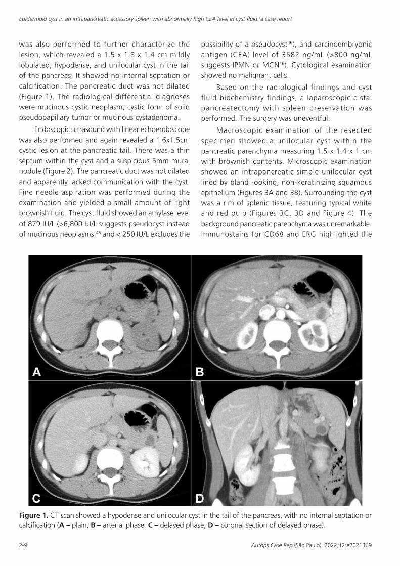
was also performed to further characterize the lesion, which revealed a 1.5 x 1.8 x 1.4 cm mildly lobulated, hypodense, and unilocular cyst in the tail of the pancreas. It showed no internal septation or calcification. The pancreatic duct was not dilated (Figure 1). The radiological differential diagnoses were mucinous cystic neoplasm, cystic form of solid pseudopapillary tumor or mucinous cystadenoma.
Endoscopic ultrasound with linear echoendoscope was also performed and again revealed a 1.6x1.5cm cystic lesion at the pancreatic tail. There was a thin septum within the cyst and a suspicious 5mm mural nodule (Figure 2). The pancreatic duct was not dilated and apparently lacked communication with the cyst. Fine needle aspiration was performed during the examination and yielded a small amount of light brownish fluid. The cyst fluid showed an amylase level of 879 IU/L (>6,800 IU/L suggests pseudocyst instead of mucinous neoplasms,45 and < 250 IU/L excludes the
possibility of a pseudocyst46), and carcinoembryonic antigen (CEA) level of 3582 ng/mL (>800 ng/mL suggests IPMN or MCN46). Cytological examination showed no malignant cells.
Based on the radiological findings and cyst fluid biochemistry findings, a laparoscopic distal pancreatectomy with spleen preservation was performed. The surgery was uneventful.
Macroscopic examination of the resected specimen showed a unilocular cyst within the pancreatic parenchyma measuring 1.5 x 1.4 x 1 cm with brownish contents. Microscopic examination showed an intrapancreatic simple unilocular cyst lined by bland -ooking, non-keratinizing squamous epithelium (Figures 3A and 3B). Surrounding the cyst was a rim of splenic tissue, featuring typical white and red pulp (Figures 3C, 3D and Figure 4). The background pancreatic parenchyma was unremarkable. Immunostains for CD68 and ERG highlighted the
Figure 1. CT scan showed a hypodense and unilocular cyst in the tail of the pancreas, with no internal septation or calcification (A – plain, B – arterial phase, C – delayed phase, D – coronal section of delayed phase).

Lo CH, Tsang PM, Cheng SY, Ling CN, Ho CL
3-9Autops Case Rep (São Paulo). 2022;12:e2021369
Figure 2. Endoscopic ultrasound with linear echoendoscope revealed a 1.6x1.5cm thinly septated cystic lesion at the pancreatic tail (A), with a suspicious 5mm mural nodule (B).
Figure 3. Photomicrographs of the cystic pancreatic lesion. A and B – H&E sections show a well-circumscribed, intrapancreatic unilocular cyst with a rim of splenic tissue. Normal pancreatic parenchyma is on the right side (20x); C and D – H&E sections show typical red pulp and white pulp of the splenic tissue (40x).

Epidermoid cyst in an intrapancreatic accessory spleen with abnormally high CEA level in cyst fluid: a case report
4-9 Autops Case Rep (São Paulo). 2022;12:e2021369
littoral cells in the splenic tissue. The squamous epithelium was positive for p63 and CEA (Figure 5). The final diagnosis was ECIAS.
The patient was discharged one week after surgery, following an uneventful postoperative course.
DISCUSSION
Accessory spleens are congenital and caused by incomplete fusion of multiple buds of splenic tissue in the dorsal mesogastrium during embryogenesis. The most frequent location is in the splenic hilum and is occasionally found in the pancreas.1,2 The histogenesis of epidermoid cysts is thought to be mesothelial cells included in the spleen parenchyma form an inclusion cyst and then develop squamous metaplasia.47 Typical histological findings of an epidermoid cyst of spleen/
accessory spleen are benign keratinizing or non- keratinizing squamous epithelial lining within normal splenic tissue.48,49
The accurate diagnosis of ECIAS by preoperative imaging and cyst fluid biochemistry assessment is difficult. Among the reported cases of ECIAS, only five of them have been diagnosed accurately before surgery.
Cyst fluid analysis for cytology and biochemistry markers have been widely used to aid the diagnosis of pancreatic cysts. Cyst fluid carcinoembryonic antigen (CEA) is useful in distinguishing intraductal papillary mucinous neoplasm (IPMN) and mucinous cystic neoplasm (MCN) from other cysts types, with relatively high diagnostic accuracy.46,50,51 A pooled analysis of 12 studies showed that the cyst fluid CEA cutoff value of >800 ng/mL had a sensitivity of 48% and a specificity of 98% for discriminating IPMN and MCN from
Figure 4. Photomicrographs of the cystic pancreatic lesion. 4A to 4D. H&E sections show cyst lining composed of bland, non-keratinizing squamous epithelium with adjacent splenic tissue (A and B – 100x; C and D – 200x).

Lo CH, Tsang PM, Cheng SY, Ling CN, Ho CL
5-9Autops Case Rep (São Paulo). 2022;12:e2021369
other non-mucinous cysts, and very low CEA levels of <5 ng/mL has a very high specificity of 95%, with 50% sensitivity, for non-mucinous cysts, such as serous cystadenomas and pseudocysts.46 Cyst fluid amylase is also a useful marker as it is high in pseudocyst, in contrast to intraductal papillary mucinous neoplasm (IPMN) and mucinous cystic neoplasm (MCN).45,52,53 A cutoff value of 6,800 IU/L for cyst fluid amylase showed a diagnostic accuracy of 69% in differentiating pseudocyst from mucinous neoplasms,45 and cyst fluid amylase level of < 250 IU/L had a very high specificity of 98% for excluding a pseudocyst.46
Currently, there are no reports regarding cyst fluid biochemistry in ECIAS to date in the English literature. In our case, the cyst fluid showed an amylase level of 879 IU/L, and CEA level of 3582 ng/mL. Although
the amylase level was inconclusive, the CEA level was very high and alarming. This high level of CEA and suspicious features in radiological exams prompted the surgeons to offer distal pancreatectomy for the patient. But the final histological diagnosis was ECIAS, suggesting a potential pitfall in using cyst fluid CEA level as an indicator of mucinous neoplasms.
According to a study about CEA level in the epidermoid cyst of the spleen,9 the high level of CEA in the cystic fluid are produced by the squamous epithelium lining, and the squamous cells were positive for CEA immunostain. It seems reasonable to apply the findings to ECIAS, and our case is also positive for CEA immunostain.
Histologically, the differential diagnoses of ECIAS include lymphoepithelial cyst, dermoid cyst,
Figure 5. Photomicrographs of the cystic pancreatic lesion. Immunostainings show co-expression of A – ERG and B – CD68 of the littoral cells in splenic tissue (100x); positivity for (C) p63 and (D) CEA in the squamous cells (200x).
Epidermoid cyst in an intrapancreatic accessory spleen with abnormally high CEA level in cyst fluid: a case report
6-9 Autops Case Rep (São Paulo). 2022;12:e2021369
and retention cyst. Lymphoepithelial cyst is also lined by squamous epithelium, but there should be abundant lymphocytes in the wall with germinal center formation.54-56 Dermoid cyst (monodermal teratoma), are composed of sebaceous appendages, hair follicles, and columnar or respiratory epithelium in addition to squamous epithelium.54 Squamoid cyst is a distinct type of retention cyst, results from unilocular cystic dilatation of pancreatic ducts due to obstruction. It is lined by squamous epithelium with no lymphocytes in the wall.57
CONCLUSION
ECIAS is a rare benign cystic lesion of the pancreas. It is difficult to diagnose preoperatively and often misinterpreted as other cystic neoplasms of the pancreas, such as IPMN and MCN. Cyst fluid analysis for amylase and CEA have been widely used to aid the diagnosis of pancreatic cysts. A high CEA level is reported to be relatively accurate in discriminating IPMN and MCN from other non-mucinous cysts. However, due to the ability of squamous epithelium in ECIAS to produce CEA, the high CEA level in cyst fluid can be misleading. We should be cautious in interpreting cyst fluid biochemistry results and always correlate with clinical and radiological findings to have a more accurate preoperative diagnosis for pancreatic cysts. The typical histological findings of an epidermoid cyst of the spleen/accessory spleen are benign keratinizing or non- keratinizing squamous epithelial lining within normal splenic tissue. The presence of normal splenic tissue around the cyst lining allows differentiation from other cysts, which are also lined by squamous epithelium, including lymphoepithelial cyst, dermoid cyst, and retention cyst.
REFERENCES
1. Vikse J, Sanna B, Henry BM, et al. The prevalence and morphometry of an accessory spleen: a meta-analysis and systematic review of 22,487 patients. Int J Surg. 2017;45:18-28. http://dx.doi.org/10.1016/j.ijsu.2017.07.045. PMid:28716661.
2. Halpert B, Alden ZA. Accessory spleens in or at the tail of the pancreas. A survey of 2,700 additional necropsies. Arch Pathol. 1964;77:652-4. PMid:14130052.
3. Rana AP, Kaur M, Singh P, Malhotra S, Kuka AS. Splenic epidermoid cyst - a rare entity. J Clin Diagn Res. 2014;8(2):175-6. PMid:24701525.
4. Davidson ED, Campbell WG, Hersh T. Epidermoid splenic cyst occurring in an intrapancreatic accessory spleen. Dig Dis Sci. 1980;25(12):964-7. http://dx.doi.org/10.1007/BF01308048. PMid:7449592.
5. Hanada M, Kimura M, Kitada M, Nakajima T, Yamada K, Yoshii M. Epidermoid cyst of accessory spleen. Acta Pathol Jpn. 1981;31(5):863-72. PMid:7304174.
6. Nakae Y, Hayakawa T, Kondo T, et al. Epidermoid cyst occurring in a pancreatic accessory spleen. J Clin Gastroenterol. 1991;13(3):362-4. http://dx.doi.org/10.1097/00004836-199106000-00024. PMid:2066557.
7. Morohoshi T, Hamamoto T, Kunimura T, et al. Epidermoid cyst derived from an accessory spleen in the pancreas. A case report with literature survey. Acta Pathol Jpn. 1991;41(12):916-21. PMid:1785350.
8. Tang X, Tanaka Y, Tsutsumi Y. Epithelial inclusion cysts in an intrapancreat ic accessory spleen. Pathol Int . 1994;44(8) :652-4. ht tp: / /dx .doi .o r g / 1 0 . 1 1 1 1 / j . 1 4 4 0 - 1 8 2 7 . 1 9 9 4 . t b 0 1 7 2 6 . x . PMid:7952152.
9. Higaki K, Jimi A, Watanabe J, Kusaba A, Kojiro M. Epidermoid cyst of the spleen with CA19-9 or carcinoembryonic antigen productions: report of three cases. Am J Surg Pathol. 1998;22(6):704-8. http://dx.doi.org/10.1097/00000478-199806000-00007. PMid:9630177.
10. Tateyama H, Tada T, Murase T, Fujitake S, Eimoto T. Lymphoepithelial cyst and epidermoid cyst of the accessory spleen in the pancreas. Mod Pathol. 1998;11(12):1171-7. PMid:9872647.
11. Furukawa H, Kosuge T, Kanai Y, Mukai K. Epidermoid cyst in an intrapancreatic accessory spleen: CT and pathologic findings. AJR Am J Roentgenol. 1998;171(1):271. http://dx.doi.org/10.2214/ajr.171.1.9648813. PMid:9648813.
12. Sasou S, Nakamura S, Inomata M. Epithelial splenic cysts in an intrapancreatic accessory spleen and spleen. Pathol Int. 1999;49(12):1078-83. http://dx.doi.org/10.1046/j.1440-1827.1999.00983.x. PMid:10632928.
13. Tsutsumi S, Kojima T, Fukai Y, et al. Epidermoid cyst of an intrapancreatic accessory spleen--a case report. Hepatogastroenterology. 2000;47(35):1462-4. PMid:11100377.
14. Choi SK, Ahn SI, Hong KC, et al. A case of epidermoid cyst of the intrapancreatic accessory spleen. J Korean Med Sci. 2000;15(5):589-92. http://dx.doi.org/10.3346/jkms.2000.15.5.589. PMid:11068999.
15. Horibe Y, Murakami M, Yamao K, Imaeda Y, Tashiro K, Kasahara M. Epithelial inclusion cyst (epidermoid

Lo CH, Tsang PM, Cheng SY, Ling CN, Ho CL
7-9Autops Case Rep (São Paulo). 2022;12:e2021369
cyst) formation with epithelioid cell granuloma in an intrapancreatic accessory spleen. Pathol Int. 2001;51(1):50-4. http://dx.doi.org/10.1046/j.1440-1827.2001.01155.x. PMid:11148465.
16. Fink AM, Kulkarni S, Crowley P, Crameri JA. Epidermoid cyst in a pancreatic accessory spleen mimicking an infected abdominal cyst in a child. AJR Am J Roentgenol. 2002;179(1):206-8. http://dx.doi.org/10.2214/ajr.179.1.1790206. PMid:12076937.
17. Yokomizo H, Hifumi M, Yamane T, et al. Epidermoid cyst of an accessory spleen at the pancreatic tail: diagnostic value of MRI. Abdom Imaging. 2002;27(5):557-9. http://dx.doi.org/10.1007/s00261-001-0055-2. PMid:12172997.
18. Sonomura T, Kataoka S, Chikugo T, et al. Epidermoid cyst originating from an intrapancreatic accessory spleen. Abdom Imaging. 2002;27(5):560-2. http://dx.doi.org/10.1007/s00261-001-0145-1. PMid:12172998.
19. Watanabe H, Yamaguchi Y, Ohtsubo K, et al. Epidermoid cyst of the intrapancreatic accessory spleen producing CA19-9. Dig Endosc. 2004;16(3):244-8. http://dx.doi.org/10.1111/j.1443-1661.2004.00347.x.
20. Kanazawa H, Kamiya J, Nagino M, et al. Epidermoid cyst in an intrapancreatic accessory spleen: a case report. J Hepatobiliary Pancreat Surg. 2004;11(1):61-3. http://dx.doi.org/10.1007/s00534-003-0844-9. PMid:15754048.
21. Won JK, Lee YJ, Kang GH. Epithelial cyst in the intrapancreatic accessory spleen that clinically mimic pancreatic cystic tumor. Korean J Pathol. 2005;39:437-41.
22. Ru K, Kalra A, Ucci A. Epidermoid cyst of intrapancreatic accessory spleen. Dig Dis Sci. 2007;52(5):1229-32. http://dx.doi.org/10.1007/s10620-006-9376-x. PMid:17385039.
23. Itano O, Shiraga N, Kouta E, et al. Epidermoid cyst originating from an intrapancreatic accessory spleen. J Hepatobiliary Pancreat Surg. 2008;15(4):436-9. http://dx.doi.org/10.1007/s00534-007-1243-4. PMid:18670847.
24. Servais EL, Sarkaria IS, Solomon GJ, Gumpeni P, Lieberman MD. Giant epidermoid cyst within an intrapancreatic accessory spleen mimicking a cystic neoplasm of the pancreas: case report and review of the literature. Pancreas. 2008;36(1):98-100. http://dx.doi.org/10.1097/MPA.0b013e3181359e36. PMid:18192891.
25. Gleeson FC, Kendrick ML, Chari ST, Zhang L, Levy MJ. Epidermoid accessory splenic cyst masquerading as a pancreat i c muc inous cys t i c neop lasm. Endoscopy. 2008;40(Suppl 2):E141-2. http://dx.doi.org/10.1055/s-2007-995735. PMid:18633876.
26. Reiss G, Sickel JZ, See-Tho K, Ramrakhiani S. Intrapancreatic splenic cyst mimicking pancreatic
cystic neoplasm diagnosed by EUS-FNA. Gastrointest Endosc. 2009;70(3):557-8, discussion 558. http://dx.doi.org/10.1016/j.gie.2009.04.050. PMid:19608182.
27. Zhang Z, Wang JC. An epithelial splenic cyst in an intrapancreatic accessory spleen. A case report. JOP. 2009;10(6):664-6. PMid:19890189.
28. Itano O, Chiba N, Wada T, et al. Laparoscopic resection of an epidermoid cyst originating from an intrapancreatic accessory spleen: report of a case. Surg Today. 2010;40(1):72-5. http://dx.doi.org/10.1007/s00595-009-4006-9. PMid:20037845.
29. Motosugi U, Yamaguchi H, Ichikawa T, et al. Epidermoid cyst in intrapancreatic accessory spleen: radiological findings including superparamagnetic iron oxide-enhanced magnetic resonance imaging. J Comput Assist Tomogr. 2010;34(2):217-22. http://dx.doi.org/10.1097/RCT.0b013e3181c1b2bd. PMid:20351508.
30. Kadota K, Kushida Y, Miyai Y, et al. Epidermoid cyst in an intrapancreatic accessory spleen: three case reports and review of the literatures. Pathol Oncol Res. 2010;16(3):435-42. http://dx.doi.org/10.1007/s12253-009-9229-y. PMid:19949910.
31. Iwasaki Y, Tagaya N, Nakagawa A, et al. Laparoscopic resection of epidermoid cyst ar is ing from an intrapancreatic accessory spleen: a case report with a review of the literature. Surg Laparosc Endosc Percutan Tech. 2011;21(5):e275-9. http://dx.doi.org/10.1097/SLE.0b013e31822dd14a. PMid:22002295.
32. Horn AJ, Lele SM. Epidermoid cyst occurring within an intrapancreatic accessory spleen. A case report and review of the literature. JOP. 2011;12(3):279-82. PMid:21546709.
33. Urakami A, Yoshida K, Hirabayashi Y, et al. Laparoscopy-assisted spleen-preserving pancreatic resection for epidermoid cyst in an intrapancreatic accessory spleen. Asian J Endosc Surg. 2011;4(4):185-8. http://dx.doi.org/10.1111/j.1758-5910.2011.00102.x. PMid:22776306.
34. Yamanishi H, Kumagi T, Yokota T, et al. Epithelial cyst arising in an intrapancreatic accessory spleen: a diagnostic dilemma. Intern Med. 2011;50(18):1947-52. http://dx.doi.org/10.2169/internalmedicine.50.5340. PMid:21921374.
35. Khashab MA, Canto MI, Singh VK, Hruban RH, Makary MA, Giday S. Endosonographic and elastographic features of a rare epidermoid cyst of an intrapancreatic accessory spleen. Endoscopy. 2011;43(Suppl 2):E193-4. http://dx.doi.org/10.1055/s-0030-1256272. PMid:21590599.
36. Hu S, Zhu L, Song Q, Chen K. Epidermoid cyst in intrapancreatic accessory spleen: computed tomography findings and clinical manifestation. Abdom Imaging. 2012;37(5):828-33. http://dx.doi.org/10.1007/s00261-012-9851-0. PMid:22327420.

Epidermoid cyst in an intrapancreatic accessory spleen with abnormally high CEA level in cyst fluid: a case report
8-9 Autops Case Rep (São Paulo). 2022;12:e2021369
37. Harris AC, Chaudry MA, Menzies D, Conn PC. Laparoscopic resection of an epidermoid cyst within an intrapancreatic accessory spleen: a case report and review article. Surg Laparosc Endosc Percutan Tech. 2012;22(4):e246-9. http://dx.doi.org/10.1097/SLE.0b013e31825b3761. PMid:22874714.
38. Hong R, Choi N, Sun K, Lim S, Han Y. Epidermoid cyst arising from an intrapancreatic accessory spleen: A case report and review of the literature. Oncol Lett. 2013;5(2):469-72. http://dx.doi.org/10.3892/ol.2012.1061. PMid:23420784.
39. Zavras N, Machairas N, Foukas P, Lazaris A, Patapis P, Machairas A. Epidermoid cyst of an intrapancreatic accessory spleen: a case report and literature review. World J Surg Oncol. 2014;12(1):92. http://dx.doi.org/10.1186/1477-7819-12-92. PMid:24721745.
40. Kato S, Mori H, Zakimi M, et al. Epidermoid cyst in an intrapancreatic accessory spleen: case report and literature review of the preoperative imaging findings. Intern Med. 2016;55(23):3445-52. http://dx.doi.org/10.2169/internalmedicine.55.7140. PMid:27904107.
41. Kwak MK, Lee NK, Kim S, et al. A case of epidermoid cyst in an intrapancreatic accessory spleen mimicking pancreas neoplasms: MRI with DWI. Clin Imaging. 2016;40(1):164-6. http://dx.doi.org/10.1016/j.clinimag.2015.09.004. PMid:26422768.
42. Ko HJ, Shim JR, Lee TB, et al. Epidermoid cyst in an intrapancreatic accessory spleen in the pancreas head: a case report. BMC Gastroenterol. 2020;20(1):392. http://dx.doi.org/10.1186/s12876-020-01540-4. PMid:33218300.
43. Takagi C, Hoshi N, Kikuchi Y, et al. Epidermoid cyst within an intrapancreatic accessory spleen exhibiting abrupt changes in serum carbohydrate antigen 19-9 level: a case report. Surg Case Rep. 2020;6(1):133. http://dx.doi.org/10.1186/s40792-020-00892-z. PMid:32533275.
44. Park MS, Kim KW, Lim JS, et al. Unusual cystic neoplasms in the pancreas: radiologic-pathologic correlation. J Comput Assist Tomogr. 2005;29(5):610-6. http://dx.doi.org/10.1097/01.rct.0000172561.62810.6c. PMid:16163029.
45. Oh HC, Kang H, Brugge WR. Cyst fluid amylase and CEA levels in the differential diagnosis of pancreatic cysts: a single-center experience with histologically proven cysts. Dig Dis Sci. 2014;59(12):3111-6. http://dx.doi.org/10.1007/s10620-014-3254-8. PMid:24965184.
46. van der Waaij LA, van Dullemen HM, Porte RJ. Cyst fluid analysis in the differential diagnosis of pancreatic cystic lesions: a pooled analysis. Gastrointest Endosc. 2005;62(3):383-9. http://dx.doi.org/10.1016/S0016-5107(05)01581-6. PMid:16111956.
47. Bürrig KF. Epithelial (true) splenic cysts. Pathogenesis of the mesothelial and so-called epidermoid cyst of the spleen. Am J Surg Pathol. 1988;12(4):275-81. PMid:3354753.
48. Cianci P, Tartaglia N, Altamura A, et al. A recurrent epidermoid cyst of the spleen: report of a case and literature review. World J Surg Oncol. 2016;14(1):98. http://dx.doi.org/10.1186/s12957-016-0857-x. PMid:27036391.
49. Vo QD, Monnard E, Hoogewoud HM. Epidermoid cyst of the spleen. BMJ Case Rep. 2013;2013:bcr2013009707. ht tp : / /dx .do i .org /10.1136/bcr -2013-009707. PMid:23667225.
50. Brugge WR, Lewandrowski K, Lee-Lewandrowski E, et al. Diagnosis of pancreatic cystic neoplasms: a report of the cooperative pancreatic cyst study. Gastroenterology. 2004;126(5):1330-6. http://dx.doi.org/10.1053/j.gastro.2004.02.013. PMid:15131794.
51. Cizginer S, Turner B, Bilge AR, Karaca C, Pitman MB, Brugge WR. Cyst fluid carcinoembryonic antigen is an accurate diagnostic marker of pancreatic mucinous cysts. Pancreas. 2011;40(7):1024-8. http://dx.doi.org/10.1097/MPA.0b013e31821bd62f. PMid:21775920.
52. Lewandrowski KB, Southern JF, Pins MR, Compton CC, Warshaw AL. Cyst fluid analysis in the differential diagnosis of pancreatic cysts. A comparison of pseudocysts, serous cystadenomas, mucinous cystic neoplasms, and mucinous cystadenocarcinoma. Ann Surg . 1993;217(1 ) :41-7 . h t tp : / /dx .do i .org/10.1097/00000658-199301000-00008. PMID: 8424699.
53. Sand JA, Hyoty MK, Mattila J, Dagorn JC, Nordback IH. Clinical assessment compared with cyst fluid analysis in the differential diagnosis of cystic lesions in the pancreas. Surgery. 1996;119(3):275-80. http://dx.doi.org/10.1016/S0039-6060(96)80113-9. PMid:8619182.
54. Adsay NV, Hasteh F, Cheng JD, Klimstra DS. Squamous-lined cysts of the pancreas: lymphoepithelial cysts, dermoid cysts (teratomas), and accessory-splenic epidermoid cysts. Semin Diagn Pathol. 2000;17(1):56-65. PMid:10721807.
55. Ramsden KL, Newman J. Lymphoepithelial cyst of the pancreas. Histopathology. 1991;18(3):267-8.
56. Truong LD, Stewart MG, Hao H, Yutani C, Jordan PH. A comprehensive characterization of lymphoepithelial cyst associated with the pancreas. Am J Surg. 1995;170(1):27-32. http://dx.doi.org/10.1016/S0002-9610(99)80247-5. PMid:7793490.
57. Othman M, Basturk O, Groisman G, Krasinskas A, Adsay NV. Squamoid cyst of pancreatic ducts: a distinct type of cystic lesion in the pancreas. Am J Surg Pathol. 2007;31(2):291-7. http://dx.doi.org/10.1097/01.pas.0000213349.42143.ec. PMid:17255775.
Lo CH, Tsang PM, Cheng SY, Ling CN, Ho CL
9-9Autops Case Rep (São Paulo). 2022;12:e2021369
This study was carried out at the United Christian Hospital, Hong Kong, China.
Authors’ contributions: Shui-ying Cheng was responsible for writing abstract and introduction. Chun-hai Lo was responsible for reviewing the case. Po-man Tsang was responsible for conclusion and finalization. Cheuk-nam Ling was responsible for article review. Cheuk-lam Ho was responsible for photo taking and discussion.
Ethics Statement: The authors declared they retain informed consent from the patient and approval from the head of the department to publish.
Conflicts of Interest: The authors declare that there are no conflicts of interest.
Financial support: None
Submitted on: December 7th, 2021 Accepted on: February 14th, 2022
Correspondence Chun-hai LO United Christian Hospital, Department of Pathology 130 Hip Wo Street, Kwun Tong, Hong Kong, China Phone: +852 39494325 [email protected]
Related Documents


![Epidermoid and dermoid cysts of the head and neck region · Sahalok et al. Epidermoid and dermoid cyst removal 348 cyst in the oral cavity, lower lip, or upper lip.[7] Giant epidermoid](https://static.cupdf.com/doc/110x72/5f0d065f7e708231d4384dcd/epidermoid-and-dermoid-cysts-of-the-head-and-neck-region-sahalok-et-al-epidermoid.jpg)