Pflugers Arch - Eur J Physiol (2004) 447: 830–839 DOI 10.1007/s00424-003-1211-z CELL AND MOLECULAR PHYSIOLOGY Rodrigo Franco . Ruth Lezama . Benito Ordaz . Herminia Pasantes-Morales Epidermal growth factor receptor is activated by hyposmolarity and is an early signal modulating osmolyte efflux pathways in Swiss 3T3 fibroblasts Received: 7 October 2003 / Revised: 27 October 2003 / Accepted: 4 November 2003 / Published online: 16 January 2004 # Springer-Verlag 2004 Abstract Exposure of cultured Swiss 3T3 fibroblasts to 35% hyposmotic solution activated epidermal growth factor receptor (EGFR) phosphorylation to a greater extent than the ligand, EGF. Concanavalin A (Con A) and wheat- germ agglutinin (WGA) had the same effect. EGFR phosphorylation seems to be involved in the transduction signalling for hyposmotically induced taurine release, as suggested by the latter ’ s reduction when EGFR phosphor- ylation was blocked by 50 μM AG213 or AG112 and, conversely, its potentiation by EGF (200 ng/ml). The relationship between hyposmotically induced taurine efflux and reduced osmolarity showed saturable kinetics, following a sigmoidal function. EGF shifted the relation- ship to the left, implying an increase in sensitivity to hyposmolarity. EGF increased taurine efflux only margin- ally under isosmotic conditions. EGF and agglutinins also potentiated the hyposmotically induced release of 86 Rb but, in contrast to taurine, the efflux was unaffected by EGFR inhibition. EGF and agglutinins markedly increased 86 Rb release under isosmotic conditions. The EGF-evoked isosmotic 86 Rb release, together with the hyposmotic efflux, accounted fully for the observed potentiation by EGF, raising the possibility of an overlapping of these two effects, rather than a true potentiation. A link between EGFR, phosphatidylinositide-3-kinase (PI3K) and hypos- motically induced taurine (but not 86 Rb) release is suggested by the increase in PI3K activity elicited by hyposmolarity, which was fully prevented by EGFR inhibition, and by a marked reduction of hyposmotically induced taurine (but not 86 Rb) release, by wortmannin. The present findings, together with results showing EGF activation of osmosensitive Cl − fluxes implicate EGFR as an important modulator of osmolyte efflux pathways. Keywords Taurine . EGFR . Osmolyte . Cell volume . PI3K Introduction Cell exposure to hyposmotic conditions results in cell swelling, followed by an active mechanism of volume recovery, accomplished by the extrusion of intracellular osmotically active solutes and obligated water. K + and Cl − , as well as organic molecules play this role as osmolytes [15]. Osmolytes are translocated through different path- ways: K + and Cl − are released by independent ion channels while an anion channel-like molecular identity mediates organic osmolyte efflux [15]. Although the volume-sensitive K + and Cl − /organic osmolyte channels have been studied in detail, many elements in the signalling cascade transducing the volume change into channel activation are as yet unknown, in particular the key element, the volume or osmolarity sensor [11]. In the present study on Swiss 3T3 fibroblasts, we explored the possibility of activation of the epidermal growth factor receptor (EGFR) as an early membrane signal involved in the operation of the corrective osmolyte fluxes. Previous reports have shown potentiation of the volume-sensitive Cl − currents in mouse mammary C127 cells following EGFR over-expression [1] and of 125 I and 86 Rb fluxes in the intestinal cell line 407 [25]. There is now much experimental evidence for the role of transmembrane receptors with intrinsic protein tyrosine kinase activity (TKR) in sensing changes in the cell environment. TKR activation, either by specific ligands or by a number of other stimuli, triggers numerous down- stream signalling pathways involved in a variety of cell responses [12, 35]. Tyrosine phosphorylation of TKR can be induced in the absence of ligands by a number of external conditions. Ligand-independent TKR activation is elicited by radiation, oxidants, heavy metal ions and alkylating agents [6]. A change in external osmolarity might be sensed by these receptors, as suggested by EGFR activation by hyperosmolarity [4, 22]. The ability of R. Franco . R. Lezama . B. Ordaz . H. Pasantes-Morales (*) Department of Biophysics, Institute of Cell Physiology, National University of Mexico (UNAM), Apartado Postal 70-253, 04510 México DF, Mexico e-mail: [email protected] Tel.: +52-5-6225608 Fax: +52-5-56225607

Welcome message from author
This document is posted to help you gain knowledge. Please leave a comment to let me know what you think about it! Share it to your friends and learn new things together.
Transcript
Pflugers Arch - Eur J Physiol (2004) 447: 830–839DOI 10.1007/s00424-003-1211-z
CELL AND MOLECULAR PHYSIOLOGY
Rodrigo Franco . Ruth Lezama . Benito Ordaz .Herminia Pasantes-Morales
Epidermal growth factor receptor is activated by hyposmolarity
and is an early signal modulating osmolyte efflux pathways
in Swiss 3T3 fibroblasts
Received: 7 October 2003 / Revised: 27 October 2003 / Accepted: 4 November 2003 / Published online: 16 January 2004# Springer-Verlag 2004
Abstract Exposure of cultured Swiss 3T3 fibroblasts to35% hyposmotic solution activated epidermal growthfactor receptor (EGFR) phosphorylation to a greater extentthan the ligand, EGF. Concanavalin A (Con A) and wheat-germ agglutinin (WGA) had the same effect. EGFRphosphorylation seems to be involved in the transductionsignalling for hyposmotically induced taurine release, assuggested by the latter’s reduction when EGFR phosphor-ylation was blocked by 50 µM AG213 or AG112 and,conversely, its potentiation by EGF (200 ng/ml). Therelationship between hyposmotically induced taurineefflux and reduced osmolarity showed saturable kinetics,following a sigmoidal function. EGF shifted the relation-ship to the left, implying an increase in sensitivity tohyposmolarity. EGF increased taurine efflux only margin-ally under isosmotic conditions. EGF and agglutinins alsopotentiated the hyposmotically induced release of 86Rbbut, in contrast to taurine, the efflux was unaffected byEGFR inhibition. EGF and agglutinins markedly increased86Rb release under isosmotic conditions. The EGF-evokedisosmotic 86Rb release, together with the hyposmoticefflux, accounted fully for the observed potentiation byEGF, raising the possibility of an overlapping of these twoeffects, rather than a true potentiation. A link betweenEGFR, phosphatidylinositide-3-kinase (PI3K) and hypos-motically induced taurine (but not 86Rb) release issuggested by the increase in PI3K activity elicited byhyposmolarity, which was fully prevented by EGFRinhibition, and by a marked reduction of hyposmoticallyinduced taurine (but not 86Rb) release, by wortmannin.The present findings, together with results showing EGFactivation of osmosensitive Cl− fluxes implicate EGFR asan important modulator of osmolyte efflux pathways.
Keywords Taurine . EGFR . Osmolyte . Cell volume .PI3K
Introduction
Cell exposure to hyposmotic conditions results in cellswelling, followed by an active mechanism of volumerecovery, accomplished by the extrusion of intracellularosmotically active solutes and obligated water. K+ and Cl−,as well as organic molecules play this role as osmolytes[15]. Osmolytes are translocated through different path-ways: K+ and Cl− are released by independent ionchannels while an anion channel-like molecular identitymediates organic osmolyte efflux [15]. Although thevolume-sensitive K+ and Cl−/organic osmolyte channelshave been studied in detail, many elements in thesignalling cascade transducing the volume change intochannel activation are as yet unknown, in particular thekey element, the volume or osmolarity sensor [11]. In thepresent study on Swiss 3T3 fibroblasts, we explored thepossibility of activation of the epidermal growth factorreceptor (EGFR) as an early membrane signal involved inthe operation of the corrective osmolyte fluxes. Previousreports have shown potentiation of the volume-sensitiveCl− currents in mouse mammary C127 cells followingEGFR over-expression [1] and of 125I and 86Rb fluxes inthe intestinal cell line 407 [25].
There is now much experimental evidence for the roleof transmembrane receptors with intrinsic protein tyrosinekinase activity (TKR) in sensing changes in the cellenvironment. TKR activation, either by specific ligands orby a number of other stimuli, triggers numerous down-stream signalling pathways involved in a variety of cellresponses [12, 35]. Tyrosine phosphorylation of TKR canbe induced in the absence of ligands by a number ofexternal conditions. Ligand-independent TKR activation iselicited by radiation, oxidants, heavy metal ions andalkylating agents [6]. A change in external osmolaritymight be sensed by these receptors, as suggested by EGFRactivation by hyperosmolarity [4, 22]. The ability of
R. Franco . R. Lezama . B. Ordaz . H. Pasantes-Morales (*)Department of Biophysics, Institute of Cell Physiology,National University of Mexico (UNAM),Apartado Postal 70-253, 04510 México DF, Mexicoe-mail: [email protected].: +52-5-6225608Fax: +52-5-56225607
EGFR or other TKR to sense decreases in externalosmolarity has not been examined. In the present study inSwiss 3T3 fibroblasts, we report the activation of EGFRby hyposmolarity and determined the influence of thisactivation on the efflux of taurine and K+ efflux (traced by86Rb) in hyposmotic and isosmotic conditions. Theactivation state of EGFR influences taurine efflux inhyposmotic, but not in isosmotic conditions, while theopposite was the case for K+. We selected EGFR as arepresentative TKR since it is one of the most widelydistributed among these receptors and is expressed in mostcells. EGFR activation mechanisms are well known, andpharmacological tools are available to manipulate itsactivity.
Materials and methods
Reagents
Tyrphostins (AG18, AG112, AG213 and AG1478), lavendustin A,herbimycin A, concanavalin A (Con A), wheat germ agglutinin(WGA) and EGF were from Calbiochem-Novabiochem (San Diego,Calif., USA). [3H]-taurine and 86Rb were from New EnglandNuclear (Boston, Mass., USA). All salts for solution preparationwere from Merck (Darmstadt, Germany). The antibodies used, anti-phospho-EGF (Tyr 845) anti EGF, anti-phospho-AKT, anti-AKTand cell lysis buffer were from Cell Signaling Technology (Beverly,Mass., USA). Anti-phosphotyrosine (Py20) and secondary antibodygoat anti-rabbit IgG tagged with fluorescein isothiocyanate (FITC)or horseradish peroxidase (HRP) conjugated were from Zymed (SanFrancisco, Calif., USA). Fluorescent mounting medium was fromDako Corporation (Carpinteria Calif., USA). Complete proteinaseinhibitor cocktail tablets were obtained from Roche Diagnostics(Mannheim, Germany). The enhanced chemiluminescence (ECL)Western detection system was from Amersham Pharmacia Biotech(Bucks., England). Wortmannin, formaldehyde, protein A-agaroseand Ponceau S and other reagents were from Sigma (St. Louis, Mo.,USA).
Cell culture
Stock cultures of Swiss 3T3 fibroblasts were maintained in Eaglebasal medium (Gibco), supplemented with 10% fetal bovine serum,in a humidified atmosphere containing 5% CO2 and 95% air at37°C. For experimental purposes, Swiss 3T3 fibroblasts were platedin 100-mm dishes at a density of 1×10⁶ cells/dish for Western blotimmunoassays, in 35-mm dishes at 2.5×10⁴ cells/dish for radi-olabelled taurine fluxes and on rounded cover-slips at 1×10⁴ cells/dish for immunofluorescence preparations. Cells were cultured inEagle basal medium containing 10% fetal bovine serum, 2 mMglutamine, 50 U/ml penicillin and 50 µg/ml streptomycin, and usedafter 2–3 days, when the cells were confluent or subconfluent(immunofluorescence) and after 24 h serum starvation. The culturedishes were incubated at 37°C in an humidified 5% CO2/95% airatmosphere.
Solutions and drugs
Isosmotic medium contained (in mM): 135 NaCl, 5 KCl, 1.17MgSO4, 1 CaCl2, 10 glucose and 10 HEPES, pH 7.4, with anosmolarity of 300 mOsm. Hyposmotic solutions (35% reduction,H35%: 195 mOsm) were prepared by reducing the concentration ofNaCl. Medium osmolarities were verified in a freezing-point
osmometer (Osmette A, Precision Systems, Natick, Mass., USA).In all experiments, EGF (200 ng/ml) was present only in thehyposmotic medium. For drug treatment, preincubation time isindicated in each figure. Agents were present throughout theexperiment. Control cells were always treated in parallel with thevehicle used to prepare solutions containing the tested drugs(DMSO 0.1%, acetic acid 10 µM plus 0.1% BSA for EGF).
3H-Taurine and 86Rb release experiments
Swiss 3T3 fibroblasts were preloaded with 3H-taurine (0.5 µCi/ml)or 86Rb (0.25 µCi/ml) for 1 h in isosmotic medium. Afterincubation, cells were washed for 13 min with isosmotic mediumand superfused at 1 ml/min for 5 min, after which a stable effluxbaseline was attained. Then, the isosmotic medium was replaced bythe 35% hyposmotic medium and samples collected for a further8 min. At the end of the experiments, cells were lysed with 0.4 NNaOH and the radioactivity remaining in the cells and in thecollected samples determined in a liquid scintillation counter. Theradioactivity released per minute was expressed as a percentage ofthe total incorporated during loading.
Immunofluorescence and confocal microscopy
Cells were grown on glass cover-slips in complete medium, rinsedin PBS, and then fixed with 3.7% formaldehyde at room temperaturefor 20 min. Cells were permeabilized by 3 min treatment with 0.5%Triton X-100 and then blocked with 0.5% BSA in PBS for 30 min.After rinsing with PBS, cells were incubated overnight with the firstantibody diluted 1:25 in PBS/BSA. After incubation, cells wererinsed with PBS and the secondary antibody goat anti-rabbit IgGtagged with FITC was added for 60 min at room temperature (1:50dilution with PBS/BSA). After rinsing with PBS, the cover-slipswere mounted on glass slides containing 50 µl of DAKO fluorescentmounting medium for observation in a confocal laser microscope(BioRad, Calif., USA). Optical sections were collected at 0.5-µmintervals. The fluorescence intensity was analysed using the programLaser Sharp (BioRad). Five fields, containing 10–15 cells each,were analysed for at least three independent experiments. Laseroutput power and photomultiplier settings were kept at similar levelsthroughout all experiments.
Immunoprecipitation and Western blot analysis
Cells grown on Petri dishes were submitted to the experimentalconditions, then lysed with ice-cold cell lysis buffer containingprotease inhibitors and detached gently with a rubber policeman.The whole lysate was sonicated (three 30-s cycles) and centrifuged(10,000 rpm, 10 min, 4°C). Protein concentration in the supernatantwas determined using the Bradford assay. Immunoprecipitation wascarried out by overnight incubation (4°C) of lysates (0.5–1 mgprotein) with the appropriate antibodies (1:50 dilution) plus proteinA-agarose beads (1.2 mg). Beads were washed 3 times with lysisbuffer and boiled in SDS/β-mercaptoethanol buffer. Immunopre-cipitates were fractionated by 12% SDS-polyacrylamide gel elec-trophoresis and blotted to nitrocellulose membranes. Protein loadingafter the immunoprecipitation was checked routinely by stainingmembranes with Ponceau S. Blots were blocked with 5% skim milkin TRIS-buffered saline (TBS) and then probed with the indicatedantibodies (1:500 dilution) overnight at 4°C. Blots were washed 4times (5 min each) with TBS and incubated for 1 h in a 1:2,000dilution of peroxidase-conjugated anti-IgG, washed with TBS anddeveloped with ECL reagent. The immunoblot reactive bands werequantified by fluorography scanning the blots from at least fiveindependent experiments of the same type. Integration of the areasunder the curves was performed by the gel analysis software (Sigmagel v. 1.0).
831
Results
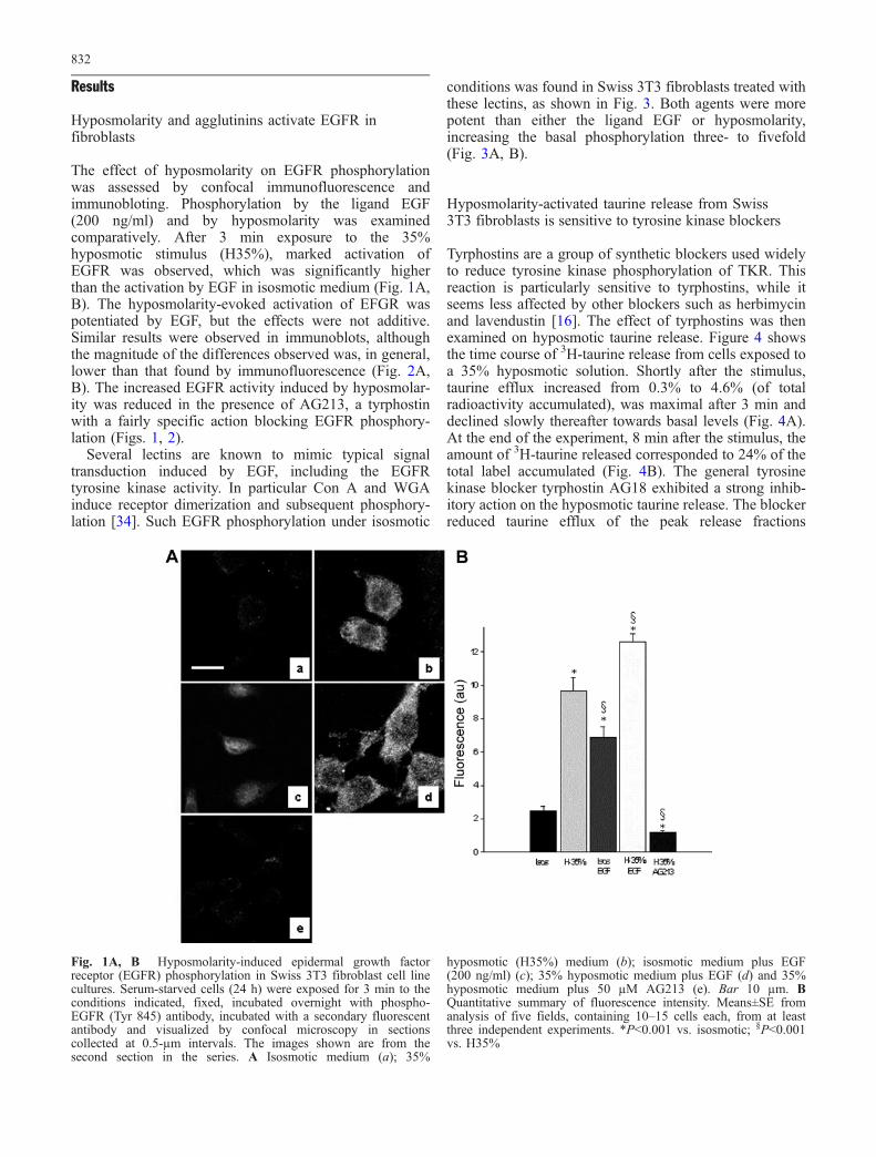
Hyposmolarity and agglutinins activate EGFR infibroblasts
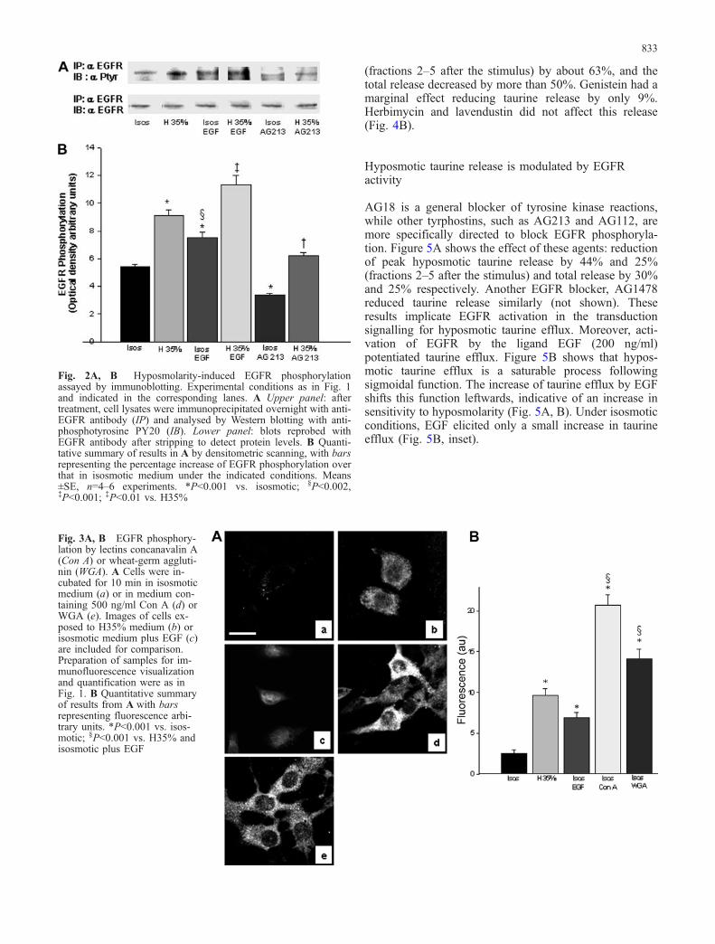
The effect of hyposmolarity on EGFR phosphorylationwas assessed by confocal immunofluorescence andimmunobloting. Phosphorylation by the ligand EGF(200 ng/ml) and by hyposmolarity was examinedcomparatively. After 3 min exposure to the 35%hyposmotic stimulus (H35%), marked activation ofEGFR was observed, which was significantly higherthan the activation by EGF in isosmotic medium (Fig. 1A,B). The hyposmolarity-evoked activation of EFGR waspotentiated by EGF, but the effects were not additive.Similar results were observed in immunoblots, althoughthe magnitude of the differences observed was, in general,lower than that found by immunofluorescence (Fig. 2A,B). The increased EGFR activity induced by hyposmolar-ity was reduced in the presence of AG213, a tyrphostinwith a fairly specific action blocking EGFR phosphory-lation (Figs. 1, 2).
Several lectins are known to mimic typical signaltransduction induced by EGF, including the EGFRtyrosine kinase activity. In particular Con A and WGAinduce receptor dimerization and subsequent phosphory-lation [34]. Such EGFR phosphorylation under isosmotic
conditions was found in Swiss 3T3 fibroblasts treated withthese lectins, as shown in Fig. 3. Both agents were morepotent than either the ligand EGF or hyposmolarity,increasing the basal phosphorylation three- to fivefold(Fig. 3A, B).
Hyposmolarity-activated taurine release from Swiss3T3 fibroblasts is sensitive to tyrosine kinase blockers
Tyrphostins are a group of synthetic blockers used widelyto reduce tyrosine kinase phosphorylation of TKR. Thisreaction is particularly sensitive to tyrphostins, while itseems less affected by other blockers such as herbimycinand lavendustin [16]. The effect of tyrphostins was thenexamined on hyposmotic taurine release. Figure 4 showsthe time course of 3H-taurine release from cells exposed toa 35% hyposmotic solution. Shortly after the stimulus,taurine efflux increased from 0.3% to 4.6% (of totalradioactivity accumulated), was maximal after 3 min anddeclined slowly thereafter towards basal levels (Fig. 4A).At the end of the experiment, 8 min after the stimulus, theamount of 3H-taurine released corresponded to 24% of thetotal label accumulated (Fig. 4B). The general tyrosinekinase blocker tyrphostin AG18 exhibited a strong inhib-itory action on the hyposmotic taurine release. The blockerreduced taurine efflux of the peak release fractions
Fig. 1A, B Hyposmolarity-induced epidermal growth factorreceptor (EGFR) phosphorylation in Swiss 3T3 fibroblast cell linecultures. Serum-starved cells (24 h) were exposed for 3 min to theconditions indicated, fixed, incubated overnight with phospho-EGFR (Tyr 845) antibody, incubated with a secondary fluorescentantibody and visualized by confocal microscopy in sectionscollected at 0.5-µm intervals. The images shown are from thesecond section in the series. A Isosmotic medium (a); 35%
hyposmotic (H35%) medium (b); isosmotic medium plus EGF(200 ng/ml) (c); 35% hyposmotic medium plus EGF (d) and 35%hyposmotic medium plus 50 µM AG213 (e). Bar 10 µm. BQuantitative summary of fluorescence intensity. Means±SE fromanalysis of five fields, containing 10–15 cells each, from at leastthree independent experiments. *P<0.001 vs. isosmotic; §P<0.001vs. H35%
832
(fractions 2–5 after the stimulus) by about 63%, and thetotal release decreased by more than 50%. Genistein had amarginal effect reducing taurine release by only 9%.Herbimycin and lavendustin did not affect this release(Fig. 4B).
Hyposmotic taurine release is modulated by EGFRactivity
AG18 is a general blocker of tyrosine kinase reactions,while other tyrphostins, such as AG213 and AG112, aremore specifically directed to block EGFR phosphoryla-tion. Figure 5A shows the effect of these agents: reductionof peak hyposmotic taurine release by 44% and 25%(fractions 2–5 after the stimulus) and total release by 30%and 25% respectively. Another EGFR blocker, AG1478reduced taurine release similarly (not shown). Theseresults implicate EGFR activation in the transductionsignalling for hyposmotic taurine efflux. Moreover, acti-vation of EGFR by the ligand EGF (200 ng/ml)potentiated taurine efflux. Figure 5B shows that hypos-motic taurine efflux is a saturable process followingsigmoidal function. The increase of taurine efflux by EGFshifts this function leftwards, indicative of an increase insensitivity to hyposmolarity (Fig. 5A, B). Under isosmoticconditions, EGF elicited only a small increase in taurineefflux (Fig. 5B, inset).
Fig. 2A, B Hyposmolarity-induced EGFR phosphorylationassayed by immunoblotting. Experimental conditions as in Fig. 1and indicated in the corresponding lanes. A Upper panel: aftertreatment, cell lysates were immunoprecipitated overnight with anti-EGFR antibody (IP) and analysed by Western blotting with anti-phosphotyrosine PY20 (IB). Lower panel: blots reprobed withEGFR antibody after stripping to detect protein levels. B Quanti-tative summary of results in A by densitometric scanning, with barsrepresenting the percentage increase of EGFR phosphorylation overthat in isosmotic medium under the indicated conditions. Means±SE, n=4–6 experiments. *P<0.001 vs. isosmotic; §P<0.002,‡P<0.001; ‡P<0.01 vs. H35%
Fig. 3A, B EGFR phosphory-lation by lectins concanavalin A(Con A) or wheat-germ aggluti-nin (WGA). A Cells were in-cubated for 10 min in isosmoticmedium (a) or in medium con-taining 500 ng/ml Con A (d) orWGA (e). Images of cells ex-posed to H35% medium (b) orisosmotic medium plus EGF (c)are included for comparison.Preparation of samples for im-munofluorescence visualizationand quantification were as inFig. 1. B Quantitative summaryof results from A with barsrepresenting fluorescence arbi-trary units. *P<0.001 vs. isos-motic; §P<0.001 vs. H35% andisosmotic plus EGF
833
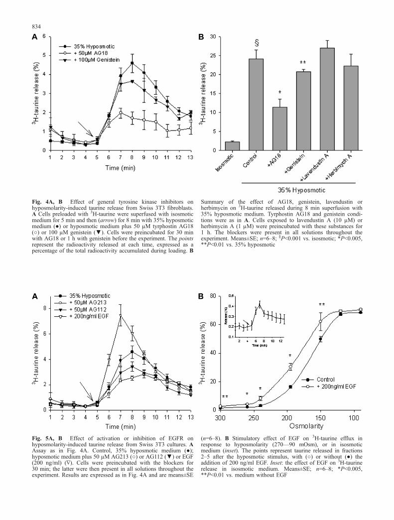
Fig. 4A, B Effect of general tyrosine kinase inhibitors onhyposmolarity-induced taurine release from Swiss 3T3 fibroblasts.A Cells preloaded with 3H-taurine were superfused with isosmoticmedium for 5 min and then (arrow) for 8 min with 35% hyposmoticmedium (●) or hyposmotic medium plus 50 µM tyrphostin AG18(○) or 100 µM genistein (▼). Cells were preincubated for 30 minwith AG18 or 1 h with genistein before the experiment. The pointsrepresent the radioactivity released at each time, expressed as apercentage of the total radioactivity accumulated during loading. B
Summary of the effect of AG18, genistein, lavendustin orherbimycin on 3H-taurine released during 8 min superfusion with35% hyposmotic medium. Tyrphostin AG18 and genistein condi-tions were as in A. Cells exposed to lavendustin A (10 µM) orherbimycin A (1 µM) were preincubated with these substances for1 h. The blockers were present in all solutions throughout theexperiment. Means±SE; n=6–8; §P<0.001 vs. isosmotic; *P<0.005,**P<0.01 vs. 35% hyposmotic
Fig. 5A, B Effect of activation or inhibition of EGFR onhyposmolarity-induced taurine release from Swiss 3T3 cultures. AAssay as in Fig. 4A. Control, 35% hyposmotic medium (●);hyposmotic medium plus 50 µM AG213 (○) or AG112 (▼) or EGF(200 ng/ml) (∇). Cells were preincubated with the blockers for30 min; the latter were then present in all solutions throughout theexperiment. Results are expressed as in Fig. 4A and are means±SE
(n=6–8). B Stimulatory effect of EGF on 3H-taurine efflux inresponse to hyposmolarity (270—90 mOsm), or in isosmoticmedium (inset). The points represent taurine released in fractions2–5 after the hyposmotic stimulus, with (○) or without (●) theaddition of 200 ng/ml EGF. Inset: the effect of EGF on 3H-taurinerelease in isosmotic medium. Means±SE; n=6–8; *P<0.005,**P<0.01 vs. medium without EGF
834
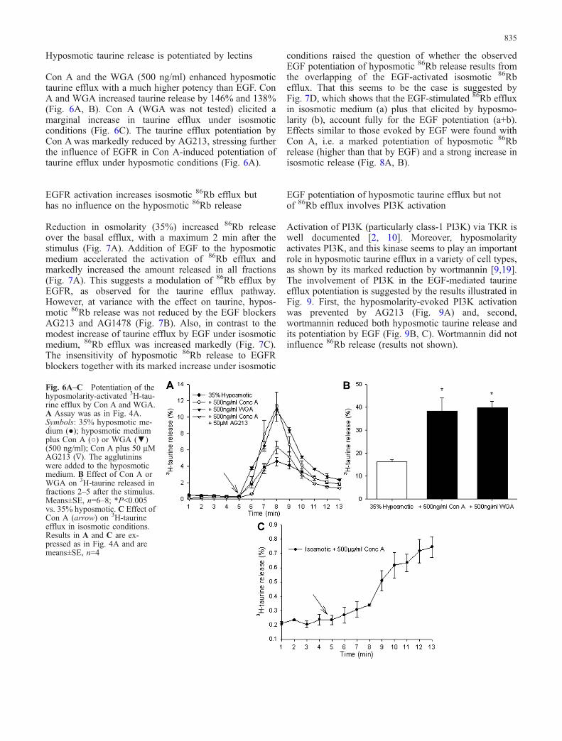
Hyposmotic taurine release is potentiated by lectins
Con A and the WGA (500 ng/ml) enhanced hyposmotictaurine efflux with a much higher potency than EGF. ConA and WGA increased taurine release by 146% and 138%(Fig. 6A, B). Con A (WGA was not tested) elicited amarginal increase in taurine efflux under isosmoticconditions (Fig. 6C). The taurine efflux potentiation byCon Awas markedly reduced by AG213, stressing furtherthe influence of EGFR in Con A-induced potentiation oftaurine efflux under hyposmotic conditions (Fig. 6A).
EGFR activation increases isosmotic 86Rb efflux buthas no influence on the hyposmotic 86Rb release
Reduction in osmolarity (35%) increased 86Rb releaseover the basal efflux, with a maximum 2 min after thestimulus (Fig. 7A). Addition of EGF to the hyposmoticmedium accelerated the activation of 86Rb efflux andmarkedly increased the amount released in all fractions(Fig. 7A). This suggests a modulation of 86Rb efflux byEGFR, as observed for the taurine efflux pathway.However, at variance with the effect on taurine, hypos-motic 86Rb release was not reduced by the EGF blockersAG213 and AG1478 (Fig. 7B). Also, in contrast to themodest increase of taurine efflux by EGF under isosmoticmedium, 86Rb efflux was increased markedly (Fig. 7C).The insensitivity of hyposmotic 86Rb release to EGFRblockers together with its marked increase under isosmotic
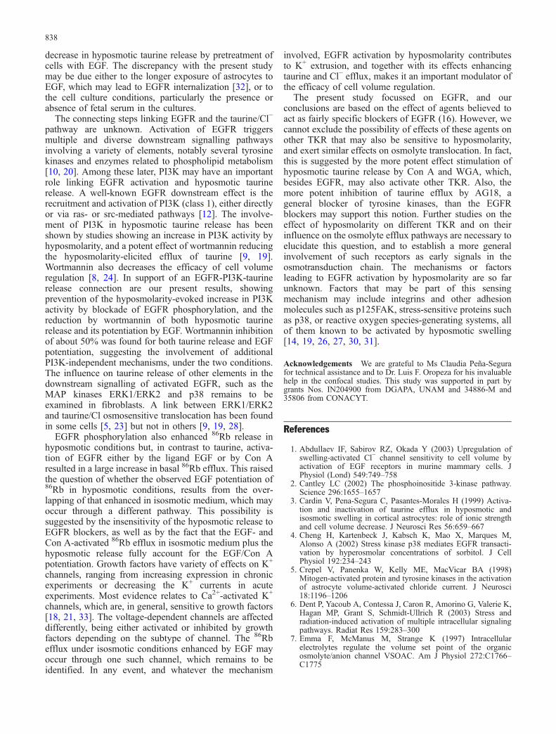
conditions raised the question of whether the observedEGF potentiation of hyposmotic 86Rb release results fromthe overlapping of the EGF-activated isosmotic 86Rbefflux. That this seems to be the case is suggested byFig. 7D, which shows that the EGF-stimulated 86Rb effluxin isosmotic medium (a) plus that elicited by hyposmo-larity (b), account fully for the EGF potentiation (a+b).Effects similar to those evoked by EGF were found withCon A, i.e. a marked potentiation of hyposmotic 86Rbrelease (higher than that by EGF) and a strong increase inisosmotic release (Fig. 8A, B).
EGF potentiation of hyposmotic taurine efflux but notof 86Rb efflux involves PI3K activation
Activation of PI3K (particularly class-1 PI3K) via TKR iswell documented [2, 10]. Moreover, hyposmolarityactivates PI3K, and this kinase seems to play an importantrole in hyposmotic taurine efflux in a variety of cell types,as shown by its marked reduction by wortmannin [9,19].The involvement of PI3K in the EGF-mediated taurineefflux potentiation is suggested by the results illustrated inFig. 9. First, the hyposmolarity-evoked PI3K activationwas prevented by AG213 (Fig. 9A) and, second,wortmannin reduced both hyposmotic taurine release andits potentiation by EGF (Fig. 9B, C). Wortmannin did notinfluence 86Rb release (results not shown).
Fig. 6A–C Potentiation of thehyposmolarity-activated 3H-tau-rine efflux by Con A and WGA.A Assay was as in Fig. 4A.Symbols: 35% hyposmotic me-dium (●); hyposmotic mediumplus Con A (○) or WGA (▼)(500 ng/ml); Con A plus 50 µMAG213 (∇). The agglutininswere added to the hyposmoticmedium. B Effect of Con A orWGA on 3H-taurine released infractions 2–5 after the stimulus.Means±SE, n=6–8; *P<0.005vs. 35% hyposmotic. C Effect ofCon A (arrow) on 3H-taurineefflux in isosmotic conditions.Results in A and C are ex-pressed as in Fig. 4A and aremeans±SE, n=4
835
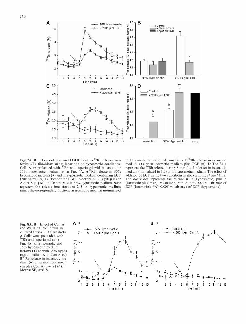
Fig. 7A–D Effects of EGF and EGFR blockers 86Rb release fromSwiss 3T3 fibroblasts under isosmotic or hyposmotic conditions.Cells were preloaded with 86Rb and superfused with isosmotic or35% hyposmotic medium as in Fig. 4A. A86Rb release in 35%hyposmotic medium (●) and in hyposmotic medium containing EGF(200 ng/ml) (○). B Effect of the EGFR blockers AG213 (50 µM) orAG1478 (1 µM) on 86Rb release in 35% hyposmotic medium. Barsrepresent the release into fractions 2–5 in hyposmotic mediumminus the corresponding fractions in isosmotic medium (normalized
to 1.0) under the indicated conditions. C86Rb release in isosmoticmedium (●) or in isosmotic medium plus EGF (○). D The barsrepresent the 86Rb release during 8 min (total release) in isosmoticmedium (normalized to 1.0) or in hyposmotic medium. The effect ofaddition of EGF in the two conditions is shown in the shaded bars.The black bar represents the release in a (hyposmotic) plus b(isosmotic plus EGF). Means±SE, n=6–8; *P<0.005 vs. absence ofEGF (isosmotic); **P<0.005 vs. absence of EGF (hyposmotic)
Fig. 8A, B Effect of Con Aand WGA on Rb86 efflux incultured Swiss 3T3 fibroblasts.A Cells were preloaded with86Rb and superfused as inFig. 4A, with isosmotic and35% hyposmotic medium(arrow) (●) or with 35% hypos-motic medium with Con A (○).B86Rb release in isosmotic me-dium (●) or in isosmotic medi-um plus Con A (arrow) (○).Means±SE, n=6–8
836

Discussion
The present study in Swiss 3T3 fibroblasts showed theactivation of EGFR by hyposmolarity, a finding that to ourknowledge has not been reported so far. EGFR activationby hyposmolarity was higher than that elicited by asaturating EGF concentration and the hyposmotic effectwas potentiated by the ligand. These results suggest eitheran increase in exposed EGFR in the hyposmotic condition,or a delay in the internalization process occurringsubsequent to phosphorylation [32]. Another importantfinding of our study is the implication of EGFR as anelement in the transduction signalling for the pathwayoperation releasing taurine after the hyposmotic stimulus.This is supported by the decrease of hyposmotic taurineefflux by EGFR blockade and by its potentiation by EGFRactivation in the presence of EGF or agglutinins. Thepattern of EGF influence on the seemingly saturableprocess of hyposmotic taurine release is suggestive of anincrease by EGF of the sensitivity of taurine efflux to
hyposmolarity. Previous studies in intestinal cell lines 407and C127 have shown potentiation by EGF of thehyposmotic efflux of Cl− (traced by 125I) and modulationof the osmosensitive Cl− current by EGFR over-expression[1, 25]. A common hyposmotic release pathway for Cl−
and organic osmolyte has been proposed on the basis oftheir similar pharmacological profiles [13], and thispathway may be that modulated by EGFR activation.Similar to the effect of EGF on Cl− efflux in intestinal 407cells [25], our results showed that EGFR activation byEGF or Con A elicited only a small increase in taurineefflux under isosmotic conditions. These results suggestthat although EGFR activation may be able to initiatetaurine efflux by itself, other elements may be requiredconcurrently with hyposmolarity to activate the taurineefflux pathway fully. A decrease in ionic strength may beone such element, as suggested by studies on taurineefflux in astrocytes and on the volume-sensitive Cl−
current in endothelial cells [3, 7, 29]. A study on culturedastrocytes [17] has shown, at variance with our results, a
Fig. 9A–D Phosphatidylinositide-3-kinase (PI3K) activation byhyposmolarity and the effect of EGFR blockade. A Activation ofPI3K by EGF in isosmotic medium and the effect of wortmanninand AG213. Upper panel: cell lysates prepared after treatment wereimmunoprecipitated overnight with anti-AKT antibody (IP) andanalysed by western blotting with anti-pAKT (IB). Blots werereprobed with AKT antibody after stripping to detect protein levels.Lower panel: PI3K activity in hyposmotic (H35%) conditions, andeffects of EGF, wortmannin and AG213. B Quantitative summary ofresults in A, with bars representing the percentage increase of AKT
phosphorylation by hyposmotic medium over that in isosmoticmedium, under the indicated conditions. Means±SE, n=4–6experiments. *P<0.001, §P<0.005, †P>0.05 vs. isosmotic;‡P<0.001 vs. 35% hyposmotic (H35%). C, D Effect of wortmannin(▼) on the hyposmotic (●) and the EGF-potentiated efflux of 3H-taurine (○). The superfusion, release and results expression as inFig. 4A. Cells exposed to wortmannin (100 nM) were preincubatedwith it for 1 h and wortmannin was present in all solutionsthroughout the experiment. Means±SE, n=6–8
837
decrease in hyposmotic taurine release by pretreatment ofcells with EGF. The discrepancy with the present studymay be due either to the longer exposure of astrocytes toEGF, which may lead to EGFR internalization [32], or tothe cell culture conditions, particularly the presence orabsence of fetal serum in the cultures.
The connecting steps linking EGFR and the taurine/Cl−
pathway are unknown. Activation of EGFR triggersmultiple and diverse downstream signalling pathwaysinvolving a variety of elements, notably several tyrosinekinases and enzymes related to phospholipid metabolism[10, 20]. Among these later, PI3K may have an importantrole linking EGFR activation and hyposmotic taurinerelease. A well-known EGFR downstream effect is therecruitment and activation of PI3K (class 1), either directlyor via ras- or src-mediated pathways [12]. The involve-ment of PI3K in hyposmotic taurine release has beenshown by studies showing an increase in PI3K activity byhyposmolarity, and a potent effect of wortmannin reducingthe hyposmolarity-elicited efflux of taurine [9, 19].Wortmannin also decreases the efficacy of cell volumeregulation [8, 24]. In support of an EGFR-PI3K-taurinerelease connection are our present results, showingprevention of the hyposmolarity-evoked increase in PI3Kactivity by blockade of EGFR phosphorylation, and thereduction by wortmannin of both hyposmotic taurinerelease and its potentiation by EGF. Wortmannin inhibitionof about 50% was found for both taurine release and EGFpotentiation, suggesting the involvement of additionalPI3K-independent mechanisms, under the two conditions.The influence on taurine release of other elements in thedownstream signalling of activated EGFR, such as theMAP kinases ERK1/ERK2 and p38 remains to beexamined in fibroblasts. A link between ERK1/ERK2and taurine/Cl osmosensitive translocation has been foundin some cells [5, 23] but not in others [9, 19, 28].
EGFR phosphorylation also enhanced 86Rb release inhyposmotic conditions but, in contrast to taurine, activa-tion of EGFR either by the ligand EGF or by Con Aresulted in a large increase in basal 86Rb efflux. This raisedthe question of whether the observed EGF potentiation of86Rb in hyposmotic conditions, results from the over-lapping of that enhanced in isosmotic medium, which mayoccur through a different pathway. This possibility issuggested by the insensitivity of the hyposmotic release toEGFR blockers, as well as by the fact that the EGF- andCon A-activated 86Rb efflux in isosmotic medium plus thehyposmotic release fully account for the EGF/Con Apotentiation. Growth factors have variety of effects on K+
channels, ranging from increasing expression in chronicexperiments or decreasing the K+ currents in acuteexperiments. Most evidence relates to Ca2+-activated K+
channels, which are, in general, sensitive to growth factors[18, 21, 33]. The voltage-dependent channels are affecteddifferently, being either activated or inhibited by growthfactors depending on the subtype of channel. The 86Rbefflux under isosmotic conditions enhanced by EGF mayoccur through one such channel, which remains to beidentified. In any event, and whatever the mechanism
involved, EGFR activation by hyposmolarity contributesto K+ extrusion, and together with its effects enhancingtaurine and Cl− efflux, makes it an important modulator ofthe efficacy of cell volume regulation.
The present study focussed on EGFR, and ourconclusions are based on the effect of agents believed toact as fairly specific blockers of EGFR (16). However, wecannot exclude the possibility of effects of these agents onother TKR that may also be sensitive to hyposmolarity,and exert similar effects on osmolyte translocation. In fact,this is suggested by the more potent effect stimulation ofhyposmotic taurine release by Con A and WGA, which,besides EGFR, may also activate other TKR. Also, themore potent inhibition of taurine efflux by AG18, ageneral blocker of tyrosine kinases, than the EGFRblockers may support this notion. Further studies on theeffect of hyposmolarity on different TKR and on theirinfluence on the osmolyte efflux pathways are necessary toelucidate this question, and to establish a more generalinvolvement of such receptors as early signals in theosmotransduction chain. The mechanisms or factorsleading to EGFR activation by hyposmolarity are so farunknown. Factors that may be part of this sensingmechanism may include integrins and other adhesionmolecules such as p125FAK, stress-sensitive proteins suchas p38, or reactive oxygen species-generating systems, allof them known to be activated by hyposmotic swelling[14, 19, 26, 27, 30, 31].
Acknowledgements We are grateful to Ms Claudia Peña-Segurafor technical assistance and to Dr. Luis F. Oropeza for his invaluablehelp in the confocal studies. This study was supported in part bygrants Nos. IN204900 from DGAPA, UNAM and 34886-M and35806 from CONACYT.
References
1. Abdullaev IF, Sabirov RZ, Okada Y (2003) Upregulation ofswelling-activated Cl− channel sensitivity to cell volume byactivation of EGF receptors in murine mammary cells. JPhysiol (Lond) 549:749–758
2. Cantley LC (2002) The phosphoinositide 3-kinase pathway.Science 296:1655–1657
3. Cardin V, Pena-Segura C, Pasantes-Morales H (1999) Activa-tion and inactivation of taurine efflux in hyposmotic andisosmotic swelling in cortical astrocytes: role of ionic strengthand cell volume decrease. J Neurosci Res 56:659–667
4. Cheng H, Kartenbeck J, Kabsch K, Mao X, Marques M,Alonso A (2002) Stress kinase p38 mediates EGFR transacti-vation by hyperosmolar concentrations of sorbitol. J CellPhysiol 192:234–243
5. Crepel V, Panenka W, Kelly ME, MacVicar BA (1998)Mitogen-activated protein and tyrosine kinases in the activationof astrocyte volume-activated chloride current. J Neurosci18:1196–1206
6. Dent P, Yacoub A, Contessa J, Caron R, Amorino G, Valerie K,Hagan MP, Grant S, Schmidt-Ullrich R (2003) Stress andradiation-induced activation of multiple intracellular signalingpathways. Radiat Res 159:283–300
7. Emma F, McManus M, Strange K (1997) Intracellularelectrolytes regulate the volume set point of the organicosmolyte/anion channel VSOAC. Am J Physiol 272:C1766–C1775
838
8. Feranchak AP, Roman RM, Schwiebert EM, Fitz JG (1998)Phosphatidylinositol 3-kinase contributes to cell volume regu-lation through effects on ATP release. J Biol Chem 273:14906–14911
9. Franco R, Torres-Marquez ME, Pasantes-Morales H (2001)Evidence for two mechanisms of amino acid osmolyte releasefrom hippocampal slices. Pflugers Arch 442:791–800
10. Grant S, Qiao L, Dent P (2002) Roles of ERBB family receptortyrosine kinases, and downstream signaling pathways, in thecontrol of cell growth and survival. Front Biosci 7:d376–d389
11. Jakab M, Furst J, Gschwentner M, Botta G, Garavaglia ML,Bazzini C, Rodighiero S, Meyer G, Eichmueller S, Woll E,Chwatal S, Ritter M, Paulmichl M (2002) Mechanisms sensingand modulating signals arising from cell swelling. Cell PhysiolBiochem 12:235–258
12. Jorissen RN, Walker F, Pouliot N, Garrett TP, Ward CW,Burgess AW (2003) Epidermal growth factor receptor:mechanisms of activation and signalling. Exp Cell Res284:31–53
13. Kirk K, Strange K (1998) Functional properties and physio-logical roles of organic solute channels. Annu Rev Physiol60:719–739
14. Lambert IH (2003) Reactive oxygen species regulate swelling-induced taurine efflux in NIH3T3 mouse fibroblasts. J MembrBiol 192:19–32
15. Lang F, Busch GL, Ritter M, Volkl H, Waldegger S, Gulbins E,Haussinger D (1998) Functional significance of cell volumeregulatory mechanisms. Physiol Rev 78:247–306
16. Levitzki A, Gazit A (1995) Tyrosine kinase inhibition: anapproach to drug development. Science 267:1782–1788
17. Mongin AA, Reddi JM, Charniga C, Kimelberg HK (1999)[3H]taurine and D-[3H]aspartate release from astrocyte culturesare differently regulated by tyrosine kinases. Am J Physiol 276:C1226–C1230
18. Neylon CB (2002) Potassium channels and vascular prolifer-ation. Vascul Pharmacol 38:35–41
19. Ochoa De La Paz LD, Lezama R, Torres-Marquez ME,Pasantes-Morales H (2002) Tyrosine kinases and amino acidefflux under hyposmotic and ischaemic conditions in thechicken retina. Pflugers Arch 445:87–96
20. Prenzel N, Fischer OM, Streit S, Hart S, Ullrich A (2001) Theepidermal growth factor receptor family as a central element forcellular signal transduction and diversification. Endocr RelatCancer 8:11–31
21. Rane SG (1999) Ion channels as physiological effectors forgrowth factor receptor and Ras/ERK signaling pathways. AdvSecond Messenger Phosphoprotein Res 33:107–127
22. Rosette C, Karin M (1996) Ultraviolet light and osmotic stress:activation of the JNK cascade through multiple growth factorand cytokine receptors. Science 274:1194–1197
23. Shen MR, Chou CY, Browning JA, Wilkins RJ, Ellory JC(2001) Human cervical cancer cells use Ca2+ signalling, proteintyrosine phosphorylation and MAP kinase in regulatory volumedecrease. J Physiol (Lond) 537:347–362
24. Shi C, Barnes S, Coca-Prados M, Kelly ME (2002) Proteintyrosine kinase and protein phosphatase signaling pathwaysregulate volume-sensitive chloride currents in a nonpigmentedciliary epithelial cell line. Invest Ophthalmol Vis Sci 43:1525–1532
25. Tilly BC, Van den Berghe N, Tertoolen LG, Edixhoven MJ, DeJonge HR (1993) Protein tyrosine phosphorylation is involvedin osmoregulation of ionic conductances. J Biol Chem268:19919–19922
26. Tilly BC, Edixhoven MJ, Tertoolen LG, Morii N, Saitoh Y,Narumiya S, De Jonge HR (1996) Activation of the osmo-sensitive chloride conductance involves P21rho and isaccompanied by a transient reorganization of the F-actincytoskeleton. Mol Biol Cell 7:1419–1427
27. Tilly BC, Gaestel M, Engel K, Edixhoven MJ, de Jonge HR(1996) Hypo-osmotic cell swelling activates the p38 MAPkinase signalling cascade. FEBS Lett 395:133–136
28. Van der Wijk T, De Jonge HR, Tilly BC (1999) Osmotic cellswelling-induced ATP release mediates the activation ofextracellular signal-regulated protein kinase (Erk)-1/2 but notthe activation of osmo-sensitive anion channels. Biochem J343:579–586
29. Voets T, Droogmans G, Raskin G, Eggermont J, Nilius B(1999) Reduced intracellular ionic strength as the initial triggerfor activation of endothelial volume-regulated anion channels.Proc Natl Acad Sci USA 96:5298–5303
30. Vom Dahl S, Schliess F, Graf D, Haussinger D (2001) Role ofp38(MAPK) in cell volume regulation of perfused rat liver. CellPhysiol Biochem 11:285–294
31. Vom Dahl S, Schliess F, Reissmann R, Gorg B, Weiergraber O,Kacalkova M, Dombrowski F, Haussinger D (2003) Involve-ment of integrins in osmosensing and signalling towardsautophagic proteolysis in rat liver. J Biol Chem 278:27088–27095
32. Wiley HS (2003) Trafficking of the ErbB receptors and itsinfluence on signaling. Exp Cell Res 284:78–88
33. Wonderlin WF, Strobl JS (1996) Potassium channels, prolifer-ation and G1 progression. J Membr Biol 154:91–107
34. Zeng FY, Benguria A, Kafert S, Andre S, Gabius HJ, VillaloboA (1995) Differential response of the epidermal growth factorreceptor tyrosine kinase activity to several plant and mamma-lian lectins. Mol Cell Biochem 142:117–124
35. Zwick E, Hackel PO, Prenzel N, Ullrich A (1999) The EGFreceptor as central transducer of heterologous signallingsystems. Trends Pharmacol Sci 20:408–412
839
Related Documents