PAAVO-ILARI KUIKKA Epidemiology and Magnetic Resonance Imaging-Based Diagnostics of Knee Injuries and Anterior Knee Pain in Young Adults ACADEMIC DISSERTATION To be presented, with the permission of the board of the School of Medicine of the University of Tampere, for public discussion in the Auditorium of Finn-Medi 5, Biokatu 12, Tampere, on August 23rd, 2012, at 12 o’clock. UNIVERSITY OF TAMPERE

Welcome message from author
This document is posted to help you gain knowledge. Please leave a comment to let me know what you think about it! Share it to your friends and learn new things together.
Transcript

PAAVO-ILARI KUIKKA
Epidemiology and Magnetic Resonance Imaging-Based Diagnostics of Knee Injuries
and Anterior Knee Pain in Young Adults
ACADEMIC DISSERTATIONTo be presented, with the permission of
the board of the School of Medicine of the University of Tampere,for public discussion in the Auditorium of Finn-Medi 5,
Biokatu 12, Tampere, on August 23rd, 2012, at 12 o’clock.
UNIVERSITY OF TAMPERE

Reviewed byDocent Arsi HarilainenUniversity of HelsinkiFinlandDocent Juhana LeppilahtiUniversity of OuluFinland
DistributionBookshop TAJUP.O. Box 61733014 University of TampereFinland
Tel. +358 40 190 9800Fax +358 3 3551 7685 [email protected]/tajuhttp://granum.uta.fi
Cover design byMikko Reinikka
TaittoSirpa Randell
Acta Universitatis Tamperensis 1753ISBN 978-951-44-8876-4 (print)ISSN-L 1455-1616ISSN 1455-1616
Acta Electronica Universitatis Tamperensis 1226ISBN 978-951-44-8877-1 (pdf )ISSN 1456-954Xhttp://acta.uta.fi
Tampereen Yliopistopaino Oy – Juvenes PrintTampere 2012
ACADEMIC DISSERTATIONUniversity of Tampere, School of Medicine Centre for Military MedicineCentral Military Hospital, Department of Orthopedic SurgeryFinland
Supervised byDocent Harri PihlajamäkiUniversity of HelsinkiFinlandDocent Ville MattilaUniversity of TampereFinland
Copyright ©2012 Tampere University Press and the author
Table of Contents
1 List of Original Publications ........................................................................................ 7
2 Abbreviations ................................................................................................................. 8
3 Tiivistelmä ...................................................................................................................... 9
4 Abstract ..........................................................................................................................11
5 Introduction ................................................................................................................. 13
6 Review of the Literature ............................................................................................. 166.1 Health Problems in Young Adults in Finland .............................................. 166.2 Epidemiology of Knee Injuries in Young Adults ......................................... 17
6.2.1 Overall Incidence of Knee Injuries ..................................................... 176.2.2 Incidence and Relative Frequency of Common Specified Knee Injuries and Anterior Knee Pain ............................................. 186.2.3 Risk Factors for Musculoskeletal Injuries in General ...................... 266.2.4 Risk Factors for Knee Injuries and Anterior Knee Pain.................. 27
6.3 Diagnostics of Meniscal Tears, Chondral Lesions and Anterior Knee pain .......................................................................................................... 32
6.3.1 History and Physical Examination ..................................................... 326.3.2 Imaging of Knee Injuries and Anterior Knee Pain .......................... 35
6.3.2.1 Plain radiographs and Computed Tomography .............. 356.3.2.2 Magnetic Resonance Imaging ............................................ 36
6.4 General Treatment Principles of Meniscal Tears, Chondral Lesions and Anterior Knee Pain ................................................................... 41
6.4.1 Conservative Management .................................................................. 416.4.2 Arthroscopy and Arthrotomy............................................................. 42
7 Aims of the Study ........................................................................................................ 44

8 Materials and Methods ............................................................................................... 458.1 Study Population .............................................................................................. 45
8.1.1 Diagnostic Samples (I–III)................................................................... 468.1.2 Epidemiological Sample (IV) .............................................................. 48
8.2 Physical Examination Methods (I–III) ......................................................... 488.3 Imaging Methods (I–III) ................................................................................. 488.4 Arthroscopies (I–III) ....................................................................................... 498.5 Epidemiological Methods (IV) ....................................................................... 50
8.5.1 Outcomes ............................................................................................... 508.5.2 Data of Inpatient Care Admissions .................................................... 508.5.3 Disability Data ....................................................................................... 51
8.6 Statistical Methods of the Diagnostic Section (I–III) ................................. 518.6.1 Data of Fresh Chondral Lesions (I) .................................................... 518.6.2 Data of Meniscal Tears (II) .................................................................. 528.6.3 Data of Anterior Knee Pain and Patellar Chondral Lesions (III) ........................................................... 53
8.7 Statistical Methods of the Epidemiological Section (IV) ........................... 54
9 Results ........................................................................................................................... 569.1 Clinical and Arthroscopic Diagnoses (I–III) ............................................... 56
9.1.1 Fresh Traumatic Chondral Lesions (I) ............................................... 569.1.2 Meniscal Tears (II) ................................................................................ 569.1.3 Anterior Knee Pain and Patellar Chondral Lesions (III) ................ 56
9.2 Radiographs in Anterior Knee Pain (III) ...................................................... 579.3 Diagnostic Validity of MRI (I–III) ................................................................ 58
9.3.1 Fresh Chondral Lesions (I) .................................................................. 589.3.2 Fresh and Old Meniscal Tears (II) ...................................................... 609.3.3 Patellar Chondral Lesions Associated with Anterior Knee Pain (III) ...................................................................... 64
9.4 Incidence of Knee Injuries Needing Inpatient Care (IV) ........................... 669.5 Readmissions, Disability, and Need for Surgery (IV) ................................. 679.6 Risk Factors for Knee Injuries Needing Inpatient Care (IV) ..................... 68
10 Discussion ..................................................................................................................... 7110.1 Diagnostics (I–III) ............................................................................................ 71
10.1.1 Fresh Chondral Lesions (I) .................................................................. 7110.1.2 Fresh and Old Meniscal Tears (II) .......................................................7410.1.3 Patellar Chondral Lesions Associated with Anterior Knee Pain (III) ...................................................................... 77

10.2 Epidemiology of Knee Injuries (IV) .............................................................. 8110.2.1 Incidence of Knee Injuries Needing Inpatient Care ........................ 8110.2.2 Readmissions, Disability, and Need for Surgery .............................. 8510.2.3 Risk Factors for Knee Injuries Needing Inpatient Care .................. 85
11 Conclusions .................................................................................................................. 88
12 Acknowledgements ..................................................................................................... 90
13 References ..................................................................................................................... 92
14 Original Publications ................................................................................................ 105


7
1 List of Original Publications
This thesis is based on the following original publications, which are referred to in the text by the Roman numerals I–IV:
I Kuikka, P. I., Kiuru, M. J., Niva, M. H., Kröger, H., Pihlajamäki, H. K. (2006). Sensitivity of routine 1.0-Tesla magnetic resonance imaging versus arthroscopy as gold standard in fresh traumatic chondral lesions of the knee in young adults. Arthroscopy, 22(10), 1033–1039.
II Kuikka, P. I., Sillanpää, P., Mattila, V. M., Niva, M. H., Pihlajamäki, H. K. (2009). Magnetic resonance imaging in acute traumatic and chronic meniscal tears of the knee: A diagnostic accuracy study in young adults. Am J Sports Med, 37(5), 1003–1008.
III Pihlajamäki, H. K., Kuikka, P. I., Leppänen, V. V., Kiuru, M. J., Mattila, V. M. (2010). Reliability of clinical findings and magnetic resonance imaging for the diagnosis of chondromalacia patellae. J Bone Joint Surg Am, 92(4), 927–934.
IV Kuikka, P. I., Pihlajamäki, H. K., Mattila, V.M. (2011). Knee injuries related to sports in young adult males during military service – Incidence and risk factors. Scand J Med Sci Sports. Epub 2011 Oct 19.
Articles I–IV have been reprinted in this thesis with the permission from Elsevier (I), SAGE Publications (II), J Bone Joint Surg Am. (III) and Blackwell Munksgaard (IV).
8
2 Abbreviations
ACL = Anterior cruciate ligamentAKP= Anterior knee painBMI = Body mass indexCI= Confidence IntervalCMP = Chondromalacia patellaeCT = Computed tomographyICRS = International Cartilage Repair SocietyLCL = Lateral collateral ligamentMCL = Medial collateral ligamentMRI = Magnetic Resonance ImagingNHDR = National hospital discharge registerOR = Odds RatioPCL = Posterior cruciate ligamentPFM = Patellofemoral malalignmentPFPS = Patellofemoral pain syndromeSPGR = Spoiled gradient echoT = TeslaUS = United States of America
9
3 Tiivistelmä
Tämän väitöskirjan tarkoituksena on selvittää nuorten aikuisten miesten polvivammo-jen ilmaantuvuutta ja riskitekijöitä sekä magneettikuvauspainotteista diagnostiikkaa tuoreissa polven rustovaurioissa, nivelkierukan repeämissä ja polven etuosan kivussa.
Epidemiologisena aineistona tutkimuksessa olivat kaikki tutkimusajanjakson ai-kana varusmiespalveluksensa suorittaneet 18–30-vuotiaat miespuoliset varusmiehet (n=128 584). Altistumisaika oli 97 503 henkilövuotta. Pakollisen asevelvollisuuden vuoksi varusmiehet edustavat erittäin hyvin perustervettä nuorta aikuista miesväestöä Suomessa. Kaikki sairaalahoitoon johtaneet polvivammat tässä aineistossa selvitettiin kansallisen hoitoilmoitusrekisterin avulla. Polvivammoja käsiteltiin kokonaisuutena ja lisäksi jaoteltuina erillisiin diagnooseihin, joita olivat ristiside- ja sivusidevammat, ni-velkierukkarepeämät, traumaattiset rustovauriot ja polvilumpion sijoiltaanmenot. Tie-dot palveluskelpoisuusluokan muutoksista ja mahdollisesti polvivammoihin yhteydessä olevista riskitekijöistä saatiin puolustusvoimien sisäisistä rekistereistä.
Magneettikuvauksen diagnostista validiteettia tuoreissa rustovaurioissa sekä tuo-reissa ja vanhoissa nivelkierukkarepeämissä tutkittiin retrospektiivisesti. Aineistojen kaikille potilaille oli tehty magneettikuvaus ja artroskopia keskussotilassairaalassa nor-maalin kliinisen käytännön mukaisesti. Tuoreiden rustovaurioiden magneettikuvausta käsittelevään aineistoon otettiin mukaan 32 potilasta, joilla oli todettu artroskopiassa tuore traumaattinen rustovaurio. Potilaiden mediaani-ikä oli 19 vuotta (vaihteluväli 19–21).
Tuoreiden ja vanhojen nivelkierukkarepeämien magneettikuvausdiagnostiikkaa ver-tailevien aineistojen mukaanottokriteerit täytti 82 potilaista, joilla oli tuore polvivamma, ja 40 potilasta, joilla oli pidempikestoinen polviongelma. Potilaiden mediaani-ikä oli 20 vuotta (vaihteluväli 18–25).
Polven etuosan kipuun liittyvien oireiden ja statuslöydösten yhteyttä artroskooppi-siin löydöksiin tutkittiin prospektiivisesti 56 varusmiehen aineistossa. Kaikilla oli pi-dempiaikainen, vammaan liittymätön polven etuosan kiputila. Potilaiden mediaani-ikä oli 19.5 vuotta (vaihteluväli 18–25). Kaikille tutkimukseen valituille tehtiin magneetti-kuvaus ja artroskopia keskussotilassairaalassa.
Sairaalahoitoon johtaneiden polvivammojen ilmaantuvuus kokonaisuutena oli 11 potilasta 1000 henkilövuotta kohden [95 % luottamusväli (CI): 10.4–11.7]. Polvivammo-jen riskitekijöitä analysoitiin logistisella regressiolla. Merkittävimmät riskitekijät polvi-

10
vammoille olivat korkeampi ikä (OR 1.7; 95 % CI: 1.3–2.2) ja ylipaino (OR 1.6; 95 % CI: 1.03–2.5). Kirurginen toimenpide tehtiin kahdelle kolmasosalle kaikista sairaalassa hoidetuista potilaista ja pidempiaikainen haitta (palveluskelpoisuusluokan muutos) jäi yhdelle kolmasosalle potilaista.
Tuoreiden traumaattisten rustovaurioiden todentamisessa 1.0 Teslan magneetti-kuvauk sen sensitiivisyys oli alhainen. Sensitiivisyys oli riippuvainen rustovaurion syvyydestä, ollen pinnallisissa vaurioissa vain 17 %. Koko ruston syvyyden kattaville vaurioille sensitiivisyys oli hieman parempi, 57 %. Tulos tarkoittaa, että lähes puolet sy-vistä rustovaurioista jäi diagnosoimatta magneettikuvauksessa. Huolimatta normaalista magneettikuvauslöydöksestä saattaa artroskopia paljastaa korjaustoimenpiteisiin sovel-tuvia syviä rustovaurioita.
Tuoreiden ja vanhojen nivelkierukkarepeämien magneettikuvauksen diagnostisessa validiteetissa ei todettu merkittävää eroa. Tutkimustulokset antoivat myös viitteitä siitä, että polvinivelen turvotus tai veripolvi ei heikennä diagnostista validiteettia.
Polven etuosan kipua käsittelevässä 56 potilaan aineistossa todettiin polvilumpion rustovaurio 25 tapauksessa (45 %). Nivelkalvon poimu todettiin myös 25 polvessa, ni-velkierukan repeämä 4 polvessa, femorotibiaalinen rustovaurio neljässä polvessa ja nor-maali anatomia kuudessa polvessa. Polvilumpion takainen krepitaatio tai polvilumpion painamisesta provosoituva kipu ei lisännyt rustovaurioiden todennäköisyyttä potilail-la, joilla oli tyypilliset polven etuosan kiputilaan liittyvät oireet. Oireiden, löydösten ja polvilumpion rustovaurion asteen välillä ei todettu selkeää yhteyttä (p = 0.83). Tulokset tukevat aiempia havaintoja siitä, että polvilumpion rustovaurioita ei voida luotettavas-ti diagnosoida oireiden ja statuslöydösten perusteella. 1.0 Teslan magneettikuvauksen sensitiivisyys oli alhainen luokan I rustovaurioille (13 %), mutta selkeästi parempi sy-vemmille rustovaurioille (83 %). Magneettikuvausta voidaankin käyttää diagnostisena apuvälineenä polvilumpion syvempien, mahdollisesti operatiivista hoitoa tarvitsevien rustovaurioiden diagnostiikassa.

11
4 Abstract
Knee injuries and anterior knee pain are frequently encountered and treated by orthopaedic surgeons and general practitioners in daily clinical practice. Knee injuries are most common in those under 30 years of age and especially in males. Accurate incidence rates of knee injuries requiring hospitalisation (i.e. inpatient care admission) in this high-risk subgroup, however, are not known. Also unclear are the roles of intrinsic modifiable factors, such as body mass index (BMI), weight, aerobic fitness, and muscular strength, as risk factors for knee injuries.
The epidemiologic section of this dissertation is based on population-based data among Finnish young adult male conscripts. Our aim was to determine the incidence and possible risk factors for knee injuries requiring inpatient care. Moreover, knee injuries were analysed by specified diagnosis (cruciate and collateral ligament tears, meniscal tears, traumatic chondral lesions, and patellar dislocations). The total number of Finnish male conscripts performing their compulsory military service during the study period was 128,584 and total exposure time was 97,503 person-years. Risk factor analyses were performed by logistic regression. The person-based incidence of inpatient care admissions for knee injury in general was 11 cases per 1000 person-years (95% confidence interval [CI]: 10.4–11.7). The most important risk factors were higher age (odds ratio [OR] 1.7; 95% CI: 1.3–2.2) and obesity (OR 1.6; 95% CI: 1.03–2.5). Two-thirds of all subjects admitted to inpatient care for knee injuries had surgery, and one-third had long-term notable disability.
The diagnostic section of this dissertation addresses three diagnostic challenges: fresh traumatic chondral lesions, fresh meniscal tears, and anterior knee pain (AKP). Study populations were based on conscripts treated at the Central Military Hospital in Helsinki, Finland. Arthroscopic results served as the gold standard for calculating the sensitivity, specificity, and accuracy of magnetic resonance imaging (MRI) findings.
The validity of MRI for fresh traumatic chondral lesions and for fresh vs. old meniscal tears was studied retrospectively. Study populations comprised young adult conscripts in whom both knee MRI and arthroscopy were performed at the Central Military Hospital. In the first sample, 32 patients, ranging in age from 19 to 21 years (median, 19 years), with arthroscopically proven fresh traumatic chondral lesions of the knee met the inclusion criteria.

12
In the samples used for comparing MRI validity in fresh traumatic and old meniscal tears, 82 patients, ranging in age from 18 to 25 (median, 20 years) met the inclusion criteria with acute knee trauma (MRI within 30 days from trauma) and 40 patients with chronic knee symptoms (symptoms lasting over 6 months before MRI).
Diagnostic studies revealed that routine clinical use of 1.0 Tesla (T) MRI has poor sensitivity (36%) for detecting fresh traumatic articular cartilage lesions. Sensitivity was associated with the lesion grade and was only 17% for superficial lesions and moderately better, 57%, for full-thickness lesions. Thus, almost half of the full-thickness cartilage lesions remained undiagnosed following preoperative MRI. Despite negative MRI findings, arthroscopy may reveal lesions amenable to cartilage repair procedures.
The diagnostic validity of MRI for meniscal tears in acute knee trauma and in knee symptoms lasting over 6 months in young adults was similar. This study also suggests that effusion and haemarthrosis are not associated with the diagnostic validity of MRI for meniscal tears.
The association between the clinical symptoms and arthroscopic findings, and the role of MRI in AKP were studied prospectively. Fifty-six young adult conscripts (median age, 19.5 years) with AKP were prospectively selected for the study and MRI of the knee followed by arthroscopy was performed at the Central Military Hospital in Finland.
Arthroscopy confirmed the presence of patellar chondral lesions in 25 (45%) of 56 knees of patients with AKP. Synovial plicae were as common a finding as patellar chondral lesions and was present in 25 knees. Normal anatomy was observed in only six knees. The presence of retropatellar crepitus or pain on manipulation of the patella was not associated with a higher proportion of patellar chondral lesions in patients with typical clinical AKP symptoms. The severity of patellar chondral lesions observed at arthroscopy was not associated with clinical symptoms of AKP syndrome (p = 0.83). This data supports earlier reports that patellar chondral lesions cannot be distinguished from other causes of AKP based on clinical symptoms and physical examination signs. The routine MRI protocol used for patients with AKP showed a sensitivity of only 13% for superficial patellar chondral lesions. For more severe lesions, the sensitivity was substantially higher, 83%, and 1.0T MRI may be considered a sensitive diagnostic tool in these cases.

13
5 Introduction
Injuries are the major cause of morbidity in young adults in Finland (Koskinen, 2005; Haikonen & Lounamaa, 2010). Sports injuries are the most common type of injury and young men in particular are a clear high-risk subgroup (Heiskanen, Sirén, et al., 2004; Parkkari, Kannus, et al., 2004; Haikonen & Lounamaa, 2010). Knee injuries comprise a significant portion of all sport and leisure time injuries (Haikonen & Lounamaa, 2010; Majewski, et al., 2006; Parkkari, et al., 2004). Knee injuries are a major cause of pain and disability in individuals and they are also a public health concern due to the costs associated with health care, work disability, and incapacitation. Population-based studies of the incidence and risk factors of knee injuries in young adult populations, however, are sparse. This information is essential because health differences during young adulthood may predict greater differences in health at an older age. Young adulthood is a critical age to target intervention programs because health habits are usually stabilising during that age period (Koskinen, 2005). Preventive strategies for knee injuries can only be utilised if their risk factors are known.
Valid diagnostics of knee injuries are critical for choosing the appropriate treatment methods and avoiding unnecessary treatments. Knee injury diagnostics are based on obtaining an accurate history and physical examination. Imaging modalities such as MRI can be performed when the diagnosis remains unclear after examination by a physician and analysis of plain radiographs. MRI became clinically available in the 1980s (Rappeport, et al., 1996), and is frequently used for diagnosing knee pathologies. Today, MRI of the knee joint is routine practice for the detection of trauma-related lesions, and is considered a sensitive and specific diagnostic method for evaluating meniscus and ligament injuries (Crawford, et al., 2007; Fischer, et al., 1991; Mackenzie, et al., 1996; Oei, et al., 2003; Rappeport, et al., 1996). Lower diagnostic validity is reported, however, when only acute knee injuries are included in the study (Lundberg, et al., 1996). Whether this is due to properties associated with acutely injured knees, such as haemarthrosis or catabolic processes of the meniscal tissue, or to varying methodologies and populations between studies, however, is unclear. There are no studies comparing the diagnostic validity of MRI for fresh and old meniscal lesions in an equivalent population with similar imaging methods.
Most previous studies of the diagnostic value of MRI for chondral lesions have focused on older populations in which the prevalence of osteoarthritis is high (Bredella,

14
et al., 1999; Disler, et al., 1996; Felson, 1988; Handelberg, et al., 1990; Hodler, et al., 1992; Potter, et al., 1998; Recht, et al., 1993; Riel, et al., 1999). MRI sensitivity varies widely between these studies (0%–100%) (Handelberg, et al., 1990; Munk, et al., 1998; Spiers, et al., 1993) and continues to be challenging (Figueroa, et al., 2007; von Engelhardt, et al., 2007). It remains unclear whether fresh traumatic chondral lesions of the knee in young adults can be diagnosed preoperatively by the routine MRI protocols used for overall knee examination.
AKP is one of the most common knee complaints in young adults. The pathophysiology behind AKP is poorly known and controversial. Patellar chondral lesions (chondromalacia patellae, CMP) was previously thought to be the reason for AKP, but this has been called into question because not all patients with AKP have patellar chondral lesions (Leslie & Bentley, 1978). According to the recently proposed tissue homeostasis theory, increased loading of the patellofemoral joint leads to a loss of tissue homeostasis in the surrounding innervated tissues, which causes the pain. Only in the most severe cases is loss of tissue homeostasis characterized by macrostructural damage such as chondral lesions (Dye, 2005). The widely accepted patellofemoral malalignment (PFM) theory is complementary to the tissue homeostasis theory. Patellofemoral malalignment may cause increased loading to the patellofemoral joint, which leads to a loss of tissue homeostasis (Sanchis-Alfonso, Vicente, 2011).
Further, the association between clinical symptoms and arthroscopic findings in AKP is not clear. Whether MRI can confirm possible patellar chondral lesions as an underlying cause of AKP is also uncertain, even if cartilage-specific sequences such as axial T1-weighted three-dimensional (3D) spoiled gradient echo (SPGR) is included in the routine knee evaluation protocol (Disler, et al., 1996; Gagliardi, et al., 1994; von Engelhardt, et al., 2007).
A reliable diagnosis of possible articular cartilage lesions as well as many other knee pathologies can be reached by arthroscopy, which allows for a direct view of the patellofemoral joint (Casscells, 1971; Figueroa, et al., 2007). Arthroscopic examination and treatment of the knee, however, also has some potential disadvantages. Because the arthroscopy procedure is invasive, it may lead to work disability, pain, and stress for the patient, and it is also associated with risks related to anaesthesia and surgery. Furthermore, unnecessary arthroscopies consume already-limited health care resources. In cases in which no surgically treatable lesion is found, arthroscopy can be considered an unnecessary diagnostic method and should be avoided. Valid preoperative diagnostics of chondral lesions are especially important in the young adult population. Within the last two decades, new treatment methods for chondral lesions, such as autologous chondrocyte implantation, have evolved and their results are generally moderate to good with a follow-up of a few years (Brittberg, et al., 1994; Gomoll, et al., 2010; Peterson, et al., 2002). These methods are especially useful for young adults with traumatic chondral lesions (Gomoll, et al., 2010; Kiviranta & Vasara, 2004; Peterson, et al., 2002; Vasara, et al., 2006). These

15
advanced surgical procedures are available at only a few hospitals in Finland (Vasara, et al., 2006). Valid, noninvasive diagnostic methods for deep symptomatic chondral lesions could help to choose the appropriate cartilage repair procedure.

16
6 Review of the Literature
6.1 Health Problems in Young Adults in FinlandThe results of the Health 2000 Survey (Koskinen, 2005) conducted to evaluate the health of 18 to 29-year-old Finnish young adults indicated that young adults in Finland are generally rather healthy. For example cardiovascular diseases, diabetes, permanent injuries and severe disabilities are relatively rare in this age group.
Injuries are the major cause of morbidity in young adults in Finland as well as internationally (Haikonen & Lounamaa, 2010; Heiskanen, et al., 2004; Jones & Knapik, 1999; Parkkari, et al., 2004). In military populations, injuries can cause 10 times more limited duty days than illnesses (Jones & Knapik, 1999). In the Health 2000 Survey, 9% of the 18 to 29 year-old males and 4% of the females reported some permanent injury (Koskinen, 2005). Several studies demonstrated that sports injuries are the most common injury type in Finland, especially in younger people (Haikonen & Lounamaa, 2010; Heiskanen, et al., 2004; Parkkari, et al., 2004; Peltonen, et al., 2008) In a national victimisation survey, the incidence of sports injuries in the 15 to 24-year age group was 178 cases/1000 persons per year in 2009 (Haikonen & Lounamaa, 2010). Sports injuries were followed by injuries at home (90/1000 persons per year), at work (70/1000 persons per year), traffic-related (50/1000 persons per year), and during other leisure-time activities (30/1000 persons per year). Sports and traffic-related injuries were most common in the younger age groups, and clearly decreased with increasing age. Young men are especially a clear high-risk subgroup. Men aged 15 to 25 had 204 sports-related injuries per 1000 persons in 2009 and women in this age group reported 155 sports-related injuries per 1000 persons. The higher incidence rates in young persons may be due to more intensive sports participation (Parkkari, et al., 2004). In the Health 2000 study, approximately two-thirds of the 18 to 29 year-old responders reported taking part in leisure time sports activities two or more times a week (Koskinen, 2005). In Finnish conscripts, the person-based incidence of hospitalised injuries is close to 100 per 1000 conscripts annually, whereas the event-based incidence of musculoskeletal injuries leading to a military primary care visit is reported to be approximately 10 to 40 times higher (Heir, T. & Glomsaker, 1996; Mattila, et al., 2006; Taanila, et al., 2010). Lower limb injuries comprise a significant
17
portion of all musculoskeletal injuries (Almeida, et al., 1999; Heir, T. & Glomsaker, 1996; Mattila, et al., 2007; Mattila, et al., 2006; Smith & Cashman, 2002).
6.2 Epidemiology of Knee Injuries in Young Adults
6.2.1 Overall Incidence of Knee Injuries
Of the all physical activity-related injuries, knee injuries comprise approximately 15% in Finland (Haapasalo, et al., 2007). A study of sports injuries in the general population in Finland revealed that the knee was the most commonly injured body part, accounting for approximately 12.5% of all sports-related injuries in men and 17% in women (Parkkari, et al., 2004). In the national victimisation survey of 2009 (Haikonen & Lounamaa, 2010), the most commonly injured body part during sports was the ankle (26% of the sports injuries), followed by the knee (17%) and back (9%). In other leisure time activities, the knee was injured most often (25%), followed by the ankle (13%). In the US army and in Norwegian conscripts, knee injuries comprise approximately 25% of all injuries (Almeida, et al., 1999; Heir, T. & Glomsaker, 1996) and similar results are reported in Finnish conscripts for knee and shin injuries combined (Mattila, et al., 2007). At sports injury clinics, knee injuries may account for up to 40% of all injuries (Majewski, et al., 2006).
Knee pain is also a common complaint. In the Health 2000 Survey, 8% of males aged 18 to 29 and 6% of females reported knee pain in the preceding month (Koskinen, 2005). The difference between the sexes was not statistically significant. In a UK population, the prevalence of knee pain in males between 16 to 44 years was reported to be 15%, of which about 16% was disabling and 7% intense and disabling (Webb, et al., 2004). The prevalence of knee pain and osteoarthritis, which is a main reason for knee pain, increases with age in both sexes (Kaila-Kangas, 2007).
Accurate injury incidence rates of knee injuries in young adults are still unknown, especially in Finland. Knee injuries are most common in those under 30 years of age and especially in males (Gianotti, et al., 2009; Kannus & Järvinen, 1989; Majewski, et al., 2006). Kannus and coworkers studied the incidence of knee injuries in Finnish community health centre patients in the 1980s (Kannus & Järvinen, 1989). In their prospective study of 148 patients, the cumulative incidence of knee injuries was 11 cases per 1000 persons during a 1-year study period. Most of the injuries occurred between the ages 10 to 39, but the incidence rates for different age groups were not reported. Ten percent of the visits led to surgical consultation. The study population was older than the mean age of the general population in Finland. In Denmark, the cumulative incidence of acute knee injuries treated in emergency departments at the primary health care institutions is 13.3 per 1000 inhabitants annually in males and 9.7 in females (Nielsen & Yde, 1991). Not

18
all knee injuries in the area were treated at the same institution and only injuries less than 24 hours old were included, which lowered the reported incidence rate. Also, two-thirds of the patients were classified as having a contusion or distortion, which may have caused only minor morbidity. In the US in 2000, a markedly lower cumulative annual incidence rate of less than 3 acute knee injuries requiring a physician’s attention per 1000 persons annually was reported (Yawn, et al., 2000). Men aged 25 to 29 had a peak incidence of approximately 6 injuries per 1000 persons per year. The lower cumulative incidence rate may be due to differences in inclusion criteria, a smaller number of sports-related injuries in the US population, or even differences in insurance policies that may decrease the capture of mild injuries in the US study (Yawn, et al., 2000). In Finnish conscripts, the event-based incidence of knee disorders leading to a visit to the garrison clinic is reported to be approximately 730 cases per 1000 person years with a mean of two health care visits per knee disorder (Taanila, et al., 2010). Another report of the same study group showed that 35% of the knee disorders at the garrison clinics are acute injuries (Taanila, et al., 2009). Based on these data, the patient-based incidence of acute knee injuries treated as outpatients in garrison clinics are estimated to be as high as 130 cases per 1000 person-years. Most of the above-mentioned studies reported injuries only according to anatomical location (knee); did not describe specified injury diagnoses, or included nonspecific diagnoses such as knee sprains, strains, or contusions (Kannus & Järvinen, 1989; Taanila, et al., 2010; Yawn, et al., 2000); or the study methodology led to a clear underestimation of the injury incidence rate (Nielsen & Yde, 1991). Also, rather than reporting exact person-time (i.e., first admission due to knee injury, death, or moving out of the area as a censoring event), investigators usually reported the cumulative annual incidence, which assumes that the person is at risk for the whole study period (Kannus & Järvinen, 1989; Nielsen & Yde, 1991; Yawn, et al., 2000) and leads to a minor overestimation of time at risk and thus lowers the incidence rate.
To our knowledge, there are no comprehensive studies in the literature reporting the accurate incidence rates of knee injuries needing inpatient care in general and according to specified diagnoses in the young adult population.
6.2.2 Incidence and Relative Frequency of Common Specified Knee Injuries and Anterior Knee Pain
The National Institute for Health and Welfare collects statistics from all outpatient visits to the special health care clinics in Finland and the most common main diagnoses are reported on a yearly basis. In the latest report from the year 2009, 28,035 patients were included in the diagnosis group S80–89 (injury of knee or shin). Of these, diagnosis code S83 (dislocation or distortion of the knee joint or ligaments) was applied to 8127 patients. A main diagnosis of M23, internal derangement of the knee, including old meniscal tears, was applied to 10,964 patients. A main diagnosis of M22, patellar diseases, was applied

19
to 2579 patients (Forsström & Pelanteri, 2011). Majewski and coworkers (Majewski, et al., 2006) studied the epidemiology and relative frequency of knee injuries in sports. Knee sprain without clearly identifiable internal derangement was the most common injury reported. The most commonly identified internal knee derangement was an ACL tear (20.3%), followed by a medial meniscal tear (10.8%), chondral lesion (10.6%), MCL tear (7.9%), contusion injury due to direct trauma (5.5%), lateral meniscal tear (3.7%), and patellar dislocation (3.3%). Also, a few lateral collateral ligament injuries (1.1%) and PCL injuries (0.65%) were reported. In acute knee haemarthrosis patients, the most common arthroscopic finding was ACL injury (45% of patients), followed by patellar dislocation (23%), MCL or meniscal rupture (23% each), chondral lesions (18%), and PCL rupture (3%) (Sarimo, et al., 2002). The most common nontraumatic knee disorder in young adults is AKP (Devereaux & Lachmann, 1984; Sanchis-Alfonso, Vicente, 2011).
Most ACL injuries occur during sports (Gianotti, et al., 2009). In the Finnish adolescent and young adult population, the injury incidence rate for hospitalisations due to cruciate ligament injuries is reported to be 1 per 1000 person-years for males and 0.3 per 1000 person years for females (Parkkari, et al., 2008). A slightly higher incidence rate in this age group is reported for surgically-treated ACL injuries in the general population of New Zealand (Gianotti, et al., 2009). For all age groups, the cumulative incidence rate for ACL surgeries was only 0.4 per 1000 persons annually. The injury incidence rate was highest among young adult males in their early thirties, reaching 1.6 cases per 1000 persons annually. In females, the peak incidence occurred in the late thirties: 0.8 cases per 1000 persons annually. The study is limited, however, in that the number of nonsurgically treated ACL injuries was not reported. In a US population of all ages, the incidence rate for ACL injuries treated at the orthopaedic knee injury clinic was 0.4 per 1000 persons annually (Miyasaka, et al., 1991). The incidence rate included only those injuries with pathological anteroposterior motion in physical examination. In a general population-based UK study, the cumulative person-based incidence of ACL ruptures treated at the orthopaedic trauma unit was markedly lower, 0.08 per 1000 persons annually (Clayton & Court-Brown, 2008). Both inpatient and outpatient care were taken into account and the age of the patient ranged from 12 to 89 years. The cumulative incidence was highest in males aged 20 to 29 years, nearly 0.4/1000 persons per year, and declining rapidly after that. In females, the age-related incidence curve was similar, but peak cumulative incidence was clearly lower, less than 0.1/1000 persons per year. The overall male to female ratio was 76/24. It must be noted that many of the patients were referred to a knee surgeon instead of the clinic involved, leading to an underestimation of the incidence rates.
The reported cumulative incidence rates in primary health care emergency department patients in Denmark are only 0.4 for isolated or combined acute cruciate ligament injuries per 1000 inhabitants per year for males and 0.2 for females (Nielsen &

20
Yde, 1991). Only injuries less than 24 hours old were included, which lowers the incidence reported.
Posterior cruciate ligament injury typically occurs in car collisions when the shin forcefully strikes the dashboard or during sports when a player falls forcefully on the flexed knee (Moore & Dalley, 2006). Patients are usually young adult males (Schulz, et al., 2003). Posterior cruciate ligament and posterolateral corner injuries are frequently combined with other ligamentous injuries (LaPrade, et al., 2007). Isolated PCL injuries are present in 5% of the knee injuries with acute knee haemarthrosis (LaPrade, et al., 2007). The accurate population-based incidence of PCL injuries is not known. In a study by Mattila and co-workers (Mattila, et al., 2008), PCL injuries constituted less than 10% of all cruciate ligament injuries in Finnish conscripts. Similar results were reported in Denmark (Nielsen & Yde, 1991), suggesting that the cumulative incidence of acute PCL injuries leading to a primary health care visit within 24 hours of injury might be as low as 0.02 cases per 1000 inhabitants per year. The cumulative person-based incidence of PCL injuries with associated pathologic anteroposterior motion was approximately 0.04 per 1000 persons annually in the US (Miyasaka, et al., 1991). Only those PCL injuries that were repaired were reported in the UK study mentioned above (Clayton & Court-Brown, 2008). The clearly underestimated incidence was only 0.005/1000 persons annually. The male to female ratio was 83/17 and the incidence was highest in males in the second or third decade, and declined thereafter (Clayton & Court-Brown, 2008).
Meniscal tears may represent as many as 15% of all sports-related knee injuries and 25% of all knee injuries requiring surgery (Majewski, et al., 2006). Slightly more than one-third of meniscal tears include associated ACL injury. This occurs most commonly in males aged 21 to 30, whereas solitary meniscal tears are most common in those 31 to 40 years of age (Poehling, et al., 1990).
A cumulative incidence of 0.24 to 0.27 meniscal tears per 1000 persons annually was reported at orthopaedic clinics in the UK and US (Clayton & Court-Brown, 2008; Miyasaka, et al., 1991). The overall male to female ratio in a UK study was 75/25 and the incidence was highest in young adult males, declining after the third decade. The prevalence of asymptomatic degenerative meniscal tears was high, as much as 6%, in older populations, which may, among other factors, lower the reported incidence rate (LaPrade, et al., 1994; Negendank, et al., 1990). Higher incidence rates are reported in primary health care emergency department patients in Denmark where the corresponding incidence for males is 1/1000 persons per year and for females 0.6/1000. This is also considered a possible underestimate. In Finnish primary health care, meniscal tears are reported to account for 20% of all knee injury visits, which can be recalculated to correspond to a cumulative annual person-based incidence of 2 cases per 1000 persons (Kannus & Järvinen, 1989). The highest person-based incidence rates reported are from the garrison clinics of Norwegian military conscripts, approximately 15.6 cases per 1000 person-years (Heir, T. & Glomsaker, 1996).

21
Traumatic chondral lesions of the knee are common in physically active young adults. Detected injuries are not always reported as a separate diagnosis, and the incidence rates and localisations are not well known (Kiviranta & Vasara, 2004). Curl and coworkers examined 31,516 knee arthroscopies, and chondral lesions were found in 63% of the patients with the mean age of 43 years (Curl, et al., 1997). The male to female ratio was 62/38. The prevalence of osteochondritis dissecans in performed arthroscopies was 0.7%, articular fractures 1.3%, grade I chondral lesions 28%, grade II 28%, grade III 41%, and grade IV 19%. Sarimo and coworkers arthroscopically examined 320 consecutive knee haemarthrosis patients in Turku University Hospital and found that 18% of the patients had chondral lesions. The precise incidence rate was not reported, but the mean annual incidence of acute traumatic haemarthrosis was 0.5 per 1000 inhabitants. Based on these statistics, the cumulative annual incidence of chondral lesions in traumatic knee haemarthrosis patients is approximately 0.1 per 1000 inhabitants. This assumption, however, has two severe limitations. First, only acute knee haemarthroses were included in the study and thus only a minor part of the all knee injuries requiring treatment were captured in the study. Second, these knee injuries were treated at the university hospital and not all haemarthrosis patients of the area were treated there.
Acute dislocations of the patella typically occur in young adults (Atkin, et al., 2000). Redislocations are common (Mäenpää, et al., 1997). The incidence of primary traumatic patellar dislocations in Finnish young adult male conscripts is 0.77 per 1000 person-years (Sillanpää, et al., 2008). In the US, the cumulative incidence rates of acute first time patellar dislocations (also nontraumatic) for Kaiser Foundation health plan members in the second decade of life are 0.3 cases per 1000 persons annually for females and 0.3 for males. In the third decade of life, the annual incidence rates are reported to be 0.09/1000 for females and 0.14/1000 for males (Atkin, et al., 2000). In the general population of Denmark, the annual incidence of patellar dislocations is 0.2/1000 inhabitants for males and 0.5/1000 for females. It must be taken into account that the US numbers are based on patients visiting a knee injury clinic and the Denmark study did not include all injuries in the study area. Of these, the study of Sillanpää and coworkers is the most generalisable to the general population of Finnish young adult males without known underestimations of incidence rates.
The annual cumulative incidence of acute collateral ligament injuries leading to an immediate visit to emergency department is 1 case per 1000 Danish men and 0.5 per 1000 women (Nielsen & Yde, 1991). Of the collateral ligament injuries, MCL injuries are the most common by far. In the US, cumulative person-based incidence of collateral ligament injuries treated at orthopaedic knee injury clinics was reported to be 0.68/1000 persons annually for MCL and 0.04/1000 for LCL injuries (Miyasaka, et al., 1991). The corresponding UK incidence rates reported in 2008 were 0.05 per 1000 persons annually for MCL injuries compared with 0.002/1000 for LCL injuries (Clayton & Court-Brown, 2008). The male to female ratio was 75/25 for MCL injuries. For LCL injuries, the male to

22
female ratio was 100/0, but there were only 5 cases included in the study. The incidence curve for both MCL and LCL injuries was similar to PCL injuries, highest in males in the second or third decade of life, and subsequently declining.
Anterior knee pain is suggested to be the most common knee complaint in adolescents and young adults (Sanchis-Alfonso, Vicente, 2011). The multifactorial uncertain aetiology, and confusing and changing terminology makes comprehensive analysis of the condition challenging. The earliest theory was that pain was caused by CMP (i.e., patellar chondral lesions) alone. This theory was brought into question by the finding that only half of the patients with clinical symptoms show patellar chondral lesions in arthroscopy (Leslie & Bentley, 1978). Later, the term CMP was applied only when chondral lesions and degeneration of the articular cartilage of the patella were present. Since the 1970s, PFM has been the most important and widely used theory to explain the genesis of AKP and the term patellofemoral pain syndrome (PFPS) has replaced the term CMP. The PFM theory, however, represents similar problems as CMP. While some people with remarkable PFM are asymptomatic, others have AKP and normal patellofemoral alignment (Sanchis-Alfonso, Vicente, 2011). Various other knee pathologies can also cause pain to the anterior aspect of the knee and new theories have challenged the traditional views of the origin of the pain. Some of the pathological conditions most commonly linked to AKP other than PFM and patellar chondral lesions are patellar instability and synovial impingement syndrome, including pathological plicae and fat pad impingement, patellar tendinopathy, osteochondroses, iliotibial band syndrome (runner’s knee), and bursitis (Houghton, 2007; Kodali, et al., 2011; Llopis & Padron, 2007). New theories of the origins of the pain includes pathophysiological processes such as neural proliferation in the lateral retinaculum due to periodic short-term ischaemia (Sanchis-Alfonso, V. & Rosello-Sastre, 2003) or a tissue homeostasis imbalance (Dye, 2005). The tissue homeostasis theory has received widespread attention over the last few years. The basic idea behind the theory is that the knee joint is a living, metabolically active unit and normal tissue homeostasis is required for proper maintenance and healing of the tissues. Increased loading of the patellofemoral joint leads to a loss of tissue homeostasis in the surrounding innervated tissues, which causes pain. The normal knee joint can withstand a wide range of loading and maintains its tissue homeostasis. This is called the zone of homeostatic loading. Increased repetitive loading or a single loading event sufficient to cause loss of tissue homeostasis but not macrostructural damage is considered supraphysiological. When loading is increased further, it may cause macrostructural damage such as a chondral lesion or ligament rupture. This is termed the zone of macrostructural failure. According to Dye, the various pathological conditions described above can be considered together as a loss of tissue homeostasis (Dye, 2005). One of the key points of the tissue homeostasis theory is that it can explain pain in knees with normal anatomy. The tissue homeostasis theory can be considered complementary to the PFM theory. PFM may cause increased

23
loading to the patellofemoral joint, thus making it more susceptible to a loss of tissue homeostasis (Sanchis-Alfonso, Vicente, 2011).
PFPS can be considered only as a nonsensical replacement of the term CMP (Sanchis-Alfonso, Vicente, 2011). Abandonment of both terms, CMP and PFPS, has been proposed (Grelsamer, 2005; Sanchis-Alfonso, Vicente, 2011). Instead of CMP, the term chondral or cartilage lesion should be used with a clear description of the detected injury. AKP appears to be the most appropriate term to be used as a main heading to describe pain in the anterior aspect of the knee that may be caused by various pathological knee conditions.
The reported incidence rates of AKP are highly varied and there are no accurate statistics taking actual exposure time into account. In sports medicine clinics, AKP may comprise up to 25% of all knee problems (Devereaux & Lachmann, 1984). In athletic young adult students, AKP was reported to occur in 24 of 282 persons during a 2-year study period, but the total exposure time and incidence rates were not reported (Witvrouw, et al., 2000). Calculations based on these numbers indicate a cumulative incidence rate of up to 43 persons per 1000 persons annually. In Norwegian conscripts, the person-based incidence of overuse knee injury, such as PFPS, iliotibial band friction syndrome, and patellar tendinitis, during basic military training is reported to be more than 200 per 1000 persons annually (Heir, T. & Glomsaker, 1996). In Israeli male infantry recruits, AKP occurred in 60 (15%) of a total of 390 study subjects during 14 weeks of basic training (Milgrom, et al., 1991). Again, total exposure time and incidence rate were not reported, but the statistics presented indicate that annual cumulative person-based incidence could be as high as 573 per 1000 persons.
These statistics provide general information concerning the importance and relative frequency of knee injuries in Finland, as well as internationally. The main results are represented in Table 1. Accurate injury incidence rates of even the most common specified knee injuries in young adults remain unclear. In Finland, the incidence rates for this high-risk subgroup are available only for cruciate ligaments and primary traumatic patellar dislocations (Parkkari, et al., 2008; Sillanpää, et al., 2008). Also, many other available studies reporting the incidence of knee injuries or disorders are based on patient populations of specialized clinics and do not include all injuries from the study area (Atkin, et al., 2000; Clayton & Court-Brown, 2008), specific patient groups like athletes or military cadets (Uhorchak, et al., 2003; Witvrouw, et al., 2000), or only very specific subgroups of injuries like surgically-treated ACL tears, knee haemarthrosis, or only injuries less than 24 hours old (Gianotti, et al., 2009; Nielsen & Yde, 1991; Sarimo, et al., 2002). This makes generalisation of these results challenging and their application for clinical practice difficult.
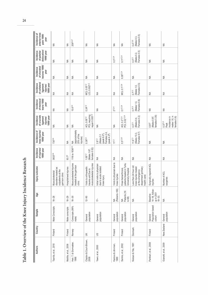
24Ta
ble
1. O
verv
iew
of t
he K
nee
Inju
ry In
cide
nce
Res
earc
h
Auth
ors
Coun
trySa
mpl
eAg
e In
jury
out
com
eIn
cide
nce
of in
jurie
s/
1000
/ yea
r
Inci
denc
e of
knee
inju
ries/
10
00/ y
ear
Inci
denc
e of
cru
ciat
e lig
amen
t in
jurie
s/
1000
/ yea
r
Inci
denc
e of
men
isca
l te
ars/
1000
/ ye
ar
Inci
denc
e of
col
late
ral
ligam
ent
inju
ries/
10
00/ y
ear
Inci
denc
e of
chon
dral
le
sion
s/ 10
00/
year
Inci
denc
e of
pat
ella
r di
sloc
atio
n/
1000
/ yea
r
Inci
denc
e of
ante
rior k
nee
pain
/ 100
0/ ye
ar
Taan
ila, e
t al.,
2010
Finla
ndM
ale C
onsc
ripts
18–2
8M
uscu
loske
letal
disor
ders
trea
ted a
s ou
tpat
ients
at th
e ga
rriso
n clin
ic
3833
3,4
7303,
4NA
NANA
NANA
NA
Mat
tila, e
t al.,
2006
Finla
ndM
ale co
nscr
ipts
18–2
9Ho
spita
lised
injur
ies93
.72
NANA
NANA
NANA
NA
Heir,
T. &
Glom
sake
r, 19
96No
rway
Cons
cript
s (99
%
male
)18
–30
Mus
culos
kelet
al inj
uries
tre
ated
at th
e gar
rison
cli
nic
1116
to 18
363,
4NA (a
ppro
ximat
ely
25%
of t
he
injur
ies)
NA15
.63,
4NA
NANA
2093,
4,7
Clay
ton &
Cou
rt-Br
own,
2008
UKGe
nera
l po
pulat
ion12
–90
Visit
s to o
rthop
aedic
tra
uma u
nit D
ue to
m
uscu
loske
letal
injur
ies
1.061,
2 (M
ales 1
.67,
fem
ales 0
.52)
0.38
1,2
ACL 0
.081,
2
PCL r
equir
ing
repa
ir 0.
0051,
2
0.24
1,2
MCL
0.0
51,2
LCL 0
.002
1,2
NANA
NA
Yawn
, et a
l., 20
00US
Gene
ral
popu
lation
17–
Initia
l visi
ts to
phy
sician
du
e to a
cute
isola
ted
knee
injur
y
NA2.
81,3
(Male
s 3.1
(pea
k 6.0
4 ),Fe
male
s 2.4
, (p
eak 4
.24 )
NANA
NANA
NANA
Kann
us &
Järv
inen,
1989
Finla
ndGe
nera
l po
pulat
ion
NA (Pat
ients
<90)
Visit
s to p
hysic
ian d
ue to
kn
ee in
juries
NA11
1,2
NA21,
2,4
NANA
0.31,
2,4
NA
Sarim
o, et
al., 2
002
Finla
ndGe
nera
l po
pulat
ionNA Pa
tient
s (16
to
68)
Knee
hem
arth
rosis
pa
tient
s tre
ated
in Tu
rku
Unive
rsity
Hos
pital
NA0.
51,2,
6AC
L 0.2
1,2,
4
PCL 0
.021,
2,4
0.11,
2,4
MCL
0.11,
2,4
0.08
1,2,
40.1
11,2,
4NA
Niels
en &
Yde
, 199
1De
nmar
kGe
nera
l po
pulat
ionNA
Less
than
24 h
our o
ld kn
ee in
juries
trea
ted
at th
e em
erge
ncy
depa
rtmen
t (pr
imar
y he
alth c
are)
NA11
.61,3
(Male
s 13.
3,
Fem
ales 9
.7)
0.31,3
(Male
s 0.4
, Fe
male
s 0.2
)
0.71,3
(Male
s 1.0
, Fe
male
s 0.6
)
0.71,3
(Male
s 1.0
, Fe
male
s 0.5
)
NA0.
31,3
(Male
s 0.2
, Fe
male
s 0.5
)
0.41,3
,5
(Male
s 0.3
, Fe
male
s 0.5
)
Park
kari,
et a
l., 20
08Fi
nland
Gene
ral
Popu
lation
bas
ed
coho
rt
Base
line
14–1
8, fo
llow-
up in
200
1 19
–33.
Surg
ically
repa
ired A
CL
injur
iesNA
NA0.
612
(Male
s 0.9
7, fe
male
s 0.3
0)
NANA
NANA
NA
Gian
otti,
et al.
, 200
9Ne
w Ze
aland
Gene
ral
popu
lation
NANu
mbe
r of A
CL
surg
eries
NANA
0.37
1,3
Peak
In
ciden
ce in
m
ales 1
.6, in
fe
male
s 0.8
)
NANA
NANA
NA

25
Silla
npää
, et a
l., 20
08Fi
nland
Male
cons
cript
s17
–30
Hosp
italis
ed p
rimar
y tra
umat
ic pa
tella
r dis
locat
ion
NANA
NANA
NANA
0.77
3NA
Atkin
, et a
l., 20
00US
Kaise
r hea
lth p
lan
popu
lation
NA (Pat
ients
10–5
9)
Prim
ary p
atell
ar
disloc
ation
s tre
ated
at
the k
nee i
njury
clini
c
NANA
NANA
NANA
0.07
1,3
(Pea
k inc
idenc
e in
male
s 0.3
0 an
d in f
emale
s 03
3)
NA
Witv
rouw
, et a
l., 20
00Be
lgium
Stud
ents
takin
g ph
ysica
l edu
catio
n cla
sses
17–2
1De
velop
men
t of a
nter
ior
knee
pain
dur
ing tw
o ye
ar st
udy p
eriod
NANA
NANA
NANA
NA42
.61,
2,4
(Male
s 36.
4, fe
male
s 49.
6)
Milg
rom
, et a
l., 19
91Isr
ael
Male
infa
ntry
re
cruit
sNA
Deve
lopm
ent o
f pa
tello
fem
oral
pain
durin
g 14-
week
s bas
ic tra
ining
NANA
NANA
NANA
NA57
3.01,
2,4
Miya
saka
, et a
l., 19
91US
Kaise
r hea
lth p
lan
popu
lation
NAKn
ee lig
amen
t injur
ies
treat
ed at
the k
nee i
njury
cli
nic
NA1.
241,
2,4,
8AC
L0.
381,
2,4,
9
PCL 0
.041,
2,4,
9
0.27
1,2,
4M
CL 0
.631,
2,4
LCL 0
.041,
2,4
NANA
NA
1 Cum
ulativ
e ann
ual in
ciden
ce2 P
erso
n-ba
sed i
ncide
nce
3 Eve
nt-b
ased
incid
ence
4 App
roxim
ation
/conv
ersio
n/ca
lculat
ion fr
om th
e dat
a pre
sent
ed5 P
atell
ofem
oral
over
use
6 Acu
te kn
ee h
aem
arth
rosis
7 Ove
ruse
knee
injur
ies in
cludin
g pat
ellof
emor
al pa
in sy
ndro
me,
iliotib
ial b
and f
rictio
n syn
drom
e and
pat
ellar
tend
initis
8 Inclu
ding o
nly lig
amen
t and
men
iscal
tear
s9 In
cludin
g only
ligam
ent in
juries
with
pat
holog
ic an
tero
poste
rior m
otion
NA=
Not A
vaila
ble

26
6.2.3 Risk Factors for Musculoskeletal Injuries in General
Risk factor research has focused mainly on injuries as a whole rather than knee injuries or their underlying diagnoses. Low aerobic fitness as measured by running capability is considered a risk factor for injuries in studies conducted in conscripts as well as in recruit armies (Jones, et al., 1993; Jones & Knapik, 1999; Taanila, et al., 2010). Low aerobic fitness also increases the risk for medical discharge from military service in Finnish conscripts (Taanila, et al., 2011). It must be taken into account that opposite results have also been reported. In a study considering Finnish conscripts, those with excellent aerobic fitness had an OR of 1.3 for inpatient care due to lower limb injury compared with those with poor results in running tests (Mattila, et al., 2007). In Norwegian conscripts, poor running fitness seemed to be a risk factor for musculoskeletal injuries in univariate analysis, but adjusted multivariate analysis showed it to be statistically nonsignificant (Heir, T. & Eide, 1996, 1997). In addition, in the study of Jones and coworkers (Jones, et al., 1993) dealing with male US army infantry trainees, statistical significance was obtained only in univariate analysis and running test results were not included in the multivariate analysis.
Co-impairments in muscular strength tests and Cooper’s 12-minute running tests are a risk factor for musculoskeletal disorders leading to a visit at the garrison clinic as well as for medical discharge from the military service in Finland (Taanila, et al., 2011; Taanila, et al., 2010). The overall results in muscular fitness index used by the Finnish defence forces were not associated with injuries in either of these studies, and another study concerning Finnish conscripts showed that good muscular fitness increases the injury hospitalisation risk (Mattila, et al., 2007).
The association of BMI with injuries as a whole remains controversial. In Norwegian conscripts, higher BMI increased the risk for musculoskeletal injuries treated at the military primary care in one study, but not in another (Heir, T. & Eide, 1996, 1997). Two studies reported that BMI is not associated with the injuries in US army (Jones, et al., 1993; Reynolds, et al., 2000). In Finnish conscripts, a higher BMI increases risk for musculoskeletal disorders leading to a visit at the garrison clinic and for injuries requiring inpatient care (Mattila, et al., 2007; Taanila, et al., 2010), but is not a risk factor for medical discharge from the military service (Taanila, et al., 2011). Height and weight independently are not associated with the injury risk (Heir, T. & Eide, 1996, 1997; Taanila, et al., 2011; Taanila, et al., 2010). Older age seems to increase the risk for musculoskeletal injuries in young adult Norwegian conscripts in univariate analysis, but multivariate analysis showed conflicting results (Heir, T. & Eide, 1996, 1997). Other multivariate analyses reported that older age was a significant risk factor in US army trainees, but a nonsignificant risk factor in Finnish conscripts (Jones, et al., 1993; Taanila, et al., 2010). Older age is also not a risk factor for medical discharge from the military service in Finland (Taanila, et al., 2011).

27
6.2.4 Risk Factors for Knee Injuries and Anterior Knee Pain
The role of demographic and anthropometric characteristics as well as physical fitness as a possible risk factor for knee injuries is not well established. An overview of the available risk factor data is presented in Table 2. There are currently no data for whether factors like height, weight, BMI, or physical fitness predisposes to knee injuries in general. The overall risk of physical activity-related knee injuries is highest in the 15 to 25 year-old age group (Haapasalo, et al., 2007). The risk of disabling knee injuries among young men in the US army increases until the age of 26 years (Sulsky, et al., 2000). In general practice populations in UK, obese subjects were nearly four times more likely to report knee pain with disability than those with a normal BMI (Webb, et al., 2004). Whether knee pain was caused by injury or osteoarthritis was not reported. Other known risk factors include sports participation, and as many as half of all knee injuries may occur during sports activities (Haapasalo, et al., 2007; Kannus & Järvinen, 1989). In Finland, females have a higher risk for knee injuries in commuting activities, whereas hazard ratios are not significantly different for lifestyle activities or recreational and competitive sports. In some specific activities, such as endurance, power, and contact sports, females have a higher injury risk than men (Haapasalo, et al., 2007).
Few investigators have examined the risk factors for specific knee diagnoses. The amount of data on intrinsic, modifiable risk factors such as BMI, weight, aerobic fitness, and muscular strength in general populations is very limited. For example, there is no single study of collateral ligament tears or chondral lesions.
Data is not available regarding whether poor aerobic fitness or older age predisposes to cruciate ligament injuries in Finland or internationally. The strength of the knee flexors and extensors is not associated with noncontact arthroscopically-confirmed ACL injuries in US Military Academy cadets (Uhorchak, et al., 2003). The authors also reported that height is a nonsignificant factor in both sexes. Higher weight and BMI increases ACL injury risk in females, but are nonsignificant in males (Uhorchak, et al., 2003). In Finnish adolescents and the young adult population, BMI is not associated with cruciate ligament injuries. The data for weight, height, and muscle strength, however, were not reported (Parkkari, et al., 2008). Participation in sports is a well-known risk factor for cruciate ligament injuries in young adults (Parkkari, et al., 2008). Even if the injury incidence rate for ACL injuries is higher for men than for women in the general population (Gianotti, et al., 2009; Parkkari, et al., 2008), the injury incidence rate and risk for cruciate ligament injuries is higher for female athletes (Mountcastle, et al., 2007; Parkkari, et al., 2008; Prodromos, et al., 2007). There are also other, nonmodifiable risk factors for ACL injuries, such as a small femoral notch width and generalised joint laxity (Uhorchak, et al., 2003). Another study, however, reported that femoral notch width did not predict ACL injuries (Lombardo, et al., 2005).
To our knowledge, there are no studies describing whether the risk of meniscal tears is associated with physical fitness. In the study of Baker and coworkers, higher weight

28
was shown to increase the risk when acute and degenerative meniscal tears were analysed together. Height was not associated with meniscal tears. A higher BMI increased the risk for degenerative tears, but not acute tears (Baker, et al., 2002). Degeneration occurs with aging and weakens the meniscal structures, making them susceptible to injuries (Negendank, et al., 1990). No direct evidence supports an increase in the injury risk as aging progresses. Weak quadriceps muscles, mechanical deviations, and congenital abnormalities are also suggested to increase injury rates(Miller III, 2003). The risk for meniscal tears is increased among sports participants, especially those playing soccer or rugby (Baker, et al., 2002; Baker, et al., 2003).
Young adult male patients in their early twenties with deep chondral lesions have a significantly higher mean body mass and BMI and are a little older than patients with superficial lesions (Eskelinen, et al., 2004). Height was not associated with chondral lesions in that study and physical fitness data was not available. A limitation of the study, however, was that all of the patients included had arthroscopically diagnosed chondral lesions, and thus the risk factors that predispose to chondral lesions in general are not known.
Patellar dislocations often occur during sports (Atkin, et al., 2000). Mean standing limb alignment, quadriceps angle, and rotational measurements are not associated with acute patellar dislocations. Fithian and coworkers conducted a prospective cohort study in an orthopaedic knee injury clinic to evaluate risk factors for primary and recurrent patellar dislocations. The risk factors for recurrent dislocations were female sex and older age. Their study also showed that the odds for subsequent instability are 7 times higher in patients with a previous history of patellofemoral instability (Fithian, et al., 2004). Only one study described the association of modifiable risk factors such as physical fitness and BMI with primary traumatic patellar dislocations in young adult males (Sillanpää, et al., 2008). In that study, male patients with primary traumatic patellar dislocation were typically taller and weighed more than controls (Sillanpää, et al., 2008). Factors of poor physical performance, older age, and higher BMI do not increase the risk. Most of the cases occurred during sports activities and the injury mechanism was knee flexion with a tibial valgus position in more than 90% of the injuries. Concomitant injuries were detected in almost half of the cases and haemarthrosis was present in all of the knees. Service class had to be lowered in 1 of every 5 patients, indicating a remarkable inability to cope with physically challenging duties (Sillanpää, et al., 2008).
Witvrouw and coworkers conducted a 2-year prospective study of the intrinsic risk factors associated with the development of AKP (Witvrouw, et al., 2000). Anthropometric characteristics such as weight and height, as well as cardiovascular endurance measured by an ergometer test, were not associated with the development of AKP. Of the various physical fitness tests, only a decreased explosive strength predisposed subjects to AKP. Other statistically significant risk factors were shortened quadriceps muscle, altered vastus medialis obliquus reflex time, and a hypermobile patella. Another prospective

29
study by Milgrom et al. reported that physical fitness and muscle strength measured by 2 km running and number of push-ups and sit-ups were not risk factors for AKP in a logistic regression analysis (Milgrom, et al., 1991). Increased isometric strength of the quadriceps muscle, medial tibial intercondylar distance, weight, and height were also not associated with the injuries. According to pooled analyses in a recent meta-analysis, BMI and older age are also not risk factors (Lankhorst, et al., 2012). Although PFM plays a major role in the genesis of AKP, there is evidence that patellofemoral alignment characteristics such as the Q-angle and genu varum/valgum and foot pronation are not associated with the development of AKP (Hetsroni, et al., 2006; Park & Stefanyshyn, 2011; Witvrouw, et al., 2000). A recent systematic review confirmed that most of the earlier suggested risk factors for AKP rely on very limited evidence (Lankhorst, et al., 2012).
As mentioned above, there is extensive research concerning musculoskeletal injuries as a whole and a limited amount of research concerning knee injuries. Most of the study populations do not represent the general population and the patient sample is restricted to a special subpopulation such as elite athletes and patient catchment of a specialised clinic or voluntary recruit armies (Jones, et al., 1993; Jones & Knapik, 1999; Majewski, et al., 2006; Milgrom, et al., 1991; Sulsky, et al., 2000; Uhorchak, et al., 2003; Witvrouw, et al., 2000). The incidence and risk factor data available are therefore difficult to generalise to the general population in clinical practice.
Another limitation of the data available is that most of the studies published only took into account knee or other musculoskeletal injuries as a whole or alternatively only according to a solitary specific knee diagnosis, such as cruciate ligament tears or AKP (Baker, et al., 2002; Heir, T. & Eide, 1996, 1997; Milgrom, et al., 1991; Parkkari, et al., 2008). This limits the ability to evaluate whether the suggested risk factors are valid for all knee injuries or only for a certain knee diagnosis. There are no comprehensive studies taking knee injuries into account as a whole or according to specific knee diagnoses. Knowledge about the specific risk factors for different injuries will allow for better analysis of possible causal relationships and facilitate the development of possible preventive measures for the appropriate targets.

30Ta
ble
2. O
verv
iew
of t
he ri
sk fa
ctor
rese
arch
Auth
ors
Coun
trySa
mpl
eAg
e In
jury
out
com
eHi
gher
hei
ght
High
er w
eigh
tHi
gher
BM
IOl
der a
gePo
or ru
nnin
g fit
ness
Poor
mus
cle
stre
ngth
Heir,
T. &
Eide
, 199
6No
rway
Cons
cript
s18
-29
Mus
culos
kelet
al inj
uries
trea
ted
at th
e milit
ary t
raini
ng ca
mps
NS1,
2NS
1,2
+1,2
+1 NS2
+1 NS2
NA
Heir,
T. &
Eide
, 199
7No
rway
Cons
cript
s19
- 28
Mus
culos
kelet
al inj
uries
trea
ted
at th
e milit
ary t
raini
ng ca
mps
NS1,
2NS
1,2
NS1,
2+1,
2+1 NS
2NA
Mat
tila, e
t al.,
2007
Finla
ndCo
nscr
ipts
18-3
0In
jury h
ospit
alisa
tions
Mult
iple i
njury
hos
pitali
satio
nsLo
wer l
imb i
njury
hos
pitali
satio
ns
NA NA NA
NA NA NA
+2 +2 +2
NA NA NA
NA NA -2
-2 NS2
NA
Jone
s, et
al., 1
993
USM
ale a
rmy i
nfan
try
traine
es17
-35
Injur
ies re
quiri
ng si
ck ca
ll visi
t du
ring 1
2 wee
ks o
f bas
ic tra
ining
NANA
NS1,
2+1,
2+1 NA
2+1,3 NA
2
Taan
ila, e
t al.,
2010
Finla
ndM
ale C
onsc
ripts
18-2
8M
uscu
loske
letal
disor
ders
tre
ated
at th
e gar
rison
clini
c as
outp
atien
ts
NS2
NA+2
NS2
+2NS
2
+2,4
Taan
ila, e
t al.,
2011
Finla
ndM
ale co
nscr
ipts
18-2
8M
edica
l disc
harg
e fro
m th
e m
ilitar
y ser
vice
NS2
NANS
2NS
2
+2,5
+2NS
2 +2,
4
Uhor
chak
, et a
l., 20
03US
US M
ilitar
y Aca
demy
ca
dets
17-2
36No
n-co
ntac
t arth
rosc
opica
lly
conf
irmed
ACL
injur
iesNS
1,2
NS1,
2 (M
ales)
+1,2 (
Fem
ales)
NS1,
2 (M
ales)
+1,2 (
Fem
ales)
NANA
NS1,
2,7
Park
kari,
et a
l., 20
08Fi
nland
Gene
ral P
opula
tion
base
d coh
ort
Base
line 1
4-18
, fo
llow-
up in
200
1 19
-33.
Hosp
italis
ed cr
uciat
e liga
men
t inj
uries
NANA
NS2
NANA
NA
Bake
r, et
al., 2
002
UKGe
nera
l Pop
ulatio
n20
-59
arth
rosc
opica
lly p
rove
n prim
ary
acut
e men
iscal
tear
sDe
gene
rativ
e men
iscal
tear
s
NS2,
8
NS2,
8+2,
8
+2,8
NS2
+2NA
NANA
Eske
linen
, et a
l., 20
04Fi
nland
Male
Con
scrip
ts wi
th
arth
rosc
opica
lly p
rove
n ch
ondr
al les
ion
NAAr
thro
scop
ically
pro
ven c
hond
ral
lesion
sNS
1+1,9
+1,9+1,9
NANA
Silla
npää
, et a
l., 20
08Fi
nland
Male
cons
cript
s17
-30
Hosp
italis
ed p
rimar
y tra
umat
ic pa
tella
r disl
ocat
ions
+1+1
NS1
NS1
NS1
NS1
Milg
rom
, et a
l., 19
91Isr
ael
Male
infa
ntry
recr
uits
NADe
velop
men
t of p
atell
ofem
oral
pain
durin
g 14-
week
s bas
ic tra
ining
NS1,
2NS
1,2
NANA
NS1,
2-1,
2,10
NS1,1
1
+1,3
Witv
rouw
, et a
l., 20
00Be
lgium
Stud
ents
takin
g ph
ysica
l edu
catio
n cla
sses
17-2
1De
velop
men
t of a
nter
ior kn
ee
pain
durin
g two
year
stud
y per
iodNS
1,2
NS1,
2NA
NANA
12NS
1,2
+1,2,1
3

31
Lank
hors
t, et
al., 2
011
Neth
erlan
ds/
mult
inatio
nal
7 pro
spec
tive s
tudie
s ch
osen
from
the 3
845
pote
ntial
ly re
levan
t ar
ticles
NARe
view
of p
rosp
ectiv
e risk
facto
r stu
dies c
once
rning
pat
ellof
emor
al pa
in sy
ndro
me
NS14
NS14
NS14
NS14
NA+3,1
0
Sulsk
y, et
al., 2
000
USAc
tive-
duty
enlis
ted
soldi
ers
17-5
4Di
scha
rge f
rom
army
for k
nee-
relat
ed d
isabil
ityNA
NANA
+2,15
NANA
Haap
asalo
, et a
l., 20
06Fi
nland
Popu
lation
coho
rt15
-74Kn
ee in
juries
repo
rted b
y stu
dy
subje
ctsNA
NANA
+16 -17NA
NA
+ =
Incr
ease
s the
risk
- = D
ecre
ases
the r
iskNA
= N
ot av
ailab
leNS
= N
ot st
atist
ically
sign
ifican
t1 U
nivar
iate a
nalys
is2 M
ultiva
riate
ana
lysis
3 Pus
h-up
test
4 Co-
impa
irmen
ts in
Coop
er’s
runn
ing te
st an
d mus
cular
fitne
ss te
sts5 O
nly fo
r disc
harg
es d
ue to
men
tal h
ealth
pro
blem
s6 A
t the
time o
f enr
olmen
t, fo
llow-
up tim
e 4 ye
ars
7 Stre
ngth
of k
nee f
lexor
s and
exte
nsor
s8 B
oth a
cute
and
deg
ener
ative
tear
s wer
e ana
lysed
toge
ther
9 inc
reas
es ri
sk fo
r dee
p cho
ndra
l lesio
ns10
Knee
exte
nsion
stre
ngth
11Nu
mbe
r of s
it-up
s12
Card
iores
pirat
ory f
itnes
s eva
luate
d by s
ubm
axim
al er
gom
eter
test
was N
S13
Explo
sive s
treng
th m
easu
red b
y ver
tical
jump t
est
14Po
oled a
nalys
es15
Odds
ratio
incr
ease
d in w
hite E
urop
ean m
en u
ntil 2
6 yea
rs o
f age
and
then
dec
reas
ed16
In lif
esty
le ac
tivitie
s11
In co
mm
uting
acti
vities
and
in re
crea
tiona
l and
com
petit
ive sp
orts.
Ove
rall k
nee i
njury
risk
was
high
est in
the a
ge g
roup
15 to
25 y
ears
.

32
6.3 Diagnostics of Meniscal Tears, Chondral Lesions and Anterior Knee pain
6.3.1 History and Physical Examination
Medical imaging technology is continuously advancing, but the basis of knee injury diagnostics remains history taking and physical examination by which the majority of knee injuries can be diagnosed prior to any imaging or surgical procedures (Orndorff, et al., 2005). Useful factors in the differential diagnosis of knee pain are patient’s age, activity level, and whether the pain is of traumatic origin. In older patients, there is an increased prevalence of osteoarthritis (Felson, 1988) and malignancies and infections are possibilities. In younger patients, knee pain often has a traumatic origin (Gianotti, et al., 2009; Kannus & Järvinen, 1989; Majewski, et al., 2006). In cases of high energy trauma with deformity or significant pain and swelling of the knee joint, the possibility of fractures must be taken into account (Orndorff, et al., 2005). Fractures are fairly rare in young adults (Kannus & Järvinen, 1989), however, and usually their knee pain of traumatic origin is caused by ligamentous, meniscal, or chondral lesions of the knee (Majewski, et al., 2006).
A typical injury mechanism for meniscal tear is twisting or hyperflexion of the knee. Meniscal injuries usually occur between the ages of 21 to 30 years in men and are frequently associated with an ACL injury (Munjal & Krackow, 2005). The most commonly used clinical tests for evaluating meniscal injury are McMurray’s, Apley’s, and Thessaly’s tests, and joint line tenderness (Bhandari, 2011). A high quality meta-analysis published in 2001 reported that mean sensitivity of joint line tenderness for meniscal tears is 79% and specificity 15% (Solomon, et al., 2001). For McMurray’s test, mean sensitivity is 53% and specificity is 59%. The sensitivity of the composite examination of different tests is 77% and the specificity 91%. A new clinical test for diagnosing meniscal tears named after the Thessaly region in Greece was published in 2005 (Karachalios, et al., 2005). The Thessaly test at 20° knee flexion is considered the most effective first-line screening test for meniscal tears. The sensitivity for medial meniscal tears is as high as 89%, for lateral meniscal tears 92%, and for combined injuries of the ACL and meniscus 80% (Karachalios, et al., 2005). Corresponding figures for other tests in the same study were lower, 66%, 81%, and 65%, respectively, for the Thessaly test at 5° knee flexion; 71%, 78%, and 65% for joint-line tenderness; 41%, 41%, and 20% for Apley’s test; and 48%, 65%, and 45% for the McMurray test. The Thessaly test at 20° knee flexion also has superior specificity: 97% for medial meniscus, 96% for lateral meniscus, and 91% for combined injuries of the ACL and meniscus.
Articular cartilage injuries are commonly diagnosed together with other knee injuries. The most commonly associated injuries in young adults are ACL injuries and

33
meniscal tears (Curl, et al., 1997). The mechanism of injury is not clear, but a twisting force and direct blow to the knee are suggested (Rubin, et al., 2000). According to the tissue homeostasis theory of Dye, increased loading of the knee joint can also lead to a loss of tissue homeostasis and even macrostructural failure, such as chondral lesions (Dye, 2005). Articular cartilage has a very limited ability to heal spontaneously and chondral lesions likely lead to early osteoarthritis (Bhandari, 2011; Messner & Maletius, 1996). When symptomatic, chondral lesions may cause prolonged or activity-related pain, effusion, and sometimes catching of the knee joint (Gomoll, et al., 2010; Messner & Maletius, 1996; Sellards, et al., 2002). At their worst, focal chondral lesions may impair quality of life as much as severe osteoarthritis (Heir, S., et al., 2010). Patients with more severe cartilage lesions of the patellofemoral joint are reported to have more subjective symptoms and functional limitations than those without lesions or with small cartilage lesions (Kettunen, et al., 2005). In contrast, chondral lesions are asymptomatic even on direct probing, which is logical as articular cartilage has no innervation (Dye, et al., 1998). An explanation for the contradictory results might be that more extensive pressure on the weight-bearing knee during daily activities irritates the richly innervated subchondral bone and causes pain. Patients with chondral lesions do not have any specific findings in clinical examination (Gomoll, et al., 2010). Retropatellar crepitus, effusion, and quadriceps wasting are sometimes associated with chondral lesions, but none of these signs is pathognomonic (Leslie & Bentley, 1978). Pain in the patellar compression (grind) test may be indicative of patellofemoral chondral lesions (Houghton, 2007). The diagnostic validity of this test, as well as the commonly used patellar apprehension, patellar inhibition, and sustained knee flexion tests, for the diagnosis of patellar chondral lesions is poor. Pain provoked by patellar compression against the femoral groove has the best sensitivity (56%), but the poorest specificity (55%), of these tests (Niskanen, et al., 2001). In the sustained knee flexion test, intraosseous pressure on the patella is significantly higher in painful knees, which may be the cause of the pain originating from the patella (Hejgaard & Arnoldi, 1984). For arthroscopically detected patellar chondral lesions, the sensitivity of this test is only 35%. The specificity, 85%, however, is acceptable (Niskanen, et al., 2001). Altogether, the clinical diagnosis of chondral lesions is very difficult. The diagnostic validity of the history, symptoms, and clinical findings is poor or not known, and chondral lesions are often associated and easily confused with other injuries such as meniscal tears (Rubin, et al., 2000).
The multifactorial and uncertain aetiology of AKP makes diagnostics both challenging and imperative. The bases for the diagnosis are physical examination and symptoms. Typical clinical symptoms of AKP syndrome are dull pain in the anterior aspect of the knee that worsens when climbing stairs, sitting with the knees flexed, and rising from a sitting position. Extending the knee against resistance may also cause pain, and there may also be catching or locking and giving-way sensations. Sometimes the pain can be sharp and sudden in nature. The exact pain location may vary and can

34
be difficult to localize (Gambardella, 1999; Houghton, 2007; Sanchis-Alfonso, Vicente, 2011). Insecurity and giving-way sensations are usually related to walking or other straight-ahead activities, rather than twisting or torquing activities as in ligamentous instability or meniscal tears (Gambardella, 1999). Locking or catching sensations are also usually milder than with meniscal tears and patients can actively unlock the knee (Sanchis-Alfonso, Vicente, 2011).
Differential diagnostics are extensive. Menisci and cruciate ligaments must be kept in mind, especially for AKP of traumatic origin and swelling of the knee joint. Almost all patients with AKP have pain on the lateral retinaculum, which may be due to repeated strain causing chronic irritation of the area (Fulkerson, 1983). Also, iliotibial band syndrome (runner’s knee) causes pain to the lateral aspect of the knee. Ober’s test (Sanchis-Alfonso, Vicente, 2011) can be used to evaluate the tightness of the iliotibial band. Iliotibial band syndrome may also be associated with lateral patellar displacement and tilt (Sanchis-Alfonso, Vicente, 2011). Medial collateral bursitis must be taken into account in patients who have pain on the medial joint line, especially if the pain is worse during valgus stress of the knee. Other commonly tender sites include the patella itself and the patellar tendon. Remarkable palpation tenderness in the inferior pole of the patella might indicate patellar tendinopathy, especially if exacerbated by activity or resisted leg extension (Kodali, et al., 2011; Wilson & Best, 2005). A decline squat test places increased stress on the patellar tendon and pain during this test suggests patellar tendinopathy (Wilson & Best, 2005). Behind the patellar tendon is Hoffa’s fat pad(Hoffa, 1904). Pain or apprehension provoked by a Hoffa manoeuvre or knee extension reveals whether this highly innervated structure is irritated (Ellen, et al., 1999). The Hoffa manoeuvre is performed by applying compression to the fat pad on the sides of the patellar tendon while simultaneously extending the knee (Kodali, et al., 2011). In synovial impingement syndromes, pain is typically aggravated by knee flexion and relieved by extension (Kodali, et al., 2011). Positive active extension as a kick test and the flexion test may establish a painful medial plica (Irha & Vrdoljak, 2003).
Clinical evaluation of PFM and chondral lesions is considered important although their roles in the genesis of AKP is now questioned. In cases in which no possible pain source other than patellofemoral chondral lesions are found, PFM and chondral lesions must be taken into account when planning treatment (Gomoll, et al., 2010). Clinical diagnosis of patellofemoral chondral lesions is described in the above chapter on chondral lesions in all knee compartments. Patellofemoral malalignment and instability must be taken into consideration especially in nontraumatic AKP with bilateral symptoms (Houghton, 2007). Determining whether the main complaint is pain or instability possibly related to patellar subluxation is important (Sanchis-Alfonso, Vicente, 2011). Patellofemoral malalignment and patellar instability can be evaluated by observing whether genu varum/valgum, an abnormal Q-angle, external torsion of the tibia, or femoral anteversion is present. Abnormal patellar or tibial tuberosity positions can also

35
be observed (Kodali, et al., 2011; Llopis & Padron, 2007). A patellar tilt test is a special test used to evaluate patellar malalignment and lateral retinaculum tightness. The patient has lateral patellar tilt if the lateral border of the patella is posterior to the medial border. This indicates lateral retinaculum tightness and leads to abnormally high contact forces in the patellofemoral joint. The J-sign is used to describe patellar maltracking during knee motions. It is considered pathonomic for lateral patellar subluxation (Boden, et al., 1997). Patellar mobility and possible instability can be tested with a patellar glide test. Subjective feelings of dislocation during a patellar apprehension test indicate patellar instability (Kodali, et al., 2011). The sensitivities for these tests in detecting AKP are generally poor, whereas specificities are somewhat better (Haim, et al., 2006). Poor diagnostic validity is logical as PFM and patellofemoral instability itself are considered to be very nonspecific findings in the genesis of AKP (Sanchis-Alfonso, Vicente, 2011).
Physical examination and differential diagnosis of AKP are difficult. There are various suggested aetiologies and clinical tests. The validity of these tests is generally poor or not known. No single test exists to determine the exact diagnosis, and comprehensive evaluation of AKP requires a combination of thorough history taking, multiple tests, and radiologic imaging (Fredericson & Yoon, 2006). When no accurate diagnosis can be established, the patient is often diagnosed with idiopathic AKP (Kodali, et al., 2011).
6.3.2 Imaging of Knee Injuries and Anterior Knee Pain
6.3.2.1 PlainRadiographsandComputedTomographyDiagnostic imaging of acute knee injuries and AKP is routinely performed when diagnosis remains uncertain after a clinician’s physical examination and when imaging might influence the treatment decisions. Knee radiographs in young adults are used mainly to confirm or exclude fracture of the injured knee. Fractures are far less common than ligamentous, meniscal, or chondral lesions (Kannus & Järvinen, 1989; Majewski, et al., 2006). When determining whether knee radiographs should be performed to exclude fractures in acute knee injuries, the Ottawa knee rule can be used (Table 3). The Ottawa knee rule is very sensitive but not specific for predicting fractures (Jackson, et al., 2003; Stiell, et al., 1995). Radiographs can also reveal osteochondritis dissecans or joint-space narrowing related to deep large chondral lesions or severe osteoarthritis. It must be noted, however, that radiographs are useful only in the advanced stages of disease and cannot reveal superficial defects (Chan, et al., 1991; Wright, et al., 2006). Routine radiograph views used to evaluate knee injuries are standing weight-bearing anteroposterior and lateral views. The posteroanterior semiflexed view may be helpful for evaluating the posterior margin of the joint space. In addition, tunnel views of the intercondylar notch and axial (skyline/sunrise) views of the patella may be indicated (Hollingworth, et al., 2011; Potter & Foo, 2006). Skyline views at 30° knee flexion are

36
routine for AKP and patellar instability imaging. The main purpose of these views is to evaluate possible patellar malalignment. The main radiographic malalignment patterns evaluated are patellar subluxation with and without tilt, and tilt without subluxation. Other malalignments that are commonly evaluated radiographically include increased Q-angle and trochlear-tubercle distance (Christian, et al., 2006; Schutzer, et al., 1986). Altogether, physical examination is more useful than radiographs for evaluating AKP. Only patellar subluxation is increased in patients with AKP as compared with healthy controls (Haim, et al., 2006). Even if the role of PFM in the genesis of AKP is questioned, addressing it is important especially when planning cartilage repair techniques. In this setting, the mechanical axis of the knee and possible varus or valgus malalignment should also be evaluated from full-length anteroposterior hip-to-ankle radiographs. Undiagnosed and uncorrected malalignments may account for premature failure of cartilage repair (Gomoll, et al., 2010; Potter & Foo, 2006).
In complicated or prolonged cases of AKP, computed tomography (CT) can be used alternatively to MRI for more subtle evaluation of osseous structures and possible malalignment than routine radiographs. Kinetic CT can be used to evaluate patellar malalignment in loaded and more physiological conditions than conventional techniques. This may reveal PFM or maltracking in some cases with normal findings in conventional CT images (Christian, et al., 2006). CT can also be used to detect occult or intra-articular fractures of the injured knee (Onyema, et al., 2011).
Table 3. Ottawa knee rule criteria for radiography of acute knee injury (Stiell, et al., 1995)
Age 55 or older; orTenderness at head of fibula; or Isolated tenderness of the patella; orInability to flex 90°; orInability to bear weight both immediately and in the emergency department for four steps
6.3.2.2 MagneticResonanceImagingWhen clinical diagnosis and proper treatment are uncertain, the value of MRI is emphasized and is currently commonly used to evaluate various knee joint injuries. Magnetic resonance imaging is especially useful in multiple knee lesions for which the diagnostic accuracy of physical examination decreases significantly and may be as low as 30% (Oberlander 1993). Magnetic resonance imaging can reduce the need for arthroscopy and the number of negative arthroscopic examinations in patients for whom arthroscopy is considered after physical examination (Hollingworth, et al., 2011; Munshi, et al., 2000; Spiers, et al., 1993; Vincken, et al., 2002). In patients with non-acute (lasting over 4 weeks) knee symptoms and clinically highly suspected intra-articular knee pathology, MRI
37
is also a cost-effective method for obviating knee arthroscopy (Vincken, et al., 2007). For acute knee injuries, it is not clear if MRI is cost effective and this requires further research (Hollingworth, et al., 2011).
Magnetic resonance imaging examination of the knee can be performed with various methods and only general guidelines for the routine protocol selection can be given. Overall MRI examination of the knee routinely includes axial, coronal, and sagittal planes and multiple sequences that are selected according to suspected internal derangements. Coronal plane images can be used primarily to evaluate the collateral ligaments. Other structures that can be evaluated in this plane include the cruciate ligaments, the menisci, articular cartilage, osseous structures, and the iliotibial tract. Sagittal images are used to evaluate the menisci, the cruciate ligaments, and the articular cartilage surfaces of the medial and lateral compartments and trochlear groove. Sagittal plane images are also used to evaluate the articular cartilage of the patellar facets, structures of the posteromedial and lateral corner, the patellar and quadriceps tendons, subchondral bone and bone marrow, joint fluid and possible effusion, Hoffa’s fat pad, plicae, tibiofibular joint, collateral ligaments, and lateral tendons. Axial plane images are used to evaluate the articular cartilage surfaces of the patellofemoral joint, especially the patellar facets. The other structures examined in this plane include the intercondylar notch, the menisci, the collateral ligaments, proximal attachment of ACL, patellar tendon, and the amount of joint fluid (Stoller, 2007).
A routine knee imaging protocol should include fat suppressed proton density fast spin echo or some other form of T2 weighting in each of the three planes. This allows for sensible evaluation of the articular cartilage, bone marrow, pathological fluid, ligament injuries, and meniscal morphology. Visualisation of meniscal degeneration or tears can be further improved by choosing fat suppressed proton density conventional spin echo or T2* gradient echo sequences in the sagittal plane. The T1-weighted images should be obtained at least in one plane as they allow for the evaluation of marrow fat signal changes in sclerosis or oedema that may be associated with trauma, infection, or neoplasias. (Stoller, 2007). Due to the short repetition times, T1 images can be obtained quickly, which allows for the production of 3D volumetric datasets. Fat-suppressed T1-weighted 3D SPGR is a valuable tool for evaluating articular cartilage surfaces (Disler, et al., 1996; Yoshioka, et al., 2004).
The diagnostic validity of MRI is good for ligament and meniscal injuries of the knee (Crawford, et al., 2007; Fischer, et al., 1991; Mackenzie, et al., 1996; Oei, et al., 2003; Rappeport, et al., 1996). The results of several meta-analysis and multicentre studies revealed that the mean sensitivity of MRI ranges from 91% to 93% for medial meniscal tears and is much lower, 69% to 79%, for lateral meniscal tears (Crawford, et al., 2007; Fischer, et al., 1991; Mackenzie, et al., 1996; Oei, et al., 2003). Mean specificity of MRI ranges from 81% to 88% for medial meniscal tears and 93% to 96% for lateral meniscal tears. For ACL tears, sensitivity ranges from 87% to 94% and specificity from 94%

38
to 95%. For PCL injuries, the sensitivity ranges from 91% to 94% and the specificity is approximately 99%. It must be noted, however, that the results vary widely among different studies. Published sensitivity rates are at lowest 19% for medial meniscal tears, 10% for lateral meniscal tears, 39% for ACL tears, and 33% for PCL tears (Krampla, et al., 2009; Lundberg, et al., 1996). The lowest specificities are reported in the older studies, 29%, 27% (Adalberth, et al., 1997), 82% (Glashow, et al., 1989), and 96% (Niitsu, et al., 1991).
Lower than average diagnostic validity is reported when only acute knee injuries are included (Lundberg, et al., 1996). Whether this is associated with haemarthrosis or other characteristics of acutely injured knees of the study or due to the MRI methods, sample selection, or other methodological variables as compared to other studies is not clear. Higher MRI field strength improves diagnostic performance only modestly, and significant effects can be seen only for injuries of the ACL (Oei, et al., 2003). This result is further supported by new results using 3.0T MRI. Sampson and coworkers reported that the sensitivity and specificity, respectively, of 3.0T MRI is average for medial meniscal tears (91%, 93%) and lateral meniscal tears (77%, 93%), and as high as 100% (100%) for ACL tears (Sampson, et al., 2008). Very good diagnostic validity can be also achieved with low field systems. For example, Riel et al (Riel, et al., 1999) used a 0.2T scanner and reported sensitivity, specificity, and accuracy rates of 93%, 97%, and 95% for medial meniscal tears, and 82%, 96%, and 93% for lateral meniscal tears. The diagnostic validity of the 0.2T MRI scanner was similar or even slightly better than the 3T MRI scanner used by Sampson. Grossman and coworkers compared the diagnostic validity of 1.5T MRI to 3.0T MRI in the diagnosis of meniscal tears. In a series of 100 consecutive patients who underwent 1.5T MRI, sensitivity was 93% for medial meniscal tears and 68% for lateral meniscal tears. In a comparative sample of 100 consecutive patients who underwent 3.0T MRI, the sensitivities were 93% and 69%, respectively. The specificities for medial meniscal tears were 82% for 1.5T MRI and 76% for 3.0T MRI. For the lateral meniscal tears, specificities were 95% and 92%, respectively. None of these small differences between field strengths, however, was statistically significant (Grossman, et al., 2009). Another recently published study comparing the 1.0T, 1.5T, and 3.0T field strengths reported no differences in the diagnostic validity between the field strengths for the detection of meniscal tears or ACL injuries (Krampla, et al., 2009).
Imaging cartilage with MRI is challenging. Depending on the location and depth of the lesion, MRI sequences, or other methodological variations, sensitivity has ranged from 0% to 100%, and specificity from 50% to 100% (Bredella, et al., 1999; Disler, et al., 1996; Friemert, et al., 2004; Handelberg, et al., 1990; Irie, et al., 2000; Munk, et al., 1998; Potter, et al., 1998; Recht, et al., 1993; Riel, et al., 1999; Spiers, et al., 1993; Vallotton, et al., 1995; Yoshioka, et al., 2004). Fat-suppressed T1-weighted 3D SPGR is one of the most valid and currently generally available sequences for evaluating articular cartilage lesions related to trauma and/or osteoarthritis of the knee, with a sensitivity ranging

39
from 75% to 97% and specificity ranging from 85% to 97% (Disler, et al., 1996; Yoshioka, et al., 2004). Fat-suppressed fast spin echo sequences can also show good results with sensitivity as high as 100% and specificity of 68% for articular cartilage lesions related to knee osteoarthritis (Yoshioka, et al., 2004). The advantage of fast spin echo techniques is that they can used to evaluate meniscal and ligamentous tears, whereas SPGR techniques are limited to cartilage imaging (Potter, et al., 1998).
Even with this increased knowledge of cartilage-sensitive sequences, the clinical use of MRI for overall knee evaluation still has poor sensitivity, especially for superficial chondral lesions. For 1.5T MRI, the sensitivity was 13% for grade 1 lesions, 23% for grade II, 64% for grade III, and 73% for grade IV lesions (Figueroa, et al., 2007). The corresponding sensitivities for 3.0T MRI were 29%, 62%, 63%, and 74%, respectively (von Engelhardt, et al., 2007). In the first study, chondral lesions were classified according to depth as recommended by the International Cartilage Repair Society (ICRS) (Brittberg & Winalski, 2003). The ICRS classification is presented in detail in Table 4. In the latter study, the lesions were classified according to the Bachmann classification (Bachmann, et al., 1997), which is basically similar to the ICRS classification in grading the depth of the lesions. Detection of deep lesions is most important because they may become symptomatic and require treatment (Curl, et al., 1997; Kettunen, et al., 2005). Preoperative diagnosis of these lesions could allow for appropriate preparations for the potentially forthcoming cartilage repair procedure. Basic repair methods can be performed in most facilities, but more advanced techniques are centralised to few hospitals in Finland (Vasara, et al., 2006).
Higher magnetic field strengths may be beneficial for imaging cartilage, but only limited evidence supports this assumption. Experimental evaluation of artificial articular cartilage injuries in pig knees indicates that diagnostic performance of a conventional high-field 1.0T MRI system is superior to a low-field 0.18T system for detecting deeper chondral lesions (Woertler, et al., 2000). In another study comparing image quality of 1.5T MRI to 3.0T MRI, the 3.0T MRI had a better signal-to-noise ratio and subjectively better delineation of the cartilage. A severe limitation of the study was that it compared only image quality and not diagnostic validity (Schoth, et al., 2008). In the aforementioned study of Krampla and coworkers, the diagnostic validity of 1.0T, 1.5T, and 3.0T field strengths was also compared for chondral lesions, and no differences in sensitivities or specificities were detected (Krampla, et al., 2009). Wong and coworkers reported in a small, retrospective series of 19 patients that the sensitivity of 3.0T MRI was slightly higher than that of 1.5T MRI (76% versus 71%) in diagnosing knee chondral lesions (Wong, et al., 2009). The specificity was 95% for both field strengths. In a recent study using 1.5T and 3.0T MRI with fast spin echo sequences in a larger sample of 200 symptomatic patients, no differences were detected between sensitivities (71% for 3.0T and 69% for 1.5T MRI), but the specificity was slightly higher for the 3.0T system (85.9% versus 78%) (Kijowski, et al., 2009). The sensitivities for the different grades of chondral

40
lesions were not statistically different. A 1.5T MRI had a sensitivity of 41% for cartilage softening, 50% to 82% for partial thickness lesions, and 95% for full thickness lesions. The corresponding sensitivities for 3.0T MRI were 42%, and 49% to 85%, 98%. There is no evidence from clinical studies to support a superior diagnostic validity of 3.0T MRI compared with lower field systems. The undisputed advantage of 3.0-T systems is that the imaging procedure is faster.
Bone bruises represent trabecular bone marrow oedema and are frequently seen in MRIs of an injured knee, especially in association with osteochondral lesions, patellar dislocations, and ACL injuries (Bretlau, et al., 2002; Paakkala, et al., 2010; Yoon, et al., 2011). It is generally accepted that bone bruises in young adults are of traumatic origin and caused by compression forces to the bone. They usually resolve within 1 year from the injury (Bretlau, et al., 2002). More than 80% of ACL injuries have a concomitant bone bruise, most commonly in the lateral aspect of the tibial plateau or lateral femoral condyle (Yoon, et al., 2011). In primary traumatic patellar dislocations, practically all patients have bone bruises in the lateral femoral condyle and in the patella (Paakkala, et al., 2010). After a knee trauma that has led to bone bruising, the overlying cartilage may be seemingly uninjured. Nevertheless, a bone bruise may indicate that the overlying cartilage is substantially damaged (Johnson, et al., 1998). Of the potentially treatable, traumatic chondral lesions of the knee visible on routine MRI, almost all have associated focal subchondral oedema. It was suggested that a bone bruise might be an important diagnostic clue for detecting traumatic chondral lesions (Rubin, et al., 2000). A limitation of this study was that the presence of bone bruises was not recorded if there was no visible cartilage defect in MRI. In the study by Bretlau and coworkers, however, all bone bruises and associated injuries detected by MRI in acutely injured knees were reported. Four out of five patients with a bone bruise also had an osteochondral lesion and 100% of the patients with osteochondral lesions had a bone bruise (Bretlau, et al., 2002). Magnetic resonance imaging was used as the gold standard for detecting injuries, which is a limitation of the study, especially for chondral lesions for which MRI is generally not sensitive (Figueroa, et al., 2007; von Engelhardt, et al., 2007). The presence of a bone bruise also correlates strongly with femorotibial osteoarthritis (Oda, et al., 2008).
Magnetic resonance imaging studies have also been conducted with a contrast agent, a technique known as gadolinium-enhanced MRI or MR arthrography, which can be performed as direct MR arthrography (intra-articular injection of Gd-DTPA) or indirect MR arthrography (intravenous injection of Gd-DTPA). Although the sensitivity of MRI for chondral lesions may be increased by the use of contrast-agents (Gagliardi, et al., 1994; Kramer, et al., 1994), both of these MR arthrography methods are seldomly used in routine clinical practice. These methods make MRI more invasive and increase the risk for iatrogenic complications as well as the expense of the imaging process (Potter & Foo, 2006).

41
Magnetic resonance imaging can be used as an alternative to CT to diagnose AKP and patellar instability. As with CT imaging, MRI allows for comprehensive evaluation of patellar malalignments and kinetic MRI methods are also available. An advantage of MRI over CT is that cartilage injuries, bone bruises, and especially soft tissue structures like patellar tendinopathy, Hoffa’s fat pad, and bursae can be evaluated. Other imaging methods for AKP include nuclear scintigraphy, which is useful in limited cases like in occult fractures and malignancies. Ultrasound is useful for diagnosing patellar tendinopathy (Christian, et al., 2006; Peers & Lysens, 2005).
Table 4. ICRS classification of articular cartilage injuries (Brittberg & Winalski, 2003)
Grade Description0 Normal1 Superficial fibrillation, softening, fissures or lacerations2 Defects less than 50% of the cartilage depth3 Defects more than 50% but not to subchondral bone4 Defects to the subchondral bone
6.4 General Treatment Principles of Meniscal Tears, Chondral Lesions and Anterior Knee Pain
6.4.1 Conservative Management
A detailed description of the management of knee injuries and AKP is beyond the scope of this dissertation and only some general principles are presented here. The basic aims for the treatment of knee disorders are to manage pain and other symptoms, maintain normal knee function, and improve the long-term prognosis of a disorder. A 1-month trial of conservative management is considered acceptable for minimally symptomatic small meniscal tears, as some of them may become asymptomatic. In general, however, meniscal tears require arthroscopic operation (Scotney, 2010). Anterior knee pain is initially treated conservatively. According to the most recent Cochrane review updated in 2009, physiotherapy can be considered to treat AKP but the evidence of its effectiveness is limited. Open and closed kinetic chain exercises were found to be equally effective (Heintjes Edith, et al., 2003). For full-thickness chondral lesions, especially in the patellofemoral compartment, conservative treatment and physiotherapy should be continued for a minimum of 3 to 6 months before surgery is considered. Physiotherapy should include strengthening of the quadriceps/vastus medialis muscle and a four-way

42
hip program in addition to reduction of overweight and activity modification (Gomoll, et al., 2010).
6.4.2 Arthroscopy and Arthrotomy
Knee arthroscopy is a minimally invasive technique that allows for the evaluation and treatment of intra-articular knee injuries and disorders. It became a mainstream technique in the 1970s and radically changed the treatment of various knee disorders, especially meniscal and cruciate ligament tears. Arthroscopy is indicated when the disorder is amenable to arthroscopic operation and conservative treatment is insufficient. Some of the common indications include knee-locking, knee instability (frequently in multiple ligament injuries), loose intra-articular bodies, symptomatic plicae, meniscal, ACL and LCL tears, and painful knee haemarthrosis associated with patellar dislocation or other injuries (Onyema, et al., 2011; Scotney, 2010). Basic cartilage repair procedures for small (<2–4 cm2) lesions may also be performed arthroscopically. These techniques include debridement and chondroplasty, microfracture, and osteochondral autografts (Gomoll, et al., 2010; Kiviranta & Vasara, 2004). Arthroscopy may also be necessary when the diagnosis remains uncertain after a thorough physical examination and imaging and it is suspected that arthroscopically treatable lesions might still exist. Arthroscopy is also sometimes inevitable for prolonged knee symptoms (Onyema, et al., 2011). The current consensus is that diagnostic arthroscopies should be avoided if possible. Compared to open surgery, arthroscopy allows for minimal disruption of tissue, smaller skin scars, shorter recovery time, more rapid mobilisation, and it is cost-effective (Moore & Dalley, 2006; Onyema, et al., 2011; Rappeport, et al., 1996). Arthroscopy, however, also has some potential disadvantages. Even if minimally invasive, it causes short-term work disability, pain, and involves possible risks related to the operation itself such as unsuccessful treatment, vascular injuries or knee joint infection. It can be performed with general, regional, or local anaesthesia, each having their own risks (Onyema, et al., 2011). Sometimes no surgically treatable lesion is found in arthroscopy and in these cases it can be considered as an unnecessary diagnostic method that consumed health care resources.
Arthroscopy is considered the gold standard for diagnosing knee injuries and disorders as it allows for direct view of the knee joint (Casscells, 1971; Figueroa, et al., 2007). Nonetheless, its accuracy in the evaluation of chondral lesions and meniscal tears has been questioned (Hodler & Resnick, 1992; Quinn & Brown, 1991). Arthroscopy allows for evaluation of the cartilage surfaces, but reveals nothing about the possible lesions within the cartilage tissue which is a limitation (Van Breuseghem, 2004). In case that MRI detects lesions of this type the finding can be considered as a false positive as it cannot be confirmed at the arthroscopy.

43
Open knee surgery is currently rarely needed for treating knee injuries and AKP in young adult patients. Fractures of the knee region, however, may require open knee surgery. One of the other possible indications for arthrotomy in this population is the treatment of large (>2–4cm2) traumatic chondral lesions with osteochondral allografts or autologous chondrocyte implantation (Gomoll, et al., 2010; Kiviranta & Vasara, 2004). Surgical treatment of traumatic chondral lesions has improved during the last two decades and new treatment methods for chondral lesions, such as autologous chondrocyte implantation, have evolved and show promising results (Brittberg, et al., 1994; Gomoll, et al., 2010; Kiviranta & Vasara, 2004; Peterson, et al., 2002; Vasara, et al., 2006). Surgical treatment can be considered in young adult patients when the symptoms are due to deep ICRS grade 3 to 4 chondral lesions and conservative treatment has failed to provide acceptable pain relief. The rehabilitation process following surgical repair, however, is demanding and long-lasting. In AKP patients, cartilage repair procedures may be performed when no source of pain other than deep patellofemoral chondral lesions is found (Gomoll, et al., 2010). For those patients who receive open knee surgery, both autologous chondrocyte implantation and osteochondral allografts are used. To optimize the healing environment for the repaired cartilage, mechanical deviations of the knee must be corrected. This is often performed by osteotomy or surgical adjustments of the tibial tuberosity location, medial patellofemoral ligament, and lateral soft tissues (Gomoll, et al., 2010).

44
7 Aims of the Study
The principal aims of this study were to investigate the epidemiology and MRI-based diagnostics of common knee injuries and AKP in a young adult population. The detailed aims of the study were to assess:
1) Diagnostic validity of 1.0T MRI in fresh traumatic chondral lesions of the knee in routine clinical practice (I).
2) Possible differences in diagnostic validity of MRI in meniscal tears related to acute knee trauma and in knee symptoms lasting over 6 months in young adults (II).
3) Diagnostic validity of MRI for patellar chondral lesions in nontraumatic AKP patients (III).
4) The association between clinical symptoms, physical findings, and patellar chondral lesions in young adults with nontraumatic AKP (III).
5) Incidence and risk factors for the need of inpatient care in knee injuries of young adult males (IV).

45
8 Materials and Methods
The study was performed at the Research Institute of Military Medicine, Central Military Hospital, Helsinki, Finland. The studies were approved by the medical ethics committee of the Central Military Hospital and by the Medical Ethics Committee of the Hospital District of Helsinki and Uusimaa. For the prospectively collected data, written informed consent was obtained from each subject.
8.1 Study PopulationStudy populations were conscripts performing their compulsory military service. All Finnish males are subject to compulsory military service at the age of 18 years, and service must be performed at the latest by the age of 30. The majority of conscripts are 19 years old at the beginning of their basic training. All conscripts are examined by a physician prior to military service and at the beginning of the service. The purpose of the examinations is to determine whether the subject is healthy enough to perform physically demanding military training. During the study period, approximately 80% of all potentially eligible males in Finland performed military service. Approximately 700 to 800 males a year are relieved from service before entry and the main reason for being excused from military service is chronic disease such as diabetes, asthma, and mental health problems. Musculoskeletal disorders are the main reason for premature discharge for those serving in the military (Taanila, et al., 2011). Because of the compulsory nature of the service, conscripts represent an extensive sample of the general population of young adult males in Finland.
A new batch of conscripts enters the service twice a year, in January and in July. The military service period ranges from a maximum of 362 days (officers and conscripts trained for particularly demanding duties) to 270 days (conscripts trained for work requiring special skills), with a minimum of 180 days (other rank-and-file duties). The military service begins with a basic training period of 8 weeks comprising increasing levels of walking, marching, bicycling, running, and other physical activities. The intensity of military training gradually increases, averaging 17 hours per week during the first 8-week period. In addition, conscripts participate in other physical training exercises, such as floorball, weight training, jogging, and circuit training for an average of 7 hours per

46
week. After the basic training period, conscripts are expected to have attained a physical level enabling them to march or ski a distance of 15 km on 2 consecutive days while carrying a rifle and full military pack weighing 25 kg and without losing their capability to engage in battle. The amount of moderate and high-intensity physical training is maintained at the same level in different companies over the following 4 months of service. The conscripts are expected to undergo approximately 450 hours of instructed physical training (19 hours per week) during the first 6 months of military service. Until the end of the service, combat training, marching, and other physical training activities are performed almost daily. Physical and combat training causes increased loads on the lower limbs and especially on the knees because kneeling and crawling are often included in the training activities. Young adults performing their compulsory military service are considered to be representative of how the general population reacts to the challenges of progressive physical exercise. Minor disorders are often provoked under these circumstances and knee pain may markedly interfere with the ability to complete military service.
8.1.1 Diagnostic Samples (I–III)
The diagnostic section of the dissertation deals with the three diagnostic challenges: fresh traumatic chondral lesions (I), fresh meniscal tears (II) and AKP (III). The study samples were based on the data of conscripts treated at the Central Military Hospital in Helsinki, Finland. The Central Military Hospital provided all of the surgical services for the conscripts at that time.
The sample of subjects with arthroscopically confirmed fresh traumatic chondral lesions of the knee comprised 32 conscripts. The age of the included patients ranged from 19 to 21 years (median, 19). Of these, 30 were men and 2 were women. The sample was collected retrospectively from the Central Military Hospital database containing 578 consecutive patients who had undergone MRI examination of the knee and subsequent arthroscopy during the 6-year period from March 1, 1997 to February 28, 2003. The primary inclusion criterion was a trauma-related, solitary or multiple, arthroscopically proven, fresh articular cartilage lesion of the knee joint. Other requirements were that MRI was performed as a part of routine clinical practice with routine protocols prior to arthroscopy and both MRI and arthroscopy were performed within 6 weeks of the trauma. Only patients younger than 25 years of age were included in the study to eliminate the possibility of osteoarthritic changes of the cartilage. Patients were excluded if there was no clear trauma or if the trauma had occurred more than 6 weeks prior to the arthroscopy. Patients with recurrent patellar dislocations or patellar chondral lesions (CMP) that were not assumed to be trauma-related were also excluded.
The samples of subjects with fresh and old meniscal tears included 82 patients (77 men, 5 women) with 27 meniscal tears related to acute knee trauma and 40 patients (39

47
men, 1 woman) with 14 meniscal tears related to chronic knee symptoms. The age of the subjects ranged from 18 to 25 years (median 20). The Central Military Hospital database of the patients who had undergone MRI examination of the knee and subsequent arthroscopy during the same 6-year period was used. Primary inclusion requirements were MRI performed as a part of routine clinical practice with routine protocols within 30 days prior to arthroscopy. A maximum interval between examinations of 30 days was allowed to ensure comparability between the findings in the MRI and arthroscopy. Patients were excluded in cases of an increase in knee symptoms or trauma between the time of the MRI and arthroscopy. All patients over 25 years of age or with previous surgical operations were excluded to ensure comparability between the groups and to minimize degenerative changes in the menisci. A total of 235 patients met these primary criteria and two groups were then formed based on the time of the injury and duration of the symptoms; the first group included fresh meniscal tears related to acute knee trauma and the second group included older meniscal tears related to chronic knee symptoms.
For the first group including those meniscal tears related to acute knee trauma, there were two additional inclusion criteria: the knee problem was caused by clear trauma and MRI had been performed no later than 1 month (30 days) after the trauma. To ensure that the tears were fresh, patients with a history of previous knee trauma or degenerative changes observed at arthroscopy were excluded. A total of 82 patients met the final inclusion criteria for this group and were accepted for the study sample.
For the second group including those with meniscal tears related to chronic knee symptoms, there were two additional exclusion criteria: knee symptoms had lasted for less than 6 months before MRI, and knee trauma had occurred during the 6 months preceding the MRI. In this group, a total of 40 patients met the final inclusion criteria and were accepted for the study sample.
The sample of subjects with nontraumatic AKP comprised 56 prospectively chosen conscripts (54 men, 2 women) with clinically diagnosed AKP and MRI examination followed by arthroscopy. Only one knee was examined in 21 patients, and both knees were examined in the remaining 35 patients. In bilateral cases, one knee was randomly selected for the study to ensure the independence of the observations. The age of the subjects ranged from 18 to 25 years and the median age was 19.5 years. The data were collected at the Central Military Hospital between the years 1999 to 2002. The main inclusion criterion was clinical diagnosis of nontraumatic, prolonged AKP. Exclusion criteria included a previous fracture, surgical procedure, or injury in the knee region. Arthroscopy was chosen only when the response to nonsurgical treatment was insufficient or differential diagnosis between patellar chondral lesions and other knee disorders required invasive methods. In cases for which arthroscopy was considered necessary, all patients were also examined with MRI. For subjects who had previously undergone MRI of the knee, the scan was repeated to minimize the delay between the MRI and the arthroscopy.

48
8.1.2 Epidemiological Sample (IV)
The epidemiological section of this dissertation is based on population-based data of Finnish young adult male conscripts. All 18- to 30-year old Finnish male conscripts performing their compulsory military service between the years 1998-2002 were included in the study. The incidence and risk factors for knee injuries leading to inpatient care were reported as well as the number of subsequent surgical operations and military service class changes indicative of longer-term notable disability. The total number of conscripts was 128,584 with a total exposure-time of 97,503 person-years. Of these, 1073 (1%) had a knee injury leading to at least one inpatient care admission.
8.2 Physical Examination Methods (I–III)In the samples of fresh traumatic chondral lesions and fresh and old meniscal tears, the medical history of the former traumas, knee disorders, and symptoms was taken into account when selecting subjects into the study.
The sample of AKP patients was chosen according to clinical diagnosis of nontraumatic AKP syndrome. Physical examination was performed by an orthopaedic surgeon according to the standard policy of the institution. Examination included inspection; palpation; measurements of motion, muscle strength, limb lengths, and muscle girth; functional tests to assess walking gait, stationary running, hopping, and squatting; tests to detect mechanical or meniscal problems such as the McMurray test or Apley test; and evaluations of ligament stability, the extensor mechanism, and the popliteal space. The clinical symptoms and physical signs of AKP syndrome were then divided into three categories to assess their association with patellar chondral lesions: (1) diffuse pain in the anterior part of the knee as a single symptom, (2) typical clinical AKP syndrome without patellofemoral crepitus or pain on manipulation of the patella, and (3) typical clinical AKP syndrome with patellofemoral crepitus and/or pain on manipulation of the patella.
8.3 Imaging Methods (I–III)All patients underwent MRI scans at the Central Military Hospital with a 1.0T scanner (Signa Horizon; GE Medical Systems, Milwaukee, WI). A standard knee coil with a field of view of 10 to 16 cm was used. The slice thickness was 3 to 4 mm, with a 0.5- or 1.0-mm intersection gap. The basic MRI protocol used in routine clinical practice for the overall knee injury examination included:
1. Sagittal proton density spin-echo with fat suppression 2. Sagittal T1-weighted spin-echo
49
3. Axial T2-weighted fast spin-echo with fat suppression 4. Coronal T2-weighted fast spin-echo with fat suppression
Other routinely used sequences to evaluate knee injury patients and all AKP patients were sagittal T2 and T2* GRE images, axial T1-weighted 3D fast SPGR images with fat suppression, and coronal proton density images with fat suppression. Of these, axial T1-weighted 3D fast SPGR was the most important routine sequence used to evaluate the patellar articular cartilage in AKP patients.
All MRIs were first evaluated by an experienced radiologist soon after the imaging process. All articular cartilage surfaces, bone bruises, menisci, cruciate ligaments, and other visible structures were registered into the medical records.
Plain radiographs, including anteroposterior, lateral, and sunrise views, were routinely obtained for the AKP patients before the physical examination by an orthopaedic surgeon.
8.4 Arthroscopies (I–III)For all study subjects, arthroscopies were performed at the Central Military Hospital after MRI examination. Experienced orthopaedic surgeons performed all arthroscopies. All articular cartilage surfaces, menisci, cruciate ligament tears, and other structures were carefully examined under direct vision and with careful probing. In a prospective sample of patellar chondral lesions, special attention was paid to the patellar surface and the degree of fibrillation, fragmentation, and possible softening of the cartilage surface. Postoperative mobilization, and follow-up examinations were performed according to the standard practice of the institution when needed.
All arthroscopies were performed only when conservative treatment was considered insufficient or when differential diagnosis required invasive procedures. In samples of fresh traumatic chondral lesions and meniscal tears, the decision to perform arthroscopy was based on examination by an orthopaedic surgeon and subsequent MRIs. For the prospectively chosen sample of AKP patients, the initial treatment was nonsurgical, consisting mainly of activity restrictions or rest and nonsteroidal anti-inflammatory drugs when necessary. Before considering arthroscopy, a physiotherapist instructed and encouraged all patients to perform exercises such as lower extremity muscle strengthening, especially of the quadriceps.
For the sample of fresh traumatic chondral lesions, the median time from trauma onset to arthroscopy was 8 days (range, 2–40). For the sample of fresh meniscal tears, the median time between injury and MRI was 4.5 days (range, 0–30) and between MRI and arthroscopy 4 days (range, 0–29). For the sample of old meniscal tears, the median time delay between symptom onset and MRI was 3 years (range, 6 months–10 years), and the median time between MRI and arthroscopy was 20 days (range, 1–30). For the prospective sample of patients with AKP, arthroscopy was performed the first day

50
following the MRI examination except in seven cases, for which the time period between the MRI and arthroscopy ranged from 2 to 4 days.
8.5 Epidemiological Methods (IV)
8.5.1 Outcomes
The main outcome variables of the epidemiological analysis were 1) number and incidence of inpatient care admissions due to knee injuries in general and 2) number and incidence of inpatient care admissions due to knee injuries, as categorized into the specific ICD-10 diagnoses [ACL and PCL tears (S83.5), fresh and old meniscal tears (S83.2, M23.2), MCL and LCL tears (S83.4), traumatic chondral lesions (S83.3), and patellar dislocations (S83.0)]. Person-time injury-incidence rates, number of subjects with surgical operations, and military service class changes indicative of longer-term notable disability were reported. Risk factors analysed were conscript age, length of military service, weight, height, BMI, and physical performance, as measured with Cooper’s 12-minute running test and muscle strength tests.
8.5.2 Data of Inpatient Care Admissions
During the study period all conscripts performing military service were required to use only the services of the primary military health-care units or the Central Military Hospital for medical treatment. All hospital and health-care unit admission information in Finland is collected in the National Hospital Discharge Register (NHDR). The NHDR data included all military conscripts admitted to primary military health-care units and the Central Military Hospital as inpatients from January 1, 1998, to December 31, 2002. Injuries to the conscripts during their leisure time or on vacation, as well as injuries occurring during military service were included. Information regarding inpatient care admissions due to knee injuries was collected by the NHDR with diagnoses coded according to the International Classification of Diseases (WHO, 1992, 10th revision). Both the main and the first secondary diagnoses were included. Information about any surgeries performed was also collected.
Coverage of the diagnosis in the Finnish NHDR between 1997 and 2002 was examined by Mattila et al. (Mattila, et al., 2008) by comparing patients with a confirmed cruciate ligament injury in the arthroscopy register of the Finnish Central Military Hospital to the data in the NHDR, and accuracy of the diagnosis in the Finnish NHDR between 1997 and 2002 was assessed by reviewing original medical reports, MRI, and arthroscopy files. The NHDR register coverage for cruciate ligament injuries was 92%, and the accuracy

51
was 89%. Previous studies demonstrated that the NHDR database is complete and highly accurate (Keskimäki & Aro, 1991; Salmela & Koistinen, 1987).
8.5.3 Disability Data
Data regarding longer-term disability (i.e., service class change) caused by knee injuries were obtained from the national register of the Finnish Defence Forces. Information regarding the ICD-10 diagnosis leading to a change of the service class was included in the register. Both the main diagnosis and the first secondary diagnosis, obtained from the register, were used in calculating the number of subjects requiring a change of the service class. Service class changes are usually recommended by the garrison physician when a knee injury causes disability and problems coping with military service for 2 or more weeks. A thorough clinical examination, including radiographs, and MRI and arthroscopy when needed, is performed before making a final decision to lower the service class, either to end military service prematurely, or to transfer the conscript to a physically less-demanding duty. A service class change resulting from a knee injury indicates a longer-term disability.
8.6 Statistical Methods of the Diagnostic Section (I–III)Arthroscopic findings served as the gold standard when the diagnostic validity of MRI was analysed. Sensitivity, specificity, diagnostic accuracy, as well as positive and negative predictive values, were calculated with single table analysis. When the results for the different grades of chondral lesions were analysed, only sensitivity and accuracy were calculated because the number of true-negative and false-positive results in this setting is always 0. (Specificity = True-negative results/[False-negative results + False-positive results]). For the same reason, diagnostic accuracy was identical with the sensitivity (Sensitivity = True-positives/[True Positives + False-negatives] and accuracy = [True-positives + True Negatives]/[True Positives + True negatives + False-positives + False-negatives]). The Fisher’s exact test was used in two-way tables and 95% CI was analysed with the Wilson score method (Agresti & Coull, 1998).
8.6.1 Data of Fresh Chondral Lesions (I)
To analyse fresh traumatic chondral lesions found at MRI and arthroscopy, the data were classified according to the numeric grading system developed by Tyrrell et al (Tyrrell, et al., 1988) (Table 5). This system is similar to ICRS classification in that chondral lesions are graded according to their depth. Articular surfaces of the patella, femoral sulcus, medial and lateral femoral condyles, and medial and lateral tibial plateaus were included

52
in the analysis. Possible bone bruises seen in MRI were also taken into account as they may indicate damage to the overlying articular cartilage (Johnson, et al., 1998).
These data were collected in connection with normal clinical practice, and thus the orthopaedic surgeon received the original MRI reports before the arthroscopy. The exact grading of the chondral lesions was not included in the data. For the purposes of this study, the MRI data were re-evaluated by a musculoskeletal radiologist blinded to the prior results of arthroscopy or MRI. Sensitivity, specificity, and diagnostic accuracy of MRI for fresh traumatic chondral lesions were calculated. Bone bruises in MRI were primarily interpreted as a negative result for chondral lesions. As the underlying cartilage may be injured, however, the sensitivity, specificity, and diagnostic accuracy were also calculated to allow for such an interpretation.
Table 5. Depth of articular cartilage lesions according to grading scale developed by Tyrrell et al. (Tyrrell, et al., 1988).
Grade Description0 Normal1 Moderate irregularity2 Severe irregularity but not full thickness3 Full-thickness loss
ReprintedfromoriginalpublicationI (Kuikka, et al., 2006) withpermissionfromElsevier.
8.6.2 Data of Meniscal Tears (II)
Generally used MRI criteria for meniscal tears are an internal tear line extending to the upper, lower, or apical surface of the meniscus, or a tear line splitting the meniscus into two or more pieces and showing partial dislocation of the meniscus (Crues, et al., 1987). These criteria were also used for samples with fresh and old meniscal tears. Due to the retrospective nature of the study, the arthroscopist was aware of the MRI report before surgery. The original MRI findings were reviewed and compared with the arthroscopic findings. Lateral and medial menisci of all the knees were evaluated and interpreted independently. The sensitivity, specificity, and diagnostic accuracy of MRI for both acute and chronic meniscal tears were calculated. To calculate the interobserver correlations and intraobserver agreement, the original MRI and arthroscopy records were reviewed twice by another radiologist with musculoskeletal training who was blinded to previous MRI and arthroscopy findings. Agreement was almost perfect when the kappa-value was 0.81 to 1.00, substantial at 0.61 to 0.80, moderate at 0.41 to 0.60, fair at 0.21 to 0.40, and poor at a kappa-value of less than 0.20 (Landis & Koch, 1977).
53
8.6.3 Data of Anterior Knee Pain and Patellar Chondral Lesions (III)
The MRI and arthroscopic findings of patellar chondral lesions in a prospective sample of nontraumatic AKP patients were recorded as showing either normal cartilage thickness or cartilage lesions of different depths or severity. Grading was based on the arthroscopic grading system by Shahriaree (Shahriaree, 1985), with 0 indicating normal; I, softening or a blister; II, fissuring; III, fragmentation and fissuring; and IV, full-thickness fissuring and exposed bone (Table 6). This grading system is a modification of the Outerbridge classification in which grade I represents softening and swelling of the cartilage, grade II/III represent fissure and fragmentation in an area less/more than 1.3 cm in diameter and grade IV represents erosion of cartilage down to bone (Outerbridge, 1961).
The images were initially evaluated according to a standard protocol for interpreting MRI findings by a musculoskeletal radiologist on duty. In addition, another musculoskeletal radiologist who was blinded to the previous MRI and arthroscopy findings then re-evaluated the images. These re-evaluations differed from the original interpretation in three cases, and thus a final decision was reached by consensus. Arthroscopy was performed by an experienced orthopaedic surgeon who evaluated the articular surfaces being blinded to the MRI findings.
When calculating the results for grade-I patellar chondral lesions, only arthroscopically detected grade-I lesions were taken into account as positive results (the gold standard) for patellar chondral lesions, and they were compared with the MRI results (with grade 0 on the images considered negative and grades I through IV considered positive). The results for grades II, III, and IV were calculated with arthroscopically detected grade-II, III, and IV lesions considered positive and compared with the MRI results (with grade 0 on the images considered negative and grades I through IV considered positive). Other internal derangements of the knee revealed by arthroscopy were also recorded and taken into account.
Possible PFM was evaluated on patellofemoral radiographs by measuring the lateral patellofemoral angle by drawing a line connecting the anterior aspects of the femoral condyles and a second line along the lateral facet of the patella (Figure 1) (Laurin, et al., 1978). The sulcus angle was measured between the lines extending from the deepest point of the intercondylar sulcus, both medially and laterally, to the tops of the femoral condyles. A sulcus angle >138˚ was considered to be shallow (Merchant, et al., 1974).
A line bisecting the sulcus angle was then compared with a line drawn from the apex of the sulcus angle through the lowest point of the articular ridge of the patella (the congruence angle) to detect possible patellar lateralization.

54
Table 6. Classification of patellar chondral lesions (CMP) according to the system described by Shahriaree (Shahriaree, 1985).
GradeI FibrillationII Fissure formationIII FragmentationIV Crater formation and eburnation
SlightlyreformattedfromTableIinoriginalpublicationIII (Pihlajamäki, et al., 2010).
8.7 Statistical Methods of the Epidemiological Section (IV)For the epidemiological section of this study, the follow-up time for each conscript was calculated from entry into military service to the first inpatient care admission due to knee injury or to the end of the service if no hospitalisations occurred. Total exposure time for the population at risk was calculated from the total number of realized service days during the study period, which was obtained from the register. Total exposure time for the population at risk during the study period was 97,503 person-years. The person-time incidence rates for knee injuries leading to inpatient care were calculated by dividing the number of persons with a knee diagnosis by the total exposure time, and the 95% CIs were reported. Because subjects might have been at inpatient care multiple times for the same diagnosis, only first admissions were taken into account for calculating the incidence. The number of possible readmissions, surgical operations, and service class changes by diagnoses were also reported.
For the risk factor analysis, the height and weight of the conscripts were divided into quartiles. Age was divided into three groups matched by size. The BMI was calculated as weight in kilograms divided by the square of the height in meters (kg/m2). Conscripts were then categorized as underweight, overweight, or obese based on the international classification according to BMI by the World Health Organization, as follows: BMI <18.5 was classified as underweight, 18.5–24.99 as normal, 25–29.99 as overweight, and ≥ 30 as
Figure 1. Patellofemoral radiograph of a left knee shows normal patellar anatomy. Lateral patellofemoral angle (white lines) opens laterally approximately 7˚ and sulcus angle is 135˚ (red lines).

55
obese. Aerobic condition was determined based on the distance covered in a 12-minute Cooper running test and the results were divided in four categories as follows: <2200 meters (poor), 2200–2599 m (fair), 2600–2999 m (good), and ≥ 3000 m (very good). To measure strength, we used the Finnish Defence Force muscular strength test, which includes distance of horizontal jump without speed and number of sit-ups, push-ups, pull-ups, and back lifts performed in 1 minute, with values ranging from 0 to 3 points per movement. A total of 0–4 points was considered poor, 5–8 points as fair, 9–12 points as good, and 13–15 points as very good. Logistic regression was used to calculate risk factor analyses for knee injuries. First, logistic regression models adjusted by age and length of military service were constructed, each model including one of the possible risk factors. Only those risk factors significantly associated (p<0.05) with outcome were included in the final logistic regression model. Because of the strong interaction between BMI and weight, only BMI was included in the final logistic regression model. Odds ratios were calculated with a 95% CI. Statistical analyses were performed using SPSS 17.0 (IBM Corporation, NY) software for Windows.

56
9 Results
9.1 Clinical and Arthroscopic Diagnoses (I–III)
9.1.1 Fresh Traumatic Chondral Lesions (I)
A total of 192 articular cartilage surfaces in 32 knees with fresh traumatic chondral lesions were evaluated with MRI and arthroscopy. Arthroscopy revealed intact and normal surfaces in 147 (77%), and led to the classification of 12 chondral lesions (6%) as grade I, 21 (11%) as grade II, and 12 (6%) as grade III. Thus, the total number of damaged articular surfaces was 45 (23%). A sprain caused 40 (89%) of the lesions, contusion caused 3 (7%), and the combination of sprain and contusion caused 2 (4%). Of the 45 chondral lesions 19 (42%) were situated in the patellar surface, 12 (27%) in the lateral femoral condyle, 9 (20%) in the medial femoral condyle, 3 (7%) in the lateral tibial plateau, 2 (4%) in the medial tibial plateau, and 0 in the femoral sulcus. Lesions of multiple articular surfaces were found in 13 patients. Trauma to the knee resulted in first time acute patellar dislocation in 10 cases.
9.1.2 Meniscal Tears (II)
The total number of fresh meniscal tears identified by arthroscopy in 82 knees with acute knee trauma was 27 of which 12 were located in the medial meniscus and 15 in the lateral meniscus. Effusion or haemarthrosis was present in 64 knees (80%). The total number of old meniscal tears in 40 knees with chronic symptoms was 14; 9 were situated in the medial and 5 in the lateral menisci. Effusion or haemarthrosis was present in 2 of the 40 knees (5%).
9.1.3 Anterior Knee Pain and Patellar Chondral Lesions (III)
Arthroscopy confirmed the presence of patellar chondral lesions in 25 (45%) of the 56 knees with AKP and of these 8 (32%) were grade I, 9 (36%) were grade II, 8 (32%) were

57
grade III, and none were grade IV. Synovial plicae were found in 25 (45%) of the knees, meniscal tear in 4 (7%), femorotibial chondral lesion in 4 (7%) and normal anatomy with no abnormal arthroscopic findings in 6 (11%).
Diffuse AKP was the only symptom in 8 knees. A typical clinical presentation of AKP (etc. pain in the anterior aspect of the knee in prolonged sitting and other typical symptoms such as mild catching or giving-way sensations in walking) was present in 48 knees. In physical examination of the 48 knees, patellofemoral crepitus or pain on patellar manipulation was absent in 12 knees and present in 36 knees.
There was no association between the severity of patellar chondral lesions observed with arthroscopy and the clinical symptoms of AKP syndrome (p = 0.83). All arthroscopic findings (one or more per knee) and their association with the clinical symptoms and physical signs are shown in Table 7.
Table 7. Symptoms and Physical Signs, and Arthroscopic FindingsSymptoms and/or Physical Signs Patellar
Chondral Lesion
Synovial Plica
Meniscal Rupture
Femo-rotibial
Chondral Lesion
Normal Findings
Total Number
ofFindings
Diffuse anterior knee pain as single symptom (n=8 pa-tients)
1 4 0 0 3 8
Typical clinical symptoms without patellofemoral crepitus or pain on manipulating of patella (n=12)
6 7 0 0 0 13
Typical clinical symptoms with patellofemoral crepitus and/or pain on manipulating of patella (n=36)
18 14 4 4 3 43
Total 25 25 4 4 6 64
SlightlyreformattedfromTableIIIinoriginalpublicationIII (Pihlajamäki, et al., 2010).
9.2 Radiographs in Anterior Knee Pain (III)Plain radiographs were obtained for 43 knees with AKP, and 36 of them showed normal findings (Table 8). Patellar subluxation or lateralization was found in six knees, and a flat sulcus angle was found in three knees, Osgood-Schlatter’s disease was detected in one knee, and surface irregularity of the patellar facets was seen in one knee. Subsequent arthroscopy revealed patellar chondral lesions in 14 of the 36 knees with normal radiographic findings.

58
Table 8. Radiographic and Arthroscopic Findings
Findings on Plain Radiographs
Patellar chondral lesion in the same knee
Normal radiographic findings 36 14Subluxation/lateralization 6 4Flat sulcus angle 3 2Osgood-Schlatter disease 1 1Surface irregularity of patellar facets 1 0
SlightlyreformattedfromTableIVinoriginalpublicationIII (Pihlajamäki, et al., 2010).
9.3 Diagnostic Validity of MRI (I–III)
9.3.1 Fresh Chondral Lesions (I)
Routine clinical practice MRI detected fresh traumatic chondral lesions with an overall sensitivity of 36% (95% CI, 23% to 50%), a specificity of 91% (95% CI, 86% to 95%), and a diagnostic accuracy of 78% (95% CI, 72% to 83%) when arthroscopy was used as the gold standard. Of the articular surfaces, 143 were identically graded on arthroscopy and MRI. When MRI results were compared with arthroscopy results, 24 surfaces differed by 1 grade, 17 surfaces differed by 2 grades, and 8 surfaces differed by 3 grades (Table 9). Grade I lesions (Figure 2) were detected by MRI with a sensitivity of only 17% (95% CI, 5% to 45%). Deeper (grade II) lesions were detected with a sensitivity of 32% (95% CI, 15% to 54%), and the deepest (grade III) lesions (Figure 3) were detected with a sensitivity of 57% (95% CI, 33% to 79%).
Of 45 cases with damaged articular surfaces, MRI revealed only a bone bruise in 7 (16%), and of the 147 healthy articular surfaces, MRI showed bone bruises in 20 cases (14%). When a bone bruise was interpreted as a positive result (i.e., among chondral lesions), the MRI findings showed a sensitivity of 51% (95% CI, 37% to 65%), a specificity of 77% (95% CI, 69% to 83%), and a diagnostic accuracy of 71% (95% CI, 64% to 77%). When both a bone bruise and chondral lesion were indicated by MRI, only the chondral lesion was included in the calculations, because it was regarded as a more severe condition. This combination was observed in 3 patients, each with a grade III lesion and a bone bruise in the lateral femoral condyle.

59
Table 9. MRI and Arthroscopy Results for Fresh Traumatic Chondral Lesions in 32 Knees
ArthroscopyMRI Grade 0 Grade I Grade II Grade III Grade IVGrade 0 114 8 11 3 136Grade I 7 1 2 0 10Grade II 4 1 2 2 9Grade III 2 0 2 6 10Bone bruise 20 2 2 3 27Total 147 12 19 14 192
SlightlyreformattedfromTable2inoriginalpublicationI (Kuikka, et al., 2006).
Figure 2A. Coronal T2-weighted MRI scan of a right knee 1 day after injury. A bone bruise is visible in the lateral condyle of the tibia (arrows). The articular cartilage seems normal and intact.
ReprintedfromoriginalpublicationI (Kuikka, et al., 2006) withpermissionfromElsevier.
Figure 2B. The same knee 6 days after injury. Moderate irregularity (grade I) of the articular cartilage is visible in the lateral tibial condyle (arrow). Rupture of a lateral meniscus can also be seen.
ReprintedfromoriginalpublicationI (Kuikka, et al., 2006) withpermissionfromElsevier.

60
9.3.2 Fresh and Old Meniscal Tears (II)
MRI detected fresh meniscal tears related to acute knee trauma (figure 4) with an overall sensitivity of 67% (95% CI, 48%–81%), a specificity of 93% (95% CI, 87%–96%), and a diagnostic accuracy of 88% (95% CI, 83%–93%). For fresh medial meniscal tears, the sensitivity was 92% (95% CI, 65%–99% and the specificity 87% (95% CI, 77%–93%). Sensitivity for fresh lateral meniscal tears was lower, 47% (95% CI, 25%–70%), and specificity was higher, 99% (95% CI, 92%–100%).
For old meniscal tears related to chronic knee symptoms (figure 5), the overall sensitivity was 64% (95% CI, 39%–84%), the specificity 91% (95% CI, 82%–96%), and the diagnostic accuracy 86% (95% CI, 77%–92%). Old medial meniscal tears were detected with a sensitivity of 89% (95% CI, 57%–98%) and a specificity of 87% (95% CI, 71%–95%).
Figure 3A. T2-weighted axial MRI scan of a right knee 4 days after injury. A chondral lesion extends down to the subchondral bone in the medial facet of the patella (arrows), interpreted to be grade III.
ReprintedfromoriginalpublicationI(Kuikka, et al., 2006) withpermissionfromElsevier.
Figure 3B. The same knee 12 days after injury. Subchondral bone is exposed in the medial facet of the patella, and the lesion is also interpreted to be grade III on arthroscopy.
ReprintedfromoriginalpublicationI (Kuikka, et al., 2006) withpermissionfromElsevier.

61
For old lateral meniscal tears, the sensitivity and specificity were 20% (95% CI, 4%–63%) and 94% (95% CI, 81%–98%), respectively. Detailed results for the diagnostic validity of 1.0T MRI in detecting meniscal tears of the knee are presented in Tables 10 to 12, in which group A refers to fresh meniscal tears related to acute knee trauma and group B refers to old meniscal tears related to chronic knee symptoms.
Overall (both menisci) sensitivities and specificities did not significantly differ between the two groups (difference for sensitivities, P = .94; for specificities, P = .93). Further, statistical analysis performed independently for the lateral (difference for sensitivities, P = .47; for specificities, P = .88) and medial menisci (difference for sensitivities. P = .96; for specificities, P = .999) also revealed no significant difference between groups. The kappa- values for interobserver agreement between readers show moderate agreement (0.56 and 0.58; Table 13), and the results for intraobserver agreement showed substantial or almost perfect agreement (0.70 and 0.85; Table 14).
Table 10. Single Table Analysis for Both Menisci in Group A
ArthroscopyMRI Positive Negative TotalPositive 18 10 28Negative 9 127 136Total 27 137 164
ReprintedfromoriginalpublicationII (Kuikka, et al., 2009) withpermissionfromSAGEPublications.
Table 11. Single Table Analysis for Both Menisci in Group B
ArthroscopyMRI Positive Negative TotalPositive 9 6 15Negative 5 60 65Total 14 66 80
ReprintedfromoriginalpublicationII (Kuikka, et al., 2009) withpermissionfromSAGEPublications.

62
Table 12. Diagnostic Validity of 1.0 Tesla MRI for Meniscal Ruptures
Values (With 95% CI)Expressed as Percentages
Location Parameter Group A Group B
Medial MeniscusSensitivitySpecificityDiagnostic accuracy
92 (65–99)87 (77–93)88 (79–93)
89 (57–98)87 (71–95)88(74–95)
Lateral MeniscusSensitivitySpecificityDiagnostic accuracy
47 (25–70)99 (92–100)89 (80–94)
20 (4–63)94 (81–98)85 (71–93)
Both MenisciSensitivitySpecificityDiagnostic accuracy
67 (48–81)93 (87–96)88 (83–93)
64 (39–84)91 (82–96)86 (77–92)
SlightlyreformattedfromTable3inoriginalpublicationII (Kuikka, et al., 2009).
Table 13. κ Values for Interobserver Agreement Between Readers
κ Index InterpretationGroup A, both menisci 0.58 Moderate agreementGroup B, both menisci 0.56 Moderate agreementAll patients, both menisci 0.57 Moderate agreement
ReprintedfromoriginalpublicationII (Kuikka, et al., 2009) withpermissionfromSAGEPublications.
Table 14. κ Values for Intraobserver Agreement Between Readers
κ Index InterpretationGroup A, both menisci 0.70 Substantial agreementGroup B, both menisci 0.85 Almost perfect agreementAll patients, both menisci 0.76 Substantial agreement
ReprintedfromoriginalpublicationII (Kuikka, et al., 2009) withpermissionfromSAGEPublications.

63
Figure 4A. A 19-year-old conscript sustained a trauma to the left knee in a football game. Sagittal T2-weighted MRI scan of the left knee 6 days after injury showing a displaced tear of the lateral meniscus accompanied with empty anterior meniscal recess (arrow) and posteriorly displaced torn meniscus (arrowhead).
ReprintedfromoriginalpublicationII (Kuikka, et al., 2009) withpermissionfromSAGEPublications.
Figure 4B. Arthroscopy 9 days after injury reveals a large bucket-handle–type tear in the discoid lateral meniscus.
ReprintedfromoriginalpublicationII (Kuikka, et al., 2009) withpermissionfromSAGEPublications.
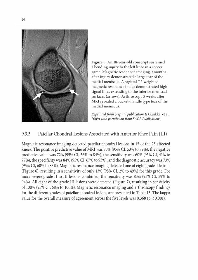
64
9.3.3 Patellar Chondral Lesions Associated with Anterior Knee Pain (III)
Magnetic resonance imaging detected patellar chondral lesions in 15 of the 25 affected knees. The positive predictive value of MRI was 75% (95% CI, 53% to 89%), the negative predictive value was 72% (95% CI, 56% to 84%), the sensitivity was 60% (95% CI, 41% to 77%), the specificity was 84% (95% CI, 67% to 93%), and the diagnostic accuracy was 73% (95% CI, 60% to 83%). Magnetic resonance imaging detected one of eight grade-I lesions (Figure 6), resulting in a sensitivity of only 13% (95% CI, 2% to 49%) for this grade. For more severe grade II to III lesions combined, the sensitivity was 83% (95% CI, 59% to 94%). All eight of the grade III lesions were detected (Figure 7), resulting in sensitivity of 100% (95% CI, 68% to 100%). Magnetic resonance imaging and arthroscopy findings for the different grades of patellar chondral lesions are presented in Table 15. The kappa value for the overall measure of agreement across the five levels was 0.368 (p < 0.001).
Figure 5. An 18-year-old conscript sustained a bending injury to the left knee in a soccer game. Magnetic resonance imaging 9 months after injury demonstrated a large tear of the medial meniscus. A sagittal T2-weighted magnetic resonance image demonstrated high signal lines extending to the inferior meniscal surfaces (arrows). Arthroscopy 5 weeks after MRI revealed a bucket–handle type tear of the medial meniscus.
ReprintedfromoriginalpublicationII (Kuikka, et al., 2009) withpermissionfromSAGEPublications.

65
Table 15. Magnetic Resonance Imaging and Arthroscopic Findings in 56 knees
ArthroscopyMRI Grade 0 Grade I Grade II Grade III Grade IV TotalGrade 0 26 7 3 0 0 36Grade I 1 0 0 1 0 2Grade II 2 1 5 3 0 11Grade III 2 0 1 4 0 7Grade IV 0 0 0 0 0 0Total 31 8 9 8 0 56
SlightlyreformattedfromTableIIinoriginalpublicationIII (Pihlajamäki, et al., 2010).
Figure 6A. A nineteen-year-old man reported chronic activity-related anterior knee pain in the left knee. Physical examination revealed patellofemoral crepitus. An axial, fat-suppressed T1-weighted 3D SPGR MRI of the knee showed intact cartilage surfaces (arrows).
ReprintedfromoriginalpublicationIIIwithpermissionfromJBoneJointSurgAm (Pihlajamäki, et al., 2010). Figurelegendisslightlyreformatted.
Figure 6B. Subsequent arthroscopy showed changes suggestive of grade-I patellar chondral lesions on both facets of the patella (arrows). Probing revealed softening of the cartilage. On the femoral side, the cartilage surfaces were normal, and gliding of the patella in the sulcus was unobstructed.
ReprintedfromoriginalpublicationIIIwithpermissionfromJBoneJointSurgAm (Pihlajamäki, et al., 2010). Figurelegendisslightlyreformatted.

66
9.4 Incidence of Knee Injuries Needing Inpatient Care (IV)The total number of subjects with a knee injury leading to at least one inpatient care admission was 1073 (1% of all conscripts), and the person-based incidence rate was 11 cases per 1000 person-years (95% CI:10.4–11.7). There were 298 subjects (28% of the primary admissions) admitted to inpatient care for old meniscal tear, 236 cases (22%) for acute patellar dislocation, 214 (20%) for fresh meniscal tear, 208 (19%) for ACL/PCL tear, 99 cases (9%) for MCL/LCL tear, and 18 (2%) for fresh chondral lesion. The detailed person-time incidence rates of inpatient care admissions are presented in Table 16.
Figure 7A. A twenty-one-year-old man with chronic, activity-related anterior knee pain in both knees. Pain was also worsened while sitting sitting with the knees flexed. An axial, fat-suppressed T1-weighted 3D FSPGR MRI of the right knee shows mediocentral, grade II cartilage lesion of the patella. Subchondral bone is intact.
Figure 7B. Arthroscopy one day after the MRI showed mediocentral, grade III patellar chondral lesions (fragmentation and fissuring). The diameter of the lesion is only 0.5 cm. Lesion does not reach the subchondral bone. The fragmented areas of cartilage were trimmed by shaving. Also small medial synovial plica was detected and resected.

67
Table 16. Incidence (per 1000 person-years) of inpatient care admissions due to knee injuries among 128,584 conscripts in 1998–2002 in Finland.
Cases Incidence* 95% CIs**MenOld meniscal tear 298 3.1 2.7 to 3.4Patellar dislocation 236 2.4 2.1 to 2.7Fresh meniscal tear 214 2.2 1.9 to 2.5ACL/PCL tear 208 2.1 1.9 to 2.4MCL/LCL tear 99 1.0 0.8 to 1.2Fresh chondral lesion 18 0.2 0.1 to 0.3Total 1073 11.0 10.4 to 11.7
* Cases per 1000 person-years**Confidence IntervalsSlightlyreformattedfromTable1inoriginalpublicationIV (Kuikka, et al., 2011)
Mean time from the beginning of military service to the first inpatient care admission was 126 days. Twenty-five percent of the injuries occurred during the basic training period (first 8 weeks, days 1–56), 49% during the special and team training period (days 57 to 180), and 26% during the leadership period (days 180 to 365). The corresponding numbers were 13%, 57%, and 31% for old meniscal tears, 43%, 34%, and 23% for patellar dislocations, 22%, 54%, and 24% for fresh meniscal tears, 18%, 51%, and 31% for cruciate ligament tears, 32%, 53%, and 15% for collateral ligament tears, and 10%, 60%, and 30% for fresh chondral lesions. The three most common injuries during the first 8 weeks were patellar dislocation (47% of the cases), fresh meniscal tear (16%), and old meniscal tear (14%). The corresponding numbers for days 57–180 were 32% for old meniscal tears, 21% for fresh meniscal tears, and 20% for patellar dislocations. During days 180–365, 33% of the injuries were old meniscal tears, 24% were patellar dislocations, and 19% were cruciate ligament tears.
9.5 Readmissions, Disability, and Need for Surgery (IV)The readmission status and number of surgical operations of any kind for each subject admitted to inpatient care are presented in Table 17. Consecutive admissions were required in approximately 30% of all cases after the first admission. Surgery was required in 59% of the cases, and knee injuries led to a longer-term notable disability requiring a service class change in 32% of the cases. Readmission was required in 45% of subjects admitted to inpatient care for ACL/PCL tears, with more than half of the primary cases resulting in a service class change. There were also many inpatient care admissions and further readmissions for patellar dislocation, but only 23% of the cases necessitated a

68
service class change. A service class change resulted in approximately one-third of the cases with meniscal tears and fresh chondral lesions, but in only 11% of cases of MCL/LCL tears. Surgery was required for 83% of subjects hospitalised for old meniscal tears, 62% for fresh meniscal tears, 67% for ACL/PCL tears, 44% for fresh chondral lesions, 31% for patellar dislocations, and 28% for MCL/LCL tears.
Table 17. Readmission status, service class changes and need for surgical operations among Finnish conscripts with knee injury in 1998–2002.
Primaryadmission
(N of subjects)
Cases requiring readmission
Service class change
Cases requiring surgery
Old meniscal tear 298 70 (23%) 91 (31%) 248 (83%)Patellar dislocation 236 84 (36%) 55 (23%) 73 (31%)Fresh meniscal tear 214 60 (28%) 66 (31%) 133 (62%)ACL/PCL tear 208 94 (45%) 110 (53% 140 (67%)MCL/LCL tear 99 10 (10%) 11 (11%) 28 (28%)Fresh chondral lesion 18 3 (17%) 6 (33%) 8 (44%)Total 1073 321 (30%) 339 (32%) 630 (59%)
Reprinted from original publication IV (Kuikka, et al., 2011) with permission from BlackwellMunksgaard.
9.6 Risk Factors for Knee Injuries Needing Inpatient Care (IV)
Risk factor analyses were performed with logistic regression. Older age was a significant risk factor for inpatient care admission due to knee injury (OR 1.7; 95% CI: 1.3–2.2; Table 18). Obesity was also significantly associated with inpatient care for knee injuries, and subjects with BMI ≥ 30 had an OR of 1.6 (95% CI: 1.03–2.5 compared with subjects with BMI < 25. Subjects with the highest level of muscular strength had 1.6 (95% CI: 1.2–2.4) times the risk of inpatient care for knee injury compared with those in the lowest quartile. There was no association between the results of the 12-min running test, underweight, height and inpatient care admission for knee injury.
Final logistic regression models (Table 19) were used to determine the risk factors for inpatient care admissions for specific ICD-10 knee diagnoses. Obesity was found to be a statistically significant risk factor for inpatient care admission due to patellar dislocation and MCL/LCL tears, with an OR of 2.0 (95% CI: 1.1–3.5) for admission due to patellar dislocation, and an OR of 3.1 (95% CI: 1.4–7.0) for admission due to MCL/LCL tear. Higher weight was an independent risk factor for meniscal tears. For inpatient care admission due to a fresh meniscal tear, the heaviest weight quartile had an OR of

69
2.6 (95% CI: 1.4–5.0) and for admission due to an old meniscal tear the heaviest weight quartile had an OR of 1.8 (95% CI: 1.1–2.8). The risk for old meniscal tears in the oldest age group was 2.4 times (95% CI: 1.7–4.5) greater than that of the youngest age group. The highest muscle strength quartile showed an increased risk for both fresh meniscal tears and ACL/PCL tears.
Table 18. Logistic regression predicting inpatient care admissions due to knee injuries among 128,584 Finnish conscripts in 1998–2002.
Predictor Odds Ratio 95% CI for Odds RatioLower Upper
Age18 to 192021–30
11.01.7
0.91.3
1.32.2
Length of military service180 days270 days360 days
11.51.3
0.91.03
2.41.5
UnderweightNoYes
10.7 0.4 1.1
Overweight and obesityNoOverweightObesity
11.01.6
0.81.03
1.32.5
Muscle strengthlowest quartilesecond quartilethird quartilehighest quartile
11.01.11.6
0.70.81.2
1.31.52.4
SlightlyreformattedfromTable3inoriginalpublicationIV (Kuikka, et al., 2011)

70
Table 19. Multivariate logistic regression model of specific knee diagnoses among 128,584 Finnish conscripts in 1998–2002. Only significant variables are shown.Predictor Odds Ratio 95% CI for Odds Ratio
Lower UpperPatellar DislocationOverweight and obesity
NoOverweightObesity
11.12.0
0.81.1
1.73.5
Age 18 to 192021-30
11.41.1
1.010.6
2.01.9
MCL/LCL tear Overweight and obesity
NoOverweightObesity
10.93.1
0.41.4
2.07.0
Fresh Meniscal TearWeight
lightest quartilesecond quartilethird quartileheaviest quartile
11.81.92.6
0.970.991.4
3.43.55.0
Muscle strengthlowest quartilesecond quartilethird quartilehighest quartile
11.02.23.5
0.40.81.04
2.56.012.2
Old Meniscal TearAge
18 to 192021-30
11.22.4
0.81.5
1.73.6
Weightlightest quartilesecond quartilethird quartileheaviest quartile
11.21.91.8
0.81.21.1
1.92.92.8
ACL/PCL tearMuscle strength
lowest quartilesecond quartilethird quartilehighest quartile
11.21.72.6
0.50.71.04
2.93.86.5
SlightlyreformattedfromTable4inoriginalpublicationIV (Kuikka, et al., 2011)
71
10 Discussion
10.1 Diagnostics (I–III)
10.1.1 Fresh Chondral Lesions (I)
In the sample of fresh traumatic chondral lesions, the sensitivity of 1.0T MRI used in routine clinical practice was only 36% whereas the specificity was 91%. Sensitivity was associated with the lesion grade and was only 17% for superficial lesions and moderately better, 57%, for full-thickness lesions.
A limitation of our study was the relatively small number of patients examined. Another limitation was that data covering several years were collected retrospectively in connection with normal clinical practice. Arthroscopy was used as gold standard when the diagnostic validity of MRI was analysed. As described in the literature review above, however, there are some concerns regarding its accuracy in the evaluation of chondral lesions. The field of view may be limited and possible hidden injuries within the articular cartilage can only be evaluated by probing (Hodler & Resnick, 1992; Van Breuseghem, 2004).
In the present study sprains were the most common cause of chondral lesions of the knee, and the patellar surface and the lateral femoral condyle were the most common lesion locations. It is noteworthy that one-third of the patients had acute traumatic patellar dislocations, which may increase the amount of chondral lesions in these surfaces (Sallay, et al., 1996).
Our study included only fresh traumatic chondral lesions of young adult patients with no osteoarthritic changes in the knee joint. This distinguishes our study from the previous literature. Most of the previous studies investigating the diagnostic validity of MRI for articular cartilage defects have focused on older patients with more osteoarthritic changes (Bredella, et al., 1999; Disler, et al., 1996; Felson, 1988; Handelberg, et al., 1990; Hodler, et al., 1992; Potter, et al., 1998; Recht, et al., 1993; Riel, et al., 1999). Magnetic resonance imaging of chronic osteoarthritis is somewhat different from that of fresh chondral lesions. For traumatic chondral lesions, it is clinically important to detect cartilage shear or a displaced flap that may clinically mimic the symptoms of meniscal tears. Even more

72
crucial is the detection of full-thickness lesions or osteochondral injuries as they may be amenable to repair (Potter & Foo, 2006). Chronic severe osteoarthritis frequently generates bone marrow oedema visible in MRI. As compared to bone bruises associated with traumatic chondral lesions, bone bruises in osteoarthritis are more often observed on both sides of the weight-bearing joint (Potter & Foo, 2006). Whether this leads to the radiologist paying more attention to the articular cartilage surfaces of the area and further increases the sensitivity of MRI for osteoarthritis, however, is not known.
In knee injuries, MRI planes and sequences are generally selected for overall knee examination and good visualization of possible ligamentous and meniscal tears. Traumatic chondral lesions often receive less attention in the selection of MRI protocol. This can explain why the overall sensitivity of MRI for fresh traumatic chondral lesions in this study was poor, only 36%. The interpretation of bone bruises detected by MRI in our study as a positive finding improved the sensitivity of MRI, but led to a deterioration of the specificity and diagnostic accuracy. Bone bruises on MRI may indicate damage to the articular cartilage (Johnson, et al., 1998).
As expected, superficial grade I lesions were detected with a very poor sensitivity (17%), whereas sensitivity improved modestly to 57% for full-thickness lesions. Even if routine 1.0T MRI was more sensitive for deeper lesions, its diagnostic validity can be considered insufficient even for the full-thickness lesions. A valid diagnosis is especially important for deep lesions as they may become symptomatic and require treatment (Curl, et al., 1997; Kettunen, et al., 2005) In young adults, surgical repair of full-thickness traumatic chondral lesions may be indicated if nonsurgical treatment fails to provide adequate pain relief (Gomoll, et al., 2010).
The results of many other studies using routine MRI protocols allowing for overall knee injury evaluation were similar to our findings. Friemert and coworkers used 1.5T MRI and reported a sensitivity of only 33% and specificity of 99% for chondral lesions in patients with knee pain of variable origin (Friemert, et al., 2004). In the study by Disler et al. (n=47, mean age, 36 years), standard 1.5T MRI sequences provided a sensitivity of only 29% to 38%, whereas the specificity was 97% (Disler, et al., 1996). These results are further supported by recent results of the conventional use of 1.5T and 3.0T MRI in clinical practice. Figueroa and Calvo reported a sensitivity of 13% for superficial chondral lesions and of 73% for full thickness lesions with the 1.5T MRI (Figueroa, et al., 2007). The corresponding sensitivities of 3.0T MRI were 29% and 74%, with a specificity of 95% (von Engelhardt, et al., 2007). Also, 3.0T MRI and low-field 0.2T MRI with sequences allowing for comprehensive knee evaluation achieved similar results in deep lesions. The sensitivity of low-field 0.2T MRI for non-fresh, full thickness articular cartilage lesions is reported to be 72%, with a specificity of 100% and accuracy of 92% (Riel, et al., 1999).
These routine protocols may also show very good diagnostic validities for chondral lesions. Bredella et al. included 130 patients (age range 23-72, mean 41 years) who had undergone both 1.5T MRI and arthroscopy for suspected internal knee derangement.

73
The sensitivity of axial and coronal fat-suppressed T2-weighted fast spin-echo combined with sagittal T2 spin-echo was 94%, with a specificity 99% and accuracy of 98% (Bredella, et al., 1999). The sensitivity of fat suppressed fast spin echo sequences in osteoarthritis may be as high as 100%. In this study, however, the specificity was substantially lower, 68% (Yoshioka, et al., 2004). Kijowski and coworkers reported no differences between sensitivities of 1.5T MRI and 3.0T MRI using fast spin echo sequences (71% for 3.0T and 69% for 1.5T MRI (Kijowski, et al., 2009). The specificity was slightly higher for the 3.0T system (85.9% versus 78%). Sensitivities were similar for different grades of chondral lesions. The sensitivity of 1.5T MRI was 41% for cartilage softening, 50% to 82% for partial thickness lesions, and 95% for full thickness lesions. Corresponding sensitivities of the 3.0T MRI were 42, 49% to 85%, and 98%.
It is not clear why the sensitivity of MRI in some of these studies was markedly higher than that in the present study. Increasing MRI field strength makes fat suppression more homogeneous and improves visualization of the articular cartilage (Schoth, et al., 2008; Stoller, 2007). As described in detail in the literature review above, increasing the field strength may not improve the diagnostic validity of MRI for chondral lesions (Kijowski, et al., 2009; Krampla, et al., 2009). There are other potentially more remarkable differences than MRI field strength, however, between the studies comparing the diagnostic validity of MRI. Differences between subjects, methodology, MRI sequences, gold standards, as well as grading of the chondral lesions, and especially the proportion of deep and superficial chondral lesions in the study material may significantly affect the results, making comparison of the results very challenging. A comparison of the diagnostic validity of MRI in the present study to that the previous literature indicates that sensitivity was higher in many of the previous studies. Rather similar results (i.e., insufficient sensitivity for valid diagnosis) with the MRI sequences used in routine clinical practice have likewise been reported, some of them only after the original publication of our article and even with the new 3.0T MRI systems (Figueroa, et al., 2007; von Engelhardt, et al., 2007). Biases, such as a selection or publication bias must also be taken into account, especially in cases when the diagnostic validity or other outcome of the study is poorer than that achieved in experimental or in state-of-the-art studies authors may be disinclined to submit their manuscript for publication or publication may be more easily rejected. The best results achieved, however, may significantly differ from those in real-world clinical practice (Lubowitz & Poehling, 2006).
The present study showed that routine clinical use of 1.0T MRI is not sensitive but is specific for the diagnosis of fresh traumatic chondral lesions. Sensitivity improved only modestly for deeper lesions. We hypothesized that 1.0T MRI could be used to replace diagnostic arthroscopy for the diagnosis of fresh traumatic chondral lesions. Our results failed to support this hypothesis due to the low sensitivity obtained with MRI. Almost half of the full-thickness cartilage lesions remained undiagnosed following preoperative MRI. Despite negative MRI findings, arthroscopy may reveal lesions amenable to

74
cartilage repair procedures. Maintaining the readiness and instrumentation available for at least for basic cartilage repair procedures at the time of arthroscopy is therefore advisable.
10.1.2 Fresh and Old Meniscal Tears (II)
The purpose of the present study was to investigate whether there is difference in diagnostic validity of MRI between meniscal tears related to acute knee trauma and meniscal tears related to chronic knee symptoms. No statistically significant difference was detected. Sensitivity was clearly lower, however, for lateral meniscal tears.
The patient material in the present study was very homogenous with regard to age, race, nationality, general health, and sex. MRI examinations were performed at a single facility with a standardized protocol. A reliable comparison of the diagnostic validity of MRI between acute and chronic meniscal tears can be performed only if the patients and methods are similar within the groups. No comparative studies of this type are found in the previous literature. Degeneration occurs with aging and weakens the meniscal structures, making them susceptible to injuries (Negendank, et al., 1990; Noble & Hamblen, 1975). As the median age of the patients in the present study was 20 years with a maximum age of 25 years, we expected no degenerative changes to be present, which distinguishes our study from many others.
The study is limited, however, by the retrospective data collection. Another limitation is that it is possible that patients with chronic symptoms and false-negative MRI results were treated nonsurgically at a higher rate than the acute group, thereby skewing the statistics. Still, it is unlikely that patients with long-lasting symptoms and false-negative MRI results would become asymptomatic during the physically demanding military service. As the availability of arthroscopy was good, it was performed despite negative MRI findings if the knee pain was prolonged and resulted in difficulties in performing duties required for military training. MRI field strength in the present study was 1.0T. As described in detail in the review of the literature section, recent clinical studies demonstrated that the diagnostic validity of 1.0T, 1.5T, and 3.0T MRI for meniscal tears is similar (Grossman, et al., 2009; Krampla, et al., 2009; Oei, et al., 2003).
Multicentre studies and meta-analyses revealed that, in general, the diagnostic validity of MRI for meniscal tears is good. The pooled sensitivity of MRI varies from 91% to 93% for medial meniscal tears and 69% to 79% for lateral meniscal tears. The pooled specificity varies from 81% to 88% for medial meniscal tears and from 93% to 96% for lateral meniscal tears (Crawford, et al., 2007; Fischer, et al., 1991; Mackenzie, et al., 1996; Oei, et al., 2003). These meta-analyses comprised a large number of studies and a variety of methods and samples were used. In general, there was no special attention paid to the freshness of the lesions. As a result, the intervals between the onset of the symptoms and MRI varied widely and tended to be rather long.

75
There are few published studies investigating acute meniscal tears using MRI. In acutely injured knees, sensitivity as low as 25% is reported for the lateral meniscal tears (Kinnunen, et al., 1994). Kinnunen and coworkers, however, reported 88% sensitivity for medial meniscal tears. A small study by Munshi et al. (Munshi, et al., 2000) reported an MRI sensitivity of 50% for medial meniscal tears and 88% for lateral meniscal tears in acute knee haemarthrosis patients. The specificities in these two studies ranged from 80% to 86% for medial meniscal tears and from 73% to 97% for lateral meniscal tears. Higher MRI sensitivity in traumatic knee haemarthrosis has also been reported, 94% for lateral and 83% for medial meniscal tears (Adalberth, et al., 1997) In that study, however, specificities were very poor – only 27% and 29%, respectively.
Lundberg et al (Lundberg, et al., 1996) studied 69 patients with acute traumatic knee haemarthrosis who were evaluated by 1.5T MRI a mean of 3 days after traumatic onset with arthroscopy being performed shortly thereafter. The authors reported a sensitivity of 74% and a specificity of 66% for medial meniscal tears, and a sensitivity of 50% and a specificity of 84% for lateral meniscal tears. The diagnostic validity of MRI for meniscal tears was lower than the mean results documented in the literature (Crawford, et al., 2007; Fischer, et al., 1991; Mackenzie, et al., 1996; Oei, et al., 2003).
Lundberg and coworkers suggested that this is due to properties associated with acutely injured knees, such as haemarthrosis or catabolic processes of the meniscal tissue. Our results suggest that effusion or haemarthrosis do not weaken the diagnostic validity of MRI. Effusion or haemarthrosis was present in most (80%) of the knees with acute injury, but in only a very small number (5%) of knees with chronic symptoms.
Although the freshness of the meniscal tears in the aforementioned studies (Adalberth, et al., 1997; Kinnunen, et al., 1994; Lundberg, et al., 1996; Munshi, et al., 2000) was equal to that of the sample including acute meniscal tears in our data, the previous authors did not investigate chronic tears. Whether there is real association between the freshness of the injury and decreased diagnostic validity of MRI, however, is not clear. In the present study, using comparable materials and methods, we found no statistically significant difference in the MRI results between meniscal tears related to acute knee trauma and meniscal tears related to chronic knee symptoms. The intraobserver agreement reported in this study was substantial to almost perfect, indicating that the same observer interpreted the images similarly over time. Interobserver agreement was slightly lower, showing moderate agreement.
Previous results suggesting that a lower diagnostic validity of MRI in acute traumas may be explained by factors other than the properties associated with acutely injured knees, such as interobserver variability, sequence or sample selections, or other variability between the studies. The diagnostic validity of MRI in acutely injured knees varied between studies, and in relation to menisci examined (medial or lateral), and acute setting did not necessarily lead to a lower diagnostic validity of MRI (Adalberth, et al., 1997; Kinnunen, et al., 1994; Munshi, et al., 2000). Even if many of the MRI studies

76
of acutely injured knees have reported poorer than average diagnostic validities, poor results were also reported in studies not limited to acute knee injuries and performed with commonly used field strengths (1.0T to 3.0T) (Krampla, et al., 2009).
Magnetic resonance imaging can be as accurate as clinical examination for meniscal tears (Rose & Gold, 1996). Although our study did not deal with clinical examination, the relatively poor overall MRI sensitivity for meniscal tears indirectly emphasizes its value. MRI sensitivity for medial meniscal tears in the present study was good; 92% for acute meniscal tears and 89% for old meniscal tears. For lateral meniscal tears, the corresponding sensitivities were very low, from 47% to 20%, respectively. The poor sensitivity for lateral meniscal tears in the present study also led to a decrease in the overall sensitivity for both groups. There were no statistically significant differences in relation to symptom duration. The decreased MRI sensitivity for tears of the lateral meniscus is well known and demonstrated in the literature (Crawford, et al., 2007; Fischer, et al., 1991; Kinnunen, et al., 1994; Lundberg, et al., 1996; Mackenzie, et al., 1996; Oei, et al., 2003; Rose & Gold, 1996). Very low sensitivities (25%) for lateral meniscal tears are reported both for acute knee injuries (Kinnunen, et al., 1994) and for patients with knee pain lasting 3 months or more (35%) (Rose & Gold, 1996). It is not clear why overall MRI sensitivity for lateral meniscal tears was so low in the present study. De Smet and Mukherjee reported that lateral meniscal tears are more likely missed if the tear involves only one third of the meniscus or is located in the posterior horn (De Smet & Mukherjee, 2008). They also reported that lower sensitivity for the lateral meniscal tears was associated with concomitant ACL tears. This finding was not statistically significant, however, possibly due to the relatively small patient sample. Other hypothesized causes for the lower MRI sensitivity in diagnosing lateral meniscal tears include oblique visualization of the posterior horn of the lateral meniscus due to its anatomy, the magic angle effect, and arterial pulsation artefacts (De Smet & Mukherjee, 2008).
The specificity in the present study was slightly lower for medial meniscal tears than for lateral meniscal tears (87% vs. 94%–99%). Visualization of the posterior horn of the medial meniscus is limited in arthroscopy and the diagnosis of longitudinal tears in the inferior surface of the menisci is based on probing. Some of the “false” positive MRI findings may be attributed to inadequate visualization of this area in arthroscopy. Arthroscopy is considered to be the gold standard, and the specificity of MRI decreases with an increase in the number of “false” positive findings. The precise location of the tears was not registered in the present study, so it is not known whether the results were affected by the location of the lesions or by other factors presented above. The diagnostic validity of MRI is thought to be strongly affected by the menisci involved and independent evaluation of the menisci is far more informative than evaluation of both menisci together.
In conclusion, MRI has similar diagnostic validity for meniscal tears in acute knee trauma and for knee symptoms lasting over 6 months in young adults. This study also

77
suggests that the diagnostic validity of MRI for meniscal tears is not weakened by effusion or haemarthrosis.
Magnetic resonance imaging showed relatively poor overall sensitivity for meniscal tears in both groups. For clinical practice, this finding reveals that up to one-third of meniscal tears failed to be detected by MRI in young adults, regardless of the age of the tear. The overall specificity was good, indicating a small number of false-positive findings. However, there was a substantial difference in the diagnostic validity of MRI between lateral and medial meniscal tears. As many as 9 of 10 of the medial meniscal tears were detected by MRI, whereas lateral meniscal tears were missed in more than half of the cases, regardless of the duration of the symptoms. In clinical practice, arthroscopy should be considered if justified by the patient’s symptoms despite negative MRI findings, especially in cases of clinically suspected lateral meniscal tears.
10.1.3 Patellar Chondral Lesions Associated with Anterior Knee Pain (III)
In the 56 knees with AKP, there were patellar chondral lesions in 25 (45%) . Twenty-five patients also had pathological plicae and only 6 knees had normal arthroscopic findings. The presence of retropatellar crepitus or pain on patellar manipulation was not associated with the higher proportion of patellar chondral lesions in patients with typical clinical AKP symptoms. Neither was an association found between the severity of chondral lesions and clinical symptoms and signs of the subjects. The routine MRI protocol used in AKP patients showed a sensitivity of 13% for superficial patellar chondral lesions, which was significantly higher (83%) for more severe lesions.
The present study has several strengths, such as the prospective design and the homogeneity of the study population. In addition, the age group studied here is optimal because individuals in this age group do not normally show degenerative chondral changes such as osteoarthritis (Felson, 1988). Further, this relatively large group of skeletally mature young adults underwent a uniform examination and treatment for AKP according to the policy of one institution. The standard methods and protocols of the institution were used for the evaluation of clinical symptoms, plain radiographs, MRI, and arthroscopic findings. Magnetic resonance imaging was usually performed on the day prior to the arthroscopy to avoid possible bias caused by any delay. As mentioned in the Material and Methods section above, a total of 91 knees were examined but, in cases of bilateral findings, only one knee was randomly selected for the study. This limited the number of the knees in the present study, but, on the on the other hand, ensured the independence of the observations.
According to our results, patients with diffuse AKP as the sole symptom very rarely have patellar chondral lesions. Only one of eight of these patients had patellar chondral lesion in arthroscopy. Half of the patients with AKP combined with other typical clinical symptoms had patellar chondral lesions, regardless of the presence or absence
78
of patellofemoral crepitus and/or pain on manipulation of the patella. Leslie and Bentley found that 58% of the patients with corresponding symptoms and retropatellar crepitus had patellar chondral lesions (chondromalacia), which is similar to our results (Leslie & Bentley, 1978). Leslie and Bentley suggested that patellofemoral crepitus is one of the signs most indicative of patellar chondral lesions. Niskanen and coworkers studied the validity of clinical tests for detecting patellar chondral lesions in a sample of 100 consecutive patients undergoing arthroscopy for various knee problems (Niskanen, et al., 2001). The best sensitivity was reported for the patellar tracking test in which the patella was moved up and down and compressed against the femoral groove. Pain in this test resulted in a sensitivity of 56% and a specificity of 55%. For patients with patellar pain or otherwise suspected patellar chondral lesions, the sensitivity of this test was slightly higher, 67% (Niskanen, et al., 2001).
Based on both the present data and these previous studies (Leslie & Bentley, 1978; Niskanen, et al., 2001), it is clear that painful patellar compression or retropatellar crepitus are far from pathognomonic tests and have poor diagnostic validity for detecting patellar chondral lesions. In the present study, the presence of pain in patellar compression or retropatellar crepitus was not associated with the higher proportion of patellar chondral lesions if the symptoms were otherwise similar.
An important finding of the present study was the lack of an association between the severity of patellar chondral lesions and the clinical symptoms and signs. These findings are contradictory to the observations of Kettunen et al., who reported that patients with more severe cartilage lesions of the patellofemoral joint had more subjective symptoms and functional limitations than those without lesions or with small cartilage lesions (Kettunen, et al., 2005). On the other hand, our results are supported by the finding that chondral lesions are asymptomatic even with direct probing (Dye, et al., 1998). Several other knee pathologies can cause pain to the anterior aspect of the knee and new theories have challenged the traditional views of the origins of the pain (Dye, 2005; Houghton, 2007; Kodali, et al., 2011; Llopis & Padron, 2007). A medial synovial plica of the knee was as common an arthroscopic finding as patellar chondral lesions in the present study and was detected in nearly half of the knees. Pathologic medial synovial plicae may impinge between the medial femoral condyle and patella, eventually leading to cartilage damage or synovitis due to repetitive contact forces (Christoforakis, et al., 2006; Llopis & Padron, 2007; Lyu & Hsu, 2006; Munzinger, et al., 1981). The severity of symptoms, however, is not proportional to the size of the plica and the relationship between the plica and AKP is controversial (Jee, et al., 1998; Llopis & Padron, 2007). Even if synovial plicae may be a source of AKP, their presence should be cautiously evaluated as the cause of the symptoms (Llopis & Padron, 2007).
Only 6 of the 56 symptomatic knees had normal arthroscopic findings. Pain in these knees might be explained by the tissue homeostasis theory (Dye, 2005). According to Dye, increased loading of the patellofemoral joint leads to a loss of tissue homeostasis
79
in the surrounding innervated tissues and causes pain in the anterior aspect of the knee. Only in the most severe cases is the loss of tissue homeostasis characterized by arthroscopically visible damage such as chondral lesions (Dye, 2005).
T2-weighted or other sequences that allow for the evaluation of meniscal and ligament tears exhibit variable sensitivities for patellar chondral lesions. In the study by McCauley and coworkers, the diagnostic validity of MRI with T2-weighted axial plane images showed a sensitivity of 72% and a specificity of 87% for patellar chondral lesions. For axial proton density images, the corresponding figures were 62% and 78%. The combination of these sequences resulted in 86% sensitivity and 74% specificity (McCauley, et al., 1992). Murphy evaluated only severe grade III and IV patellar chondral lesions using T2*-weighted 3D gradient-echo MRI and reported good sensitivity (83%), similar to our results (Murphy, 2001). It must be noted, that even 100% sensitivity is sometimes achieved with T2-weighted spin-echo pulse sequences in the sagittal and axial planes and a 3D fast sequence in the sagittal plane. This combination can lead to a number of false negative findings, however, and the specificity is only 50% (Handelberg, et al., 1990).
Better diagnostic validity of MRI for chondral lesions may be achieved with the use of sequences specifically designed for cartilage imaging. Fat-suppressed 3D SPGR sequences are now routinely used in MRI examination when chondral lesions are considered a possibility. In routine evaluation of AKP patients, these sequences should be obtained in the axial plane. This allows for good visualization of the articular cartilage surfaces of the patellofemoral joint and especially the patellar facets. The 3D SPGR technique requires a long acquisition time and is not optimal for the visualization of menisci, ligaments, and subchondral bone (Potter, et al., 1998). As symptoms of AKP may be confused with those of meniscal tears, it is important that routine knee MRI also includes some meniscal-sensitive sequence in the sagittal plane, such as T2* GRE or proton density spin-echo with fat suppression. For these reasons, the routine knee imaging protocol for AKP patients in this study included an axial fat suppressed 3D SPGR sequence and a sagittal T2* GRE sequence. With this protocol, the overall MRI sensitivity for chondral lesions of all grades in the present study was 60% and the specificity was 84%.
Disler et al. used fat suppressed 3D SPGR imaging to evaluate chondral lesions in all articular cartilage surfaces of the knee. Two readers evaluated the images and the sensitivity for chondral lesions in combined surfaces ranged from 75% to 85%. The specificity was 97% regardless of the reader (Disler, et al., 1996). For patellar chondral lesions, the sensitivity varied from 87% to 100% and the specificity ranged from 92% and 100%. In detecting osteoarthrotic chondral lesions, sensitivities for this sequence ranged from 96% to 97% (Recht, et al., 1993; Yoshioka, et al., 2004). The reference standard for MRI results were anatomic sections of cadaveric knees in Recht’s study and arthroscopy findings in Yoshioka’s study.
The results of the present study demonstrate that the evaluation and comparison of the diagnostic validity of MRI is far more informative when the results are analysed

80
independently for the different grades. The relatively low sensitivity of 60% in this study is partly explained by the fact that sensitivity was only 13% for grade I chondral lesions. This finding indicates that superficial patellar chondral defects cannot be validly detected by MRI even when an SPGR sequence allowing for visualization of the cartilage is included in the routine protocol. For grade II and III lesions, the sensitivity was considerably higher (83%). For grade III lesions including fragmentation of the cartilage, MRI detected all of the lesions and sensitivity was 100%. Our results clearly indicated that the highest sensitivity occurred for the deepest lesions. This finding is consistent with previous findings (Figueroa, et al., 2007; Gagliardi, et al., 1994; Murphy, 2001; von Engelhardt, et al., 2007).
Gagliardi et al. examined 27 AKP patients with 1.5T MRI obtained in axial plane images with T1-, T2 spin echo, proton-density, and fat suppressed 2D SPGR sequences (Gagliardi, et al., 1994). They graded the patellar chondral lesions using the same system that we used (Shahriaree, 1985). The sensitivity of MRI for grade-I patellar chondral lesions was 0%. For grade-II and III lesions, the sensitivity was 13% for proton-density sequences, 20% for SPGR and T1 sequences, and 47% for T2-weighted imaging. For grade-IV lesions, the sensitivity ranged from 50% (SPGR imaging) to 75% (T1-weighted, T2-weighted, and proton-density-weighted imaging. Specificities for all grades and sequences were high, between 91% and 100% (Gagliardi, et al., 1994). The sensitivities reported by Gagliardi et al. (1994) for all of the sequences that they used were lower than the sensitivities found in the present study.
A recent study of conventional clinical practice use of 3.0T MRI including 3D T1-weighted gradient echo sequence in axial plane showed an overall sensitivity for patellar chondral lesions of 87% and a specificity of 89%. A high proportion of the lesions in this location were deep, grade III to IV lesions. In addition, grade I lesions were considered to indicate a disease-negative status. When the sensitivity of knee chondral lesions was presented according to lesion severity, the sensitivity was 29% for grade I lesions, 62% for grade II, 63% for grade III, and 74% for grade IV presenting full-thickness lesions (von Engelhardt, et al., 2007). These results are consistent with our results and demonstrate that increasing the field strength may not be beneficial for the diagnosis of articular cartilage lesions.
Skyline views are a routine method of AKP imaging. The main purpose is to evaluate possible patellar malalignment. Of the various suggested malalignments, only patellar subluxation is increased in patients with AKP as compared to healthy controls (Haim, et al., 2006). Patellar subluxation, however, does not seem to be associated with arthroscopically detected patellar chondral lesions (Leslie & Bentley, 1978). In the present study, only 16% (7 of 43) of the knee radiographs in AKP patients showed some malalignment. No conclusion can be drawn regarding the association between the reported malalignments and patellar chondral lesions due to their small number. In addition, skyline radiographs are a very rough method to evaluate patellar malalignment.

81
Computed tomography, MRI or kinetic CT allow for the detection of more subtle cases of malalignment (Christian, et al., 2006). Although patellar malalignment may not be associated with AKP or patellar chondral lesions, it may affect the outcome of cartilage repair procedures and even account for premature failure of the repair (Gomoll, et al., 2010; Potter & Foo, 2006). Patellar chondral lesions are an off-label indication for autologous chondrocyte implantation. which has become an option for treatment of all but the smallest chondral lesions in the patellofemoral compartment (Gomoll, et al., 2010).
The association between clinical symptoms and physical signs, and possible patellar chondral lesions was assessed in young adults with AKP syndrome. The findings of the present study support earlier reports that patellar chondral lesions cannot be distinguished from other causes of AKP based only on symptoms and signs on physical examination. Arthroscopic examinations revealed patellar chondral lesions in less than half of all subjects. The presence of retropatellar crepitus or pain on manipulation of the patella was not associated with a higher proportion of patellar chondral lesions in patients with typical clinical AKP symptoms. There was also no association found between the severity of chondral lesions and clinical symptoms and signs among the subjects. Thus, these symptoms and signs are not helpful when considering whether the patient might have deep chondral lesions possibly amenable to surgical repair. Pathological synovial plicae were as common a finding as patellar chondral lesions. Of the 56 six knees, arthroscopy revealed pathology in 50.
Another aim of this study was to investigate the diagnostic validity of MRI for patellar chondral lesions in AKP patients. We used a routine MRI protocol that allows for overall knee examination as well as visualization of the patellar cartilage. Our hypothesis was that MRI could be used to confirm the diagnosis of chondromalacia patellae at least in the case of more severe, deeper chondral lesions. For superficial patellar chondral lesions the sensitivity was insufficient, only 13%. For more severe lesions, 1.0T MRI may be considered a valid diagnostic tool as the sensitivity was substantially higher, 83%.
10.2 Epidemiology of Knee Injuries (IV)
10.2.1 Incidence of Knee Injuries Needing Inpatient Care
The overall person-time incidence rate for knee injuries requiring inpatient care in the present study was 11 per 1000 person-years. The most common injuries were meniscal tears, patellar dislocation, and cruciate ligament tears.
There are several strengths in the present study. It involved a population of 128,584 conscripts with a total exposure time of nearly 100,000 person-years. Due to the compulsory nature of military service, these subjects represent ordinary young adult

82
males of Finland. All conscripts were required to use only the services of the primary military health-care units and Central Military Hospital for medical treatment. The NHDR database from which the data were obtained is complete and highly accurate (Keskimäki & Aro, 1991; Mattila, et al., 2008; Salmela & Koistinen, 1987). These factors together allowed us to include knee injuries requiring inpatient care during the follow-up time with very high accuracy and coverage. To our knowledge, there are no published studies reporting the accurate incidence rates of hospitalised knee injuries. Among Finnish military conscripts, the overall incidence rate of injury-related inpatient care admissions is 94 per 1000 person-years (Mattila, et al., 2006). The sample population of the previous study and that of the present study are congruent, and suggest that more than one-tenth of all injuries leading to hospitalisation are knee injuries.
There are also weaknesses in the present study. Despite the fact that all hospitalised injuries were taken into account, data on less severe injuries were lacking. In the US Army, it is estimated that only 1 of every 20 military injuries results in hospitalisation (Jones, et al., 2000). As presented earlier in the review of the literature section, re-analysis of previous reports of Finnish conscripts suggests that the patient-based incidence of acute knee injuries treated at the garrison clinics on an outpatient basis is approximately 130 per 1000 person-years (Taanila, et al., 2009, 2010). This is more than 10-fold higher than the incidence of hospitalised knee injuries in the present study.
Kannus and coworkers reported a cumulative incidence rate of 11 knee injury patients per 1000 persons per year in a study of outpatient visits at the primary health care units in the Orivesi region, Finland (Kannus & Järvinen, 1989). The incidence rate reported in their study was substantially lower than the corresponding statistics for conscripts treated as outpatients at the garrison clinics (Taanila, et al., 2010). When taking into account that approximately 1 of 10 knee injuries treated at the primary care units requires surgical consultation, the incidence rate reported by Taanila and coworkers was more consistent with our study than that reported by Kannus and coworkers (Kannus & Järvinen, 1989; Taanila, et al., 2010). The study population of Kannus and coworkers was relatively old, which evidently decreased the incidence rate of knee injuries and may explain the result. The incidence rates reported from special health care units are generally markedly lower than that in our study. For acute traumatic knee haemarthrosis treated in Turku University Hospital, the cumulative incidence is 0.5 per 1000 inhabitants annually (Sarimo, et al., 2002). Traumatic knee haemarthrosis thus represents only a small proportion of all knee injuries requiring inpatient care in Finland. In a UK population, the cumulative person-based incidence rate for knee injuries treated at the orthopaedic trauma unit was as low as 0.4 cases per 1000 persons annually (Clayton & Court-Brown, 2008). Both inpatient and outpatient care were taken into account and the age of the patient ranged from 12 to 89 years. The authors pointed out that the incidence reported is a clear underestimate as knee injuries were frequently referred to knee surgeons instead of the orthopaedic trauma unit. General population-based data from the US show a cumulative incidence rate of

83
1.2 cases per 1000 persons annually for ligamentous and meniscal injuries of the knee treated at an orthopaedic knee injury clinic (Miyasaka, et al., 1991). The age distribution of the population was not reported but it can be assumed that the average age of the patients was older than in the present study. This is probably the most likely explanation for the low incidence rates reported in the UK and US studies compared with the results of the present study. There may also be other differences between the countries, such as insurance policy differences, higher number of patients using private health care units in UK or US, or differences in the physical activity level of the populations. Also, rather than reporting the exact person-time (i.e., first admission due to knee injury, death or moving out of the area as a censoring event), most of the authors (Clayton & Court-Brown, 2008; Kannus & Järvinen, 1989; Miyasaka, et al., 1991; Sarimo, et al., 2002) reported a cumulative annual incidence that assumes that a person is at risk for the whole study period. This somewhat lowers the incidence rate.
In the present study, the incidence rate per 1000 person-years for hospitalised old meniscal tears was 3.1, for fresh meniscal tears 2.2, for patellar dislocations 2.4, for cruciate ligament tears 2.1, for collateral ligament tears 1.0, and for fresh chondral lesions 0.2 cases. There is a very limited amount of comparable data in the literature. The aforementioned studies (Clayton & Court-Brown, 2008; Miyasaka, et al., 1991; Sarimo, et al., 2002) reported remarkably lower incidence rates for all of the specified knee diagnoses in comparison with the present study. In a cohort of Finnish adolescents and a young adult population, an event-based incidence rate of 0.61 (males, 0.97) per 1000 person-years for cruciate ligament injuries leading to hospitalisation was reported (Parkkari, et al., 2008). The incidence was lower than in our study. Parkkari and coworkers reported a hazard ratio of 4.0 for cruciate ligament injuries in males participating in organized sports more than 4 times per week and this may explain the higher injury incidence rate during military service (Parkkari, et al., 2008). An incidence of 0.77 hospitalised primary traumatic patellar dislocations per 1000 person-years was reported in the Finnish conscript population (Sillanpää, et al., 2008). The present study reported a markedly higher incidence that can be explained by the inclusion of recurrent dislocations and dislocations without remarkable trauma in the study.
The most common ICD-10 diagnosis in subjects admitted to inpatient care was old meniscal tear (28%), followed by acute patellar dislocation (22%), fresh meniscal tear (20%), ACL/PCL tear (19%), MCL/LCL tear (9%), and fresh chondral lesions (2%). Minor knee distortions without damage to a particular structure were observed in 34% of the cases studied by Majewski. Anterior cruciate ligament tear was the most common specific knee diagnosis (20%), followed by medial meniscus tear (11%), chondral lesion (11%), MCL tear (8%), contusion injury due to direct trauma (6%), lateral meniscus tear (4%), and patellar dislocation (3%) (Majewski, et al., 2006). Only specified ICD-10 diagnoses were included in our analysis. The relative frequency of meniscal tears and patellar dislocations was clearly higher in our study, whereas the proportion of ACL tears and

84
chondral lesions was lower. Majewski’s study was performed in a sports injury clinic and included only athletic knee injuries, which might explain the higher percentage of ACL injuries (Parkkari, et al., 2008). In our study, all hospitalised injuries occurring during leisure time and not associated with military or sports training were also included. The registry data used in our study, however, did not include information regarding whether the knee injury occurred during sports, military exercises, or vacations. Taanila et al. (Taanila, et al., 2009) examined this issue in Finnish conscripts. Musculoskeletal disorders were included in their analysis and the knee joint was the second most common injury location (18%). The majority of injuries occurred during actual military service (91%), followed by during vacations (9%), and on the way to vacation or back to the garrison (0.5%). The most common cause of knee injury was training in combat gear, followed by marching and other physical exercises. The number of chondral lesions increases with age (Curl, et al., 1997), which may explain the lower proportion of these lesions in the present study. Chondral lesions are not always reported as a separate diagnosis (Kiviranta & Vasara, 2004) and the number of chondral lesions in the present study should be considered a clear underestimate.
This study is the largest performed and reported to date of the incidence and risk factors for knee injuries that can be generalised to the young adult male population between the ages of 18 and 30 years, i.e., the group in which most knee injuries occur (Gianotti, et al., 2009; Kannus & Järvinen, 1989; Majewski, et al., 2006) and in which participation in sports is common. The incidence rates of knee injuries needing inpatient care in this population have not been comprehensively reported. Young adults serving compulsory military service are representative of the general population and are thus an excellent indicator of how this population reacts to the challenges of progressive physical exercise. This study demonstrates that the incidence of knee injuries needing inpatient care is high in the young adult male population involved in physical training, and causes a significant burden to hospitals. Overall person-time incidence rate of knee injuries requiring inpatient care in the present study was 11 per 1000 person-years, which indicates that more than 1 of 100 young adult male conscripts will be admitted to inpatient care each year due to knee injury. This incidence is remarkably high compared with the results presented in older general populations in Finland, UK, and US (Clayton & Court-Brown, 2008; Kannus & Järvinen, 1989; Miyasaka, et al., 1991). The findings in Finnish young adults, conscripts, and knee haemarthrosis patients are more logically consistent with the results of the present study (Parkkari, et al., 2008; Sarimo, et al., 2002; Sillanpää, et al., 2008; Taanila, et al., 2010).

85
10.2.2 Readmissions, Disability, and Need for Surgery
Among all young adult male conscripts hospitalised with knee injuries, as many as one-third will undergo a service class change due to long-term disability. Almost two-thirds of all hospitalised subjects require surgery.
The majority of subjects admitted to inpatient care for old meniscal tears required surgery. Subjects with fresh meniscal tears and ACL/PCL tears also were usually treated surgically, whereas those with fresh chondral lesions, patellar dislocations, and MCL/LCL tears rarely underwent surgery. Majewski et al. (Majewski, et al., 2006) reported that 87% of clinically diagnosed ACL tears required surgery and the corresponding percentages were 85% of lateral meniscus, 84% of medial meniscus, 78% of PCL, 59% of MCL, and 49% of LCL). The proportions of injuries treated surgically reported by Majewski et al (2006) were generally higher than those in the present study. Almost all ACL tears were treated surgically in Majewski’s study and it seems likely that surgery was considered more eagerly for athletes than for ordinary young adult males in the present study.
One important and novel finding of the present study was that up to one-third of all subjects admitted to inpatient care exhibited such a severe decline in physical performance that a service class change was warranted. A service class change was deemed necessary for more than half the subjects with ACL/PCL tears. The decision for a service class change is not made easily and is therefore considered a reliable indicator of more severe injury with longer-term disability. The present study included both traumatic and nontraumatic, primary, and recurrent patellar dislocations. Approximately one-fifth of the patellar dislocations in the present study led to a service class change, indicating longer-term notable disability and one-third required surgery. In the study of Sillanpää and coworkers, the proportion of service class changes was similar, but surgery was required twice as often when only primary traumatic patellar dislocations were taken into account (Sillanpää, et al., 2008).
To our knowledge, no other studies in the literature have reported similar disability rates of different knee injuries. Information gained in the present study will help clinicians in predicting the prognosis of different knee injuries. A long-term follow-up study would be beneficial to determine whether knee injuries leading to a service class change cause permanent disability or persistent symptoms.
10.2.3 Risk Factors for Knee Injuries Needing Inpatient Care
Older age and obesity are the most important risk factors for inpatient care admission for knee injury. Obesity was a significant independent risk factor for patellar dislocation and MCL/LCL tears. Although higher weight was an independent risk factor for meniscal tears, BMI was not. The overall risk for inpatient care admissions due to knee injuries and particularly due to old meniscal tears increased with age.

86
Data regarding the association between physical fitness, or anthropometric properties, such as height, weight or BMI and knee injuries needing inpatient care has been severely lacking before the present study. Previous studies demonstrated that older age, up to 26 years, increases the risk for discharge from the US Army due to disabling knee injuries (Sulsky, et al., 2000). Obesity is a risk factor for disabling knee pain and inpatient care admission due to lower limb injury (Mattila, et al., 2007; Webb, et al., 2004). In the present study, running test results were not associated with knee injuries. Instead, better muscle strength was a statistically significant risk factor for knee injuries in general and specifically for fresh meniscal tears and cruciate ligament injuries. Those with better muscle strength may participate in more vigorous sports activities, which increases the risk for knee injuries and especially the risk for cruciate ligament tears (Haapasalo, et al., 2007; Parkkari, et al., 2008). In addition, conscripts with better muscle strength may be required to perform more physically challenging tasks during military service for which the risk of knee injury may be higher. An earlier study assessing Finnish conscripts reported that good muscular fitness increases the risk for injury-related inpatient care admissions in general, and excellent aerobic condition increases the risk for lower limb injury leading to inpatient care (Mattila, et al., 2007). In contrast, however, the risk for musculoskeletal injuries treated at the garrison clinics as outpatients and also the risk for medical discharge from the military service is reported to increase if the running test results are poor. Overall results of the muscular strength tests are not associated with these outcomes (Taanila, et al., 2011; Taanila, et al., 2010). The reasons for these contradictory results are not clear and require further research. The risk factor variables in the present study were measured during the first weeks of military service, therefore it is possible that weight or physical performance changed during the subsequent service time.
The multivariate analysis in the present study indicated that the risk for patellar dislocations is 2-fold in obese (BMI > 30) subjects. No significant association was detected between height, weight, or physical fitness and risk of inpatient care admissions due to patellar dislocations. A previous univariate analysis demonstrated that higher height and weight increase the risk when only primary traumatic patellar dislocations are taken into account (Sillanpää, et al., 2008). To our knowledge, there are no previous studies reporting risk factors for collateral ligament tears. A new important finding of the present study was that obesity is associated with inpatient care due to MCL/LCL tears and it increases the risk 3-fold. In the present study, heavier subjects had a higher risk for meniscal tears. Weight is also reported to be a risk factor for meniscal tears in a UK population (Baker, et al., 2002). Older age as a risk factor for old meniscal tears is a logical finding, as aging weakens the meniscal structures (Negendank, et al., 1990). Nevertheless, there has been lack of data actually showing that the risk increases with aging.
This study adds to the knowledge of the poorly known, intrinsic and potentially modifiable risk factors for knee injuries requiring inpatient care and can be generalised

87
onto the young adult male population of Finland. An important new finding was that obesity is a significant risk factor for inpatient care of knee injuries and especially of collateral ligament tears and patellar dislocations. Older age is associated with increased risk for inpatient care admission due to knee injury and thus entering military service at a younger age, e.g., between 18–20, might be beneficial. The association between knee injuries and muscle strength, as well as the prevention of knee injuries, requires further research.

88
11 Conclusions
The principal aims of the present study were to investigate the epidemiology and MRI-based diagnostics of common knee injuries and AKP in the young adult population. The study population comprised young adult conscripts performing their compulsory military service. Except for the knee injury under study, all were generally healthy and able to perform military service. Due to the compulsory nature of military service, male conscripts included in this study are highly representative of ordinary Finnish young adults. The findings can thus be generalised to the physically active, young adult male population.
1) Data from patients with fresh traumatic chondral lesions indicated that although routine clinical use of 1.0T MRI is not sensitive, it is specific for the diagnosis of fresh traumatic chondral lesions. Sensitivity improved only modestly for deeper lesions. Almost half of the full-thickness cartilage lesions were undiagnosed by preoperative MRI. Despite negative MRI findings, arthroscopy may reveal lesions amenable to cartilage repair procedures. Readiness and available instrumentation for basic cartilage repair procedures at the time of arthroscopy is therefore advisable (I).
2) Data of meniscal tears revealed that MRI has a similar diagnostic validity for meniscal tears in acute knee trauma and knee symptoms lasting over 6 months in young adults. Our results also suggest that neither effusion nor haemarthrosis are associated with the diagnostic validity of MRI. The diagnostic validity of MRI substantially differed between lateral and medial meniscal tears. As many as 9 of 10 of the medial meniscal tears were detected in MRI, whereas lateral meniscal tears were missed in more than half of the cases, regardless of the duration of the symptoms. Clinically, arthroscopy should be considered despite negative MRI findings if justified by the patient’s symptoms, especially in cases of suspected lateral meniscal tears (II).
3) Routine MRI protocol used in the prospective sample of patients with AKP included a cartilage-specific T1-weighted 3D SPGR sequence and allowed for an overall, clinically rational knee examination. The MRI sensitivity for superficial patellar chondral lesions was insufficient, only 13%. For more severe lesions, however, 1.0T MRI may be a valid diagnostic tool as the sensitivity was substantially higher, 83% (III).
4) The AKP data also provided complementary evidence that patellar chondral lesions cannot be distinguished from other causes of AKP based only on symptoms and signs on physical examination. Arthroscopic examinations revealed patellar chondral lesions

89
in less than half of all subjects. Pathological synovial plica was a consistently common finding, although association with the symptoms cannot be assessed. Arthroscopy revealed pathological findings in 50 of 56 knees. The presence of retropatellar crepitus or pain on patellar manipulation was not associated with a higher proportion of patellar chondral lesions in patients with typical clinical AKP symptoms. No association was found between the severity of chondral lesions and clinical symptoms and signs. Thus, these symptoms and signs are not helpful when considering whether the patient might have deep chondral lesions possibly amenable to surgical repair (III).
5) Epidemiological data revealed the incidence of inpatient care admissions due to knee injuries in the largest-ever sample that can be generalised onto the physically active young adult male population. The overall person-time incidence rate for knee injuries requiring inpatient care admission in the present study was remarkably high, 11 per 1000 person-years. Each year more than 1 of 100 young adult males will be admitted to inpatient care due to knee injury during their conscription. The incidence rates of hospitalised knee injuries in this population have not been comprehensively reported earlier.
Clinically relevant data regarding the proportions of surgical treatment and longer-term disability associated with hospitalised knee injuries were reported. Nearly two-thirds of the knee injuries needing inpatient care led to surgical treatment and as many as one-third led to longer-term notable disability. To our knowledge, no other studies in the literature have reported corresponding information for knee injuries requiring inpatient care. A long-term follow-up study would be beneficial to determine whether knee injuries leading to a service class change cause permanent disability or persistent symptoms.
This thesis also adds to the body of knowledge of poorly known, intrinsic, and potentially modifiable risk factors of knee injuries requiring inpatient care. The most important risk factors were older age and obesity. Obesity was a significant independent risk factor for patellar dislocation and MCL/LCL tears. Higher weight, but not BMI, was a risk factor for meniscal tears. Older age increased the overall risk for inpatient care admission and especially the risk for old meniscal tears. The association between knee injuries and muscle strength as well as the prevention of knee injuries warrants further research (IV).

90
12 Acknowledgements
This study was carried out at the Central Military Hospital in Helsinki and later continued at the Research Institute of Military Medicine in Helsinki, Finland.
My deepest respect and gratitude I owe to my supervisors: associate professor Harri Pihlajamäki and associate professor Ville Mattila. Harri Pihlajamäki introduced me to the principles of the scientific world and provided me with an excellent opportunity and facilities to start my research career. Ville Mattila’s constant encouragement and his ability to help me find the essence of the study has been invaluable especially while finishing this thesis. Despite their busy schedules, my supervisors have always taken the time to help me from basic clinical problems to complex scientific dilemmas. Without their assistance, this thesis would not have been completed.
I want to present my sincere gratitude to the official reviewers of this study, professor Juhana Leppilahti and associate professor Arsi Harilainen for their constructive and valuable comments which helped me to improve the thesis.
I would like to express appreciation for all co-authors of this work: Martti Kiuru, M.D., Ph.D., Maria Niva M.D., Ph.D., Heikki Kröger, M.D., Ph.D.,Petri Sillanpää, M.D., Ph.D. and Vesa-Veikko Leppänen M.D., Ph.D. Their contributions have been really valuable and their professional expertise has helped me to gain a deeper insight into the different fields of medicine.
I also greatly appreciate the support of the members of the supervisory board, associate professor Jari Parkkari and associate professor Tuomo Visuri.
I want to thank staff at the Research Institute of Military Medicine for their valuable help, especially Mr. Kari Kelho for helping me with images and computer software. I likewise want to thank Ms. Sirpa Randell from the University of Tampere for her assistance with the layout of the manuscript.

91
Important financial support for this study was received by Finnish Defence Forces, Finnish Cultural Foundation, the Emil Aaltonen Foundation and University of Tampere.
I warmly thank my parents Heikki and Helena and my sister Anniina for offering me a safe and encouraging environment to grow up and to their support I owe my success in life. Special thanks go to Helena for her help in finding articles, books and other material needed in this study.
Finally, I want to thank my wife Sara for her love, patience and support during these years.
Joensuu, 15th July 2012
Paavo Kuikka

92
13 References
Adalberth, T., Roos, H., Lauren, M., Akeson, P., Sloth, M., Jonsson, K., Lindstrand, A., & Lohmander, L. S. (1997). Magnetic resonance imaging, scintigraphy, and arthroscopic evaluation of traumatic hemarthrosis of the knee. AmJSportsMed,25(2), 231–237.
Agresti, A., & Coull, B. A. (1998). Approximate is Better than “Exact” for Interval Estimation of Binomial Proportion. AmStat,52(2), 119–125.
Almeida, S. A., Williams, K. M., Shaffer, R. A., & Brodine, S. K. (1999). Epidemiological patterns of musculoskeletal injuries and physical training. MedSciSportsExerc,31(8), 1176–1182.
Atkin, D. M., Fithian, D. C., Marangi, K. S., Stone, M. L., Dobson, B. E., & Mendelsohn, C. (2000). Characteristics of patients with primary acute lateral patellar dislocation and their recovery within the first 6 months of injury. AmJSportsMed,28(4), 472–479.
Bachmann, G., Heinrichs, C., Jurgensen, I., Rominger, M., Scheiter, A., & Rau, W. S. (1997). Comparison of different MRT techniques in the diagnosis of degenerative cartilage diseases. In vitro study of 50 joint specimens of the knee at T1.5. RoFo,166(5), 429–436.
Baker, P., Coggon, D., Reading, I., Barrett, D., McLaren, M., & Cooper, C. (2002). Sports injury, occupational physical activity, joint laxity, and meniscal damage. JRheumatol,29(3), 557–563.
Baker, P., Reading, I., Cooper, C., & Coggon, D. (2003). Knee disorders in the general population and their relation to occupation. OccupEnvironMed,60(10), 794–797.
Bhandari, M. (2011). Evidence-Based Orthopaedics; Section VI. Sports Medicine – Knee. In M. Bhandari (Ed.), Evidence-BasedOrthopaedics (pp. 796–861). Chichester, West Sussex, UK: Blackwell Publishing Ltd.
Boden, B. P., Pearsall, A. W., Garrett, W. E., Jr., & Feagin, J. A., Jr. (1997). Patellofemoral Instability: Evaluation and Management. JAmAcadOrthopSurg,5(1), 47–57.
Bredella, M. A., Tirman, P. F., Peterfy, C. G., Zarlingo, M., Feller, J. F., Bost, F. W., Belzer, J. P., Wischer, T. K., & Genant, H. K. (1999). Accuracy of T2-weighted fast spin-echo MR imaging with fat saturation in detecting cartilage defects in the knee: comparison with arthroscopy in 130 patients. AJRAmJRoentgenol,172(4), 1073–1080.
Bretlau, T., Tuxoe, J., Larsen, L., Jorgensen, U., Thomsen, H. S., & Lausten, G. S. (2002). Bone bruise in the acutely injured knee. KneeSurgSportsTraumatolArthrosc,10(2), 96–101.

93
Brittberg, M., Lindahl, A., Nilsson, A., Ohlsson, C., Isaksson, O., & Peterson, L. (1994). Treatment of deep cartilage defects in the knee with autologous chondrocyte transplantation. NEnglJMed,331(14), 889–895.
Brittberg, M., & Winalski, C. S. (2003). Evaluation of cartilage injuries and repair. JBoneJointSurgAm,85-ASuppl2, 58–69.
Casscells, S. W. (1971). Arthroscopy of the knee joint. A review of 150 cases. JBoneJointSurgAm,53(2), 287–298.
Chan, W. P., Lang, P., Stevens, M. P., Sack, K., Majumdar, S., Stoller, D. W., Basch, C., & Genant, H. K. (1991). Osteoarthritis of the knee: comparison of radiography, CT, and MR imaging to assess extent and severity.AJRAmJRoentgenol,157(4), 799–806.
Christian, S. R., Anderson, M. B., Workman, R., Conway, W. F., & Pope, T. L. (2006). Imaging of anterior knee pain. ClinSportsMed,25(4), 681–702.
Christoforakis, J. J., Sanchez-Ballester, J., Hunt, N., Thomas, R., & Strachan, R. K. (2006). Synovial shelves of the knee: association with chondral lesions. Knee Surg SportsTraumatolArthrosc,14(12), 1292–1298.
Clayton, R. A., & Court-Brown, C. M. (2008). The epidemiology of musculoskeletal tendinous and ligamentous injuries. Injury,39(12), 1338–1344.
Crawford, R., Walley, G., Bridgman, S., & Maffulli, N. (2007). Magnetic resonance imaging versus arthroscopy in the diagnosis of knee pathology, concentrating on meniscal lesions and ACL tears: a systematic review. BrMedBull,84(1), 5–23.
Crues, J. V., 3rd, Mink, J., Levy, T. L., Lotysch, M., & Stoller, D. W. (1987). Meniscal tears of the knee: accuracy of MR imaging. Radiology,164(2), 445–448.
Curl, W. W., Krome, J., Gordon, E. S., Rushing, J., Smith, B. P., & Poehling, G. G. (1997). Cartilage injuries: a review of 31,516 knee arthroscopies. Arthroscopy,13(4), 456–460.
De Smet, A. A., & Mukherjee, R. (2008). Clinical, MRI, and arthroscopic findings associated with failure to diagnose a lateral meniscal tear on knee MRI. AJRAmJRoentgenol,190(1), 22–26.
Devereaux, M. D., & Lachmann, S. M. (1984). Patello-femoral arthralgia in athletes attending a Sports Injury Clinic. BrJSportsMed,18(1), 18–21.
Disler, D. G., McCauley, T. R., Kelman, C. G., Fuchs, M. D., Ratner, L. M., Wirth, C. R., & Hospodar, P. P. (1996). Fat-suppressed three-dimensional spoiled gradient-echo MR imaging of hyaline cartilage defects in the knee: comparison with standard MR imaging and arthroscopy. AJRAmJRoentgenol,167(1), 127–132.
Dye, S. F. (2005). The pathophysiology of patellofemoral pain: a tissue homeostasis perspective. ClinOrthopRelatRes,436(7), 100–110.
Dye, S. F., Vaupel, G. L., & Dye, C. C. (1998). Conscious neurosensory mapping of the internal structures of the human knee without intraarticular anesthesia. AmJSportsMed,26(6), 773–777.

94
Ellen, M. I., Jackson, H. B., & DiBiase, S. J. (1999). Uncommon causes of anterior knee pain: a case report of infrapatellar contracture syndrome. AmJPhysMedRehabil,78(4), 376–380.
Eskelinen, A. P., Visuri, T., Larni, H. M., & Ritsilä, V. (2004). Primary cartilage lesions of the knee joint in young male adults. Overweight as a predisposing factor. An arthroscopic study. ScandJSurg,93(3), 229–233.
Felson, D. T. (1988). Epidemiology of hip and knee osteoarthritis. EpidemiolRev,10(1), 1–28.Figueroa, D., Calvo, R., Vaisman, A., Carrasco, M. A., Moraga, C., & Delgado, I. (2007).
Knee chondral lesions: incidence and correlation between arthroscopic and magnetic resonance findings. Arthroscopy,23(3), 312–315.
Fischer, S. P., Fox, J. M., Del Pizzo, W., Friedman, M. J., Snyder, S. J., & Ferkel, R. D. (1991). Accuracy of diagnoses from magnetic resonance imaging of the knee. A multi-center analysis of one thousand and fourteen patients. JBoneJointSurgAm,73(1), 2–10.
Fithian, D. C., Paxton, E. W., Stone, M. L., Silva, P., Davis, D. K., Elias, D. A., & White, L. M. (2004). Epidemiology and natural history of acute patellar dislocation. AmJSportsMed,32(5), 1114–1121.
Forsström, J., & Pelanteri, S. (2011). Erikoissairaanhoidonavohoito2009. Helsinki: Terveyden ja hyvinvoinnin laitos.
Fredericson, M., & Yoon, K. (2006). Physical examination and patellofemoral pain syndrome. AmJPhysMedRehabil,85(3), 234–243.
Friemert, B., Oberlander, Y., Schwarz, W., Haberle, H. J., Bahren, W., Gerngross, H., & Danz, B. (2004). Diagnosis of chondral lesions of the knee joint: can MRI replace arthroscopy? A prospective study. KneeSurgSportsTraumatolArthrosc,12(1), 58–64.
Fulkerson, J. P. (1983). The etiology of patellofemoral pain in young, active patients: a prospective study. ClinOrthopRelatRes,179(10), 129–133.
Gagliardi, J. A., Chung, E. M., Chandnani, V. P., Kesling, K. L., Christensen, K. P., Null, R. N., Radvany, M. G., & Hansen, M. F. (1994). Detection and staging of chondromalacia patellae: relative efficacies of conventional MR imaging, MR arthrography, and CT arthrography. AJRAmJRoentgenol,163(3), 629–636.
Gambardella, R. A. (1999). Technical pitfalls of patellofemoral surgery. Clin Sports Med,18(4), 897–903.
Gianotti, S. M., Marshall, S. W., Hume, P. A., & Bunt, L. (2009). Incidence of anterior cruciate ligament injury and other knee ligament injuries: a national population-based study. JSciMedSport,12(6), 622–627.
Glashow, J. L., Katz, R., Schneider, M., & Scott, W. N. (1989). Double-blind assessment of the value of magnetic resonance imaging in the diagnosis of anterior cruciate and meniscal lesions. JBoneJointSurgAm,71(1), 113–119.
Gomoll, A. H., Farr, J., Gillogly, S. D., Kercher, J., & Minas, T. (2010). Surgical management of articular cartilage defects of the knee. JBoneJointSurgAm,92(14), 2470–2490.

95
Grelsamer, R. P. (2005). Patellar nomenclature: the Tower of Babel revisited. Clin OrthopRelatRes,436(7), 60–65.
Grossman, J. W., De Smet, A. A., & Shinki, K. (2009). Comparison of the accuracy rates of 3-T and 1.5-T MRI of the knee in the diagnosis of meniscal tear. AJRAmJRoentgenol,193(2), 509–514.
Haapasalo, H., Parkkari, J., Kannus, P., Natri, A., & Järvinen, M. (2007). Knee Injuries in Leisure-Time Physical Activities: A Prospective One-Year Follow-Up of a Finnish Population Cohort. IntJSportsMed,28(1), 72–77.
Haikonen, K., & Lounamaa, A. (2010). Suomalaisettapaturmienuhreina2009:kansallisenuhritutkimuksentuloksia. Helsinki: Terveyden ja hyvinvoinnin laitos.
Haim, A., Yaniv, M., Dekel, S., & Amir, H. (2006). Patellofemoral pain syndrome: validity of clinical and radiological features. Clin Orthop Relat Res, 451(10), 223–228.
Handelberg, F., Shahabpour, M., & Casteleyn, P. P. (1990). Chondral lesions of the patella evaluated with computed tomography, magnetic resonance imaging, and arthroscopy. Arthroscopy,6(1), 24–29.
Heintjes Edith, M., Berger, M., Bierma-Zeinstra Sita, M. A., Bernsen Roos, M. D., Verhaar Jan, A. N., & Koes Bart, W. (2003). Exercisetherapyforpatellofemoralpainsyndrome. Chichester, UK: John Wiley & Sons, Ltd.
Heir, S., Nerhus, T. K., Rotterud, J. H., Loken, S., Ekeland, A., Engebretsen, L., & Aroen, A. (2010). Focal cartilage defects in the knee impair quality of life as much as severe osteoarthritis: a comparison of knee injury and osteoarthritis outcome score in 4 patient categories scheduled for knee surgery. AmJSportsMed,38(2), 231–237.
Heir, T., & Eide, G. (1996). Age, body composition, aerobic fitness and health condition as risk factors for musculoskeletal injuries in conscripts. ScandJMedSciSports,6(4), 222–227.
Heir, T., & Eide, G. (1997). Injury proneness in infantry conscripts undergoing a physical training programme: smokeless tobacco use, higher age, and low levels of physical fitness are risk factors. ScandJMedSciSports,7(5), 304–311.
Heir, T., & Glomsaker, P. (1996). Epidemiology of musculoskeletal injuries among Norwegian conscripts undergoing basic military training. ScandJMedSciSports,6(3), 186–191.
Heiskanen, M., Sirén, R., & Aromaa, K. (2004). SuomalaistenTurvallisuus2003. Helsinki: Oikeuspoliittinen tutkimuslaitos.
Hejgaard, N., & Arnoldi, C. C. (1984). Osteotomy of the patella in the patellofemoral pain syndrome. The significance of increased intraosseous pressure during sustained knee flexion.IntOrthop,8(3), 189–194.
Hetsroni, I., Finestone, A., Milgrom, C., Sira, D. B., Nyska, M., Radeva-Petrova, D., & Ayalon, M. (2006). A prospective biomechanical study of the association between foot pronation and the incidence of anterior knee pain among military recruits. JBoneJointSurgBr,88(7), 905–908.

96
Hodler, J., Berthiaume, M. J., Schweitzer, M. E., & Resnick, D. (1992). Knee joint hyaline cartilage defects: a comparative study of MR and anatomic sections. JComputAssistTomogr,16(4), 597–603.
Hodler, J., & Resnick, D. (1992). Chondromalacia patellae. AJRAmJRoentgenol,158(1), 106–107.
Hoffa, A. (1904). The influence of the adipose tissue with regard to the pathology of the knee joint. Jama,43(12), 795–796.
Hollingworth, W., Dixon, A. K., & Jenner, J. R. (2011). Imaging for Knee and Shoulder Problems. In L. S. Medina, C. C. Blackmore & K. E. Applegate (Eds.), Evidence-BasedImaging:ImprovingtheQualityofImaginginPatientCare (1st ed., pp. 309–326). New York: Springer.
Houghton, K. M. (2007). Review for the generalist: evaluation of anterior knee pain. PediatrRheumatolOnlineJ,5, 8.
Irha, E., & Vrdoljak, J. (2003). Medial synovial plica syndrome of the knee: a diagnostic pitfall in adolescent athletes. JPediatrOrthopB,12(1), 44–48.
Irie, K., Yamada, T., & Inoue, K. (2000). A comparison of magnetic resonance imaging and arthroscopic evaluation of chondral lesions of the knee. Orthopedics,23(6), 561–564.
Jackson, J. L., O’Malley, P. G., & Kroenke, K. (2003). Evaluation of acute knee pain in primary care. AnnInternMed,139(7), 575–588.
Jee, W. H., Choe, B. Y., Kim, J. M., Song, H. H., & Choi, K. H. (1998). The plica syndrome: diagnostic value of MRI with arthroscopic correlation. JComputAssistTomogr,22(5), 814–818.
Johnson, D. L., Urban, W. P., Jr., Caborn, D. N., Vanarthos, W. J., & Carlson, C. S. (1998). Articular cartilage changes seen with magnetic resonance imaging-detected bone bruises associated with acute anterior cruciate ligament rupture. AmJSportsMed,26(3), 409–414.
Jones, B. H., Cowan, D. N., Tomlinson, J. P., Robinson, J. R., Polly, D. W., & Frykman, P. N. (1993). Epidemiology of injuries associated with physical training among young men in the army. MedSciSportsExerc,25(2), 197–203.
Jones, B. H., & Knapik, J. J. (1999). Physical training and exercise-related injuries. Surveillance, research and injury prevention in military populations. SportsMed,27(2), 111–125.
Jones, B. H., Perrotta, D. M., Canham-Chervak, M. L., Nee, M. A., & Brundage, J. F. (2000). Injuries in the military: a review and commentary focused on prevention. AmJPrevMed,18(3 Suppl), 71–84.
Kaila-Kangas, L. (2007). Musculoskeletal disorders and diseases in Finland: results of theHealth2000survey. Helsinki: National Public Health Institute.
Kannus, P., & Järvinen, M. (1989). Incidence of knee injuries and the need for further care. A one-year prospective follow-up study. JSportsMedPhysFitness,29(4), 321–325.

97
Karachalios, T., Hantes, M., Zibis, A. H., Zachos, V., Karantanas, A. H., & Malizos, K. N. (2005). Diagnostic accuracy of a new clinical test (the Thessaly test) for early detection of meniscal tears. JBoneJointSurgAm,87(5), 955–962.
Keskimäki, I., & Aro, S. (1991). Accuracy of data on diagnosis, procedures and accidents in the Finnish Hospital Discharge Register. IntJHealthSci,2, 15–21.
Kettunen, J. A., Visuri, T., Harilainen, A., Sandelin, J., & Kujala, U. M. (2005). Primary cartilage lesions and outcome among subjects with patellofemoral pain syndrome. KneeSurgSportsTraumatolArthrosc,13(2), 131–134.
Kijowski, R., Blankenbaker, D. G., Davis, K. W., Shinki, K., Kaplan, L. D., & De Smet, A. A. (2009). Comparison of 1.5- and 3.0-T MR imaging for evaluating the articular cartilage of the knee joint. Radiology,250(3), 839–848.
Kinnunen, J., Bondestam, S., Kivioja, A., Ahovuo, J., Toivakka, S. K., Tulikoura, I., & Karjalainen, T. (1994). Diagnostic performance of low field MRI in acute knee injuries. MagnResonImaging,12(8), 1155–1160.
Kiviranta, I., & Vasara, A. (2004). Nivelrustovaurioiden nykyhoito – kokeilusta käytäntöön. Duodecim,120(9), 1071–1080.
Kodali, P., Islam, A., & Andrish, J. (2011). Anterior knee pain in the young athlete: diagnosis and treatment. SportsMedArthrosc,19(1), 27–33.
Koskinen, S. (2005). Nuorten aikuisten terveys: Terveys 2000 -tutkimuksen perustulokset18–29-vuotiaidenterveydestäjasiihenliittyvistätekijöistä. Helsinki: Kansan terveys-laitos, terveyden ja toimintakyvyn osasto.
Kramer, J., Recht, M. P., Imhof, H., Stiglbauer, R., & Engel, A. (1994). Postcontrast MR arthrography in assessment of cartilage lesions. JComputAssistTomogr,18(2), 218–224.
Krampla, W., Roesel, M., Svoboda, K., Nachbagauer, A., Gschwantler, M., & Hruby, W. (2009). MRI of the knee: how do field strength and radiologist’s experience influence diagnostic accuracy and interobserver correlation in assessing chondral and meniscal lesions and the integrity of the anterior cruciate ligament? EurRadiol,19(6), 1519–1528.
Kuikka, P. I., Kiuru, M. J., Niva, M. H., Kröger, H., & Pihlajamäki, H. K. (2006). Sensitivity of routine 1.0-Tesla magnetic resonance imaging versus arthroscopy as gold standard in fresh traumatic chondral lesions of the knee in young adults. Arthroscopy,22(10), 1033–1039.
Kuikka, P. I., Pihlajamäki, H. K., & Mattila, V. M. (2011). Knee injuries related to sports in young adult males during military service – Incidence and risk factors. ScandJMedSciSports,Epub2011Oct19.
Kuikka, P. I., Sillanpää, P., Mattila, V. M., Niva, M. H., & Pihlajamäki, H. K. (2009). Magnetic resonance imaging in acute traumatic and chronic meniscal tears of the knee: a diagnostic accuracy study in young adults. AmJSportsMed,37(5), 1003–1008.

98
Landis, J. R., & Koch, G. G. (1977). The measurement of observer agreement for categorical data. Biometrics,33(1), 159–174.
Lankhorst, N. E., Bierma-Zeinstra, S. M., & van Middelkoop, M. (2012). Risk Factors for Patellofemoral Pain Syndrome: A Systematic Review. JOrthopSportsPhysTher,42(2), 81–94.
LaPrade, R. F., Burnett, Q. M., 2nd, Veenstra, M. A., & Hodgman, C. G. (1994). The prevalence of abnormal magnetic resonance imaging findings in asymptomatic knees. With correlation of magnetic resonance imaging to arthroscopic findings in symptomatic knees. AmJSportsMed,22(6), 739–745.
LaPrade, R. F., Wentorf, F. A., Fritts, H., Gundry, C., & Hightower, C. D. (2007). A prospective magnetic resonance imaging study of the incidence of posterolateral and multiple ligament injuries in acute knee injuries presenting with a hemarthrosis. Arthroscopy,23(12), 1341–1347.
Laurin, C. A., Levesque, H. P., Dussault, R., Labelle, H., & Peides, J. P. (1978). The abnormal lateral patellofemoral angle: a diagnostic roentgenographic sign of recurrent patellar subluxation. JBoneJointSurgAm,60(1), 55–60.
Leslie, I. J., & Bentley, G. (1978). Arthroscopy in the diagnosis of chondromalacia patellae. AnnRheumDis,37(6), 540–547.
Llopis, E., & Padron, M. (2007). Anterior knee pain. EurJRadiol,62(1), 27–43.Lombardo, S., Sethi, P. M., & Starkey, C. (2005). Intercondylar notch stenosis is not a risk
factor for anterior cruciate ligament tears in professional male basketball players: an 11–year prospective study. AmJSportsMed,33(1), 29–34.
Lubowitz, J. H., & Poehling, G. G. (2006). Publication bias. Arthroscopy,22(10), 1031–1032.Lundberg, M., Odensten, M., Thuomas, K. A., & Messner, K. (1996). The diagnostic validity
of magnetic resonance imaging in acute knee injuries with hemarthrosis. A single-blinded evaluation in 69 patients using high-field MRI before arthroscopy. IntJSportsMed,17(3), 218–222.
Lyu, S. R., & Hsu, C. C. (2006). Medial plicae and degeneration of the medial femoral condyle. Arthroscopy22(1), 17–26.
Mackenzie, R., Palmer, C. R., Lomas, D. J., & Dixon, A. K. (1996). Magnetic resonance imaging of the knee: diagnostic performance statistics. ClinRadiol,51(4), 251–257.
Majewski, M., Susanne, H., & Klaus, S. (2006). Epidemiology of athletic knee injuries: A 10-year study. Knee,13(3), 184–188.
Mattila, V. M., Kuronen, P., & Pihlajamäki, H. (2007). Nature and risk factors of injury hospitalization in young adults: a follow-up of 135,987 military conscripts. ScandJPublicHealth,35(4), 418–423.
Mattila, V. M., Parkkari, J., Korpela, H., & Pihlajamäki, H. (2006). Hospitalisation for injuries among Finnish conscripts in 1990–1999. AccidAnalPrev,38(1), 99–104.

99
Mattila, V. M., Sillanpää, P., Iivonen, T., Parkkari, J., Kannus, P., & Pihlajamäki, H. (2008). Coverage and accuracy of diagnosis of cruciate ligament injury in the Finnish National Hospital Discharge Register. Injury,39(12), 1373–1376.
McCauley, T. R., Kier, R., Lynch, K. J., & Jokl, P. (1992). Chondromalacia patellae: diagnosis with MR imaging. AJRAmJRoentgenol,158(1), 101–105.
Merchant, A. C., Mercer, R. L., Jacobsen, R. H., & Cool, C. R. (1974). Roentgenographic analysis of patellofemoral congruence. JBoneJointSurgAm,56(7), 1391–1396.
Messner, K., & Maletius, W. (1996). The long-term prognosis for severe damage to weight-bearing cartilage in the knee: a 14-year clinical and radiographic follow-up in 28 young athletes. ActaOrthopScand,67(2), 165–168.
Milgrom, C., Finestone, A., Eldad, A., & Shlamkovitch, N. (1991). Patellofemoral pain caused by overactivity. A prospective study of risk factors in infantry recruits. JBoneJointSurgAm,73(7), 1041–1043.
Miller III, R. H. (2003). Campbell’s operative orthopaedics; Part XIII, Sports Medicine – Menisci. In S. T. Canale (Ed.), Campbell’soperativeorthopaedics (10th ed., Vol. 3, pp. 2182–2198). St. Louis: Mosby.
Miyasaka, K. C., Daniel, D. M., Stone, M. L., & Hirshman, P. (1991). The Incidence of Knee Ligament Injuries in the General Population. AmJKneeSurg,4(1), 3–8.
Moore, K. L., & Dalley, A. F. (2006). KneeJointClinicallyOrientedAnatomy (5th ed., pp. 684–702). Baltimore: Lippincott Williams & Wilkins.
Mountcastle, S. B., Posner, M., Kragh, J. F., Jr., & Taylor, D. C. (2007). Gender differences in anterior cruciate ligament injury vary with activity: epidemiology of anterior cruciate ligament injuries in a young, athletic population. AmJSportsMed,35(10), 1635–1642.
Munjal, S., & Krackow, K. A. (2005). The Adult Knee. In S. L. Weinstein & J. A. Buckwalter (Eds.), Turek’sOrthopaedics,PrinciplesandTheirApplication (Vol. 6th, pp. 589–631). Philadelphia: Lippincott Williams & Wilkins.
Munk, B., Madsen, F., Lundorf, E., Staunstrup, H., Schmidt, S. A., Bolvig, L., Hellfritzsch, M. B., & Jensen, J. (1998). Clinical magnetic resonance imaging and arthroscopic findings in knees: a comparative prospective study of meniscus anterior cruciate ligament and cartilage lesions. Arthroscopy,14(2), 171–175.
Munshi, M., Davidson, M., MacDonald, P. B., Froese, W., & Sutherland, K. (2000). The efficacy of magnetic resonance imaging in acute knee injuries. ClinJSportMed,10(1), 34–39.
Munzinger, U., Ruckstuhl, J., Scherrer, H., & Gschwend, N. (1981). Internal derangement of the knee joint due to pathologic synovial folds: the mediopatellar plica syndrome. ClinOrthopRelatRes,155(3–4), 59–64.
Murphy, B. J. (2001). Evaluation of grades 3 and 4 chondromalacia of the knee using T2*-weighted 3D gradient-echo articular cartilage imaging. SkeletalRadiol,30(6), 305–311.

100
Mäenpää, H., Huhtala, H., & Lehto, M. U. (1997). Recurrence after patellar dislocation. Redislocation in 37/75 patients followed for 6–24 years. Acta Orthop Scand, 68(5), 424–426.
Negendank, W. G., Fernandez-Madrid, F. R., Heilbrun, L. K., & Teitge, R. A. (1990). Magnetic resonance imaging of meniscal degeneration in asymptomatic knees. JOrthopRes,8(3), 311–320.
Nielsen, A. B., & Yde, J. (1991). Epidemiology of acute knee injuries: a prospective hospital investigation. JTrauma,31(12), 1644–1648.
Niitsu, M., Anno, I., Fukubayashi, T., Shimojo, H., Kuno, S., & Akisada, M. (1991). Tears of cruciate ligaments and menisci: evaluation with cine MR imaging. Radiology,178(3), 859–864.
Niskanen, R. O., Paavilainen, P. J., Jaakkola, M., & Korkala, O. L. (2001). Poor correlation of clinical signs with patellar cartilaginous changes. Arthroscopy,17(3), 307–310.
Noble, J., & Hamblen, D. L. (1975). The pathology of the degenerate meniscus lesion. JBoneJointSurgBr,57(2), 180–186.
Oda, H., Igarashi, M., Sase, H., Sase, T., & Yamamoto, S. (2008). Bone bruise in magnetic resonance imaging strongly correlates with the production of joint effusion and with knee osteoarthritis. JOrthopSci,13(1), 7–15.
Oei, E. H., Nikken, J. J., Verstijnen, A. C., Ginai, A. Z., & Myriam Hunink, M. G. (2003). MR imaging of the menisci and cruciate ligaments: a systematic review. Radiology,226(3), 837–848.
Onyema, C., Oragui, E., White, J., & Khan, W. S. (2011). Evidence-based practice in arthroscopic knee surgery. JPerioperPract,21(4), 128–134.
Orndorff, D. G., Hart, J. A., & Miller, M. D. (2005). Physical examination of the knee. CurrSportsMedRep,4(5), 243–248.
Outerbridge, R. E. (1961). The etiology of chondromalacia patellae. JBoneJointSurgBr,43-B(4), 752–757.
Paakkala, A., Sillanpää, P., Huhtala, H., Paakkala, T., & Mäenpää, H. (2010). Bone bruise in acute traumatic patellar dislocation: volumetric magnetic resonance imaging analysis with follow-up mean of 12 months. SkeletalRadiol,39(7), 675–682.
Park, S. K., & Stefanyshyn, D. J. (2011). Greater Q angle may not be a risk factor of patellofemoral pain syndrome. ClinBiomech(Bristol,Avon),26(4), 392–396.
Parkkari, J., Kannus, P., & Fogelholm, M. (2004). Liikuntavammat – suurin tapaturmaluokka Suomessa. SuomLaakaril,59(41), 3889–3895.
Parkkari, J., Pasanen, K., Mattila, V. M., Kannus, P., & Rimpelä, A. (2008). The risk for a cruciate ligament injury of the knee in adolescents and young adults: a population-based cohort study of 46 500 people with a 9 year follow-up. BrJSportsMed,42(6), 422–426.
Peers, K. H., & Lysens, R. J. (2005). Patellar tendinopathy in athletes: current diagnostic and therapeutic recommendations. SportsMed,35(1), 71–87.

101
Peltonen, M., Harald, K., Männistö, S., Saarikoski, L., Lund, L., Sundvall, J., Juolevi, A., Laatikainen, T., Aldén-Nieminen, H., Luoto, R., Jousilahti, P., Salomaa, V., Taimi, M., & Vartiainen, E. (2008). KansallinenFINRISKI2007-terveystutkimus–Tutkimuksentoteutusjatulokset:Taulukkoliite. Helsinki: Kansanterveyslaitos.
Peterson, L., Brittberg, M., Kiviranta, I., Akerlund, E. L., & Lindahl, A. (2002). Autologous chondrocyte transplantation. Biomechanics and long-term durability. Am J SportsMed,30(1), 2–12.
Pihlajamäki, H. K., Kuikka, P. I., Leppänen, V. V., Kiuru, M. J., & Mattila, V. M. (2010). Reliability of clinical findings and magnetic resonance imaging for the diagnosis of chondromalacia patellae. JBoneJointSurgAm,92(4), 927–934.
Poehling, G. G., Ruch, D. S., & Chabon, S. J. (1990). The landscape of meniscal injuries. ClinSportsMed,9(3), 539–549.
Potter, H. G., & Foo, L. F. (2006). Magnetic resonance imaging of articular cartilage: trauma, degeneration, and repair. AmJSportsMed,34(4), 661–677.
Potter, H. G., Linklater, J. M., Allen, A. A., Hannafin, J. A., & Haas, S. B. (1998). Magnetic resonance imaging of articular cartilage in the knee. An evaluation with use of fast-spin-echo imaging. JBoneJointSurgAm,80(9), 1276–1284.
Prodromos, C. C., Han, Y., Rogowski, J., Joyce, B., & Shi, K. (2007). A meta-analysis of the incidence of anterior cruciate ligament tears as a function of gender, sport, and a knee injury-reduction regimen. Arthroscopy,23(12), 1320–1325.
Quinn, S. F., & Brown, T. F. (1991). Meniscal tears diagnosed with MR imaging versus arthroscopy: how reliable a standard is arthroscopy? Radiology,181(3), 843–847.
Rappeport, E. D., Mehta, S., Wieslander, S. B., Lausten, G. S., & Thomsen, H. S. (1996). MR imaging before arthroscopy in knee joint disorders? ActaRadiol,37(5), 602–609.
Recht, M. P., Kramer, J., Marcelis, S., Pathria, M. N., Trudell, D., Haghighi, P., Sartoris, D. J., & Resnick, D. (1993). Abnormalities of articular cartilage in the knee: analysis of available MR techniques. Radiology,187(2), 473–478.
Reynolds, K., Williams, J., Miller, C., Mathis, A., & Dettori, J. (2000). Injuries and risk factors in an 18-day Marine winter mountain training exercise. MilMed,165(12), 905–910.
Riel, K. A., Reinisch, M., Kersting-Sommerhoff, B., Hof, N., & Merl, T. (1999). 0.2-Tesla magnetic resonance imaging of internal lesions of the knee joint: a prospective arthroscopically controlled clinical study. KneeSurgSportsTraumatolArthrosc,7(1), 37–41.
Rose, N. E., & Gold, S. M. (1996). A comparison of accuracy between clinical examination and magnetic resonance imaging in the diagnosis of meniscal and anterior cruciate ligament tears. Arthroscopy,12(4), 398–405.
Rubin, D. A., Harner, C. D., & Costello, J. M. (2000). Treatable chondral injuries in the knee: frequency of associated focal subchondral edema. AJRAmJRoentgenol,174(4), 1099–1106.

102
Sallay, P. I., Poggi, J., Speer, K. P., & Garrett, W. E. (1996). Acute dislocation of the patella. A correlative pathoanatomic study. AmJSportsMed,24(1), 52–60.
Salmela, R., & Koistinen, V. (1987). Is the discharge register of general hospitals complete and reliable? [in Finnish]. Sairaala,49, 480–482.
Sampson, M. J., Jackson, M. P., Moran, C. J., Shine, S., Moran, R., & Eustace, S. J. (2008). Three Tesla MRI for the diagnosis of meniscal and anterior cruciate ligament pathology: a comparison to arthroscopic findings. ClinRadiol,63(10), 1106–1111.
Sanchis-Alfonso, V. (2011). AnteriorKneePainandPatellar Instability (2nd ed.). London: Springer.
Sanchis-Alfonso, V., & Rosello-Sastre, E. (2003). Anterior knee pain in the young patient – what causes the pain? “Neural model”. ActaOrthopScand,74(6), 697–703.
Sarimo, J., Rantanen, J., Heikkilä, J., Helttula, I., Hiltunen, A., & Orava, S. (2002). Acute traumatic hemarthrosis of the knee. Is routine arthroscopic examination necessary? A study of 320 consecutive patients. ScandJSurg,91(4), 361–364.
Schoth, F., Kraemer, N., Niendorf, T., Hohl, C., Gunther, R. W., & Krombach, G. A. (2008). Comparison of image quality in magnetic resonance imaging of the knee at 1.5 and 3.0 Tesla using 32-channel receiver coils. EurRadiol,18(10), 2258–2264.
Schulz, M. S., Russe, K., Weiler, A., Eichhorn, H. J., & Strobel, M. J. (2003). Epidemiology of posterior cruciate ligament injuries. ArchOrthopTraumaSurg,123(4), 186–191.
Schutzer, S. F., Ramsby, G. R., & Fulkerson, J. P. (1986). Computed tomographic classification of patellofemoral pain patients. OrthopClinNorthAm,17(2), 235–248.
Scotney, B. (2010). Sports knee injuries – assessment and management. AustFamPhysician,39(1–2), 30–34.
Sellards, R. A., Nho, S. J., & Cole, B. J. (2002). Chondral injuries. CurrOpinRheumatol,14(2), 134–141.
Shahriaree, H. (1985). Chondromalacia. ContempOrthop,11(5), 27–39.Sillanpää, P., Mattila, V. M., Iivonen, T., Visuri, T., & Pihlajamäki, H. (2008). Incidence and
risk factors of acute traumatic primary patellar dislocation. Med Sci Sports Exerc,40(4), 606–611.
Smith, T. A., & Cashman, T. M. (2002). The incidence of injury in light infantry soldiers. MilMed,167(2), 104–108.
Solomon, D. H., Simel, D. L., Bates, D. W., Katz, J. N., & Schaffer, J. L. (2001). The rational clinical examination. Does this patient have a torn meniscus or ligament of the knee? Value of the physical examination. Jama,286(13), 1610–1620.
Spiers, A. S., Meagher, T., Ostlere, S. J., Wilson, D. J., & Dodd, C. A. (1993). Can MRI of the knee affect arthroscopic practice? A prospective study of 58 patients. JBoneJointSurgBr,75(1), 49–52.

103
Stiell, I. G., Greenberg, G. H., Wells, G. A., McKnight, R. D., Cwinn, A. A., Cacciotti, T., McDowell, I., & Smith, N. A. (1995). Derivation of a decision rule for the use of radiography in acute knee injuries. AnnEmergMed,26(4), 405–413.
Stoller, D. W. (2007). Magneticresonanceimaginginorthopaedicsandsportsmedicine (3rd ed. Vol. 1). Baltimore, MD: Lippincott Williams & Wilkin.
Sulsky, S. I., Mundt, K. A., Bigelow, C., & Amoroso, P. J. (2000). Case-control study of discharge from the U.S. Army for disabling occupational knee injury: the role of gender, race/ethnicity, and age. AmJPrevMed,18(3 Suppl), 103–111.
Taanila, H., Hemminki, A. J., Suni, J. H., Pihlajamäki, H., & Parkkari, J. (2011). Low physical fitness is a strong predictor of health problems among young men: a follow-up study of 1411 male conscripts. BMCPublicHealth,11, 590.
Taanila, H., Suni, J., Pihlajamäki, H., Mattila, V. M., Ohrankämmen, O., Vuorinen, P., & Parkkari, J. (2009). Musculoskeletal disorders in physically active conscripts: a one-year follow-up study in the Finnish Defence Forces. BMCMusculoskeletDisord,10, 89.
Taanila, H., Suni, J., Pihlajamäki, H., Mattila, V. M., Ohrankämmen, O., Vuorinen, P., & Parkkari, J. (2010). Aetiology and risk factors of musculoskeletal disorders in physically active conscripts: a follow-up study in the Finnish Defence Forces. BMCMusculoskeletDisord,11, 146.
Tyrrell, R. L., Gluckert, K., Pathria, M., & Modic, M. T. (1988). Fast three-dimensional MR imaging of the knee: comparison with arthroscopy. Radiology,166(3), 865–872.
Uhorchak, J. M., Scoville, C. R., Williams, G. N., Arciero, R. A., St Pierre, P., & Taylor, D. C. (2003). Risk factors associated with noncontact injury of the anterior cruciate ligament: a prospective four-year evaluation of 859 West Point cadets. AmJSportsMed,31(6), 831–842.
Vallotton, J. A., Meuli, R. A., Leyvraz, P. F., & Landry, M. (1995). Comparison between magnetic resonance imaging and arthroscopy in the diagnosis of patellar cartilage lesions: a prospective study. KneeSurgSportsTraumatolArthrosc,3(3), 157–162.
Van Breuseghem, I. (2004). Ultrastructural MR imaging techniques of the knee articular cartilage: problems for routine clinical application. EurRadiol,14(2), 184–192.
Vasara, A., Nurmi, H., & Kiviranta, I. (2006). Rustovaurioiden korjaus rustosolusiirteellä. SuomLaakaril,61(51–52), 5315–5321.
Webb, R., Brammah, T., Lunt, M., Urwin, M., Allison, T., & Symmons, D. (2004). Opportunities for prevention of ‘clinically significant’ knee pain: results from a population-based cross sectional survey. JPublicHealth(Oxf),26(3), 277–284.
WHO. (1992, 10th revision). ICD10:InternationalStatisticalClassificationofDiseasesandRelatedHealthProblems. Geneva: World Health Organization.
Wilson, J. J., & Best, T. M. (2005). Common overuse tendon problems: A review and recommendations for treatment. AmFamPhysician,72(5), 811–818.

104
Vincken, P. W., ter Braak, A. P., van Erkel, A. R., Coerkamp, E. G., de Rooy, T. P., de Lange, S., Mallens, W. M., Coene, L. N., Bloem, R. M., van Luijt, P. A., van den Hout, W. B., van Houwelingen, H. C., & Bloem, J. L. (2007). MR imaging: effectiveness and costs at triage of patients with nonacute knee symptoms. Radiology,242(1), 85–93.
Vincken, P. W., ter Braak, B. P., van Erkell, A. R., de Rooy, T. P., Mallens, W. M., Post, W., & Bloem, J. L. (2002). Effectiveness of MR imaging in selection of patients for arthroscopy of the knee. Radiology,223(3), 739–746.
Witvrouw, E., Lysens, R., Bellemans, J., Cambier, D., & Vanderstraeten, G. (2000). Intrinsic risk factors for the development of anterior knee pain in an athletic population. A two-year prospective study. AmJSportsMed,28(4), 480–489.
Woertler, K., Strothmann, M., Tombach, B., & Reimer, P. (2000). Detection of articular cartilage lesions: experimental evaluation of low- and high-field-strength MR imaging at 0.18 and 1.0 T. JMagnResonImaging,11(6), 678–685.
von Engelhardt, L. V., Kraft, C. N., Pennekamp, P. H., Schild, H. H., Schmitz, A., & von Falkenhausen, M. (2007). The evaluation of articular cartilage lesions of the knee with a 3-Tesla magnet. Arthroscopy,23(5), 496–502.
Wong, S., Steinbach, L., Zhao, J., Stehling, C., Ma, C. B., & Link, T. M. (2009). Comparative study of imaging at 3.0 T versus 1.5 T of the knee. SkeletalRadiol,38(8), 761–769.
Wright, R. W., Boyce, R. H., Michener, T., Shyr, Y., McCarty, E. C., & Spindler, K. P. (2006). Radiographs are not useful in detecting arthroscopically confirmed mild chondral damage. ClinOrthopRelatRes,442, 245–251.
Yawn, B. P., Amadio, P., Harmsen, W. S., Hill, J., Ilstrup, D., & Gabriel, S. (2000). Isolated acute knee injuries in the general population. JTrauma,48(4), 716–723.
Yoon, K. H., Yoo, J. H., & Kim, K. I. (2011). Bone contusion and associated meniscal and medial collateral ligament injury in patients with anterior cruciate ligament rupture. JBoneJointSurgAm,93(16), 1510–1518.
Yoshioka, H., Stevens, K., Hargreaves, B. A., Steines, D., Genovese, M., Dillingham, M. F., Winalski, C. S., & Lang, P. (2004). Magnetic resonance imaging of articular cartilage of the knee: comparison between fat-suppressed three-dimensional SPGR imaging, fat-suppressed FSE imaging, and fat-suppressed three-dimensional DEFT imaging, and correlation with arthroscopy. JMagnResonImaging,20(5), 857–864.

105
14 Original Publications

106
TaI
iRFHdp
KF
Sensitivity of Routine 1.0-Tesla Magnetic Resonance ImagingVersus Arthroscopy as Gold Standard in Fresh Traumatic
Chondral Lesions of the Knee in Young Adults
Paavo-Ilari Kuikka, B.M., Martti J. Kiuru, M.D., Ph.D., M.Sc., Maria H. Niva, M.D., Ph.D.,Heikki Kröger, M.D., Ph.D., and Harri K. Pihlajamäki, M.D., Ph.D.
Purpose: The purpose of this study was to evaluate the sensitivity, specificity, and accuracy ofroutine 1.0-Tesla magnetic resonance imaging (MRI) versus arthroscopy in detecting fresh traumaticchondral lesions of the knee. Methods: Over a period of 6 years, 578 consecutive military personnelunderwent MRI before arthroscopy of the knee. Of these, 32 patients with arthroscopically provenfresh traumatic chondral lesions of the knee were chosen for further analysis. A supplementarycondition was that arthroscopy was performed no later than 6 weeks after the onset of trauma. Theoriginal MRIs and hospital records were re-evaluated and the chondral lesions were graded andcompared with arthroscopic findings. The arthroscopic results served as the gold standard when thesensitivity, specificity, and accuracy of MRI were calculated. Results: The age of the patients rangedfrom 19 to 21 years (mean, 19.6 years). MRI detected cartilage defects with a sensitivity of 36% (95%confidence interval [CI], 23% to 50%), specificity of 91% (95% CI, 85% to 95%), and diagnosticaccuracy of 78% (95% CI, 72% to 83%). MRI results were affected by the grade of the chondrallesions. Conclusions: This study shows that routine 1.0-T MRI is not sensitive but is specific andsomewhat accurate in detecting fresh traumatic articular cartilage lesions. The hypothesis of thisstudy was that 1.0-T MRI could replace diagnostic arthroscopy in the diagnosis of fresh traumaticchondral lesions. Our results fail to support this hypothesis because of the poor sensitivity obtainedwith MRI. Level of Evidence: Level II, development of diagnostic criteria. Key Words: Knee—Injury—Trauma—Articular cartilage—Arthroscopy—Magnetic resonance imaging.
ottihsiftMa
inam
raumatic chondral lesions of the knee are com-mon in physically active young adults, but the
ccurate numbers and localizations are not well known.n the detection of trauma-related lesions, magnetic res-
From the Research Institute of Military Medicine, Central Mil-tary Hospital (P-I.K., M.J.K., M.H.N., H.K.P); Department ofadiology, University Central Hospital (M.J.K.); Orton Invalidoundation (M.J.K.); and Department of Surgery, Central Militaryospital (H.K.P.), Helsinki; and Department of Surgery/Orthope-ics and Traumatology, Kuopio University Hospital (H.K.), Kuo-io, Finland.The authors report no conflict of interest.Address correspondence and reprint requests to Paavo-Ilari
uikka, B.M., Research Institute of Military Medicine, PO Box 50,I-00301, Helsinki, Finland. E-mail: [email protected]© 2006 by the Arthroscopy Association of North America
i0749-8063/06/2210-5293$32.00/0doi:10.1016/j.arthro.2006.06.021
Arthroscopy: The Journal of Arthroscopic and Related Surg
nance imaging (MRI) of the knee joint is routine prac-ice. According to the literature, MRI has a good sensi-ivity and specificity when menisci and ligamentnjuries of the knee are examined.1-3 In earlier studies,owever, MRI of cartilage has produced varying re-ults when compared with arthroscopy, with sensitiv-ty ranging from 18% to 100%, specificity rangingrom 50% to 100%, and accuracy ranging from 81.5%o 98%.4-14 To our knowledge, studies reporting on
RI of fresh traumatic chondral lesions of the kneere lacking in the literature.
Knee arthroscopies are often performed because ofnjury, pain, or swelling of the knee joint. Sometimes,o surgically treatable lesion is found, in which caserthroscopy is considered an unnecessary diagnosticethod. Being invasive, arthroscopy causes an inabil-
ty to work, pain, and stress for the patient while also
1033ery, Vol 22, No 10 (October), 2006: pp 1033-1039

ctl
mclrcysaa
svdwcl
ctkr
ssThobetc
tcotittd
nWw0
s(o[q2ww(act[
cfafa
orwsHnsar
wn(staMBMflg
G
1034 P-I. KUIKKA ET AL.
reating risks related to anesthesia and surgery. Fur-hermore, unnecessary arthroscopies consume alreadyimited health care resources.
Occasionally, only chondral lesions with no liga-entous or meniscal injuries are found on arthros-
opy. We might argue that early discovery of chondralesions by MRI would alert the surgeon to a state ofeadiness, and corrective surgery during arthroscopyould be performed if necessary. Within the past fewears, treatment methods for cartilage defects haveignificantly improved.15 There is evidence that, withging, chondral lesions in physically active youngdults can lead to osteoarthritis.16
The aim of this study was to evaluate the sensitivity,pecificity, and accuracy of routine 1.0-Tesla MRIersus arthroscopy in detecting fresh traumatic chon-ral lesions of the knee. The hypothesis of this studyas that 1.0-T MRI could replace diagnostic arthros-
opy in the diagnosis of fresh traumatic chondralesions.
METHODS
A retrospective search of our hospital database wasonducted. The search identified 578 consecutive pa-ients who had undergone MRI examination of thenee and subsequent arthroscopy during a 6-year pe-iod, from March 1, 1997, to February 28, 2003.
The primary inclusion criteria for this study con-isted of trauma-related, solitary or multiple, arthro-copically proven fresh chondral lesions of the knee.here were 2 further requirements: arthroscopy had toave been performed no later than 6 weeks after thenset of trauma, and MRI examination had to haveeen conducted before arthroscopy. Furthermore, toliminate cases with osteoarthritic changes in the car-ilage, only patients aged under 25 years were in-luded in the study.
There were 4 exclusion criteria. We excluded pa-ients if the pain in the knee did not result from anylear trauma, if the pain originated from trauma thatccurred more than 6 weeks before arthroscopy, ifhey had disease-derived chondromalacia patellae, orf they had recurrent dislocation of the patella. Excep-ions to the criteria were patients with a dislocation ofhe patella that occurred for the first time and wasirectly attributable to trauma.All patients underwent MRI scans on a 1.0-T scan-
er (Signa Horizon; GE Medical Systems, Milwaukee,I). A knee coil with a field of view of 10 to 16 cmas used. The slice thickness was 3 to 4 mm, with a
.5- or 1.0-mm intersection gap. Sagittal proton den-ity spin-echo sequence images with fat suppressionrepetition time [TR] of 3,400 ms and echo time [TE]f 17 ms, with 2 signals averaged and a 256 � 256516] matrix) and sagittal T1-weighted spin-echo se-uence images (TR of 680 ms and TE of 11 ms, withsignals averaged and a 256 � 256 [512] matrix)ere obtained. T2-weighted fast spin-echo sequencesith fat suppression were obtained in the axial images
TR of 2,560 ms and TE of 85 ms, with 2 signalsveraged and a 256 � 256 [512] matrix) and in theoronal images (TR of 4,000 to 4,600 ms and TE of 72o 90 ms, with 2 signals averaged and a 256 � 256512] matrix).
All patients first underwent MRI and then arthros-opy within 2 to 40 days from trauma. The mean timerom trauma onset to arthroscopy was 13.4 days. Sixrticular surfaces in each knee were evaluated: patella,emoral sulcus, medial and lateral femoral condyles,nd medial and lateral tibial plateaus.
The MRIs were evaluated by an experienced radi-logist soon after the imaging process. Because of theetrospective nature of this study and in accordanceith the normal policy of our department, the arthro-
copist received the MRI results before the operation.owever, the exact grading of the chondral lesions didot emerge from the data. For the purposes of thistudy, the MRIs were re-evaluated retrospectively by
musculoskeletal radiologist, blinded to the prioresults of arthroscopy or MRI.
The cartilage lesions found at MRI and arthroscopyere classified according to their depth, by use of theumeric grading system developed by Tyrrell et al.17
Table 1). The arthroscopic results served as the goldtandard when the specificity, sensitivity, and diagnos-ic accuracy of MRI were calculated with single-tablenalysis. Bone bruises (bone contusions) detected byRI were primarily interpreted as a negative result.one bruises, which are occasionally detected onRIs of an injured knee, result from compression
orces to the bone and may leave the overlying carti-age seemingly uninjured. However, it has been sug-ested that a bone bruise on MRI may indicate dam-
TABLE 1. Depth of Articular Cartilage Lesions Accordingto Grading Scale Developed by Tyrrell et al.17
rade Description
0 Normal1 Moderate irregularity2 Severe irregularity but not full thickness
3 Full-thickness loss
abslct
iaTa
kas(Idsbs
sfinwtawwdals(wr
GGGGBT
F(a
1035FRESH TRAUMATIC CHONDRAL LESIONS OF THE KNEE
ge to the overlying articular cartilage.18 Therefore,ecause an injury was considered possible, sensitivity,pecificity, and diagnostic accuracy were also calcu-ated to allow such interpretation. Ninety-five percentonfidence intervals (CIs) were calculated by use ofhe Poisson approximation.19
RESULTS
Thirty-two consecutive military personnel met thenclusion criteria and were selected for further detailednalysis. Of these, 30 were men and 2 were women.heir ages ranged from 19 to 21 years, and the meange was 19.6 years.
TABLE 2. MRI and Arthroscopy Results for ChondralLesions in 32 Knees
MRI
Arthroscopy
Grade 0 Grade I Grade II Grade III Total
rade 0 114 8 11 3 136rade I 7 1 2 0 10rade II 4 1 2 2 9rade III 2 0 2 6 10one bruise 20 2 2 3 27otal 147 12 19 14 192
IGURE 1. (A) Coronal T2-weighted MRI scan of a right knee 1 d
arrows). The articular cartilage seems normal and intact. (B) The samerticular cartilage is visible in the lateral tibial condyle (arrow). RuptureAltogether, 192 articular cartilage surfaces in 32nees were evaluated with MRI and arthroscopy. Onrthroscopy, 147 (76.6%) of these surfaces were con-idered intact and normal, and 12 chondral lesions6.3%) were classified as grade I, 21 (10.9%) as gradeI, and 12 (6.3%) as grade III. The total number ofamaged articular surfaces was 45 (23.4%). Of the le-ions, 40 (88.9%) were caused by a sprain, 3 (6.7%)y a contusion, and 2 (4.4%) by a combination of aprain and a contusion.
MRI detected cartilage defects with an overall sen-itivity of 35.6% (95% CI, 23.2% to 50.2%), speci-city of 91.2% (95% CI, 85.5% to 94.8%), and diag-ostic accuracy of 78.1% (95% CI, 71.8% to 83.4%)hen arthroscopy was used as the gold standard. Of
he articular surfaces, 143 were identically graded onrthroscopy and MRI when a bone bruise seen on MRIas considered a negative result. When MRI resultsere compared with arthroscopy results, 24 surfacesiffered by 1 grade, 17 surfaces differed by 2 grades,nd 8 surfaces differed by 3 grades (Table 2). Grade Iesions (Fig 1) were detected with MRI, although theensitivity and diagnostic accuracy were only 16.7%95% CI, 4.7% to 44.8%). Deeper (grade II) lesionsere detected with a sensitivity and diagnostic accu-
acy of 31.6% (95% CI, 15.4% to 54.0%), and the
r injury. A bone bruise is visible in the lateral condyle of the tibia
ay afte knee 6 days after injury. Moderate irregularity (grade I) of theof a lateral meniscus can also be seen.

daCesraprspr
t1bitC6(bMbT
sb
pt2Tfp
lrTklltdc
d
i
1036 P-I. KUIKKA ET AL.
eepest (grade III) lesions (Fig 2) were detected withsensitivity and diagnostic accuracy of 57.1% (95%I, 32.6% to 78.6%). When the results for the differ-nt grades of chondral lesions were calculated, thepecificity could not be determined, because the dataegarding the healthy articular surfaces were lacking,nd thus the number of true-negative results and false-ositive results was 0 (Specificity � True-negativeesults/[False-negative results � False-positive re-ults]). Due to the lack of true-negative and false-ositive results, the sensitivity and diagnostic accu-acy attained were identical.
In 7 (15.6%) of the 45 damaged articular surfaces,he only MRI finding was a bone bruise, and of the47 healthy articular surfaces, MRI showed boneruises in 20 cases (13.6%). When a bone bruise wasnterpreted as a positive result (i.e., chondral lesion),he MRI findings showed a sensitivity of 51.1% (95%I, 37.0% to 65.0%), specificity of 76.9% (95% CI,9.4% to 83.0%), and diagnostic accuracy of 70.8%95% CI, 64.1% to 76.8%). When a combination ofone bruise and chondral lesion was indicated byRI, only the latter was included in the calculations,
ecause it was considered a more severe condition.
his combination was ascertained in 3 patients, each lhowing a combination of a grade III lesion and aone bruise in the lateral femoral condyle.Of the 45 chondral lesions, 19 were situated in the
atellar surface, 12 in the lateral femoral condyle, 9 inhe medial femoral condyle, 3 in the lateral tibial plateau,in the medial tibial plateau, and 0 in the femoral sulcus.hirteen patients had lesions in multiple articular sur-
aces. Trauma to the knee had resulted in first acuteatellar dislocations in 10 cases.
DISCUSSION
This study shows that routine 1.0-T MRI, thoughacking in sensitivity, is specific and somewhat accu-ate when examining fresh traumatic chondral lesions.he most common cause of chondral lesions of thenee was a simple sprain, and the most commonocations for lesions were the patellar surface and theateral femoral condyle. It is noteworthy, however,hat our study included 10 acute traumatic patellarislocations, increasing the number of lesions espe-ially on these surfaces.
It seemed clear that MRI was more sensitive inetecting deep chondral lesions. As expected, grade I
IGURE 2. (A) T2-weighted axial MRI scan of a right knee 4 daysfter injury. A chondral lesion extends down to the subchondralone in the medial facet of the patella (arrows), interpreted to berade III. (B) The same knee 12 days after injury. Subchondral bones exposed in the medial facet of the patella, and the lesion is alsonterpreted to be grade III on arthroscopy.
Fabgi
esions were detected with a very poor sensitivity and
ds(slsbrofcol1catmp
osagt
pkmttbtookMlit
twtTosrps1s
wSqFMm8sficsliarww9as89Mc
wdsattqa
ntdtGgacpt
fetsnm
1037FRESH TRAUMATIC CHONDRAL LESIONS OF THE KNEE
iagnostic accuracy (16.7%), whereas grade III le-ions were detected with only slightly better valuessensitivity and diagnostic accuracy of 57.1%). In thistudy specificity could not be determined when calcu-ating results for the different grades of chondral le-ions. Clinically, detecting deep lesions is important,ecause they are likely to become symptomatic andequire treatment.20,21 Contrary to our expectations,ur results suggest that the sensitivity of 1.0-T MRIails to suffice even for the deepest lesions. Riel et al.,7
omparing the results of 0.2-T MRI with thosef arthroscopy in nonfresh, full-thickness cartilageesions, reported a sensitivity of 72%, specificity of00%, and accuracy of 92% for MRI. In our study, inontrast, the sensitivity, specificity, and diagnosticccuracy were 35.6%, 91.2%, and 78.1%, respec-ively. However, our study also took into account theore superficial lesions, which can partly explain the
oorer results that we achieved.Interestingly, if the bone bruises detected by MRI in
ur study were interpreted as a positive result, theensitivity of MRI improved, whereas the specificitynd diagnostic accuracy deteriorated. It has been sug-ested that bone bruises on MRI may indicate damageo articular cartilage.18
Patients selected into this study were young militaryersonnel aged 19 to 21 years. Except for the presentnee injury, they were all healthy and able to performilitary service. Thus the subject sample included in
his study was very homogeneous. Because osteoar-hritic changes are very rare in young persons,22 it cane assumed that fresh-looking chondral lesions de-ected here were caused by the present trauma, notsteoarthritis. This distinguishes our study from manythers. Similar studies of fresh chondral lesions of thenee in young adults do not exist in the literature.ost of the previous studies investigating chondral
esions have focused on older patients,5,7,11-14,23 and its well known that the prevalence of knee osteoarthri-is increases with age.22
The MRI field strength used routinely at our hospi-al, as well as in this study, was 1.0 T. Similar studiesith 1.0-T MRI have not been published before. Most of
he previous studies have used 1.5-T field strength.4,5,9-14
he study of Handelberg et al.5 concentrated mainlyn patellar cartilage lesions, and MRI showed a sen-itivity of 100%, specificity of only 50%, and accu-acy of 81.5%. Disler et al.12 found in their study of 47atients (mean age, 36 years) that the fat-suppressedpoiled gradient-echo (SPGR) imaging sequence of.5-T MRI had a clearly higher sensitivity than the
tandard MRI sequences (75% to 85% v 29% to 38%), vhereas the specificity was 97% for both standard andPGR sequences. Their results for the standard se-uences compared well with our results. In addition,riemert et al.4 documented similar results with 1.5-TRI sequences used routinely for the detection ofeniscal and ligamentous injuries. In their sample of
6 patients with knee pain of variable origin, theensitivity of MRI was only 33% whereas the speci-city was 99%. However, better results with routinelinical sequences have also been achieved. In theirtudy of 130 patients, Bredella et al.11 detected carti-age abnormalities with a sensitivity of 93%, specific-ty of 99%, and accuracy of 98% for 1.5-T MRI whenrthroscopy was used as the gold standard. The betteresults achieved with SPGR sequences by Disler et al.ere consistent with the findings of Potter et al.,13
ho reported a sensitivity of 87% and specificity of4% for fast spin-echo images. Furthermore, Recht etl.14 reported similar results for fat-suppressed SPGRequences in a study of 10 cadaveric knees, aged 70 to9 years at the time of death, indicating a sensitivity of6%, specificity of 95%, and accuracy of 95% forRI in detecting chondral lesions. Recht et al. further
ompared the MRI results with the anatomic sections.Still, comparing our results with those obtainedith other field strengths is not reliable, because theifferences may derive from factors other than fieldtrength, such as selection bias. MRI field strengthffects the homogeneity of fat suppression; the higherhe field strength is, the more homogeneous and betterhe fat suppression is. Without fat suppression se-uences, interpretation of cartilage is more difficultnd less sensitive.
A limitation of our study was the relatively smallumber of patients examined. Furthermore, because ofhe rarity of these fresh traumatic chondral lesions,ata covering several years were collected retrospec-ively in connection with normal clinical practice.iven the retrospective nature of the study, the non-raded MRI results were available at the time ofrthroscopy, which may have influenced the arthros-opy results. Still, this is the true situation in clinicalractice, and the orthopaedic surgeon was unawarehat the results would be used in a study in the future.
Arthroscopy is usually considered the gold standardor articular defects. Nonetheless, its accuracy in thevaluation of cartilaginous disorders has been ques-ioned.24,25 Admittedly, arthroscopy only allows vi-ual inspection of the cartilage surface and revealsothing about the midsubstance of the tissue, whichay contain hidden defects.26 Although MRI can re-
eal possible damage even to inner tissue layers, the

gdpaw
uss9r(as
aeoMoGsootiautaa
hagmWsofwb
sdTrf
so
jp
1
1
1
1
1
1038 P-I. KUIKKA ET AL.
old standard of arthroscopy may override such evi-ence as a false-positive finding. A strongly weightedreference to one technology may distort the resultsnd should be considered a limitation in this study asell.Besides arthroscopy, anatomic sections have been
sed as the gold standard for articular cartilage le-ions. As already discussed, Recht et al.14 reported aensitivity of 96%, specificity of 95%, and accuracy of5%. Hodler et al.23 compared MRI findings of 75andomized articular surfaces in 20 cadaveric kneesmean age at the time of death, 73.8 years) withnatomic sections and showed a sensitivity of 71.4%,pecificity of 68.6%, and accuracy of 70% for MRI.
MRI studies can also be conducted by use of a contrastgent, a technique known as gadolinium (Gd–diethyl-netriamine penta-acetic acid [DTPA])–enhanced MRIr MR arthrography. This may be performed as directR arthrography (intra-articular injection of Gd-DTPA)
r indirect MR arthrography (intravenous injection ofd-DTPA). Although it has been shown that the sen-
itivity of MRI increases with intra-articular injectionf Gd-DTPA,27 both of these MR arthrography meth-ds are very seldom used in routine clinical MRI ofhe knee. In addition, in fresh traumas there is usuallyntra-articular blood that serves as a natural contrastgent, rendering intra-articular Gd-DTPA injectionnnecessary. Moreover, some investigators believehat intra-articular MR arthrography should bevoided in fresh traumas that might involve intra-rticular fractures.
Various MRI methods in different patient groupsave been previously studied. The variety of methodsnd patient groups, as well as the use of differentrading scales and gold standards for chondral lesions,akes reliable comparison of the studies challenging.hen comparing our results with those of previous
tudies, it seems that, even though most of the previ-us studies achieved a better sensitivity and accuracyor MRI in chondral lesions of the knee, similar resultsith the standard or routine MRI sequences have alsoeen reported.
CONCLUSIONS
This study shows that routine 1.0-T MRI is notensitive but is specific and somewhat accurate inetecting fresh traumatic articular cartilage lesions.he hypothesis of this study was that 1.0-T MRI could
eplace diagnostic arthroscopy in the diagnosis of
resh traumatic chondral lesions. Our results fail toupport this hypothesis because of the poor sensitivitybtained with MRI.
Acknowledgment: The authors thank Mrs Marja Va-aranta from the University of Tampere (University of Tam-ere, Finland) for language checking.
REFERENCES
1. Chang CY, Wu HT, Huang TF, Ma HL, Hung SC. Imagingevaluation of meniscal injury of the knee joint: A comparativeMR imaging and arthroscopic study. Clin Imaging 2004;28:372-376.
2. Ha TP, Li KC, Beaulieu CF, et al. Anterior cruciate ligamentinjury: Fast spin-echo MR imaging with arthroscopic correla-tion in 217 examinations. AJR Am J Roentgenol 1998;170:1215-1219.
3. Chen MC, Shih TT, Jiang CC, Su CT, Huang KM. MRI ofmeniscus and cruciate ligament tears correlated with arthros-copy. J Formos Med Assoc 1995;94:605-611.
4. Friemert B, Oberlander Y, Schwarz W, et al. Diagnosis ofchondral lesions of the knee joint: Can MRI replace arthros-copy? A prospective study. Knee Surg Sports Traumatol Ar-throsc 2004;12:58-64.
5. Handelberg F, Shahabpour M, Casteleyn PP. Chondral le-sions of the patella evaluated with computed tomography,magnetic resonance imaging, and arthroscopy. Arthroscopy1990;6:24-29.
6. Irie K, Yamada T, Inoue K. A comparison of magnetic reso-nance imaging and arthroscopic evaluation of chondral lesionsof the knee. Orthopedics 2000;23:561-564.
7. Riel KA, Reinisch M, Kersting-Sommerhoff B, Hof N, MerlT. 0.2-Tesla magnetic resonance imaging of internal lesionsof the knee joint: A prospective arthroscopically controlledclinical study. Knee Surg Sports Traumatol Arthrosc 1999;7:37-41.
8. Spiers AS, Meagher T, Ostlere SJ, Wilson DJ, Dodd CA. CanMRI of the knee affect arthroscopic practice? A prospectivestudy of 58 patients. J Bone Joint Surg Br 1993;75:49-52.
9. Vallotton JA, Meuli RA, Leyvraz PF, Landry M. Comparisonbetween magnetic resonance imaging and arthroscopy in thediagnosis of patellar cartilage lesions: A prospective study.Knee Surg Sports Traumatol Arthrosc 1995;3:157-162.
0. Yoshioka H, Stevens K, Hargreaves BA, et al. Magnetic res-onance imaging of articular cartilage of the knee: Comparisonbetween fat-suppressed three-dimensional SPGR imaging, fat-suppressed FSE imaging, and fat-suppressed three-dimen-sional DEFT imaging, and correlation with arthroscopy. JMagn Reson Imaging 2004;20:857-864.
1. Bredella MA, Tirman PF, Peterfy CG, et al. Accuracy ofT2-weighted fast spin-echo MR imaging with fat saturation indetecting cartilage defects in the knee: Comparison with ar-throscopy in 130 patients. AJR Am J Roentgenol 1999;172:1073-1080.
2. Disler DG, McCauley TR, Kelman CG, et al. Fat-suppressedthree-dimensional spoiled gradient-echo MR imaging of hya-line cartilage defects in the knee: Comparison with standardMR imaging and arthroscopy. AJR Am J Roentgenol 1996;167:127-132.
3. Potter HG, Linklater JM, Allen AA, Hannafin JA, Haas SB.Magnetic resonance imaging of articular cartilage in the knee.An evaluation with use of fast-spin-echo imaging. J Bone JointSurg Am 1998;80:1276-1284.
4. Recht MP, Kramer J, Marcelis S, et al. Abnormalities of
articular cartilage in the knee: Analysis of available MR tech-niques. Radiology 1993;187:473-478.
1
1
1
1
1
2
2
2
2
2
2
2
2
1039FRESH TRAUMATIC CHONDRAL LESIONS OF THE KNEE
5. Peterson L, Brittberg M, Kiviranta I, Akerlund EL, Lindahl A.Autologous chondrocyte transplantation. Biomechanics andlong-term durability. Am J Sports Med 2002;30:2-12.
6. Messner K, Maletius W. The long-term prognosis for severedamage to weight-bearing cartilage in the knee: A 14-yearclinical and radiographic follow-up in 28 young athletes. ActaOrthop Scand 1996;67:165-168.
7. Tyrrell RL, Gluckert K, Pathria M, Modic MT. Fast three-dimensional MR imaging of the knee: Comparison with ar-throscopy. Radiology 1988;166:865-872.
8. Johnson DL, Urban WP Jr, Caborn DN, Vanarthos WJ, CarlsonCS. Articular cartilage changes seen with magnetic resonanceimaging-detected bone bruises associated with acute anterior cru-ciate ligament rupture. Am J Sports Med 1998;26:409-414.
9. Altman D. Practical statistics for medical research. London:Chapman & Hall, 1991.
0. Kettunen JA, Visuri T, Harilainen A, Sandelin J, Kujala UM.Primary cartilage lesions and outcome among subjects with
patellofemoral pain syndrome. Knee Surg Sports TraumatolArthrosc 2005;13:131-134.1. Curl WW, Krome J, Gordon ES, Rushing J, Smith BP, Poeh-ling GG. Cartilage injuries: A review of 31,516 knee arthros-copies. Arthroscopy 1997;13:456-460.
2. Felson DT. Epidemiology of hip and knee osteoarthritis. Epi-demiol Rev 1988;10:1-28.
3. Hodler J, Berthiaume MJ, Schweitzer ME, Resnick D. Kneejoint hyaline cartilage defects: A comparative study of MR andanatomic sections. J Comput Assist Tomogr 1992;16:597-603.
4. Hodler J, Resnick D. Chondromalacia patellae. AJR Am JRoentgenol 1992;158:106-107.
5. Quinn SF, Brown TF. Meniscal tears diagnosed with MRimaging versus arthroscopy: How reliable a standard is arthros-copy? Radiology 1991;181:843-847.
6. Van Breuseghem I. Ultrastructural MR imaging techniques ofthe knee articular cartilage: Problems for routine clinical ap-plication. Eur Radiol 2004;14:184-192.
7. Kramer J, Recht MP, Imhof H, Stiglbauer R, Engel A. Post-contrast MR arthrography in assessment of cartilage lesions.
J Comput Assist Tomogr 1994;18:218-224.
1
Magnetic Resonance Imaging in Acute Traumatic and Chronic Meniscal Tears of the 1
Knee – A Diagnostic Accuracy Study in Young Adults 2
3
4
5
Paavo-Ilari Kuikka,*† MB, Petri Sillanpää,† MD, PhD, Ville M. Mattila,† MD, PhD, Maria 6
H. Niva,† MD, PhD, and Harri K. Pihlajamäki,†‡ MD, PhD 7
From the †Centre for Military Medicine, Helsinki, Finland, and the ‡Department of 8
Orthopaedic Surgery, Central Military Hospital, Helsinki, Finland 9
10
11
This is the final accepted version of the original publication II (Kuikka, P. I., Sillanpää, P., 12
Mattila, V. M., Niva, M. H., Pihlajamäki, H. K. (2009). Magnetic resonance imaging in acute 13
traumatic and chronic meniscal tears of the knee: A diagnostic accuracy study in young 14
adults. The American Journal of Sports Medicine, 37(5), 1003-1008. 15
DOI: 10.1177/0363546508329543.) 16
Reprinted with permission from SAGE Publications. 17
18
19
20
21
22
23
24
25
26
27
28
29
30
31
32
33
34

2
35
ABSTRACT 36
37
Background: No previous research has investigated the diagnostic validity of magnetic 38
resonance imaging for acute versus chronic meniscal tears using comparable materials and 39
methods. 40
41
Hypothesis: There is no difference in the diagnostic validity of magnetic resonance imaging 42
for acute versus chronic meniscal tears in young adults. 43
44
Study Design: Cohort study (diagnosis); Level of evidence, 2. 45
46
Methods: A total of 628 young adult military personnel underwent magnetic resonance 47
imaging and arthroscopy of the knee over a 6-year period. Inclusion criteria were met by 82 48
patients with acute knee trauma (magnetic resonance imaging within 30 days from trauma) 49
and 40 patients with chronic knee symptoms (symptoms lasting over 6 months before 50
magnetic resonance imaging). The original magnetic resonance imaging and arthroscopy 51
records were reviewed twice by a musculoskeletally trained radiologist, blinded to previous 52
magnetic resonance imaging and arthroscopy findings. Interobserver correlations and intraob- 53
server reliability were calculated and reported. Arthroscopy served as the gold standard when 54
calculating the diagnostic values of magnetic resonance imaging for acute and chronic 55
meniscal tears. 56
57
Results: The median age of the patients was 20 years (range, 18-25). Magnetic resonance 58
imaging detected acute meniscal tears with sensitivity of 67%, specificity of 93%, and 59
diagnostic accuracy of 88% and chronic meniscal tears with 64%, 91%, and 86%, 60
respectively. There was no statistically significant difference in magnetic resonance imaging 61
results between the 2 groups. 62
63
Conclusions: The diagnostic validity of magnetic resonance imaging is similar for meniscal 64
tears in acute knee trauma and in knee symptoms lasting over 6 months in young adults. The 65
results also suggest that effusion or hemarthrosis do not weaken the diag- nostic validity of 66
magnetic resonance imaging. The magnetic resonance imaging sensitivity achieved in the 67
present study was relatively poor, but the specificity was good for both acute and chronic 68

3
meniscal tears. Despite negative magnetic resonance imaging findings at the acute stage of 69
knee trauma, patient monitoring and readiness for arthroscopy should be considered if 70
justified by the patient’s symptoms. 71
72
Keywords: knee; injury; trauma; menisci; arthroscopy; magnetic resonance imaging 73
74

4
INTRODUCTION 75
76
Fifteen percent of physical activity–related injuries are found in the knee, and the risk of 77
sustaining a knee injury is especially high in the age group of 15 to 25 years. 7 Meniscal 78
tears represent approximately 15% of all sports-related knee injuries and almost 25% of 79
knee injuries involving surgery. 12
Noninvasive imaging modalities are often useful and 80
important after knee trauma to identify which patients have meniscal tears. Using reliable 81
methods also ensures appropriate selection of physically active young adults for arthroscopic 82
management of meniscal tears. 83
Magnetic resonance imaging is widely used for noninvasive evaluation of the knee joint, 84
and many clinicians consider it reliable in detecting internal knee derangements. This 85
method is a powerful diagnostic tool when ligamentous and meniscal injuries of the knee 86
are examined. 11, 16, 19
87
However, a lower diagnostic validity for intra-articular lesions has been reported when 88
only acute knee injuries are included. 10
The authors of that study suggest that MRI’s low 89
diagnostic validity in acute knee injuries may be attributed to the paramagnetic properties of 90
blood remains and catabolic processes. However, the conflicting reports may derive from 91
differences in the MRI methods, sample selection, or other dissimilarities between the 92
previous studies. No previous research has investigated the diagnostic validity of MRI for 93
acute versus chronic meniscal tears using comparable materials and methods. 94
The purpose of this study was, therefore, to evaluate whether the diagnostic validity of 95
routine MRI is essentially equal for assessment of both acute traumatic and chronic 96
meniscal tears of the knee joint in young adults. We hypothesized that there is no 97
difference in the diagnostic validity of MRI for acute versus chronic meniscal tears in 98
young adults. 99
100
MATERIALS AND METHODS 101
102
A retrospective search of the database of the Central Military Hospital of Finland was 103
conducted. The selected sample included 628 consecutive patients who had undergone both 104
MRI examination and arthroscopy of the knee during a 6-year period. The patient 105
population consisted of young military conscripts, cadets, and officers. 106
The main inclusion criterion for this study was an MRI examination followed by 107
arthroscopy of the same knee within 30 days to ensure comparability between 108

5
examinations. A total of 354 patients met this criterion. Patients were excluded if they 109
had undergone previous surgical procedures of the knee (43 patients), MRI was 110
performed after arthroscopy (1 patient), knee symptoms increased markedly between MRI 111
and arthroscopy (1 patient), or any knee trauma occurred between MRI and arthroscopy 112
(0 patients). Patients who underwent a special MRI examination (eg, gadolinium-113
enhanced) were excluded (69 patients). Furthermore, all patients over the age of 25 years 114
(5 patients) were excluded to ensure comparability between the groups. 115
After excluding a total of 119 patients according to the above criteria, 2 groups were 116
formed of the remaining 235 patients on the basis of the freshness of the injury; group A 117
included acute traumatic meniscal tears and group B, chronic meniscal tears. 118
For group A, there were 2 additional inclusion criteria: the knee problem had been caused 119
by clear trauma and MRI had been performed no later than 1 month (30 days) after the onset 120
of trauma (108 patients). To ensure freshness of the tears, we then excluded patients with a 121
history of previous knee trauma (25 patients). Additionally, patients with degenerative 122
changes revealed at arthroscopy were excluded (1 patient). A total of 82 patients met the 123
final inclusion criteria for group A and were thus accepted for further detailed analysis. 124
For group B, there were 2 additional exclusion criteria: knee symptoms had lasted for less 125
than 6 months before MRI, and knee trauma had occurred during the 6 months preceding 126
MRI (195 patients). In this group, a total of 40 patients met the final inclusion criteria for 127
further analysis. 128
Magnetic resonance imaging scans were performed on a 1.0-T scanner (Signa Horizon, GE 129
Healthcare, Milwaukee, Wisconsin). A knee coil with a field of view of 10 to 16 cm was 130
used. The slice thickness was 3 to 4 mm, with a 0.5- or 1.0-mm intersection gap. Axial and 131
coronal T2-weighted fast spin- echo sequences with fat suppression as well as sagittal T1- 132
weighted and proton density spin-echo sequence images with fat suppression were obtained 133
routinely. Sagittal T2-weighted images, coronal proton density images with fat suppression, 134
and axial 3-dimensional T1-weighted fast spoiled gradient- echo images with fat suppression 135
were also commonly used. 136
All the MRI findings were originally interpreted by an experienced radiologist soon 137
after the imaging process. Magnetic resonance imaging criteria for a meniscal tear were an 138
internal tear line extending to the upper, lower, or apical surface of the meniscus, or a tear 139
line splitting the meniscus into 2 or more pieces and showing partial dislocation of the 140
meniscus. These are commonly used MRI criteria for meniscal tears based on criteria 141

6
presented by Crues et al. 4 Crues et al
4 noted that a meniscal tear can be diagnosed only 142
when the intrameniscal area of high signal intensity extends to the articular surface. 143
In agreement with the normal policy of the department, and as noted in this retrospective 144
study, the arthroscopist had received the original MRI results before the operation. For the 145
purposes of the present study, the original MRI statements were reviewed and compared with 146
the arthroscopic findings. The lateral and medial menisci of all the knees were evaluated and 147
interpreted as independent cases. The arthroscopic results served as the gold standard when 148
the sensitivity, specificity, and diagnostic accuracy of MRI for both acute and chronic 149
meniscal tears were calculated with single-table analysis. The Wilson score method was 150
used to calculate the 95% confidence intervals (CIs). 2 151
152
For calculating the interobserver correlations and intraobserver agreement, the original MRI 153
and arthroscopy records were reviewed twice by a second musculoskeletally trained 154
radiologist, blinded to previous MRI and arthroscopy findings. Agreement was interpreted 155
to be almost perfect with a kappa-value of 0.81 to 1.00, substantial with 0.61 to 0.80, 156
moderate when 0.41 to 0.60, fair when 0.21 to 0.40, and poor with a kappa-value of less than 157
0.20. 9 158
159
RESULTS 160
161
Group A comprised 82 patients (77 men and 5 women, 164 menisci) who met the inclusion 162
criteria and were accepted for further detailed analysis. The median age of the patients was 20 163
years (range, 18-25). The median time between onset of injury and MRI examination was 164
4.5 days (range, 0-30), and the median time delay between MRI and arthroscopy was 4 days 165
(range, 0-29). The total number of meniscal tears identified by arthroscopy was 27 (Table 1); 166
12 were located in the medial meniscus and 15 in the lateral meniscus. Effusion or 167
hemarthrosis was present in 64 knees. In group A, MRI detected acute meniscal tears with 168
an overall sensitivity of 67% (95% CI, 48%-81%), specificity of 93% (95% CI, 87%-96%), 169
and diagnostic accuracy of 88% (95% CI, 83%-93%) (Figure 1). 170
Group B comprised 40 patients (39 men, 1 woman, 80 menisci) who met the inclusion 171
criteria for further analysis. The median age of the patients was 20 years (range, 18-25). 172
The median time between onset of symptoms and MRI was 3 years (range, 6 months–10 173
years), while the median delay between MRI and arthroscopy was 20 days (range, 1-30). 174
The total number of meniscal tears in this group was 14 (Table 2); 9 were situated in the 175

7
medial and 5 in the lateral menisci. Effusion or hemarthrosis was present in 2 knees. In 176
group B, MRI detected chronic meniscal tears with an overall sensitivity of 64% (95% CI, 177
39%-84%), specificity of 91% (95% CI, 82%-96%), and diagnostic accuracy of 86% (95% 178
CI, 77%-92%) (Figure 2). 179
More detailed results for the diagnostic validity of 1.0-T MRI in detecting meniscal tears of 180
the knee are presented in Table 3 separately for groups A and B. No statistically significant 181
difference was found between the overall (both menisci) sensitivities or specificities in the 182
2 groups (difference for sensitivities, P = .94; for specificities, P = .93). Neither was the 183
difference statistically significant between the 2 groups when statistical analysis was made 184
independently for lateral (difference for sensitivities, P = .47; for specificities, P = .88) and 185
for medial menisci (difference for sensitivities. P = .96; for specificities, P = .999). 186
The kappa- values presented in Table 4 for interobserver agreement between readers 187
show moderate agreement (0.56 and 0.58), and the results presented in Table 5 for 188
intraobserver agreement show substantial or almost perfect agreement (0.70 and 0.85). 189
190
DISCUSSION 191
192
The principal finding of the present study was in concordance with our hypothesis; there 193
was no statistically significant difference in the diagnostic validity of MRI results between 194
the meniscal tears in acute knee trauma and in knee symptoms lasting over 6 months in 195
young adults. Magnetic resonance imaging detected meniscal tears with a relatively poor 196
overall sensitivity, whereas the specificity was good in both groups. The observation that the 197
sensitivity for lateral meniscus was clearly lower than that for the medial meniscus in both 198
groups is important to clinicians. 199
Several systematic meta-analyses have investigated the use of MRI in the diagnosis of 200
meniscal tears. 11, 16, 19
However, no previous studies comparing the diagnostic validity of 201
MRI between acute and chronic meniscal tears exist in the literature. 202
Only a few studies have been published investigating fresh meniscal tears by means 203
of MRI. The study of Lundberg et al10
included 69 patients with acute traumatic knee 204
hemarthrosis, evaluated at an average of 3 days after the onset of trauma by 1.5-T MRI, 205
and arthroscopy was performed shortly afterward. The authors reported sensitivity of 74% 206
and specificity of 66% for medial meniscal tears, and sensitivity of 50% and specificity of 207
84% for lat- eral meniscal tears. Another small study concerning acute traumatic knee 208
hemarthrosis by Munshi et al13
reported a sensitivity of 50% and a specificity of 86% for 209

8
detecting medial meniscal tears, and a sensitivity of 88% and a specificity of 73% for 210
detecting lateral meniscal tears. The results of these 2 studies were lower than the 211
average overall results on meniscal tears previously documented in the literature. 11, 16, 19
One 212
study about traumatic knee hemarthrosis reported higher MRI sensitivities (94% for the 213
lateral and 83% for the medial meniscus). 1 In that study, however, specificities were 214
extremely poor—only 29% and 27%, respectively. Although the freshness of the meniscal 215
tears in the studies of Lundberg et al10
and Adalberth et al1 was equal to group A in 216
our study, they did not investigate any chronic tears. A reliable comparison of the diagnostic 217
validity of MRI between acute and chronic meniscal tears can be performed only if the 218
materials and methods are similar for both groups. In addition, the study groups should be 219
homogeneous with respect to their age and health status. This kind of comparative study 220
does not exist in the previous literature. In the present study, using comparable materials and 221
methods, we found that there was no statistically significant difference in the MRI 222
results between acute traumatic meniscal tears and chronic tears. Lundberg et al10
223
suggested that the low diagnostic validity of MRI in acute knee injuries with 224
hemarthrosis may be attributed to the shifting paramagnetic properties of the blood 225
remains and the catabolic processes in meniscal and chondral tissues during the 226
hemoglobin degradation process. However, our results also suggest that effusion or 227
hemarthrosis do not weaken the diagnostic validity of MRI. Effusion or hemarthrosis was 228
present in most of the knees in group A but in only a very small number of knees in group B. 229
Mackenzie et al11
reassessed the diagnostic performance statistics of 22 studies with sample 230
sizes between 35 and 1014 and reported sensitivity of 93%, specificity of 84% and 231
accuracy of 89% for medial meniscal tears on MRI. The sensitivity for lateral meniscal tears 232
was somewhat lower (76%), whereas the specificity and accuracy were slightly higher, 233
94% and 91%, respectively. For both menisci, the results were 89%, 92%, and 91%, 234
respectively. The meta-analysis by Rappeport et al19
included 20 studies on suspected 235
meniscal tears with a total of 3171 patients. For the meniscal tears, the sensitivity of MRI 236
ranged from 71% to 100%, typically exceeding 85%, and the specificity ranged from 237
37% to 100%, frequently exceeding 90%. A more recent meta-analysis by Oei et al16
238
included 29 studies investigating MRI of the menisci and the cruciate ligaments of the 239
knee. Each study was conducted between 1991 and 2000 and comprised over 30 patients. 240
The pooled weighted sensitivity for the medial meniscal tears was 93.3% and the pooled 241
weighted specificity was 88.4%. For the lateral meniscal tears, the results were 79.3% 242
and 95.7%, respectively. These meta-analyses consisted of a large number of studies using a 243
9
variety of methods and samples, and generally, no special attention was paid to the 244
freshness of the lesions. Thus the intervals between the onset of the symptoms and MRI 245
varied widely and tended to be rather long. 246
In the multicenter analysis (n = 1094) by Fischer et al,6 the overall sensitivity was 93% 247
for medial menisci and 69% for lateral menisci and the specificity was 84% and 94%, 248
respectively. However, results varied clearly between different centers, with the sensitivity 249
ranging from 80% to 94% for medial menisci and 66% to 86% for lateral menisci the 250
specificity varied from 58% to 85% and 87% to 96% respectively. 251
252
As noted above, most of the numerous diagnostic studies published have shown that MRI 253
allows good diagnostic performance in detecting meniscal tears. However, clearly poorer 254
results for the diagnostic validity of MRI have also been reported in studies not limited to 255
acute tears, with the lowest reported sensitivity for medial meniscal tears being 62%,24
and 256
67% for lateral meniscal tears. 17
For both menisci, Niitsu et al14
reported a sensitivity of 257
48%. The lowest specificities were reported by Raunest et al, 20
who found a specificity of 258
37% for medial meniscal tears, 69% for lateral meniscal tears, and 57% for both menisci. 259
The overall intraobserver agreement reported in this study ranged from substantial to almost 260
perfect, meaning that the same observer was able to interpret the images in a similar 261
manner repeatedly. The interobserver agreement was slightly lower, showing moderate 262
agreement. 263
Magnetic resonance imaging scans were performed on a 1.0-T scanner, which can be 264
considered a limitation because there are higher field strengths available today. However, 265
according to a comprehensive meta-analysis by Oei et al, 16
higher magnetic field strengths do 266
not significantly improve the diagnostic performance of MRI in meniscal tears. This finding 267
was confirmed in the study by Riel et al, 21
for example, and in the recently published study 268
by Sampson et al. 23
269
Riel et al21
reported sensitivity, specificity, and accuracy of 93%, 97%, and 95% for tears 270
of the medial meniscus and 82%, 96%, and 93% for tears of the lateral meniscus with use 271
of a 0.2-T scanner. Both the sensitivity and specificity were similar or slightly better with the 272
0.2-T MRI scanner as compared with the 3-T MRI scanner used by Sampson. However, 273
Ramnath et al18
achieved slightly better results, particularly for the lateral meniscus, with 274
a sensitivity of 96% and a specificity of 89% for medial meniscal tears and a 94% sensitivity 275
and specificity for lateral meniscal tears. It should be noted that the availability of higher 276
field- strength scanners is still limited due to their higher costs. 277
10
The retrospective nature of this study can be considered as a limitation. The findings of the 278
MRI examination were available to the arthroscopist before the procedure, which may have 279
influenced the arthroscopy results. However, this is routine clinical practice, and the 280
orthopaedic surgeon was unaware that the results would be used in a study in the future. 281
The study has also noteworthy strengths. The homogeneity of our study population is 282
worth mention. The patients selected into this study comprised young military conscripts, 283
cadets, and officers aged 18 to 25 years. They had passed their entry medical examination to 284
military service as healthy and were able to perform physically demanding military training. 285
Degenerative changes of the menisci often originate in the third decade of life and increase 286
the susceptibility of menisci to injury. 15
As the maximum age of all patients included in this 287
study was 25 years, no degenerative changes were expected to be present. 288
It is possible that patients with chronic symptoms and false-negative MRI results were not 289
treated surgically at a higher rate than the acute group, thereby skewing the numbers. 290
However, it is unlikely that patient with a false-negative MRI result would be asymptomatic 291
during the physically demanding military service. As the availability of arthroscopy was 292
good, the procedure was performed despite a negative MRI if the knee pain was prolonged 293
and resulted in difficulties in performing the military training. The decreased MRI 294
sensitivity for tears of the lateral meniscus has been reported, 3, 6, 8, 10-11, 16, 21
and was also 295
confirmed in the present study. Arthroscopy might be considered more often in cases with 296
clinically suspected lateral meniscal tears than medial meniscal tears despite the negative MRI 297
findings. De Smet and Mukherjee5 researched possible reasons for lower MRI sensitivity 298
in lateral meniscal tears. They found that lateral meniscal tears are more likely to be missed 299
if the tear involves only one third of the meniscus or is located in the posterior horn. 300
Rose and Gold22
compared the accuracy of MRI to clinical examination in detecting 301
suspected meniscal tears and found that clinical examination was at least as accurate as MRI 302
for meniscal tears. Although our study did not deal with clinical examination, relatively 303
poor MRI sensitivity in meniscal tears indirectly emphasizes its value. 304
In conclusion, the diagnostic validity of MRI is similar for meniscal tears in acute knee 305
trauma and in knee symptoms lasting over 6 months in young adults. Our results also 306
suggest that effusion or hemarthrosis do not weaken the diagnostic validity of MRI. Magnetic 307
resonance imaging detected meniscal tears with a relatively poor overall sensitivity in both 308
groups. For clinical practice, this finding means that MRI failed to detect one third of the 309
meniscal tears in young adults regardless of the age of the tear. Despite negative MRI 310

11
findings, arthroscopy should be considered if justified by the patient’s symptoms. However, 311
specificity was good in both groups, indicating a small number of false-positive findings. 312
313
ACKNOWLEDGMENT 314
315
The authors thank Marja Vajaranta from the University of Tampere (Tampere, Finland) for 316
revising the language; Simo Savijoki from the Seinäjoen Lääkäritalo (Seinäjoki, 317
Finland) for helping with the magnetic resonance images; and Kari Kelho from the 318
Centre for Military Medicine, Research Unit (Helsinki, Finland) for editing the images. 319
320
12
REFERENCES 321
322
1. Adalberth T, Roos H, Lauren M, et al. Magnetic resonance imaging, scintigraphy, and 323
arthroscopic evaluation of traumatic hemarthrosis of the knee. Am J Sports Med. 324
1997;25(2):231-237. 325
2. Agresti A, Coull BA. Approximate is Better than "Exact" for Interval Estimation of 326
Binomial Proportion. Am Stat. 1998;52(2):119-125. 327
3. Cheung LP, Li KC, Hollett MD, Bergman AG, Herfkens RJ. Meniscal tears of the 328
knee: accuracy of detection with fast spin-echo MR imaging and arthroscopic 329
correlation in 293 patients. Radiology. 1997;203(2):508-512. 330
4. Crues JV, 3rd, Mink J, Levy TL, Lotysch M, Stoller DW. Meniscal tears of the knee: 331
accuracy of MR imaging. Radiology. 1987;164(2):445-448. 332
5. De Smet AA, Mukherjee R. Clinical, MRI, and arthroscopic findings associated with 333
failure to diagnose a lateral meniscal tear on knee MRI. AJR Am J Roentgenol. 334
2008;190(1):22-26. 335
6. Fischer SP, Fox JM, Del Pizzo W, Friedman MJ, Snyder SJ, Ferkel RD. Accuracy of 336
diagnoses from magnetic resonance imaging of the knee. A multi-center analysis of 337
one thousand and fourteen patients. J Bone Joint Surg Am. 1991;73(1):2-10. 338
7. Haapasalo H, Parkkari J, Kannus P, Natri A, Jarvinen M. Knee Injuries in Leisure-339
Time Physical Activities: A Prospective One-Year Follow-Up of a Finnish Population 340
Cohort. Int J Sports Med. 2006. 341
8. Justice WW, Quinn SF. Error patterns in the MR imaging evaluation of menisci of the 342
knee. Radiology. 1995;196(3):617-621. 343
9. Landis JR, Koch GG. The measurement of observer agreement for categorical data. 344
Biometrics. 1977;33(1):159-174. 345
10. Lundberg M, Odensten M, Thuomas KA, Messner K. The diagnostic validity of 346
magnetic resonance imaging in acute knee injuries with hemarthrosis. A single-347
blinded evaluation in 69 patients using high-field MRI before arthroscopy. Int J Sports 348
Med. 1996;17(3):218-222. 349
11. Mackenzie R, Palmer CR, Lomas DJ, Dixon AK. Magnetic resonance imaging of the 350
knee: diagnostic performance statistics. Clin Radiol. 1996;51(4):251-257. 351
12. Majewski M, Susanne H, Klaus S. Epidemiology of athletic knee injuries: A 10-year 352
study. Knee. 2006;13(3):184-188. 353

13
13. Munshi M, Davidson M, MacDonald PB, Froese W, Sutherland K. The efficacy of 354
magnetic resonance imaging in acute knee injuries. Clin J Sport Med. 2000;10(1):34-355
39. 356
14. Niitsu M, Anno I, Fukubayashi T, Shimojo H, Kuno S, Akisada M. Tears of cruciate 357
ligaments and menisci: evaluation with cine MR imaging. Radiology. 358
1991;178(3):859-864. 359
15. Noble J, Hamblen DL. The pathology of the degenerate meniscus lesion. J Bone Joint 360
Surg Br. 1975;57(2):180-186. 361
16. Oei EH, Nikken JJ, Verstijnen AC, Ginai AZ, Myriam Hunink MG. MR imaging of 362
the menisci and cruciate ligaments: a systematic review. Radiology. 2003;226(3):837-363
848. 364
17. Polly DW, Jr., Callaghan JJ, Sikes RA, McCabe JM, McMahon K, Savory CG. The 365
accuracy of selective magnetic resonance imaging compared with the findings of 366
arthroscopy of the knee. J Bone Joint Surg Am. 1988;70(2):192-198. 367
18. Ramnath RR, Magee T, Wasudev N, Murrah R. Accuracy of 3-T MRI using fast spin-368
echo technique to detect meniscal tears of the knee. AJR Am J Roentgenol. 369
2006;187(1):221-225. 370
19. Rappeport ED, Mehta S, Wieslander SB, Lausten GS, Thomsen HS. MR imaging 371
before arthroscopy in knee joint disorders? Acta Radiol. 1996;37(5):602-609. 372
20. Raunest J, Oberle K, Loehnert J, Hoetzinger H. The clinical value of magnetic 373
resonance imaging in the evaluation of meniscal disorders. J Bone Joint Surg Am. 374
1991;73(1):11-16. 375
21. Riel KA, Reinisch M, Kersting-Sommerhoff B, Hof N, Merl T. 0.2-Tesla magnetic 376
resonance imaging of internal lesions of the knee joint: a prospective arthroscopically 377
controlled clinical study. Knee Surg Sports Traumatol Arthrosc. 1999;7(1):37-41. 378
22. Rose NE, Gold SM. A comparison of accuracy between clinical examination and 379
magnetic resonance imaging in the diagnosis of meniscal and anterior cruciate 380
ligament tears. Arthroscopy. 1996;12(4):398-405. 381
23. Sampson MJ, Jackson MP, Moran CJ, Moran R, Eustace SJ, Shine S. Three Tesla 382
MRI for the diagnosis of meniscal and anterior cruciate ligament pathology: a 383
comparison to arthroscopic findings. Clin Radiol. 2008;63(10):1106-1111. 384
24. Silva I, Jr., Silver DM. Tears of the meniscus as revealed by magnetic resonance 385
imaging. J Bone Joint Surg Am. 1988;70(2):199-202. 386
387

14
388
389
FIGURE LEGENDS 390
391
FIGURE 1. A nineteen-year old conscript sustained a trauma to the left knee in a football 392
game. (A) Sagittal T2-weighted MRI scan of the left knee six days after injury showing a 393
displaced tear of the lateral meniscus accompanied with empty anterior meniscal recess 394
(arrow) and posteriorly displaced torn meniscus (arrowhead). (B) Arthroscopy nine days after 395
injury reveals a large bucket-handle type tear in the discoid lateral meniscus. 396
397
FIGURE 2. An eighteen year old conscript sustained a bending injury to the left knee in a 398
soccer game. MR image nine months after injury demonstrated a large tear of the medial 399
meniscus. A sagittal T2-weighted MR image demonstrated high signal lines extending to the 400
inferior meniscal surfaces (arrows). Arthroscopy five weeks after MRI revealed a bucket-401
handle type tear of the medial meniscus. 402
403
404
405
406
407
408
409
410
411
412
413
414
415
416
417
418
419
420
421
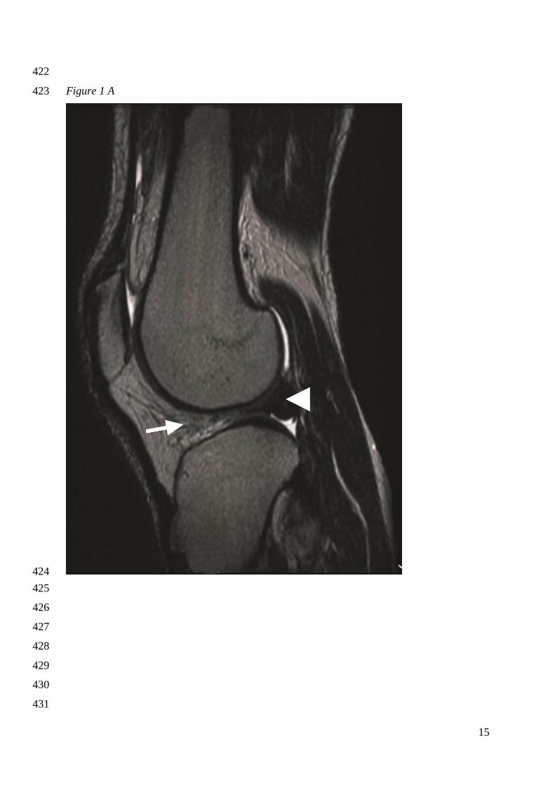
15
422
Figure 1 A 423
424
425
426
427
428
429
430
431

16
432
Figure 1B 433
434
435
436
437
438
439
440
441
442
443
444
445
446
447
448
449
450
451
452
453
454
455

17
456
Figure 2 457
458
459

18
Table 1.
Single table analysis for both menisci in group A
Arthroscopy
MRI positive negative total
Positive 18 10 28
Negative 9 127 136
27 137 164
460 Table 2.
Single table analysis for both menisci in group B
Arthroscopy
MRI positive negative total
Positive 9 6 15
Negative 5 60 65
14 66 80
461

19
Table 3.
Diagnostic validity of 1.0-Tesla MRI in meniscal ruptures
Location Parameter Values and 95% CIs* are expressed
in percentages
Group A Group B
Medial Meniscus
Sensitivity 91.7 (64.6-98.5) 88.9 (56.5-98.0)
Specificity 87.1 (77.3-93.1) 87.1 (71.2-94.9)
Diagnostic accuracy 87.8 (79.0-93.2) 87.5 (73.9-94.5)
Lateral Meniscus
Sensitivity 46.7 (24.8-69.9) 20.0 (3.6-62.5)
Specificity 98.5 (92.0-99.7) 94.3 (81.4-98.4)
Diagnostic accuracy 89.0 (80.4-94.1) 85.0 (70.9-92.9)
Both Menisci
Sensitivity 66.7 (47.8-81.4) 64.3 (38.8-83.7)
Specificity 92.7 (87.1-96.0) 90.9 (81.6-95.8)
Diagnostic accuracy 88.4 (82.6-92.5) 86.3 (77.0-92.2)
*Confidence Intervals
462
Table 4.
Kappa values for interobserver agreement
between readers
Kappa
index
Interpretation
Group A,
both
menisci
0.58 moderate
agreement
Group B,
both
menisci
0.56 moderate
agreement
All patients,
both
menisci
0.57 moderate
agreement
463

20
Table 5.
Kappa values for intraobserver agreement
between readers
Kappa
index
Interpretation
Group A,
both
menisci
0.70 substantial
agreement
Group B,
both
menisci
0.85 almost
perfect
agreement
All patients,
both
menisci
0.76 substantial
agreement
464 465

Reliability of Clinical Findings andMagnetic Resonance Imaging for the
Diagnosis of Chondromalacia PatellaeBy Harri K. Pihlajamaki, MD, PhD, Paavo-Ilari Kuikka, MD, Vesa-Veikko Leppanen, MD,
Martti J. Kiuru, MD, PhD, and Ville M. Mattila, MD, PhD
Investigation performed at the Centre for Military Medicine and the Department of Orthopaedic Surgery,Central Military Hospital, Helsinki, Finland
Background: This diagnostic study was performed to determine the correlation between anterior knee pain andchondromalacia patellae and to define the reliability of magnetic resonance imaging for the diagnosis of chondromalaciapatellae.
Methods: Fifty-six young adults (median age, 19.5 years) with anterior knee pain had magnetic resonance imaging of theknee followed by arthroscopy. The patellar chondral lesions identified by magnetic resonance imaging were comparedwith the arthroscopic findings.
Results: Arthroscopy confirmed the presence of chondromalacia patellae in twenty-five (45%) of the fifty-six knees, asynovial plica in twenty-five knees, a meniscal tear in four knees, and a femorotibial chondral lesion in four knees; normalanatomy was seen in six knees. No association was found between the severity of the chondromalacia patellae seen atarthroscopy and the clinical symptoms of anterior knee pain syndrome (p = 0.83). The positive predictive value for theability of 1.0-T magnetic resonance imaging to detect chondromalacia patellae was 75% (95% confidence interval, 53% to89%), the negative predictive value was 72% (95% confidence interval, 56% to 84%), the sensitivity was 60% (95%confidence interval, 41% to 77%), the specificity was 84% (95% confidence interval, 67% to 93%), and the diagnosticaccuracy was 73% (95% confidence interval, 60% to 83%). The sensitivity was 13% (95% confidence interval, 2% to 49%)for grade-I lesions and 83% (95% confidence interval, 59% to 94%) for grade-II, III, or IV lesions.
Conclusions: Chondromalacia patellae cannot be diagnosed on the basis of symptoms or with current physical ex-amination methods. The present study demonstrated no correlation between the severity of chondromalacia patellae andthe clinical symptoms of anterior knee pain syndrome. Thus, symptoms of anterior knee pain syndrome should not beused as an indication for knee arthroscopy. The sensitivity of 1.0-T magnetic resonance imaging was low for grade-Ilesions but considerably higher for more severe (grade-II, III, or IV) lesions. Magnetic resonance imaging may be con-sidered an accurate diagnostic tool for identification of more severe cases of chondromalacia patellae.
Level of Evidence: Diagnostic Level I. See Instructions to Authors for a complete description of levels of evidence.
Anterior knee pain is a very common disorder in ado-lescents and young adults1,2. However, the pathophys-iology underlying anterior knee pain is controversial. It
has been previously thought to be caused by chondromalaciapatellae. Patellar tendinopathy, patellofemoral malalignment2,or an imbalance in tissue homeostasis3 have also been suggestedto cause similar symptoms. The typical clinical symptoms of
anterior knee pain syndrome include a deep-seated ache andretropatellar pain that worsens when climbing stairs, sittingwith the knees flexed, and rising from a sitting position. Ex-tending the knee against resistance may also cause pain, and asense of insecurity may be felt during weight-bearing4. Theimportance of a reliable diagnosis is emphasized by the factthat the symptoms of anterior knee pain syndrome may re-
Disclosure: The authors did not receive any outside funding or grants in support of their research for or preparation of this work. Neither they nor amember of their immediate families received payments or other benefits or a commitment or agreement to provide such benefits from a commercialentity.
927
COPYRIGHT � 2010 BY THE JOURNAL OF BONE AND JOINT SURGERY, INCORPORATED
J Bone Joint Surg Am. 2010;92:927-34 d doi:10.2106/JBJS.H.01527

semble those of other conditions, such as a meniscal tear5.There is a substantial difference in the required treatment forthese disorders, especially with regard to the potential need forsurgical management. Physical signs of anterior knee pain,such as effusion, quadriceps wasting, and retropatellar crepi-tus, have been claimed to be more informative in the diagnosisof chondromalacia patellae6. However, none of these signs isconsidered specific for chondromalacia patellae6.
Radiographs have not proven to be useful in the diag-nosis of chondromalacia patellae until the disease is in theadvanced stages7,8. A reliable diagnosis can be reached by usingarthroscopy, as it allows a good view of the patellofemoraljoint9. However, surgical treatment of chondromalacia patellaeis indicated in <10% of patients10, and the initial treatment ofchondromalacia consists of a period of rehabilitation11. Thereis evidence that physical therapy can alleviate patellofemoralpain12. Arthroscopic smoothing of fibrillated and traumatizedareas of articular cartilage has been used for grade-II, III, andIV chondromalacia, although the positive treatment outcomesmay deteriorate over time5,13. If the arthroscopic examinationdoes not reveal any arthroscopically treatable lesion, it mayseem to be a costly diagnostic method unnecessarily con-suming our limited health-care resources. Moreover, arthros-copy causes short-term functional disability, pain, and stressand involves risks related to anesthesia and surgery.
It would be beneficial if magnetic resonance imaging,which is a noninvasive diagnostic method, could confirm thediagnosis of chondromalacia patellae. It is a more comfortableprocedure for the patient, and the risk of complications islower than that associated with diagnostic arthroscopy. How-ever, it is unclear whether magnetic resonance imaging canhelp to confirm the correct diagnosis of chondromalacia pa-tellae in patients with anterior knee pain. In previous reports,the overall sensitivity of magnetic resonance imaging for thediagnosis of chondromalacia patellae has ranged between 26%and 100%, the specificity has ranged between 50% and 94%,and the diagnostic accuracy has ranged between 77% and90%14-19. These studies have varied widely with regard to theimaging methods, patient samples, and grading systems uti-lized, which probably explains the different results. Severalauthors have reported that magnetic resonance imaging ismore sensitive for detecting deep chondral lesions than it is fordemonstrating superficial ones15,18,20-22.
The purpose of the present study was to assess the corre-lation between clinical symptoms of anterior knee pain syndromeand chondromalacia patellae. We also assessed the reliabilityof 1.0-T magnetic resonance imaging, by comparing the resultswith those of arthroscopy, for the diagnosis of chondromalaciapatellae in young adults. The hypothesis of this study was thatmagnetic resonance imaging would ensure the diagnosis ofchondromalacia patellae at least in patients with more severechondral lesions.
Materials and Methods
This prospective diagnostic study was performed during athree-year period at the authors’ institution, which provided
all surgical services for the entire armed forces of Finland atthat time. All male citizens in Finland become eligible formandatory military service at the age of eighteen years, and themajority of recruits are nineteen years old at the beginning oftheir basic training. The patients selected for this study wereyoung military trainees who had been deemed healthy enoughto pass the entry medical examination and were able to per-form physically demanding military training. Physical andcombat training for the recruits includes physical exercisecausing increased loads on the lower limb and especially on theknees because kneeling and crawling are often included in thetraining activities. Combat training, marching, and otherphysical training activities were performed almost dailythroughout the service period. Thus, anterior knee pain mayinterfere markedly with the performance of such militarytraining and may threaten the ability to complete militaryservice. Participation in this study was voluntary, and writteninformed consent was obtained from each patient. The studyprotocol was approved by the local ethics committee.
The inclusion criteria consisted of nontraumatic, pro-longed anterior knee pain causing problems that interferedwith military service. An orthopaedic surgeon performedphysical examinations according to the standard policy of theinstitution. These examinations consisted of a systematic andmethodical examination that included inspection; palpation;measurements of motion, muscle strength, limb lengths, andmuscle girth; functional tests to assess walking gait, stationaryrunning, hopping, and squatting; tests to detect mechanical ormeniscal problems such as the McMurray test or Apley test;and evaluations of ligament stability, the extensor mechanism,and the popliteal space. Plain radiographs, including antero-posterior, lateral, and sunrise views, were made. The clinicalsymptoms of anterior knee pain syndrome were divided intothree categories for assessment of their relationship withchondromalacia: (1) diffuse pain in the anterior part of the kneeas a single symptom, (2) typical clinical anterior knee pain syn-drome without patellofemoral crepitus or pain on manipulationof the patella, and (3) typical clinical anterior knee pain syn-drome with patellofemoral crepitus and/or pain on manipula-tion of the patella. The exclusion criteria included a previousfracture, surgical procedure, or injury in the knee region.
The initial treatment for all patients experiencing ante-rior knee pain was nonoperative, consisting mainly of activityrestrictions or rest and nonsteroidal anti-inflammatory drugswhen necessary. In addition, before arthroscopy was consid-ered, the patients were instructed and encouraged to performexercises supervised by a physiotherapist. These included lower-extremity muscle strengthening with a concentration on thequadriceps. Arthroscopy was chosen when the response to non-operative treatment was insufficient and when a reliable methodwas needed to verify the suspected diagnosis of chondroma-lacia patellae or to differentiate between chondromalacia pa-tellae and other internal derangements of the knee. The onlydeviation from the standard policy of the institution was thatmagnetic resonance imaging of the knee was performed for allof the patients included in the study. Most patients underwent
928
TH E J O U R N A L O F B O N E & JO I N T SU R G E RY d J B J S . O R G
VO LU M E 92-A d NU M B E R 4 d A P R I L 2010RE L I A B I L I T Y O F CL I N I C A L F I N D I N G S A N D MAG N E T I C RE S O N A N C E
IM AG I N G F O R T H E DI AG N O S I S O F CH O N D R O M A L AC I A PAT E L L A E

the scan one day prior to the scheduled arthroscopy date. Forpatients who had already undergone magnetic resonance im-aging of the knee as part of the normal clinical assessment andpolicy of the department, the scan was repeated for the pur-pose of the study to minimize the delay between the magneticresonance imaging and the arthroscopy.
Plain radiographs, including anteroposterior, lateral, andsunrise views, were routinely made of all of the patients beforethe physical examination by an orthopaedic surgeon. In orderto evaluate possible anatomic deviations of the patellofemoraljoint, the following measurements and assessments were per-formed. On the sunrise radiographs, the patellar angle wasmeasured between a line connecting the anterior aspects of thefemoral condyles and a second line drawn along the lateralfacet of the patella23. The sulcus angle was measured betweenlines extending from the deepest point of the intercondylarsulcus, medially and laterally, to the tops of the femoral con-dyles. A line bisecting the sulcus angle was compared with aline drawn from the apex of the sulcus angle through thelowest point of the articular ridge of the patella (the congru-ence angle) to detect possible lateralization of the patella. Thesulcus angle was considered to be flat if it was <138�24.
The magnetic resonance imaging performed for all pa-tients prior to the knee arthroscopy was carried out with a 1.0-T scanner (Signa Horizon; GE Medical Systems, Milwaukee,Wisconsin) with a standard knee coil and a field of view of 10to 16 cm. The slice thickness was 3 to 4 mm, with a 0.5 or 1.0-mm intersection gap. Sagittal T1 and T2*-weighted images,coronal proton density images with fat suppression, and axialthree-dimensional T1-weighted fast spoiled gradient-recalledimages with fat suppression were obtained routinely.
A musculoskeletal radiologist on duty initially evaluatedthe images with use of a standard protocol for interpretingmagnetic resonance images. For the purpose of this prospec-tive study, another musculoskeletal radiologist (M.J.K.) whowas blinded to the previous magnetic resonance imaging andarthroscopy findings then reevaluated the images. These re-evaluations differed from the original interpretation in threecases, and a decision was reached by consensus. The magneticresonance images were recorded as showing either normalcartilage thickness or a loss of cartilage. Grading of chondro-malacia with use of the magnetic resonance imaging was basedon the arthroscopic grading system described by Shahriaree11,with 0 indicating normal; I, softening or a blister as indicatedby high signal intensity and swelling of the cartilage; II, fis-suring; III, fragmentation and fissuring; and IV, full-thicknessfissuring and exposed bone (Table I).
Arthroscopy was performed during the first day follow-ing the magnetic resonance imaging examination except inseven cases, in which the time period between the magneticresonance imaging and arthroscopy ranged from two to fourdays. The experienced orthopaedic surgeon performing the ar-throscopy evaluated the articular surfaces while blinded to thefindings on the magnetic resonance imaging. The knees wereexamined systematically with use of a probe inserted into the kneejoint. Under direct vision and with careful probing, special atten-
tion was paid to the patellar surface and the degree of fibrillation,fragmentation, and possible softening of the cartilage surface.
Chondral lesions identified by magnetic resonance imag-ing or arthroscopy were classified into four grades accordingto the system of Shahriaree11 (Table I). All other internal de-rangements of the knee revealed by arthroscopy were recordedand taken into account. If deemed necessary on the basis ofthe arthroscopic findings, arthroscopic operations, postopera-tive mobilization, and follow-up examinations were carried outaccording to the standard practice of the institution.
Statistical MethodsTo determine the validity of the 1.0-T magnetic resonanceimaging for the diagnosis of chondromalacia patellae, themagnetic resonance imaging findings were compared with thearthroscopic findings, which served as the gold standard. Asingle table analysis was used to calculate the positive andnegative predictive values as well as the sensitivity, specificity,and diagnostic accuracy of magnetic resonance imaging. TheFisher exact test was used in two-way tables. Ninety-five per-cent confidence intervals were calculated with the Wilsonmethod. When the results were calculated for grade-I chon-dromalacia patellae, only arthroscopically detected grade-Ilesions were taken into account as positive results (the goldstandard) for chondromalacia, and they were compared withthe magnetic resonance imaging results (with grade 0 on theimages considered negative and grades I through IV consid-ered positive). Results for grades II, III, and IV were calculatedwith arthroscopically detected grade-II, III, and IV lesionsconsidered positive and compared with the magnetic reso-nance imaging results (with grade 0 on the images considerednegative and grades I through IV considered positive).
Source of FundingThere was no external funding source for the investigation.
Results
Fifty-six patients (fifty-four men and two women) met theinclusion criteria for the present study. The median age of
the patients was 19.5 years (range, eighteen to twenty-fiveyears). Twenty-one patients had only one knee examined, andthe remaining thirty-five had both knees examined. Altogether,ninety-one knees were examined but, in bilateral cases, oneknee was randomly selected for the study in order to ensure theindependence of the observations.
TABLE I Classification of Chondromalacia Patellae According
to the System Described by Shahriaree11
Grade
I Fibrillation
II Fissure formation
III Fragmentation
IV Crater formation and eburnation
929
TH E J O U R N A L O F B O N E & JO I N T SU R G E RY d J B J S . O R G
VO LU M E 92-A d NU M B E R 4 d A P R I L 2010RE L I A B I L I T Y O F CL I N I C A L F I N D I N G S A N D MAG N E T I C RE S O N A N C E
IM AG I N G F O R T H E DI AG N O S I S O F CH O N D R O M A L AC I A PAT E L L A E

All of the fifty-six patients were determined to have an-terior knee pain on the basis of the clinical examination by anexperienced orthopaedic surgeon. Arthroscopy confirmed thepresence of chondromalacia patellae in twenty-five (45%) of thefifty-six knees. Twenty knees (36%) had signs of chondroma-lacia patellae on the magnetic resonance images (Figs. 1-A and1-B). With regard to the detection of chondromalacia patellae,the positive predictive value of magnetic resonance imagingwas 75% (95% confidence interval, 53% to 89%), the negativepredictive value was 72% (95% confidence interval, 56% to84%), the sensitivity was 60% (95% confidence interval, 41%to 77%), the specificity was 84% (95% confidence interval,67% to 93%), and the diagnostic accuracy was 73% (95%confidence interval, 60% to 83%). Of eight patients with a
grade-I lesion on arthroscopy, one had a true-positive findingon magnetic resonance imaging, so that magnetic resonanceimaging had a sensitivity of 13% (95% confidence interval, 2%to 49%) for the detection of grade-I lesions. The number oftrue-positive magnetic resonance imaging findings was higherfor grade-II, III, or IV lesions (fourteen of seventeen patients),and the sensitivity was 83% (95% confidence interval, 59% to94%) for detection of those lesions. The results of magneticresonance imaging and arthroscopy for the different grades ofchondromalacia are presented in Table II. The kappa value forthe overall measure of agreement across the five levels was0.368 (p < 0.001).
Eight of the twenty-five arthroscopically confirmed casesof chondromalacia patellae were grade I, nine were grade II,
Fig. 1-A Fig. 1-B
Figs. 1-A and 1-B A nineteen-year-old man reported chronic activity-related pain in the left knee. Clinical examination revealed patellofemoral crepitus
suggestive of chondromalacia patellae. Fig. 1-A An axial fat-suppressed T1-weighted magnetic resonance image of the knee showed intact cartilage
surfaces (arrows). Fig. 1-B Subsequent arthroscopy showed changes suggestive of grade-I chondromalacia on both facets of the patella (arrows). Probing
revealed softening of the cartilage. On the femoral side, the cartilage surfaces were normal, and gliding of the patella in the sulcus was unobstructed.
TABLE II Magnetic Resonance Imaging and Arthroscopic Findings in Fifty-six Knees
Magnetic ResonanceImaging Grade
Arthroscopy Grade
0 (Negative) I (Positive) II (Positive) III (Positive) IV (Positive) Total
0 (negative) 26 7 3 0 0 36
I (positive) 1 0 0 1 0 2
II (positive) 2 1 5 3 0 11
III (positive) 2 0 1 4 0 7
IV (positive) 0 0 0 0 0 0
Total 31 8 9 8 0 56
930
TH E J O U R N A L O F B O N E & JO I N T SU R G E RY d J B J S . O R G
VO LU M E 92-A d NU M B E R 4 d A P R I L 2010RE L I A B I L I T Y O F CL I N I C A L F I N D I N G S A N D MAG N E T I C RE S O N A N C E
IM AG I N G F O R T H E DI AG N O S I S O F CH O N D R O M A L AC I A PAT E L L A E

eight were grade III, and none were grade IV (Figs. 2-A and 2-B). In eight knees, diffuse anterior pain was the only symptom.The typical clinical picture of anterior pain without patello-femoral crepitus or pain on manipulation of the patella waspresent in twelve knees, and the typical clinical picture of an-terior pain with patellofemoral crepitus and/or pain on ma-nipulation of the patella was found in thirty-six knees. Noassociation was found between the severity of chondromalaciapatellae seen on arthroscopy and the clinical symptoms ofanterior knee pain syndrome (p = 0.83).
Of the fifty-six knees with anterior knee pain, twenty-five (45%) had a synovial plica; four, a meniscal tear; four, afemorotibial chondral lesion; and six, normal anatomy with-
out any abnormal arthroscopic findings. All arthroscopicfindings (one or more per knee) and their correlation with theclinical symptoms and physical signs are shown in Table III.
Plain radiographs were made of forty-three knees withanterior knee pain, and they showed normal findings inthirty-six of them (Table IV). Subluxation or lateralization ofthe patellae was found in six knees, a flat sulcus angle wasfound in three knees, Osgood-Schlatter disease was detectedin one knee, and surface irregularity of the patellar facets wasseen in one knee. The subsequent arthroscopy performed forthe current study demonstrated chondromalacia patellae infourteen of the thirty-six knees with normal radiographicfindings.
Fig. 2-A Fig. 2-B
Figs. 2-A and 2-B A twenty-three-year-old man with activity-related swelling of both knees and patellofemoral pain since the start of military service. Plain
radiographs demonstrated a flat sulcus angle in both knees. Fig. 2-A An axial fat-suppressed T1-weighted magnetic resonance image of the left knee
showed some signal variation on the articular surface of the patella, including slight unevenness suggestive of the initial stage of grade-II cartilage defect
formation (arrow). Fig. 2-B Arthroscopy showed smooth, even articular surfaces on the femoral aspect of the patellofemoral joint. The medial aspect of the
patella showed grade-III cartilage damage, which also extended to the lateral aspect (arrows). The size of the damaged area is 2 · 2.5 cm, but the defect
does not reach the subchondral bone. The fragmented areas of cartilage were trimmed by shaving.
TABLE III Symptoms and Physical Signs, and Arthroscopic Findings
Symptoms and/or Physical SignsChondromalacia
PatellaeSynovial
PlicaMeniscalRupture
FemorotibialChondral Lesion
NormalFindings
Total Numberof Findings
Diffuse anterior knee pain assingle symptom (n = 8 patients)
1 4 0 0 3 8
Typical clinical symptoms withoutpatellofemoral crepitus or painon manipulation of patella (n = 12)
6 7 0 0 0 13
Typical clinical symptoms withpatellofemoral crepitus and/or painon manipulation of patella (n = 36)
18 14 4 4 3 43
Total 25 25 4 4 6 64
931
TH E J O U R N A L O F B O N E & JO I N T SU R G E RY d J B J S . O R G
VO LU M E 92-A d NU M B E R 4 d A P R I L 2010RE L I A B I L I T Y O F CL I N I C A L F I N D I N G S A N D MAG N E T I C RE S O N A N C E
IM AG I N G F O R T H E DI AG N O S I S O F CH O N D R O M A L AC I A PAT E L L A E

Discussion
The purposes of this study were to assess the correlation ofclinical symptoms of anterior knee pain syndrome and
chondromalacia patellae and to define the reliability of 1.0-Tmagnetic resonance imaging in the diagnosis of chondro-malacia patellae in young adults. Our hypothesis was thatmagnetic resonance imaging would confirm the diagnosis ofchondromalacia patellae at least in the case of more severe,deeper chondral lesions. In our study, chondromalacia patellaewas found in less than half of the patients with anterior kneepain and 1.0-T magnetic resonance imaging was accurate inidentifying more severe cases of chondromalacia patellae.
The principal finding of the present study was that thereis no correlation between the presence of softened articularcartilage (chondromalacia patellae) and clinical symptoms ofanterior knee pain syndrome. This finding is supported byprevious reports in the literature that also alluded to the ab-sence of pathognomonic symptoms6. According to our results,diffuse anterior knee pain as the sole symptom seems to ex-clude a diagnosis of chondromalacia patellae fairly reliablybecause the diagnosis was confirmed in only one of eight pa-tients with that symptom alone. Chondromalacia patellae wasseen in only half of the patients with typical clinical symptomswithout patellofemoral crepitus or pain on manipulation ofthe patella. Our results correspond with those of Leslie andBentley, who found that only 60% of patients with clinicallydiagnosed anterior knee pain and retropatellar crepitus hadchondromalacia6. Thus, the association between symptoms andthe identification of chondromalacia patellae with arthroscopyis not clear3. In addition, an important finding was the lack ofan association between the severity of chondromalacia patellaeand the clinical symptoms, which contradicted the observa-tions reported by Kettunen et al., who reported that patientswith more severe cartilage lesions of the patella or the femoraltrochlea had more subjective symptoms and functional limi-tations than those without lesions or with small cartilagelesions25. The findings of the present study suggest thatchondromalacia patellae cannot be distinguished by symptomsand signs on physical examination.
In addition to chondromalacia patellae (softening of thepatellar articular cartilage), a medial synovial plica of the knee
was a common arthroscopic finding in patients experiencinganterior knee pain. It was previously suggested that a medialsynovial plica can cause symptoms, possibly by generatingchondromalacia26. In the present sample, a medial synovialplica was detected arthroscopically in nearly half of the knees,but its association with symptoms must also be evaluatedcritically. Previous reports showed no correlation between thesize of the plica and the severity of the symptoms27,28 and thateven smaller plicae may impinge on the adjacent cartilage ofthe femoral condyle29. Moreover, recent reports suggest that,although aspects of the pathomechanism of plica syndromeremain unclear, arthroscopic resection should be considered ifincapacitating knee pain is accompanied by an arthroscopicfinding of a medial plica of the knee27,29,30.
In the study by Lee et al., the sensitivity of magneticresonance imaging in detecting all of the grades of chondro-malacia patellae was 57%15, which is close to the sensitivity of60% found in our study. Using T2-weighted magnetic reso-nance imaging, McCauley et al.14 found a slightly higher sen-sitivity (72%), an almost equal specificity (87%), and a slightlyhigher accuracy (79%) compared with what we found inour study. Using T2-weighted magnetic resonance imaging,Handelberg et al.19 reported an accuracy (82%) similar to thatreported by McCauley et al. However, it has also been reportedthat T2-weighted magnetic resonance imaging may producemany false-negative results20. Other results in the literaturehave deviated more substantially from those in our study. Thehighest sensitivity (100%) was achieved with T2-weighted spin-echo pulse sequences in the sagittal and axial planes and witha three-dimensional fast sequence in the sagittal plane19. Analmost equally high sensitivity (99%) was reported for three-dimensional spoiled gradient-recalled imaging with fat sup-pression after intravenous injection of contrast medium31.However, the combination that provided the highest sensitivityled to poorer specificity (50%). The highest specificity (94%)was achieved with spectral presaturation with inversion re-covery sequences16.
On the basis of the differences among the chondroma-lacia patellae grades, with a sensitivity of only 13%, grade-Ichondral defects cannot be reliably detected with use of 1.0-Tmagnetic resonance imaging. The imaging sensitivity wasconsiderably higher (83%) for grades II and III. Although nopatients in the study had grade-IV chondromalacia patellae,our results clearly indicated that the sensitivity was highest forthe deepest lesions. This finding is consistent with those pre-sented previously15,20,21. Gagliardi et al., using the same chon-dromalacia patellae grading system11 that we used, reported asensitivity of 0% for grade-I chondromalacia patellae with allof their imaging sequences20. For grade-II and III lesions, thesensitivity was between 13% (proton-density-weighted magneticresonance imaging) and 47% (T2-weighted imaging), and,for grade-IV lesions, it was between 50% (spoiled gradient-recalled imaging) and 75% (T1-weighted, T2-weighted, andproton-density-weighted imaging)20. The sensitivities reportedby Gagliardi et al. for all of the sequences that they used werelower than the sensitivities found in the present study. Murphy
TABLE IV Radiographic and Arthroscopic Findings
Findingson Plain
Radiographs
ArthroscopicallyConfirmed
ChondromalaciaPatellae inSame Knee
Normal radiographic findings 36 14
Subluxation/lateralization 6 4
Flat sulcus angle 3 2
Osgood-Schlatter disease 1 1
Surface irregularityof patellar facets
1 0
932
TH E J O U R N A L O F B O N E & JO I N T SU R G E RY d J B J S . O R G
VO LU M E 92-A d NU M B E R 4 d A P R I L 2010RE L I A B I L I T Y O F CL I N I C A L F I N D I N G S A N D MAG N E T I C RE S O N A N C E
IM AG I N G F O R T H E DI AG N O S I S O F CH O N D R O M A L AC I A PAT E L L A E

used T2*-weighted three-dimensional gradient-echo magneticresonance imaging and reported results very similar to ours,with a sensitivity of 83% for grade-III and IV lesions21. Lee et al.reported a higher sensitivity—94% for grade-III and IV chon-dromalacia patellae with use of axial inversion recovery-fastspin-echo imaging15. However, on the basis of the present study,magnetic resonance imaging should be considered instead ofarthroscopy for patients with prolonged anterior knee pain.
Plain radiographs have been considered inadequate for theidentification of early cartilage damage7,8. However, it has beensuggested that chondromalacia patellae might frequently be ac-companied by subluxation of the patella or a flat sulcus angle32,33.Contrary results have been reported in studies concerning ante-rior knee pain34 and arthroscopically diagnosed chondromalaciapatellae6. Although our results seem to support the latter stud-ies6,34, no conclusion can be drawn from the abnormal findingsreported here because of their small number. For example, al-though it may seem that chondromalacia patellae is accompaniedby a flat sulcus angle, the very small number of cases with such anangle and the seemingly normal articular surfaces on these imagesdo not support such a conclusion.
Our study has several noteworthy strengths, including theprospective design and the homogeneity of the study popula-tion. Our study sample consisted of young military traineesbetween eighteen and twenty-five years of age, who all weredeemed healthy enough to pass their military entry medicalexamination. All were expected to have attained, after com-pletion of their military basic training, a physical level enablingthem to march or ski, while carrying a full military packweighing 25 kg and a rifle, a distance of 15 km on two con-secutive days while maintaining fitness for battle. Due to thecompulsory nature of the military service, the study partici-pants reflect the young adult male population of the country.Moreover, the age group that we studied is optimal becauseindividuals in that group do not normally have degenerativechondral changes such as osteoarthritis. This relatively largegroup of skeletally mature young adults underwent a uniformexamination and treatment for anterior knee pain according tothe policy of one institution. The standard methods and pro-tocols of the institution were used for the evaluation of clinicalsymptoms, plain radiographs, magnetic resonance images, and
arthroscopic findings. Magnetic resonance imaging was ordi-narily performed on the day prior to the arthroscopy to avoidpossible bias caused by any delay.
The strength of the magnetic resonance imaging field usedin the present study (1.0 T) may be considered a limitation,although it is unclear whether higher field strength might haveresulted in a more reliable diagnosis of chondromalacia patellae.As mentioned in the Results section, a total of ninety-one kneeswere examined but, in cases of bilateral findings, only one kneewas randomly selected for the study. This limited the number ofthe knees in the present study and can be considered a limitation.
In conclusion, chondromalacia patellae cannot be diag-nosed on the basis of symptoms or with current methods ofphysical examination. The present study demonstrated nocorrelation between the severity of the chondromalacia patellaeand the clinical symptoms of anterior knee pain syndrome.Thus, these symptoms should not be used as an indication forknee arthroscopy. The sensitivity of magnetic resonanceimaging for the detection of grade-I lesions was low and couldnot confirm the diagnosis of chondromalacia patellae. However,the sensitivity of magnetic resonance imaging was considerablyhigher for the detection of more severe (grade-II or III) lesions,and 1.0-T magnetic resonance imaging may be considered anaccurate diagnostic tool for the identification of more severecases of chondromalacia patellae. In addition to chondroma-lacia patellae, a medial synovial plica of the knee was a commonarthroscopic finding in young adult patients experiencing an-terior knee pain. n
NOTE: The authors thank Marja Vajaranta from the University of Tampere (Tampere, Finland) forrevising the language and Kari Kelho from the Centre for Military Medicine, Research Unit (Hel-sinki, Finland), for editing the images.
Harri K. Pihlajamaki, MD, PhDPaavo-Ilari Kuikka, MDVesa-Veikko Leppanen, MDMartti J. Kiuru, MD, PhDVille M. Mattila, MD, PhDResearch Unit, Centre for Military Medicine, P.O. Box 50, FIN-00301Helsinki, Finland. E-mail address for H.K. Pihlajamaki:[email protected]
References
1. Wiles P, Andrews PS, Devas MB. Chondromalacia of the patella. J Bone JointSurg Br. 1956;38:95-113.
2. Sanchis-Alfonso V, ed., editor. Anterior knee pain and patellar instability. Lon-don: Springer; 2006.
3. Dye SF. The pathophysiology of patellofemoral pain: a tissue homeostasis per-spective. Clin Orthop Relat Res. 2005;436:100-10.
4. Robinson AR, Darracott J. Chondromalacia patellae. A survey conducted at theArmy Medical Rehabilitation Unit, Chester. Ann Phys Med. 1970;10:286-90.
5. Stoller DW, ed., editor. Magnetic resonance imaging in orthopaedics and sportsmedicine. 3rd ed. Baltimore: Lippincott Williams and Wilkins; 2006.
6. Leslie IJ, Bentley G. Arthroscopy in the diagnosis of chondromalacia patellae.Ann Rheum Dis. 1978;37:540-7.
7. Chan WP, Lang P, Stevens MP, Sack K, Majumdar S, Stoller DW, Basch C,Genant HK. Osteoarthritis of the knee: comparison of radiography, CT, and MR
imaging to assess extent and severity. AJR Am J Roentgenol. 1991;157:799-806.
8. Wright RW, Boyce RH, Michener T, Shyr Y, McCarty EC, Spindler KP. Radiographsare not useful in detecting arthroscopically confirmed mild chondral damage. ClinOrthop Relat Res. 2006;442:245-51.
9. Casscells SW. Arthroscopy of the knee joint. A review of 150 cases. J Bone JointSurg Am. 1971;53:287-98.
10. Dehaven KE, Dolan WA, Mayer PJ. Chondromalacia patellae in athletes.Clinical presentation and conservative management. Am J Sports Med. 1979;7:5-11.
11. Shahriaree H. Chondromalacia. Contemp Orthop. 1985;11:27-39.
12. Crossley K, Bennell K, Green S, Cowan S, McConnell J. Physical therapy forpatellofemoral pain: a randomized, double-blinded, placebo-controlled trial. Am JSports Med. 2002;30:857-65.
933
TH E J O U R N A L O F B O N E & JO I N T SU R G E RY d J B J S . O R G
VO LU M E 92-A d NU M B E R 4 d A P R I L 2010RE L I A B I L I T Y O F CL I N I C A L F I N D I N G S A N D MAG N E T I C RE S O N A N C E
IM AG I N G F O R T H E DI AG N O S I S O F CH O N D R O M A L AC I A PAT E L L A E

13. Federico DJ, Reider B. Results of isolated patellar debridement for patellofem-oral pain in patients with normal patellar alignment. Am J Sports Med. 1997;25:663-9.
14. McCauley TR, Kier R, Lynch KJ, Jokl P. Chondromalacia patellae: diagnosis withMR imaging. AJR Am J Roentgenol. 1992;158:101-5.
15. Lee SH, Suh JS, Cho J, Kim SJ, Kim SJ. Evaluation of chondromalacia of thepatella with axial inversion recovery-fast spin-echo imaging. J Magn Reson Imaging.2001;13:412-6.
16. Macarini L, Perrone A, Murrone M, Marini S, Stefanelli M. Evaluation of patellarchondromalacia with MR: comparison between T2-weighted FSE SPIR and GE MTC.Radiol Med. 2004;108:159-71.
17. Rose PM, Demlow TA, Szumowski J, Quinn SF. Chondromalacia patellae: fat-suppressed MR imaging. Radiology. 1994;193:437-40.
18. Vanarthos WJ, Pope TL Jr, Monu JU. Comparison of axial T1 spin-echo and T1fat-saturation magnetic resonance imaging techniques in the diagnosis of chon-dromalacia patellae. Orthop Rev. 1994;23:942-6.
19. Handelberg F, Shahabpour M, Casteleyn PP. Chondral lesions of the patellaevaluated with computed tomography, magnetic resonance imaging, and arthros-copy. Arthroscopy. 1990;6:24-9.
20. Gagliardi JA, Chung EM, Chandnani VP, Kesling KL, Christensen KP, Null RN,Radvany MG, Hansen MF. Detection and staging of chondromalacia patellae: rela-tive efficacies of conventional MR imaging, MR arthrography, and CT arthrography.AJR Am J Roentgenol. 1994;163:629-36.
21. Murphy BJ. Evaluation of grades 3 and 4 chondromalacia of the knee usingT2*-weighted 3D gradient-echo articular cartilage imaging. Skeletal Radiol.2001;30:305-11.
22. Brown TR, Quinn SF. Evaluation of chondromalacia of the patellofemoral com-partment with axial magnetic resonance imaging. Skeletal Radiol. 1993;22:325-8.
23. Laurin CA, Levesque HP, Dussault R, Labelle H, Peides JP. The abnormal lateralpatellofemoral angle: a diagnostic roentgenographic sign of recurrent patellar sub-luxation. J Bone Joint Surg Am. 1978;60:55-60.
24. Merchant AC, Mercer RL, Jacobsen RH, Cool CR. Roentgenographicanalysis of patellofemoral congruence. J Bone Joint Surg Am. 1974;56:1391-6.
25. Kettunen JA, Visuri T, Harilainen A, Sandelin J, Kujala UM. Primary cartilagelesions and outcome among subjects with patellofemoral pain syndrome. Knee SurgSports Traumatol Arthrosc. 2005;13:131-4.
26. Munzinger U, Ruckstuhl J, Scherrer H, Gschwend N. Internal derangement ofthe knee joint due to pathologic synovial folds: the mediopatellar plica syndrome.Clin Orthop Relat Res. 1981;155:59-64.
27. Christoforakis JJ, Sanchez-Ballester J, Hunt N, Thomas R, Strachan RK. Syno-vial shelves of the knee: association with chondral lesions. Knee Surg SportsTraumatol Arthrosc. 2006;14:1292-8.
28. Jee WH, Choe BY, Kim JM, Song HH, Choi KH. The plica syndrome: diagnosticvalue of MRI with arthroscopic correlation. J Comput Assist Tomogr. 1998;22:814-8.
29. Lyu SR, Hsu CC. Medial plicae and degeneration of the medial femoral condyle.Arthroscopy. 2006;22:17-26.
30. Farkas C, Hargitai Z, Gaspar L, Kuki A, Csernatony Z, Szepesi K. Histologicalchanges in the symptomatic mediopatellar plica. Knee. 2004;11:103-8.
31. Suh JS, Cho JH, Shin KH, Kim SJ. Chondromalacia of the knee: evaluation witha fat-suppression three-dimensional SPGR imaging after intravenous contrast in-jection. J Magn Reson Imaging. 1996;6:884-8.
32. Aglietti P, Cerulli G. Chondromalacia and recurrent subluxation of the patella: astudy of malalignment, with some indications for radiography. Ital J Orthop Trau-matol. 1979;5:187-201.
33. Langston HH. Dislocation of patella and its relation to chondromalacia patellae.Br Med J. 1958;1:155-6.
34. Laprade J, Culham E. Radiographic measures in subjects who are asymptom-atic and subjects with patellofemoral pain syndrome. Clin Orthop Relat Res.2003;414:172-82.
934
TH E J O U R N A L O F B O N E & JO I N T SU R G E RY d J B J S . O R G
VO LU M E 92-A d NU M B E R 4 d A P R I L 2010RE L I A B I L I T Y O F CL I N I C A L F I N D I N G S A N D MAG N E T I C RE S O N A N C E
IM AG I N G F O R T H E DI AG N O S I S O F CH O N D R O M A L AC I A PAT E L L A E

Knee injuries related to sports in young adult males duringmilitary service – Incidence and risk factors
P.-I. Kuikka1, H. K. Pihlajamäki1, V. M. Mattila1,2
1Centre for Military Medicine; and Department of Orthopaedic Surgery, Central Military Hospital, Helsinki, Finland, 2Department ofOrthopedics and Traumatology, Tampere University Hospital, Tampere, FinlandCorresponding author: Paavo-Ilari Kuikka, MD, Centre for Military Medicine, Research Unit, PO Box 50, FIN-00301 Helsinki,Finland. Tel: +358 9 181 25814, Fax: +358 9 181 25827, E-mail: [email protected]
Accepted for publication 4 August 2011
The population-based incidence and risk factors for kneeinjuries in young adults were assessed in Finnish maleconscripts performing their compulsory military service(n = 128 584). The main outcome variables were (1) hos-pitalization due to knee injuries overall and (2) hospitali-zation due to knee disorders as categorized into specificInternational Classification of Diseases, tenth revisiondiagnoses (cruciate and collateral ligament tears, menis-cal tears, traumatic chondral lesions, and patellar dislo-cations). Person-time injury-incidence rates werecalculated by dividing the number of persons with a diag-nosed knee injury by the total exposure time of 97 503person-years. The number of subjects with surgical
operations and military service class changes indicative oflonger term notable disability are also reported. Riskfactor analyses were performed by logistic regression.The person-based incidence of hospitalizations for kneeinjury was 11 cases per 1000 person-years [95% confi-dence interval (CI): 10.4–11.7]. The most important riskfactors were higher age (odds ratio 1.7; 95% CI: 1.3–2.2)and obesity (odds ratio 1.6; 95% CI: 1.03–2.5). Two thirdsof all subjects hospitalized for knee injuries had surgery,and one third had longer term notable disability. Thesefindings indicate that knee injuries cause a significantburden of hospitalizations, often leading to surgery andlonger term disability.
Knee injuries are frequently seen in the everyday clinicalpractice of orthopedic surgeons and general practition-ers. The incidence of all knee injuries is greatest inpatients under 30 years of age and especially in males(Kannus & Jarvinen, 1989; Majewski et al., 2006;Gianotti et al., 2009). In the general population, the inci-dence is suggested to be 11 cases per 1000 person-years(Kannus & Jarvinen, 1989). In most of the previousincidence rate studies, however, only specific knee diag-noses, such as tears of the anterior cruciate ligament(ACL; Anderson et al., 2001; Bodor, 2001; Parkkariet al., 2008) were evaluated, or the study sample com-prised a definite, restricted sample of a special subpopu-lation, such as a sports team or patient catchmentof a specialized clinic. Comprehensive studies withpopulation-based sample are scarce, and thus data ofoverall incidence rates of knee injuries are difficult togeneralize for application in clinical practice.
Majewski et al. (2006) conducted a comprehensive10-year study of the epidemiology of knee injuries insports. Almost half of the injuries were sustained bythose aged 20–29. The most common injury was a kneesprain without clearly identifiable internal derange-ment, and the most common diagnoses were ACLtear (20.3%), medial meniscus (MM) tear (10.8%),
chondral lesion (10.6%), medial collateral ligament(MCL) tear (7.9%), contusion injury due to directtrauma (5.5%), lateral meniscus (LM) tear (3.7%), andpatellar dislocation (3.3%).
Participation in sports is a significant risk factor forknee injuries, and as many as half of all knee injuriesoccur during sports activities (Kannus & Jarvinen,1989). As many as 40% of sports injuries are knee inju-ries (Majewski et al., 2006). The association betweenaerobic fitness and anthropometric properties of kneeinjuries is not well-known. A higher age increases therisk of disabling knee injuries among young men in theU.S. army (Sulsky et al., 2000). Higher age, obesity, andpoor results in a running test are suggested to be riskfactors for musculoskeletal injuries as a whole (Heir &Eide, 1996). Participation in sports, weak quadricepsmuscle, and female sex are associated with ACL tears(Anderson et al., 2001; Bodor, 2001; Parkkari et al.,2008). Participation in sports, weak quadriceps muscle,and older age are known risk factors for meniscal tears(Baker et al., 2002, 2003; Canale ebST, 2003). Patellardislocations typically occur in young adults duringsports (Atkin et al., 2000). Risk factors for acute patellardislocations are suggested to be higher height and weight(Sillanpaa et al., 2008). Overweight and older age
Scand J Med Sci Sports 2011: ••: ••–••doi: 10.1111/j.1600-0838.2011.01397.x
© 2011 John Wiley & Sons A/S
1

predispose young adult males to deeper chondral lesions(Eskelinen et al., 2004).
Population-based studies of the incidence of kneeinjuries as well as the risk factors for those injuries inthe literature are sparse. Therefore, we conducted apopulation-based study among Finnish conscripts toassess the incidence and risk factors for knee injuriesleading to hospitalization.
Materials and methodsStudy population
The study population comprised Finnish male conscripts perform-ing their compulsory military service between the years 1998–2002. Military service is compulsory for all Finnish males, and thusconscripts represent an extensive sample from the general popula-tion of young adult males in Finland. Approximately 80% of allmales in Finland perform military service. Chronic diseases such asdiabetes, asthma, and mental health problems are the main reasonsfor exemption from military service. Before entering militaryservice, all conscripts had to pass a medical examination performedby a physician. Health status was re-checked by a physician alsoduring the first week of service, indicating that conscripts werehealthy young men. In case that medical examination revealed kneeinjuries or previous operations that cause severe disability, theconscript was discharged from the service. The age of the conscriptsvaries between 18 and 30 years (median, 20 years). A new batch ofconscripts enters twice a year, in January and in July. The manda-tory military service period ranges from a maximum of 362 days(officers; conscripts trained for particularly demanding duties) to270 days (conscripts trained for work requiring special skills), witha minimum of 180 days (other rank-and-file duties).
Military service begins with a basic training period of 8 weeksconsisting of increasing levels of walking, marching, bicycling,running, and other physical activities. The intensity of militarytraining gradually increases, averaging 17 h per week. Moreover,conscripts participate in other physical training exercises, such asfloorball, weight training, jogging, and circuit training for 7 h perweek on average. Over the following 4 months of service (specialand team training period), the amount of moderate and high-intensity physical training is maintained at the same level in dif-ferent companies. During the first 6 months of military service,conscripts are expected to complete approximately 450 h ofinstructed physical training (19 h per week). More specific detailsof the military service in Finland have been published previously(Mattila et al., 2007b; Taanila et al., 2009).
Hospitalization data
During military service, all conscripts are required to use only theservices of the primary military health-care units and CentralMilitary Hospital when in need of medical treatment. Informationfrom all hospital and health-care unit admissions in Finland iscollected to the National Hospital Discharge Register (NHDR). Thecoverage and accuracy of the NHDR is good (Salmela & Koistinen,1987; Keskimaki & Aro, 1991; Mattila et al., 2008). These factorstogether allow for extensive and very reliable documentation of allhospitalizations due to knee injuries during the military service.
Data obtained from the NHDR included all military conscriptsadmitted alive to primary military health-care units and the CentralMilitary Hospital as inpatients from January 1, 1998, to December31, 2002. Injuries that occurred during the conscript’s leisure timeor on the way to a vacation or returning to the garrison, as well asthose occurring during military service were included. Informationof hospitalizations due to knee injuries was collected from the
NHDR with diagnoses coded according to the International Clas-sification of Diseases, tenth revision (ICD-10; WHO, 1992). Inclu-sion criteria were hospitalization with main or secondary diagnoseof ACL and posterior cruciate ligament (PCL) tears (S83.5), freshand old meniscal tears (S83.2, M23.2), MCL and lateral collateralligament (LCL) tears (S83.4), traumatic chondral lesions (S83.3),and patellar dislocations (S83.0). Information about surgicaloperations was also collected.
Disability data
Data concerning longer term disability (service class change)caused by knee injuries were obtained from the national register ofthe Finnish Defense Forces. The register contains informationregarding ICD-10 diagnoses leading to a service class change.Register contained main diagnose and one secondary diagnose,and both were taken into account when calculating number ofsubjects with service class change.
When a knee injury causes disability and problems coping withmilitary service for approximately 2– 3 weeks or more, a serviceclass change is considered by the garrison physician. A final deci-sion is made only after thorough clinical examination, includingradiographs, and magnetic resonance imaging (MRI) and arthros-copy when needed. When the service class is lowered, militaryservice is prematurely interrupted, or the conscript is transferred toa physically less-demanding duty. A service class change due to aknee injury diagnosis is a reliable indicator of longer term disabil-ity to cope with normal military service.
Follow-up
The follow-up time for each conscript was calculated from entryinto military service to the first hospitalization due to a knee injuryor to the discharge from the service if no hospitalizations occurred.The total exposure time for the population at risk was calculatedfrom a register containing the total number of realized service daysduring the study period. The total exposure time for the populationat risk during the study period was 97 503 person-years.
Aerobic fitness and anthropometric measurements
For the purposes of data analysis, height and weight were dividedinto quartiles. Age was divided into three groups matched by size.Body mass index (BMI) was computed as the weight in kilogramsdivided by the square of the height in meters (kg/m2). The Inter-national Classification of adult underweight, overweight, andobesity according to BMI by the World Health Organization wasused to divide the conscripts into different categories as follows:BMI <18.5 was classified as a underweight, 18.5–24.99 as normal,25–29.99 as overweight, and �30 as obese. Aerobic condition wasmeasured by determining the distance covered in a 12-min Cooperrunning test. Results were divided in four categories as follows:<2200 m (poor), 2200–2599 m (fair), 2600–2999 m (good), and�3000 m (very good). The Finnish Defence Force muscularstrength test includes distance of horizontal jump without speedand number of sit-ups, push-ups, pull-ups, and back lifts per-formed in 1 min, with values ranging from 0 to 3 points permovement. A total of 0–4 points was considered poor, 5–8 pointsas fair, 9–12 points as good, and 13–15 points as very good.
Person-time incidence rates for knee injuries leading to hospi-talization were calculated by dividing the number of persons witha knee diagnosis by the total exposure time, and 95% confidenceintervals (CI) were reported. Subjects might have been hospital-ized multiple times for the same diagnosis, and for this reason,only first admissions were taken into account when calculating theincidence. The number of possible readmissions, surgical opera-tions, and service class changes by diagnoses are also reported.
Kuikka et al.
2

Risk factor analyses for knee injuries were calculated by logis-tic regression. The main outcome variables were (1) hospitaliza-tion due to knee injuries in general and (2) hospitalization due toknee injuries as categorized into the specific ICD-10 diagnoses[ACL and PCL tears (S83.5), fresh and old meniscal tears (S83.2,M23.2), MCL and LCL tears (S83.4), traumatic chondral lesions(S83.3), and patellar dislocations (S83.0)]. Risk factors analyzedwere the conscript’s age, length of military service, weight, height,BMI, and physical performance as measured with Cooper’s12-min running test and muscle strength. First, we constructed alogistic regression model including age, the length of militaryservice, and risk factors one by one. Only those risk factors sig-nificantly associated (P < 0.05) with outcome were included intothe final logistic regression model. Because BMI and weight had astrong interaction, only BMI was included into the final logisticregression model. Odds ratios (OR) were calculated with a 95%CI. Statistical analyses were performed using SPSS 17.0 (IBMCorporation, NY, USA) for Windows software.
The Medical Ethics Committee of the institution providedapproval for this study.
Results
The total number of 18–30-year-old male conscripts whostarted their military service during the study period ofJanuary 1, 1998, to December 31, 2002, was 128 584.The total exposure time for the population at risk duringthe study period was 97 503 person-years. The meanduration of military service was 9 months.
The total number of subjects with a knee injuryleading to at least one hospitalization was 1073 (0.8% ofall conscripts), and the person-time hospitalization inci-dence rate was 11 cases per 1000 person-years (95% CI:10.4–11.7). Two hundred ninety-eight subjects (27.8%of the hospitalizations) were hospitalized for old menis-cal tear followed by acute patellar dislocation (236cases/22.0%), fresh meniscal tear (214 cases/19.9%),ACL/PCL tear (208 cases/19.4%), MCL/LCL tear (99cases/9.2%), and fresh chondral lesion (18 cases/1.7%).Detailed results for person-time hospitalization inci-dence rates are presented in Table 1.
Table 2 presents the readmission status and number ofsurgical operations of any kind for each hospitalizedsubject. Approximately 30% of all cases had consecutivehospitalizations after the first admission. Surgery wasrequired in 59% of the cases, and knee injuries led to aservice class change (notable disability) in 32% of thecases.
Subjects hospitalized for ACL/PCL tears had readmis-sion in 45% of the cases, and more than half of theprimary cases led to a service class change. Hospitaliza-tions for patellar dislocation also led to many readmis-sions but led to a service class change in only 23% of thecases. Meniscal tears and fresh chondral lesions led to aservice class change in approximately one third of thecases. MCL/LCL tears led to a service class change inonly 11% of the cases. Subjects hospitalized for oldmeniscal tears had surgery in 83% of the cases. Corre-sponding numbers were 62% for fresh meniscal tears,67% for ACL/PCL tears, 44% for fresh chondral lesions,31% for patellar dislocations, and 28% for MCL/LCLtears.
Mean time from the beginning of the service to thefirst hospitalization was 126 days. Twenty-five percentof the injuries occurred during the basic training period(first 8 weeks, days 1–56), 49% during the special andteam training period (days 57 to 180) and 26% during theleadership period (days 180 to 365). Correspondingnumbers for old meniscal tears were 13%, 57%, and31%, for patellar dislocations were 43%, 34%, and 23%,for fresh meniscal tears were 22%, 54%, and 24%, forcruciate ligament tears were 18%, 51%, and 31%, for
Table 1. Incidence (per 1000 person-years) of hospitalization due to kneeinjuries among 128 584 conscripts in 1998–2002 in Finland
Cases Incidence* 95% CI
MenOld meniscal tear 298 3.1 2.7–3.4Patellar dislocation 236 2.4 2.1–2.7Fresh meniscal tear 214 2.2 1.9–2.5ACL/PCL tear 208 2.1 1.9–2.4MCL/LCL tear 99 1.0 0.8–1.2Fresh chondral lesion 18 0.2 0.1–0.3Total 1073 11.0 10.4–11.7
*Cases per 1000 person-years.ACL, anterior cruciate ligament; CI, confidence interval; LCL, lateral col-lateral ligament; MCL, medial collateral ligament; PCL, posterior cruciateligament.
Table 2. Readmission status, service class changes, and need for surgical operations among Finnish conscripts with knee injury in 1998–2002
Primary admission(N of subjects)
Cases requiringreadmission (%)
Service classchange (%)
Cases requiringsurgery (%)
Old meniscal tear 298 70 (23) 91 (31) 248 (83)Patellar dislocation 236 84 (36) 55 (23) 73 (31)Fresh meniscal tear 214 60 (28) 66 (31) 133 (62)ACL/PCL tear 208 94 (45) 110 (53) 140 (67)MCL/LCL tear 99 10 (10) 11 (11) 28 (28)Fresh chondral lesion 18 3 (17) 6 (33) 8 (44)total 1073 321 (30) 339 (32) 630 (59)
ACL, anterior cruciate ligament; LCL, lateral collateral ligament; MCL, medial collateral ligament; PCL, posterior cruciate ligament.
Knee injuries in young adult males
3

collateral ligament tears were 32%, 53%, and 15%, andfor fresh chondral lesions approximately were 10%,60%, and 30%. The three most common injuries duringthe first 8 weeks were patellar dislocation (47% of thecases), fresh meniscal tear (16%), and old meniscal tear(14%). Corresponding numbers for days 57–180 were32% for old meniscal tears, 21% for fresh meniscal tears,and 20% for patellar dislocations. During the days 180–365, 33% of the injuries were for old meniscal tears,24% patellar for dislocations, and 19% for cruciate liga-ment tears.
Higher age was significantly associated with kneeinjury leading to hospitalization (OR 1.7; 95% CI: 1.3–2.2; Table 3). Obesity was also significantly associatedwith hospitalization for knee injury, and the riskincreased with an increase in the BMI. Subjects withBMI � 30 had an OR of 1.6 (95% CI: 1.03–2.5) forknee injuries as compared with the subjects with BMI< 25. Subjects with the greatest muscular strength had1.6 (95% CI: 1.2–2.4) times the risk of knee injuryhospitalization compared with subjects in the lowestquartile. The results of the 12-min running test, under-weight, and height were not associated with hospitali-zation for knee injury.
Final logistic regression models (Table 4) were usedto determine risk factors for hospitalizations due to spe-cific ICD-10 knee diagnoses. Obesity was a statisticallysignificant risk factor for hospitalizations due to patellardislocation and MCL/LCL tears. Obesity had an OR of2.0 (95% CI: 1.1–3.5) for hospitalization due to patellar
dislocation and an OR of 3.1 (95% CI: 1.4–7.0) forhospitalization due to MCL/LCL tear. Weight was anindependent risk factor for meniscal tears. The heaviestweight quartile had an OR of 2.6 (95% CI: 1.4–5.0) forhospitalization due to a fresh meniscal tear and an OR of1.8 (95% CI: 1.1–2.8) for hospitalization due to an oldmeniscal tear. The oldest age-group had a 2.4 times (95%CI: 1.7–4.5) greater risk for old meniscal tears comparedwith the youngest age-group. Subjects in the highestmuscle strength quartile had an increased risk for freshmeniscal tears and ACL/PCL tears.
Table 3. Logistic regression predicting hospitalization due to knee injuriesamong 128 584 Finnish conscripts in 1998–2002
Predictor Oddsratio
95% CI forodds ratio
Lower Upper
Age18–19 120 1.0 0.9 1.321–30 1.7 1.3 2.2
Length of military service180 days 1270 days 1.5 0.9 2.4360 days 1.3 1.03 1.5
UnderweightNo 1Yes 0.7 0.4 1.1
Overweight and obesityNo 1Overweight 1.0 0.8 1.3Obesity 1.6 1.03 2.5
Muscle strengthLowest quartile 1Second quartile 1.0 0.7 1.3Third quartile 1.1 0.8 1.5Highest quartile 1.6 1.2 2.4
CI, confidence interval.
Table 4. Multivariate logistic regression model of specific knee diagnosesamong 128 584 Finnish conscripts in 1998–2002. Only significant vari-ables are shown
Predictor Oddsratio
95% CI forodds ratio
Lower Upper
Patellar dislocationOverweight and obesity
No 1Overweight 1.1 0.8 1.7Obesity 2.0 1.1 3.5
Age18–19 120 1.4 1.01 2.021–30 1.1 0.6 1.9
MCL/LCL tearOverweight and obesity
No 1Overweight 0.9 0.4 2.0Obesity 3.1 1.4 7.0
Fresh meniscal tearWeight
Lightest quartile 1Second quartile 1.8 0.97 3.4Third quartile 1.9 0.99 3.5Heaviest quartile 2.6 1.4 5.0
Muscle strengthLowest quartile 1Second quartile 1.0 0.4 2.5Third quartile 2.2 0.8 6.0Highest quartile 3.5 1.04 12.2
Old meniscal tearAge
18 to 19 120 1.2 0.8 1.721–30 2.4 1.5 3.6
WeightLightest quartile 1Second quartile 1.2 0.8 1.9Third quartile 1.9 1.2 2.9Heaviest quartile 1.8 1.1 2.8
ACL/PCL tearMuscle strength
Lowest quartile 1Second quartile 1.2 0.5 2.9Third quartile 1.7 0.7 3.8Highest quartile 2.6 1.04 6.5
ACL, anterior cruciate ligament; LCL, lateral collateral ligament; MCL,medial collateral ligament; PCL, posterior cruciate ligament.
Kuikka et al.
4

Discussion
This study is the largest published investigation of theincidence and risk factors of knee injuries that can begeneralized to the young adult male population. Theincidence rate was 11 cases per 1000 person-years, indi-cating that each year, more than 1 of 100 young adultmales will be hospitalized because of knee injury duringtheir conscription. As many as one third of all youngadult males hospitalized with knee injuries will undergoa service class change, indicating long-term notable dis-ability. Older age and obesity were the most importantrisk factors for knee injuries.
This study has several strengths. First, it involved apopulation of 128 584 conscripts with a total exposuretime of nearly 100 000 person-years. Second, due to thecompulsory nature of military service, the male con-scripts of the present study well represented Finnishyoung adults who had passed their medical examinationperformed by a physician as healthy when entering intomilitary service. The findings can thus be generalized tothe young adult male population. Compulsory militaryservice is an interesting indicator of how general popu-lation reacts to the challenges of progressive physicalexercise. Minor disorders are often provoked underthese circumstances, due to the physically demandingnature of the military service. Third, all conscripts wererequired to use only the services of the primary militaryhealth-care units and Central Military Hospital when inneed of medical treatment. The NHDR database fromwhich the data were obtained is complete and highlyaccurate (Salmela & Koistinen, 1987; Keskimaki &Aro, 1991; Mattila et al., 2008). This database allowedus to include each and every hospitalized knee injurythat occurred during the follow-up time. Mattila et al.(2008) examined the coverage and accuracy of diagno-sis in the Finnish NHDR between the years 1997 and2002. Coverage was examined by comparing patientswith a confirmed cruciate ligament injury in the arthros-copy register of the Finnish Central Military Hospital totheir register data in the NHDR. Accuracy was assessedby reviewing original medical reports, MRI, andarthroscopy files. NHDR register coverage for cruciateligament injuries was 92%, and register accuracy was89%.
This study had also some weaknesses. First, the pre-dictor variables were measured during the first weeks ofmilitary service, and it is possible that weight or physicalperformance changed during the subsequent servicetime. Second, although all hospitalized injuries weretaken into account, we had no data on less-severe inju-ries. It has been estimated that only 1 of every 20 mili-tary injuries results in hospitalization (Jones et al.,2000). Third, approximately 700–800 males a year arerelieved from the service before. Chronic diseases suchas diabetes, asthma, and mental health problems are themain reasons for exemption from military service. Reg-
ister data used in this study did not include informationwhether the knee injury occurred during sports, militaryexercises, or vacations. Taanila et al. (2009) examinedthis issue in Finnish conscripts. They included muscu-loskeletal disorders, and knee joint was the second mostcommon injury location (18%). As many as 91% of theinjuries occurred during actual military service, 9%occurred during vacations, and 0.5% occurred on theway to vacation or back to the garrison. Most commonetiological circumstance was combat training in combatgear followed by marching and other physical exercises.It is also worth noticing that military service is usuallyperformed between 19 and 20 years old, and after that,the number of conscripts rapidly decreases by each ageyear. For this reason, age categories matched by thenumber of study subjects are unevenly distributed(groups: 18–19 years old, 20 years old, and 21–30 yearsold). Unevenly distributed age categories can be consid-ered as a limitation. However, distributing subjects to alarger number of age categories would have led to arelatively small number of knee injuries in older agecategories. This would have violated logistic regressionassumptions of adequate sample size and increased theeffects of chance (random error), which might producespurious results.
The overall person-time incidence rate for knee inju-ries in present study was 11 per 1000 person-years. Thisresult confirms findings from an earlier study showingsimilar incidence rates of knee injuries in the Finnishgeneral population (Kannus & Jarvinen, 1989). Theoverall incidence rate of all injury hospitalizationsamong Finnish conscripts is 94 per 1000 person-years(Mattila et al., 2006). Because the sample populationwas congruent with the present study, our results suggestthat more than one tenth of all injuries leading to hospi-talizations are knee injuries. This would be consistentwith the report by de Loes et al. (2000) that knee injuriescomprise 10% of all sports injuries in males. Majewskiet al. (2006) reported that almost 40% of all sports-related injuries were knee injuries. These studies,however, included samples of selected athletes ratherthan the general male population and included onlysports injuries, and are thus not directly comparable withour results.
The most common ICD-10 diagnosis in hospitalizedsubjects was old meniscal tear (28%), followed by acutepatellar dislocation (22%), fresh meniscal tear (20%),ACL/PCL tear (19%), MCL/LCL tear (9%), and freshchondral lesions (2%). In the study by Majewski et al.(2006), minor knee distortions without damage to anyparticular structure occurred in 34% of the cases. Themost common specific knee diagnosis was an ACL tear(20%), followed by MM tear (11%), chondral lesion(11%), MCL tear (8%), contusion injury due to directtrauma (6%), LM tear (4%), and patellar dislocation(3%). In our study, only specific ICD-10 diagnoses wereincluded in the analysis. The percentage of hospitaliza-
Knee injuries in young adult males
5

tions for meniscal tears and patellar dislocations wasclearly higher in our study, whereas the proportion ofACL tears and chondral lesions was lower. Participationin organized sports significantly increases the risk forACL tears (Parkkari et al., 2008), which would explainthe higher percentage in Majewski’s study in which onlysports injuries were taken into account. We also includedall of the hospitalized injuries that occurred duringleisure time and were not associated with military orsports training. Majewski’s study also included olderpatients than those in our study. The number of chondrallesions increases with age (Curl et al., 1997).
Two thirds of all hospitalized subjects requiredsurgery. Almost all subjects hospitalized due to oldmeniscal tears required surgery. Surgical treatment wasalso often needed for the subjects with fresh meniscaltears and ACL/PCL tears. The subjects with fresh chon-dral lesions, patellar dislocations, and MCL/LCL tearsrarely required surgery. In the study of Majewski et al.(2006), the percentage of clinically diagnosed ACLtears requiring surgery was 87%, and the percentageswere, respectively, 5% for LM, 84% for MM, 78% forPCL, 59% for MCL, and 49% for LCL. The proportionof injuries needing surgery was generally higher intheir study than in the present study. It is noteworthythat in Majewski’s study, which focused on athletes,surgery was required for ACL/PCL tears more oftenthan in our study focusing on ordinary young adultmales.
An important new finding in the present study was thatas many as one third of all hospitalized subjects hadservice class change, indicating a severe decline inphysical performance. Subjects with ACL/PCL tears hada service class change in more than half the cases. Adecision for a service class change is never made easilyand can be considered a reliable indicator of more severeinjury causing longer term disability. There are nostudies in the literature reporting disability rates of dif-ferent knee injuries. The present study provides valuableinformation for clinicians regarding the prognosis of dif-ferent knee diagnoses.
Most important risk factors for hospitalization forknee injury were older age and obesity. Obesity was asignificant independent risk factor for patellar disloca-tion and MCL/LCL tears. Higher weight, but not BMI,was an independent risk factor for meniscal tears. Olderage increased the overall risk for all hospitalizations andespecially the risk for old meniscal tears.
The association of aerobic fitness and anthropometricproperties to knee injuries has not been well studied.Heir and Eide (1996) suggested that the risk factors formusculoskeletal injuries in Norwegian conscripts arehigher BMI, age over 23 years, and poor running condi-tion. Disabling knee injuries leading to discharge fromthe U.S. army increase up to age 26, and obesity is a riskfactor for disabling knee pain (Sulsky et al., 2000; Webbet al., 2004). Mattila et al. (2007a) reaffirmed that
obesity is also an important risk factor for musculoskel-etal and lower limb injuries in Finnish conscripts. Intheir study, however, excellent aerobic performanceincreased the risk for lower limb injuries. Better musclestrength increased the risk for musculoskeletal injuriesoverall. Knee or shin injuries accounted for 27% of allinjuries and 53% of lower limb injuries. In the presentstudy, running test results were not associated with kneeinjuries. Better muscle strength instead was a statisti-cally significant risk factor for knee injuries. Those withbetter muscle strength are likely to participate in sportsmore actively, which increases the risk for knee injuries(Kannus & Jarvinen, 1989). In addition, conscripts withbetter muscle strength may be required to perform morephysically challenging tasks for which the risk of kneeinjury may be higher.
Risk factors have been studied by specific knee injurydiagnosis. The risk factors for ACL tears are well studied(Bodor, 2001; Uhorchak et al., 2003; Lombardo et al.,2005; Majewski et al., 2006; Parkkari et al., 2008),whereas there are no comprehensive studies of the riskfactors for MCL tears in the literature. A new importantfinding of the present study was that obesity (BMI > 30)was associated with a threefold greater risk of hospitali-zation due to MCL/LCL tear. Those in highest musclestrength quartile had an increased risk for ACL/PCLtears. Known risk factors for meniscal tears are sportsparticipation, weak quadriceps muscle, and older age(Baker et al., 2002, 2003; Canale ebST, 2003). In thepresent study, increasing weight was associated with theincrease in the risk for meniscal tears. Older age was asignificant risk factor for old meniscal tears, and greatermuscle strength was a significant risk factor for freshmeniscal tears. Risk factors for acute patellar dislocationare higher height and weight (Sillanpaa et al., 2008). Inthe present study, risk for knee injury increased with anincrease in BMI. Obesity increased the risk for patellardislocation twofold.
Further research is required. A long-term follow-upstudy is needed to determine whether knee injuriesleading to a service class change cause permanent dis-ability or persistent symptoms. The association betweenknee injuries and muscle strength also requires furtherstudies.
Perspectives
The present study is the largest ever published study ofincidence and risk factors for knee injuries that can begeneralized to the young adult male population. Com-pulsory military service is an interesting indicator ofhow the general population reacts to the challenges ofprogressive physical exercise. The study included malesbetween the ages of 18 and 30 years, that is, the group inwhich most knee injuries occur (Kannus & Jarvinen,1989; Majewski et al., 2006; Gianotti et al., 2009), andparticipation in sports is common. As many as half of all
Kuikka et al.
6

knee injuries are sports injuries (Kannus & Jarvinen,1989). The present findings indicate that knee injuriescause a significant burden of hospitalization, oftenleading to surgery and longer term disability. Obesityincreases the risk for hospitalization due to knee injury.Because higher age is associated with knee injuries,entering military service at the younger age of 18–20may be beneficial. The prevention of knee injuries war-rants further research.
Key words: conscripts, Finland, epidemiology, menisci,anterior cruciate ligament, posterior cruciate ligament,patellar dislocation, collateral ligaments.
Acknowledgements
The authors have no conflicts of interest relevant to the content ofthis article. This work was financially supported by the FinnishCultural Foundation and the Centre for Military Medicine.
References
Anderson AF, Dome DC, Gautam S,Awh MH, Rennirt GW. Correlation ofanthropometric measurements, strength,anterior cruciate ligament size, andintercondylar notch characteristics tosex differences in anterior cruciateligament tear rates. Am J Sports Med2001: 29: 58–66.
Atkin DM, Fithian DC, Marangi KS,Stone ML, Dobson BE, Mendelsohn C.Characteristics of patients with primaryacute lateral patellar dislocation andtheir recovery within the first 6 monthsof injury. Am J Sports Med 2000: 28:472–479.
Baker P, Coggon D, Reading I, Barrett D,McLaren M, Cooper C. Sports injury,occupational physical activity, jointlaxity, and meniscal damage.J Rheumatol 2002: 29: 557–563.
Baker P, Reading I, Cooper C,Coggon D. Knee disorders inthe general population and theirrelation to occupation. OccupEnviron Med 2003: 60: 794–797.
Bodor M. Quadriceps protects the anteriorcruciate ligament. J Orthop Res 2001:19: 629–633.
Canale ebST. Campbell’s operativeorthopaedics. 10th edn. St. Louis:Mosby, 2003: 2131–3217.
Curl WW, Krome J, Gordon ES, RushingJ, Smith BP, Poehling GG. Cartilageinjuries: a review of 31,516 kneearthroscopies. Arthroscopy 1997: 13:456–460.
Eskelinen AP, Visuri T, Larni HM,Ritsila V. Primary cartilage lesions ofthe knee joint in young male adults.Overweight as a predisposing factor.An arthroscopic study. Scand J Surg2004: 93: 229–233.
Gianotti SM, Marshall SW, Hume PA,Bunt L. Incidence of anterior cruciateligament injury and other kneeligament injuries: a nationalpopulation-based study. J Sci MedSport 2009: 12: 622–627.
Heir T, Eide G. Age, body composition,aerobic fitness and health condition asrisk factors for musculoskeletal injuries
in conscripts. Scand J Med Sci Sports1996: 6: 222–227.
Jones BH, Perrotta DM, Canham-ChervakML, Nee MA, Brundage JF. Injuries inthe military: a review and commentaryfocused on prevention. Am J Prev Med2000: 18: 71–84.
Kannus P, Jarvinen M. Incidence of kneeinjuries and the need for further care.A one-year prospective follow-upstudy. J Sports Med Phys Fitness 1989:29: 321–325.
Keskimaki I, Aro S. Accuracy of data ondiagnosis, procedures and accidents inthe Finnish Hospital DischargeRegister. Int J Health Sci 1991: 2:15–21.
de Loes M, Dahlstedt LJ, Thomee R. A7-year study on risks and costs of kneeinjuries in male and female youthparticipants in 12 sports. Scand J MedSci Sports 2000: 10: 90–97.
Lombardo S, Sethi PM, Starkey C.Intercondylar notch stenosis is not arisk factor for anterior cruciateligament tears in professional malebasketball players: an 11-yearprospective study. Am J Sports Med2005: 33: 29–34.
Majewski M, Susanne H, Klaus S.Epidemiology of athletic knee injuries:a 10-year study. Knee 2006: 13:184–188.
Mattila VM, Kuronen P, Pihlajamaki H.Nature and risk factors of injuryhospitalization in young adults: afollow-up of 135,987 militaryconscripts. Scand J Public Health2007a: 35: 418–423.
Mattila VM, Niva M, Kiuru M,Pihlajamaki H. Risk factors for bonestress injuries: a follow-up study of102,515 person-years. Med Sci SportsExerc 2007b: 39: 1061–1066.
Mattila VM, Parkkari J, Korpela H,Pihlajamaki H. Hospitalisation forinjuries among Finnish conscripts in1990–1999. Accid Anal Prev 2006: 38:99–104.
Mattila VM, Sillanpaa P, Iivonen T,Parkkari J, Kannus P, Pihlajamaki H.Coverage and accuracy of diagnosis of
cruciate ligament injury in the FinnishNational Hospital Discharge Register.Injury 2008: 39: 1373–1376.
Parkkari J, Pasanen K, Mattila VM,Kannus P, Rimpela A. The risk for acruciate ligament injury of the knee inadolescents and young adults: apopulation-based cohort study of 46500 people with a 9 year follow-up. BrJ Sports Med 2008: 42: 422–426.
Salmela R, Koistinen V. Is the dischargeregister of general hospitals completeand reliable? [In Finnish]. Sairaala1987: 49: 480–482.
Sillanpaa P, Mattila VM, Iivonen T,Visuri T, Pihlajamaki H. Incidence andrisk factors of acute traumatic primarypatellar dislocation. Med Sci SportsExerc 2008: 40: 606–611.
Sulsky SI, Mundt KA, Bigelow C,Amoroso PJ. Case-control study ofdischarge from the U.S. Army fordisabling occupational knee injury:the role of gender, race/ethnicity, andage. Am J Prev Med 2000: 18:103–111.
Taanila H, Suni J, Pihlajamaki H, MattilaVM, Ohrankammen O, Vuorinen P,Parkkari J. Musculoskeletal disorders inphysically active conscripts: a one-yearfollow-up study in the Finnish DefenceForces. BMC Musculoskelet Disord2009: 10: 89.
Uhorchak JM, Scoville CR, Williams GN,Arciero RA, St Pierre P, Taylor DC.Risk factors associated with noncontactinjury of the anterior cruciate ligament:a prospective four-year evaluation of859 West Point cadets. Am J SportsMed 2003: 31: 831–842.
Webb R, Brammah T, Lunt M, Urwin M,Allison T, Symmons D. Opportunitiesfor prevention of “clinically significant”knee pain: results from apopulation-based cross sectionalsurvey. J Public Health (Oxf) 2004: 26:277–284.
WHO. International StatisticalClassification of Diseases and RelatedHealth Problems. Tenth Revision ed.Geneva: World Health Organization,1992.
Knee injuries in young adult males
7
Related Documents