111 Garry M. Walsh (ed.), Eosinophils: Methods and Protocols, Methods in Molecular Biology, vol. 1178, DOI 10.1007/978-1-4939-1016-8_11, © Springer Science+Business Media New York 2014 Chapter 11 Eosinophil Shape Change and Secretion Lian Willetts, Sergei I. Ochkur, Elizabeth A. Jacobsen, James J. Lee, and Paige Lacy Abstract The analysis of eosinophil shape change and mediator secretion is a useful tool in understanding how eosinophils respond to immunological stimuli and chemotactic factors. Eosinophils undergo dramatic shape changes, along with secretion of the granule-derived enzyme eosinophil peroxidase (EPX) in response to chemotactic stimuli including platelet-activating factor and CCL11 (eotaxin-1). Here, we describe the analysis of eosinophil shape change by confocal microscopy analysis and provide an experi- mental approach for comparing unstimulated cells with those that have been stimulated to undergo che- motaxis. In addition, we illustrate two different degranulation assays for EPX using OPD and an enzyme-linked immunosorbent assay technique and show how eosinophil degranulation may be assessed from in vitro as well as ex vivo stimulation. Key words Chemotaxis, Platelet-activating factor, Eotaxin, Chemokine, Confocal microscopy, Secretion, Degranulation, Eosinophil peroxidase 1 Introduction Eosinophil shape change is an important cellular event that precedes the movement and transmigration of cells from the blood to tissue compartments in response to chemotactic stimuli [1–3]. Shape change occurs following chemokine receptor stimulation, which activates actin cytoskeleton remodeling through a plethora of actin-binding proteins [4]. Dynamic actin reorganization is a fundamental process in cell motility and involves the reversible transformation of soluble G-actin to filamentous F-actin [4]. The movement of actin throughout cells allows eosinophils to form leading edges, pseudopods, filopodia, lamellipodia, and uropods, and provides directionality and force to cellular migration. Many chemokines are capable of inducing migration in eosinophils, among which platelet-activating factor (PAF) and CCL11 (eotaxin-1) are among the most potent [5, 6]. Concurrently with

Welcome message from author
This document is posted to help you gain knowledge. Please leave a comment to let me know what you think about it! Share it to your friends and learn new things together.
Transcript

111
Garry M. Walsh (ed.), Eosinophils: Methods and Protocols, Methods in Molecular Biology, vol. 1178,DOI 10.1007/978-1-4939-1016-8_11, © Springer Science+Business Media New York 2014
Chapter 11
Eosinophil Shape Change and Secretion
Lian Willetts , Sergei I. Ochkur , Elizabeth A. Jacobsen , James J. Lee , and Paige Lacy
Abstract
The analysis of eosinophil shape change and mediator secretion is a useful tool in understanding how eosinophils respond to immunological stimuli and chemotactic factors. Eosinophils undergo dramatic shape changes, along with secretion of the granule-derived enzyme eosinophil peroxidase (EPX) in response to chemotactic stimuli including platelet-activating factor and CCL11 (eotaxin-1). Here, we describe the analysis of eosinophil shape change by confocal microscopy analysis and provide an experi-mental approach for comparing unstimulated cells with those that have been stimulated to undergo che-motaxis. In addition, we illustrate two different degranulation assays for EPX using OPD and an enzyme-linked immunosorbent assay technique and show how eosinophil degranulation may be assessed from in vitro as well as ex vivo stimulation.
Key words Chemotaxis , Platelet-activating factor , Eotaxin , Chemokine , Confocal microscopy , Secretion , Degranulation , Eosinophil peroxidase
1 Introduction
Eosinophil shape change is an important cellular event that precedes the movement and transmigration of cells from the blood to tissue compartments in response to chemotactic stimuli [ 1 – 3 ]. Shape change occurs following chemokine receptor stimulation, which activates actin cytoskeleton remodeling through a plethora of actin-binding proteins [ 4 ]. Dynamic actin reorganization is a fundamental process in cell motility and involves the reversible transformation of soluble G-actin to fi lamentous F-actin [ 4 ]. The movement of actin throughout cells allows eosinophils to form leading edges, pseudopods, fi lopodia, lamellipodia, and uropods, and provides directionality and force to cellular migration. Many chemokines are capable of inducing migration in eosinophils, among which platelet-activating factor (PAF) and CCL11 (eotaxin-1) are among the most potent [ 5 , 6 ]. Concurrently with

112
chemokine-stimulated shape changes, and depending on the stimulus used, eosinophils can also undergo secretion of granule- derived mediators. PAF is additionally capable of inducing eosinophil degranulation in parallel with increased shape change [ 7 ]. Actin remodeling and degranulation in eosinophils have been shown to be regulated by Rho guanine triphosphatases (GTPases), which are monomeric intracellular signaling molecules that are evolutionarily conserved from yeast to mammalian cells [ 8 ]. Specifi cally, the Rho GTPase Rac2 has an active role in eosinophil F-actin formation and shape change and contributes to degranula-tion and superoxide release [ 9 ]. The importance of these events in asthmatic patients is underscored by the recent fi nding that eosino-phil shape change and granule release correlate strongly with acute exacerbations of asthma in children [ 10 ]. Eosinophils in peripheral blood samples obtained from asthmatic children undergoing exac-erbations were found to have increased cell spreading, formation of pseudopods, and released clusters of free granules suggestive of degranulation and/or cytolysis [ 10 ].
Cellular F-actin may be visualized by the addition of the F-actin-binding phallotoxin, phalloidin, which is conjugated to a fl uorescent dye such as rhodamine or Alexa Fluor molecules. Phalloidin is a member of a group of toxins produced by the death cap mushroom ( Amanita phalloides ), which binds F-actin much more tightly than G-actin monomers and prevents F-actin depoly-merization. When conjugated to fl uorescent dyes, phalloidin displays intensely detailed staining of the F-actin cytoskeleton in adherent cells that have been fi xed and permeabilized. Using con-focal laser scanning microscopy on rhodamine-phalloidin-stained eosinophils, we have examined the effects of PAF and CCL11 on eosinophil shape change in mouse eosinophils obtained from wild- type controls and Rac2 gene knockout mice [ 9 ].
We have also assayed for the release of eosinophil peroxidase (EPX) into supernatants of PAF and calcium ionophore-stimulated cells using a cell-impermeable assay with o -phenylenediamine (OPD) as the substrate for extracellular peroxidase activity [ 11 ]. This will be compared with using an enzyme-linked immunosor-bent assay (ELISA) for EPX which has ten times greater sensitivity for EPX than the OPD assay. In addition, we describe an ex vivo degranulation assay in which purifi ed mouse eosinophils have been intratracheally administered into triple-transgenic mice overex-pressing interleukin-5 ( IL-5 ) and human eotaxin-2 ( hE2 ) and lacking EPX expression ( IL-5/hE2/EPX −/− mice). These animals have potent eosinophil-stimulating conditions in their airways and are useful for determining EPX release from exogenously introduced eosinophils. The ex vivo degranulation assay is superior to in vitro degranulation assays for mouse eosinophils for inducing maximal degranulation responses, as these cells are diffi cult to degranulate in vitro [ 12 ].
Lian Willetts et al.
113
2 Materials
Where appropriate, prepare solutions with ultrapure water that has been generated by purifi ed deionized, endotoxin-free water to attain a sensitivity of 18 MΩ-cm at 25 °C and using cell culture- grade reagents. All reagents should be prepared and used at room temperature unless otherwise indicated. Use the proper waste dis-posal protocols appropriate for each reagent.
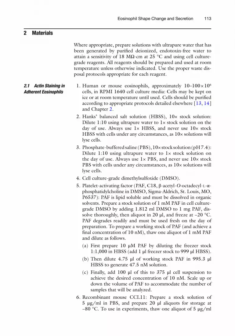
1. Human or mouse eosinophils, approximately 10–100 × 10 6 cells, in RPMI 1640 cell culture media: Cells may be kept on ice or at room temperature until used. Cells should be purifi ed according to appropriate protocols detailed elsewhere [ 13 , 14 ] and Chapter 2 .
2. Hanks’ balanced salt solution (HBSS), 10× stock solution: Dilute 1:10 using ultrapure water to 1× stock solution on the day of use. Always use 1× HBSS, and never use 10× stock HBSS with cells under any circumstances, as 10× solutions will lyse cells.
3. Phosphate-buffered saline (PBS), 10× stock solution (pH 7.4): Dilute 1:10 using ultrapure water to 1× stock solution on the day of use. Always use 1× PBS, and never use 10× stock PBS with cells under any circumstances, as 10× solutions will lyse cells.
4. Cell culture-grade dimethylsulfoxide (DMSO). 5. Platelet-activating factor (PAF, C18, β-acetyl- O -octadecyl- L -α-
phosphatidylcholine in DMSO, Sigma-Aldrich, St. Louis, MO, P6537): PAF is lipid soluble and must be dissolved in organic solvents. Prepare a stock solution of 1 mM PAF in cell culture-grade DMSO by adding 1.812 ml DMSO to 1 mg PAF, dis-solve thoroughly, then aliquot in 20 μl, and freeze at −20 °C. PAF degrades readily and must be used fresh on the day of preparation. To prepare a working stock of PAF (and achieve a fi nal concentration of 10 nM), thaw one aliquot of 1 mM PAF and dilute as follows. (a) First prepare 10 μM PAF by diluting the freezer stock
1:1,000 in HBSS (add 1 μl freezer stock to 999 μl HBSS). (b) Then dilute 4.75 μl of working stock PAF in 995.3 μl
HBSS to generate 47.5 nM solution. (c) Finally, add 100 μl of this to 375 μl cell suspension to
achieve the desired concentration of 10 nM. Scale up or down the volume of PAF to accommodate the number of samples that will be analyzed.
6. Recombinant mouse CCL11: Prepare a stock solution of 5 μg/ml in PBS, and prepare 20 μl aliquots for storage at −80 °C. To use in experiments, thaw one aliquot of 5 μg/ml
2.1 Actin Staining in Adherent Eosinophils
Eosinophil Shape Change and Secretion

114
CCL11 and dilute as follows. Add 9.5 μl of stock CCL11 to 990.5 μl HBSS to generate 47.5 ng/ml, and then add 100 μl of this to 375 cell suspension to achieve the fi nal concentration of 10 ng/ml. CCL11 is also easily degraded and must be used fresh on the day of preparation. Scale up or down the volume of CCL11 to accommodate the number of samples that will be analyzed.
7. Incubation slides (Lab-Tek Chambered #1.0 Borosilicate Coverglass System, 8 Chamber, Fig. 1 ).
8. 4 % Paraformaldehyde: Prepare using analytical grade parafor-maldehyde powder. To prepare 100 ml, weigh 4 g paraformal-dehyde and add to 50–75 ml water in a clean 150 ml glass beaker. Heat while stirring with a stirrer bar on a heated stirring platform to 55 °C, and add 1, 5, or 10 M NaOH gradually until the solution clears. Ensure that the temperature of the solution does not exceed 60–65 °C ( see Note 1 ). Cool the solution to room temperature, and vacuum fi lter through a 0.45 μm fi lter to remove large particles. Add 10 ml 10× PBS, and then bring up to the fi nal volume of 100 ml. Adjust pH of solution to 7.4 or slightly higher ( see Note 2 ), and prepare aliquots of 5 or 10 ml. Freeze at −20 °C. Thaw at room temperature or at 37 °C, but never at 50 °C or higher. This can be refrozen and thawed up to three times.
9. 0.1 % Triton X-100 in PBS: First prepare a stock solution of 10 % Triton X-100 in water which can be stored at 4 °C for up
Fig. 1 LabTek 8 Chambered Coverglass. Working volume is 0.5 ml, and chambers are made of polystyrene material attached to a glass cover slip of 1.5 borosilicate glass, suitable for high-defi nition immunofl uorescence imaging by confocal microscopy or similar systems. Modifi ed from the Sigma-Aldrich website for this product
Lian Willetts et al.

115
to 1 year. Weigh 2 g Triton X-100 in a 50 ml conical centrifuge tube, and add up to 20 ml ultrapure water. Dilute 1:100 to obtain 0.1 % for permeabilization of cells by adding 100 μl 10 % Triton X-100 to 9.9 ml PBS.
10. 2 % Bovine serum albumin (BSA) in PBS: Weigh 0.2 g BSA and make up to 10 ml in PBS.
11. Rhodamine-phalloidin (Molecular Probes, Invitrogen): This comes as a solution which can be diluted on the day of use. Dilute 1:200 by adding 5–995 μl PBS. Scale up or down the volume to accommodate the number of samples that will be analyzed.
12. ProLong Gold DAPI-containing mounting media: This should be a ready-made solution which is used directly from a small bottle.
1. 96-Well fl at-welled microtitre plates. 2. Microcentrifuge tubes (1.5 ml). 3. Human or mouse eosinophils, approximately 10–100 × 10 6
cells, in RPMI 1640 cell culture media: Cells may be kept on ice or at room temperature until used. Cells should be purifi ed according to appropriate protocols detailed elsewhere [ 13 , 14 ] and Chapter 2 .
4. Phenol red-free RPMI 1640 medium: For convenience, purchase the ready-made media and ensure that this is used prior to the expiry date. It does not have to contain HEPES, bicarbonate, or glutamine as this medium is not required for incubation of cells for long periods.
5. Cell culture-grade DMSO. 6. PAF (C18, β-acetyl- O -octadecyl- L -α-phosphatidylcholine in
DMSO, Sigma-Aldrich, P6537). See #5 above for preparation of stock PAFIonomycin. Prepare as a stock solution of 5 μg/ml in DMSO, aliquot in 20 μl, and freeze at −20 °C until needed.
1. Microcentrifuge tubes (1.5 ml). 2. Human or mouse eosinophils, approximately 10–100 × 10 6
cells, in RPMI 1640 cell culture media: Cells may be kept on ice or at room temperature until used. Cells should be purifi ed according to appropriate protocols detailed elsewhere [ 13 , 14 ] and Chapter 2 .
3. PBS, 10×: Dilute 1:10 to use 1× PBS for all experiments. 4. Recipient mice ( IL-5/hE2/EPX −/− , Lee Labs, Mayo Clinic, AZ). 5. Isofl urane, USP liquid for inhalation (Novaplus; see Note 3 ):
Isofl urane inhalation is carried out using a precision vaporizer instrument.
2.2 In Vitro Degranulation Assay
2.3 Ex Vivo Degranulation Assay
Eosinophil Shape Change and Secretion
116
6. 2 % Fetal bovine serum (FBS) in PBS: Aliquot 1 ml FBS to 49 ml PBS in a conical centrifuge tube.
7. 18 G Blue tip catheter (30 mm). 8. 1 ml Sterile syringes. 9. 75 % Ethanol with water.
10. Nembutal sodium solution (pentobarbital sodium, 50 mg/ml).
1. 96-Well fl at-welled microtitre plates. 2. 1 M Tris buffer, pH 8.0: Prepare 100 ml by adding 12.1 g Tris
base to 80 ml ultrapure water, and then adjust pH to 8.0 using concentrated hydrochloric acid. Make up to 100 ml with water, fi lter through 0.45 μm, and store for up to 1 year at 4 °C.
3. 30 % H 2 O 2 : This is a ready-made solution that should be stored at 4 °C.
4. OPD HCl substrate solution: OPD should be prepared fresh immediately before use as it degrades rapidly. Add 9 mg OPD to 1 ml ultrapure water. In a separate tube, mix 4 ml 1 M Tris (pH 8.0), 6 ml ultrapure water, and 1.25 μl H 2 O 2 (30 %). Keep these separate, and then add 800 μl OPD to this mixture just before use in the assay ( see Note 4 ).
5. Optional: If an ultrasound probe is not available, prepare 10 % 3-[(3-cholamidopropyl)dimethylammonio]-1- propanesulfonate (CHAPS) detergent, which is used to lyse cells. Weigh out 1 g CHAPS and make up to a fi nal volume of 10 ml with ultrapure water. This may be stored at room temperature for up to 1 year. Always dilute to 0.1 % for lysing cells.
6. 4 M Sulfuric acid.
1. 96-Well fl at-welled microtitre plate (Nunc-Immuno Plate MaxiSorp, Thermo Fisher Scientifi c, Waltham, MA): These must be MaxiSorp plates, and not standard 96-well plates used in other assays, for optimal antibody coating.
2. Hexadecyltrimethylammonium bromide (CTAB) in 0.3 M sucrose.
3. EPX Standards: These may be prepared by isolating peripheral blood eosinophils from NJ.1638 mice [ 13 ] or human blood [ 14 ]. Prepare standards starting with a purifi ed eosinophil sus-pension (>98 % purity) at 14.6 × 10 6 cells/ml in PBS with 2 % FBS. To prepare stock lysate for standards, spin 250 μl of eosinophil suspension at 950 × g for 10 min at 4 °C. Remove 200 μl supernatant, add 250 μl of 0.22 % CTAB in 0.3 M sucrose to the cells, and lyse the cell pellet with repeated pipet-ting. Vortex the lysate for 1 min and fl ash-freeze in liquid nitrogen for storage in 25 μl aliquots at −80 °C. On the day of use, lysates should be thawed at 37 °C until just melted, then
2.4 o -Phenylene-diamine (OPD) Assay
2.5 EPX Enzyme- Linked Immunosor-bent Assay
Lian Willetts et al.
117
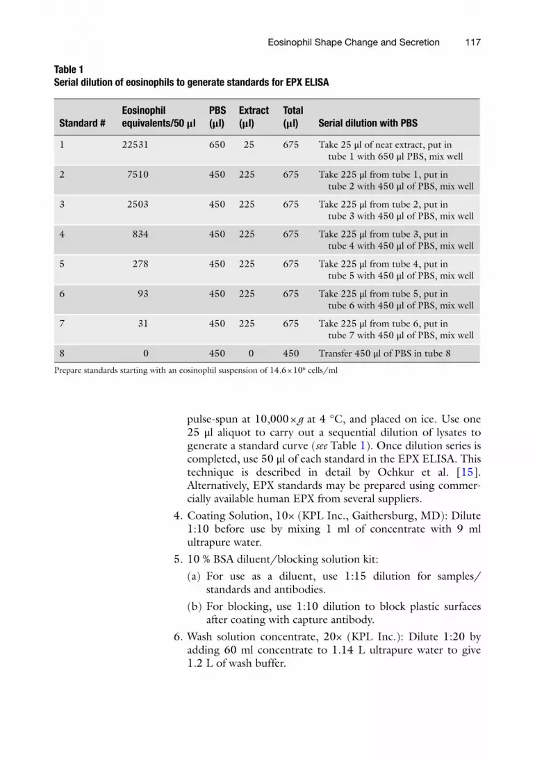
pulse-spun at 10,000 × g at 4 °C, and placed on ice. Use one 25 μl aliquot to carry out a sequential dilution of lysates to generate a standard curve ( see Table 1 ). Once dilution series is completed, use 50 μl of each standard in the EPX ELISA. This technique is described in detail by Ochkur et al. [ 15 ]. Alternatively, EPX standards may be prepared using commer-cially available human EPX from several suppliers.
4. Coating Solution, 10× (KPL Inc., Gaithersburg, MD): Dilute 1:10 before use by mixing 1 ml of concentrate with 9 ml ultrapure water.
5. 10 % BSA diluent/blocking solution kit: (a) For use as a diluent, use 1:15 dilution for samples/
standards and antibodies. (b) For blocking, use 1:10 dilution to block plastic surfaces
after coating with capture antibody. 6. Wash solution concentrate, 20× (KPL Inc.): Dilute 1:20 by
adding 60 ml concentrate to 1.14 L ultrapure water to give 1.2 L of wash buffer.
Table 1 Serial dilution of eosinophils to generate standards for EPX ELISA
Standard # Eosinophil equivalents/50 μl
PBS (μl)
Extract (μl)
Total (μl) Serial dilution with PBS
1 22531 650 25 675 Take 25 μl of neat extract, put in tube 1 with 650 μl PBS, mix well
2 7510 450 225 675 Take 225 μl from tube 1, put in tube 2 with 450 μl of PBS, mix well
3 2503 450 225 675 Take 225 μl from tube 2, put in tube 3 with 450 μl of PBS, mix well
4 834 450 225 675 Take 225 μl from tube 3, put in tube 4 with 450 μl of PBS, mix well
5 278 450 225 675 Take 225 μl from tube 4, put in tube 5 with 450 μl of PBS, mix well
6 93 450 225 675 Take 225 μl from tube 5, put in tube 6 with 450 μl of PBS, mix well
7 31 450 225 675 Take 225 μl from tube 6, put in tube 7 with 450 μl of PBS, mix well
8 0 450 0 450 Transfer 450 μl of PBS in tube 8
Prepare standards starting with an eosinophil suspension of 14.6 × 10 6 cells/ml
Eosinophil Shape Change and Secretion

118
7. Capture Antibody (mouse anti-mouse EPX, 1 mg/ml, clone MM25-429.1.1, Lee Labs, Mayo Clinic, AZ): Dilute by add-ing 20 μl of 1 mg/ml stock antibody to 10 ml of coating solu-tion to make a 2 μg/ml fi nal concentration.
8. Detection Antibody (biotinylated rat anti-EPX monoclonal antibody, clone MM25-82.2.1, Lee Labs, Mayo Clinic, AZ): Prepare 0.8 μg/ml from 1.4 mg/ml stock solution by diluting 1:1,750. For one 96-well plate, add 5.7 μl antibody to 10 ml PBS and use 100 μl per well.
9. Streptavidin–alkaline phosphatase: Dilute 1:500 to 1:1,000 in 1 % BSA, 0.05 % Tween 20, 0.025 M Tris (pH 7.4), and 0.5 M NaCl. For a 1:500 dilution to use for a 96-well microtitre plate, add 22 μl streptavidin–alkaline phosphatase to 11 ml diluent, and use 100 μl per well.
10. Trizma hydrochloride buffer solution, pH 7.5: This solution is ready-made for use in this assay.
11. BluPhos Microwell Phosphatase Substrate System, 600 ml (KPL Inc.): Prepare 26 ml of substrate solution no earlier than 15 min before the reaction. Mix 13 ml of solution A with 13 ml of solution B, and add 100 μl per well.
12. EDTA (2.5 %) stop solution: Prepare a 0.5 M EDTA stock solution by adding 14 g EDTA (acid form) to 100 ml and autoclaving to dissolve salt with a fi nal concentration of 14 % EDTA. Alternatively, 10 M NaOH may be added with con-tinuous stirring to keep pH of the solution neutral and allow dissolving of EDTA. Take care not to exceed a neutral pH range of 7.0–7.5. Before use, dilute this by adding 2.5 ml 14 % EDTA to 11.5 ml ultrapure water to make 2.5 % EDTA.
3 Methods
1. Prepare eosinophils as described in Chapter 2 . Resuspend eosinophils to 5–6 × 10 6 cells/ml in HBSS (no serum). If any serum or protein is present in media, cells will not adhere to glass surfaces, which is essential for imaging.
2. Plate 375 μl of cell suspension (containing 2–2.5 × 10 6 cells in HBSS) in each well of a LabTek chamber slide (Fig. 1 ). Incubate for 15 min at 37 °C in tissue culture incubator to adhere cells to glass surfaces. Each slide should have alternat-ing control and test samples for direct comparison.
3. Prewarm agonists for 15 min at the same time in separate tubes (PAF, recombinant mouse CCL11).
4. Set up a timer to record a time of 10 s per well.
3.1 Actin and Nuclear Staining in Adherent Eosinophils to Detect Eosinophil Shape Change
Lian Willetts et al.

119
5. Before adding agonist, load two micropipettes: one with 100 μl agonist and the other with 200 μl paraformaldehyde.
6. To start stimulation, add the following reagents with the incu-bator door open and the LabTek slide on the incubator shelf: (a) Control (100 μl HBSS + 0.0001 % DMSO). (b) PAF (100 μl, fi nal concentration 10 nM). (c) Recombinant mouse CCL11 (100 μl, fi nal concentration
10 ng/ml). 7. Incubate for 10 s. Stagger the timing of stimulation of plates if
more than one plate is used. 8. At the end of the incubation, immediately aspirate media with
micropipette without dislodging cells, and add 200 μl 4 % paraformaldehyde, again without disturbing cells. A useful method is to place the micropipette tip at an angle in the cor-ner of the well so that liquid forces do not impact cells exces-sively and detach them.
9. Fix cells at room temperature for 20 min. Do not let slides incubate in fi xative for longer than 30 min.
10. Wash wells twice with PBS. Fill each well to the top with PBS each time to ensure that they are adequately washed. Aspirate washings using a vacuum trap for this and all subsequent steps. Prior to the next step, aspirate all PBS out of wells and quickly add the next reagent to avoid dehydrating cells.
11. Permeabilize cells with 200 μl 0.1 % Triton X-100 for 3 min. 12. Wash wells twice with PBS. After the last wash, aspirate all PBS
and quickly add the next reagent. 13. Block with 200 μl of 2 % BSA in PBS for 20 min at room
temperature. 14. Wash cells twice with PBS. After the last wash, aspirate all PBS
and quickly add the next reagent. 15. Stain cells with 200 μl rhodamine-phalloidin (diluted 1:200)
for 1 h at room temperature. Keep slides in the dark from this point onwards to avoid bleaching of fl uorescence.
16. Wash cells three times for 5 min each with PBS. After the last wash, aspirate all PBS and quickly add the next reagent.
17. Add several drops per well of ProLong Gold DAPI-containing mounting media. Make sure that the cells are fully covered with mounting media to prevent cells from drying out.
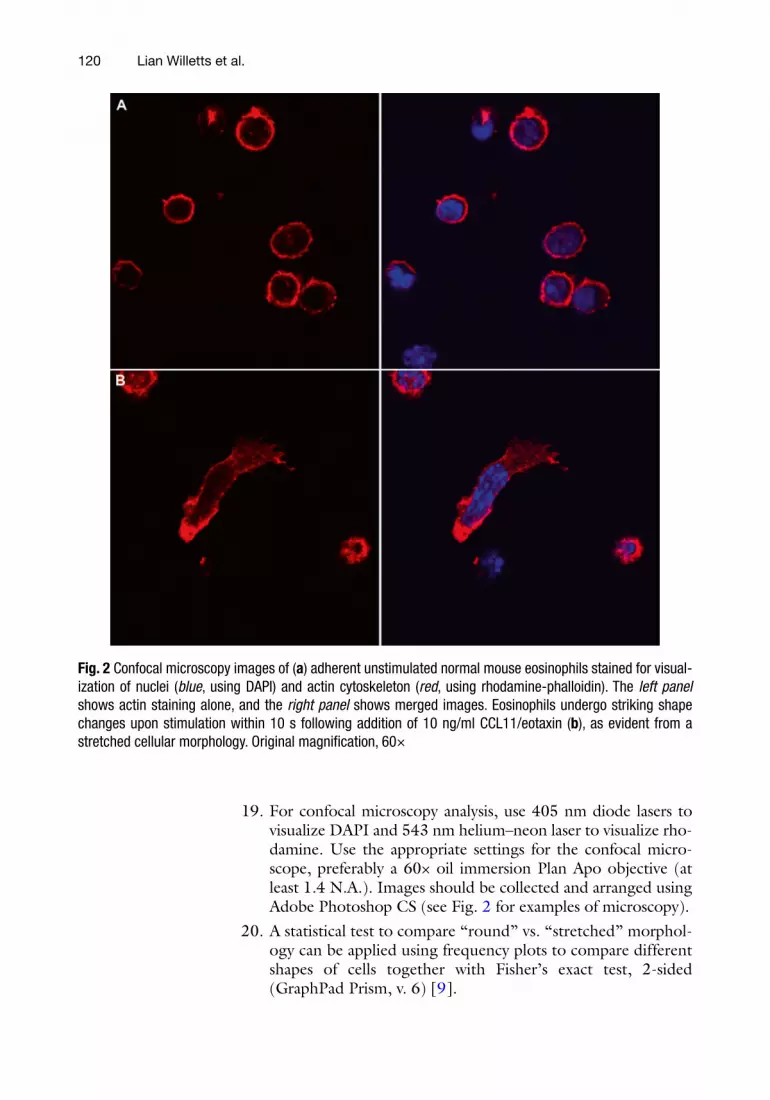
18. Place slides in a petri dish and wrap in foil for transport to a confocal microscope for immediate analysis, or store slides at 4 °C in a dark container until analysis by confocal microscopy (Fig. 2 ).
Eosinophil Shape Change and Secretion
120
19. For confocal microscopy analysis, use 405 nm diode lasers to visualize DAPI and 543 nm helium–neon laser to visualize rho-damine. Use the appropriate settings for the confocal micro-scope, preferably a 60× oil immersion Plan Apo objective (at least 1.4 N.A.). Images should be collected and arranged using Adobe Photoshop CS (see Fig. 2 for examples of microscopy).
20. A statistical test to compare “round” vs. “stretched” morphol-ogy can be applied using frequency plots to compare different shapes of cells together with Fisher’s exact test, 2-sided (GraphPad Prism, v. 6) [ 9 ].
Fig. 2 Confocal microscopy images of ( a ) adherent unstimulated normal mouse eosinophils stained for visual-ization of nuclei ( blue , using DAPI) and actin cytoskeleton ( red , using rhodamine-phalloidin). The left panel shows actin staining alone, and the right panel shows merged images. Eosinophils undergo striking shape changes upon stimulation within 10 s following addition of 10 ng/ml CCL11/eotaxin ( b ), as evident from a stretched cellular morphology. Original magnifi cation, 60×
Lian Willetts et al.

121
1. Isolate mouse or human peripheral blood eosinophils as described previously [ 13 , 14 ], Chapter 2 .
2. Centrifuge (300 × g at 4 °C for 5 min) and resuspend purifi ed eosinophils in phenol red-free RPMI 1640 at 2.5 × 10 6 cells/ml. Use phenol red-free RPMI for all steps to prevent interference with absorbance in later assays.
3. Prepare lysed unstimulated cells for obtaining total EPX activ-ity or concentration. Sonicate separate tubes of cells for lysate values using a sonication probe (Ultrasonic), set at power level 3, for 5 s, in a microcentrifuge tube on ice containing 2.5 × 10 5 cells in 100 μl RPMI. Alternatively, use a detergent such as CHAPS and add this to a fi nal concentration of 0.1 % to cells. Place on ice during incubation of cells in the following steps.
4. Add 100 μl cell suspension (containing 2.5 × 10 5 cells) to each well in a 96-well microtitre plate. In separate wells, add ago-nists (150 μl each of PAF, 400 ng/ml, and/or ionomycin, 100 ng/ml, in RPMI). Carry out all stimulation in triplicate for more accurate average values.
5. Prepare DMSO vehicle controls for additional cells in separate wells using the percentage of DMSO used after dilution of the original stock solutions for PAF and ionomycin.
6. Prewarm the whole plate for at least 15 min at 37 °C ( see Note 5 ). 7. Add 100 μl of PAF (for a fi nal concentration of 200 ng/ml)
and/or ionomycin (for a fi nal concentration of 50 ng/ml) or DMSO in RPMI.
8. Incubate plate for 30 min. 9. Spin down microtitre plate at 300 × g for 5 min at 4 °C using
an appropriate microtitre plate rotor. Collect 200 μl from each supernatant without disturbing cell pellets.
10. Add cells to a separate series of wells to check their viability later using trypan blue or an equivalent viability assay.
11. Centrifuge the collected supernatants again at 500 × g (or higher speeds) for 5 min at 4 °C to remove cells, organelles, and debris. These supernatants can be analyzed for EPX using the OPD assay (Subheading 3.4 ) to determine EPX activity and/or the EPX ELISA technique (Subheading 3.5 ) to deter-mine total EPX concentration.
12. Store supernatants and lysates at −80 °C for further analysis.
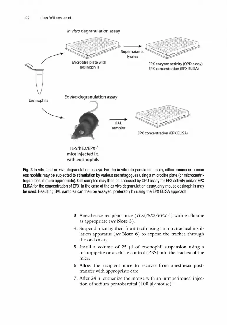
This procedure involves intratracheal instillation of peripheral blood eosinophils into IL-5/hE2/EPX −/− recipient mice (Fig. 3 ).
1. Isolate mouse eosinophils as described previously [ 13 ]. 2. Resuspend purifi ed mouse eosinophils in PBS at 1 × 10 7 cells in
25 μl and keep on ice until needed.
3.2 In Vitro Degranulation Assay
3.3 Ex Vivo Degranulation Assay
Eosinophil Shape Change and Secretion
122
3. Anesthetize recipient mice ( IL-5/hE2/EPX −/− ) with isofl urane as appropriate ( see Note 3 ).
4. Suspend mice by their front teeth using an intratracheal instil-lation apparatus ( see Note 6 ) to expose the trachea through the oral cavity.
5. Instill a volume of 25 μl of eosinophil suspension using a micropipette or a vehicle control (PBS) into the trachea of the mice.
6. Allow the recipient mice to recover from anesthesia post- transfer with appropriate care.
7. After 24 h, euthanize the mouse with an intraperitoneal injec-tion of sodium pentobarbital (100 μl/mouse).
Fig. 3 In vitro and ex vivo degranulation assays. For the in vitro degranulation assay, either mouse or human eosinophils may be subjected to stimulation by various secretagogues using a microtitre plate (or microcentri-fuge tubes, if more appropriate). Cell samples may then be assessed by OPD assay for EPX activity and/or EPX ELISA for the concentration of EPX. In the case of the ex vivo degranulation assay, only mouse eosinophils may be used. Resulting BAL samples can then be assayed, preferably by using the EPX ELISA approach
Lian Willetts et al.

123
8. Carry out surgical incision in the upper thoracic area of the mouse to expose the trachea. Ensure that blood vessels are not compromised and limit bleeding in the tracheal area.
9. Cut a horizontal incision in the trachea with fi ne scissors. 10. Slowly introduce 1 ml PBS using a 1 ml syringe via the tracheal
incision into the lungs, and then gently retrieve the solution into the same syringe. This generates the bronchoalveolar lavage (BAL) sample.
11. Eject BAL from the syringe into a labeled microcentrifuge tube, place on ice, and repeat for each mouse.
12. Centrifuge BAL samples at 500 × g for 5 min at 4 °C to remove cells and debris.
13. Centrifuge the supernatants again at 10,000 × g for 10 min at 4 °C to remove cellular debris and clarify supernatants.
14. Store supernatants at −80 °C for later analysis by OPD assay or EPX ELISA.
There are two ways to carry out the OPD assay for EPX activity. The simplest approach is to measure EPX activity in supernatants of stimulated eosinophils (after centrifugation) with OPD substrate solution. The way to carry out the OPD assay on supernatants is as follows:
1. Prepare OPD substrate solution as detailed in the Materials (Section 2.4). Store solution in the dark until needed, and keep OPD separate from the rest of the mixture until just before use.
2. Thaw or prepare supernatants and lysates from in vitro or ex vivo degranulation assays. If thawing previously frozen supernatants, place tubes in 37 °C until just thawed, and then place on ice until use.
3. Add 50 μl supernatants to each well of a 96-well microtitre plate. Include a series of wells with RPMI alone (“blanks”) for background subtraction, and include a series of lysed cells to give total EPX activity.
4. Add 50 μl OPD substrate solution. 5. Incubate the plate at room temperature for up to 30 min
depending on the color development. Place the plate in the dark during incubation as OPD is light sensitive. Color reac-tion should show a rapid development to a light orange/brown. Do not allow the reaction to proceed to too dark a color, as this will generate off-scale readings on the plate reader (>2.0 absorbance units).
6. Terminate the reaction by the addition of 100 μl 4 M H 2 SO 4 . Subtract background from each well using the absorbance
3.4 OPD Assay for EPX Activity
Eosinophil Shape Change and Secretion

124
value obtained with the blanks. Divide the supernatant values into the lysed values to get a percentage of total EPX release. However, EPX is highly adhesive due to its cationic nature and
precipitates readily out of solution, making its recovery from super-natants very difficult if not impossible. In addition, some EPX activ-ity from supernatants is lost upon freeze–thaw. A more appropriate technique is to incubate OPD substrate solution directly with live, viable eosinophils following their activation [ 11 ]. OPD is soluble in aqueous media and does not require organic solvents to dissolve it. This has the added advantage of preventing damage to eosinophils when OPD substrate solution is added to live cells or leading to inadvertent detection of intracellular EPX. The following technique describes how to incubate OPD substrate solution with live eosino-phils for detection of EPX activity immediately after its release.
7. Prepare OPD substrate solution as detailed in the Materials (Section 2.4). Store solution in the dark until needed, and keep OPD separate from the rest of the mixture until just before use ( see Note 7 ).
8. Resuspend eosinophils in phenol red-free RPMI to a concen-tration of 2.5 × 10 6 cells/ml. All steps in this assay require phe-nol red-free RPMI to avoid interference with the colored product generated by OPD substrate.
9. Prepare lysed unstimulated cells for obtaining total EPX activ-ity or concentration. Sonicate separate tubes of cells for lysate values using a sonication probe (Ultrasonic), set at power level 3, for 5 s, in a microcentrifuge tube on ice containing 2.5 × 10 5 cells in 100 μl RPMI. Alternatively, use a detergent such as CHAPS and add this to a fi nal concentration of 0.1 % to cells. Place on ice during incubation of cells in the following steps.
10. Add 100 μl cell suspension (containing 2.5 × 10 5 cells) to each well in a 96-well microtitre plate. In separate wells, add ago-nists (150 μl each of PAF, 400 ng/ml, and/or ionomycin, 100 ng/ml, in RPMI). Carry out all stimulation in triplicate for more accurate average values.
11. Prepare DMSO vehicle controls for additional cells in separate wells using the percentage of DMSO used after dilution of the original stock solutions for PAF and ionomycin.
12. Prewarm the whole plate for at least 15 min at 37 °C ( see Note 5 ). 13. Add 100 μl of PAF (for a fi nal concentration of 200 ng/ml)
and/or ionomycin (for a fi nal concentration of 50 ng/ml) or DMSO in RPMI.
14. Incubate plate for 30 min. While incubating cells, prepare fresh OPD solution.
Lian Willetts et al.
125
15. At the end of the incubation period, add 200 μl of OPD solution into each well of cells, blanks, and lysates and wait for 2 min. Color reaction should show rapid development to light orange/brown. Do not allow the reaction to proceed to too dark a color as this will be off-scale (absorbance value > 1.0) in the plate reader.
16. Add 100 μl of 4 M H 2 SO 4 into each well to stop the reaction using a multichannel pipette.
17. Read plate on spectrophotometric plate reader at wavelength 490 nm. Subtract the background absorbance using the blank wells (containing RPMI only) from all sample wells. Divide the value obtained for stimulated cells into the lysed cell absor-bance average to obtain the percentage of EPX release.
This technique measures the concentration of EPX in mouse or human eosinophil samples, as opposed to the OPD assay which measures EPX activity. The EPX ELISA can be used to measure human [ 16 ] or mouse [17] samples.
1. Add 100 μl capture antibody diluted in coating solution to appropriate wells in a 96-well microtitre MaxiSorp plate, and incubate for 1 h at room temperature or overnight at 4 °C.
2. Discard solution, and tap out residual liquid on paper towels. 3. Wash plate by fi lling each well to the top with wash solution.
Discard solution into sink, and tap out residual liquid onto clean paper towels ( see Note 8 ). Repeat three times. The same paper towels can be used throughout the wash procedure.
4. Add 300 μl blocking solution to each well. 5. Incubate for 15 min. 6. Discard solution into sink, and tap out residual liquid onto
clean paper towels. 7. Add 50 μl sample, standard, or blank solution (RPMI) into
each well. Be sure to include the standards shown in Table 1 and carry out all samples, standards, and blanks in triplicate for more accurate averages.
8. Incubate for 1 h at room temperature or overnight at 4 °C. 9. Remove samples with a multichannel pipette and discard to
prevent cross-contamination of neighboring wells. 10. Tap out residual liquid onto clean paper towels. 11. Wash plate by fi lling each well to the top with wash solution.
Discard solution into sink, and tap out residual liquid onto clean paper towels. Repeat three times.
12. Add 100 μl detection antibody to each well. 13. Incubate for 1 h at room temperature.
3.5 EPX ELISA
Eosinophil Shape Change and Secretion

126
14. Discard solution into sink, and tap out residual liquid onto clean paper towels.
15. Wash plate by fi lling each well to the top with wash solution. Discard solution into sink, and tap out residual liquid onto clean paper towels. Repeat three times. Leave the fi nal wash solution for 5 min to fully wash wells before discarding and tapping plate out.
16. Add 100 μl streptavidin–alkaline phosphatase solution to each well.
17. Incubate for 20 min at room temperature. 18. Discard solution into sink, and tap out residual liquid onto
clean paper towels. 19. Wash plate by fi lling each well to the top with wash solution.
Discard solution into sink, and tap out residual liquid onto clean paper towels. Repeat three times.
20. Dispense 100 μl BluePhos Microwell Phosphatase Substrate into each well.
21. Incubate for up to 1 h. 22. If desired, after suffi cient color development, and add 100 μl of
stop solution to each well. 23. Take care to remove all bubbles from the well surfaces as these
interfere with absorbance readings. In addition, clean the bot-tom of the plate if there is any material there to prevent interference.
24. Read the absorbance of wells at 595–650 nm. Subtract the background absorbance obtained in blank wells from all sam-ples and standards.
4 Notes
1. Ensure that this procedure is done in a suitable fume hood with functional extraction fans to avoid inhaling toxic fumes. Handle this solution with great care, and avoid breathing fumes during the heating step, as formaldehyde is a known carcinogen. If the paraformaldehyde solution exceeds 60–65 °C, discard the solu-tion through suitable chemical waste disposal and start again with a new preparation using clean glassware.
2. Paraformaldehyde forms formaldehyde when in solution and must be buffered to prevent degradation into inactive prod-ucts. Adjusting the pH to higher values than 7.4 (e.g., 7.6–7.8) is advantageous as formaldehyde fi xes better at higher pH values. It is critical that the pH range of the formaldehyde solution is kept in this range as this maintains its ability to fi x specimens for detailed high-magnifi cation microscopy.
Lian Willetts et al.
127
Formaldehyde in water readily breaks down into formic acid, leading to poor fi xation and specimen degradation. Avoid using commercially prepared buffered formalin as this is less effective at fi xation of fi ne intracellular structures than freshly prepared buffered and fi ltered paraformaldehyde solution.
3. Isofl urane produces rapid induction and recovery from anes-thesia, and the depth of anesthesia can be altered easily. There are minimal adverse effects, toxicity, or interference with drug metabolism when using isofl urane. Start with 0.5 % isofl urane, and increase by 0.5 % increments every few breaths. If the ani-mal becomes agitated or excited, then quickly deliver a higher percentage of anesthetic to no more than 5 %. Maintain the animal on isofl urane (0.5–3.0 %) and oxygen mixture during the procedure. Once intratracheal injection is completed, remove the animal from isofl urane, allow it to breathe oxygen until fully recovered, and then place it in recovery. Use appro-priate recovery and monitoring approaches as approved by the institutional ethics board.
4. OPD is a colored substrate which will spontaneously darken and form dark precipitates that will interfere with absorbance measurements if left in solution for several hours. It is impor-tant to prepare this substrate immediately before use rather than leaving in storage.
5. Eosinophils that have been stored on ice or at room tempera-ture prior to stimulation must be prewarmed for at least 15 min to allow membranes to reach a physiological temperature and facilitate receptor-stimulated degranulation responses.
6. An intratracheal instillation apparatus can be made in-house. Wrap two ends of a thin (20–23 gauge) wire around two arms of an extension clamp. Make sure that the wire is taut. Secure the extension clamp with the wire on a ring stand. Anesthetized mice are hung by their front teeth on the wire between the two arms of the extension clamp.
7. During washing for ELISA technique, avoid reusing the same paper towels from previous steps. This will cause contamina-tion of neighboring wells with samples or materials that could cause a spurious signal to appear in the fi nal absorbance measurement.
Acknowledgements
This work was supported by funding from an NSERC Discovery Grant and a CIHR operating grant (MOP 89748) to PL. We would like to acknowledge the outstanding support of our techni-cian, Renjith Pillai, for assisting with experimental details.
Eosinophil Shape Change and Secretion

128
References
1. Hogan SP, Rosenberg HF, Moqbel R et al (2008) Eosinophils: biological properties and role in health and disease. Clin Exp Allergy 38:709–750
2. Rosenberg HF, Dyer KD, Foster PS (2013) Eosinophils: changing perspectives in health and disease. Nat Rev Immunol 13:9–22
3. Rothenberg ME, Hogan SP (2006) The eosinophil. Annu Rev Immunol 24:147–174
4. Winder SJ, Ayscough KR (2005) Actin-binding proteins. J Cell Sci 118:651–654
5. Schweizer RC, van Kessel-Welmers BA, Warringa RA et al (1996) Mechanisms involved in eosinophil migration. Platelet-activating factor- induced chemotaxis and interleukin-5- induced chemokinesis are mediated by differ-ent signals. J Leukoc Biol 59:347–356
6. Garcia-Zepeda EA, Rothenberg ME, Ownbey RT et al (1996) Human eotaxin is a specifi c chemoattractant for eosinophil cells and provides a new mechanism to explain tissue eosino-philia. Nat Med 2:449–456
7. Dyer KD, Percopo CM, Xie Z et al (2010) Mouse and human eosinophils degranulate in response to platelet-activating factor (PAF) and lysoPAF via a PAF-receptor-independent mechanism: evidence for a novel receptor. J Immunol 184:6327–6334
8. Heasman SJ, Ridley AJ (2008) Mammalian Rho GTPases: new insights into their func-tions from in vivo studies. Nat Rev Mol Cell Biol 9:690–701
9. Lacy P, Willetts L, Kim JD et al (2011) Agonist activation of F-actin-mediated eosinophil shape change and mediator release is dependent on Rac2. Int Arch Allergy Immunol 156:137–147
10. Muniz-Junqueira MI, Barbosa-Marques SM, Junqueira LF Jr (2013) Morphological changes in eosinophils are reliable markers of the severity of an acute asthma exacerbation in children. Allergy 68:911–920
11. Adamko DJ, Wu Y, Gleich GJ et al (2004) The induction of eosinophil peroxidase release: improved methods of measurement and stimulation. J Immunol Methods 291:101–108
12. Kim JD, Willetts L, Ochkur SI et al (2013) An essential role for Rab27a GTPase in eosinophil exocytosis. J Leukoc Biol 94(6):1265–1274
13. Lee NA, McGarry MP, Larson KA et al (1997) Expression of IL-5 in thymocytes/T cells leads to the development of a massive eosinophilia, extramedullary eosinophilopoi-esis, and unique histopathologies. J Immunol 158:1332–1344
14. Levi-Schaffer F, Lacy P, Severs NJ et al (1995) Association of granulocyte-macrophage colony- stimulating factor with the crystalloid granules of human eosinophils. Blood 85:2579–2586
15. Ochkur SI, Kim JD, Protheroe CA et al (2012) A sensitive high throughput ELISA for human eosinophil peroxidase: a specifi c assay to quan-tify eosinophil degranulation from patient- derived sources. J Immunol Methods 384:10–20
16. Ochkur SI, Kim JD, Protheroe CA et al (2012) The development of a sensitive and specifi c ELISA for mouse eosinophil peroxidase: assessment of eosinophil degranulation ex vivo and in models of human disease. J Immunol Methods 375:138–147
Lian Willetts et al.
Related Documents