EORGIAN EDICAL EWS ЕЖЕМЕСЯЧНЫЙ НАУЧНЫЙ ЖУРНАЛ Медицинские новости Грузии cfmfhsdtkjc cfvtlbwbyj cbf[ktyb No 5 (278) Май 2018 ISSN 1512-0112 ТБИЛИСИ - NEW YORK

Welcome message from author
This document is posted to help you gain knowledge. Please leave a comment to let me know what you think about it! Share it to your friends and learn new things together.
Transcript
E O R G I A N EDICAL
EWS
ЕЖЕМЕСЯЧНЫЙ НАУЧНЫЙ ЖУРНАЛ
Медицинские новости Грузииcfmfhsdtkjc cfvtlbwbyj cbf[ktyb
No 5 (278) Май 2018ISSN 1512-0112
ТБИЛИСИ - NEW YORK
GEORGIAN MEDICAL
NEWSNo 5 (278) 2018
ЕЖЕМЕСЯЧНЫЙ НАУЧНЫЙ ЖУРНАЛ
ТБИЛИСИ - НЬЮ-ЙОРК
Published in cooperation with and under the patronage of the Tbilisi State Medical University
Издается в сотрудничестве и под патронажем Тбилисского государственного медицинского университета
gamoicema Tbilisis saxelmwifo samedicino universitetTan TanamSromlobiTa da misi patrona;iT
GMN: Georgian Medical News is peer-reviewed, published monthly journal committed to promoting
the science and art of medicine and the betterment of public health, published by the GMN Editorial
Board and The International Academy of Sciences, Education, Industry and Arts (U.S.A.) since
1994. GMN carries original scientific articles on medicine, biology and pharmacy, which are of
experimental, theoretical and practical character; publishes original research, reviews, commentaries,
editorials, essays, medical news, and correspondence in English and Russian.
GMN is indexed in MEDLINE, SCOPUS, PubMed and VINITI Russian Academy of Sciences. The
full text content is available through EBSCO databases.
GMN: Медицинские новости Грузии - ежемесячный рецензируе мый научный журнал,
издаётся Редакционной коллегией и Международной академией наук, образования, искусств и естествознания (IASEIA) США с 1994 года на русском и английском языках в целях поддержки
медицинской науки и улучшения здравоохранения. В журнале публикуются оригинальные
научные статьи в области медицины, биологии и фармации, статьи обзорного характера,
научные сообщения, новости медицины и здравоохранения.
Журнал индексируется в MEDLINE, отражён в базе данных SCOPUS, PubMed и ВИНИТИ РАН.
Полнотекстовые статьи журнала доступны через БД EBSCO.
GMN: Georgian Medical News – saqarTvelos samedicino siaxleni – aris yovelTviuri
samecniero samedicino recenzirebadi Jurnali, gamoicema 1994 wlidan, warmoadgens
saredaqcio kolegiisa da aSS-is mecnierebis, ganaTlebis, industriis, xelovnebisa
da bunebismetyvelebis saerTaSoriso akademiis erTobliv gamocemas. GMN-Si rusul
da inglisur enebze qveyndeba eqsperimentuli, Teoriuli da praqtikuli xasiaTis
originaluri samecniero statiebi medicinis, biologiisa da farmaciis sferoSi,
mimoxilviTi xasiaTis statiebi.
Jurnali indeqsirebulia MEDLINE-is saerTaSoriso sistemaSi , asaxulia
SCOPUS-is, PubMed-is da ВИНИТИ РАН-is monacemTa bazebSi. statiebis sruli teqsti
xelmisawvdomia EBSCO-s monacemTa bazebidan.
МЕДИЦИНСКИЕ НОВОСТИ ГРУЗИИ
Ежемесячный совместный грузино-американский научный электронно-печатный журнал Агентства медицинской информации Ассоциации деловой прессы Грузии,
Академии медицинских наук Грузии, Международной академии наук, индустрии, образования и искусств США.
Издается с 1994 г., распространяется в СНГ, ЕС и США
НАУЧНЫЙ РЕДАКТОР
Лаури Манагадзе
ГЛАВНЫЙ РЕДАКТОР
Нино Микаберидзе
ЗАМЕСТИТЕЛЬ ГЛАВНОГО РЕДАКТОРАНиколай Пирцхалаишвили
НАУЧНО-РЕДАКЦИОННЫЙ СОВЕТЗураб Вадачкориа - председатель Научно-редакционного совета
Михаил Бахмутский (США), Александр Геннинг (Германия), Амиран Гамкрелидзе (Грузия), Константин Кипиани (Грузия), Георгий Кавтарадзе (Грузия), Георгий Камкамидзе (Грузия),
Паата Куртанидзе (Грузия), Вахтанг Масхулия (Грузия), Тамара Микаберидзе (Грузия), Тенгиз Ризнис (США), Реваз Сепиашвили (Грузия), Дэвид Элуа (США)
НАУЧНО-РЕДАКЦИОННАЯ КОЛЛЕГИЯЛаури Манагадзе - председатель Научно-редакционной коллегии
Архимандрит Адам - Вахтанг Ахаладзе, Амиран Антадзе, Нелли Антелава, Тенгиз Асатиани,Гия Берадзе, Рима Бериашвили, Лео Бокерия, Отар Герзмава, Лиана Гогиашвили, Нодар Гогебашвили,
Николай Гонгадзе, Лия Дваладзе, Манана Жвания, Ирина Квачадзе, Нана Квирквелия, Зураб Кеванишвили, Гурам Кикнадзе, Палико Кинтраиа, Теймураз Лежава,
Джанлуиджи Мелотти, Караман Пагава, Мамука Пирцхалаишвили, Кеннет Уолкер, Рамаз Хецуриани, Рудольф Хохенфеллнер, Кахабер Челидзе,
Тинатин Чиковани, Арчил Чхотуа, Рамаз Шенгелия
Website:www.geomednews.org
The International Academy of Sciences, Education, Industry & Arts. P.O.Box 390177, Mountain View, CA, 94039-0177, USA. Tel/Fax: (650) 967-4733
Версия: печатная. Цена: свободная. Условия подписки: подписка принимается на 6 и 12 месяцев.
По вопросам подписки обращаться по тел.: 293 66 78.Контактный адрес: Грузия, 0177, Тбилиси, ул. Асатиани 7, III этаж, комната 313
тел.: 995(32) 254 24 91, 995(32) 222 54 18, 995(32) 253 70 58 Fax: +995(32) 253 70 58, e-mail: [email protected]; [email protected]
По вопросам размещения рекламы обращаться по тел.: 5(99) 97 95 93
© 2001. Ассоциация деловой прессы Грузии © 2001. The International Academy of Sciences, Education, Industry & Arts (USA)

GEORGIAN MEDICAL NEWSMonthly Georgia-US joint scientific journal published both in electronic and paper
formats of the Agency of Medical Information of the Georgian Association of Business Press; Georgian Academy of Medical Sciences; International Academy of Sciences,
Education, Industry and Arts (USA).Published since 1994. Distributed in NIS, EU and USA.
SCIENTIFIC EDITORLauri Managadze
EDITOR IN CHIEFNino Mikaberidze
DEPUTY CHIEF EDITORNicholas Pirtskhalaishvili
SCIENTIFIC EDITORIAL COUNCILZurab Vadachkoria - Head of Editorial council
Michael Bakhmutsky (USA), Alexander Gënning (Germany), Amiran Gamkrelidze (Georgia), David Elua (USA), Konstantin Kipiani (Georgia), Giorgi Kavtaradze
(Georgia), Giorgi Kamkamidze (Georgia), Paata Kurtanidze (Georgia), Vakhtang Maskhulia (Georgia), Tamara Mikaberidze (Georgia), Tengiz Riznis (USA),
Revaz Sepiashvili (Georgia)SCIENTIFIC EDITORIAL BOARD
Lauri Managadze - Head of Editorial board Archimandrite Adam - Vakhtang Akhaladze, Amiran Antadze, Nelly Antelava,
Tengiz Asatiani, Gia Beradze, Rima Beriashvili, Leo Bokeria, Kakhaber Chelidze, Tinatin Chikovani, Archil Chkhotua, Lia Dvaladze, Otar Gerzmava, Liana Gogiashvili,
Nodar Gogebashvili, Nicholas Gongadze, Rudolf Hohenfellner, Zurab Kevanishvili, Ramaz Khetsuriani, Guram Kiknadze, Paliko Kintraia, Irina Kvachadze, Nana Kvirkvelia,
Teymuraz Lezhava, Gianluigi Melotti, Kharaman Pagava, Mamuka Pirtskhalaishvili, Ramaz Shengelia, Kenneth Walker, Manana Zhvania
CONTACT ADDRESS IN TBILISI
GMN Editorial Board7 Asatiani Street, 3th FloorTbilisi, Georgia 0177
Phone: 995 (32) 254-24-91 995 (32) 222-54-18 995 (32) 253-70-58
Fax: 995 (32) 253-70-58
CONTACT ADDRESS IN NEW YORKNINITEX INTERNATIONAL, INC.3 PINE DRIVE SOUTHROSLYN, NY 11576 U.S.A.
Phone: +1 (917) 327-7732
WEBSITEwww.geomednews.org

К СВЕДЕНИЮ АВТОРОВ!
При направлении статьи в редакцию необходимо соблюдать следующие правила:
1. Статья должна быть представлена в двух экземплярах, на русском или английском язы-ках, напечатанная через полтора интервала на одной стороне стандартного листа с шириной левого поля в три сантиметра. Используемый компьютерный шрифт для текста на русском и английском языках - Times New Roman (Кириллица), для текста на грузинском языке следует использовать AcadNusx. Размер шрифта - 12. К рукописи, напечатанной на компьютере, должен быть приложен CD со статьей. 2. Размер статьи должен быть не менее десяти и не более двадцати страниц машинописи, включая указатель литературы и резюме на английском, русском и грузинском языках. 3. В статье должны быть освещены актуальность данного материала, методы и результаты исследования и их обсуждение. При представлении в печать научных экспериментальных работ авторы должны указывать вид и количество экспериментальных животных, применявшиеся методы обезболивания и усыпления (в ходе острых опытов). 4. К статье должны быть приложены краткое (на полстраницы) резюме на английском, русском и грузинском языках (включающее следующие разделы: цель исследования, материал и методы, результаты и заключение) и список ключевых слов (key words). 5. Таблицы необходимо представлять в печатной форме. Фотокопии не принимаются. Все цифровые, итоговые и процентные данные в таблицах должны соответствовать таковым в тексте статьи. Таблицы и графики должны быть озаглавлены. 6. Фотографии должны быть контрастными, фотокопии с рентгенограмм - в позитивном изображении. Рисунки, чертежи и диаграммы следует озаглавить, пронумеровать и вставить в соответствующее место текста в tiff формате. В подписях к микрофотографиям следует указывать степень увеличения через окуляр или объектив и метод окраски или импрегнации срезов. 7. Фамилии отечественных авторов приводятся в оригинальной транскрипции. 8. При оформлении и направлении статей в журнал МНГ просим авторов соблюдать правила, изложенные в «Единых требованиях к рукописям, представляемым в биомедицинские журналы», принятых Международным комитетом редакторов медицинских журналов - http://www.spinesurgery.ru/files/publish.pdf и http://www.nlm.nih.gov/bsd/uniform_requirements.htmlВ конце каждой оригинальной статьи приводится библиографический список. В список литера-туры включаются все материалы, на которые имеются ссылки в тексте. Список составляется в алфавитном порядке и нумеруется. Литературный источник приводится на языке оригинала. В списке литературы сначала приводятся работы, написанные знаками грузинского алфавита, затем кириллицей и латиницей. Ссылки на цитируемые работы в тексте статьи даются в квадратных скобках в виде номера, соответствующего номеру данной работы в списке литературы. Большин-ство цитированных источников должны быть за последние 5-7 лет. 9. Для получения права на публикацию статья должна иметь от руководителя работы или учреждения визу и сопроводительное отношение, написанные или напечатанные на бланке и заверенные подписью и печатью. 10. В конце статьи должны быть подписи всех авторов, полностью приведены их фамилии, имена и отчества, указаны служебный и домашний номера телефонов и адреса или иные координаты. Количество авторов (соавторов) не должно превышать пяти человек. 11. Редакция оставляет за собой право сокращать и исправлять статьи. Корректура авторам не высылается, вся работа и сверка проводится по авторскому оригиналу. 12. Недопустимо направление в редакцию работ, представленных к печати в иных издательствах или опубликованных в других изданиях.
При нарушении указанных правил статьи не рассматриваются.
REQUIREMENTS
Please note, materials submitted to the Editorial Office Staff are supposed to meet the following requirements: 1. Articles must be provided with a double copy, in English or Russian languages and typed or compu-ter-printed on a single side of standard typing paper, with the left margin of 3 centimeters width, and 1.5 spacing between the lines, typeface - Times New Roman (Cyrillic), print size - 12 (referring to Georgian and Russian materials). With computer-printed texts please enclose a CD carrying the same file titled with Latin symbols. 2. Size of the article, including index and resume in English, Russian and Georgian languages must be at least 10 pages and not exceed the limit of 20 pages of typed or computer-printed text. 3. Submitted material must include a coverage of a topical subject, research methods, results, and review. Authors of the scientific-research works must indicate the number of experimental biological spe-cies drawn in, list the employed methods of anesthetization and soporific means used during acute tests. 4. Articles must have a short (half page) abstract in English, Russian and Georgian (including the following sections: aim of study, material and methods, results and conclusions) and a list of key words. 5. Tables must be presented in an original typed or computer-printed form, instead of a photocopied version. Numbers, totals, percentile data on the tables must coincide with those in the texts of the articles. Tables and graphs must be headed. 6. Photographs are required to be contrasted and must be submitted with doubles. Please number each photograph with a pencil on its back, indicate author’s name, title of the article (short version), and mark out its top and bottom parts. Drawings must be accurate, drafts and diagrams drawn in Indian ink (or black ink). Photocopies of the X-ray photographs must be presented in a positive image in tiff format. Accurately numbered subtitles for each illustration must be listed on a separate sheet of paper. In the subtitles for the microphotographs please indicate the ocular and objective lens magnification power, method of coloring or impregnation of the microscopic sections (preparations). 7. Please indicate last names, first and middle initials of the native authors, present names and initials of the foreign authors in the transcription of the original language, enclose in parenthesis corresponding number under which the author is listed in the reference materials. 8. Please follow guidance offered to authors by The International Committee of Medical Journal Editors guidance in its Uniform Requirements for Manuscripts Submitted to Biomedical Journals publica-tion available online at: http://www.nlm.nih.gov/bsd/uniform_requirements.html http://www.icmje.org/urm_full.pdfIn GMN style for each work cited in the text, a bibliographic reference is given, and this is located at the end of the article under the title “References”. All references cited in the text must be listed. The list of refer-ences should be arranged alphabetically and then numbered. References are numbered in the text [numbers in square brackets] and in the reference list and numbers are repeated throughout the text as needed. The bibliographic description is given in the language of publication (citations in Georgian script are followed by Cyrillic and Latin). 9. To obtain the rights of publication articles must be accompanied by a visa from the project in-structor or the establishment, where the work has been performed, and a reference letter, both written or typed on a special signed form, certified by a stamp or a seal. 10. Articles must be signed by all of the authors at the end, and they must be provided with a list of full names, office and home phone numbers and addresses or other non-office locations where the authors could be reached. The number of the authors (co-authors) must not exceed the limit of 5 people. 11. Editorial Staff reserves the rights to cut down in size and correct the articles. Proof-sheets are not sent out to the authors. The entire editorial and collation work is performed according to the author’s original text. 12. Sending in the works that have already been assigned to the press by other Editorial Staffs or have been printed by other publishers is not permissible.
Articles that Fail to Meet the Aforementioned Requirements are not Assigned to be Reviewed.
avtorTa sayuradRebod!
redaqciaSi statiis warmodgenisas saWiroa davicvaT Semdegi wesebi:
1. statia unda warmoadginoT 2 calad, rusul an inglisur enebze, dabeWdili standartuli furclis 1 gverdze, 3 sm siganis marcxena velisa da striqonebs Soris 1,5 intervalis dacviT. gamoyenebuli kompiuteruli Srifti rusul da ing-lisurenovan teqstebSi - Times New Roman (Кириллица), xolo qarTulenovan teqstSi saWiroa gamoviyenoT AcadNusx. Sriftis zoma – 12. statias Tan unda axldes CD statiiT. 2. statiis moculoba ar unda Seadgendes 10 gverdze naklebs da 20 gverdze mets literaturis siis da reziumeebis (inglisur, rusul da qarTul enebze) CaTvliT. 3. statiaSi saWiroa gaSuqdes: sakiTxis aqtualoba; kvlevis mizani; sakvlevi masala da gamoyenebuli meTodebi; miRebuli Sedegebi da maTi gansja. eqsperimen-tuli xasiaTis statiebis warmodgenisas avtorebma unda miuTiTon saeqsperimento cxovelebis saxeoba da raodenoba; gautkivarebisa da daZinebis meTodebi (mwvave cdebis pirobebSi). 4. statias Tan unda axldes reziume inglisur, rusul da qarTul enebze aranakleb naxevari gverdis moculobisa (saTauris, avtorebis, dawesebulebis miTiTebiT da unda Seicavdes Semdeg ganyofilebebs: mizani, masala da meTodebi, Sedegebi da daskvnebi; teqstualuri nawili ar unda iyos 15 striqonze naklebi) da sakvanZo sityvebis CamonaTvali (key words). 5. cxrilebi saWiroa warmoadginoT nabeWdi saxiT. yvela cifruli, Sema-jamebeli da procentuli monacemebi unda Seesabamebodes teqstSi moyvanils. 6. fotosuraTebi unda iyos kontrastuli; suraTebi, naxazebi, diagramebi - dasaTaurebuli, danomrili da saTanado adgilas Casmuli. rentgenogramebis fotoaslebi warmoadgineT pozitiuri gamosaxulebiT tiff formatSi. mikrofoto-suraTebis warwerebSi saWiroa miuTiToT okularis an obieqtivis saSualebiT gadidebis xarisxi, anaTalebis SeRebvis an impregnaciis meTodi da aRniSnoT su-raTis zeda da qveda nawilebi. 7. samamulo avtorebis gvarebi statiaSi aRiniSneba inicialebis TandarTviT, ucxourisa – ucxouri transkripciiT. 8. statias Tan unda axldes avtoris mier gamoyenebuli samamulo da ucxo-uri Sromebis bibliografiuli sia (bolo 5-8 wlis siRrmiT). anbanuri wyobiT warmodgenil bibliografiul siaSi miuTiTeT jer samamulo, Semdeg ucxoeli avtorebi (gvari, inicialebi, statiis saTauri, Jurnalis dasaxeleba, gamocemis adgili, weli, Jurnalis #, pirveli da bolo gverdebi). monografiis SemTxvevaSi miuTiTeT gamocemis weli, adgili da gverdebis saerTo raodenoba. teqstSi kvadratul fCxilebSi unda miuTiToT avtoris Sesabamisi N literaturis siis mixedviT. mizanSewonilia, rom citirebuli wyaroebis umetesi nawili iyos 5-6 wlis siRrmis. 9. statias Tan unda axldes: a) dawesebulebis an samecniero xelmZRvane-lis wardgineba, damowmebuli xelmoweriTa da beWdiT; b) dargis specialistis damowmebuli recenzia, romelSic miTiTebuli iqneba sakiTxis aqtualoba, masalis sakmaoba, meTodis sandooba, Sedegebis samecniero-praqtikuli mniSvneloba. 10. statiis bolos saWiroa yvela avtoris xelmowera, romelTa raodenoba ar unda aRematebodes 5-s. 11. redaqcia itovebs uflebas Seasworos statia. teqstze muSaoba da Se-jereba xdeba saavtoro originalis mixedviT. 12. dauSvebelia redaqciaSi iseTi statiis wardgena, romelic dasabeWdad wardgenili iyo sxva redaqciaSi an gamoqveynebuli iyo sxva gamocemebSi.
aRniSnuli wesebis darRvevis SemTxvevaSi statiebi ar ganixileba.
GEORGIAN MEDICAL NEWS No 5 (278) 2018
© GMN 5
Содержание:
Boyko V., Savvi S., Korolevska A., Zhydetskyy V., Novikov Y., Bytiak S., Shuba D. SURGICAL TREATMENT OF BENING ESOPHAGEAL STRICTURES AFTER CORROSIVE INJURIES ...............................7
Krikunov D., Akimov V., Toidze V., Churgulia M., Dvаladze L.COMPARATIVE EVALUATION OF TAPP HERNIOPLASTY WITH USE OF VARIOUS METHODS OF FIXING THE RETICULAR ENDOPROSTHESIS AND TEP IN THE TREATMENT OF INGUINAL HERNIAS ......................................15
Грубник В.В., Ильяшенко В.В., Бугридзе З.Д., Грубник Виктор В., Гиуашвили Ш.Т.ЭФФЕКТИВНОСТЬ ЛАПАРОСКОПИЧЕСКИХ ОПЕРАЦИЙ ПРИ ЛЕЧЕНИИ ЭХИНОКОККОЗА ПЕЧЕНИ .....................20
Олжаев С.Т.ЭНДОТЕЛИАЛЬНАЯ ДИСФУНКЦИЯ ПРИ РАКЕ ПЕЧЕНИ И ЕЁ ВЛИЯНИЕ НА КЛИНИЧЕСКИЕ РЕЗУЛЬТАТЫ ЛЕЧЕНИЯ ............................................................................................................................25
Mardaleishvili K., Orkodashvili G.USE OF PERFUSION MRI FOR DETERMINATION OF IRRADIATION VOLUMES IN RADIOTHERAPY OF PATIENTS WITH BRAIN GLIOMA .......................................................................................................30
Korovay S.THE FEATURES OF THE WOMEN’S SIMPATHOADRENAL SYSTEM FUNCTIONAL STATE WITH RISK OF EARLY PREGNANCY TERMINATION................................................................................................................34
Morchiladze N., Tkeshelashvili B., Gagua T., Gagua D.IMPORTANCE OF ISOLATED GESTATIONAL HYPOTHYROXINEMIA IN THE DEVELOPMENT OF OBSTETRIC AND SOMATIC PATHOLOGIES .............................................................................39
Левандовский Р.А., Беликова Н.И., Беликов А.Б., Годованец О.И., Накашидзе Г.Н.ОБОСНОВАНИЕ ВЫБОРА РАЗМЕЩЕНИЯ АРМИРУЮЩЕГО ЭЛЕМЕНТА СТЕКЛОВОЛОКОННОЙ АДГЕЗИВНОЙ ШИНЫ ПОСРЕДСТВОМ ИЗУЧЕНИЯ ДИАГНОСТИЧЕСКИХ МОДЕЛЕЙ В АРТИКУЛЯТОРЕ И ПОСЛЕДУЮЩЕГО ПАРАЛЛЕЛОМЕТРИЧЕСКОГО ИССЛЕДОВАНИЯ.............................................................................45
Накудашвили З.К., Мгебришвили С.А., Барбакадзе И.Дж., Саникидзе Т.В.CРАВНИТЕЛЬНАЯ ОЦЕНКА ВЛИЯНИЯ ЗУБНЫХ ПРОТЕЗОВ ИЗ РАЗЛИЧНЫХ МАТЕРИАЛОВ НА ИММУНОЛОГИЧЕСКИЙ И РЕДОКС-ЗАВИСИМЫЙ ГОМЕОСТАЗ ПОЛОСТИ РТА ....................................................50
Flis P., Filonenko V., Doroshenko N.TACTICS OF THE TREATMENT OF TEETH TRANSPOSITION (CASE REPORTS) ................................................................55
Wollina U., Wiegand C., Hipler U-C.CALCIUM HYDROXYLAPATITE MICROSPHERES – BIOCOMPATIBILITY AND CLINICAL EFFECTS .............................62
Wollina U., Hansel G., Schönlebe J.CUTANEOUS POLYPOID MELANOMA OF HEAD AND NECK .................................................................................................68
Kanashvili B., Saganelidze Kh., Ratiani L. RECENT PRINCIPLES OF ANTIMICROBIAL TREATMENT IN POLYTRAUMA INDUCED SEPSIS AND SEPTIC SHOCK (REVIEW) ...................................................................................72
Халаби Г., Буланова Н.А., Александрова C.Г., Иванов Г.Г., Александрова М.Р.СЕЗОННЫЕ КОЛЕБАНИЯ МИКРОАЛЬТЕРНАЦИЙ Т-ЗУБЦА У ЗДОРОВЫХ И БОЛЬНЫХ СЕРДЕЧНО-СОСУДИСТЫМИ ЗАБОЛЕВАНИЯМИ ............................................................................................80
Саганелидзе Х.З., Кавтарадзе Н.Н.СОВРЕМЕННЫЕ АСПЕКТЫ ДИАГНОСТИКИ И ЛЕЧЕНИЯ СЕРДЕЧНОЙ НЕДОСТАТОЧНОСТИ КАК ПРОЯВЛЕНИЯ АНТРАЦИКЛИНОВОЙ КАРДИОТОКСИЧНОСТИ (ОБЗОР) .................................................................87
Хамидулла А.А., Кабдрахманова Г.Б., Утепкалиева А.П., Дарин Д.Б., Урашева Ж.У.СОВРЕМЕННЫЕ ПОДХОДЫ К ЛЕЧЕНИЮ РАССЕЯННОГО СКЛЕРОЗА (ОБЗОР ЛИТЕРАТУРЫ И СЛУЧАЙ ИЗ ПРАКТИКИ)...................................................................................................................93
Slyvka N., Virstyuk N., Abdelrahman F.VALIDATION OF CLIF-C-ACLF SCORE FOR ALCOHOLIC LIVER CIRRHOSIS .....................................................................98
Bazargaliyev Y., Batyrova G., Zhamankulova D., Agzamova R.ASSESSMENT OF ADEQUATE IODINE AVAILABILITY TO THE POPULATION OF WEST KAZAKHSTAN BASED ON THE DATA OF INORGANIC IODINE IN URINARY EXCRETION ......................................................................103
Talash V., Bevzenko T., Yarmola T., Tkachenko L., Pustovoyt H.GOODPASCHER’S SYNDROME - THE CHALLENGES IN A TIMELY DIAGNOSIS AND TREATMENT IN MEDICAL PRACTICE (CLINICAL CASE) ..............................................................................................107
6
МЕДИЦИНСКИЕ НОВОСТИ ГРУЗИИ
CFMFHSDTKJC CFVTLBWBYJ CBF[KTYB
Маденбай К.М., Шалхарова Ж.С., Шалхарова Ж.Н., Нускабаева Г.О., Садыкова К.Ж.АССОЦИАЦИЯ МЕЖДУ КОМПОНЕНТАМИ МЕТАБОЛИЧЕСКОГО СИНДРОМА И КОГНИТИВНОЙ ДИСФУНКЦИЕЙ: ОДНОМОМЕНТНОЕ ПОПЕРЕЧНОЕ ИССЛЕДОВАНИЕ СРЕДИ НАСЕЛЕНИЯ ТУРКЕСТАНСКОГО РЕГИОНА ...........114
Lekishvili S., Chayen B., Chayen S. SUSPECTED ENVIRONMENTAL AND SOCIO-ECONOMIC CAUSES OF DIABETES MELLITUS AND ASSOCIATED OCULAR COMPLICATIONS IN THE SUMY REGION, UKRAINE, FOR THE PERIOD OF 2011-2016 ............................120
Hodovanets Y., Babintseva A., Agafonova L., Makarova O., Frunza A.URINARY MALONDIALDEHYDE AS A PREDICTIVE AND DIAGNOSTIC MARKER ...........................................................126 FOR NEONATAL ACUTE KIDNEY INJURY
Колесник Я.В., Жаркова Т.С., Ржевская О.А., Кварацхелия Т.М., Сорокина О.Г.КЛИНИКО-ИММУНОЛОГИЧЕСКИЕ КРИТЕРИИ НЕБЛАГОПРИЯТНОГО ТЕЧЕНИЯ ИНФЕКЦИОННОГО МОНОНУКЛЕОЗА У ДЕТЕЙ .....................................................................................................................132
Обернихин С.С., Яглова Н.В., Цомартова Д.А., Торбек В.Э., Иванова М.Ю.ЭПИГЕНЕТИЧЕСКАЯ РЕГУЛЯЦИЯ РАЗВИТИЯ ХРОМАФФИННЫХ КЛЕТОК НАДПОЧЕЧНИКОВ (ОБЗОР) ....................................................................................................138
Davydenko V., Starchenko I., Davydenko А., Trufanova V., Kuznetsov V.THE IMPACT OF THE ACRYLIC MONOMER ON THE MORPHOLOGICAL STRUCTURE OF RAT LINGUAL MUCOSA ..........................................................................146
Черненко В.Н., Любченко А.В.СРАВНИТЕЛЬНОЕ МОРФОЛОГИЧЕСКОЕ ИССЛЕДОВАНИЕ НАПРАВЛЕННОЙ РЕГЕНЕРАЦИИ КОСТНОЙ ТКАНИ ПРИ ИСПОЛЬЗОВАНИИ КСЕНОГЕННЫХ ОСТЕОПЛАСТИЧЕСКИХ МАТЕРИАЛОВ БИОПЛАСТ-ДЕНТ И CERABONE .......................................................................151
Kipiani E.CHARACTERISTICS OF GAMMA OSCILLATIONS INDUCED BY KAINATE PRESSURE EJECTION ON CA1 HIPPOCAMPUS OF MICE BRAIN SLICES IN SUBMERGED CHAMBERS ...............................................................158
Николаева О.В., Письменная О.Т.ВЛИЯНИЕ НЕСБАЛАНСИРОВАННОГО ПИТАНИЯ БЕРЕМЕННЫХ КРЫС НА СОДЕРЖАНИЕ БИОГЕННЫХ ЭЛЕМЕНТОВ В ТВЁРДЫХ ТКАНЯХ ЗУБОВ И СЫВОРОТКЕ КРОВИ У ИХ ПОТОМСТВА .......................................163
Iatsyna O., Diachkova N., Kharkhota M., Kostev F.ENERGY PROFILE OF RATS WITH OVERACTIVE BLADDER SYNDROME PHARMACOLOGICALLY CORRECTED WITH QUERCETIN ...............................................................................................168
Самсония \М.Д., Канделаки М.А., Бараташвили Н.А.ОЦЕНКА НЕЙРОПРОТЕКТОРНОЙ АКТИВНОСТИ КОМПЛЕКСНОГО ВОЗДЕЙСТВИЯ МАГНИЯ СУЛЬФАТА, ЛАМОТРИДЖИНА И АЦЕТИЛЦИСТЕИНА В УСЛОВИЯХ КОМБИНИРОВАННОЙ НОРМОБАРИЧЕСКОЙ ГИПОКСИИ С ПЕРЕВЯЗКОЙ ПРАВОЙ СОННОЙ АРТЕРИИ У КРЫС ..........................................................................................................172
Гвилава И.В., Чхиквишвили И.Д., Саникидзе Т.В., Гиоргобиани М.Т., Кипиани Нана В., Ормоцадзе Г.Л.ИССЛЕДОВАНИЕ ОБЩЕГО АНТИОКСИДАНТНОГО СТАТУСА ОРГАНИЗМА В КАЧЕСТВЕ ВОЗМОЖНОГО БИОМАРКЕРА ДОЗЫ И ЭФФЕКТА РАДИАЦИОННОГО ОБЛУЧЕНИЯ ..............................................................177
Umbetzhanova A., Bekbergenova Zh., Koikov V., Derbissalina G.,Tuleshova G.MODEL OF CREATING PROPER RESEARCH ENVIRONMENT IN MEDICAL EDUCATION ORGANIZATIONS ...............184
Chikvaidze E., Gogoladze T., Miminoshvili A.DETERMINATION OF ANTIOXIDANT ACTIVITY OF WINES AND WINE’S MAJOR PHENOLIC COMPOUNDS BY ELECTRON SPIN RESONANS, USING SPIN-TRAPS METHOD ...........................................................................................189
Шаймбетов Ж.М., Сатыбалдиева У.А., Мамырбаев А.А., Путкарадзе М., Глонти С.СОСТОЯНИЕ КАДРОВОГО ОБЕСПЕЧЕНИЯ АМБУЛАТОРНО-ПОЛИКЛИНИЧЕСКИХ УЧРЕЖДЕНИЙ, ПРОВОДЯЩИХ МЕДИЦИНСКИЕ ОСМОТРЫ НАСЕЛЕНИЯ .............................................................................................194
Sulashvili N., Beglaryan M., Kvijinadze N., Matoshvili M.VOCATIONAL TRAINING AND ACTIVITY OF PHARMACISTS IN GEORGIA ........................................................................199
WE EXPRESS OUR BEST WISHES TO PROFESSOR DR. KARAMAN PAGAVA ON HIS 70TH BIRTHDAY CELEBRATION .................................................................................................................................207
GEORGIAN MEDICAL NEWS No 5 (278) 2018
© GMN 7
Bening esophageal strictures after corrosive injuries (BESACI) is currently considered to be a pending issue requiring further deeper study to improve the treatment efficiency and patients’ life quality [1,8,10,11,12,14,15]. It is connected primarily with the large amount of patients with this pathology and their difficult, prolonged treatment and also with often necessity to perfom complicated recon-structive operations that do not exclude disability of the pa-tients [8,10,11,18]. So the tactic of these patients’ treatment has remained complicated and contradictory so far.
The consensus on the optimal method of esophago-plasty has not been achieved. It concerns all the aspects of surgical treatment – from choosing of the optimal ac-cess and ending with the сhoice of organ for replacement of cicatricially changed esophagus. We can identify two main trends: esophagoplasty with the help of stomach and colon. It is practically impossible to evaluate reliably the effectiveness of one or another methodology: each author demonstrates their advantages and arguments in favor of one or another esophagoplasty, an insufficient number of observations to obtain statistically reliable indices; volu-metric samples most often include etiologically heteroge-neous patients, operated for a long time. In this case, the ultimate results of treatment are affected by numerous factors – improving the methods of perioperative provision, improv-ing the instrumentation, professional experience of the sur-geon. With the accumulation of professional surgical experi-ence, the final results of operations are improving and this factor affects the outcome of the operation, especially since esophageal surgery is considered to be the one of the most complex problem of digestive tract surgery [1,2,10,14,16]. The ultimate goal of treating patients with BESACI is to rec-reate the natural passage of food. The use of transhital access for esophagoplasty makes it possible to perform reconstruc-tion for patients that are contraindicated surgical intervention with thoracic access. With this approach, the overall mor-tality rate has been reduced to 1%, and more than 70% of patients do not have any postoperative complications. Tran-shiatial plasty allows to perform not only resection of the esophagus, but also to form an esophageal anastomosis on the neck, with the amount of thoracic complications greatly reduced [2,18,19].
The aim of this research was to analise the results of surgical treatment of patients with BESACI and to find the optimal approach of it.
Material and methods. The research is based on the results of the examination and treatment of 156 patients who received treatment for BESACI in the department of diseases of the esophagus and the gastrointestinal tract of the State Institution “V. T. Zaitsev Institute of General and Urgent Surger of NAMS of Ukraine” for the period from 2000 to 2016. In this work, pro- and retrospective ana-lyzes of the treatment of these patients were performed. The criteria for inclusion of the patients in the study were the presence of BESACI (according to X-ray and endos-copy), which was accompanied by light, medium and se-vere stage of malnutrition, hypotrophy of I-III degrees by V.M. Louft (1995) [3-5]. According to the data obtained in the preoperative period, the main clinical and laborato-ry indicators and the above-mentioned methods of instru-mental examination, all patients had subcompensated and decompensated state. In the analysis of patients in both groups, the most frequently observed BESACI were those that took two or more anatomical parts of the esophagus.
In order to rationalize the research and ensure homogen-ity in the groups of patients under study, the patients were divided into clusters. Each cluster had the main group – sur-gical treatment of the patients performed by the developed in our hospital technique and comparison group – the classical methods of surgery were used (Table 1).
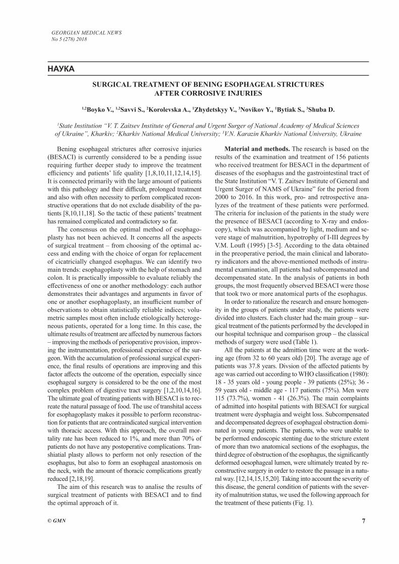
All the patients at the admittion time were at the work-ing age (from 32 to 60 years old) [20]. The average age of patients was 37.8 years. Divsion of the affected patients by age was carried out according to WHO classification (1980): 18 - 35 years old - young people - 39 patients (25%); 36 - 59 years old - middle age - 117 patients (75%). Men were 115 (73.7%), women - 41 (26.3%). The main complaints of admitted into hospital patients with BESACI for surgical treatment were dysphagia and weight loss. Subcompensated and decompensated degrees of esophageal obstruction domi-nated in young patients. The patients, who were unable to be performed endoscopic stenting due to the stricture extent of more than two anatomical sections of the esophagus, the third degree of obstruction of the esophagus, the significantly deformed oesophageal lumen, were ultimately treated by re-constructive surgery in order to restore the passage in a natu-ral way. [12,14,15,15,20]. Taking into account the severity of this disease, the general condition of patients with the sever-ity of malnutrition status, we used the following approach for the treatment of these patients (Fig. 1).
НАУКА
SURGICAL TREATMENT OF BENING ESOPHAGEAL STRICTURES AFTER CORROSIVE INJURIES
1,2Boyko V., 1,2Savvi S., 2Korolevska A., 1Zhydetskyy V., 3Novikov Y., 1Bytiak S., 3Shuba D.
1State Institution “V. T. Zaitsev Institute of General and Urgent Surger of National Academy of Medical Sciences of Ukraine”, Kharkiv; 2Kharkiv National Medical University; 3V.N. Karazin Kharkiv National University, Ukraine
8
МЕДИЦИНСКИЕ НОВОСТИ ГРУЗИИ
CFMFHSDTKJC CFVTLBWBYJ CBF[KTYB
Table 1. Study Layout
One-step surgical treatment Two-step surgical treatment
І stage - -
Contact gastrostomy (II cluster)54 patients
main group comparison group26 patients 28 patients
Gastrostomy by developed in our hospital technique Classic techniques
ІІ stage
One-step esophagoplasty (I cluster) Esophagoplasty (III cluster)50 patients 52 patientsmain group main group comparison group
24 patients 25 patients 27 patientsEsophagogastroplasty by developed
in our hospital techniqueEsophagogastroplasty by devel-oped in our hospital technique Classic techniques
Fig. 1 Surgical tactics of treatment of patients with BESACI
It is known that all patients with the formed BESA-CI with violations of nutritional status have indications for dilatation procedures. When the passage through the esophagus is restored, a passage of food is reproduced naturally temporally in some cases [3-5]. In the future, these patients need in repeated courses of esophageal bou-ginage or balloon dilation, which reduces the life qual-ity of the patient. Anyway sooner or later the question of reconstructive surgery necessity for the patients with ineffective result of dilatation procedures become actual. After preoperative preparation of the patients elective esophagoplasty is performed, which ensures the repro-duction of the natural food passage [3,5,10,12,14,15,19].
If the courses of dilatation procedures are not effec-tive and it is impossible to provide them, exhaustion of patients with severe metabolic disorders develops, and sometimes it can lead to cachexia [20]. Such patients need
two-step surgical treatment. At the first step contact gas-trostomy was formed in order to establish enteral nutrition and improve the nutritive status. At the second step of sur-gical treatment after the restoring of the nutritive status of a patient, resection of the esophagus, esophagoplasty and closure of the gastrostomic opening is perfomed.
Results and their discussion. Patients with BESACI of the comparison group of the first cluster underwent the following traditional one-step reconstructive surgery: esophagogastroplasty - 16 (61.5%) patients, esophago-coloplasty - 10 (38.5%) patients. Postoperative complica-tions developed in 12 (46.2%) cases. The most frequent postoperative complications were pleurisy in 3 (11.5%) cases and pneumonia in 2 (7.7%) cases. After reconstruc-tive operations of the comparison group, the mortality was 3 (11.5%) cases, which were caused by mediastinitis, acute cardiovascular and multiple organ failure [17-19].
In this regard, a technique of one-step transhiatal esopha-gogastroplasty and performing a single anastomosis on the neck in patients with BESACI (main group) was devel-oped and implemented in our hospital - Patent of Ukraine №. 92357 “Method of one-step esophagogastroplasty” [7]. Obligatory conditions for performing of one-step transhiatal esophagogastroplasty and performing a single anastomosis on the neck for patients with BESACI are absence of signifi-cant nutritional disoders; satisfactory patient condition; labo-ratory and instrumental parameters within the norm; absence of severe chronic pathology; the absence of previously car-ried out volumetric operations on the stomach [7,12,13,15].
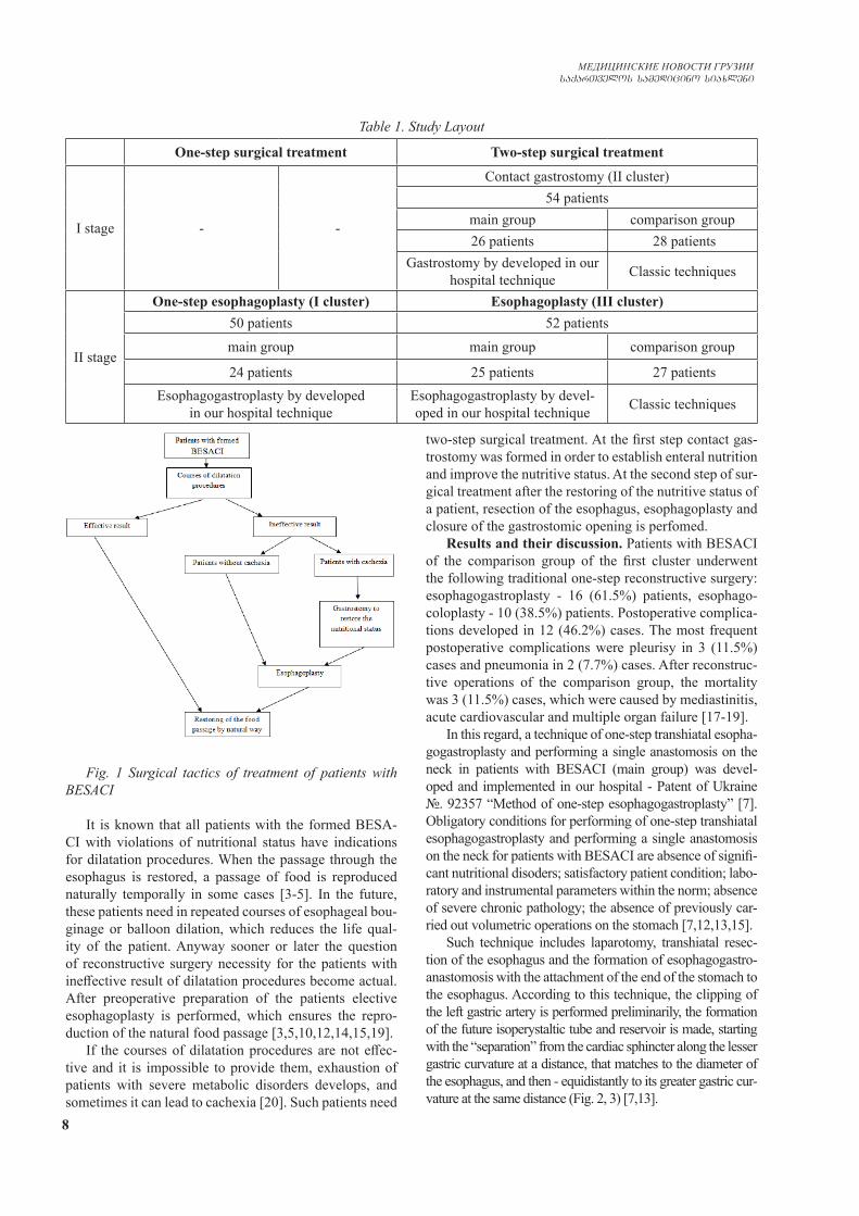
Such technique includes laparotomy, transhiatal resec-tion of the esophagus and the formation of esophagogastro-anastomosis with the attachment of the end of the stomach to the esophagus. According to this technique, the clipping of the left gastric artery is performed preliminarily, the formation of the future isoperystaltic tube and reservoir is made, starting with the “separation” from the cardiac sphincter along the lesser gastric curvature at a distance, that matches to the diameter of the esophagus, and then - equidistantly to its greater gastric cur-vature at the same distance (Fig. 2, 3) [7,13].
GEORGIAN MEDICAL NEWS No 5 (278) 2018
© GMN 9
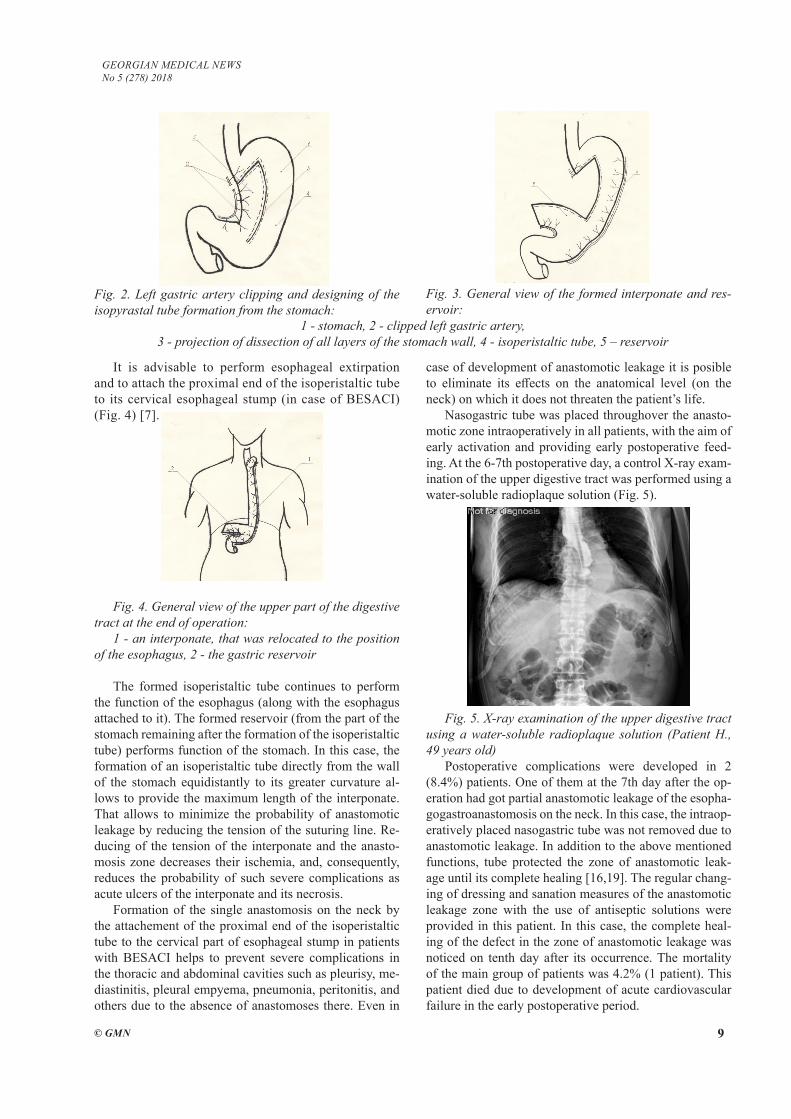
It is advisable to perform esophageal extirpation and to attach the proximal end of the isoperistaltic tube to its cervical esophageal stump (in case of BESACI) (Fig. 4) [7].
Fig. 4. General view of the upper part of the digestive tract at the end of operation:
1 - an interponate, that was relocated to the position of the esophagus, 2 - the gastric reservoir
The formed isoperistaltic tube continues to perform the function of the esophagus (along with the esophagus attached to it). The formed reservoir (from the part of the stomach remaining after the formation of the isoperistaltic tube) performs function of the stomach. In this case, the formation of an isoperistaltic tube directly from the wall of the stomach equidistantly to its greater curvature al-lows to provide the maximum length of the interponate. That allows to minimize the probability of anastomotic leakage by reducing the tension of the suturing line. Re-ducing of the tension of the interponate and the anasto-mosis zone decreases their ischemia, and, consequently, reduces the probability of such severe complications as acute ulcers of the interponate and its necrosis.
Formation of the single anastomosis on the neck by the attachement of the proximal end of the isoperistaltic tube to the cervical part of esophageal stump in patients with BESACI helps to prevent severe complications in the thoracic and abdominal cavities such as pleurisy, me-diastinitis, pleural empyema, pneumonia, peritonitis, and others due to the absence of anastomoses there. Even in
case of development of anastomotic leakage it is posible to eliminate its effects on the anatomical level (on the neck) on which it does not threaten the patient’s life.
Nasogastric tube was placed throughover the anasto-motic zone intraoperatively in all patients, with the aim of early activation and providing early postoperative feed-ing. At the 6-7th postoperative day, a control X-ray exam-ination of the upper digestive tract was performed using a water-soluble radioplaque solution (Fig. 5).
Fig. 5. X-ray examination of the upper digestive tract using a water-soluble radioplaque solution (Patient H., 49 years old)
Postoperative complications were developed in 2 (8.4%) patients. One of them at the 7th day after the op-eration had got partial anastomotic leakage of the esopha-gogastroanastomosis on the neck. In this case, the intraop-eratively placed nasogastric tube was not removed due to anastomotic leakage. In addition to the above mentioned functions, tube protected the zone of anastomotic leak-age until its complete healing [16,19]. The regular chang-ing of dressing and sanation measures of the anastomotic leakage zone with the use of antiseptic solutions were provided in this patient. In this case, the complete heal-ing of the defect in the zone of anastomotic leakage was noticed on tenth day after its occurrence. The mortality of the main group of patients was 4.2% (1 patient). This patient died due to development of acute cardiovascular failure in the early postoperative period.
Fig. 2. Left gastric artery clipping and designing of the isopyrastal tube formation from the stomach:
Fig. 3. General view of the formed interponate and res-ervoir:
1 - stomach, 2 - clipped left gastric artery, 3 - projection of dissection of all layers of the stomach wall, 4 - isoperistaltic tube, 5 – reservoir
10
МЕДИЦИНСКИЕ НОВОСТИ ГРУЗИИ
CFMFHSDTKJC CFVTLBWBYJ CBF[KTYB
The advantage of traditional methods of one-step esophagoplasty is the reduction of the time of staying in the clinic and the absence of the need for repeated admis-sion for reconstructive surgery in a two-step treatment, reducing the time of postoperative rehabilitation.
In our clinic for the patients with BESACI that have severe nutritive disorders two-step surgical treatment was developed and implemented. The purpose of the first step of surgical treatment is the restoration of the nutritive sta-tus by means of nutritional support through the formed contact gastrostomy, delineation of the affected zone. Af-ter the restoration of the body weight deficiency, the sec-ond step of surgical treatment for patients with BESACI with persistent severe dysphagia is a reconstructive opera-tion with the closure of gastrostomy and restoring of the natural food passage.
This two-step tactic involves the formation of a con-tact gastrostomy at the first step and the closure of gas-trostomy with the esophagoplasty using the interponate, prepared from the wall of the stomach at the first step with the formation of a single anastomosis on the neck using transhiatal access – at second step.
The patients with BESACI which have severe dyspha-gy have progressive deterioration of the general condition due to the inability to provide adequate nutrition and de-compensation of the protective forces and adaptive pos-sibilities of the organism. It can lead to the general body exhaustion and sometimes to cachexia. That is why such patients need to perform contact gastrostomy for enteral nutrition at the earliest date as a priority issue. For these patients, the limitation of surgery volume and time to re-duce the risks of intra- and postoperative complications is the most important [3-5,20].
In all cases of the II cluster decompensated degree of esophageal obstruction was marked (100%). The weakened state of patients requires performing contact gastrostomy as soon as possible [9,12,15]. Patients were admitted into our clinic mainly in the late post-burn period of the formation of cicatricial stricture (52 patients - 96.3%), according to the post-burn cicatricial stricture classification developed in our clinic [1]. In the early postoperative period, Kader`s gastrostomy was performed in two patients (3.7%) with severe dysphagia in order to provide nutritional support, enteral nutrition and exclusion of esophagus from the food passage.
The severe dysphagia and the attenuated condition of patients with BESACI require the performing of gastrostomy in the shortest period of time to achieve normalization of nutritional status, delimitation of the zone of damage and enteral nutrition. That was achieved in all patients of both groups of II cluster. The reported methods intraoperatively did not need to extend the operative access and were performed with minilaparotomy in all cases.
Kader`s gastrostomy was formed for all patients in the comparison group of II cluster. In 4 (15.38%) patients there was a cleft of the purse-string suture with leakage of
the gastric contents and an increase in the diameter of the gastrostomy hole with maceration of the skin around it. That further complicated the process of care and required frequent changing of dressing, sometimes up to 5 times and more per day. Also, there was a loss of gastrostomic tube in these patients. That created additional discomfort for the patients and limited their stay in the society (the presence of unpleasant smell, etc.).
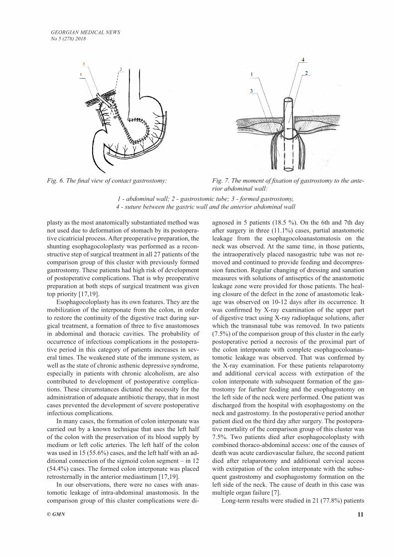
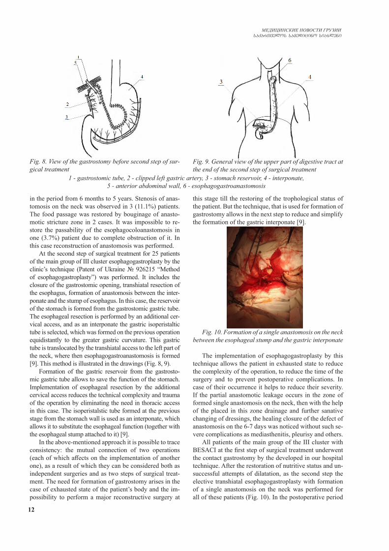
A contact gastrostomy by the clinic’s technique (Pat-ent of Ukraine № 92441, «Method of contact gastros-tomy») was formed for all patients of the main group II cluster. It includes the formation of contact gastrostomy from the wall of the stomach in its lesser curvature and its atachment to the anterior abdominal wall and the insertion of tube for feeding into the formed gastrostomy. Partial processing of the lesser gastric curvature with clipping of the left gastric artery is performed. Then gastrostomy is formed as an isoperistaltic tube ecvidistantly to the lesser gastric curvature. After that, taking into account the diam-eter of the hole and the length of the replacing gastrosto-mic tube, the place of attachment on the anterior abdomi-nal wall is selected and the gastrostomy is performed at this point. The distal end of this tube attached with the help of sutures to the parietal peritoneum, posterior and anterior sheets of the rectal sheath and to the skin. Then tube for feeding is inserted into the formed gastrostomy [6].
Formation of gastrostomy as an isoperistaltic tube is equidistant to a lesser gastric curvature with clipping of the left gastric artery, that reduces trauma level and isch-emia of the stomach. Because its main blood supply is carried out by arteries that pass along greater gastric cur-vature. Partial processing of the lesser gastric curvature with clipping of the left gastric artery does not affect the blood supply of the stomach. The blood supply of the stomach in this case will be carried out by collaterals from the right gastric artery, left and right gastroepiploic arter-ies, short gastric vessels. So trauma of the gastric stem is minimal with minimal affection of its blood supply [6].
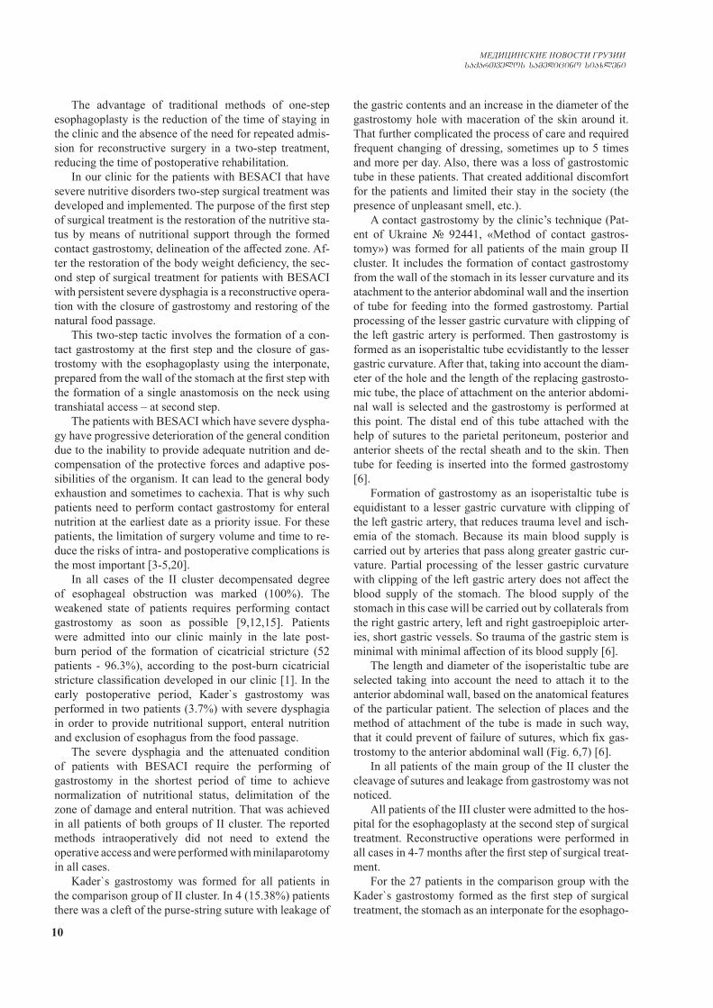
The length and diameter of the isoperistaltic tube are selected taking into account the need to attach it to the anterior abdominal wall, based on the anatomical features of the particular patient. The selection of places and the method of attachment of the tube is made in such way, that it could prevent of failure of sutures, which fix gas-trostomy to the anterior abdominal wall (Fig. 6,7) [6].
In all patients of the main group of the II cluster the cleavage of sutures and leakage from gastrostomy was not noticed.
All patients of the III cluster were admitted to the hos-pital for the esophagoplasty at the second step of surgical treatment. Reconstructive operations were performed in all cases in 4-7 months after the first step of surgical treat-ment.
For the 27 patients in the comparison group with the Kader`s gastrostomy formed as the first step of surgical treatment, the stomach as an interponate for the esophago-
GEORGIAN MEDICAL NEWS No 5 (278) 2018
© GMN 11
plasty as the most anatomically substantiated method was not used due to deformation of stomach by its postopera-tive cicatricial process. After preoperative preparation, the shunting esophagocoloplasty was performed as a recon-structive step of surgical treatment in all 27 patients of the comparison group of this cluster with previously formed gastrostomy. These patients had high risk of development of postoperative complications. That is why preoperative preparation at both steps of surgical treatment was given top priority [17,19].
Esophagocoloplasty has its own features. They are the mobilization of the interponate from the colon, in order to restore the continuity of the digestive tract during sur-gical treatment, a formation of three to five anastomoses in abdominal and thoracic cavities. The probability of occurrence of infectious complications in the postopera-tive period in this category of patients increases in sev-eral times. The weakened state of the immune system, as well as the state of chronic asthenic depressive syndrome, especially in patients with chronic alcoholism, are also contributed to development of postoperative complica-tions. These circumstances dictated the necessity for the administration of adequate antibiotic therapy, that in most cases prevented the development of severe postoperative infectious complications.
In many cases, the formation of colon interponate was carried out by a known technique that uses the left half of the colon with the preservation of its blood supply by medium or left colic arteries. The left half of the colon was used in 15 (55.6%) cases, and the left half with an ad-ditional connection of the sigmoid colon segment – in 12 (54.4%) cases. The formed colon interponate was placed retrosternally in the anterior mediastinum [17,19].
In our observations, there were no cases with anas-tomotic leakage of intra-abdominal anastomosis. In the comparison group of this cluster complications were di-
agnosed in 5 patients (18.5 %). On the 6th and 7th day after surgery in three (11.1%) cases, partial anastomotic leakage from the esophagocoloanastomatosis on the neck was observed. At the same time, in those patients, the intraoperatively placed nasogastric tube was not re-moved and continued to provide feeding and decompres-sion function. Regular changing of dressing and sanation measures with solutions of antiseptics of the anastomotic leakage zone were provided for those patients. The heal-ing closure of the defect in the zone of anastomotic leak-age was observed on 10-12 days after its occurrence. It was confirmed by X-ray examination of the upper part of digestive tract using X-ray radioplaque solutions, after which the transnasal tube was removed. In two patients (7.5%) of the comparison group of this cluster in the early postoperative period a necrosis of the proximal part of the colon interponate with complete esophagocoloanas-tomotic leakage was observed. That was confirmed by the X-ray examination. For these patients relaparotomy and additional cervical access with extirpation of the colon interponate with subsequent formation of the gas-trostomy for further feeding and the esophagostomy on the left side of the neck were performed. One patient was discharged from the hospital with esophagostomy on the neck and gastrostomy. In the postoperative period another patient died on the third day after surgery. The postopera-tive mortality of the comparison group of this cluster was 7.5%. Two patients died after esophagocoloplasty with combined thoraco-abdominal access: one of the causes of death was acute cardiovascular failure, the second patient died after relaparotomy and additional cervical access with extirpation of the colon interponate with the subse-quent gastrostomy and esophagostomy formation on the left side of the neck. The cause of death in this case was multiple organ failure [7].
Long-term results were studied in 21 (77.8%) patients
Fig. 6. The final view of contact gastrostomy: Fig. 7. The moment of fixation of gastrostomy to the ante-rior abdominal wall:
1 - abdominal wall; 2 - gastrostomic tube; 3 - formed gastrostomy, 4 - suture between the gastric wall and the anterior abdominal wall
12
МЕДИЦИНСКИЕ НОВОСТИ ГРУЗИИ
CFMFHSDTKJC CFVTLBWBYJ CBF[KTYB
in the period from 6 months to 5 years. Stenosis of anas-tomosis on the neck was observed in 3 (11.1%) patients. The food passage was restored by bouginage of anasto-motic stricture zone in 2 cases. It was impossible to re-store the passability of the esophagocoloanastomosis in one (3.7%) patient due to complete obstruction of it. In this case reconstruction of anastomosis was performed.
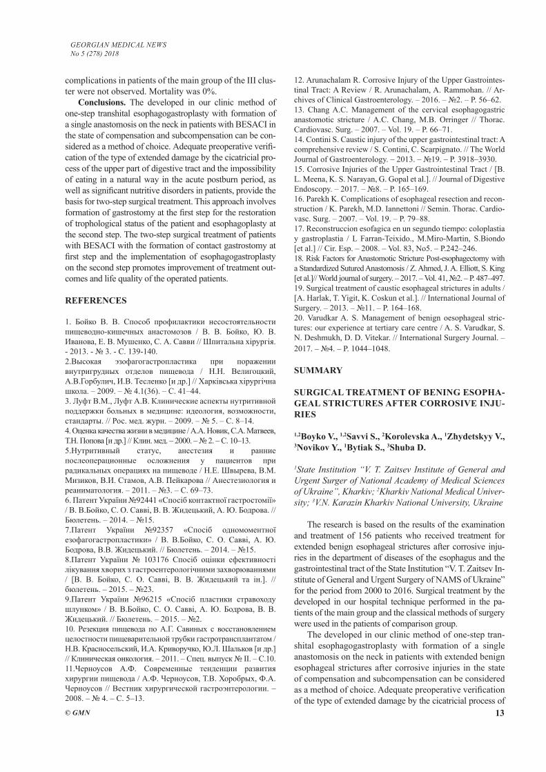
At the second step of surgical treatment for 25 patients of the main group of III cluster esophagogastroplasty by the clinic’s technique (Patent of Ukraine № 926215 “Method of esophagogastroplasty”) was performed. It includes the closure of the gastrostomic opening, transhiatal resection of the esophagus, formation of anastomosis between the inter-ponate and the stump of esophagus. In this case, the reservoir of the stomach is formed from the gastrostomic gastric tube. The esophageal resection is performed by an additional cer-vical access, and as an interponate the gastric isoperistaltic tube is selected, which was formed on the previous operation equidistantly to the greater gastric curvature. This gastric tube is translocated by the transhiatal access to the left part of the neck, where then esophagogastroanastomosis is formed [9]. This method is illustrated in the drawings (Fig. 8, 9).
Formation of the gastric reservoir from the gastrosto-mic gastric tube allows to save the function of the stomach. Implementation of esophageal resection by the additional cervical access reduces the technical complexity and trauma of the operation by eliminating the need in thoracic access in this case. The isoperistalstic tube formed at the previous stage from the stomach wall is used as an interponate, which allows it to substitute the esophageal function (together with the esophageal stump attached to it) [9].
In the above-mentioned approach it is possible to trace consistency: the mutual connection of two operations (each of which affects on the implementation of another one), as a result of which they can be considered both as independent surgeries and as two steps of surgical treat-ment. The need for formation of gastrostomy arises in the case of exhausted state of the patient’s body and the im-possibility to perform a major reconstructive surgery at
this stage till the restoring of the trophological status of the patient. But the technique, that is used for formation of gastrostomy allows in the next step to reduce and simplify the formation of the gastric interponate [9].
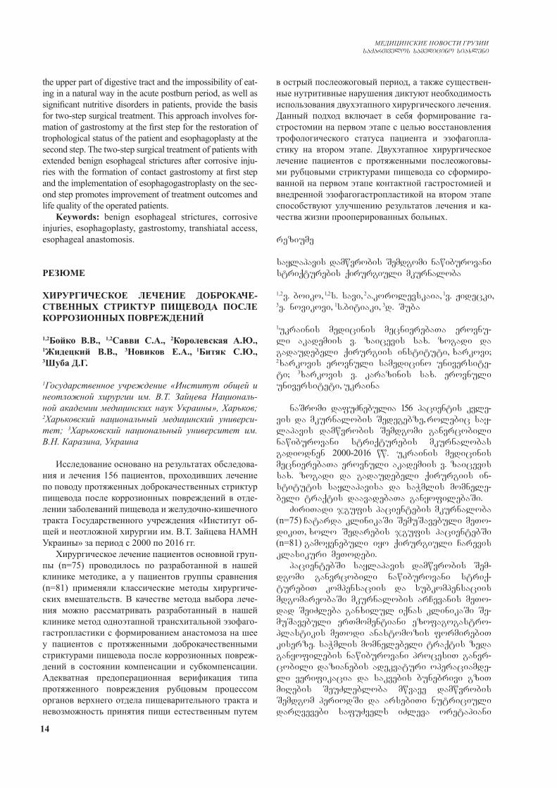
Fig. 10. Formation of a single anastomosis on the neck between the esophageal stump and the gastric interponate
The implementation of esophagogastroplasty by this technique allows the patient in exhausted state to reduce the complexity of the operation, to reduce the time of the surgery and to prevent postoperative complications. In case of their occurrence it helps to reduce their severity. If the partial anastomotic leakage occurs in the zone of formed single anastomosis on the neck, then with the help of the placed in this zone drainage and further sanative changing of dressings, the healing closure of the defect of anastomosis on the 6-7 days was noticed without such se-vere complications as mediasthenitis, pleurisy and others.
All patients of the main group of the III cluster with BESACI at the first step of surgical treatment underwent the contact gastrostomy by the developed in our hospital technique. After the restoration of nutritive status and un-successful attempts of dilatation, as the second step the elective transhiatal esophagogastroplasty with formation of a single anastomosis on the neck was performed for all of these patients (Fig. 10). In the postoperative period
Fig. 8. View of the gastrostomy before second step of sur-gical treatment
Fig. 9. General view of the upper part of digestive tract at the end of the second step of surgical treatment
1 - gastrostomic tube, 2 - clipped left gastric artery, 3 - stomach reservoir, 4 - interponate, 5 - anterior abdominal wall, 6 - esophagogastroanastomosis
GEORGIAN MEDICAL NEWS No 5 (278) 2018
© GMN 13
complications in patients of the main group of the III clus-ter were not observed. Mortality was 0%.
Conclusions. The developed in our clinic method of one-step transhital esophagogastroplasty with formation of a single anastomosis on the neck in patients with BESACI in the state of compensation and subcompensation can be con-sidered as a method of choice. Adequate preoperative verifi-cation of the type of extended damage by the cicatricial pro-cess of the upper part of digestive tract and the impossibility of eating in a natural way in the acute postburn period, as well as significant nutritive disorders in patients, provide the basis for two-step surgical treatment. This approach involves formation of gastrostomy at the first step for the restoration of trophological status of the patient and esophagoplasty at the second step. The two-step surgical treatment of patients with BESACI with the formation of contact gastrostomy at first step and the implementation of esophagogastroplasty on the second step promotes improvement of treatment out-comes and life quality of the operated patients.
REFERENCES
1. Бойко В. В. Способ профилактики несостоятельности пищеводно-кишечных анастомозов / В. В. Бойко, Ю. В. Иванова, Е. В. Мушенко, С. А. Савви // Шпитальна хірургія. - 2013. - № 3. - С. 139-140.2.Высокая эзофагогастропластика при поражении внутригрудных отделов пищевода / Н.Н. Велигоцкий, А.В.Горбулич, И.В. Тесленко [и др.] // Харківська хірургічна школа. – 2009. – № 4.1(36). – С. 41–44.3. Луфт В.М., Луфт А.В. Клинические аспекты нутритивной поддержки больных в медицине: идеология, возможности, стандарты. // Рос. мед. журн. – 2009. – № 5. – С. 8–14.4. Оценка качества жизни в медицине / А.А. Новик, С.А. Матвеев, Т.Н. Попова [и др.] // Клин. мед. – 2000. – № 2. – С. 10–13.5.Нутритивный статус, анестезия и ранние послеоперационные осложнения у пациентов при радикальных операциях на пищеводе / Н.Е. Швырева, В.М. Мизиков, В.И. Стамов, А.В. Пейкарова // Анестезиология и реаниматология. – 2011. – №3. – С. 69–73.6. Патент України №92441 «Спосіб контактної гастростомії» / В. В.Бойко, С. О. Савві, В. В. Жидецький, А. Ю. Бодрова. // Бюлетень. – 2014. – №15.7.Патент України №92357 «Спосіб одномоментної езофагогастропластики» / В. В.Бойко, С. О. Савві, А. Ю. Бодрова, В.В. Жидецький. // Бюлетень. – 2014. – №15.8.Патент України № 103176 Спосіб оцінки ефективності лікування хворих з гастроентерологічними захворюваннями / [В. В. Бойко, С. О. Савві, В. В. Жидецький та ін.]. // бюлетень. – 2015. – №23.9.Патент України №96215 «Спосіб пластики стравоходу шлунком» / В. В.Бойко, С. О. Савві, А. Ю. Бодрова, В. В. Жидецький. // Бюлетень. – 2015. – №2. 10. Резекция пищевода по А.Г. Савиных с восстановлением целостности пищеварительной трубки гастротрансплантатом / Н.В. Красносельский, И.А. Криворучко, Ю.Л. Шальков [и др.] // Клиническая онкология. – 2011. – Спец. выпуск № ІІ. – С.10.11.Черноусов А.Ф. Современные тенденции развития хирургии пищевода / А.Ф. Черноусов, Т.В. Хоробрых, Ф.А. Черноусов // Вестник хирургической гастроэнтерологии. – 2008. – № 4. – С. 5–13.
12. Arunachalam R. Corrosive Injury of the Upper Gastrointes-tinal Tract: A Review / R. Arunachalam, A. Rammohan. // Ar-chives of Clinical Gastroenterology. – 2016. – №2. – P. 56–62.13. Chang A.C. Management of the cervical esophagogastric anastomotic stricture / A.C. Chang, M.B. Orringer // Thorac. Cardiovasc. Surg. – 2007. – Vol. 19. – P. 66–71.14. Contini S. Caustic injury of the upper gastrointestinal tract: A comprehensive review / S. Contini, C. Scarpignato. // The World Journal of Gastroenterology. – 2013. – №19. – P. 3918–3930.15. Corrosive Injuries of the Upper Gastrointestinal Tract / [B. L. Meena, K. S. Narayan, G. Gopal et al.]. // Journal of Digestive Endoscopy. – 2017. – №8. – P. 165–169.16. Parekh K. Complications of esophageal resection and recon-struction / K. Parekh, M.D. Iannettoni // Semin. Thorac. Cardio-vasc. Surg. – 2007. – Vol. 19. – P. 79–88.17. Reconstruccion esofagica en un segundo tiempo: coloplastia y gastroplastia / L Farran-Teixido., M.Miro-Martin, S.Biondo [et al.] // Cir. Esp. – 2008. – Vol. 83, No5. – P.242–246.18. Risk Factors for Anastomotic Stricture Post-esophagectomy with a Standardized Sutured Anastomosis / Z. Ahmed, J. A. Elliott, S. King [et al.]// World journal of surgery. – 2017. – Vol. 41, №2. – P. 487–497.19. Surgical treatment of caustic esophageal strictures in adults / [A. Harlak, T. Yigit, K. Coskun et al.]. // International Journal of Surgery. – 2013. – №11. – P. 164–168.20. Varudkar A. S. Management of benign oesophageal stric-tures: our experience at tertiary care centre / A. S. Varudkar, S. N. Deshmukh, D. D. Vitekar. // International Surgery Journal. – 2017. – №4. – P. 1044–1048.
SUMMARY
SURGICAL TREATMENT OF BENING ESOPHA-GEAL STRICTURES AFTER CORROSIVE INJU-RIES
1,2Boyko V., 1,2Savvi S., 2Korolevska A., 1Zhydetskyy V., 3Novikov Y., 1Bytiak S., 3Shuba D.
1State Institution “V. T. Zaitsev Institute of General and Urgent Surger of National Academy of Medical Sciences of Ukraine”, Kharkiv; 2Kharkiv National Medical Univer-sity; 3V.N. Karazin Kharkiv National University, Ukraine
The research is based on the results of the examination and treatment of 156 patients who received treatment for extended benign esophageal strictures after corrosive inju-ries in the department of diseases of the esophagus and the gastrointestinal tract of the State Institution “V. T. Zaitsev In-stitute of General and Urgent Surgery of NAMS of Ukraine” for the period from 2000 to 2016. Surgical treatment by the developed in our hospital technique performed in the pa-tients of the main group and the classical methods of surgery were used in the patients of comparison group.
The developed in our clinic method of one-step tran-shital esophagogastroplasty with formation of a single anastomosis on the neck in patients with extended benign esophageal strictures after corrosive injuries in the state of compensation and subcompensation can be considered as a method of choice. Adequate preoperative verification of the type of extended damage by the cicatricial process of
14
МЕДИЦИНСКИЕ НОВОСТИ ГРУЗИИ
CFMFHSDTKJC CFVTLBWBYJ CBF[KTYB
the upper part of digestive tract and the impossibility of eat-ing in a natural way in the acute postburn period, as well as significant nutritive disorders in patients, provide the basis for two-step surgical treatment. This approach involves for-mation of gastrostomy at the first step for the restoration of trophological status of the patient and esophagoplasty at the second step. The two-step surgical treatment of patients with extended benign esophageal strictures after corrosive inju-ries with the formation of contact gastrostomy at first step and the implementation of esophagogastroplasty on the sec-ond step promotes improvement of treatment outcomes and life quality of the operated patients.
Keywords: benign esophageal strictures, corrosive injuries, esophagoplasty, gastrostomy, transhiatal access, esophageal anastomosis.
РЕЗЮМЕ
ХИРУРГИЧЕСКОЕ ЛЕЧЕНИЕ ДОБРОКАЧЕ-СТВЕННЫХ СТРИКТУР ПИЩЕВОДА ПОСЛЕ КОРРОЗИОННЫХ ПОВРЕЖДЕНИЙ
1,2Бойко В.В., 1,2Савви С.А., 2Королевская А.Ю., 1Жидецкий В.В., 3Новиков Е.А., 1Битяк С.Ю., 3Шуба Д.Г.
1Государственное учреждение «Институт общей и неотложной хирургии им. В.Т. Зайцева Националь-ной академии медицинских наук Украины», Харьков; 2Харьковский национальный медицинский универси-тет; 3Харьковский национальный университет им. В.Н. Каразина, Украина
Исследование основано на результатах обследова-ния и лечения 156 пациентов, проходивших лечение по поводу протяженных доброкачественных стриктур пищевода после коррозионных повреждений в отде-лении заболеваний пищевода и желудочно-кишечного тракта Государственного учреждения «Институт об-щей и неотложной хирургии им. В.Т. Зайцева НАМН Украины» за период с 2000 по 2016 гг.
Хирургическое лечение пациентов основной груп-пы (n=75) проводилось по разработанной в нашей клинике методике, а у пациентов группы сравнения (n=81) применяли классические методы хирургиче-ских вмешательств. В качестве метода выбора лече-ния можно рассматривать разработанный в нашей клинике метод одноэтапной трансхитальной эзофаго-гастропластики с формированием анастомоза на шее у пациентов с протяженными доброкачественными стриктурами пищевода после коррозионных повреж-дений в состоянии компенсации и субкомпенсации. Адекватная предоперационная верификация типа протяженного повреждения рубцовым процессом органов верхнего отдела пищеварительного тракта и невозможность принятия пищи естественным путем
в острый послеожоговый период, а также существен-ные нутритивные нарушения диктуют необходимость использования двухэтапного хирургического лечения. Данный подход включает в себя формирование га-стростомии на первом этапе с целью восстановления трофологического статуса пациента и эзофагопла-стику на втором этапе. Двухэтапное хирургическое лечение пациентов с протяженными послеожоговы-ми рубцовыми стриктурами пищевода со сформиро-ванной на первом этапе контактной гастростомией и внедренной эзофагогастропластикой на втором этапе способствуют улучшению результатов лечения и ка-чества жизни прооперированных больных.
reziume
saylapavis damwvrobis Semdgomi nawiburovani striqturebis qirurgiuli mkurnaloba
1,2v. boiko, 1,2s. savi, 2a.korolevskaia, 1v. Jidecki, 3e. novikovi, 1s.bitiaki, 3d. Suba
1ukrainis medicinis mecnierebaTa erovnu-li akademiis v. zaicevis sax. zogadi da gadaudebeli qirurgiis instituti, xarkovi; 2xarkovis erovnuli samedicino universite-ti; 3xarkovis v. karazinis sax. erovnuli universiteti, ukraina
naSromi dafuZnebulia 156 pacientis kvle-vis da mkurnalobis Sedegebze, rolebic say-lapavis damwvrobis Semdgomi ganvrcobili nawiburovani striqturebis mkurnalobas gadiodnen 2000-2016 ww. ukrainis medicinis mecnierebaTa erovnuli akademiis v. zaicevis sax. zogadi da gadaudebeli qirurgiis in-stitutis saylapavisa da saWmlis momnele-beli traqtis daavadebaTa ganyofilebaSi.
ZiriTadi jgufis pacientebis mkurnaloba (n=75) Catarda klinikaSi SemuSavebuli meTo-dikiT, xolo Sedarebis jgufis pacientebSi (n=81) gamoyenebuli iyo qirurgiuli Carevis klasikuri meTodebi.
pacientebSi saylapavis damwvrobis Sem-dgomi ganvrcobili nawiburovani striq-turebiT kompensaciis da subkompensaciis mdgomareobaSi mkurnalobis arCevanis meTo-dad SeiZleba ganxilul iqnas klinikaSi Se-muSavebuli erTmomentiani ezofagogastro-plastikis meTodi anastomozis formirebiT kiserze. saWmlis momnelebeli traqtis zeda ganyofilebis nawiburovani procesiT ganvr-cobili dazianebis adekvaturi operaciamde-li verifikacia da sakvebis bunebrivi gziT miRebis SeuZlebloba mwvave damwvrobis Semdgom periodSi da arsebiTi nutriciuli darRvevebi safuZvels iZleva oretapiani
GEORGIAN MEDICAL NEWS No 5 (278) 2018
© GMN 15
qirurgiuli mkurnalobisaTvis. aseTi mid-goma gulisxmobs pirvel etapze gastrosto-mias pacientis trofologiuri statusis aRdgenis mizniT, meore etapze ki - ezofago-plastikas. saylapavis damwvrobis Semdgomi ganvrcobili nawiburovani striqturebis
mqone pacientebis oretapiani qirurgiuli mkurnaloba pirvel etapze - kontaqturi gastrostomiiT, meore etapze ki - Canergili ezofagogastroplastikiT xels uwyobs mkurnalobis Sedegebis da pacientebis si-cocxlis xarisxis gaumjobesebas.
COMPARATIVE EVALUATION OF TAPP HERNIOPLASTY WITH USE OF VARIOUS METHODS OF FIXING THE RETICULAR ENDOPROSTHESIS
AND TEP IN THE TREATMENT OF INGUINAL HERNIAS
2Krikunov D., 1,2Akimov V., 1Toidze V., 1Churgulia M., 1Dvаladze L.
1 L.G. Sokolov Memorial Clinical Hospital № 122, Federal Medico-Biologic Agency, St. Petersburg;2N.D. Monastyrsky Department of Surgery, North-Western State Medical University
Named After I.I. Mechnikov, Ministry of Healthcare of Russian Federation, St. Petersburg, Ruassia
Inguinal hernia is one of the most common patholo-gies in abdominal surgery. Statistically, percentage of surgeries performed for inguinal hernia repair makes up about 80% of general amount of all hernioplasties [4]. Ac-cording to the age-specific morbidity statistics, inguinal hernia occurs in 14:1000 population of 25 to 34 years age group, and increases up to 53:1000 in population of 55 to 64 years age group. More than 20 million hernia surgeries are carried out worldwide annually [13].
Since the end of the last century endovideosurgery op-erations have become more vastly applied in all spheres of surgery and are rightly considered a new stage in the development of surgical field. Characterized by compara-tively minimal invasiveness and injury rate, and a great degree of visualization of anatomic structures and high efficiency, in most cases they have became a worthy re-placement for traditional modes of hernioplasty, however, the incipient pain syndrome and incidence of chronic pain are still remained in this group of patients.
In May of 1991 two US surgeons, R. Nagan and M. Arregui, described the procedure of laparoscopic her-nioplasty for the first time. Its stages included: abdomen opening, thorough dissection of posterior wall of ingui-nal region; preperitoneal placement of allograft covering all “weak” areas, fixation of implant with the staples and closure of abdominal defect above the endoprosthesis (peritonization) [7,11,12]. This method was called ‘’lapa-roscopic transabdominal preperitoneal inguinal hernia repair’’ (TAPP). French surgeon R.E Stoppa developed a methodology of preperitoneal repair with use of large mesh endoimplants. Method is considered as a trauma-tizing and nowadays its classical variant is used seldom, however, it served as a prototype for the development of laparoscopic extraperitoneal hernioplasty (TEP) [2,13].
According to European Hernia Society Guidelines on the Treatment of Inguinal Hernia in Adult Patients (Up-date with level 1 studies of the European Hernia Society
guidelines on the treatment of inguinal hernia in adult pa-tients, M. Miserez et al.,2014) – penetrating fixation or traumatic devices such as surgical sutures, staples and fix-ators, lead to the local trauma which can be accompanied by the nerve damage and development of chronic pain. This is described by a few authors [1,3,5,8,10,14] - the main criterion of surgical treatment efficiency became not only frequency of relapses, but also occurrence of an acute, as well as chronic postoperative pain. Therefore, use of sutureless fixation (glue, mesh with “adhesive tape” principle) acquires larger popularity, since the risk of postoperative complications decreases significantly. In this connection the guideline includes the following rec-ommendations: Level 1B – There is a possibility of short-term advantage by the criterion of postoperative pain under the nontraumatic fixation of mesh used within the framework of Lichtenstein method and also under endo-scopic procedures (TAPP). Level B – Under the hernio-plasty by Lichtenstein method and endoscopic procedures by the TAPP method, it’s possible to use non-traumatic fixation of mesh, which does not lead to the increase of the frequency of relapses in a year after operation.
Having analyzed scientific data, available during the last decade, comparing the postoperative period of different laparoscopic methods of hernioplasty, with different variants of implant fixation [1,3,5,8,10], we are able to say that they don’t give unambiguous answer to sometimes disturbing question about the optimal methodology of carrying out of surgery and the method of implant fixation and, sometimes, they are rather controversial. In connection with these cir-cumstances the authors have carried out the current study.
The aim of the study - Improvement of the results of operative treatment of patients with inguinal hernia by the way of improvement of method of allograft under endo-videosurgery hernioplasty.
Material and methods. The study included 96 male patients, who had undergone laparoscopic herniоplаsty in
16
МЕДИЦИНСКИЕ НОВОСТИ ГРУЗИИ
CFMFHSDTKJC CFVTLBWBYJ CBF[KTYB
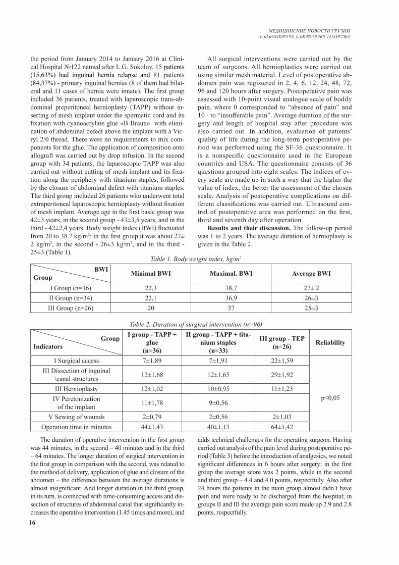
the period from January 2014 to January 2016 at Clini-cal Hospital №122 named after L.G. Sokolov. 15 patients (15,63%) had inguinal hernia relapse and 81 patients (84,37%) - primary inguinal hernias (8 of them had bilat-eral and 11 cases of hernia were innate). The first group included 36 patients, treated with laparoscopic trans-ab-dominal preperitoneal hernioplasty (TAPP) without in-serting of mesh implant under the spermatic cord and its fixation with cyanoacrylate glue «В-Вraun» with elimi-nation of abdominal defect above the implant with a Vic-ryl 2/0 thread. There were no requirements to mix com-ponents for the glue. The application of composition onto allograft was carried out by drop infusion. In the second group with 34 patients, the laparoscopic TAPP was also carried out without cutting of mesh implant and its fixa-tion along the periphery with titanium staples, followed by the closure of abdominal defect with titanium staples. The third group included 26 patients who underwent total extraperitonеal laparoscopic hernioplasty without fixation of mesh implant. Average age in the first basic group was 42±3 years, in the second group - 43±3,5 years, and in the third - 42±2,4 years. Body weight index (BWI) fluctuated from 20 to 38.7 kg/m2: in the first group it was about 27± 2 kg/m2, in the second - 26±3 kg/m2, and in the third - 25±3 (Table 1).
All surgical interventions were carried out by the team of surgeons. All hernioplasties were carried out using similar mesh material. Level of postoperative ab-domen pain was registered in 2, 4, 6, 12, 24, 48, 72, 96 and 120 hours after surgery. Postoperative pain was assessed with 10-point visual analogue scale of bodily pain, where 0 corresponded to “absence of pain” and 10 - to “insufferable pain”. Average duration of the sur-gery and length of hospital stay after procedure was also carried out. In addition, evaluation of patients’ quality of life during the long-term postoperative pe-riod was performed using the SF-36 questionnaire. It is a nonspecific questionnaire used in the European countries and USA. The questionnaire consists of 36 questions grouped into eight scales. The indices of ev-ery scale are made up in such a way that the higher the value of index, the better the assessment of the chosen scale. Analysis of postoperative complications on dif-ferent classifications was carried out. Ultrasound con-trol of postoperative area was performed on the first, third and seventh day after operation.
Results and their discussion. The follow-up period was 1 to 2 years. The average duration of hernioplasty is given in the Table 2.
Table 1. Body weight index, kg/m2
BWIGroup Minimal BWI Maximal. BWI Average BWI
I Group (n=36) 22,3 38,7 27± 2II Group (n=34) 22,1 36,9 26±3III Group (n=26) 20 37 25±3
Table 2. Duration of surgical intervention (n=96)
GroupIndicators
I group - TAPP + glue
(n=36)
II group - TAPP + tita-nium staples
(n=33)
III group - ТEР(n=26) Reliability
I Surgical access 7±1,89 7±1,91 22±1,59
р<0,05
III Dissection of inguinal \canal structures 12±1,68 12±1,65 29±1,92
III Hernioplasty 12±1,02 10±0,95 11±1,23IV Peretonization
of the implant 11±1,78 9±0,56
V Sewing of wounds 2±0,79 2±0,56 2±1,03Operation time in minutes 44±1,43 40±1,13 64±1,42
The duration of operative intervention in the first group was 44 minutes, in the second – 40 minutes and in the third – 64 minutes. The longer duration of surgical intervention in the first group in comparison with the second, was related to the method of delivery, application of glue and closure of the abdomen – the difference between the average durations is almost insignificant. And longer duration in the third group, in its turn, is connected with time-consuming access and dis-section of structures of abdominal canal that significantly in-creases the operative intervention (1.45 times and more), and
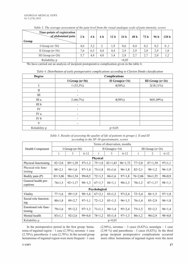
adds technical challenges for the operating surgeon. Having carried out analysis of the pain level during postoperative pe-riod (Table 3) before the introduction of analgesics, we noted significant differences in 6 hours after surgery: in the first group the average score was 2 points, while in the second and third group – 4.4 and 4.0 points, respectfully. Also after 24 hours the patients in the main group almost didn’t have pain and were ready to be discharged from the hospital; in groups II and III the average pain score made up 2.9 and 2.8 points, respectfully.
GEORGIAN MEDICAL NEWS No 5 (278) 2018
© GMN 17
Table 3. The average assessment of the pain level from the visual analogue scale of pain intensity, scores Time-points of registration
of abdominal painGroup
2 h 4 h 6 h 12 h 24 h 48 h 72 h 96 h 120 h
I Group (n=36) 4,6 3,2 2 1,9 0,6 0,4 0,2 0,2 0 ,1II Group (n=34) 7,6 6,5 4,4 4,4 2,9 2,9 2,8 2,9 1,8 III Group (n=26) 5,7 4,8 4,0 3,4 2,8 2,7 2,7 2,0 1,2
Reliability р <0,05We have carried out an analysis of incipient postoperative complication given in the table 4.
Table 4. Distribution of early postoperative complications according to Clavien Dindo classificationDegree Complications
I Group (n=36) II Group(n=34) III Group (n=26)I 1 (33,3%) 4(50%) 2(18,11%)II -III -
III a 2 (66,7%) 4(50%) 9(81,89%)III b -IV -
IV a -IV b -
V -Reliability р р<0,05
Table 5. Results of assessing the quality of life of patients in groups I, II and III according to the SF-36 questionnaire, scores
Health ComponentTerms of observation, months
I Group (n=36) II Group(n=34) III Group (n=26)1 3 6-12 1 3 6-12 1 2 6-12
PhysicalPhysical functioning 82+2,0 89+1,29 97±1,3 75+1,8 82+1,43 96+1,72 77+2,0 87+1,39 97±1,1Physical role func-tioning 88+2,1 94+1,6 97+1,4 73±1,8 81±1,6 96+1,8 82+2,1 90+1,2 96+1,9
Bodily pain (P) 83+3,06 98±1,54 99±0,5 72+1,5 86±1,6 97+1,8 76+2,06 94±1,55 98±0,9General health per-ceptions 74±1,3 92+1,17 98+1,3 67+1,7 84+1,1 98±1,3 70±1,2 87+1,17 98+1,1
PsychologicalVitality 77+1,6 89+1,9 98+1,6 67+2,1 83±1,2 97±2,4 72+2,4 86+1,5 97+1,8Social role function-ing 80±1,8 89+2,7 97+1,1 72+1,3 83+1,3 96+1,3 76±1,4 85+2,9 96+1,8
Emotional role func-tioning 79±1,6 95+2,2 97+1,3 71±1,1 90+1,4 95+2,4 75±1,5 92+2,3 96+1,4
Mental health 83±1,1 92±2,6 99+0,8 78+1,2 85±1,4 97+1,3 80±1,2 90±2,9 98+0,8Reliability р р<0,05
In the postoperative period in the first group: hema-toma of inguinal region – 1 case (2,78%), seromas -1 case (2,78%), paresthesia -1 case (2,78%). In the second group hematomas of inguinal region were more frequent - 1 case
(2,94%), seromas – 3 cases (8,82%), neuralgia – 1 case (2,94 %) and paresthesia - 3 cases (8,82%). In the third group incipient postoperative complications occurred more often: hematomas of inguinal region were the most
18
МЕДИЦИНСКИЕ НОВОСТИ ГРУЗИИ
CFMFHSDTKJC CFVTLBWBYJ CBF[KTYB
frequent - 7 cases (26,92%), seromas – 2 cases (7,69%) (this is explained by larger area of dissection), neuralgia – 1 case (3.85%) and paresthesia – 1 case (3,85%). Elimi-nation of hematomas and congestions of serous fluid re-quired from 1 to 3 punctures. Disturbance of sensitivity and paresthesia often developed on the anterior and lateral surfaces of the thigh, neuralgia - in the groin and scrotum. These symptoms were temporary and did not limit the ac-tivity of patients.
There were no relapses or fatal outcomes. The average duration of hospital bed-days in the first group made up 1.2, in the third – 2.5 days while in the second, duration of stay in the hospital was rather higher – 3.8 days.
Patients’ quality of life after hernioplasties was as-sessed in all groups. All patients took part in the interview based on the questionnaire SF-36 (Table 5).
The survey was conducted in the following postop-erative time intervals: 1) 1 month; 2) 3 months;, 3) six months, and 4) 12 months.
The table shows that during the repeated interviews of different groups of patients in 6 and 12 months after the operation, the life quality indicators were not statistically or practically different. The pain sensations in the oper-ated area (the physical component of P) in the patients of the first group (83 + 3.06) were significantly lower (1.16 times) according to the survey 1 month after the surgery than in the patients of the second group (72 + 1.5) and 1.06 times significantly lower than in patients of the third group (76 + 2.06). And in 3 months after the surgery the same component in the first group (98 ± 1.54) on aver-age was lower 1.12 times than in the second group and 1.1 times than in the third group. Similarly, in all other components of the quality of life, both in physical and psychological terms, for the period of 1 and 3 months af-ter the surgery, the first group value is significantly more reliable by 1.14 - 1.3 times than in the other two groups.
Conclusions:1. In the early postoperative period the pain syndrome
was the most intensive after hernioplasty using titanium staples for fixation of endoprosthesis and in the group with total extraperitoneal hernioplasty in comparison with transabdominal peritoneal hernioplasty with glue fixation. On the first day the result in group II made up 2.9 points, in group III – 2.8 points. That is 4.7 and 4.6 times higher in relation to the group I, respectfully. On the fourth day this difference was registered at18 and 12 times, respect-fully.
2. Use of clips to fix the mesh allograft and its further peritonization significantly increases early pain syndrome during postoperative period.
3. Patients’ quality of life: indices of physical compo-nents of pain and general health condition according to the questionnaire SF-36 in patients in the group I at 1 and 3 month follow-up are better than in the groups II and III.
4. We have revealed that complications in the second and third groups occur more often (2.25 times or more),
than after hernioplasty with use of glue composition. Thus, the method of fixation of mesh allograft with glue during the treatment of inguinal hernias can be used in surgery as an alternative to other fixation methods.
REFERENCES
1. Аболмасов, А.В. Клеевая фиксация сетчатых эндопро-тезов в лапароскопической хирургии паховых грыж / А.В. Аболмасов // Эндоскопическая хирургия.- 2014.- №1.- С.3-4.2. Акимов, В.П. Руководство по эндовидеохирургии / В.П. Акимов, Л.А. Левин, О.Г. Хурцилава, В.А. Кащенко, К.Г. Ку-бачев // – СПб.: «Издательство “Левша.” Санкт-Петербург», 2016. – 412с.:ил. 3. Луцевич, О.Э. Лапароскопическая герниопластика: тех-нология будующего. / О.Э. Луцевич, А.Э. Галлямов, С.А. Гордеев и др. // Журнал имени акад. Б.В. Петровского.-2014. - №3. – С. 62-69.4. Сажин, А.В. Технические особенности и непосредствен-ные результаты лапароскопической трансперитонеальной и тотальной экстраперитонеальной герниопластики. / А.В. Сажин и др. // Российский Медицинский Журнал. – 2016 - № 3. – С.123-129.5. Стрижелецкий, В.В Опыт эндовидеохирургических тех-нологий в лечении больных с паховыми грыжами. / В.В. Стрижелецкий и др. // Вестник хирургии им. И.И. Грекова. – 2017. - №3. – С. 74-76.6. Тарасенко, С.В., Качество жизни пациентов после лече-ния паховых грыж методом ТАРР И ТЕР / С.В.Тарасенко, О.В.Зайцев, Ш.И.Ахмедов // Вестник Российского универ-ситета дружбы народов. Серия: Медицина.- 2016.- С.107-110 .7. Ayyaz, M. Mesh fixation vs. non-fixation in total extra perito-neal mesh hernioplasty. / M. Ayyaz, M.W. Farooka, A.A. Malik et al/ // J Pak Med Assoc. – 2015 – Vol.65, №3 – Р.270-272.8. Bittner, R. Update of guidelines on laparoscopic (TAPP) and (TEP) treatment of inguinal hernia. / R. Bittner et al. // Surg Endosc. – 2015 – Vol.29, №2.- Р.289-321.9. Engelen, M. Laparoscopic treated neuralgia after inguinal hernia repair. / M. Engelen et al.// Acta Chirurgica Belgica. – 2017 - №5 – Р.283-289.10. Ferrarese, A. Fibrin glue versus stapler fixation in laparo-scopic transabdominal inguinal hernia repair: a single center 5-year experience and analysis of the results in the elderly / A. Ferrarese et al. // Int J Surg.- 2014.- Vol.12 (Suppl 2).- P.S94- 98. doi: 10.1016 / j.ijsu.2014.08.371. Epub 2014 Aug 23.11. Fuglestad, M.A. Two-port totally extraperitoneal inguinal hernia repair: a 10-year experience. / M.A. Fuglestad et al. // Hernia. – 2015. – DOI 10.1007s10029-015-1402-5.12. Niebuhr, H. Differentiated application of recommended guideline techniques for treatment of inguinal hernia. / H. Niebuhr et al. // Der Chirurg. – 2017 - №4 – Р.276-280.13. Phillips, A.W. Use of fibrin glue for fixation of mesh and approximation of Peritoneum in Transabdominal Preperitoneal (TAPP) inguinal hernia repair: technical feasibility and early surgical outcomes. / A.W. Phillips et al. // Surg Laparosc Endosc Percutan Tech.- 2014.- Vol.24, № 2.- P.43-45.14. Ross, S.W. Tacks, staples, or suture: method of peritoneal closure in laparoscopic transabdominal pre-peritoneal inguinal hernia repair effects early quality of life. / S.W. Ross еt al. // Surg Endosc.- 2014.
GEORGIAN MEDICAL NEWS No 5 (278) 2018
© GMN 19
The study was conducted with the aim to improve the results of treatment of patients with inguinal hernia by the mode of glue fixation of mesh implant in laparoscopic hernioplasty. Laparoscopic hernioplasty was performed on 96 patients at the N.D. Monastyrsky Department of Surgery of North-Western State Medical University named after I.I. Mechnikov, on the base of L.G. Soko-lov Clinical Hospital № 122, in the period from 2014 to 2016. The patients were divided into three groups: I group – 36 patients, who underwent laparoscopic trans-abdominal preperitoneal inguinal hernia repair (TAPP) without supplying of mesh implant under spermatic cord and its fixation with cyanoacrylate glue “B-Braun” with elimination of peritoneum defect above the implant with a thread V-loc; II group consisted of 34 patients, treated with laparoscopic TAPP without cutting of mesh implant and its fixation along the periphery with titanium staples
with the subsequent closing of peritoneum defect with ti-tanium staples. Group III included 26 patients who under-went total extraperitonеal laparoscopic hernioplasty with-out fixation of mesh implant. All patients were males. Complex analysis of the duration of surgical procedure, patients’ quality of life, number of complications and level of abdominal pain in incipient post-operative period was carried out. Laparoscopic transabdominal preperi-toneal inguinal hernia repair in combination with use of glue composition for fixation of mesh implant improves the quality of life of a patient during post-operative pe-riod, contributes to early discharge from the hospital and quicker recovery for resuming job activities.
Keywords: inguinal hernia, transabdominal preperi-toneal hernioplasty, total extraperitoneal hernioplasty, glutinous technique of fixing the reticular implant.
SUMMARY
COMPARATIVE EVALUATION OF TAPP HERNIOPLASTY WITH USE OF VARIOUS METHODS OF FIXING THE RETICULAR ENDOPROSTHESIS AND TEP IN THE TREATMENT OF INGUINAL HERNIAS
2Krikunov D., 1,2Akimov V., 1Toidze V., 1Churgulia M., 1Dvаladze L.
1 L.G. Sokolov Memorial Clinical Hospital № 122, Federal Medico-Biologic Agency, St. Petersburg;2N.D. Monastyrsky Department of Surgery, North-Western State Medical University
Named After I.I. Mechnikov, Ministry of Healthcare of Russian Federation, St. Petersburg, Ruassia
РЕЗЮМЕ
СРАВНИТЕЛьНАЯ ОЦЕНКА РЕЗУЛЬТАТОВ ТРАНСАБДОМИНАЛЬНОЙ ПРЕПЕРИТОНЕАЛЬНОЙ ГЕРНИОПЛАСТИКИ С ИСПОЛьЗОВАНИЕМ РАЗЛИЧНЫХ МЕТОДОВ ФИКСАЦИИ СЕТЧАТОГО ИМПЛАНТА И ТОТАЛЬНОЙ ЭКСТРАПЕРИТОНЕАЛЬНОЙ ЛАПАРОСКОПИЧЕСКОЙ ГЕРНИО-
ПЛАСТИКИ ПРИ ЛЕЧЕНИИ ПАХОВЫХ ГРЫЖ
2Крикунов Д.Ю., 1,2Акимов В.П., 1Тоидзе В.В., 1Чургулиа М.З., 1Дваладзе Л.Г.
1Федеральное Государственное бюджетное учреждение здравоохранения, Клиническая больница № 122 им. Л.Г. Соколова Федерального медико-биологического агентства России, Санкт-Петербург;
2Северо-Западный Государственный Медицинский Университет им. И.И. Мечникова, кафедра хирургии им. Н.Д. Монастырского, Минздрав России, Санкт-Петербург, Россия
Целью исследования явилось улучшение резуль-татов лечения больных с паховыми грыжами путем использования клеевого метода фиксации сетчатого имплантата при лапароскопической герниопластике. С 2014 по 2016 гг. на базе кафедры хирургии им. Н.Д. Монастырского СЗГМУ им. И.И. Мечникова в Клини-ческой больнице №122 им. Л.Г. Соколова лапароскопи-ческая герниопластика выполнена у 96 пациентов. Боль-ные разделены на три группы: I группа – 36 пациентов, которым выполнена лапароскопическая трансабдоми-нальная преперитонеальная герниопластика (ТАРР) без подведения сетчатого имплантата под семенной канатик и его фиксацией цианоакрилатным клеем «В-Вraun» с устранением дефекта брюшины над имплантатом ни-тью V-loc; II группа - 34 пациента, которым выполнена лапароскопическая ТАРР также без раскроя сетчатого
имплантата и его фиксацией по периферии титановы-ми скобами, с последующим закрытием дефекта брю-шины титановыми скобами; III группа – 26 пациентов, которым выполнена тотальная экстраперитонеальная лапароскопическая герниопластика без фиксации сет-чатого имплантата. Все больные были мужского пола. Проведен комплексный анализ продолжительности оперативного вмешательства, качества жизни больных, количества осложнений и уровня абдоминальной боли в раннем послеоперационном периоде. Лапароскопиче-ская трансабдоминальная предбрюшинная герниопла-стика в сочетании с применением клеевой композиции для фиксации сетчатого имплантата достоверно улуч-шает качество жизни пациента в послеоперационном периоде, способствует ранней выписке из стационара и более быстрому восстановлению трудоспособности.
20
МЕДИЦИНСКИЕ НОВОСТИ ГРУЗИИ
CFMFHSDTKJC CFVTLBWBYJ CBF[KTYB
reziume
sazardulis Tiaqaris mkurnalobis dros gamoyenebuli transabdominuri preperito-neuli da totaluri eqtraperitoneuli her-nioplastikis SedarebiTi analizi
d. krikunovi, v. akimovi, v. ToiZe, m. WurRulia, l. dvalaZe
l. sokolovis sax. #122 klinikuri saavad-mkofo, federaluri mediko-biologiuri sa-agento, sanqt-peterburgi; i. meCnikovis sax. Crdilo-dasavleTis saxelmwifo samedicino universiteti, n. monastirskis sax. qirurgiis kaTedra, ruseTis jandacvis saministro
kvlevis mizans warmoadgenda sazardu-lis Tiaqaris mqone pacientebis mkurnalo-bis Sedegebis gaumjobeseba laparoskopuli hernioplastikis dros badisebri implantis webovani fiqsaciis meTodis gamoyenebiT. 2014-2016 ww. n. monastirskis sax. qirurgi-is kaTed-risa da l. sokolovis sax. #122 klinikuri saavadmyofos bazaze 96 pacients gaukeTda hernioplastika. avadmyofebi dayo-fili iyo 3 jgufad: I jgufi - 36 pacienti, romlebsac Cautarda laparoskopuli trans-
abdominu-ri preperitoneuli (TAPP) hernio-plastika saTesle bagirakis qveS badisebri implantis mitanis gareSe da misi fiqsaci-iT „B-Braun“ cianakrilis weboTi, vikrilis 2/0 ZafiT imlantis zeviT, peritoneumis de-feqtis da-xurviT. II jgufi Sedgeboda 34 pacientisgan, romlebsac aseve CautardaT laparoskopuli TAPP, badisebri implantis gamoWris gareSe da misi titanis kavebiT fiqsacia periferiaze, Semdgom peritoneu-mis defeqtis daxurviT titanis kavebiT. III jgufs Seadgenda 26 pacienti, romlebsac Cautarda totaluri eqstraperitoneuli laparoskopuli hernioplastika badisebri implantis fiqsaciis gareSe. yvela paci-enti iyo mamrobiTi sqesis.
Catarda operaciuli Carevis xangrZli-vobis, avadmyofTa cxovrebis xarisxis, gar-Tulebebis ricxvisa da adreul postope-raciul periodSi abdominuri tkivilis donis kompleqsuri analizi.
laparoskopuli transabdominuri pre-peritoneuli hernioplastika badisebri implantis webovani kompoziciis gamoyene-basTan erTad mniSvnelovnad aumjobesebs pacientebis cxovrebis xarisxs postopera-ciul periodSi, xels uwyobs saavadmyofo-dan naadrev gaweras da swraf aRdgenas.
ЭФФЕКТИВНОСТЬ ЛАПАРОСКОПИЧЕСКИХ ОПЕРАЦИЙ ПРИ ЛЕЧЕНИИ ЭХИНОКОККОЗА ПЕЧЕНИ
Грубник В.В., Ильяшенко В.В., Бугридзе З.Д., Грубник Виктор В., Гиуашвили Ш.Т.
Одесский национальный медицинский университет, Украина
Эхинококкоз – паразитарное заболевание энде-мического характера, весьма широко распростра-ненное в регионах Средней Азии, Азербайджане, Грузии, Молдавии, Крыму, на юге Одесской об-ласти. Наиболее часто поражаемым органом при эхинококкозе является печень. Несмотря на то, что эхинококкоз печени является доброкачественным заболеванием, при несвоевременном и неадекват-ном лечении поражаются многие органы, в том чис-ле легкие, органы брюшной полости, почки, мозг и глазные яблоки; заболевание приобретает зло-качественное течение и может привести к смерти больного. Медикаментозная терапия эхинококкоза печени малоэффективна. До настоящего времени основным и самым действенным методом лечения остается оперативное вмешательство [2,6,12]. В
последние годы появились сообщения о возможно-стях применения лапароскопических вмешательств при эхинококкозе печени [5,12,13,17].
Первое сообщение о лапароскопическом лечении эхинококкоза получено от американского хирурга N. Katkhouda [9], который выполнил лапароско-пическую лазерную резекцию печени у больной эхинококкозом. Хирурги из Бейрута G. Khoury и T. Geagea в 1994 г. произвели первую лапароскопическую консервативную эхинококкэктомию с пломбированием остаточной полости большим сальником [цит. по 10]. Ряд авторов показали возможность использования ла-пароскопических вмешательств при хирургическом лечении эхинококкоза печени [3,5,7,12,15]. В то же время высказывается мнение, что лапароскопический метод может приводить к диссеминации сколексов и
GEORGIAN MEDICAL NEWS No 5 (278) 2018
© GMN 21
распространению эхинококкоза по всей брюшной по-лости [8]. Большинство хирургов по сей день убеж-дены, что более безопасным является лапаротомный метод лечения эхинококкоза печени [4,16].
Одесская область является эндемической по рас-пространению эхинококкоза в Украине. В Одесской областной клинической больнице накоплен весьма большой клинический материал по лечению эхино-коккоза печени и легких, насчитывающий более 1000 пациентов.
Лапароскопические операции при лечении эхино-коккоза печени в Одесской областной клинической больнице выполняются с 1999 г.
Целью настоящего исследования явилась сравни-тельная оценка эффективности лапароскопических вмешательств при эхинококкозе печени.
Материал и методы. Проведен ретроспектив-ный анализ результатов оперативного лечения эхи-нококкоза печени за период с 2003 по 2013 гг. За это время прооперировано 348 больных эхинококкозом печени. У 283 пациентов выполнены лапароскопи-ческие вмешательства, у 65 – открытые лапаротом-ные операции. Возраст больных составил 18-72 лет, средний возраст - 42,3±7,9 лет. Среди оперирован-ных преобладали женщины - 214 (61,5%), мужчин было 134 (38,5%). Основным методом диагностики было ультразвуковое исследование брюшной поло-сти. 136 (39%) пациентам дополнительно проводи-ли КТ или МРТ исследование. Единичные эхино-кокковые кисты выявлены у 218 (62,6%) больных, у остальных 130 (37,4%) пациентов имелись мно-жественные кисты. Поражение правой доли печени выявлено у 242 (69,5%) больных, левой доли печени – у 78 (22,5%), обеих долей – у 28 (8%) пациентов. У 6 пациентов выявлены эхинококковые кисты се-лезенки, у 14 больных имелись рецидивные кисты с обсеменением всей брюшной полости.
Лапаротомные операции проводились больным, у которых отмечалось нагноение эхинококковых кист печени, имелись рецидивные эхинококковые кисты в печени либо всей брюшной полости, а также призна-ки попадания сколексов в желчные протоки с возник-новением механической желтухи.
Открытые операции выполняли, как правило, че-рез срединную лапаротомию, реже использовали ко-сой разрез в правом подреберье. В брюшную полость вводились салфетки, смоченные гипертоническим раствором или 10% бетадином. Вначале эхинокок-ковые кисты пунктировали, для обеззараживания в просвет кисты вводили 2-5% бетадин на 5-10 минут, затем содержимое кисты эвакуировали отсосом. У большинства больных производили консервативную эхинококкэктомию – удаляли хитиновые оболочки па-разита и все дочерние кисты, производили частичную перицистэктомию, тщательно обрабатывали остаточ-ную полость кисты с последующим наружным дре-нированием. Радикальную цистэктомию выполнили у
10 больных при локализации кист в правых долях пе-чени, удаление левой доли печени с эхинококковыми кистами проведено 8 больным.
Лапароскопические вмешательства выполняли через 3 или 4 троакара. Как правило, вводили два троакара диаметром 10 мм и один или два - диаметром 5 мм. Пер-вым этапом накладывали пневмоперитонеум иглой Вереша и в околопупочную область вводили первый 10 мм троакар. Через этот порт вводили лапароскоп со скошенной под углом 300 оптикой и осматривали брюшную полость. Паразитарная киста обычно име-ла белесовато-серый цвет, несколько выступала над поверхностью окружающей печеночной паренхимы. При пальпаторном обследовании с помощью инстру-мента в центре кисты определялось размягчение. Во-круг длительно существующих, поверхностно распо-ложенных паразитарных кист наблюдался спаечный процесс различной интенсивности. При локализации кисты на диафрагмальной поверхности печени прихо-дилось выполнять рассечение фиброзных тяжей меж-ду стенкой паразитарной кисты и диафрагмальной. Дополнительные троакары вводили под лапароско-пическим контролем в точке, которая непосредственно находится над кистой (троакар диаметром 10 или 12 мм) и по передней подмышечной линии (троакар диаме-тром 5 мм).
При наличии нескольких паразитарных кист вво-дили дополнительные троакары диаметром 5 мм, че-рез которые выполняли манипуляции и последующее дренирование остаточных полостей. При локализа-ции паразитарных кист в VII или VIII сегментах для улучшения их визуализации дополнительно выполня-ли мобилизацию печени путем пересечения круглой связки и частичного пересечения серповидной связки печени.
После мобилизации печени в брюшную полость вводили турунды, смоченные 10% раствором бетади-на, которыми обкладывали зону вокруг кисты. Вво-дили 20-30 мл 10% раствора бетадина в поддиафраг-мальное и подпеченочное пространство. Следующий этап операции – пункция кисты специальной иглой, которая вводилась через троакар и плотно прилегала к стенке кисты, не позволяя ее содержимому изли-ваться в брюшную полость. Через иглу производили аспирацию содержимого кисты, а затем, не вынимая иглу, в ее полость вводили 2-5% раствор бетадина. После 5-минутной экспозиции осуществляли пол-ную аспирацию содержимого кисты. На следующем этапе стенку кисты вскрывали с помощью ножниц или электрокоагуляционного крючка и в просвет ки-сты вводили толстую 12 мм металлическую трубку, подсоединенную к электроотсосу. Торец трубки был скошен под углом в 300, что позволяет увеличить пло-щадь отверстия и повысить эффективность эвакуации хитиновых оболочек; при создании отрицательного давления в системе до 1 атм. наступает фрагментация хитиновых оболочек и их полное удаление.
22
МЕДИЦИНСКИЕ НОВОСТИ ГРУЗИИ
CFMFHSDTKJC CFVTLBWBYJ CBF[KTYB
Значимым фактором предупреждения подтекания содержимого паразитарной кисты в брюшную по-лость является правильный выбор расположения тро-акара, через который вводится электроотсос и выпол-няется дренирование остаточной полости. Он должен быть расположен в максимальной близости от кисты.
На следующем этапе выполняли частичную пери-цистэктомию - иссекали с помощью электроножниц стенку паренхиматозной кисты вплоть до печеночной паренхимы. Возникающее при этом кровотечение легко контролируется с помощью моно- или биполяр-ной коагуляции.
Значимым этапом лапароскопической эхинокок-кэктомии является ревизия остаточной полости. Для этого в полость кисты вводили лапароскоп. Сложная оптика позволяет обследовать практически всю по-верхность фиброзной капсулы. Контролировались полнота удаления хитиновой оболочки паразита и эхинококковых сколексов.
В остаточную полость открываются желчные свищи. При выявлении перфорантных желчных протоков диаметром 0,5-2 мм выполняли их коа-гуляцию с помощью шаровидного электрода либо использовали лазерную коагуляцию волокном све-товода в контактном режиме с использованием не-одимового АИГ-лазера при исходной мощности из-лучения 20-30 Вт.
Стенки фиброзной капсулы обрабатывали рас-твором бетадина, а затем производили термическую обработку фиброзной капсулы, используя аргоно-плазменный коагулятор либо контактную коагуляцию электродом.
Операцию заканчивали дренированием остаточ-ной полости для предотвращения накопления экссу-дата и ее санации в послеоперационном периоде.
После операции, с целью профилактики возникно-вения рецидивов эхинококкоза, больным на протяже-нии 4 недель назначали антигельминтные препараты (мебендазол, альбендазол). Контрольное обследова-ние больных производили спуся 1, 3, 6, 12 месяцев, а затем 1 раз в год.
Результаты и их обсуждение. Лапароскопические операции выполнены у 283 больных, среди них 154 женщины и 129 мужчин. Средний возраст больных, у которых выполнялись лапароскопические вмешатель-ства, составил 37,5±7,8 лет. У 42 больных были мно-жественные кисты в обеих долях печени.
Поражение II-III сегментов печени выявлено у 32 пациентов, V-VI сегментов – у 97, VII-VIII сегментов – у 112 больных. Размеры эхинококковых кист менее 5 см были у 72 больных, от 5 до 10 см – у 167, от 10 до 16 см - у 44.
Частичная (консервативная) цистэктомия выпол-нена у 249 (88%) пациентов, радикальное удаление эхинококковых кист с резекцией печени произведено у 34 (12%) пациентов. Из них частичная резекция ле-вой доли печени произведена у 22 больных, иссечение
кист в области VI сегмента выполнено у 12 больных. У 3 (1%) больных произведена конверсия, обуслов-ленная тем, что паразитарные кисты локализовались внутри паренхимы печени и при лапароскопическом осмотре поверхности печени их невозможно было точно локализовать. При лапаротомии использовали ультразвуковой датчик, под контролем которого были вскрыты паразитарные кисты. Длительность лапаро-скопических вмешательств составила от 28 до 136 ми-нут, в среднем 52±14 минут.
Средняя интраоперационная кровопотеря соста-вила 112±25,7 мл. Летальных исходов не отмечено. У 61 (21,6%) пациента во время операции выявлены цистобилиарные фистулы. Видимые желчные прото-ки, открывающиеся в просвет кисты, были тщательно коагулированы и, тем не менее, у 27 (9,5%) пациен-тов наблюдалось подтекание желчи по дренажам. У 5 из них отмечено интенсивное желчеистечение до 500-800 мл в сутки. Этим больным в послеопераци-онном периоде выполнена эндоскопическая ретро-градная холангиопанкреатография с папиллотомией и установкой эндобиллиарного стента, после чего желчеистечение прекратилось. У остальных пациен-тов желчеистечение самостоятельно прекратилось в течение 7-12 суток.
Нагноение остаточной кисты наблюдалось у 21 (7,4%) пациента. Причиной нагноения остаточных кист была плохая функция установленных дренажей. При установке простых дренажей, они, как правило, смещались из просвета остаточной полости в брюш-ную полость, поэтому остаточная полость кисты пло-хо дренировалась.
Учитывая вышеизложенное, в последнее время устанавливались специальные дренажи с изогнутым концом типа «pigtail» либо дренажи в виде «ракет-ки», позволяющие адекватно промывать и санировать остаточную полость кисты. При нагноении остаточ-ных полостей последние дренировались дополни-тельно под контролем УЗИ, что позволило избежать повторных оперативных вмешательств.
При лапароскопических вмешательствах больные на вторые сутки после операции начинали ходить и принимать пищу. Из стационара больные выписы-вались спустя 2-13 суток после операции, средний койко-день составил 5,6±2,3 дня.
Все больные выписывались на амбулаторное ле-чение с дренажами. Последние удалялись на 10-20 сутки после операции. Перед удалением дренажей осуществлялся УЗИ контроль. У 2 больных была от-мечена аллергическая реакция на раствор бетадина. Им были назначены антигистаминные препараты и глюкокортикоиды.
Отдаленные результаты прослежены у 226 (79,8%) больных в сроки от 6 месяцев до 7 лет. Рецидив эхи-нококкоза выявлен у 7 (2,5%) пациентов. Причинами рецидивов были, по-видимому, технические дефекты проведенной операции, недостаточно тщательная об-
GEORGIAN MEDICAL NEWS No 5 (278) 2018
© GMN 23
работка остаточной полости кисты, а также подтека-ние содержимого кисты при пункции. Обсеменения брюшной полости ни в одном случае не отмечено. При обнаружении рецидивов эхинококкоза печени у 3 больных выполнены повторные лапароскопические вмешательства, у 4 – открытые операции.
Сравнение результатов лапароскопических вме-шательств с открытыми лапаротомными опера-циями выявило их преимущество. При открытых операциях кровопотеря была в 36 раз выше и со-ставила 387±76,9 мл (р<0,05). Продолжительность лапаротомных операций была почти в 2 раза больше – 92,7±7,3 минут (р<0,05). Продолжительность бо-левого синдрома составила 4,7±0,9 суток, в то время как при лапароскопических операциях обезболева-ние требовалось только в течение первых суток по-сле вмешательства. Частота нагноений остаточной полости после лапаротомных операций была в 2 раза выше (14%), чем после лапароскопических вмеша-тельств (р<0,05). Средняя длительность пребывания в стационаре после лапаротомных операций составила 14,4±2,1 дня и была достоверно больше, чем после ла-пароскопических вмешательств (р<0,05).
Частота послеоперационных осложнений была достоверно больше после лапаротомных операций и обусловлена достаточно частым нагноением после-операционных ран. Оказалось, что частота рецидивов эхинококкоза после лапаротомных операций была не ниже, а даже выше, чем после лапароскопических вмешательств и составила 8,5%.
Необходимо отметить, что группы больных с лапа-ротомными и лапароскопическими операциями были не совсем сопоставимы. У больных, которым выполнялись лапаротомные операции, было более тяжелое течение заболевания. Но тем не менее нами отчетливо показано, что лапароскопические операции имеют целый ряд су-щественных преимуществ: меньшую травматичность, меньшее число осложнений, более быструю реабилита-цию больных и прекрасный косметический эффект.
К таким же выводам пришли румынские хирурги [17], которые отметили высокую эффективность ме-тодики. Опасения хирургов из Туниса [8], которые получили большое число рецидивов после лапароско-пических операций, не совсем оправданы и не под-тверждаются нашими наблюдениями.
Высокий процент рецидивов наблюдений хирур-гами из Туниса, 19%, обусловлен, по-видимому, осо-бенностями их хирургической техники.
По данным мировой литературы частота ре-цидивов после лапаротомных операций по пово-ду эхинококкоза печени составляет, в среднем, 10% [1,11,13,14,16].
В наших исследованиях частота рецидивов в от-даленные сроки после лапаротомных операций была практически такой же – 8,5%, а после лапароскопиче-ских вмешательств составила всего 2,5%.
Полученный результат достигнут благодаря усо-
вершенствованию техники и методики выполнения лапароскопических операций.
Выводы. Таким образом, при неосложненном эхинококкозе печени лапароскопические операции следует рассматривать как операции выбора. При правильном выполнении лапароскопические вмеша-тельства позволяют добиться хороших результатов, с небольшим количеством осложнений и рецидивов и прекрасным косметическим результатом. По ради-кальности они не уступают открытым лапаротомным вмешательствам.
ЛИТЕРАТУРА
1. Abdelaal AM, Dabbousdi H. Hydatic disease of the liver: laparoscopic appoarch, initial result in Egypt. J Egypt Soc Para-sitol. 2014; 44(3): 619-25.2. Aghayev RM. Liver Echinococcosis complicated with Les-sions of Bile Ducts in Azerbaijan. Euroasian J Hepatogastroen-terol. 2016; 6(2):125-130.3. Ahumada V. Moraga F. Rada G. Laparoscopy or open surgery for the treatment of hydatic cyst? Medwave 2016; 1:e6385.4. Akbulut S, Senol A, Sezgin A, Cakabay B, Dursun M, Sati-ci O. Radical vs conservative surgery for hydatid liver cysts. Experience from single center. World J Gastroenterol. 2010; 16:953–959.5. Chopra N, Gupta V, Rahul, Kumar S, Joshi P, Chandra A. Livar hydatic cyst with cystobilliary communication: Laparo-scopic surgery remains an effective option. J Minim Access Surg. 2017; doi:10.4103. 6. Cirenei A, Bertoldi I. Evolution of surgery for liver hydatido-sis from 1950 to today: analysis of personal experience. World J Surg. 2001; 25:87–92.7. Emel’ianov SI, Khamidov MA, Fedenko VV. Video endos-copy of the residual cavity in hydatidectomy of the liver. Vestn Khir Im I I Grek 2000; 159:81–2. 8. Jerraya H, Khalfallah M, Osman S Ben, Nouira R, Dziri C. Predictive factors of recurrence after surgical treatment for liver hydatid cyst. Surg Endosc. 2015; 29:86–93. 9. Katkhouda N, Hurwitz M, Gugenheim J, Waldrep DJ, Rivera RT, Chandra M, Campos GM, Offerman S, Trussler A, Fabiani P, Mouiel J. Laparoscopic management of benign solid and cys-tic lesions of the liver. Ann Surg. 1999; 229:460–6.10. Khoury G, Abiad F, Geagea T, Nabout G, Jabbour S. Lapa-roscopic treatment of hydatid cysts of the liver and spleen. Surg Endosc. 2000; 14:243–5.11. Li H, Shao Y, Aji T, Zhang J, Kashif K, Ma Q, Ran B, Wen H. Laparoscopic appoarch for total cystectomy in treating he-patic cystic echinococcosis. Parasite. 2014; 21:65. 12. Manterola C, Fernández O, Muñoz S, Vial M, Losada H, Carrasco R et al. Laparoscopic pericystectomy for liver hydatid cysts. Surg Endosc. 2002; 16:521–524.13. Musaev GK, Fatyanova AS, Levkin VV. Principles and mod-ern trends in liver echinococcosis treatment. Khirurgia (Mosk). 2017; 12:90-94. 14. Nychytailo Mlu, Lytvinenko OM, Moshkivskyi Hlu, Zah-richuk MS, Buluk II, Homan AV, Stokolos AV, Prysiazhniuk VV. Clasification, differentiated and topic diagnosis, and treatment of hepatic cystic lesions. Klin. Khir. 2014; 9: 5-9. 15. Seven R, Berber E, Mercan S, Eminoglu L, Budak D. Lapa-roscopic treatment of hepatic hydatid disease. Surgery 2002; 128:36–40.
24
МЕДИЦИНСКИЕ НОВОСТИ ГРУЗИИ
CFMFHSDTKJC CFVTLBWBYJ CBF[KTYB
16. Sielaff TD, Taylor B, Langer B. Recurrence of hydatid dis-ease. World J Surg. 2001; 25:83–86.17. Zaharie F, Bartos D, Mocan L, Zaharie R, Iancu C, Tomus
C. Open or laparoscopic treatment for hydatid disease of the liv-er? A 10-year single-institution experience. Surg Endosc 2013; 27:2110–2116.
SUMMARY
LIVER CYSTIC ECHINOCOCCOSIS LAPAROSCOPIC TREATMENT EFFECTIVENESS
Grubnik V., Iliashenko V., Bugridze Z., Grubnik Viqtor, Giuashvili Sh.
Odessa National Medical University, UkraineThe main goal of the research was to study echino-
coccosis liver cyst laparoscopic treatment’s effective-ness. The retrospective analysis of liver echinococcosis cyst surgical treatment in the period from 2003 to 2013 years was conducted. 348 patients underwent surgical treatment, among them 283 patients - laparoscopic proce-dure, 65 – open surgery. Medial age was 42,3±7,9 years. Female - 214, male - 134. 249 patients underwent lapa-roscopic partial (conservative) cystectomy, 34 - laparo-scopic radical cystectomy with liver resection, 3 (1%) – conversion. 47 patients underwent partial (conservative) cystectomy using open approach. 18 – radical cystectomy using either typical or atypical hemihepatectomy. A long term analysis of 226 (79,8%) patients in a period from 6 month to 7 years was conducted. Comparison of the re-sults revealed the advantage of laparoscopic operations. It was manifested in less blood loss, duration of the proce-dure, pain syndrome, and a smaller number of bed-days.
Infections of the residual cavity after open surgery were twice as much comparing to ones after laparoscopic sur-gery. Complications after open surgery were significantly higher than after laparoscopic procedures due to postop-erative wound infections. After open surgery frequency of echinococcosis recurrence was higher than after laparo-scopic one (8,5% vs 2,5%) (р<0,05).
Nowadays it is possible to say that treatment of choice of noncomplicated liver echinococceal cysts is laparo-scopic one. Correctly made laparoscopical procedure has a good long term results, few postoperative compications, less frequent recurrences and the excellent cosmetic ef-fect. From the principles of radical surgical treatment’s point of view laparoscopic approach is the same as an open surgery.
Keywords: liver cystic echinococcosis, laparoscopic technique effectiveness.
РЕЗЮМЕ
ЭФФЕКТИВНОСТЬ ЛАПАРОСКОПИЧЕСКИХ ОПЕРАЦИЙ ПРИ ЛЕЧЕНИИ ЭХИНОКОККОЗА ПЕЧЕНИ
Грубник В.В., Ильяшенко В.В., Бугридзе З.Д., Грубник Виктор В., Гиуашвили Ш.Т.
Одесский национальный медицинский университет, УкраинаЦелью настоящего исследования явилось изуче-
ние эффективности лапароскопических вмешательств при эхинококкозе печени. Проведен ретроспективный анализ результатов оперативного лечения эхинококко-за печени за период с 2003 по 2013 гг. Прооперирова-но 348 больных эхинококкозом печени: 283 пациентов - лапароскопические вмешательства, 65 – открытые лапаротомные операции. Средний возраст - 42,3±7,9 лет. Женщин было 214, мужчин – 134. У 249 пациен-тов выполнена лапароскопическая частичная (консер-вативная) цистэктомия, у 34 - радикальное удаление эхинококковых кист с резекцией печени, у 3 – конвер-сия. У 47 пациентов произведена открытая частичная (консервативная) цистэктомия, у 18 - радикальная ци-стэктомия с типичной и атипичной резекцией печени. Отдаленные результаты прослежены у 226 (79,8%) больных в сроки от 6 месяцев до 7 лет. Сравнение ре-зультатов выявило преимущество лапароскопических
операций, что проявлялось в меньшей кровопотере, продолжительности операций и болевого синдрома, в меньшем количестве койко-дней. Частота нагноений остаточной полости после лапаротомных операций была в 2 раза выше, чем после лапароскопических вмешательств (р<0,05). Частота послеоперационных осложнений была достоверно больше после лапаро-томных операций и обусловлена достаточно частым нагноением послеоперационных ран. Оказалось, что частота рецидивов эхинококкоза после лапаротомных операций была выше, чем после лапароскопических вмешательств (8,5% vs 2,5%) (р<0,05). Таким образом, в настоящее время можно считать, что у больных с не-осложненным эхинококкозом печени лапароскопиче-ские операции могут рассматриваться, как операции выбора. При правильном выполнении лапароскопи-ческие вмешательства позволяют добиться хороших результатов, с небольшим количеством осложнений и
GEORGIAN MEDICAL NEWS No 5 (278) 2018
© GMN 25
рецидивов и прекрасным косметическим результатом. По радикальности они не уступают открытым лапаро-томным вмешательствам.
reziume
RviZlis eqinokokozis laparoskopiuli mkurnalobis efeqturoba
v. grubniki, v. iliaSenko, z. buRriZe, viqtor grubniki, S. giuaSvili
odesis nacionaluri samedicino univer-siteti, ukraina
kvlevis mizans warmoadgenda RviZlis eqinokokozis dros laparoskopiuli ope-raciebis efeqturobis Sefaseba. Catare-bulia RviZlis eqinokokozis qirurgiuli mkurnalobis retrospeqtuli kvleva 2003 -2013 ww. operacia Cautarda 348 pacients, sa-Sualo asaki - 42,3±7,9 weli, qali – 214, ma-makaci – 134. laparoskopiuli nawilobrivi (konservatiuli) cisteqtomia Cautarda 249 pacientTs, laparoskopiuli midgomiT eqino-kokuri kistis radikaluri amokveTa RviZlis rezeqciiT - 34, konversia - 3 pacients. 47 pa-cients Cautarda laparotomiuli midgomiT nawilobrivi (konservatiuli) cisteqtomia,
18 – laparotomia, radikaluri cisteqtomia RviZlis rezeqciiT an mis gareSe.Soreuli Sedegebi Seswavlilia 226 pacientTan, 6 Tvi-dan 7 wlamde periodSi. Soreuli Sedegebis Sefasebis dros gamovlinda laparoskopi-uli Carevebis upiratesoba, rac gamoixata sisxlis naklebi danakargiT, operaciebis da tkivilis sindromis naklebi xangrZlivobiT, Semcirebuli sawol-dReebiT. narCeni Rrus daCirqeba laparotomiuli Carevis Sedgom iyo 2-jer meti, vidre laparoskopiulisa. laparotomiuli operaciebis Semdgomi gar-Tulebebis sixSire SesamCnevad maRali iyo, rac ganpirobebulia operaciis Semdgomi Wrilobebis daCirqebiT. aRmoCnda rom, eqi-nokokozis recidivis sixSire Ria wesiT op-eraciis SemTxvevaSi iyo ufro maRali, vi-dre laparoskopiulis (8,5% vs 2,5%) (р<0,05). kvlevis Sedegebis monacemebze dayrdnobiT, avtorebs gamotanili aqvT daskvna, rom Rvi-Zlis gaurTulebeli eqinokokuri kistis SemTxvevaSi laparoskopiuli midgomiT op-eraciuli mkurnaloba arCevis meTodia. swo-rad warmoebuli laparoskopiuli operacia iZleva karg Soreul Sedegebs, axasiaTebs mcireodeni postoperaciuli garTulebebi, naklebi recidivi da saukeTeso kosmetikuri efeqti radikalurobis principis gaTval-iswinebiT.
ЭНДОТЕЛИАЛЬНАЯ ДИСФУНКЦИЯ ПРИ РАКЕ ПЕЧЕНИ И ЕЁ ВЛИЯНИЕ НА КЛИНИЧЕСКИЕ РЕЗУЛЬТАТЫ ЛЕЧЕНИЯ
Олжаев С.Т.
Государственное коммунальное предприятие на праве хозяйственного ведения «Алматинский региональный онкологический диспансер», Республика Казахстан
Рак печени – одна из наиболее тяжелых форм злокачественных новообразований соматической локализации, характризующаяся, в частности сложностью хирургической анатомии и высокой частотой запущенных форм заболевания [18]. Хи-рургическое вмешательство является основным способом радикального лечения при данной лока-лизации рака [10].
Состояние макроорганизма определяет резуль-тативность оперативного лечения не только при злокачественных новообразованиях, но и в общей хирургической практике. При раке печени большой объем, продолжительность и травматичность вме-шательства предопределяют особые требования к
подготовке пациентов, осуществлению коррекции нарушений гомеостаза и послеоперационному ве-дению [16].
Состояние сосудистого эндотелия является од-ним из ведущих системных параметров организма. Нарушения морфологической структуры и функции эндотелия приводят к дезорганизации системного и локального кровообращения, иммунной дисфункции, нарушению в системе гемостаза. Все эти аспекты весьма значимы в онкологической практике и особен-но в онкохирургии [15].
Формирование системного воспалительного отве-та непосредственно связывают с эндотелиальной дис-функцией, а послеоперационного иммунодефицита
26
МЕДИЦИНСКИЕ НОВОСТИ ГРУЗИИ
CFMFHSDTKJC CFVTLBWBYJ CBF[KTYB
– с изменениями продукции цитокинов клетками раз-личных классов, в том числе и эндотелиальными [6]. Эндотелиальная дисфункция играет значительную роль в формировании риска развития тромбоэмболи-ческих осложнений. У больных гепатоцеллюлярным раком риск осложнений оказывается заведомо повы-шенным по причине ведущей роли печени в функци-онировании плазменно-коагуляционного звена гемо-стаза.
Цель исследования - анализ влияния морфофунк-ционального состояния сосудистого эндотелия на ос-ложнения, исходы и вероятность развития отдаленных осложнений при оперативном лечении рака печени.
Материал и методы. Обследование, лечение и по-следующее наблюдение больных в стационарных и амбулаторных условиях проводилось на клинических базах Алматинского областного онкологического дис-пансера, Национального центра хирургии Министер-ства здравоохранения РК (г. Алматы) и Регионального онкологического диспансера г. Семей в течение 2008-2013 гг.
Обследованы 39 больных раком печени (гепато-целлюлярная карцинома), в том числе 28 (71,8%) муж-чин и 11 (28,2%) женщин, средний возраст по группе 62,4±3,0 г.
Из числа обследованных больных II клин. стадия (Т2N0M0 или Т1-2N1М0) диагностирована у 17 (43,6%), IIIА клин. стадия (T3N0M0) – у 22 (56,4%) больных.
Критериями включения в исследование являлись: наличие рака печени II-IIIА клинической стадии при первичном обследовании, возраст в пределах от 40 до 75 лет, радикальное хирургическое лечение первич-ной опухоли изолированно или в качестве основного лечения в сочетании с другими методами специфиче-ской терапии, информированное согласие пациентов на проведение комбинированного лечения, включа-ющего применение дополнительных, не входящих в протоколы лечения, препаратов, воздействующих на состояние эндотелия, а также на анонимное использо-вание полученных данных в рамках научного иссле-дования.
Критерии исключения из исследования: возраст менее 40 и более 75 полных лет, наличие метастати-ческого рака печени, отсутствие информированно-го согласия или отказ от участия в исследовании на любом этапе кроме последнего (представление обра-ботанной информации), тяжелые соматические забо-левания (острые формы ИБС, неконтролируемая арте-риальная гипертензия III ст., хроническая сердечная недостаточность II ФК и выше, бронхиальная астма, хронический обструктивный бронхит и хроническая обструктивная болезнь легких, сопровождающиеся хронической дыхательной недостаточностью II ФК), хронический гломерулонефрит, пиелонефрит в со-четании с хронической почечной недостаточностью, системные заболевания (системная красная волчанка, ревматоидный артрит).
Первичная диагностика и верификация диагноза осуществлялась с использованием комплекса мето-дов, включенных в действующие протоколы диагно-стики и лечения онкологических заболеваний. Окон-чательная верификация диагноза осуществлялась по данным морфологического исследования операци-онного препарата и комплекса данных, полученных интраоперационно, причем при отсутствии верифи-кации диагноза, предусмотренного критериями вклю-чения в исследование, данные пациента исключались из базы.
Для исследования морфофункционального состо-яния сосудистого эндотелия использовались следу-ющие методы: определение содержания слущенных эндотелиоцитов в крови по Hladovec J. et al. [13], со-держания фактора фон Виллебранда (ФВ) в сыворот-ке крови [1], допплер-эхокардиографическая проба с реактивной гиперемией на плечевой артерии (эндоте-лий-зависимая вазодилятация – ЭЗВД) по J. Celermajer в модификации Затейщикова Д.А. [3]. Исследования проводились непосредственно перед хирургическим лечением, не ранее, чем за 1 сутки до осуществления вмешательства.
При анализе клинических данных использова-ны параметрические и непараметрические методы. Численные (непрерывные) величины представлены в виде медианы и 25-го и 75-го перцентилей (П25 и П75). Непараметрический метод анализа значимости различий между группами осуществлялся по крите-рию Манна-Уитни, корреляционный анализ - с расче-том коэффициента корреляции по Пирсону. Опреде-ление различий частотных показателей проводилось с использованием двустороннего точного критерия Фишера [2].
В качестве граничного критерия статистической значимости для опровержения нулевой гипотезы при-нимали р<0,05, α<0,05.
Результаты и их обсуждение. В таблице 1 пред-ставлены данные, полученные в результате анализа показателей сосудистого эндотелия у больных гепато-целлюлярным раком (ГЦР).
Число циркулирующих эндотелиоцитов (ЦЭ) у больных было увеличено весьма значительно (на 5,9 на 1000 тромбоцитов, относительные различия с кон-тролем 3,68, p<0,01).
Уровень ФВ также имел статистически значимое превышение над средним показателем в контрольной группе. Различия составили 40,7%, относительные различия с контролем 1,79 (p<0,05).
Степень снижения показателя ЭЗВД относительно контрольной группы достигла 8,9% от исходного диа-метра артерии (1,91 раза, p<0,01).
В таблице 2 представлены данные, характери-зующие взаимосвязи между уровнем нарушений функционального состояния в исходном периоде и развитием осложнений после оперативного вмеша-тельства.
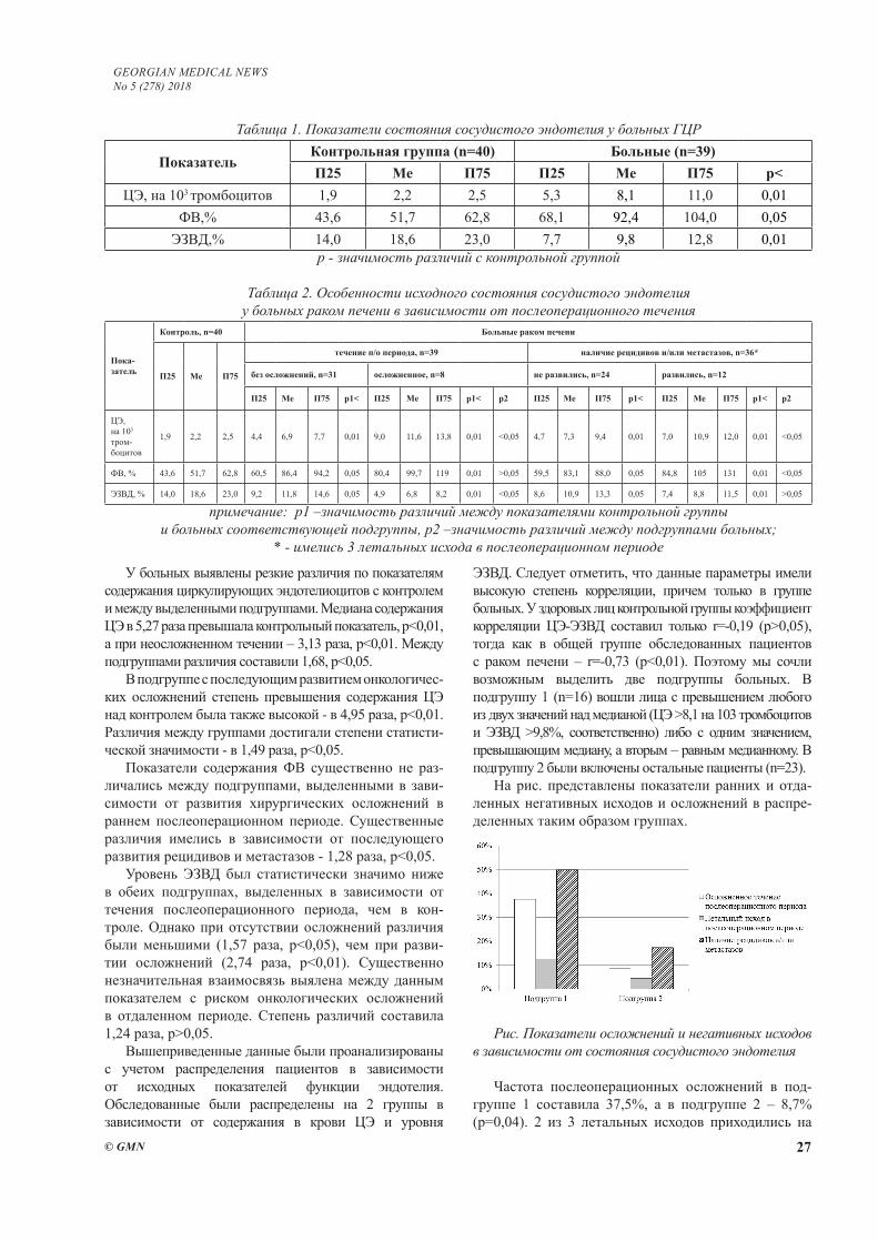
GEORGIAN MEDICAL NEWS No 5 (278) 2018
© GMN 27
У больных выявлены резкие различия по показателям содержания циркулирующих эндотелиоцитов с контролем и между выделенными подгруппами. Медиана содержания ЦЭ в 5,27 раза превышала контрольный показатель, p<0,01, а при неосложненном течении – 3,13 раза, p<0,01. Между подгруппами различия составили 1,68, p<0,05.
В подгруппе с последующим развитием онкологичес-ких осложнений степень превышения содержания ЦЭ над контролем была также высокой - в 4,95 раза, p<0,01. Различия между группами достигали степени статисти-ческой значимости - в 1,49 раза, p<0,05.
Показатели содержания ФВ существенно не раз-личались между подгруппами, выделенными в зави-симости от развития хирургических осложнений в раннем послеоперационном периоде. Существенные различия имелись в зависимости от последующего развития рецидивов и метастазов - 1,28 раза, p<0,05.
Уровень ЭЗВД был статистически значимо ниже в обеих подгруппах, выделенных в зависимости от течения послеоперационного периода, чем в кон-троле. Однако при отсутствии осложнений различия были меньшими (1,57 раза, p<0,05), чем при разви-тии осложнений (2,74 раза, p<0,01). Существенно незначительная взаимосвязь выялена между данным показателем с риском онкологических осложнений в отдаленном периоде. Степень различий составила 1,24 раза, p>0,05.
Вышеприведенные данные были проанализированы с учетом распределения пациентов в зависимости от исходных показателей функции эндотелия. Обследованные были распределены на 2 группы в зависимости от содержания в крови ЦЭ и уровня
ЭЗВД. Следует отметить, что данные параметры имели высокую степень корреляции, причем только в группе больных. У здоровых лиц контрольной группы коэффициент корреляции ЦЭ-ЭЗВД составил только r=-0,19 (p>0,05), тогда как в общей группе обследованных пациентов с раком печени – r=-0,73 (p<0,01). Поэтому мы сочли возможным выделить две подгруппы больных. В подгруппу 1 (n=16) вошли лица с превышением любого из двух значений над медианой (ЦЭ >8,1 на 103 тромбоцитов и ЭЗВД >9,8%, соответственно) либо с одним значением, превышающим медиану, а вторым – равным медианному. В подгруппу 2 были включены остальные пациенты (n=23).
На рис. представлены показатели ранних и отда-ленных негативных исходов и осложнений в распре-деленных таким образом группах.
Рис. Показатели осложнений и негативных исходов в зависимости от состояния сосудистого эндотелия
Частота послеоперационных осложнений в под-группе 1 составила 37,5%, а в подгруппе 2 – 8,7% (p=0,04). 2 из 3 летальных исходов приходились на
Таблица 1. Показатели состояния сосудистого эндотелия у больных ГЦР
ПоказательКонтрольная группа (n=40) Больные (n=39)П25 Me П75 П25 Me П75 p<
ЦЭ, на 103 тромбоцитов 1,9 2,2 2,5 5,3 8,1 11,0 0,01ФВ,% 43,6 51,7 62,8 68,1 92,4 104,0 0,05
ЭЗВД,% 14,0 18,6 23,0 7,7 9,8 12,8 0,01р - значимость различий с контрольной группой
Таблица 2. Особенности исходного состояния сосудистого эндотелия у больных раком печени в зависимости от послеоперационного течения
Пока-затель
Контроль, n=40 Больные раком печени
П25 Me П75
течение п/о периода, n=39 наличие рецидивов и/или метастазов, n=36*
без осложнений, n=31 осложненное, n=8 не развились, n=24 развились, n=12
П25 Me П75 р1< П25 Me П75 р1< р2 П25 Me П75 р1< П25 Me П75 р1< р2
ЦЭ, на 103
тром-боцитов
1,9 2,2 2,5 4,4 6,9 7,7 0,01 9,0 11,6 13,8 0,01 <0,05 4,7 7,3 9,4 0,01 7,0 10,9 12,0 0,01 <0,05
ФВ, % 43,6 51,7 62,8 60,5 86,4 94,2 0,05 80,4 99,7 119 0,01 >0,05 59,5 83,1 88,0 0,05 84,8 105 131 0,01 <0,05
ЭЗВД, % 14,0 18,6 23,0 9,2 11,8 14,6 0,05 4,9 6,8 8,2 0,01 <0,05 8,6 10,9 13,3 0,05 7,4 8,8 11,5 0,01 >0,05
примечание: р1 –значимость различий между показателями контрольной группы и больных соответствующей подгруппы, р2 –значимость различий между подгруппами больных;
* - имелись 3 летальных исхода в послеоперационном периоде
28
МЕДИЦИНСКИЕ НОВОСТИ ГРУЗИИ
CFMFHSDTKJC CFVTLBWBYJ CBF[KTYB
подгруппу с высоким уровнем нарушений эндотели-альной функции в предоперационном периоде, хотя значимых различий не выявлено. Рецидивы и мета-стазы, т.е. онкологические осложнения в отдаленном периоде после вмешательства развились у 50% паци-ентов подгруппы 1 и у 17,4% подгруппы 2 (p=0,03). Таким образом, вероятность негативных результатов оперативного вмешательства и лечения больных ра-ком печени в целом была значительно выше при вы-раженных нарушениях эндотелиальной функции. Мы далеки от того, чтобы считать эти результаты только лишь последствием эндотелиальной дисфункции, од-нако последняя является как отражением системных нарушений состояния организма, так и значимой ее составной частью.
Изменения реактивности сосудистой стенки, контролируемые эндотелиальными механизмами у онкологических больных связаны с ухудшением клинических результатов лечения за счет повыше-ния частоты послеоперационных тромбозов, нега-тивных реакций иммунной системы и локальной гемоциркуляции. Сочетание этих факторов может служить причиной резкого увеличения частоты ранних летальных исходов.
В настоящее время в качестве определяемых кри-териев нарушений функций эндотелия приняты из-менения параметров реакции сосудистой стенки, контролируемых эндотелиальными механизмами [7], особенности продукции эндотелием факторов меж-клеточного взаимодействия [4], параметры, непо-средственно отражающие повреждение эндотелия и эндотелиальные факторы системы гемостаза [9]. Со-четание всех этих аспектов определяет, с точки зре-ния современной науки, состояние сосудистого эндо-телия.
Считается, что весьма трудно оказывать целена-правленное воздействие на мультифункциональные системы, осуществляющие эндокринные функции. Однако позитивные клинические результаты, полу-ченные при коррекции эндотелиальной дисфункции в кардиологической и ангиохирургической практике [8], свидетельствуют об обратном.
Кроме того, активно изучается роль эндотелиаль-ного и эпителиального факторов роста в процессе увеличения солидных опухолей и метастазировании [5,12,14].
За последние годы получены новые данные о роли оксида азота (NO) как о факторе прогрессиро-вания опухоли и метастазирования, выполняющего значимую роль в анестезиологии [17,20]. На сегод-няшний день эти данные вызывают обоснованные сомнения [11,19] и наиболее вероятным представ-ляется наличие существенных различий эффектов экзогенного NO в большой концентрации, что име-ет место при наркозе и эндогенного NO, образуемо-го эндотелием сосудов и являющегося важнейшим регуляторным фактором.
Выводы. Таким образом, взаимосвязь эндоте-лиальной дисфункции с осложнениями, выявленны-ми после хирургического лечения злокачественных новообразований печени, свидетельствует о потен-циальной клинической значимости лечения больных данной нозологией методами коррекции эндотелиаль-ной дисфункции.
Применение методов коррекции эндотелиальной дисфункции в онкологической практике по сей день является скорее исключением, чем правилом. По-этому полагаем, что развитие данного направления исследования может служить основанием для улуч-шения как ранних послеоперационных, так и отдален-ных результатов лечения онкологических больных, в том числе раком печени.
ЛИТЕРАТУРА
1. Баркаган З.С., Момот А.П. Основы диагностики наруше-ний гемостаза. М.: Ньюдиамед-АО, 1999: 224 с.2. Гланц С. Медико-биологическая статистика. Пер. с англ. М., Практика, 1998: 459 с.3. Затейщиков Д.А., Минушкина Л.О., Кудряшова О.Ю. и др. Функциональное состояние эндотелия у больных артериаль-ной гипертонией и ИБС. Кардиология. 2000; (6): 14-17.4. Спирина Л.В., Кондакова И.В., Усынин Е.А. Регуляция ангиогенеза при злокачественных новообразованиях поч-ки и мочевого пузыря. Сибирский онкологический журнал. 2008; (4): 65-70.5. Шаназаров Н.А., Сабиров А.Х., Сироткина С.М. Роль эпидермального фактора роста и его рецептора в канцеро-генезе: молекулярные механизмы их действия. Российский биотерапевтический журнал. 2009; 8(4): 85-90.6. Ahmad J., Grimes N., Farid S., Morris-Stiff G. Inflammatory response related scoring systems in assessing the prognosis of patients with pancreatic ductal adenocarcinoma: a systematic review. Hepatobiliary Pancreat Dis Int. 2014; 13(5): 474-481.7. Barua R.S., Ambrose J.A., Srivastava S., DeVoe M.C., Eales-Reynolds L.J. Reactive oxygen species are involved in smoking-induced dysfunction of nitric oxide biosynthesis and upregulation of endothelial nitric oxide synthase: an in vitro demonstration in human coronary artery endothelial cells. Cir-culation. 2003; 107(18): 2342-2347.8. Boros M. Microcirculatory dysfunction during intestinal ischemia-reperfusion. Acta Physiol Hung. 2003; 90(4): 263-279.9. Bouvy C., Gheldof D., Chatelain C., Mullier F., Dogné J.M. Con-tributing role of extracellular vesicles on vascular endothelium hae-mostatic balance in cancer. J Extracell Vesicles. 2014; 11: 3.10. Bruix J., Han K.H., Gores G., Llovet J.M., Mazzaferro V. Liver cancer: Approaching a personalized care. J Hepatol. 2015; 62(1S): S144-S156.11. Choudhari S.K., Chaudhary M., Bagde S., Gadbail A.R., Joshi V. Nitric oxide and cancer: a review. World J Surg Oncol. 2013; 11: 118. 12. Giannelli G., Koudelkova P., Dituri F., Mikulits W. Role of epithelial to mesenchymal transition in hepatocellular carcino-ma. J Hepatol. 2016; 65(4): 798-808.13. Hladovec J. Circulating endothelial cells as a sign of vessel wall lesions. Physiol Bohemoslov. 1978; 27(2): 140-144.14. Kaseb A.O., Hanbali A., Cotant M., Hassan M.M., Wollner I., Philip P.A. Vascular endothelial growth factor in the manage-
GEORGIAN MEDICAL NEWS No 5 (278) 2018
© GMN 29
ment of hepatocellular carcinoma: a review of literature. Cancer. 2009; 115(21): 4895-4906.15. Khan A.S., Fowler K.J., Chapman W.C. Current surgi-cal treatment strategies for hepatocellular carcinoma in North America. World J Gastroenterol. 2014; 20(41): 15007-15017.16. Liou I.W. Management of end-stage liver disease. Med Clin North Am. 2014; 98(1): 119-152.17. Prazma J., Pertrusz P., Mims W., Ball S.S., Weissler M.C. Immunohistochemical characterization of NOS activity in squa-mous cell carcinoma of head and neck. Otolaryngol Head Neck Surg. 1995; 11: 541-549.18. Ramesh H. Resection for hepatocellular carcinoma. J Clin Exp Hepatol. 2014; 4(Suppl 3): S90-6.19. Rigas B. Novel agents for cancer prevention based on nitric oxide. Biochem Soc Trans. 2007; 11: 136-138.20. Ying L., Hofseth L.J. An emerging role for endothelial nitric oxide synthase in chronic inflammation and cancer. Cancer Res. 2007; 11: 1407-1410.
SUMMARY
ENDOTHELIAL DYSFUNCTION IN LIVER CAN-CER AND ITS EFFECT ON CLINICAL RESULTS OF TREATMENT
Olzhayev S.
"Almaty Regional Oncology Center", Republic of Ka-zakhstan
The aim of the study was to analyze the effect of the morphofunctional state of the vascular endothelium in the operative treatment of liver cancer on complica-tions, outcomes and the probability of long-term com-plications. We examined 39 patients with liver cancer, the mean age of the group was 62.4±3.0 years. II clini-cal stage was diagnosed in 43.6%, and IIIA stage in 56.4%. The content of deceased endotheliocytes in the blood, von Willebrand factor was determined, a dop-pler-echocardiographic test with reactive hyperemia (endothelium-dependent vasodilatation - EDVD) was performed.
Significant differences in the indices of the state of the vascular endothelium in patients with liver cancer with a control group were revealed. Disturbances in the state of the vascular endothelium in patients with developed complications and subsequently diagnosed relapses and metastases were significantly higher than without complications. In the distribution of patients, depending on the degree of endothelial dysfunction be-fore the operation, it was found that the incidence of early and distant oncological complications in marked changes significantly exceeds that in a subgroup with moderate changes.
Keywords: liver cancer; endothelial dysfunction; surgical treatment; complications; outcomes.
РЕЗЮМЕ
ЭНДОТЕЛИАЛЬНАЯ ДИСФУНКЦИЯ ПРИ РАКЕ ПЕЧЕНИ И ЕЁ ВЛИЯНИЕ НА КЛИНИЧЕСКИЕ РЕЗУЛЬТАТЫ ЛЕЧЕНИЯ
Олжаев С.Т.
Государственное коммунальное предприятие на праве хозяйственного ведения «Алматинский региональный онкологический диспансер», Республика Казахстан
Целью исследования явился анализ влияния морфо-функционального состояния сосудистого эндотелия на осложнения, исход и вероятность развития отдаленных осложнений при оперативном лечении рака печени.
Обследованы 39 больных раком печени, средний возраст по группе 62,4±3,0 года. II клиническая стадия диагностирована в 43,6%, а IIIА стадия - в 56,4%. Опре-делено содержание слущенных эндотелиоцитов в крови и фактора фон Виллебранда - в сыворотке крови, про-ведена допплер-эхокардиографическая проба с реактив-ной гиперемией (эндотелий-зависимая вазодилятация).
Выявлены значимые различия в показателях со-стояния сосудистого эндотелия у больных раком пе-чени в сравнении с контрольной группой. Нарушения состояния сосудистого эндотелия у пациентов с раз-вившимися осложнениями и диагностированными впоследствии рецидивами и метастазами были более значительными, чем в случаях без осложнений.
Анализ распределения пациентов в зависимлсти от степени нарушения состояния эндотелия до опе-рации выявил, что частота ранних и отдаленных онко-логических осложнений при выраженных изменениях значимо превышает таковую в подгруппе с умеренны-ми изменениями.
reziume
endoTeluri disfunqcia RviZlis kibos dros da misi gavlena mkurnalobis klini-kur Sedegebze
s. olJaevi
almatis regionuli onkologiuri dispan-seri, yazaxeTis respublika
kvlevis mizans Seadgenda sisxlZarRvTa endoTeliumis morfofunqciuri mdgomare-obis gavlenis Sefaseba mogvianebiTi gar-Tulebebis ganviTarebis albaTobasa da maT gamosavalze RviZlis kibos operaciuli mkurnalobis dros.
gamokvleulia 39 avadmyofi RviZlis ki-boTi, saSualo asaki - 62,4±3,0 weli. II klini-kuri stadia diagnostirda 43,6%-Si, IIIa sta-
30
МЕДИЦИНСКИЕ НОВОСТИ ГРУЗИИ
CFMFHSDTKJC CFVTLBWBYJ CBF[KTYB
dia ki - 56,4%-Si. ganisazRvra Svileuli eriTrocitebis, fon vilebrandis faqtoris Semcveloba, Catarda dopler-eqokardio-grafiuli sinji reaqtiuli hiperemiiT (en-doTelium-damokidebuli vazodilatacia).
gamovlenda sisxlZarRvebis endoTeliumis maCvveneblebis arsebiTi gansxvavebani RviZ-lis kiboTi pacientebSi sakontrolo jguf-Tan SedarebiT. sisxlZarRvebis endoTeliu-mis mdgomareobis darRvevebis maCveneblebi
pacientebSi garTulebiT, diagnostirebuli recidivebiT da metastazebiT ufro maRali iyo, vidre gaurTulebel SemTxvevebSi.
operaciamde endoTeliumis darRvevebis xarisxis gaTvaliswinebiT pacientebis ga-nawilebis analizma gamoavlina, rom ad-reuli da mogvianebiTi onkologiuri garTulebebis sixSire gamoxatuli cvlile-bebis dros mniSvnelovnad metia, vidre zo-mierad gamoxatuli cvlilebebis qvejgufSi.
USE OF PERFUSION MRI FOR DETERMINATION OF IRRADIATION VOLUMES IN RADIOTHERAPY OF PATIENTS WITH BRAIN GLIOMA
Mardaleishvili K., Orkodashvili G.
Kote Mardaleishvili Medical Center; Radiation Medicine Center; David Agmashenebeli Georgian University, Faculty of Stomatology and Medicine, Tbilisi, Georgia
The post-treatment diagnosis of high-grade glioma is a significant challenge to neuro-oncology. Modern treat-ment consists of three basic modalities: surgery, chemo- and radio-therapy. The contrasted (CE) MRI study is used as the diagnostic tool both in pre-treatment and post-treat-ment observation process. Contrast substances containing gadolinium, which usually do not penetrate blood-brain barrier, in case of breaking the BBB, can show contrast enhancement of tumors and other lesions. Based on Mc-Donald’s criteria, new or increased CE MRI lesions indi-cate the growth of the tumor and the failure of treatment, which is associated with bad prognosis. The CE-MRI is also used in conjunction with computed tomography for radiotherapy planning. However, early studies identified biopsy‐proven tumor cells in the area of MRI T2 abnor-malities outside contrast‐enhancing CT abnormalities. Based on these results of research, the T2 MRI has been included in radiotherapy planning. T2 MR Study has a disadvantage associated with the greater strength of the CSF signal compared to the brain substance, decreasing conspicuity in the regions adjacent to CSF, which led to the development of the fluid attenuation inversion recov-ery (FLAIR) sequence.
CE‐MRI and FLAIR are both nonspecific tools for identifying recurrent tumor burden. Specifically, thera-py‐induced parenchymal injury, called Post‐treatment Radiation Effect (PTRE), often develops concurrently with tumor regrowth and mimics its appearance on CE‐MRI [3]. It is important to distinguish between these two lesions because the tumor recurrence refers to the failure of treatment, and the PTRE marks successful treatment. PTRE is presented in two forms: pseudo progression (PP) and radiation necrosis (RN). Both indicate the success of treatment and are characterized by good prognosis, yet
represent erroneous declaration of treatment failure in up to half of cases [3,5]. Consequently, with conventional re-search the progression of tumors is often misdiagnosed, which significantly reduces the effectiveness of the treat-ment and the life expectancy.
Surgical biopsy definitively distinguishes recurrent HGG from PTRE [6]. Studies suggest that quantifying tumor burden provides more meaningful prognostic in-formation than simply reporting its presence, since his-tologic tumor fraction provides a direct measure of tumor progression that strongly correlates with survival [4,8,9]. Unfortunately, shortcomings of surgical biopsy, such as medical risk, cost and potential mistake of sampling, re-duce its clinical use. Moreover, the biopsy cannot be car-ried out regularly and is often performed only when the tumor progresses.
Perfusion MRI (pMRI) noninvasively detects HGG and PTRE microvascular characteristics and offers a promising alternative to surgical biopsy [2]. This technique is based on the Dynamic Susceptibility Weighted Contrast Enhanced (DSC) method, which, in theory, measures Relative Cere-bral Blood Volume (rCBV) to detect the differences in mi-crovascular density (MVD) that separate PTRE and tumor growth. PTRE demonstrates decreased capillary perfusion and MVD, while tumor growth typically demonstrates an-giogenesis and increased MVD. DSC research is rarely used, because it is distinguished by the absence of reliable levels of indicators and the difficulty of correction of hemato-en-cephalic barrier disruption. However, modern technological improvement of this method allowed us to accurately diag-nose tumor and radiation damage and accurately determine the tissue microvasculature and histologically different sub-regions of the tumor and radiation damage with almost 95% accuracy.
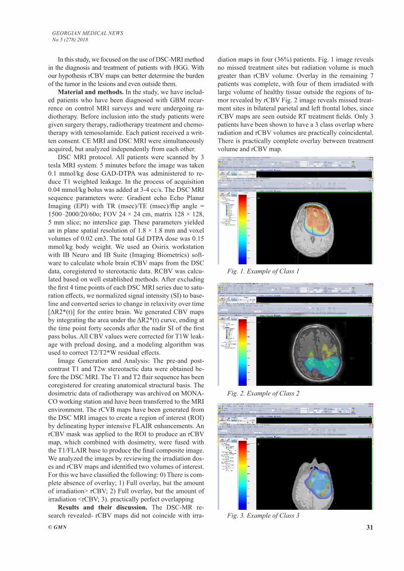
GEORGIAN MEDICAL NEWS No 5 (278) 2018
© GMN 31
In this study, we focused on the use of DSC-MRI method in the diagnosis and treatment of patients with HGG. With our hypothesis rCBV maps can better determine the burden of the tumor in the lesions and even outside them.
Material and methods. In the study, we have includ-ed patients who have been diagnosed with GBM recur-rence on control MRI surveys and were undergoing ra-diotherapy. Before inclusion into the study patients were given surgery therapy, radiotherapy treatment and chemo-therapy with temosolamide. Each patient received a writ-ten consent. CE MRI and DSC MRI were simultaneously acquired, but analyzed independently from each other.
DSC MRI protocol. All patients were scanned by 3 tesla MRI system. 5 minutes before the image was taken 0.1 mmol/kg dose GAD-DTPA was administered to re-duce T1 weighted leakage. In the process of acquisition 0.04 mmol/kg bolus was added at 3-4 cc/s. The DSC MRI sequence parameters were: Gradient echo Echo Planar Imaging (EPI) with TR (msec)/TE (msec)/flip angle = 1500–2000/20/60o; FOV 24 × 24 cm, matrix 128 × 128, 5 mm slice; no interslice gap. These parameters yielded an in plane spatial resolution of 1.8 × 1.8 mm and voxel volumes of 0.02 cm3. The total Gd DTPA dose was 0.15 mmol/kg body weight. We used an Osirix workstation with IB Neuro and IB Suite (Imaging Biometrics) soft-ware to calculate whole brain rCBV maps from the DSC data, coregistered to stereotactic data. RCBV was calcu-lated based on well established methods. After excluding the first 4 time points of each DSC MRI series due to satu-ration effects, we normalized signal intensity (SI) to base-line and converted series to change in relaxivity over time [ΔR2*(t)] for the entire brain. We generated CBV maps by integrating the area under the ΔR2*(t) curve, ending at the time point forty seconds after the nadir SI of the first pass bolus. All CBV values were corrected for T1W leak-age with preload dosing, and a modeling algorithm was used to correct T2/T2*W residual effects.
Image Generation and Analysis: The pre-and post-contrast T1 and T2w stereotactic data were obtained be-fore the DSC MRI. The T1 and T2 flair sequence has been coregistered for creating anatomical structural basis. The dosimetric data of radiotherapy was archived on MONA-CO working station and have been transferred to the MRI environment. The rCVB maps have been generated from the DSC MRI images to create a region of interest (ROI) by delineating hyper intensive FLAIR enhancements. An rCBV mask was applied to the ROI to produce an rCBV map, which combined with dosimetry, were fused with the T1/FLAIR base to produce the final composite image. We analyzed the images by reviewing the irradiation dos-es and rCBV maps and identified two volumes of interest. For this we have classified the following: 0) There is com-plete absence of overlay; 1) Full overlay, but the amount of irradiation> rCBV; 2) Full overlay, but the amount of irradiation <rCBV; 3). practically perfect overlapping
Results and their discussion. The DSC-MR re-search revealed- rCBV maps did not coincide with irra-
diation maps in four (36%) patients. Fig. 1 image reveals no missed treatment sites but radiation volume is much greater than rCBV volume. Overlay in the remaining 7 patients was complete, with four of them irradiated with large volume of healthy tissue outside the regions of tu-mor revealed by rCBV Fig. 2 image reveals missed treat-ment sites in bilateral parietal and left frontal lobes, since rCBV maps are seen outside RT treatment fields. Only 3 patients have been shown to have a 3 class overlap where radiation and rCBV volumes are practically coincidental. There is practically complete overlay between treatment volume and rCBV map.
Fig. 1. Example of Class 1
Fig. 2. Example of Class 2
Fig. 3. Example of Class 3
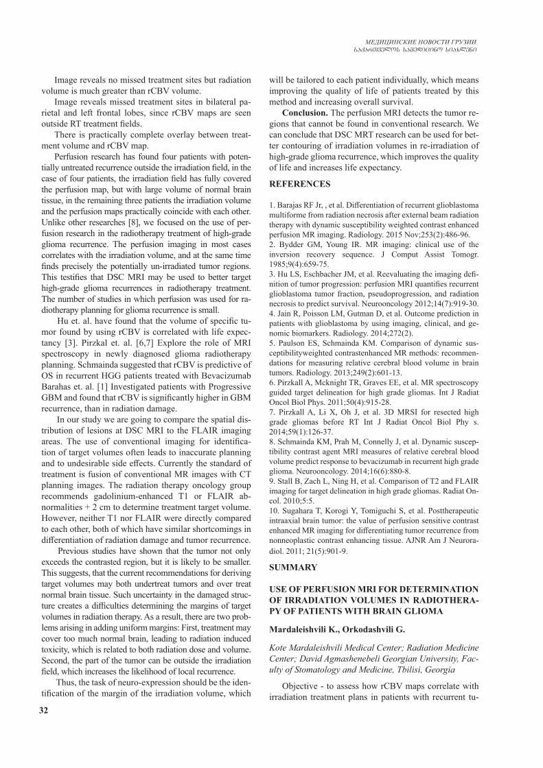
32
МЕДИЦИНСКИЕ НОВОСТИ ГРУЗИИ
CFMFHSDTKJC CFVTLBWBYJ CBF[KTYB
Image reveals no missed treatment sites but radiation volume is much greater than rCBV volume.
Image reveals missed treatment sites in bilateral pa-rietal and left frontal lobes, since rCBV maps are seen outside RT treatment fields.
There is practically complete overlay between treat-ment volume and rCBV map.
Perfusion research has found four patients with poten-tially untreated recurrence outside the irradiation field, in the case of four patients, the irradiation field has fully covered the perfusion map, but with large volume of normal brain tissue, in the remaining three patients the irradiation volume and the perfusion maps practically coincide with each other. Unlike other researches [8], we focused on the use of per-fusion research in the radiotherapy treatment of high-grade glioma recurrence. The perfusion imaging in most cases correlates with the irradiation volume, and at the same time finds precisely the potentially un-irradiated tumor regions. This testifies that DSC MRI may be used to better target high-grade glioma recurrences in radiotherapy treatment. The number of studies in which perfusion was used for ra-diotherapy planning for glioma recurrence is small.
Hu et. al. have found that the volume of specific tu-mor found by using rCBV is correlated with life expec-tancy [3]. Pirzkal et. al. [6,7] Explore the role of MRI spectroscopy in newly diagnosed glioma radiotherapy planning. Schmainda suggested that rCBV is predictive of OS in recurrent HGG patients treated with Bevacizumab Barahas et. al. [1] Investigated patients with Progressive GBM and found that rCBV is significantly higher in GBM recurrence, than in radiation damage.
In our study we are going to compare the spatial dis-tribution of lesions at DSC MRI to the FLAIR imaging areas. The use of conventional imaging for identifica-tion of target volumes often leads to inaccurate planning and to undesirable side effects. Currently the standard of treatment is fusion of conventional MR images with CT planning images. The radiation therapy oncology group recommends gadolinium‐enhanced T1 or FLAIR ab-normalities + 2 cm to determine treatment target volume. However, neither T1 nor FLAIR were directly compared to each other, both of which have similar shortcomings in differentiation of radiation damage and tumor recurrence.
Previous studies have shown that the tumor not only exceeds the contrasted region, but it is likely to be smaller. This suggests, that the current recommendations for deriving target volumes may both undertreat tumors and over treat normal brain tissue. Such uncertainty in the damaged struc-ture creates a difficulties determining the margins of target volumes in radiation therapy. As a result, there are two prob-lems arising in adding uniform margins: First, treatment may cover too much normal brain, leading to radiation induced toxicity, which is related to both radiation dose and volume. Second, the part of the tumor can be outside the irradiation field, which increases the likelihood of local recurrence.
Thus, the task of neuro-expression should be the iden-tification of the margin of the irradiation volume, which
will be tailored to each patient individually, which means improving the quality of life of patients treated by this method and increasing overall survival.
Conclusion. The perfusion MRI detects the tumor re-gions that cannot be found in conventional research. We can conclude that DSC MRT research can be used for bet-ter contouring of irradiation volumes in re-irradiation of high-grade glioma recurrence, which improves the quality of life and increases life expectancy.
REFERENCES
1. Barajas RF Jr, , et al. Differentiation of recurrent glioblastoma multiforme from radiation necrosis after external beam radiation therapy with dynamic susceptibility weighted contrast enhanced perfusion MR imaging. Radiology. 2015 Nov;253(2):486-96.2. Bydder GM, Young IR. MR imaging: clinical use of the inversion recovery sequence. J Comput Assist Tomogr. 1985;9(4):659-75. 3. Hu LS, Eschbacher JM, et al. Reevaluating the imaging defi-nition of tumor progression: perfusion MRI quantifies recurrent glioblastoma tumor fraction, pseudoprogression, and radiation necrosis to predict survival. Neurooncology 2012;14(7):919-30.4. Jain R, Poisson LM, Gutman D, et al. Outcome prediction in patients with glioblastoma by using imaging, clinical, and ge-nomic biomarkers. Radiology. 2014;272(2).5. Paulson ES, Schmainda KM. Comparison of dynamic sus-ceptibilityweighted contrastenhanced MR methods: recommen-dations for measuring relative cerebral blood volume in brain tumors. Radiology. 2013;249(2):601-13.6. Pirzkall A, Mcknight TR, Graves EE, et al. MR spectroscopy guided target delineation for high grade gliomas. Int J Radiat Oncol Biol Phys. 2011;50(4):915-28.7. Pirzkall A, Li X, Oh J, et al. 3D MRSI for resected high grade gliomas before RT Int J Radiat Oncol Biol Phy s. 2014;59(1):126-37.8. Schmainda KM, Prah M, Connelly J, et al. Dynamic suscep-tibility contrast agent MRI measures of relative cerebral blood volume predict response to bevacizumab in recurrent high grade glioma. Neurooncology. 2014;16(6):880-8. 9. Stall B, Zach L, Ning H, et al. Comparison of T2 and FLAIR imaging for target delineation in high grade gliomas. Radiat On-col. 2010;5:5.10. Sugahara T, Korogi Y, Tomiguchi S, et al. Posttherapeutic intraaxial brain tumor: the value of perfusion sensitive contrast enhanced MR imaging for differentiating tumor recurrence from nonneoplastic contrast enhancing tissue. AJNR Am J Neurora-diol. 2011; 21(5):901-9.
SUMMARY
USE OF PERFUSION MRI FOR DETERMINATION OF IRRADIATION VOLUMES IN RADIOTHERA-PY OF PATIENTS WITH BRAIN GLIOMA
Mardaleishvili K., Orkodashvili G.
Kote Mardaleishvili Medical Center; Radiation Medicine Center; David Agmashenebeli Georgian University, Fac-ulty of Stomatology and Medicine, Tbilisi, Georgia
Objective - to assess how rCBV maps correlate with irradiation treatment plans in patients with recurrent tu-
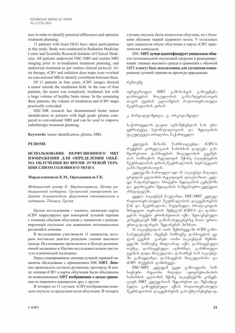
GEORGIAN MEDICAL NEWS No 5 (278) 2018
© GMN 33
mor in order to identify potential differences and optimize treatment planning.
11 patients with brain HGG have taken participation in this study. Study was conducted in Radiation Medicine Center and Scientific Research Institute of Clinical Medi-cine. All patients underwent DSC-MRI and routine MRI imaging prior to re-irradiation treatment planning, and underwent treatment as per routine clinical protocol. Af-ter therapy, rCBV and radiation dose maps were overlaid on conventional MR to identify correlation between them.
Of 11 patients in four cases, rCBV images showed a tumor outside the irradiation field. In the case of four patients, the tumor was completely irradiated, but with a large volume of healthy brain tissue. In the remaining three patients, the volume of irradiation and rCBV maps practically coincided.
DSC-MR research has demonstrated better tumor identification in patients with high grade glioma com-pared to conventional MRI and can be used to improve radiotherapy treatment planning.
Keywords: tumor identification, glioma, MRI.
РЕЗЮМЕ
ИСПОЛЬЗОВАНИЕ ПЕРФУЗИОННОГО MRT ИЗОБРАЖЕНИЯ ДЛЯ ОПРЕДЕЛЕНИЯ ОБЪЁ-МА ОБЛУЧЕНИЯ ВО ВРЕМЯ ЛУЧЕВОЙ ТЕРА-ПИИ ГЛИОМ ГОЛОВНОГО МОЗГА
Мардалеишвили К.М., Оркодашвили Г.К.
Медицинский центр К. Мардалеишвили, Центр ра-диационной медицины; Грузинский университет им. Давида Агмашенебели, факультет стоматологии и медицины, Тбилиси, Грузия
Целью исследования - оценить, насколько карты rCBV коррелируют при повторной лучевой терапии с планами обьемов облучения у пациентов с рециди-вирующей опухолью для выявления потенциальных различий в лечении.
В исследовании участвовали 11 пациентов, кото-рым поставлен диагноз рецидива глиомы высокого греида. Исследование проводилось в Центре радиаци-онной медицины и Научно-исследовательском инсти-туте клинической медицины.
Перед планированием лечения лучевой терапией па-циенты обследованы с применением DSC-MRT. Лече-MRT. Лече-. Лече-ние проводилось согласно рутинному протоколу. В кон-це лечения rCBV и карты облучения были объединены на конвенционных MRT изображениях с целью сравне-MRT изображениях с целью сравне- изображениях с целью сравне-ния их взаимного перекрытия друг с другом.
В четырех из 11 случаев rСBV-изображения пока-зали опухоль за пределами поля облучения. В четырех
случаях опухоль была полностью облучена, но с боль-шим обьемом тканей здорового мозга. У остальных трех пациентов обьем облучения и карты rCBV прак-тически совпадали.
DSC-MRT лучше идентифицирует уникальные обла-MRT лучше идентифицирует уникальные обла- лучше идентифицирует уникальные обла-сти потенциальной опухолевой нагрузки в рецидивиру-ющих глиомах высокого греида в сравнении с обычной MRT и могут быть использованы для улучшения плани- и могут быть использованы для улучшения плани-рования лучевой терапии во время ре-иррадиации.
reziume
perfuziuli MRT gamosaxvis gamoyeneba dasxivebis moculobis gansazRvrisaTvis Tavis tvinis gliomebis radioTerapiuli mkurnalobis dros
k. mardaleiSvili, g. orkodaSvili
saqarTvelos daviT aRmaSeneblis sax. uni-versiteti, stomatologiis da medicinis fakulteti, Tbilisi, saqarTvelo
kvlevis mizans warmoadgenda rCBV-s ruqebis korelaciis xarisxis dadgena gan-meorebiTi dasxivebis moculobis gegmeb-Tan simsivnis recidivis mqone pacientebis mkurnalobis dros, mkurnalobis strategiis gansazRvrisaTvis.
kvlevaSi CarTuli iyo 11 pacienti maRali greidis gliomis recidivis diagnoziT. kvl-eva Catarebulia sxivuri medicinis centrSi da klinikuri medicinis samecniero-kvleviT institutSi.
yvela pacients Cautarda DSC-MRT kvleva radioTerapiuli mkurnalobis dagegmarebis win da mkurnaloba rutinuli protokolis mixedviT. Terapiis Semdgom rCBV-s da dasxi-vebis ruqebi erTmaneTTan iqna SeTavsebuli konvenciur MR gamosaxulebebze maTi urTi-erT-gadafarvis Sedarebis mizniT.
11 pacientidan oTx SemTxvevaSi rCBV gamo-saxulebebma aCvenes simsivne dasxivebis ve-lis gegmis gareT. oTxi pacientis SemTx-vevaSi simsivne mTlianad iqna dasxivebuli, Tumca dasxivebuli aRmoCnda janmrTeli tvinis didi moculoba. danarCen sam pacient-Si gamovlinda dasxivebis moculobisa da rCBV ruqebis damTxveva.
DSC-MRT kvlevam ukeT gamoavlina sim-sivnuri regioni maRali avTvisebianobis xarisxis gliomis mqone pacientebSi konven-ciur MRT kvlevasTan SedarebiT da SesaZle-belia gamoyenebuli iqnas radioTerapiuli mkurnalobis dagegmarebis gasaumjobeseblad.
34
МЕДИЦИНСКИЕ НОВОСТИ ГРУЗИИ
CFMFHSDTKJC CFVTLBWBYJ CBF[KTYB
Preterm labor is an urgent medical-social and demo-graphic issue at the present stage. The frequency of this pa-thology varies in the range of 10-30% with the absence of a tendency to decrease. Pathogenesis of the premature abor-tion development is quite complicated and has a multifacto-rial nature [3,11]. It stimulates specialists on its deep study in order to determine the effective complex of treatment and prevention measures for reproductive loss prevention and development of complications in the further [3].
A considerable number of factors affects the course of pregnancy and its outcome, their effect is realized at the level of the central nervous system through numerical metabolic interactions, where monoaminergic systems play an impor-tant role. First of all, they are included in the compensatory-adaptive processes, which is necessary for the normal course of pregnancy and the preparation of the woman’s body for childbirth [7]. The sympathoadrenal system plays a cen-tral role in this while providing the woman’s body energy resources and is responsible for a variety of metabolic and hemodynamic changes occurring during pregnancy [2]. The connection of sympathoadrenal system activity with the de-velopment of obstetric complications is proved, including premature births [1,4,5,9,10]; but in many cases the data are isolated or controversial, which stimulates further study in this direction in order to detect initial violations, to develop new measures for their correction and prevention in order to prevent premature births in women.
It had been shown that the level of indicators of the sympathoadrenal system state - dopamine, norepineph-rine, adrenaline and their precursor dioksiphenylalanine (DOPA) in the daily urine is a mirror image of their con-tent in the blood [6]. Hence the conclusion that the func-tional state of the sympathoadrenal system, the displace-ment of epinephrine, norepinephrine and dopaminergic reactions by the excretion of catecholamines with urine is considered plausible.
Objective - to study the features of the sympathoadrenal system state by determining the excretion level of dioksiphe-nylalanine, dopamine, norepinephrine and epinephrine in women’s daily urine with different periods of abortion.
Material and methods. 227 pregnant women who were admitted to the Kharkiv perinatal center have been examined, 190 of them had clinical signs of premature delivery in the gestation period of 23-36 weeks. Forma-tion of clinical groups was carried out depending on the pregnancy term in the form of premature and timely de-livery. 48 women with early premature pregnancy (mean age 23.6 ± 5.1 years), completed by birth in the period from 23 to 27 weeks, were included in Group I. All preg-
nant women with pre-nasal congestion had the following distribution: 23-25 weeks - 23 pregnant women (Ia sub-group); 26-27 weeks - 25 pregnant women (Ib subgroup) depending on gestational age. 142 women were included in Group II (mean age 24.7 ± 4.2 years) with late term premature pregnancy in the period from 28 to 36 weeks, which depending on the term of delivery, were divided into subgroups: IIa - 38 people, 28-30 weeks; IIb - 48 people, 31-33 weeks; IIв - 56 people, 34-36 weeks. The III (control) group included 37 women with a physiologi-cal pregnancy (mean age 26.1 ± 2.7 years), which ended in childbirth without complications in the period of 38-41 weeks. The criteria for including women into groups were: young reproductive age, single-pregnancy, absence of gestosis, acute and chronic gynecological and somatic diseases. Diagnosis of preterm labor was carried out in the presence of abdominal pain syndrome and structural changes in the cervix. The research was carried out in compliance with the bioethics principles. Determination of the daily excretion level of DOPHA and its metabolites - dopamine, norepinephrine, and epinephrine were carried out in two stages. In the first step, by column chromatogra-phy on aluminum oxide, absorption and elution with 0.25 n acetic acid (DPOFA remaining on the adsorbent was eluted with 1N hydrochloric acid). At the second stage, DOPA and its metabolites were oxidized with potassium ferricyanide at various pH values, followed by measuring their concentration by fluorimetric method on the Hita-chi (MP) -4A spectrofluorometer. At first, a set of filters with a maximum absorption of 436 nm and 550 nm was used; the intensity of the experimental samples fluores-cence with a pH of 4.2 and pH 6.2 (epinephrine, norepi-nephrine, acetic acid DOPA) and control were measured. Epinephrine fluoresces only at pH 4.2, and epinephrine and norepinephrine at pH 6.2. Another set of filters with a maximum absorption of 360 nm and 550 nm was used after that; the fluorescence intensity of the samples at pH 6.2 (epinephrine, norepinephrine, DOPA) and samples of hydrochloric acid eluate oxidized at pH 6.2 were mea-sured. The difference between the intensity of the fluores-cence of the experimental and control samples was upon completion of the measurements. The statistical process-ing of the obtained results was carried out using licensed standardized packages of multivariate statistical analysis Statistica 6.1 applications. Median of sample (Me), the values of the lower (Q25) and upper (Q75) quartiles are used given the lack of normal data distribution. Compari-son of the indices in the groups was carried out using the non-parametric Mann-Whitney test.
THE FEATURES OF THE WOMEN’S SIMPATHOADRENAL SYSTEM FUNCTIONAL STATE WITH RISK OF EARLY PREGNANCY TERMINATION
Korovay S.
Kharkiv Regional Perinatal Center, Department of Obstetrics, Perinatology and Gynecology, Kharkiv Medical Academy of Postgraduate Education,Ukraine
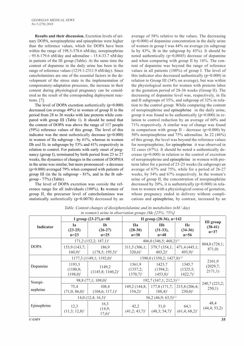
GEORGIAN MEDICAL NEWS No 5 (278) 2018
© GMN 35
Results and their discussion. Excretion levels of uri-nary DOPA, norepinephrine and epinephrine were higher than the reference values, which for DOPA have been within the range of 198.3-378.6 nM/day, norepinephrine - 95.8-179.6 nM/day and adrenaline - 15.4-33.7 nM/day in patients of the III group (Table). At the same time the content of dopamine in the daily urine has been in the range of reference values (1219.1-2311.9 nM/day). Since catecholamines are one of the essential factors in the de-velopment of the stress state in the implementation of compensatory-adaptation processes, the increase in their content during physiological pregnancy can be consid-ered as the result of the corresponding deployment reac-tions. [7].
The level of DOPA excretion authentically (p=0.000) decreased (on average 49%) in women of group II in the period from 28 to 36 weeks with late preterm while com-pared with group III (Table 1). It should be noted that the content of DOPA was above the range of 117 people (59%) reference values of this group. The level of this indicator was the most authentically decrease (p=0.000) in women of IIa subgroup - by 61%, whereas in women IIb and IIc in subgroups by 53% and 41% respectively in relation to control. For patients with early onset of preg-nancy (group I), terminated by birth period from 23 to 27 weeks, the dynamics of changes in the content of DOPHA in the urine was similar, but more pronounced - a decrease (p=0.000) averaged 79% when compared with patients of group III (in the Ia subgroup - 81%, and in the Ib sub-group - 77%) (Table).
The level of DOPA excretion was outside the ref-erence range for all individuals (100%). Іn women of group II, the precursor level of catecholamines was statistically authentically (p=0.0078) decreased by an
average of 58% relative to the values. The decreasing (p=0.000) of dopamine concentration in the daily urine of women in group I was 44% on average (in subgroup Ia by 43%, Ib in the subgroup by 45%). It should be noted authentically (p=0,0045) decrease of dopamine and when comparing with group II by 16%. The con-tent of dopamine was beyond the range of reference values in all patients (100%) of group I. The level of this indicator also decreased authentically (p=0.000) in relation to Group III (34% on average), but was within the physiological norm for women with preterm labor at the gestation period of 28-36 weeks (Group II). The decreasing of dopamine level was, respectively, in IIa and II subgroups of 35%, and subgroup of 32% in rela-tion to the control group. While comparing the content of norepinephrine and epinephrine in the daily urine, group it was found to be authentically (p=0.000) in re-lation to control reduction by an average of 60% and 71% respectively. A similar way of change was found in comparison with group II - decrease (p=0.000) by 50% norepinephrine and 75% adrenaline. In 22 (46%) of this group, the level was beyond the reference values for norepinephrine, for epinephrine it was observed in 32 cases (67%). It should be noted a authentically de-crease (p=0.000) in relation to the control of excretion of norepinephrine and epinephrine in women with pre-term labor for a period of 23-25 weeks (Ia subgroup) an average of 67% and 75%, while for a period of 26-27 weeks, by 54% and 67% respectively. In the women’s urine of group II, the concentration of norepinephrine decreased by 20%, it is authentically (p=0.000) in rela-tion to women with a physiological course of gestation, whose pregnancy ended in delivery without compli-cations and epinephrine, by contrast, increased by an
Table. Content changes of dioxiphenylalanine and its metabolites (nM / day) in women’s urine in observation groups (Me [25%; 75%]
Indicator
I group (23-27),n=48 II group (28-36), n=142III group(38-41)n=37
Iа (23-25)n=23
Ib(26-27)n=25
IIа(28-30)n=38
IIb(31-33),
n=48
IIc(34-36)n=56
DOPA 171,2 (152,2; 187,1)1 406,0 (346,5; 460,2)1,2
804,0 (728,1; 871,0)153,9 (143,7;
160,9)1186,9
(178,5; 195,5)1311,5 (306,1;
320,6)1379,7 (354,1;
403,2)1471,4 (445,1;
493,9)1
Dopamine
1177,3 (1149,1; 1192,0)1 1390,0 (1350,2; 1427,8)1,2
2101,9 (2029,7; 2171,1)
1193,3 (1180,8; 1198,0)1
1149,2 (1145,8; 1160,2)1
1361,9 (1357,2; 1370,7)1
1423,7 (1394,2; 1453,8)1
1345,7 (1325,5; 1422,7)1
Norepi-
nephrine
98,8 (77,1; 109,0)1 192,7 (167,1; 212,3)1,2
240,7 (223,2; 250,1)75,4
(71,0; 86,0)1108,4
(104,6; 117,1)1149,2 (144,8;
154,2)1177,8 (171,7;
188,4)1215,8 (206,4;
230,0)1
Epinephrine
14,0 (12,4; 16,3)1 56,2 (46,9; 63,5)1,2
48,4 (44,4; 53,2)12,3
(11,3; 12,8)1
16,3 (14,9; 17,6)1
42,2 (41,2; 43,7)1
51,0 (48,3; 54,7)1
64,1 (61,4; 68,2)1
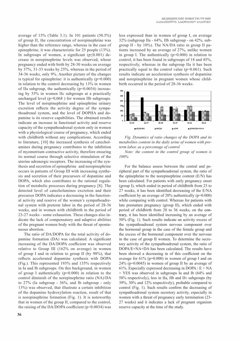
36
МЕДИЦИНСКИЕ НОВОСТИ ГРУЗИИ
CFMFHSDTKJC CFVTLBWBYJ CBF[KTYB
average of 15% (Table 3.1). In 101 patients (50.5%) of group II, the concentration of norepinephrine was higher than the reference range, whereas in the case of epinephrine, it was characteristic for 25 people (13%). In subgroups of women, a significant (p≤0.001) de-crease in norepinephrine levels was observed, whose pregnancy ended with birth by 28-30 weeks on average by 37%, 31-33 weeks by 25%, whereas in the period of 34-36 weeks, only 9%. Another picture of the changes is typical for epinephrine: it is authentically (p=0.000) in relation to the control decreasing by 13% in women of IIa subgroup, the authentically (p=0.0034) increas-ing by 35% in women IIc subgroups at a practically unchanged level (p=0,068 ) for women IIb subgroups. The level of norepinephrine and epinephrine urinary excretion reflects the activity degree of the sympa-thoadrenal system, and the level of DOPHA and do-pamine is its reserve capabilities. The obtained results indicate an increase in functional activity and reserve capacity of the sympathoadrenal system only in women with a physiological course of pregnancy, which ended with childbirth without any complications. According to literature, [10] the increased synthesis of catechol-amines during pregnancy contributes to the inhibition of myometrium contractive activity, therefore ensuring its normal course through selective stimulation of the uterine adrenergic receptors. The increasing of the syn-thesis and secretion of epinephrine and norepinephrine occurs in patients of Group III with increasing synthe-sis and secretion of their precursors of dopamine and DOPA, which also contributes to the rational regula-tion of metabolic processes during pregnancy [8]. The detected level of catecholamines excretion and their precursor DOPA indicates a decreasing in the function-al activity and reserve of the women’s sympathoadre-nal system with preterm labor in the period of 28-36 weeks, and in women with childbirth in the period of 23-27 weeks - some exhaustion. These changes also in-dicate the lack of compensatory and adaptive abilities of the pregnant women body with the threat of sponta-neous abortion.
The ratio of DA/DOPA for the total activity of do-pamine formation (DA) was calculated. A significant increasing of the DA/DOPA coefficient was observed relative to Group III (162% on average) in women of group I and in relation to group II (by 98%), that reflects accelerated dopamine synthesis with DOPA (Fig.). This represented 193% and 135% respectively in Ia and Ib subgroups. On this background, in women of group I authentically (p=0.000) in relation to the control diminish of the norepinephrine ratio (NA)/DA to 27% (Ia subgroup - 36%, and Ib subgroup - only 13%) was observed, that illustrate a certain inhibition of the dopamine hydroxylation reaction, result of that is norepinephrine formation (Fig. 1). It is noteworthy that in women of the group II, compared to the control, the raising of the DA/DOPA coefficient (p=0.0034) was
less expressed than in women of group I, on average 32% (subgroup IIa - 64%, IIb subgroup - on 42%, sub-group II - by 10%). The NA/DA ratio in group II pa-tients increased by an average of 27%, unlike women in group I. The authentically (p=0.000) in relation to control, it has been found in subgroups of 18 and 45%, respectively, whereas in the subgroup IIa it has been practically equal to the control value (p=0.061). Such results indicate an acceleration synthesis of dopamine and norepinephrine in pregnant women whose child-birth occurred in the period of 28-36 weeks.
Fig. Dynamics of ratio changes of the DOPA and its metabolites content in the daily urine of women with pre-term labor, as a percentage of control
Note: the content in the control group of women is 100%
For the balance assess between the central and pe-ripheral part of the sympathoadrenal system, the ratio of the epinephrine to the norepinephrine content (E/N) has been calculated. For patients with early pregnancy onset (group I), which ended in period of childbirth from 23 to 27 weeks, it has been identified decreasing of the E/NA coefficient by an average of 20% authentically (p=0.000) while comparing with control. Whereas for patients with late premature pregnancy (group II), which ended with period of childbirth from 28 to 36 weeks, on the con-trary, it has been identified increasing by an average of 50% (Fig. 1). Such results indicate an activity excess of the sympathoadrenal system nervous component over the hormonal group in the case of the female group and the excess of the hormonal component over the nervous in the case of group II women. To determine the secre-tory activity of the sympathoadrenal system, the ratio of DOPA/E+NA+DA has been calculated. The results have been showed a decreasing in of this coefficient on the average for 61% (p=0.000) in women of group I and on 24% (p=0.0045) in women of group II by an average of 61%. Especially expressed decreasing in DOPA / E + NA + YES was observed in subgroups Ia and Ib (64% and 58% respectively), less in IIa, IIb and IIv subgroups (by 39%, 30% and 12% respectively), probable compared to control (Fig. 1). Such results confirm the decreasing of sympathoadrenal system secretory activity, especially in women with a threat of pregnancy early termination (23-27 weeks) and it indicates a lack of pregnant organism reserve capacity at the time of the study.
GEORGIAN MEDICAL NEWS No 5 (278) 2018
© GMN 37
Consequently, pregnancy compensatory and adaptive mechanisms are complex of neurohumoral process, which are realized through monoaminergic systems and a sig-nificant factor in its interruption is their destabilization. According to the results, in pregnant women with a threat of preterm labor, the intensity of neuroendocrine regula-tion of the gestational process with the development of the disruption of compensatory and adaptation reactions arises, which is confirmed by the changes dynamics of the sympathoadrenal system monoamines content- do-pamine, norepinephrine, adrenaline and their precursor DOPA. Reducing of sympathoadrenal system activity and reserve capacity in pregnant women may be a pathoge-netic factor in the development of preterm labor. There-fore determination of the imbalance initial manifestations in the catecholamines exchange may possibly prevent the loss of pregnancy in the early stages.
REFERENCES
1. Володченко А.И., Циркин В.И., Хлыбова С.В. Адреноре-активность эритроцитов, определяемая по их адренозависи-мой агглютинации, у женщин на различныхэтапах репро-дуктивного процесса / А.И. Володченко, В.И. Циркин, С.В. Хлыбова, С.Л. Дмитриева // Вятский медицинский вестник. – 2013. - № 1. – С. 25-31. 2. Воробей Л.І. Роль порушень функції симпатоадреналової си-стеми в патогенезі плацентарної дисфункції / Л.І. Воробей, Р.Р. Ткачук // Здоровье женщины. – 2015. - № 8 (104). – С. 135-139.3. Воробйова І.І., Живецька-Денисова А.А., Ткаченко В.Б. Невиношувння вагітності: сучасні погляди на проблему (огляд літератури) / І.І. Воробйова, А.А. Живецька-Денисо-ва, В.Б. Ткаченко [та ін.] // Здоровье женщины. – 2017. – № 3 (119). – С. 113-117.4. Козырина А. А., Назарова А. О., Малышкина А. И. Взаи-мосвязь показателей биоэлектрической активности матки и продукции катехоламинов у женщин при угрожающих пре-ждевременных родах / А. А. Козырина, А. О. Назарова, А. И. Малышкина, С. Б. Назаров // Актуальные вопросы аку-шерства, гинекологии, неонатологии и педиатрии: сборник научных трудов, посвященный 35-летию со дня основания института. – Иваново: изд-во МИК, 2015. – С. 18-22. 5. Козырина А.А., Малышкина А.И., Назаров С.Б. Пока-затели регуляторних газов крови и адренореактивности у женщин с физиологической беременностью и угрозой пре-ждевременных родов / А.А. Козырина, А.И. Малышкина, С.Б. Назаров [и др.] // Новости дерматовенерологии и ре-продуктивного здоровья. – 2015. – № 2. – С. 161-165.6. Мириманова Р.П. Особенности сдвигов уровней био-генных моноаминов у женщин с болезнью поликистозных яичников и функциональной гиперпролактинемией / Р.П. Мириманова, Ю.М. Демин, Е.А. Торосян // Мед. наука Ар-мении. – 1999. – Т. 39, № 4. – С. 98-105.7. Сосюка В.Г. Оценка взаимодействия симпато-адренало-вой системы и психоэмоционального состояния беремен-ных / В.Г. Сосюка // Запорожский медицинский журнал. – 2015. – № 1 (88). – С. 66-69.8. Хлыбова С.В. Симпатическая активность по данням карди-оинтервалографии у женщин с физиологическим и осложнен-ным течением беременности / С.В. Хлыбова // Рос. Вестник
акушерства и гинекологии. – 2007. – № 1, Т. 7. – С. 7-10.9. В. Modzelewska, M. Jоzwik, S. Sulkowski Altered uterine contractility in response to β-adrenoceptoragonist sinovarian cancer / В. Modzelewska, M. Jоzwik, S. Sulkowski [etal.] // J. Of Physiological Sciences. – 2017. – Vol. 67. – P. 711-722. 10. PikeI.L. Maternal stress and fetal responses: evolution a ryperspectives on preterm delivery / I.L. Pike // Amer. J. Of Hu-man Biology. – 2005. – Vol. 17, № 1. – Р. 55-65.11. Risk Factors for premature birth in a hospital/ Margarita E. Ahumada-Barrios, German F. Alvarado// Rev. Latino-Am. Enfermagem.– 2016.– Vol.24.–P.1-8.
SUMMARY
THE FEATURES OF THE WOMEN’S SIMPA-THOADRENAL SYSTEM FUNCTIONAL STATE WITH RISK OF EARLY PREGNANCY TERMI-NATION
Korovay S.
Kharkiv Regional Perinatal Center, Department of Ob-stetrics, Perinatology and Gynecology, Kharkiv Medical Academy of Postgraduate Education,Ukraine
Preterm labor is an urgent medical-social and demo-graphic issue at the present stage. A considerable number of factors affects the course of pregnancy and its outcome, their effect is realized at the level of the central nervous system through numerical metabolic interactions, where monoaminergic systems play an important role.
Objective - to study the features of the sympathoad-renal system state by determining the excretion level of DOPHA, dopamine, norepinephrine and epinephrine in women’s daily urine with different periods of abortion.
227 pregnant women who were admitted to the Kharkiv perinatal center have been examined, 190 of them had clinical signs of premature delivery in the gesta-tion period of 23-36 weeks. Formation of clinical groups was carried out depending on the pregnancy term in the form of premature and timely delivery. Diagnosis of pre-term labor was carried out in the presence of abdominal pain syndrome and structural changes in the cervix.
Consequently, pregnancy compensatory and adaptive mechanisms are complex of neurohumoral process, which are realized through monoaminergic systems and a sig-nificant factor in its interruption is their destabilization. Reducing of sympathoadrenal system activity and reserve capacity in pregnant women may be a pathogenetic factor in the development of preterm labor. Therefore determi-nation of the imbalance initial manifestations in the cat-echolamines exchange may possibly prevent the loss of pregnancy in the early stages.
Keywords: premature birth, monoaminergic systems, DOPA, dopamine, norepinephrine, epinephrine, sympa-thoadrenal system.
38
МЕДИЦИНСКИЕ НОВОСТИ ГРУЗИИ
CFMFHSDTKJC CFVTLBWBYJ CBF[KTYB
РЕЗЮМЕ
ОСОБЕННОСТИ ФУНКЦИОНАЛЬНОГО СО-СТОЯНИЯ СИМПАТОАДРЕНАЛОВОЙ СИСТЕ-МЫ У ЖЕНЩИН С УГРОЗОЙ НЕВЫНАШИВА-НИЯ БЕРЕМЕННОСТИ
Коровай С.В.
Харьковский областной перинатальный центр; Харь-ковская медицинская академия последипломного об-разования, кафедра акушерства, перинатологии и гинекологии, Украина
Целью исследования явилось определение осо-бенностей состояния симпатоадреналовой системы путем изучения уровня экскреции с суточной мочой ДОФА, дофамина, норадреналина и адреналина у женщин с различными сроками прерывания бере-менности.
Обследовано 227 беременных, 190 из них с кли-ническими признаками преждевременных родов в период беременности 23-36 недель. Критериями включения в исследование являлись: молодой репро-дуктивный возраст, одноплодная беременность, от-сутствие гестоза и острых, хронических гинекологи-ческих и соматических заболеваний.
Согласно результатам, у беременных жен-щин с угрозой преждевременных родов возника-ет интенсивность нейроэндокринной регуляции гестационного процесса с развитием нарушения компенсаторных и адаптационных реакций, что подтверждается динамикой изменений содержания моноаминов симпатоадреналовой системы. Сниже-ние активности симпатоадреналовой системы и ре-зервных возможностей у беременных женщин, по всей вероятности, является патогенетическим фак-тором развития преждевременных родов. Авторы предполагают, что определение начальных проявле-ний дисбаланса при обмене катехоламинов может предотвратить потерю беременности.
reziume
simpaToadrenalinuri sistemis funqciuri mdgomareoba qalebSi orsulobis bolomde miutanlobis saSiSroebiT
s. korovai
xarkovis saolqo perinataluri centri, xar-kovis diplomisSemdgomi ganaTlebis samedi-cino akademia, meanobis, perinatologiisa da ginekologiis kaTedra, ukraina
kvlevis mizans Sedgenda simpaToadrena-linuri sistemis funqciuri mdgomareobis Sefaseba dReRamur SardSi dofas, dofami-nis, noradrenalinis da adrenalinis eqskre-ciis donis gansazRvriT qalebSi sxvadasxva vadaze Sewyvetili orsulobiT. gamokvlu-lia 227 orsuli, maTgan 190 vadaze adre mSo-biarobis klinikuri niSnebiT orsulobis 22-36 kviraze. kvlevaSi CarTvis kriteriumebs warmoadgenda: axalgazrda reproduqciuli asaki, erTnayofiani orsuloba, gestozis, mwva-ve da qronikuli ginekologiuri da soma-turi daavadebebis ararseboba.
kvlevis Sedegebis mixedviT, orsulebSi vadaze adre mSobiarobis saSiSroebiT Zli-erdeba gestaciuri procesis neiroendokri-nuli regulaciis intensivoba sakompensacio da adaptaciuri reaqciebis darRvevebis gan-viTarebiT, rac dasturdeba simpaToadrena-linuri sistemis monoaminebis Semcvelobis cvlilebis dinamikiT. simpaToadrenalinuri sistemis aqtivobis da sarezervo SesaZle-blobebis daqveiTeba orsulebSi, rogorc Cans, warmoadgens vadaze adre mSobiarobis ganviTarebis paTogenezur faqtors. avtore-bi varaudoben, rom kateqolaminebis cvlis disbalansis adreuli gamovlinebis gansaz-RvriT SesaZlebelia orsulobis Sewyvetis Tavidan acileba.
GEORGIAN MEDICAL NEWS No 5 (278) 2018
© GMN 39
The problem of isolated gestational hypothyroxin-emia (IGH) has been under special attention during recent years as found by medical literature data [1,12,16]. The diagnostic criteria for the mentioned disease are <10.3 pmol/l level of free thyroxine fraction (FT4) at the back-ground of normal level of thyroid stimulating hormone (TSH) specific for the given trimester [2,3,17].
According to different authors, prevalence of IGH in the 1st trimester of pregnancy is approximately 3.2%, in 2nd and 3rd trimesters it can even achieve to 25.4% [2,12].
The main causes of the disease are considered to in-clude mild or moderate iodine deficit and/or chronic au-toimmune thyroiditis [9,16]. The possible risk factors are overweight [6-8,18] and iron deficit [23,25].
Epidemiological studies have shown that IGH in pregnant females is highly correlated with increased risk of pre-eclampsia, hypertensive state and spontaneous abortion [7,10,15,24]. In addition, IGH of first trimester is associated with premature delivery (OR=1.62, 95% CI=1.00-2.62), and IGH developed in second trimester is related with gestational diabetes (OR-1.795% CI=1.02-2.84) (11,19,20,21,22), and delivery with overweight fe-tus irrespective with pregnancy stage [26].
There is wide experience in correction of hypothy-reosis and iodine deficit conditions during pregnancy, al-though improvement of maternal and fetal outcome is not evidenced to be related with above mentioned activities [7,21]. Regardless of huge number of studies, investiga-tion of clinical characteristics of IGH still remains actual in different populations of pregnant females taking into account the geographic and ethnic differences, also in the aspect of possible contribution to the development of ob-stetric and somatic pathologies.
Study objective - To define the clinical characteristics of IGH and its possible importance in the development of obstetric/somatic pathologies in the population of ethni-cally Georgian pregnant females residing in urban areas.
Material and methods. Prospective study was per-formed at the base of “David Gagua Clinic” Ltd based on outpatient referral; 1500 ethnically Georgian pregnant fe-males residing in urban area were studied without random selection; they underwent the screening for thyroid status: thyroid stimulating hormone (TSH), free thyroxin (FT4) and anti-thyroid peroxidase (Anti-TPO) antibodies were detected by immune-enzyme study.
The clinical recommendation of European Thyroid Association for detecting thyroid hormones by trimester-specific referral indices was taken into account during hormonal status assessment (TSH – 1st trimester 0.1-2.5 m.unit/L, 2nd trimester – 0.2-3.0 m.unit/L, 3rd trimester
– 0.3-3.0 mm.unit/L; FT4 – 10.3-24.5 pmol/L, anti-TPO < 30-40 unit/ml). Diagnosis of isolated hypothyroxinemia was performed by criteria indicated in literature: FT4 level <10.30 pmol/L and TSH trimester-specific normal level [1,11].
104 pregnant females who were diagnosed with iso-lated hypothyroxinemia in 1st semester were included in the main group. The Study Inclusion criteria for main group was the following:1. Reproductive age (16-44 years)2. 1st trimester of pregnancy3. Evidenced diagnosis of maternal isolated hypothyrox-inemia 4. Informed consent from pregnant female for participa-tion in the studyThe Study exclusion criteria was the following:1. Patient’s denial on participation in the study;2. Accompanied decompensate somatic pathology
The control group involved 58 pregnant females of reproductive age who were not diagnosed to have thyroid pathology on screening studies. The questionnaire used in the study included medical history data, demographic indicies, information about hereditary burden of somatic pathology and concomitant disorders, also regarding the specificities of sleep and diet regimens. The test studies about thyroid status was performed every trimester and 1 month postpartum.
The program softwares Microsoft Excel (2010) and SPSS v.12 were used for statistical treatment of data. Digital data are presented in M±STD type, where M is arithmetic mean and STD – standard deviation from arith-metic mean. The odds ratio (OR) was defined for the data of comparable groups with 95% confidence interval (CI) by use of tetra choric table.
PPierson criterion χ2 was used to evaluate the statisti-cal confidence with respective value (P<0.05), by using the calculator of evidence-based medicine society.
Results and ther discussion. The majority of studied pregnant females were residents of Tbilisi. Their mean age was 25.0±3,9 years. In both main and control group the females with first pregnancy were prevailing (51% and 517%, respectively). Unsatisfactory living conditions and low economic income was noted by 48.1% of preg-nant females with isolated hypothyroxinemia and 22.4% of pregnant females from control group (P<0.003), the conflict situations in family was noted by 12.5% from main group and 17.2% from the control group; percentage of non-occupied participants was 48.1% from main group and 31.0% from control group (P<0.052).
IMPORTANCE OF ISOLATED GESTATIONAL HYPOTHYROXINEMIA IN THE DEVELOPMENT OF OBSTETRIC AND SOMATIC PATHOLOGIES
Morchiladze N., Tkeshelashvili B., Gagua T., Gagua D.
Tbilisi State Medical University; “David Gagua Clinic” Ltd., Tbilisi, Georgia
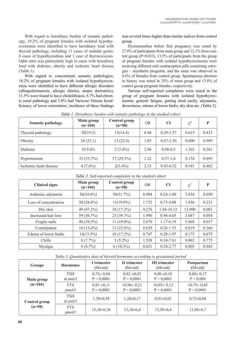
40
МЕДИЦИНСКИЕ НОВОСТИ ГРУЗИИ
CFMFHSDTKJC CFVTLBWBYJ CBF[KTYB
With regard to hereditary burden of somatic pathol-ogy, 19.2% of pregnant females with isolated hypothy-roxinemia were identified to have hereditary load with thyroid pathology, including 11 cases of nodular goiter, 8 cases of hypothyroidism and 1 case of thyrotoxicosis. Odds ratio was particularly high in cases with hereditary load with diabetes, obesity and ischemic heart disease (Table 1).
With regard to concomitant somatic pathologies, 18.2% of pregnant females with isolated hypothyroxin-emia were identified to have different allergic disorders (allergodermatosis, allergic rhinitis, atopic dermatitis), 11.5% were found to have cholelithiasis, 6.7% had chron-ic renal pathology and 3.8% had Varicose Venous Insuf-ficiency of lower extremities; incidence of these findings
was several times higher than similar indices from control group.
Dysmennorhea before first pregnancy was noted by 27.8% of participants from main group and 12.1% from con-trol group (P<0.033). 13.5% of participants from the group of pregnant females with isolated hypothyroxinemia were recieving different oral contraception pills containing estro-gen – asynthetic progestin, and the same was observed in 8.6% of females from control group. Spontaneous abortion in history was noted in 25% of main group and 13.8% of control group pregnant females, respectively.
Various self-reported complaints were noted in the group of pregnant females with isolated hypothyrox-inemia: general fatigue, getting tired easily, adynamia, drowsiness, edema of lower limbs, dry skin etc. (Table 2).
Table 1. Hereditary burden with somatic pathology in the studied cohort
Somatic pathology Main group (n=104)
Control group(n=58) OR CI χ2 P
Thyroid pathology 20(19.2) 15(14.4) 0.68 0.29-1.57 0.615 0.433
Obesity 24 (23,1) 13 (22.4) 1.85 0.47-2.50 0.000 0.989
Diabetes 10 9.6% 2 (3.4%) 2.96 0.58-0.5 1.263 0.261
Hypertension 35 (33,7%) 17 (29.3%) 1.22 0.57-1.6 0.154 0.695
Ischemic heart disease 8 (7.6%) 2(3.4%) 2.33 0.43-6.52 0.541 0.462
Table 2. Self-reported complaints in the studied cohort
Clinical signs Main group (n=104)
Control group (n=58) OR CI χ2 P
Asthenia, adynamia 36(34.6%) 30(51.7%) 0.494 0.24-1.00 3.834 0.050
Loss of concentration 30.(28,8%) 11(19.0%) 1.732 0.75-4.08 1.436 0.231
Dry skin 49 (47,1%) 10 (17.2%) 4.276 1.84-10.12 13.090 0.001Increased hair loss 59 (56,7%) 23 (39.7%) 1.990 0.98-4.05 3.687 0.054
Fragile nails 40 (38,5%) 11 (19.0%) 2.670 1.17-6.19 5.668 0.017Constipation 16 (15,4%) 13 (22.4%) 0.629 0.26-1.53 0.819 0.366
Edema of lower limbs 14(13.5%) 10 (17.2%) 0.747 0.28-1.97 0.175 0.675Chills 8 (7.7%) 3 (5.2%) 1.528 0.34-7.61 0.082 0.775
Myalgia 9 (8,7%) 6 (10.3%) 0.821 0.24-2.77 0.005 0.942
Table 3. Quantitative data of thyroid hormones according to gestational period
Groups Hormones I trimester(М±std)
II trimester(М±std)
III trimester(М±std)
Postpartum (М±std)
Main group(n=104)
TSHm.unit/l
0,75± 0,04P = 0,0001
0,82 ±0,03P = 0,0001
0,90 ±0,10P = 0,0001
0,88± 0,17P < 0,004
FT4pmol/l
8,83 ±0,11P = 0,0001
10,96± 0,21P = 0,0001
10,93± 0,12P = 0,0001
10,75± 0,45P = 0,0001
Control group(n=58)
TSHm.unit/l 1,29±0,58 1,28±0,17 0,91±0,03 0,73±0,04
FT4pmol/l 15,38±0,36 13,18±0,4 13,58±0,4 11,98±0,7
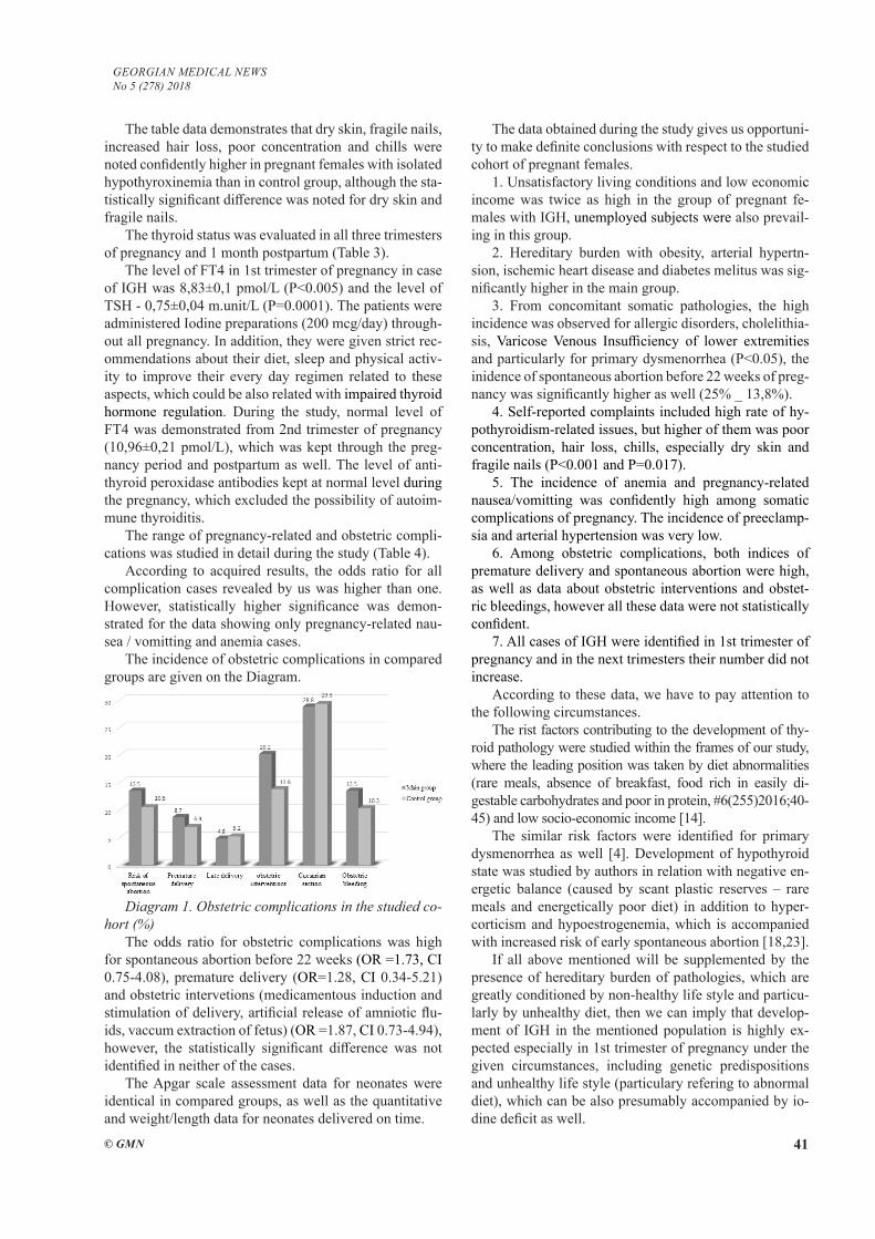
GEORGIAN MEDICAL NEWS No 5 (278) 2018
© GMN 41
The table data demonstrates that dry skin, fragile nails, increased hair loss, poor concentration and chills were noted confidently higher in pregnant females with isolated hypothyroxinemia than in control group, although the sta-tistically significant difference was noted for dry skin and fragile nails.
The thyroid status was evaluated in all three trimesters of pregnancy and 1 month postpartum (Table 3).
The level of FT4 in 1st trimester of pregnancy in case of IGH was 8,83±0,1 pmol/L (P<0.005) and the level of TSH - 0,75±0,04 m.unit/L (P=0.0001). The patients were administered Iodine preparations (200 mcg/day) through-out all pregnancy. In addition, they were given strict rec-ommendations about their diet, sleep and physical activ-ity to improve their every day regimen related to these aspects, which could be also related with impaired thyroid hormone regulation. During the study, normal level of FT4 was demonstrated from 2nd trimester of pregnancy (10,96±0,21 pmol/L), which was kept through the preg-nancy period and postpartum as well. The level of anti-thyroid peroxidase antibodies kept at normal level during the pregnancy, which excluded the possibility of autoim-mune thyroiditis.
The range of pregnancy-related and obstetric compli-cations was studied in detail during the study (Table 4).
According to acquired results, the odds ratio for all complication cases revealed by us was higher than one. However, statistically higher significance was demon-strated for the data showing only pregnancy-related nau-sea / vomitting and anemia cases.
The incidence of obstetric complications in compared groups are given on the Diagram.
Diagram 1. Obstetric complications in the studied co-hort (%)
The odds ratio for obstetric complications was high for spontaneous abortion before 22 weeks (OR =1.73, CI 0.75-4.08), premature delivery (OR=1.28, CI 0.34-5.21) and obstetric intervetions (medicamentous induction and stimulation of delivery, artificial release of amniotic flu-ids, vaccum extraction of fetus) (OR =1.87, CI 0.73-4.94), however, the statistically significant difference was not identified in neither of the cases.
The Apgar scale assessment data for neonates were identical in compared groups, as well as the quantitative and weight/length data for neonates delivered on time.
The data obtained during the study gives us opportuni-ty to make definite conclusions with respect to the studied cohort of pregnant females.
1. Unsatisfactory living conditions and low economic income was twice as high in the group of pregnant fe-males with IGH, unemployed subjects were also prevail-ing in this group.
2. Hereditary burden with obesity, arterial hypertn-sion, ischemic heart disease and diabetes melitus was sig-nificantly higher in the main group.
3. From concomitant somatic pathologies, the high incidence was observed for allergic disorders, cholelithia-sis, Varicose Venous Insufficiency of lower extremities and particularly for primary dysmenorrhea (P<0.05), the inidence of spontaneous abortion before 22 weeks of preg-nancy was significantly higher as well (25% _ 13,8%).
4. Self-reported complaints included high rate of hy-pothyroidism-related issues, but higher of them was poor concentration, hair loss, chills, especially dry skin and fragile nails (P<0.001 and P=0.017).
5. The incidence of anemia and pregnancy-related nausea/vomitting was confidently high among somatic complications of pregnancy. The incidence of preeclamp-sia and arterial hypertension was very low.
6. Among obstetric complications, both indices of premature delivery and spontaneous abortion were high, as well as data about obstetric interventions and obstet-ric bleedings, however all these data were not statistically confident.
7. All cases of IGH were identified in 1st trimester of pregnancy and in the next trimesters their number did not increase.
According to these data, we have to pay attention to the following circumstances.
The rist factors contributing to the development of thy-roid pathology were studied within the frames of our study, where the leading position was taken by diet abnormalities (rare meals, absence of breakfast, food rich in easily di-gestable carbohydrates and poor in protein, #6(255)2016;40-45) and low socio-economic income [14].
The similar risk factors were identified for primary dysmenorrhea as well [4]. Development of hypothyroid state was studied by authors in relation with negative en-ergetic balance (caused by scant plastic reserves – rare meals and energetically poor diet) in addition to hyper-corticism and hypoestrogenemia, which is accompanied with increased risk of early spontaneous abortion [18,23].
If all above mentioned will be supplemented by the presence of hereditary burden of pathologies, which are greatly conditioned by non-healthy life style and particu-larly by unhealthy diet, then we can imply that develop-ment of IGH in the mentioned population is highly ex-pected especially in 1st trimester of pregnancy under the given circumstances, including genetic predispositions and unhealthy life style (particulary refering to abnormal diet), which can be also presumably accompanied by io-dine deficit as well.
42
МЕДИЦИНСКИЕ НОВОСТИ ГРУЗИИ
CFMFHSDTKJC CFVTLBWBYJ CBF[KTYB
At the same time, on first meeting with pregnant pa-tients and throughout the pregnancy course, unhealthy life style was practically eliminated by strict recommenda-tions and explanations, patients were administered to take 200 mcg Iodine daily and thyroid, somatic and obstetric status was constantly monitored in dynamic follow-up. We consider that this exactly resulted in the low incidence of obstetric – neonatal and especially somatic complica-tions (preeclampsia, arterial hypertension, gestational dia-betes).
Anemia that developes as a complication concomitant with hypothyroid state during pregnancy and referred to as thyroprive anemi . It is not surprise that poor diet could have a particular role in this case as well, however, this can be the subject of further detailed studies in future. The special interest is given to the high incidence of pregnan-cy-related nausea/vomitting; its known that such cases are mostly associated with hyperthyroid state instead of hypothyroidism. The cause and mechanism of the devel-opment of the latter is by far unknown.
Conclusion. In the 1st trimester of pregnancy, IGH still increases the risk for development of obstetric and somatic pathologies, regardless of applied diagnostic and treatment measures.
Thus, to prevent the above mentioned complica-tions, we consider it important to use Iodine preparations at pre-gravid preparation stage in addition to complete elimination of behavioral (diet) abnormalities, planning of pregnancy at positive energetic balance and stable nor-mo thyroxinemia background, and as soon as pregnancy starts, evaluation of the thyroid hormonal status and its timely correction if necessary.
REFERENCES
1. subklinikuri hipoTireozis marTva orsu-lobis dros. qarTul enaze mTargmneli da re-daqtori prof. d. metreveli: 2016. European Thy-roid Association Guidelines for the Management of Subclinical Hypothyroidism in Pregnancy and in Children // Eur Thyroid J 2014;3:76–94.2. Фадеев В.В., Лесникова С.В. Гипотиреоз и изолированная гипотироксинемия во время беременности // Клиническая и экспериментальная тиреоидология 2011; том 7; №1.3. Akbaba et al. Pregnancy and Isolated Hypothyroxinemia. Turk-ish Journal of Endocrinology and Metabolism 2014;18: 106-110.4. Gagua T, Tkeshelashvili B, Gagua D. Primary dysmenorrhea: prevalence in adolescentpopulation of Tbilisi, Georgia and Risk Factors // J Turkish –German Gynecological Association N13(3) 2012, 162-8.5. Gowachirapant S, Melse-Boonstra A, Winichagoon P, Zim-mermann MB: Overweight increases risk of first trimester hy-pothyroxinaemia in iodine-deficient pregnant women // Matern Child Nutr. 2014;10:61-71.6. Haddow JE, Craig WY, PAlomaki G, E, Etal. Impact Of Adjusting For the Reciprocal Relationship Between Maternal Weight and Free Thyroxin During Early Pregnancy // Thyroid. 2013;23(2):225-30.7. Haddow JE, Nereux LM, Palomaki GE, Lambert-Messerlian G, Malone FD, D’Alton ME. An inverse Relationship Between
Weight and Free Thyroxine During Early Gestatcion Among Wom-en Treated For Hypothyroidis // Thyroid. 2015; 25(8):949-53.8. Han Ch, Li Ch, Mao, J. etal. High Bmi Is An Indication Of Maternal Hypothyroidism, Hypothroxinemin and Thyroid-per-oxidase Antibody Positivity During Early Pregnancy // Bio Med Resarch International. 2015 ID 35831; 7.9. He Y, He T, Wang Y, Xu Z, Xu Y, Wu Y, Ji J, Mi Y. Com-parison of the effect of different diagnostic criteria of subclinical hypothyroidism and positive TPO-Ab on pregnancy outcomes. // Zhonghua Fu Chan Ke Za Zhi. 2014 Nov;49(11):824-8.10. Hirsch D, Levy S, Nadler V, Kopel V, Shainberg B, Toledano Y. Pregnancy outcomes in women with severe hypothyroidism // Eur J Endocrinol. 2013 Jul 29;169(3):313-20. 11. Korevaar TI, Schalekamp-Timmermans S, de Rijke YB, Edward Visser W, Visser W, de Muinck Keizer-Schrama SM, Hofman A, Alec Ross H, Hooijkaas H, Tiemeier H, Bongers-Schokking JJ, Jaddoe VW, Visser TJ, Steegers EA, Medici M, Peeters RP. Hypothyroxinemia and TPO-antibody positivity are risk factors for premature delivery: the Generation R Study // J Clin Endocrinol Metab. September 13, 2013. 12. Lazarus J., Brown R.S., Daumerie C., Hubalewska-Dydejc-zyk A., Negro R. Vaidya B. 2014 European Thyroid Association Guidelines for the Management of Subclinical Hypothyroidism in Pregnancy and in Children // Eur Thyroid J 2014;3:76-94. 13. Martin B, Golden E., Carlson OD., Egan JM., Mattson M.P., Maudsley S. Caloric restriction: impact upon pituitary function and reproduction // Ageing Res. Rev, 2009; 7(3): 224.14. Morchiladze N, Tkeshelashvili B, Gagua D, Gagua T. Risk Factors of Thyroid Pathology Formation in Outpatient Pregnant Population // Georgian Medical News, 2016; N6(255): 40-45.15. Nazarpour S, Ramezani Tehrani F, Simbar M, Azizi F.Thyroid dysfunction and pregnancy outcomes // Iran J Reprod Med. 2015 Jul;13(7):387-96.16. Negro R, Soldin OP, Obregon MJ, Stagnaro-Green A. Hy-pothyroxinemia and pregnancy // Endocr Pract.2011 May-Jun;17(3):422-9.17. Pearce S, Brabant G, Duntas L, Monzani F, Peeters R, Razvi S, Wemeau J-L: 2013 ETA guideline: management of subclini-cal hypothyroidism // Eur Thyroid J 2013;2:215-228.18. Sellix M.T, Menaker M. Circadion cloks in mammalian re-productive physiology: effects of the ,,other” biological clok on fertility //Discover Med. 2011 Apr; 11(59):237-81.19. Sheehan PM1, Nankervis A, Araujo Júnior E1, Da Silva Cos-ta F1.Maternal Thyroid Disease and Preterm Birth: Systematic Review and Meta-Analysis // J Clin Endocrinol Metab. 2015 Nov;100(11):4325-31. 20. Spencer L, Bubner T, Bain E, Middleton P.Screening and subsequent management for thyroid dysfunction pre-pregnancy and during pregnancy for improving maternal and infant health // Cochrane Database Syst Rev. 2015 Sep 21;(9):CD011263. 21. Vissenberg R, Goddijn M, Mol BW, Van Der Post JA, Flierrs E, B:sschop PW. Thyroid dysfunction in / pregnant women: clin-ical dilemmas // Ned Tijdschr. Geneeskd. 2012; 156(49):A516322. Wang S, Li M, Chu D, Liang L, Zhao X, Zhang J.Clinical or subclinical hypothyroidism and thyroid autoantibody before 20 weeks pregnancy and risk of preterm birth: a systematic review. // Zhonghua Fu Chan Ke Za Zhi. 2014 Nov;49(11):816-22.23. Xiaona Hu, Xiaochun Teng, Hongzhi Zheng, Zhongyan Shan, Jing Li, Ting Jin, Chuhui Xiong, Hongmei Zhang, Chen-ling Fan, Weiping TengIron deficiency without anemia causes maternal hypothyroxinemia in pregnant rats // Nutrition Re-searchVolume 34, Issue 7, July 2014, Pages 604–61224. Yang J, Guo H, Ding S, Tao B, Zhang X. [Effect of the treatment acceptance on the perinatal outcomes in women with
GEORGIAN MEDICAL NEWS No 5 (278) 2018
© GMN 43
subclinical hypothyroidism, positive thyroid gland peroxidase antibody in early pregnancy] // Zhonghua Fu Chan Ke Za Zhi. 2015 Sep;50(9):652-7.25. Yu X, Shan Z, Li C, Mao J, Wang W, Xie X, Liu A, Teng X, Zhou W, Li C, Xu B, Bi L, Meng T, Du J, Zhang S, Gao Z, Zhang X, Yang L, Fan C, Teng W.Iron deficiency, an indepen-dent risk factor for isolated hypothyroxynemia in pregnant and nonpregnant women of childbearing age in China // J Clin Endo-crinol Metab. 2015 Apr;100(4):1594-601. doi: 10.1210/jc.2014-3887. Epub 2015 Jan 19.26. Zhu YD, Han Y, Huang K, Zhu BB., Yan SQ, Ge X, Zhou SS, Xu YY, Ren L, Sheng J, Pan WJ, Hao JH1, Zhu P, Tao FB. The impact of isolated maternal hypothyroxinaemia on the incidence of large-for-gestational-age infants: the Ma’anshan Birth Cohort study// BJOG. - 2017 Dec 19. doi: 10.1111/1471-0528.15107.
SUMMARY
IMPORTANCE OF ISOLATED GESTATIONAL HYPOTHYROXINEMIA IN THE DEVELOPMENT OF OBSTETRIC AND SOMATIC PATHOLOGIES Morchiladze N., Tkeshelashvili B., Gagua T., Gagua D.
Tbilisi State Medical University; “David Gagua Clinic” Ltd., Tbilisi, Georgia
The isolated hypothyroxinemia of pregnancy (IHP) has gained specific attention in the specialized literature during the recent years as the possible factor impeding the intellectual development of fetus and increasing the risk of complications related with pregnancy, delivery and perinatal period.
Aim of the study was to define the importance of iso-lated hypothyroxinemia in the development of obstetric and somatic pathologies in outpatient population of preg-nant females.
The study of prospective design was performed at the base of “David Gagua Clinic” Ltd. Based on hospital re-ferral we selected the pregnant patients who were diag-nosed for isolated hypothyroxinemia in the 1st trimester of pregnancy by clinical-laboratory studies.
104 pregnant females with isolated hypothyroxin-emia were included in the main group, and 58 pregrant females of reproductive age who were not identified to have thyroid pathology by screening studies were includ-ed in the control group,. The questionnaire used in the study process included the retrospective medical history data, demographic findings, information about premorbid background, genetic burden of somatic pathology, social-economical factors (including education level, living con-ditions, economic income, family environment etc.) and concomitant somatic pathology.
In addition, it included the clinical and para-clinical study data and pregnancy follow-up findings. The test studies for thyroid status were performed every trimester and after one month postpartum.
The software packages Microsoft Excel (2010) and SPSS/v.12 was used for statistical treatment of data. The
digital data is presented by M±STD, where M is the arith-metic mean and STD is the standard deviation of arithme-tic mean. To define the confidence interval for the indices and their relation, we calculated 2 and p, whose critical value was defined to be 0.05.
Based on analysis of the acquired data, we found out that pregnant females with isolated hypothyroxinemia were more statistically demonstrating asthenia, dry skin, increased hair loss and fragile nails, and from somatic disorders – pregnancy-associated vomiting and anemia. From concomitant diseases, allergic disorders (18.2%), primary dysmenorrhea (27.8%), spontaneous abortions (25%) were taking the highest incidence rate and other obstetric complications (premature delivery, late delivery) were higher in the main group, though statistically signifi-cant difference was not demonstrated.
It must be noted that isolated hypothyroxinemia in the studied cohort was mostly found in 1st trimester of pregnancy, whereas according to the literature data, the latter is demonstrated more frequently in the second or third trimester.
The above mentioned makes us consider that the io-dine deficit in the cohort of pregnant females studied by us was probably present before pregnancy as well and maybe with even higher extent.
Thus, the isolated hypothyroxinemia developed in the very first trimester of pregnancy still has its negative impact on the pregnancy course and outcome, despite of applied treatment. According to performed studies and their results, for the prevention of obstetric and perinatal complications, its important to administer iodine preparations together with folic acid at pregravid stage in addition to complete elimi-nation of diet abnormalities, plan the pregnancy in stable normothyroxinemia conditions and at positive energetic bal-ance. In addition, its desirable to perform the repeated thy-roid status evaluation in the first trimester of pregnancy and timely administration of adequate therapeutic measures in case of finding any pathology.
Keywords: isolated hypothyroxinemia of pregnancy, pregnant females, obstetric and somatic pathologies.
РЕЗЮМЕ
ЗНАЧЕНИЕ ИЗОЛИРОВАННОЙ ГИПОТИРОК-СИНЕМИИ БЕРЕМЕННЫХ В ФОРМИРОВА-НИИ АКУШЕРСКОЙ И СОМАТИЧЕСКОЙ ПА-ТОЛОГИИ
Морчиладзе Н.А., Ткшелашвили Б.Д., Гагуа Т.Д., Гагуа Д.А.
Тбилисский государственный медицинский универси-тет; ООО «Клиника Гагуа», Тбилиси, Грузия
В последние годы в специальной литературе осо-бое внимание уделяется изолированной гипотирекси-
44
МЕДИЦИНСКИЕ НОВОСТИ ГРУЗИИ
CFMFHSDTKJC CFVTLBWBYJ CBF[KTYB
немии беременных (ИГБ) как возможному фактору, обуславливающему препятствие для интеллектуаль-ного развития плода и рост осложнений беременно-сти, родов и послеродового периода.
Целью исследования явилось установление значе-ния изолированной гипотирексинемии беременных в формировании акушерской и соматической патологии в амбулаторной популяции беременных.
Проведено открытое контролируемое проспек-тивное исследование на базе ООО «Клиника Давида Гагуа». На основании обращений в клинику подобра-ны беременные, у которых в результате клинических и лабораторных исследований в первом триместре беременности установлен диагноз ИГБ. Эти бере-менные составили основную группу (n=104), кон-(n=104), кон-, кон-трольную группу составили беременные (n=58), у которых тироидная патология не обнаружена. Опрос-ный лист, используемый в ходе исследования, охва-тывал ретроспективные анамнезные и демографиче-ские показатели, информацию о преморбидном фоне, наследственности соматической патологии, социаль-но-экономических факторах и существующей сомати-ческой патологии. Исследования тироидного статуса проводились в каждом триместре и спустя месяц по-сле родов.
Статистическая обработка полученных данных проводилась посредством программных пакетов Microsoft Excel (2010) и SPSS v.12. Цифровые показа-тели представлены в виде M±m. Для установления до-стоверности связи между показателями использованы критерии χ2 и p, данные считались достоверными при p<0,05.
На основании анализа полученных результатов установлено, что в группе беременных с ИГ стати-стически достоверно более часто отмечалась астения, сухость кожи, усиленное выпадение волос, ломкость ногтей, а из осложнений беременности – рвота и ане-мия. Среди сопутствующей патологии превалировали аллергические заболевания (18,2%), первичная дис-аминорея (27,8%), а также в анамнезе случаи само-произвольного прерывания беременности в сроке до 22 недель (25%).
Следует отметить, что в исследуемом контингенте беременных ИГ чаще всего выявлена в первом триме-стре, а по литературным данным, ИГБ, в основном, проявляется на более поздних сроках беременности.
Таким образом, развитие ИГБ в первом триме-стре, несмотря на проведенное лечение, негативно влияет на течение и исход беременности. С целью превенции акушерских и перинатальных осложне-ний желательно планировать беременность только при достижении устойчивой нормотироксинемии, оценить тироидный статус на ранних этапах геста-ции и в случае выявления нарушений – своевремен-но начать адекватное лечение.
reziume
orsulTa izolirebuli hipoTiroqsinemiis mniSvneloba sameano da somaturi paTolo-giis formirebaSi n. morCilaZe, b. tyeSelaSvili, T. gagua, d. gagua
Tbilisis saxelmwifo samedicino univer-siteti; Sps `gaguas klinika~, Tbilisi, saqar-Tvelo
ukanaskneli wlebis specialur litera-turaSi gansakuTrebuli yuradReba eTmoba orsulTa izolirebul hipoTiroqsinemias (oih), rogorc nayofis inteleqtualuri gan-viTarebis Semaferxebel, orsulobis, mSo-biarobisa da perinataluri garTulebebis riskis matebis SesaZlo ganmapirobebel faq-tors.
Kkvlevis mizani iyo orsulTa ambula-torul populaciaSi izolirebuli hi-poTiroqsinemiis mniSvnelobis dadgena same-ano da somaturi paTologiis formirebaSi.
Catarda prospeqtuli tipis kvleva Sps `daviT gaguas klinikis~ bazaze. klinikaSi momarTvianobis safuZvelze SerCeuli iyo orsulebi, romelTac klinikur-labora-toriuli kvlevis Sedegad I trimestrSi daudgindaT izolirebuli hipoTireqsinemi-is diagnozi.
ZiriTad jgufSi gaerTianda 104 orsuli izolirebuli hipoTiroqsinemiiT, sakontro-lo jgufi Seadgina reproduqciuli asakis 58 orsulma, romelTac skrininguli kvle-viT ar gamouvlinda Tiroiduli paTologia. Kkvlevis procesSi gamoyenebuli kiTxvari moicavda retrospeqtul anamnezur monace-mebs, demografiul maCveneblebs, informacias premorbiduli fonis, somaturi paTologiiT memkvidruli datvirTvis, socialur-ekonomi-uri (ganaTlebis done, sayofacxovrebo piro-bebi, ekonomiuri Semosavali, ojaxuri gare-mo) faqtorebis da TandarTuli somaturi paTologiis Sesaxeb, agreTve, klinikuri da paraklinikuri kvlevis monacemebsa da orsu-lobis mimdinareobaze dakvirvebis Sedegebs. Tiroiduli statusis testuri kvlevebi tar-deboda yovel trimestrSi da mSobiarobidan erTi Tvis Semdeg.
monacemebis statistikuri damuSavebis mizniT gamoyenebuli iyo programuli pakete-bi Microsoft Excel (2010) da SPSS v.12. cifrobrivi monacemebi warmodgenilia M±STD saxiT, sadac
GEORGIAN MEDICAL NEWS No 5 (278) 2018
© GMN 45
M aris saSualo ariTmetikuli da STD-saSu-alo ariTmetikulis standartuli gadaxra. maCveneblebs Soris kavSiris sandoobis dasadgenad gamoTvlilia χ2 da p, romlis kri-tikul mniSvnelobad miCneuli iyo <0,05.
miRebuli monacemebis gaanalizebis safuZvelze dadginilia, rom ih-is mqone or-sulebs statistikurad sarwmunod ufro xSirad aReniSnebodaT asTenia, kanis simS-rale, Tmis gaZlierebuli cvena da frCxi-lebis mtvrevadoba, xolo somaturi da-avadebebidan _ orsulTa Rebineba da anemia. Tanmxlebi daavadebebis mxriv Warbobda alergiuli daavadebebi (18,2%), pirveladi dismenorea (27,8%).
aRsaniSnavia, rom Seswavlil kontigent-Si ih gamovlinda orsulobis I trimestrSi, xolo literaturaSi arsebuli monacemebiT es ukanaskneli ufro xSirad meore an mesame trimestrSi iCens Tavs.
savaraudoa, rom Cvens mier Seswavlil
orsulTa kontingentSi iodis deficiti or-sulobamdec aRiniSneboda da SesaZloa aR-matebuli xarisxiTac.
amrigad, orsulobis pirvelive trimestr-Si ganviTarebuli izolirebuli hipoTiroq-sinemia, miuxedavad Catarebuli mkurnalobisa, negatiur gavlenas mainc axdens orsulobis mimdinareobasa da gamosavalze. ACatare-buli kvlevis Sedegebis mixedviT, sameano da perinataluri garTulebebis prevenciis mizniT, mniSvnelovania pregraviduli momza-debis etapze foliumis mJavasTan erTad iodis preparatebis gamoyeneba, dietis modi-fikacia da myari normoTiroqsinemiis da dadebiTi energetikuli balansis miRwevis pirobebSi orsulobis dagegmva. amavdrou-lad, sasurvelia orsulobis pirvelive trimestrSi Tiroiduli hormonuli statu-sis ganmeorebiTi Sefaseba da darRvevebis gamovlenis SemTxvevaSi _ adeqvaturi Tera-piuli RonisZiebebis droulad dawyeba.
ОБОСНОВАНИЕ ВЫБОРА РАЗМЕЩЕНИЯ АРМИРУЮЩЕГО ЭЛЕМЕНТА СТЕКЛОВОЛОКОННОЙ АДГЕЗИВНОЙ ШИНЫ ПОСРЕДСТВОМ ИЗУЧЕНИЯ
ДИАГНОСТИЧЕСКИХ МОДЕЛЕЙ В АРТИКУЛЯТОРЕ И ПОСЛЕДУЮЩЕГО ПАРАЛЛЕЛОМЕТРИЧЕСКОГО ИССЛЕДОВАНИЯ
1Левандовский Р.А., 1Беликова Н.И., 1,2 Беликов А.Б., 1Годованец О.И., 1Накашидзе Г.Н.
1Высшее государственное учебное заведение "Буковинский государственный медицинский университет", Черновцы; 2Украинская академия наук, Киев, Украина
Известно, что волоконные адгезивные шины, благодаря целому ряду преимуществ, в сравнении с традиционными методами шинирования, позволяют эффективно устранить один из ведущих симптомов генерализованного пародонтита - патологическую подвижность [6,7,23,11], что дает основание рекомен-довать их для более широкого использования в ком-плексном лечении этой патологии [2-4,12]. Однако, данные литературы свидетельствуют, что сравнитель-но высокий процент (94,7%) клинико-рентгенологи-ческой стабилизации воспалительно-деструктивного процесса, происходящего в пародонте на проятжении первых 6 месяцев, спустя год понижается до 62,6% [13,14]. Вышеприведенные данные диктуют необхо-димость поиска способов оптимизации проводимого шинирования. Несмотря на весьма богатый выбор шин и способов шинирования [1,2,17-20], существу-ющие конструкции имеют небольшой срок эксплуа-
тации, поскольку происходит снижение их функци-ональной и эстетической ценности в виде рецессии десны и усиления подвижности всего блока шиниро-ванных зубов [3]. Кроме того, шинирующие конструк-ции не всегда соответствуют основным критериям биомеханики зубных рядов [5,12]. Проблема выбора оптимальной конструкции шинирующего устройства для стабилизации подвижных зубов при пародонтите по сей день остается актуальной [3-4,7,15,17,22]. При шинировании зубов с пораженным пародонтом роль параллелометрии имеет особое значение, так как уве-личивается смещение и непараллельность подвиж-ных зубов. При изготовлении шин на различные груп-пы зубов имеются особенности, которые необходимо учитывать при проведении параллелометрии [21].
Целью исследования явилась разработка наиболее оптимального варианта размещения армирующего элемента стекловолоконной шины при подвижности
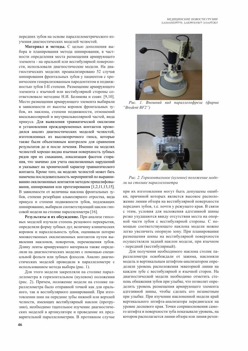
46
МЕДИЦИНСКИЕ НОВОСТИ ГРУЗИИ
CFMFHSDTKJC CFVTLBWBYJ CBF[KTYB
передних зубов на основе параллелометрического из-учения диагностических моделей челюстей.
Материал и методы. С целью дополнения вы-бора и планирования метода шинирования, в част-ности определения места размещения армирующего элемента - на оральной или вестибулярной поверхно-сти, использовали диагностические модели. На диа-гностических моделях проанализировано 52 случая шинирования фронтальных зубов у пациентов с хро-ническим генерализованным пародонтитом и подвиж-ностью зубов І-ІІ степени. Размещение армирующего элемента с язычной или вестибулярной стороны со-ответсвовало методике Н.И. Беликова и соавт. [9,10]. Место размещения армирующего элемента выбирали в зависимости от высоты коронок фронтальных зу-бов, их наклона, степени подвижности, отношений внеальвеолярной и внутриальвеолярной частей, вида прикуса. Для выявления травматической окклюзии и установления преждевременных контактов прово-дился анализ диагностических моделей челюстей, изготовленных из высокопрочного гипса, которые также были объективным контролем для сравнения результатов до и после лечения. Именно на моделях челюстей хорошо видна язычная поверхность зубных рядов при их смыкании, локализация фасеток стира-ния, что значимо для учета окклюзионных нарушений и указывает на хронический характер травматического контакта. Кроме того, на моделях челюстей может бать намечена последовательность мероприятий по выравни-ванию окклюзионных контактов методом пришлифовы-вания, шинирования или протезирования [1,2,11,13,15]. В зависимости от величины наклона фронтальных зу-бов, степени резорбции альвеолярного отростка, вида прикуса и степени подвижности зубов, подлежащих шинированию, выбирали соответствующий наклон гип-совой модели на столике параллелометра [16].
Результаты и их обсуждение. При анализе гипсо-вых моделей изучали степень резцового перекрытия, определяли форму зубных дуг, величину клинических коронок и параллельность зубов, оценивали потерю множественных окклюзионных контактов путем вы-явления наклонов, поворотов, перемещения зубов. Длину ленты армирующего материала также опреде-ляли на диагностических моделях с помощью специ-альной фольги или зубных флоссов. Анализ диагно-стических моделей проводили в параллелометре с использованием метода выбора (рис. 1).
Для этого модели закрепляли на столике парал-лелометра в горизонтальном (нулевом) положении (рис. 2). Причем, положение модели на столике па-раллелометра было отправной точкой как для ораль-ного, так и вестибулярного шинирований. При изго-товлении шин на передние зубы нижней или верхней челюсти, имеющих вестибулярный наклон (протру-зию), необходимо тщательное изучение диагностиче-ских моделей в артикуляторе и проведение их пред-варительной параллелометрии. В противном случае
при их изготовлении могут быть допущены ошиб-ки, причиной которых является высокое располо-жение линии обзора на вестибулярной поверхности передних зубов, т.е. почти у режущего края. В связи с этим, условия для наложения адгезивной шины резко ухудшаются ввиду отсутствия места на опор-ной части зубов с вестибулярной стороны. С по-мощью соответствующего наклона модели можно легко увеличить опорную зону. При планировании размещения шины на вестибулярной поверхности осуществляли задний наклон модели, при язычном - передний (вестибулярный).
Для получения необходимого наклона столик па-раллелометра освобождали от зажима, наклоняли модель и вертикальным штифтом-анализатором опре-деляли уровень расположения экваторной линии на каждом зубе с вестибулярной и язычной сторон. На диагностической модели необходимо отметить сте-пень обнажения зубов при улыбке, что позволит опре-делить уровень размещения армирующего элемента адгезивной шины, чтобы сделать его незаметным при улыбке. При изучении наклоненной модели край вертикального штифта-анализатора передвигался на уровне десневого края. Точки соприкосновения само-го штифта и поверхности зуба показывали уровень, на котором располагается линия обзора или линия ретен-
Рис. 1. Внешний вид параллелофреза (фирма “Bredent BF2”)
Рис. 2. Горизонтальное (нулевое) положение моде-ли на столике параллелометра
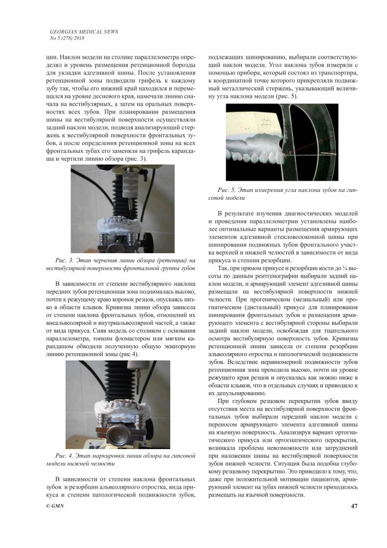
GEORGIAN MEDICAL NEWS No 5 (278) 2018
© GMN 47
ции. Наклон модели на столике параллелометра опре-делял и уровень размещения ретенционной борозды для укладки адгезивной шины. После установления ретенционной зоны подводили грифель к каждому зубу так, чтобы его нижний край находился и переме-щался на уровне десневого края, намечали линию сна-чала на вестибулярных, а затем на оральных поверх-ностях всех зубов. При планировании размещения шины на вестибулярной поверхности осуществляли задний наклон модели, подводя анализирующий стер-жень к вестибулярной поверхности фронтальных зу-бов, а после определения ретенционной зоны на всех фронтальных зубах его заменяли на грифель каранда-ша и чертили линию обзора (рис. 3).
Рис. 3. Этап черчения линии обзора (ретенции) на вестибулярной поверхности фронтальной группы зубов
В зависимости от степени вестибулярного наклона передних зубов ретенционная зона поднималась высоко, почти к режущему краю коронок резцов, опускаясь низ-ко в области клыков. Кривизна линии обзора зависела от степени наклона фронтальных зубов, отношений их внеальвеолярной и внутриальвеолярной частей, а также от вида прикуса. Сняв модель со столиком с основания параллелометра, тонким фломастером или мягким ка-рандашом обводили полученную общую экваторную линию ретенционной зоны (рис 4).
Рис. 4. Этап маркировки линии обзора на гипсовой модели нижней челюсти
В зависимости от степени наклона фронтальных зубов и резорбции альвеолярного отростка, вида при-куса и степени патологической подвижности зубов,
подлежащих шинированию, выбирали соответствую-щий наклон модели. Угол наклона зубов измеряли с помощью прибора, который состоял из транспортира, к координатной точке которого прикрепляли подвиж-ный металлический стержень, указывающий величи-ну угла наклона модели (рис. 5).
Рис. 5. Этап измерения угла наклона зубов на гип-совой модели
В результате изучения диагностических моделей и проведения параллелометрии установлены наибо-лее оптимальные варианты размещения армирующих элементов адгезивной стекловолоконной шины при шинировании подвижных зубов фронтального участ-ка верхней и нижней челюстей в зависимости от вида прикуса и степени резорбции.
Так, при прямом прикусе и резорбции кости до ¼ вы-соты по данным рентгенографии выбирали задний на-клон модели, и армирующий элемент адгезивной шины размещали на вестибулярной поверхности нижней челюсти. При прогеническом (мезиальный) или про-гнатическом (дистальный) прикусе для планирования шинирования фронтальных зубов и размещения арми-рующего элемента с вестибулярной стороны выбирали задний наклон модели, освобождая для тщательного осмотра вестибулярную поверхность зубов. Кривизна ретенционной линии зависела от степени резорбции альвеолярного отростка и патологической подвижности зубов. Вследствие неравномерной подвижности зубов ретенционная зона проходила высоко, почти на уровне режущего края резцов и опускалась как можно ниже в области клыков, что в отдельных случаях и приводило к их депульпированию.
При глубоком резцовом перекрытии зубов ввиду отсутствия места на вестибулярной поверхности фрон-тальных зубов выбирали передний наклон модели с переносом армирующего элемента адгезивной шины на язычную поверхность. Анализируя вариант ортогна-тического прикуса или ортогнатического перекрытия, возникала проблема невозможности или затруднений при наложении шины на вестибулярной поверхности зубов нижней челюсти. Ситуация была подобна глубо-кому резцовому перекрытию. Это приводило к тому, что, даже при положительной мотивации пациентов, арми-рующий элемент на зубах нижней челюсти приходилось размещать на язычной поверхности.
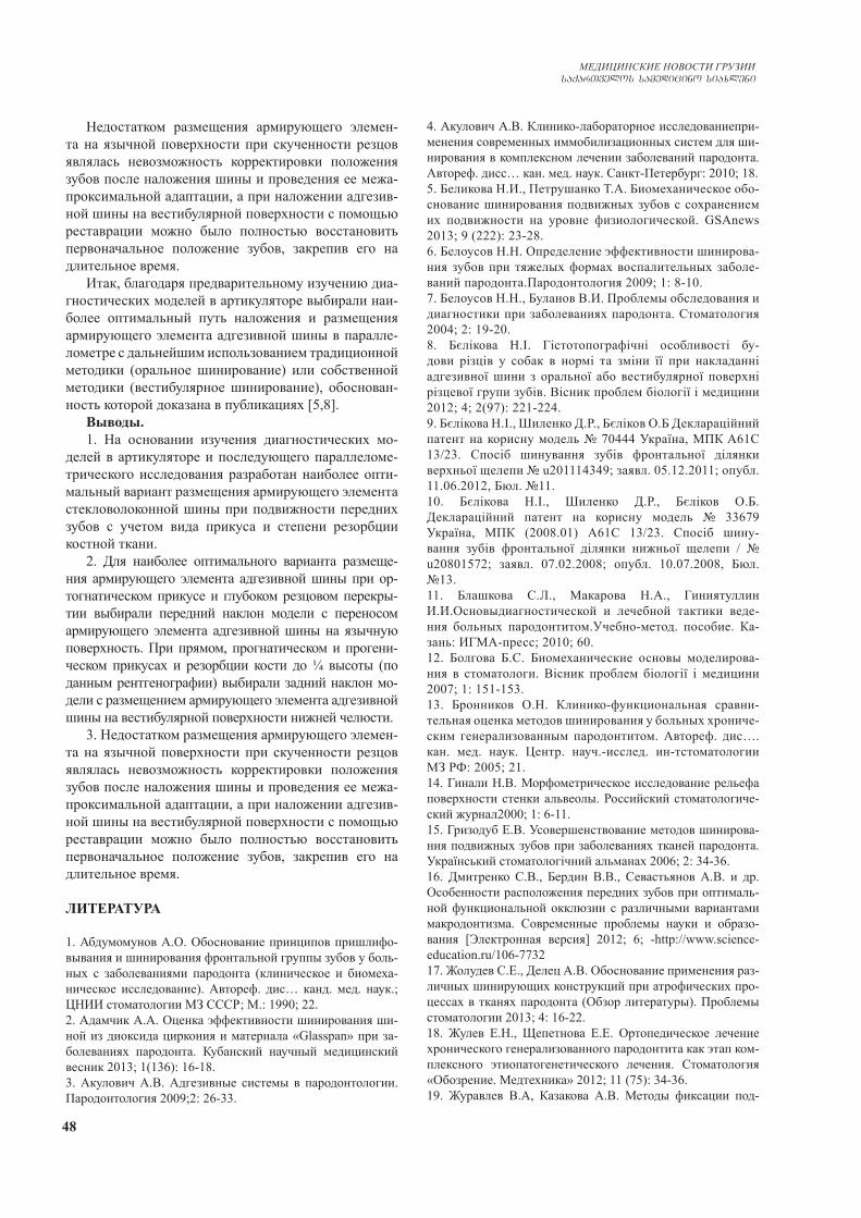
48
МЕДИЦИНСКИЕ НОВОСТИ ГРУЗИИ
CFMFHSDTKJC CFVTLBWBYJ CBF[KTYB
Недостатком размещения армирующего элемен-та на язычной поверхности при скученности резцов являлась невозможность корректировки положения зубов после наложения шины и проведения ее межа-проксимальной адаптации, а при наложении адгезив-ной шины на вестибулярной поверхности с помощью реставрации можно было полностью восстановить первоначальное положение зубов, закрепив его на длительное время.
Итак, благодаря предварительному изучению диа-гностических моделей в артикуляторе выбирали наи-более оптимальный путь наложения и размещения армирующего элемента адгезивной шины в паралле-лометре с дальнейшим использованием традиционной методики (оральное шинирование) или собственной методики (вестибулярное шинирование), обоснован-ность которой доказана в публикациях [5,8].
Выводы.1. На основании изучения диагностических мо-
делей в артикуляторе и последующего параллеломе-трического исследования разработан наиболее опти-мальный вариант размещения армирующего элемента стекловолоконной шины при подвижности передних зубов с учетом вида прикуса и степени резорбции костной ткани.
2. Для наиболее оптимального варианта размеще-ния армирующего элемента адгезивной шины при ор-тогнатическом прикусе и глубоком резцовом перекры-тии выбирали передний наклон модели с переносом армирующего элемента адгезивной шины на язычную поверхность. При прямом, прогнатическом и прогени-ческом прикусах и резорбции кости до ¼ высоты (по данным рентгенографии) выбирали задний наклон мо-дели с размещением армирующего элемента адгезивной шины на вестибулярной поверхности нижней челюсти.
3. Недостатком размещения армирующего элемен-та на язычной поверхности при скученности резцов являлась невозможность корректировки положения зубов после наложения шины и проведения ее межа-проксимальной адаптации, а при наложении адгезив-ной шины на вестибулярной поверхности с помощью реставрации можно было полностью восстановить первоначальное положение зубов, закрепив его на длительное время.
ЛИТЕРАТУРА
1. Абдумомунов А.О. Обоснование принципов пришлифо-вывания и шинирования фронтальной группы зубов у боль-ных с заболеваниями пародонта (клиническое и биомеха-ническое исследование). Автореф. дис… канд. мед. наук.; ЦНИИ стоматологии МЗ СССР; М.: 1990; 22. 2. Адамчик А.А. Оценка эффективности шинирования ши-ной из диоксида циркония и материала «Glasspan» при за-болеваниях пародонта. Кубанский научный медицинский весник 2013; 1(136): 16-18.3. Акулович А.В. Адгезивные системы в пародонтологии.Пародонтология 2009;2: 26-33.
4. Акулович А.В. Клинико-лабораторное исследованиепри-менения современных иммобилизационных систем для ши-нирования в комплексном лечении заболеваний пародонта.Автореф. дисс… кан. мед. наук. Санкт-Петербург: 2010; 18. 5. Беликова Н.И., Петрушанко Т.А. Биомеханическое обо-снование шинирования подвижных зубов c сохранением их подвижности на уровне физиологической. GSAnews 2013; 9 (222): 23-28. 6. Белоусов Н.Н. Определение эффективности шинирова-ния зубов при тяжелых формах воспалительных заболе-ваний пародонта.Пародонтология 2009; 1: 8-10.7. Белоусов Н.Н., Буланов В.И. Проблемы обследования и диагностики при заболеваниях пародонта. Стоматология 2004; 2: 19-20. 8. Бєлікова Н.І. Гістотопографічні особливості бу-дови різців у собак в нормі та зміни її при накладанні адгезивної шини з оральної або вестибулярної поверхні різцевої групи зубів. Вісник проблем біології і медицини 2012; 4; 2(97): 221-224.9. Бєлікова Н.І., Шиленко Д.Р., Бєліков О.Б Деклараційний патент на корисну модель № 70444 Україна, МПК A61C 13/23. Спосіб шинування зубів фронтальної ділянки верхньої щелепи № u201114349; заявл. 05.12.2011; опубл. 11.06.2012, Бюл. №11. 10. Бєлікова Н.І., Шиленко Д.Р., Бєліков О.Б. Деклараційний патент на корисну модель № 33679 Україна, МПК (2008.01) А61С 13/23. Спосіб шину-вання зубів фронтальної ділянки нижньої щелепи / № u20801572; заявл. 07.02.2008; опубл. 10.07.2008, Бюл. №13. 11. Блашкова С.Л., Макарова Н.А., Гиниятуллин И.И.Основыдиагностической и лечебной тактики веде-ния больных пародонтитом.Учебно-метод. пособие. Ка-зань: ИГМА-пресс; 2010; 60.12. Болгова Б.С. Биомеханические основы моделирова-ния в стоматологи. Вісник проблем біології і медицини 2007; 1: 151-153.13. Бронников О.Н. Клинико-функциональная сравни-тельная оценка методов шинирования у больных хрониче-ским генерализованным пародонтитом. Автореф. дис…. кан. мед. наук. Центр. науч.-исслед. ин-тстоматологии МЗ РФ: 2005; 21.14. Гинали Н.В. Морфометрическое исследование рельефа поверхности стенки альвеолы. Российский стоматологиче-ский журнал2000; 1: 6-11. 15. Гризодуб Е.В. Усовершенствование методов шинирова-ния подвижных зубов при заболеваниях тканей пародонта. Український стоматологічний альманах 2006; 2: 34-36.16. Дмитренко С.В., Бердин В.В., Севастьянов А.В. и др. Особенности расположения передних зубов при оптималь-ной функциональной окклюзии с различными вариантами макродонтизма. Современные проблемы науки и образо-вания [Электронная версия] 2012; 6; -http://www.science-education.ru/106-773217. Жолудев С.Е., Делец А.В. Обоснование применения раз-личных шинирующих конструкций при атрофических про-цессах в тканях пародонта (Обзор литературы). Проблемы стоматологии 2013; 4: 16-22. 18. Жулев Е.Н., Щепетнова Е.Е. Ортопедическое лечение хронического генерализованного пародонтита как этап ком-плексного этиопатогенетического лечения. Стоматология «Обозрение. Медтехника» 2012; 11 (75): 34-36. 19. Журавлев В.А, Казакова А.В. Методы фиксации под-
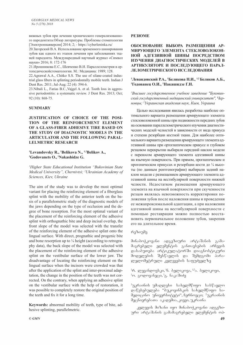
GEORGIAN MEDICAL NEWS No 5 (278) 2018
© GMN 49
вижных зубов при лечении хронического генерализованно-го пародонтита.Обзор литературы. Проблемы стоматологии [Электроннаяверсия] 2014; 2; - https://cyberleninka.ru/20.Загорский В.А. Использование временного шинирования зубов как одного из этапов лечения при заболеваниях тка-ней пародонта. Международный научный журнал «Символ науки» 2016; 8: 172-17621.Ирошникова Е.С., Шевченко В.И. Параллелометрия в ор-топедическойстоматологии. М.: Медицина: 1989; 128. 22.Agrawal A.A., Chitko S.S. The use of silane-coated indus-trial glass fibers in splinting periodontally mobile teeth. Indian J Dent Res. 2011; Jul-Aug; 22 (4): 594-6.23.Nibali L., Farias B.C.,Vajgel A. et al. Tooth loss in aggres-sive periodontitis: a systematic review. J Dent Res. 2013; Oct; 92 (10): 868-75.
SUMMARY
JUSTIFICATION OF CHOICE OF THE POSI-TION OF THE REINFORCEMENT ELEMENT OF A GLASS-FIBER ADHESIVE TIRE BASED ON THE STUDY OF DIAGNOSTIC MODELS IN THE ARTICULATOR AND THE FOLLOWING PARAL-LELMETRIC RESEARCH
1Levandovsky R., 1Belikova N., 1,2Belikov A., 1Godovanets O., 1Nakashidze G.
1Higher State Educational Institution “Bukovinian State Medical University”, Chernivtsi; 2Ukrainian Academy of Sciences, Kiev, Ukraine
The aim of the study was to develop the most optimal variant for placing the reinforcing element of a fiberglass splint with the mobility of the anterior teeth on the ba-sis of a parallelometric study of the diagnostic models of the jaws depending on the type of occlusion and the de-gree of bone resorption. For the most optimal variant of the placement of the reinforcing element of the adhesive splint with orthognathic bite and deep incisal overlap, the front slope of the model was selected with the transfer of the reinforcing element of the adhesive splint onto the lingual surface. With direct, prognathic and progenic bite and bone resorption up to ¼ height (according to retrogra-phy data), the back slope of the model was selected with the placement of the reinforcing element of the adhesive splint on the vestibular surface of the lower jaw. The disadvantage of locating the reinforcing element on the lingual surface when the incisors were crowded was that after the application of the splint and inter-proximal adap-tation, the change in the position of the teeth was not cor-rected. On the contrary, when applying an adhesive splint on the vestibular surface with the help of restoration, it was possible to completely restore the original position of the teeth and fix it for a long time.
Keywords: abnormal mobility of teeth, type of bite, ad-hesive splinting, parallelmetric.
РЕЗЮМЕ
ОБОСНОВАНИЕ ВЫБОРА РАЗМЕЩЕНИЯ АР-МИРУЮЩЕГО ЭЛЕМЕНТА СТЕКЛОВОЛОКОН-НОЙ АДГЕЗИВНОЙ ШИНЫ ПОСРЕДСТВОМ ИЗУЧЕНИЯ ДИАГНОСТИЧЕСКИХ МОДЕЛЕЙ В АРТИКУЛЯТОРЕ И ПОСЛЕДУЮЩЕГО ПАРАЛ-ЛЕЛОМЕТРИЧЕСКОГО ИССЛЕДОВАНИЯ
1Левандовский Р.А., 1Беликова Н.И., 1,2 Беликов А.Б., 1Годованец О.И., 1Накашидзе Г.Н.
1Высшее государственное учебное заведение "Буковин-ский государственный медицинский университет", Чер-новцы; 2Украинская академия наук, Киев, Украина
Целью исследования явилась разработка наиболее оп-тимального варианта размещения армирующего элемента стекловолоконной шины при подвижности передних зубов на основании параллелометрического изучения диагности-ческих моделей челюстей в зависимости от вида прикуса и степени резорбции костной ткани. Для наиболее опти-мального варианта размещения армирующего элемента ад-гезивной шины при ортогнатическом прикусе и глубоком резцовом перекрытии выбирали передний наклон модели с переносом армирующего элемента адгезивной шины на язычную поверхность. При прямом, прогнатическом и прогеническом прикусах и резорбции кости до ¼ высо-ты (по данным рентгенографии) выбирали задний на-клон модели с размещением армирующего элемента ад-гезивной шины на вестибулярной поверхности нижней челюсти. Недостатком размещения армирующего элемента на язычной поверхности при скученности резцов являлась невозможность корректировки по-ложения зубов после наложения шины и проведения ее межапроксимальной адаптации, а при наложении адгезивной шины на вестибулярной поверхности с помощью реставрации можно полностью восста-новить первоначальное положение зубов, закрепив его на длительное время.
reziume
minaboWkovani adgeziuri artaSanis gama-magrebeli elementis ganTavsebis arCevis dasabuTeba artikulatorSi diagnostikuri modelebis Seswavlis da Semdgomi para-lelometruli kvlevebis safuZvelze
1r. levandovski, 1n. belikova, 1,2a. belikovi, 1o. godovaneci, 1g. nakaSiZe
1ukrainis umaRlesi saxelmwifo saswavlo dawesebuleba “bukovinskis saxelmwifo sa-medicino universiteti“, Cernovci; 2ukrainis mecnierebaTa akademia, kievi, ukraina
kvlevis mizani iyo minaboWkovani adgezi-uri artaSanis gamamagrebeli elementis op-
50
МЕДИЦИНСКИЕ НОВОСТИ ГРУЗИИ
CFMFHSDTKJC CFVTLBWBYJ CBF[KTYB
timaluri variantis SemuSaveba wina kbilebis moZraobisas ybebis diagnostikuri modelebis paralelometruli Seswavlis safuZvelze Tankbilvis saxeobasa da Zvlovani qsovi-lis rezorbciis xarisxTan damokidebule-baSi. minaboWkovani gamamagrebeli elementis adgeziuri artaSanebis ganTavsebis ufro optimaluri variantisaTvis orTognatuli Tankbilvisa da Rrma saWreli gadafarvisas irCevdnen modelis wina daxrilobas gama-magrebeli elementis adgeziuri artaSanis enis zedapirze gadataniT. pirdapiri, progna-tuli da progenuli Tankbilvisa da Zvlebis rezorbciis ¼ simaRlis, rentgenografiis mo-
nacemebis mixedviT, xdeboda modelis ukana daxrilobis arCerva minaboWkovani gamamagre-beli elementis adgeziuri artaSanebis qve-da ybis vestibulur zedapirze ganTavsebiT. gamamagrebeli elementis enobriv zedapirze ganTavsebis nakls warmoadgens kbilebis mdgomareobis koreqciis SeuZlebloba saW-relebis mWidrod ganlagebis pirobebSi ar-taSanis dadebisa da misi proqsimalTSorisi adaptaciis gavlisas. xolo, restavraciis daxmarebiT adgeziuri artaSanis dadebis SemTxvevaSi SesaZlebelia kbilebis Tavda-pirveli mdgomareobis mTlianad aRgdgena da maTi damagreba xangrZlivi droiT.
CРАВНИТЕЛЬНАЯ ОЦЕНКА ВЛИЯНИЯ ЗУБНЫХ ПРОТЕЗОВ ИЗ РАЗЛИЧНЫХ МАТЕРИАЛОВ НА ИММУНОЛОГИЧЕСКИЙ И РЕДОКС-ЗАВИСИМЫЙ
ГОМЕОСТАЗ ПОЛОСТИ РТА
Накудашвили З.К., Мгебришвили С.А., Барбакадзе И.Дж., Саникидзе Т.В.
Тбилисский государственный медицинский университет; Тбилисский государственный университет им. И. Джавахишвили, Грузия
На современном этапе развития ортопедической стоматологии восстановление дефектов зубного ряда связано с размещением в полости рта чужеродного тела – протеза [2,5,13]. В результате взаимодействия протеза с тканями ротовой полости возможно физическое по-вреждение слизистой оболочки, деструкция эпителия, образование язв и фиброзной соединительной ткани [7,11,14]. Используемые в съемных протезах биоактив-ные соединения (смола Polimethyl methacrylate) способ-ствуют массивной гибели фибробластов [3], развитию аллергических, воспалительных или травматических повреждений, что является причиной интервенции ми-кроорганизмов и способстует нарушению иммунного и окислительного баланса в организме [10]. Вышеиз-ложенное вызывает изменение метаболизма, интенси-фикацию окислительных процессов, пролиферацию или, в некоторых случаях, апоптоз клеток, изменения плоидизма ДНК [12], развитие воспалительных процес-сов (стоматит, гингивит), деструкцию ткани парадонта и отмирание эпителиальных и мезенхимальных тканей ротовой полости [9].
Устранение и предотвращение повреждающих воздействий стоматологических материалов на тка-ни ротовой полости является одной из значимых и по сей день не решенных проблем современной стома-тологии. Многочисленные исследования проводятся в направлении поиска новых нетравматических про-тезных материалов.
Целью исследования явилась сравнительная оцен-ка влияния протезов из различных материалов Prothyl Hot, Vertex BasiQ 20 (материалы для зубного проте-зирования из пластмассы на основе полиметилмета-
крилата) и Perflex Flexi Nylon (материал для зубного протезирования из эластических термопластических полимерных материалов, производных пропилена и нейлона) на иммунологический и редокс-зависимый гомеостаз полости рта.
Материал и методы. Наблюдались 60 пациентов, которым были установлены съемные протезы; паци-енты были разделены на 3 группы: I группа – 15 паци-ентов, которым съемные протезы изготовлены на базе пластмассы Prothyl Hot (полимеризация этой пласт-массы происходит по общепринятой методике: кюве-ту помещают в воду при температуре 60°C, доводят температуру до 100°C приблизительно за 30 минут и поддерживают эту температуру в течение 30 минут); II группа - 15 пациентов, которым съемные протезы изго-товлены на базе пластмассы Vertex BasiQ 20 (пластмас-са для быстрой полимеризации - 20 мин в кипящей воде); III группа - 15 пациентов, которым съемные протезы изготовлены на базе Perflex Flexi Nylon (эла-стичный термопластический полимерный материал, изготовленный литьем под давлением: температура 2600С, время 11 мин, давление 9.5 бар, время охлаж-дения под давлением - 5 мин). Контрольная группа состояла из 15 добровольцев с практически здоровой полостью рта, которые не употребляли протезов.
Слюну собирали в стеклянную пробирку без приме-нения стимулятора, натощак, до установления протеза и спустя 3 дня и 1 месяц после установления протеза. Со-держание белка Р-53 в слюне определяли посредством иммуноферментной тест-системы реактивом „Cusabio“. Содержание цитокинов IL-1β, IL-10 в слюне определяли посредством иммуноферментного метода.
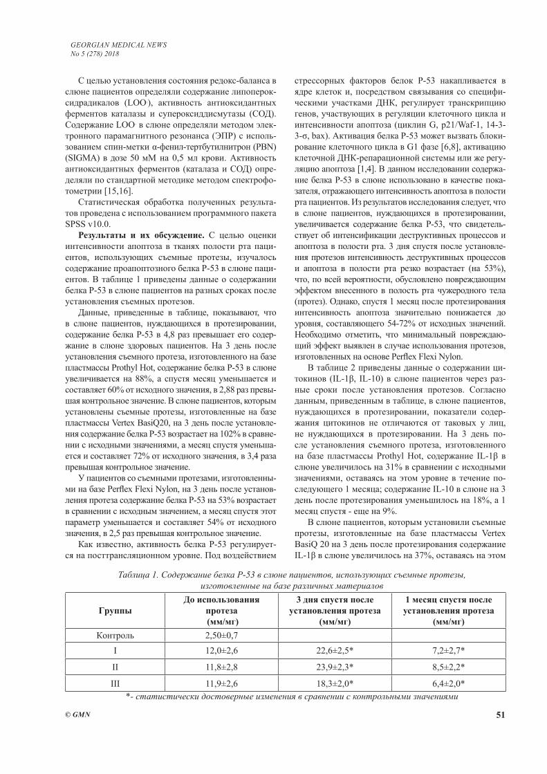
GEORGIAN MEDICAL NEWS No 5 (278) 2018
© GMN 51
С целью установления состояния редокс-баланса в слюне пациентов определяли содержание липоперок-сидрадикалов (LOO.), активность антиоксидантных ферментов каталазы и cупероксиддисмутазы (СОД). Содержание LOO. в слюне определяли методом элек-тронного парамагнитного резонанса (ЭПР) с исполь-зованием спин-метки α-фенил-тертбутилнитрон (PBN) (SIGMA) в дозе 50 мM на 0,5 мл крови. Активность антиоксидантных ферментов (каталаза и СОД) опре-деляли по стандартной методике методом спектрофо-тометрии [15,16].
Статистическая обработка полученных результа-тов проведена с использованием программного пакета SPSS v10.0.
Результаты и их обсуждение. С целью оценки интенсивности апоптоза в тканях полости рта паци-ентов, использующих съемные протезы, изучалось содержание проапоптозного белка P-53 в слюне паци-ентов. В таблице 1 приведены данные о содержании белка Р-53 в слюне пациентов на разных сроках после установления съемных протезов.
Данные, приведенные в таблице, показывают, что в слюне пациентов, нуждающихся в протезировании, содержание белка Р-53 в 4,8 раз превышает его содер-жание в слюне здоровых пациентов. На 3 день после установления съемного протеза, изготовленного на базе пластмассы Prothyl Hot, содержание белка Р-53 в слюне увеличивается на 88%, а спустя месяц уменьшается и составляет 60% от исходного значения, в 2,88 раз превы-шая контрольное значение. В слюне пациентов, которым установлены съемные протезы, изготовленные на базе пластмассы Vertex BasiQ20, на 3 день после установле-ния содержание белка P-53 возрастает на 102% в сравне-нии с исходными значениями, а месяц спустя уменьша-ется и составляет 72% от исходного значения, в 3,4 раза превышая контрольное значение.
У пациентов со съемными протезами, изготовленны-ми на базе Perflex Flexi Nylon, на 3 день после установ-ления протеза содержание белка P-53 на 53% возрастает в сравнении с исходным значением, а месяц спустя этот параметр уменьшается и составляет 54% от исходного значения, в 2,5 раз превышая контрольное значение.
Как известно, активность белка Р-53 регулирует-ся на посттрансляционном уровне. Под воздействием
стрессорных факторов белок P-53 накапливается в ядре клеток и, посредством связывания со специфи-ческими участками ДНК, регулирует транскрипцию генов, участвующих в регуляции клеточного цикла и интенсивности апоптоза (циклин G, p21/Waf-1, 14-3-3-σ, bax). Активация белка Р-53 может вызвать блоки-рование клеточного цикла в G1 фазе [6,8], активацию клеточной ДНК-репарационной системы или же регу-ляцию апоптоза [1,4]. В данном исследовании содержа-ние белка Р-53 в слюне использовано в качестве пока-зателя, отражающего интенсивность апоптоза в полости рта пациентов. Из результатов исследования следует, что в слюне пациентов, нуждающихся в протезировании, увеличивается содержание белка Р-53, что свидетель-ствует об интенсификации деструктивных процессов и апоптоза в полости рта. 3 дня спустя после установле-ния протезов интенсивность деструктивных процессов и апоптоза в полости рта резко возрастает (на 53%), что, по всей вероятности, обусловлено повреждающим эффектом внесенного в полость рта чужеродного тела (протез). Однако, спустя 1 месяц после протезирования интенсивность апоптоза значительно понижается до уровня, составляющего 54-72% от исходных значений. Необходимо отметить, что минимальный повреждаю-щий эффект выявлен в случае использования протезов, изготовленных на основе Perflex Flexi Nylon.
В таблице 2 приведены данные о содержании ци-токинов (IL-1β, IL-10) в слюне пациентов через раз-ные сроки после установления протезов. Согласно данным, приведенным в таблице, в слюне пациентов, нуждающихся в протезировании, показатели содер-жания цитокинов не отличаются от таковых у лиц, не нуждающихся в протезировании. На 3 день по-сле установления съемного протеза, изготовленного на базе пластмассы Prothyl Hot, содержание IL-1β в слюне увеличилось на 31% в сравнении с исходными значениями, оставаясь на этом уровне в течение по-следующего 1 месяца; содержание IL-10 в слюне на 3 день после протезирования уменьшилось на 18%, а 1 месяц спустя - еще на 9%.
В слюне пациентов, которым установили съемные протезы, изготовленные на базе пластмассы Vertex BasiQ 20 на 3 день после протезирования содержание IL-1β в слюне увеличилось на 37%, оставаясь на этом
Таблица 1. Содержание белка P-53 в слюне пациентов, использующих съемные протезы, изготовленные на базе различных материалов
ГруппыДо использования
протеза (мм/мг)
3 дня спустя после установления протеза
(мм/мг)
1 месяц спустя после установления протеза
(мм/мг) Контроль 2,50±0,7
І 12,0±2,6 22,6±2,5* 7,2±2,7*
ІІ 11,8±2,8 23,9±2,3* 8,5±2,2*
ІІІ 11,9±2,6 18,3±2,0* 6,4±2,0**- статистически достоверные изменения в сравнении с контрольными значениями
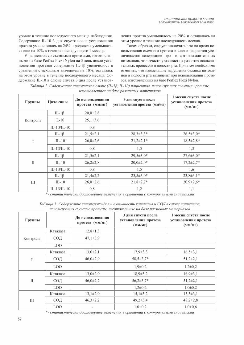
52
МЕДИЦИНСКИЕ НОВОСТИ ГРУЗИИ
CFMFHSDTKJC CFVTLBWBYJ CBF[KTYB
уровне в течение последующего месяца наблюдения. Содержание IL-10 3 дня спустя после установления протеза уменьшилось на 24%, продолжая уменьшать-ся еще на 10% в течение последующего 1 месяца.
У пациентов со съемными протезами, изготовлен-ными на базе Perflex Flexi Nylon на 3 день после уста-новления протезов содержание IL-1β увеличилось в сравнении с исходным значением на 10%, оставаясь на этом уровне в течение последующего месяца. Со-держание IL-10 в слюне спустя 3 дня после установ-
ления протеза уменьшилось на 20% и оставалось на этом уровне в течение последующего месяца.
Таким образом, следует заключить, что во время ис-пользования съемного протеза в слюне пациентов уве-личивается содержание про– и антивоспалительных цитокинов, что отчасти указывает на развитие воспали-тельных процессов в полости рта. При этом необходимо отметить, что наименьшие нарушения баланса цитоки-нов в полости рта выявлены при использовании проте-зов, изготовленных на базе Perflex Flexi Nylon.
Таблица 2. Содержание цитокинов в слюне (IL-1β, IL-10) пациентов, использующих съемные протезы, изготовленные на базе различных материалов
Группы Цитокины До использования протеза (мм/мг)
3 дня спустя после установления протеза (мм/мг)
1 месяц спустя после установления протеза
(мм/мг)
Контроль
IL-1β 20,0±2,8
L-10 25,1±3,6
IL-1β/IL-10 0,8
І
IL-1β 21,5±2,1 28,3±3,3* 26,5±3,0*
IL-10 26,0±2,6 21,2±2,1* 18,5±2,8*
IL-1β/IL-10 0,8 1,5 1,3
ІІ
IL-1β 21,5±2,1 29,5±3,0* 27,6±3,0*
IL-10 26,2±2,8 20,0±2,0* 17,2±2,7*
IL-1β/IL-10 0,8 1,5 1,6
ІІІIL-1β 21,4±2,2 23,5±3,0* 23,8±3,1*IL-10 26,0±2,6 21,8±2,7* 20,9±2,6*
IL-1β/IL-10 0,8 1,2 1,1*- статистически достоверные изменения в сравнении с контрольными значениями
Таблица 3. Содержание липопероксидов и активность каталазы и СОД в слюне пациентов, использующих съемные протезы, изготовленные на базе различных материалов
Группы До использования протеза (мм/мг)
3 дня спустя после установления протеза
(мм/мг)
1 месяц спустя после установления протеза
(мм/мг)
Контроль
Каталаза 12,8±1,8
СОД 47,1±3,9
LOO -
І
Каталаза 13,0±2,1 17,9±3,3 16,5±3,1
СОД 46,0±2,9 58,5±3,7* 51,2±2,1
LOO - 1,9±0,2 1,2±0,2
ІІ
Каталаза 13,0±2,0 18,9±3,2 16,9±3,1
СОД 46,0±2,2 56,2±3,7* 51,2±2,1
LOO - 1,2±0,2 1,0±0,2
ІІІКаталаза 13,1±2,0 15,1±3,2 13,3±3,1
СОД 46,3±2,2 49,2±3,4 48,2±2,8LOO - 1,0±0,2 1,0±0,6
*- статистически достоверные изменения в сравнении с контрольными значениями
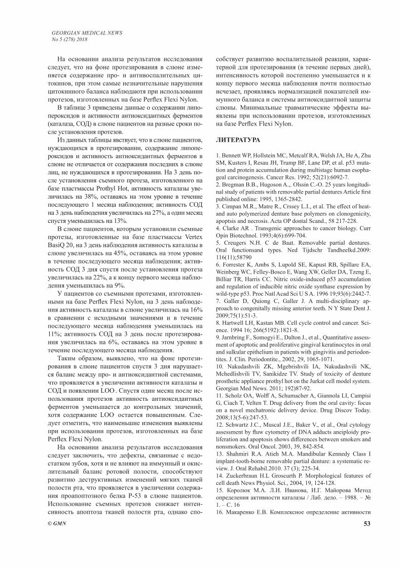
GEORGIAN MEDICAL NEWS No 5 (278) 2018
© GMN 53
На основании анализа результатов исследования следует, что на фоне протезирования в слюне изме-няется содержание про- и антивоспалительных ци-токинов, при этом самые незначительные нарушения цитокинного баланса наблюдаютя при использовании протезов, изготовленных на базе Perflex Flexi Nylon.
В таблице 3 приведены данные о содержании липо-пероксидов и активности антиоксидантных ферментов (каталаза, СОД) в слюне пациентов на разные сроки по-сле установления протезов.
Из данных таблицы явствует, что в слюне пациентов, нуждающихся в протезировании, содержание липопе-роксидов и активность антиоксидантных ферментов в слюне не отличается от содержания последних в слюне лиц, не нуждающихся в протезировании. На 3 день по-сле установления съемного протеза, изготовленного на базе пластмассы Prothyl Hot, активность каталазы уве-личилась на 38%, оставаясь на этом уровне в течение последующего 1 месяца наблюдения; активность СОД на 3 день наблюдения увеличилась на 27%, а один месяц спустя уменьшилась на 13%.
В слюне пациентов, которым установили съемные протезы, изготовленные на базе пластмассы Vertex BasiQ 20, на 3 день наблюдения активность каталазы в слюне увеличилась на 45%, оставаясь на этом уровне в течение последующего месяца наблюдения; актив-ность СОД 3 дня спустя после установления протеза увеличилась на 22%, а к концу первого месяца наблю-дения уменьшилась на 9%.
У пациентов со съемными протезами, изготовлен-ными на базе Perflex Flexi Nylon, на 3 день наблюде-ния активность каталазы в слюне увеличилась на 16% в сравнении с исходными значениями и в течение последующего месяца наблюдения уменьшилась на 11%; активность СОД на 3 день после протезирова-ния увеличилась на 6%, оставаясь на этом уровне в течение последующего месяца наблюдения.
Таким образом, выявлено, что на фоне протези-рования в слюне пациентов спустя 3 дня нарушает-ся баланс между про- и антиоксидантной системами, что проявляется в увеличении активности каталазы и СОД и появлении LOO.. Спустя один месяц после ис-пользования протезов активность антиоксидантных ферментов уменьшается до контрольных значений, хотя содержание LOO. остается повышенным. Сле-дует отметить, что наименьшие изменения выявлены при использовании протезов, изготовленных на базе Perflex Flexi Nylon.
На основании анализа результатов исследования следует заключить, что дефекты, связанные с недо-статком зубов, хотя и не влияют на иммунный и окис-лительный баланс ротовой полости, способствуют развитию деструктивных изменений мягких тканей полости рта, что проявляется в увеличении содержа-ния проапоптозного белка Р-53 в слюне пациентов. Использование съемных протезов снижает интен-сивность апоптоза тканей полости рта, однако спо-
собствует развитию воспалительной реакции, харак-терной для протезирования (в течение первых дней), интенсивность которой постепенно уменьшается и к концу первого месяца наблюдения почти полностью исчезает, проявляясь нормализацией показателей им-мунного баланса и системы антиоксидантной защиты слюны. Минимальные травматические эффекты вы-явлены при использовании протезов, изготовленных на базе Perflex Flexi Nylon.
ЛИТЕРАТУРА
1. Bennett WP, Hollstein MC, Metcalf RA, Welsh JA, He A, Zhu SM, Kusters I, Resau JH, Trump BF, Lane DP, et al. p53 muta-tion and protein accumulation during multistage human esopha-geal carcinogenesis. Cancer Res. 1992; 52(21):6092-7. 2. Bregman B.B., Hugoson A.,. Olssin C.-O. 25 years longitudi-nal study of patients with removable partial dentures Article first published online: 1995, 1365-2842. 3. Cimpan M.R., Matre R., Crssey L.I., et al. The effect of heat- and auto polymerized denture base polymers on clonogenicity, apoptsis and necrosis. Acta OP dontal Scand., 58 217-228. 4. Clarke AR . Transgenic approaches to cancer biology. Curr Opin Biotechnol. 1993;4(6):699-704. 5. Creugers N.H. C de Baat. Removable partial dentures. Oral functionsand types. Ned Tijdschr Tandheelkd.2009: 116(11);58790 6. Forrester K, Ambs S, Lupold SE, Kapust RB, Spillare EA, Weinberg WC, Felley-Bosco E, Wang XW, Geller DA, Tzeng E, Billiar TR, Harris CC. Nitric oxide-induced p53 accumulation and regulation of inducible nitric oxide synthase expression by wild-type p53. Proc Natl Acad Sci U S A. 1996 19;93(6):2442-7. 7. Galler D, Quiong C, Galler J. A multi-disciplinary ap-proach to congenitally missing anterior teeth. N Y State Dent J. 2009;75(1):51-3. 8. Hartwell LH, Kastan MB. Cell cycle control and cancer. Sci-ence. 1994 16; 266(5192):1821-8. 9. Jarnbring F., Somogyi E., Dalton J., et al., Quantitative assess-ment of apoptotic and proliferative gingival keratinocytes in oral and sulkular epithelium in patients with gingivitis and periodon-tites. J. Clin. Periodontite., 2002, 29, 1065-1071. 10. Nakudashvili ZK, Mgebrishvili IA, Nakudashvili NK, Mchedlishvili TV, Sanikidze TV. Study of toxicity of denture prosthetic appliance prothyl hot on the Jurkat cell model system. Georgian Med News. 2011; 192)87-92.11. Scholz OA, Wolff A, Schumacher A, Giannola LI, Campisi G, Ciach T, Velten T. Drug delivery from the oral cavity: focus on a novel mechatronic delivery device. Drug Discov Today. 2008;13(5-6):247-53. 12. Schwartz J.C., Muscal J.E., Baker V., et al., Oral cytology assessment by fluw cytometry of DNA adducts aneiploidy pro-liferation and apoptosis shows differences between smokers and nonsmokers. Oral Oncol. 2003, 39, 842-854. 13. Shahmiri R.A. Atieh M.A. Mandibular Kennedy Class I implant-tooth-borne removable partial denture: a systematic re-view. J. Oral Rehabil.2010. 37 (3); 225-34. 14. Zuckerbraun H.L Groscurth P. Morphological features of cell death News Physiol. Sci., 2004, 19, 124-128. 15. Королюк М.А. Л.И. Иванова, И.Г. Майорова Метод определения активности каталазы / Лаб. дело. – 1988. – № 1. – С. 1616. Макаренко Е.В. Комплексное определение активности
54
МЕДИЦИНСКИЕ НОВОСТИ ГРУЗИИ
CFMFHSDTKJC CFVTLBWBYJ CBF[KTYB
СОД и глутатионредуктазы в эритроцитах у больных с хроническими заболевания печени /Е.В. Макаренко //Лаб. дело.-1988.-№11.-С.48-501, 2.
SUMMARY
A COMPARATIVE EVALUATION OF THE EF-FECT OF DENTURES FROM VARIOUS MATERI-ALS ON THE ORAL CAVITY’S IMMUNOLOGI-CAL AND REDOX-DEPENDENT HOMEOSTASIS
Nakudashvili Z., Mgebrishvili I., Barbakadze I., Sanikidze T.
Tbilisi State Medical University; I. Javakhishvili Tbilisi State University, Georgia
The purpose of our study was a comparative analysis of the effect of dentures from various materials on the immuno-logical and redox-dependent homeostasis of the oral cavity.
We studied 60 patients with removable dentures made based on plastics Prothyl Hot, Vertex BasiQ 20 (differ-ing by polymerization regime) and elastic thermoplastic polymer Perflex Flexi Nylon. The control group consisted of 15 volunteers with a practically healthy oral cavity, who did not use dentures. Saliva collected on an empty stomach in a glass tube without the use of a stimulator before the establishment of a denture and 3 days and 1 month after. The content of the protein P-53 in saliva de-termined by immunoenzymatic assay with use of “Cusa-bio” reagent. The cytokines (IL1β, IL10) content in saliva was determined immunoenzymatic assay. To determine the redox balance in the saliva of patients, the lipoper-oxydradicals content (LOO.) content (by EPR method, using the spin-labeled α-phenyl-tertbutylnitron (PBN) (SIGMA)) and the activity of antioxidant enzymes (cata-lase and SOD) (by spectrophotometry) studied. Statistical processing of the results was carried out using the soft-ware package SPSS (version 10.0).
Results of analysis show that defects associated with a lack of teeth do not affect the immune and oxidative bal-ance of the oral cavity, but contribute to the development of destructive changes in the oral cavity’s soft tissues, which manifested by an increase in the content of the pro-apoptotic protein P-53 in the saliva. After establishment of a denture, the intensity of apoptosis in the oral cav-ity tissues reduced. Establishment of a denture induced development of an inflammatory reaction during the first days, the intensity of which gradually decreased and com-pletely disappeared at the end of the first month of the observation (manifested by the normalization of the pa-rameters of the immune balance and antioxidant system). Minimal traumatic effects observed during establishment of a denture made based on Perflex Flexi Nylon.
Keywords: removable dentures, impact on immune and oxidative balance, soft tissues, oral cavity.
РЕЗЮМЕ
CРАВНИТЕЛЬНАЯ ОЦЕНКА ВЛИЯНИЯ ЗУБ-НЫХ ПРОТЕЗОВ ИЗ РАЗЛИЧНЫХ МАТЕРИА-ЛОВ НА ИММУНОЛОГИЧЕСКИЙ И РЕДОКС–ЗАВИСИМЫЙ ГОМЕОСТАЗ ПОЛОСТИ РТА
Накудашвили З.К., Мгебришвили С.А., Барбакадзе И.Дж., Саникидзе Т.В.
Тбилисский государственный медицинский универси-тет; Тбилисский государственный университет им. И. Джавахишвили, Грузия
Целью исследования явилась сравнительная оцен-ка влияния протезов из различных материалов на им-мунологический и редокс-зависимый гомеостаз поло-сти рта.
Наблюдались 60 пациентов, которым установлены съемные протезы, изготовленные на базе пластмасс Prothyl Hot, Vertex BasiQ 20 (различающихся режимом полимеризации) и на базе эластичного термопластиче-ского полимера Perflex Flexi Nylon. Контрольная группа состояла из 15 добровольцев с практически здоровой полостью рта, которые не употребляли протезов. Слю-ну собирали в стеклянную пробирку без использования стимулятора, натощак, до установления протеза, спустя 3 дня, а затем 1 месяц спустя после установления про-теза. Содержание белка Р-53 в слюне определяли по-средством иммуноферментной тест-системы реактивом „Cusabio“. Содержание цитокинов IL-1β, IL-10 в слюне определяли посредством иммуноферментного метода.С целью установления состояния редокс-баланса в слюне пациентов определяли содержание липопероксидради-калов методом электронного парамагнитного резонанса с использованием спин-метки PBN (SIGMA), актив-ность антиоксидантных ферментов (каталаза и СОД) методом спектрофотометрии.
На основании анализа результатов исследования следует заключить, что дефекты, связанные с недо-статком зубов, хотя и не влияют на иммунный и окис-лительный баланс ротовой полости, способствуют развитию деструктивных изменений мягких тканей полости рта, что проявляется в увеличении содержа-ния проапоптозного белка Р-53 в слюне пациентов. Использование съемных протезов способствует сни-жению интенсивности апоптоза тканей полости рта, однако способствует развитию воспалительной реак-ции, характерной для протезирования (в течение пер-вых дней), интенсивность которой постепенно умень-шается и к концу первого месяца наблюдения почти полностью исчезает, что проявляется в нормализации показателей иммунного баланса и системы антиокси-дантной защиты слюны. Минимальные травматиче-ские эффекты выявлены при использовании протезов, изготовленных на базе Perflex Flexi Nylon.
GEORGIAN MEDICAL NEWS No 5 (278) 2018
© GMN 55
reziume
sxvadasxva masalebisgan damzadebuli kbilT-proTezebis gavlenis SedarebiTi Se-faseba piris Rrus imunologiur da redoqs-damokidebul homeostazze
z. nukudaSvili, s. mRebriSvili, i. barbaqaZe, T. sanikiZe
Tbilisis saxelmwifo samedicino univer-siteti; Tbilisis iv. javaxiSvilis sax. saxe-lmwifo universiteti saqarTvelo
kvlevis mizans Seadgenda sxvadasxva ma-salebisagan damzadebuli proTezebis gav-lenis dadgena piris Rrus imunologiur da redoqs-damokidebul homeostazze.
Seswavlilia 60 pacienti mosaxsneli proTe-zebiT, romlebic damzadebuli iyo sxvadasxva plastmasebis Prothyl Hot da Vertex BasiQ 20 (gansx-vavebuli polimerizaciis reJimiT) da elas-tiuri Termoplastiuri polimeris Perflex Flexi Nylon-is bazaze. sakontrolo jgufSi Sedio-da 15 janmrTeli piris Rrus mqone moxalise, romlbic ar iyenebdnen proTezebs. nerwyvis Segroveba xdeboda uzmoze minis WurWelSi, stimulaciis gareSe, proTezis dayenebamde, dayenebidan 3 dRis da 1 Tvis Semdeg. cilis P-53
Semcveloba nerwyvSi ganisazRvra imunofer-mentuli meTodiT reagentis “Cusabio”–s gamoy-enebiT, xolo nerwyvSi citokinebis (IL-1β, IL-10) Semcveloba – imunofermentuli meTodiT. pacientebis nerwyvSi redoqs-balansis dadge-nis mizniT ganisazRvra lipoperoqsidradi-kalebis Semcveloba epr meTodiT spin-xafan-gis α-fenil-tertbutylnitron (SIGMA) gamoyenebiT da antioqsidanturi fermentebis (katalaza da sod) aqtivoba – speqtrofotometruli meTo-diT. statistikuri damuSaveba ganxorcielda programuli paketiT SPSS versia 10.0.
analizis Sedegad dadginda, rom kbilebis uqonloba ar axdens zemoqmede-bas pacientebis piris Rrus imunur da redoq-balansze, magram xels uwyobs apop-tozis intensifikacias, rac vlindeba ner-wyvSi proapoptozuli cilis P-53 Semcv-elobis matebiT. proTezis dayenebis me-3 dRis Semdeg pacientebis piris RruSi vi-Tardeba anTebiTi procesi, romlis inten-sivoba TandaTan mcirdeba da erTi Tvis Semdeg mTlianad qreba. xdeba imunuri balansis da antioqsidanturi sistemis maCveneblebis normalizacia.
Catarebulma kvlevam gamoavlina Perflex Flexi Nylon-is bazaze damzadebuli proTeze-bis didi upiratesoba.
TACTICS OF THE TREATMENT OF TEETH TRANSPOSITION (CASE REPORTS)
Flis P., Filonenko V., Doroshenko N.
O.O. Bogomolets National Medical University, Kiev, Ukraine
Teeth transposition is a rather rare anomaly of the dento-gnathic apparatus, the prevalence of which, according to the literature, is about 0.2-0.08% [2,3,5,13,14,20,22].
Teeth transposition is the reciprocal exchange of teeth position in the dental arch. There is a complete transposition, which is a complete body displacement of two teeth, and in-complete one – the crowns of the teeth are replaced, but the roots remain their physiological position [1,2,8,9,15,19].
Most often the upper canines are involved in the trans-position process in combination with the first premolars. Sometimes lateral incisors may be involved. One-sided transpositions is more frequent, especially on the left side, which are observed predominantly [1,3,5,8,11,16-18].
The causes of this phenomenon have not been studied sufficiently. Etiological factors are indicated: hereditary factor, transposition of the germs during dentinogenesis, movement of the teeth during eruption, ankylosis of tem-
porary teeth, trauma. Often, the germs of the teeth are dis-placed as a result of insufficient space or due to provoca-tive factors (extra teeth, odontogenic neoplasms etc.). The anomaly is mostly caused by the irregular position of the germs [5,6,8,9,13,14].
According to the WHO classification, transposition is mentioned in the section “Teeth position anomalies”. But according to the clinical and morphological anomalies classification of D. Kalvelis, transposition refers to the dental arch anomalies, but it is often combined with mal-occlusion. Transposed tooth can be placed outside the dental arch, sometimes it is rotated on the axis [1,8,9]. S. Peck and L. Peck classify the transposition as follows: the maxilla canine - the first premolar, the maxilla canine – the lateral incisor, the maxilla canine - the first molar, the lateral incisor - the central incisor, the maxilla canine – the central incisor, the mandible canine - the lateral incisor [17].
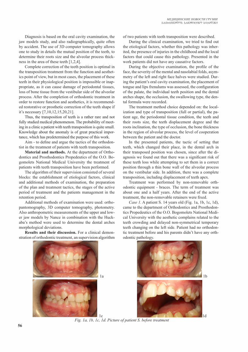
56
МЕДИЦИНСКИЕ НОВОСТИ ГРУЗИИ
CFMFHSDTKJC CFVTLBWBYJ CBF[KTYB
Diagnosis is based on the oral cavity examination, the jaw models study, and also radiographically, quite often by accident. The use of 3D computer tomography allows one to study in details the mutual position of the teeth, to determine their roots size and the alveolar process thick-ness in the area of these teeth [1,2,4].
Complete correction of the teeth position is optimal in the transposition treatment from the function and aesthet-ics point of view, but in most cases, the placement of these teeth in their physiological position is impossible or inap-propriate, as it can cause damage of periodontal tissues, loss of bone tissue from the vestibular side of the alveolar process. After the completion of orthodontic treatment in order to restore function and aesthetics, it is recommend-ed restorative or prosthetic correction of the teeth shape if it’s necessary [7,10,12,15,20,21].
Thus, the transposition of teeth is a rather rare and not fully studied medical phenomenon. The probability of meet-ing in a clinic a patient with teeth transposition is quite small. Knowledge about the anomaly is of great practical impor-tance, which has predetermined the purpose of this work.
Aim - to define and argue the tactics of the orthodon-tist in the treatment of patients with teeth transposition.
Material and methods. At the department of Ortho-dontics and Prosthodontics Propedeutics of the O.O. Bo-gomolets National Medical University the treatment of patients with teeth transposition have been performed.
The algorithm of their supervision consisted of several blocks: the establishment of etiological factors, clinical and additional methods of examination, the preparation of the plan and treatment tactics, the stages of the active period of treatment and the patients management in the retention period.
Additional methods of examination were used: ortho-pantomography, 3D computer tomography, photometry. Also anthropometric measurements of the upper and low-er jaw models by Nance in combination with the Huck-aba’s method were used to determine the dental arches morphological deviations.
Results and their discussion. For a clinical demon-stration of orthodontic treatment, an supervision algorithm
of two patients with teeth transposition were described.During the clinical examination, we tried to find out
the etiological factors, whether this pathology was inher-ited, the presence of injuries in the childhood and the local factors that could cause this pathology. Presented in the work patients did not have any causative factors.
During the objective examination, the profile of the face, the severity of the mental and nasolabial folds, asym-metry of the left and right face halves were studied. Dur-ing the patient’s oral cavity examination, the placement of tongue and lips frenulums was assessed, the configuration of the palate, the individual teeth position and the dental arches shape, the occlusion, the swallowing type, the den-tal formula were recorded.
The treatment method choice depended on: the local-ization and type of transposition (full or partial), the pa-tient age, the periodontal tissue condition, the teeth and their roots size, the teeth displacement degree and the roots inclination, the type of occlusion, the bone thickness in the region of alveolar process, the level of cooperation between the patient and the doctor.
In the presented patients, the tactic of setting that teeth, which changed their place, in the dental arch in their transposed position was chosen, since after the di-agnosis we found out that there was a significant risk of these teeth loss while attempting to set them in a correct position through a thin bone wall of the alveolar process on the vestibular side. In addition, there was a complete transposition, including displacement of teeth apex.
Treatment was performed by non-removable orth-odontic equipment - braces. The term of treatment was about one and a half years. After the end of the active treatment, the non-removable retainers were fixed.
Case 1. A patient S. 14 years old (Fig. 1a, 1b, 1с, 1d), came to the department of Orthodontics and Prosthodon-tics Propedeutics of the O.O. Bogomolets National Medi-cal University with the aesthetic complains related to the teeth crowding and delayed non-symmetrical temporary teeth changing on the left side. Patient had no orthodon-tic treatment before and his parents didn’t have any orth-odontic pathology.
Fig. 1a, 1b, 1с, 1d. Picture of patient S. before treatment
1a 1b
1c 1d
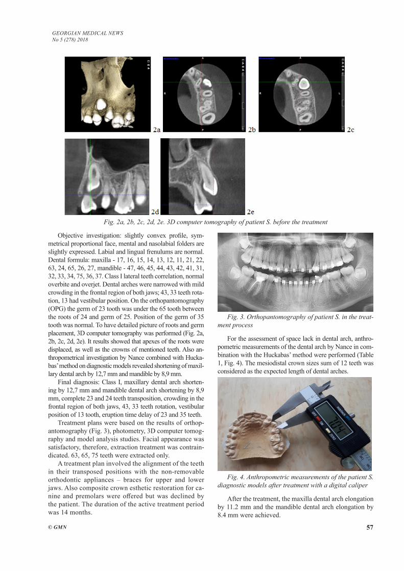
GEORGIAN MEDICAL NEWS No 5 (278) 2018
© GMN 57
Objective investigation: slightly convex profile, sym-metrical proportional face, mental and nasolabial folders are slightly expressed. Labial and lingual frenulums are normal. Dental formula: maxilla - 17, 16, 15, 14, 13, 12, 11, 21, 22, 63, 24, 65, 26, 27, mandible - 47, 46, 45, 44, 43, 42, 41, 31, 32, 33, 34, 75, 36, 37. Class I lateral teeth correlation, normal overbite and overjet. Dental arches were narrowed with mild crowding in the frontal region of both jaws; 43, 33 teeth rota-tion, 13 had vestibular position. On the orthopantomography (OPG) the germ of 23 tooth was under the 65 tooth between the roots of 24 and germ of 25. Position of the germ of 35 tooth was normal. To have detailed picture of roots and germ placement, 3D computer tomography was performed (Fig. 2a, 2b, 2c, 2d, 2e). It results showed that apexes of the roots were displaced, as well as the crowns of mentioned teeth. Also an-thropometrical investigation by Nance combined with Hucka-bas’ method on diagnostic models revealed shortening of maxil-lary dental arch by 12,7 mm and mandible by 8,9 mm.
Final diagnosis: Class I, maxillary dental arch shorten-ing by 12,7 mm and mandible dental arch shortening by 8,9 mm, complete 23 and 24 teeth transposition, crowding in the frontal region of both jaws, 43, 33 teeth rotation, vestibular position of 13 tooth, eruption time delay of 23 and 35 teeth.
Treatment plans were based on the results of orthop-antomography (Fig. 3), photometry, 3D computer tomog-raphy and model analysis studies. Facial appearance was satisfactory, therefore, extraction treatment was contrain-dicated. 63, 65, 75 teeth were extracted only.
A treatment plan involved the alignment of the teeth in their transposed positions with the non-removable orthodontic appliances – braces for upper and lower jaws. Also composite crown esthetic restoration for ca-nine and premolars were offered but was declined by the patient. The duration of the active treatment period was 14 months.
Fig. 3. Orthopantomography of patient S. in the treat-ment process
For the assessment of space lack in dental arch, anthro-pometric measurements of the dental arch by Nance in com-bination with the Huckabas’ method were performed (Table 1, Fig. 4). The mesiodistal crown sizes sum of 12 teeth was considered as the expected length of dental arches.
Fig. 4. Anthropometric measurements of the patient S. diagnostic models after treatment with a digital caliper
After the treatment, the maxilla dental arch elongation by 11.2 mm and the mandible dental arch elongation by 8.4 mm were achieved.
Fig. 2a, 2b, 2c, 2d, 2e. 3D computer tomography of patient S. before the treatment

58
МЕДИЦИНСКИЕ НОВОСТИ ГРУЗИИ
CFMFHSDTKJC CFVTLBWBYJ CBF[KTYB
After treatment, patient S. has positive morphological, functional and aesthetic changes. (Fig. 5a, 5b, 5c, 5d).
Case 2. Patient V., 11 years old (Fig. 6a, 6b, 6с, 6d), came for the consultation to the department of Orthodon-
tics and Prosthodontics Propedeutics of the O.O. Bogo-molets National Medical University with the complains concerning the assumption of an extra tooth. Patient was getting treatment of Class III in anamnesis.
Table 1. The study results of patient S. diagnostic models, investigated by using the method of Nance in combination with the Huckabas’ method before and after treatment
Sum of mesiodistal sizes of crowns of 12
teeth (expected length of dental arches), mm
Actual length of dental arches
Lack of space in dental arches
Difference, mmBefore treat-
ment, mmAfter treat-ment, mm
Before treatment,
mm
After treat-ment, mm
Maxilla 90,2 77,5 88,7 12,7 1,5 11,2
Mandible 84,6 75,7 84,1 8,9 0,5 8,4
5a 5b
5c 5dFig. 5a, 5b, 5с, 5d. Pictures of the patient S. after the treatment.
The canine is in the place of the first premolar, and the first premolar in the place of the canine
6a 6b
6c 6d
Fig. 6a, 6b, 6с, 6d. Pictures of the patient V. before the treatment
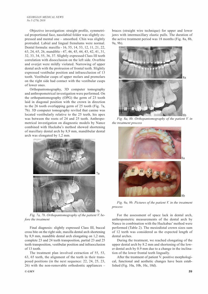
GEORGIAN MEDICAL NEWS No 5 (278) 2018
© GMN 59
Objective investigation: straight profile, symmetri-cal proportional face, nasolabial folder was slightly ex-pressed and mental one – smoothed. Chin was slightly protruded. Labial and lingual frenulums were normal. Dental formula: maxilla - 16, 55, 14, 53, 12, 11, 21, 22, 63, 24, 65, 26, mandible - 47, 46, 45, 44, 43, 42, 41, 31, 32, 33, 34, 55, 36, 37. Slightly expressed Class III teeth correlation with disocclusion on the left side. Overbite and overjet were mildly violated. Narrowing of upper dental arch with the protrusion of frontal teeth. Slightly expressed vestibular position and infraocclusion of 13 tooth. Vestibular cusps of upper molars and premolars on the right side had contact with the vestibular cusps of lower ones.
Orthopantomography, 3D computer tomography and anthropometrical investigation were performed. On the orthopantomography (OPG) the germ of 23 tooth laid in diagonal position with the crown in direction to the 26 tooth overlapping germ of 25 tooth (Fig. 7a, 7b). 3D computer tomography reviled that canine was located vestibularly relative to the 25 teeth, his apex was between the roots of 24 and 25 teeth. Anthropo-metrical investigation on diagnostic models by Nance combined with Huckaba’s method showed shortening of maxillary dental arch by 8,9 mm, mandibular dental arch was elongated by 1,2 mm.
Fig. 7a, 7b. Orthopantomography of the patient V. be-fore the treatment
Final diagnosis: slightly expressed Class III, buccal cross bite on the right side, maxilla dental arch shortening by 8,9 mm, mandible dental arch elongating on 1,2 mm, complete 23 and 24 teeth transposition, partial 23 and 25 teeth transposition, vestibular position and infraocclusion of 13 tooth.
The treatment plan involved extraction of 55, 53, 63, 65 teeth, the alignment of the teeth in their trans-posed positions (in the next sequence: 22, 24, 25, 23, 26) with the non-removable orthodontic appliances –
braces (straight wire technique) for upper and lower jaws with intermaxillary elastic pulls. The duration of the active treatment period was 18 months (Fig. 8a, 8b, 9a, 9b).
Fig. 8a, 8b. Orthopantomography of the patient V. in the treatment process
Fig. 9a, 9b. Pictures of the patient V. in the treatment process
For the assessment of space lack in dental arch, anthropometric measurements of the dental arch by Nance in combination with the Huckabas’ method were performed (Table 2). The mesiodistal crown sizes sum of 12 teeth was considered as the expected length of dental arches.
During the treatment, we reached elongating of the upper dental arch by 8.2 mm and shortening of the low-er dental arch by 0.9 mm due to a change in the inclina-tion of the lower frontal teeth lingually.
After the treatment of patient V. positive morphologi-cal, functional and aesthetic changes have been estab-lished (Fig. 10a, 10b, 10c, 10d).
7a
7b
8a
8b
8a
8b
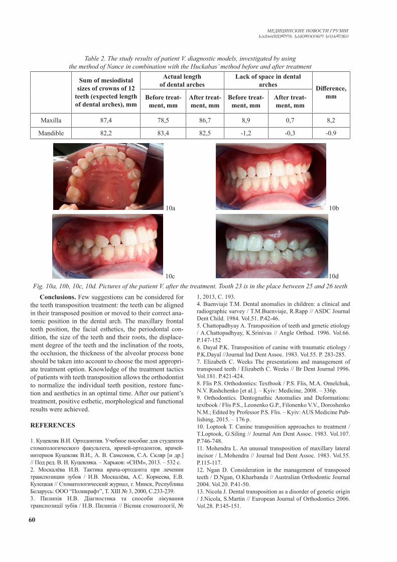
60
МЕДИЦИНСКИЕ НОВОСТИ ГРУЗИИ
CFMFHSDTKJC CFVTLBWBYJ CBF[KTYB
Conclusions. Few suggestions can be considered for the teeth transposition treatment: the teeth can be aligned in their transposed position or moved to their correct ana-tomic position in the dental arch. The maxillary frontal teeth position, the facial esthetics, the periodontal con-dition, the size of the teeth and their roots, the displace-ment degree of the teeth and the inclination of the roots, the occlusion, the thickness of the alveolar process bone should be taken into account to choose the most appropri-ate treatment option. Knowledge of the treatment tactics of patients with teeth transposition allows the orthodontist to normalize the individual teeth position, restore func-tion and aesthetics in an optimal time. After our patient’s treatment, positive esthetic, morphological and functional results were achieved.
REFERENCES
1. Куцевляк В.И. Ортодонтия. Учебное пособие для студентов стоматологического факультета, врачей-ортодонтов, врачей-интернов Куцевляк В.И., А. В. Самсонов, С.А. Скляр [и др.] // Под ред. B. И. Куцевляка. – Харьков: «СИМ», 2013. – 532 с.2. Москалёва И.В. Тактика врача-ортодонта при лечении транспозиции зубов / И.В. Москалёва, А.С. Корнеева, Е.В. Кулецкая // Стоматологический журнал, г. Минск, Республика Беларусь: ООО “Поликрафт”, Т. XIII № 3, 2000, С.233-239. 3. Пилипів Н.В. Діагностика та способи лікування транспозиції зубів / Н.В. Пилипів // Вісник стоматології, №
1, 2013, С. 193.4. Buenviaje T.M. Dental anomalies in children: a clinical and radiographic survey / T.M.Buenviaje, R.Rapp // ASDC Journal Dent Child. 1984. Vol.51. Р.42-46.5. Chattopadhyay A. Transposition of teeth and genetic etiology / A.Chattopadhyay, K.Srinivas // Angle Orthod. 1996. Vol.66. Р.147-1526. Dayal P.K. Transposition of canine with traumatic etiology / P.K.Dayal //Journal Ind Dent Assoc. 1983. Vol.55. Р. 283-285. 7. Elizabeth C. Weeks The presentations and management of transposed teeth / Elizabeth C. Weeks // Br Dent Journal 1996. Vol.181. P.421-424.8. Flis P.S. Orthodontics: Textbook / P.S. Flis, M.A. Omelchuk, N.V. Rashchenko [et al.]. – Kyiv: Medicine, 2008. – 336p.9. Orthodontics. Dentognathic Anomalies and Deformations: textbook / Flis P.S., Leonenko G.P., Filonenko V.V., Doroshenko N.M.; Edited by Professor P.S. Flis. – Kyiv: AUS Medicine Pub-lishing, 2015. – 176 p.10. Loptook T. Canine transposition approaches to treatment / T.Loptook, G.Siling // Journal Am Dent Assoc. 1983. Vol.107. P.746-748.11. Mohendra L. An unusual transposition of maxillary lateral incisor / L.Mohendra // Journal Ind Dent Assoc. 1983. Vol.55. P.115-117. 12. Ngan D. Consideration in the management of transposed teeth / D.Ngan, O.Kharbanda // Australian Orthodontic Journal 2004. Vol.20. P.41-50.13. Nicola J. Dental transposition as a disorder of genetic origin / J.Nicola, S.Martin // European Journal of Orthodontics 2006. Vol.28. P.145-151.
Table 2. The study results of patient V. diagnostic models, investigated by using the method of Nance in combination with the Huckabas’ method before and after treatment
Sum of mesiodistal sizes of crowns of 12
teeth (expected length of dental arches), mm
Actual length of dental arches
Lack of space in dental arches Difference,
mmBefore treat-ment, mm
After treat-ment, mm
Before treat-ment, mm
After treat-ment, mm
Maxilla 87,4 78,5 86,7 8,9 0,7 8,2
Mandible 82,2 83,4 82,5 -1,2 -0,3 -0.9
10a 10b
10c 10dFig. 10a, 10b, 10с, 10d. Pictures of the patient V. after the treatment. Tooth 23 is in the place between 25 and 26 teeth
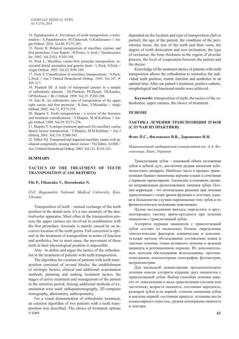
GEORGIAN MEDICAL NEWS No 5 (278) 2018
© GMN 61
14. Papadopoulos A. Prevalence of tooth transposition, a meta-analisis / A.Papadopoulos, M.Chatzoudi, G.Kaklamanos // An-gle Orthod. 2010. Vol.80. P.275-285.15. Peretz B. Bilateral transposition of maxillary canines and first premolars: Case Report / B.Peretz, A.Arad // Quintessence Int. 1992. Vol.23(5). Р.345-348. 16. Peck L. Maxillary canine-first premolar transposition, as-sociated dental anomalies and genetic basis / L.Peck, S.Peck //Angle Orthod. 1993. Vol.63. Р.99-109.17. Peck S. Classification of maxillary transpositions / S.Peck, L.Peck // Am J Orthod Dentofacial Orthop. 1995. Vol.107. P. 505-517.18. Plunkett DJ. A study of transposed canines in a sample of orthodontic patients / DJ.Plunkett, PS.Dysart, TB.Kardos, GP.Herbison // Br J Orthod. 1998. Vol.25. Р.203-208.19. Sato K. An orthodontic case of transposition of the upper right canine and first premolar / K.Sato, Y.Masahiko // Angle Orthod. 2002. Vol.72. P.275-278.20. Shapira Y. Tooth transpositions - a review of the literature and treatment considerations / Y.Shapira, M.М.Kuftinec // An-gle Orthod. 1989. Vol.59. Р.271-276.21. Shapira Y. A unique treatment approach for maxillary canine lateral incisor transposition / Y.Shapira, M.М.Kuftinec // Am J Orthod. 2001. Vol.119. Р.540-545. 22. Talbot TQ. Transposed and impacted maxillary canine with ip-silateral congenitally missing lateral incisor / TQ.Talbot, AJ.Hill // Am J Orthod Dentofacial Orthop. 2002. Vol.121. Р.316-323.
SUMMARY
TACTICS OF THE TREATMENT OF TEETH TRANSPOSITION (CASE REPORTS)
Flis P., Filonenko V., Doroshenko N.
O.O. Bogomolets National Medical University, Kiev, Ukraine
Transposition of teeth – mutual exchange of the teeth position in the dental arch, it’s a rare anomaly of the den-toalveolar apparatus. Most often in the transposition pro-cess the upper canines are involved in combination with the first premolars. Anomaly is mainly caused by an in-correct location of the teeth germs. Full correction is opti-mal in the treatment of transposition in terms of function and aesthetics, but in most cases, the movement of these teeth in their physiological position is impossible.
Aim - to define and argue the tactics of the orthodon-tist in the treatment of patients with teeth transposition.
The algorithm for curation of patients with teeth trans-position consisted of several blocks: the establishment of etiologic factors, clinical and additional examination methods, planning and making treatment tactics, the stages of active treatment and management of the patient in the retention period. Among additional methods of ex-amination were used: orthopantomography, 3D computer tomography, photometry, anthropometry.
For a visual demonstration of orthodontic treatment, an curation algorithm of two patients with a teeth trans-position was described. The choice of treatment options
depended on the location and type of transposition (full or partial), the age of the patient, the condition of the peri-odontal tissue, the size of the teeth and their roots, the degree of tooth dislocation and root inclination, the type of occlusion, the bone thickness in the region of alveolar process, the level of cooperation between the patient and the doctor.
Knowledge of the treatment tactics of patients with teeth transposition allows the orthodontist to normalize the indi-vidual teeth position, restore function and aesthetics in an optimal time. After our patient’s treatment, positive esthetic, morphological and functional results were achieved.
Keywords: transposition of teeth, the tactics of the or-thodontist, upper canines, the choice of treatment.
РЕЗЮМЕ
ТАКТИКА ЛЕЧЕНИЯ ТРАНСПОЗИЦИИ ЗУБОВ (СЛУЧАИ ИЗ ПРАКТИКИ)
Флис П.С., Филоненко В.В., Дорошенко Н.Н.
Национальный медицинский университет им. А.А. Бо-гомольца, Киев, Украина
Транспозиция зубов – взаимный обмен положения зубов в зубной дуге, достаточно редкая аномалия зубо-челюстного аппарата. Наиболее часто в процесс транс-позиции бывают вовлечены верхние клыки в сочетании с первыми премолярами. Аномалия, в основном, вызва-на неправильным расположением зачатков зубов. Пол-ная коррекция - это оптимальное решение при лечении транспозиции с точки зрения функции и эстетики, одна-ко в большинстве случаев перемещение этих зубов в их физиологическое положение невозможно.
Целью исследования явилось определить и аргу-ментировать тактику врача-ортодонта при лечении пациентов с транспозицией зубов.
Алгоритм курации пациентов с транспозицией зубов состоял из нескольких блоков: определение этиологических факторов, клинические и дополни-тельные методы обследования, составление плана и тактики лечения, этапы активного лечения и ведения пациента в ретенционном периоде. Из дополнитель-ных методов обследования использованы: ортопан-томограмма, компьютерная томография, фотометрия, антропометрия.
Для наглядной демонстрации ортодонтического лечения описан алгоритм курации двух пациентов с транспозицией зубов. Выбор способов лечения зави-сел от локализации и вида транспозиции (полная или частичная), возраста пациента, состояния пародонта, размеров зубов и их корней, степени смещения зубов и наклона корней, состояния прикуса, толщины кости альвеолярного отростка, уровня кооперации пациента и доктора.
62
МЕДИЦИНСКИЕ НОВОСТИ ГРУЗИИ
CFMFHSDTKJC CFVTLBWBYJ CBF[KTYB
Определение адекватной тактики лечения пациен-тов с транспозицией зубов позволяет врачу-ортодонту в короткие сроки нормализовать положение отдельных зубов, восстановить функцию и эстетику. В результате лечения пациентов достигнуты положительные эстетиче-ские, морфологические и функциональные результаты.
reziume
kbilebis transpoziciis mkurnalobis taq-tika (klinikuri SemTxvevebi)
p. flis, v. filonenko, n. doroSenko
a. bogomolcis sax. erovnuli samedicino universiteti, kievi, ukraina
kbilebis transpozicia - kbilTa rkal-Si kbilebis mdgomareobis urTierT gacvla, yba-kbilTa aparatis sakmaod iSviaTi anoma-liaa. yvelaze ufro xSirad transpozici-is procesSi Careulia zeda eSvebi pirvel premolarebTan erTad. anomalia ZiriTa-dad gamowveulia kbilebis Canasaxebis ara-swori ganlagebiT. sruli koreqcia - es aris funqciis da esTetikis TvalsazrisiT optimaluri gadawyvetileba transpozi-ciis mkurnalobis dros, umetes SemTx-vevaSi, amgvari kbilebis gadaadgileba maT fiziologiur mdgomareobaSi SeuZlebelia.
kvlevis mizans Seadgenda kbilebis transpoziciis mqone pacientebis mkurnalo-
bis dros eqim-orTodontis taqtikis gansaz-Rvra da argumentireba.
kbilebis transpoziciis mqone pacien-tebis kuraciis algoriTmi Sesdgeboda ram-denime blokisgan: etiologiuri faqtorebis gansazRvra, kvlevis klinikuri da damatebi-Ti meTodebi, mkurnalobis gegmis da taqti-kis Sedgena, aqtiuri mkurnalobis da reten-ciur periodSi pacientis gaZRolis etapebi. kvlevis damatebiT meTodebidan gamoyenebu-lia orTopanTomograma, kompiuteruli to-mografia, fotometria, antropometria.
orTodontuli mkurnalobis TvalsaCino demonstraciis mizniT aRwerilia kbilebis transpoziciis mqone ori pacientis kura-ciis algoriTmi. mkurnalobis meTodis Ser-Ceva damokidebulia ramdenime faqtorze: transpoziciis lokalizacia da tipi (sru-li an nawilobrivi), pacientis asaki, parodon-tis mdgomareoba, kbilebis da maTi fesvebis zoma, kbilebis gadaadgilebis da fesvebis daxrilobis xarisxi, Tankbilvis mdgomareo-ba, alveoluri morCis Zvlis sisqe, pacientis da eqimis kooperaciis done.
transpoziciis mqone pacientebis mkurnalobis taqtikis codna aZlevs eqim - orTodonts saSualebas umokles vadaSi moaxdinos calkeuli kbilebis mdgomareo-bis normalizeba, aRadginos funqcia da esTe-tika. Cveni pacientebis mkurnalobis Sedegad miRweulia dadebiTi esTetikuri, morfolo-giuri da funqciuri Sedegebi.
CALCIUM HYDROXYLAPATITE MICROSPHERES – BIOCOMPATIBILITY AND CLINICAL EFFECTS
1Wollina U., 2Wiegand C., 2Hipler U-C.
1Department of Dermatology and Allergology, Städtisches Klinikum Dresden, Academic Teaching Hospital, Dresden; 2Department of Dermatology, Jena University Hospital, Germany
In esthetic medicine as well as in other medical fields the use of soft tissue fillers has been widely utilized. Fillers can be divided into permanent fillers and biodegradable fillers. Hyaluronic acid is the most frequently used biode-gradable filler nowadays [1]. Among such there are some products known as biostimulatory fillers based on poly-lactic acid or calcium hydroxylapatite (CaHA). Radiesse® is a versatile, biodegradable, biostimulatory CaHA soft tissue filler used in esthetic medicine for correction of deeper folds and wrinkles. CaHA microspheres of 25 to 45µm are sus-pended in a matrix composed of glycerin, methylcellulose
gel, and sterile water. After injection of CaHA an increased expression of collagen I, collagen III, increased elastin and improved angiogenesis have been noted. These factors are thought to contribute to improvements in skin pliability and elasticity and dermal thickness [2,3].
Supra-periostal placement of the filler produces the best lifting effect for midface rejuvenation [4,5]. CaHA is 9.5 times more viscous than carboxymethyl cellulose. Heating temporarily decreased the viscosity of CaHA by 32%. Heating and shearing together can temporarily re-duce the viscosity of CaHA by 52% [6].
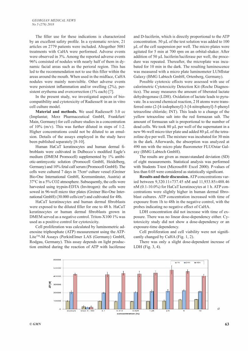
GEORGIAN MEDICAL NEWS No 5 (278) 2018
© GMN 63
The filler use for these indications is characterized by an excellent safety profile. In a systematic review, 21 articles on 2779 patients were included. Altogether 5081 treatments with CaHA were performed. Adverse events were observed in 3%. Among the reported adverse events 96% consisted of nodules with nearly half of them in dy-namic facial areas such as the perioral region. This has led to the recommendation not to use this filler within the areas around the mouth. When used in the midface, CaHA nodules were mainly nonvisible. Other adverse events were persistent inflammation and/or swelling (2%), per-sistent erythema and overcorrection (1% each) [7].
In the present study, we investigated aspects of bio-compatibility and cytotoxicity of Radiesse® in an in vitro cell culture model.
Material and methods. We used Radiesse® 3.0 cc (Implantat; Merz Pharmaceutical GmbH, Frankfurt/ Main, Germany) for cell culture studies in a concentration of 10% (m/v). This was further diluted in steps of 1:2. Higher concentrations could not be diluted to an emul-sion. Details of the assays employed in the study have been published separately [8-10].
Human HaCaT keratinocytes and human dermal fi-broblasts were cultivated in Dulbecco´s modified Eagle’s medium (DMEM Promocell) supplemented by 1% antibi-otic-antimycotic solution (Promocell GmbH, Heidelberg, Germany) and 10% fetal calf serum (Promocell GmbH). The cells were cultured 7 days in 75cm² culture vessel (Greiner Bio-One International GmbH, Kremsmünster, Austria) at 37°C in a 5% CO2 atmosphere. Subsequently, the cells were harvested using trypsin-EDTA (Invitrogen): the cells were sowed in 96-well micro titer plates (Greiner Bio-One Inter-national GmbH) (30.000 cells/cm²) and cultivated for 48h.
HaCaT keratinocytes and human dermal fibroblasts were exposed to the diluted filler for one to 48 h. HaCaT keratinocytes or human dermal fibroblasts grown in DMEM served as a negative control. Triton-X100 1% was used as a positive control for cytotoxicity.
Cell proliferation was calculated by luminometric ad-enosine triphosphate (ATP) measurement using the ATP-LiteTM-M Assays (PerkinElmer LAS (Germany) GmbH, Rodgau, Germany). This assay depends on light produc-tion emitted during the reaction of ATP with luciferase
and D-luciferin, which is directly proportional to the ATP concentration. 50 µL of the test solution was added to 100 µL of the cell suspension per well. The micro plates were agitated for 5 min at 700 rpm on an orbital-shaker. After addition of 50 µL luciferin/luciferase per well, the proce-dure was repeated. Thereafter, the microplate was incu-bated for 10 min in the dark. The resulting luminescence was measured with a micro plate luminometer LUMIstar Galaxy (BMG Labtech GmbH, Ortenberg, Germany).
Possible cytotoxic effects were assessed with use of calorimetric Cytotoxicity Detection Kit (Roche Diagnos-tics). The assay measures the amount of liberated lactate dehydrogenase (LDH). Oxidation of lactate leads to pyru-vate. In a second chemical reaction, 2 H atoms were trans-ferred onto (2-[4-iodophenyl]-3-[4-nitrophenyl]-5-phenyl tetrazoline chloride; INT). This leads to a change of the yellow tetrazoline salt into the red formazan salt. The amount of formazan salt is proportional to the number of dead cells. We took 80 µL per well of the supernatant in a new 96-well micro titer plate and added 80 µL of the tetra-zoline dye per well. The mixture was incubated for 30 min in the dark. Afterwards, the absorption was analyzed at 490 nm with the micro plate fluorometer FLUOstar Gal-axy (BMG Labtech GmbH).
The results are given as mean±standard deviation (SD) of eight measurements. Statistical analysis was performed with Students T-test (Microsoft® Excel 2000). P-values of less than 0.05 were considered as statistically significant.
Results and their discussion. ATP concentrations var-ied between 9,320.11±737.45 nM and 11,933.85±488.46 nM (0.1-10.0%) for HaCaT keratinocytes at 1 h. ATP con-centrations were slightly higher in human dermal fibro-blast cultures. ATP concentration increased with time of exposure from 1h to 48h in the negative control, with the probes indicating no negative effect of CaHA.
LDH concentration did not increase with time of ex-posure. There was no linear dose-dependency either. Cy-totoxicity study did not show a dose-dependency or an exposure-time dependency.
Cell proliferation and cell viability were not signifi-cantly changed by CaHA (Fig. 1, 2).
There was only a slight dose-dependent increase of LDH (Fig. 3, 4).
a
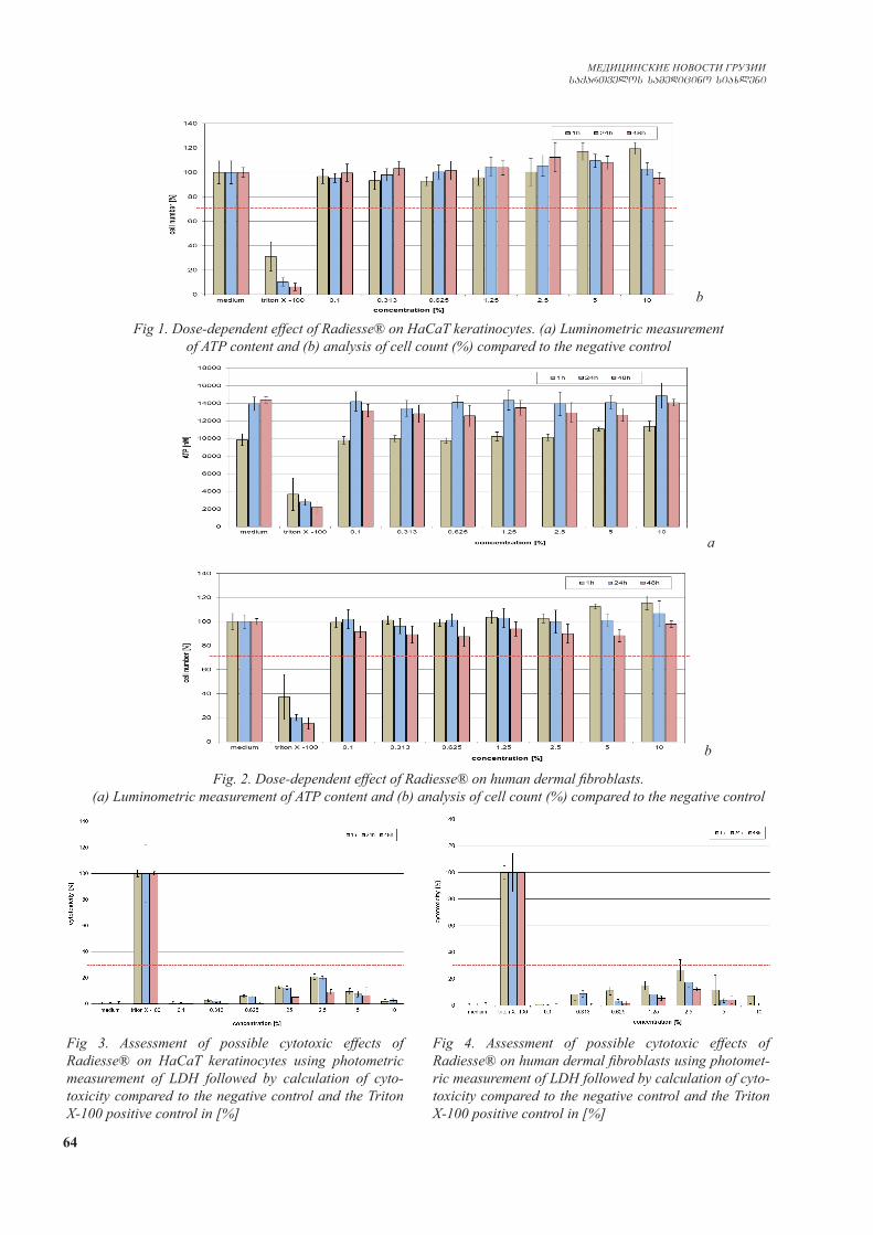
64
МЕДИЦИНСКИЕ НОВОСТИ ГРУЗИИ
CFMFHSDTKJC CFVTLBWBYJ CBF[KTYB
Fig 1. Dose-dependent effect of Radiesse® on HaCaT keratinocytes. (a) Luminometric measurement of ATP content and (b) analysis of cell count (%) compared to the negative control
Fig. 2. Dose-dependent effect of Radiesse® on human dermal fibroblasts. (a) Luminometric measurement of ATP content and (b) analysis of cell count (%) compared to the negative control
Fig 3. Assessment of possible cytotoxic effects of Radiesse® on HaCaT keratinocytes using photometric measurement of LDH followed by calculation of cyto-toxicity compared to the negative control and the Triton X-100 positive control in [%]
Fig 4. Assessment of possible cytotoxic effects of Radiesse® on human dermal fibroblasts using photomet-ric measurement of LDH followed by calculation of cyto-toxicity compared to the negative control and the Triton X-100 positive control in [%]
b
a
b
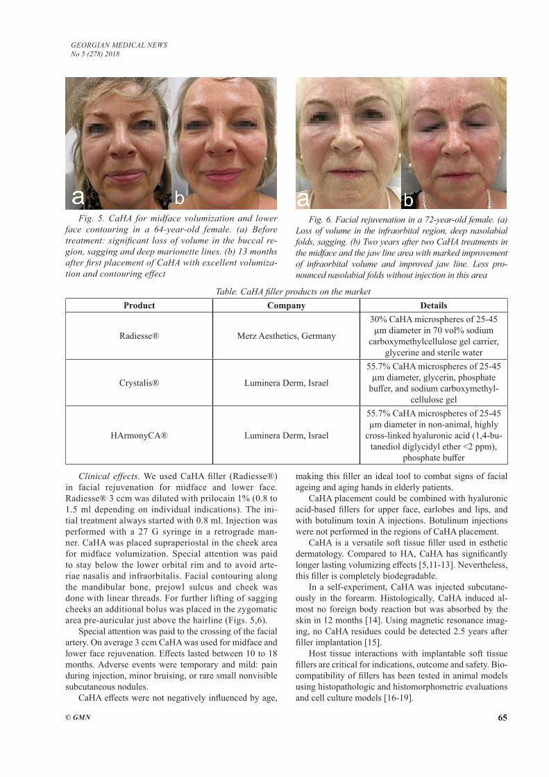
GEORGIAN MEDICAL NEWS No 5 (278) 2018
© GMN 65
Fig. 5. CaHA for midface volumization and lower face contouring in a 64-year-old female. (a) Before treatment: significant loss of volume in the buccal re-gion, sagging and deep marionette lines. (b) 13 months after first placement of CaHA with excellent volumiza-tion and contouring effect
Fig. 6. Facial rejuvenation in a 72-year-old female. (a) Loss of volume in the infraorbital region, deep nasolabial folds, sagging. (b) Two years after two CaHA treatments in the midface and the jaw line area with marked improvement of infraorbital volume and improved jaw line. Less pro-nounced nasolabial folds without injection in this area
Table. CaHA filler products on the marketProduct Company Details
Radiesse® Merz Aesthetics, Germany
30% CaHA microspheres of 25-45 µm diameter in 70 vol% sodium
carboxymethylcellulose gel carrier, glycerine and sterile water
Crystalis® Luminera Derm, Israel
55.7% CaHA microspheres of 25-45 µm diameter, glycerin, phosphate
buffer, and sodium carboxymethyl-cellulose gel
HArmonyCA® Luminera Derm, Israel
55.7% CaHA microspheres of 25-45 µm diameter in non-animal, highly
cross-linked hyaluronic acid (1,4-bu-tanediol diglycidyl ether <2 ppm),
phosphate buffer
Clinical effects. We used CaHA filler (Radiesse®) in facial rejuvenation for midface and lower face. Radiesse® 3 ccm was diluted with prilocain 1% (0.8 to 1.5 ml depending on individual indications). The ini-tial treatment always started with 0.8 ml. Injection was performed with a 27 G syringe in a retrograde man-ner. CaHA was placed supraperiostal in the cheek area for midface volumization. Special attention was paid to stay below the lower orbital rim and to avoid arte-riae nasalis and infraorbitalis. Facial contouring along the mandibular bone, prejowl sulcus and cheek was done with linear threads. For further lifting of sagging cheeks an additional bolus was placed in the zygomatic area pre-auricular just above the hairline (Figs. 5,6).
Special attention was paid to the crossing of the facial artery. On average 3 ccm CaHA was used for midface and lower face rejuvenation. Effects lasted between 10 to 18 months. Adverse events were temporary and mild: pain during injection, minor bruising, or rare small nonvisible subcutaneous nodules.
CaHA effects were not negatively influenced by age,
making this filler an ideal tool to combat signs of facial ageing and aging hands in elderly patients.
CaHA placement could be combined with hyaluronic acid-based fillers for upper face, earlobes and lips, and with botulinum toxin A injections. Botulinum injections were not performed in the regions of CaHA placement.
CaHA is a versatile soft tissue filler used in esthetic dermatology. Compared to HA, CaHA has significantly longer lasting volumizing effects [5,11-13]. Nevertheless, this filler is completely biodegradable.
In a self-experiment, CaHA was injected subcutane-ously in the forearm. Histologically, CaHA induced al-most no foreign body reaction but was absorbed by the skin in 12 months [14]. Using magnetic resonance imag-ing, no CaHA residues could be detected 2.5 years after filler implantation [15].
Host tissue interactions with implantable soft tissue fillers are critical for indications, outcome and safety. Bio-compatibility of fillers has been tested in animal models using histopathologic and histomorphometric evaluations and cell culture models [16-19].
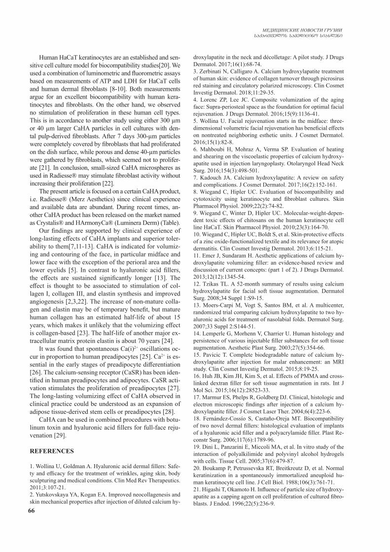
66
МЕДИЦИНСКИЕ НОВОСТИ ГРУЗИИ
CFMFHSDTKJC CFVTLBWBYJ CBF[KTYB
Human HaCaT keratinocytes are an established and sen-sitive cell culture model for biocompatibility studies[20]. We used a combination of luminometric and fluorometric assays based on measurements of ATP and LDH for HaCaT cells and human dermal fibroblasts [8-10]. Both measurements argue for an excellent biocompatibility with human kera-tinocytes and fibroblasts. On the other hand, we observed no stimulation of proliferation in these human cell types. This is in accordance to another study using either 300 µm or 40 µm larger CaHA particles in cell cultures with den-tal pulp-derived fibroblasts. After 7 days 300-µm particles were completely covered by fibroblasts that had proliferated on the dish surface, while porous and dense 40-µm particles were gathered by fibroblasts, which seemed not to prolifer-ate [21]. In conclusion, small-sized CaHA microspheres as used in Radiesse® may stimulate fibroblast activity without increasing their proliferation [22].
The present article is focused on a certain CaHA product, i.e. Radiesse® (Merz Aesthetics) since clinical experience and available data are abundant. During recent times, an-other CaHA product has been released on the market named as Crystalis® and HArmonyCa® (Luminera Derm) (Table).
Our findings are supported by clinical experience of long-lasting effects of CaHA implants and superior toler-ability to them[7,11-13]. CaHA is indicated for volumiz-ing and contouring of the face, in particular midface and lower face with the exception of the perioral area and the lower eyelids [5]. In contrast to hyaluronic acid fillers, the effects are sustained significantly longer [13]. The effect is thought to be associated to stimulation of col-lagen I, collagen III, and elastin synthesis and improved angiogenesis [2,3,22]. The increase of non-mature colla-gen and elastin may be of temporary benefit, but mature human collagen has an estimated half-life of about 15 years, which makes it unlikely that the volumizing effect is collagen-based [23]. The half-life of another major ex-tracellular matrix protein elastin is about 70 years [24].
It was found that spontaneous Ca(i)2+ oscillations oc-cur in proportion to human preadipocytes [25]. Ca2+ is es-sential in the early stages of preadipocyte differentiation [26]. The calcium-sensing receptor (CaSR) has been iden-tified in human preadipocytes and adipocytes. CaSR acti-vation stimulates the proliferation of preadipocytes [27]. The long-lasting volumizing effect of CaHA observed in clinical practice could be understood as an expansion of adipose tissue-derived stem cells or preadipocytes [28].
CaHA can be used in combined procedures with botu-linum toxin and hyaluronic acid fillers for full-face reju-venation [29].
REFERENCES
1. Wollina U, Goldman A. Hyaluronic acid dermal fillers: Safe-ty and efficacy for the treatment of wrinkles, aging skin, body sculpturing and medical conditions. Clin Med Rev Therapeutics. 2011;3:107-21.2. Yutskovskaya YA, Kogan EA. Improved neocollagenesis and skin mechanical properties after injection of diluted calcium hy-
droxylapatite in the neck and decolletage: A pilot study. J Drugs Dermatol. 2017;16(1):68-74.3. Zerbinati N, Calligaro A. Calcium hydroxylapatite treatment of human skin: evidence of collagen turnover through picrosirus red staining and circulatory polarized microscopy. Clin Cosmet Investig Dermatol. 2018;11:29-35.4. Lorenc ZP, Lee JC. Composite volumization of the aging face: Supra-periosteal space as the foundation for optimal facial rejuvenation. J Drugs Dermatol. 2016;15(9):1136-41.5. Wollina U. Facial rejuvenation starts in the midface: three-dimensional volumetric facial rejuvenation has beneficial effects on nontreated neighboring esthetic units. J Cosmet Dermatol. 2016;15(1):82-8.6. Mahboubi H, Mohraz A, Verma SP. Evaluation of heating and shearing on the viscoelastic properties of calcium hydroxy-apatite used in injection laryngoplasty. Otolaryngol Head Neck Surg. 2016;154(3):498-501.7. Kadouch JA. Calcium hydroxylapatite: A review on safety and complications. J Cosmet Dermatol. 2017;16(2):152-161.8. Wiegand C, Hipler UC. Evaluation of biocompatibility and cytotoxicity using keratinocyte and fibroblast cultures. Skin Pharmacol Physiol. 2009;22(2):74-82.9. Wiegand C, Winter D, Hipler UC. Molecular-weight-depen-dent toxic effects of chitosans on the human keratinocyte cell line HaCaT. Skin Pharmacol Physiol. 2010;23(3):164-70.10. Wiegand C, Hipler UC, Boldt S, et al. Skin-protective effects of a zinc oxide-functionalized textile and its relevance for atopic dermatitis. Clin Cosmet Investig Dermatol. 2013;6:115-21.11. Emer J, Sundaram H. Aesthetic applications of calcium hy-droxylapatite volumizing filler: an evidence-based review and discussion of current concepts: (part 1 of 2). J Drugs Dermatol. 2013;12(12):1345-54.12. Tzikas TL. A 52-month summary of results using calcium hydroxylapatite for facial soft tissue augmentation. Dermatol Surg. 2008;34 Suppl 1:S9-15.13. Moers-Carpi M, Vogt S, Santos BM, et al. A multicenter, randomized trial comparing calcium hydroxylapatite to two hy-aluronic acids for treatment of nasolabial folds. Dermatol Surg. 2007;33 Suppl 2:S144-51.14. Lemperle G, Morhenn V, Charrier U. Human histology and persistence of various injectable filler substances for soft tissue augmentation. Aesthetic Plast Surg. 2003;27(5):354-66.15. Pavicic T. Complete biodegradable nature of calcium hy-droxylapatite after injection for malar enhancement: an MRI study. Clin Cosmet Investig Dermatol. 2015;8:19-25.16. Huh JB, Kim JH, Kim S, et al. Effects of PMMA and cross-linked dextran filler for soft tissue augmentation in rats. Int J Mol Sci. 2015;16(12):28523-33. 17. Marmur ES, Phelps R, Goldberg DJ. Clinical, histologic and electron microscopic findings after injection of a calcium hy-droxylapatite filler. J Cosmet Laser Ther. 2004;6(4):223-6.18. Fernández-Cossío S, Castaño-Oreja MT. Biocompatibility of two novel dermal fillers: histological evaluation of implants of a hyaluronic acid filler and a polyacrylamide filler. Plast Re-constr Surg. 2006;117(6):1789-96.19. Dini L, Panzarini E, Miccoli MA, et al. In vitro study of the interaction of polyalkilimide and polyvinyl alcohol hydrogels with cells. Tissue Cell. 2005;37(6):479-87. 20. Boukamp P, Petrussevska RT, Breitkreutz D, et al. Normal keratinization in a spontaneously immortalized aneuploid hu-man keratinocyte cell line. J Cell Biol. 1988;106(3):761-71.21. Higashi T, Okamoto H. Influence of particle size of hydroxy-apatite as a capping agent on cell proliferation of cultured fibro-blasts. J Endod. 1996;22(5):236-9.
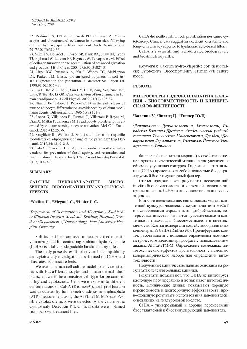
GEORGIAN MEDICAL NEWS No 5 (278) 2018
© GMN 67
22. Zerbinati N, D’Este E, Parodi PC, Calligaro A. Micro-scopic and ultrastructural evidences in human skin following calcium hydroxylapatite filler treatment. Arch Dermatol Res. 2017;309(5):389-96.23. Verzijl N, DeGroot J, Thorpe SR, Bank RA, Shaw JN, Lyons TJ, Bijlsma JW, Lafeber FP, Baynes JW, TeKoppele JM. Effect of collagen turnover on the accumulation of advanced glycation end products. J Biol Chem. 2000;275(50):39027-31.24. Urry DW, Pattanaik A, Xu J, Woods TC, McPherson DT, Parker TM. Elastic protein-based polymers in soft tis-sue augmentation and generation. J Biomater Sci Polym Ed. 1998;9(10):1015-48.25. Hu H, He ML, Tao R, Sun HY, Hu R, Zang WJ, Yuan BX, Lau CP, Tse HF, Li GR. Characterization of ion channels in hu-man preadipocytes. J Cell Physiol. 2009;218(2):427-35.26. Ntambi JM, Takova T. Role of Ca2+ in the early stages of murine adipocyte differentiation as evidenced by calcium mobi-lizing agents. Differentiation. 1996;60(3):151-8.27. Rocha G, Villalobos E, Fuentes C, Villarroel P, Reyes M, Díaz X, Mattar P, Cifuentes M. Preadipocyte proliferation is el-evated by calcium sensing receptor activation. Mol Cell Endo-crinol. 2015;412:251-6. 28. Kruglikov IL, Wollina U. Soft tissue fillers as non-specific modulators of adipogenesis: change of the paradigm? Exp Der-matol. 2015;24(12):912-5.29. Fabi S, Pavicic T, Braz A, et al. Combined aesthetic inter-ventions for prevention of facial ageing, and restoration and beautification of face and body. Clin Cosmet Investig Dermatol. 2017;10:423-9.
SUMMARY
CALCIUM HYDROXYLAPATITE MICRO-SPHERES – BIOCOMPATIBILITY AND CLINICAL EFFECTS
1Wollina U., 2Wiegand C., 2Hipler U-C.
1Department of Dermatology and Allergology, Städtisch-es Klinikum Dresden, Academic Teaching Hospital, Dres-den; 2Department of Dermatology, Jena University Hos-pital, Germany
Soft tissue fillers are used in aesthetic medicine for volumizing and for contouring. Calcium hydroxylapatite (CaHA) is a fully biodegradable biostimulatory filler.
The study presents results of in vitro biocompatibility and cytotoxicity investigations performed on CaHA and illustrates its clinical effects.
We used a human cell culture model for in vitro stud-ies with HaCaT keratinocytes and human dermal fibro-blasts, known to be a sensitive cell type for biocompat-ibility and cytotoxicity. Cells were exposed to different concentrations of CaHA (Radiesse®). Cell proliferation was calculated by luminometric adenosine triphosphate (ATP) measurement using the ATPLiteTM-M Assay. Pos-sible cytotoxic effects were detected by the calorimetric Cytotoxicity Detection Kit. Clinical data were obtained from our own treatment files.
CaHA did neither inhibit cell proliferation nor cause cy-totoxicity. Clinical data suggest an excellent tolerability and long-term efficacy superior to hyaluronic acid-based fillers.
CaHA is a versatile and well-tolerated biodegradable and biostimulatory filler.
Keywords: Calcium hydroxylapatite; Soft tissue fill-ers; Cytotoxicity; Biocompatibility; Human cell culture model.
РЕЗЮМЕ
МИКРОСФЕРЫ ГИДРОКСИЛАПАТИТА КАЛЬ-ЦИЯ - БИОСОВМЕСТИМОСТЬ И КЛИНИЧЕ-СКАЯ ЭФФЕКТИВНОСТЬ
1Воллина У., 2Виганд Ц., 2Гиплер Ю-Ц. 1Департамент Дерматологии и Аллергологии, Го-родская Больница Дрездена, Академический учебный госпиталь Технического Университета, Дрезден; 2Де-партамент Дерматологии, Госпиталь Йенского Уни-верситета, Германия
Филлеры (заполнители морщин) мягкой ткани ис-пользуются в эстетической медицине для увеличения объема и улучшения контуров. Гидроксилапатит каль-ция (CaHA) представляет собой полностью биодегра-дируемый биостимуляторный филлер.
Статья предоставляет результаты исследований in-vitro биосовместимости и клеточной токсичности, проведенных на CaHA, и описывает его клинические эффекты.
В in-vitro исследованиях использована модель кле-точной культуры человека с кератиноцитами HaCaT и человеческими дермальными фибробластами, ко-торые, как известно, являются чувствительными кле-точными типами для биосовместимости и цитоток-сичности. Клетки подвергали воздействию различных концентраций CaHA (Radiesse®). Пролиферацию кле-ток рассчитывали с помощью определения люмино-метрического аденозинтрифосфата с использованием анализа ATPLiteTM-M. Определение возможных ци-тотоксических эффектов производилось с помощью калориметрического набора для определения цито-токсичности.
Полученные клинические данные основаны на ре-зультатах лечения больных клиники.
Результаты показывают, что CaHA не ингибирует клеточную пролиферацию и не вызывает цитотоксич-ность. Клинические данные показывают хорошую переносимость и долгосрочную эффективность, пре-восходящую результаты использования заполнителей, основанных на гиалуроновой кислоте.
CaHA - универсальный и хорошо переносимый биоразлагаемый и биостимулирующий заполнитель.
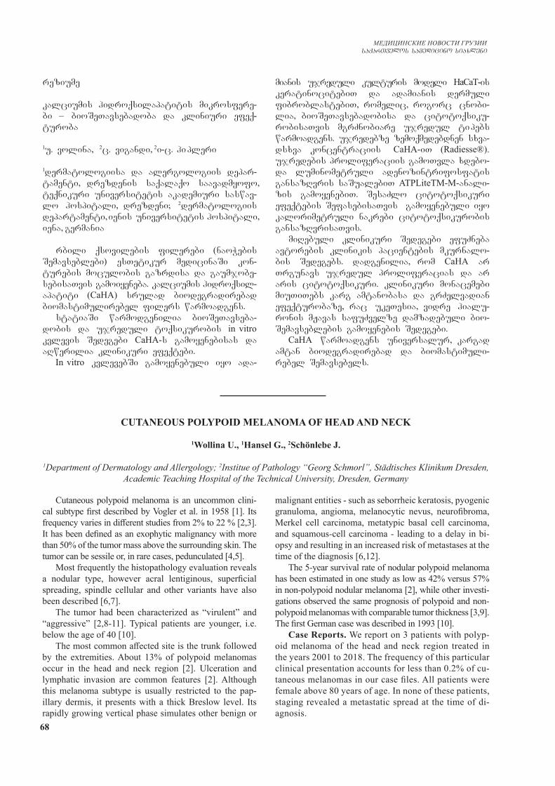
68
МЕДИЦИНСКИЕ НОВОСТИ ГРУЗИИ
CFMFHSDTKJC CFVTLBWBYJ CBF[KTYB
reziume
kalciumis hidroqsilapatitis mikrosfere-bi – bioSeTavsebadoba da kliniuri efeq-turoba
1u. volina, 2c. vigandi, 2i-c. hipleri
1dermatologiisa da alergologiis depar-tamenti, drezdenis saqalaqo saavadmyofo, teqnikuri universitetis akademiuri saswav-lo hospitali, drezdeni; 2dermatologiis departamenti, ienis universitetis hospitali, iena, germania
rbili qsovilebis filerebi (naoWebis Semavseblebi) esTetikur medicinaSi kon-turebis moculobis gazrdisa da gaumjobe-sebisaTvis gamoiyeneba. kalciumis hidroqsil-apatiti (CaHA) srulad biodegradirebad biomastimulirebel filers warmoadgens.
statiaSi warmodgenilia bioSeTavseba-dobis da ujreduli toqsikurobis in vitro kvlevis Sedegebi CaHA-s gamoyenebisas da aRwerilia klinikuri efeqtebi.
In vitro kvlevebSi gamoyenebuli iyo ada-
mianis ujreduli kulturis modeli HaCaT-is keratinocitebiT da adamianis dermuli fibroblastebiT, romelic, rogorc cnobi-lia, bioSeTavsebadobisa da citotoqsiku-robisaTvis mgrZnobiare ujredul tipebs warmoadgens. ujredebze zemoqmedebdnen sxva-dsxva koncentraciis CaHA-iT (Radiesse®). ujredebis proliferaciis gamoTvla xdebo-da luminometruli adenozintrifosfatis gansazRvris saSualebiT ATPLiteTM-M-anali-zis gamoyenebiT. SesaZlo citotoqsikuri efeqtebis SefasebisaTvis gamoyenebuli iyo kalorimetruli nakrebi citotoqsikurobis gansazRvrisaTvis.
miRebuli klinikuri Sedegebi efuZneba avtorebis klinikis pacientebis mkurnalo-bis Sedegebs. dadgenilia, rom CaHA ar Trgunavs ujredul proliferacias da ar aris citotoqsikuri. klinikuri monacemebi miuTiTebs karg amtanobasa da grZelvadian efeqturobaze, rac ukeTesia, vidre hialu-ronis mJavas safuZvelze damzadebuli bio-Semavseblebis gamoyenebis Sedegebi.
CaHA warmoadgens universalur, kargad amtan biodegradirebad da biomastimuli-rebel Semavsebels.
CUTANEOUS POLYPOID MELANOMA OF HEAD AND NECK
1Wollina U., 1Hansel G., 2Schönlebe J.
1Department of Dermatology and Allergology; 2Institue of Pathology “Georg Schmorl”, Städtisches Klinikum Dresden, Academic Teaching Hospital of the Technical University, Dresden, Germany
Cutaneous polypoid melanoma is an uncommon clini-cal subtype first described by Vogler et al. in 1958 [1]. Its frequency varies in different studies from 2% to 22 % [2,3]. It has been defined as an exophytic malignancy with more than 50% of the tumor mass above the surrounding skin. The tumor can be sessile or, in rare cases, pedunculated [4,5].
Most frequently the histopathology evaluation reveals a nodular type, however acral lentiginous, superficial spreading, spindle cellular and other variants have also been described [6,7].
The tumor had been characterized as “virulent” and “aggressive” [2,8-11]. Typical patients are younger, i.e. below the age of 40 [10].
The most common affected site is the trunk followed by the extremities. About 13% of polypoid melanomas occur in the head and neck region [2]. Ulceration and lymphatic invasion are common features [2]. Although this melanoma subtype is usually restricted to the pap-illary dermis, it presents with a thick Breslow level. Its rapidly growing vertical phase simulates other benign or
malignant entities - such as seborrheic keratosis, pyogenic granuloma, angioma, melanocytic nevus, neurofibroma, Merkel cell carcinoma, metatypic basal cell carcinoma, and squamous-cell carcinoma - leading to a delay in bi-opsy and resulting in an increased risk of metastases at the time of the diagnosis [6,12].
The 5-year survival rate of nodular polypoid melanoma has been estimated in one study as low as 42% versus 57% in non-polypoid nodular melanoma [2], while other investi-gations observed the same prognosis of polypoid and non-polypoid melanomas with comparable tumor thickness [3,9]. The first German case was described in 1993 [10].
Case Reports. We report on 3 patients with polyp-oid melanoma of the head and neck region treated in the years 2001 to 2018. The frequency of this particular clinical presentation accounts for less than 0.2% of cu-taneous melanomas in our case files. All patients were female above 80 years of age. In none of these patients, staging revealed a metastatic spread at the time of di-agnosis.
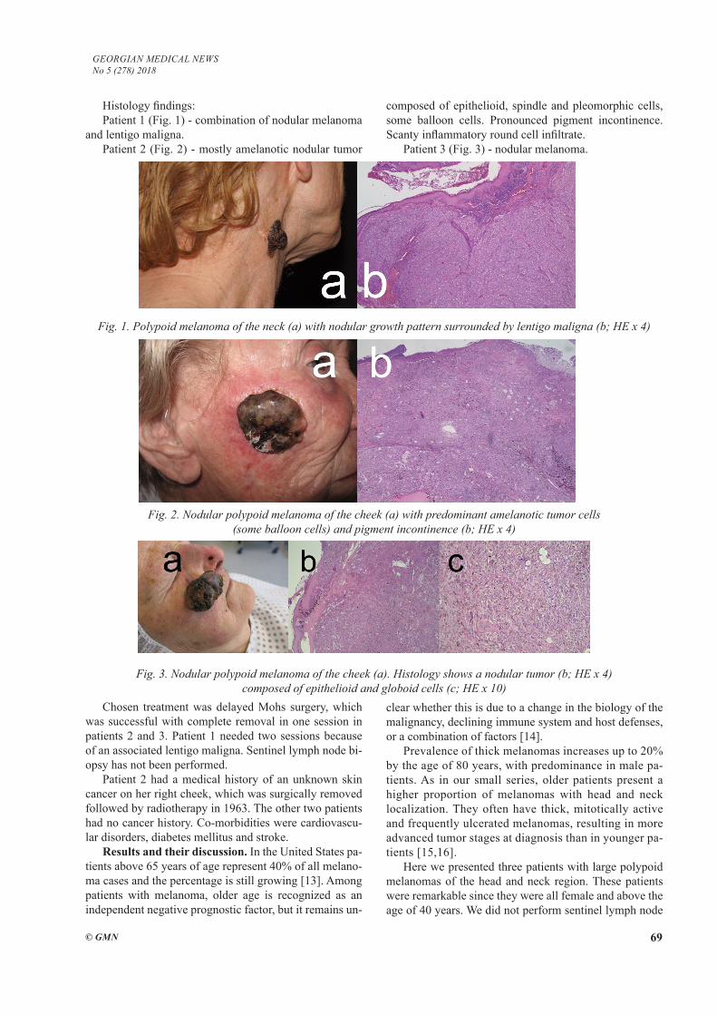
GEORGIAN MEDICAL NEWS No 5 (278) 2018
© GMN 69
Histology findings:Patient 1 (Fig. 1) - combination of nodular melanoma
and lentigo maligna. Patient 2 (Fig. 2) - mostly amelanotic nodular tumor
composed of epithelioid, spindle and pleomorphic cells, some balloon cells. Pronounced pigment incontinence. Scanty inflammatory round cell infiltrate.
Patient 3 (Fig. 3) - nodular melanoma.
Fig. 1. Polypoid melanoma of the neck (a) with nodular growth pattern surrounded by lentigo maligna (b; HE x 4)
Fig. 2. Nodular polypoid melanoma of the cheek (a) with predominant amelanotic tumor cells (some balloon cells) and pigment incontinence (b; HE x 4)
Fig. 3. Nodular polypoid melanoma of the cheek (a). Histology shows a nodular tumor (b; HE x 4) composed of epithelioid and globoid cells (c; HE x 10)
Chosen treatment was delayed Mohs surgery, which was successful with complete removal in one session in patients 2 and 3. Patient 1 needed two sessions because of an associated lentigo maligna. Sentinel lymph node bi-opsy has not been performed.
Patient 2 had a medical history of an unknown skin cancer on her right cheek, which was surgically removed followed by radiotherapy in 1963. The other two patients had no cancer history. Co-morbidities were cardiovascu-lar disorders, diabetes mellitus and stroke.
Results and their discussion. In the United States pa-tients above 65 years of age represent 40% of all melano-ma cases and the percentage is still growing [13]. Among patients with melanoma, older age is recognized as an independent negative prognostic factor, but it remains un-
clear whether this is due to a change in the biology of the malignancy, declining immune system and host defenses, or a combination of factors [14].
Prevalence of thick melanomas increases up to 20% by the age of 80 years, with predominance in male pa-tients. As in our small series, older patients present a higher proportion of melanomas with head and neck localization. They often have thick, mitotically active and frequently ulcerated melanomas, resulting in more advanced tumor stages at diagnosis than in younger pa-tients [15,16].
Here we presented three patients with large polypoid melanomas of the head and neck region. These patients were remarkable since they were all female and above the age of 40 years. We did not perform sentinel lymph node
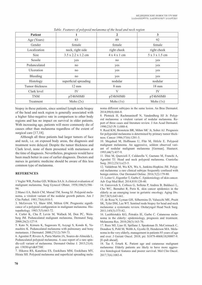
70
МЕДИЦИНСКИЕ НОВОСТИ ГРУЗИИ
CFMFHSDTKJC CFVTLBWBYJ CBF[KTYB
biopsy in these patients, since sentinel lymph node biopsy of the head and neck region is generally associated with a higher false-negative rate in comparison to other body regions and has no impact on survival in older patients. With increasing age, patients will more commonly die of causes other than melanoma regardless of the extent of surgical care [17,18].
Although all three patients had larger tumors of face and neck, i.e. on exposed body areas, the diagnosis and treatment were delayed. Despite the tumor thickness and Clark level, none of them presented with metastases at the time of diagnosis. Nevertheless, prognosis would have been much better in case of earlier diagnosis. Doctors and nurses in geriatric medicine should be aware of this less common type of melanoma.
REFERENCES
1 Vogler WR, Perdue GD, Wilkins SA Jr. A clinical evaluation of malignant melanoma. Surg Gynecol Obstet. 1958;106(5):586–94. 2.Manci EA, Balch CM, Murad TM, Soong SJ. Polypoid mela-noma, a virulent variant of the nodular growth pattern. Am J Clin Pathol. 1981;75(6):810-5.3. McGovern VJ, Shaw HM, Milton GW. Prognostic signifi-cance of a polypoid configuration in malignant melanoma. His-topathology. 1983;7(5):663-72.4. Cutler K, Chu P, Levin M, Wallack M, Don PC, Wein-berg JM. Pedunculated malignant melanoma. Dermatol Surg. 2000;26(2):127-9.5. Kato N, Kimura K, Sugawara H, Aoyagi S, Kondo K, Ya-mashiro K. Pedunculated melanoma with pulmonary and bony metastases. J Dermatol. 2000;27(12):769-73.6. Agostini P, Rivero A, Parra Martin JA, Soares-de-Almeida L. Pedunculated polypoid melanoma. A case report of a rare spin-dle-cell variant of melanoma. Dermatol Online J. 2015;21(4). pii: 13030/qt1dk8750f.7. Hikawa RS, Kanehisa ES, Enokihara MM, Enokihara MY, Hirata SH. Polypoid melanoma and superficial spreading mela-
noma different subtypes in the same lesion. An Bras Dermatol. 2014;89(4):666-8.8. Plotnick H, Rachmaninoff N, VandenBerg HJ Jr. Polyp-oid melanoma: a virulent variant of nodular melanoma. Re-port of three cases and literature review. J Am Acad Dermatol. 1990;23(5 Pt 1):880-4.9. Reed KM, Bronstein BR, Mihm MC Jr, Sober AJ. Prognosis for polypoidal melanoma is determined by primary tumor thick-ness. Cancer 1986;57(6):1201-3. 10. Megahed M, Hoffmann U, Flür M, Hölzle E. Polypoid malignant melanoma. An aggressive, seldom observed vari-ant of nodular malignant melanoma [German]. Hautarzt. 1993;44(7):437-9. 11. Dini M, Quercioli F, Caldarella V, Gaetano M, Franchi A, Agostini TJ. Head and neck polypoid melanoma. Craniofac Surg. 2012;23(1):e23-5.12. Valdebran M, Wu KN, Wu A, Junkins-Hopkins JM. Polyp-oid melanoma: a rare clinical subtype frequently confused with benign entities. Our Dermatol Online. 2016;7(2):179-80.13. Leiter U, Eigentler T, Garbe C. Epidemiology of skin cancer. Adv Exp Med Biol. 2014;810:120-40.14. Garcovich S, Colloca G, Sollena P, Andrea B, Balducci L, Cho WC, Bernabei R, Peris K. skin cancer epidemics in the elderly as an emerging issue in geriatric oncology. Aging Dis. 2017;8(5):643-661.15. de Rosa N, Lyman GH, Silbermins D, Valsecchi ME, Pruitt SK, Tyler DM, Lee WT. Sentinel node biopsy for head and neck melanoma: a systematic review. Otolaryngol Head Neck Surg. 2011;145(3):375-82.16. Lasithiotakis KG, Petrakis IE, Garbe C. Cutaneous mela-noma in the elderly: epidemiology, prognosis and treatment. Melanoma Res. 2010;20(3):163-70.17. Rees MJ, Liao H, Spillane J, Speakman D, McCormack C, Donahoe S, Pohl M, Webb A, Gyorki D, Henderson MA. Mela-noma in the very elderly, management in patients 85 years of age and over. J Geriatr Oncol. 2018; pii: S1879-4068(18)30007-9. [E-pub ahead].18. Tas F, Erturk K. Patient age and cutaneous malignant melanoma: Elderly patients are likely to have more aggres-sive histological features and poorer survival. Mol Clin Oncol. 2017;7(6):1083-8.
Table. Features of polypoid melanoma of the head and neck regionPatient 1 2 3
Age (Years) 83 89 92Gender female female female
Localization neck, right side right cheek right cheekSize 3.5 x 2.2 x 1.2 cm 4 x 4 x 1 cm 5 x 3 x 1.5 cm
Sessile yes no yesPedunculated no yes yes
Ulceration no yes yes
Bleeding no yes yesHistology superficial spreading nodular nodular
Tumor thickness 12 mm 8 mm 18 mmClark level IV V IV
TNM pT4bN0M0 pT4bN0M0 pT4bN0M0Treatment Mohs (2x) Mohs (1x) Mohs (1x)
GEORGIAN MEDICAL NEWS No 5 (278) 2018
© GMN 71
SUMMARY
CUTANEOUS POLYPOID MELANOMA OF HEAD AND NECK
1Wollina U., 1Hansel G., 2Schönlebe J.
1Department of Dermatology and Allergology; 2Institue of Pathology “Georg Schmorl”, Städtisches Klinikum Dres-den, Academic Teaching Hospital of the Technical Uni-versity, Dresden, Germany
Cutaneous polypoid melanoma is an uncommon subtype of malignant melanoma. The clinical behavior of this sub-type has been described as aggressive. Tumors can be sessile or pedunculated. The typical patient is a younger adult.
Authors have analyzed the case histories of patients with polypoid melanomas of the head and neck region treated at our clinic from 2001 to 2018.
We identified 3 female patients with tumors of the neck and cheek. In contrast to other reports, all patients were older than 80-years of age. None of them had a metastatic spread at the time of diagnosis. Treatment was delayed Mohs surgery.
Polypoid melanomas of the head and neck region can occur in elderly patients without an aggressive course. In those patients a number of differential diagnoses have to be considered including Merkel cell carcinoma, metatypi-cal basal cell carcinoma, squamous cell carcinoma and tumor metastases.
Keywords: melanoma, mohs surgery, head and neck region, polypoid growth pattern.
РЕЗЮМЕ
ПОЛИПОИДНАЯ МЕЛАНОМА КОЖИ ОБЛА-СТИ ГОЛОВЫ И ШЕИ
1Воллина У., 1Гансел Г., 2Шенлебе Дж.
1Департамент Дерматологии и Аллергологии, Го-родская Больница Дрездена, Академический учебный госпиталь Технического Университета, Дрезден, Гер-мания; 2Институт Патологии „Георг Шморл“, Го-родская Больница Дрездена, Академический учебный госпиталь Технического Университета, Дрезден, Гер-мания
Кожная полипоидная меланома является необыч-ным подтипом злокачественной меланомы. Клиниче-ское поведение данного подтипа описано как агрес-сивное. Опухоли могут быть плоскими или на ножке. Типичные пациенты - это взрослые люди в молодом возрасте.
Авторами представлены 3 клинических случая па-
циентов женского пола, лечившихся в нашей клинике с 2001 по 2018 гн. по поводу полипоидной меланомы кожи области головы и шеи.
В отличие от других сообщений, все пациенты были старше 80 лет. К моменту ни в одном случае ди-агностирования метастатического распространения не отмечалось. Лечение было проведено операцией МОХС.
Полипоидная меланома области головы и шеи мо-жет возникать у пожилых пациентов без агрессивно-го течения. Среди таких пациентов необходимо учи-тывать ряд дифференциальных диагнозов, включая карциномы клеток Меркель, метатипическую базаль-но-клеточную карциному, плоскоклеточный рак и ме-тастазирование опухолей.
reziume
Tavis da kisris midamos kanis polipoiduri melanoma
1u. volina, 1g. ganseli, 2 j. Sionlebe
1dermatologiisa da alergologiis depar-tamenti, drezdenis saqalaqo saavadmyofo, teqnikuri universitetis akademiuri sas-wavlo hospitali, drezdeni; 2paTologiis instituti “georg Smorli”, drezdenis saqa-laqo saavadmyofo, teqnikuri universitetis akademiuri saswavlo hospitali, drezdeni, germania
kanis polipoiduri melanoma avTvisebi-ani melanomis uCveulo qvetips warmoadgens. am qvetipis klinikuri “qceva” agresiulia. simsivne SesaZloa iyos brtyelic da fexzec. tipobrivi pacientebi axalgazrda adamian-ebia. avtorebis mier warmodgenilia 3 klini-kuri SemTxveva pacienti-qalebisa, romlebic 2001-2018 ww. avtorebis klinikaSi gadiodnen Tavis da kisris midamos kanis polipoiduri melanomis mkurnalobas. sxva publikaciebis monacemebisagan gansxvavebiT, yvela pacienti iyo 80 welze meti asakis. diagnostikis mo-mentisaTvis metastazireba arc erT SemTx-vevaSi ar aRiniSneboda. mkurnaloba Catarda МОHS-is operaciiT.
Tavis da kisris midamos kanis poli-poiduri melanoma SeiZleba ganviTardes xandazmul pacientebSi agresiuli mimdin-areobis gareSe. aseT pacientebSi aucile-belia diferenciuli diagostika rig da-avadebebTan, merkelis ujredebis karcinomis, metatipuri bazalur-ujredul karcinomis, brtyelujredovani kibos da simsivneebis metastazirebis CaTvliT.

72
МЕДИЦИНСКИЕ НОВОСТИ ГРУЗИИ
CFMFHSDTKJC CFVTLBWBYJ CBF[KTYB
Polytrauma is a condition with high potential for caus-ing death or disability. Trauma is the leading cause of death for people aged 1-44 years and is exceeded only by cancer and atherosclerotic disease in all age groups. Major trauma might be caused by penetrating and blunt injuries: Road-Traffic accidents, gunshot wounds, falls, stabbing wounds etc [7].
Only in the USA nearly 199,800 people die from inju-ry each year—1 person every 3 minutes. 2.5 million Peo-ple were hospitalized due to injuries in 2014. . Violence and injury has an alarming economic toll. The total cost for this in the United States was $671 billion in 2013. Globally more than 6 million deaths per year are caused by different types of injuries. 20% of trauma deaths occur late after the injury. It is usually the result of sepsis, multi-system organ failure, or other complications [7].
Sepsis is life-threatening organ dysfunction caused by a dysregulated host response to infection. Septic shock is a subset of sepsis with circulatory and cellular/metabolic dysfunction associated with a higher risk of mortality. Sepsis and septic shock are major healthcare problems, affecting millions of people around the world each year [44]. Sepsis and septic shock are medical emergencies and similar to Polytrauma, acute myocardial infarction, or stroke, early identification and appropriate manage-ment in the initial hours after sepsis develops improves outcomes.
Before the advent of antibiotics, sepsis was consid-ered almost inevitable after injury [45]. Although no lon-ger the case, infection continues to be a common com-plication after traumatic injury and is associated with an increase in morbidity and mortality, and longer hospital stays [44].
The purpose of this literature review is to revisit the current strategy in diagnosing and in antimicrobial treat-ment of Polytrauma induced sepsis and septic shock in patients. The review is intended to compare the different guidelines and reveal the most recently evidence-based effective methods.
This search produced 112 abstracts that were reviewed for relevance. From these 52 potentially relevant papers were then reviewed.
Sterilization of cultures may happen just a few hours later after antimicrobial starts. Making identification of a pathogen is depended on obtaining cultures prior to the administration of antimicrobials. Identification makes de-escalation of therapy possible in Polytrauma induced sepsis and septic shock patients and this is associated with fewer side effects, lower costs and less resistant micro-organisms - leading to improved outcomes. The disad-
vantage is that, delaying antimicrobials in order to obtain cultures increases the mortality risk in critically ill Poly-trauma induced septic shock patients. Microbial cultures should be obtained from body fluids (wounds, cerebrospi-nal liquid, blood etc.). The decision from which sites to culture is made by treatment team. If the source of sepsis is apparent, taking cultures from all sites is not necessary. By suggestion 45 minutes is acceptable delay in order to obtain the cultures [15].
Initiating aggressive fluid resuscitation is very impor-tant when managing Polytrauma induced sepsis or septic shock patients; prompt intra-venous infusion of antimi-crobial agents is also a priority. This may require addi-tional vascular access ports. Intraosseous access, which can be quickly and reliably established (even in adults), can be used to rapidly administer the initial doses of any antimicrobial [33]. In addition, intramuscular preparations are approved and available for several first-line β-lactams, including imipenem/cilastatin, cefepime, ceftriaxone, and ertapenem. If timely establishment of vascular access is not possible in intramuscular administration should be considered.
Septic shock is used by gram-negative bacteria, gram-positive, and mixed bacterial microorganisms. Some cer-tain specific conditions put patients at risk for resistant or atypical pathogens. Neutropenic patients are at risk for an especially wide range of potential pathogens, including resistant gram-negative bacilli and Candida species. Pa-tients with nosocomial acquisition of infection are prone to sepsis with vancomycin-resistant Enterococci and methicillin-resistant Staphylococcus aureus (MRSA).
Failure to initiate appropriate empiric therapy in Poly-trauma sepsis and septic shock patients is associated with a significant increase in morbidity and mortality [31]. The initial selection of antimicrobial therapy must be broad enough to cover all likely pathogens. The choice of em-piric antimicrobial therapy depends on complex issues related to the patient’s history, clinical status, and local epidemiologic factors. Key patient factors include the chronic organ failures, medications, nature of the site of infection, concomitant underlying diseases, the different forms of immunocompromise, recent known infection and its treatment. In addition, the patient’s location at the time of infection acquisition and local pathogen preva-lence. Potential drug side effects must also be considered.
Optimal empiric antimicrobial regimen in Polytrauma induced sepsis and septic shock is the central determi-nants of outcome. The choice of empiric antimicrobial regimens in these patients is complex. Several factors must be assessed and used in determining the appropriate
RECENT PRINCIPLES OF ANTIMICROBIAL TREATMENT IN POLYTRAUMA INDUCED SEPSIS AND SEPTIC SHOCK (REVIEW)
Kanashvili B., Saganelidze Kh., Ratiani L.
New Vision University, Tbilisi, Georgia
GEORGIAN MEDICAL NEWS No 5 (278) 2018
© GMN 73
antimicrobial regimen at each medical center and for each patient. These include:
- Age and patient comorbidities including chronic ill-ness and chronic organ dysfunction (e.g., liver or renal failure), the presence of invasive devices.
- The anatomic site of infection considering the typical pathogen profile
- The resistance patterns of those prevalent pathogens- The presence of specific immune defects- Prevalent pathogens within the community, hospital,
and even hospital ward.The clinician must assess risk factors for infection
with resistant pathogens including prolonged hospital fa-cility stay, prior hospitalization, recent antimicrobial use and prior colonization. The occurrence of Polytrauma in-duced septic shock may be intrinsically associated with a higher probability of resistant isolates due to selection in failure to respond to earlier antimicrobials.
As the majority of Polytrauma induced severe sepsis and septic shock patients have immunocompromised con-dition, the initial empiric regimen should be broad enough to cover most pathogens isolated in healthcare-associated infections. Most frequently, a broad-spectrum carbape-nem or extended-range penicillin/β-lactamase inhibitor combination (e.g., ticarcillin/clavulanate or piperacillin/tazobactam) is used. However, third- or higher-generation cephalosporins can also be used, especially as part of a multidrug regimen. The specific regimen can and should be modified by knowledge of local microbiologic flora and by the anatomic site of infection if it is apparent.
To ensure a sufficiently broad spectrum of empiric coverage initially, multidrug therapy is often required. The addition of a supplemental gram-negative agent to the empiric regimen is recommended for critically ill Polytrauma induced septic patients at high risk of infec-tion with such multidrug-resistant pathogens [27]. When the risk factors for MRSA exist, Teicoplanin, Vancomycin or another anti-MRSA agent can be used. A significant risk of infection with Legionella species mandates the ad-dition of a fluoroquinolone or macrolide.
Candida species are likely pathogens when choosing initial therapy. Risk factors for Candida infections include prolonged administration of broad-spectrum antibiotics, prolonged hospital/ICU admission, recent fungal infec-tion, immunocompromised status, prolonged invasive vascular devices, total parenteral nutrition, necrotizing pancreatitis, recent major surgeries and multisite coloni-zation [49]. If the risk of Candida sepsis is sufficient to justify empiric antifungal therapy, the local pattern of the most prevalent Candida species, the selection of the spe-cific agent should be tailored to the severity of illness and any recent exposure to antifungal drugs. Empiric use of an Echinocandin (Micafungin, Anidulafungin or Caspo-fungin) is preferred in most patients with severe illness; especially in those Polytrauma induced septic shock pa-tients, who have recently been treated with other antifun-gal agents [49]. Triazoles are acceptable in hemodynami-
cally stable and less ill patients. Liposomal formulations of Amphotericin B are a reasonable alternative to Echi-nocandins in patients with Echinocandin toxicity or in-tolerance [29]. Knowledge of local resistance patterns to antifungal agents should guide drug selection until fungal susceptibility test results are received. Rapid diagnostic testing using β-D-glucan or rapid polymerase chain reac-tion assays to minimize inappropriate anti-Candida thera-py may have a supportive role.
Superior empiric coverage can be obtained using unit-specific local antibiograms. Early involvement of infec-tious diseases specialists improve outcome in some cir-cumstances (e.g., S Aureus bacteremia) [3].
Generally Polytrauma induced sepsis or septic shock patients warrant empiric broad-spectrum therapy until the causative organism and its antimicrobial susceptibilities are defined. Restriction of antimicrobials is an important strategy to reduce both the development of pathogen re-sistance and cost, it is not an appropriate strategy in the initial therapy for this patient population. The spectrum of coverage should be narrowed by eliminating unneed-ed antimicrobials and replacing broad-spectrum agents with more specific agents [18]. If pathogen is identified, de-escalation to the narrowest effective agent should be implemented for most serious infections. However, ap-proximately one third of patients with sepsis do not have a causative pathogen identified [18]. When infection is found not to be present, antimicrobial therapy should be stopped promptly to minimize the likelihood that the pa-tient will become infected with an antimicrobial-resistant pathogen or develop a drug-related adverse effect. The decisions to continue, narrow, or stop antimicrobial thera-py must be made on the basis of clinical information.
Early optimization of antimicrobial pharmacokinetics can improve the outcome of patients with severe infec-tion. It should be considered that critically ill Polytrauma induced sepsis and septic shock patients have distinct dif-ferences from the typical infected patient that affect the optimal antimicrobial management strategy. These differ-ences include a high prevalence of unrecognized immune dysfunction, an increased frequency of hepatic and renal dysfunction and a predisposition to infection with resis-tant organisms. Due to the rapid expansion of extracellular volume as a consequence of aggressive fluid resuscitation, most importantly with respect to initial empiric antimi-crobial dosing is an increased volume of distribution for most antimicrobials. This results in a high frequency of suboptimal drug levels with a variety of antimicrobials in Polytrauma induced sepsis and septic shock patients [4]. Appropriate antimicrobial dosing is central to improving outcome given the marked increase in mortality and other adverse outcomes if there is a failure of rapid initiation of effective therapy. Antimicrobial therapy in these patients should always be initiated with a full, high end-loading dose of each agent used.
Antimicrobials have different required plasma targets for optimal outcomes. Failure to achieve peak plasma tar-
74
МЕДИЦИНСКИЕ НОВОСТИ ГРУЗИИ
CFMFHSDTKJC CFVTLBWBYJ CBF[KTYB
gets on initial dosing has been associated with clinical failure with Aminoglycosides. The clinical success rate for treat-ment of infections correlates with higher peak blood levels of Fluoroquinolones (e.g. nosocomial pneumonia) and Ami-noglycosides (nosocomial pneumonia, gram-negative bacte-remia and other serious infections) [8].
The dosing strategy for aminoglycosides and fluoro-quinolones involves optimizing peak drug plasma con-centrations. For aminoglycosides once daily dosing (5-7 mg/kg daily gentamicin equivalent). Once-daily dosing yields at least comparable clinical efficacy with possibly decreased renal toxicity compared to multiple daily dosing regimens. Patients with chronically mildly impaired renal function should still receive a once-daily-equivalent dose but would normally have an extended period (up to 3 days) before the next dose. This dosing regimen should not be used in patients with severe renal function in whom the aminoglycoside is not expected to clear within several days. Therapeutic drug monitoring of aminoglycosides is primarily meant to ensure that trough concentrations are sufficiently low to minimize the poten-tial for renal toxicity. For fluoroquinolones, an approach that optimizes the dose within a nontoxic range (e.g. levo-floxacin, 750 mg every 24 hours or levofloxacin 750 mg every 24 hours) should provide the highest probability of a favorable microbiologic and clinical response [4].
Vancomycin is antibiotic whose efficacy is at least partially concentration-dependent. Dosing to a target of 15–20 mg/L is recommended by several authorities to maximize the probability of achieving appropriate phar-macodynamic targets, improve tissue penetration and op-timize clinical outcomes [26,47]. For Polytrauma induced sepsis and septic shock an intra-venous loading dose of 25–30 mg/kg is suggested to rapidly achieve the target trough drug concentration. A loading dose of 1 gram of vancomycin will fail to achieve early therapeutic levels for a significant subset of patients. In fact, loading doses of antimicrobials with low volumes of distribution (teico-planin, vancomycin, and colistin) are warranted in criti-cally ill patients to more rapidly achieve therapeutic drug levels due to their expanded extracellular volume related to volume expansion following fluid resuscitation [24,28]. Loading doses are also recommended for β-lactams ad-ministered as continuous or extended infusions to acceler-ate accumulation of drug to therapeutic levels [38].
Earlier that initial doses of β-lactams can be given as a bolus or rapid infusion to rapidly achieve therapeu-tic blood levels. However, following the initial dose, an extended infusion of drug over several hours rather than the standard 30 minutes has been recommended by some authorities [53]. In addition, some meta-analyses suggest that extended/continuous infusion of β-lactams may be more effective than intermittent rapid infusion, particu-larly for relatively resistant organisms and in critically ill patients with sepsis [14,54].
The critically ill Polytrauma induced septic patients exhibit a variety of physiologic perturbations that dra-
matically alter antimicrobial pharmacokinetics. Pharma-cokinetically optimized antimicrobial dosing strategies in these patients is very difficult. These include increased cardiac output, unstable hemodynamics, increased extra-cellular volume, affecting drug clearance (variable kidney and hepatic perfusion) and altered drug binding due to re-duced serum albumin [39]. In addition, augmented renal clearance is a recently described phenomenon that may lead to decreased serum antimicrobial levels in the early phase of sepsis [19]. These factors make individual as-sessment of optimal drug dosing difficult in critically ill patients and under-dosing is common in critically ill sep-tic patients, but drug toxicity such as central nervous sys-tem irritation with β-lactams and renal injury with colistin is also seen [4,40]. These problems make therapeutic drug monitoring important part of treatment for multiple anti-microbials for critically ill patients with sepsis.
The initial use of multidrug therapy is often required to ensure an appropriately broad-spectrum range of coverage for initial empiric treatment. The use of multidrug therapy for this purpose in severe infections is well understood.
The combination therapy means the use of two differ-ent classes of antibiotics (usually a β-lactam with a fluo-roquinolone, aminoglycoside, or macrolide) for a single putative pathogen expected to be sensitive to both, par-ticularly for purposes of accelerating pathogen clearance.
Combination therapy produces higher survival in pa-tients with Polytrauma induced septic shock. A meta-re-gression study recommended combination therapy in pa-tients with a mortality risk greater than 25% [9,11]. When using a carbapenem as empiric therapy in a population at low risk for infection with resistant microorganisms, the addition of a fluoroquinolone does not improve patients’ outcomes [6]. Despite the overall favorable evidence for combination therapy in septic shock in clinical scenarios of severe clinical illness (particularly septic shock), sev-eral days of combination therapy is biologically plausible and is likely to be clinically useful [9].
Prospective trials support initial combination therapy for selected patients with specific pathogens [34] but in most cases the offending pathogen is not known at the time of presentation. Therefore, specifying combination therapy to specific identified pathogens is useful only if more pro-longed and targeted combination therapy is contemplated. In addition, with respect to multidrug-resistant pathogens, both individual studies and meta-analyses provide variable results depending on the pathogen and the clinical scenario [5,13,23,34,50]. The use of a specific form of combination therapy is for streptococcal toxic shock syndrome, for which animal models and uncontrolled, clinical experience demon-strate a survival advantage with penicillin and clindamycin, the latter as a transcriptional inhibitor to pyrogenic exotoxin super antigens [48].
The benefit of combination therapy in Polytrauma induced septic shock patients includes extended empiric therapy for culture-negative infections and extended de-finitive/targeted therapy where a pathogen is identified. In
GEORGIAN MEDICAL NEWS No 5 (278) 2018
© GMN 75
the case of neutropenia in the absence of septic shock, stud-ies suggest that, while multidrug therapy to broaden patho-gen coverage (e.g., to include Candida species) may be useful, combination therapy using a β-lactam and an amino-glycoside for purposes of accelerating pathogen clearance is not beneficial for less severely ill patients [16]. Combination therapy of this sort for even “high-risk” neutropenic patients with sepsis is inconsistently supported by several interna-tional expert groups [16]. This position against combination therapy for a single pathogen in any form of neutropenic in-fection emphatically does not preclude the use of multidrug therapy for the purpose of broadening the spectrum of anti-microbial treatment.
Early de-escalation of antimicrobial therapy has not been studied. However, observational studies have shown that early de-escalation of multidrug therapy is associated with equivalent or superior clinical outcomes in Polytrau-ma induced sepsis and septic shock [18,25]; despite this, one study has indicated an increased frequency of super infection and longer ICU stay [25]. In addition to institu-tional benefit with respect to limiting a driver of antimi-crobial resistance, early de-escalation can also benefit the individual patient [41].
Agreement is lacking on precise criteria for triggering de-escalation. Among approaches used by panel members are de-escalation based on: a) clinical progress, b) infec-tion resolution as indicated by biomarkers (especially procalcitonin) and c) a relatively fixed duration of com-bination therapy. This lack of consensus on de-escalation criteria for combination therapy reflects the lack of solid data addressing this issue.
Prolonged unnecessary administration of antimi-crobials is detrimental to society and to the individ-ual patient. For individual patients, prolonged antibi-otic therapy is associated with specific illnesses such as Clostridium difficile colitis and more broadly, an increased mortality risk [17].
The patient factors will influence the length of antibi-otic therapy, but treatment duration up to 10 days is gen-erally adequate for most serious infections [36]. Recent data recommends that some serious infections may be treated with shorter courses especially if there is a need for and successful provision of source control [12,42]. Subgroup analysis of the most critically ill subjects in the short course of antimicrobials in the intra-abdominal sepsis study of Sawyer et al demonstrated no difference in outcome based on the duration of therapy (as with the overall group) [42]. Treatment duration of 3 to 5 days or fewer was the same effective like to the duration of up to 10 days. The conditions where is an undrainable foci of infection, a slow clinical response, bacteremia with resis-tant S aureus, candidemia/invasive candidiasis [29] and other fungal infections, some viral infections and immu-nologic deficiencies [16].
The duration of therapy in critically ill patients should include immune status like a host factor. The patients with neutropenic infection and sepsis often require therapy for
at least the duration of their neutropenia. The nature of the infecting pathogen also plays a role. In particular, uncom-plicated S aureus-bacteremia requires at least 2 weeks of therapy, while as an endovascular infection complicated bacteremia requires treatment with 1.5 months of therapy. Uncomplicated bacteremia has been defined as: a) no im-planted prostheses, b) exclusion of endocarditis, c) nega-tive results of follow-up blood cultures drawn 2 to 4 days after the initial set, d) defervescence within 72 hours after the initiation of effective antibiotic therapy, and e) no evi-dence of metastatic infection. Patients with candidemia (whether or not catheter-associated) and deep Candida in-fections, whether or not associated with sepsis, require more prolonged therapy [29]. Highly resistant gram-nega-tive pathogens with borderline sensitivity to utilized anti-microbials may be slow to clear and represent another ex-ample. The Duration of therapy might also get affected by the nature and site of infection. Osteomyelitis and larger abscesses require longer therapy as they have limited drug penetration. The severe disease more typically presents as cardiac failure/cardiogenic shock and emboli rather than as sepsis or septic shock, still it is well known that endo-carditis requires prolonged antimicrobial therapy [2,19]. In critically ill infected patients, a variety of other factors may play a role in determining the optimal duration of therapy. If the clinician is uncertain, infectious diseases consultation should be sought.
The decisions to narrow or stop antimicrobial therapy must ultimately be made on the basis of sound clinical judg-ment. Standard recommendations on duration of therapy in this document depend on inferences from less ill cohorts.
For complicated, critically ill patients with serious infections, noninfectious concurrent illness and medi-cal interventions, even following control of infection - may produce signs and symptoms consistent with ac-tive infection. E.G.: an elevated white cell count may oc-cur as a consequence of corticosteroid administration or physiologic stress, pulmonary infiltrates and shortness of breath may be caused by pulmonary edema in addition to pneumonia; fever may be associated with certain drugs, including phenytoin and β-lactams. Additionally, there is a natural tendency to want to continue a therapy that is often seen as benign longs enough to be confident of cure, but antimicrobials are not an entirely benign therapy. In low-risk patients, the adverse effects can outweigh any benefit. The potential harm associated with unnecessar-ily prolonged antimicrobial therapy, daily assessment for de-escalation of antimicrobial therapy is recommended in patients with septic shock and sepsis.
For the last several years, the role of biomarkers to assist in the diagnosis and management of infections has been extensively explored. The use of β-D-glucan and ga-lactomannan to assist in the assessment of a broad range of fungal pathogens has become well accepted [1,22]. The measurement of serum procalcitonin is commonly used in different parts of the world to assist in the diagnosis of acute infection and to help define the duration of an-
76
МЕДИЦИНСКИЕ НОВОСТИ ГРУЗИИ
CFMFHSDTKJC CFVTLBWBYJ CBF[KTYB
timicrobial therapy. A wide range of procalcitonin-based algorithms have been used to direct de-escalation of anti-microbial therapy in severe infections [35,46,51,52]. Still, no particular algorithm provides a clinical advantage over another. Literature suggests that use of such algorithms can speed safe antimicrobial de-escalation compared to standard clinical approaches with reduced antimicrobial consumption without an adverse effect on mortality. Re-cently, a randomized trial on procalcitonin use in critically ill patients with presumed bacterial infection demonstrated evidence of a reduction in duration of treatment and daily de-fined doses of antimicrobials [10]. Additionally, the procalci-tonin group showed a significant decrease in mortality. The abovementioned finding is relevant with studies demonstrat-ing an association between early antimicrobial de-escalation and survival in observational studies of Polytrauma induced sepsis and septic shock [17]. This benefit is not clear though, because another meta-analysis of randomized controlled studies of de-escalation failed to demonstrate a similar survival [30]. Meta-analyses also suggest that procalci-tonin can also be used to assist in differentiating nonin-fectious and infectious conditions at presentation [46,52]. The strongest evidence appears to relate to noninfectious pulmonary pathology vs bacterial pneumonia [43,46]; in this study meta-analysis suggests that procalcitonin may assist in ICU patients - predicting the presence of bacte-remia [21].
The procalcitonin and all other biomarkers can pro-vide only supplemental and supportive data to clinical as-sessment. Decisions on initiating, altering, or discontinu-ing antimicrobial therapy should never be made solely on the basis of changes in any biomarker.
Polytrauma induced Sepsis and Septic shock is de-fined as a critical condition. Before advent of antibiotics, this condition was considered to be hopeless, so most of the literature suggested starting antimicrobial treatment as soon as possible. All the high quality studies agree that, the rapidity of administration appropriate antimicrobials is decreasing the chance of mortality, but still by sug-gestion 45 minutes is acceptable delay in order to obtain the cultures [15]. This time should be enough to obtain cultures prior to the administration of antimicrobials. The reason is that sterilization of cultures may happen just a few hours later after antimicrobial starts. Making identi-fication of a pathogen in Polytrauma induced sepsis and a septic shock patient is depended on obtaining. Identi-fication makes de-escalation of therapy possible. This is associated with fewer side effects, lower costs and less resistant microorganisms - leading to improved outcomes [17]. Recent studies demonstrated that in order to control severe presentations of Polytrauma induced sepsis, source control is crucial. As Polytrauma is a condition caused by multiple injuries, including abdominal, thoracic, and musculoskeletal and others, patients with sepsis and sep-tic shock are more likely to suffer from decreased plas-ma levels. Thus in Polytrauma induced sepsis and septic shock patients initiating aggressive fluid resuscitation is
more important compared to standard cases of sepsis and septic shock, prompt intra-venous infusion of antimicrobial agents is also a priority. In this type of patients more intra-vascular access devices are needed. The clinical success rate for treatment of infections correlates with higher peak blood levels of fluoroquinolones and aminoglycosides [8]. For the critically ill patients, unlikely to previously recommended standard dosing of antibiotics, recent studies have shown that loading doses are more effective (explained due to their expanded extracellular volume related to volume expansion following fluid resuscitation) [24].
Failure to initiate appropriate empiric therapy with Polytrauma induced sepsis and septic shock patients are associated with a significant increase in morbidity and mortality [31]. The initial selection of antimicrobial ther-apy must be broad enough to cover all likely pathogens. Even if septic shock is commonly used by gram-negative and gram-positive bacteria, unlike to previous studies the recent reviews demonstrated that there is no exact scheme of empiric antibiotics. The choice of optimal empiric an-timicrobial therapy depends on: patient’s age, comorbitities, the anatomical site of infection (considering typical pathogen profile), resistance patterns of prevalent pathogens, the pres-ence of specific immune defects and prevalent pathogens within the community. Recent articles show the increased in-cidence of immunologic deficiencies and resistance and the early de-escalation is considered to be even more important in reduction of mortality [15]. Most frequently, a broad-spec-trum carbapenem or extended-range penicillin/β-lactamase inhibitor combination is used.
When the risk factors for MRSA exist, Teicoplanin, Van-comycin or another anti-MRSA agent can be used [27].
Due to increased prevalence of Candida infections, recent studies emphasize the importance of antifungal treatment more. The reason for this increase is: the pro-longed administration of broad-spectrum antibiotics, pro-longed hospital/ICU admission, recent fungal infection, immunocompromised status, prolonged invasive vascular devices, total parenteral nutrition, necrotizing pancreati-tis, recent major surgeries and multisite colonization. Em-piric use of an echinocandin is preferred in most patients with severe illness [49].
Early de-escalation of antimicrobial therapy has not been well-studied. One study has indicated an increased frequency of super infection and longer ICU stay [25]. Still current guidelines recommend that some serious in-fections may be treated with shorter courses especially if there is a need for and successful provision of source con-trol [12,42]. The decisions to narrow or stop antimicrobial therapy must ultimately be made on the basis of sound clinical judgment
For the last several years, the role of biomarkers to assist in the diagnosis and management of infections has been extensively explored. The procalcitonin group showed a significant decrease in mortality. The above-mentioned finding is relevant with studies demonstrating an association between early antimicrobial de-escalation
GEORGIAN MEDICAL NEWS No 5 (278) 2018
© GMN 77
and survival in observational studies of Polytrauma in-duced sepsis and septic shock patients [17]. This benefit is not clear though, because another meta-analysis of randomized controlled studies of de-escalation failed to demonstrate a similar survival [30]. As there is no strong evidence, Procalcitonin can provide only supplemental and supportive data to clinical assessment. Decisions in treatment options should never be made solely on the ba-sis of changes in any biomarker.
Conclusions. In case of Polytrauma induced sepsis and septic patients the administration of empiric broad-spectrum intra-venous antimicrobials (one or more) needs to be initiated within one hour, in order to cover all likely pathogens - including bacterial and potentially fungal or viral coverage. The empiric antimicrobial therapy is rec-ommended to be narrowed once pathogen is identified. The dosing strategies of antimicrobials are optimized based on specific drug properties, on accepted pharmaco-dynamic and pharmacokinetic principles. The combina-tion therapy should not be routinely used for Polytrauma induced sepsis and bacteremia without shock. The combi-nation therapy is initially used for septic shock, de-esca-lation with discontinuation of combination therapy within the first few days in response to clinical improvement and/or evidence of infection resolution is recommended. This applies to both targeted and empiric combination therapy. In standard cases, antimicrobial treatment duration up to 10 days is adequate for most serious infections associ-ated with Polytrauma induced septic shock and sepsis. The longer courses are appropriate in patients who have a slow clinical response, some fungal and viral infections, or immunologic deficiencies and undrainable foci of in-fection, bacteremia with S aureus. The measurement of procalcitonin levels can be used to support shortening the duration of antimicrobial therapy in Polytrauma induced sepsis patients and to support discontinuation of empiric antibiotics in patients who initially appeared to have sep-sis, but have limited clinical evidence of infection.
REFERENCES
1. Aguado JM, Vázquez L, Fernández-Ruiz M, et al; PCRAGA Study Group; Spanish Stem Cell Transplantation Group; Study Group of Medical Mycology of the Spanish Society of Clini-cal Microbiology and Infectious Diseases; Spanish Network for Research in Infectious Diseases. Serum galactomannan versus a combination of galactomannan and polymerase chain reaction-based Aspergillus DNA detection for early therapy of invasive aspergillosis in high-risk hematological patients: a randomized controlled trial. Clin Infect Dis. 2015;60:405–414.2. Baddour LM, Wilson WR, Bayer AS, et al; American Heart Association Committee on Rheumatic Fever, Endocarditis, and Kawasaki Disease of the Council on Cardiovascular Disease in the Young, Council on Clinical Cardiology, Council on Cardio-vascular Surgery and Anesthesia, and Stroke Council. Infec-tive Endocarditis in Adults: Diagnosis, Antimicrobial Therapy, and Management of Complications: A Scientific Statement for Healthcare Professionals From the American Heart Association. Circulation. 2015;132:1435–1486.
3. Bai AD, Showler A, Burry L, et al. Impact of Infectious Dis-ease Consultation on Quality of Care, Mortality, and Length of Stay in Staphylococcus aureus Bacteremia: Results From a Large Multicenter Cohort Study. Clin Infect Dis. 2015;60:1451–1461.4. Blot S, Koulenti D, Akova M, et al. Does contemporary van-comycin dosing achieve therapeutic targets in a heterogeneous clinical cohort of critically ill patients? Data from the multina-tional DALI study. Crit Care. 2014;18:R99.5. Bass SN, Bauer SR, Neuner EA, et al. Impact of combination antimicrobial therapy on mortality risk for critically ill patients with carbapenem-resistant bacteremia. Antimicrob Agents Che-mother. 2015;59:3748–3753.6. Brunkhorst FM, Oppert M, Marx G, et al; German Study Group Competence Network Sepsis. Effect of empirical treat-ment with moxifloxacin and meropenem vs meropenem on sepsis-related organ dysfunction in patients with severe sepsis: a randomized trial. JAMA. 2012;307:2390–2399.7. Centers for Disease Control and Prevention, National Center for Injury Prevention and Control. Web-based Injury Statistics Query and Reporting System (WISQARS) Injury Data. (20168. Crandon JL, Bulik CC, Kuti JL, et al. Clinical pharmacody-namics of cefepime in patients infected with Pseudomonas ae-ruginosa. Antimicrob Agents Chemother. 2010;54:1111–1116.9. Delannoy PY, Boussekey N, Devos P, et al. Impact of com-bination therapy with aminoglycosides on the outcome of ICU-acquired bacteraemias. Eur J Clin Microbiol Infect Dis. 2012;31:2293–2299.10. de Jong E, van Oers JA, Beishuizen A, et al. Efficacy and safety of procalcitonin guidance in reducing the duration of antibiotic treatment in critically ill patients: a randomised, con-trolled, open-label trial. Lancet Infect Dis. 2016;16:819–827.11. Díaz-Martín A, Martínez-González ML, Ferrer R, et al; Edusepsis Study Group. Antibiotic prescription patterns in the empiric therapy of severe sepsis: combination of antimicrobi-als with different mechanisms of action reduces mortality. Crit Care. 2012;16:R223.12. Eliakim-Raz N, Yahav D, Paul M, et al. Duration of antibi-otic treatment for acute pyelonephritis and septic urinary tract infection– 7 days or less versus longer treatment: systematic review and meta-analysis of randomized controlled trials. J An-timicrob Chemother. 2013;68:2183–2191.13. Falagas ME, Lourida P, Poulikakos P, et al. Antibiotic treat-ment of infections due to carbapenem-resistant Enterobacteria-ceae: systematic evaluation of the available evidence. Antimi-crob Agents Chemother. 2014;58:654–663.14. Falagas ME, Tansarli GS, Ikawa K, et al. Clinical outcomes with extended or continuous versus short-term intravenous infu-sion of carbapenems and piperacillin/tazobactam: a systematic review and meta-analysis. Clin Infect Dis. 2013;56:272–282.15. Ferrer R, Martin-Loeches I, Phillips G, et al. Empiric antibi-otic treatment reduces mortality in severe sepsis and septic shock from the first hour: results from a guideline-based performance improvement program. Crit Care Med. 2014;42:1749–1755.16. Freifeld AG, Bow EJ, Sepkowitz KA, et al; Infectious Dis-eases Society of America. Clinical practice guideline for the use of antimicrobial agents in neutropenic patients with cancer: 2010 update by the infectious diseases society of america. Clin Infect Dis. 2011;52:e56–e93.17. Garnacho-Montero J, Gutierrez-Pizarraya A, Escoresca-Ortega A, et al. De-escalation of empirical therapy is associated with lower mortality in patients with severe sepsis and septic shock. Intensive Care Med. 20131–9.
78
МЕДИЦИНСКИЕ НОВОСТИ ГРУЗИИ
CFMFHSDTKJC CFVTLBWBYJ CBF[KTYB
18. Guo Y, Gao W, Yang H, et al. De-escalation of empiric an-tibiotics in patients with severe sepsis or septic shock: A meta-analysis. Heart Lung. 2016;45:454–459.19. Habib G, Lancellotti P, Antunes MJ, et al; Document Re-viewers. 2015 ESC Guidelines for the management of infective endocarditis: The Task Force for the Management of Infective Endocarditis of the European Society of Cardiology (ESC). Endorsed by: European Association for Cardio-Thoracic Sur-gery (EACTS), the European Association of Nuclear Medicine (EANM). Eur Heart J. 2015;36:3075–3128.20. Hobbs AL, Shea KM, Roberts KM, et al. Implications of Augmented Renal Clearance on Drug Dosing in Criti-cally Ill Patients: A Focus on Antibiotics. Pharmacotherapy. 2015;35:1063–1075.21. Hoeboer SH, van der Geest PJ, Nieboer D, et al. The diagnostic accuracy of procalcitonin for bacteraemia: a systematic review and meta-analysis. Clin Microbiol Infect. 2015;21:474–481.22. Hou TY, Wang SH, Liang SX, et al. The Screening Perfor-mance of Serum 1,3-Beta-D-Glucan in Patients with Invasive Fungal Diseases: A Meta-Analysis of Prospective Cohort Stud-ies. PLoS One. 2015;10:e0131602.23. Hu Y, Li L, Li W, et al. Combination antibiotic therapy ver-sus monotherapy for Pseudomonas aeruginosa bacteraemia: a meta-analysis of retrospective and prospective studies. Int J An-timicrob Agents. 2013;42:492–496.24. Kumar A. An alternate pathophysiologic paradigm of sep-sis and septic shock: implications for optimizing antimicrobial therapy. Virulence. 2014;5:80–97.25. Leone M, Bechis C, Baumstarck K, et al; AZUREA Net-work Investigators. De-escalation versus continuation of empiri-cal antimicrobial treatment in severe sepsis: a multicenter non-blinded randomized noninferiority trial. Intensive Care Med. 2014;40:1399–1408.26. Matsumoto K, Takesue Y, Ohmagari N, et al. Practice guide-lines for therapeutic drug monitoring of vancomycin: a consen-sus review of the Japanese Society of Chemotherapy and the Japanese Society of Therapeutic Drug Monitoring. J Infect Che-mother. 2013;19:365–380.27. Micek ST, Welch EC, Khan J, et al. Empiric combination antibiotic therapy is associated with improved outcome against sepsis due to Gram-negative bacteria: a retrospective analysis. Antimicrob Agents Chemother. 2010;54:1742–1748.28. Mohamed AF, Karaiskos I, Plachouras D, et al. Applica-tion of a loading dose of colistin methanesulfonate in critically ill patients: population pharmacokinetics, protein binding, and prediction of bacterial kill. Antimicrob Agents Chemother. 2012;56:4241–4249.29. Pappas PG, Kauffman CA, Andes DR, et al. Clinical Prac-tice Guideline for the Management of Candidiasis: 2016 Update by the Infectious Diseases Society of America. Clin Infect Dis. 2016;62:e1–50.30. Paul M, Dickstein Y, Raz-Pasteur A. Antibiotic de-escalation for bloodstream infections and pneumonia: systematic review and meta-analysis. Clin Microbiol Infect. 2016;22:960–967.31. Paul M, Muchtar E, et al. Systematic review and meta-anal-ysis of the efficacy of appropriate empiric antibiotic therapy for sepsis. Antimicrob Agents Chemother. 2010;54:4851–4863.32. Penack O, Becker C, Buchheidt D, et al. Management of sepsis in neutropenic patients: 2014 updated guidelines from the Infectious Diseases Working Party of the German Society of Hematology and Medical Oncology (AGIHO). Ann Hematol. 2014;93:1083–1095.33. Petitpas F, Guenezan J, Vendeuvre T, et al. Use of intra-osse-
ous access in adults: a systematic review. Crit Care. 2016;20:102.34. Poulikakos P, Tansarli GS, Falagas ME. Combination an-tibiotic treatment versus monotherapy for multidrug-resistant, extensively drug-resistant, and pandrug-resistant Acinetobacter infections: a systematic review. Eur J Clin Microbiol Infect Dis. 2014;33:1675–1685.35. Prkno A, Wacker C, et al. Procalcitonin-guided therapy in in-tensive care unit patients with severe sepsis and septic shock–a systematic review and meta-analysis. Crit Care. 2013;17:R291.36. Pugh R, Grant C, Cooke RP et al.: Short-course versus prolonged-course antibiotic therapy for hospital-acquired pneu-monia in critically ill adults. Cochrane Database Syst Rev. 20158CD007577.37. Rattan R, Allen CJ, et al. Patients with Complicated Intra-Ab-dominal Infection Presenting with Sepsis Do Not Require Longer Duration of Antimicrobials. J Am Coll Surg.2016;222:440–446.38. Rhodes NJ, MacVane SH, Kuti JL, et al. Impact of loading doses on the time to adequate predicted beta-lactam concentra-tions in prolonged and continuous infusion dosing schemes. Clin Infect Dis. 2014;59:905–907.39. Roberts JA, Abdul-Aziz MH, et al; International Society of Anti-Infective Pharmacology and the Pharmacokinetics and Pharmacodynamics Study Group of the European Society of Clinical Microbiology and Infectious Diseases. Individualised antibiotic dosing for patients who are critically ill: challenges and potential solutions. Lancet Infect Dis. 2014;14:498–509.40. Roberts JA, Paul SK, Akova M, et al; DALI Study. DALI: defining antibiotic levels in intensive care unit patients: are cur-rent β-lactam antibiotic doses sufficient for critically ill patients? Clin Infect Dis. 2014;58:1072–1083.41. Riccio LM, Popovsky KA, Hranjec T, et al. Association of excessive duration of antibiotic therapy for intra-abdominal in-fection with subsequent extra-abdominal infection and death: a study of 2,552 consecutive infections. Surg Infect (Larchmt). 2014;15:417–424.42. Sawyer RG, Claridge JA, Nathens AB, et al. Trial of short-course antimicrobial therapy for intraabdominal infection. N Engl J Med. 2015;372:1996–2005.43. Schuetz P, Kutz A, Grolimund E, et al; ProHOSP Study Group. Excluding infection through procalcitonin testing im-proves outcomes of congestive heart failure patients presenting with acute respiratory symptoms: results from the randomized ProHOSP trial. Int J Cardiol. 2014;175:464–472.44. Seymour CW, Liu VX, Iwashyna TJ, et al. Assessment of Clinical Criteria for Sepsis: For the Third International Consen-sus Definitions for Sepsis and Septic Shock (Sepsis-3). JAMA. 2016;315:762–774.45. Shankar-Hari M, Phillips GS, Levy ML, et al; Sepsis Defini-tions Task Force. Developing a New Definition and Assessing New Clinical Criteria for Septic Shock: For the Third Interna-tional Consensus Definitions for Sepsis and Septic Shock (Sep-sis-3). JAMA. 2016;315:775–787.46. Soni NJ, Samson DJ, Galaydick JL, et al. Procalcitonin-guided antibiotic therapy: a systematic review and meta-analy-sis. J Hosp Med. 2013;8:530–540.47. Steinmetz T, Eliakim-Raz N, Goldberg E, et al. Association of vancomycin serum concentrations with efficacy in patients with MRSA infections: a systematic review and meta-analysis. Clin Microbiol Infect. 2015;21:665–673.48. Stevens DL, Bisno AL, Chambers HF, et al. Practice guide-lines for the diagnosis and management of skin and soft tissue infections: 2014 update by the infectious diseases society of America. Clin Infect Dis. 2014;59:147–159.
GEORGIAN MEDICAL NEWS No 5 (278) 2018
© GMN 79
49. Timsit JF, Azoulay E, Schwebel C, et al. Empirical Micafun-gin Treatment and Survival Without Invasive Fungal Infection in Adults With ICU-Acquired Sepsis, Candida Colonization, and Multiple Organ Failure: The EMPIRICUS Randomized Clinical Trial. JAMA 2016;316:1555-64. 10.1001/jama.2016.14655 50. Vardakas KZ, Tansarli GS, Bliziotis IA, et al. β-Lactam plus aminoglycoside or fluoroquinolone combination versus β-lactam monotherapy for Pseudomonas aeruginosa infections: a meta-analysis. Int J Antimicrob Agents. 2013;41:301–310.51. Wacker C, Prkno A, Brunkhorst FM, et al. Procalcitonin as a diagnostic marker for sepsis: a systematic review and meta-analysis. Lancet Infect Dis. 2013;13:426–435.52. Westwood M, Ramaekers B, Whiting P, et al. Procalcitonin test-ing to guide antibiotic therapy for the treatment of sepsis in inten-sive care settings and for suspected bacterial infection in emergency department settings: a systematic review and cost-effectiveness analysis. Health Technol Assess. 2015;1996v–xxv, 1–236.53. Yost RJ, Cappelletty DM; RECEIPT Study group. The Ret-rospective Cohort of Extended-Infusion Piperacillin-Tazobac-tam (RECEIPT) study: a multicenter study. Pharmacotherapy. 2011;31:767–775.54. Yusuf E, Spapen H, Piérard D. Prolonged vs intermittent in-fusion of piperacillin/tazobactam in critically ill patients: a nar-rative and systematic review. J Crit Care. 2014;29:1089–1095.
SUMMARY
RECENT PRINCIPLES OF ANTIMICROBIAL TREATMENT IN POLYTRAUMA INDUCED SEP-SIS AND SEPTIC SHOCK (REVIEW)
Kanashvili B., Saganelidze Kh., Ratiani L.
New Vision University, Tbilisi, Georgia
20% of trauma deaths occur late after the injury. It is usually the result of sepsis, multi-system organ failure, or other complications. In Polytrauma induced sepsis and septic shock patients, antibacterial management is crucial. The knowledge of recent aspects of treatment is decreas-ing the costs and the resistance of pathogens, morbidity and mortality. Different models of treatment are suggested by authors, basically they are depended on: the patients age, there health condition, the factors of immunodefi-ciency, at the location of infection and others. Using the key words, the search engines produced articles. The re-view was made on the studies about Polytrauma induced sepsis and septic shock patients, about their antimicrobial treatment dosage and duration, about the source control and about the methods of early identification of pathogens (Bacteria and Candida). The advantages and disadvantag-es of early identification were also studied. Also the role if biomarkers were also reviewed. Based on the review, recommendations are given about the recent principles of antibacterial treatment of Polytrauma induced sepsis and septic shock patients.
Keywords: polytrauma, intensive care unit, sepsis, septic shock, antimicrobial treatment, source control,
guidelines, evidence-based, literature, resistance, immu-nodeficiency, analysis, biomarkers, empiric antibiotics.
РЕЗЮМЕ
СОВРЕМЕННЫЕ ПРИНЦИПЫ АНТИБАКТЕ-РИАЛЬНОЙ ТЕРАПИИ СЕПСИСА И СЕПТИЧЕ-СКОГО ШОКА, ИНДУЦИРОВАННОГО ПОЛИ-ТРАВМОЙ (ОБЗОР)
Канашвили Б.В., Саганелидзе Х.З., Ратиани Л.Р.
Нью Вижен Университет, Тбилиси, Грузия
Смертность пациентов с политравмой в 20% слу-чаев, преимущественно, вызвана сепсисом и септиче-ским шоком, который приходится на поздний пери-од. Эффективным лечением сепсиса и септического шока, индуцированного политравмой, является анти-бактериальная терапия.
Современные аспекты лечения сепсиса и септиче-ского шока нацелены на снижение патогенной устой-чивости, расхода лекарств и показателей смертности.
Проанализированы 54 источника текущей и ре-троспективной литературы по вопросу антибак-териальной терапии сепсиса и септического шока пациентов с политравмой, рассмотрены различные схемы и методы лечения по разным критериям: воз-раст, общее состояние, иммунный дефицит, область повреждения.
Определены особенности дозировки и продол-жительности антибактериальной терапии сепсиса и септического шока, индуцированного политравмой, идентифицирован каузативный источник бактерий и грибковых заболеваний; рассмотрены наиболее рас-пространённые методы терапии и определена их эф-фективность. Изучена роль биомаркеров для прогно-зирования сепсиса и септического шока, вызванного политравмой. На основании анализа литературных данных разработаны рекомендации по антибактери-альному управлению сепсиса и септического шока, индуцированного политравмой.
reziume
politravmiT inducirebuli sefsisis da septiuri Sokis Tanamedrove antibaqteri-uli mkurnalobis principebi (mimoxilva)
b. kanaSvili, x. saganeliZe, l. ratiani
niu viJenis universiteti, Tbilisi, saqarT-velo
politravmul pacientebSi sikvdiliano-bis 20% modis gvian periodze, rac upirates-ad sefsisiT da septiuri SokiTaa ganpirobe-
80
МЕДИЦИНСКИЕ НОВОСТИ ГРУЗИИ
CFMFHSDTKJC CFVTLBWBYJ CBF[KTYB
buli. politravmiT inducirebuli sefsisis da septiuri Sokis marTvis erT-erTi sakvan-Zo nawilia antibaqteriuli Terapia. misi Tanamedrove aspeqtebis codna mizanmimarTu-lia wamlebisadmi rezistentobis da maTi xarjvis Semcirebaze, rac sabolood aisaxe-ba sikvdilianobis Semcirebis maCvenebelze. mowodebulia mkurnalobis sxvadasxva sqe-mebi rig kriteriumebis mixedviT (pacien-tis asaki, zogadi mdgomareoba, imunodefi-citi, dazianebis ubani). sakvanZo sityvebis da saZiebo programebis gamoyenebiT, moZie-bulia 54 statia. ganxilulia politravmiT
inducirebuli sefsisis da septiuri SokiT pacientebSi antibiotikoTerapiis dozirebis da xangrZlivobis Taviseburebani, gamomwvevi wyaros da baqteriebis (aseve sokovani da-avadebebis) identifikaciis maqsimalurad efeqturi RonisZiebebi da maTi mniSvneloba. Seswavlilia biomarkerebis roli poli-travmiT inducirebuli sefsisis da septiuri Sokis prognozirebaSi. Tanamedrove statiebis literaturuli mimoxilvis analizis gziT, SemuSavbulia rekomendaciebi politravmiT inducirebuli sefsisis da septiuri Sokis Tanamedrove antibaqteriuli marTvis Sesaxeb.
СЕЗОННЫЕ КОЛЕБАНИЯ МИКРОАЛЬТЕРНАЦИЙ Т-ЗУБЦА У ЗДОРОВЫХ И БОЛЬНЫХ СЕРДЕЧНО-СОСУДИСТЫМИ ЗАБОЛЕВАНИЯМИ
3Халаби Г., 1Буланова Н.А., 2Александрова C.Г., 1,2Иванов Г.Г., 2Александрова М.Р.
1Первый Московский государственный медицинский университет им. И.М. Сеченова МЗ РФ, Научно-исследовательский отдел кардиологии Научно-технологического парка биомедицины, Москва;
2ФГАОУ ВО Российский университет дружбы народов, кафедра госпитальной терапии с курсом клинической лабораторной диагностики, Москва, Россия; 3Медицинский госпиталь 2000, Бейрут, Ливан
Изучение биоритмов представляет огромный инте-рес для различных областей медицины: диагностики, организации скорой помощи, гигиены труда, профес-сионального отбора, оценки адаптационных возмож-ностей человека [3,9]. Терапия больных с учетом их биоритмов имеет наибольшее практическое значение.
В основе временной организации живой материи лежит эндогенная природа биоритмов, корригируемая экзогенными факторами. Устойчивость эндогенной компоненты биологических часов поддерживается тесным взаимодействием нервной и гуморальной си-стем [1,2,11]. Выявлены вариации амплитуды и време-ни акрофазы циркадианного ритма сердца с сезонами года, наличие типовой биоритмологической реакции сердца на воздействие различных внешних факторов, включая геомагнитную активность.
Согласно литературным данным [4,14,15], смерт-ность больных ишемической болезнью сердца (ИБС) всех возрастов высокая в зимне-весенний период, превышая этот показатель летом, особенно для лиц старше 75 лет (на 37%).
Динамика сезонной заболеваемости и смертности зависит от особенностей климатических условий – для региона Западной Сибири, характеризующегося резким контрастом температурных режимов в летний и зимний периоды неблагоприятными сезонами явля-ются как лето, так и зима [4].
Метод дисперсионного картирования (ДК) элек-
трокардиограммы (ЭКГ) и анализа микроальтернаций – инновационная технология анализа низкоамплитуд-ных колебаний ЭКГ-сигнала, позволяющая регистри-ровать электрофизиологические процессы, которые являются интегральной составляющей временных из-менений перфузии, микроциркуляции и метаболиче-ских процессов в миокарде [5-8,13]. Микровольтная альтернация реполяризации в виде изменения фор-мы амплитуды, длительности или полярности зубца Т в динамике от одного сердечного цикла к другому является электрофизиологическим феноменом, кото-рый клинически рассматривается как эффективный неинвазивный предиктор развития жизнеопасных желудочковых нарушений ритма сердца и внезапной сердечной смерти [10,12].
Исследований, посвященных изучению сезонной периодики микроколебаний ЭКГ-сигнала на протя-жении T-зубца у здоровых лиц и больных различной патологией, практически не проводилось.
Целью настоящего исследования явился анализ се-зонных колебаний микроальтернаций Т-зубца и оцен-ка влияния различных климатических условий на его характеристики у здоровых и больных сердечно-сосу-дистыми заболеваниями.
Материал и методы. Обследовано 283 человека, из них 189 – жители Ливана, города Бейрут: больные сердечно-сосудистыми заболеваниями (n=138) и ус-ловно здоровые лица (n=51) без клинических и ин-
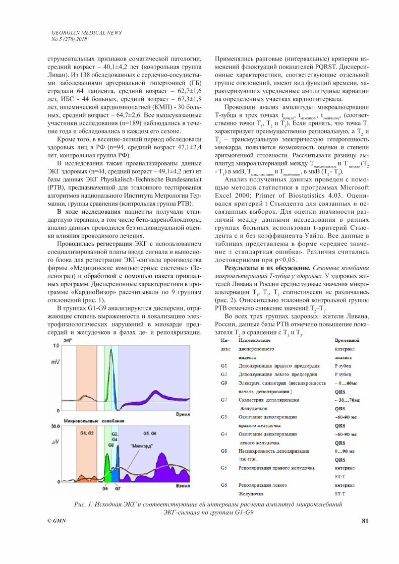
GEORGIAN MEDICAL NEWS No 5 (278) 2018
© GMN 81
струментальных признаков соматической патологии, средний возраст – 40,1±4,2 лет (контрольная группа Ливан). Из 138 обследованных с сердечно-сосудисты-ми заболеваниями артериальной гипертонией (ГБ) страдали 64 пациента, средний возраст – 62,7±1,6 лет, ИБС - 44 больных, средний возраст – 67,3±1,8 лет, ишемической кардиомиопатией (КМП) - 30 боль-ных, средний возраст – 64,7±2,6. Все вышеуказанные участники исследования (n=189) наблюдались в тече-ние года и обследовались в каждом его сезоне.
Кроме того, в весенне-летний период обследовали здоровых лиц в РФ (n=94, средний возраст 47,1±2,4 лет, контрольная группа РФ).
В исследовании также проанализированы данные ЭКГ здоровых (n=44, средний возраст – 49,1±4,2 лет) из базы данных ЭКГ Physikalisch-Technische Bundesanstalt (PTB), предназначенной для эталонного тестирования алгоритмов национального Института Метрологии Гер-мании, группы сравнения (контрольная группа PTB).
В ходе исследования пациенты получали стан-дартную терапию, в том числе бета-адреноблокаторы, анализ данных проводился без индивидуальной оцен-ки влияния проводимого лечения.
Проводилась регистрация ЭКГ с использованием специализированной платы ввода сигнала и выносно-го блока для регистрации ЭКГ-сигнала производства фирмы «Медицинские компьютерные системы» (Зе-леноград) и обработкой с помощью пакета приклад-ных программ. Дисперсионные характеристики в про-грамме «КардиоВизор» рассчитывали по 9 группам отклонений (рис. 1).
В группах G1-G9 анализируются дисперсии, отра-жающие степень выраженности и локализацию элек-трофизиологических нарушений в миокарде пред-сердий и желудочков в фазах де- и реполяризации.
Применялись ранговые (интервальные) критерии из-менений флюктуаций показателей PQRST. Дисперси-онные характеристики, соответствующие отдельной группе отклонений, имеют вид функций времени, ха-рактеризующих усредненные амплитудные вариации на определенных участках кардиоинтервала.
Проводили анализ амплитуды микроальтернации Т-зубца в трех точках tначало, tмаксимум, tокончание, (соответ-ственно точки Т1, Т2 и Т3). Если принять, что точка Т1 характеризует преимущественно региональную, а T2 и Т3 – трансмуральную электрическую гетерогенность миокарда, появляется возможность оценки и степени аритмогенной готовности. Рассчитывали разницу ам-плитуд микроальтернаций между Тмаксимальное и Т начало (T2 - Т1) в мкВ, Тмаксимальное и Токончание , в мкВ (T2 - Т3).
Анализ полученных данных проведен с помо-щью методов статистики в программах Microsoft Excel 2000; Primer of Biostatistics 4.03. Оцени-вался критерий t Стьюдента для связанных и не-связанных выборок. Для оценки значимости раз-личий между данными исследования в разных группах больных использован t-критерий Стью-дента с и без коэффициента Уайта. Все данные в таблицах представлены в форме «среднее значе-ние ± стандартная ошибка». Различия считались достоверными при p<0,05.
Результаты и их обсуждение. Сезонные колебания микроальтернаций Т-зубца у здоровых. У здоровых жи-телей Ливана и России среднегодовые значения микро-альтернации T1, T2, T3 статистически не различались (рис. 2). Относительно эталонной контрольной группы PTB отмечено снижение значений Т2–Т1.
Во всех трех группах здоровых: жители Ливана, России, данные базы PTB отмечено повышение пока-зателя Т2 в сравнении с Т1 и Т3.
Рис. 1. Исходная ЭКГ и соответствующие ей интервалы расчета амплитуд микроколебаний ЭКГ-сигнала по группам G1-G9

82
МЕДИЦИНСКИЕ НОВОСТИ ГРУЗИИ
CFMFHSDTKJC CFVTLBWBYJ CBF[KTYB
Рис. 2. Среднегодовые значения показателей ми-кроальтернации Т-зубца в 3 точках (в начале, на пике и в конце) в контрольных группах.
примечание: различия между Тмаксимум и Тначало ста-тистически значимы для каждой контрольной группы (p<0,05), различия показателя Т2-T1 группы PTB отно-сительно I и II групп статистически значимы (p<0,05)
При сравнении данных здоровых жителей Ливана моложе и старше 40 лет средние значения показателя микроальтернаций T-зубца во всех трех анализируе-мых точках были ниже у лиц более молодого возрас-та (p<0,05, таблица 1). У лиц до 40 лет максимальные значения показателя T2 отмечались весной и осенью - 8,4±0,3 и 8,5±0,4 мкВ. Различия между средними значениями Т2-Т3 были минимальны в зимний период -0,8±0,4 мкВ и максимальны осенью - 1,6±0,4 мкВ.
У здоровых жителей Ливана в возрасте старше 40 лет выявлено повышение амплитуды Т2 в зимний период - 10,7±0,7 мкВ. Увеличение средних значений дельты между Т2-Т3 на протяжении года составило от 0,9±0,4 осенью до 2,2±0,3 мкВ зимой.
Характеристики микроальтернации Т-зубца у здоро-вых жителей Ливана в разные сезоны года наблюдения статистически значимо не различались (таблица 1,2).
Таким образом, показатели микроальтернации Т-зубца в трех анализируемых точках у здоровых
Таблица 1. Годичные колебания показателей альтернации Т-зубца в трех его точках в I контрольной группе до и после 40 лет
Контрольные подгруппы (n=51)
Сезоныгода
T начало, мкВТ1
T максимум, мкВ Т2
T окончание, мкВТ3
Т2-Т1,мкВ
Т2-Т3,мкВ
20- 40 лет (n=31)
Зима 4,5±0,3^ 7,7±0,2* 6,9±0,4^ 3,2±0,3^ 0,8±0,4^Весна 5,0±0,5 8,4±0,3*^ 7,4±0,4 3,4±0,2^ 1,0±0,4Лето 4,9±0,4^ 7,6±0,3*^ 6,4±0,3^ 2,6±0,3 1,2±0,5
Осень 5,6±0,5 8,5±0,4*^ 6,9±0,5^ 2,9±0,3^ 1,6±0,4^Среднее 5,0±0,4^ 8,1±0,6*^ 6,9±0,6^ 3,0±0,2^ 1,5±0,4
41-60 лет (n=20)
Зима 6,5±0,3 10,7±0,7* 8,5±0,5 4,2±0,4 2,2±0,3Весна 5,9±0,4 9,3±0,6* 8,0±0,3 3,4±0,2 1,3±0,4Лето 6,4±0,5 9,6±0,5* 8,3±0,4 3,1±0,3 1,3±0,3
Осень 6,9±0,3 9,5±0,4* 8,6±0,3 4,1±0,3 0,9±0,4Среднее 6,4±0,4 9,8±0,3 8,5±0,4 3,7±0,2 1,3±0,4
20-60 (n=51) Cреднее 5,5±0,4 9,0±0,3 7,7±0,3 3,4±0,3 1,4±0,2примечание: * - различия относительно Т1 статистически значимы (p<0,05),
^ - различия относительно группы 41-60 лет статистически значимы (p<0,05)
Таблица 2. Сезонные колебания показателей альтернации Т-зубца в обследованных группах больных кардиальной патологией
ГруппыЗима Весна Лето Осень
Tначало , мкВ (Т1)Контроль (n=51) 5,5±0,3 5,4±0,3 5,7±0,4 5,5±0,5
ИБС+ГБ+КМП (n=138) 12,3±0,2** 13,2±0,3^,** 11,8±0,5** 12,3±0,4 Tмакс, мкВ (Т2)
Контроль (n=51) 9,2±0,2* 8,9±0,3* 8,6±0,3* 9,5±0,4*ИБС+ГБ+КМП(n=138) 20,1±0,9*, 20,6±0,8*, 20,6±0,10* 20,5±0,5*,**
Tокон , мкВ (Т3)Контроль (n=51) 7,7±0,4* 7,7±0,4 7,4±0,3* 7,8±0,5*
ИБС+ГБ+КМП (n=138) 16,4±0,3*, 15,4±0,3*,^ 16,8±0,6*, 16,0±0,4*,**примечание: * - различия относительно Т1 статистически значимы (p<0,05), ^
- различия относительно значений зимой статистически значимы (p<0,05), ** - различия относительно контрольной группы статистически значимы (p<0,05)
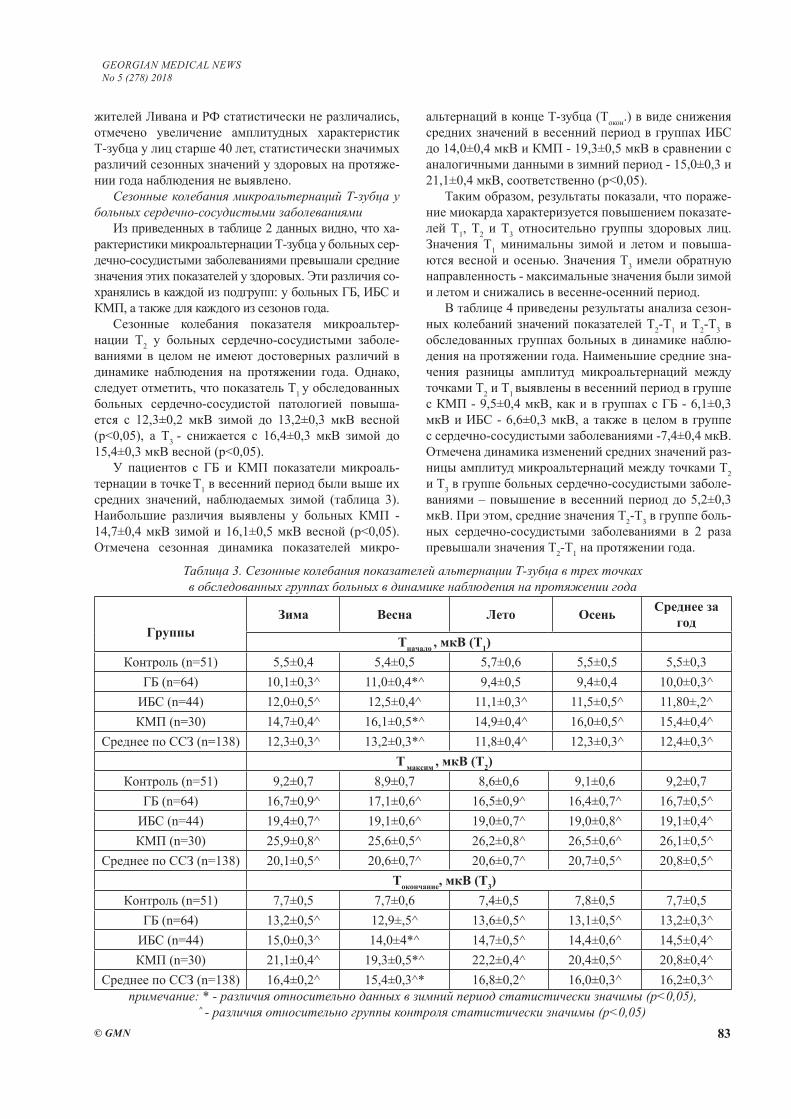
GEORGIAN MEDICAL NEWS No 5 (278) 2018
© GMN 83
жителей Ливана и РФ статистически не различались, отмечено увеличение амплитудных характеристик Т-зубца у лиц старше 40 лет, статистически значимых различий сезонных значений у здоровых на протяже-нии года наблюдения не выявлено.
Сезонные колебания микроальтернаций Т-зубца у больных сердечно-сосудистыми заболеваниями
Из приведенных в таблице 2 данных видно, что ха-рактеристики микроальтернации Т-зубца у больных сер-дечно-сосудистыми заболеваниями превышали средние значения этих показателей у здоровых. Эти различия со-хранялись в каждой из подгрупп: у больных ГБ, ИБС и КМП, а также для каждого из сезонов года.
Сезонные колебания показателя микроальтер-нации Т2 у больных сердечно-сосудистыми заболе-ваниями в целом не имеют достоверных различий в динамике наблюдения на протяжении года. Однако, следует отметить, что показатель Т1 у обследованных больных сердечно-сосудистой патологией повыша-ется с 12,3±0,2 мкВ зимой до 13,2±0,3 мкВ весной (p<0,05), а Т3 - снижается с 16,4±0,3 мкВ зимой до 15,4±0,3 мкВ весной (p<0,05).
У пациентов с ГБ и КМП показатели микроаль-тернации в точке Т1 в весенний период были выше их средних значений, наблюдаемых зимой (таблица 3). Наибольшие различия выявлены у больных КМП - 14,7±0,4 мкВ зимой и 16,1±0,5 мкВ весной (p<0,05). Отмечена сезонная динамика показателей микро-
альтернаций в конце Т-зубца (Tокон.) в виде снижения средних значений в весенний период в группах ИБС до 14,0±0,4 мкВ и КМП - 19,3±0,5 мкВ в сравнении с аналогичными данными в зимний период - 15,0±0,3 и 21,1±0,4 мкВ, соответственно (p<0,05).
Таким образом, результаты показали, что пораже-ние миокарда характеризуется повышением показате-лей Т1, Т2 и Т3 относительно группы здоровых лиц. Значения Т1 минимальны зимой и летом и повыша-ются весной и осенью. Значения Т3 имели обратную направленность - максимальные значения были зимой и летом и снижались в весенне-осенний период.
В таблице 4 приведены результаты анализа сезон-ных колебаний значений показателей Т2-Т1 и Т2-Т3 в обследованных группах больных в динамике наблю-дения на протяжении года. Наименьшие средние зна-чения разницы амплитуд микроальтернаций между точками Т2 и Т1 выявлены в весенний период в группе с КМП - 9,5±0,4 мкВ, как и в группах с ГБ - 6,1±0,3 мкВ и ИБС - 6,6±0,3 мкВ, а также в целом в группе с сердечно-сосудистыми заболеваниями -7,4±0,4 мкВ. Отмечена динамика изменений средних значений раз-ницы амплитуд микроальтернаций между точками Т2 и Т3 в группе больных сердечно-сосудистыми заболе-ваниями – повышение в весенний период до 5,2±0,3 мкВ. При этом, средние значения Т2-Т3 в группе боль-ных сердечно-сосудистыми заболеваниями в 2 раза превышали значения Т2-Т1 на протяжении года.
Таблица 3. Сезонные колебания показателей альтернации Т-зубца в трех точках в обследованных группах больных в динамике наблюдения на протяжении года
ГруппыЗима Весна Лето Осень Среднее за
годTначало , мкВ (Т1)
Контроль (n=51) 5,5±0,4 5,4±0,5 5,7±0,6 5,5±0,5 5,5±0,3ГБ (n=64) 10,1±0,3^ 11,0±0,4*^ 9,4±0,5 9,4±0,4 10,0±0,3^
ИБС (n=44) 12,0±0,5^ 12,5±0,4^ 11,1±0,3^ 11,5±0,5^ 11,80±,2^КМП (n=30) 14,7±0,4^ 16,1±0,5*^ 14,9±0,4^ 16,0±0,5^ 15,4±0,4^
Среднее по ССЗ (n=138) 12,3±0,3^ 13,2±0,3*^ 11,8±0,4^ 12,3±0,3^ 12,4±0,3^T максим , мкВ (Т2)
Контроль (n=51) 9,2±0,7 8,9±0,7 8,6±0,6 9,1±0,6 9,2±0,7ГБ (n=64) 16,7±0,9^ 17,1±0,6^ 16,5±0,9^ 16,4±0,7^ 16,7±0,5^
ИБС (n=44) 19,4±0,7^ 19,1±0,6^ 19,0±0,7^ 19,0±0,8^ 19,1±0,4^КМП (n=30) 25,9±0,8^ 25,6±0,5^ 26,2±0,8^ 26,5±0,6^ 26,1±0,5^
Среднее по ССЗ (n=138) 20,1±0,5^ 20,6±0,7^ 20,6±0,7^ 20,7±0,5^ 20,8±0,5^Tокончание, мкВ (Т3)
Контроль (n=51) 7,7±0,5 7,7±0,6 7,4±0,5 7,8±0,5 7,7±0,5ГБ (n=64) 13,2±0,5^ 12,9±,5^ 13,6±0,5^ 13,1±0,5^ 13,2±0,3^
ИБС (n=44) 15,0±0,3^ 14,0±4*^ 14,7±0,5^ 14,4±0,6^ 14,5±0,4^КМП (n=30) 21,1±0,4^ 19,3±0,5*^ 22,2±0,4^ 20,4±0,5^ 20,8±0,4^
Среднее по ССЗ (n=138) 16,4±0,2^ 15,4±0,3^* 16,8±0,2^ 16,0±0,3^ 16,2±0,3^примечание: * - различия относительно данных в зимний период статистически значимы (p<0,05),
^ - различия относительно группы контроля статистически значимы (p<0,05)
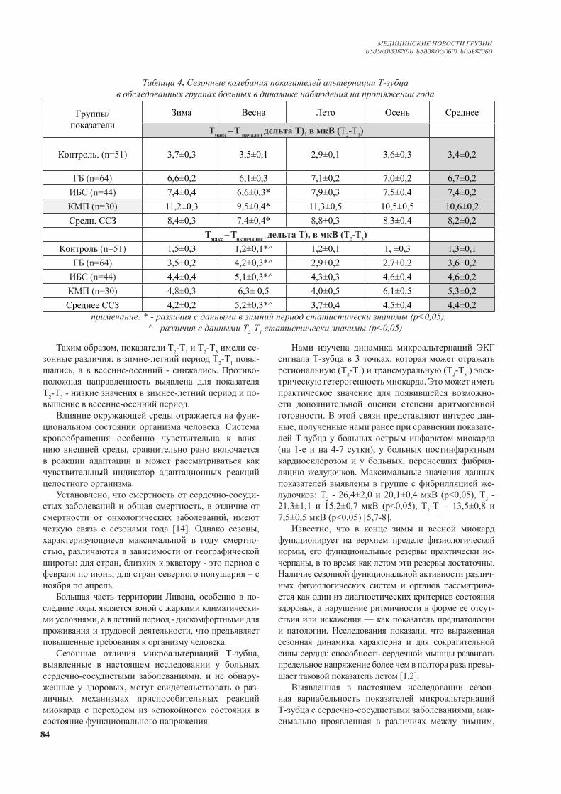
84
МЕДИЦИНСКИЕ НОВОСТИ ГРУЗИИ
CFMFHSDTKJC CFVTLBWBYJ CBF[KTYB
Таким образом, показатели Т2-Т1 и Т2-Т3 имели се-зонные различия: в зимне-летний период Т2-Т1 повы-шались, а в весенне-осенний - снижались. Противо-положная направленность выявлена для показателя Т2-Т3 - низкие значения в зимнее-летний период и по-вышение в весенне-осенний период.
Влияние окружающей среды отражается на функ-циональном состоянии организма человека. Система кровообращения особенно чувствительна к влия-нию внешней среды, сравнительно рано включается в реакции адаптации и может рассматриваться как чувствительный индикатор адаптационных реакций целостного организма.
Установлено, что смертность от сердечно-сосуди-стых заболеваний и общая смертность, в отличие от смертности от онкологических заболеваний, имеют четкую связь с сезонами года [14]. Однако сезоны, характеризующиеся максимальной в году смертно-стью, различаются в зависимости от географической широты: для стран, близких к экватору - это период с февраля по июнь, для стран северного полушария – с ноября по апрель.
Большая часть территории Ливана, особенно в по-следние годы, является зоной с жаркими климатически-ми условиями, а в летний период - дискомфортными для проживания и трудовой деятельности, что предъявляет повышенные требования к организму человека.
Сезонные отличия микроальтернаций Т-зубца, выявленные в настоящем исследовании у больных сердечно-сосудистыми заболеваниями, и не обнару-женные у здоровых, могут свидетельствовать о раз-личных механизмах приспособительных реакций миокарда с переходом из «спокойного» состояния в состояние функционального напряжения.
Нами изучена динамика микроальтернаций ЭКГ сигнала Т-зубца в 3 точках, которая может отражать региональную (Т2-Т1) и трансмуральную (Т2-Т3 ) элек-трическую гетерогенность миокарда. Это может иметь практическое значение для появившейся возможно-сти дополнительной оценки степени аритмогенной готовности. В этой связи представляют интерес дан-ные, полученные нами ранее при сравнении показате-лей Т-зубца у больных острым инфарктом миокарда (на 1-е и на 4-7 сутки), у больных постинфарктным кардиосклерозом и у больных, перенесших фибрил-ляцию желудочков. Максимальные значения данных показателей выявлены в группе с фибрилляцией же-лудочков: Т2 - 26,4±2,0 и 20,1±0,4 мкВ (р<0,05), Т3 - 21,3±1,1 и 15,2±0,7 мкВ (р<0,05), Т2-Т1 - 13,5±0,8 и 7,5±0,5 мкВ (р<0,05) [5,7-8].
Известно, что в конце зимы и весной миокард функционирует на верхнем пределе физиологической нормы, его функциональные резервы практически ис-черпаны, в то время как летом эти резервы достаточны. Наличие сезонной функциональной активности различ-ных физиологических систем и органов рассматрива-ется как один из диагностических критериев состояния здоровья, а нарушение ритмичности в форме ее отсут-ствия или искажения — как показатель предпатологии и патологии. Исследования показали, что выраженная сезонная динамика характерна и для сократительной силы сердца: способность сердечной мышцы развивать предельное напряжение более чем в полтора раза превы-шает таковой показатель летом [1,2].
Выявленная в настоящем исследовании сезон-ная вариабельность показателей микроальтернаций Т-зубца с сердечно-сосудистыми заболеваниями, мак-симально проявленная в различиях между зимним,
Таблица 4. Сезонные колебания показателей альтернации Т-зубца в обследованных группах больных в динамике наблюдения на протяжении года
Группы/показатели
Зима Весна Лето Осень Среднее
Тмакс – Т начало ( дельта Т), в мкВ (Т2-Т1)
Контроль. (n=51) 3,7±0,3 3,5±0,1 2,9±0,1 3,6±0,3 3,4±0,2
ГБ (n=64) 6,6±0,2 6,1±0,3 7,1±0,2 7,0±0,2 6,7±0,2ИБС (n=44) 7,4±0,4 6,6±0,3* 7,9±0,3 7,5±0,4 7,4±0,2КМП (n=30) 11,2±0,3 9,5±0,4* 11,3±0,5 10,5±0,5 10,6±0,2Средн. ССЗ 8,4±0,3 7,4±0,4* 8,8+0,3 8.3±0,4 8,2±0,2
Тмакс – Токончание ( дельта Т), в мкВ (Т2-Т3)Контроль (n=51) 1,5±0,3 1,2±0,1*^ 1,2±0,1 1, ±0,3 1,3±0,1
ГБ (n=64) 3,5±0,2 4,2±0,3*^ 2,9±0,2 2,7±0,2 3,6±0,2ИБС (n=44) 4,4±0,4 5,1±0,3*^ 4,3±0,3 4,6±0,4 4,6±0,2КМП (n=30) 4,8±0,3 6,3± 0,5 4,0±0,5 6,1±0,5 5,3±0,2Среднее ССЗ 4,2±0,2 5,2±0,3*^ 3,7±0,4 4,5±0,4 4,4±0,2
примечание: * - различия с данными в зимний период статистически значимы (p<0,05), ^ - различия с данными Т2-Т1 статистически значимы (p<0,05)
GEORGIAN MEDICAL NEWS No 5 (278) 2018
© GMN 85
летним сезонами относительно весеннего, осеннего сезонов несомненно отражает изменения реполяриза-ции миокарда, значимые для оценки течения заболе-вания и лечения больных. Этот вопрос требует более тщательного изучения и дальнейших исследований, которые определят возможности метода в ведении больных сердечно- сосудистыми заболеваниями.
Заключение. Таким образом, полученные данные о диагностических возможностях нового метода ана-лиза микроальтернационных характеристик ЭКГ в детальных исследованиях в области хроноструктуры ритмов представляются весьма значимыми. Очевид-но, что недооценка сезонных колебаний характери-стик микроальтернаций ЭКГ-сигнала существенно снижает качество диагностики и лечения пациентов в обследованных группах больных, которым показана эта диагностика и возможная последующая коррекция проводимой терапии. Необходимо дальнейшее углу-бленное биофизическое исследование по изучению механизмов микроальтернаций, включая диагностику годичных колебаний мембранных структур, устойчи-вости ионных каналов, активность аденилат-циклаз-ной системы, уровня АТФ, гормональных изменений и других факторов.
ЛИТЕРАТУРА
1. Агаджанян Н.А., Башкиров А.А., Власова И. Г. Успехи физиол. наук 1987;4:80-104. 2. Агаджанян Н.А., Чеснокова С.А., Михайловская Т.А. Фактор времени и физиологические реакции. М.: Изд-во Университета дружбы народов. 1989. 55 с. 3. Арушанян Э.Б., Батурин В.А. Основы хронофармаколо-гии: Учеб. пособие. Ставрополь: СГМИ, 1989. 84 с. 4. Барбараш О.Л., Фомина Н.В., Минеева Е.В. и соавт. Био-ритмологический подход к прогнозированию течения ише-мической болезни сердца. Кардиоваскулярная терапия и профилактика 2008;8:4-11.5. Иванов Г.Г., Дворников В.Е., Стрельникова Ю.Н. и др. Показатели микроальтернации ЭКГ у больных с сердечно-сосудистой патологией при проведении тензорной пробы. Функциональная диагностика 2009;2:13-21.6.Иванов Г.Г., Лещинский С.П., Буланова Н.А. Метод дис-персионного картирования ЭКГ в оценке электрической ак-тивности предсердий и желудочков. Сеченовский вестник 2012;4 (10):21-27.7. Иванов Г. Г., Сула А.С. Анализ микроальтернаций ЭКГ методом дисперсионного картирования в клинической практике. М.: Техносфера, 2014. 104 с. 8. Каждан А.М., Арквасов М.Ю., Иванов Г.Г. и др. Тести-рование показателей дисперсионного картирования на базе данных “The PTB diagnostic ECG database“. Вестник Рос-сийского университета дружбы народов. Серия: Медици-на.2009;4:98-101.9. Саркисов Д.С, Пальцын А.А., Втюрин Б.В. Приспособи-тельная перестройка биоритмов. М.: Медицина., 1975. 184 с. 10. Трешкур Т.В., Татаринова А.А., Пармон Е.В. Альтерна-ция зубца Т: способна ли предсказывать непредсказуемое? Вестник аритмологии. 2009;58:42-51.11. Чибисов С.М. Хроноструктура заболеваний сердечно-
сосудистой системы. Материалы Всероссийской научно-практической конференции «Современные проблемы арте-риальной гипертонии». М.. 2003;239-241. 12. Aro A.L., Kenttä T.V., Huikuri H.V. Microvolt T-wave Al-ternans: Where Are We Now? Arrhythm Electrophysiol Rev. 2016;1:37-40. 13. Kellett J., Clifford M. The prediction of death up to 100 days after admission to hospital for acute medical illness - the com-parison of two ECG interpretation methods with ECG-disper-sion mapping. Acute Med. 2015;14:4:151-158. 14. Marti-S.H., Gonseth S., Gubelmann C. et al. Seasonal variation of overall and cardiovascular mortality: a study in 19 countries from different geographic locations. PLoS One. 2014; 9(11):e113500. 15. Rolden H.J., Rohling J.H., van Bodegom D., Westendorp R.G. Seasonal Variation in Mortality, Medical Care Expendi-ture and Institutionalization in Older People: Evidence from a Dutch Cohort of Older Health Insurance Clients. PLoS One. 2015;10(11):e0143154.
SUMMARY
SEASONAL VARIATION OF MICROVOLT T-WAVE ALTERNANS IN PATIENTS WITH CARDIOVAS-CULAR DISEASE AND HEALTHY SUBJECTS
3Halabi Gh., 1Bulanova N., 2Aleksandrova S., 1,2Ivanov G., 2Aleksandrova M.
1Sechenov University, Moscow; 2Peoples’ Friendship University of Russia, Moscow, Russia; 3Hospital 2000, Beirut, Lebanon
Objective - to access seasonal variation of microvolt T-wave alternans of ECG dispersion mapping in patients with cardiovascular disease and healthy subjects.
ECG data of the three groups of healthy subjects have been compared: inhabitants of Beirut, Lebanon (n=51), inhabitants of Moscow, Russia (n=94) and ECG data of healthy subjects (n=44) from the testing ECG database of the PTB - The National Metrology Institute of Germany as well as a group of patients with cardiovascular disease (n=138), inhabitants of Beirut, Lebanon. Microvolt T-wave alternans of ECG dispersion mapping was evaluated in three points - Tbeginning, Tmaximum, Tend.
In healthy subjects, the seasonal variation of ECG dis-persion mapping microvolt T-wave alternans was nonex-istent. Myocardial lesion is characterized by an increase in Tbeg, Tmax, Tend in relation to the healthy individu-als. Tbeg values are minimal in winter and summer and increase in spring and autumn. Tend values were reversed - they were maximal in winter and summer, decreasing in spring-autumn period. Seasonal variation of Tmax - Tbeg, and Tmax -Tend was detected: Tmax - Tbeg increased in the winter-summer period and decreased in spring and au-tumn, Tmax-Tend - increased in the spring-autumn period in relation to the winter-summer period. In patients with cardiovascular disease, in contrast to the healthy, there is a seasonal variation in microvolt T-wave alternans of ECG
86
МЕДИЦИНСКИЕ НОВОСТИ ГРУЗИИ
CFMFHSDTKJC CFVTLBWBYJ CBF[KTYB
dispersion mapping, with the maximum differences in the winter and spring seasons, which should be taken into ac-count when applying the method in clinical practice.
Keywords: ECG dispersion mapping, early detection of myocardial lesion; microvolt T-wave alternans; sea-sonal variation.
РЕЗЮМЕ
СЕЗОННЫЕ КОЛЕБАНИЯ МИКРОАЛЬТЕР-НАЦИЙ Т-ЗУБЦА У ЗДОРОВЫХ И БОЛЬНЫХ СЕРДЕЧНО-СОСУДИСТЫМИ ЗАБОЛЕВАНИ-ЯМИ
3Халаби Г., 1Буланова Н.А., 2Александрова C.Г., 1,2Иванов Г.Г., 2Александрова М.Р.
1Первый Московский государственный медицинский университет им. И.М. Сеченова МЗ РФ, Научно-ис-следовательский отдел кардиологии Научно-техно-логического парка биомедицины, Москва; 2ФГАОУ ВО Российский университет дружбы народов, кафедра госпитальной терапии с курсом клинической лабора-торной диагностики, Москва, Россия; 3Медицинский госпиталь 2000, Бейрут, Ливан
Целью исследования явился анализ сезонных ко-лебаний микроальтернаций Т-зубца дисперсионного картирования ЭКГ у здоровых и больных сердечно-сосудистыми заболеваниями.
Проведено сравнение данных 3 групп практиче-ски здоровых лиц: жители Ливана (n=51), жители РФ (n=94) и ЭКГ здоровых (n=44) из базы данных ЭКГ для эталонного тестирования алгоритмов Националь-ного института метрологии Германии, а также груп-пы больных сердечно-сосудистыми заболеваниями, проживающих в Ливане (n=138). Микроальтернации Т-зубца в трёх точках: Tнач, Tмакс, Tокон оценивали при помощи метода дисперсионного картирования ЭКГ. Cтатистически значимых различий сезонных значе-ний у здоровых не выявлено. Поражение миокарда характеризуется повышением показателей Tнач, Tмакс, Tокон относительно группы здоровых лиц. Значения Тнач минимальны зимой и летом и повышаются весной и осенью. Значения Токон имели обратную направлен-ность - были максимальны зимой и летом, снижаясь в весенне-осенний период. Показатели Tмакс - Tнач, и Тмакс -Tокон имели сезонные различия: в зимне-летний период Tмакс - Tнач повышались, а в весенне-осенний – снижались, Тмакс-Токон - повышение в весенне-осенний период относительно зимне-летнего периода.
У больных сердечно-сосудистыми заболеваниями, в отличие от здоровых, выявлены сезонные изменения микроальтернаций Т-зубца с максимальными отличия-ми в зимний и весенний сезоны, что необходимо учи-тывать при применении метода в клинической практике.
reziume
jansaRi da gul-sisxlZarRvTa daavadebebiT pacientebis t-kbilanis mikroalternaciis sezonuri ryevebi
3g. halabi, 1n. bulanova, 2s. aleqsandrova, 1.2g. ivanovi, 2m. alqsandrova
1jandacvis saministros federaluri sa-xelmwifo avtonomiuri saganmanaTleblo dawesebuleba moskovis i. seCenovis sax. pirveli saxelmwifo samedicino universite-ti, samecniero-teqnologiuri biomedicinis parki, kardiologiis samecniero-kvleviTi ganyofileba, moskovi; 2federaluri saxelm-wifo avtonomiuri saganmanaTleblo dawe-sebuleba ruseTis xalxTa megobrobis uni-versiteti, hospitaluri Terapiis kaTedra klinikuri da laboratoriuli diagnosti-kis kursiT, moskovi, ruseTi; 3samedicino hos-pitali 2000, beiruTi, libani
kvlevis mizans warmoadgenda jansaRi da gul-sisxlZarRvTa daavadebebiT pacien-tebis eleqtrokardiogramis dispersiuli kartirebis t-kbilanis mikroalternaciis sezonuri ryevebis analizi. Catarda mona-cemebis Sedareba praqtikulad janmrTe-li pirebis 3 jgufSi: libanis mosaxleo-ba (n=51), ruseTis federaciis mosaxleoba (n=94) da jansaRi pirebis ekg (n=44) germani-is metrologiis nacionaluri institutis etalonuri testirebis algoriTmebis ekg monacemTa bazidan, agreTve libanis mo-saxleobis gul-sisxlZarRvTa daavadebebiT avadmyofebis jgufi (n=138). t-kbilanis mik-roalternacia sam wertilSi: t
das, t
maqs, t
dasasr.
iyo Sefasebuli ekg-s dispersiuli kar-tirebis meTodiT.
jansaR pirebSi statistikurad mniS-vnelovani sezonuri maCveneblebis gan-sxvavebebi ar gamovlenda. miokardiumis da-zianeba xasiaTdeba t
das, t
maqs, t
dasasr gazrdili
maCveneblebiT janmrTel pirebis jgufTan SedarebiT. t
das maCveneblebi minimaluria
zamTarSi da zafxulSi, matulobs gazaf-xulze da Semodgomaze. t
dasasr maCeneblebSi
aReniSna sapirispiro tendencia - maqsimumi zafxulSi da zamTarSi, mcirdeboda gazaf-xulze da Semodgomaze. t
das, t
maqs, t
dasasr.
maCveneblebs hqonda sezonuri gansxvavebebi: zamTar-zafulis periodSi t
maqs-t
das matulob-
da, gazafxul-saSemodgomo periodSi, piriqiT, mcirdeboda, t
maqs -t
dasasr gazafxul-saSemodgo-
mo periodSi matulobda zamTar-zafxulis periodTan SedarebiT.
GEORGIAN MEDICAL NEWS No 5 (278) 2018
© GMN 87
pacientebSi gul-sisxlZarRvTa da-avadebebiT, jansaR pirebisgan gansxvavebiT, gamovlinda t-kbilanis mikroalternaciis sezonuri ryevebi, zamTris da gazafxulis
sezonSi maqsimaluri gansxvavebiT, rac aucileblad unda iyos gaTvaliswinebuli meTodis gamoyenebis dros klinikur praq-tikaSi.
СОВРЕМЕННЫЕ АСПЕКТЫ ДИАГНОСТИКИ И ЛЕЧЕНИЯ СЕРДЕЧНОЙ НЕДОСТАТОЧНОСТИ КАК ПРОЯВЛЕНИЯ
АНТРАЦИКЛИНОВОЙ КАРДИОТОКСИЧНОСТИ (ОБЗОР)
Саганелидзе Х.З., Кавтарадзе Н.Н.
Нью-Вижен Университет, Тбилиси, Грузия
Кардиотоксичность – дисфункция миокарда и сер-дечная недостаточность на фоне прогрессирующего снижения фракции выброса (ФВ) является серьезным осложнением химиотерапии и может осложнить про-ведение противоопухолевого лечения. В настоящее время кардиотоксические эффекты на фоне химио-терапевтического лечения – проблема современно-сти, связанная с большим количеством неизученных вопросов в связи с увеличением продолжительности жизни пациентов, появлением новых химиопрепара-тов и схем их введения.
Актуальность проблемы обусловлена сложностя-ми определения токсичности препаратов в долгосроч-ном прогнозе, сроками и проявлениями сердечной недостаточности на фоне и после лечения, сопутству-ющими заболеваниями и перекрестными схемами ле-чения. На фоне сочетания разных химиопрепаратов и схем их введения кардиотоксичность может проявляться в разных видах и с различной степенью выраженности. Кардиомиоциты, как клетки с ограниченной регенера-тивной способностью, подвержены токсическому дей-ствию онкопрепаратов, хотя, в большинстве случаев токсичность цитостатиков проявляется в действии на быстро пролиферирующие клетки [22].
По мнению экспертов Европейского общества кар-диологов (ESC), сердечно-сосудистые осложнения, которые возникают на фоне химиотерапевтического лечения, представлены девятью основными катего-риями: дисфункция миокарда и сердечная недоста-точность, ишемическая болезнь сердца, патология клапанов, нарушения ритма, в основном вызванные лекарствами, удлиняющими QT; артериальная ги-пертензия, тромбоэмболические осложнения, забо-левания периферических сосудов и инсульт, легочная гипертензия, перикардиальные осложнения [5]. Учи-тывая сложность проблемы лечения этих пациентов, в меморандуме ESC по лечению онкологических забо-леваний и сердечно-сосудистой токсичности предло-жено формирование мультидисциплинарных команд, состоящих из специалистов разных областей – карди-ологов, онкологов, химиотерапевтов [5].
На сегодняшний день наиболее кардиотоксичными
химиопрепаратами в сравнении с остальными счита-ются антрациклины, которые широко применяются для лечения онкологических больных при солидных опухо-лях и злокачественных заболеваниях крови. Антраци-клины – эффективные противопухолевые препараты, которые включены также в схемы лечения онкологиче-ских пациентов, имеют мощный протовоопухолевый и выраженные токсические эффекты, в том числе и кар-диотоксичность [3,17]. Они вызывают практически все перечисленные возможные кардиотоксические ослож-нения. Согласно рекомендациям рабочей группы по заболеваниям миокарда и перикарда Европейского об-щества кардиологов, кардиомиопатия на фоне химиоте-рапии относится к подгруппе ненаследственных кардио-патий на фоне воздействия лекарств (антрациклинов). Все химиотерапевические препараты предложено клас-сифицировать по проявлению кардиотоксичности. От-личают различные типы кардиотоксичности: 1) кардио-токсичность первого типа – необратимая дисфункция (антрациклины) вследствие гибели кардиомиоцитов. Доксорубицину свойственен первый тип кардиотоксич-ности, который является необратимым и характеризует-ся деструктивно-некротическим поражением клеток; 2) кардиотоксичность второго типа – обратимая (трасту-зумаб, он относится к моноклональным антителам) - за счет повреждения митохондрий и протеинов. Однако, это деление условно, так как, например, трастузумаб при воздействии на уже поврежденный миокард может иметь кардиотоксичность I типа [22].
Антрациклиновая кардиотоксичность бывает острая, ранняя, поздняя. Острая, обычно, разви-вается сразу после инфузии препарата и чаще об-ратима. Она проявляется в виде преходящей дис-функции левого желудочка, суправентрикулярных тахикардий, ЭКГ-изменений. Неизвестно как пове-дет себя острая кардиотоксичность впоследствии, так как может превратиться в раннюю или позднюю кардиотоксичность или стать обратимой. Ранняя кардиоксичность проявляется в течение первого года лечения, поздняя кардиотоксичность – через несколько лет [13,28].
Антрациклиновая кардиотоксичность по некото-
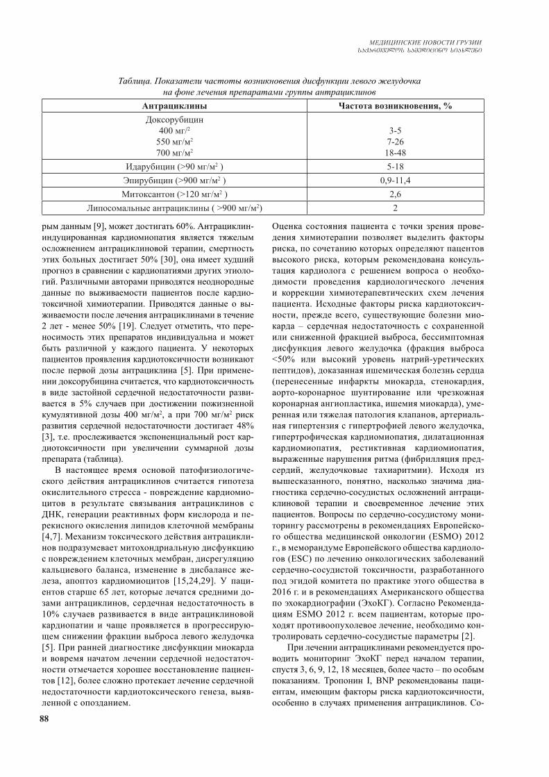
88
МЕДИЦИНСКИЕ НОВОСТИ ГРУЗИИ
CFMFHSDTKJC CFVTLBWBYJ CBF[KTYB
рым данным [9], может достигать 60%. Антрациклин-индуцированная кардиомиопатия является тяжелым осложнением антрациклиновой терапии, смертность этих больных достигает 50% [30], она имеет худший прогноз в сравнении с кардиопатиями других этиоло-гий. Различными авторами приводятся неоднородные данные по выживаемости пациентов после кардио-токсичной химиотерапии. Приводятся данные о вы-живаемости после лечения антрациклинами в течение 2 лет - менее 50% [19]. Следует отметить, что пере-носимость этих препаратов индивидуальна и может быть различной у каждого пациента. У некоторых пациентов проявления кардиотоксичности возникают после первой дозы антрациклина [5]. При примене-нии доксорубицина считается, что кардиотоксичность в виде застойной сердечной недостаточности разви-вается в 5% случаев при достижении пожизненной кумулятивной дозы 400 мг/м2, а при 700 мг/м2 риск развития сердечной недостаточности достигает 48% [3], т.е. прослеживается экспоненциальный рост кар-диотоксичности при увеличении суммарной дозы препарата (таблица).
В настоящее время основой патофизиологиче-ского действия антрациклинов считается гипотеза окислительного стресса - повреждение кардиомио-цитов в результате связывания антрациклинов с ДНК, генерации реактивных форм кислорода и пе-рекисного окисления липидов клеточной мембраны [4,7]. Механизм токсического действия антрацикли-нов подразумевает митохондриальную дисфункцию с повреждением клеточных мембран, дисрегуляцию кальциевого баланса, изменение в дисбалансе же-леза, апоптоз кардиомиоцитов [15,24,29]. У паци-ентов старше 65 лет, которые лечатся средними до-зами антрациклинов, сердечная недостаточность в 10% случаев развивается в виде антрациклиновой кардиопатии и чаще проявляется в прогрессирую-щем снижении фракции выброса левого желудочка [5]. При ранней диагностике дисфункции миокарда и вовремя начатом лечении сердечной недостаточ-ности отмечается хорошее восстановление пациен-тов [12], более сложно протекает лечение сердечной недостаточности кардиотоксического генеза, выяв-ленной с опозданием.
Оценка состояния пациента с точки зрения прове-дения химиотерапии позволяет выделить факторы риска, по сочетанию которых определяют пацентов высокого риска, которым рекомендована консуль-тация кардиолога с решением вопроса о необхо-димости проведения кардиологического лечения и коррекции химиотерапевтических схем лечения пациента. Исходные факторы риска кардиотоксич-ности, прежде всего, существующие болезни мио-карда – сердечная недостаточность с сохраненной или сниженной фракцией выброса, бессимптомная дисфункция левого желудочка (фракция выброса <50% или высокий уровень натрий-уретических пептидов), доказанная ишемическая болезнь сердца (перенесенные инфаркты миокарда, стенокардия, аорто-коронарное шунтирование или чрезкожная коронарная ангиопластика, ишемия миокарда), уме-ренная или тяжелая патология клапанов, артериаль-ная гипертензия с гипертрофией левого желудочка, гипертрофическая кардиомиопатия, дилатационная кардиомиопатия, рестиктивная кардиомиопатия, выраженные нарушения ритма (фибрилляция пред-сердий, желудочковые тахиаритмии). Исходя из вышесказанного, понятно, насколько значима диа-гностика сердечно-сосудистых осложнений антраци-клиновой терапии и своевременное лечение этих пациентов. Вопросы по сердечно-сосудистому мони-торингу рассмотрены в рекомендациях Европейско-го общества медицинской онкологии (ESMO) 2012 г., в меморандуме Европейского общества кардиоло-гов (ESC) по лечению онкологических заболеваний сердечно-сосудистой токсичности, разработанного под эгидой комитета по практике этого общества в 2016 г. и в рекомендациях Американского общества по эхокардиографии (ЭхоКГ). Согласно Рекоменда-циям ESMO 2012 г. всем пациентам, которые про-ходят противоопухолевое лечение, необходимо кон-тролировать сердечно-сосудистые параметры [2].
При лечении антрациклинами рекомендуется про-водить мониторинг ЭхоКГ перед началом терапии, спустя 3, 6, 9, 12, 18 месяцев, более часто – по особым показаниям. Тропонин I, BNP рекомендованы паци-ентам, имеющим факторы риска кардиотоксичности, особенно в случаях применения антрациклинов. Со-
Таблица. Показатели частоты возникновения дисфункции левого желудочка на фоне лечения препаратами группы антрациклинов
Антрациклины Частота возникновения, %Доксорубицин
400 мг/2
550 мг/м2
700 мг/м2
3-57-2618-48
Идарубицин (>90 мг/м2 ) 5-18Эпирубицин (>900 мг/м2 ) 0,9-11,4Митоксантон (>120 мг/м2 ) 2,6
Липосомальные антрациклины ( >900 мг/м2) 2
GEORGIAN MEDICAL NEWS No 5 (278) 2018
© GMN 89
гласно рекомендациям, при выявлении субклиниче-ской кардиотоксичности, например, бессимптомной дисфункции левого желудочка и увеличении уровня тропонина показано лечение пациента И-АПФ, пре-вентивно, для предотвращения снижения фракции выброса левого желудочка (ФВ ЛЖ). Проявлением кардиотоксичности химиопрепаратов, а также других заболеваний может быть снижение ФВ ЛЖ. Понятно, что важно проводить исследование ФВ ЛЖ до начала терапии, чтобы понять генез изменения сократимости левого желудочка. Подходы к оценке сократимости ле-вого желудочка различаются у разных авторов. Принято считать кардитоксичным действием снижение ФВ более 20% от исхода или любое снижение менее 50%. В не-которых исследованиях использовались более строгие подходы – снижение ФВ более, чем на 5%, до уровня менее 55% с признаками хронической сердечной недо-статочности (ХСН) или бессимптомное снижение ФВ более 10% до уровня ФВ менее 55% [20]. Американским обществом по ЭхоКГ кардиотоксичностью считается снижение ФВ ЛЖ более 5% от исхода до уровня менее 55% с признаками ХСН или бессимптомное снижение ФВ более, чем на 10% и менее 55% [11,23].
В Меморандуме ESC, упомянутом выше, отмеча-ется, что факторами риска развития антрациклиновой недостаточности являются: возраст старше 65 лет, сум-марная доза антрациклина за время лечения, схема ле-чения, лучевая медиастинальная терапия, заболевания сердца, артериальная гипертензия [5]. Рекомендовано проводить кардиологическое обследование, в том числе оценку функции левого желудочка, всем пациентам, ко-торые получают кардиотоксичную терапию во время и после окончания лечения. В плане диагностики кардио-токсичности используются методики:
- электрокардиография (ЭКГ) – до и во время лече-ния (тахикардия, нарушения ритма и проводимости, изменения интервалов ST, T, QT);
- эхоКГ. 3D-ЭхоКГ обладает возможностями более точного определеня ФВ ЛЖ по сравнению с 2-мерной (2D) ЭхоКГ и сравнима с МРТ. При невозможности проведения 3-мерной (3D) ЭхоКГ рекомендована оценка ФВ ЛЖ с помощью 2D-ЭхоКГ биплановым методом Симпсона. Испрользуются контрастная ЭхоКГ, стресс ЭхоКГ, методика оценки УЗИ изобра-жения методом speckle-tracking – оценка глобальной продольной деформации GLS (снижение >15% счи-тается маркером ранней систолический дисфункции - 1,34-36). Кардиальной дисфункцией считается сни-жение ФВ >10% до 50% (при снижении ФВ >10% ре-комендовано провести сравнительную контрольную ЭхоКГ спустя 2-3 недели после выявления сниженной ЭхоКГ) [5]. При терапии антрациклинами рекомендо-вано проведение исходной ЭхоКГ. При наличии дис-функции левого желудочка исходно или других изме-нениях рекомендуется изменение схемы лечения либо назначение терапии кардиопротекторами, следующее ЭхоКГ выполняется в конце лечения. При лечении
высокими дозами антрациклинов и для пациентов вы-сокого риска – повторная ЭхоКГ – при достижении ку-мулятивной дозы доксорубицина 240 мг/м2 [5].
- магнитно-резонансная томография (МРТ) серд-ца. МРТ – это методика раннего выявления кардио-токсичности. Методика обладает хорошими диагно-стическими возможностями, может использоваться в динамике, не подвергает пациента ионизирующей ра-диации. Американский колледж кардиологов и Аме-риканская ассоциация сердца признают ее как метод выявления дисфункции левого желудочка после хи-миотерапии. Использование МРТ ограничено ввиду высокой стоимости этого обследования [8].
- радионуклидная диагностика. Фракция выброса левого желудочка может быть оценена с помощью радионуклидной равновесной ангиографии или мето-дом радионуклидной равновесной вентрикулографии, однако эти методики в связи с ионизирующей нагруз-кой не часто применяются.
- лабораторные биомаркеры (тропонин I, натрий -уретический пептид - НУП). Тропонин I может быть маркером токсичности и определяется в начале и в конце каждого цикла антрациклинов. Его повышение является предиктором кардиотоксичности; исследо-вания показали, что в этих случаях целесообразно проведение лечения И-АПФ [1,2]. В плане кардио-логического подхода к этим больным важно раннее выявление дисфункции миокарда с использованием ЭхоКГ и биомаркеров. При выявлении бессимптом-ной дисфункции левого желудочка или сердечной недостаточности рекомендовано назначение И-АПФ или блокаторов рецепторов ангиотензина (БРА) и β-адреноблокаторов, чем раньше будет назначена эта терапия в этом случае, тем лучше прогноз [5].
Для пациентов невсегда доступны дорогостоящие методики обследования. Однако и с помощью рутин-ных методик, внимательно наблюдая пациентов, воз-можно судить о кардиотоксичных осложнениях на фоне химиотерапевтического лечения: физикальное обследование пациента, ЭКГ исследование, при необ-ходимости – лабораторное обследование (тропонин и электролиты крови).
В исследования по наблюдению за пациентами, получающими химиотерапию, в плане мониторинга сократимости были включены 850 пациентов, кото-рым проводилось лечение антрациклинами и трасту-зумабом. При наблюдении в течение 3 лет у пациентов развилась СН: при ФВ ЛЖ до начала терапии 50-54% - у 12,5%, при ФВ ЛЖ до начала терапии 55-64% - у 3,8%, при ФВ ЛЖ до начала терапии более 65% - у 0,9%, т.е., чем ниже начальная сократимость левого желудочка, тем более выражено в динамике сниже-ние фракции выброса левого желудочка, что можно расценить как проявление кардиотоксического дей-ствия препаратов, которое возникает в большинстве случаев при более низких значениях сократимости, т.е. при исходно заинтересованном миокарде [23]. На
90
МЕДИЦИНСКИЕ НОВОСТИ ГРУЗИИ
CFMFHSDTKJC CFVTLBWBYJ CBF[KTYB
фоне лечения химиопрепаратами, преимущественно антрациклинами может возникнуть или усугубиться сердечная недостаточность, после чего трудно ее ле-чить и прогноз у пациента ухудшается. В связи с этим понятны попытки усовершенствования вариантов ранней диагностики сердечной недостаточности [6].
Крайне важно раннее выявление кардиотоксично-сти на фоне химиотерапии, так как возможно умень-шение кардиотоксического эффекта путем изменения дозировок, скорости введения препаратов, замены препаратов на менее кардиотоксичные и назначения новой комбинации химиопрепаратов. Предлагаются алгоритмы диагностики и лечения сердечной недо-статочности кардиотоксического генеза [2]. Лечение антрациклин-индуцированной сердечной недостаточ-ности проводится по стандартной схеме ее лечения. Необходимо отметить, что доказательная база по ле-чению этих пациентов ограничена. Имеются исследо-вания об эффективности применения у этих больных И-АПФ и сочетания И-АПФ с β-блокаторами, указы-вается на лучший эффект при раннем начале терапии. Антрациклин-индуцированная кардиотоксичность уменьшается на фоне применения эналаприла, позва-ляющего снизить проявления кардиотоксичности при лечении антрациклинами [21]. При антрациклин-ин-дуцированной кардиомиопатии может быть исполь-зован карведилол [10]. Метопролол применяется как профилактика антрациклин-индуцированной кардио-миопатии [16]. Небиволол может быть назначен для лечения кардиотоксичности [14]. В исследовании PRADA показан кардиопротективный эффект сартана канденсартана при химиотерапевтическом лечении. Ивабрадин может быть использован для лечения ан-трациклиновой кардиотоксичности [27].
Проведен ряд исследований по эффективности антагонистов минералокортикоидных рецепторов. Большинство препаратов, которые используются в лечении кардиотоксичности, вызывают артериаль-ную гипотензию. Таким образом, лечение пациентов, у которых имеется низкое давление, этими препара-тами ограничено. В связи с этим повышается роль метаболической терапии (триметазидин), статинов, селективного ингибитора ионных f каналов синусо-вого узла ивабрадина. Заслуживают интерес исследо-вания, посвященные применению ивабрадина в плане лечения и профилактики антрациклиновой кардио-токсичности. Показана эффективность ивабрадина в купировании кардиотоксических проявлений (сердце-биение, перебои в работе сердца, одышка), наруше-ний на ЭКГ, в уменьшении проявлений признаков СН за счет поддержания сердечного выброса на должном уровне, в предотвращении снижения глобальной про-дольной деформации миокарда [18,25,26]. Лечение этих пациентов всегда сложная задача, что связано с сопутствующими заболеваниями, осложнениями течения заболеваний и действия медикаментов. На-
личие ишемической болезни сердца является факто-ром риска в плане развития антрациклиновой карди-отоксичности. Тромбоцитопения, возникшая на фоне химиотерапевтического лечения, по всей вероят-ности, является противопоказанием для назначе-ния антикоагулянтов и дезагрегантов. Это может касаться пациентов после чрезкожного коронарно-го вмешательства (ЧКВ), которым показана двой-ная дезагрегантная терапия. Согласно последним рекомендациям, назначение после ЧКВ двойной дезагрегантной терапии должно быть ограничено минимально-разумной продолжительностью. Воз-обновление прерванной после коронароспазма хи-миотерапии возможно только в особых ситуациях, после тщательного наблюдения за больным и про-ведения лечения нитратами и/или блокаторами кальциевых каналов.
До начала химиотерапии следует оценить ис-ходный кардиотоксический риск пациента. При исходно высоком кардиологическим риске не-обходимо скомпенсировать пациента (пролечить сердечно-сосудистые заболевания), назначить профилактическую терапию - И-АПФ или БРА, β-блокаторы, антагонисты альдостерона или дек-сразоксан [5]. При исходно низком риске сердеч-но-сосудистого осложнения профилактическая терапия может рассматриваться для пациентов, которым планируется лечение высокими сум-марными дозами антрациклинов (>250-300 мг/м2 доксорубицина). Дексразоксан – препарат, зарегистрированный в Европе и показанный для лечения запущенных или метастатических форм рака молочной железы у пациентов, которые уже получили накопленную дозу доксорубицина >300 мг/м2 и могут получить пользу от последующего лечения доксорубицином [5]. При возникновении снижения ФВ ЛЖ, симптомного или бессмптом-ного, в процессе или после проведения противо-опухолевого лечения необходимо проведение те-рапии пациентов согласно рекомендациям ESC по лечению сердечной недостаточности [5]. Если сердечная недостаточность появляется при хими-отерапии возможно ее прерывание. Во всех слу-чаях подход индивидуальный и вопрос решается совместно кардиологами и онкологами.
Таким образом, лечение дисфункции миокарда и сердечной недостаточности на фоне кардиомио-патии токсического генеза как осложнения антра-циклиновой терапии является серьезной пробле-мой, осложняющей проведение лечения тяжелых онкологических больных. Обследование этих па-циентов, сроки начала терапии, превентивное ле-чение, терапия сердечной недостаточности, дозы и комбинации химиопрепаратов необходимо решать мультдисциплинарной командой врачей для успеш-ного ведения этих пациентов.
GEORGIAN MEDICAL NEWS No 5 (278) 2018
© GMN 91
ЛИТЕРАТУРА
1. Cardinale D., Sandri M.T., Colombo A., Colombo N., Boeri M., Lamantia G., Civeli M., Peccatori F, Martineli G, Fiorentini C, Cipola CM. Prognost value of troponin in cardiac risk strati-fication of cancer pacients undergoing high-dose chemotherapy. Circulation 2004;109: 2749-2754. 2. Cirugliano G., Cardinale D., Suter T., Plataniotis G., de Azambuja E., Sandri M.T. et al. Cardiovascular toxity induced by chemotherapy, targeted agents and radiotherapy, ESMO Clinical Practice Guideline. Ann Oncol.2012, Oct, 23 ( Suppl 7 ): vii 155-166.3. De Santis C., Siege R., Jermal R. et al. American Cancer Society. Breast Cancer Facts and Figures 2007-2008. Atlanta: American Cancer Society, Inc. 2007; 1-32.4. Ershov V.I., Litvitski P.F., Kochkatova Y.B. Free radical perodidation and cardiotoxity during the treatment of malignant lymphomas Clinical Medicine 2006, 84(9): 47-51 (in russian).5. ESC Position Paper on cancer treatments and cardiovascu-lar toxity developed under the auspices of the ESC Committee for Practice Guidelines-2016: The Task Force for cancer treate-ments and cardiovascular toxity of the European Society of Car-diology (ESC). Eurepean Heart Journal, Volume 37, Issue 36, 21 Septemre 2016, P.2768-2801.6. Felker G.M., Thompson R.E., Hare J.M., Hruban R.H., Clem-etson D.E., Howard D.L. et al. Underlying cause and long-term survival in pacients with initially unexplained cardiomyopathy. N Engl J Med .2000 Apr 13, 342 (15): 1077-84. 7. Gendlin G.E., Storojakov G.I., Shuikova K.V. Acutr cardio-vascular event during use of anticancer chemotherapeutic agents clinical observatiots. Clinical oncohematology 211, 2(4): 155-164 (in russian).8. Handley WG, Bluemke DA, Finn JP, Flamm SD, Fogel MA, Freidrich MG et al. ACCF/ACR/AHA/NASCI/SCMR 2010 ex-pert concensus document on cardiovascular magnetic resinance: a report of the American College of Cardiology Foundation Tasc Forceon Expert Concensus Documents. J Am Coll Cardiol. 2010 Jun 8;55 (23):2614-62. 9. Higgins A.Y., O”Halloran T.D., Chang J.D. Chemotherapy- induced cardiomyopathy. Heart Fail Rev 2015; 20(6):721-730.10. Kalay N., Basar E., Ozdogru I., Er O., Cetinkaya Y., Dogan A. et al. Protective effect of carvedilol against antracycline-induced cardiomyopathy. J Am Coll Cardiol. 2006, Dec 5; 48(11):2258-62.11. Lang R.M., Biering M., Devereux R.B., Flachskampf F.A,, Foster E., Pellikka R.M. et al. Recommendation for chamder quantification: a report from the American Society of Echocar-diograhhy’s guideline and Standards Committee and the Cham-bre Quantification Writing Group, developed in conjunction with the Evropean Association of Echocadiography, and branch of the European Society of Cardiology. J Am Soc Echocardiogr. 2005 Dec;18 (12):1440-63.12. Leitman M., Iysyanski P., Sidenko S., Shir V., Peleg E., Binen-baum M. et al. Two- dimentional strain a novel software for real time quantitative echocardiographic assessment of myocardial function. Am Soc Echocardiogr.2004, Oct; 17(10):1021-9.13. Lipshultz S.E., Rifai N., Dalton V.M. et al. The affect of dexrazoxzn on myocardial injury in doxorubicine-treated chil-dren with acutr lymphoblastic leukemia. N Tngl J med 2004, 351:145-153.14. Mason R.P., Kalinowski L., Jacob R.F., Jacob R.F., Jacobi
A.M., Malinski T. Nebivolol reduces nitrixidative stress and re-stores nitric oxides biovalability in endothelium of black Ameri-cans. Circulation. 2005, Dec 13; 112 (24):3795-801.15. Minotti G., Menna P., Salvatorelli E. at al. Antracyclines: molecular advances and pharmacologic developments in antitu-mor activity and cardiotoxity. Pharmacol Rev 2004, 56:185-229. 16. Nohria A. B-Adrenergic blocade for antracycline – and trastuzumab-induced cardiotoxity is prevention better than cure? Cire Heart Fail. May; 6(3):358-61.17. O’Shaughnessy J., Twelves C, Aapro M. Treatment for an-tracycline-pretreated metastatic breast cancer. Oncologist 2002; 7 (Suppl 6):4-12.18. Reshina I.V., Kaliagin A.N., Sereda N.N. Application of iv-abradin for the purpose of arresting cardiotoxic effect in pacient with onkological diseases receiving polychemotherapy. Consi-lum Medi-cum. 2010, 12(5), 110-113.19. Rickard J., Kumbhani D.J., Baranowski B., Martin D.O., Tang W.H., Wilkoff B.L. Usefulness of cardiac resyncronizacion therahy in pacients with adriamecin-induced cardiomyopathy. Am J Cardiol. 2010, Feb 15, 105 (4): 522-6. 20. Seidman A., Hudis C., Pierri M.K., Shak S., Paton V., Ashby M. et al. Cardiac dysfuntion in the trastusumab clinical trials experience. J Clin Oncol. 2002 Mar (5): 1215-21. 21. Silber J.N., Chaan A., Clark B.J., Paridon S.M., Chin A.J., Ry-chik J. et al. Enalapril to prevent cardiac function decline in long-term survivors of pediatric cancer exposed to antracyclines. J. Clin Oncol. 2004, Mar 1; 22 (5): 820-8.22. Suter T.M., Ewer M.S. Cancer drugs and the heart, importance and management. Eur Heart J.2013, Apr, 34 (15 ): 1102-11.23. Tan-chiu E., Yothers G., Romond E., Geyer J.C.E., Ever M., Keefe D. et al. Assessment of cardiac dysfunction in a ran-domized trial comparing doxorubicin and cyclophosamide fol-lowed by paklitaxel, with or without trastuzumab as adjuvant therapy in nodeposiyive, human epidermal growth factor recep-tor 2-overexpressing breast cancer: NSABP B-31. J Clin Oncol. 2005 Nov; 23 (31):7811-9.24. Tokarska-Schlattner M., Walimann T. Schlattner U. Altera-tions in metabolism unduced by the anti-cancer drug doxorubi-cin. C R Biol 2006, 329 (9):657-668.25. Vasuk U.A., Nesvetov V.V., Shkolnic E.L. et al. The possibil-ity of a selective inhibitor of F sinus node channels ivabradin in the prevention of antrcycline cardiotoxity in patients with breast cancer. Rational pharmacotherapy in cardiology. 2017.1-17.26. Vasuk U.A., Shkolnik E.L., Nesvetov V.V., Shkolnic L.D., Varlan G.V.. Cardiooncology: modern question of prophilaxis of antracycline cardiotoxiti. Сardiology 2016; 12: 33. 27. Vasuk U.A.Shkolnik E.V., Nesvetov V.V., Shkolnik L.D.Varlan G.V., Pilsikov A.V. Antracycline-induced car-diomyopathy: prospects use of ivabradin. Cardiosomatics. 2012;4:65-9.28. Venturini M., Miechelotti A., Del Mastro L et al. Multicenter randomized controlled clinical trial to evaluate cardioprotection of dexstrazoxan no cardioprotection in women receiving epi-rubicin chemotherapy for advance breast cancer.J Clin Oncol 1996, 14:3112-3120.29. Vergely C., Delemasure S., Cottin Y., Rochette L. Preventing the cardiotoxic effects of anthracyclines: from basic conceptsto clinical data. Heart Metab 2007, 35:1-7.30. Von Hoff D.D., Layard M.W., Basa P. et al. Risk factors for doxorubicin-induced congestive heart failure. Ann Intern Med 1979; 1:710-7.
92
МЕДИЦИНСКИЕ НОВОСТИ ГРУЗИИ
CFMFHSDTKJC CFVTLBWBYJ CBF[KTYB
SUMMARY
MODERN ASPECTS OF DIAGNOSIS AND TREAT-MENT OF HEART FAILURE, AS A MANIFESTA-TION OF ANTHRACYCLINE CARDIOTOXICITY (REVIEW)
Saganelidze Kh., Kavtaradze N.
Niu-Vigen University, Tbilisi, Georgia
This review is devoted to the urgent problem of cardi-ology and oncology – the cardiotoxicity of chemotherapy drugs - anthracyclines, which are considered to be one of the most cardiotoxic and cause a variety of cardiotoxic ef-fects. The review also examines the diagnosis, treatment or preventive treatment of this pathology. The urgency of the problem is associated with the possibility of early or late manifestations of cardiotoxicity, manifestations of cardiotoxicity against the background of cross treatment, manifestations of cardiotoxicity against the background of various concomitant diseases. The review identified 9 main categories of cardiovascular complications dur-ing chemotherapeutic treatment and presents the types of cardiotoxicity caused by the use of anthracyclines. The issue of heart failure as a manifestation of anthracycline cardiotoxicity and the approaches to early diagnosis of cardiac insufficiency were examined in detail. The recom-mendations of the European Society of Medical Oncol-ogy (ESMO) (2012), the recommendations the European Society of Cardiology (ESC) in 2016 regarding the di-agnosis and treatment of these patients are reviewed. An overview of the literature on the treatment and diagnosis of this category of patients is presented, especially with concomitant diseases. Examination of the patients, the timing of the initiation of therapy, preventive treatment, medication correction of heart failure, the doses, the com-binations of chemotherapeutic drugs are issues that are recommended for the multidisciplinary team to success-fully manage these patients.
Keywords: cardiotoxicity of chemotherapy, anthracy-clines, heart failure, diagnosis, treatment.
РЕЗЮМЕ
СОВРЕМЕННЫЕ АСПЕКТЫ ДИАГНОСТИКИ И ЛЕЧЕНИЯ СЕРДЕЧНОЙ НЕДОСТАТОЧНОСТИ КАК ПРОЯВЛЕНИЯ АНТРАЦИКЛИНОВОЙ КАРДИОТОКСИЧНОСТИ (ОБЗОР) Саганелидзе Х.З., Кавтарадзе Н.Н
Нью-Вижен Университет, Тбилиси, Грузия
Представленный обзор посвящен актуальной проблеме современной кардиологии и онкологии –
кардиотоксичности химиопрепаратов, в частности антрациклинов, которые считаются наиболее кардио-токсичными и вызывают разнообразные кардиоток-сичные эффекты. Рассмотрены вопросы диагностики, лечения или превентивной терапии этой патологии. Актуальность проблемы связана с возможностью ран-них или отдаленных проявлений кардиотоксичности на фоне перекрестных схем лечения и разнообразных сопутствующих заболеваний. Выделены 9 основных категорий сердечно-сосудистых осложнений на фоне химиотерапевтического лечения, представ-лены типы кардиотоксичности при применении антрациклинов. Подробно рассмотрен вопрос о сердечной недостаточности как проявления антра-циклиновой кардиотоксичности, подходы к ее ран-ней диагностике. Рассмотрены рекомендации Евро-пейского общества медицинской онкологии (ESMO) 2012 г., рекомендации Рабочей группы по онко-логическим заболеваниям и сердечно-сосудистой токсичности Европейского общества кардиологов (ESC) 2016 г. по диагностике и лечению пациентов с этой патологией. Проведен анализ литературы по лечению и диагностике больных. Обследование этой категории пациентов, сроки начала терапии, превентивное лечение, медикаментозная коррек-ция сердечной недостаточности, дозы и комбина-ции химиопрепаратов – вопросы, которые необ-ходимо решать мультидисциплинарной команде врачей для успешного ведения этих пациентов.
reziume
gulis ukmarisobis, rogorc antracikli-nuri kardiotoqsikurobis gamovlinebis, di-agnostikisa da mkurnalobis Tanamedrove aspeqtebi (mimoxilva) x. saganeliZe, n. qavTaraZe
niu-viJen universiteti, Tbilisi, saqarTvelo
warmodgenili mimoxilva eZRvneba kardio-logiis da onkologiis Tanamedrove prob-lemas - qimioTerapiis kardiotoqsiuro-bas, ufro konkretulad antraciklinebis jgufs, romelic iTvleba erT-erT yvelaze kardiotoqsiur jgufad da iwvevs kardio-vaskularul arasasurvel efeqtebs. ganxi-lulia diagnostikis, mkurnalobis da pre-venciuli Terapiis sakiTxebi. aRniSnuli paTologiis aqtualoba warmodgenilia ad-reuli da Soreuli kardiotoqsiuri gamov-lenebiT jvaredin mkurnalobis sqemebis da Tanmxleb daavadebebis fonze. ganxilulia 9 ZiriTadi kardiologiuri garTuleba qi-mioTerapiis fonze da kardiotoqsiuro-bis tipebi - antraciklinebiT mkurnalo-
GEORGIAN MEDICAL NEWS No 5 (278) 2018
© GMN 93
bis fonze. dawvrilebiTaa gaanalizebuli antraciklinebiT ganpirobebuli gulis ukmarisobis sakiTxebi da maTi adreuli diagnostika. ganxilulia 2012 wlis evro-pis medicinis onkologiuri sazogadoebis (ESMO) da 2016 wlis evropis kardiologTa sazogadoebis (ESC) onkologiuri da gul- sisxlZarRvTa daavadebebis samuSao jgufis rekomendaciebi, romlebic am kategoriis pacientebis diagnostikas da mkurnalobas exeba. gaanalizebulia mkurnalobis da di-
agnostikis Tanamedrove meTodebi pacien-tebisaTvis TandarTuli paTologiebiT. av-torebis mier gamotanilia daskvna, rom am kategoriis pacientTa gamokvleva, mkurnalo-bis dawyebis vadebi, prevenciuli Terapia, gulis ukmarisobis medikamenturi koreqcia, qimioTerapiuli preparatebis dozebi da kombinaciebi mizanSewonilia ganixilos da gadawyvitos mravaldisciplinaruli eqi-mebis gundma optimaluri mkurnalobis taq-tikis SerCeviT.
СОВРЕМЕННЫЕ ПОДХОДЫ К ЛЕЧЕНИЮ РАССЕЯННОГО СКЛЕРОЗА (ОБЗОР ЛИТЕРАТУРЫ И СЛУЧАЙ ИЗ ПРАКТИКИ)
Хамидулла А.А., Кабдрахманова Г.Б., Утепкалиева А.П., Дарин Д.Б., Урашева Ж.У.
Западно-Казахстанский государственный медицинский университет им. М. Оспанова, Актобе, Казахстан
Согласно современным представлениям, рассеян-ный склероз (PC) - хроническое дизиммунное нейро-дегенеративное заболевание центральной нервной си-стемы (ЦНС), которое характеризуется непрерывным прогрессированием и различными темпами инвалиди-запии [6]. В последние годы наблюдается тенденция роста заболеваемости PC во всем мире [16]. В насто-ящее время насчитывается около 3 млн. больных РС [10]. При этом женщины страдают РС в 3 раза чаще, чем мужчины. Средний возраст начала заболевания - около тридцати лет. Согласно данным Всемирной ор-ганизации здравоохранения, среди неврологических заболеваний РС является одной из основных причин инвалидизации лиц молодого возраста [9,11]. Более 50% больных РС примерно спустя 10 лет от начала за-болевания имеют затруднения в выполнении профес-сиональных обязанностей, а спустя 15 лет трудности в самостоятельном передвижении. При длительности заболевания более 20 лет утрачивается способность к самообслуживанию [8].
Распространенность РС в разных странах неодина-кова. Выявлена прямая зависимость между частотой встречаемости заболевания и географическим распо-ложением места проживания: частота заболеваемости уменьшается с Севера на Юг по мере приближения к экватору. Наиболее широко РС распространен в стра-нах Северной и частично Центральной Европы, се-верных районах США, на юге Канады и Австралии, а также в Новой Зеландии (более 50 случаев на 100 тыс. населения). Зоной среднего риска (от 10 до 49 случаев на 100 тыс. населения) считаются некоторые области
центральной и северной Европы, восточная и южная Европа, юг США и часть территории Австралии. Наи-меньшая распространенность РС отмечается в боль-шинстве регионов Центральной и Южной Америки, Азии, Африки и Океании (менее 10 случаев на 100 тыс. населения) [2,17].
Существующие на сегодняшний день данные по-зволяют определить РС как полигенное мультифак-ториальное заболевание, подверженность к которому определяется сочетанием вариантов генов, взаимо-действующих с различными факторами окружающей среды [22]. Основным патофизиологическим меха-низмом развития РС является аутоагрессия против миелина ЦНС, что проявляется в виде демиелиниза-ции. При этом каскад патологических изменений в виде активации и пролиферации Т-клеток начинает-ся не в ЦНС, а на периферии. Сенсибилизированные Т-клетки проникают через поврежденный гематоэн-цефалический барьер в ткань мозга. В результате вза-имодействия активированных Т1-лимфоцитов с анти-генами собственного миелина в головном и спинном мозге развивается воспалительный ответ, что и приво-дит к демиелинизации с развитием стойкого невроло-гического дефицита. Морфологическим изменениям при рассеянном склерозе подвергаются не только обо-лочки нейронов, но и сами аксоны, повреждение ко-торых происходит еще на ранней стадии заболевания. Таким образом, воспалительные и нейродегенератив-ные изменения взаимосвязаны и протекают одновре-менно, с преобладанием того или иного процесса на разных стадиях РС [14,18].

94
МЕДИЦИНСКИЕ НОВОСТИ ГРУЗИИ
CFMFHSDTKJC CFVTLBWBYJ CBF[KTYB
В настоящее время клинические формы РС выде-лены с учетом типа течения заболевания: ремиттиру-ющий РС, вторично-прогрессирующий РС, первично-прогрессирующий РС и первично-прогрессирующий РС с обострениями. В начальном периоде заболева-ния у 70-85% больных отмечается ремиттирующее те-чение, при котором периоды обострений завершают-ся полной или частичной ремиссией; вне обострения состояние больных остается стабильным. По мере прогрессирования течение РС трансформируется во вторично-прогрессирующий РС, характеризую-щийся постепенным нарастанием неврологических расстройств, на фоне которого могут наблюдаться эпизоды обострений. Первично-прогрессирующий РС встречается в 10-15% случаев. При этом с самого начала заболевания наблюдается неуклонное нараста-ние неврологического дефицита. Ремиссии при дан-ной форме не наблюдаются [12,28].
В отдельную группу выделяют клинически изоли-рованный синдром, который является первым эпизо-дом развития неврологической симптоматики. Паци-енты с клинически изолированным синдромом имеют высокий риск перехода в клинически достоверный РС в ближайшие сроки [26]. Клиническая картина забо-левания характеризуется полиморфизмом, особенно-сти которого и характер течения заболевания индиви-дуальны для каждого пациента. К наиболее типичным неврологическим расстройствам при РС относятся спастические парезы конечностей, мозжечковые рас-стройства, нарушения чувствительности и функций тазовых органов. Достаточно часто встречаются ког-нитивные расстройства [1,15].
В диагностике РС особое значение придается маг-нитно-резонансной томографии (МРТ), в том числе с контрастированием. Для определения диссеминации очагов поражения ЦНС во времени и в пространстве под руководством профессора Мак Дональда разработа-ны диагностические критерии РС [29]. Характерные из-менения на МРТ являются значимым прогностическим критерием вероятности развития достоверного РС при клинически изолированном синдроме и прогноза скоро-сти развития нетрудоспособности у пациентов с досто-верным РС [13,20,32].
Лечение РС является сложным процессом, что об-условлено гетерогенностью течения активности забо-левания. Единого метода лечения, предотвращающего активность РС пациентов, не существует. Предложе-ны алгоритмы, помогающие врачу выбрать терапию, наиболее подходящую конкретному пациенту на той или иной стадии заболевания. С этой целью использу-ются препараты, изменяющие течение РС (ПИТРС). С учетом патогенеза РС, современный алгоритм ле-чения включает 4 группы препаратов. К препаратам I ряда относятся интерфероны-бета (ИНФ-бета) и ко-паксон; II ряда – моноклональные антитела (натализу-маб и др.), цитостатик - митоксантрон, иммуномоду-лятор - финголимод (гиления) и лаквинимод; III ряда
– иммуноглобулины для внутривенного введения, ци-клофосфамид; IV ряда - препараты, находящиеся на стадии клинических исследований [5].
В настоящее время приняты 2 основные стратегии назначения препаратов для патогенетического лечения РС: эскалация и деэскалация (индукция). Под эскалаци-ей подразумевается начало лечения препаратами первой линии ПИТРС. В случае неэффективности препаратов первой линии ПИТРС, проявляющейся частыми обо-стрениями, прогрессированием инвалидизации или при появлении нейтрализующих антител к препаратам ИНФ-бета их заменяют препаратами второй линии. Де-эскалация может применяться в следующих случаях: агрессивный дебют РС, тяжелые обострения с накопле-нием остаточного неврологического дефицита, короткие ремиссии, активное вовлечение в патологический про-цесс ствола головного мозга или шейного отдела спин-ного мозга. В подобных случаях назначается короткий курс лечения препаратами сильного воздействия. После агрессивной терапии предполагается переход на более мягкое, легкопереносимое, длительное лечение. На се-годняшний день препаратами выбора для индукции яв-ляются митоксантрон, натализумаб, иммуносупрессоры, кладрибин, алемтузумаб. При выборе тактики лечения необходимо ориентироваться на клинические данные (активность и тяжесть РС, тип течения) и параклини-ческие критерии, в первую очередь, МРТ и биомаркеры (иммунологические, биохимические) [4,19,23,33,34].
Прогрессирование инвалидизации является клю-чевым показателем долгосрочного эффекта ПИТРС и может определяться разными способами: устойчи-вое увеличение показателя степени инвалидизации по шкале EDSS; время до перехода во вторично-прогрес-сирующее течение; время до достижения 4,0 или 6,0 баллов по шкале EDSS; изменение баллов в несколь-ких функциональных системах по шкале поврежде-ний Куртцке. Однако, стойкое увеличение на 1,0 балл и более, сохраняющееся при двух последовательных посещениях с интервалом в 6 месяцев, признается как точный показатель отсутствия ответа на ИНФ-бета-терапию [21,25].
Для оценки эффективности проводимого лечения при РС, наряду с клиническими параметрами, необхо-димо проведение МРТ, т.к. новые активные очаги на МРТ выявляются примерно в 5-10 раз чаще до клини-ческих проявлений [16]. В основе МРТ-мониторинга при РС лежит оценка воздействия ИНФ на патогене-тические звенья развития заболевания. Как известно, одним из направлений действия ИНФ является влия-ние на молекулы адгезии и матриксные металлопро-теазы, способствующее снижению проницаемости гематоэнцефалического барьера (ГЭБ) и предотвра-щению появления новых очагов демиелинизации. При неэффективности ИНФ процессы демиелиниза-ции прогрессируют и отражаются в картине МРТ в виде новых активных очагов [30,35].
Представлен клинический случай больной РС с
GEORGIAN MEDICAL NEWS No 5 (278) 2018
© GMN 95
ранней трансформацией во вторично-прогрессирую-щее течение. Больная Н., 1994 г.р., впервые поступила в неврологическое отделение Больницы скорой меди-цинской помощи г. Актобе 29 октября 2010 г. с жа-лобами на слабость в ногах, больше справа, шаткость походки, затруднение в мочеиспускании.
Анамнез заболевания: считает себя больной с июля 2009 г., когда впервые появилось чувство тяжести и онемение в ногах, которые самостоятельно регресси-ровали в течение 3 недель. В 2010 г. в июле и октябре отмечались эпизоды слабости в правых конечностях, лечилась амбулаторно с клиническим улучшением. Следующее ухудшение наступило в октябре 2010 г.: отмечалась слабость в правой ноге, которая постепен-но увеличивалась, с 25 октября присоединилась сла-бость в левой ноге и правой руке, больная стала плохо ходить, отмечалось затруднение в мочеиспускании. 29 октября 2010 г. доставлена в стационар с подозрением на спинальный инсульт.
Объективный статус: общее состояние средней тя-жести за счет выраженной неврологической патоло-гии, пониженного питания. Кожные покровы чистые, обычной окраски. Периферические лимфатические узлы не увеличены. В легких везикулярное дыхание, хрипов нет. ЧДД – 16 в мин. Тоны сердца ритмичные, ЧСС - 74 в мин. АД 110/70 мм рт.ст. Живот мягкий, безболезненный. Стул склонен к запорам. Мочеиспу-скание затруднено.
Неврологический статус: адекватна, крупноразма-шистый горизонтальный нистагм в крайних отведениях, легкая сглаженность носогубной складки справа, язык по средней линии, сухожильные рефлексы равномерно повышены; сила мышц в правой руке 4,0 балла, в правой ноге – 2,0, в левой ноге – 3,0. Диффузная мышечная ги-потония. Гипестезия с уровня Th8. Симптом Бабинско-го положительный с обеих сторон. Брюшные рефлексы отсутствуют. В позе Ромберга неустойчива. Координа-торные пробы выполняет с небольшой интенцией. На-рушение тазовых функций по центральному типу в виде задержки мочеиспускания и стула. Когнитивных нару-шений не отмечается. МРТ головного мозга от ноября 2010 г.: перивентрикулярно в белом веществе больших полушарий, мозолистом теле, ножках и полушариях мозжечка определяются множественные (общим коли-чеством более 20) очаги демиелинизации размером от 2-3 до 9 мм. МРТ шейного отдела спинного мозга от ноября 2010 г.: на уровне С2-С7 определяются крупные, склонные к слиянию, очаги демиелинизации, сопро-вождающиеся на верхнешейном уровне незначитель-ным утолщением спинного мозга. Поставлен диагноз: рассеянный склероз. Ремитирующе-рецидивирующее течение, стадия обострения. После проведенного лече-ния - пульс-терапия метилпреднизолоном 1000 мг №5 отмечалось улучшение с восстановлением силы мышц в конечностях до 5 баллов и регрессом мозжечковой атаксии. По шкале EDSS уровень неврологических рас-стройств во время ремиссии составил 1,5 балла.
С января 2011 г. назначена иммуномодулирую-щая терапия препаратом бетаферон. В феврале 2011 г. повторное обострение в виде нижнего парапареза, мозжечковой атаксии и нарушения тазовых функций центрального характера (EDSS – 4,5 балла). Больная получила в условиях стационара плазмаферез №5 с регрессом неврологических расстройств по шкале EDSS до 1,5 баллов. В марте 2011 г. больная пере-несла ретробульбарный неврит с последующим вос-становлением зрения после курса пульс-терапии. МРТ головного мозга от марта 2011 г.: в глубоких и субкортикальных отделах белого вещества обоих по-лушарий, перивентрикулярно, в мозолистом теле, в подкорковых образованиях справа, в среднем мозге, в средних мозжечковых ножках, в обоих полушариях мозжечка определяются множественные небольшие очаги, диаметром до 0,8 см, повышенной интенсив-ности МР-сигнала в режимах Е2 и Е2d-f, слабо пони-женной – в Т1. После введения контрастного вещества отмечается интенсивное гомогенное накопление в двух очагах в глубинных отделах правого полушария и в двух перивентрикулярных очагах левого полушария большого мозга. По сравнению с исследованием от 2010 г. наблюдается отрицательная динамика в виде увеличения количества и размеров очагов.
В динамике наблюдения в 2012-2013 гг. у больной отмечалось прогрессирование течения заболевания в виде учащения частоты обострений до 9 раз в год. На фоне лечения обострений наблюдался регресс не-врологических расстройств до 1,5 баллов в 2012 г. и до 2,5 баллов в 2013 г., что свидетельствует о разви-тии стойких признаков инвалидизации. Все это время больная продолжала принимать бетаферон. МРТ го-ловного мозга от сентября 2012 г.: в белом веществе головного мозга, преимущественно перивентрикуляр-но, определяются множественные очаги гиперинтен-сивного МР-сигнала в режимах Т2 ВИ и Flair разме-рами до 0,35 см, связанные с боковыми желудочками. Очаги гипоинтенсивны на Т1-томограммах.
В 2014-2015 гг. отмечалось прогрессирование невро-логического дефицита с суммой баллов до 4,5 по шка-ле EDSS и количеством обострений до 11 раз в год. На МРТ головного мозга также отмечалось появление «свежих» очагов демиелинизации. В связи с разви-тием стойкой прогрессии инвалидизации у больной в 2014 г. был отменен бетаферон. С учетом прогрес-сирующего течения заболевания поставлен диагноз: вторично-прогрессирующий рассеянный склероз. Решено назначить больной препарат 2-й линии - ми-токсантрон №4 через каждые 3 месяца в дозе 12 мг/м2. За время наблюдения на фоне проводимой терапии в 2016-2017 гг. отмечалось урежение обострений до 9 и 6, соответственно, однако продолжалось прогрес-сирование инвалидизации по шкале EDSS от 5,5 (ам-булаторный индекс - 4) до 6,0 баллов (амбулаторный индекс - 6).
В представленном клиническом случае, несмотря
96
МЕДИЦИНСКИЕ НОВОСТИ ГРУЗИИ
CFMFHSDTKJC CFVTLBWBYJ CBF[KTYB
на проводимую иммуномодулирующую терапию, у больной произошла трансформация ремиттирующего рассеянного склероза во вторично-прогрессирующее течение через 5 лет. Согласно литературным данным у 24% больных ремитирующий РС переходит во вторич-но-прогрессирующее течение уже на ранних стадиях за-болевания [2]. У нашей пациентки сохранялась как кли-ническая, так и радиологическая активность процесса, что возможно связано с процессом образования антител к бетаферону и изначально неблагоприятным течением. Согласно литературным данным, антитела к ИФН-бета появляются у 28-45% больных на 3-18 месяцах от начала терапии [27]. В клинической картине данной пациентки можно выделить такие неблагоприятные прогностиче-ские признаки, как полисимптомный дебют с наличи-ем пирамидной симптоматики, короткий период после первого обострения, наличие ретробульбарного неврита в первые годы от начала заболевания, что согласуется с данными литературных источников [7].
Темп развития PC в значительной степени зависит от частоты обострений в первые 2 года заболевания: при од-ной экзацербации за этот период пациенты достигают сред-ней степени инвалидизации (3 балла по EDSS) через 13 лет, а тяжело инвалидизированными становятся через 20 лет. В случае развития 5 атак за первые 2 года спустя 3 года инва-лидизация достигает 3-х баллов, а через 7 лет – 6-ти баллов с выраженным нарушением двигательных функций [16]. Таким образом, в представленном клиническом случае в течение первых 2 лет наблюдалось 5 обострений, что, по всей вероятности, является прогностическим признаком дальнейшего прогрессирования заболевания в течение 7 лет до 6,0 баллов по шкале EDSS.
Таким образом, приведенный клинический случай свидетельствует о быстром прогрессировании РС с пе-реходом от ремитирующего-рецидивирующего течения до вторично-прогрессирующего за 5-летний период на-блюдения и лечения с назначением препаратов 1-й и 2-й линий. Дальнейшая тактика ведения больной требует решения вопроса о назначении терапии с возможным подбором препаратов из группы 3-й линии.
Выводы: 1. Выбор тактики лечения больных РС зависит от
течения заболевания.2. Назначение дифференцированной терапии при
РС требует индивидуального подхода к каждому кон-кретному пациенту с учетом четких показаний к на-значению ПИТРС.
ЛИТЕРАТУРА
1. Барабанова М.А., Иванова Е.М., Стоянова О.В., Петропавлов-ская Т.А., Музлаев Г.Г., Никитина И.А., Арутюнова В.К. Клинико-неврологические проявления рассеянного склероза // Кубанский научный медицинский вестник. – 2012. - № 5 (134). – 107-110.2. Баранов А.А. Федеральные клинические рекомендации по оказанию медицинской помощи детям с рассеянным склерозом. – 2015. – 36с.3. Бойко А.Н., Смирнова Н.Ф., Золотова С.Н. Эпидемиоло-
гия и этиология рассеянного склероза // Consilium medicum. - 2008. - Т. 10, № 7.- С. 5-8.4. Бойко А.Н, Бойко О.В., Гусев Е.И. Выбор оптимального препарата для патогенетического лечения рассеянного скле-роза: современное состояние проблемы (обзор литературы) // Журнал неврологии и психиатрии им. С.С. Корсакова. Спецвыпуски. - 2014;114(10): С. 77-91. 5. Василовский В.В., Волошина Н.П., Негреба Т.В., Черненко М.Е. Эффективность митоксанторона у больных с прогреди-ентными типами течения рассеянного склероза // Междуна-родный неврологический журнал. – 2015. - №7(77). – С. 17-27.6. Гусев, Е.И., Завалишин И.А., Бойко А.Н. Рассеянный скле-роз: клиническое руководство. – М.: Реал Тайм, 2011. – 528 с.7. Евтушенко С.К., Москаленко М.А. Тяжелые прогрессиру-ющие формы рассеянного склероза у детей // Международ-ный неврологический журнал. -2012. - №5(51). – С. 154-161.8. Кичерова, О.А., Рейхарт Л. И., Быченко С. М.Рассеянный склероз /– Тюмень: Сити-пресс, 2007.9. Ланн Д. Новые исследования в области неврологии – эпи-лепсия и рассеянный склероз. СКОР информ – апрель 2014; 11-18. 10. Макаров А.Ю., Помников В.Г., Сенькина А.Г. Доброкаче-ственный рассеянный склероз, оценка трудоспособности боль-ных // Практическая медицина. – 2013. - №1-1 (68). – С. 98-100.11. Мирошникова В.В., Саранов А.А., Аракелян А.С., Рыбак В.А. Современная терапия рассеянного склероза. Лекар-ственный Вестник. - 2013. - №7. 1(49). – С.20-35.12. Новая эра в изучении рассеянного склероза: взгляды на терапевтические подходы / пер. с англ.: СвитГрупп-Пресс, 2012. – 94 с.13. Столяров И.Д. Современные методы диагностики и ле-чения рассеянного склероза // Вестник Росздавнадзора. - №4. – 2010. – С.64-67.14. Хабиров Ф.А., Бабичева Н.Н., Хайбуллин Т.И. Клинико-эпидемиологические характеристики рассеянного склероза в Республике Татарстан // Неврологический вестник. – 2010. – Т.43, вып.1. – С. 9-13.15. Хенрих Маттле, Марко М. Неврология: учебник: пер. с нем. / Марко Мументалер Хейнрих Маттле. – 2-изд. – М.: МЕДпресс-информ, 2009.16. Шмидт Т.Е. Рассеянный склероз: руководство для вра-чей / Т. Е. Шмидт, Н. Н. Яхно. – 3-е изд. – М.: МЕДпресс-информ, 2012.17. Шмидт Т.Е. Рассеянный склероз в материалах 23-го кон-гресса европейских неврологических обществ, Испания, Барселона, 8-11 июня 2013 г. / Т.Е.Шмидт // Неврологиче-ский журнал. – 2013. - № 4. - С. 48-55.18. Шмидт Т.Е. Нейродегенерация при рассеянном склерозе и нейропротективное действие глатирамера ацетата (обзор литературы) / Т.Е.Шмидт // Журнал неврологии и психиа-трии. – 2012. - № 9. - С. 123-128.19. Arnold D.L., Campagnolo D., Panitch H., Bar-Or A., Dunn J., Freedman M.S., Gazda S.K., Vollmer T. Glatiramer acetate after mitoxantrone induction improves MRI markers of lesion volume and permanent tissue injury in MS. J Neurol 2008; 255: 10: 1473-1478.20. Bergamaschi R. Prognostic factors in multiple sclerosis. Int. Rev. Neurobiol. 2007; 79: 423-447.21. Cutter GR, Baier ML, Rudick RA, et al. Development of a multiple sclerosis functional composite as a clinical trial out-come measure. Brain. 1999;122:871–882.22. Epidemiology and etiology of multiple sclerosis / A. N. Boyko, O. O. Phavorova, O. G. Kulakova, E. I. Gusev // In
GEORGIAN MEDICAL NEWS No 5 (278) 2018
© GMN 97
book: Multiple sclerosis / Under the editorship of E. I. Gusev, I. A. Zavalishin, A. N. Boyko. - M, 2011. - P. 7-43. 23. Edan G., Comi G., Le Page E., Leray E., Rocca M.A., Filippi M. Mitoxantrone prior to interferon beta-1b in aggressive re-lapsing multiple sclerosis: a 3-year randomised trial. J Neurol Neurosurg Psychiatry 2011; 82: 12: 1344-1350.24. Ehers G. Prognostic factors for multiple sclerosis: the im-portance of natural history studies //J. Neurol. - 2005. - Vol. 252 (Suppl. 3). - P. 11115- 11120. 25. Kurtzke JF. Rating neurological impairment in Multiple Sclerosis: an expanded disability status scale (EDSS). Neurol-ogy. 1983;33: 1444–1452. 26. Lublin FD, Reingold SC, Cohen JA, Cutter GR, Sorensen PS, Thompson AJ, et al. Defining the clinical course of multiple sclerosis. The 2013 revisions. Neurology. 2014; 83(3):278-86. 27. Namaka M, Pollitt-Smith M, Gupta A, Klowak M, Vascon-celos M, Turcotte D, Gong Y, Melanson M. The clinical impor-tance of neutralizing antibodies in relapsing-remitting multiple sclerosis. Curr Med Res Opin 2006; 22: 223-39.28. Pittock SJ, Rodriguez M. Benign multiple sclerosis: a dis-tinct clinical intity with therapeutic implications. Curr Top Mi-crobiolImmunol 2008; 318: 1-17.29. Polman C.H., Reingold S.C., Edan G., et. al. Diagnostic cri-teria for multiple sclerosis: 2005 revisions to the «McDonald Criteria». Ann Neurol. 2005; 58(6): 840—846.30. Prosperini et al. Identifying responders and nonresponders to interferon therapy in multiple sclerosis. Degenerative Neuro-logical and Neuromuscular Disease 2014:4 75–84.31. Río J, Nos C, Tintore M, et al. Assessment of different treat-ment failure criteria in a cohort of relapsing-remitting multiple sclerosis patients treated with interferon beta: implications for clinical trials. Ann Neurol. 2002;52:400–406.32. Rovaris M., Agosta F., Sormani M.P. et al. Conventional and magnetization transfer MRI predictors of clinical multiple scle-rosis evolution: a medium-term follow-up study. Brain 2003; 126(10): 2323—2332.33. Vollmer T., Panitch H., Bar-Or A. et al. Glatiramer acetate after induction therapy with mitoxantrone in relapsing multiple sclerosis. Mult Scler 2008; 14: 663-670.34. Zaffaroni M., Rizzo A., Baldini S.M., Ghezzi A., Comi G. Induction and add-on therapy with mitoxantrone and inter-feron beta in multiple sclerosis. Neurol Sci 2008; 29 Suppl 2: 230-235.35. Yong VW, Chabot S, Stuve O, Williams G. Interferon beta in the treatment of multiple sclerosis: mechanisms of action. Neu-rology. 1998;51:682–689.
SUMMARY
MODERN APPROACHES TO THE TREATMENT OF MULTIPLE SCLEROSIS (REVIEW AND CLIN-ICAL CASE)
Khamidulla A., Kabdrakhmanova G., Utepkaliyeva A., Darin D., Urasheva Zh.
West Kazakhstan Marat Ospanov State Medical Univer-sity, Actobe, Kazakhstan
Multiple sclerosis is a chronic dysimmune neurode-generative disease of the central nervous system, that affects people of working age and inevitably leads to
disability. Treatment of the disease is one of the urgent problems of modern clinical neurology, which is ex-plained by the variety of clinical variants of the flow, the lack of an effective method of treatment. This article is devoted to a review of modern approaches to the issues of etiology, pathogenesis and treatment of multiple sclerosis. Also, the classification of multiple sclerosis depending on the course of the disease, data on modern approaches to the treatment of multiple sclerosis, the criteria for the appointment of differentiated therapy depending on the course of the disease with the use of Disease-Modifying Drugs (DMDs) are given. The main strategies for the use of DMDs are described: escalation and de-escalation. The authors of the article presented a clinical case of observa-tion and treatment of a patient with a remitting-relapsing type of multiple sclerosis with a rapid transformation to the secondary-progressive course. The analysis of the dy-namics of clinical manifestations with the reflection of the degree of progression on the EDSS scale, analysis of the the radiological study data, as well as the analysis of the prescription of the drugs of the first-line and the second-line treatment to this patient during the observation period from 2010 to 2017 are given.
Keywords: multiple sclerosis, demyelination, Remit-ting multiple sclerosis, Secondary-progressive multiple sclerosis, Primary-progressive multiple sclerosis, inter-feron-beta, mitoxantrone.
РЕЗЮМЕ
СОВРЕМЕННЫЕ ПОДХОДЫ К ЛЕЧЕНИЮ РАС-СЕЯННОГО СКЛЕРОЗА (ОБЗОР ЛИТЕРАТУРЫ И СЛУЧАЙ ИЗ ПРАКТИКИ)
Хамидулла А.А., Кабдрахманова Г.Б., Утепкалиева А.П., Дарин Д.Б., Урашева Ж.У.
Западно-Казахстанский государственный медицин-ский университет им. М. Оспанова, Актобе, Казах-стан
Рассеянный склероз – хроническое дизиммунное нейродегенеративное заболевание центральной нерв-ной системы, поражающее лица трудоспособного возраста и неизбежно приводящее к инвалидизации. Лечение данного заболевания является одной из ак-туальных проблем современной клинической невро-логии, что объясняется разнообразием клинических вариантов течения, отсутствием эффективного метода лечения. Данная статья посвящена обзору современ-ных подходов к вопросам этиологии, патогенеза и лечения рассеянного склероза. Приведена классифи-кация рассеянного склероза в зависимости от течения заболевания, данные по современным подходам к ле-чению рассеянного склероза, выделены критерии к на-значению дифференцированной терапии в зависимо-
98
МЕДИЦИНСКИЕ НОВОСТИ ГРУЗИИ
CFMFHSDTKJC CFVTLBWBYJ CBF[KTYB
сти от течения заболевания с применением препаратов, изменяющих течение рассеянного склероза (ПИТРС). Описаны основные стратегии применения препаратов, изменяющих течение рассеянного склероза: эскалация и деэскалация. Авторами статьи приведен клинический случай наблюдения и лечения больной с ремиттирую-ще-рецидивирующим типом течения рассеянного скле-роза с быстрым переходом во вторично-прогрессирую-щее течение. Приводится анализ динамики клинических проявлений с отражением степени прогрессирования по шкале EDSS, данных радиологического исследования, а также назначения препаратов 1-й и 2-й линий ПИТРС за 2010 по 2017 гг.
reziume
gafantuli sklerozis mkurnalobis Tanamed-rove midgomebi (literaturis mimoxilva da SemTxveva praqtikidan)
a. xamidula, g. kabdraxmanova, a. utepkalieva, d. darini, J. uraSeva
dasavleT yazaxeTis m. ospanovis sax. sa-xelmwifo samedicino universiteti, aqtobe, yazaxeTi
gafantuli sklerozi centraluri ner-vuli sistemis qronikuli dizimunuri neirodegeneraciuli daavadebaa, ro-melic Sromisunariani asakis pirebs So-ris aris gavrcelebuli da iwvevs gardu-
val invalidizebas. sadReisod daavadebis mkurnaloba warmoadgens Tanamedrove klinikuri nevrologiis erT-erT aqtualur problemas, rac daavadebis klinikuri mim-dinareobis variantebis mravalferovnebiT da mkurnalobis efeqturi meTodis ararse-bobiT aixsneba. winamdebare statia eZRvneba gafantuli sklerozis etiologiis, paTo-genezis da mkurnalobis Tanamedrove mid-gomebis mimoxilvas. motanilia gafantuli sklerozis klasifikacia daavadebis mimdi-nareobis mixedviT, monacemebi mkurnalobis Tanamedrove midgomebis Sesaxeb, gamoyofi-lia kriteriumebi daavadebis mimdinareobis gaTvaliswinebiT diferenciuli Terapiis daniSvnisaTvis daavadebis mimdinareobis Semcvleli preparatebis gamoyenebiT. aRweri-lia ZiriTadi strategiebi am preparatebis gamoyenebasTan dakavSirebiT: eskalacia da deeskalacia. avtorebis mier aRwerilia ga-fantuli sklerozis remisiul-recidivu-li tipis mqone pacient qalze dakvirvebis da misi mkurnalobis klinikuri SemTx-veva, swrafi gadasvliT meorad-progresul mkurnalobaze. motanilia 2010-2017 ww. mo-nacemebi klinikuri gamovlinebebis dina-mikis analizis mixedviT daavadebis pro-gresirebis xarisxis asaxviT EDSS-skaliT, aseve, radiologiuri kvlevis Sedegebis, ga-fantuli sklerozis mimdinareobis Sem-cvleli pirveli da meore rigis prepa-ratebis daniSvnis Sesaxeb.
VALIDATION OF CLIF-C-ACLF SCORE FOR ALCOHOLIC LIVER CIRRHOSIS
¹Slyvka N., 2Virstyuk N., ¹Abdelrahman F.
¹The highest state educational institution of Ukraine “Bukovinian State Medical University”, Chernivtsi; ²The highest state educational institution of Ukraine “Ivano-Frankivsk National State Medical University”, Ukraine
Recently, the concept of acute-on-chronic liver fail-ure (ACLF), which involves a sharp deterioration of the liver function in patients with cirrhosis, is becoming more and more recognized, i.e. development of the fulminant hepatitis caused by secondary or extra hepatic causative factors - precipitating factors (PF), such as infections that lead to dysfunction of the target organs.
In some cases, it is impossible to clearly identify any PF. Although the exact pathophysiology of the develop-ment of the ACLF is still in the study stage, an unregulat-ed inflammatory response is considered as one of the main
PF. The specific feature of ACLF is a rapid progression, multiple organs failure, and a high level of short-term (STM) and medium-term mortality in the long run - 50-90% [2]. The definition of ACLF is still in the process of development, but there are two of its options, developed by appropriate consensus [7]. The first definition was ad-vanced by the Asia-Pacific Association for the Study of Liver Disease [8]: «Acute liver damage, which is mani-fested by jaundice and coagulopathy, which is complicat-ed during the first 4 weeks by ascites and/or encephalopa-thy in a patient with pre-existing/diagnosed chronic liver
GEORGIAN MEDICAL NEWS No 5 (278) 2018
© GMN 99
disease». The second definition of ACLF was formulated at the monothematic conference of the European Association for the Study of Liver EASL-AASLD [6]: «A sharp dete-rioration of the pre-existing chronic liver disease due to the precipitating factor, associated with increased mortality over the next 3 months due to multiple organ failure».
In the study of the predictive value of the Child-Pugh and MELD (Model of End Stage Liver Disease) scores in pa-tients with alcoholic liver cirrhosis (ALC), these tools were found to be less accurate than the SOFA (Sequential Organ Failure Assessment) ) and APACHE (Acute Physiology, Age and Chronic Health Assessment) [6,9]. A concept similar to the concept of PIRO (predisposition, infection (or insult), re-sponse and organ dysfunction) with sepsis (predisposition, predisposition), infection/inflammation, multiple organ fail-ure [1,3] may be useful for describing the pathophysiology and clinical categories of these complicated patients. Cur-rently, the Child-Pugh prognostic model is most often used in clinical practice for ALC. MELD scale may be used to determine the patients for liver transplantation [4]. The main result of discovery of ACLF syndrome is the creation of a modified CLIF-C-ACLF (Chronic Liver Failure-Consor-tium-Acute-On-Chronic Liver Failure) score for stratified European patients with ALC (according to the CANONIC study 2013) [8]. The CLIF-C-ACLF scale takes into ac-count the number of decompensated organs/systems (liver, kidneys, brain, coagulation, circulation, lungs) and is recom-mended for predicting the outcome in ALC patients [5].
Objectives - to compare the diagnostic value of the Child-Pugh score and CLIF-C-ACLF score to predict short-term mortality in ALC patients.
Material and methods. We have performed the ret-rospective analysis of clinical data of 150 ALC patients of the gastroenterological department of the Chernivtsi Regional Clinical Hospital from January 2013 to August 2016. The average age of patients at the time of inclu-sion to the study was (42.34±12.57) years; average dura-tion of the ALC - (3,5±1,54) years; average age of alco-hol abuse (8.42±3.53 years); gender distribution: 79.6% (n=119) males, 20.4% (n=31) - females (p<0.05). The ante-mortem diagnosis was set on the basis of data of lab-oratory-instrumental research methods, post-mortem di-agnosis - on the basis of macro and micro morphological investigations of autopsy specimens, taking into account the clinical data. Infectious complications (spontaneous bacterial peritonitis (SPB), pneumonia, urinary tract in-fections, soft tissues and skin infections, etc.) were con-sidered as PF. Types of infections were diagnosed on the basis of standard criteria.
Among examined patients (n=150), PF was detected in 67 (44.4%; 95% CI 36.46-52.28%), which corresponds to the rates of most researchers of this problem [5, 6]. Ac-cordingly, all patients were divided into 2 groups depend-ing on the presence/absence of PF for 3 months before the death: Group I (n=83) - without PF (CLF), Group II (n=67) – with PF (ACLF). For the assessment of the se-verity of ALC and prediction of STM, Child-Pugh and
CLIF-C-ACLF scores were used. CLIF-C-ACLF score organ/system failure criteria were: liver - bilirubin, kid-ney - creatinine, brain - liver encephalopathy, coagulation - international normalized ratio (INR), blood circulation - use of vasopressors (dopamine), lungs - SpO²/FiO² [7].
The lethal outcome from complications of ALC in up to 28 days was considered as the primary endpoint. To determine the informativity of the scores studied, we con-structed the receiver operating characteristic curves (ROC curves). The diagnostic significance of the method was determined by the height of the location of the ROC curve with the determination of the area under the curve (AUC). For a cutoff point we took a point that is closest to the overlap of the graph. The variables were evaluated when the patients were admitted to the hospital and when their condition deteriorated.
Statistical processing of the study results was carried out using the program package STATISTICA 15.0. To assess the difference between laboratory, instrumental or clinical parameters in patients of the study groups, Student’s t-test for independent samples was used. To measure the degree of linear dependence between the values of the indices in the groups, the Pearson correlation coefficient (r) was used for parametric values, and the chi-square criteria (χ²) for non-parametric ones. For a better estimation of accuracy com-pared with the point, E. Neumann’s method of confidence intervals (CI) was used. The assessment of patients’ survival was performed using the Kaplan Meier method. The statis-tics are given in the format M±σ. The level p <0.05 was ac-cepted as statistically significant.
Results and their discussion. The age of patients with ALC at the time of inclusion in the study was in the range from 28 to 56 years: median (Me) age in group I was 42.5 years (Q1=33; Q3=41); persons of working age - 93 (61.6%, 95% CI 53.3-69.4); in group II Me age was 42 years old (Q1=32; Q3=43); persons of working age - 94 (62,3%, 95% of DІ 54,5-68,7). The gender distribution in group I was: 66 men (79.5%), 17 women (20.5%); in group II: 53 men (79.1%), 14 women (20.9%). Gender dependence of the frequency of PF in group II was 61% (95% CI 48.5-72.9) among men, and 31% (95% CI 21.3-42.0) among women, the differences were statistically significant (χ²=13.81, p <0.001).
By the Child-Pugh score, most patients in Group II observed decompensated ALC: class A - 3 persons (4.5%), class B - 23 persons (34.3%), class C - 41 persons (61.2%). Patients in Group I by the Child-Pugh score had the following distribution: grade A - 38 (45.8%), class B - 27 (32.5%), class C - 18 (21.7%).
By CLIF-C-ACLF score, patients in Group I had the following distribution: І - 40 (48.2%), ІІ - 25 (30.1%), ІІІ - 12 (14.5%), ІV - 6 persons (7,2%). Patients of the second group by CLIF-C-ACLF score were distributed as follows: І - 3 persons (4,5%), ІІ - 19 persons (29,9%), ІІІ - 27 persons (40,3%), ІV - 18 persons (26,9%). The structure of PF (infectious complications) in patients of group II is presented in Table 1.
100
МЕДИЦИНСКИЕ НОВОСТИ ГРУЗИИ
CFMFHSDTKJC CFVTLBWBYJ CBF[KTYB
Table 1. Structure of precipitating factors in enrolled patients with alcoholic liver cirrhosis
Infectious complicationsPortion in the structure of all infectious
complications, n = 67Distribution in general
group of patients with ALC, n=150 (%; 95% CI)absolute %
Spontaneous bacterial eritonitis 6 8,9 4,0 (4,0–21,5)Urinary tract infections 24 35,8 16,0 (14,4–27,9)Pneumonia 16 23,9 10,7 (10,5–22,7)Sepsis 1 1,5 0,7 (0–3,6)Bacteremia 6 8,9 4,0 (7,7–38,6)Erysipelas 4 5,9 2,7 (0,7–6,6)Other 10 14,9 6,7 (3,7–12,7)
Table 2. Degree of organs/systems dysfunction in the examined patients by CLIF-C-ACLF scoreIndicator, units of measure І stage ІІ stage ІІІ stage ІV stage
Bilirubin(mg/dl)
I gr (n=83) 1,2±0,34 3,2±1,72 7,5±2,67 12,5±2,74II gr (n=67) 1,9±0,35 5,8±0,95* 10,9±1,55* 14,6±1,68*
Creatinine(mg/dl)
I gr (n=83) 1,2±0,41 2,8±0,65 3,5±0,69 5,1±0,89II gr (n=67) 1,9±0,41 3,5±0,75 4,9±0,78 6,8±0,89*
Degree of liver encephalopathy
I gr (n=83) 1 2 3 4II gr (n=67) 1 2 3 4
INRI gr (n=83) 1,1±0,07 1,3±0,11 1,6±0,34 2,6±0,20II gr (n=67) 1,2±0,07 1,5±0,12* 2,5±0,49* 2,9±0,21
Dopamine(mcg/kg/min)
I gr (n=83) 0 4,5±0,21 10,5±3,35 15,2±4,21II gr (n=67) 0 4,8±0,21 15,5±4,98* 16,7±4,65
SpO²/FiO²I gr (n=83) 365±82 286±71 183±43 85±12II gr (n=67) 335±77* 234±81* 151±63* 72±17*
note: FiO2 is an oxygen fraction in the inhaled mixture; SpO2 - oxygenation; * - the difference is statistically significant compared to the indicator in group І
The spectrum of the most frequent bacterial complica-tions (SPB) in 150 examined patients was traditional for ALC - urinary infection 16.0% (95% CI 14.4-27.9), pneu-monia 16.7% (95% CI 10.5-22.7), bacteremia 4.0% (95% CI 7.7-38.6), skin infections (Erysipelas) 2.7% (95% CI 0.7-6.6). Rarely, other infections were detected (6.7%): pulmonary tuberculosis, abscess, abscess of the soft tis-sues, osteomyelitis, bedsores. The SBP, taking into ac-count all options, was established in 6 cases (10.5%, 95% CI 4.0-21.5). Group II had the more severe stages of ALC by the Child-Pugh score as compared to group I (Class B - 29.9%, class C - 70.1% vs.: class B - 46.4%, class C - 53, 6%), the differences were statistically significant (χ²=4.30, p=0.038)., The lethal outcome at the hospital was observed in 6 (8.9%) cases among the 67 patients of group II. In Group I (n=83), 1 patient died (1.2%), the differences were statistically significant (p=0.030). It is known that the prognosis for a number of events in pa-tients with ALC (including lethal outcome) depends on the severity of the disease. Determination of organ/sys-tem dysfunction was performed using the CLIF-C-ACLF scale, the presence/absence of ACLF was evaluated ac-
cording to the diagnostic criteria developed on the basis of the results of the CANONIC study (Table 2).
The most significant difference between the indica-tors of two groups was observed for SpO²/FiO² (I, II, III, IV stage by CLIF-C-ACLF), total bilirubin (II, III, IV stage by CLIF-C-ACLF) and MES ( ІІ, ІІІ stage on a scale CLIF-C-ACLF) (р˂0,05). The dosage of dopa-mine, which reflects the level of hemodynamic instabil-ity, differed only at the 3rd stage by CLIF-C-ACLF score (p˂0.05).
First, we have performed the comparative analysis of the diagnostic significance of the Child-Pugh Scale and CLIF-C-ACLF score for predicting the fatal outcome in the general group of ALC patients. In predicting short-term mortality in Group I for Child-Pugh score, the fol-lowing results were obtained: 100% sensitivity (95% CI 58.9-100), specificity 38.9% (95% CI 30.9-47.4). When using the criteria of the CLIF-C-ACLF score, the sensi-tivity was 100% (95% CI 58.9-100), specificity 93.75% (95% CI 88.5-97.1). The comparison of AUC of the Child-Pugh score and CLIF-C-ACLF score for both groups is presented in Table 3.
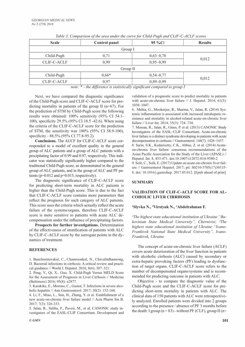
GEORGIAN MEDICAL NEWS No 5 (278) 2018
© GMN 101
Next, we have compared the diagnostic significance of the Child-Pugh score and CLIF-C-ACLF score for pre-dicting mortality in patients of the group II (n=67). For the prediction of STM by Child-Pugh score the following results were obtained: 100% sensitivity (95% CI 54.1-100), specificity 29.5% (95% CI 18.5 -42.6). When using the criteria of the CLIF-C-ACLF score for the prediction of STM, the sensitivity was 100% (95% CI 58.9-100), specificity - 88.5% (95% CI 77.8-95.2).
Conclusions. The AUCF for CLIF-C-ACLF score cor-responded to a model of excellent quality in the general group of ALC patients and a group of ALC patients with a precipitating factor of 0.99 and 0.97, respectively. This indi-cator was statistically significantly higher compared to the traditional Child-Pugh score, as demonstrated in the general group of ALC patients, and in the group of ALC and PF pa-tients (p=0.012 and p=0.015, respectively).
The diagnostic significance of CLIF-C-ACLF score for predicting short-term mortality in ALC patients is higher than the Child-Pugh score. This is due to the fact that CLIF-C-ACLF score contains more parameters that reflect the prognosis for such category of ALC patients. This score uses the criteria which actually reflect the acute failure of the systems/organs, therefore CLIF-C-ACLF score is more sensitive to patients with acute ALC de-compensation under the influence of precipitating factors.
Prospects for further investigations. Determination of the effectiveness of stratification of patients with ALC by CLIF-C-ACLF score by the surrogate points in the dy-namics of treatment.
REFERENCES
1. Bunchorntavakul, C., Chamroonkul, N., Chavalitdhamrong, D. Bacterial infections in cirrhosis: A critical review and practi-cal guidance // World J. Hepatol. 2016; 8(6); 307–321. 2. Peng, Y., Qi, X., Guo, X. Child-Pugh Versus MELD Score for the Assessment of Prognosis in Liver Cirrhosis // Medicine (Baltimore) 2016; 95(8): e2877. 3. Karakike, E., Moreno, C., Gustot, T. Infections in severe alco-holic hepatitis // Ann Gastroenterol. 2017; 30(2): 152-160. 4. Li, F., Miao, L., Sun, H., Zhang, Y. et al. Establishment of a new acute-on-chronic liver failure model // Acta Pharm Sin B. 2017; 7(3): 326-333. 5. Jalan, R., Saliba, F., Pavesi, M., et al. CANONIC study in-vestigators of the EASL-CLIF Consortium. Development and
validation of a prognostic score to predict mortality in patients with acute-on-chronic liver failure // J. Hepatol. 2014; 61(5): 1038–1047. 6. Mehta, G., Mookerjee, R., Sharma, V., Jalan, R. (2014) Sys-temic inflammation is associated with increased intrahepatic re-sistance and mortality in alcohol-related acute-on-chronic liver failure // Liver Int. 2014; 35(3): 724–734. 7. Moreau, R., Jalan, R., Gines, P. et al. (2013) CANONIC Study Investigators of the EASL–CLIF Consortium. Acute-on-chronic liver failure is a distinct syndrome developing in patients with acute decompensation in cirrhosis // Gastroenterol. 144(7), 1426–1437. 8. Sarin, S.K., Kedarisetty, C.K., Abbas, Z. et al. (2014) Acute-on-chronic liver failure: consensus recommendations of the Asian Pacific Association for the Study of the Liver (APASL) // Hepatol. Int. 8, 453-471. doi:10.1007/s12072-014-9580-2 9. Sole, C., Solà, E. (2017) Update on acute-on-chronic liver fail-ure // Gastroenterol Hepatol. 2017; pii: S0210-5705(17)30135-8. doi: 10.1016/j.gastrohep. 2017.05.012. [Epub ahead of print]
SUMMARY
VALIDATION OF CLIF-C-ACLF SCORE FOR AL-COHOLIC LIVER CIRRHOSIS
¹Slyvka N., 2Virstyuk N., ¹Abdelrahman F.
¹The highest state educational institution of Ukraine “Bu-kovinian State Medical University”, Chernivtsi; ²The highest state educational institution of Ukraine “Ivano-Frankivsk National State Medical University”, Ivano-Frankivsk, Ukraine
The concept of acute-on-chronic liver failure (ACLF) covers acute deterioration of the liver function in patients with alcoholic cirrhosis (ALC) caused by secondary or extra-hepatic provoking factors (PF) leading to dysfunc-tion of target organs. CLIF-C-ACLF score refers to the number of decompensated organs/systems and is recom-mended for predicting outcome in patients with ALC.
Objective - to compare the diagnostic value of the Child-Pugh score and the CLIF-C-ACLF score for pre-dicting short-term mortality in patients with ALC. The clinical data of 150 patients with ALC were retrospective-ly analyzed. Enrolled patients were divided into 2 groups according to the presence / absence of PF 3 months before the death: I group (n = 83) - without PF (CLF), group II (n=
Table 3. Comparison of the area under the curve for Child-Pugh and CLIF-C-ACLF scoresScale Control panel 95 %CІ Results
Group IChild-Pugh 0,71 0,63–0,78
0,012CLIF-C-ACLF 0,99 0,95–0,99
Group IIChild-Pugh 0,66* 0,54–0,77
0,012CLIF-C-ACLF 0,97 0,89–0,99
note: * - the difference is statistically significant compared to group I
102
МЕДИЦИНСКИЕ НОВОСТИ ГРУЗИИ
CFMFHSDTKJC CFVTLBWBYJ CBF[KTYB
67) - with PF (ACLF). To assess the severity of ALC we used the Child-Pugh score and the CLIF-C-ACLF score. Infectious complications were considered as PF. The sen-sitivity of the STMP by Child-Pugh score in group 1 was 100% (95% CI 58.9-100), specificity was 38.9% (95% CI 30.9-47.4). The sensitivity for the CLIF-C-ACLF score was 100% (95% CI 58.9-100), specificity-93.75% (95% CI 88.5-97.1).A. The sensitivity of STMP by Child-Pugh score in group II was 100% (95% CI 54.1-100), speci-ficity was 29.5% (95% CI -42.6 to 18.5). The sensitivity of STMP by CLIF-C-ACLF in score II was 100% (95% CI 58.9-100), specificity was 88.5% (95% CI 77.8-95.2). The CLIF-C-ACLF corresponded to the model of excel-lent quality in groups I (0.99) and II (0.97) and was higher than the Child-Pugh score in both groups (p = 0.012 and p = 0.015 respectively).
The diagnostic value of the CLIF-C-ACLF score for predicting short-term mortality in patients with ALC is higher than Child-Pugh, especially for acute decompen-sation of ALC caused by precipitating factors.
Keywords: alcoholic liver cirrhosis, short-term mor-tality, CLIF-C-ACLF score.
РЕЗЮМЕ
ВАЛИДАЦИЯ ШКАЛЫ CLIF-C-ACLF ДЛЯ АЛ-КОГОЛЬНОГО ЦИРРОЗА ПЕЧЕНИ
1Сливка Н.А., 2Вирстюк Н.Г., 1Абдельрахман Ф.А.
1Высшее образовательное учреждение Украины «Буко-винский государственный медицинский университет», Черновцы; 2Высшее образовательное учреждение Украины «Ивано-Франковский национальный государ-ственный медицинский университет», Украина
Целью исследования явилась сравнительная оцен-ка диагностической ценности двух шкал Child-Pugh и CLIF-C-ACLF для прогнозирования ранней смертно-сти у пациентов с алкогольным циррозом печени.
Ретроспективно проанализированы клинические данные 150 пациентов с алкогольным циррозом пе-чени (АЦП). Пациенты в зависимости от наличия/отсутствия провоцирующих факторов (ПФ) за 3 меся-ца до смерти разделены на 2 группы: I группа (n=83) - без ПФ – только хроническая печеночная недоста-точность (ХПН), II группа (n=67) - с ПФ и ОХПН. Для оценки степени тяжести АЦП и прогноза ранней смертности (ПРС) использованы шкалы Child-Pugh и CLIF-C-ACLF. Инфекционные осложнения учитыва-лись как ПФ. Чувствительность ПРС по шкале Child-Pugh в I группе составила 100% (95% ДИ 58,9-100), специфичность - 38,9% (95% ДИ 30,9-47,4). Чувстви-тельность ПРС для шкалы CLIF-C-ACLF составила 100% (95% ДИ 58,9-100), специфичность - 93,75% (95% ДИ 88,5-97,1). Чувствительность ПРС по шка-ле Child-Pugh во II группе составила 100% (95% ДИ
54,1-100), специфичность - 29,5% (95% ДИ 42,6-18,5); чувствительность ПРС по CLIF-C-ACLF во II группе составила 100% (95% ДИ 58,9-100), специфичность - 88,5% (95% ДИ 77,8-95,2).
Исходя из полученных в результате проведенного ис-следования данных следует заключить, что диагности-ческая ценность шкалы CLIF-C-ACLF для прогнозиро-вания ранней смертности у пациентов с АЦП выше, чем у шкалы Child-Pugh, особенно для острой декомпенса-ции АЦП, вызванной преципитирующими факторами.
reziume
Child-Pugh da CLIF-C-ACLF skalebis efeqturo-bis SedarebiTi Sefaseba RviZlis alkoho-luri cirozis diagnostikaSi
1n. slivka, 2n. virstiuki, 1f. abdelraxmani
1bukovinis saxelmwifo samedicino univer-siteti, Cernovci; 2ivano-frankovskis erovnuli saxelmwifo samedicino universiteti, ukraina
kvlevis mizans warmoadgenda Child-Pugh da CLIF-C-ACLF skalebis diagnostikuri efeqturobis SedarebiTi Sefaseba adreuli sikvdilis prognozirebaSi RviZlis alko-holuri ciroziT avadmyofebSi.
retrospeqtulad gaanalizebulia RviZ-lis alkoholuri ciroziT (Rac) 150 paci-entis klinikuri monacemebi. provocirebadi faqtorebis gaTvaliswinebiT pacientebi ga-nawilebuli iyo 2 jgufad: I jgufi – pro-vocirebadi faqtorebis gareSe – mxolod RviZlis qronikuli uknarisobiT (Rqu), n=83. II jgufi^- Rqu provocirebadi faqtorebiT, n=67. Rac simZmis xarisxis SefasebisaTvis da adreuli sikvdilis prognozisaTvis gamo-yenebuli iyo zemoxsenebuli orive skala infeqciuri garTulebebis, rogorc provoci-rebadi faqtorebis gaTvaliswinebiT.
I jgufSi sikvdilianobis prognozi Child-Pugh skalis mixedviT Seadgenda 100% (95% sarwmuno intervaliT 58,9-100), specifiuroba – 38,9% (95% sarwmuno intervaliT 30,9-47,4), xolo CLIF-C-ACLF skaliT naadrevi sikvdi-lobis prognozi Seadgenda 100% (95% sar-wmuno intervaliT 58,9-100), specifiuroba – 93,75% (95% sarwmuno intervaliT 88,8-97,1).
II jgufSi sikvdilobis adreuli prog-nozis mgrZnobeloba Child-Pugh skaliT Seadgenda 100% (95% sarwmuno intervaliT 54,1-100), specifiuroba 29,5% (95% sarwmuno in-tervaliT 42,6-18,50).
CLIF-C-ACLF skalis mixedviT mgrZnobelo-ba II jgufis pacientebSi Seadgenda 100% (95% sarwmuno intervaliT 58,9-100), specifiuroba
GEORGIAN MEDICAL NEWS No 5 (278) 2018
© GMN 103
88,5% (95% sarwmuno intervaliT 77,7-95,2). Cata-rebuli kvlevis Sedegebze dayrdnobiT, av-torebs gamotanili aqvT daskvna, rom CLIF-C-ACLF skalis diagnostikuri efeqturoba
adreuli sikvdilobis prognozirebisaTvis pacientebSi Rac-iT gacilebiT ufro maRalia, vidre Child-Pugh skalis, gansakuTrebiT Rac-is mwvave dekompensaciis pirobebSi.
ASSESSMENT OF ADEQUATE IODINE AVAILABILITY TO THE POPULATION OF WEST KAZAKHSTAN BASED ON THE DATA OF INORGANIC IODINE IN URINARY EXCRETION
Bazargaliyev Y., Batyrova G., Zhamankulova D., Agzamova R.
West Kazakhstan Marat Ospanov State Medical University, Aktobe, Republic of Kazakhstan
The iodine deficiency in the environment and the health disorders caused by it - iodine deficiency disorders (IDD) - are a serious medical and social problem throughout the world. Thyroid hormones are extremely important for brain development and the formation of intelligence in the period of prenatal development. The main cerebral functions are laid in the early stages of intrauterine life [6,12].
Accumulated up to date, international experience has shown that universal iodization of salt is the simplest, most cost-effective and most reliable way to combat iodine deficiency. The target groups for IDD prevention programs are school-age children, women of childbearing age and newborns. Children of school age are characterized, on the one hand, by high vulnerability, and on the other hand, by accessibility for observation. Children, who are susceptible to the disease, have an enlarged thyroid gland, which is a reaction to iodine deficiency, and children are readily available for screening in schools [11,13,14]. The main problem with surveys in schools is that not all children are covered, and this can distort the results of surveys. In Kazakhstan, primary and secondary education is free and generally accessible, which guarantees full coverage of the children surveyed in the study.
The determination the median of urinary iodine concentration (UIC) is a reliable biochemical indicator for an adequate assessment of the population’s iodine supply [13]. Most of the iodine that enters the human body is excreted in the urine. Therefore, urinary excretion of iodine is the evidence of the recent intake of iodine along with food.
The aim of the research: to study the iodine supply of the region according to the degree of urinary iodine excretion in the West region of the Republic of Kazakhstan.
Material and methods. The study was conducted in the territory of West Kazakhstan (Atyrau, Aktobe, West Kazakhstan provinces) in accordance with the recommendations of WHO [13]. The study protocol was approved by the University Ethics Committee (Minutes No. 04 of 08.10.2013). In this study were involved schoolchildren 7-11 years old. The criteria for the inclusion in the study are: the availability of written
informed consent from the parents or guardians before being included in the study. Children with serious diseases of the heart, liver, kidneys, thyroid (thyroid gland) are excluded. The size of the thyroid gland and the standard standards of thyroid gland volume (in ml) in schoolchildren were determined and evaluated by the ultrasound of the thyroid gland depending on the body surface area and sex in accordance with the recommendations of the WHO (2007) on MB Zimmermann [13,16]. To determine the excretion of inorganic iodine in a single portion of urine in the field, express diagnostics, the “Iodine test” kits (manufactured in Ukraine), was applied. Of 6493 schoolchildren participating in the study of the frequency of goiter, random sampling was applied to select 884 children to determine UIC. The collection of urine for the determination of UIC was carried out in disposable cups, hermetically sealed with stoppers to prevent the entry of iodine vapors into the test samples. The test was carried out immediately after urine collection. The concentration of iodine in the urine was expressed in μg/L. The method is semiquantitative and makes it possible to distinguish urine samples with iodine content below 70, from 70 to 100, from 100 to 300 and above 300 μg/L.
Statistical processing was carried out using parametric and nonparametric methods using the Statistica,10 program (Stat Soft.USA). The results of the study are presented as the mean (M), standard deviation (SD), in the case of an abnormal distribution, in the form of a median (Me). Categorical data are shown as numbers and percentages with 95% confidence intervals (CI). Statistically significant were the values of the criteria corresponding to p˂0.05.
Results and their discussion. The analysis of the obtained data on the study of UIC in schoolchildren in the West Kazakhstan showed that the proportion of children with optimal urinary excretion of iodine (100-300 μg/L) is 62.67% (95% CI: 59.48–65.86%). Indices ranging from 100 to 200 μg/L were detected in 27.15% (95% CI: 24.22–30.08%), more than 200 μg/L and up to 300 μg/L - in 35.52% (95% CI: 32.37–38.68%) of schoolchildren (Fig. 1).
104
МЕДИЦИНСКИЕ НОВОСТИ ГРУЗИИ
CFMFHSDTKJC CFVTLBWBYJ CBF[KTYB
Fig. 1. Excretion of iodine in a single portion of the urine of schoolchildren aged 7-11 years (West Kazakh-stan)
UIC exceeds 300 μg/L - in 18.89% (95% CI: 16.31–21.47%) of schoolchildren. Concentration of iodine in the urine of more than 400 μg/L was determined in 1.81% (95% CI: 0.93–2.69%) of children. According to the literature, for a population that has had a prolonged iodine deficiency in food, the increase in the median concentration of iodine in the urine of more than 300 μg/L against a background of a rapid increase in iodine intake is considered undesirable due to the risk of occurrence of iodine-induced hyperthyroidism and autoimmune thyroid diseases. These complications develop during the first 5-10 years after the beginning of salt iodization. It should be noted that iodine-induced hyperthyroidism, according to the studies, is more often found in older people who had previously been diagnosed with nodular goiter, but can also occur when iodine intake did not go beyond the norm [13,15, 17]. Low excretion rates of iodine, less than 70 μg/L, were detected in 2.83 % (95% CI: 1.74–3.92%) of schoolchildren, and from 70 μg/L to 100 μg/L - in 13.8% (95% CI: 11.53–16.07%).
According to the recommendations of WHO, UNICEF, ICCIDD the main criteria for the effectiveness of iodine prophylaxis programs are: target UIC median level of more than 100 μg/L, detected in more than half of those surveyed; The level of UIC is below 50 μg/L in less than 20% of the examined. Epidemiological criteria for assessing iodine availability based on median UIC in school-age children (WHO, 2007): with iodine median ˂ 20 μg/L iodine intake is considered insufficient and is regarded as severe iodine deficiency; Median 20-49 μg/L - moderate deficiency; Median 50-99 μg/L - weak deficiency; 100-199 μg/L - optimal availability; 200-299 μg/L - danger of iodine-induced hyperthyroidism; More than 300 μg/L - the danger of negative health effects [13].
The individual indices of UIC are very variable and the generalized analysis of UIC is carried out only at the population level. Nevertheless, we carried out the correlation analysis between thyroid volume and the degree of UIC showed no connection between these parameters (r=0.20, p=0.004), which is confirmed by our literary data.
Fig. 2 shows the results of studying UIC in the context of the urban areas/rural districts. Optimum iodide supply was detected in 64.39% (95% CI: 58.76–70.02%) of urban children and 61.88% (95% CI: 58.01–65.75%) of children from rural districts. Iodine deficiency in the regions of the rural districts was observed more often (18.32% (95% CI: 15.24–21.40%)) than in the urban areas (12.95% (95% CI: 9.00–16.90%)) (p <0.05).
Fig. 2. Indices of iodine excretion in a single portion of urine of schoolchildren 7-11 years old in the context of the urban areas / rural districts (%)
The excess of the indicators is mainly due to the difference in the easy deficit, so in the city it was 22.66% (95% CI: 17.74–27.58%), then in the rural districts it was diagnosed in 19.80% (95% CI: 16.63–22.97%) of children. Severe and moderate severity of iodine deficiency in the regional center was 0.72% (95% CI: -0.27–1.71%), in the districts it was detected in 3.80% (95% CI: 2.27–5.32%) of children. The data obtained are consistent with the results of a nationwide study conducted by the Academy of Nutrition in 2006 in Kazakhstan, where the median UIC is 235.9 μg/L, on the basis of which in 2008 Kazakhstan was assigned to countries where the goals for eliminating iodine deficiency in the diet [1,2,4].
Today, universal iodization of food and feed salt in Kazakhstan is regulated by the Law of the Republic of Kazakhstan «On the Prevention of Iodine Deficiency Diseases» of 14.10.2003, No. 489-PZRK. Currently, almost all the salt produced in Kazakhstan, as well as imported into the Republic, is enriched with iodine. In order to implement the law on salt iodization, the Order of the Ministry of Health of the Republic of Kazakhstan «On approval of the Rules for the monitoring of the quality, production, storage, import and sale of iodized edible salt and other enriched iodine compounds with food products» dated 18.08.2006 No. 641.
Within the framework of the research «Epidemiology of endemic goiter in the Western region of Kazakhstan and the development of recommendations for the prevention of iodine deficiency states,» the study was made of the incidence of goiter in the West Kazakhstan, where it was revealed that the region has a moderate to severe goiter endemia [8,9].
Thus, on the basis of studying the concentration of iodine in the urine in schoolchildren, it was revealed that in 62.67%
GEORGIAN MEDICAL NEWS No 5 (278) 2018
© GMN 105
(95% CI: 59.48–65.86%) there is an optimal allocation of iodine in the urine, also using data from earlier studies in the Republic of Kazakhstan [1,2], it can be considered that iodine status of the population of West Kazakhstan is adequate. It should be noted that the high percentage of children diagnosed with iodine densities of more than 300 μg/L requires further monitoring and possible correction of salt iodization standards. The high prevalence of goiter in the region against the background of increased iodine supply from the data of urinary iodine excretion suggests the presence of other goitrogenic factors in the region.
The data of numerous studies show that the development of endemic goiter is due to the effect of a complex of dif-ferent (natural, climatic, ecological, etc.) factors that have a strumogenic effect. The increase in the interest in finding ad-ditional goitrogenic factors: the deterioration of the radiation situation, the impact of macro- and microelements, and the clarification of the possibility of anthropogenic pollution of the environment by various eco-pollutants, is explained by the fact that territories with a tense endemic [3,7,10]. The absence of iodine deficiency in the region, and also the area where the severity of goiter prevalence does not correspond to the severity of iodine deficiency [5].
Conclusion. In West Kazakhstan, in the absence of iodine deficiency, a strained goiter endemia remains. The formation of goiter endemia in the region does not exclude the influence of other strumogenic factors, which need further study.
Acknowledgements. The research was performed during 2013-2015 in frames of the scientific Project “Epi-demiology of Endemic Goiter in the Western region of the Republic of Kazakhstan and development of recommen-dations for the prevention of Iodine deficiency states”, funded by the Committee of Science of the Ministry of Education and Science of the Republic of Kazakhstan (State Registration No. of the National Research Center of the Technical Institute 0113RK00439, Kazakhstan).
REFERENCES
1. Беисбекова А.К., Кайнарбаева М.С., Быкыбаева С.А., Ка-рагизова А.Б., А.Б. Чуенбекова, Е.Ю. Ушанская, А.Б. Бер-дыгалиев, Ф.Е. Оспанова. Обеспеченность йодом детей и женщин репродуктивного возраста в Павлодарской, Северо-Казахстанской и Актюбинской областях. Вестник КазНМУ. 2015; 1: 404-407.2. Беисбекова А.К., Оспанова Ф.Е., Толысбаева Ж.Т., Адлет М. Оценка йодной ситуации среди женщин репродуктивно-го возраста РК. Вестник КазНМУ. 2013; 3 (1): 82-84.3. Кудабаева Х.И., Батырова Г.А., Базаргалиев Е.Ш., Агза-мова Р.Т., Нуфтиева А.И. Микроэлементный статус детей с увеличенной щитовидной железой в Западно-Казахстан-ской области. Georgian Med News. 2017; (263): 64-71.4. Тиммер А., Герасимов Г.А. Прогресс в устойчивом устра-нении ЙДЗ в странах региона центральной и восточной Ев-ропы и содружества независимых государств. Клиническая и экспериментальная тиреоидология. 2008; 4 (2): 4-7.5. Рослякова Е.В. Роль факторов среды обитания в формиро-вании риска йоддефицитных заболеваний. Здравоохранение
Российской Федерации. 2009; 3: 30-34.6. Andersson M., Karumbunathan V., Zimmermann M.B. Glob-al iodine status in 2011 and trends over the past decade. J Nutr. 2012;142(4):744.7. Gorbachev A.L., Skalny A.V., Koubassov R.V. Bioelement ef-fects on thyroid gland in children living in iodine-adequate terri-tory. J Trace Elem Med Biol. 2007; 21 Suppl 1: 56-58. 8. Kudabaeva K.I., Yermukhanova L.S., Koshmaganbetova G.K., Bazargaliev Y.S., Baspakova A.M., Kaldybaev K.K., Kaldybaeva A.T. Estimation of the thyroid gland volume by means of ultraso-nography among school children in Aktobe Area, Kazakhstan. Res. J. Pharm., Biol. Chem. Sci. 2015; 6(2): 87-93. 9. Kudabayeva K. I., Bazargaliev Y. S., Baspakova A. M., Dar-zhanova K. B. Estimation of thyroid volume in children from oil-gas producing areas of West Kazakhstan. Biology and Medi-cine 2014; 6(1): 1–6.10. Kudabayeva K.I., Koshmaganbetova G.K., Mickuviene N., Skalnaya M.G., Tinkov A.A., Skalny A.V. Hair Trace Elements are Associated with Increased Thyroid Volume in Schoolchil-dren with Goiter. Biol. Trace Elem. Res. 2016; 174 (2): 261-266.11. Rendl J., Bier D., Groh T., Reiners C. Rapid urinary iodide test. J Clin Endocrinol Metab.1998; 83(3): 1007-1012.12. Speeckaert M.M., Speeckaert R., Wierckx K., Delanghe J.R., Kaufman J.M. Value and pitfalls in iodine fortification and supple-mentation in the 21st century. Br J Nutr. 2011; 106(7): 964-973. 13. WHO, UNICEF, ICCIDD: Assessment of iodine deficiency disorders and monitoring their elimination: a guide for pro-gramme managers. Third edition. - Geneva: World Health Or-ganization, 2007; 98 р. 14. Zimmermann M.В., Аndersson М. Prevalence of iodine de-ficiency in Europe in 2010. Ann. Endocrinol. (Paris). 2011; 72( 2): 164-166.15. Zimmermann M.B., Jooste P.L., Pandav C.S. Iodine-defiien-cy disorders. Lancet. 2008; 372(9645): 1251-1262. 16. Zimmermann M.B., Hess S.Y., Molinari L., De Benoist B., Delange F., Braverman L.E., Fujieda K, Ito Y, Jooste PL, Moosa K, Pearce EN, Pretell EA, Shishiba Y. New reference values for thyroid volume by ultrasound in iodine-sufficient schoolchil-dren: a World Health Organization/Nutrition for Health and De-velopment Iodine Deficiency Study Group Report. Am J Clin Nutr. 2004; 79(2): 231-237. 17. Zou Y., Ding G., Lou X., Zhu W., Mao G., Zhou J., Mo Z. Factors influencing thyroid volume in Chinese children. Eur J Clin Nutr. 2013; 67(11): 1138-1141.
SUMMARY
ASSESSMENT OF ADEQUATE IODINE AVAILA-BILITY TO THE POPULATION OF WEST KA-ZAKHSTAN BASED ON THE DATA OF INOR-GANIC IODINE IN URINARY EXCRETION
Bazargaliyev Y., Batyrova G., Zhamankulova D., Agzamova R.
West Kazakhstan Marat Ospanov State Medical Univer-sity, Aktobe, Republic of Kazakhstan
Aim of research – to study the iodine supply of the region according to the degree of urinary iodine excretion in the West region of the Republic of Kazakhstan.
106
МЕДИЦИНСКИЕ НОВОСТИ ГРУЗИИ
CFMFHSDTKJC CFVTLBWBYJ CBF[KTYB
Of 6493 schoolchildren participating in the study of the frequency of goiter, random sampling was applied to select 884 children to determine UIC. To establish the excretion of inorganic iodine in a single portion of urine in the field, express diagnostics, the “Iodine test” kits (manufactured in Ukraine), was applied. The collection of urine for the determination of UIC was carried out in disposable cups, hermetically sealed with stoppers to prevent the entry of iodine vapors into the test samples. The test was carried out immediately after urine collection. The concentration of iodine in the urine was expressed in μg/l. The method is semiquantitative and makes it possible to distinguish urine samples with iodine content below 70, from 70 to 100, from 100 to 300 and above 300 μg/L.
The obtained data on the study of UIC in schoolchildren in the West Kazakhstan showed that the proportion of children with optimal urinary excretion of iodine (100-300 μg/L) is 62.67% (95% CI: 59.48–65.86%). Indices ranging from 100 to 200 μg/L were detected in 27.15% (95% CI: 24.22–30.08%, more than 200 μg/L and up to 300 μg/L - in 35.52% (95% CI: 32.37–38.68%) of schoolchildren. UIC exceeds 300 μg/L - in 18.89% (95% CI: 16.31–21.47%) of schoolchildren. Concentration of iodine in the urine of more than 400 μg/L was determined in 1.81% (95% CI: 0.93–2.69%) of children. Low excretion rates of iodine, less than 70 μg/L, were detected in 2.83 % (95% CI: 1.74–3.92%) of schoolchildren, and from 70 μg/L to 100 μg/L - in 13.8 % (95% CI: 11.53–16.07%). Thus, in 62.67% (95% CI: 59.48–65.86%) there is an optimal allocation of iodine in the urine, it can be considered that iodine status of the population of West Kazakhstan is adequate.
In West Kazakhstan, in the absence of iodine deficiency, a strained goiter endemia remains.The formation of goiter endemia in the region does not exclude the influence of other strumogenic factors, which need further study.
Keywords: iodine, urinary iodine concentration (UIC), iodine deficiency, goiter, West Kazakhstan.
РЕЗЮМЕ
ОЦЕНКА ЙОДООБЕСПЕЧЕНИЯ НАСЕЛЕНИЯ ЗАПАДНОГО КАЗАХСТАНА ПО ДАННЫМ ЭКС-КРЕЦИИ НЕОРГАНИЧЕСКОГО ЙОДА В МОЧЕ
Базаргалиев Е.Ш., Батырова Г.А., Жаманкулова Д.Г., Агзамова Р.Т.
Западно-Казахстанский государственный медицин-ский университет им. Марата Оспанова, Актобе, Республика Казахстан
Целью исследования явилось изучение йодообе-спечения региона по степени йодурии в Западном ре-гионе Республики Казахстан.
Для определения экскреции неорганического йода
в разовой порции мочи в полевых условиях исполь-зовалась экспресс-диагностика с помощью наборов «Йод – тест» (Украина). Из 6493 школьников, уча-ствующих в исследовании по изучению частоты зоба, методом случайной выборки отобрано 884 детей для изучения йодурии. Сбор мочи проводился в одноразо-вые стаканчики, герметично закупоренные пробками для исключения попадания паров йода в исследуемые образцы. Тест проводился сразу после забора мочи. Концентрация йода в моче выражалась в мкг/л. Метод определения полуколичественный и позволяет отли-чать образцы мочи с содержанием йода ниже 70, от 70 до 100, от 100 до 300 и выше 300 мкг/л.
Согласно полученным данным, доля детей, имею-щих оптимальные показатели экскреции йода мочой (100-300 мкг/л), составила 62.67% (95% ДИ: 59.48–65.86%). Показатели в пределах 100-200 мкг/л выявле-ны у 27.15% (95% ДИ: 24.22–30.08%); более 200 мкг/л и до 300 мкг/л – у 35.52% (95% ДИ: 32.37–38.68%) школьников. Концентрация йода в моче превышала 300 у 18.89% (95% ДИ: 16.31–21.47%) и 400 мкг/л - у 1.81% (95% ДИ: 0.93–2.69%) школьников. Низкие по-казатели экскреции йода менее 70 мкг/л обнаружены у 2,83% (95% ДИ: 1,74–3,92%) детей, от 70 мкг/л до 100 мкг/л - у 13,8% (95% ДИ: 11,53–16,07%). Таким образом, у 62.67% (95% ДИ: 59.48–65.86%) детей на-блюдалось оптимальное содержание йода в моче, что свидетельствует об адекватном йодном статусе насе-ления Западного Казахстана.
Данные проведенного исследования позволяют за-ключить, что в Западном Казахстане при отсутствии йодной недостаточности сохраняется напряженная зоб-ная эндемия. На формирование зобной эндемии в регио-не не исключается влияние других струмогенных факто-ров, которые нуждаются в дальнейшем изучении.
reziume
dasavleT yazaxeTis mosaxleobis iodmoma-ragebis Sefaseba araorganuli iodis Sar-diT eqskreciis maCvenebelebis safuZvelze
e. bazargalievi, g. batirova, d. Jamankulova, r. agzamova
dasavleT yazaxeTis marat ospanovis sax. saxelmwifo samedicino universiteti, aqtobe, yazaxeTis respublika
kvlevis mizans Seadgenda regionis iod-momaragebis Sefaseba ioduriis xarisxis mixedviT yazaxeTis respublikis dasavleT regionSi.
araorganuli iodis eqskreciis gansaz-RvrisaTvis Sardis erTjerad ulufaSi savele pirobebSi gamoyenebuli iyo eqspres-diagnostika nakrebiT “iod-testi” (ukraina).
GEORGIAN MEDICAL NEWS No 5 (278) 2018
© GMN 107
Ciyvis sixSiris kvlevaSi monawile 6493 mos-wavlidan ioduriis SeswavlisaTvisDSemTx-veviTi amorCevis meTodiT SeirCa 884 bavSvi. Sardi grovdeboda erTjerad WiqebSi, her-metuli TavsaxuriT - Sesaswavl nimuSebSi iodis orTqlis moxvedris gamosaricxad. tes-ti tardeboda Sardis miRebisTanave. SardSi iodis koncentracia gamoixateboda mkg/l-Si. gansazRvris meTodi naxevrad-raodenobrivia da iZleva Sardis nimuSebis diferencirebis saSualebas iodis 70-ze naklebi, 70-100, 100-300 da 300 mkg/l-is Semcve-lobiT.
miRebuli Sedegebis mixedviT, SardiT io-dis eqskreciis optimaluri maCveneblebiT (100-300 mkg/l) gamovlinda bavSvebis 62.67%; 100-200 mkg/l-is farglebSi - moswavleTa
27.15%-Si, 200-300 mkg/l - 35.52%-Si; 300 mkg/l meti odenobiT - 18.89%-Si; SardSi iodis 70 mkg/l-ze dabali Semcveloba aRmoCnda bavS-vebis 2,83%-Si, 70-100 mkg/l ki - 13,8%-Si.
amrigad, bavSvebis 62.67%-Si aRiniSneba SardSi iodis optimaluri Semcveloba, rac miuTiTebs iodis adekvatur statusze dasav-leT yazaxeTis mosaxleobaSi.
Catarebuli kvlevis Sedegebi iZleva daskvnis safuZvels imis Sesaxeb, rom dasav-leT yazaxeTSi iodis ukmarisobis ararse-bobis pirobebSi aRiniSneba Ciyvis daZabuli endemia. savaraudoa, rom mis formirebaSi ar aris gamoricxuli sxva strumogenuli faq-torebis gavlena, rac momaval gamokvlevebs saWiroebs.
GOODPASCHER’S SYNDROME - THE CHALLENGES IN A TIMELY DIAGNOSIS AND TREATMENT IN MEDICAL PRACTICE (CLINICAL CASE)
1Talash V., 2Bevzenko T., 1Yarmola T., 1Tkachenko L., 1Pustovoyt H.
1Higher state educational establishment of Ukraine “Ukrainian medical stomatological academy”, Poltava; 2State scientific establishment “Scientific-practical centre of prophylactic and clinical medicine”
of State affairs management, Kyiv
There is an increase in incidence of Goodpascher’s syndrome (GS) in modern practice of doctors. This syn-drome is one of the fast developing, prognostically unfa-vorable diseases with extremely complex diagnosis. The adequacy of the provision of medical care depends from timeliness of diagnosis. It slows the progression of the disease and increases the average life expectancy of pa-tients. Unfortunately, most of the described clinical cases have not been diagnosed in vivo and had a fatal outcome. All this determines the relevance of the coverage of each individual clinical case of GS for the accumulation of general experience and timely recognition of the symp-toms. Let’s dwell briefly on the literature review.
The term “Goodpascher’s syndrome” was introduced as a nosological unit by M. Stanton and J. Tange in 1958. Syndrome was named in honor of the Harvard’s Univer-city pathologist E.W. Goodpasture, who in 1919 first de-scribed the disease, which combined in itself pulmonary haemorrhage and severe glomerulonephritis (GN) with a lethal outcome [3,8,10].
According to the current classification of systemic vasculitis (Chapel Hill Consensus Conference, 2012), GS refers to immunocomplex vasculitis with lesions of small-caliber vessels [3,8,12]. By WHO definition GS is an anti-glomerular basal membrane GN associated with hemorrhage in pulmonary alveoli and with the presence
of circulating antibodies (IgG) to the basal membrane of glomerular capillaries (BMC) and/or alveoli (BMA) [5,8,10]. This syndrome in the literature can be found un-der various names: “glomerulonephritis with pulmonary hemorrhage”, “hemorrhagic pneumonitis with nephritis”, “pulmonary syndrome with glomerulonephritis” and most often - pulmonary kidney syndrome (PKS) or “anti-GBM illness” [3,13,14]. At the same time, some authors point out the possibility of developing GS under condition of vasculitis associated with anti-neutrophil cytoplasmic an-tibodies (ANCA) [9,13,14].
Incedence of is only 0.5-0.6 cases per 1 million of popu-lation. Most often it is observed in two age groups: 20-30 and 50-60 years old. There is prevalence of GS in men in young age group, but in the older age group, men and women suffer with the same frequency [2,7,14]. The average life expec-tancy of patients does not exceed 7.3 years [12].
The reason for the occurrence of GS is not well known, however there are several proven factors in the etiology of GS: genetic predisposition (histocompatibility antigen HLA of DR class (HLA-DRW2, HLA-DR15 and HLA-DR4) [3,7,8], tobacco smoking, hypothermia, the influence of bacterial infection, viruses (influenza A2 virus), medicines (carbimasole, D-penicillamine, amphetamine etc.), organic solvents, varnishes, gasoline [8-10], the possible after-effects of shock-wave lithotripsy and ureteral obstruction [1,10,13].
108
МЕДИЦИНСКИЕ НОВОСТИ ГРУЗИИ
CFMFHSDTKJC CFVTLBWBYJ CBF[KTYB
Diagnostics of GS is extremely complicated. Clini-cal manifestations depend on the degree of damage to the lungs and kidneys. Depending on the localization of the primary lesion, the disease may start with pulmonary or nephrology symptoms, so the clinical picture of GS may vary. There are also cases when the disease began with general nonspecific symptoms like manifestations of acute respiratory viral infection, arthralgia, myalgia, dyspeptic phenomena, abdominal syndrome, fever, weight loss [8], with the appearance of pericarditis, hemorrhages on the skin and mucous membranes, general weakness. Later to these manifestations the symptoms of pulmonary-renal syndrome (PRS) may join [3,8,13].
The thorough examination of the patient with the detec-tion of pathognomonic symptoms and the evaluation of the results of laboratory-instrumental diagnostic methods is de-cisive in diagnostic of GS. The diagnostic criteria for classi-cal course of GS include: cough, acute prophylactic alveolar hemorrhage or slowly passing hemosiderosis of the lungs with hemoptysis; dyspnea; GN with hematuria, rarely - ne-phrotic syndrome and rapidly progressive renal insufficiency (RI) with oligo-anuria; anemia, arterial hypertension (AH) (rarely) [4,6,7]. The main diagnostic markers of GS are the positive results of immunological studies of serum for the presence of antibodies to BMC and BMA [12-14] and anti-bodies to the immune globulins G or M and C3 fractions of the complement along the glomerular basal membrane of the glomeruli revealed by immunofluorescence study of the renal tissue biopsy [5,8,15]. Certain role in the diagnosis of GS have: X-ray (Ro-gram) of the chest cavity organs (CCO), which can reveal symmetrical cloudy, diffuse infiltrative or focal changes in the basal and central lung departments; nonspecific laboratory changes in the general blood test (GBT), which can show signs of iron deficiency anemia, neutrophilic leukocytosis, accelera-tion of erythrocyte sedation rate (ESR); general analysis of urine (GUA) can reveal proteinuria, erythrocyturia, cylindruria; in the biochemical analysis of blood (BBA) can show increase in the content of creatinine, urea, uric acid, potassium, total cholesterol and triglycerides (even in the absence of nephrotic syndrome); in the Reberg test there may be a decrease in the velocity of glomerular filtration (GFV) [4,7,13]; in the study of sputum we can discover hemosiderin and during bronchoscopy with bron-choalveolar lavage there will be siderophages and a large num-ber of red blood cells [8,10,13].
There are three variants of the course of PRS under GS: 1) recurrent hemorrhagic pneumonia and diffuse fast developing GN with RI; 2) slowly progressing lung and kidney damage; 3) GN with RI, to which join symptoms of lung injury on the final stage: cough, often without he-moptysis or pulmonary hemorrhage [6,7,9]; cardiac asth-ma and pulmonary edema are also often present [8,13,15].
The presence of different variants of the PKS and the lack of awareness of the general medical community re-garding GS complicates early diagnosis and timely and adequate appointment of treatment. Such situation leads to acceleration of the the disease progression and reduces the average life expectancy of patients [1,3,11].
One of the main factors determining the prognosis of such patients is the timeliness and adequacy of medi-cal care. In the absence of treatment of GS the lethality among patients reaches 75-90% [1,8,10].
Modern therapy of GS include urgent immunosup-pression and standard induction therapy with glucocor-ticosteroids (GCS), cytostatics and the conduct of plas-mapheresis sessions, and when remission is achieved, cytostatic maintenance therapy in combination with mini-mal or moderate doses of GCS [1,2,11]. For patients with terminal renal insufficiency, baseline therapy is combined with hemodialysis sessions, and, if necessary, in the ab-sence of antibodies in the blood for 6 months - permanent substitution renal therapy [2,3,9].
However, despite the timeliness and the adequacy of treatment, the mortality rate varies from 10 to 40% [1,8,11].
In our practice we encountered a complicated diagnos-tic case of the rapidly progressing GS, which manifested a GN with urinary tract syndrome. To this symptoms on the 5th month of decese joned AH, progressive RI, anemia, and symptoms of lung injury, accompanied by shortness of breath, episodic appearance of blood streams and pul-monary hemorrhage that arose on the penultimate day of the patient’s life.
Case report. We propose to your attention history of illness of patient L., 22 years old, arrived at the reception office of the POKL in an urgent order on August 31, 2017.
His complaints upon arrival were: edema of the legs, face, increase of arterial pressure (AP) up to 200/100, pain in the lumbar region of both sides, severe weakness, dys-pnea during physical activity, dizziness during changes of body position, nausea.
From anamnesis: sick since April 2017, when for the first time, without evidence, there were swelling on his legs. He received medical help on May 26, 2017. Hospi-talized in the therapeutic department of the Kyiv Clinical Hospital. In the course of the examination, for the first time, the presence of GN with uric acid syndrome and low urine concentration ability were detected. Left-sided nephropathy and neoplasm of the left kidney (angioli-poma?) were detected by ultrasound examination of the abdominal cavity organs (ACO). Following the diagnos-tic procedures appropriate antibacterial, diuretic, anti-ag-gregate therapy was conducted. On June 6, 2017, during computed tomography (CT) with intravenous contrast of the chest organs (CCO) and ACO, signs of hypertrophy of the Berten’s pillar in the left kidney and cyst of right kid-ney were revealed. The patient did not apply for further medical assistance.
The deterioration of the condition was noticed in the beginning of August when new edema on legs and face appeared again. Edema was not treated. On August 25, 2017 apeared headaches, dizziness, nausea, vomiting and AP went up to 160/90 mmHg. Patient used antihy-pertensive drugs, but the condition did not improve. On August 26 patient had cough, hemoptysis, raised body temperature to 37.8 0C. Later he was hospitalized in the

GEORGIAN MEDICAL NEWS No 5 (278) 2018
© GMN 109
therapeutic department of the Clinical Hospital in Kyiv. On August 28, 2017 he spontaneously left the hospital, and on August 29 he was hospitalized to the CRH at the place of residence with complaints of headache, increased blood pressure, coughing, periodic sensation of heaviness in the chest, edema of the legs and general weakness. On August 29, 2017, in the course of examination follow-ing changes were observed: in GBT - anemia (erythro-cytes - 3,5x10¹²/L, Hb - 108 g/L), leukopenia with a shift of the leukocyte formula to the left (white blood cells - 3,8x10⁹/L, rod-nuclear neutrophil -8%); in GUA - protein-uria - 1.0 g/L, red blood cells - 5-6 in field of vision (f/v), leukocyturia - 20-15 in f/v and cylinduria (grainy - 1-2 in f/v); on an electrocardiogram (ECG) an intravenous con-duction violation; on the R0-gram of CCO in the middle pulmonary fields: to the left the infiltration of the pul-monary figure was found, on the right focal shadows of medium intensity was found. Phthisiatrician conclusion: bilateral mid-partial non-hospital pneumonia, hemopty-sis. On August 30, 2017 after ultrasound scanning follow-ing changes were observed: ultrasound signs of hepato-megaly, dyskinesia of the biliary tract, nephritis, ascites. Assigned antibacterial, antihypertensive and mucolytic therapy did not have effect. Symptoms of anemia have increased with time (Hb - 108 - 106 g/L; Er. – 3.5x10¹²/L; Leu. - 3,4x10¹²/L); there were signs of lymphopenia (11%) and accelerated speed of erythrocyte sinking (SES) to 20 mm / hour. In GBT there was azotemia (creatinine 278 - 293 μmol/L), in urine there was proteinuria (1.0-1.47 g/L) and microhematuria (5-6 - 20-25 - 25-30 in f/v). Therefore on August 30th the patient was transferred to the depart-ment of intensive care, and on August 31 he was sent to the center of nephrology and dialysis POKL named after M.V. Sklifosovsky for stationary treatment with a diag-nosis: “Acute glomerulonephritis. Acute kidney damage of the second degree. Anemia of a mild degree. Arterial hypertension stage I, stage 2, the risk is very high. Uremic pneumonitis? Right kidney cyst”.
From anamnesis of life: he often suffered from ARVI. Patient worked in construction, performed hard physical work. There were frequent overcooling events. He smokes up to 20 cigarettes a day, abuses alcohol, energy tonics. Pa-tient states that he has taken amphetamine for 4 months. Al-lergic and hereditary anamnesis is not burdened.
When observed: Height - 175 cm, body weight - 74 kg. Patient was in moderate condition, skin and visible mucous membranes were pale, there was a pastosity of the legs. Frequency of respiratory movements was about (FRM) - 18/min. Lungs examination revealed: breath-ing rigid, weakened in the middle and lower parts. Pulse (Ps) was 88/min, rhythmic. AP was 190/120 mmHg. The rythm of the heart was correct, the tones were weakened, there was an accent of the second tone over the aorta. Abdomen was soft, painless. The tapping symptom was slightly positive on both sides. Daily diuresis (DD) was without changes, roughly 1.5 liters. Patient had no act of defecation for four days.
Analysis on Cito was conducted. On ECG there were signs of sinus tachycardia. Heart rate was 107/min. The electric heart apex was slightly deviated to the left. QRS Voltage was reduced. There were signs of hypoxia of the anterior wall of the left ventricle. On the R0-gram of CCO: pulmonary image is thickened, strengthened and deformed - mostly prurond (possibly due to the vascular component). Roots of lungs are with bands. Sinuses are free. Heart has no peculiarities. In GBT: (Er. 3.1-3.5x10¹²/L, Hb - 88 - 108 g/L), leukocytosis (7.5-17.1x10⁹/L), ESR 15 mm/hour; in the GUA: increased proteinuria (1.47-1.87 g/L), leukocyturia (20-15 - 8-10 in f/v), hematuria (er. - 20-30-1/2 (unchanged ) and 8 in f / v (changed), isolated single waxed cylinders were detected; in the BBT: alanine aminotransferase (ALT) - 76 units/L, aspartate aminotransferase (AST) - 91 units/L, total protein - 58 g/L, creatinine - 330.6 μmol/L, urea - 9.2 mmol/L, potassium - 6.8 mmol/L. Patient received detoxica-tion therapy, correction of arterial hypertension, anemia and hyperkalaemia. Treatment of ulcer was performed.
During the next four days, the patient’s condition did not change significantly. During physical examination: skin was pale, legs had signs of swelling, FRM was 18-20/min, in the middle and lower parts of the lungs crepitation was periodi-cally heard on the background of weakened vesicular breath-ing, a tachycardia was observed (heart rate - 94-98/minute), AP was stabilized at 140-150/90-100 mm Hg. Palpation of the abdomen revealed minor pains in the right hypochon-drium, along the intestine, without signs of irritation of the peritoneum. DD was within 1800-2400 ml.
From 1st till 4th of September, 2017, the patient was consulted by following specialists: a pulmonologist, a gastroenterologist, a cardiologist, an urologist, a hema-tologist, an oculist. Patient received following diagnos-tic procedures: esophagogastroduodenoscopy (erosive duodenopathy, erythematous gastropathy), ultrasound examination of the ACO and urogenital system (ultra-sound examination revealed signs of the nephritis, moder-ate strengthening of the liver structure). During control ECG we observed a decreased heart rate to 80 / min and reduced the amplitude of the T wave in V2-V5, echocar-diography there were no changes detected. We performed control of clinical and laboratory parameters of the pa-tient every day. In the dynamics of GBT we observed following changes: anemia has increased (Hb 86-74 g/L, Er. 2.98 - 2.63 - 2.47x10¹²/L), leukocytosis has decreased (10.2 - 5.8x10⁹/L), ESR changed in range: 36 - 10 - 37 mm/h. In BBT: ALT, AST, α-amylase, alkaline phospha-tase, and γ-GGT indices were normalized, however, there was an increase in LDH (620 units/L), cholesterol (6.68 mmol/L), triglycerides (2.42 mmol/L) and a decrease in albumin concentration (30 g/L) and total protein (48-54 g/L). There was a tendency to decrease of creatinine con-centration (354.8-328-290.7 μmol/L), urea (19.7 - 13.2 - 14.6 mmol/L) and potassium (5.5-5.3 - 4.32 mmol/L). The level of serum iron and the latent iron binding ability of the blood serum were within the normal range (15.8 and 24.5 μmol/L, respectively). Analysing blood proteins
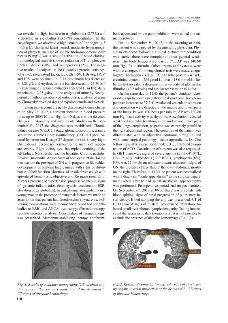
110
МЕДИЦИНСКИЕ НОВОСТИ ГРУЗИИ
CFMFHSDTKJC CFVTLBWBYJ CBF[KTYB
we revealed a slight increase in α2-globulins (12.73%) and a decrease in γ-globulins (13.59%) concentration. In the coagulogram we observed a high content of fibrinogen (4,5 - 4,6 g/L), shortened latent period, moderate hyperagrega-tion of platelets, increase of soluble fibrin-monomeric com-plexes (9 mg%) was a sing of activation of blood clotting. Immunological analysis showed reduction of T-lymphocytes (39%): T-helper (28%) and T-suppressor (12%). The nega-tive results of analyzes on the C-reactive protein, antistrep-tolysin-O, rheumatoid factor, LE-cells, RW, HBs-Ag, HCV, and HIV were obtained. In GUA proteinuria has decreased to 1.28 g/L and erythrocytriuria has decreased to 25-30 in f / v (unchanged); grained cylinders appeared (1 in f/v); daily proteinuria - 2.23 g/day; in the analysis of urine by Nechy-porenko method we observed eritocyturia; analysis of urine by Zimnytsky revealed signs of hypoisostenuria and nicturia.
Taking into account the newly discovered kidney chang-es on May 26, 2017, a constant increase in blood pressure (max up to 200/110 mm Hg) for 10 days and the detected changes in laboratory and instrumental studies on the Sep-tember 4th, 2017 the diagnosis was established: Chronic kidney disease (CKD) III stage: glomerulonephritis, urinary syndrome. Cronic kidney insufficiency (CKI) II degree. Ar-terial hypertension II stage 3rd degree, the risk is very high. Dislipidemia. Secondary normochromic anemia of moder-ate severity. Right kidney cyst. Incomplete doubling of the left kidney. Nonspecific reactive hepatitis. Chronic gastritis. Erosive Duodenitis. Angiospasm of both eyes’ retina. Taking into account the presence of GN with progressive RI, sudden development of bilateral lung damage with signs of distur-bance of their function (shortness of breath, fever, cough with episode of hemoptysis, objective and Ro-gram research in history), presence of hypertension, progressive anemia, signs of systemic inflammation (leukocytosis, acceleration ESR, elevation of α2 globulins), hyperkalaemia, dyslipidemia in a young man, in the presence of many risk factors, we made an assumption that patient had Goodpascher’s syndrome. Fol-lowing examinations were recomended: blood test for anti-bodies to BMC and ANCA, cystoscopy; fibrocolonoscopy, prostate secretion analysis. Consultation of reumathologist was prescribed. Membrane-stabilizing therapy, antithrom-
botic agents and proton pump inhibitors were added to treat-ment protocol.
On the September 5th, 2017, in the morning at 8.00, the patient was inspected by the attending physician. Phy-sician observed following clinical picture: the condition was stable, there were complaints about general weak-ness. The body temperature was 37.50C. AP was 140/90 mm Hg., Ps - 100/min. Other organs and systems were without changes. Following clinical tests were made: coagu-logram: fibrinogen - 4.8 g/L; GUA: total protein - 47 g/L, creatinine content - 284 μmol/L, urea - 13.0 mmol/L; Re-berg’s test revealed a decrease in the velocity of glomerular filtration (42.2 ml/min) and tubular reabsorption (93.1%).
On the same day at 11.45 the patient’s condition dete-riorated rapidly: developed abdominal syndrome, body tem-perature increased to 37.7 0C, weakened vesicular respiration and crepitation were detected in the middle and lower parts of the lungs. Ps was 100 beats per minute, AP was 130/80 mm Hg, heart activity was rhythmic. Auscultation revealed weakened vesicular breathing in the middle and lower parts of the lungs, crepitation, palpation was extremely painful in the right abdominal region. The condition of the patient was differentiated with an adjunctive syndrome during GS and with acute surgical pathology - acute appendicitis. On Cito following analysis were performed: GBT, ultrasound exami-nation of ACO. Consultation of surgeon was also requested. In GBT there were signs of severe anemia (Er. 2.64x10¹²/L, Hb - 75 g/L), leukocytosis (12.9x10⁹/L), lymphopenia (8%), ESR was 27 mm/h; on ultrasound scan: ultrasound signs of GN, the presence of free fluid in the lower abdomen, mostly on the right. Therefore, at 13.30 the patient was hospitalized with a diagnosis “acute appendicitis” to the surgical depart-ment, where after he had spinal anesthesia, appendectomy was performed. Postoperative period had no peculiarities. On September 6th, 2017 at 06.00 there was a cough with blood spitting, signs of rapid progression of pulmonary in-sufficiency. Blood stopping therapy was prescribed. CT of CCO shawed signs of bilateral pronounced infiltration, bi-lateral small hydrothorax, lymphadenopathy. Taking into ac-count the anamnestic data (hemoptysis), it is not possible to exclude the presence of alveolar hemorrhage (Fig. 1,2).
Fig. 2. Results of computer tomography (CT) of chest cav-ity organs in axial projection of the deceased L. CT-signs of alveolar hemorrhage
Fig. 1. Results of computer tomography (CT) of chest cav-ity organs in the coronary projection of the deceased L. CT-signs of alveolar hemorrhage
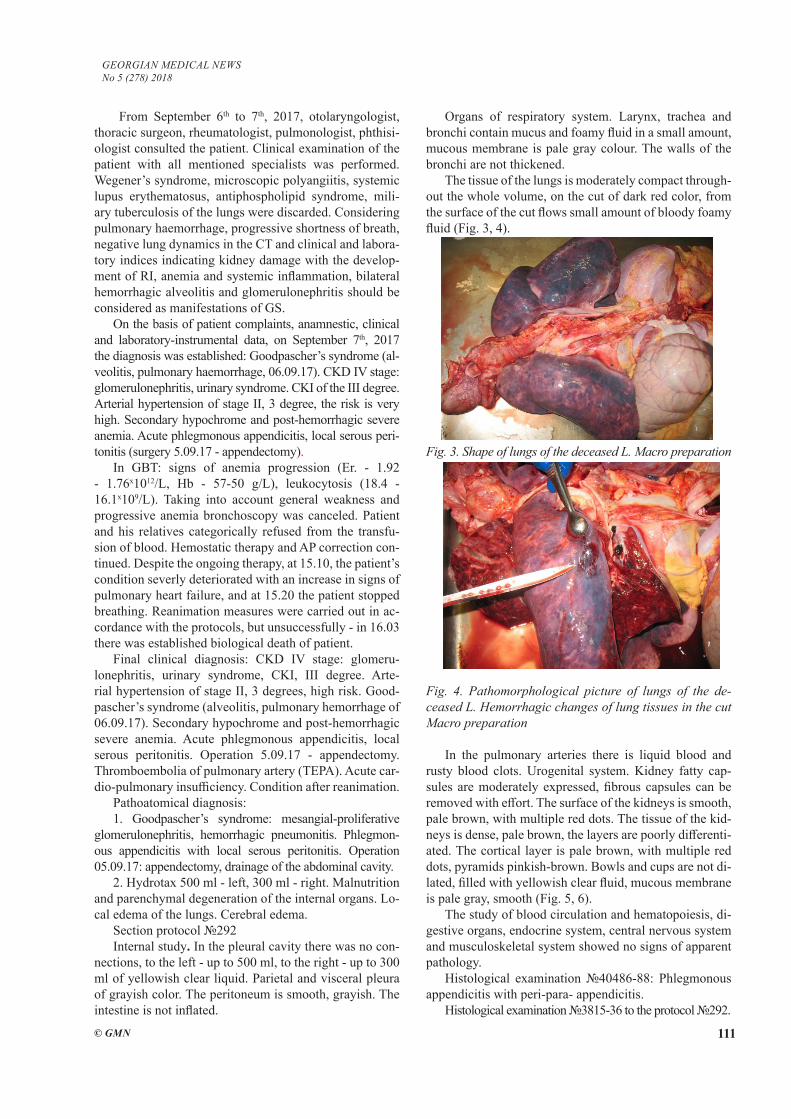
GEORGIAN MEDICAL NEWS No 5 (278) 2018
© GMN 111
From September 6th to 7th, 2017, otolaryngologist, thoracic surgeon, rheumatologist, pulmonologist, phthisi-ologist consulted the patient. Clinical examination of the patient with all mentioned specialists was performed. Wegener’s syndrome, microscopic polyangiitis, systemic lupus erythematosus, antiphospholipid syndrome, mili-ary tuberculosis of the lungs were discarded. Considering pulmonary haemorrhage, progressive shortness of breath, negative lung dynamics in the CT and clinical and labora-tory indices indicating kidney damage with the develop-ment of RI, anemia and systemic inflammation, bilateral hemorrhagic alveolitis and glomerulonephritis should be considered as manifestations of GS.
On the basis of patient complaints, anamnestic, clinical and laboratory-instrumental data, on September 7th, 2017 the diagnosis was established: Goodpascher’s syndrome (al-veolitis, pulmonary haemorrhage, 06.09.17). CKD IV stage: glomerulonephritis, urinary syndrome. CKI of the III degree. Arterial hypertension of stage II, 3 degree, the risk is very high. Secondary hypochrome and post-hemorrhagic severe anemia. Acute phlegmonous appendicitis, local serous peri-tonitis (surgery 5.09.17 - appendectomy).
In GBT: signs of anemia progression (Er. - 1.92 - 1.76x10¹²/L, Hb - 57-50 g/L), leukocytosis (18.4 - 16.1x10⁹/L). Taking into account general weakness and progressive anemia bronchoscopy was canceled. Patient and his relatives categorically refused from the transfu-sion of blood. Hemostatic therapy and AP correction con-tinued. Despite the ongoing therapy, at 15.10, the patient’s condition severly deteriorated with an increase in signs of pulmonary heart failure, and at 15.20 the patient stopped breathing. Reanimation measures were carried out in ac-cordance with the protocols, but unsuccessfully - in 16.03 there was established biological death of patient.
Final clinical diagnosis: CKD IV stage: glomeru-lonephritis, urinary syndrome, CKI, III degree. Arte-rial hypertension of stage II, 3 degrees, high risk. Good-pascher’s syndrome (alveolitis, pulmonary hemorrhage of 06.09.17). Secondary hypochrome and post-hemorrhagic severe anemia. Acute phlegmonous appendicitis, local serous peritonitis. Operation 5.09.17 - appendectomy. Thromboembolia of pulmonary artery (TEPA). Acute car-dio-pulmonary insufficiency. Condition after reanimation.
Pathoatomical diagnosis: 1. Goodpascher’s syndrome: mesangial-proliferative
glomerulonephritis, hemorrhagic pneumonitis. Phlegmon-ous appendicitis with local serous peritonitis. Operation 05.09.17: appendectomy, drainage of the abdominal cavity.
2. Hydrotax 500 ml - left, 300 ml - right. Malnutrition and parenchymal degeneration of the internal organs. Lo-cal edema of the lungs. Cerebral edema.
Section protocol №292Internal study. In the pleural cavity there was no con-
nections, to the left - up to 500 ml, to the right - up to 300 ml of yellowish clear liquid. Parietal and visceral pleura of grayish color. The peritoneum is smooth, grayish. The intestine is not inflated.
Organs of respiratory system. Larynx, trachea and bronchi contain mucus and foamy fluid in a small amount, mucous membrane is pale gray colour. The walls of the bronchi are not thickened.
The tissue of the lungs is moderately compact through-out the whole volume, on the cut of dark red color, from the surface of the cut flows small amount of bloody foamy fluid (Fig. 3, 4).
Fig. 3. Shape of lungs of the deceased L. Macro preparation
Fig. 4. Pathomorphological picture of lungs of the de-ceased L. Hemorrhagic changes of lung tissues in the cut Macro preparation
In the pulmonary arteries there is liquid blood and rusty blood clots. Urogenital system. Kidney fatty cap-sules are moderately expressed, fibrous capsules can be removed with effort. The surface of the kidneys is smooth, pale brown, with multiple red dots. The tissue of the kid-neys is dense, pale brown, the layers are poorly differenti-ated. The cortical layer is pale brown, with multiple red dots, pyramids pinkish-brown. Bowls and cups are not di-lated, filled with yellowish clear fluid, mucous membrane is pale gray, smooth (Fig. 5, 6).
The study of blood circulation and hematopoiesis, di-gestive organs, endocrine system, central nervous system and musculoskeletal system showed no signs of apparent pathology.
Histological examination №40486-88: Phlegmonous appendicitis with peri-para- appendicitis.
Histological examination №3815-36 to the protocol №292.
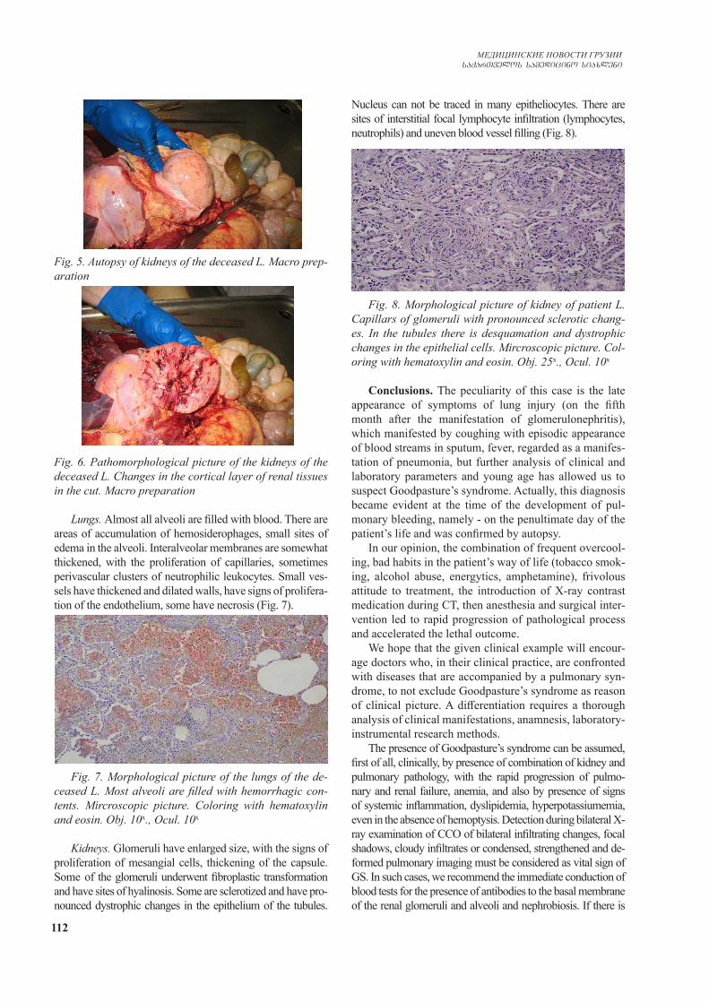
112
МЕДИЦИНСКИЕ НОВОСТИ ГРУЗИИ
CFMFHSDTKJC CFVTLBWBYJ CBF[KTYB
Fig. 5. Autopsy of kidneys of the deceased L. Macro prep-aration
Fig. 6. Pathomorphological picture of the kidneys of the deceased L. Changes in the cortical layer of renal tissues in the cut. Macro preparation
Lungs. Almost all alveoli are filled with blood. There are areas of accumulation of hemosiderophages, small sites of edema in the alveoli. Interalveolar membranes are somewhat thickened, with the proliferation of capillaries, sometimes perivascular clusters of neutrophilic leukocytes. Small ves-sels have thickened and dilated walls, have signs of prolifera-tion of the endothelium, some have necrosis (Fig. 7).
Fig. 7. Morphological picture of the lungs of the de-ceased L. Most alveoli are filled with hemorrhagic con-tents. Mircroscopic picture. Coloring with hematoxylin and eosin. Obj. 10х., Ocul. 10х
Kidneys. Glomeruli have enlarged size, with the signs of proliferation of mesangial cells, thickening of the capsule. Some of the glomeruli underwent fibroplastic transformation and have sites of hyalinosis. Some are sclerotized and have pro-nounced dystrophic changes in the epithelium of the tubules.
Nucleus can not be traced in many epitheliocytes. There are sites of interstitial focal lymphocyte infiltration (lymphocytes, neutrophils) and uneven blood vessel filling (Fig. 8).
Fig. 8. Morphological picture of kidney of patient L. Capillars of glomeruli with pronounced sclerotic chang-es. In the tubules there is desquamation and dystrophic changes in the epithelial cells. Mircroscopic picture. Col-oring with hematoxylin and eosin. Obj. 25х., Ocul. 10х
Conclusions. The peculiarity of this case is the late appearance of symptoms of lung injury (on the fifth month after the manifestation of glomerulonephritis), which manifested by coughing with episodic appearance of blood streams in sputum, fever, regarded as a manifes-tation of pneumonia, but further analysis of clinical and laboratory parameters and young age has allowed us to suspect Goodpasture’s syndrome. Actually, this diagnosis became evident at the time of the development of pul-monary bleeding, namely - on the penultimate day of the patient’s life and was confirmed by autopsy.
In our opinion, the combination of frequent overcool-ing, bad habits in the patient’s way of life (tobacco smok-ing, alcohol abuse, energytics, amphetamine), frivolous attitude to treatment, the introduction of X-ray contrast medication during CT, then anesthesia and surgical inter-vention led to rapid progression of pathological process and accelerated the lethal outcome.
We hope that the given clinical example will encour-age doctors who, in their clinical practice, are confronted with diseases that are accompanied by a pulmonary syn-drome, to not exclude Goodpasture’s syndrome as reason of clinical picture. A differentiation requires a thorough analysis of clinical manifestations, anamnesis, laboratory-instrumental research methods.
The presence of Goodpasture’s syndrome can be assumed, first of all, clinically, by presence of combination of kidney and pulmonary pathology, with the rapid progression of pulmo-nary and renal failure, anemia, and also by presence of signs of systemic inflammation, dyslipidemia, hyperpotassiumemia, even in the absence of hemoptysis. Detection during bilateral X-ray examination of CCO of bilateral infiltrating changes, focal shadows, cloudy infiltrates or condensed, strengthened and de-formed pulmonary imaging must be considered as vital sign of GS. In such cases, we recommend the immediate conduction of blood tests for the presence of antibodies to the basal membrane of the renal glomeruli and alveoli and nephrobiosis. If there is
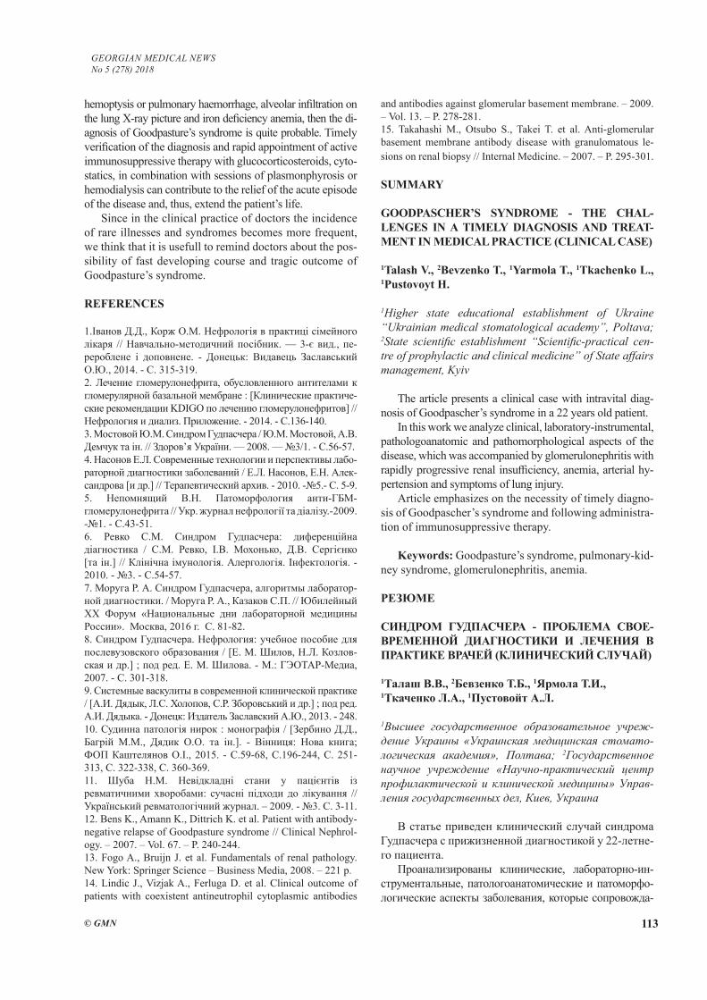
GEORGIAN MEDICAL NEWS No 5 (278) 2018
© GMN 113
hemoptysis or pulmonary haemorrhage, alveolar infiltration on the lung X-ray picture and iron deficiency anemia, then the di-agnosis of Goodpasture’s syndrome is quite probable. Timely verification of the diagnosis and rapid appointment of active immunosuppressive therapy with glucocorticosteroids, cyto-statics, in combination with sessions of plasmonphyrosis or hemodialysis can contribute to the relief of the acute episode of the disease and, thus, extend the patient’s life.
Since in the clinical practice of doctors the incidence of rare illnesses and syndromes becomes more frequent, we think that it is usefull to remind doctors about the pos-sibility of fast developing course and tragic outcome of Goodpasture’s syndrome.
REFERENCES
1.Іванов Д.Д., Корж О.М. Нефрологія в практиці сімейного лікаря // Навчально-методичний посібник. — 3-є вид., пе-рероблене і доповнене. - Донецьк: Видавець Заславський О.Ю., 2014. - С. 315-319. 2. Лечение гломерулонефрита, обусловленного антителами к гломерулярной базальной мембране : [Клинические практиче-ские рекомендации KDIGO по лечению гломерулонефритов] // Нефрология и диализ. Приложение. - 2014. - С.136-140.3. Мостовой Ю.М. Синдром Гудпасчера / Ю.М. Мостовой, А.В. Демчук та ін. // Здоров’я України. — 2008. — №3/1. - С.56-57.4. Насонов Е.Л. Современные технологии и перспективы лабо-раторной диагностики заболеваний / Е.Л. Насонов, Е.Н. Алек-сандрова [и др.] // Терапевтический архив. - 2010. -№5.- С. 5-9.5. Непомнящий В.Н. Патоморфология анти-ГБМ-гломерулонефрита // Укр. журнал нефрології та діалізу.-2009. -№1. - С.43-51.6. Ревко С.М. Синдром Гудпасчера: диференційна діагностика / С.М. Ревко, І.В. Мохонько, Д.В. Сергієнко [та ін.] // Клінічна імунологія. Алергологія. Інфектологія. - 2010. - №3. - С.54-57.7. Моруга Р. А. Синдром Гудпасчера, алгоритмы лаборатор-ной диагностики. / Моруга Р. А., Казаков С.П. // Юбилейный ХХ Форум «Национальные дни лабораторной медицины России». Москва, 2016 г. С. 81-82.8. Синдром Гудпасчера. Нефрология: учебное пособие для послевузовского образования / [Е. М. Шилов, Н.Л. Козлов-ская и др.] ; под ред. Е. М. Шилова. - М.: ГЭОТАР-Медиа, 2007. - С. 301-318. 9. Системные васкулиты в современной клинической практике / [А.И. Дядык, Л.С. Холопов, С.Р. Зборовський и др.] ; под ред. А.И. Дядыка. - Донецк: Издатель Заславский А.Ю., 2013. - 248.10. Судинна патологія нирок : монографія / [Зербино Д.Д., Багрій М.М., Дядик О.О. та ін.]. - Вінниця: Нова книга; ФОП Каштелянов О.І., 2015. - С.59-68, C.196-244, С. 251-313, C. 322-338, С. 360-369.11. Шуба Н.М. Невідкладні стани у пацієнтів із ревматичними хворобами: сучасні підходи до лікування // Український ревматологічний журнал. – 2009. - №3. С. 3-11.12. Bens K., Amann K., Dittrich K. et al. Patient with antibody-negative relapse of Goodpasture syndrome // Clinical Nephrol-ogy. – 2007. – Vol. 67. – P. 240-244.13. Fogo A., Bruijn J. et al. Fundamentals of renal pathology. New York: Springer Science – Business Media, 2008. – 221 p.14. Lindic J., Vizjak A., Ferluga D. et al. Clinical outcome of patients with coexistent antineutrophil cytoplasmic antibodies
and antibodies against glomerular basement membrane. – 2009. – Vol. 13. – P. 278-281.15. Takahashi M., Otsubo S., Takei T. et al. Anti-glomerular basement membrane antibody disease with granulomatous le-sions on renal biopsy // Internal Medicine. – 2007. – P. 295-301.
SUMMARY
GOODPASCHER’S SYNDROME - THE CHAL-LENGES IN A TIMELY DIAGNOSIS AND TREAT-MENT IN MEDICAL PRACTICE (CLINICAL CASE)
1Talash V., 2Bevzenko T., 1Yarmola T., 1Tkachenko L., 1Pustovoyt H.
1Higher state educational establishment of Ukraine “Ukrainian medical stomatological academy”, Poltava; 2State scientific establishment “Scientific-practical cen-tre of prophylactic and clinical medicine” of State affairs management, Kyiv
The article presents a clinical case with intravital diag-nosis of Goodpascher’s syndrome in a 22 years old patient.
In this work we analyze clinical, laboratory-instrumental, pathologoanatomic and pathomorphological aspects of the disease, which was accompanied by glomerulonephritis with rapidly progressive renal insufficiency, anemia, arterial hy-pertension and symptoms of lung injury.
Article emphasizes on the necessity of timely diagno-sis of Goodpascher’s syndrome and following administra-tion of immunosuppressive therapy.
Keywords: Goodpasture’s syndrome, pulmonary-kid-ney syndrome, glomerulonephritis, anemia.
РЕЗЮМЕ
СИНДРОМ ГУДПАСЧЕРА - ПРОБЛЕМА СВОЕ-ВРЕМЕННОЙ ДИАГНОСТИКИ И ЛЕЧЕНИЯ В ПРАКТИКЕ ВРАЧЕЙ (КЛИНИЧЕСКИЙ СЛУЧАЙ)
1Талаш В.В., 2Бевзенко Т.Б., 1Ярмола Т.И., 1Ткаченко Л.А., 1Пустовойт А.Л.
1Высшее государственное образовательное учреж-дение Украины «Украинская медицинская стомато-логическая академия», Полтава; 2Государственное научное учреждение «Научно-практический центр профилактической и клинической медицины» Управ-ления государственных дел, Киев, Украина
В статье приведен клинический случай синдрома Гудпасчера с прижизненной диагностикой у 22-летне-го пациента.
Проанализированы клинические, лабораторно-ин-струментальные, патологоанатомические и патоморфо-логические аспекты заболевания, которые сопровожда-
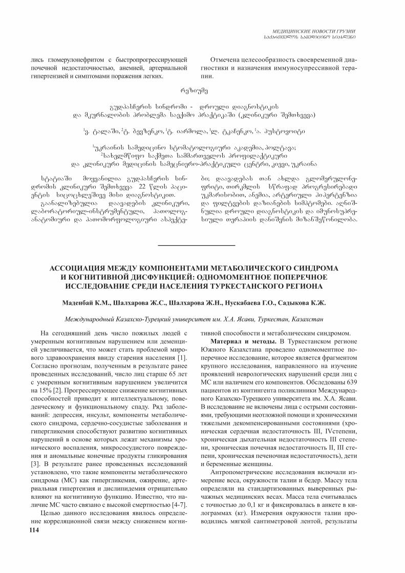
114
МЕДИЦИНСКИЕ НОВОСТИ ГРУЗИИ
CFMFHSDTKJC CFVTLBWBYJ CBF[KTYB
лись гломерулонефритом с быстропрогрессирующей почечной недостаточностью, анемией, артериальной гипертензией и симптомами поражения легких.
Отмечена целесообразность своевременной диа-гностики и назначения иммуносупрессивной тера-пии.
reziume
gudpasCeris sindromi - drouli diagnostikis da mkurnalobis problema saeqimo praqtikaSi (klinikuri SemTxveva)
1v. talaSi, 2t. bevzenko, 1t. iarmola, 1l. tkaCenko, 1a. pustovoiti
1ukrainis samedicino stomatologiuri akademia, poltava; 2saxelmwifo saqmeTa sammarTvelos profilaqtikuri
da klinikuri medicinis samecniero-praqtikuli centri, kievi, ukraina
statiaSi moyvanilia gudpasCeris sin-dromis klinikuri SemTxveva 22 wlis paci-entis sicocxleSive misi diagnostikiT.
gaanalizebulia daavadebis klinikuri, laboratoriul-instrumentuli, paTolog-anatomiuri da paTomorfologiuri aspeqte-
bi; daavadebas Tan axlda glomerulone-friti, Tirkmlis swrafad progresirebadi ukmarisobiT, anemia, arteriuli hipertenzia da filtvebis dazianebis simptomebi. aRniS-nulia drouli diagnostikis da imunosupre-siuli Terapiis daniSvnis mizanSewoniloba.
АССОЦИАЦИЯ МЕЖДУ КОМПОНЕНТАМИ МЕТАБОЛИЧЕСКОГО СИНДРОМА И КОГНИТИВНОЙ ДИСФУНКЦИЕЙ: ОДНОМОМЕНТНОЕ ПОПЕРЕЧНОЕ
ИССЛЕДОВАНИЕ СРЕДИ НАСЕЛЕНИЯ ТУРКЕСТАНСКОГО РЕГИОНА
Маденбай К.М., Шалхарова Ж.С., Шалхарова Ж.Н., Нускабаева Г.О., Садыкова К.Ж.
Международный Казахско-Турецкий университет им. Х.А. Ясави, Туркестан, Казахстан
На сегодняшний день число пожилых людей с умеренным когнитивным нарушением или деменци-ей увеличивается, что может стать проблемой миро-вого здравоохранения ввиду старения населения [1]. Согласно прогнозам, полученным в результате ранее проведенных исследований, число лиц старше 65 лет с умеренным когнитивным нарушением увеличится на 15% [2]. Прогрессирующее снижение когнитивных способностей приводит к интеллектуальному, пове-денческому и функциональному спаду. Ряд заболе-ваний: депрессия, инсульт, компоненты метаболиче-ского синдрома, сердечно-сосудистые заболевания и гипергликемия способствуют развитию когнитивных нарушений в основе которых лежат механизмы хро-нического воспаления, микрососудистого поврежде-ния и аномальные конечные продукты гликирования [3]. В результате ранее проведенных исследований установлено, что такие компоненты метаболического синдрома (МС) как гипергликемия, ожирение, арте-риальная гипертензия и дислипидемия отрицательно влияют на когнитивную функцию. Известно, что на-личие МС часто связано с высокой смертностью [4-7].
Целью данного исследования явилось определе-ние корреляционной связи между снижением когни-
тивной способности и метаболическим синдромом. Материал и методы. В Туркестанском регионе
Южного Казахстана проведено одномоментное по-перечное исследование, которое является фрагментом крупного исследования, направленного на изучение проявлений неврологических нарушений среди лиц с МС или наличием его компонентов. Обследованы 639 пациентов из контингента поликлиники Международ-ного Казахско-Турецкого университета им. Х.А. Ясави. В исследование не включены лица с острыми состояни-ями, требующими неотложной помощи и хроническими тяжелыми декомпенсированными состояниями (хро-ническая сердечная недостаточность III, IVстепени, хроническая дыхательная недостаточность III степе-ни, хроническая почечная недостаточность II, III сте-пени, хроническая печеночная недостаточность), дети и беременные женщины.
Антропометрические исследования включали из-мерение веса, окружности талии и бедер. Массу тела определяли на стандартизованных выверенных ры-чажных медицинских весах. Масса тела считывалась с точностью до 0,1 кг и фиксировалась в анкете в ки-лограммах (кг). Измерения окружности талии про-водились мягкой сантиметровой лентой, результаты
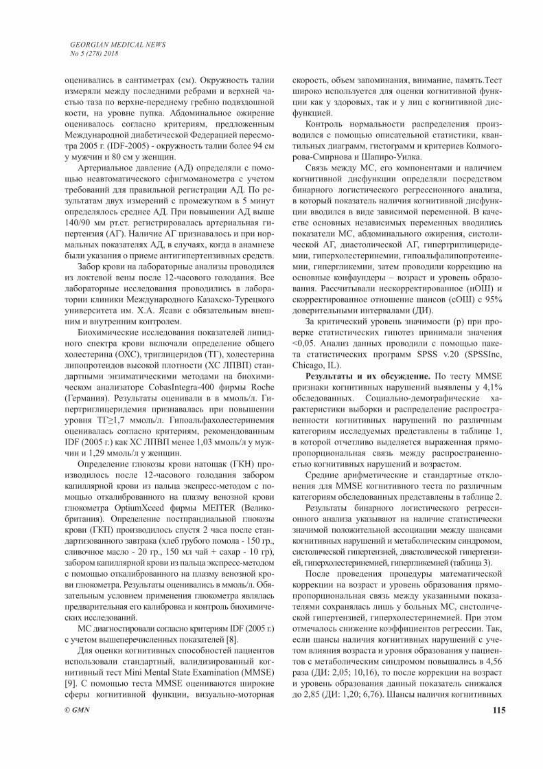
GEORGIAN MEDICAL NEWS No 5 (278) 2018
© GMN 115
оценивались в сантиметрах (см). Окружность талии измеряли между последними ребрами и верхней ча-стью таза по верхне-переднему гребню подвздошной кости, на уровне пупка. Абдоминальное ожирение оценивалось согласно критериям, предложенным Международной диабетической Федерацией пересмо-тра 2005 г. (IDF-2005) - окружность талии более 94 см у мужчин и 80 см у женщин.
Артериальное давление (АД) определяли с помо-щью неавтоматического сфигмоманометра с учетом требований для правильной регистрации АД. По ре-зультатам двух измерений с промежутком в 5 минут определялось среднее АД. При повышении АД выше 140/90 мм рт.ст. регистрировалась артериальная ги-пертензия (АГ). Наличие АГ признавалось и при нор-мальных показателях АД, в случаях, когда в анамнезе были указания о приеме антигипертензивных средств.
Забор крови на лабораторные анализы проводился из локтевой вены после 12-часового голодания. Все лабораторные исследования проводились в лабора-тории клиники Международного Казахско-Турецкого университета им. Х.А. Ясави с обязательным внеш-ним и внутренним контролем.
Биохимические исследования показателей липид-ного спектра крови включали определение общего холестерина (ОХС), триглицеридов (ТГ), холестерина липопротеидов высокой плотности (ХС ЛПВП) стан-дартными энзиматическими методами на биохими-ческом анализаторе CobasIntegra-400 фирмы Roche (Германия). Результаты оценивали в в ммоль/л. Ги-пертриглицеридемия признавалась при повышении уровня ТГ≥1,7 ммоль/л. Гипоальфахолестеринемия оценивалась согласно критериям, рекомендованным IDF (2005 г.) как ХС ЛПВП менее 1,03 ммоль/л у муж-чин и 1,29 ммоль/л у женщин.
Определение глюкозы крови натощак (ГКН) про-изводилось после 12-часового голодания забором капиллярной крови из пальца экспресс-методом с по-мощью откалиброванного на плазму венозной крови глюкометра OptiumXceed фирмы MEITER (Велико-британия). Определение постпрандиальной глюкозы крови (ГКП) производилось спустя 2 часа после стан-дартизованного завтрака (хлеб грубого помола - 150 гр., сливочное масло - 20 гр., 150 мл чай + сахар - 10 гр), забором капиллярной крови из пальца экспресс-методом с помощью откалиброванного на плазму венозной кро-ви глюкометра. Результаты оценивались в ммоль/л. Обя-зательным условием применения глюкометра являлась предварительная его калибровка и контроль биохимиче-ских исследований.
МС диагностировали согласно критериям IDF (2005 г.) с учетом вышеперечисленных показателей [8].
Для оценки когнитивных способностей пациентов использовали стандартный, валидизированный ког-нитивный тест Mini Mental State Examination (MMSE) [9]. С помощью теста MMSE оцениваются широкие сферы когнитивной функции, визуально-моторная
скорость, объем запоминания, внимание, память.Тест широко используется для оценки когнитивной функ-ции как у здоровых, так и у лиц с когнитивной дис-функцией.
Контроль нормальности распределения произ-водился с помощью описательной статистики, кван-тильных диаграмм, гистограмм и критериев Колмого-рова-Смирнова и Шапиро-Уилка.
Связь между МС, его компонентами и наличием когнитивной дисфункции определяли посредством бинарного логистического регрессионного анализа, в который показатель наличия когнитивной дисфунк-ции вводился в виде зависимой переменной. В каче-стве основных независимых переменных вводились показатели МС, абдоминального ожирения, систоли-ческой АГ, диастолической АГ, гипертриглицериде-мии, гиперхолестеринемии, гипоальфалипопротеине-мии, гипергликемии, затем проводили коррекцию на основные конфаундеры – возраст и уровень образо-вания. Рассчитывали нескорректированное (нОШ) и скорректированное отношение шансов (сОШ) с 95% доверительными интервалами (ДИ).
За критический уровень значимости (р) при про-верке статистических гипотез принимали значения <0,05. Анализ данных проводили с помощью паке-та статистических программ SPSS v.20 (SPSSInc, Chicago, IL).
Результаты и их обсуждение. По тесту MMSE признаки когнитивных нарушений выявлены у 4,1% обследованных. Социально-демографические ха-рактеристики выборки и распределение распростра-ненности когнитивных нарушений по различным категориям исследуемых представлены в таблице 1, в которой отчетливо выделяется выраженная прямо-пропорциональная связь между распространенно-стью когнитивных нарушений и возрастом.
Средние арифметические и стандартные откло-нения для MMSE когнитивного теста по различным категориям обследованных представлены в таблице 2.
Результаты бинарного логистического регресси-онного анализа указывают на наличие статистически значимой положительной ассоциации между шансами когнитивных нарушений и метаболическим синдромом, систолической гипертензией, диастолической гипертензи-ей, гиперхолестеринемией, гипергликемией (таблица 3).
После проведения процедуры математической коррекции на возраст и уровень образования прямо-пропорциональная связь между указанными показа-телями сохранялась лишь у больных МС, систоличе-ской гипертензией, гиперхолестеринемией. При этом отмечалось снижение коэффициентов регрессии. Так, если шансы наличия когнитивных нарушений с уче-том влияния возраста и уровня образования у пациен-тов с метаболическим синдромом повышались в 4,56 раза (ДИ: 2,05; 10,16), то после коррекции на возраст и уровень образования данный показатель снижался до 2,85 (ДИ: 1,20; 6,76). Шансы наличия когнитивных
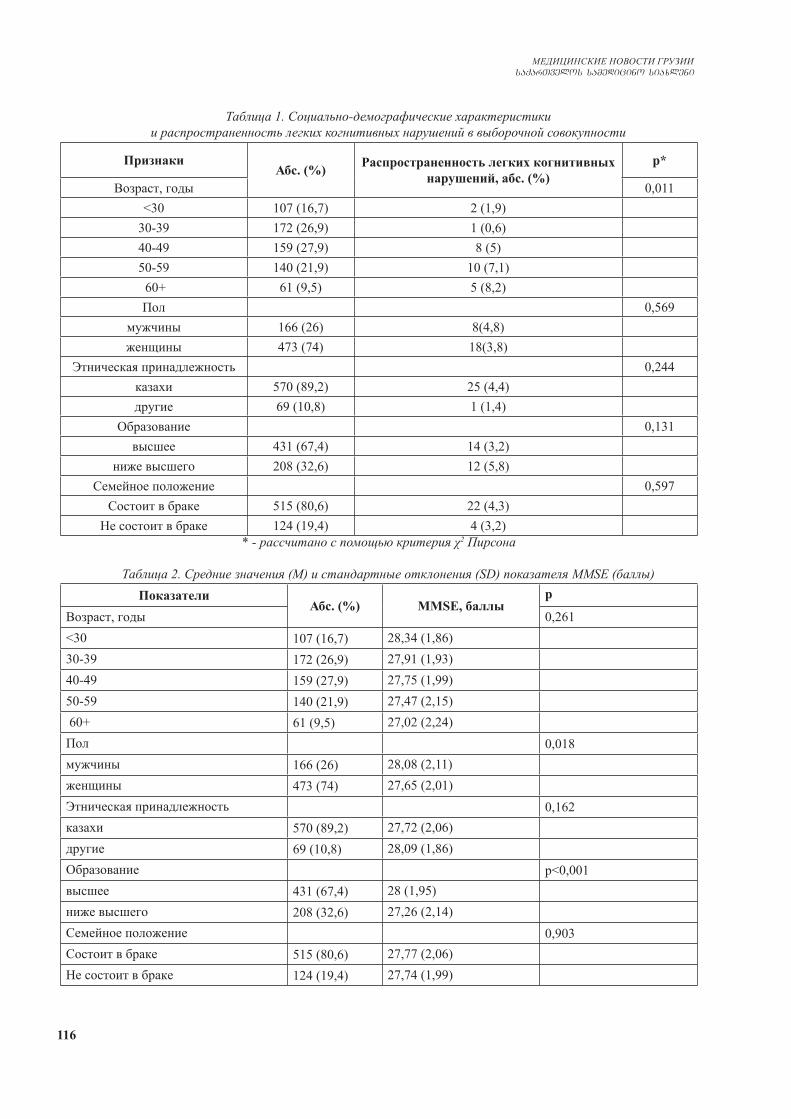
116
МЕДИЦИНСКИЕ НОВОСТИ ГРУЗИИ
CFMFHSDTKJC CFVTLBWBYJ CBF[KTYB
Таблица 1. Социально-демографические характеристики и распространенность легких когнитивных нарушений в выборочной совокупности
ПризнакиАбс. (%) Распространенность легких когнитивных
нарушений, абс. (%)p*
Возраст, годы 0,011<30 107 (16,7) 2 (1,9)
30-39 172 (26,9) 1 (0,6)40-49 159 (27,9) 8 (5)50-59 140 (21,9) 10 (7,1) 60+ 61 (9,5) 5 (8,2)Пол 0,569
мужчины 166 (26) 8(4,8)женщины 473 (74) 18(3,8)
Этническая принадлежность 0,244казахи 570 (89,2) 25 (4,4)другие 69 (10,8) 1 (1,4)
Образование 0,131высшее 431 (67,4) 14 (3,2)
ниже высшего 208 (32,6) 12 (5,8)Семейное положение 0,597
Состоит в браке 515 (80,6) 22 (4,3)Не состоит в браке 124 (19,4) 4 (3,2)
* - рассчитано с помощью критерия χ2 Пирсона
Таблица 2. Средние значения (М) и стандартные отклонения (SD) показателя MMSE (баллы)Показатели
Абс. (%) MMSE, баллыp
Возраст, годы 0,261<30 107 (16,7) 28,34 (1,86)30-39 172 (26,9) 27,91 (1,93)40-49 159 (27,9) 27,75 (1,99)50-59 140 (21,9) 27,47 (2,15) 60+ 61 (9,5) 27,02 (2,24)Пол 0,018мужчины 166 (26) 28,08 (2,11)женщины 473 (74) 27,65 (2,01)Этническая принадлежность 0,162казахи 570 (89,2) 27,72 (2,06)другие 69 (10,8) 28,09 (1,86)Образование p<0,001высшее 431 (67,4) 28 (1,95)ниже высшего 208 (32,6) 27,26 (2,14)Семейное положение 0,903Состоит в браке 515 (80,6) 27,77 (2,06)Не состоит в браке 124 (19,4) 27,74 (1,99)
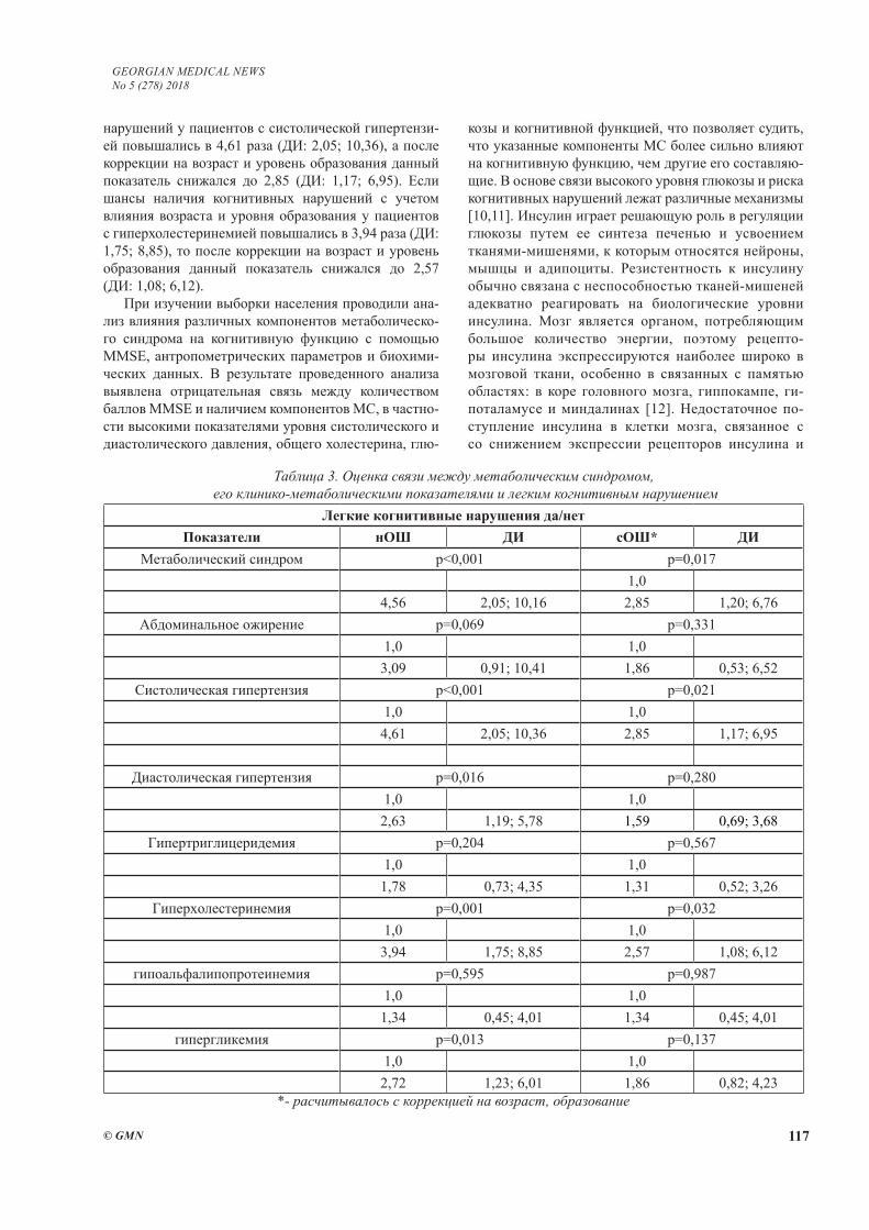
GEORGIAN MEDICAL NEWS No 5 (278) 2018
© GMN 117
Таблица 3. Оценка связи между метаболическим синдромом, его клинико-метаболическими показателями и легким когнитивным нарушением
Легкие когнитивные нарушения да/нетПоказатели нОШ ДИ сОШ* ДИ
Метаболический синдром p<0,001 p=0,0171,0
4,56 2,05; 10,16 2,85 1,20; 6,76Абдоминальное ожирение p=0,069 p=0,331
1,0 1,03,09 0,91; 10,41 1,86 0,53; 6,52
Систолическая гипертензия p<0,001 p=0,0211,0 1,0
4,61 2,05; 10,36 2,85 1,17; 6,95
Диастолическая гипертензия p=0,016 p=0,2801,0 1,0
2,63 1,19; 5,78 1,59 0,69; 3,68Гипертриглицеридемия p=0,204 p=0,567
1,0 1,01,78 0,73; 4,35 1,31 0,52; 3,26
Гиперхолестеринемия p=0,001 p=0,0321,0 1,03,94 1,75; 8,85 2,57 1,08; 6,12
гипоальфалипопротеинемия p=0,595 p=0,9871,0 1,0
1,34 0,45; 4,01 1,34 0,45; 4,01гипергликемия p=0,013 p=0,137
1,0 1,02,72 1,23; 6,01 1,86 0,82; 4,23
*- расчитывалось с коррекцией на возраст, образование
нарушений у пациентов с систолической гипертензи-ей повышались в 4,61 раза (ДИ: 2,05; 10,36), а после коррекции на возраст и уровень образования данный показатель снижался до 2,85 (ДИ: 1,17; 6,95). Если шансы наличия когнитивных нарушений с учетом влияния возраста и уровня образования у пациентов с гиперхолестеринемией повышались в 3,94 раза (ДИ: 1,75; 8,85), то после коррекции на возраст и уровень образования данный показатель снижался до 2,57 (ДИ: 1,08; 6,12).
При изучении выборки населения проводили ана-лиз влияния различных компонентов метаболическо-го синдрома на когнитивную функцию с помощью MMSE, антропометрических параметров и биохими-ческих данных. В результате проведенного анализа выявлена отрицательная связь между количеством баллов MMSE и наличием компонентов МС, в частно-сти высокими показателями уровня систолического и диастолического давления, общего холестерина, глю-
козы и когнитивной функцией, что позволяет судить, что указанные компоненты МС более сильно влияют на когнитивную функцию, чем другие его составляю-щие. В основе связи высокого уровня глюкозы и риска когнитивных нарушений лежат различные механизмы [10,11]. Инсулин играет решающую роль в регуляции глюкозы путем ее синтеза печенью и усвоением тканями-мишенями, к которым относятся нейроны, мышцы и адипоциты. Резистентность к инсулину обычно связана с неспособностью тканей-мишеней адекватно реагировать на биологические уровни инсулина. Мозг является органом, потребляющим большое количество энергии, поэтому рецепто-ры инсулина экспрессируются наиболее широко в мозговой ткани, особенно в связанных с памятью областях: в коре головного мозга, гиппокампе, ги-поталамусе и миндалинах [12]. Недостаточное по-ступление инсулина в клетки мозга, связанное с со снижением экспрессии рецепторов инсулина и
118
МЕДИЦИНСКИЕ НОВОСТИ ГРУЗИИ
CFMFHSDTKJC CFVTLBWBYJ CBF[KTYB
накоплением метаболитов глюкозы в жизненно зна-чимых функциональных и структурных белках, приво-дит к нарушению функции нейронов, так как нейроны весьма восприимчивы к метаболическому стрессу [13]. В результате исследования обнаружена связь между тяжестью инсулинорезистентности и увеличением на-копления амилоида в лобной и теменной облястях [14]. Выявлено также, что высокое систолическое давление и повышенный уровень общего холестерина являются компонентами МС, которые ассоциированы с когнитив-ными нарушениями даже в случае коррекции на такие конфаундинг-факторы как образование и возраст; эти данные согласуются с таковыми других исследований [5,15]. Установлена связь между степенью артериальной гипертензии в среднем возрасте и снижением когнитив-ных способностей в пожилом возрасте [16]. В результате обследования женщин среднего возраста когнитивные нарушения выявлены чаще у больных сахарным диа-бетом и артериальной гипертензией в сравнении с жен-щинами, страдающими только сахарным диабетом [17]. Контроль высокого артериального давления может сни-зить риск развития когнитивных нарушений в преклон-ном возрасте, особенно у лиц с сопутствующими забо-леваниями и сахарным диабетом 2 типа. Предполагается несколько механизмов влияния высокого артериального давления на развитие когнитивных нарушений. При вы-соком артериальном давлении нарушается сосудистая целостность, в данном случае целостность сосудов го-ловного мозга. Данное состояние способствует разви-тию ангиопатии, связанной с накоплением амилоида в стенках сосудов и снижением клиренса β-амилоида, что связано с патогенезом деменции [18]. Снижение обще-го объема головного мозга сопровождается понижением когнитивных способностей [19]. Согласно данным ис-следования объемы поражения белого вещества у па-циентов с артериальной гипертензией ассоциируются с риском когнитивных нарушений [20,21]. Приведенные данные подчеркивают необходимость контроля уровня артериального давления с целью предотвращения раз-вития когнитивных нарушений.
Имеются несколько ограничений ведения данного исследования: во-первых, исследование является по-перечным, что не дает возможность оценки причин-но-следственной связи между метаболическим син-дромом и результатами когнитивного теста. Оценка данной связи возможна лишь в когортном исследова-нии; во-вторых, физические и психические состояния участников, основные заболевания, состояние настро-ения и социально-экономический фон, по всей веро-ятности, влияют на результаты выполняемого когни-тивного теста.
Результаты проведенного исследования показали, что компоненты метаболического синдрома коррели-руют с когнитивными нарушениями. Исходя из этого, авторы считают целесообразным проведение иссле-дований по определению влияния метаболического синдрома на когнитивные способности.
ЛИТЕРАТУРА
1. Brookmeyer R, Gray S. Methods for projecting the incidence and prevalence of chronic diseases in ageing populations: ap-plication to Alzheimer’s disease // Stat Med. – 2000. – Vol.19. - P.1481–93. 2. Jorm AF, Jolley D. The incidence of dementia: a meta-analy-sis // Neurology. - 1998. - Vol. 51. – P. 728–33.3. Liu M, He Y, Jiang B, Wu L, Wang J, Yang S, Wang Y. Association between metabolic syndrome and mild cogni-tive impairment and its age difference in a Chinese com-munity elderly population // Clinical endocrinology. – 2015. Vol.82(6).- P.844-53.4. Dahl AK, Hassing LB, Fransson EI, Gatz M, Reynolds CA, Pedersen NL. Body mass index across midlife and cognitive change in late life // Int J Obes. – 2013. Vol.37. – P.296–302.5. Novak V, Hajjar I. The relationship between blood pressure and cognitive function // Nat Rev Cardiol. - 2010. Vol. 7. – P. 686–98.6. Koch M, Jensen MK. HDL-cholesterol and apolipoproteins in re-lation to dementia // Curr Opin Lipidol. – 2016. Vol. 27. – P. 76–87.7. Sachdev P, Kalaria R, O’Brien J, Skoog I, Alladi S, Black SE, Blacker D, Blazer DG, Chen C, Chui H, Ganguli M, Jellinger K, Jeste DV, Pasquier F, Paulsen J, Prins N, Rockwood K, Roman G, Scheltens P. Diagnostic criteria for vascular cognitive disor-ders: a VASCOG statement // Alzheimer disease and associated disorders. – 2014. Vol. 28(3). – P. 206-18. 8. International Diabetes Federation. Worldwide definition of the metabolic syndrome. Available at: http://www.idf.org/web-data/docs/IDF_ Metasyndrome_definition.pdf. Accessed August 24, 2014.9. Tombaugh TN, McIntyre NJ. The Mini- Mental State Exami-nation: a comprehensive review // J American Geriatrics Soci-ety. – 1992. – Vol. (40)9. – P. 11. 10. Yaffe K, Falvey C, Hamilton N, et al. Diabetes, glucose con-trol, and 9- year cognitive decline among older adults without dementia // Arch Neurol. – 2012. Vol.69. - P.1170–5.11. Satyajeet Roy, Nami Kim, Anjali Desai, Mahathi Komara-giri, Namrata Baxi, Navinder Jassil, Megan Blessinger,Maliha Khan, Robert Cole, Nayan Desai, Rocco Terrigno, and Krys-tal Hunter. Cognitive Function and Control of Type 2 Diabetes Mellitus in Young Adults // N Am J Med Sci. – 2015. Vol. 7(5). - P. 220–226. 12. Unger J, Livingston J, Moss A. Insulin receptors in the central nervous system: localization, signalling mechanisms and functional aspects // Prog Neurobiol. - 1991. Vol.36. – P.343–62.13. Moloney AM , Griffin RJ, Timmons S, O’Connor R, Ravid R, O’Neill C. Defects in IGF-1 receptor, insulin receptor and IRS-1/2 in Alzheimer’s disease indicate possible resistance to IGF-1 and insulin signaling // Neurobiol Aging. – 2010. Vol. 31. – P. 224–43.14. Willette AA, Johnson SC, Birdsill AC. Insulin resistance predicts brain amyloid deposition in late middle-aged adults // Alzheimers Dement. – 2015. Vol.11. – P.504–10.15. Gifford KA, Badaracco M, Liu D. Blood pressure and cogni-tion among older adults: a meta-analysis // Arch Clin Neuropsy-chol. – 2013. – Vol.28. – P.649–64.16. Launer LJ, Ross GW, Petrovitch H. Midlife blood pressure and dementia: the Honolulu–Asia aging study // Neurobiol Ag-ing. - 2000. – Vol.21. – P. 49–55.17. Petrova M, Prokopenko S, Pronina E. Diabetes type 2, hy-
GEORGIAN MEDICAL NEWS No 5 (278) 2018
© GMN 119
pertension and cognitive dysfunction in middle age women // J Neurol Sci. – 2010. Vol.299. – P. 39–41. 18. Shah NS, Vidal JS, Masaki K. Midlife blood pressure, plasma betaamyloid, and the risk for Alzheimer disease: the Honolulu Asia Aging Study // Hypertension. – 2012. – Vol.59. -P.780–6.19. Nagai M, Hoshide S, Ishikawa J. Ambulatory blood pressure as an independent determinant of brain atrophy and cognitive function in elderly hypertension // J Hypertens. - 2008. – Vol.26 – P.1636–41.20. Henskens LH, Kroon AA, van Oostenbrugge RJ. Associa-tions of ambulatory blood pressure levels with white matter hy-perintensity volumes in hypertensive patients // J Hypertens. - 2009. - Vol.27. – P.1446–52.21. de Groot JC, de Leeuw FE, Oudkerk M. Cerebral white mat-ter lesions and cognitive function: the Rotterdam Scan Study // Ann Neurol. – 2000. – Vol.47. – P.145–51.
SUMMARY
ASSOCIATION BETWEEN COMPONENTS OF METABOLIC SYNDROME AND COGNITIVE DYSFUNCTION: CROSS-SECTIONAL STUDY AMONG THE POPULATION OF THE TURKES-TAN REGION
Madenbay K., Shalkharova Zh.S., Shalkharova Zh.N., Nuskabayeva G., Sadykova K.
Kh. A. Yassawi International Kazakh-Turkish University, Turkestan, Kazakhstan
Metabolic syndrome (MetS) being a risk factor for many vascular pathologic processes is associated with cognitive dysfunction with vascular genesis.
Objective - to study the relationship between clinical and metabolic indicators and the presence of cognitive dysfunction in persons with MetS.
A transverse study was conducted in the Turkestan region of Southern Kazakhstan during which a general clinical, laboratory examination and evaluation of cogni-tive function was performed in 639 patients. To assess the relationship between the MetS, its individual components and cognitive function with Mini Mental State Examina-tion test (MMSE) multinominal logistic regression analy-sis was used.
According to the MMSE test, signs of cognitive im-pairment were revealed in 4.1% of the examined. A statis-tically significant positive association was found between the chances of having cognitive impairment andMetS, systolic hypertension, diastolic hypertension, hypercho-lesterolemia, hyperglycaemia.
Our results showed that the components of MetS cor-relate with cognitive impairment. Further research should be directed towards determining the impact of early de-tection and treatment of metabolic syndrome on cognitive ability.
Keywords: cognitive function, metabolic syndrome, hyperglycemia, dementia, diagnosis.
РЕЗЮМЕ
АССОЦИАЦИЯ МЕЖДУ КОМПОНЕНТАМИ МЕТАБОЛИЧЕСКОГО СИНДРОМА И КОГНИ-ТИВНОЙ ДИСФУНКЦИЕЙ: ОДНОМОМЕНТ-НОЕ ПОПЕРЕЧНОЕ ИССЛЕДОВАНИЕ СРЕДИ НАСЕЛЕНИЯ ТУРКЕСТАНСКОГО РЕГИОНА
Маденбай К.М., Шалхарова Ж.С., Шалхарова Ж.Н., Нускабаева Г.О., Садыкова К.Ж.
Международный Казахско-Турецкий университет им. Х.А. Ясави, Туркестан, Казахстан
Метаболический синдром (МС), являясь фактором риска для многих сосудистых патологий, ассоцииру-ется с когнитивной дисфункцией сосудистого генеза.
Цель исследования - изучение связи между клинико-метаболическими показателями и наличием когнитив-ной дисфункции у лиц с метаболическим синдромом.
Проведено поперечное исследование в Туркестан-ском регионе Южного Казахстана, общеклиническое, лабораторное обследование и оценка когнитивной функции с помощью Mini Mental State Examination (MMSE) у 639 пациентов. Для оценки связи между МС, его отдельными компонентами и MMSE использовался многомерный логистический регрессионный анализ. С использованием теста MMSE выявлены признаки ког-нитивных нарушений у 4,1% обследованных. Установ-лена статистически значимая положительная ассоциа-ция между шансами наличия когнитивных нарушений и МС, систолической гипертензией, диастолической гипертензией, гиперхолестеринемией, гипергликемией.
Результаты проведенного исследования показали, что компоненты метаболического синдрома коррели-руют с когнитивными нарушениями. Исходя из этого, авторы считают целесообразным проведение иссле-дований по определению влияния метаболического синдрома на когнитивные способности.
reziume
metaboluri sindromis komponentebis da kognitiur disfunqcias Soris kavSiri: erT-momentiani ganivi kvleva Turqestanis mo-saxleobaSi
k. madenbai, J.s. Salxarova, J.n. Salxarova, g. nuskabaeva, k. sadigova
x. iasavis sax. yazaxeT-Turquli saerTaSo-riso universiteti, Turqestani, yazaxeTi
metaboluri sindromi warmaodgens mara-vali sisxlZarRvovani paTologiebis ganvi-Tarebis risk-faqtors, asocirdeba sisxl-ZarRvovani genezis kognitiur disfunqciasTan.
120
МЕДИЦИНСКИЕ НОВОСТИ ГРУЗИИ
CFMFHSDTKJC CFVTLBWBYJ CBF[KTYB
kvlevis mizani iyo klinikur-meatabolur maCveneblebsa da kognitiur disfunqciis ar-sebobas Soris urTierTkavSiris Seswavla avadmyofebSi metaboluri sindromiT.
Catarda ganivi kvleva samxreT yazaxeTis Turqestanul regionSi; Seswavlilia 639 pa-cientis zogadklinikuri, laboratoriuli maCveneblebi. kognitiuri disfunqcia Sefas-da Mini Mental State Examination (MMSE) meTodis meSveobiT. metabolur sindroms, mis calkeul komponentebsa da MMSE-s Soris kavSiris
SefasebisaTvis gamoyenebuli iyo mraval-ganzomilebiani logistikuri regresiuli analizi. MMSE testis meSveobiT gamovlinda kognitiuri darRvevebis niSnebi 4,1% SemTx-vevaSi. dadgenilia statistikurad sarwmuno dadebiTi korelacia kognitiuri darRvevebis niSnebsa da metabolur sindroms, sistolur hipertenzias, hiperqolisterinemias, hiper-glikemias Soris. kvlevis Sedegebma gamoavli-na, rom metaboluri sindromis komponentebi korelaciaSia kognitiur darRvevebTan.
SUSPECTED ENVIRONMENTAL AND SOCIO-ECONOMIC CAUSES OF DIABETES MELLITUS AND ASSOCIATED OCULAR COMPLICATIONS
IN THE SUMY REGION, UKRAINE, FOR THE PERIOD OF 2011-2016
1Lekishvili S., 1Chayen B., 2Chayen S.
1Medical Institute Sumy State University; 2Sumy State University, Oleg Balatskyi Academic and Research Institute of Finance, Economics and Management, Economic Theory Department, Ukraine
Diabetes mellitus (DM) is one of the most common endocrine disorders. DM affects more than 240 million people worldwide, and this number is expected to reach roughly 370 million by 2030 [30]. The prevalence of dia-betes in Ukraine was 2,861.6 people per 100,000 people in 2015 and annual mortality rate was 5 per 100,000 people [31]. Complications of DM are progressive and almost al-ways resulting by chronic exposure to high levels of blood glucose caused by impairments in insulin metabolism and of biological macromolecules such as carbohydrates, lip-ids, proteins and nucleic acids [7]. Ocular complications associated with DM are progressive and rapidly becoming the world’s most significant cause of loss of health. They are preventable with early detection and timely treatment. This research provides an overview of two main ocular complications associated with DM - diabetic retinopathy and cataract [17].
Diabetic cataract. Definition and featuresCataract is considered a major cause of visual impair-
ment in diabetic patients. Morphologically it is cloudy opacifications of the lens of the eye which can lead to blurred vision which can be dazzled by bright lights, see-ing a circle of light around lights, a yellowing of vision and finally it can also lead to a loss of vision. True diabet-ic cataract, which is bilateral and often develops quickly, can affect individuals at younger ages with severe diabe-tes. This condition is relatively rare: the development of senile cataract is a problem of more practical importance though it seems no more common in diabetics than in non-diabetics in a similar age group. This view is supported by
the findings of Waite and Beetham (1935), who found the incidence of senile cataract in 2000 diabetic patients no greater than in non-diabetics of a comparable age [22].
Cataract frequency varies between ethnic populations and geographic locations (ranges from 35% to 48%), and in these circumstances it can be found higher in diabetics when compared to non-diabetics. For example, in Chi-nese, Indonesian and Indian populations rate of diabetic cataract was higher than in other populations [3,5,15].
Cataract formation in diabetics maybe related to hy-perglycemia or to old age [16].
Unfortunately, diabetic cataract extraction is associ-ated with higher complication rates than nondiabetic sur-gery and may be accompanied by macular edema, pro-gression of retinopathy, frank neovascularization, uveitis, and posterior capsular thickening. The precise cause of diabetic cataract is unknown [20].
Pathogenesis. The base of pathogenesis of diabetic cataracts is accumulation of glucose in the lens, following background episodes of hyperglycemia. Some part of glu-cose transforms into sorbitol. Glucose and sorbitol have a high osmolarity and are able to delay the fluid, which causes swelling of the lens, change of its shape and refrac-tive properties. These changes cause degeneration of the lens and later cataract (Fig. 1).
Diagnostic. A comprehensive ophthalmologic exami-nation includes:
- visual acuity measurement; evaluation of relative affer-ent pupil defect; slit-lamp biomicroscopy; gonioscopy; intra-ocular pressure measurement; dilated fundus examination.
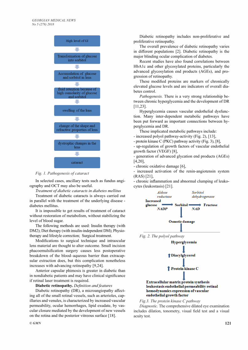
GEORGIAN MEDICAL NEWS No 5 (278) 2018
© GMN 121
In selected cases, ancillary tests such as fundus angi-ography and OCT may also be useful.
Treatment of diabetic cataracts in diabetes mellitusTreatment of diabetic cataracts is always carried out
in parallel with the treatment of the underlying disease - diabetes mellitus.
It is impossible to get results of treatment of cataract without restoration of metabolism, without stabilizing the level of blood sugar.
The following methods are used: Insulin therapy (with DM2); Diet therapy (with insulin-independent DM); Physio-therapy and lifestyle correction; Surgical treatment.
Modifications to surgical technique and intraocular lens material are thought to alter outcome. Small incision phacoemulsification surgery causes less postoperative breakdown of the blood–aqueous barrier than extracap-sular extraction does, but this complication nonetheless increases with advancing retinopathy [9,24].
Anterior capsular phimosis is greater in diabetic than in nondiabetic patients and may have clinical significance if retinal laser treatment is required.
Diabetic retinopathy. Definition and featuresDiabetic retinopathy (DR), a microangiopathy affect-
ing all of the small retinal vessels, such as arterioles, cap-illaries and venules, is characterized by increased vascular permeability, ocular hemorrhages, lipid exudate, by vas-cular closure mediated by the development of new vessels on the retina and the posterior vitreous surface [18].
Diabetic retinopathy includes non-proliferative and proliferative retinopathy.
The overall prevalence of diabetic retinopathy varies in different populations [2]. Diabetic retinopathy is the major blinding ocular complication of diabetes.
Recent studies have also found correlations between HbA1c and other glycosylated proteins, particularly the advanced glycosylation end products (AGEs), and pro-gression of retinopathy.
These modified proteins are markers of chronically elevated glucose levels and are indicators of overall dia-betes control.
Pathogenesis. There is a very strong relationship be-tween chronic hyperglycemia and the development of DR [11,23].
Hyperglycemia causes vascular endothelial dysfunc-tion. Many inter-dependent metabolic pathways have been put forward as important connections between hy-perglycemia and DR.
These implicated metabolic pathways include: - increased polyol pathway-activity (Fig. 2), [13],- protein kinase C (PKC) pathway activity (Fig. 3), [8], - up-regulation of growth factors of vascular endothelial growth factor (VEGF) [8], - generation of advanced glycation end products (AGEs) [4,20], - chronic oxidative damage [6], - increased activation of the renin-angiotensin system (RAS) [21], - chronic inflammation and abnormal clumping of leuko-cytes (leukostasis) [21].
Fig. 2. The polyol pathway
Fig.3. The protein kinase C pathwayDiagnostic. The comprehensive dilated eye examination
includes dilation, tonometry, visual field test and a visual acuity test.
Fig. 1. Pathogenesis of cataract
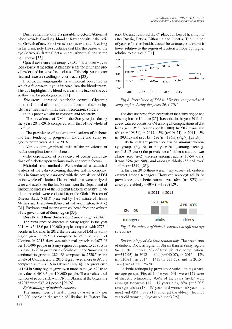
122
МЕДИЦИНСКИЕ НОВОСТИ ГРУЗИИ
CFMFHSDTKJC CFVTLBWBYJ CBF[KTYB
During examinations it is possible to detect: Abnormal blood vessels; Swelling, blood or fatty deposits in the reti-na; Growth of new blood vessels and scar tissue; Bleeding in the clear, jelly-like substance that fills the center of the eye (vitreous); Retinal detachment; Abnormalities in the optic nerve [32].
Optical coherence tomography (OCT) is another way to look closely at the retina. A machine scans the retina and pro-vides detailed images of its thickness. This helps your doctor find and measure swelling of your macula [33].
Fluorescein angiography is a medical procedure in which a fluorescent dye is injected into the bloodstream. The dye highlights the blood vessels in the back of the eye so they can be photographed [34].
Treatment: increased metabolic control; Glycemic control; Control of blood pressure; Control of serum lip-ids; laser treatment; intravitreal medication; surgery.
In this paper we aim to compare and research:- The prevalence of DM in the Sumy region during
the years 2011-2016 compared with that of the whole of Ukraine.
- The prevalence of ocular complications of diabetes and their tendency to progress in Ukraine and Sumy re-gion over the years 2011 – 2016.
- Various demographical traits of the prevalence of ocular complications of diabetes.
- The dependence of prevalence of ocular complica-tions of diabetes upon various socio-economic factors.
Material and methods. We conducted a statistical analysis of the data concerning diabetes and its complica-tions in Sumy region campared with the prevalence of DM in the whole of Ukraine. The materials that were analyzed were collected over the last 6 years from the Department of Endocrine diseases of the Regional Hospital of Sumy. In ad-dition materials were collected from the Global Burden of Disease Study (GBD) presented by the Institute of Health Metrics and Evaluation (University of Washington, Seattle) [31]. Environmental reports were collected from the website of the government of Sumy region [35].
Results and their discussion. Epidemiology of DMThe prevalence of diabetes in Sumy region in the year
2011 was 3418.6 per 100,000 people compared with 2773.1 people in Ukraine. In 2012 the prevalence of DM in Sumy region grew to 3527.34 compared to 2885 in whole of Ukraine. In 2013 there was additional growth to 3673.06 per 100,000 people in Sumy region compared to 2780.5 in Ukraine. In 2014 prevalence of diabetes in the Sumy region continued to grow to 3804.68 compared to 2730.7 in the whole of Ukraine, and in 2015 it grew even more to 3877.1 compared with 2861.6 in Ukraine (Fig. 4). The prevalence of DM in Sumy region grew even more in the year 2016 to the value of 4018.3 per 100,000 people. The absolute total number of people sick with DM in Ukraine at the beginning of 2017 were 537 641 people [25-29].
Epidemiology of diabetic cataractThe annual loss of health from cataract is 57 per
100,000 people in the whole of Ukraine. In Eastern Eu-
rope Ukraine reserved the 6th place for loss of healthy life after Russia, Latvia, Lithuania and Croatia. The number of years of loss of health, caused be cataract, in Ukraine is lower relative to the region of Eastern Europe but higher relative to the world [31]
Fig.4. Prevalence of DM in Ukraine compared with Sumy region during the years 2011-2015
The data analyzed from hospitals in the Sumy region and other regions in Ukraine [25] shows that in the year 2011, di-abetic cataract counts for 6% among all complications of dia-betes (n = 195.35 persons per 100,000). In 2012 it was also 6% (n = 198.51), in 2013 – 5% (n=196.74), in 2014 – 5% (n=203.72) and in 2015 – 5% (n = 196.5) (Fig.7), [25-29].
Diabetic cataract prevalence varies amongst various age-groups (Fig. 5). In the year 2011, amongst teenag-ers (15-17 years) the prevalence of diabetic cataract was almost zero (n=2) whereas amongst adults (18-54 years) it was 59% (n=1908), and amongst elderly (55 and over) – 41% (n=1310) [25].
In the year 2015 there weren’t any cases with diabetic cataract among teenagers. However, amongst adults he prevalence of diabetic cataract was 60% (n=1923) and among the elderly – 40% (n=1395) [29].
Fig. 5. Prevalence of diabetic cataract in different age categories
Epidemiology of diabetic retinopathy. The prevalence of diabetic DR was higher in Ukrain than in Sumy region. So, in 2011 it was 16% of total diabetic complications (n=542.95), in 2012 – 15% (n=540.07), in 2013 – 17% (n=626.61), in 2014 – 14% (n=531.52), and in 2015 – 14% (n=541.52) [25-29].
Diabetic retinopathy prevalence varies amongst vari-ous age-groups (Fig. 6). In the year 2011 were 9129 cases of diabetic retinopathy: 0,6% of the cases (n=15) were amongst teenagers (15 – 17 years old), 58% (n=5,303) amongst adults (18 – 55 years old women, 60 years old men) and 42% ( n=3,811) amongst the elderly (from 55 years old women, 60 years old men) [25].

GEORGIAN MEDICAL NEWS No 5 (278) 2018
© GMN 123
Then in 2015 the total level of DR cases declined from 9129 cases to 8857cases. The prevalence amongst teenag-ers 0.2% (n = 5), 60% (n = 5,316) amongst adults and 40% (n=3,536) amongst elderly group [29].
Fig. 6. Prevalence of diabetic retinopathy in different age categories
Fig. 7. Prevalence of DM ophthalmic complications
The prevalence of DM in Sumy region grew between the years 2011-2016 by 17.5%. During that period there was no significant change in the prevalence of DM in the whole of Ukraine. This difference between Sumy and the whole of Ukraine will be discussed later in this discus-sion.
The prevalence of ocular complications of DM are ap-proximately 22% of which 6% are for DC and 16% are for DR. The prevalence of ocular complications did not change over the years. At the time point of writing this article there are 88 ophthalmologists in Sumy region, of which 47 serve in the town of Sumy. Out of 88 ophthal-mologists, 14 doctors treat the juvenile sector. This ade-quate number of eye-doctors, work on the prevention and treatment of ocular complication of DM and are capable of keeping the prevalence of ocular complications of DM constant.
It can be seen that these ocular disorders do not al-most occur within the juvenile group, are highly preva-lent with adults (60%) and decrease a little within the elderly group (40%). This is due to the low life ex-pectancy of adults that are sick with DM with ocular complications who will not reach the age of the elderly group.
The absolute number of endocrinologists in the whole of Sumy region is 36 and stands on the 6th place from the bottom of the list of occurrence of endocrinologist per region (0.32 per 10 000 people). Compared to Kyiv that has 201 endocrinologists (0.7 per 10 000 people) only for
the town, this number is extremely low. During the years 2011-2016 this number did not change. In addition, ac-cording to our investigation there was no significant anti-diabetes campaign during these years, not in the Sumy region and neither in the whole of Ukraine. Therefore, the increase in prevalence of DM must be explained not merely by hyperdiagnostic but by real new causes of sick-ness.
Since the Sumy region is well known as an industrial area, we suspect environmental hazardous materials to be the causes of the increase in DM prevalence. We therefore investigated the last environmental report for the Sumy region in the year 2017 [35]. The findings indicated that there is a strong case for ‘accusing’ environmental fac-tors with the increase of DM prevalence. Findings of the report show that during the five-year observation period (2012-2016), there was a significant increase in the ob-served level of atmospheric air pollution. In 2016 there was an increase of 13.2% in emissions of pollutants into the air from stationary sources compared to 2015 (in ab-solute numbers there was an increase of 2.3 thousand tons of pollutants). The main pollutants were: nitrogen compounds (by 1.0 thousand tons or by 53.2%); carbon monoxide (by 0.8 thousand tons or by 17.6%); methane (by 0.1 thousand tons or by 4.8%); sulfur dioxide (by 0.1 thousand tons or by 4.1%); sulfuric acid (by 36.7 tons or by 43.3%); POP (benzapyrene) (by 0.006 tons or 7 times); bromine and its compounds (by 0.031 tons or 2.7 times) [35].
In addition to the increase of air pollution during the years 2012-2016, there was also significant increase in contamination of surface water by sewage which might have reached ground water. This water is used in the Sumy region as drinking water. In 2016, the total dis-charge of sewage into surface water was 48.38 million m3 in comparison with 2015 when it was 45.59 million m3. The absolute increase in contamination was by 2.79 million m 3 [35].
Environmental pollution is a well-known cause of DM and has been reported in the works of David Spero [36] and Jacquelyn K. Beals [33]. Spero claims that chemicals in food, water, plastic packaging, and cleaning and beauty care products have all been found to increase insulin re-sistance and diabetes [36]. In her study, Beals looked for significant association between factors such as levels of heavy metals or pesticides in blood or urine and cases of T2D [1].
In addition to environmental pollution as a cause of increase of DM prevalence, in the last years another significant cause has been found – moving from rural style of life to an urban style of life. In India, it was found that the prevalence of DM was two and half times higher in urban areas than in rural areas [12].
We now know that this process of urbanization changes the daily life style of people in the form of increase in fat consumption, physical inactivity, and substance abuse with associated risk of development of
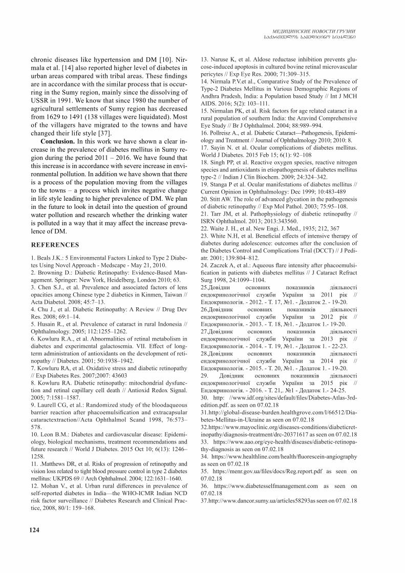
124
МЕДИЦИНСКИЕ НОВОСТИ ГРУЗИИ
CFMFHSDTKJC CFVTLBWBYJ CBF[KTYB
chronic diseases like hypertension and DM [10]. Nir-mala et al. [14] also reported higher level of diabetes in urban areas compared with tribal areas. These findings are in accordance with the similar process that is occur-ring in the Sumy region, mainly since the dissolving of USSR in 1991. We know that since 1980 the number of agricultural settlements of Sumy region has decreased from 1629 to 1491 (138 villages were liquidated). Most of the villagers have migrated to the towns and have changed their life style [37].
Conclusion. In this work we have shown a clear in-crease in the prevalence of diabetes mellitus in Sumy re-gion during the period 2011 – 2016. We have found that this increase is in accordance with severe increase in envi-ronmental pollution. In addition we have shown that there is a process of the population moving from the villages to the towns – a process which invites negative change in life style leading to higher prevalence of DM. We plan in the future to look in detail into the question of ground water pollution and research whether the drinking water is polluted in a way that it may affect the increase preva-lence of DM.
REFERENCES
1. Beals J.K.: 5 Environmental Factors Linked to Type 2 Diabe-tes Using Novel Approach - Medscape - May 21, 2010. 2. Browning D.: Diabetic Retinopathy: Evidence-Based Man-agement. Springer: New York, Heidelberg, London 2010; 63.3. Chen S.J., et al. Prevalence and associated factors of lens opacities among Chinese type 2 diabetics in Kinmen, Taiwan // Acta Diabetol. 2008; 45:7–13.4. Chu J., et al. Diabetic Retinopathy: A Review // Drug Dev Res. 2008; 69:1–14. 5. Husain R., et al. Prevalence of cataract in rural Indonesia // Ophthalmology. 2005; 112:1255–1262.6. Kowluru R.A., et al. Abnormalities of retinal metabolism in diabetes and experimental galactosemia. VII. Effect of long-term administration of antioxidants on the development of reti-nopathy // Diabetes. 2001; 50:1938–1942.7. Kowluru RA, et al. Oxidative stress and diabetic retinopathy // Exp Diabetes Res. 2007;2007: 436038. Kowluru RA. Diabetic retinopathy: mitochondrial dysfunc-tion and retinal capillary cell death // Antioxid Redox Signal. 2005; 7:1581–1587.9. Laurell CG, et al.: Randomized study of the bloodaqueous barrier reaction after phacoemulsification and extracapsular cataractextraction//Acta Ophthalmol Scand 1998, 76:573–578. 10. Leon B.M.: Diabetes and cardiovascular disease: Epidemi-ology, biological mechanisms, treatment recommendations and future research // World J Diabetes. 2015 Oct 10; 6(13): 1246–1258.11. .Matthews DR, et al. Risks of progression of retinopathy and vision loss related to tight blood pressure control in type 2 diabetes mellitus: UKPDS 69 // Arch Ophthalmol. 2004; 122:1631–1640.12. Mohan V., et al. Urban rural differences in prevalence of self-reported diabetes in India—the WHO-ICMR Indian NCD risk factor surveillance // Diabetes Research and Clinical Prac-tice, 2008, 80/1: 159–168.
13. Naruse K, et al. Aldose reductase inhibition prevents glu-cose-induced apoptosis in cultured bovine retinal microvascular pericytes // Exp Eye Res. 2000; 71:309–315.14. Nirmala P.V.et al., Comparative Study of the Prevalence of Type-2 Diabetes Mellitus in Various Demographic Regions of Andhra Pradesh, India: a Population based Study // Int J MCH AIDS. 2016; 5(2): 103–111.15. Nirmalan PK, et al. Risk factors for age related cataract in a rural population of southern India: the Aravind Comprehensive Eye Study // Br J Ophthalmol. 2004; 88:989–994.16. Pollreisz A., et al. Diabetic Cataract—Pathogenesis, Epidemi-ology and Treatment // Journal of Ophthalmology 2010; 2010: 8.17. Sayin N. et al. Ocular complications of diabetes mellitus. World J Diabetes. 2015 Feb 15; 6(1): 92–108 18. Singh PP, et al. Reactive oxygen species, reactive nitrogen species and antioxidants in etiopathogenesis of diabetes mellitus type-2 // Indian J Clin Biochem. 2009; 24:324–342.19. Stanga P et al. Ocular manifestations of diabetes mellitus // Current Opinion in Ophthalmology: Dec 1999; 10:483-489 20. Stitt AW. The role of advanced glycation in the pathogenesis of diabetic retinopathy // Exp Mol Pathol. 2003; 75:95–108.21. Tarr JM, et al. Pathophysiology of diabetic retinopathy //ISRN Ophthalmol. 2013; 2013:343560.22. Waite J. H., et al. New Engi. J. Med., 1935; 212, 36723. White N.H, et al. Beneficial effects of intensive therapy of diabetes during adolescence: outcomes after the conclusion of the Diabetes Control and Complications Trial (DCCT) // J Pedi-atr. 2001; 139:804–812.24. Zaczek A, et al.: Aqueous flare intensity after phacoemulsi-fication in patients with diabetes mellitus // J Cataract Refract Surg 1998, 24:1099–1104.25.Довідни основних показників діяльності ендокринологічної служби України за 2011 рік // Ендокринологія. - 2012. - Т. 17, №1. - Додаток 2. - 19-20.26.Довідник основних показників діяльності ендокринологічної служби України за 2012 рік // Ендокринологія. - 2013. - Т. 18, №1. - Додаток 1.- 19-20.27.Довідник основних показників діяльності ендокринологічної служби України за 2013 рік // Ендокринологія. - 2014. - Т. 19, №1. - Додаток 1. - 22-23.28.Довідник основних показників діяльності ендокринологічної служби України за 2014 рік // Ендокринологія. - 2015. - Т. 20, №1. - Додаток 1. - 19-20.29. Довідник основних показників діяльності ендокринологічної служби України за 2015 рік // Ендокринологія. - 2016. - Т. 21,. №1 - Додаток 1.- 24-25.30. http: //www.idf.org/sites/default/files/Diabetes-Atlas-3rd-edition.pdf. as seen on 07.02.1831.http://global-disease-burden.healthgrove.com/l/66512/Dia-betes-Mellitus-in-Ukraine as seen on 07.02.1832.https://www.mayoclinic.org/diseases-conditions/diabeticret-inopathy/diagnosis-treatment/drc-20371617 as seen on 07.02.1833. https://www.aao.org/eye-health/diseases/diabetic-retinopa-thy-diagnosis as seen on 07.02.1834. https://www.healthline.com/health/fluorescein-angiography as seen on 07.02.1835. https://menr.gov.ua/files/docs/Reg.report.pdf as seen on 07.02.1836. https://www.diabetesselfmanagement.com as seen on 07.02.1837.http://www.dancor.sumy.ua/articles58293as seen on 07.02.18
GEORGIAN MEDICAL NEWS No 5 (278) 2018
© GMN 125
The aim of the study was to compare and research: the general prevalence of DM in the Sumy region compared with the prevalence of DM in the whole of Ukraine during the years 2011-2016; the prevalence of ocular complica-tions of diabetes and their tendency to progress in Ukraine and Sumy region over this period; various demographical traits of the prevalence of ocular complications of diabe-tes and the dependence of prevalence of ocular complica-tions of diabetes upon environmental causes and various socio-economic factors.
We conducted a statistical analysis of data on diabetes and its complications in the whole of Ukraine and the Sumy region. The materials that were analyzed were collected over the last 6 years from the Department of Endocrine diseases of the Regional Hospital of Sumy. In addition materials were collected from the Global Burden of Disease Study (GBD) presented by the Institute of Health Metrics and Evaluation (University of Washington, Seattle).
In this work we have shown an increase of 17.5% in the prevalence of diabetes mellitus in the Sumy region
during the period 2011 – 2016. During that period there was no significant change in the prevalence of DM in the whole of Ukraine. The prevalence of ocular complica-tions of DM are approximately 22% of which 6% for DC and 16% for DR. The prevalence of ocular complications did not change over the years. We have shown that the increase of DM prevalence is in accordance with severe increase in environmental pollution. In addition we have shown that there is a process of the population moving from the villages to the towns – a process which invites negative change in life style leading to higher prevalence of DM.
We plan in the future to look in detail into the ques-tion of ground water pollution and research whether the drinking water is polluted in a way that it may affect the increased prevalence of DM.
Keywords: diabetes mellitus, ocular complications of diabetes, diabetic retinopathy, diabetic cataract, environ-mental pollution, Sumy region, Ukraine.
SUMMARY
SUSPECTED ENVIRONMENTAL AND SOCIO-ECONOMIC CAUSES OF DIABETES MELLITUS AND ASSOCIATED OCULAR COMPLICATIONS IN THE SUMY REGION, UKRAINE,
FOR THE PERIOD OF 2011-2016
1Lekishvili S., 1Chayen B., 2Chayen S.
1Medical Institute Sumy State University; 2Sumy State University, Oleg Balatskyi Academic and Research Institute of Finance, Economics and Management, Economic Theory Department, Ukraine
РЕЗЮМЕ
ОПРЕДЕЛЕНИЕ ЭКОЛОГИЧЕСКИХ И СОЦИАЛЬНО-ЭКОНОМИЧЕСКИХ ПРИЧИН САХАРНОГО ДИАБЕТА И ЕГО ОФТАЛЬМОЛОГИЧЕСКИХ ОСЛОЖНЕНИЙ
В СУМСКОЙ ОБЛАСТИ И УКРАИНЕ ЗА ПЕРИОД 2011-2016 ГГ.
Лекишвили С.Э., Чейн Б.Я.1, Чейн С.В.2
Сумской государственный университет, 1Медицинский институт, 2Учебно-научный институт финансов, экономики и менеджмента им. Олега Балацкого, кафедра экономической теории, Украина
Целью исследования явилось определение эко-логических и социально-экономических причин развития сахарного диабета и его офтальмологиче-ских осложнений в Украине и Сумской области в течение 2011-2016 гг., анализ их зависимости от со-циально-экономических факторов, разработка мер для предотвращения офтальмологических ослож-нений сахарного диабета.
Проведен статистический анализ данных по сахар-ному диабету (СД) и его осложнениям в Украине в целом и в ее Сумской области. Проанализированы материалы из отделения эндокринологии Област-ной больницы г. Сумы за 6 лет. Некоторые данные заимствованы из Global Burden of Disease Study (GBD), Institute of Health Metrics and Evaluation (University of Washington, Seattle). Проведен срав-
нительный анализ результатов исследований с дан-ными других авторов.
Показано достоверное увеличение распростра-ненности CД в Сумской области за период с 2011 по 2016 гг. Существенных изменений в распростра-ненности СД по Украине, в целом, не отмечено. За последние годы показатели распространенности офтальмологических осложнений стабилизирова-лись.
Установлено, что распространенность СД обу-словлена ростом загрязнения окружающей среды. В будущем планируется детальное изучение во-проса о загрязнении грунтовых вод и проведение исследований на предмет выявления роли сте-пени загрязнения питьевой воды на распростра-нение СД.
126
МЕДИЦИНСКИЕ НОВОСТИ ГРУЗИИ
CFMFHSDTKJC CFVTLBWBYJ CBF[KTYB
reziume
Saqriani diabetis savaraudo ekologiuri da socialur-ekonomikuri mizezebi da misi ofTalmologiuri garTulebebi sumis re-gionSi, ukraina, 2011-2016 ww. periodSi
1s. lekiSvili, 1b. Cein, 2s. Cein
sumis saxelmwifo universiteti, 1samedicino instituti, 2oleg balackis sax. finansTa, ekonomikisa da marTvis saganmanaTleblo-samecniero instituti, ekonomikis Teoriis departamenti, ukraina
gamokvlevis mizans warmoadgenda Saqri-ani diabetis ofTalmologiuri garTule-bebis gavrcelebis tendenciis Sefaseba ukrainaSi da sumis regionSi 2011-2016 ww., aseve, am garTulebaTa gavrcelebis demo-grafiuli aspeqtebis da socialur-ekonomi-kur faqtorebTan maTi korelaciis analizi.
Catarebulia diabetis da misi garTulebis statistikuri analizi mTel ukrainaSi da su-mis regionSi. masala Segrovebulia 6 wlis manZilze sumis regionuli saavadmyofos en-dokrinologiur ganyofilebaSi; gamoyenebu-lia, aseve, monacemebi Global Burden of Disease Study (GBD), Institute of Health Metrics and Evaluation (Univer-sity of Washington, Seattle)-is bazebidan.
dadgenilia 2011-2016 ww. Saqriani diabetis gavrcelebis maCveneblebis sarwmuno zrda sumis regionSi. am periodis ganmavlobaSi mniSvnelovni cvlileba Saqriani diabetis gavrcelebis mxriv ukrainaSi ar aRiniSna. ukanasknel wlebSi ofTalmologiuri gar-Tulebebis gavrcelebis maCveneblebi stabi-luria.
savaraudoa, rom Saqriani diabetis gavrcelebis mateba ganpirobebulia gare-mos dabinZurebis mniSvnelovani zrdiT. mo-mavalSi dagegmilia gruntis wylebis da sa-smeli wylis dabinZurebis gavlenis kvleva Saqriani diabetis gavrcelebaze.
URINARY MALONDIALDEHYDE AS A PREDICTIVE AND DIAGNOSTIC MARKER FOR NEONATAL ACUTE KIDNEY INJURY
1Hodovanets Y., 1,3Babintseva A., 1,3Agafonova L., 2Makarova O., 1Frunza A.
1Higher State Educational Establishment of Ukraine «Bukovinian State Medical University», Department of Pediatrics, Neonatology and Perinatal Medicine; 2Department of Nursing and Higher Nursing Education, Chernivtsi;
3Neonatal Intensive Care Unit, Maternity Hospital №2 Chernivtsi, Ukraine
Newborns are especially prone to oxidative stress (OS). There are several reasons for it. Very often infants (1) are exposed to high oxygen concentrations, (2) in-fections or inflammation, (3) have reduced antioxidant defense and (4) high levels of free iron which enhance Fenton reaction leading to the production of highly toxic radicals [2,9]. OS is likely to exacerbate several neonatal conditions to the extent that Ola Didrik Saugstad coined in the phrase “oxygen radical disease of neonatology” [3]. Many free radical-mediated diseases have been described in both newborns and premature human infants including bronchopulmonary dysplasia, chronic lung disease, respi-ratory distress syndrome, periventricular leukomalacia, neonatal encephalopathy, retinopathy of prematurity, nec-rotizing enterocolitis, and kidney injury [10,13].
Free radicals are highly reactive oxidizing agents containing one or more unpaired electrons. Free radical reactions may cause alteration of macromolecules, such as polyunsaturated fatty acids and proteins. Lipoper-oxidation of membrane lipids may lead to alteration in
the functional properties of the lipid bilayer of cellular membranes followed by consequent profound changes in its permeability [1]. Oxidation of proteins may trigger changes in the stereo-isomeric structure of the membrane pores and may damage DNA. This would induce frag-mentation, base modifications and strand breaks which, in their turn, result in mutations and oncogenesis [9].
Free radicals are kept under control by the adequate antioxidant system which activation depends upon the entity of oxidative injury itself. When pro-oxidant and an-tioxidant factors are imbalanced, OS is produced leading to cellular and tissue damage. An increasing role of OS in the pathogenesis of neonatal diseases requires careful research to better understand this phenomenon and its im-pact on infants’ health [13,17].
Malondialdehyde (MDA) is a well-known secondary product of lipid peroxidation. It is commonly used as an indicator of lipid peroxidation and, consequently, as an indicator of injuries to cellular membranes [1,6].
Objective of this work was to determine the predic-
GEORGIAN MEDICAL NEWS No 5 (278) 2018
© GMN 127
tive and diagnostic value of urinary malondialdehyde (UMDA) as a marker for acute kidney injury (AKI) in critically sick full-term newborns.
Material and methods. A prospective cohort study from January 2014 to December 2016 was performed. 107 critically sick full-term neonates were enrolled in the study including 35 newborns with AKI (group I) and 36 newborns without AKI (group II). The control group in-cluded 40 healthy full-term neonates (group III).
The critically sick neonates were grouped on the basis of the neonatal Therapeutic Intervention Scoring System (nTISS) [12]. All 67 newborns from groups I and II had clinical symptoms of severe disorders in their first post-natal week, and had maximum nTISS score 20 or higher. The babies from the control group III had nTISS score zero. Patients were excluded if their (1) body weight was ≤2500 g, (2) had early neonatal sepsis and/or (3) major congenital anomalies of the kidney and urinary tract. From the patient medical records the following clinical details were taken: (1) antenatal and (2) perinatal data, (3) signs of perinatal pathology, (4) records of therapeutic interventions and (5) signs of renal dysfunction.
The definition of AKI suggested by Jetton and Askenazi based on the Neonatal Acute Kidney Injury classification was used: increase of SCr by 0.3 mg/dl (25.6 µmol/l) or by 150-200% from the previous value and/or level of urine out-put less 0.5 ml/kg/h for 6 to 12 hours [14].
Blood and urine samples were collected on the 3rd day, in cases of anuria/oliguria after restoration of diuresis. A blood sample of 1 ml was taken from the peripheral vein and 0.1 ml (500 IU) of heparin was added to it, diluted in 0.4 ml of 0.9% NaCl (ratio 1:4). Heparinized blood was centrifuged at 3000 revolutions per min for 3 min and separated from plasma. The red blood cells were washed three times in 0.9% NaCl; then blood plasma and eryth-rocytes were frozen in plastic tubes at the temperature of -120C. Urine samples were collected in urine collection bags. Urine samples were centrifuged at 3000 revolutions per min for 5 min, supernatants were frozen in plastic tubes at the temperature of -120C.
Serum creatinine (SCr) and urinary creatinine (UCr) levels were measured by means of enzymatic method, se-rum urea (SUr) and urinary urea (UUr) levels – the kinetic method. MDA levels in erythrocytes (ErMDA) and urine (UMDA) were measured by means of the test based on the reaction of MDA to thiobarbituric acid [10].
The data obtained were statistically processed by means of the program Statistica 7.0 (StatSoftInc., USA). The results of each group are expressed as mean and 95% confidence interval (95% CI) for a symmetric distribu-tion. The normality of data distribution was tested using the Shapiro-Wilks test for sample size ≥ 30. To compare continuous variables parametric tests (independent t test) were used. Fisher’s exact test was used to compare cat-egorical variables. The difference of the parameters was considered to be statistically significant with р<0.05.
In case the data were available, 2×2 tables were con-
structed to derive sensitivity (Se), specificity (Sp), posi-tive predictive value (PPV), negative predictive value (NPV), positive likelihood ratio (PLR), negative likeli-hood ratio (NLR) and cut-off level of UMDA. The area under the receiver operating characteristic (ROC) curve was used to deduce the diagnostic accuracies of UMDA.
Ethical approval was obtained from the research eth-ics committee of the Higher State Educational Establish-ment of Ukraine “Bukovinian State Medical University”. Informed written consent was obtained from parents prior to enrollment of their babies into the study. All studies were conducted in compliance with the basic provisions of the Good Clinical Practice (1996), Council of Europe Convention on Human Rights and Biomedicine (1997), Helsinki Declaration of the World Medical Association on Ethical Principles for Medical Research (1964 – 2008).
Results and their discussion. 67 critically sick full-term neonates were enrolled in the study including 31 newborns with AKI (group I) and 36 newborns without AKI (group II). The control group included 40 healthy full-term neonates (group III). According to the existing recommendations [14] AKI was defined in the group I in 10 neonates (32.2%) on the basis of increased SCr level, in 15 newborns (48.4%) by urine output, and 6 patients (19.4%) by combination of the two criteria.
Demographic, antenatal/intranatal, and clinical char-acteristics of critically sick infants with AKI and without AKI, and healthy newborns are presented in Table 1. No statistical differences in the gestational age at birth, body mass and length, sex of the three groups were found.
Our results showed an association between AKI and mother’s pathological conditions: age more than 35 years (25.7% in group I and 9.7% in group II, p<0.05), ma-ternal urinary disorders (46.7% and 20.8% respectively, p<0.05), severe preeclampsia (5.7% in group I, p<0.05). The association between AKI and degree of complex neonatal so-matic/neurological pathologies were found: severe asphyxia (20.0% in group I and 4.2% in group II, p<0.05), cardiovas-cular failure (60.0% and 27.8% respectively, p<0.05), milk intolerance (71.4% and 38.9% respectively, p<0.05), necro-tizing enterocolitis (14.2% and 5.6% respectively, p<0.05). In addition, the association between AKI and drugs were found: inotropic drugs (77.1% in group I and 15.8% in group II, p<0.05), diuretic drugs (54.2% and 19.4% respectively, p<0.05), and drugs for the central nervous system (93.3% and 44.6% respectively, p<0.05).
The results of measurement of biochemical serum and urine markers are presented in Table 2. As compared to healthy babies and critically sick newborns without AKI, the patients with AKI had higher mean values of SCr and SUr, and lower mean values of GFR and UCr. Moreover, as compared to healthy infants, critically sick newborns had significantly higher level of ErMDA but without sig-nificantly difference between patients with and without AKI. However, sick full-term newborns with AKI had higher level of UMDA as compared to patients without AKI and healthy babies.
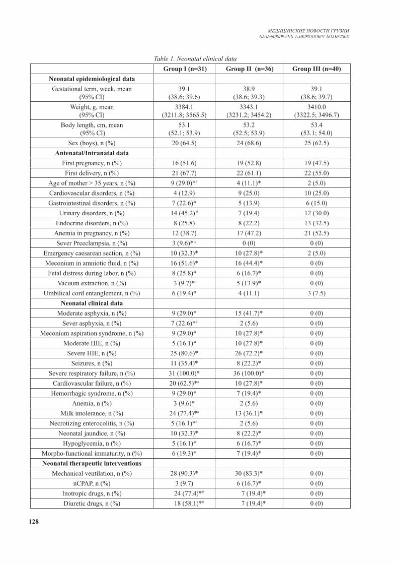
128
МЕДИЦИНСКИЕ НОВОСТИ ГРУЗИИ
CFMFHSDTKJC CFVTLBWBYJ CBF[KTYB
Table 1. Neonatal clinical data Group I (n=31) Group II (n=36) Group III (n=40)
Neonatal epidemiological dataGestational term, week, mean
(95% CI)39.1
(38.6; 39.6)38.9
(38.6; 39.3)39.1
(38.6; 39.7)Weight, g, mean
(95% CI)3384.1
(3211.8; 3565.5)3343.1
(3231.2; 3454.2)3410.0
(3322.5; 3496.7)Body length, cm, mean
(95% CI)53.1
(52.1; 53.9)53.2
(52.5; 53.9)53.4
(53.1; 54.0)Sex (boys), n (%) 20 (64.5) 24 (68.6) 25 (62.5)
Antenatal/Intranatal dataFirst pregnancy, n (%) 16 (51.6) 19 (52.8) 19 (47.5)First delivery, n (%) 21 (67.7) 22 (61.1) 22 (55.0)
Age of mother > 35 years, n (%) 9 (29.0)*# 4 (11.1)* 2 (5.0)Cardiovascular disorders, n (%) 4 (12.9) 9 (25.0) 10 (25.0)Gastrointestinal disorders, n (%) 7 (22.6)* 5 (13.9) 6 (15.0)
Urinary disorders, n (%) 14 (45.2) # 7 (19.4) 12 (30.0)Endocrine disorders, n (%) 8 (25.8) 8 (22.2) 13 (32.5)
Anemia in pregnancy, n (%) 12 (38.7) 17 (47.2) 21 (52.5)Sever Preeclampsia, n (%) 3 (9.6)* # 0 (0) 0 (0)
Emergency caesarean section, n (%) 10 (32.3)* 10 (27.8)* 2 (5.0)Meconium in amniotic fluid, n (%) 16 (51.6)* 16 (44.4)* 0 (0)Fetal distress during labor, n (%) 8 (25.8)* 6 (16.7)* 0 (0)
Vacuum extraction, n (%) 3 (9.7)* 5 (13.9)* 0 (0)Umbilical cord entanglement, n (%) 6 (19.4)* 4 (11.1) 3 (7.5)
Neonatal clinical dataModerate asphyxia, n (%) 9 (29.0)* 15 (41.7)* 0 (0)
Sever asphyxia, n (%) 7 (22.6)*# 2 (5.6) 0 (0)Meconium aspiration syndrome, n (%) 9 (29.0)* 10 (27.8)* 0 (0)
Moderate HIE, n (%) 5 (16.1)* 10 (27.8)* 0 (0)Severe HIE, n (%) 25 (80.6)* 26 (72.2)* 0 (0)
Seizures, n (%) 11 (35.4)* 8 (22.2)* 0 (0)Severe respiratory failure, n (%) 31 (100.0)* 36 (100.0)* 0 (0)
Cardiovascular failure, n (%) 20 (62.5)*# 10 (27.8)* 0 (0)Hemorrhagic syndrome, n (%) 9 (29.0)* 7 (19.4)* 0 (0)
Anemia, n (%) 3 (9.6)* 2 (5.6) 0 (0)Milk intolerance, n (%) 24 (77.4)*# 13 (36.1)* 0 (0)
Necrotizing enterocolitis, n (%) 5 (16.1)*# 2 (5.6) 0 (0)Neonatal jaundice, n (%) 10 (32.3)* 8 (22.2)* 0 (0)
Hypoglycemia, n (%) 5 (16.1)* 6 (16.7)* 0 (0)Morpho-functional immaturity, n (%) 6 (19.3)* 7 (19.4)* 0 (0)Neonatal therapeutic interventions
Mechanical ventilation, n (%) 28 (90.3)* 30 (83.3)* 0 (0)nCPAP, n (%) 3 (9.7) 6 (16.7)* 0 (0)
Inotropic drugs, n (%) 24 (77.4)*# 7 (19.4)* 0 (0)Diuretic drugs, n (%) 18 (58.1)*# 7 (19.4)* 0 (0)

GEORGIAN MEDICAL NEWS No 5 (278) 2018
© GMN 129
Drugs for CNS, n (%) 27 (87.1)*# 15 (41.7) 0 (0)Antibiotics, n (%) 31 (100.0)* 36 (100.0)* 0 (0)
Plasma, n (%) 7 (22.6)* 6 (16.7)* 0 (0)Erythrocyte, n (%) 3 (9.7)* 2 (5.6) 0 (0)
* − significant difference from group III, p<0.05; # − significant difference between groups I and II, p<0.05HIE - hypoxic-ischemic encephalopathy; nCPAP - nasal continuous constant positive airway pressure;
CNS - central nervous system
Table 2. Neonatal renal data
Group I (n=31) Group II (n=36)
Group III (n=40)
SCr, µmol/l, mean (95% CI)
74.7*#
(65.6; 83.8)56.0*
(51.7; 60.4)42.1
(39.2; 45.0)GFR, ml/min/1.73m2, mean
(95% CI)30.3*#
(26.7; 33.9)39.8
(36.6; 42.9)52.8
(48.9; 56.6)SUr, mmol/l, mean
(95% CI)6.62*#
(5.69; 7.56)3.79*
(3.28; 4.29)2.76
(2.57; 2.95)UCr, µmol/l, mean
(95% CI)1375.5*#
(1181.4; 1569.5)1979.8*
(1701.7; 2257.9)3564.9
(3167.3; 3962.6)UUr, mmol/l, mean
(95% CI)38.3*#
(34.5; 42.1)34.1*
(32.1; 36.1)51.6
(43.4; 59.8)ErMDA, µmol/l, mean
(95% CI)25.1*
(24.1; 26.1)24.0*
(23.2; 24.9)20.3
(19.5; 21.0)UMDA, µmol/l, mean
(95% CI)12.7*#
(11.5; 13.9)10.2*
(9.61; 10.8)9.01
(8.16; 9.93)* − significant difference from group III, p<0.05; # − significant difference between groups I and II, p<0.05.
SC - serum creatinine; GFR - glomerular filtration rate; SUr - serum urea; UCr - urine creatinine; UUr - urine urea; ErMDA - erythrocytes malondialdehyde; UMDA - urine malondialdehyde
ROC curve analysis showed that UMDA had an AUC 0.80 (95% CI 0.68; 0.93, p=0.0014) for AKI. The opti-mum UMDA cut-point was 12.0 µmol/l. For AKI, Se and Sp of UMDA with a cut-point value of 12.0 were 68.2% (95% CI 45.1; 86.1%) and 85.7% (95% CI 69.7; 95.1%) respectively, PPV value of 75.0% (95% CI 55.9; 87.6%), and NPV value of 81.1% (95% CI 69.7; 88.9%), PLR value of 4.77 (95% CI 2.02; 11.3), and NLR value of 0.37 (95% CI 0.20; 0.69). The result of ROC analysis was demonstrated on Fig.
Fig. Receiver operating curves (ROC) for UMDA
MDA is a final product of lipid peroxidation. It can be regarded as a marker for the extent of damage to cells and the anti-oxidative system capacity. MDA is the best indi-cator of lipid peroxidation as well as of OS in both mother and newborn [3,5,6]. The result of the study conducted by
Gülbayzar et al. [5] indicates that MDA level serves as an indicator of intrauterine growth retardation. They demon-strated that MDA level was significantly higher in babies with low hemoglobin and hematocrit values in the cord blood in the emergency cesarean section group [3]. Dol-apo et al. [12] found significant negative correlations of MDA with placenta weight as well as of MDA with birth weight. The study conducted by Jain et al. [6] showed OS in cesarean delivery time. They found that MDA level was significantly higher in the cord blood during cesarean section than during physiological delivery.
The experiment with neonatal rats performed by Pi-mentel et al. [11] demonstrated that higher levels of MDA in urine in the first day of post hypoxia-ischemia may be related to inadequate scavenging abilities of the im-mature nervous system. Being noninvasive, it suggests that UMDA may be a marker for lipid peroxidation after hypoxic-ischemic insult. El Bana et al. [4] reported that newborns with perinatal asphyxia had significantly higher levels of MDA in the cord blood and mean MDA/Cr ratio than healthy neonates. The babies who died due to perina-tal asphyxia had significantly higher levels of cord blood MDA, and also urinary MDA:Cr ratio than the babies who survived [8].
OS was found to occur not only in newborns with perinatal asphyxia but also with common severe perina-
0
10
20
30
40
50
60
70
80
90
100
0 10 20 30 40 50 60 70 80 90 100
1 - Sp, %
Se, %
130
МЕДИЦИНСКИЕ НОВОСТИ ГРУЗИИ
CFMFHSDTKJC CFVTLBWBYJ CBF[KTYB
tal pathology. In the current study there was significant increase of ErMDA level in critically sick full-term new-borns with severe perinatal pathology as compared to healthy neonates. But significant difference between sick group full-term newborns with AKI and without AKI was not found. On the contrary, Mishra et al. reported that plasma MDA level was significantly higher in AKI pa-tients in comparison with that of the controls. In addition, significantly higher levels of plasma MDA were found in AKI non-survivors in comparison with survivors. The au-thors selected children at the age of 0-10 years with AKI and healthy children with similar age and sex [8].
The production of free radicals can cause kidney injury [15,16]. The level of UMDA has been claimed to reflect the level of lipid peroxidation in renal structures. On the other hand, UMDA as a measure of impaired kidney function in patients is difficult to interpret because renal clearance of MDA may provide an adaptive mechanism that prevents ac-cumulation of lipid peroxidation within kidney tubular cells [15]. The study evidenced that critically sick newborns with AKI had higher urinary excretion of MDA as compared to sick babies without AKI and healthy neonates. But the re-sults of our ROC curve analysis indicate moderate diagnostic accuracy of this model. However, they indicated high lev-els of Sp (85.7% (95% CI 69.7%; 95.1%)) and NPV value (81.1% (95% CI 69.7%; 88.9%)).
An insufficient diagnostic accuracy of the present model can be explained by some limitation factors of this study. (1) It was a single-center study (2) with a small patient cohort suffering from AKI. The diagnostic and prognostic values of other serum and urinary markers of OS and specific renal dysfunction should be studied and a common mathematical model for prognosis and diagno-sis of AKI in newborns formulated. (3) It is necessary to continue studing of the relationships between individual nosologies, AKI and UMDA level, since the nosological structure of the basic pathology in newborns of group I and group II are not homogeneous.
In conclusion, this data demonstrated that UMDA may be useful biomarkers for AKI in critically sick full-term newborns in early neonatal period but further study is re-quired to confirm the promising findings.
REFERENCES
1. Ayala A, Munoz MF, Arguelles S. Lipid peroxidation: produc-tion, metabolism, and signaling mechanisms of malondialdehyde and 4-hydroxy-2-nonenal. Oxid Med Cell Longev 2014; Avail-able at External link http://dx.doi.org/10.1155/2014/360438.2. Diaz-Castro J, Florido J, Kajarabille N, Prados S, de Paco C, Ocon O, et al. A new approach to oxidative stress and inflamma-tory signaling during labour in healthy mothers and neonates. Oxid Med Cell Longev 2015; Available at External link http://dx.doi.org/10.1155/2015/178536. 3. Dolapo PO, Daniel AA, Adeniran SA, Abolape AI, Patric TA. Cord blood oxidative stress markers correlate with birth and pla-centa. J Asian Sci Res 2013; 3: 365-72. 4. El Bana SM, Maher SE, Gaber AF, Aly SS. Serum and urinary
malondialdehyde (MDA), uric acid, and protein as markers of peri-natal asphyxia. Electron Physician 2016; 8 Suppl 7: 2614–2619. 5. Gülbayzar S, Arica V, Hatipoglu S, Kaya A, Arica S, Kara-tekin G. Malondialdehyde level in the cord blood of newborn infants. Iran J Pediatr 2011; 21: 313-9. 6. Jain S, Nair A, Shrivastava C. Evaluation of oxidative stress marker malondialdehyde level in the cord blood of newborn infants. International Journal of Scientific Study 2015; 3 Suppl 6: 73-76. 7. Korobeinikov EN. [Modification of the definition of LPO products in reaction with thiobarbituric acid.] Laboratornoe Delo 1989; 7: 8-10.8. Mishra OP, Pooniya V, Ali Z, Upadhyay RS, Prasad R. Anti-oxidant status of children with acute renal failure. Pediatric Ne-phrology 2008; 23: 2047. 9. Mutinati M, Pantaleo M, Roncetti M, Piccinno M, Rizzo A, Sciorsci RL. Oxidative stress in neonatology. A Review. Reprod Dom Anim 2014; 49: 7-16. 10. Perrone S, Tataranno ML, Stazzoni G, Buonocore G. Oxi-dative stress and free radicals related diseases of the newborn. Advances in Bioscience and Biotechnology 2012; 3: 1043-1050. 11. Pimentel VC, Pinheiro FV, Kaefer M, Moresco RN, Moretto MB Assessment of uric acid and lipid peroxidation in serum and urine after hypoxia–ischemia neonatal in rats. Neurol Sci 2011; 32 Suppl 1: 59-65. 12. Richardson DK, Gray JE, McCormick MC, Workman K, Goldmann DA. Score for Neonatal Acute Physiology: a physio-logic severity index for neonatal intensive care. Pediatrics 1993; 91 Suppl 3: 617-623.13. Saugstad Ola Didrik Oxygen and oxidative stress in bron-chopulmonary dysplasia. Journal of Perinatal Medicine 2010; 38 Suppl 6: 571-577.14. Selewski DT, Charlton JR, Jetton JG, Guillet R, Mhanna MJ, Askenazi DJ, et al. Neonatal acute kidney injury. Pediatrics 2015; 136 Suppl 3: Е463-E473. DOI: 10.1542/peds.2014-3819.15. Small DM, Coombes JS, Bennet N, Johnson DW, Gobe GC. Oxidative stress, anti-oxidant therapies and chronic kidney dis-ease. Nephrology 2012; 17: 311-321. 16. Tamadon MR, Zahmatkesh M, Mousavi SSB. Administra-tion of antioxidants in chronic kidney disease. J Nephropharma-col 2015; 4 Suppl 1: 9-11.17. Weber D, Stuetz W, Bernhard W, Raith M, Grune T. Oxidative stress markers and micronutrients in maternal and cord blood in relation to neonatal outcome. Eur J Clin Nutr 2014; 68: 215-22.
SUMMARY
URINARY MALONDIALDEHYDE AS A PREDIC-TIVE AND DIAGNOSTIC MARKER FOR NEONA-TAL ACUTE KIDNEY INJURY
1Hodovanets Y., 1,3Babintseva A., 1,3Agafonova L., 2Makarova O., 1Frunza A.
1Higher State Educational Establishment of Ukraine «Bu-kovinian State Medical University», Department of Pedi-atrics, Neonatology and Perinatal Medicine; 2Department of Nursing and Higher Nursing Education, Chernivtsi; 3Neonatal Intensive Care Unit, Maternity Hospital №2 Chernivtsi, Ukraine
Newborns are especially prone to oxidative stress (OS). Many free radical-mediated diseases have been de-
GEORGIAN MEDICAL NEWS No 5 (278) 2018
© GMN 131
scribed in newborns including bronchopulmonary dyspla-sia, respiratory distress syndrome, encephalopathy, and kidney injury. Objective of this work was to determine predictive and diagnostic value of urinary malondialde-hyde (UMDA) as a marker for acute kidney injury (AKI) in critically sick full-term newborns.
67 critically sick full-term neonates were enrolled in the study including 31 newborns with AKI (group I) and 36 newborns without AKI (group II). The control group included 40 healthy full-term neonates (group III). The level of UMDA was measured by means of the test based on the reaction of MDA to thiobarbituric acid.
The mean and 95% confidence interval (CI) for UMDA was 12.7 (11.5; 13.9) µmol/l in group I, 10.2 (9.61; 10.8) µmol/l in group II, 9.01 (8.16; 9.93) µmol/l in group III (pI-II<0.05; pI-III<0.05; pII-III<0.05). ROC curve analy-sis showed that UMDA had AUC 0.80 (95% CI 0.68; 0.93, p=0.0014) for AKI. The optimum UMDA cut-point was 12.0µmol/l. For AKI sensitivity and specificity of UMDA were determined to be 68.2% (95% CI 45.1; 86.1%) and 85.7% (95% CI 69.7; 95.1%) respectively, with positive pre-dictive value of 75.0% (95% CI 55.9; 87.6%), and negative predictive value of 81.1% (95% CI 69.7; 88.9%), positive likelihood ratio of 4.77 (95% CI 2.02; 11.3), and negative likelihood ratio of 0.37 (95% CI 0.20; 0.69).
This data support studies to evaluate UMDA as an im-mediate biomarker for AKI in critically sick newborns. However, a larger study should be conducted to assess the diagnostic accuracy of other serum and urinary markers for OS and renal dysfunction, which would enable us to formulate a mathematical model for the prognosis and di-agnosis of AKI in newborns.
Keywords: acute kidney injury; term newborns; oxi-dative stress; urinary malondialdehyde.
РЕЗЮМЕ
МАЛОНОВЫЙ ДИАЛЬДЕГИД МОЧИ КАК ПРО-ГНОСТИЧЕСКИЙ И ДИАГНОСТИЧЕСКИЙ МАРКЕР НЕОНАТАЛЬНОГО ОСТРОГО ПО-ВРЕЖДЕНИЯ ПОЧЕК
1Годованец Ю.Д., 1,3Бабинцева А.Г., 1,3Агафонова Л.В., 2Макарова Е.В., 1Фрунза А.В.
Высшее государственное учебное учреждение Украи-ны «Буковинский государственный медицинский уни-верситет», 1кафедра педиатрии, неонатологии и пе-ринатальной медицины; 2кафедра ухода за больными и высшего медсестринского образования, Черновцы; 3Родильный дом №2, отделение интенсивной терапии новорожденных, Черновцы, Украина
Целью исследования явилось определить прогно-стическую и диагностическую ценность малонового диальдегида мочи в качестве маркера острого повреж-
дения почек у критически больных доношенных но-ворожденных детей.
В исследование включено 67 критически боль-ных доношенных новорожденных детей, в том числе 31 новорожденный с острым повреждением почек (ОПП) - I группа и 36 новорожденных без ОПП - II группа. Контрольная (III группа) включала 40 здоро-вых доношенных детей. Уровень малонового диаль-дегида мочи (МДА) измеряли с помощью теста, осно-ванного на реакции МДА с тиобарбитуровой кислотой. Полученные данные выявили, что средний и 95% дове-рительный интервал (95% ДИ) для МДА мочи состави-ли 12,7 (11,5; 13,9) мкмоль/л в I группе, 10,2 (9,61; 10,8) мкмоль/л - во II группе, 9,01 (8,16; 9,93) мкмоль/л в III группе (pI-II<0,05, pI-III<0,05, pII-III<0,05). Анализ кривой ROC показал, что МДА мочи имеет AUC 0,80 (95% ДИ 0,68; 0,93, p=0,0014) для ОПП. Оптимальная точка МДА мочи составила 12,0 мкмоль/л; для ОПП чувствитель-ность и специфичность МДА мочи - 68,2% (95% ДИ 45,1%; 86,1%) и 85,7% (95% ДИ 69,7%; 95,1%), соот-ветственно, с прогностической ценностью позитивного результата 75,0% (95% ДИ 55,9%; 87,6%) и прогности-ческой ценностью негативного результата 81,1% (95% ДИ 69,7%; 88,9%), с соотношением правдоподобности позитивного результата - 4,77 (95% ДИ 2,02; 11,3), со-отношением правдоподобности негативного результата - 0,37 (95% ДИ 0,20; 0,69).
Представленные данные подтверждают результа-ты исследований о возможности использования МДА мочи как биомаркера ОПП у критически больных новорожденных. Для оценки точности диагностики других сывороточных и мочевых маркеров оксидатив-ного стресса и почечной дисфункции, что позволит разработать математическую модель прогноза и диа-гностики ОПП у новорожденных детей, необходимо продолжить исследования в этом направлении.
reziume
Sardis malonuri dialdehidi, rogorc Tirkmlebis neonataluri mwvave dazianebis prognozuli da diagnostikuri markeri
1i. godovaneci, 1,3a. babincevi, 1,3l. agafonova, 2e. makarova, 1a. frunza
bukovinis saxelmwifo samedicino univer-siteti, 1pediatriis, neonatologiis da peri-nataluri medicinis kaTedra; 2avadmyofis movlisa da umaRlesi saeqTno ganaTlebis kaTedra; 3samSobiaro saxli #2, axalSo-bilTa intensiuri Terapiis ganyofileba, Cernovci, ukraina
kvlevis mizans Seadgenda Sardis malo-nuri dialdehidis prognozuli da diagnos-tikuri markeruli Rirebulebis gansazRvra
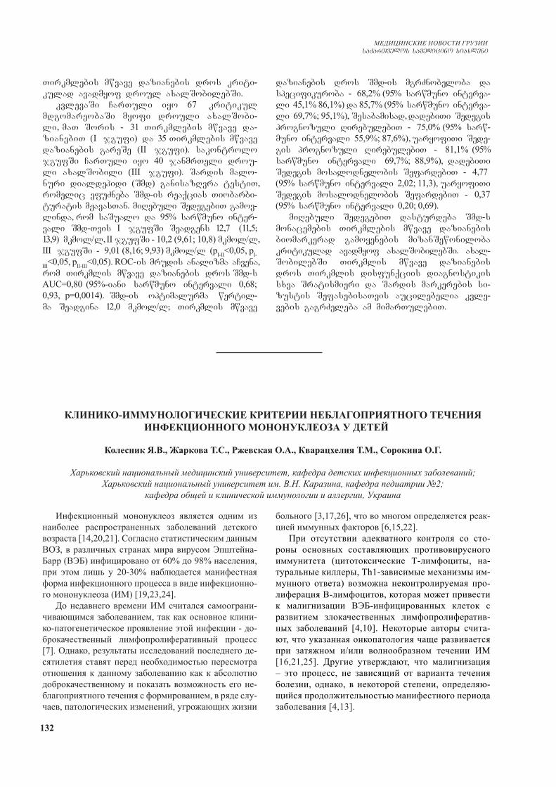
132
МЕДИЦИНСКИЕ НОВОСТИ ГРУЗИИ
CFMFHSDTKJC CFVTLBWBYJ CBF[KTYB
Tirkmlebis mwvave dazianebis dros kriti-kulad avadmyof droul axalSobilebSi.
kvlevaSi CarTuli iyo 67 kritikul mdgomareobaSi myofi drouli axalSobi-li, maT Soris - 31 Tirkmlebis mwvave da-zianebiT (I jgufi) da 35 Tirkmlebis mwvave dazianebis gareSe (II jgufi). sakontrolo jgufSi CarTuli iyo 40 janmrTeli drou-li axalSobili (III jgufi). Sardis malo-nuri dialdehidi (Smd) ganisazRvra testiT, romelic efuZneba Smd-is reaqcias Tiobarbi-turatis mJavasTan. miRebuli SedegebiT gamov-linda, rom saSualo da 95% sarwmuno inter-vali Smd-Tvis I jgufSi Seadgens 12,7 (11,5; 13,9) mkmol/l, II jgufSi - 10,2 (9,61; 10,8) mkmol/l, III jgufSi - 9,01 (8,16; 9,93) mkmol/l (pI-II<0,05, pI-
III<0,05, pII-III<0,05). ROC-is mrudis analizma aCvena, rom Tirkmlis mwvave dazianebis dros Smd-s AUC=0,80 (95%-iani sarwmuno intervali 0,68; 0,93, p=0,0014). Smd-is optimalurma wertil-ma Seadgina 12,0 mkmol/l; Tirkmlis mwvave
dazianebis dros Smd-is mgrZnobeloba da specifikuroba - 68,2% (95% sarwmuno interva-li 45,1% 86,1%) da 85,7% (95% sarwmuno interva-li 69,7%; 95,1%), Sesabamisad, dadebiTi Sedegis prognozuli RirebulebiT - 75,0% (95% sarw-muno intervali 55,9%; 87,6%), uaryofiTi Sede-gis prognozuli RirebulebiT - 81,1% (95% sarwmuno intervali 69,7%; 88,9%), dadebiTi Sedegis mosalodnelobis SefardebiT - 4,77 (95% sarwmuno intervali 2,02; 11,3), uaryofiTi Sedegis mosalodnelobis SefardebiT - 0,37 (95% sarwmuno intervali 0,20; 0,69).
miRebuli SedegebiT dasturdeba Smd-s monacemebis Tirkmlebis mwvave dazianebis biomarkerad gamoyenebis mizanSewoniloba kritikulad avadmyof axalSobilebSi. axal-SobilebSi Tirkmlis mwvave dazianebis dros Tirkmlis disfunqciis diagnostikis sxva Sratismieri da Sardis markerebis si-zustis SefasebisaTvis aucilebelia kvle-vebis gagrZeleba am mimarTulebiT.
КЛИНИКО-ИММУНОЛОГИЧЕСКИЕ КРИТЕРИИ НЕБЛАГОПРИЯТНОГО ТЕЧЕНИЯ ИНФЕКЦИОННОГО МОНОНУКЛЕОЗА У ДЕТЕЙ
Колесник Я.В., Жаркова Т.С., Ржевская О.А., Кварацхелия Т.М., Сорокина О.Г.
Харьковский национальный медицинский университет, кафедра детских инфекционных заболеваний; Харьковский национальный университет им. В.Н. Каразина, кафедра педиатрии №2;
кафедра общей и клинической иммунологии и аллергии, Украина
Инфекционный мононуклеоз является одним из наиболее распространенных заболеваний детского возраста [14,20,21]. Согласно статистическим данным ВОЗ, в различных странах мира вирусом Эпштейна-Барр (ВЭБ) инфицировано от 60% до 98% населения, при этом лишь у 20-30% наблюдается манифестная форма инфекционного процесса в виде инфекционно-го мононуклеоза (ИМ) [19,23,24].
До недавнего времени ИМ считался самоограни-чивающимся заболеванием, так как основное клини-ко-патогенетическое проявление этой инфекции - до-брокачественный лимфопролиферативный процесс [7]. Однако, результаты исследований последнего де-сятилетия ставят перед необходимостью пересмотра отношения к данному заболеванию как к абсолютно доброкачественному и показать возможность его не-благоприятного течения с формированием, в ряде слу-чаев, патологических изменений, угрожающих жизни
больного [3,17,26], что во многом определяется реак-цией иммунных факторов [6,15,22].
При отсутствии адекватного контроля со сто-роны основных составляющих противовирусного иммунитета (цитотоксические Т-лимфоциты, на-туральные киллеры, Th1-зависимые механизмы им-мунного ответа) возможна неконтролируемая про-лиферация В-лимфоцитов, которая может привести к малигнизации ВЭБ-инфицированных клеток с развитием злокачественных лимфопролифератив-ных заболеваний [4,10]. Некоторые авторы счита-ют, что указанная онкопатология чаще развивается при затяжном и/или волнообразном течении ИМ [16,21,25]. Другие утверждают, что малигнизация – это процесс, не зависящий от варианта течения болезни, однако, в некоторой степени, определяю-щийся продолжительностью манифестного периода заболевания [4,13].
GEORGIAN MEDICAL NEWS No 5 (278) 2018
© GMN 133
Необходимо отметить, что в литературных ис-точниках по сей день нет четких данных о клини-ко-лабораторных критериях прогнозирования воз-можного течения ИМ у детей, тогда как решение данного вопроса позволит более адекватно подойти к формированию комплекса терапевтических меро-приятий, необходимых для выявления путей совер-шенствования методов прогнозирования течения и исходов ИМ у детей.
Целью исследования явилось определение кли-нико-лабораторных критериев неблагоприятного те-чения инфекционного мононуклеоза Эпштейна-Барр вирусной этиологии у детей.
Материал и методы. Под наблюдением нахо-дились дети (n=161) в возрасте 3-15 лет, больных ИМ. Из них у 140 (86,9%) заболевание протекало в среднетяжелой, у 21 (13,1%) - в тяжелой фор-мах. Степень тяжести заболевания устанавлива-лась на основе клинических проявлений болезни и изменений параметров лабораторных данных. Детям проводились стандартные клинические и лабораторно-инструментальные обследования. Диагноз ИМ устанавливался с использованием методов ПЦР (выявление ДНК ВЭБ в крови) и ИФА (анти-ВЭБ IgM и IgG). У 140 (86,9%) детей ИМ протекал остро, гладко (первая группа), у 21 (13,1%) – неблагоприятно (волнообразное и/или затяжное течение) - вторая группа. Группы были сопоставимы по возрасту, тяжести заболевания и другим параметрам. Все дети получали терапию, согласно утвержденным протоколам (Приказ МОЗ Украины №354 от 09.07.2004г.). Иммунный статус детей оценивался по результатам опреде-ления относительного содержания CD3+, CD4+, CD8+, CD16+, CD19+ клеток крови с помощью соответствующих моноклональных антител, кон-центрации сывороточных IgA, IgM, IgG - по мето-ду Манчини и показателям цитокинового ответа – интерлейкинов (IL)-1β и IL-4, фактор некроза опухоли (ФНО-α) – твердофазным иммунофер-ментным методом. Полученные результаты стати-стически обрабатывались с использованием обще-принятых методов вариационной статистики.
Результаты и их обсуждение. Установлено, что на сегодняшний день ИМ у детей сохраняет свой характерный симптомокомплекс, в котором при-сутствуют интоксикация, лихорадка, тонзиллит, затрудненное носовое дыхание, лимфаденопатия, увеличение печени и селезенки. Однако частота вы-явления и выраженность указанных проявлений в дебюте болезни, а также показатели лабораторных исследований в сравниваемых группах были раз-личны. Наиболее характерные клинические прояв-ления на начальных этапах заболевания в зависи-мости от его дальнейшего течения представлены на рис. 1.
Интоксикация, проявлением которой были: сла-бость, снижение аппетита, головная боль, бледность кожных покровов, фебрильная лихорадка, при не-благоприятном течении ИМ встречалась у больных достоверно чаще в дебюте болезни, чем при гладком течении заболевания (p<0,05).
Постоянным симптомом заболевания являлось по-ражение носоглоточной миндалины, с чем и связана заложенность носа (затруднение носового дыхания). При этом значимых различий в выраженности сим-птомов аденоидита у детей в группах сравнения не наблюдалось (р>0,05).
Наиболее ранним и постоянным симптомом ИМ являлось поражение ротоглотки, характеризовавшееся гиперемией слизистой, бугристостью ее задней стенки, гипертрофией небных миндалин. У всех больных ука-занные изменения сочетались с явлениями тонзиллита. Установлено, что гнойный тонзиллит в остром периоде заболевания достоверно чаще встречался при дальней-шем неблагоприятном его течении (p<0,05).
Лимфопролиферативный синдром выявлен прак-тически у всех госпитализированных детей. Одна-ко у пациентов с формированием на последующих этапах неблагоприятного течения болезни имела место генерализованная лимфаденопатия с вовле-чением в процесс 5-6 групп лимфатических узлов и более значимым увеличением подчелюстных лим-фатических образований (3,8±0,6 см), в то время как, при гладком течении отмечалось, в основном, увеличение размеров тонзиллярных (1,9±0,3 см) и заднешейных (1,2±0,12см) лимфатических узлов (p<0,05). У всех больных при пальпации лимфо-узлы были плотной консистенции, эластичные, не спаянные между собой и с подлежащими тканями. Цвет кожи над ними не изменялся.
Практически у всех больных определялся гепа-толиенальный синдром. В ходе исследования уста-новлено, что более значимое увеличение размеров печени (4,4±0,94 см) наблюдалось у детей второй группы в сравнении с первой (2,7±0,24 см) (p<0,05), которое сопровождалось развитием реактивного ге-патита, о чем свидетельствует повышение активно-сти АЛТ.
Рис. 1. Клинические проявления ИМ с формирова-нием различных вариантов его течения
134
МЕДИЦИНСКИЕ НОВОСТИ ГРУЗИИ
CFMFHSDTKJC CFVTLBWBYJ CBF[KTYB
Спленомегалия встречалась достоверно чаще при развитии неблагоприятного течения ИМ. Выявле-но, что размеры селезенки увеличивались у детей с последующим гладким течением ИМ, в среднем, на 1,45±0,24 см, в то время как у сравниваемых больных – на 2,85±0,41 см (p<0,05).
Среди наблюдаемых больных в 15% случаев имела место экзантема, без специфической локализации, что, на наш взгляд, в большинстве случаев связано с приемом антибиотиков (ампициллин и его аналоги). Аналогичное обьяснение появления сыпи при ИМ приводят в своих работах многие авторы [8,12,20]. Морфологически сыпь была точечной, мелко - и/или крупно - пятнистой, с ге-моррагическим компонентом, нередко имела тенденцию к слиянию. Появление экзантемы не отражало дальней-ший вариант течения заболевания и встречалась на ран-них этапах ИМ практически с одинаковой частотой у де-тей обеих сравниваемых групп. Показатели гемограммы приведены на рис. 2.
В результате исследования установлено, что в периферической крови анемия и тромбоцитопения встречались достоверно чаще при формировании неблагоприятного течения ИМ (p<0,05). В сравни-ваемых группах детей общевоспалительные изме-нения в гемограмме (лейкоцитоз, повышение СОЭ) регистрировались с одинаковой частотой.
Гематологическая манифестация ИМ с развитием в дальнейшем неблагоприятного течения заболевания сопровождалась значительной нейтропенией в отли-чие от детей группы сравнения (p<0,05). Предполо-жительно, что развитие нейтропении связано с уси-лением апоптотического потенциала нейтрофильных гранулоцитов, аналогичного мнения придерживаются Барычева Л.Ю. и соавт. [2].
Повышение числа лимфоцитов и моноцитов от-мечалось в обеих сравниваемых группах. Атипичные мононуклеары отсутствовали у 15 больных с после-дующим неблагоприятным течением ИМ; в то время как, у детей с гладким течением заболевания атипич-ные мононуклеары не выявлены только в 9% случаев (p<0,05). Некоторые авторы [5,12] полагают, что на этапах манифестации заболевания атипичные моно-нуклеары - это В-лимфоциты, активированные ВЭБ. В поздние сроки клинических проявлений большая часть атипичных мононуклеаров - это Т-лимфоциты [8,15]. Дальнейшее отсутствие атипичных монону-клеаров на поздних этапах болезни, на наш взгляд, свидетельствует о выраженном апоптозе Т-клеток и иммунной дисфункции, которая проявляется в угне-тении отдельных составляющих иммунного ответа. Показатели клеточного и гуморального звеньев имму-нитета отражены в таблице 1.
Рис. 2. Показатели гемограммы
Таблица 1. Показатели клеточного и гуморального звеньев иммунитета
ПоказательПоказатели иммунного ответа (М±m)
Р1 Р 2ЗдоровыеN=22
1 группа N=140
2 группаN=21
CD3+-клетки (%) 58,2±2,4 79,4±3,5 38,3 ±2,3 P<0,05 P<0,05CD4+-клетки (%) 33,8±1,5 28,4±2,9 19,2±1,8 P>0,05 P<0,05CD8+-клетки (%) 28,3±1.2 41,6±2,4 9,4 ±1,8 P<0,05 P<0,05CD16+-клетки (%) 15,6±1,1 21,6±2,3 9,3±1,4 P<0,05 P<0,05CD19+-клетки (%) 20,2±1,2 41,1±2,12 50,2±2,11 P<0,05 P<0,05
Ig М (г/л) 1,03±0,2 2,16±0,1 6,1±0,1 P<0,05 P<0,05Ig G (г/л) 7,03±0,3 16,1±0,6 8,02±1,2 P<0,05 P>0,05Ig A (г/л) 1,01±0,15 3.8±0,9 4,5±0,8 P<0,05 P<0,05
Р 1-достоверность различий между детьми первой группы и здоровыми.Р 2- достоверность различий между детьми второй группы и здоровыми
GEORGIAN MEDICAL NEWS No 5 (278) 2018
© GMN 135
При исследовании иммунного ответа больных ИМ выявлено, что в большинстве случаев гладкое течение ИМ сопровождается активацией как клеточного, так и гуморального звеньев иммунитета, о чем свидетель-ствуют высокие показатели относительного содержа-ния CD3+, CD8+, CD16+, CD19+ лимфоцитов и имму-ноглобулинов класса А, М, G (таблица 1). Полученные результаты совпадают с данными множества ученых, полагающих, что при эффективной иммунной реакции организма, приводящей в дальнейшем к выздоров-лению, в острый период ИМ отмечается активация клеточного и гуморального механизмов иммунного ответа [4,10].
У пациентов с развившимся в последующем не-благоприятным течением заболевания отмечалась иммунная дисфункция, которая проявлялась в угне-тении клеточного звена и усилении гуморальных ме-ханизмов иммунного ответа (снижение экспрессии маркеров лимфоцитов: CD3+, CD4+, CD8+, CD16+ и повышение титра IgA IgM, IgG). Результаты прове-денных исследований совпадают с данными авторов о возникновении на ранних этапах ИМ, при развитии неблагоприятного течения болезни, депрессии кле-точного звена иммунитета с преобладанием снижения содержания CD-клеток и повышением уровня антите-лообразования [3,18]. Показатели цитокинового отве-та представлены в таблице 2.
Анализ цитокинового статуса детей в дебюте ИМ выявил выраженные различия между сравни-ваемыми группами. Из данных таблицы 2 явствует, что при формировании у детей гладкого течения за-болевания показатель уровня IL-1β и ФНО-α пре-вышает таковые группы контроля, что отражает вы-раженный Т-хелперный ответ в сторону Тh1–типа. У детей с развитием неблагоприятного течения болезни активация IL-1β и ФНО-α была менее зна-чительной (р<0,05). Содержание IL-4 у пациентов I группы сохранялось на уровне показателей группы контроля, а у больных с развившимся впоследствии неблагоприятным течением ИМ имела место выра-женная активация синтеза этого цитокина, в част-ности, функционирование иммунной системы у де-тей второй группы происходило по Th2-зависимому пути. Аналогичная трактовка формирования раз-личных вариантов клинического течения с позиции
оценки цитокинового статуса детей встречается и у других исследователей [10,13].
Выводы:1. Клиническими критериями развития неблагоприят-ного течения ИМ на ранних этапах манифестации забо-левания являются: генерализованная лимфаденопатия с вовлечением в процесс 5-6 групп лимфатических узлов, с преимущественной реакцией подчелюстных лимфати-ческих образований, гнойный тонзиллит, выраженное увеличение размеров печени и селезенки на фоне ане-мии, тромбоцитопении, нейтропении и отсутствия ати-пичных мононуклеаров в гемограмме.2. Формирование иммунного ответа у детей с после-дующим благоприятным течением инфекционного мононуклеоза характеризуется активацией как кле-точного, так и гуморального звеньев иммунитета. При развитии неблагоприятного течения ИМ отмечается угнетение клеточного звена и усиление гуморальных механизмов иммунного ответа.3. Выявленные в результате проведенного исследова-ния клинико-иммунологические различия могут быть основой создания в дальнейшем прогностических алгоритмов установления течения ИМ у детей и воз-можных его исходов, что позволит снизить процент неблагоприятных результатов болезни и наметить пути усовершенствования лечения больных.
ЛИТЕРАТУРА
1. Барычева Л.Ю. Показатели адаптивного иммунитета у детей с инфекционным мононуклеозом, обусловленным вирусом Эпштейна-Барр / Л.Ю.Барычева, М.В.Голубева, А.В.Волкова. // Кубанский научный медицинский вестник. – 2012. - №2(131). – С. 30-33.2. Барычева Л.Ю. Функциональное состояние системы нейтрофильных гранулоцитов у детей с инфекцией, об-условленной вирусом Эпштейна-Барр / Л.Ю.Барычева, М.С.Шалина, М.В.Голубева // Медицинский вестник Север-ного Кавказа. - 2012. - №1. - С. 21-25.3. Блохина Е. Б. Роль латентной инфекции, вызванной ви-русом Эпштейна–Барр, в развитии лимфопролиферативных заболеваний // Вопросы гематологии/онкологии и иммуно-патологии в педиатрии. — 2003. — Т. 2, № 3. — С. 65–70.4. Выговская О. В., Крамарев С. А., Дорошенко В. О., Шпак И. В. Инфекционный мононуклеоз Эпштейна- Барр вирусной этиологии у детей: вопросы этиологии, патогенеза, иммуноге-неза, лечения // Практикуючий лікар 2012. - № 4. - С. 29-34.
Таблица 2. Показатели цитокинового ответа
ПоказательСредние величины показателей (М±m)
Р 1 Р 2ЗдоровыеN=22
1 гр.N=140
2 гр.N=21
IL 1β пг/мл 26,41±2,3 98,07±13,16 34,7±2,1 P<0,05 P>0,05
IL 4 пг/мл 25,24±2,4 24,01 ±2,03 231,1±26,06 P>0,05 P<0,05
ФНО α пг/мл 23,88±2,3 81,27±12,91 25,02±2,07 P<0,05 P>0,05
Р 1-достоверность различий между детьми первой группой и здоровыми;Р 2- достоверность различий между детьми второй группой и здоровыми

136
МЕДИЦИНСКИЕ НОВОСТИ ГРУЗИИ
CFMFHSDTKJC CFVTLBWBYJ CBF[KTYB
5. Гилева Р.А. Клинико-лабораторная характеристика ин-фекционного мононуклеоза, обусловленного вирусом Эпштейна-Барр / Р.А.Гилева, З.А.Хохлова, Ю.С.Чечет, А.С.Арсененко, Л.М.Поволоцкая // Казанский медицинский журнал. – 2014. – Т. 95, №5. – С.722-725.6. Глей А. І. Хронічні форми Епштейн-Барр вірусної інфекції // Клінічна імунологія. Алергологія. Інфектологія. -2009. - № 2. – С.-69-71.7. Дрыганова М.Б. Инфекционный мононуклеоз, вызванный вирусом Эпштейна-Барр у детей: клинико-иммунологические параллели / М.Б. Дрыганова, Г.П. Мартынова, Л.М. Куртасова // Сибирское медицинское обозрение. – 2010. - № 3. – С. 13-17.8. Дутлова Д.В. Клинико-лабораторные особенности инфек-ционного мононуклеоза у детей в зависимости от этиологии заболевания / Д.В.Дутлова, О.И.Уразова, А.П.Помогаева // Детские инфекции. – 2016. - №1. – С. 30-34.9. Калинина Н.М. Современные представления об иммуно-патогенезе инфекции, вызванной вирусом Эпштейна-Барр / Н.М.Калинина, Л.Б.Дрыгина, Т.В.Горейко // Инфекция и иммунитет. – 2001. – Т. 1, №2. – С. 121-130.10. Крамарев С.А., Выговская О.В. Эпштейна — Барр ви-русная инфекция у детей // Актуальная инфектология. 2013. - №1(1). - С. 73-78.11. Крамарь Л.В. Оценка показателей общего анализа крови у детей при инфекционном мононуклеозе различной этио-логии / Л.В.Крамарь, О.А.Карпухина. // Современные про-блемы науки и образования. – 2012. - №6. – С. 98-10212. Краснова Е.И. Клиника и лиагностика инфекционного мононуклеоза у детей грудного возраста / Е.И.Краснова, А.В.Васюнин, Н.А.Никифорова. А.С.Поздняков // Детские инфекции. – 2004. - №1. – С. 6-10.13. Кудин А. П. Эта «безобидная» вирус Эпштейна-Барр инфекция. Часть 3. Хроническая ВЭБ-инфекция и хрони-ческие ВЭБ-ассоциированные заболевания // Медицинские новости. – 2006. – Т. 1, №8. – С. 25-31.14. Кудин А. П. Показатели состояния иммунитета при ин-фекционном мононуклеозе у детей / А. П. Кудин, Т. Р. Ро-мановская, М. В. Белевцев //Весці НАН Беларусі. Серыя Медыцынскіх навук. – 2007. – №2. – С. 13-20.15. Лесина О. Н. Катамнез часто болеющих пациентов, пере-несших инфекционный мононуклеоз и эффективность имму-нореабилитации / О. Н. Лесина, Д. Ю. Курмаева. // Известия высших учебных заведений. – 2010. - №2(14). – С. 62–67.16. Леженко Г.О. Инфекционный мононуклеоз у детей: кли-нико-иммунологическая характеристика / Г.О. Леженко, Е.В. Усачева, Е.А. Силина, Т.Н. Пахольчук // Актуальная ин-фектология. 2013. - №.1(1). – С. 56-60.17. Разгуляева А.В. Современные представления об этиологии и патогенезе инфекционного мононуклеоза / А.В. Разгуляева, О.П. Уханова // Наука и современность. - 2012. - № 2. - С. 62-67.18. Собчак Д.М. Оценка содержания медиаторов иммунного от-вета у больных с циклическим и прогредиентным течением ВЭБ-инфекционного мононуклеоза / Д.М.Собчак, О.В.Корочкина, Т.А.Свинцова, Т.В.Щуклина, К.В.Кушман, Т.Ю.Бутина // Меди-цинский альманах. – 2014. - №4(34). – С. 56-60.19. Сравнительная характеристика клинического течения и лабораторных данных первичной Эпштейна-Барр вирусной инфекции и ее реактивации у детей различных возрастных групп / [М. В. Антонова, Э. А. Кашуба, Т. Г. Дроздова и др.]. // Вестник совета молодых ученых и специалистов Челя-бинской области. – 2016. - Т. 5, №2. – С. 19–24.20. Тюняева Н. О., Софронова Л. В. Инфекционный моно-нуклеоз: этиологические факторы, проблемы диагностики
и лечения // Вестник новых медицинских технологий. 2014. - Т. 21, №3. - С. 184-190.21. Хмилевская С. А. Клинико-эпидемиологические аспек-ты инфекционного мононуклеоза у детей / С. А. Хмилев-ская, И. А. Зайцева. // Эпидемиология и вакцинопрофилак-тика. – 2010. - №5(54). – С. 45–50.22. Хмилевская С. А. Эпштейна-Барр вирусная инфекция у детей: особенности цитокинового ответа и иммунопатоло-гические реакции / С. А. Хмилевская, И. А. Зайцева, Е. В. Михайлова. // Саратовский медицинский журнал. – 2009. - Т. 5, №2. – С. 222–226.23. Шарипова Е. В. Герпес-вирусные инфекции и инфекци-онный мононуклеоз / Е. В. Шарипова, И. В. Бабаченко. // Журнал инфектологии. – 2013. – Т 5, № 2. – С. 5-12.24. Шведова Н.М. Инфекционный мононуклеоз у детей: кли-нико-лабораторное обоснование и экономическая эффектив-ность применения иммунокорректоров / Н. М. Шведова, Е. В. Михайлова, Ю. С. Цека, Т. К. Чудакова. // Саратовский меди-цинский журнал. – 2013. - Т. 9, №3. – С. 512–517.25. Balfour H.H.. Infectious mononucleosis / H.H. Balfour Jr, S.K. Dunmire, K.A. Hogquist // Clinical & Translational Im-munology. – 2015. – Vol. 4.26. Cohen J.I. Epstein–Barr virus-associated lymphoprolifera-tive disease in non immunocompromised hosts: a status report and summary of an international meeting, 8–9 September 2008. / J.I. Cohen, H.Kimura, S.Nakamura, Y.H. Ko, E.S.Jaffe. // Ann. Oncol. — 2009. — 20 (9). — 1472–1482.
SUMMARY
CLINICAL AND IMMUNOLOGICAL CRITERIA FOR THE ADVERSE COURSE OF INFECTIOUS MONONUCLEOSIS IN CHILDREN
Kolesnik Ya., Zharkova T., Rzhevskaya O., Kvaratskheliya T., Sorokina O.
Kharkiv National Medical University, Department of Children’s Infectious Diseases; V.N. Karasin Kharkov Na-tional University, Department of Pediatrics №2; Depart-ment of Clinical Immunology and Allergy, Ukraine
The article presents the results of our own studies to determine the criteria for the adverse variants of the course of infectious mononucleosis (IM) in children. The study was conducted in the regional children’s infectious clinical hospital in Kharkov. 161 children aged three to fifteen years were under observation with diagnosis of infectious moninucleosis. Out of 161 ill children, 140 (86.9%) had moderate severity of disease, and 21 (13.1%) had severe forms. All children were prescribed standard clinical and laboratory-instrumental examinations. The diagnosis of IM was verified by PCR (detection of VEB DNA in the blood) and ELISA (anti-VEB Ig M and Ig G). In 140 children (86.9%) IM proceeded sharply, smoothly (the first group), in 21 (13.1%) - unfavorably (wave and / or prolonged course) - the second group. The groups were comparable according to age, the severity of the disease and other parameters. All children received therapy ac-cording to approved protocols (Order of the Ministry of
GEORGIAN MEDICAL NEWS No 5 (278) 2018
© GMN 137
Health of Ukraine No. 354 of 09.07.2004). Immune status of children was assessed by determining the relative con-tents of CD3 +, CD4 +, CD8 +, CD16 +, CD19 + blood cells with appropriate monoclonal antibodies, serum IgA, IgM, IgG concentration by Mancini and interleukin (IL) -1β cytokine response and - 4, tumor necrosis factor (TNF α) is a solid-phase enzyme-linked immunosorbent assay. Based on the results of observations, it was established that the prognostically unfavorable criteria of IМ at the stages of manifestation of disease include: generalized lymphadenopathy involving 5-6 groups of lymph nodes and a significant increasing of them, purulent tonsillitis, marked increasing of size of liver and spleen on the back-ground of anemia, thrombocytopenia, neutropenia and the absence of atypical mononuclears in the complete blood count. There is a depression of the cellular link and an increase in the humoral mechanisms of immune responses in case of development of adverse course of IM.
Keywords: children, infectious mononucleosis, clini-cal symptoms, immunity, prognosis
РЕЗЮМЕ
КЛИНИКО-ИММУНОЛОГИЧЕСКИЕ КРИТЕ-РИИ НЕБЛАГОПРИЯТНОГО ТЕЧЕНИЯ ИН-ФЕКЦИОННОГО МОНОНУКЛЕОЗА У ДЕТЕЙ
Колесник Я.В., Жаркова Т.С., Ржевская О.А., Кварацхелия Т.М., Сорокина О.Г.
Харьковский национальный медицинский универси-тет, кафедра детских инфекционных заболеваний; Харьковский национальный университет им. В.Н. Кара-зина, кафедра педиатрии №2; кафедра общей и клини-ческой иммунологии и аллергии, Украина
В статье приведены результаты исследований по определению критериев неблагоприятных вариантов течения инфекционного мононуклеоза (ИМ) у детей. Исследование проведено на базе областной детской инфекционной клинической больницы г. Харьков. Под наблюдением находились дети (n=161) в возрас-n=161) в возрас-161) в возрас-те 3-15 лет, больные ИМ. Из них у 140 (86,9%) забо-левание протекало в среднетяжелой, у 21 (13,1%) - в тяжелой форме. Всем детям проводилось стандартное клиническое и лабораторно-инструментальное об-следование. Диагноз ИМ верифицировался методами ПЦР (выявление ДНК ВЭБ в крови) и ИФА (анти-ВЭБ IgM и IgG). У 140 (86,9%) детей ИМ протекал остро, гладко (первая группа), у 21 (13,1%) – неблагоприят-но (волнообразное и/или затяжное течение) - вторая группа. Группы были сопоставимы по возрасту, тяже-сти заболевания и другим параметрам. Все дети по-лучали терапию, согласно утвержденным протоколам (Приказ МОЗ Украины №354 от 09.07.2004г.). Иммун-ный статус детей оценивался по результатам опре-
деления относительного содержания CD3+, CD4+, CD8+, CD16+, CD19+ клеток крови с помощью соот-ветствующих моноклональных антител, концентрации сывороточных IgA, IgM, IgG по методу Манчини и по-казателям цитокинового ответа – интерлейкинов (IL)-1β, и -4, фактор некроза опухоли (ФНО-α) – твердофазный иммуноферментный метод. По результатам наблюдений установлено, что к прогностически неблагоприятным критериям ИМ на этапах манифестации заболевания относятся: генерализованная лимфаденопатия с во-влечением в процесс 5-6 групп лимфатических узлов со значительным их увеличением, гнойный тонзиллит, выраженное увеличение размеров печени и селезенки на фоне анемии, тромбоцитопения, нейтропения и от-сутствие атипичных мононуклеаров в гемограмме. При развитии неблагоприятного течения ИМ отмечается уг-нетение клеточного звена и усиление гуморальных ме-ханизмов иммунного ответа.
reziume
infeqciuri mononukleozis arakeTilsai-medo mimdinareobis klinikur-imunologi-uri kriteriumebi bavSvebSi
i. kolesniki, t. Jarkova, o. rJevskaia, t. kvaracxelia, o. sorokina
xarkovis erovnuli samedicino universite-ti, bavSvTa infeqciur daavadebaTa kaTedra, ukraina; xarkovis v.karazinis sax. erovnuli universiteti, pediatriis #2 kaTedra, zoga-di da klinikuri imunologiis da alergiis kaTadra, ukraina
statiaSi motanilia kvlevis Sedegebi bavSvebSi infeqciuri mononukleozis mim-dinareobis arakeTilsaimedo variantebis kriteriumebis gansazRvris Sesaxeb. kvleva Ca-tarda xarkovis saolqo bavSvTa infeqciuri klinikuri saavadmyofos bazaze. dakvirvebis qveS imyofeboda infeqciuri mononukleoz-iT daavadebuli 3-15 ww. 161 bavSvi; maTgan 140 (86,9%) bavSvSi daavadeba mimdinareobda saSualo simZimis, xolo 21-Si (13,1%) - mZime formiT. yvela bavSvs Cautarda standar-tuli klinikuri da laboratoriul-in-strumentuli gamkvleva. infeqciuri mono-nukleozis diagnozi verificirebuli iyo polimerazul-jaWvuri reaqciis da imuno-fermentuli analizis meTodebiT. 140 (86,9%) bavSvSi infeqciuri mononukleozi mimdin-areobda mwvaved, Tanabrad (I jgufi), 21-Si (13,1%) – arakeTilsaimedod (talRovani da/an gaxangrZlivebuli mimdinareoba; II jgufi). jgufebi urTierTSesabamisi iyo bavSvebis asakis, daavadebis mimdinareobis da sxva
138
МЕДИЦИНСКИЕ НОВОСТИ ГРУЗИИ
CFMFHSDTKJC CFVTLBWBYJ CBF[KTYB
parametrebis mixedviT. yvela bavSvs Cautar-da Sesabamisi protokoliT damtkicebuli mkurnaloba (ukrainis janmrTelobis dacvis ministris brZaneba № 354, 09.07.2004). bavSvebis imunuri statusi Sefasda CD3+, CD4+, CD8+, CD16+, CD19+-ujredebis SefardebiTi Semcv-elobis gansazRvriT Sesabamisi monoklon-uri antisxeulebis saSualebiT da Sratis-mieri IgA, IgM, IgG koncentraciis dadgeniT manCinis meTodiT, aseve, citokinuri pasuxis maCvenebelebiT – IL-1β- da 4-iT, nekrozuli zrdis faqtoriT – myarfazovani imunofer-mentuli meTodiT. dakvirvebis Sedegebis mixedviT dadgenilia, rom infeqciuri mono-
nukleozis prognozulad arakeTilsaime-do kriteriumebs daavadebis manifestaciis etapze miekuTvneba: generalizebuli lim-fadenopaTia, procesSi limfuri kvanZebis 5-6 jgufis CaTreviT da maTi mniSvnelovani gadidebiT, Cirqovani tonziliti, RviZlis da elenTis zomebis gamoxatuli momateba anemiis, Trombocitopeniis, neitropeniis da hemogramaSi atipiuri mononuklearebis ararsebobis fonze. infeqciuri mononuk-leozis arakeTilsaimedo mimdinareobis ganviTarebisas aRiniSneba imunuri pasuxis ujreduli rgolis daTrgunva da humoruli meqanizmebis gaZliereba.
ЭПИГЕНЕТИЧЕСКАЯ РЕГУЛЯЦИЯ РАЗВИТИЯ ХРОМАФФИННЫХ КЛЕТОК НАДПОЧЕЧНИКОВ (ОБЗОР)
1Обернихин С.С., 1Яглова Н.В., 2Цомартова Д.А., 2Торбек В.Э., 2Иванова М.Ю.
1Федеральное государственное бюджетное научное учреждение «НИИ морфологии человека», Москва; 2Федеральное государственное автономное образовательное учреждение
«Первый московский государственный медицинский университет им. И.М. Сеченова», Москва, Россия
Транскрипционная и посттранскрипционная регу-ляция эмбрионального и постэмбрионального гисто-генезов – активно развивающееся направление био-логии развития. Полученные данные в этой области не только расширили имеющиеся знания, но зачастую существенно изменили представление о морфогенезе некоторых органов [1, 28, 47]. Надпочечники состо-ят из двух гистогенетически разнородных структур – коркового и мозгового вещества. Уникальность стро-ения и функции мозгового вещества приобретаются в результате реализации сложной программы развития, обусловленной микроокружением формирующегося коркового вещества. Помимо этого в прошлом веке в научной литературе неоднократно описывались клетки на границе коркового и мозгового вещества, морфологически демонстрирующие черты кортико-стероцитов и хромаффинных клеток. Таким образом, возникает вопрос о пластичности в реализации гене-тической программы развития мозгового вещества, что возможно достигается транскрипционной и пост-транскрипционной регуляцией гистогенеза и требует дальнейших исследований.
Надпочечники развиваются из двух различных эм-бриональных зачатков. Корковое вещество развивает-ся из висцерального листка спланхнотома мезодермы, тогда как мозговое происходит из клеток нервного гребня [29]. Надпочечники появляются в виде адрено-гонадного зачатка (АГЗ) на 28-30-ые сутки беремен-ности у человека (9-й день эмбрионального развития
у мышей) и характеризуются экспрессией стероидо-генного фактора 1 (SF1) [31]. Как парный орган АГЗ впервые появляется в виде утолщения целомического эпителия между урогенитальным гребнем и дорсаль-ной брыжейкой. Каждый АГЗ содержит смешанную популяцию клеток адренокортикального и гонадных прогениторных клеток. SF1-положительные клетки АГЗ, затем отслаиваются от эпителия и внедряются в мезенхиму мезодермы [65]. После расслаивания боль-шинство клеток АГЗ мигрируют дорсолатерально для образования первичных гонад. Субпопуляция клеток АГЗ, которые экспрессируют более высокие уровни SF1, мигрирует дорсомедиально для формирования первичных надпочечников, так называемой надпочеч-никовой фетальной зоны. На 48-ой день эмбриональ-ного развития у человека (11,5-13,5 день у мышей) клетки нервного гребня мигрируют в развивающу-юся фетальную зону [54]. Эти клетки сохраняются в виде отдельных островков, разбросанных по всему эмбриональному надпочечнику вплоть до рождения, и, в конечном счёте, сливаются и дифференцируют-ся в продуцирующие катехоламины хромаффинные клетки [17]. Далее надпочечник начинает отделяться от окружающей мезенхимы и инкапсулируется с об-разованием волокнистого слоя над развивающимиcя кортикальными клетками. Этот процесс в основном завершается к 52-му дню внутриутробного развития у человека (на 14,5 сутки у мышей) [12]. Адренокор-тикальные и хромаффинные клетки тесно связаны в
GEORGIAN MEDICAL NEWS No 5 (278) 2018
© GMN 139
перииодах эмбрионального и постнатального раз-вития. Глюкокортикоиды надпочечников играют зна-чимую роль в продукции гормонов хромаффинных клеток, регулируя экспрессию фенилэтаноламин-N-метилтрансферазы (PNMT), что приводит к тому, что адреналин, а не норадреналин является доминирую-щим катехоламином мозгового вещества надпочечни-ков в постнатальном периоде [21]. После инкапсули-рования корковое вещество надпочечников эмбриона быстро расширяется. Надпочечник плода человека является одним из крупнейших органов на этом сроке (0,2% от общей массы тела и почти размером с поч-ку), причем 80% железы состоит из клеток фетальной зоны [43].
Клетки нервного гребня, мигрируя по развиваю-щемуся эмбриону, генерируют широкий спектр раз-личных клеточных популяций, в том числе хромаф-финные клетки мозгового вещества надпочечников и параганглии [19]. Параганглии состоят из катехо-ламинсодержащих нейроэндокринных клеток. Не-которые параганглии являются хеморецепторами и связаны с ветвями блуждающего и языкоглоточного нервов головы и шеи. Остальные параганглии вы-полняют эндокринные функции и тесно взаимодей-ствуют с вегетативной нервной системой, секретируя катехоламины для поддержки гомеостаза в условиях стресса [42]. Они формируют вненадпочечниковые (экстраадренальные) хромаффинные клетки. Вне-надпочечниковые хромаффинные клетки появляются в кластерах от уровня верхнего шейного ганглия до таза, часто в ассоциации с симпатическими гангли-ями, брюшной аортой и урогенитальными органами [60]. Вненадпочечниковые (экстраадренальные) хро-маффинные клетки в отличие от надпочечниковых в основном норадренергические [5,60]. Считается, что они являются важным источником катехоламинов во время развития плода [86,87,98]. Oрган Цуккеркандля является самым большим кластером вненадпочечни-ковых (экстраадренальных) хромаффинных клеток и находится вблизи надпочечников, вокруг зарождаю-щейся нижней брыжеечной артерии [60,82]. Он исче-зает в постнатальном периоде.
Раннее развитие клеток нервного гребняНервный гребень – уникальная популяция кле-
ток позвоночных, которая часто называется «четвер-тым зародышевым листком», так как она образуется вместе с другими зародышевыми листками и вносит вклад в формирование разнообразных типов клеток и тканей [66]. Клетки нервного гребня представляют со-бой переходную популяцию эмбриональных предше-ственников, которая возникает из дорсального отдела нервной трубки на раннем этапе развития. Индукция нервного гребня начинается с взаимодействия между эпидермальной эктодермой и нервной трубкой с обра-зованием границы между ними. Впоследствии клетки нервного гребня формируются в пограничных обла-стях, которые поднимаются в виде складок и, в конеч-
ном счете, сходятся, образуя дорсальную среднюю линию нервной трубки. Эти клетки мигрируют по пе-риферии по стереотипным путям. Во время и после миграции клетки дифференцируется в очень широкий диапазон производных. Таким образом, существуют две ключевые особенности клеток нервного гребня – способность к миграции и мультипотентность [16].
Развитие хромаффинных клеток надпочечниковХромаффинные клетки надпочечников вначале
появляются среди тирозингидроксилаза-экспресси-рующих клеток, у которых подавляется экспрессия нейронных маркеров и сохраняется экспрессия ка-техоламинергических [84]. У мышей анатомическая сегрегация развивающихся хромаффинных клеток происходит путём дифференцировки превертебраль-ных симпатических нейробластов, когда хромафин-ные клетки мигрируют в поперечном направлении, сливаясь в центре с клетками коры надпочечников, образуя тем самым мозговое вещество [38,45]. Толь-ко на 14-ый день эмбрионального развития у мышей субпопуляция хромаффинных клеток начинает экс-прессировать PNMT [13, 56]. Долгое время счита-лось, что хромаффинные клетки и симпатические нейробласты имели общую клетку-предшественницу – симпатоадреналовую клетку [53]. Это предположе-ние основано на способности хромаффинных клеток in vivo и in vitro приобретать нейроподобный фено-тип при стимуляции фактором роста нервов (nerve growth factor, NGF) [7,17]. Отмечено также, что не-возможно отличить хромаффинные клетки и симпа-тические нейробласты на 11,5 день эмбрионального развития у мышей доступными методами, так как они экспрессируют одинаковый набор поверхностных антигенов [8]. Однако, недавние исследования пока-зали, что большинство хромаффинных клеток имеют другое происхождение и возникают из предшествен-ников шванновских клеток [26], которые также явля-ются производными клеток нервного гребня (рис. 1). Предшественники шванновских клеток дают начало широкому диапазону других видов клеток, таких как эндонейральные фибробласты в периферических не-рвах [44], меланоциты [4], парасимпатические нейро-ны черепных, тазовых и сердечных ганглиев [20,23], одонтобласты [49] и нейроны кишечника [89].
Факторы транскрипции и нейроэндокринная диф-ференцировка
Развитие хромаффинных клеток и симпатических нейронов зависит от микроокружения и внешних сиг-налов, поступающих в процессе дифференцировки. Глюкокортикоиды, локально синтезируемые корой надпочечников, хорошо известны как регуляторы определенных аспектов дифференцировки хромаф-финных клеток. У млекопитающих они необходимы для индукции адренергического фенотипа, посколь-ку экспрессия PNMT зависит от глюкокортикоидов. Кроме того, глюкокортикоиды требуются для экс-прессии нейроэндокринного секреторного белка хро-
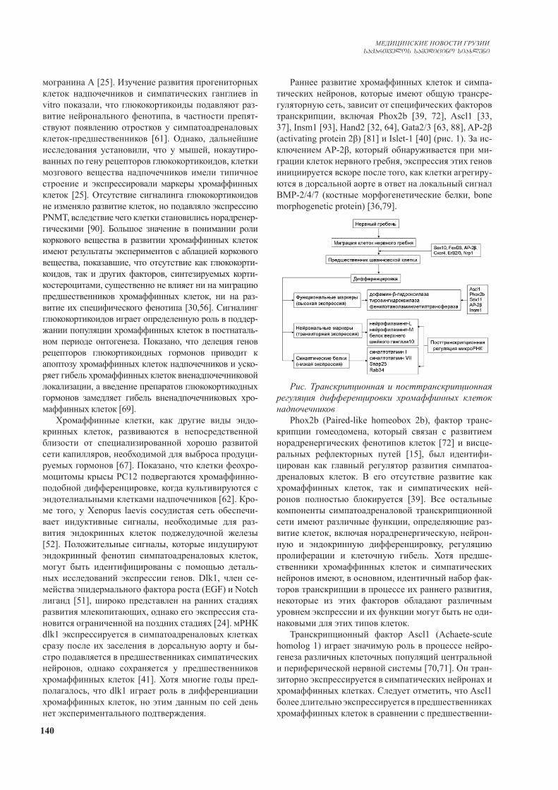
140
МЕДИЦИНСКИЕ НОВОСТИ ГРУЗИИ
CFMFHSDTKJC CFVTLBWBYJ CBF[KTYB
могранина А [25]. Изучение развития прогениторных клеток надпочечников и симпатических ганглиев in vitro показали, что глюкокортикоиды подавляют раз-витие нейронального фенотипа, в частности препят-ствуют появлению отростков у симпатоадреналовых клеток-предшественников [61]. Однако, дальнейшие исследования установили, что у мышей, нокаутиро-ванных по гену рецепторов глюкокортикоидов, клетки мозгового вещества надпочечников имели типичное строение и экспрессировали маркеры хромаффинных клеток [25]. Отсутствие сигналинга глюкокортикоидов не изменяло развитие клеток, но подавляло экспрессию PNMT, вследствие чего клетки становились норадренер-гическими [90]. Большое значение в понимании роли коркового вещества в развитии хромаффинных клеток имеют результаты экспериментов с аблацией коркового вещества, показавшие, что отсутствие как глюкокорти-коидов, так и других факторов, синтезируемых корти-костероцитами, существенно не влияет ни на миграцию предшественников хромаффинных клеток, ни на раз-витие их специфического фенотипа [30,56]. Сигналинг глюкокортикоидов играет определенную роль в поддер-жании популяции хромаффинных клеток в постнаталь-ном периоде онтогенеза. Показано, что делеция генов рецепторов глюкортикоидных гормонов приводит к апоптозу хромаффинных клеток надпочечников и уско-ряет гибель хромаффинных клеток вненадпочечниковой локализации, а введение препаратов глюкокортикодных гормонов замедляет гибель вненадпочечниковых хро-маффинных клеток [69].
Хромаффинные клетки, как другие виды эндо-кринных клеток, развиваются в непосредственной близости от специализированной хорошо развитой сети капилляров, необходимой для выброса продуци-руемых гормонов [67]. Показано, что клетки феохро-моцитомы крысы PC12 подвергаются хромаффинно-подобной дифференцировке, когда культивируются с эндотелиальными клетками надпочечников [62]. Кро-ме того, у Xenopus laevis сосудистая сеть обеспечи-вает индуктивные сигналы, необходимые для раз-вития эндокринных клеток поджелудочной железы [52]. Положительные сигналы, которые индуцируют эндокринный фенотип симпатоадреналовых клеток, могут быть идентифицированы с помощью деталь-ных исследований экспрессии генов. Dlk1, член се-мейства эпидермального фактора роста (EGF) и Notch лиганд [51], широко представлен на ранних стадиях развития млекопитающих, однако его экспрессия ста-новится ограниченной на поздних стадиях [24]. мРНК dlk1 экспрессируется в симпатоадреналовых клетках сразу после их заселения в дорcальную аорту и бы-стро подавляется в предшественниках симпатических нейронов, однако сохраняется у предшественников хромаффинных клеток [41]. Хотя многие годы пред-полагалось, что dlk1 играет роль в дифференциации хромаффинных клеток, но этим данным по сей день нет экспериментального подтверждения.
Раннее развитие хромаффинных клеток и симпа-тических нейронов, которые имеют общую трансре-гуляторную сеть, зависит от специфических факторов транскрипции, включая Phox2b [39, 72], Ascl1 [33, 37], Insm1 [93], Hand2 [32, 64], Gata2/3 [63, 88], AP-2β (activating protein 2β) [81] и Islet-1 [40] (рис. 1). За ис-ключением AP-2β, который обнаруживается при ми-грации клеток нервного гребня, экспрессия этих генов инициируется вскоре после того, как клетки агрегиру-ются в дорсальной аорте в ответ на локальный сигнал BMP-2/4/7 (костные морфогенетические белки, bone morphogenetic protein) [36,79].
Рис. Транскрипционная и посттранскрипционная регуляция дифференцировки хромаффинных клеток надпочечников
Phox2b (Paired-like homeobox 2b), фактор транс-крипции гомеодомена, который связан с развитием норадренергических фенотипов клеток [72] и висце-ральных рефлекторных путей [15], был идентифи-цирован как главный регулятор развития симпатоа-дреналовых клеток. В его отсутствие развитие как хромаффинных клеток, так и симпатических ней-ронов полностью блокируется [39]. Все остальные компоненты симпатоадреналовой транскрипционной сети имеют различные функции, определяющие раз-витие клеток, включая норадренергическую, нейрон-ную и эндокринную дифференцировку, регуляцию пролиферации и клеточную гибель. Хотя предше-ственники хромаффинных клеток и симпатических нейронов имеют, в основном, идентичный набор фак-торов транскрипции в процессе их раннего развития, некоторые из этих факторов обладают различным уровнем экспрессии и их функции могут быть не оди-наковыми для этих типов клеток.
Транскрипционный фактор Ascl1 (Achaete-scute homolog 1) играет значимую роль в процессе нейро-генеза различных клеточных популяций центральной и периферической нервной системы [70,71]. Он тран-зиторно экспрессируется в симпатических нейронах и хромаффинных клетках. Следует отметить, что Ascl1 более длительно экспрессируется в предшественниках хромаффинных клеток в сравнении с предшественни-
GEORGIAN MEDICAL NEWS No 5 (278) 2018
© GMN 141
ками симпатических нейронов, что может иметь зна-чение для их дифференцировки в нейроэндокринные клетки. Действительно, большинство эмбриональных хромаффинных клеток Ascl1-дефицитных мышей ха-рактеризуются повышенными уровнями экспрессии нескольких нейронных маркеров, включая нейрофила-мент 68, и отсутствием типичных секреторных гранул, что свидетельствует о нарушении нейроэндокринной дифференцировки [37]. Ascl1 также играет ключевую роль в развитии различных других типов клеток, таких как эндокринные клетки легких [68], С-клетки щитовид-ной железы [46]. Ascl1а, гомолог Ascl1, необходим для терминальной дифференцировки эндокринных клеток гипофиза [73].
Ascl1 имеет важное значение для экспрессии Insm1 (Insulinoma-associated protein 1, zinc-finger transcription factor), который также является частью симпатоадреналовой транскрипционной сети [93]. Insm1 является значимым регулятором целого ряда эндокринных клеток, включая хромаффинные клет-ки [93], эндокринные клетки гипофиза [92], клетки островков поджелудочной железы [27], а также кар-циномы лёгких нейроэндокринного происхождения [14]. Помимо своей роли в развитии эндокринных клеток, Insm1 также необходим для ранней диффе-ренцировки некоторых групп нейронов, включая симпатические нейроны [93], сенсорные нейроны спинального ганглия [18] и обонятельные нейроны [80]. При изучении потери функциональной активно-сти Insm1 показано, что он играет значимую роль в развитии симпатических нейронов и хромаффинных клеток. Дифференцировка симпатических нейронов у Insm1-дефицитных мышей происходит правильно, хотя с большой задержкой, их пролиферативная ак-тивность снижена. Напротив, Insm1-null мутация зна-чительно влияет на терминальную дифференцировку хромаффинных клеток [93]. В частности, уровень экс-прессии PNMT и хромогранина A, которые являются маркерами эндокринного фенотипа, значительно сни-жен, в то время как экспрессия нейронного маркера нейрофиламента-L повышена [27].
LIM-гомеодоменный транскрипционный фактор Islet-1 является регулятором дифференцировки кле-ток островков поджелудочной железы, но также при-нимает участие в развитии симпатоадреналовых кле-ток [40], мотонейронов [55], спинальных сенсорных нейронов [85], нейронов сетчатки [22] и сердца [11]. В симпатоадреналовых клетках экспрессия Islet-1 начинается вскоре после заселения ими дорсальной аорты и сохраняется в симпатических ганглиях и хро-маффинных клетках надпочечников, хотя и на гораздо более низких уровнях. Islet-1 не требуется для началь-ной дифференцировки симпатоадреналовых клеток из нервного гребня, однако необходим для выживания предшественников симпатических нейронов и экс-прессии Gata2[40].
Подобным действием обладает и транскрипцион-
ный фактор AP-2β, который также необходим для вы-живания субпопуляции симпатических нейронов [81]. Он требуется для эндокринной дифференцировки и созревания хромаффинных клеток, включая правиль-ное образование хромаффинных везикул и регулиро-вание уровней экспрессии PNMT [34].
Кроме того, развитие симпатоадреналовых клеток регулируется различными факторами транскрипции Sox. В то время как экспрессия белка Sox10 из группы SoxE происходит еще в мигрирующих клетках нервно-го гребня и, по существу, необходима для инициации развития, как симпатических ганглиев, так и мозгового вещества надпочечников [78], инициирование из SoxC группы белков Sox4 и Sox11 следует за Phox2B [74]. Sox4 и Sox11 идентифицированы как дополнительные значимые регуляторы выживаемости и пролиферации предшественников симпатических нейронов [74]. Sox4 также участвует в развитии эндокринных клеток остров-ков поджелудочной железы [94].
Таким образом, хромаффинные клетки и симпати-ческие нейроны, несмотря на экспрессию аналогич-ного набора факторов транскрипции во время их ран-него развития, по-разному зависят от этих факторов. Необходимо отметить, что некоторые факторы транс-крипции, которые в основном влияют на раннее раз-витие предшественников симпатических нейронов, важны для терминальной дифференцировки и созре-вания хромаффинных клеток.
В наших исследованиях впервые показано, что постнатальное поддержание пула хромаффинных клеток надпочечников регулируется также и транс-крипционным фактором PRH/Hhex. PRH/Hhex явля-ется супрессором пролиферации клеток и одним из регуляторов развития производных энтодермы в пре-натальном и постнатальном периодах онтогенеза [58]. Ранее считалось, что нейроны периферической нерв-ной системы, в том числе и хромаффинные клетки, не экспрессируют PRH/Hhex в постнатальном развитии [83]. Исследование PRH/Hhex выявило его присут-ствие в ядрах хромаффинных клеток мозгового веще-ства надпочечников крыс пубертатного и постпубер-татного возраста, а также снижение пролиферативной активности клеток при его активации [2]. Эти данные позволяют предположить, что PRH/Hhex может уча-ствовать и в регуляции эмбрионального гистогенеза хромаффинных клеток.
Эпигенетическая роль микроРНК в посттран-скрипционной регуляции
В течение последнего десятилетия роль пост-транскрипционной регуляции в морфогенетических процессах в различных органах получила признание научного сообщества. МикроРНК (miRs) представ-ляют собой небольшие некодирующие РНК, которые играют значимую роль в отключении генов [59]. Вы-яснилось, что микроРНК принимают активное уча-стие в развитии различного типа клеток, включая производные клеток нервного гребня [35, 96]. Wnt1-

142
МЕДИЦИНСКИЕ НОВОСТИ ГРУЗИИ
CFMFHSDTKJC CFVTLBWBYJ CBF[KTYB
cre-опосредованная утрата функции эндорибонукле-азы дайсер, фермента необходимого для биогенеза микроРНК [10], приводит к существенной гибели предшественников симпатических нейронов, тогда как их дифференцировка остаётся незатронутой [35, 96]. В хромаффинных клетках дайсер-дефицитных мышей повышена экспрессия нейрофиламента-М и, следовательно, они фенотипически напоминают ней-роны. С другой стороны, в симпатических нейронах наблюдается снижение экспрессии синаптического белка и они, в свою очередь, напоминают хромаф-финные клетки [84]. Хотя эти наблюдения позволяют предположить, что дайсер может играть существен-ную роль в дифференцировке симпатоадреналовых линий, тем не менее, его утрата заметно не приво-дит к полной конверсии хромаффинных клеток в симпатические нейроны и наоборот. Тем не менее, дайсер-зависимый механизм с участием микроРНК, по-видимому, необходим для различных вариантов экспрессии генов хромаффинных клеток и симпати-ческих нейронов. Интересно отметить, что эндокрин-ным клеткам островков поджелудочной железы также требуется дайсер для подавления экспрессии несколь-ких паннейрональных генов, включая SCG10 (superior cervical ganglion 10 protein), нейрофиламент-М и нейрофиламент-L. Предполагается, что основным ме-ханизмом является вовлечение фактора транскрипции RE1 (Silencing Transcription Factor, REST), который репрессирует нейрональные гены в ненейрональных клетках, являясь, ингибитором нейронной дифферен-цировки и отрицательным регулятором нейрогенеза [48]. Некоторые микроРНК по-разному экспрессиру-ются в хромаффинных клетках и симпатических ней-ронах. К ним, в частности относятся микроРНК-124 и микроРНК-375 [84]. MикроРНК-124 специфически связана с нейронами и четко обнаруживается в симпа-тических нейронах, но отсутствует в хромаффинных клетках. Показано, что микроРНК-124 играет роль в нейрогенезе [91,6], способна стимулировать рост ней-ритов [95] и нацелена на РНК-связывающий белок PTBP1 (polypyrimidine tract binding protein 1) глобаль-ного репрессора альтернативного сплайсинга в не-нейрональных клетках [57] и SCP1 (small C-terminal domain phosphatase 1), который связан с REST [91].
Уровень экспрессии микроРНК-375 в хромаффин-ных клетках надпочечников достаточно высок, но её уровень гораздо менее выражен в симпатических ней-ронах и мозге [50, 77]. Функция микроРНК-375 до-статочно хорошо изучена в поджелудочной железе, в которой она необходима для правильного развития клеток островков и регуляции секреции инсулина [75]. Экспрессия микроРНК-375 наблюдалась также в гипофизе и нейроэндокринных клетках легкого, что указывает на то, что она играет общую роль в диф-ференцировке эндокринных клеток [76]. Мишеня-ми, которые подавляются микроРНК-375, являются транскрипционный коактиватор YAP1 (yes-associated
protein 1) – белок-регулятор, активирующий транс-крипцию генов, участвующих в пролиферации кле-ток и подавляющий транскрипцию проапоптотиче-ских генов [68, 97], а также HuD – РНК-связывающий белок, который необходим при развитии нейронов, включая рост нейритов [3,9]. Более выраженная экс-прессия Ascl1 в хромаффинных клетках по сравне-нию с симпатическими ганглиями и повышенная экспрессия нейронных маркеров в хромаффинных клетках у Ascl1- и дайсер-дефицитных мышей, ука-зывает на возможность того, что микроРНК-375-опосредованная функция Ascl1 играет значимую роль в нейроэндокринной дифференцировке симпатоадре-наловых клеток.
В совокупности все эти данные свидетельству-ют, что посттранскрипционная регуляция микроРНК играет ведущую роль в развитии симпатоадренало-вых клеток, в утрате ими черт, характерных для ге-нерирующих и проводящих импульс нейронов и приобретении ими фенотипа эндокринных клеток с регулируемым гранулярным типом секреции. Про-филь микроРНК этих клеток определен только на от-носительно поздней стадии развития, поскольку их более ранняя изоляция чрезвычайно сложна. Опреде-ление микроРНК в ранних стадиях дифференцировки клеток может стать основой для понимания механиз-мов развития в норме и злокачественной трансформа-ции клеток.
ЛИТЕРАТУРА
1. Яглова Н.В., Обернихин С.С. Развитие иммунной систе-мы потомства после иммуностимулирующего воздействия в ранние сроки беременности. М.: ИНФРА-М, 2017. 220с.2. Яглова Н.В., Обернихин С.С., Цомартова Д.А., Назимова С.В., Яглов В.В. Экспрессия транскрипционного фактора PRH/Hhex в хромаффинных клетках надпочечников в пост-натальном развитии и его роль в регуляции пролифератив-ных процессов. Бюллетень экспериментальной биологии и медицины. 2018; 165(4):505-508.3. Abdelmohsen K, Hutchison ER, Lee EK et al. miR-375 inhib-its differentiation of neurites by loweringHuDlevels. Mol Cell Biol. 2010; 30:4197–4210.4. Adameyko I, Lallemend F, Aquino JB. et al. Schwann cell precursors from nerve innervation are a cellular origin of mela-nocytes in skin. Cell. 2009; 139:366–379. 5. Ahonen M, Soinila S, Joh TH. Pre- and postnatal development of rat retroperitoneal paraganglia. J Auton Nerv Syst. 1987; 18:111–120.6. Åkerblom M, Sachdeva R, Barde I et al. MicroRNA-124 is a subventricular zone neuronal fate determinant. J Neurosci. 2012; 32: 8879–8889.7. Anderson DJ, Axel R. A bipotential neuroendocrine precursor whose choice of cell fate is determined byNGF and glucocorti-coids. Cell. 1986; 47:1079–1090. 8. Anderson DJ, Carnahan JF, Michelsohn A, Patterson PH. An-tibody markers identify a common progenitor to sympathetic neurons and chromaffin cells in vivo and reveal the timing of commitment to neuronal differentiation in the sympathoadrenal lineage. J Neurosci. 1991; 11:3507–3519.
GEORGIAN MEDICAL NEWS No 5 (278) 2018
© GMN 143
9. Anderson KD, Sengupta J, Morin M, Neve RL, Valenzuela CF, Perrone-Bizzozero NI. Overexpression of HuD accelerates neurite outgrowth and increases GAP-43 mRNA expression in cortical neurons and retinoic acid-induced embryonic stem cells in vitro. Exp Neurol. 2001; 168:250-258. 10.Bernstein E, Caudy AA, Hammond SM, Hannon GJ. Role for a bidentate ribonuclease in the initiation step of RNA interfer-ence. Nature. 2001; 409:363–366.11.Cai CL, Liang X, Shi Y, Chu PH, Pfaff SL, Chen J, Evans S. Isl1 identifies a cardiac progenitor population that proliferates prior to differentiation and contributes a majority of cells to the heart. Dev Cell. 2003; 5:877–889.12.Ce K, Hammer GD. Recent insights into organogenesis of the adrenal cortex. Trends Endocrinol Metab. 2002; 13(5):200–208.13.Chan WH, Gonsalvez DG, Young HM, Southard-Smith EM, Cane KN, Anderson CR. Differences in CART expression and cell cycle behavior discriminate sympathetic neuroblast from chromaffin cell lineages in mouse sympathoadrenal cells. Dev. Neurobiol. 2016; 76: 137–149.14.Cueto A, Burigana F, Nicolini A, Lugnani F. Neuroendo-crine tumors of the lung: histological classification, diagno-sis, traditional and newtherapeutic approaches. Curr. Med. Chem. 2014; 21(9):1107-16. 15.Dauger S, Pattyn A, Lofaso F et al. Phox2b controls the de-velopment of peripheral chemoreceptors and afferent visceral pathways. Development. 2003; 130:6635–6642.16.Donoghue PC, Graham A, Kelsh RN. The origin and evolu-tion of the neural crest. Bioessays. 2008; 30(6):530-41. 17.Doupe AJ, Landis SC, Patterson PH. Environmental influ-ences in the development of neural crest derivatives: glucocorti-coids, growth factors, and chromaffin cell plasticity. J Neurosci. 1985; 5(8):2119–2142.18.Duggan A, Madathany T, de Castro SCP et al. Transient ex-pression of the conserved zinc finger gene INSM1 in progenitors and nascent neurons throughout embryonic and adult neurogen-esis. J Comp Neurol. 2008; 507:1497–1520.19. Dupin E, Le Douarin NM. The neural crest, a multifaceted structure of the vertebrates. Birth Defects Res. 2014; 102:187–209.20.Dyachuk V, Furlan A, Shahidi MK, et al. Neurodevelopment. Parasympathetic neurons originate from nerve-associated pe-ripheral glial progenitors. Science. 2014; 345:82–87; 21.Ehrhart-Bornstein M, Hinson JP, Bornstein SR, Scherbaum WA, Vinson GP. Intraadrenal interactions in the regulation of ad-renocortical steroidogenesis. Endocr Rev. 1998; 19(2):101–143.22.Elshatory Y, Everhart D, Deng M et al. Islet-1 controls the differentiation of retinal bipolar and cholinergic amacrine cells. J Neurosci. 2007; 27:12707–12720.23.Espinosa-Medina I, Outin E, Picard CA, et al. Neurodevelop-ment. Parasympathetic ganglia derive from Schwann cell pre-cursors. Science. 2014; 345:87–90. 24.Falix FA, Tjon-A-Loi MRS, Gaemers IC et al. DLK1 protein expression during mouse development provides new insights into its function. ISRN Dev Biol. 2013; 2013. doi:10.1155/2013/628962.25.Finotto, S., Krieglstein, K., Schober, A., et al. Analysis of mice carrying targeted mutations of the glucocorticoid receptor gene argues against an essential role of glucocorticoid signalling for generating adrenal chromaffin cells. Development. 1999; 126: 2935–2944. 26.Furlan A, Dyachuk V, Kastriti ME, et al. Multipotent periph-eral glial cells generate neuroendocrine cells of the adrenal me-dulla. Science. 2017; 357(4346): eaal3753. 27.Gierl MS, Karoulias N, et al. The zinc-finger factor Insm1
(IA-1) is essential for the development of pancreatic beta cells and intestinal endocrine cells. Genes Dev. 2006; 20:2465–2478.28.Gordon J., Manley N. Mechanisms of thymus organogenesis and morphogenesis. Development. 2011; 138(18): 3865–3878.29.Gruenwald P. Embryonic and postnatal development of the adrenal cortex, particularly the zona glomerulosa and accessory nodules. Anat Rec. 1946; 95:391–421.30.Gut P, Huber K, Lohr J, et al. Lack of an adrenal cortex in Sf1 mutant mice is compatible with the generation and differentia-tion of chromaffin cells. Development. 2005; 132:463–476.31.Hatano O, Takakusu A, Nomura M, Morohashi K. Identical origin of adrenal cortex and gonad revealed by expression pro-files of Ad4BP/SF-1. Genes Cells. 1996; 1(7):663–671.32.Hendershot TJ, Clouthier DE et al. Conditional deletion of Hand2 reveals critical functions in neurogenesis and cell type-specific gene expression for development of neural crest-derived noradrenergic sympathetic ganglion neurons. Dev Biol. 2008; 319:179–191.33.Hirsch MR, Tiveron MC, Guillemot F et al. Control of noradren-ergic differentiation and Phox2a expression by MASH1 in the central and peripheral nervous system. Development. 1998; 125:599–608.34.Hong SJ, Huh YH, Leung A, Choi et al. Transcription factor AP-2β regulates the neurotransmitter phenotype and maturation of chromaffin cells. Mol Cell Neurosci. 2011; 46:245–251. 35.Huang T, Liu Y, Huang M et al. Wnt1-cre-mediated condi-tional loss of Dicer results inmalformation of themidbrain and cerebellum and failure of neural crest and dopaminergic differ-entiation in mice. J Mol Cell Biol. 2010; 2:152–163.36.Huber K. The sympathoadrenal cell lineage: specification, di-versification, and new perspectives. Dev Biol. 2006; 298:335–343.37.Huber K, Brühl B, Guillemot F et al. Development of chro-maffin cells depends on MASH1 function. Development. 2002; 129:4729–4738.38.Huber K, Kalcheim C, Unsicker K. The development of the chromaffin cell lineage from the neural crest. Auton Neurosci. 2009; 151:10–16.39.Huber K, Karch N, Ernsberger U et al. The role of Phox2B in chromaffin cell development. Dev Biol. 2005; 279:501–508.40.Huber K, Narasimhan P, Shtukmaster S et al. The LIMhome-odomain transcription factor Islet-1 is required for the develop-ment of sympathetic neurons and adrenal chromaffin cells. Dev Biol. 2013; 380:286–298.41.Huber K. Segregation of neuronal and neuroendocrine differ-entiation in the sympathoadrenal lineage. Cell Tissue Res. 2015; 359:333–341. 42.Jänig W. Autonomic nervous system. In: Schmidt RF, Thews G(eds) Human physiology. Springer, Berlin, 1989; pp 333–370. 43.Johannison E. The foetal adrenal cortex in the human. Its ul-trastructure at different stages of development and in different functional states. Acta Endocrinol. 1968; 58. Suppl.130. 7+44.Joseph NM, Mukouyama YS,Mosher JT, et al. Neural crest stem cells undergo multilineage differentiation in developing peripheral nerves to generate endoneurial fibroblasts in addition to Schwann cells. Development. 2004; 131:5599–5612. 45.Kameda Y. Signaling molecules and transcription factors in-volved in the development of the sympathetic nervous system, with special emphasis on the superior cervical ganglion. Cell Tissue Res. 2014; 357: 527–548]. 46.Kameda Y, Nishimaki T, Miura M et al. Mash1 regulates the development of C cells in mouse thyroid glands. Dev Dyn. 2007; 236: 262–270. 47.Kameda Y. Cellular and molecular events on the development of mammalian thyroid C cells. Dev Dyn. 2016 Mar;245(3):323-41.
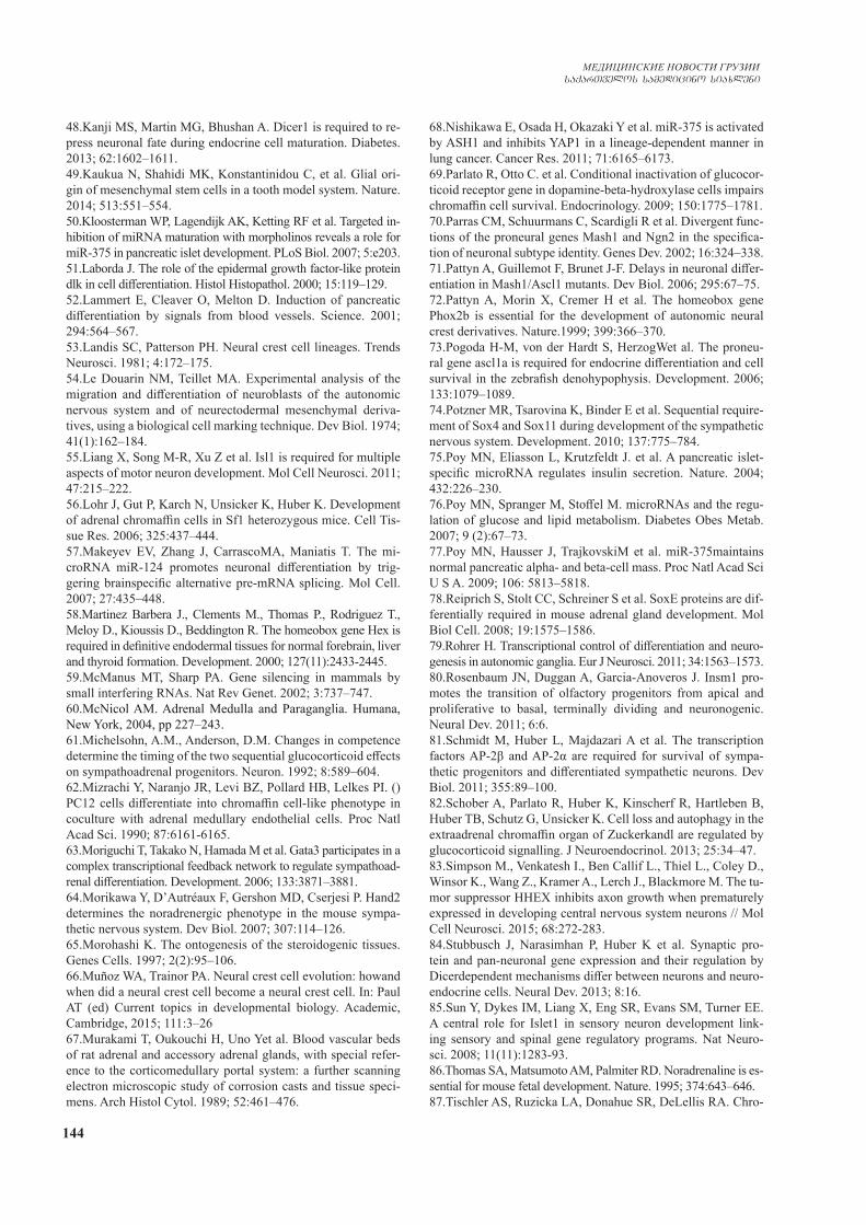
144
МЕДИЦИНСКИЕ НОВОСТИ ГРУЗИИ
CFMFHSDTKJC CFVTLBWBYJ CBF[KTYB
48.Kanji MS, Martin MG, Bhushan A. Dicer1 is required to re-press neuronal fate during endocrine cell maturation. Diabetes. 2013; 62:1602–1611.49.Kaukua N, Shahidi MK, Konstantinidou C, et al. Glial ori-gin of mesenchymal stem cells in a tooth model system. Nature. 2014; 513:551–554.50.Kloosterman WP, Lagendijk AK, Ketting RF et al. Targeted in-hibition of miRNA maturation with morpholinos reveals a role for miR-375 in pancreatic islet development. PLoS Biol. 2007; 5:e203.51.Laborda J. The role of the epidermal growth factor-like protein dlk in cell differentiation. Histol Histopathol. 2000; 15:119–129. 52.Lammert E, Cleaver O, Melton D. Induction of pancreatic differentiation by signals from blood vessels. Science. 2001; 294:564–567. 53.Landis SC, Patterson PH. Neural crest cell lineages. Trends Neurosci. 1981; 4:172–175.54.Le Douarin NM, Teillet MA. Experimental analysis of the migration and differentiation of neuroblasts of the autonomic nervous system and of neurectodermal mesenchymal deriva-tives, using a biological cell marking technique. Dev Biol. 1974; 41(1):162–184. 55.Liang X, Song M-R, Xu Z et al. Isl1 is required for multiple aspects of motor neuron development. Mol Cell Neurosci. 2011; 47:215–222. 56.Lohr J, Gut P, Karch N, Unsicker K, Huber K. Development of adrenal chromaffin cells in Sf1 heterozygous mice. Cell Tis-sue Res. 2006; 325:437–444.57.Makeyev EV, Zhang J, CarrascoMA, Maniatis T. The mi-croRNA miR-124 promotes neuronal differentiation by trig-gering brainspecific alternative pre-mRNA splicing. Mol Cell. 2007; 27:435–448. 58.Martinez Barbera J., Clements M., Thomas P., Rodriguez T., Meloy D., Kioussis D., Beddington R. The homeobox gene Hex is required in definitive endodermal tissues for normal forebrain, liver and thyroid formation. Development. 2000; 127(11):2433-2445.59.McManus MT, Sharp PA. Gene silencing in mammals by small interfering RNAs. Nat Rev Genet. 2002; 3:737–747. 60.McNicol AM. Adrenal Medulla and Paraganglia. Humana, New York, 2004, pp 227–243.61.Michelsohn, A.M., Anderson, D.M. Changes in competence determine the timing of the two sequential glucocorticoid effects on sympathoadrenal progenitors. Neuron. 1992; 8:589–604.62.Mizrachi Y, Naranjo JR, Levi BZ, Pollard HB, Lelkes PI. () PC12 cells differentiate into chromaffin cell-like phenotype in coculture with adrenal medullary endothelial cells. Proc Natl Acad Sci. 1990; 87:6161-6165. 63.Moriguchi T, Takako N, Hamada M et al. Gata3 participates in a complex transcriptional feedback network to regulate sympathoad-renal differentiation. Development. 2006; 133:3871–3881.64.Morikawa Y, D’Autréaux F, Gershon MD, Cserjesi P. Hand2 determines the noradrenergic phenotype in the mouse sympa-thetic nervous system. Dev Biol. 2007; 307:114–126.65.Morohashi K. The ontogenesis of the steroidogenic tissues. Genes Cells. 1997; 2(2):95–106.66.Muñoz WA, Trainor PA. Neural crest cell evolution: howand when did a neural crest cell become a neural crest cell. In: Paul AT (ed) Current topics in developmental biology. Academic, Cambridge, 2015; 111:3–2667.Murakami T, Oukouchi H, Uno Yet al. Blood vascular beds of rat adrenal and accessory adrenal glands, with special refer-ence to the corticomedullary portal system: a further scanning electron microscopic study of corrosion casts and tissue speci-mens. Arch Histol Cytol. 1989; 52:461–476.
68.Nishikawa E, Osada H, Okazaki Y et al. miR-375 is activated by ASH1 and inhibits YAP1 in a lineage-dependent manner in lung cancer. Cancer Res. 2011; 71:6165–6173.69.Parlato R, Otto C. et al. Conditional inactivation of glucocor-ticoid receptor gene in dopamine-beta-hydroxylase cells impairs chromaffin cell survival. Endocrinology. 2009; 150:1775–1781.70.Parras CM, Schuurmans C, Scardigli R et al. Divergent func-tions of the proneural genes Mash1 and Ngn2 in the specifica-tion of neuronal subtype identity. Genes Dev. 2002; 16:324–338.71.Pattyn A, Guillemot F, Brunet J-F. Delays in neuronal differ-entiation in Mash1/Ascl1 mutants. Dev Biol. 2006; 295:67–75. 72.Pattyn A, Morin X, Cremer H et al. The homeobox gene Phox2b is essential for the development of autonomic neural crest derivatives. Nature.1999; 399:366–370.73.Pogoda H-M, von der Hardt S, HerzogWet al. The proneu-ral gene ascl1a is required for endocrine differentiation and cell survival in the zebrafish denohypophysis. Development. 2006; 133:1079–1089. 74.Potzner MR, Tsarovina K, Binder E et al. Sequential require-ment of Sox4 and Sox11 during development of the sympathetic nervous system. Development. 2010; 137:775–784. 75.Poy MN, Eliasson L, Krutzfeldt J. et al. A pancreatic islet-specific microRNA regulates insulin secretion. Nature. 2004; 432:226–230. 76.Poy MN, Spranger M, Stoffel M. microRNAs and the regu-lation of glucose and lipid metabolism. Diabetes Obes Metab. 2007; 9 (2):67–73. 77.Poy MN, Hausser J, TrajkovskiM et al. miR-375maintains normal pancreatic alpha- and beta-cell mass. Proc Natl Acad Sci U S A. 2009; 106: 5813–5818. 78.Reiprich S, Stolt CC, Schreiner S et al. SoxE proteins are dif-ferentially required in mouse adrenal gland development. Mol Biol Cell. 2008; 19:1575–1586. 79.Rohrer H. Transcriptional control of differentiation and neuro-genesis in autonomic ganglia. Eur J Neurosci. 2011; 34:1563–1573.80.Rosenbaum JN, Duggan A, Garcia-Anoveros J. Insm1 pro-motes the transition of olfactory progenitors from apical and proliferative to basal, terminally dividing and neuronogenic. Neural Dev. 2011; 6:6.81.Schmidt M, Huber L, Majdazari A et al. The transcription factors AP-2β and AP-2α are required for survival of sympa-thetic progenitors and differentiated sympathetic neurons. Dev Biol. 2011; 355:89–100.82.Schober A, Parlato R, Huber K, Kinscherf R, Hartleben B, Huber TB, Schutz G, Unsicker K. Cell loss and autophagy in the extraadrenal chromaffin organ of Zuckerkandl are regulated by glucocorticoid signalling. J Neuroendocrinol. 2013; 25:34–47.83.Simpson M., Venkatesh I., Ben Callif L., Thiel L., Coley D., Winsor K., Wang Z., Kramer A., Lerch J., Blackmore M. The tu-mor suppressor HHEX inhibits axon growth when prematurely expressed in developing central nervous system neurons // Mol Cell Neurosci. 2015; 68:272-283. 84.Stubbusch J, Narasimhan P, Huber K et al. Synaptic pro-tein and pan-neuronal gene expression and their regulation by Dicerdependent mechanisms differ between neurons and neuro-endocrine cells. Neural Dev. 2013; 8:16.85.Sun Y, Dykes IM, Liang X, Eng SR, Evans SM, Turner EE. A central role for Islet1 in sensory neuron development link-ing sensory and spinal gene regulatory programs. Nat Neuro-sci. 2008; 11(11):1283-93. 86.Thomas SA, Matsumoto AM, Palmiter RD. Noradrenaline is es-sential for mouse fetal development. Nature. 1995; 374:643–646.87.Tischler AS, Ruzicka LA, Donahue SR, DeLellis RA. Chro-
GEORGIAN MEDICAL NEWS No 5 (278) 2018
© GMN 145
maffin cell proliferation in the adult rat adrenal medulla. Int J Dev Neurosci. 1989; 7:439–448.88.Tsarovina K, Reiff T, Stubbusch J et al. The Gata3 transcrip-tion factor is required for the survival of embryonic and adult sympathetic neurons. J Neurosci. 2010; 30:10833–10843.89.Uesaka T, Nagashimada M, Enomoto H. Neuronal differen-tiation in Schwann cell lineage underlies postnatal neurogenesis in the enteric nervous system. J Neurosci. 2015; 35:9879–9888.90.Unsicker K, Huber K, Schober A, Kalcheim C. Resolved and open issues in chromaffin cell development // Mechanisms of development. 2013; 130:324-329. 91.Visvanathan J, Lee S, Lee B et al. The microRNA miR-124 antagonizes the anti-neural REST/SCP1 pathway during embry-onic CNS development. Genes Dev. 2007; 21:744–749.92.Welcker JE, Hernandez-Miranda LR, Paul FE et al. Insm1 controls development of pituitary endocrine cells and requires a SNAG domain for function and for recruitment of histone-modifying factors. Development. 2013; 140:4947–4958. 93.Wildner H, Gierl MS, Strehle M et al. Insm1 (IA-1) is a crucial component of the transcriptional network that controls differentiation of the sympatho-adrenal lineage. Development. 2008; 135:473–481.94.Wilson ME, Yang KY, Kalousova A et al. The HMG box transcription factor Sox4 contributes to the development of the endocrine pancreas. Diabetes. 2005; 54:3402–3409. 95.Yu J-Y, Chung K-H, Deo M et al. MicroRNA miR-124 regu-lates neurite outgrowth during neuronal differentiation. Exp Cell Res. 2008; 314: 2618–2633. 96.Zehir A, Hua LL, Maska EL et al. Dicer is required for survival of differentiating neural crest cells. Dev Biol. 2010; 340:459–467. 97. Zhang Z-W, Men T, Feng R-C et al. miR-375 inhibits prolif-eration of mouse pancreatic progenitor cells by targeting YAP1. Cell Physiol Biochem. 2013b; 32:1808–1817. 98.Zhou QY, Quaife CJ, Palmiter RD. Targeted disruption of the tyrosine hydroxylase gene reveals that catecholamines are required for mouse fetal development. Nature, 1995; 374:640–643.
SUMMARY
EPIGENETIC REGULATION OF ADRENAL CHRO-MAFFIN CELLS DEVELOPMENT (REVIEW)
1Obernikhin S., 1Yaglova N., 2Tsomartova D., 2Torbek V., 2Ivanova M.
1Federal State Budgetary Institution Research Institute of Human Morphology, Moscow; 2Federal State Autono-mous Educational Institution of Higher Education I.M. Sechenov First Moscow State Medical University, Russia
Chromaffin cells of adrenal medulla have tradition-ally been considered as modified sympathetic neurons. However, the results of recent studies indicate the need to revise this concept. The article reviews recent findings in origin and ontogeny of adrenal chromaffin cells and transcriptional and posttranscriptional regulation of de-velopmental processes. The article summarizes data on transcriptional control of chromaffin cells proliferation and maturation and participation of microRNA in regula-tion of chromaffin and sympathetic neuronal phenotype gene expression.
Keywords: chromaffin cells, adrenals, transcription factors, epigenetic regulation, microRNA.
РЕЗЮМЕ
ЭПИГЕНЕТИЧЕСКАЯ РЕГУЛЯЦИЯ РАЗВИТИЯ ХРОМАФФИННЫХ КЛЕТОК НАДПОЧЕЧНИ-КОВ (ОБЗОР)
1Обернихин С.С., 1Яглова Н.В., 2Цомартова Д.А., 2Торбек В.Э., 2Иванова М.Ю.
1Федеральное государственное бюджетное научное учреждение «НИИ морфологии человека», Москва; 2Федеральное государственное автономное образо-вательное учреждение «Первый московский государ-ственный медицинский университет им. И.М. Сече-нова», Москва, Россия
Хромаффинные клетки мозгового вещества надпо-чечников традиционно рассматривались как произво-дные симпатических нейронов. Однако результаты ис-следований последних лет указывают на необходимость пересмотра этой концепции. Статья представляет собой обзор современных представлений о происхождении и развитии хромаффинных клеток надпочечников и их транскрипционной и посттранскрипционной регуляции в пренатальном и постнатальном периодах онтогенеза. Обобщены сведения об основных транскрипционных факторах, участвующих в пролиферации и дифференци-ации хромаффинных клеток, а также о роли микроРНК в регуляции экспрессии генов хромаффинных клеток и симпатических нейронов.
reziume
Tirkmelzeda jirkvlis qromafinuri ujre-debis ganviTarebis epigenetikuri regula-cia (mimoxilva)
1s. obernixini, 1n. iaglova, 2d. comartova, 2v. torbeki, 2m. ivanova
1adamianis morfologiis samecniero-kvlevi-Ti instituti, moskovi; 2moskovis i.seCenovis saxelobis saxelmwifo samedicino univer-siteti, moskovi, ruseTi
Tirkmelzeda jirkvlis tvinovani niv-Tierebis qromafinuri ujredebi tradici-ulad simpaTikuri neironebis warmoebu-lebad ganixileboda. Tumca, bolo wlebis gamokvlevebi miuTiTebs am koncefciis gadaxedvis aucileblobaze. statiaSi war-modgenilia Tanamedrove Sexedulebebis mi-moxilva Tirkmelzeda jirkvlis qromafin-uri ujredebis warmoSobisa da ganviTarebis,
146
МЕДИЦИНСКИЕ НОВОСТИ ГРУЗИИ
CFMFHSDTKJC CFVTLBWBYJ CBF[KTYB
aseve, misi transkripciuli da postran-skripciuli regulaciis Sesaxeb onto-genezis pre- da postnatalur periodebSi. ganzogadebulia monacemebi qromafinuri ujredebis proliferaciasa da diferen-
cirebaSi monawile ZiriTadi transkrip-ciuli faqtorebis da mikrornm-is rolis Sesaxeb qromafinuri ujredebis da simpa-Tikuri neironebis genebis eqspresiis reg-ulaciaSi.
THE IMPACT OF THE ACRYLIC MONOMER ON THE MORPHOLOGICAL STRUCTURE OF RAT LINGUAL MUCOSA
Davydenko V., Starchenko I., Davydenko А., Trufanova V., Kuznetsov V.
Higher State Educational Establishment of Ukraine “Ukrainian Medical Stomatological Academy”, Poltava, Ukraine
It has been proved by evidence that monomers, even in small amount, causes inflammation of mucous mem-branes and pathological growth of epithelial cells, and can also cause local and general allergic reactions. And this is related not only to the mucous membrane of the prosthet-ic bed, but also to those ones that are in contact with the denture during the wearing, especially the lingual mucosa [6,7,9]. Causes of ulcerative lesions of the mucous mem-branes of periodontal tissues, the mouth floor, as well as the causes of occurrence of various types of stomatitis are known in dentistry. However, no data on the impact of the acrylic monomer on the oral mucosa and its effect on the morphologic structure of the taste receptors of the tongue have been found in the publications.
The analysis of publications [1,4,5] shows that diverse multiple factors can induce changes in taste sensitivity and the main irritants are the chemicals of different types. However, the study of the effect of the components of dental structural materials on the state of lingual mucosa, in particular, taste sensors, has not been fully elucidated to date. This gives us the grounds for further investiga-tions of the impact of base acrylic resins on the state of taste sensitivity in patients during the adaptation to re-movable laminar dentures.
The previous paper [2] presents the findings of the study on the impact of the monomer of the «Ftoraks» base acrylic resin on the state of the rats’ lingual mucosa in the early period (1 to 7 days) and its subsequent regeneration. The studies have found that the greatest changes in the lingual mucosa occur on day 3 and 7 after the application of monomer, and are of erosive-inflammatory origin. Re-generation of the lingual epithelium is delayed.
The purpose of the paper was the study of the effect of monomer of the “Ftoraks” base acrylic resin on the state of the rats’ lingual mucosa within 2-4 weeks after its impact.
Material and methods. To achieve the objective of the research, the experimental studies were conducted on laboratory Wistar rats, aged 1 to 1.5 years (20 rats, 5 of which were assigned to the control group). Lingual mu-cosa of the rest of the animals was smeared with 2% aque-
ous solution of the monomer of the “Ftoraks” base acrylic resin twice a day. “Ftoraks” is the hot curing plastic of the powder-liquid type on the basis of the fluorine-containing acrylic copolymers. The powder (polymer) is the pink-stained fine-dispersed suspension graft-copolymer of the methacrylic acid methyl ether and fluorelastomer. The liq-uid (monomer) is the methacrylic acid methyl ether that is stabilized and contains coupling agent (diphenylopropane dimethacrylic ether).
The animals underwent euthanasia on the 14th (Group 5 of animals), 21st (Group 6) and 28th days (Group 7). All studies were conducted according to the rules of humane treatment of animals in accordance with the requirements of the Tokyo Declaration of the World Medical Associa-tion and the general ethical principles of working with ex-perimental animals, which were approved by the first Na-tional Bioethics Congress [8]. After euthanasia the tongue were removed in rats which was subsequently fragmented on the root, lateral and frontal sections that were embed-ded into liquid paraffin using the «Microm» station for the embedding of paraffin blocks; sections from paraffin blocks were obtained and stained with haematoxylin-eosin by conventional methods [10,11]. Visualization of the microspecimens, their photographing and determina-tion of the correlation between the multiple cell elements in the inflammatory infiltrate has been carried out on the microscope Olimpus BX 41 with digital micro photo at-tachment and package of enclosed licensing software de-signed for morphometric research and statistical process-ing of the resulting data.
Results and their discussion. Morphological chang-es of the lingual mucosa were observed while contacting with the monomer during 14 days, indicating about the followed-up conversion of the acute inflammatory process into the chronic one, as well as the continued development of atrophic and compensatory-adaptive alterations in the lingual mucosa. First of all, the change in the nature of inflammatory infiltration was noteworthy, which was oc-casionally detected in the muscles of the tongue. The fo-cal inflammatory infiltrates with predominantly lympho-
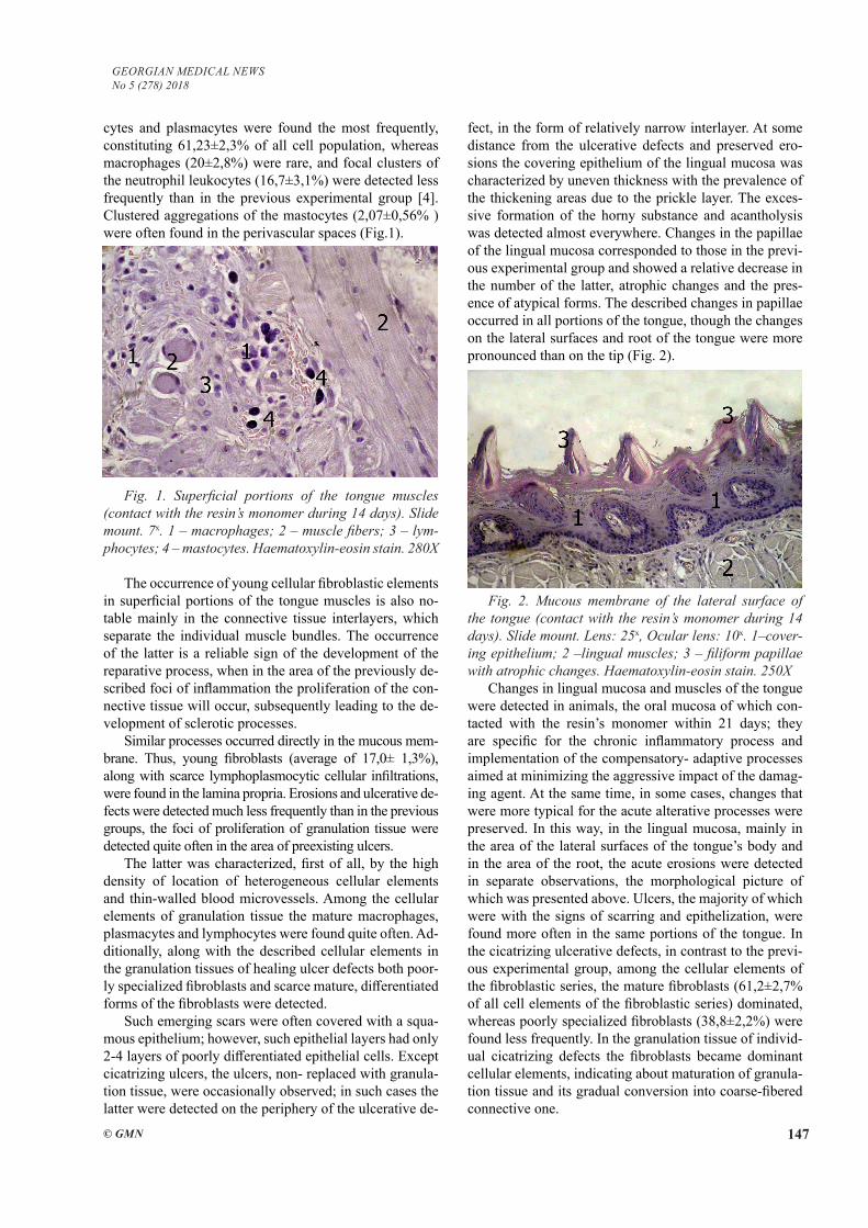
GEORGIAN MEDICAL NEWS No 5 (278) 2018
© GMN 147
cytes and plasmacytes were found the most frequently, constituting 61,23±2,3% of all cell population, whereas macrophages (20±2,8%) were rare, and focal clusters of the neutrophil leukocytes (16,7±3,1%) were detected less frequently than in the previous experimental group [4]. Clustered aggregations of the mastocytes (2,07±0,56% ) were often found in the perivascular spaces (Fig.1).
Fig. 1. Superficial portions of the tongue muscles (contact with the resin’s monomer during 14 days). Slide mount. 7х. 1 – macrophages; 2 – muscle fibers; 3 – lym-phocytes; 4 – mastocytes. Haematoxylin-eosin stain. 280X
The occurrence of young cellular fibroblastic elements in superficial portions of the tongue muscles is also no-table mainly in the connective tissue interlayers, which separate the individual muscle bundles. The occurrence of the latter is a reliable sign of the development of the reparative process, when in the area of the previously de-scribed foci of inflammation the proliferation of the con-nective tissue will occur, subsequently leading to the de-velopment of sclerotic processes.
Similar processes occurred directly in the mucous mem-brane. Thus, young fibroblasts (average of 17,0± 1,3%), along with scarce lymphoplasmocytic cellular infiltrations, were found in the lamina propria. Erosions and ulcerative de-fects were detected much less frequently than in the previous groups, the foci of proliferation of granulation tissue were detected quite often in the area of preexisting ulcers.
The latter was characterized, first of all, by the high density of location of heterogeneous cellular elements and thin-walled blood microvessels. Among the cellular elements of granulation tissue the mature macrophages, plasmacytes and lymphocytes were found quite often. Ad-ditionally, along with the described cellular elements in the granulation tissues of healing ulcer defects both poor-ly specialized fibroblasts and scarce mature, differentiated forms of the fibroblasts were detected.
Such emerging scars were often covered with a squa-mous epithelium; however, such epithelial layers had only 2-4 layers of poorly differentiated epithelial cells. Except cicatrizing ulcers, the ulcers, non- replaced with granula-tion tissue, were occasionally observed; in such cases the latter were detected on the periphery of the ulcerative de-
fect, in the form of relatively narrow interlayer. At some distance from the ulcerative defects and preserved ero-sions the covering epithelium of the lingual mucosa was characterized by uneven thickness with the prevalence of the thickening areas due to the prickle layer. The exces-sive formation of the horny substance and acantholysis was detected almost everywhere. Changes in the papillae of the lingual mucosa corresponded to those in the previ-ous experimental group and showed a relative decrease in the number of the latter, atrophic changes and the pres-ence of atypical forms. The described changes in papillae occurred in all portions of the tongue, though the changes on the lateral surfaces and root of the tongue were more pronounced than on the tip (Fig. 2).
Fig. 2. Mucous membrane of the lateral surface of the tongue (contact with the resin’s monomer during 14 days). Slide mount. Lens: 25х, Ocular lens: 10х. 1–cover-ing epithelium; 2 –lingual muscles; 3 – filiform papillae with atrophic changes. Haematoxylin-eosin stain. 250X
Changes in lingual mucosa and muscles of the tongue were detected in animals, the oral mucosa of which con-tacted with the resin’s monomer within 21 days; they are specific for the chronic inflammatory process and implementation of the compensatory- adaptive processes aimed at minimizing the aggressive impact of the damag-ing agent. At the same time, in some cases, changes that were more typical for the acute alterative processes were preserved. In this way, in the lingual mucosa, mainly in the area of the lateral surfaces of the tongue’s body and in the area of the root, the acute erosions were detected in separate observations, the morphological picture of which was presented above. Ulcers, the majority of which were with the signs of scarring and epithelization, were found more often in the same portions of the tongue. In the cicatrizing ulcerative defects, in contrast to the previ-ous experimental group, among the cellular elements of the fibroblastic series, the mature fibroblasts (61,2±2,7% of all cell elements of the fibroblastic series) dominated, whereas poorly specialized fibroblasts (38,8±2,2%) were found less frequently. In the granulation tissue of individ-ual cicatrizing defects the fibroblasts became dominant cellular elements, indicating about maturation of granula-tion tissue and its gradual conversion into coarse-fibered connective one.
148
МЕДИЦИНСКИЕ НОВОСТИ ГРУЗИИ
CFMFHSDTKJC CFVTLBWBYJ CBF[KTYB
Inflammatory infiltrates directly in the muscles of the tongue were detected less frequently than in the previous ex-perimental group. Inflammatory infiltration was represented exclusively by lymphocytes and plasmacytes, whereas mac-rophages were found less frequently, and leukocytes were detected in single observations and were presented, basi-cally, by eosinophilic granulocytes. At the same time, it is notable, that on the separate areas in the superficial portions of the tongue some thickening of muscles of connective tis-sue interlayers (on the average of 10-12%), that separate in-dividual bundles of the cross-striated muscles was detected, with some depletion of the latter by the cellular elements and increase of the relative amount of collagen fibers, indicating about the development of sclerotic processes.
Similar changes occur in the proper plate of the mu-cous membrane, where, in contrast to previously described experimental groups, there is a relative decrease in the num-ber of blood vessels and cellular elements, presented mainly by fibroblasts. In some cases, lymphocytes and plasmacytes formed small clusters near the basal membrane and perivas-cular spaces. Additionally, in the area of the lateral surfaces of the tongue a straightening of a border between the proper plate and the covering epithelium of a mucous membrane, at the expense of flattening of connective tissue papillae, were observed quite often (Fig. 3).
Fig. 3. Mucous membrane of the lateral surface of the tongue (contact with the resin’s monomer during 21 days). Slide mount. Lens: 25х, Ocular lens: 10х. 1– lamina pro-pria of the mucous membrane; 2 – the area of atrophy of the covering epithelium; 3 – the areas of the thickening of the covering epithelium; 4 – altered filiform papilla. Haematoxylin-eosin stain. 250X
Changes in the covering epithelium of the mucous membrane, similarly to the previous experimental group, were characterized by thickening of the prickle layer, ex-cessive keratinization, and events of keratolysis. At the same time, the areas, where the thickness of the covering epithelium was much less, were occasionally detected; first of all it concerns cicatrizing ulcerative defects.
The decrease in the density of localization of the fi-liform papillae was noted everywhere in the covering epithelium; many of them were with atrophic changes, and
there was a decrease in the number of foliate papillae in the root portions of the lateral surfaces of the tongue (Fig. 3). Noteworthy, the reduction of density of localization of fun-giform papillae was detected in area of the tip of the tongue significantly rarely than in the intact animals, is [3, 12].
During the 28-day contact of the lingual mucosa with the monomer mainly atrophic and sclerotic processes, proliferating on the superficial parts of the cross-striated muscles, were observed in the lingual mucosa. Destruc-tive processes in the mucous membrane, manifested by the formation of superficial erosions and deep ulcers were relatively rare and observed mainly on the lateral surfaces of the body of the tongue, with the overwhelming number of ulcerative defects with the phenomena of scarring and epithelization. In the cross-striated muscles of the tongue, located in the immediate vicinity of the mucous mem-brane, the inflammatory infiltrates were seldom detected, localizing mainly in the form of small focal infiltrates in connective tissue interlayers, separating individual mus-cle bundles. In the described cellular aggregations the amount of lymphocytes and plasmacytes (on the aver-age of 81±3,8%) was significant, whereas macrophages (12,4±2,7%) and eosinophylic leukocytes (4,7±0,9%), mastocytes (1,9±0,7%) were scarce.
It should be noted that in this experimental group the interlayers of intermuscular connective tissue were signifi-cantly thickened (on the average of 15-20%) with excessive formation of collagen fibers at relative scarcity of the cellular elements and insignificant number of blood vessels.
Similarly to the previous experimental group, the ex-cessive number of the collagen fibers with a small amount of mature fibroblasts and minor accumulations of leuko-cytes and plasmacytes were detected in the proper plate of the connective tissue. At the same time the proper plate itself was slightly thickened in some cases, as compared with the intact group, and in the majority of observa-tions, mainly on the lateral surface of the tongue’s body the flattening of connective tissue papillae was observed, contributing to the flattening of the boundary between the proper plate and the covering epithelium.
In the covering epithelium of all parts of the tongue a thickening of the prickle layer was found, and the cellu-lar series of epithelial cells were differentiated relatively clearly. It should be noted that the number of intraepithe-lial lymphocytes in the epithelial layer was significantly lower than in the previous experimental groups, but in comparison with the animals of the intact group their number was somewhat larger. As before, in almost all observations an excessive keratinization of the covering epithelium in all portions of gastric mucosa was detected with some decrease of density of filiform papillae loca-tion. There was also a decrease in the number of fungi-form and foliate papillae; the preserved papillae of these types were characterized by reduced size, indicting about the atrophic changes. In circumvallate papillae, along with atrophic changes, a decrease in the number of gusta-tory buds, located on the lateral surfaces is noteworthy.
GEORGIAN MEDICAL NEWS No 5 (278) 2018
© GMN 149
Conclusion: The studies confirm that the monomer of acrylic resin causes a number of pathological changes in the mucous membrane and muscles of the rat tongue, the nature of which varies depending on the duration of its impact.
On day 14 in the lingual mucosa the destructive process-es are significantly delayed, substituting for the sclerotic pro-cesses in the proper plate and atrophic processes, observed, first of all, in the papillae of the tongue. It is appropriate to assume that such changes in the papillae will lead to viola-tion of the taste reception, first of all, in the areas of lateral surfaces of the body of the tongue and in the root area.
At the same time, it should be noted that at the end of the experimental period (on day 28 of the contact of the monomer with the lingual mucosa), in the mucous mem-brane of the tongue, along with atrophic and sclerotic pro-cesses, the destructive changes and inflammatory reaction are evident. We hypothesize that this may indicate about partial recovery of taste sensitivity due to the decrease in the number of gustatory buds, taste papillae of different types and the increase in the period of their regeneration.
REFERENCES
1.Будылина С.В. Исследование функциональной мобильности вкусовых рецепторов при гастроэзофагеальном рефлюксе / С.В. Будылина, Н.В. Костина // Научные труды IV съезда фи-зиологов СНГ. – М. : Медицина-Здоровье, 2014. 94.2.Давыденко В. Ю. Морфологические особенности слизи-стой оболочки языка крысы в ранний период воздействия на нее мономера акриловой пластмассы / В. Ю. Давыден-ко, М.Я. Нидзельский, И.И. Старченко [и др.] // Georgian Мedical News. – 2016. – № 3 (252). – С. 102-107.3.Давиденко В.Ю. Морфологічні особливості слизової обо-лонки різних ділянок язика щурів у нормі / В.Ю. Давиденко, М.Я. Нідзельський, І.І. Старченко [і ін.] // Вісник проблем біології і медицини. – 2016. – Вип.2.– Том 2 (129). – С. 82-86.4. Крихели Н.И. Вкусовая чувствительность и ее изменения / Н.И. Крихели, Д.И. Гаматаева, Н.Г. Дмитриева // Россий-ская стоматология. – 2011. – № 2. – С. 15-19.5. Куцевляк В. Ф. Изменение показателей вкусовой чувстви-тельности сосочков языка при наличии в полости рта пломб из различных материалов / В.Ф. Куцевляк, М.Г. Щеголева // Експериментальна і клінічна медицина. – 2004. – № 1. – С. 203-206.6. Неделко С.В. Клинико-лабораторная оценка состояния про-тезного ложа у ортопедических больных при использовании акриловых протезов с покрытием / С.В. Неделко // Кримський терапевтичний журнал. – 2012. – № 3. – С. 119–122.7. Нідзельський М.Я. Порівняльна характеристика рівня залишкового мономеру в базисах знімних протезів із акри-лових пластмас, виготовлених за різними технологіями полімеризації / М.Я. Нідзельський, В.Ю. Давиденко, Г.М. Давиденко [та ін.] // Вісник проблем біології і медицини.– 2014. – Вип.2.– Том 2 (108). – С. 45-48.8. Общие этические принципы работы с эксперименталь-ными животными при проведении медицинских биологи-ческих исследований / Національний конгресс з біоетики (Київ, 17-20 вересня 2001р.) // Ж.АМН України.– 2001.– Т.7, №4.– С. 814-816.9.Садовский В.В. Эффективность применения стоматологи-ческих гелей для экранирования съемных зубных протезов
/ В.В. Садовский, Ю.Г. Романова // Стоматология для всех. – 2013. – № 2. – С. 54-56.10. Сапожников А.Г. Гистологическая и микроскопическая техника : руководство / А.Г.Сапожников, А.Е. Доросевич. – Смоленск : САУ, 2000. – 476 с.11. Саркисов Д.С. Микроскопическая техника: руководство для врачей и лаборантов / Д.С. Саркисов, Ю.Л. Перов // М.: Медицина, 1996.– 544 с.12. Семенова А.К. Морфофункціональна характеристика епітелію слизової оболонки спинки язика щурів у нормі / А.К. Семенова, Г.А. Єрошенко, Н.В. Гасюк [та ін.] // Вісник проблем біології і медицини.– 2014. – Вип.2.– Том 2 (108). – С. 134-136.
SUMMARY
THE IMPACT OF THE ACRYLIC MONOMER ON THE MORPHOLOGICAL STRUCTURE OF RAT LINGUAL MUCOSA
Davydenko V., Starchenko I., Davydenko А., Trufanova V., Kuznetsov V. Higher State Educational Establishment of Ukraine “Ukrai-nian Medical Stomatological Academy”, Poltava, Ukraine
The analysis of publications shows that diverse mul-tiple factors can induce changes in taste sensitivity and the main irritants are the chemicals of different types. How-ever, the study of the effect of the components of dental structural materials on the state of lingual mucosa, in par-ticular, taste sensors, has not been fully elucidated to date.
The purpose of the paper was the study of the effect of monomer of the “Ftoraks” base acrylic resin on the state of the rats’ lingual mucosa within 2-4 weeks after its impact.
The previous paper [5] presents the findings of the study on the impact of the monomer of the “Ftoraks” base acrylic resin on the state of the rats’ lingual mucosa in the early period (1 to 7 days) and its subsequent regeneration. The studies have found that the greatest changes in the lingual mucosa occur on day 3 and 7 after the application of monomer, and are of erosive-inflammatory origin. Re-generation of the lingual epithelium is delayed.
The studies confirm that the monomer of acrylic resin causes a number of pathological changes in the mucous membrane and muscles of the rat tongue, the nature of which varies depending on the duration of its impact.
On day 14 in the lingual mucosa the destructive pro-cesses are significantly delayed, substituting for the scle-rotic processes in the proper plate and atrophic processes, observed, first of all, in the papillae of the tongue. It is appropriate to assume that such changes in the papillae will lead to violation of the taste reception, first of all, in the areas of lateral surfaces of the body of the tongue and in the root area.
At the same time, it should be noted that at the end of the experimental period (on day 28 of the contact of the monomer with the lingual mucosa), in the mucous mem-
150
МЕДИЦИНСКИЕ НОВОСТИ ГРУЗИИ
CFMFHSDTKJC CFVTLBWBYJ CBF[KTYB
brane of the tongue, along with atrophic and sclerotic pro-cesses, the destructive changes and inflammatory reaction are evident. We hypothesize that this may indicate about partial recovery of taste sensitivity due to the decrease in the number of gustatory buds, taste papillae of different types and the increase in the period of their regeneration.
Keywords: acrylic resin’s monomer, lingual mucosa, taste papillae, regeneration, morphological structure.
РЕЗЮМЕ
ВЛИЯНИЕ МОНОМЕРА АКРИЛОВОЙ ПЛАСТ-МАССЫ НА МОРФОЛОГИЧЕСКОЕ СТРОЕНИЕ СЛИЗИСТОЙ ОБОЛОЧКИ ЯЗЫКА У КРЫС
Давыденко В.Ю., Старченко И.И., Давыденко А.Н., Труфанова В.П., Кузнецов В.В.
Высшее государственное учебное заведение Украины «Украинская медицинская стоматологическая акаде-мия», Полтава, Украина
Анализ литературных источников указывает, что на вкусовую чувствительность влияет множество раз-личных факторов; основными раздражителями явля-ются химические вещества различных видов. Воздей-ствие составляющих компонентов стоматологических конструкционных материалов на состояние слизистой оболочки языка, в частности на вкусовые рецепторы по сей день недостаточно изучено.
Целью исследования явилось определение влия-ния мономера базисной акриловой пластмассы «Фто-ракс» на состояние слизистой оболочки языка у крыс спустя 2-4 недели после его воздействия.
В ранее опубликованной авторами статье пред-ставлены результаты исследования по воздействию мономера базисной акриловой пластмассы «Фторакс» на состояние слизистой оболочки языка и ее регенера-цию у крыс в раннем периоде (от 1 до 7 суток). Уста-новлено, что наибольшие изменения в слизистой обо-лочке языка наступают на 3 и 7 сутки после нанесения мономера, которые носят эрозивно-воспалительный характер. Регенерация эпителия языка замедляется.
Полученные результаты ныне проведенного ис-следования подтверждают, что мономер акриловой пластмассы в слизистой оболочке и мышцах языка крысы вызывает ряд патологических изменений, ха-рактер которых зависит от продолжительности его воздействия.
На 14 сутки деструктивные процессы в слизистой оболочке языка существенно ослабевают, уступая ме-сто склеротическим и атрофическим процессам, на-блюдаемым в сосочках языка. Следует предположить, что подобные изменения в сосочках ведут к наруше-нию вкусовой рецепции, в частности в области боко-вых поверхностей тела языка и его корня.
Необходимо отметить, что в конце эксперимен-тального срока - на 28 сутки контакта мономера со слизистой оболочкой языка, в его слизистой оболочке, наряду с атрофическими и склеротическими процес-сами, продолжают развиваться деструктивные изме-нения и воспалительная реакция, что, по всей вероят-ности, свидетельствует о частичном восстановлении вкусовой чувствительности ввиду уменьшения числа вкусовых луковиц и вкусовых сосочков разных видов, увеличивая сроки их регенерации. reziume
akrilis plastmasis monomeris gavlena vir-Tagvas enis lorwovani garsis morfolo-giur aRnagobaze
v. davidenko, i. starCenko, a. davidenko, v. trufanova, v. kuznecovi
ukrainis samedicino stomatologiuri aka-demia, poltava, ukraina
literaturis wyaroebis analizi miuTiTebs, rom sagemovno mgrZnobelobis cvli-lebas sxvadasxva faqtori ganapirobebs. ZiriTadi gamRizianebeli sxvadasxva tipis qimikatia; amasTan, naklebi yuradReba eqceva stomatologiuri konstruqciuli masalebis Semadgeneli komponentebis gavlenis Seswav-las enis lorwovani garsis mdgomareobaze, kerZod – sagemovno receptorebze.
eqsperimentuli kvlevis mizans Seadgen-da akrilis plastmasis bazisuri monomeris «ftoraqsi»-s gavlenis Seswavla virTag-vas enis lorwovan garsze am nivTierebis 2-4- kviriani zemoqmedebis Semdeg.
avtorebis adreul publikaciaSi warmodge-nili iyo kvlevis Sedegebi akrilis plastma-sis bazisuri monomeris «ftoraqsi»-s gavlenis Sesaxeb enis lorwovan garsze virTagvebSi moqmedebis adreul periodSi (1-7 dRe) da Semdgomi regeneraciis Sesaxeb. am kvlevebiT dadgenilia, rom enis lorwovan garsSi mniS-vnelovani eroziul-anTebiT xasiaTis cvli-lebebi monomeris gamoyenebidan me-3 da me-7 dRes viTardeba, enis epiTeliumis regenera-ciis procesi neldeba.
winamdebare kvleva adasturebs, rom akri-lis plastmasis monomeri virTagvas enis lorwovan garssa da kunTebSi iwvevs rig paTologiur cvlilebebs, romelTa xasiaTi, zemoqmedebis xangrZlivobis mixedviT, cva-lebadia.
me-14 dRes enis lorwovan garsze destruqciuli procesebi mniSvnelovnad sustdeba, xolo sklerozuli procesebi sa-
GEORGIAN MEDICAL NEWS No 5 (278) 2018
© GMN 151
kuTar firfitaSi da atrofiuli procesebi - upirvelesad, enis dvrilebSi Zlierdeba. savaraudod, aseTi cvlilebebi dvrilebSi gamoiwvevs sagemovno recepciis darRvevas, pirvel rigSi, enis sxeulis gverdiT zeda-pirebze da enis Ziris midamoSi.
amave dros, aRsaniSnavia, rom saeqsperi-mento periodis bolos (monomeris enis lorwovan garsze kontaqtis 28–e dRes) enis
lorwovan garsSi, atrofiul da sklerozul procesebTan erTad, destruqciuli cvlilebe-bi da anTebiTi reaqcia viTardeba. avtorebi varaudoben, rom es SeiZleba mowmobdes sage-movno mgrZnobelobis nawilobriv aRdgenas sagemovno bolqvebis, sxvadasxva saxis sage-movno dvrilebis raodenobis Semcirebis da maTi regeneraciis periodis vadis zrdasTan dakavSirebiT.
СРАВНИТЕЛЬНОЕ МОРФОЛОГИЧЕСКОЕ ИССЛЕДОВАНИЕ НАПРАВЛЕННОЙ РЕГЕНЕРАЦИИ КОСТНОЙ ТКАНИ ПРИ ИСПОЛЬЗОВАНИИ КСЕНОГЕННЫХ
ОСТЕОПЛАСТИЧЕСКИХ МАТЕРИАЛОВ БИОПЛАСТ-ДЕНТ И CERABONE
1,2 Черненко В.Н., 1Любченко А.В.
1Харьковская медицинская академия последипломного образования;2Сумский государственный университет, Медицинский институт, Украина
Направленная регенерация тканей при сложных клинических ситуациях является неотъемлемой ча-стью в имплантологическом лечении. Наличие доста-точного запаса костной ткани как в вертикальной, так и в горизонтальной плоскости обеспечивает более про-гнозируемый результат остеоинтеграции, а также эсте-тический успех будущей ортопедической конструкции. К сожалению, хорошие условия не всегда присутству-ют. Данная ситуация в основном обусловлена атрофией альвеолярного отростка из-за отсутствия физиологиче-ской нагрузки с замедлением протекания естественных биологических процессов на фоне вторичной адентии [6]. Клиническая ситуация ухудшается ввиду индивиду-альных анатомических особенностей (пневматическая гайморова пазуха, отвесная форма альвеолярного греб-ня, близлежащие нижнечелюстной канал и носовая по-лость) [7].
Несмотря на множество существующих методик, направленных на регенерацию ткани (менеджмент мягких тканей с использованием остеопластических материалов), клинические ситуации, возникающие при появлении новых методов лечения, требуют до-полнительных морфологических и клинических ис-следований.
Необходимость сокращения сроков лечения, исходя из социальных потребностей и пожеланий пациентов, решила методика непосредственной имплантации с не-медленной нагрузкой [2]. С появлением этой методики, возникли дополнительные вопросы. Однако, улучше-ние остеоинтеграции в связи с дополнительным вы-ходом из костномозговых полостей необходимых остеогенных клеток и морфогенетических белков, предупреждение атрофии лунки после экстракции, которая составляет до 30% ее высоты в первый год отсутствия зуба и сокращение числа хирургических
вмешательств являются существенными положи-тельными моментами для ее применения [11,17]. С установлением порога микроподвижности при нали-чии условий первичной стабильности имплантанта устранено доминирующее понятие выдержанности физиологических сроков оссификации с обязатель-ным условием отсутствия нагрузки. Появление ди-зайна имплантантов с макро- и микрорезьбой способ-ствует применению немедленной нагрузки, несмотря на ослабление механической фиксации, вызванной перестройкой костной ткани ввиду травматизации. Редким и приятным случаем в практике хируга-сто-матолога в данной ситуации является совпадение формы лунки с формой имлантанта. В таких случаях особенности дизайна современных имлантантов с их формирователями десны, без особого труда, позво-лят сформировать контур слизистой пришеечной об-ласти с профилактикой ее инвагинации. В остальных преимущественно клинических ситуациях возникает необходимость использования остеопластических ма-териалов с менеджментом мягких тканей, поведение которых в условиях микроподвижности неизучено [3]. Проблемной зоной со значительным истончением костной ткани и с особенностями строения слизистой оболочки на фоне ее дефицита является пришеечная область. Смещение слизисто-надкостничного лоскута в данной области приведет к обязательной атрофии альвеолярного края, снижению качества остеоинте-грации и негативному косметическому результату ор-топедической конструкции [15].
Преимущество выбора остеопластического ма-териала зависит от наличия присущих ему свойств. Только аутологический трансплантат имеет остеоген-ные, остеокондуктивные и остеоиндуктивные свой-ства. Отрицательным моментом в ограничении его
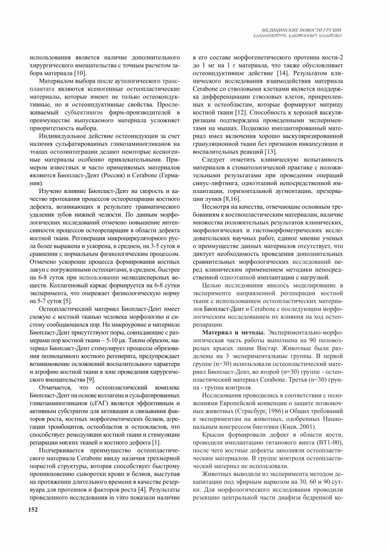
152
МЕДИЦИНСКИЕ НОВОСТИ ГРУЗИИ
CFMFHSDTKJC CFVTLBWBYJ CBF[KTYB
использования является наличие дополнительного хирургического вмешательства с точным расчетом за-бора материала [10].
Материалом выбора после аутологического транс-плантата являются ксеногенные остеопластические материалы, которые имеют не только остеокондук-тивные, но и остеоиндуктивные свойства. Просле-живаемый субъективизм фирм-производителей в преимуществе выпускаемого материала усложняет приоритетность выбора.
Индивидуальное действие остеоиндукции за счет наличия сульфатированных гликозаминогликанов на этапах остеоинтеграции делают некоторые ксеноген-ные материалы особенно привлекательными. При-мером известных и часто применяемых материалов являются Биопласт-Дент (Россия) и Cerabone (Герма-ния).
Изучено влияние Биопласт-Дент на скорость и ка-чество протекания процессов остеорепарации костного дефекта, возникающих в результате травматического удаления зубов нижней челюсти. По данным морфо-логических исследований отмечено повышение интен-сивности процессов остеорепарации в области дефекта костной ткани. Регенерация микроциркуляторного рус-ла более выражена и ускорена, в среднем, на 3-5 суток в сравнении с нормальным физиологическим процессом. Отмечено ускорение процесса формирования костных лакун с погруженными остеоцитами, в среднем, быстрее на 6-8 суток при использовании мелкодисперсных ве-ществ. Коллагеновый каркас формируется на 6-8 сутки эксперимента, что опережает физиологическую норму на 5-7 суток [5].
Остеопластический материал Биопласт-Дент имеет схожую с костной тканью человека морфологию и си-стему сообщающихся пор. На микроуровне в материале Биопласт-Дент присутствуют поры, совпадающие с раз-мерами пор костной ткани ~ 5-10 μк. Таким образом, ма-териал Биопласт-Дент стимулирует процессы образова-ния полноценного костного регенерата, предупреждает возникновение осложнений воспалительного характера и атрофию костной ткани в зоне проведения хирургиче-ского вмешательства [9].
Отмечается, что остеопластический комплекс Биопласт-Дент на основе коллагена и сульфатированных гликозаминогликанов (сГАГ) является эффективным и активным субстратом для активации и связывания фак-торов роста, костных морфогенетических белков, агре-гации тромбоцитов, остеобластов и остеокластов, что способствует ремодуляции костной ткани и стимуляции репарации мягких тканей и костного дефекта [1].
Подчеркивается преимущество остеопластиче-ского материала Cerabone ввиду наличия трехмерной пористой структуры, которая способствует быстрому проникновению сыворотки крови и белков, выступая на протяжении длительного времени в качестве резер-вуара для протеинов и факторов роста [4]. Результаты проведенного исследования in vitro показали наличие
в его составе морфогенетического протеина кости-2 до 1 мг на 1 г материала, что также обусловливает остеоиндуктивное действие [14]. Результатом кли-нического исследования взаимодействия материала Cerabone со стволовыми клетками является поддерж-ка дифференциации стволовых клеток, прикреплен-ных к остеобластам, которые формируют матрицу костной ткани [12]. Способность к хорошей васкуля-ризации подтверждена проведенными эксперимен-тами на мышах. Подкожно имплантированный мате-риал имел включения хорошо васкуляризированной грануляционной ткани без признаков инкапсуляции и воспалительных реакций [13].
Следует отметить клиническую испытанность материалов в стоматологической практике с положи-тельными результатами при проведении операций синус-лифтинга, одноэтапной непосредственной им-плантации, горизонтальной аугментации, презерва-ции лунки [8,16].
Несмотря на качества, отвечающие основным тре-бованиям к костнопластическим материалам, наличие множества положительных результатов клинических, морфологических и гистоморфометрических иссле-довательских научных работ, единое мнение ученых о преимуществе данных материалов отсутствует, что диктует необходимость проведения дополнительных сравнительных морфологических исследований пе-ред клиническим применением методики непосред-ственной одноэтапной имплантации с нагрузкой.
Целью исследования явилось моделирование в эксперименте направленной регенерации костной ткани с использованием остеопластических материа-лов Биопласт-Дент и Cerabone с последующим морфо-логическим исследованием их влияния на ход остео-репарации.
Материал и методы. Экспериментально-морфо-логическая часть работы выполнена на 90 половоз-релых крысах линии Вистар. Животные были раз-делены на 3 экспериментальные группы. В первой группе (n=30) использовали остеопластический мате-риал Биопласт-Дент, во второй (n=30) группе - остео-пластический материал Cerabone. Третья (n=30) груп-па - группа контроля.
Исследования проводились в соответствии с поло-жениями Европейской конвенции о защите позвоноч-ных животных (Страсбург, 1986) и Общих требований к экспериментам на животных, одобренных Нацио-нальным конгрессом биоэтики (Киев, 2001).
Крысам формировали дефект в области кости, проводили имплантацию титанового винта (ВТ1-00), после чего костные дефекты заполняли остеопласти-ческим материалом. В группе контроля остеопласти-ческий материал не использовали.
Животных выводили из эксперимента методом де-капитации под эфирным наркозом на 30, 60 и 90 сут-ки. Для морфологического исследования проводили резекцию центральной части диафиза бедренной ко-
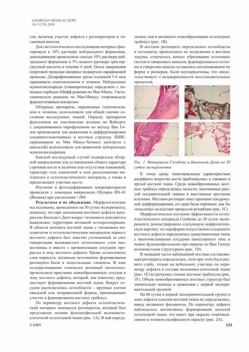
GEORGIAN MEDICAL NEWS No 5 (278) 2018
© GMN 153
сти, включая участок дефекта с регенератором и ти-тановым винтом.
Для гистологического исследования материал фик-сировали в 10% растворе нейтрального формалина, декальцинация происходила смесью 10% раствора ней-трального формалина и 5% водного раствора трихлор-уксусной кислоты в течение 4 дней. После завершения спиртовой проводки материал подвергали парафиновой проводке. Депарафинованные срезы толщиной 5-6 мкм окрашивали гематоксилином и эозином. Нейтральные мукополисахариды (гликопротеиды) определяли с по-мощью перйодат-Шифф реакции по Мак-Манус. Гисто-химическую реакцию по Мак-Манусу сопровождали ферментативным контролем.
Обзорные препараты, окрашенные гематоксили-ном и эозином, использовали для общей оценки со-стояния исследуемых тканей. Окраску препаратов фукселеном на эластические волокна по Вейгерту с докрашиванием пирофуксином по методу Ван Ги-зон производили для выявления и дифференцировки соединительнотканных и костных структур. ШИК-окрашивание по Мак Манус-Хочкису (контроль с амилазой) использовали для выявления нейтральных мукополисахаридов.
Каждый исследуемый случай подвергался обзор-ной микроскопии для установления общего характера строения кости и наличия или отсутствия изменений, характера этих изменений в зоне расположения им-плантата и остеопластического материала, а также в прилегающих участках кости.
Изучение и фотографирование микропрепаратов проводили с помощью микроскопа Olympus BX-41 (Япония) при увеличении ×200.
Результаты и их обсуждение. Морфологическое исследование, проведенное на 30 сутки эксперимента, показало, что при заполнении костного дефекта мате-риалом Биопласт-Дент вокруг титанового имплантата выявлялись территории активной остеорегенерации. В области контакта костной ткани с титановым им-плантатом и остеопластическим материалом периост костного дефекта был заметно утолщенный за счет гиперплазии волокнистого остеогенного слоя над-костницы и вместе с кровеносными сосудами про-растал в зону костного дефекта. Кроме остеогенного слоя периоста, активным источником формирования регенерата были и эндостальные элементы. В зоне остеорегенерации отмечался активный ангиогенез: происходило врастание новообразованных сосудов в зону костного дефекта, который, как известно, пред-шествует формированию костной ткани. Вокруг со-судов располагались остеобласты - крупные клетки овальной или неправильной формы, принимающие участие в формировании костных трабекул.
По периметру костного дефекта остеопластиче-ский материал замещался регенератом, который был представлен полями фуксинофильной волокнисто-клеточной остеогенной ткани (рис. 1A). В ней опреде-
лялись очаги активного новообразования остеоидных трабекул (рис. 1B).
В костном регенерате определялись остеобласты и остеоциты, происходило их погружение в костные лакуны, отмечалось начало образования остеонных систем и гаверсовых каналов, формирующиеся остео-ны и гаверсовы каналы оставались неодинаковыми по форме и размерам, были неупорядочены, что свиде-тельствовует о незавершенности восстановительных процессов.
Рис. 1. Материалы Cerabone и Биопласт-Дент на 30 сутки эксперимента
К этому сроку тинкториальные характеристики аморфного вещества кости приближались к таковым в зрелой костной ткани. Среди новообразованных кост-ных трабекул определялись полости, заполненные рых-лой соединительной тканью и выстланные круглыми клетками. Местами регенерат имел признаки хондроид-ной дифференцировки, его края были неровные, как бы «изъедены» вследствие процессов резорбции (рис. 1C).
Морфологическое изучение эффективности остео-пластического материала Cerabone до 30 суток экспе-римента демонстрировало следующую морфологиче-скую картину: по периферии искусственно созданного костного дефекта определялась грануляционная ткань с многочисленными сосудами капиллярного типа и нежно фуксинофильными при окраске по Ван Гизону волокнистыми структурами (рис. 1D).
В меньшей части наблюдений костная составляю-щая регенерата определялась, хотя при этом была раз-вита слабо, только на небольших участках по пери-метру дефекта в составе волоконно-клеточной ткани (рис. 1E) встречались тонкие костные трабекулы (рис. 1F). Объем новообразованных костных структур был значительно меньше в сравнении с первой экспери-ментальной группой.
На 60 сутки в первой экспериментальной группе в зоне дефекта осколки костной ткани не определялись, ввиду активного фагоцитоза. По периметру дефекта наблюдалось интенсивное формирование молодой остеоидной ткани, что имеет при окраске гематокси-лином и эозином оксифильную окраску (рис. 2А).
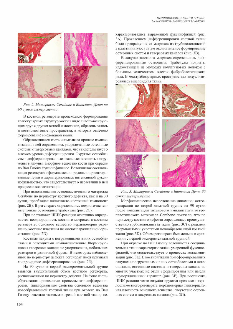
154
МЕДИЦИНСКИЕ НОВОСТИ ГРУЗИИ
CFMFHSDTKJC CFVTLBWBYJ CBF[KTYB
Рис. 2. Материалы Cerabone и Биопласт-Дент на 60 сутки эксперимента
В костном регенерате происходило формирование трабекулярных структур кости в виде анастомозирую-щих друг с другом ветвей и мостиков, образовывались и костномозговые пространства, в которых отмечено формирование миелоидной ткани.
Образовавшаяся кость испытывала процесс компак-тизации, в ней определялись упорядоченные остеонные системы с гаверсовыми каналами, что свидетельствует о высоком уровне дифференцировки. Округлые остеобла-сты и дифференцированные овальные остеоциты погру-жены в лакуны, аморфное вещество кости при окраске по Ван Гизону фуксинофильное. Волокнистая составля-ющая регенерата оформлялась в продольно ориентиро-ванные пучки и характеризовалась интенсивной фукси-нофильностью, что свидетельствует о нарастании в ней процессов коллагенизации.
При использовании остеопластического материала Cerabone по периметру костного дефекта, как и на 30 сутки, преобладал волокнисто-клеточный компонент (рис. 2B). В регенерате определялись немногочислен-ные тонкие остеоидные трабекулы (рис. 2C).
При постановке ШИК-реакции отчетливо опреде-ляется неоднородность костного матрикса в костном регенерате, основное вещество неравномерно окра-шено, костные пластины не имеют параллельной ори-ентации (рис. 2D).
Костные лакуны с погруженными в них остеобла-стами и остеоцитами немногочисленны. Формирую-щиеся гаверсовы каналы не упорядочены, небольших размеров и различной формы. В некоторых наблюде-ниях по периметру дефекта регенерат имел признаки хондроидного дифференцирования (рис. 2E).
На 90 сутки в первой экспериментальной группе выявлен внушительный объем костного регенерата, расположенного по периметру дефекта. На фоне косте-образования происходили процессы его дифференци-ровки. Тинкториальные свойства основного вещества новообразованной костной ткани при окраске по Ван Гизону отвечали таковым в зрелой костной ткани, т.е.
характеризовались выраженной фуксинофилией (рис. 3А). Проявлением дифференцировки костной ткани было превращение ее матрикса из грубоволокнистой в пластинчатую, а затем окончательное формирование остеонных систем и гаверсовых каналов (рис. 3B).
В лакунах костного матрикса определялись диф-ференцированные остеоциты. Трабекулы покрыты надкостницей из молодых коллагеновых волокон с большим количеством клеток фибробластического ряда. В межтрабекулярных пространствах визуализи-ровалась миелоидная ткань.
Рис. 3. Материалы Cerabone и Биопласт-Дент 90 сутки эксперимента
Морфологическое исследование динамики остео-репарации во второй опытной группе на 90 сутки после имплантации титанового имплантата и остео-пластического материала Cerabone показало, что по периметру костного дефекта определялась преимуще-ственно грубоволокнистая ткань (рис. 3C) с редкими прерывистыми участками новообразованной костной ткани (рис. 3D). Объем регенерата был меньше в срав-нении с первой экспериментальной группой.
При окраске по Ван Гизону волокнистая соедини-тельная ткань характеризовалась умеренной фуксино-филией, что свидетельствует о процессах коллагени-зации (рис. 3E). В костной ткани при сформированных лакунах с погруженными в них остеобластами и осте-оцитами, остеонные системы и гаверсовы каналы во многих участках не были сформированы или имели неупорядоченный характер (рис. 3F). При постановке ШИК-реакции четко визуализируются признаки незре-лости костного регенерата: неравномерная тинкториаль-ная плотность основного вещества, отсутствие остеон-ных систем и гаверсовых каналов (рис. 3G).
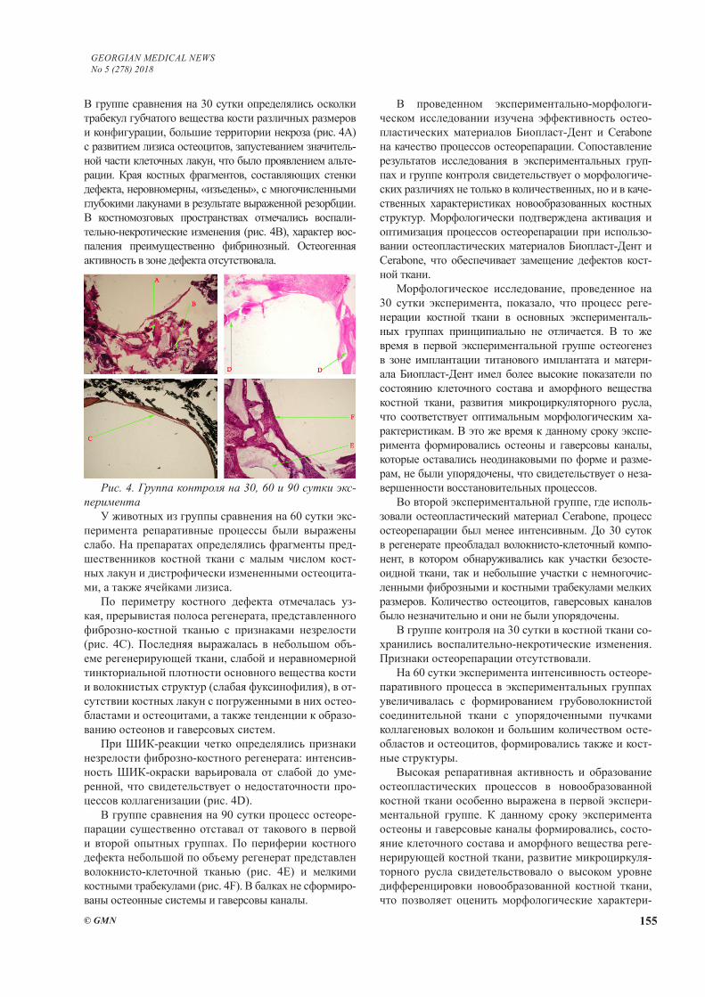
GEORGIAN MEDICAL NEWS No 5 (278) 2018
© GMN 155
В группе сравнения на 30 сутки определялись осколки трабекул губчатого вещества кости различных размеров и конфигурации, большие территории некроза (рис. 4А) с развитием лизиса остеоцитов, запустеванием значитель-ной части клеточных лакун, что было проявлением альте-рации. Края костных фрагментов, составляющих стенки дефекта, неровномерны, «изъедены», с многочисленными глубокими лакунами в результате выраженной резорбции. В костномозговых пространствах отмечались воспали-тельно-некротические изменения (рис. 4B), характер вос-паления преимущественно фибринозный. Остеогенная активность в зоне дефекта отсутствовала.
Рис. 4. Группа контроля на 30, 60 и 90 сутки экс-перимента
У животных из группы сравнения на 60 сутки экс-перимента репаративные процессы были выражены слабо. На препаратах определялись фрагменты пред-шественников костной ткани с малым числом кост-ных лакун и дистрофически измененными остеоцита-ми, а также ячейками лизиса.
По периметру костного дефекта отмечалась уз-кая, прерывистая полоса регенерата, представленного фиброзно-костной тканью с признаками незрелости (рис. 4C). Последняя выражалась в небольшом объ-еме регенерирующей ткани, слабой и неравномерной тинкториальной плотности основного вещества кости и волокнистых структур (слабая фуксинофилия), в от-сутствии костных лакун с погруженными в них остео-бластами и остеоцитами, а также тенденции к образо-ванию остеонов и гаверсовых систем.
При ШИК-реакции четко определялись признаки незрелости фиброзно-костного регенерата: интенсив-ность ШИК-окраски варьировала от слабой до уме-ренной, что свидетельствует о недостаточности про-цессов коллагенизации (рис. 4D).
В группе сравнения на 90 сутки процесс остеоре-парации существенно отставал от такового в первой и второй опытных группах. По периферии костного дефекта небольшой по объему регенерат представлен волокнисто-клеточной тканью (рис. 4E) и мелкими костными трабекулами (рис. 4F). В балках не сформиро-ваны остеонные системы и гаверсовы каналы.
В проведенном экспериментально-морфологи-ческом исследовании изучена эффективность остео-пластических материалов Биопласт-Дент и Cerabone на качество процессов остеорепарации. Сопоставление результатов исследования в экспериментальных груп-пах и группе контроля свидетельствует о морфологиче-ских различиях не только в количественных, но и в каче-ственных характеристиках новообразованных костных структур. Морфологически подтверждена активация и оптимизация процессов остеорепарации при использо-вании остеопластических материалов Биопласт-Дент и Cerabone, что обеспечивает замещение дефектов кост-ной ткани.
Морфологическое исследование, проведенное на 30 сутки эксперимента, показало, что процесс реге-нерации костной ткани в основных эксперименталь-ных группах принципиально не отличается. В то же время в первой экспериментальной группе остеогенез в зоне имплантации титанового имплантата и матери-ала Биопласт-Дент имел более высокие показатели по состоянию клеточного состава и аморфного вещества костной ткани, развития микроциркуляторного русла, что соответствует оптимальным морфологическим ха-рактеристикам. В это же время к данному сроку экспе-римента формировались остеоны и гаверсовы каналы, которые оставались неодинаковыми по форме и разме-рам, не были упорядочены, что свидетельствует о неза-вершенности восстановительных процессов.
Во второй экспериментальной группе, где исполь-зовали остеопластический материал Cerabone, процесс остеорепарации был менее интенсивным. До 30 суток в регенерате преобладал волокнисто-клеточный компо-нент, в котором обнаруживались как участки безосте-оидной ткани, так и небольшие участки с немногочис-ленными фиброзными и костными трабекулами мелких размеров. Количество остеоцитов, гаверсовых каналов было незначительно и они не были упорядочены.
В группе контроля на 30 сутки в костной ткани со-хранились воспалительно-некротические изменения. Признаки остеорепарации отсутствовали.
На 60 сутки эксперимента интенсивность остеоре-паративного процесса в экспериментальных группах увеличивалась с формированием грубоволокнистой соединительной ткани с упорядоченными пучками коллагеновых волокон и большим количеством осте-областов и остеоцитов, формировались также и кост-ные структуры.
Высокая репаративная активность и образование остеопластических процессов в новообразованной костной ткани особенно выражена в первой экспери-ментальной группе. К данному сроку эксперимента остеоны и гаверсовые каналы формировались, состо-яние клеточного состава и аморфного вещества реге-нерирующей костной ткани, развитие микроциркуля-торного русла свидетельствовало о высоком уровне дифференцировки новообразованной костной ткани, что позволяет оценить морфологические характери-
156
МЕДИЦИНСКИЕ НОВОСТИ ГРУЗИИ
CFMFHSDTKJC CFVTLBWBYJ CBF[KTYB
стики остеорепаративного процесса как оптимальные.Морфологическое изучение эффективности остео-
пластического материала Cerabone показало, что осте-орепарация в зоне дефекта была слабее в сравнении с материалом Биопласт-Дент, что проявлялось в преоб-ладании волокнисто-клеточного и хондроидного ком-понентов в составе регенерата, более слабых темпах формирования костных трабекул небольших разме-ров, в которых костные лакуны и остеонные системы с гаверсовыми каналами не упорядочены. Эти харак-теристики свидетельствуют о недостаточной зрелости регенерата до 60 суток во второй экспериментальной группе.
В группе сравнения на 60 сутки эксперимента в сравнении с первой и второй экспериментальными группами отмечалась слабая репаративная актив-ность, что выражалось в малом количестве новооб-разованных трабекул или в полном их отсутствии. По периметру костного дефекта был сформирован небольшой по объему регенерат, представленный кле-точно-волокнистой соединительной тканью.
В первой экспериментальной группе на 90 сутки эксперимента остеорепарация имела завершенный характер: сформированы костные структуры зрелого типа с упорядоченными остеонными системами и га-версовыми каналами.
Во второй экспериментальной группе темпы и качество остеорепарации несколько снижены в сравнении с первой группой, что выражалось в пре-обладании волокнисто-клеточного и хондроидного компонентов в составе регенерата, слабом развитии костных трабекул, в которых лакуны и гаверсовы каналы были немногочисленными и сохраняли не-упорядоченность.
У животных из группы контроля на 90 сутки репа-ративные процессы наиболее слабо выражены. Реге-нерирующая ткань представлена небольшим по объ-ему волокнисто-клеточным компонентом и мелкими фиброзными и костными трабекулами, а также участ-ками хондроидной дифференцировки.
Сравнительный морфологический анализ остео-пластических материалов Биопласт-Дент и Cerabone показал преимущество первого из них, что подтверждено наиболее выраженной актива-цией репаративного остеогенеза и оптимальными морфологическими характеристиками новообразо-ванной костной ткани.
В группе сравнения отмечались не только слабое образование костных структур, но и незрелость осте-областических элементов. Процесс остеорепарации до окончания эксперимента не завершился.
Выводы. 1. Результаты морфологического ис-следования показали, что при заполнении костных дефектов остеопластическим материалом Cerabone или Биопласт-Дент на всех сроках эксперимента от-сутствовали воспалительные изменения, отторжение или аллергические реакции в ответ на имплантацию.
2. Полученные морфологические данные свиде-тельствуют, что процесс регенерации костной ткани с использованием остеомодификаторов Биопласт и Cerabone усиливался в сравнении с группой контроля.
3. Темпы восстановления костных структур и каче-ственные характеристики новообразованной кости в исследовательских группах (Биопласт и Cerabone) прин-ципиально не отличались, однако при использовании Биопласт остеорепарация протекала более активно, с более оптимальными морфологическими характеристи-ками новообразованной костной ткани.
4. В группе контроля остеонеогенез был самым сла-бым и до окончания эксперимента не завершенным.
ЛИТЕРАТУРА
1. Иванкин А. Н., Васюков С. Е., Панов В. П. Получение, свойства и применение хондроитин сульфатов // Химико-фармацевтический журнал. - 1985. - №3. - С. 192 -202.2. Иванов С. Ю., Мураев А. А., Рукина Е. А., Бунёв А. А. Метод непосредственной дентальной имплантации // Современные проблемы науки и образования. - 2015. - №5. – С. 230.3. Кампос Ф. Е. Вплив розміру ложа імплантату на ранній етап остеоінтеграції / [Ф. Е. Кампос, Х. Б. Гомес, Ч. Марін та ін.]. // Імплантологія, Пародонтологія, Остеологія. – 2013. – №1. – С. 27–34.4. Касай А. Mucoderm — материал для трехмерной регенера-ции тканей // Новое в стоматологии. - 2012. - №5. - С. 97–103.5. Коротких Н.Г., Харитонов Д.Ю., Азарова Е.А., Степа-нов И.В. Экспериментальное обоснование использования материалов «Клипдент», «Биопласт-Дент» при замещении костных дефектов после травматического удаления зубов // Вестник новых медицинских технологий (электронный журнал). - 2014. - №1. 6. Куцевляк В.И., Гречко Н.Б., Алтунина С.В., Старикова С.Л. Учебное пособие. – Харьков: ХГМУ, 2005. – С-34.7. Маланчук В. А., Мамедов С. А. Непосредственная ден-тальная имплантация. - Киев: Научно-учебное издание для студентов и врачей, 2008. – С. 121-135.8. Флейшер Г.М., Посохова В.Ф., Лыкова И.В. Использова-ние биопластического материала «Биопласт-Дент» в стома-тологии и челюстно-лицевой хирургии. // Дентальная им-плантология и хирургия. – 2016. - № 1(22). – С. 70-72.9. Харитонов Д.Ю., Домашевская Э.П., Азарова Е.А., Голо-щапов Д.Л. Сравнение морфологических и структурных ха-рактеристик костной ткани человека и остеопластического материала «Биопласт-Дент» // Фундаментальные исследо-вания. - 2014. - №10. - С. 1389-1393.10. Шишковська А., Кравчик П. Матеріали для заміщення кісткових дефектів у стоматологіїї. Огляд // Імплантологія, Пародонтологія, Остеологія. - 2014. - №3(35). - С. 18-23.11. Becker W. Immediate implant placement: diagnosis,treatment planning and treatment steps/or successful outcomes // J Calif Dent Assoc. - 2005. - №33. - P. 303 – 310.12. Bigi A. In vitro culture of mesenchymal cells onto nanocrys-talline hydroxyapatite coated Ti13Nb13Zr alloy / [A. Bigi, N. Nicoli-Aldini, B. Bracci et al.]. // J. Biomed. Mater. Res. – 2007. – №82 (1). – P. 213–221.13. Ghanaati S. Nanocrystalline hydroxyapatite bone substitute leads to sufficient bone tissue formation already after 3 months: histological and histomorphometrical analysis 3 and 6 months following human sinus cavity augmentation / [S. Ghanaati, M.
GEORGIAN MEDICAL NEWS No 5 (278) 2018
© GMN 157
Barbeck, I. Willershausen et al.]. // Clin Implant Dent Relat Res. – 2013. – №15. – P. 883–892.14. Laub M. Aspects of BMP-2 binding to receptors and col-lagen: Influence of cell senescence on receptor binding and ab-sence of high-affinity stoichiometric binding to collagen / M. Laub, M. Chatzinikolaidou, H. P. Jennissen. // Materials Science & Engineering Technology. – 2007. – P. 1019–1026. 15. Rosenquist B., Grenthe B. Immediate placement of implants into extraction sockets: implant survival // Int J Oral Maxillofac Implant. - 1996. - №11. – P. 205-211.16. Rothamel D. Sinus floor elevation using a sintered natural bone mineral. A histologic case report study / [D. Rothamel, F. Schwarz, R. Smeetset al.] // Deutscher Ärzte-Verlag Zahnärztl Impl. – 2011. – P. 60-70.17. Tarazona B. Relationship between indication for tooth ex-traction and outcome of immediate implants: A retrospective study with 5 years of follow-up / [B. Tarazona, P. Tarazona-Alvarez, D. Penarrocha-Oltra et al.]. // J ClinExp Dent. - 2014. - №6 (4). - P. 383-387.
SUMMARY
COMPARATIVE MORPHOLOGICAL STUDY OF GUIDED BONE TISSUE REGENERATION USING XENOGENIC OSTEOPLASTIC MATERIAL BIO-PLAST-DENT AND CERABONE
1,2 Chernenko V., 1Lyubchenko A.
1Kharkov Medical Academy of Postgraduate Education, Kharkov; 2Medical Institute at Sumy State University, Ukraine
The purpose of the work is the modeling in the experi-ment of guided regeneration of bone tissue with the use of osteoplastic materials Bioplast and Cerabone with the subsequent morphological study of their influence on the course of osteoreparation.
The experimental-morphological part of the work was performed on 90 mature rats of the Wistar line. The animals were divided into 3 experimental groups. In the first group (30 rats), the osteoplastic material Bioplast-Dent was used; in the second group (30 rats), the osteoplastic material Cerabo-ne was used. The third (30 rats) group was the control group. A defect in the bone region was formed in rats, a titanium screw (BT1-00) was implanted, then the bone defects were filled with osteoplastic material. In the control group, the os-teoplastic material was not used. The animals were removed from the experiment by decapitation under ether anesthesia in 30, 60 and 90 days. For morphological examination the resection of the central part of the femur shaft was carried out, including a defect site with a regenerator and a titanium screw. Each studied case was subjected to a microscopy sur-vey in which the general character of the bone structure was assessed, as well as the presence or absence of changes and their nature in the zone of implant and osteoplastic material, as well as in dependent bone areas.
The result of the comparative morphological study of xenogeneic osteoplastic materials Bioplast-Dent and
Cerabone resulted in the recommended use of one of the materials in this implantation technique.
Regeneration time of bone structures and qualitative characteristics of the newly formed bone did not differ fundamentally, however, when using Bioplast-Dent ma-terial, osteoreparation proceeded more actively and with more optimal morphological characteristics; there were no inflammatory changes, rejections or allergic reactions in response to implantation at all stages of the experiment.
Keywords: direct implantation, osseointegration, osteo-plastic xenogeneic material, immediate loading.
РЕЗЮМЕ
СРАВНИТЕЛЬНОЕ МОРФОЛОГИЧЕСКОЕ ИССЛЕДОВАНИЕ НАПРАВЛЕННОЙ РЕГЕНЕ-РАЦИИ КОСТНОЙ ТКАНИ ПРИ ИСПОЛЬЗО-ВАНИИ КСЕНОГЕННЫХ ОСТЕОПЛАСТИ-ЧЕСКИХ МАТЕРИАЛОВ БИОПЛАСТ-ДЕНТ И CERABONE
1,2Черненко В.Н., 1Любченко А.В.
1Харьковская медицинская академия последипломного образования; 2Сумский государственный универси-тет, Медицинский институт, Украина
Целью исследования явилось моделирование в эксперименте направленной регенерации костной ткани с использованием остеопластических материа-лов Биопласт-Дент и Cerabone с последующим мор-фологическим исследованием их влияния на ход осте-орепарации.
Экспериментально-морфологическая часть работы выполнена на 90 половозрелых крысах линии Вистар. Животные разделены на 3 экспериментальные груп-пы: в первой группе (n=30) использовали остеопла-стический материал Биопласт-Дент, во второй группе (n=30) - остеопластический материал Cerabone. Тре-тья группа (n=30) - контроль. Крысам формировали дефект в области кости, проводили имплантацию ти-танового винта (ВТ1-00), после чего костные дефекты заполняли остеопластическим материалом. В группе контроля остеопластический материал не использо-вали. Животных выводили из эксперимента методом декапитации под эфирным наркозом на 30, 60 и 90 сутки. Для морфологического исследования проводи-ли резекцию центральной части диафиза бедренной кости, включая участок дефекта с регенератором и титановым винтом. Каждый исследуемый случай под-вергали обзорной микроскопии, при которой оценива-ли общий характер строения кости, а также наличие или отсутствие изменений, их характер в зоне распо-ложения имплантата, остеопластического материала и в прилегающих участках кости.
Полученный результат сравнительного морфоло-

158
МЕДИЦИНСКИЕ НОВОСТИ ГРУЗИИ
CFMFHSDTKJC CFVTLBWBYJ CBF[KTYB
гического исследования ксеногенных остеопластиче-ских материалов Биопласт-Дент и Cerabone позволя-ет рекомендовать использование обоих материалов в данной методике имплантации. Темпы восстановле-ния костных структур и качественные характеристики новообразованной кости принципиально не отлича-лись, однако при использовании материала Биопласт-Дент остеорепарация протекала более активно и с более оптимальными морфологическими характери-стиками, на всех сроках эксперимента отсутствовали воспалительные изменения, отторжения или аллерги-ческие реакции в ответ на имплантацию.
reziume
Zvlis qsovilis mimarTuli regeneraciis Se-darebiTi morfologiuri kvleva qsenogenu-ri ostaoplastiuri masalebis bioplast-denti da Cerabone gamoyenebiT
1,2v. Cernenko, 1a. liubCenko
1xarkovis diplomisSemdgomi ganaTlebis sa-medicino akademia; 2sumis saxelmwifo uni-versiteti, samdicino instituti, ukraina
kvlevis mizans warmoadgenda Zvlovani qsovilis mimarTuli regeneraciis eqsperi-mentuli modelireba osteoplastiuri masa-lebis bioplasti da Cerabone gamoyenebiT da maTi osteoreparaciis ganviTarebaze gavle-nis morfologiuri kvleviT.
samuSaos eqsperimentul-morfologiuri nawili Sesrulda vistaris xazis 90 sqesob-rivad momwifebul virTagvaze. cxovele-bi daiyo 3 eqsperimentul jgufad: pirvel
jgufSi (n=30) gamoyenebuli iyo osteoplas-tiuri masala bioplasti-denti, meore jguf-Si (n=30) - osteoplastiuri masala Cerabone. mesame (n=30) jgufi warmoadgenda sakontro-lo jgufs. cxovelebs gaukeTda defeqti Zvlis midamoSi, Cautarda titanis xraxnis implantacia (ВТ1-00), ris Semdegac Zvlovani defeqtebi Seivso osteoplastiuri masaliT. sakontrolo jgufSi osteoplastiuri masa-la gamoyenebuli ar iyo. cxovelebi eqsperi-mentidan gamohyavdaT dekapitaciis meTodiT eTeris narkozis qveS, 30–e, me–60 da 90–e dReze. morfologiuri gamokvlevisaTvis Catarda barZayis Zvlis diafizis central-uri nawilis rezeqcia defeqtis ubnis CaTv-liT, regeneratorTan da titanis xraxnilTan erTad. Catarda masalis daTvaliereba mikros-kopiT, Sefasda Zvlis age-bulebis zogadi wyoba, agreTve, cvlilebebis arseboba, an ararseboba, maTi xasiaTi implantatisa da osteoplastiuri masalis ganTavsebis zonaSi da Zvlis mimdebare ubnebSi.
qsenogenuri osteoplastiuri masalebis „bioplasti“ da «Cerabone» SedarebiTi mor-fologiuri kvleviT miRebuli Sedegebis safuZvelze rekomendebulia am masalaTagan erT–erTis gamoyeneba implantaciis am meTo-dikaSi. Zvlovani struqturebis aRdgenis tempi da axlad Seqmnili Zvlis xarisxis maxasiaTeblebi principulad ar gansxvavde-ba, magram „bioplasti-denti“-s gamoyenebisas osteoreparacia mimdinareobda ufro aqtiu-rad da optimaluri morfologiuri maxasi-aTeblebiT; anTebiTi cvlilebebi an aler-giuli reaqciebi implantaciis sapasuxod ar gamovlinda eqsperimentis arc erT vadaze.
CHARACTERISTICS OF GAMMA OSCILLATIONS INDUCED BY KAINATE PRESSURE EJECTION ON CA1 HIPPOCAMPUS
OF MICE BRAIN SLICES IN SUBMERGED CHAMBERS
Kipiani E.
Institute for Neurophysiology, Charite, Berlin, Germany; Department of Informatics, Mathematics and Natural Sciences, St. Andrew the First, Tbilisi, Georgia
Assemblies in the waking brain typically synchronize within gamma range. Gamma oscillations are very impor-tant rhythms of the brain involved in attention, conscious-ness, working memory, sensory binding and in its abnor-
malities [4] are registered during schizophrenia, epilepsy and Alzheimer’s disease.
Although physiologically realistic patterns of network activity have been successfully induced in brain slices
GEORGIAN MEDICAL NEWS No 5 (278) 2018
© GMN 159
maintained in interface-type recording chambers, it was hard to obtain results in submerged conditions, which of-fer significant experimental advantages, including patch-clamp recordings and imaging techniques.
Recently it was discovered that gamma oscillations in area CA1 of hippocampus can be induced when both CA3 and subiculum are disconnected from the circuit, implying a local generator [2,8]. There are some stud-ies describing gamma oscillations in CA1 together with various types of hippocampal cells [2,3,6]. However, in above mentioned investigations either gamma induction methods are different or less attention is paid to character-istics of CA1 gamma oscillations themselves and gamma cycles are considered in relation to discharge probabilities of various types of neurons.
Sustained carbachol (CCh) induced network oscilla-tions were successfully recorded in submerged hippocam-pal slices in CA3 area at increased flow rates of super-fusion solution or from increased thickness of slices. To receive spontaneous population rhythmic activities Wu and colleagues developed a novel slice preparation of 1 mm thickness from adult mice hippocampus [10]. They claimed that in rodent hippocampal slices thinner than 500 µm, population rhythmic activities do not occur due to limited network connectivity. However, kainate pres-sure ejection evoked gamma oscillations were also re-corded recently from 400 µm hippocampal slices [2].
Differ from above mentioned details presented data was recorded from 350 or 300 µm thick slices at room temperature in area CA1 of hippocampus. Here will be discussed heterogeneity of short-term transient gamma oscillations in CA1 hippocampus.
Material and methods. Slice preparationExperiments were performed on horizontal combined
entorhynal-cortex-hippocampal slices (300-350 µm thick) prepared with a tissue slicer (Leica VT 1000 S, Nussloch, Germany). Brains were taken from C57BL6 or transgenic mice that expressed enhanced fluorescent protein under the control of parvalbumin promoter. Animals were decapitated under deep ether anesthesia, brains were rapidly removed and incubated in 4 degree Celsius cold and oxygenated arti-ficial cerebrospinal fluid (ACSF) as described elsewhere [3].
Field recordings were performed at room temperature in submerged chambers. Rate of superfusion with ACSF was up to 3 ml per min. The kainite pressure ejection and recording electrons were close to each other (0.2 mm distance) to get prominent field recordings. Both glass devices were in CA1 hippocampus between the stratum radiatum and lacunosum-moleculare. Field potentials were recorded with axoclamp 2B amplifier (Axon Instru-ments, Union City, CA, USA) and analysed by Axograph. Oscillatory peak frequency was determined by averaging several consecutive Fourier transforms, contained within a 1 second epoch at the beginning and the middle part of the oscillation. For statistical comparisons t-test was used. Average values are expressed as the means ± standard de-viation (SD).
Results and their discussion. Transient gamma oscil-lations were induced in CA1 hippocampus of mice slices by the pressure ejection of 1 mM kainic acid (Tocris/Biotrend, Cologne, Germany) onto the boundary of stra-tum radiatum and stratum lacunosum moleculare of the hippocampal CA1 area.
Kainate pressure ejection for 10 ms caused short (0.5 second) negative (1.5 or 2 mV) shift of DC in the field recordings. Local field potential (LFP) oscillations were recorded from 44 hippocampal slices from 32 mice. Alto-gether, oscillations where registered 70 times.
In 13 out of 44 slices LFP oscillations were induced from three to six times. In the rest 31 slices oscillations were mostly induced twice with different time intervals. Due to the little power and/or sparse number of gamma cycles of the second time induced oscillations their char-acterization is not discussed with more details.
In 13 slices gamma oscillations were successfully in-duced various times: twice (n = 6), three times (n=3), four times (n=2) and five times (n=2). In each case one more induction of gamma activity was tried but it either failed or some sparse gamma cycles were registered. Therefore, last results are not presented here. Altogether, there were recorded 39 oscillatory events from 13 slices.
To develop the field oscillation 0.16±0.21 seconds were required. Often, the field oscillation had a waxing and wan-ing character. Oscillation power (amplitude) was generally bigger at the beginning of induction that in the following part of oscillation. However, in some cases it increased after sev-eral seconds from kainite pressure ejection.
Development of field oscillation needed different time. Rarely, its induction needed several seconds and only then after 1.3 seconds did the amplitude show increase and re-ceived structured nature.
Whole duration of LFP oscillations were divided into three or two epochs because of their heterogeneous nature: LFP oscillations with maximum power (initial state of os-cillation) (Fig. 1a), with declining power (middle or last part of oscillations) (Fig. 1b) and with the lowest power. The last activity was expressed as alteration of appear-ance and disappearance of sparse gamma cycles with little power. With the other words, reduction of the amplitude length was sometimes expressed with the sparse gamma cycles as the indicator of the declining oscillations. Re-duction in gamma power was also registered during each consecutive induction of oscillation in all 13 slices.
Mean initial amplitude and middle part amplitude of oscillations recorded from 13 slices were significantly dif-ferent from each other (p=0.0001). Mean initial oscilla-tion amplitude was 0.23±0.6 mV. Mean middle part oscil-lation amplitude was 0.19±0.3 mV.
Studying the frequency of the same activities in 13 slices showed that period of gamma cycles was significantly differ-ent in the middle part of oscillation compared with the initial section (44.24±8.42 Hz vs. 49.31±10.96 Hz, p=0.03, n=39) (Fig. 2 a and b). However, the frequency of consecutive gam-ma oscillations did not change noticeably.
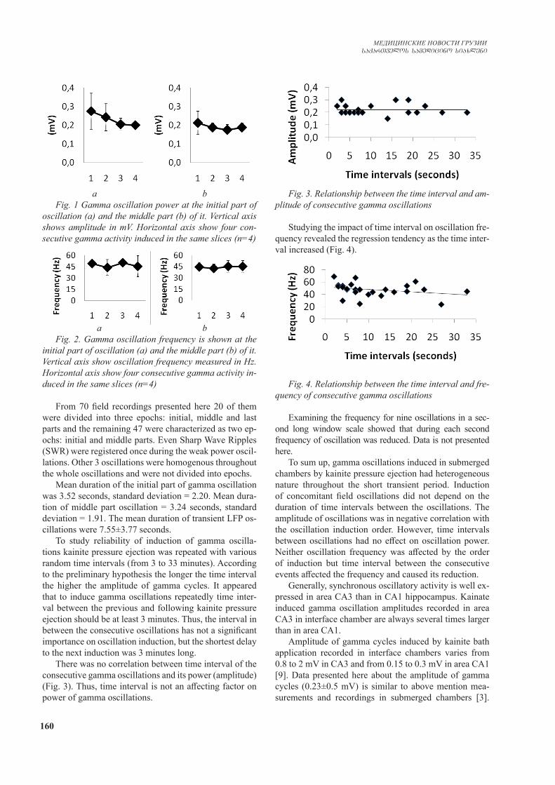
160
МЕДИЦИНСКИЕ НОВОСТИ ГРУЗИИ
CFMFHSDTKJC CFVTLBWBYJ CBF[KTYB
a bFig. 1 Gamma oscillation power at the initial part of
oscillation (a) and the middle part (b) of it. Vertical axis shows amplitude in mV. Horizontal axis show four con-secutive gamma activity induced in the same slices (n=4)
a bFig. 2. Gamma oscillation frequency is shown at the
initial part of oscillation (a) and the middle part (b) of it. Vertical axis show oscillation frequency measured in Hz. Horizontal axis show four consecutive gamma activity in-duced in the same slices (n=4)
From 70 field recordings presented here 20 of them were divided into three epochs: initial, middle and last parts and the remaining 47 were characterized as two ep-ochs: initial and middle parts. Even Sharp Wave Ripples (SWR) were registered once during the weak power oscil-lations. Other 3 oscillations were homogenous throughout the whole oscillations and were not divided into epochs.
Mean duration of the initial part of gamma oscillation was 3.52 seconds, standard deviation = 2.20. Mean dura-tion of middle part oscillation = 3.24 seconds, standard deviation = 1.91. The mean duration of transient LFP os-cillations were 7.55±3.77 seconds.
To study reliability of induction of gamma oscilla-tions kainite pressure ejection was repeated with various random time intervals (from 3 to 33 minutes). According to the preliminary hypothesis the longer the time interval the higher the amplitude of gamma cycles. It appeared that to induce gamma oscillations repeatedly time inter-val between the previous and following kainite pressure ejection should be at least 3 minutes. Thus, the interval in between the consecutive oscillations has not a significant importance on oscillation induction, but the shortest delay to the next induction was 3 minutes long.
There was no correlation between time interval of the consecutive gamma oscillations and its power (amplitude) (Fig. 3). Thus, time interval is not an affecting factor on power of gamma oscillations.
Fig. 3. Relationship between the time interval and am-plitude of consecutive gamma oscillations
Studying the impact of time interval on oscillation fre-quency revealed the regression tendency as the time inter-val increased (Fig. 4).
Fig. 4. Relationship between the time interval and fre-quency of consecutive gamma oscillations
Examining the frequency for nine oscillations in a sec-ond long window scale showed that during each second frequency of oscillation was reduced. Data is not presented here.
To sum up, gamma oscillations induced in submerged chambers by kainite pressure ejection had heterogeneous nature throughout the short transient period. Induction of concomitant field oscillations did not depend on the duration of time intervals between the oscillations. The amplitude of oscillations was in negative correlation with the oscillation induction order. However, time intervals between oscillations had no effect on oscillation power. Neither oscillation frequency was affected by the order of induction but time interval between the consecutive events affected the frequency and caused its reduction.
Generally, synchronous oscillatory activity is well ex-pressed in area CA3 than in CA1 hippocampus. Kainate induced gamma oscillation amplitudes recorded in area CA3 in interface chamber are always several times larger than in area CA1.
Amplitude of gamma cycles induced by kainite bath application recorded in interface chambers varies from 0.8 to 2 mV in CA3 and from 0.15 to 0.3 mV in area CA1 [9]. Data presented here about the amplitude of gamma cycles (0.23±0.5 mV) is similar to above mention mea-surements and recordings in submerged chambers [3].
GEORGIAN MEDICAL NEWS No 5 (278) 2018
© GMN 161
However, the duration of transient gamma oscillations of CA1 lasted for shorter period (7.44±3.56 seconds) than it was reported elswhere [2,3,8].
Limitations of the experiments were thickness of slic-es (300 and 350 µm) and the speed of ACSF supply that was 3 ml/min. It was shown that gamma oscillations are well developed in thicker slices during the high rate of superfusion (5 ml/min) [2,8,10]. However, the thicker the slices, the harder to select an interneuron for whole-cell patch clamp recordings. So does the high rate of superfu-sion: ACSF drops may vibrate the slice and risk losing the patched cell. Here presented recording were done at room temperature. To increase the temperature up to 37 degree Celsius may show comparatively different results.
REFERENCES
1. Buzsáki G, Wang XJ. Mechanisms of gamma oscillations //Annu Rev Neurosci. 2012;35:203-25. 2. Craig MT, McBain CJ. Fast gamma oscillations are generated intrinsically in CA1 without the involvement of fast-spiking basket cells // J Neurosci. 2015 Feb 25;35(8):3616-24. 3. Gloveli T, Dugladze T, Saha S, Monyer H, Heinemann U, Traub RD, Whittington MA, Buhl EH. Differential involvement of oriens/pyramidale interneurones in hippocampal network os-cillations in vitro //J Physiol. 2005 Jan 1;562(Pt 1):131-47. 4. Grützner C, Wibral M, Sun L, Rivolta D, Singer W, Maurer K,
Uhlhaas PJ. Deficits in high- (>60 Hz) gamma-band oscillations during visual processing in schizophrenia // Front Hum Neuro-sci. 2013 Mar 26;7:88. 5. Hájos N, Ellender TJ, Zemankovics R, Mann EO, Exley R, Cragg SJ, Freund TF, Paulsen O. Maintaining network activity in submerged hippocampal slices: importance of oxygen supply // Eur J Neurosci. 2009 Jan;29 (2):319-27. 6. Kipiani E. OLM interneurons are transiently recruited into field gamma oscillations evoked by brief kainate pressure ejec-tions onto area CA1 in mice hippocampal slices //Georgian Med News. 2009 Feb;(167):63-8. 7. Lu CB, Wang ZH, Zhou YH, Vreugdenhil M. Temperature- and concentration-dependence of kainate-induced γ oscillation in rat hippocampal slices under submerged condition // Acta Pharmacol Sin. 2012 Feb;33(2):214-20.8. Pietersen AN, Ward PD, Hagger-Vaughan N, Wiggins J, Jef-ferys JG, Vreugdenhil M. Transition between fast and slow gamma modes in rat hippocampus area CA1 in vitro is modu-lated by slow CA3 gamma oscillations // J Physiol. 2014 Feb 15;592(4):605-20. 9. Wójtowicz AM, van den Boom L, Chakrabarty A, Maggio N, Haq RU, Behrens CJ, Heinemann U. Monoamines block kai-nate- and carbachol-induced gamma-oscillations but augment stimulus-induced gamma-oscillations in rat hippocampus in vi-tro //Hippocampus. 2009 Mar;19(3):273-88. 10. Wu, C., Luk, W.P., Gillis, J., Skinner, F. & Zhang, L. (2005) Size does matter: generation of intrinsic network rhythms in thick mouse hippocampal slices // J. Neurophysiol., 93, 2302–2317.
SUMMARY
CHARACTERISTICS OF GAMMA OSCILLATIONS INDUCED BY KAINATE PRESSURE EJECTION ON CA1 HIPPOCAMPUS OF MICE BRAIN SLICES IN SUBMERGED CHAMBERS
Kipiani E.
Institute for Neurophysiology, Charite, Berlin, Germany; Department of Informatics, Mathematics and Natural Sciences, St. Andrew the First, Tbilisi, Georgia
Aim - mostly, gamma oscillations are studied in inter-
face-type chambers. The purpose of the presented investi-gation is to describe the characteristics of gamma oscilla-tions induced in submerged chambers by kainite pressure ejection.
Horizontal combined entorhynal-hippocampal slices 300-350 µm were prepared from young mice (P18-28). Gamma oscillations were induced by 1 mM kainite pres-sure ejection at the boundary of stratum radiatum and lacunosum-moleculare of area CA1. Field potential re-cordings were registered from the vicinity of kainite ap-plication.
Induced CA1 local field potential (LFP) oscillations were brief (7.55±3.77 sec.) and had heterogeneous na-ture; they could be divided into three epochs: well developed initial part of oscillation, middle part with reduced gamma power and last part of the rhythm with sporadic immergence
of sparse (3 to 5) gamma cycles. Generally, initial parts of oscillations had higher amplitude and frequency than the middle part of it. Induction of consecutive gamma oscilla-tions did not depend on the duration of the time intervals be-tween oscillations. Their amplitude was affected by the order of induction but not by time intervals between oscillations. Neither the frequency was affected by the order of induced activities in the same slice. However, comparatively lower frequency oscillations were recorded after long time inter-vals between gamma activities.
Induction of CA1 gamma oscillations in submerged conditions will offer significant experimental advantage, like using patch-clamp techniques to study the mecha-nism of this activity.
Keywords: gamma oscillations, submerged chamber, CA1 hippocampus, kainite.
162
МЕДИЦИНСКИЕ НОВОСТИ ГРУЗИИ
CFMFHSDTKJC CFVTLBWBYJ CBF[KTYB
Целью исследования явилась характеристика гам-ма-осцилляций, вызванных выбросом каинатного давле-ния в подводных камерах в эксперименте.
Использованы горизонтальные комбинирован-ные энторинал-гиппокампальные 300-350 мкм срезы, приготовленные из головного мозга молодых мышей 18-28 дней. Гамма-осцилляции индуцировались 1 мМ каинатом на границах стратум-радиатум и лакунозум-молекуларе области СА1. Записи потенциала поля регистрировались около аппликационной пипеты каината. Индуцированные колебания потенциала ло-кального поля CA1 были краткими (7,55±3,77 сек.) и носили гетерогенный характер; их можно разделить на три части: хорошо развитая начальная часть коле-баний, средняя часть - с уменьшенной гамма-мощно-стью, последняя часть - спорадическое прорастание
гамма-циклов (от 3 до 5). Начальные части колеба-ний, как правило, имели более высокую амплитуду и частоту, чем средняя часть. Индукция последова-тельных гамма-осцилляций не зависила от продол-жительности временных интервалов между колеба-ниями. На их амплитуду влиял порядок индукции, а не временные интервалы между колебаниями. При увеличении интервала времени регистрировались осцилляции более низкой частоты. Однако, на ча-стоту не влиял порядок индуцированных действий в одном и том же срезе.
Результаты исследования позволяют заключить, что индукция гамма-осцилляций CA1 в подводных камерах наряду с использованием методов "патч-клемпа" создает хорошие экспериментальные условия для изучения ме-ханизмов гамма-активности.
РЕЗЮМЕ
ХАРАКТЕРИСТИКА ГАММА-ОСЦИЛЛЯЦИЙ, ВЫЗВАННЫХ ВЫБРОСОМ КАИНАТНОГО ДАВЛЕНИЯ НА ГИППОКАМП CA1 СРЕЗОВ МОЗГА МЫШЕЙ В ПОДВОДНЫХ КАМЕРАХ
Кипиани Е.Э.
Институт нейрофизиологии, Шарите, Берлин, Германия; Грузинский университет Патриархии Грузии им. Андрея Первозванного, факультет информатики, математики и естественных наук, Тбилиси, Грузия
reziume
Tagvebis Tavis tvinis hipokampis CA1 velze wnevis qveS miwodebuli kainatiT gamowveuli gama-oscilaciis maxasiaTeblebi CaZiruli tipis kameraSi
e. yifiani
neirofiziologiis instituti, Sarite, berlini, germania; saqarTvelos sapatriarqos wminda andria pirvelwodebulis sax. qarTuli universiteti, informatikis, maTematikisa
da sabunebismetyvelo mecnierebaTa fakulteti, Tbilisi, saqarTvelo
kvlevis mizans warmoadgenda Tagvebis Tavis tvinis hipokampis CA1 velze kainatiT gamowveuli gama-oscilaciis maxasiaTeb-lebis Seswavla CaZiruli tipis kameraSi.
damzadda 18-28 dRis Tagvebis Tavis tvi-nis 300-350 mkm sisqis horizontaluri anaT-lebi, romlebic Seicavda hipokamp-entori-nul qerqs. gama-oscilaciebis gamosawvevad 1 mlmoli kainati wnevis qveS ewveTeboda hipokampis CA1 velSi, radiatumisa da laku-nozum-molekulares Sreebis sazRvarze. ve-lis potencialis aRricxvisTvis gamomyvani eleqtrodi ganTavsebuli iyo kainatis sa-aplikacio pipetTan axlos.
hipokampis CA1 velSi gamowveuli gama-oscilacia xanmokle (7.55±3.77 wm) da hetero-genuli bunebisaa; aRiniSneba sami tipis aq-tivoba: sawyisi etapi - kargad gamoxatuli oscilaciiT, Sua etapi - klebadi amplitu-dis oscilaciiT, bolo etapi - sporadulad
aRmocenebadi 3-5 gama-cikliT. sawyis etapze aRiniSneba ufro maRali sixSiris aqtivoba, vidre Sua etapze. gama-oscilaciebis gan-meorebiTi induqcia ar aris damokidebuli drois intervalze rxevebs Soris. maT ampli-tudaze zemoqmedebs induqciis Tanmimdevroba, da ara intervali rxevebs Soris. aRsaniSna-via amplitudis kleba yoveli momdevno osci-laciis dros; Tumca, amplitudaze erTi da igive anaTalSi gavlenas ar axdens drois in-tervali gamowveul oscilaciebs Soris. Se-darebiT dabali sixSiris oscilaciebi dare-gistrirda drois intervalis zrdis dros, magram sixSiris amgvari kleba araa damokide-buli oscilaciebis gamowvevis rigiTobaze.
CA1 velis gama-oscilaciebis induqcia Ca-Ziruli tipis kameraSi sxva, magaliTad, „patch-clamp“ teqnikis gamoyenebasTan erTad Seqmnis karg eqsperimentul pirobebs gama-oscilaci-is meqanizmebis ukeT SeswavlisaTvis.
GEORGIAN MEDICAL NEWS No 5 (278) 2018
© GMN 163
Широкая распространенность кариеса среди дет-ского населения в некоторых регионах Украины по данным разных авторов достигает от 60% до 72,3% [1,9]. Этот показатель является предопределяющим актуальность данной проблемы и диктует необходи-мость совершенствования методов профилактики за-болеваний зубов у детей. Считается, что фактором риска возникновения стоматологических заболеваний у детей является низкая осведомленность беременных женщин о влиянии питания как на их собственный ор-ганизм, так и на организм ребенка. В результате не-полноценное питание беременных женщин зачастую вызывает замедление созревания тканей зубов, фор-мирование их низкой кариесрезистентности и разви-тие кариеса зубов у потомства [2-4,8,10]. Проведены исследования, благодаря которым стало известно, что нормальное содержание белка в пищевом рационе и умеренное содержание таких макроэлементов, как кальций и фосфор положительно влияют на снижение частоты возникновения кариеса [3,11]. Однако по сей день остаются недостаточно изученными вопросы, касающиеся конкретных изменений макро- и микро-элементного состава твердых тканей зубов и их зна-чимости в формировании патологических изменений у потомства, матери которых в период беременности находятся на несбалансированном питании.
Целью исследования явилось определение со-держания биогенных элементов в твердых тканях зубов и сыворотке крови у потомства крыс-матерей, которые во время беременности получали несба-лансированное питание с избытком или недостат-ком питательных веществ.
Материал и методы. Экспериментальное иссле-дование проведено на 129 рандомбредных крысах линии WAG/G Sto. Исследованы твёрдые ткани зубов у потомства крыс, получающих диету с недостатком питательных веществ, которые составили группы 1.1зуб (одномесячные животные, n=6) и 1.2зуб (двух-месячные крысята, n=7). Потомство от матерей, полу-чавших во время беременности гиперкалорийную ди-ету с избытком углеводов и жиров, составили группы 2.1зуб (одномесячные животные, n=7) и 2.2зуб (двухме-сячные животные, n=7). Группами контроля для них было потомство крыс, получавших во время беремен-ности сбалансированное питание (группа К.1зуб – 6 од-номесячных крысят и группа К.2зуб – 7 двухмесячных крысят). Сыворотка крови исследована у потомства крыс, получавших диету с недостатком питательных веществ, которые составили группы 1.1сыв (одноме-
сячные животные, n=11) и 1.2сыв (двухмесячные кры-сята, n=13). Потомство от матерей, получавших во время беременности гиперкалорийную диету с избыт-ком углеводов и жиров, составили группы 2.1сыв (од-номесячные животные, n=20) и 2.2сыв (двухмесячные животные, n=25). Группами контроля для них было потомство крыс, получавших во время беременности сбалансированное питание (группа К.1сыв – одноме-сячные крысята, n=11 и группа К.2сыв – двухмесячные крысята, n=10).
Моделирование влияния алиментарного фактора на крыс осуществлялось с использованием эксперимен-тальных моделей, разработанных на кафедре патологи-ческой физиологии им. Д.Е. Альперна, Харьковского на-ционального медицинского университета [9,10].
В твёрдых тканях зубов животных исследовано со-держание биогенных элементов: кальция (Са), меди (Сu), железа (Fe), магния (Mg), фосфора (P), цинка (Zn) методом атомно-эмиссионной спектрометрии с индуктивно-связанной плазмой на аппарате iCAP 6300 DuoThermoFisherScientificInc (США). Содержа-ние указанных биогенных элементов определялось процентным содержанием в 100 граммах ткани зуба (w,%). Исследование содержания биогенных элемен-тов в сыворотке крови проводилось фотометрическим методом на аппарате СФPV 1251c: Са, Р и Fe с по-мощью набора реактивов фирмы Філісіт – Діагност (Украина, Днепр), Mg – набора реактивов фирмы «Ольвикс» (Россия), Zn и Cu – набора реактивов Copper – DAC –Lq фирмы Spectromed (Молдова).
Содержание животных и эксперименты проводились согласно положений «Европейской конвенции о защите позвоночных животных, которые используются для экс-периментов и других научных целей» (Страсбург, 1986) и «Загальних етичних принципів експериментів на тва-ринах», утвержденных Первым национальным конгрес-сом по биоэтике (Киев, 2001). Выведение животных из эксперимента проводилось с использованием ингаляци-онного введения углекислого газа.
Статистическую обработку результатов проводили с использованием программы STATISTICA v.10. Для оцен-ки достоверности различий применяли критерий U Манна-Уитни. Различия считали достоверными при p<0,05.
Результаты и их обсуждение. Результаты иссле-дования содержания биогенных элементов в твёрдых тканях зубов у 1-месячных крысят (группы 1.1зуб и 2.1зуб) представлены в таблице 1. Установлено, что у 1-месячного потомства крыс, которые во время бере-менности получали диету с недостатком питательных
ВЛИЯНИЕ НЕСБАЛАНСИРОВАННОГО ПИТАНИЯ БЕРЕМЕННЫХ КРЫС НА СОДЕРЖАНИЕ БИОГЕННЫХ ЭЛЕМЕНТОВ В ТВЕРДЫХ ТКАНЯХ ЗУБОВ
И СЫВОРОТКЕ КРОВИ У ИХ ПОТОМСТВА
Николаева О.В., Письменная О.Т.
Харьковский национальный медицинский университет, Украина
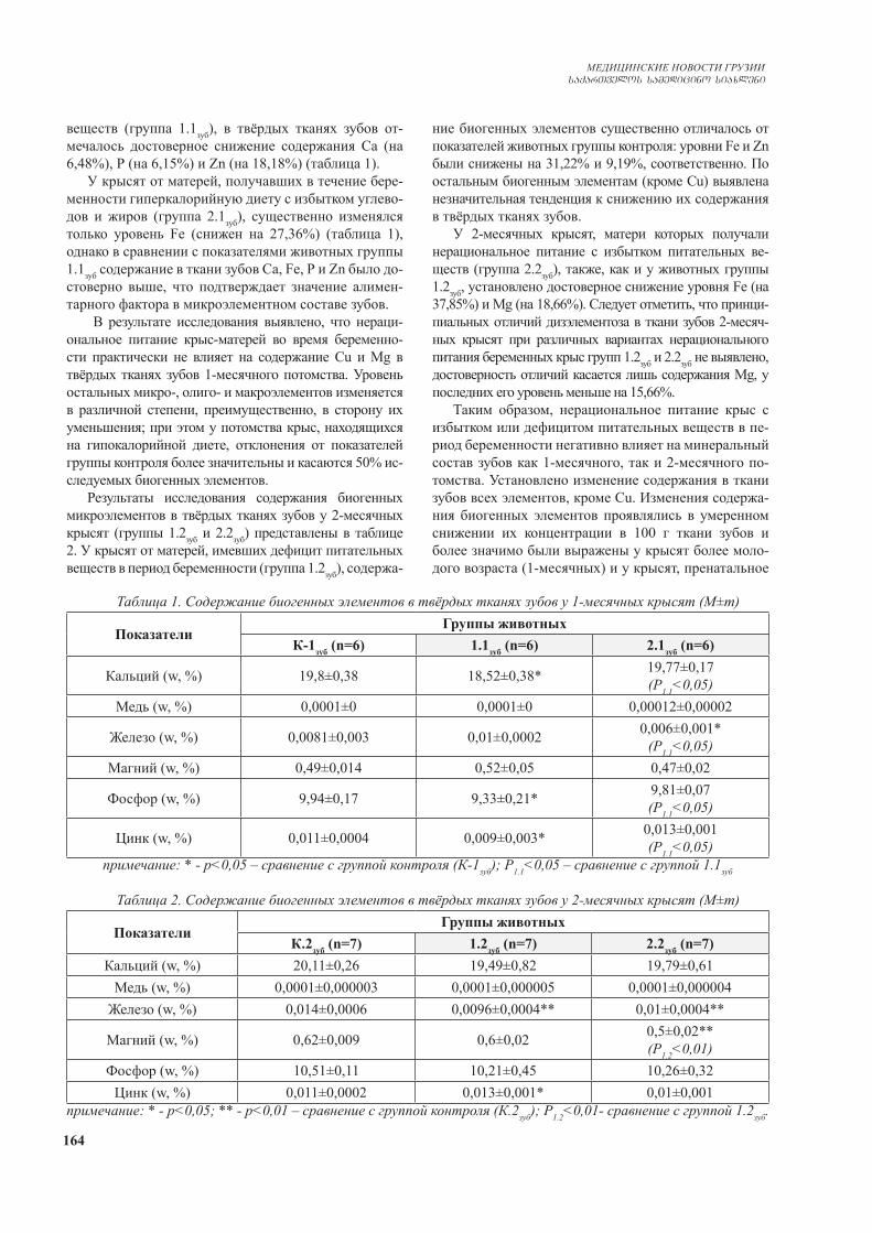
164
МЕДИЦИНСКИЕ НОВОСТИ ГРУЗИИ
CFMFHSDTKJC CFVTLBWBYJ CBF[KTYB
веществ (группа 1.1зуб), в твёрдых тканях зубов от-мечалось достоверное снижение содержания Са (на 6,48%), Р (на 6,15%) и Zn (на 18,18%) (таблица 1).
У крысят от матерей, получавших в течение бере-менности гиперкалорийную диету с избытком углево-дов и жиров (группа 2.1зуб), существенно изменялся только уровень Fe (снижен на 27,36%) (таблица 1), однако в сравнении с показателями животных группы 1.1зуб содержание в ткани зубов Са, Fe, Р и Zn было до-стоверно выше, что подтверждает значение алимен-тарного фактора в микроэлементном составе зубов.
В результате исследования выявлено, что нераци-ональное питание крыс-матерей во время беременно-сти практически не влияет на содержание Cu и Mg в твёрдых тканях зубов 1-месячного потомства. Уровень остальных микро-, олиго- и макроэлементов изменяется в различной степени, преимущественно, в сторону их уменьшения; при этом у потомства крыс, находящихся на гипокалорийной диете, отклонения от показателей группы контроля более значительны и касаются 50% ис-следуемых биогенных элементов.
Результаты исследования содержания биогенных микроэлементов в твёрдых тканях зубов у 2-месячных крысят (группы 1.2зуб и 2.2зуб) представлены в таблице 2. У крысят от матерей, имевших дефицит питательных веществ в период беременности (группа 1.2зуб), содержа-
ние биогенных элементов существенно отличалось от показателей животных группы контроля: уровни Fe и Zn были снижены на 31,22% и 9,19%, соответственно. По остальным биогенным элементам (кроме Cu) выявлена незначительная тенденция к снижению их содержания в твёрдых тканях зубов.
У 2-месячных крысят, матери которых получали нерациональное питание с избытком питательных ве-ществ (группа 2.2зуб), также, как и у животных группы 1.2зуб, установлено достоверное снижение уровня Fe (на 37,85%) и Mg (на 18,66%). Следует отметить, что принци-пиальных отличий дизэлементоза в ткани зубов 2-месяч-ных крысят при различных вариантах нерационального питания беременных крыс групп 1.2зуб и 2.2зуб не выявлено, достоверность отличий касается лишь содержания Mg, у последних его уровень меньше на 15,66%.
Таким образом, нерациональное питание крыс с избытком или дефицитом питательных веществ в пе-риод беременности негативно влияет на минеральный состав зубов как 1-месячного, так и 2-месячного по-томства. Установлено изменение содержания в ткани зубов всех элементов, кроме Cu. Изменения содержа-ния биогенных элементов проявлялись в умеренном снижении их концентрации в 100 г ткани зубов и более значимо были выражены у крысят более моло-дого возраста (1-месячных) и у крысят, пренатальное
Таблица 1. Содержание биогенных элементов в твёрдых тканях зубов у 1-месячных крысят (М±m)
ПоказателиГруппы животных
К-1зуб (n=6) 1.1зуб (n=6) 2.1зуб (n=6)
Кальций (w, %) 19,8±0,38 18,52±0,38* 19,77±0,17(Р1.1<0,05)
Медь (w, %) 0,0001±0 0,0001±0 0,00012±0,00002
Железо (w, %) 0,0081±0,003 0,01±0,0002 0,006±0,001*(Р1.1<0,05)
Магний (w, %) 0,49±0,014 0,52±0,05 0,47±0,02
Фосфор (w, %) 9,94±0,17 9,33±0,21* 9,81±0,07(Р1.1<0,05)
Цинк (w, %) 0,011±0,0004 0,009±0,003* 0,013±0,001(Р1.1<0,05)
примечание: * - р<0,05 – сравнение с группой контроля (К-1зуб); Р1.1<0,05 – сравнение с группой 1.1зуб
Таблица 2. Содержание биогенных элементов в твёрдых тканях зубов у 2-месячных крысят (М±m)
ПоказателиГруппы животных
К.2зуб (n=7) 1.2зуб (n=7) 2.2зуб (n=7)Кальций (w, %) 20,11±0,26 19,49±0,82 19,79±0,61
Медь (w, %) 0,0001±0,000003 0,0001±0,000005 0,0001±0,000004Железо (w, %) 0,014±0,0006 0,0096±0,0004** 0,01±0,0004**
Магний (w, %) 0,62±0,009 0,6±0,02 0,5±0,02**(Р1.2<0,01)
Фосфор (w, %) 10,51±0,11 10,21±0,45 10,26±0,32Цинк (w, %) 0,011±0,0002 0,013±0,001* 0,01±0,001
примечание: * - р<0,05; ** - р<0,01 – сравнение с группой контроля (К.2зуб); Р1.2<0,01- сравнение с группой 1.2зуб.
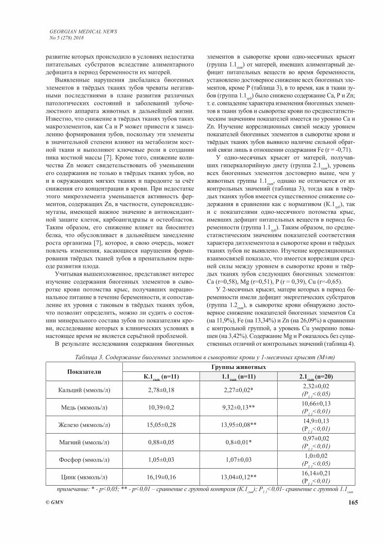
GEORGIAN MEDICAL NEWS No 5 (278) 2018
© GMN 165
развитие которых происходило в условиях недостатка питательных субстратов вследствие алиментарного дефицита в период беременности их матерей.
Выявленные нарушения дисбаланса биогенных элементов в твёрдых тканях зубов чреваты негатив-ными последствиями в плане развития различных патологических состояний и заболеваний зубоче-люстного аппарата животных в дальнейшей жизни. Известно, что снижение в твёрдых тканях зубов таких макроэлементов, как Са и Р может привести к замед-лению формирования зубов, поскольку эти элементы в значительной степени влияют на метаболизм кост-ной ткани и выполняют ключевые роли в создании пика костной массы [7]. Кроме того, снижение коли-чества Zn может свидетельствовать об уменьшении его содержания не только в твёрдых тканях зубов, но и в окружающих мягких тканях и пародонте за счёт снижения его концентрации в крови. При недостатке этого микроэлемента уменьшается активность фер-ментов, содержащих Zn, в частности, супероксиддис-мутазы, имеющей важное значение в антиоксидант-ной защите клеток, карбоангидразы и остеобластов. Таким образом, его снижение влияет на биосинтез белка, что обусловливает в дальнейшем замедление роста организма [7], которое, в свою очередь, может повлечь изменения, касающиеся нарушения форми-рования твёрдых тканей зубов в пренатальном пери-оде развития плода.
Учитывая вышеизложенное, представляет интерес изучение содержания биогенных элементов в сыво-ротке крови потомства крыс, получавших нерацио-нальное питание в течение беременности, и сопостав-ление их уровня с таковым в твёрдых тканях зубов, что позволит определить, можно ли судить о состоя-нии минерального состава зубов по показателям кро-ви, исследование которых в клинических условиях в настоящее время не является серьёзной проблемой.
В результате исследования содержания биогенных
элементов в сыворотке крови одно-месячных крысят (группа 1.1сыв) от матерей, имевших алиментарный де-фицит питательных веществ во время беременности, установлено достоверное снижение всех биогенных эле-ментов, кроме Р (таблица 3), в то время, как в ткани зу-бов (группа 1.1зуб) было снижено содержание Са, Р и Zn; т. е. совпадение характера изменения биогенных элемен-тов в ткани зубов и сыворотке крови по среднестатисти-ческим значениям показателей имеется по уровню Са и Zn. Изучение корреляционных связей между уровнем показателей биогенных элементов в сыворотке крови и твёрдых тканях зубов выявило наличие сильной обрат-ной связи лишь в отношении содержания Fe (r = -0,71).
У одно-месячных крысят от матерей, получав-ших гиперкалорийную диету (группа 2.1сыв), уровень всех биогенных элементов достоверно выше, чем у животных группы 1.1сыв, однако не отличается от их контрольных значений (таблица 3), тогда как в твёр-дых тканях зубов имеется существенное снижение со-держания в сравнении как с нормативом (К.1зуб), так и с показателями одно-месячного потомства крыс, имевших дефицит питательных веществ в период бе-ременности (группа 1.1зуб). Таким образом, по средне-статистическим значениям показателей соответствия характера дизэлементоза в сыворотке крови и твёрдых тканях зубов не выявлено. Изучение корреляционных взаимосвязей показало, что имеется корреляция сред-ней силы между уровнем в сыворотке крови и твёр-дых тканях зубов следующих биогенных элементов: Са (r=0,58), Mg (r=0,51), P (r = 0,39), Cu (r=-0,65).
У 2-месячных крысят, матери которых в период бе-ременности имели дефицит энергетических субстратов (группа 1.2сыв), в сыворотке крови обнаружено досто-верное снижение показателей биогенных элементов Са (на 11,9%), Fe (на 13,34%) и Zn (на 26,09%) в сравнении с контрольной группой, а уровень Cu умеренно повы-шен (на 3,42%). Содержание Mg и P оказалось без суще-ственных отличий от контрольных значений (таблица 4).
Таблица 3. Содержание биогенных элементов в сыворотке крови у 1-месячных крысят (М±m)
ПоказателиГруппы животных
К.1сыв (n=11) 1.1сыв (n=11) 2.1сыв (n=20)
Кальций (ммоль/л) 2,78±0,18 2,27±0,02* 2,32±0,02(Р1.1<0,05)
Медь (мкмоль/л) 10,39±0,2 9,32±0,13** 10,66±0,13(Р1.1<0,01)
Железо (мкмоль/л) 15,05±0,28 13,95±0,08** 14,9±0,13(Р1.1<0,01)
Магний (ммоль/л) 0,88±0,05 0,8±0,01* 0,97±0,02(Р1.1<0,01)
Фосфор (ммоль/л) 1,05±0,03 1,07±0,03 1,0±0,02(Р1.1<0,05)
Цинк (мкмоль/л) 16,19±0,16 13,04±0,12** 16,14±0,21(Р1.1<0,01)
примечание: * - р<0,05; ** - р<0,01 – сравнение с группой контроля (К.1сыв); Р1.1<0,01- сравнение с группой 1.1сыв
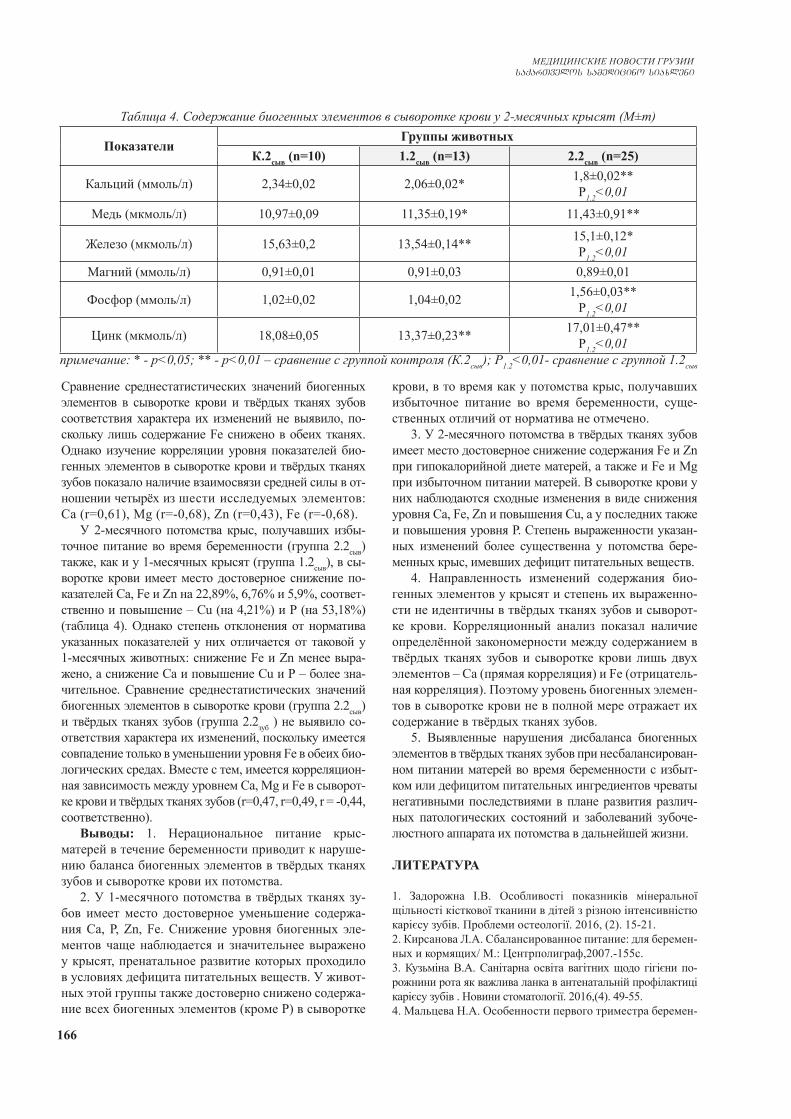
166
МЕДИЦИНСКИЕ НОВОСТИ ГРУЗИИ
CFMFHSDTKJC CFVTLBWBYJ CBF[KTYB
Сравнение среднестатистических значений биогенных элементов в сыворотке крови и твёрдых тканях зубов соответствия характера их изменений не выявило, по-скольку лишь содержание Fe снижено в обеих тканях. Однако изучение корреляции уровня показателей био-генных элементов в сыворотке крови и твёрдых тканях зубов показало наличие взаимосвязи средней силы в от-ношении четырёх из шести исследуемых элементов: Са (r=0,61), Mg (r=-0,68), Zn (r=0,43), Fe (r=-0,68).
У 2-месячного потомства крыс, получавших избы-точное питание во время беременности (группа 2.2сыв) также, как и у 1-месячных крысят (группа 1.2сыв), в сы-воротке крови имеет место достоверное снижение по-казателей Са, Fe и Zn на 22,89%, 6,76% и 5,9%, соответ-ственно и повышение – Cu (на 4,21%) и Р (на 53,18%) (таблица 4). Однако степень отклонения от норматива указанных показателей у них отличается от таковой у 1-месячных животных: снижение Fe и Zn менее выра-жено, а снижение Са и повышение Cu и Р – более зна-чительное. Сравнение среднестатистических значений биогенных элементов в сыворотке крови (группа 2.2сыв) и твёрдых тканях зубов (группа 2.2зуб ) не выявило со-ответствия характера их изменений, поскольку имеется совпадение только в уменьшении уровня Fe в обеих био-логических средах. Вместе с тем, имеется корреляцион-ная зависимость между уровнем Са, Mg и Fe в сыворот-ке крови и твёрдых тканях зубов (r=0,47, r=0,49, r = -0,44, соответственно).
Выводы: 1. Нерациональное питание крыс-матерей в течение беременности приводит к наруше-нию баланса биогенных элементов в твёрдых тканях зубов и сыворотке крови их потомства.
2. У 1-месячного потомства в твёрдых тканях зу-бов имеет место достоверное уменьшение содержа-ния Са, Р, Zn, Fe. Снижение уровня биогенных эле-ментов чаще наблюдается и значительнее выражено у крысят, пренатальное развитие которых проходило в условиях дефицита питательных веществ. У живот-ных этой группы также достоверно снижено содержа-ние всех биогенных элементов (кроме Р) в сыворотке
крови, в то время как у потомства крыс, получавших избыточное питание во время беременности, суще-ственных отличий от норматива не отмечено.
3. У 2-месячного потомства в твёрдых тканях зубов имеет место достоверное снижение содержания Fe и Zn при гипокалорийной диете матерей, а также и Fe и Mg при избыточном питании матерей. В сыворотке крови у них наблюдаются сходные изменения в виде снижения уровня Са, Fe, Zn и повышения Сu, а у последних также и повышения уровня Р. Степень выраженности указан-ных изменений более существенна у потомства бере-менных крыс, имевших дефицит питательных веществ.
4. Направленность изменений содержания био-генных элементов у крысят и степень их выраженно-сти не идентичны в твёрдых тканях зубов и сыворот-ке крови. Корреляционный анализ показал наличие определённой закономерности между содержанием в твёрдых тканях зубов и сыворотке крови лишь двух элементов – Са (прямая корреляция) и Fe (отрицатель-ная корреляция). Поэтому уровень биогенных элемен-тов в сыворотке крови не в полной мере отражает их содержание в твёрдых тканях зубов.
5. Выявленные нарушения дисбаланса биогенных элементов в твёрдых тканях зубов при несбалансирован-ном питании матерей во время беременности с избыт-ком или дефицитом питательных ингредиентов чреваты негативными последствиями в плане развития различ-ных патологических состояний и заболеваний зубоче-люстного аппарата их потомства в дальнейшей жизни.
ЛИТЕРАТУРА
1. Задорожна І.В. Особливості показників мінеральної щільності кісткової тканини в дітей з різною інтенсивністю карієсу зубів. Проблеми остеології. 2016, (2). 15-21.2. Кирсанова Л.А. Сбалансированное питание: для беремен-ных и кормящих/ М.: Центрполиграф,2007.-155с.3. Кузьміна В.А. Санітарна освіта вагітних щодо гігієни по-рожнини рота як важлива ланка в антенатальній профілактиці карієсу зубів . Новини стоматології. 2016,(4). 49-55.4. Мальцева Н.А. Особенности первого триместра беремен-
Таблица 4. Содержание биогенных элементов в сыворотке крови у 2-месячных крысят (М±m)
ПоказателиГруппы животных
К.2сыв (n=10) 1.2сыв (n=13) 2.2сыв (n=25)
Кальций (ммоль/л) 2,34±0,02 2,06±0,02* 1,8±0,02**Р1.2<0,01
Медь (мкмоль/л) 10,97±0,09 11,35±0,19* 11,43±0,91**
Железо (мкмоль/л) 15,63±0,2 13,54±0,14** 15,1±0,12*Р1.2<0,01
Магний (ммоль/л) 0,91±0,01 0,91±0,03 0,89±0,01
Фосфор (ммоль/л) 1,02±0,02 1,04±0,02 1,56±0,03**Р1.2<0,01
Цинк (мкмоль/л) 18,08±0,05 13,37±0,23** 17,01±0,47**Р1.2<0,01
примечание: * - р<0,05; ** - р<0,01 – сравнение с группой контроля (К.2сыв); Р1.2<0,01- сравнение с группой 1.2сыв
GEORGIAN MEDICAL NEWS No 5 (278) 2018
© GMN 167
ности у женщин с нарушением жирового обмена.М., 2008. .5. Пат.80979 Україна МПКG09B 23/28(2006.01) Спосіб мо-делювання надмірної ваги/Ніколаєва О.В., Ковальцова М.В., Євтушенко Т.Г.; заявник та патентовласник Харківський національний медичний університет.-№ и 2013 01221; за-явл. 01.02.2013; опубл. 10.06.2013, Бюл. №116. Пат.81453 Україна МПКG09B 23/28(2006.01) G 09B 23/34(2006.01) Спосіб моделювання аліментарної недостатності/Ніколаєва О.В., Ковальцова М.В., Євтушенко Т.Г.; заявник та патентовласник Харківський національний медичний університет.-№ и 2013 01910; заявл. 18.02.2013; опубл. 25.06.2013, Бюл. №12. 7. Поворознюк В.В., Мазур И.П. Костная система и заболева-ния пародонта. 3 изд. В: Современные принципы профилак-тики и лечения системного остеопороза. Киев:2005. 247-325.8. Хоменко Л.А., Чайковский Ю.Б., Савичук А.В. Терапев-тическая стоматология детского возраста. Л.А.Хоменко, ред. К.: Книга плюс, 2007.9. Якубова І.І. Впровадження схеми диспансерізації вагітних жінок у стоматолога-перший крок до збереження стоматологічного здоров’я матері і формування його у дити-ни. Новини стоматології.2012,(2). 56-59.10. Якубова І.І. Л. В. Тумановська, Л. Ф. Каськова Морфологічні зміни зачатків зубів у ембріонів мишей під впливом cholesterol. Світ медицини та біології. 2012.(1).182-185.11. Hunter L.P. Oral health and oral health care practices among low income pregnant women / L.P. Hunter, S.M. Yount // Journ. of Midwifery & Women’s Health.2011.Vol. 56. 103–109.
SUMMARY
THE INFLUENCE OF UNBALANCED NUTRI-TION OF PREGNANT RATS ON THE CONTENT OF BIOGENIC ELEMENTS IN THE ENAMEL AND BLOOD SERUM OF THEIR OFFSPRING
Nikolayeva O., Pysmenna O.
Kharkiv National Medical University, Ukraine
Malnutrition of pregnant women can slow down den-tal tissue development in offspring, cause growth of tooth decay and other disruptions, associated with changes amount of biogenic elements (BE), which are received into child in the womb body. The aim of this study-de-termination of the BE in the enamel and blood serum of mother rats, who had unbalanced nutrition with excess or lack of nutrients during the pregnancy. The content of Ca, Mg, Cu, Fe, P and Zn was studied in 129 species 1- and 2-month offspring. Reduction of BE in the enamel and blood serum mote often exist and more raised in pups, whose prenatal development took place under a shortage of nutrients. The direction of content changes of BE in pups and their degree of intensity are not identical in the enamel and blood serum. Certain pattern was identified between the content in the enamel and blood serum only two elements- Ca (direct correlation) and Fe (negative correlation). Irregularities of BE imbalance in the enamel during mothers’ unbalanced nutrition at the time of preg-nancy threaten by negative effects in terms of growth
various pathological conditions and diseases of dentition apparatus their offspring in later life.
Keywords: Malnutrition, pregnancy, biogenic ele-ments, enamel, blood serum, mother rats.
РЕЗЮМЕ
ВЛИЯНИЕ НЕСБАЛАНСИРОВАННОГО ПИ-ТАНИЯ БЕРЕМЕННЫХ КРЫС НА СОДЕРЖА-НИЕ БИОГЕННЫХ ЭЛЕМЕНТОВ В ТВЁРДЫХ ТКАНЯХ ЗУБОВ И СЫВОРОТКЕ КРОВИ У ИХ ПОТОМСТВА
Николаева О.В., Письменная О.Т.
Харьковский национальный медицинский университет, Украина
Цель исследования – определение содержания био-генных элементов в твёрдых тканях зубов и сыворотке крови у потомства крыс-матерей, которые во время бе-ременности получали несбалансированное питание с избытком или недостатком питательных веществ.
Исследовано содержание Ca, Mg, Cu, Fe, P и Zn у 129 особей 1- и 2-месячного потомства. Снижение уровня биогенных элементов (БЭ) в ткани зубов и сыворотке крови чаще отмечается и более выражено у крысят, пренатальное развитие которых проходит в условиях дефицита питательных веществ. Направлен-ность изменений содержания БЭ у крысят и степень их выраженности не идентичны в твёрдых тканях зу-бов и сыворотке крови. Установлена определённая за-кономерность между содержанием в твёрдых тканях зубов и сыворотке крови лишь двух элементов – Са (прямая корреляция) и Fe (отрицательная корреля-ция). Нарушения дисбаланса БЭ в твёрдых тканях зубов при несбалансированном питании матерей во время беременности чреваты негативными послед-ствиями в плане развития различных патологических состояний и заболеваний зубочелюстного аппарата их потомства в дальнейшей жизни.
reziume
make virTagvebis aradabalansebuli kvebis gavlena biogenuri elementebis Semcveloba-ze maTi nayaris kbilebis magar qsovilebsa da sisxlis SratSi
o. nikolaeva, o. pismennaia
xarkovis erovnuli samedicino universite-ti, ukraina
kvlevis mizans warmoadgenda biogenuri elementebis Semcvelobis gansazRvra makeo-
168
МЕДИЦИНСКИЕ НОВОСТИ ГРУЗИИ
CFMFHSDTKJC CFVTLBWBYJ CBF[KTYB
bis dros aradabalansebulad nakvebi vir-Tagvebis nayaris kbilebis magar qsovilebsa da sisxlis SratSi.
gamokvleulia Ca-, Mg-, Cu-, Fe-, P- da Zn-is Semcveloba 1 da 2 Tvis asakis nayarSi (n=129). kbilebis myar qsovilebsa da sisxlis SratSi biogenuri elementebis donis Semci-reba ufro xSirad da ufro mniSvnelovnad iyo gamoxatuli im axalSobil virTagvebSi, romelTa prenataluri ganviTareba mimdin-areobda sakvebi nivTierebebis deficitis pirobebSi.
amasTan, kbilebis magar qsovilebsa da
sisxlis SratSi biogenuri elementebis Semcvelobis cvlilebebis mimarTuleba da gamoxatvis xarisxi ar iyo identuri; kanon-zomiereba dadgenilia mxolod ori ele-mentis SemcvelobasTan dakavSirebiT – Са (pirdapiri korelacia) da Fe (uaryofiTi korelacia). makeobis periodSi aradabalan-sebuli kvebiT gamowveul biogenuri ele-mentebis disbalanss nayaris kbilebis magar qsovilebSi SesaZloa mohyves negatiuri Sedegebi, maT Soris - sxvadasxva paTolo-giuri mdgomareobis da daavadebis ganviTa-reba yba-kbilTa aparatSi.
ENERGY PROFILE OF RATS WITH OVERACTIVE BLADDER SYNDROME PHARMACOLOGICALLY CORRECTED WITH QUERCETIN
1Iatsyna O., 2Diachkova N., 3Kharkhota M., 1Kostev F.
1Odessa National Medical University; 2State Institute “Shalimov National Institute of Surgery and Transplantation” of the National Academy of Medical Sciences of Ukraine;
3D.K. Zabolotny Institute of Microbiology and Virology of the NAS of Ukraine, Kyiv, Ukraine
The overactive bladder syndrome (OAB) is a common pathological condition manifested by symptoms of frequent and urgent urination. OAB is not a life-threatening disease, but it significantly impairs the quality of life of patients. De-spite the widespread occurrence of OAB, the question of its etiology is still open [3]. There is a lot of information evi-dencing a versatile genesis of the disease [4]. Given the va-riety of OAB development mechanisms, medical specialists have to use a comprehensive approach to its diagnostics and treatment [4,5]. Today, they use various approaches to OAB therapy and a variety of medicines for its treatment [5].
We are aware of the prospects of using flavonoids for OAB therapy [9-11], including some methods protected by patents [14,15]. However, there is still no evidence of the mechanisms of their therapeutic effect.
An important element of cellular metabolism is the adenine system, which determines the integration of en-ergy production processes with multiple intracellular energy-consuming reactions. In this respect, erythrocytes may be considered convenient, easily accessible object for studying the adenylate metabolism, in particular, the intracellular regulation as the most important indicators of energy metabolism. Along with this, erythrocytes have close functional junction with somatic cells, and we can assess functioning cells of various organs and tissues by the intensity of intra-erythrocyte reactions determined by the state of the adenine system.
The purpose of our work was to study changing quali-tative and quantitative composition of adenyl nucleotides in rat erythrocytes while using Quercetin for correction of the overactive bladder syndrome in the course of the experiment.
Material and methods. The experiments were car-ried out on 160-180 g Wistar rat models of both sexes. The rats were kept in the standard conditions of vivarium belonging to experimental laboratory of the State Institute “Shalimov National Institute of Surgery and Transplan-tation” of the National Academy of Medical Sciences of Ukraine, Kiev.
All experiments on animal models were carried out according to the European Convention for the Protection of Vertebrate Animals used for Experimental and Other Scientific Purposes. The animals were randomly assigned to three groups. Intact animals of the first group (n=20), were prescribed an equivalent volume of physiological saline instead of the pharmaceuticals. Animals of the sec-ond group (n=45) with experimentally created OAB did not receive study medicines for correction of the pathol-ogy, therefore they comprised a control group, and the third group of animals (n=45) with OAB were adminis-tered Quercetin for pharmacological correction.
To create the experimental OAB models, we used a technique proposed by V.M. Derzhavin et al. [1]. The pathological condition was corrected by daily oral admin-istration of Quercetin to the experimental animal models. The flavonoid was administered to animals at a dose of 10 mg OD. Blood (1.5 ml) for quantification of adenyl nucleotides was sampled from the tail vein of the animals.
The quantification of ATP, ADP, and AMP was carried out by HPLC method using Agilent 1200 chromatograph (Agilent Technologies, USA) equipped with a DOD de-tector located at the Common Use Center at the Zabolotny Institute of Microbiology and Virology of the National Academy of Sciences of Ukraine.
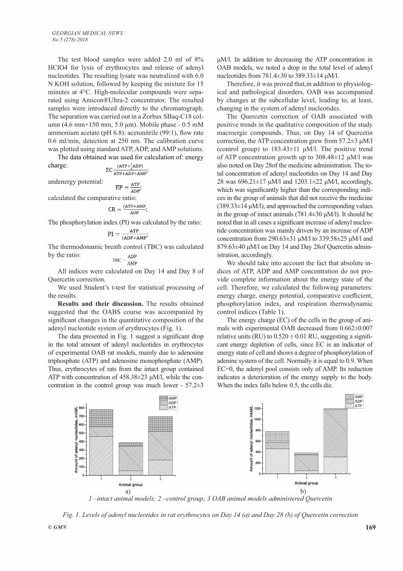
GEORGIAN MEDICAL NEWS No 5 (278) 2018
© GMN 169
The test blood samples were added 2.0 ml of 8% HClO4 for lysis of erythrocytes and release of adenyl nucleotides. The resulting lysate was neutralized with 6.0 N KOH solution, followed by keeping the mixture for 15 minutes at 4°C. High-molecular compounds were sepa-rated using Amicon®Ultra-2 concentrator. The resulted samples were introduced directly to the chromatograph. The separation was carried out in a Zorbax SBaq-C18 col-umn (4.6 mm×150 mm, 5.0 μm). Mobile phase - 0.5 mM ammonium acetate (pH 6.8): acetonitrile (99:1), flow rate 0.6 ml/min, detection at 250 nm. The calibration curve was plotted using standard ATP, ADP, and AMP solutions.
The data obtained was used for calculation of: energy charge:
andenergy potential:
calculated the comparative ratio:
The phosphorylation index (PI) was calculated by the ratio:
The thermodonamic breath control (TBC) was calculated by the ratio:
All indices were calculated on Day 14 and Day 8 of Quercetin correction.
We used Student’s t-test for statistical processing of the results.
Results and their discussion. The results obtained suggested that the OABS course was accompanied by significant changes in the quantitative composition of the adenyl nucleotide system of erythrocytes (Fig. 1).
The data presented in Fig. 1 suggest a significant drop in the total amount of adenyl nucleotides in erythrocytes of experimental OAB rat models, mainly due to adenosine triphosphate (ATP) and adenosine monophosphate (AMP). Thus, erythrocytes of rats from the intact group contained ATP with concentration of 458.38±23 μM/l, while the con-centration in the control group was much lower - 57.2±3
μM/l. In addition to decreasing the ATP concentration in OAB models, we noted a drop in the total level of adenyl nucleotides from 781.4±30 to 389.33±14 μM/l.
Therefore, it was proved that,in addition to physiolog-ical and pathological disorders, OAB was accompanied by changes at the subcellular level, leading to, at least, changing in the system of adenyl nucleotides.
The Quercetin correction of OAB associated with positive trends in the qualitative composition of the study macroergic compounds. Thus, on Day 14 of Quercetin correction, the ATP concentration grew from 57.2±3 μM/l (control group) to 183.43±11 μM/l. The positive trend of ATP concentration growth up to 308.48±12 μM/l was also noted on Day 28of the medicine administration. The to-tal concentration of adenyl nucleotides on Day 14 and Day 28 was 696.21±17 μM/l and 1203.1±22 μM/l, accordingly, which was significantly higher than the corresponding indi-ces in the group of animals that did not receive the medicine (389.33±14 μM/l), and approached the corresponding values in the group of intact animals (781.4±30 μM/l). It should be noted that in all cases a significant increase of adenyl nucleo-tide concentration was mainly driven by an increase of ADP concentration from 290.63±31 μM/l to 339.58±25 μM/l and 879.63±40 μM/l on Day 14 and Day 28of Quercetin admin-istration, accordingly.
We should take into account the fact that absolute in-dices of ATP, ADP and AMP concentration do not pro-vide complete information about the energy state of the cell. Therefore, we calculated the following parameters: energy charge, energy potential, comparative coefficient, phosphorylation index, and respiration thermodynamic control indices (Table 1).
The energy charge (EC) of the cells in the group of ani-mals with experimental OAB decreased from 0.662±0.007 relative units (RU) to 0.520 ± 0.01 RU, suggesting a signifi-cant energy depletion of cells, since EC is an indicator of energy state of cell and shows a degree of phosphorylation of adenine system of the cell. Normally it is equal to 0.9. When EC=0, the adenyl pool consists only of AMP. Its reduction indicates a deterioration of the energy supply to the body. When the index falls below 0.5, the cells die.
1 2 30
100
200
300
400
500
600
700
800
Am
ount
of a
deny
l nuc
leot
ides
, mkM
/L
Animal group
AMP ADP ATP
1 2 30
200
400
600
800
1000
1200
Am
ount
of a
deny
l nuc
leot
ides
, mkM
/L
Animal group
AMP ADP ATP
1 –intact animal models; 2 –control group; 3 OAB animal models administered Quercetin
Fig. 1. Levels of adenyl nucleotides in rat erythrocytes on Day 14 (a) and Day 28 (b) of Quercetin correction
а) b)
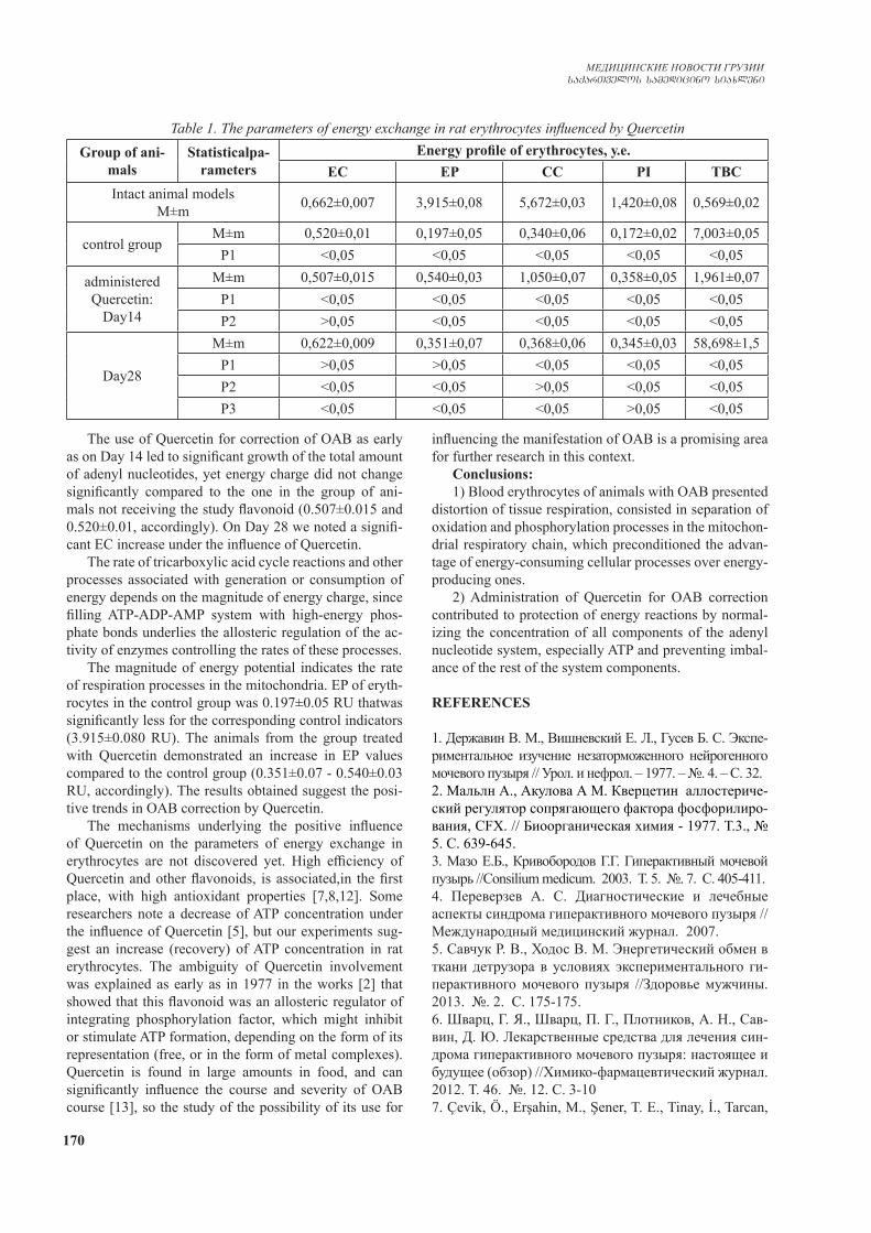
170
МЕДИЦИНСКИЕ НОВОСТИ ГРУЗИИ
CFMFHSDTKJC CFVTLBWBYJ CBF[KTYB
The use of Quercetin for correction of OAB as early as on Day 14 led to significant growth of the total amount of adenyl nucleotides, yet energy charge did not change significantly compared to the one in the group of ani-mals not receiving the study flavonoid (0.507±0.015 and 0.520±0.01, accordingly). On Day 28 we noted a signifi-cant EC increase under the influence of Quercetin.
The rate of tricarboxylic acid cycle reactions and other processes associated with generation or consumption of energy depends on the magnitude of energy charge, since filling ATP-ADP-AMP system with high-energy phos-phate bonds underlies the allosteric regulation of the ac-tivity of enzymes controlling the rates of these processes.
The magnitude of energy potential indicates the rate of respiration processes in the mitochondria. EP of eryth-rocytes in the control group was 0.197±0.05 RU thatwas significantly less for the corresponding control indicators (3.915±0.080 RU). The animals from the group treated with Quercetin demonstrated an increase in EP values compared to the control group (0.351±0.07 - 0.540±0.03 RU, accordingly). The results obtained suggest the posi-tive trends in OAB correction by Quercetin.
The mechanisms underlying the positive influence of Quercetin on the parameters of energy exchange in erythrocytes are not discovered yet. High efficiency of Quercetin and other flavonoids, is associated,in the first place, with high antioxidant properties [7,8,12]. Some researchers note a decrease of ATP concentration under the influence of Quercetin [5], but our experiments sug-gest an increase (recovery) of ATP concentration in rat erythrocytes. The ambiguity of Quercetin involvement was explained as early as in 1977 in the works [2] that showed that this flavonoid was an allosteric regulator of integrating phosphorylation factor, which might inhibit or stimulate ATP formation, depending on the form of its representation (free, or in the form of metal complexes). Quercetin is found in large amounts in food, and can significantly influence the course and severity of OAB course [13], so the study of the possibility of its use for
influencing the manifestation of OAB is a promising area for further research in this context.
Conclusions:1) Blood erythrocytes of animals with OAB presented
distortion of tissue respiration, consisted in separation of oxidation and phosphorylation processes in the mitochon-drial respiratory chain, which preconditioned the advan-tage of energy-consuming cellular processes over energy-producing ones.
2) Administration of Quercetin for OAB correction contributed to protection of energy reactions by normal-izing the concentration of all components of the adenyl nucleotide system, especially ATP and preventing imbal-ance of the rest of the system components.
REFERENCES
1. Державин В. М., Вишневский Е. Л., Гусев Б. С. Экспе-риментальное изучение незаторможенного нейрогенного мочевого пузыря // Урол. и нефрол. – 1977. – №. 4. – С. 32.2. Мальлн А., Акулова А М. Кверцетин аллостериче-ский регулятор сопрягающего фактора фосфорилиро-вания, CFX. // Биоорганическая химия - 1977. Т.3., № 5. С. 639-645.3. Мазо Е.Б., Кривобородов Г.Г. Гиперактивный мочевой пузырь //Consilium medicum. 2003. Т. 5. №. 7. С. 405-411.4. Переверзев А. С. Диагностические и лечебные аспекты синдрома гиперактивного мочевого пузыря //Международный медицинский журнал. 2007.5. Савчук Р. В., Ходос В. М. Энергетический обмен в ткани детрузора в условиях экспериментального ги-перактивного мочевого пузыря //Здоровье мужчины. 2013. №. 2. С. 175-175.6. Шварц, Г. Я., Шварц, П. Г., Плотников, А. Н., Сав-вин, Д. Ю. Лекарственные средства для лечения син-дрома гиперактивного мочевого пузыря: настоящее и будущее (обзор) //Химико-фармацевтический журнал. 2012. Т. 46. №. 12. С. 3-107. Çevik, Ö., Erşahin, M., Şener, T. E., Tinay, İ., Tarcan,
Table 1. The parameters of energy exchange in rat erythrocytes influenced by QuercetinGroup of ani-
malsStatisticalpa-
rametersEnergy profile of erythrocytes, у.е.
EC EP CC PI TBCIntact animal models
М±m 0,662±0,007 3,915±0,08 5,672±0,03 1,420±0,08 0,569±0,02
control group М±m 0,520±0,01 0,197±0,05 0,340±0,06 0,172±0,02 7,003±0,05
Р1 ˂0,05 ˂0,05 ˂0,05 ˂0,05 ˂0,05
administered Quercetin:
Day14
М±m 0,507±0,015 0,540±0,03 1,050±0,07 0,358±0,05 1,961±0,07Р1 ˂0,05 ˂0,05 ˂0,05 ˂0,05 ˂0,05Р2 ˃0,05 ˂0,05 ˂0,05 ˂0,05 ˂0,05
Day28
М±m 0,622±0,009 0,351±0,07 0,368±0,06 0,345±0,03 58,698±1,5Р1 ˃0,05 ˃0,05 ˂0,05 ˂0,05 ˂0,05Р2 ˂0,05 ˂0,05 ˃0,05 ˂0,05 ˂0,05Р3 ˂0,05 ˂0,05 ˂0,05 ˃0,05 ˂0,05
GEORGIAN MEDICAL NEWS No 5 (278) 2018
© GMN 171
T., Çetinel, Ş., Şener, G. Beneficial effects of quercetin on rat urinary bladder after spinal cord injury //journal of surgical research. 2013. Т. 183. №. 2. С. 695-703.8. Dambros, M., Jongh, R., Koeveringe, G. A., Bast, A., Hei-jnen, C. G. M., & Kerrebroeck, P. E. Flavonoid galangin pre-vents smooth muscle fatigue of pig urinary bladder //Journal of pharmacy and pharmacology. 2005. Т. 57. №. 5. С. 617-622.9. Fürer, K., Eberli, D., Betschart, C., Brenneisen, R., De Mieri, M., Hamburger, M., .von Mandach, U. Inhibition of porcine detrusor contractility by the flavonoid fraction of Bryophyllum pinnatuma potential phytotherapeutic drug for the treatment of the overactive bladder syndrome //Phytomedicine. 2015. Т. 22. №. 1. С. 158-164.10. Huang Y. B. et al. Anti‐oxidant activity and attenuation of bladder hyperactivity by the flavonoid compound kaempferol //International Journal of Urology. 2014. Т. 21. №. 1. С. 94-98.11. Juan, Y. S., Chuang, S. M., Lee, Y. L., Long, C. Y., Wu, T. H., Chang, W. C., Huang, C. H. Green tea cat-echins decrease oxidative stress in surgical menopause‐induced overactive bladder in a rat model //BJU interna-tional. 2012. Т. 110. №. 6b.12. Masuda H. Editorial Comment to Anti-oxidant activ-ity and attenuation of bladder hyperactivity by the fla-vonoid compound kaempferol //International Journal of Urology. 2014. Т. 21. №. 1. С. 98-99.13. Maltz H. C., Staskin D. R., Zoltan E. Medication and food issues for overactive bladder/lower urinary tract symptoms: When do we eat? //Current Bladder Dysfunc-tion Reports. 2009. Т. 4. №. 2. С. 93-9714. Seipel, Tracey Anne. “Herbal compositions for the prevention or treatment of urinary incontinence and over-active bladder.” U.S. Patent No. 7,378,115. 27 May 2008.15. Rodriguez, Al. “Quercetin-containing composition, methods of making, and methods of using.” U.S. Patent Application No. 11/497,318.
SUMMARY
ENERGY PROFILE OF RATS WITH OVERAC-TIVE BLADDER SYNDROME PHARMACOLOGI-CALLY CORRECTED WITH QUERCETIN
1Iatsyna O., 2Diachkova N., 3Kharkhota M., 1Kostev F.
1Odessa National Medical University; 2State Institute “Shalimov National Institute of Surgery and Transplan-tation” of the National Academy of Medical Sciences of Ukraine; 3D.K. Zabolotny Institute of Microbiology and Virology of the NAS of Ukraine, Kyiv, Ukraine
We studied the effect of Quercetin on energy profile of erythrocytes of rats with the overactive bladder syndrome. It was shown that animal models with the subject patholo-gy demonstrated alteration of quantitative composition of the adenine nucleotide system in erythrocytes. Abrupt de-crease of the total concentration of adenyl nucleotides and ATP compared to the intact group of animals was noted in
the study. Administration of Quercetin significantly im-proved the energy profile of erythrocytes in rats with the overactive bladder syndrome. The data obtained provide new findings of the pathogenesis and correction regimens for the subject pathology.
Keywords: energy profile, overactive bladder, Quer-cetin, correction.
РЕЗЮМЕ
ЭНЕРГЕТИЧЕСКИЙ ПРОФИЛЬ У КРЫС ПРИ СИНДРОМЕ ГИПЕРАКТИВНОГО МОЧЕ-ВОГО ПУЗЫРЯ И ФАРМАКОЛОГИЧЕСКОЙ КОРРЕКЦИИ КВЕРЦЕТИНОМ
1Яцына А.И., 2Дьячкова Н.В., 3Хархота М.А., Костев Ф.И.
1Одесский национальной медицинский университет МОЗ Украины; 2Государственное учреждеие "Нацио-нальный институт хирургии и транспланталогии им. Шалимова НАМН Украины"; 3Институт микробио-логии и вирусологии им. Д.К. Заболотного Националь-ной академии наук Украины, Киев, Украина
Исследовано влияние кверцетина на энергетиче-ский профиль эритроцитов крыс с синдромом гиперак-тивного мочевого пузыря. Показано, что у животных с исследуемой патологией изменяется количественный состав системы адениловых нуклеотидов в эритроци-тах крови. Отмечается резкое уменьшение суммар-ной концентрации адениловых нуклеотидов, в част-ности аденозинтрифосфата в сравнении с интактной группой животных. Прием кверцетина значительно улучшает показатели энергетического профиля эри-троцитов крыс с синдромом гиперактивного мочево-го пузыря. Полученные данные расширяют знания о патогенезе и возможностях коррекции исследуемого патологического состояния.
reziume
virTagvebis energetikuli profili hiper-aqtiuri Sardis buStis sindromis dros da misi farmakologiuri koreqcia kvercetiniT
1a. iacina, 2n. diaCenko, 3m. xarxota, 1f. kostevi
1odesis erovnuli samedicino universiteti; 2ukrainis medicinis mecnierebeTa samecnie-ro akademiis Salimovis sax. qirurgiis da transplantalogiis instituti; 3d. zabo-lotnis sax. mikrobiologiis da virusolo-giis instituti, kievi, ukraina
gamokvleulia kvercetinis gavlena eri-Trocitebis energetikul profilze vir-
172
МЕДИЦИНСКИЕ НОВОСТИ ГРУЗИИ
CFMFHSDTKJC CFVTLBWBYJ CBF[KTYB
TagvebSi hiperaqtiuri Sardis buStis sindromiT. naCvenebia, rom aRniSnuli pa-TologiiT cxovelebis eriTrocitebSi adeniluri nukleotidebis Semadgenloba ganicdis raodenobriv cvlilebas: xdeba adeniluri nukleotidebis jamuri kon-centraciis, kerZod, adenozintrifosfa-tis mkveTri Semcireba intaqturi jgufis
cxovelebTan SedarebiT. kvercetinis miReba mniSvnelovnad aumjobesebs hiperaqtiuri Sardis buStis sindromiT virTagvebis ener-getikuli profilis maCveneblebs. Catarebu-li kvlevis Sedegebi gaafarTovebs codnas aRniSnuli paTologiis Sesaxeb da uzrun-velyofs am daavadebis drouli da adekva-turi mkurnalobis taqtikis SemuSavebas.
ОЦЕНКА НЕЙРОПРОТЕКТОРНОЙ АКТИВНОСТИ КОМПЛЕКСНОГО ВОЗДЕЙСТВИЯ МАГНИЯ СУЛЬФАТА, ЛАМОТРИДЖИНА И АЦЕТИЛЦИСТЕИНА
В УСЛОВИЯХ КОМБИНИРОВАННОЙ НОРМОБАРИЧЕСКОЙ ГИПОКСИИ С ПЕРЕВЯЗКОЙ ПРАВОЙ СОННОЙ АРТЕРИИ У КРЫС
1,2Самсония М.Д., 1,2Канделаки М.А., 1Бараташвили Н.А.
1Кутаисский государственный университет им. А. Церетели, медицинский факультет, департамент фармации; 2Фармацевтическая компания – Legion “Provisus”, Кутаиси, Грузия
Актуальной задачей экспериментальной и кли-нической неврологии является разработка эффек-тивных методов фармакологической коррекции гипоксических повреждений мозга при остром ише-мическом инсульте [4,13,18]. Известно, что в усло-виях острого нарушения мозгового крово-обраще-ния резко ухудшается доставка кислорода, глюкозы и других питательных веществ. Следовательно, на фоне кислородо-глюкозной депривации активиру-ется анаэробный гликолиз и падает скорость обра-зования аденозинтрифосфата (АТФ), что приводит к нарушению работы ионных насосов [6,9,11,18]. Изменение концентрационных градиентов электро-литов стимулирует высвобождение и накопление глутамата, который начинает взаимодействовать с рецепторами N-метил-D-аспартата (NMDA) и α-амино-3-гидрокси-5-метил-4-изоксанола (AMPA) [15,18]. Гиперактивированные глутаматные рецеп-торы длительно удерживают в открытом состоянии управляемые ими ионные каналы, существенно увеличивая внутриклеточную концентрацию ионов Са2+ и Na+. В конечном итоге, нарушается ионная асимметрия, развивается митохондриальная недо-статочность и энергодефицит, усугубляется ацидоз, происходит образование супероксидных радика-лов, включается «липидная триада» повреждения клеток [9,11,18]. Продукты перекисного окисления липидов (ПОЛ) угнетают активность мембранных ферментов, блокируя их сульфгидрильные группы, и повышают проницаемость клеточной мембраны. Соответственно, изменение осмотического гради-
ента, индуцированное дисбалансом ионов, приво-дит к притоку воды, которая вызывает цитотоксиче-ский и вазогенный отек мозга [2,5,18].
Следует отметить, что кислородо-глюкозная де-привация почти всегда сопровождается энергодефици-том и быстрым образованием избытка свободных ради-калов, на фоне истощения ресурсов антиоксидантных систем нейронов [2,5]. Поэтому, решающее значение в процессе сохранения жизнеспособности клеток голов-ного мозга при остром ишемическом инсульте, по всей вероятности, имеет только одновременное комплексное воздействие медикаментами на ключевые звенья в меха-низмах повреждения нейронов.
В связи с вышеизложенным, целью исследования явилась оценка нейропротекторной активности ком-плексного воздействия магния сульфата, ламотрид-жина и ацетилцистеина в условиях комбинированной нормобарической гипоксии с перевязкой правой сон-ной артерии у крыс.
Материал и методы. Опыты проведены на 70 бе-лых беспородных крысах-самцах с исходной массой тела 170-200 г. Их содержали в условиях вивария на стандартном рационе питания. Исследования вы-полнялись в соответствии с положениями Европей-кой конвенции по защите позвоночных животных, используемых для экспериментальных и других на-учных целей (European Convention for the Protection of Vertebrate Animals Used for Experimental and other Scientific Purposes (ETS 123), Strasburg, 1986) [1]. Ла-бораторные животные были разделены на 5 групп (та-блица 1).
GEORGIAN MEDICAL NEWS No 5 (278) 2018
© GMN 173
I группа служила контролем. Локальную ишемию го-группа служила контролем. Локальную ишемию го-ловного мозга (ЛИГМ) моделировали путем перевязки правой сонной артерии. Подопытных крыс фиксировали на станке и под эфирным наркозом лигировали правую сонную артерию. Рану обрабатывали антисептиком и за-шивали. Спустя 5 мин. после наложения лигатуры, кры-се внутрибрюшинно вводили физиологический раствор натрия хлорида (0,8 мл), а 5 мин спустя лабораторное животное помещали в прозрачную емкость (объем 800 мл). Емкость герметично закрывали и в условиях нормобарической гипоксии (при комнатной температу-ре) фиксировали время до остановки дыхания (апноэ). После остановки сердца животных декапитировали, из-влекали головной мозг и фиксировали в растворе фор-мальдегида. Парафиновые срезы мозга окрашивали по Нисслю. Результаты гистологического исследования оценивали посредством световой микроскопии.
В отличие от контрольной группы, животным II группы, вместо физиологического раствора, внутри-брюшинно вводили стандартный 25% раствор магния сульфата в виде инъекции (в дозе 1000 мг/кг). В III группе изучали нейропротекторную активность ламо-триджина, который вводили перорально через зонд в дозе 20 мг/кг за 2 ч до перевязки правой сонной ар-терии. Лабораторные животные из IV группы получали ацетилцистеин (per os) в дозе 200 мг/кг аналогичным образом (за 2 ч до перевязки правой сонной артерии). В V группе (таблица 1) оценивали антигипоксическую и нейропротекторную активность комбинации магния сульфата (1000 мг/кг, внутрибрюшинно), ламотриджи-на (20 мг/кг, per os) и ацетилцистеина (200 мг/кг, per os). Индекс защиты (ИЗ) на модели нормобарической ги-поксии [14] определяли по формуле:
ИЗ(гипоксия) = To/Тk , где To – время до развития апноэ на фоне применения препаратов, Тk – время до развития апноэ в контроль-ной группе.
При статистической обработке результатов веро-ятность различий считали достоверной при р<0,05. Средние данные оценивали с помощью t-критерия Стьюдента [1].
Результаты и их обсуждение. При моделирова-нии ЛИГМ летальных исходов не наблюдали, так как кровоснабжение мозга у крыс имеет свою специ-фику. Даже полное прекращение кровотока (после перевязки обеих общих сонных артерий) не вызы-вает гибель лабораторных животных ввиду того, что средний мозг хорошо кровоснабжается через коллатеральные сосуды из позвоночных артерий и крысы живут минимум 2 недели после оперативно-го вмешательства [1].
Морфологическое исследование нервной тка-ни крыс (I группа), которые после перевязки правой сонной артерии подверглись воздействию нормоба-рической гипоксии, выявило картину острого ци-тотоксического отека головного мозга без четкого формирования зон некроза. В головном мозге всех животных наблюдали полнокровие сосудов, много-численные мелкоточечные кровоизлияния в вещество мозга и дегенеративные изменения в ганглиозных клетках, что совпадает с данными литературы [12,16]. В коре, стволовой части мозга и мозжечке обнаруже-ны симметрично расположенные очаги размягчения, которые содержали погибшие нейроны.
Следует подчеркнуть, что гомеостаз глюкозы, электролитов и кислорода в крови и клетке является одним из определяющих факторов нормальной жиз-недеятельности нервной системы. Не случайно ней-роны более 1/3 своих энергетических ресурсов тратят на поддержание градиента глюкозы и на сохранение ионной асимметрии. Однако, собственные запасы данного углевода в ткани мозга чрезвычайно малы, в сравнении с высокой интенсивностью его потребле-ния, и поэтому даже кратковременная остановка (бо-
Таблица 1. Схема эксперимента
Группы
Количество животных в
группе
Перевязка правой сонной артерий
Нормобарическая гипоксия
Препарат(доза)
I (контроль) 14 + + Физиологический раствор (0,8 мл, внутрибрюшинно)
II 14 + +25% раствор магния сульфата
для инъекции (1000 мг/кг, внутрибрюшинно)
III 14 + + Ламотриджин* (20 мг/кг, per os)
IV 14 + + Ацетилцистеин*(200 мг/кг, per os)
V 14 + +
Комбинация (магний сульфат + ламотриджин* + ацетилцистеин*)
примечание: * - препарат вводили перорально через зонд (за 2 ч до перевязки правой сонной артерии)
174
МЕДИЦИНСКИЕ НОВОСТИ ГРУЗИИ
CFMFHSDTKJC CFVTLBWBYJ CBF[KTYB
лее 5 мин) церебрального кровотока, вызывает необ-ратимые изменения в нейронах [16,18].
ЛИГМ в условиях нормобарической гипоксии всегда индуцирует рефлекторную гипервентиляцию, которая имеет ряд отрицательных последствий: про-исходит торможение дыхательного центра; развивает-ся дыхательный алкалоз; наступает спазм мозговых и венечных сосудов, кривая диссоциации оксигемогло-бина смещается влево [14,18]. Дефицит кислорода и глюкозы снижает скорость гликолиза. Соответствен-но, уменьшается поступление пирувата в цикл три-карбоновых кислот и подавляется процесс генерации АТФ с развитием асептического воспаления и цито-токсического отека мозга [5,9,11,18].
Данные, представленные в таблице 2, свидетель-ствуют, что индекс защиты самый высокий у сульфа-та магния (после введения лекарственных средств в монорежиме). Этот препарат, являясь физиологиче-ским антагонистом кальция, по данным литературы, ограничивает кальциевую перегрузку клеток в очаге ишемии, уменьшает вазомоторную дисфункцию, по-вышая устойчивость головного мозга к тяжелым ише-мическим повреждениям [3,14,17].
Следует подчеркнуть, что ионы магния, высту-пая в роли блокаторов потенциал-зависимых каль-циевых каналов, существенно подавляют экзоцитоз возбуждающих нейромедиаторов в коре головного мозга и вызывают, по мере повышения концентра-ции в плазме, вначале седативно-снотворный, а за-тем противосудорожный и, наконец - общеанестези-рующий эффект [14,16,17]. Высокая эффективность магния сульфата на модели ЛИГМ обусловлена как мембранотропным и вазотропным, так и синапто-тропным действием этого препарата, В данном ис-следовании магния сульфат достоверно увеличивал индекс защиты (р<0,05).
В III группе при пероральном введении ламо-триджина в дозе 20 мг/кг ИЗ был в 1,33 раза выше, в сравнении с контрольной группой (р<0,05). Как известно, глутамат при гипоксии мозга может на-капливаться снаружи клетки, стимулируя открытие вольтаж-зависимых кальциевых каналов, в результате чего в клетку поступает большое количество ионов кальция через каналы NMDA-рецепторов, что, в свою очередь, вызывает повреждение и даже гибель клет-ки (эксайтотоксичность). Ламотриджин стабилизиру-ет нейрональные мембраны посредством влияния на пресинаптические потенциалзависимые Na+-каналы, подавляет патологическое высвобождение глутами-новой кислоты и ингибирует деполяризацию, вы-званную глутаматом [7,8,12]. Следует подчеркнуть, что при применении в высоких дозах этот препарат не влияет на спонтанный или вызванный калием эк-зоцитоз возбуждающих нейромедиаторов, и поэтому режим дозирования ламотриджина имеет большое значение [15,18].
Профилактическое применение ацетилцистеи-
на в дозе 200 мг/кг (IV группа), как нейропротекто-ра (цитопротектора) и антиоксиданта, благоприятно влияет на ИЗ, однако различия (таблица 2) оказались недостоверными, в сравнении с III группой (р>0,05). В многочисленных экспериментальных работах по-казано, что при кальциевой перегрузке, вызываемой ишемией и гипогликемией, интенсивно образуются активные формы кислорода и высокотоксичные про-дукты перекисного окисления липидов, с которыми не может справиться антиоксидантная система клетки [2,5,18].
С одной стороны, ацетилцистеин, благодаря на-личию свободной сульфгидрильной группы, напря-мую связывает (нейтрализует) свободные радикалы, а с другой стороны – поставляя цистеин для синтеза глутатиона, содержание которого в клетках во многом определяет устойчивоcть нейронов к окислительным воздействиям, препарат повышает активность мито-хондриальных антиоксидантов [2,5,10,18].
В обзорных статьях [2,5,15], в которых изучалась нейропротекторная активность ацетилцистеина, при-водятся сведения, касающиеся в основном коэффи-циента GSH/GSSG (соотношение восстановленного и окисленного глутатиона). Считается, что именно увеличение восстановленного глутатиона (GSH) обе-спечивает антиоксидантную защиту клеток и тканей [2,5]. Следует подчеркнуть, что обе формы глутати-она - окисленная и восстановленная, являются также лигандом для NMDA- и AMPA-рецепторов и могут оказать модулирующее влияние на глутаматную ней-ротрансмиссию [10,18].
В V группе применение комбинации магния суль-фата, ламотриджина и ацетилцистеина сопровожда-лось усилением антиоксидантной и нейропротек-торной активности лекарств (таблица 2). ИЗ в этой группе составил 2,18 и различия были статистически достоверными в сравнении с контрольной группой (р<0,05). Нейропротекторное влияние ионов магния и ламотриджина на клетки мозга, по всей вероятности, обусловлено в основном способностью к поддержа-нию кальциевого гомеостаза. Хотя нейроны поддер-живают градиент концентрации целого ряда ионов, нарушение именно кальциевого градиента представ-ляется наиболее значимым фактором, вызывающим повреждение клетки [14,16]. Усиление ацетилцистеи-ном антиоксидантных систем нейронов, ингибирова-ние ламотриджином эксайтотоксичности глутамата и коррекция ионного стресса ионами магния резко уве-личивали время до развития апноэ (таблица 2). Оче-видно, высокая эффективность вышеупомянутой ком-бинации связана с влиянием препаратов на ключевые звенья в механизмах повреждения нейронов.
Таким образом, на основании проведенного ис-следования и анализа литературных источников сле-дует заключить, что нейропротекторная активность магния сульфата обусловлена мембранотропным, вазотропным и синаптотропным действиями этого
GEORGIAN MEDICAL NEWS No 5 (278) 2018
© GMN 175
препарата. В присутствии ионов магния ингибирует-ся запуск кальциевых и электролитно-осмотических механизмов повреждения клеток. Ламотриджин, ста-билизируя нейрональные мембраны, резко уменьша-ет глутаматную токсичность (эксайтотоксичность) возбуждающих нейромедиаторов, а ацетилцистеин активирует антиоксидантную защиту нейронов, стимулируя синтез глутатиона. Соответственно, комбинация магния сульфата, ламотриджина и аце-тилцистеина способна защитить клетки головно-го мозга от повреждающего действия кислородо-глюкозной депривации и эможет быть ффективно использована для фармакологической коррекции гипоксических повреждений мозга при остром ишемическом инсульте.
ЛИТЕРАТУРА
1. Руководство по экспериментальному (доклиниче-скому) изучению новых фармакологических веществ. М.: Минздрав РФ, ЗАО «ИИА «Ремедиум»; 2000: 398.2. Chen G, Shi J, Hu Z, Hang C. Inhibitory effect on ce-rebral inflammatory response following traumatic brain injury in rats: a potential neuroprotective mechanism of N-acetylcysteine. Mediators Inflamm 2008; Article ID 716458, 8 p. 3. Crowther CA, Middleton PF, Wilkinson D, Ashwood P, Haslam R. Magnesium sulphate at 30 to 34 weeks’ ges-tational age: neuroprotection trial (MAGENTA) - study protocol. BMC Pregnancy Childbirth. 2013;13(1):91. 4. Del Zoppo GJ, Saver JL, Jauch EC, Adams HP., Jr Ex-pansion of the time window for treatment of acute isch-emic stroke with intravenous tissue plasminogen activator: a science advisory from the American Heart Association/American Stroke Association. Stroke. 2009;40:2945–8.5. Eakin K., Baratz-Goldstein R, et al. Efficacy of N-Ace-tyl Cysteine in Traumatic Brain Injury. PLoS One. 2014; 9(4): 90-97.6. Gromova OA, Torshin IIu, Kalacheva AG, Kuramshina DB. Molecular-biological basics of neuroprotection ef-fects of magnesium. Zh Nevrol Psikhiatr Im S S Korsa-kova 2011;111(12):90-101.7. Hashimoto R, Hough C, Nakazawa T, Yamamoto T, Chuang DM. Lithium protection against glutamate exci-totoxicity in rat cerebral cortical neurons: involvement of NMDA receptor inhibition possibly by decreasing NR2B tyrosine phosphorylation. J Neurochem. 2002;80:589–97. 8. Kondziella D, Strandberg J, Lindquist C, Asztely F. La-motrigine increases the number of BrdU-labeled cells in the rat hippocampus. Neuroreport. 2011;22:97–100. 9. Mantegazza M, Curia G, Biagini G, Ragsdale DS, Avo-li M. Voltage-gated sodium channels as therapeutic tar-gets in epilepsy and other neurological disorders. Lancet Neurol. 2010;9:413–24. 10. Miguel J, Kulak A, Gholam-Razaee MM, Cuenod M, Gruetter R, et al. N-Acetylcysteine Normalizes Neuro-chemical Changes in the Glutathione-Deficient Schizo-
phrenia Mouse Model During Development. Biol Psy-chiatry 2012; 71: 1006–1014.11. Mori K, Yamamoto T, Nakao Y, Miyazaki M, Iwata J, Tamura M, Shiroishi T. Novel neuroprotective effect of cis-ternal and intra-cerebral magnesium sulfate solution infusion on delayed cerebral death in rat hippocampal neurons after transient global ischemia. Brain Res. 2012;1480:72-80. 12. Papazisis G, Kallaras K, Kaiki-Astara A, Pourzitaki C, Tzachanis D, Dagklis T, et al. Neuroprotection by lamotrig-ine in a rat model of neonatal hypoxic-ischaemic encepha-lopathy. Int J Neuropsychopharmacol. 2008;11:321–9. 13. Roger VL, Go AS, Lloyd-Jones DM, Adams RJ, Berry JD, Brown TM, et al. Heart disease and stroke statistics − 2011 update: a report from the American Heart Associa-tion. Circulation. 2011;123:18–29.14. Samsonia MD, Kandelaki MA. A comparative study of antiarrhythmic and antihypoxic effects of magnesium sul-fate, its prolonged form and blockers of calcium channels. Georgian Medical News, 2013, №7-8 (220-221), p. 56-58.15. Seckin H, Yigitkanli K, Besalti O, Kosemehmetoglu K, Ozturk E, Simsek S, et al. Lamotrigine attenuates ce-rebral vasospasm after experimental subarachnoid hemor-rhage in rabbits. Surg Neurol. 2008;70:344–51.16. Singh H, Jalodia S, Gupta MS, Talapatra P, Gupta V, Singh I. Role of magnesium sulfate in neuroprotec-tion in acute ischemic stroke. Ann Indian Acad Neurol. 2012;15(3):177-80. 17. Venkatasubba Rao CP, Suarez J. Magnesium and neu-roprotection in subarachnoid haemorrhage. Lancet. 2012;380(9836):9-11. 18. Zhi-fei Wang, Emily Bame Fessler, De-Maw Chuang. Beneficial effects of mood stabilizers lithium, valpro-ate and lamotrigine in experimental stroke models. Acta Pharmacol Sin. 2011; 32(12): 1433–1445.
SUMMARY
EVALUATION OF NEUROPROTECTIVE ACTIV-ITY OF COMPLEX IMPACT OF MAGNESIUM OF SULFATE, LAMOTRYGINE AND N-ACETYL-CYSTEINE UNDER CONDITIONS OF COMBINED NORMOBARIC HYPOXIA WITH LIGATION OF RIGHT CAROTID ARTERY IN RATS
1,2Samsonia M., 1,2Kandelaki M., 1Baratashvili N.
1A. Tsereteli Kutaisi State University, Department Of Pharmacy; 2Pharmaceutical Company – Legion “Provi-sus”, Kutaisi, Georgia
Cerebrovascular diseases of ischemic origin still remain the leading cause of death and disability of the population. In acute cerebral discirculation conditions, anaerobic glycolysis is activated, ATP formation rate de-creases, the ion pumps work is disrupted and superoxide radicals are formed. Eventually, ionic asymmetry leads to the cytotoxic and vasogenic brain edema. Therefore, it is
176
МЕДИЦИНСКИЕ НОВОСТИ ГРУЗИИ
CFMFHSDTKJC CFVTLBWBYJ CBF[KTYB
extremely important to induce simultaneous (complex) effect of medications on the key mechanisms of neuro-nal damage in process of survival of brain cells during acute ischemic stroke. On this evidence, in the experi-ments on rats (n=70), the neuroprotective activity of magnesium sulfate, lamotrigine and acetylcysteine as a combination was studied, under normobaric hypoxia conditions (after carotid ligation of the right carotid ar-tery). Researches have shown the use of magnesium sul-fate (1000 mg/kg, ip), lamotrigine (20 mg/kg, per os) and acetylcysteine (200 mg/kg, per os) as a combination, is ac-companied by increasing antioxidant and neuroprotective activity of drugs. The protective index in this group was 2.18 and the differences were statistically significant com-pared to control group (p<0.05). All factors - enhancement of the antioxidant systems of neurons by acetylcysteine, inhibition of glutamate excitotoxicity by lamotrigine and correction of ion stress by magnesium ions, critically in-crease the time prior to apnea. Accordingly, the combina-tion of magnesium sulfate, lamotrigine and acetylcysteine is able to protect brain cells from the damaging effects of oxygen-glucose deprivation and can be effectively used for pharmacological correction of hypoxic brain damage (in acute ischemic stroke).
Keywords: acute ischemic stroke, neuroprotection, excitotoxicity, magnesium sulfate, lamotrigine, N-Acetyl-L-cysteine (NAC).
РЕЗЮМЕ
ОЦЕНКА НЕЙРОПРОТЕКТОРНОЙ АКТИВНО-СТИ КОМПЛЕКСНОГО ВОЗДЕЙСТВИЯ МАГНИЯ СУЛЬФАТА, ЛАМОТРИДЖИНА И АЦЕТИЛЦИ-СТЕИНА В УСЛОВИЯХ КОМБИНИРОВАННОЙ НОРМОБАРИЧЕСКОЙ ГИПОКСИИ С ПЕРЕВЯЗ-КОЙ ПРАВОЙ СОННОЙ АРТЕРИИ У КРЫС
1,2Самсония М.Д., 1,2Канделаки М.А., 1Бараташвили Н.А.
1Кутаисский государственный университет им. А. Це-ретели, медицинский факультет, департамент фарма-ции; 2Фармацевтическая компания – Legion “Provisus”, Кутаиси, Грузия
Цереброваскулярные заболевания ишемического генеза остаются основной причиной смертности и инвалидизации населения. В условиях острого нару-шения мозгового кровообращения активируется ана-эробный гликолиз, падает скорость образования аде-нозинтрифрсфата, нарушается работа ионных насосов и происходит образование супероксидных радикалов. В конечном итоге, нарушается ионная асимметрия и развивается цитотоксический и вазогенный отек моз-га. Поэтому решающее значение в процессе сохране-ния жизнеспособности клеток головного мозга при
остром ишемическом инсульте, по всей вероятности, имеет только одновременное, комплексное воздей-ствие медикаментами на ключевые звенья в механиз-мах повреждения нейронов. В связи с этим, в опытах на крысах (n=70) исследована нейропротекторная активность комбинации магния сульфата, ламотрид-жина и ацетилцистеина в условиях нормобарической гипоксии после перевязки правой сонной артерии. Показано, что применение комбинации магния суль-фата (1000 мг/кг, внутрибрюшинно), ламотриджина (20 мг/кг, per os) и ацетилцистейна (200 мг/кг, per os) со-провождалось усилением антиоксидантной и нейро-протекторной активности лекарств. Индекс защиты в этой группе составил 2,18 и различия были статисти-чески значимыми в сравнении с контрольной группой (р<0,05). Усиление ацетилцистеином антиоксидант-ных систем нейронов, ингибирование ламотриджи-ном эксайтотоксичности глутамата и коррекция ион-ного стресса ионами магния резко увеличивали время до развития апноэ. Соответственно, комбинация маг-ния сульфата, ламотриджина и ацетилцистеина спо-собна защитить клетки головного мозга от поврежда-ющего действия кислородо-глюкозной депривации и может быть эффективно использована для фармако-логической коррекции гипоксических повреждений мозга при остром ишемическом инсульте.
reziume
magniumis sulfatis, lamotrijinis da ace-tilcisteinis kompleqsuri neiroproteqtu-li aqtivobis Sefaseba normobaruli hipo-qsiis pirobebSi marjvena saZile arteriis ligirebis Semdeg
1,2m. samsonia, 1,2m. kandelaki, 1n. baraTaSvili
1a. wereTlis sax. saxelmwifo universiteti, medicinis fakulteti, farmaciis departamen-ti; 2farmacevtuli kompania - Legion «Provisus», quTaisi, saqarTvelo
iSemiuri genezis cerebrovaskularuli daavadebebi dRemde gvevlinebian mosaxle-obis sikvdilianobis da dainvalidebis ZiriTad mizezad. Tavis tvinSi, sisxlis mi-moqcevis darRvevis pirobebSi, aqtiurdeba anaerobuli glikolizi, mcirdeba adenozin-trifosfatis biosinTezis siCqare, irRveva ionuri tumboebis funqciuri aqtivoba da adgili aqvs didi raodenobiT superoqsi-duri radikalebis warmoqmnas. Sesabamisad, vRebulobT ionur asimetrias da Tavis tvi-nis citotoqsikur da vazogenur SeSupe-bas. aRniSnulidan gamomdinare, neironebis sicocxlisunarianobis SenarCuneba Tavis tvinis mwvave iSemiis dros SesaZlebelia
GEORGIAN MEDICAL NEWS No 5 (278) 2018
© GMN 177
mxolod medikamentebis erTdrouli, komple-qsuri zemoqmedebiT neironebis dazianebis meqanizmebis sxvadasxva rgolebze da swored amitom, TeTr virTagvebze Seswavlili iyo magniumis sulfatis, lamotrijinis da ace-tilcisteinis kombinaciis neiroproteqtu-li aqtivoba normobaruli hipoqsiis piro-bebSi, marjvena saZile arteriis ligirebis Semdeg. dadgenilia, rom magniumis sulfatis (1000 mg/kg, intraperitonealurad), lamotri-jinis (20 mg/kg, per os) da acetilcisteinis (200 mg/kg, per os) kombinirebisas Zlierdeba medikamentebis neiroproteqtuli da antioq-sidaciuri aqtivoba. aRniSnul jgufSi dac-
vis indeqsma Seadgina 2.18, rac iyo statis-tikurad sarwmuno (р<0,05). acetilcisteinis mier neironebis antioqsidanturi siste-mebis aqtivacia, lamotrijiniT glutamatis citotoqsikurobis Semcireba da magniumis sulfatiT ionuri stresis koreqcia mkveT-rad axangrZlivebs drois monakveTs apnoes ganviTarebamde. Sesabamisad, naCvenebia, rom magniumis sulfatis, lamotrijinis da ace-tilcisteinis kombinacia xasiaTdeba maRali neiroproteqtuli aqtivobiT da warmatebiT SeiZleba gamoyenebuli iyos mwvave iSemiuri insultis dros Tavis tvinis hipoqsiuri da-zianebebis farmakokoreqciis mizniT.
ИССЛЕДОВАНИЕ ОБЩЕГО АНТИОКСИДАНТНОГО СТАТУСА ОРГАНИЗМА В КАЧЕСТВЕ ВОЗМОЖНОГО БИОМАРКЕРА ДОЗЫ
И ЭФФЕКТА РАДИАЦИОННОГО ОБЛУЧЕНИЯ
Гвилава И.В., Чхиквишвили И.Д., Саникидзе Т.В., Гиоргобиани М.Т., Кипиани Нана В., Ормоцадзе Г.Л.
Тбилисский государственный медицинский университет; Центр экспериментальной биомедицины им. И. Бериташвили, Тбилиси, Грузия
Методологические трудности выявления стати-стически значимых причинно-следственных связей между уровнями стрессоров внешней среды и за-болеваемостью населения обусловлены низким зна-чением атрибутивного риска развития заболеваний, связанных с неблагоприятным влиянием экологиче-ских факторов риска, в том числе и ионизирующей радиации (относительный риск = 1,2-2) [3]. Данное обстоятельство, а также тот факт, что длительность скрытого периода развития патологического процес-са многократно увеличивает вероятность эффектов синергизма и, следовательно, “неопределенность» в оценках, обусловливает необходимость проведения широкомасштабных и дорогостоящих эпидемиологи-ческих исследований.
Современный взгляд на решение вышеупомянутых проблем подразумевает комплексный подход, включаю-щий развитие новых математических методов и инфор-мационных технологий идентификации географических зон с повышенным риском для здоровья (кластеров), разработку экологических моделей миграции токсикан-тов, процесса экспозиции, а также био-кинетических и дозиметрических моделей для оценок поглощённых и эффективных доз [3,6,12]. Особое внимание уделяется разработке биологических маркеров внешнего воздей-ствия, предикторов чувствительности, а также выявле-нию ранних структурно-функциональных отклонений организма, обусловленных внешним воздействием и тесно связанных с вероятностью развития заболевания - маркеров эффекта [3,14].
Биологические маркеры дозы и эффекта, как из-вестно, включают генетический и фенотипический полиморфизм, и с этих позиций их исследование ак-туально не только для решения типичных задач эко-логической медицины, но и внедрения в клиническую практику подходов «персонализированной медици-ны».
Целью данного исследования явилась разработка нового метода оценки антиоксидантного статуса ор-ганизма и установление возможности его применения в качестве маркера радиационной дозы и эффекта воз-действия.
Материал и методы. Эксперименты проводились на белых беспородных мышах (n=54, в возрасте 4-6 недель). Животные рандомизированно были разделе-ны на группы облученных в 5 Гр (группа I, n=18), в 7 Гр (группа II, n=18) и ложнооблученных животных (группа III, n=18). Животные содержались в стандарт-ных условиях на стандартной диете и потребляли воду ad libitum. Протокол проведения экспериментов и содержания животных одобрен Этическим комите-том проведения экспериментов на животных Тбилис-ского государственного медицинского университета.
Мыши подвергались общему гамма облучению на установке GUPOS-3M источником Cesium-137 (137Cs) в сухой камере при температуре 25±10 С до-зой 5 Гр (группа I) и 7 Гр (группа II) (мощность дозы 1,1 Гр/мин). Ложнооблучённые животные (группа III) помещались в радиационную камеру без наличия ис-точника облучения. Впоследствии животные распре-
178
МЕДИЦИНСКИЕ НОВОСТИ ГРУЗИИ
CFMFHSDTKJC CFVTLBWBYJ CBF[KTYB
делялись по клеткам. Спустя 5 дней после облучения по 6 животных из каждой группы умерщвлялись ме-тодом декапитации (под эфирным наркозом) и произ-водили забор образцов крови для исследования анти-оксидантного статуса. Кровь центрифугировали при 1500 g в течение 10 минут; в плазме крови определяли активность антиоксидантных ферментов - суперок-сиддисмутазы (СОД), каталазы (КАТ) и общую анти-оксидантную активность (ОАА).
Метод определения активности КАТ основывал-ся на способности перекиси водорода образовывать окрашенный комплекс с солями молибдена; интен-сивность окраски определяли спектрофотометриче-ски на длине волны 110 нм. Определение активности СОД основывалось на способности фермента конку-рировать с тетразолем за присоединение супероксид-радикалов [5,9].
Определение ОАА производилось посред-ством модифицированного DPPH (2.2-дифенил-1-пикрилгидразил) теста [1,11]. Образцы плазмы крови (1 мл) депротонировали добавлением 3 мл ацетони-трила и центрифугировали в течение 10 минут (4 0C, 9500 g). К 1 мл супернатанта добавляли 3 мл DPPH (2,5 мг радикала DPPH растворяли в 100 мл мета-нола) и после 30 мин инкубационного периода при комнатной температуре (250C) спектрофотометриче-ски определяли оптическую плотность раствора на длине волны 515 нм. Отрицательный контроль про-водили с ацетонитрилом при той же концентрации, осуществляли трёхкратные измерения. Калибровоч-ную кривую строили по галловой кислоте, где значе-ния поглощения были интерполированы и результаты выражены как эквиваленты галловой кислоты. Для выявления различий и сравнения образцов рассчиты-вали DPPH-DPPH0, где DPPH – эквивалент галловой кислоты в произвольный момент времени, а DPPH0 эквивалент галловой кислоты в начальный момент времени.
Для сравнительного анализа эффектов радиаци-онного воздействия на уровни показателей антиок-сидантного статуса организма (СОД, КАТ, ОАА) при-меняли методы дисперсионного анализа (ANOVA). Характер причинной связи между уровнями показате-лей антиоксидантного статуса и продолжительностью жизни (время - эффект) лабораторных животных в норме и после радиационного облучения в дозах 5 Гр и 7 Гр анализировали на базе регрессионной модели пропорциональных интенсивностей Кокса [2]. Пред-варительную обработку данных, основные расчеты и визуализацию результатов проводили с помощью ма-тематического пакета “STATISTICA 12”.
Результаты и их обсуждение. Согласно результатам проведенного исследования, в крови гамма облученных мышей в дозе 5 Гр и 7 Гр активность антиоксидантных ферментов дозозависимо уменьшалась в сравнении с контрольными значениями - СОД на 65% и 68%, КАТ на 49% и 53%, соответственно, что соответствует данным
литературы [8]. Активность общего антиоксидантного статуса плазмы крови также дозозависимо понижалась на 30% в случае облучения дозой 5 Гр и на 45% - при облучении дозой 7 Гр.
На первом этапе исследования контролирова-лась возможность применения показателей редокс-статуса крови в качестве маркеров экспозиционной дозы по критериям их чувствительности и линейно-сти. В качестве характеристики чувствительности показателя применяли значение F-распределения Фишера-Снедекора при соответствующих степенях свободы (меж- и внутригрупповая свобода). Вели-чина F, являющаяся соотношением средних квадра-тов эффекта - межгруповая изменчивость, и средних квадратов ошибки - внутригрупповая изменчивость (MSEffect/MSError), представляет собой интегральный показатель как дозозависимой, так и дозонезави-симой индивидуальной вариабельности (система-тическая ошибка измерения), следовательно, с пол-ным основанием может быть применена в качестве характеристики чувствительности исследуемого показателя.
В качестве характеристики линейности приме-няли соотношения межгрупповой изменчивости на единицу дозы для каждой отдельной пары дозовых диапазонов:
Данная величина при линейной зависимости доза-эффект составляет 1 и стремится к 0 при увеличении степени нелинейности.
Данные таблицы 1 указывают, что соотношение общей дозозависимой вариабельности к суммар-ной дозонезависимой вариабельности для СОД принимает наибольшее значение F (F=84,60), для КАТ и ОАА значения F, приблизительно, в два раза меньше в сравнении со значением, соответ-ствующим СОД, которые также являются досто-верными. Однако, значение критерия линейности исследуемой зависимости P(D) для СОД равно 0, следовательно, в диапазоне доз 5-7 Гр активность СОД практически не зависит от дозы облучения (рис. 1). Для показателя общего редокс-статуса крови ОАА выявлена практически прямолиней-ная зависимость показателя линейности P(D) от дозы экспозиции. На основании выявленной за-висимости следует заключить о высокой чувстви-тельности показателя ОАА к дозе облучения и с полным основанием рассматривать его в качестве перспективного биологического маркера дозы экспозиции.
На следующем этапе оценивалась перспекти-ва применения показателя общего редокс-статуса крови в качестве маркера тяжести течения луче-вой патологии и прогностического критерия ее ис-
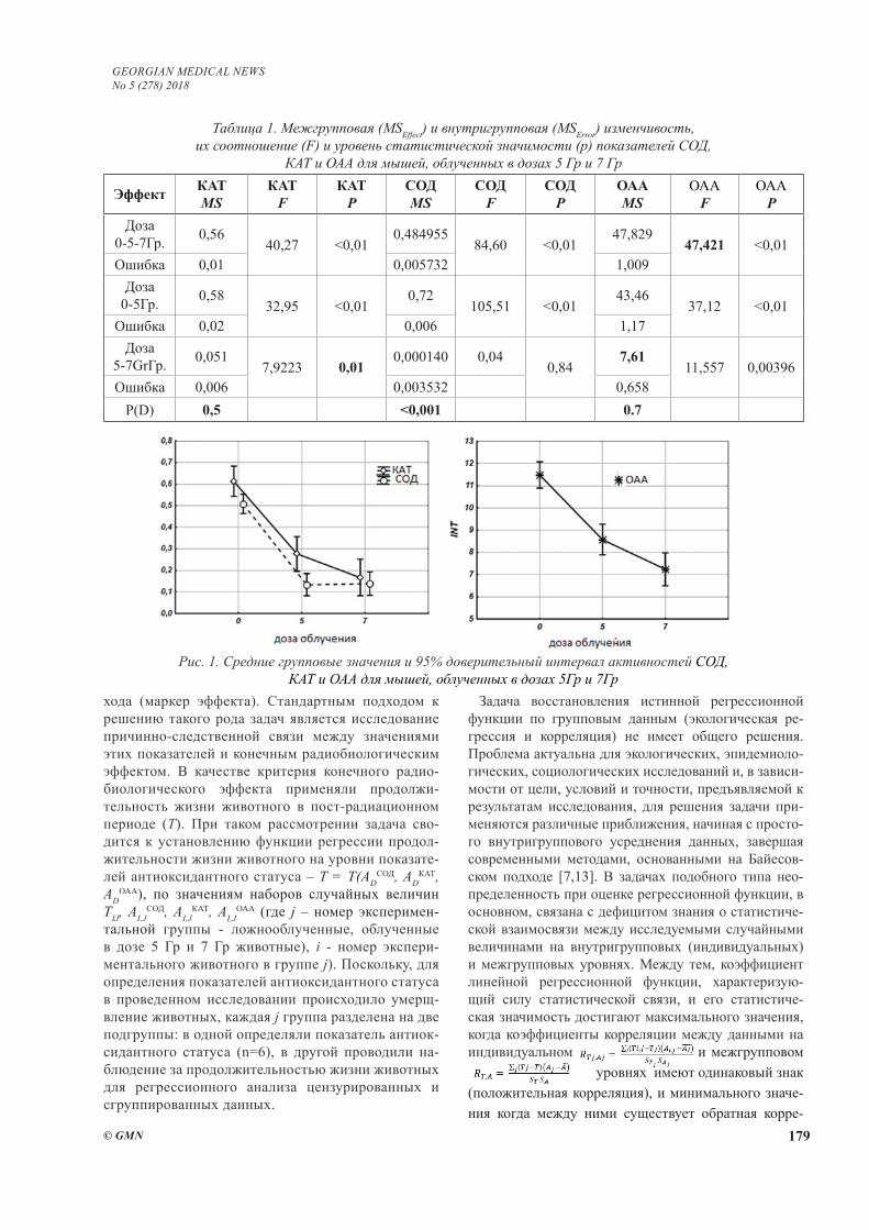
GEORGIAN MEDICAL NEWS No 5 (278) 2018
© GMN 179
хода (маркер эффекта). Стандартным подходом к решению такого рода задач является исследование причинно-следственной связи между значениями этих показателей и конечным радиобиологическим эффектом. В качестве критерия конечного радио-биологического эффекта применяли продолжи-тельность жизни животного в пост-радиационном периоде (T). При таком рассмотрении задача сво-дится к установлению функции регрессии продол-жительности жизни животного на уровни показате-лей антиоксидантного статуса – T = T(AD
СОД, ADКАТ,
ADОАА), по значениям наборов случайных величин
TIJ, AI,JСОД, AI,J
КАТ, AI,JОАА (где j – номер эксперимен-
тальной группы - ложнооблученные, облученные в дозе 5 Гр и 7 Гр животные), i - номер экспери-ментального животного в группе j). Поскольку, для определения показателей антиоксидантного статуса в проведенном исследовании происходило умерщ-вление животных, каждая j группа разделена на две подгруппы: в одной определяли показатель антиок-сидантного статуса (n=6), в другой проводили на-блюдение за продолжительностью жизни животных для регрессионного анализа цензурированных и сгруппированных данных.
Задача восстановления истинной регрессионной функции по групповым данным (экологическая ре-грессия и корреляция) не имеет общего решения. Проблема актуальна для экологических, эпидемиоло-гических, социологических исследований и, в зависи-мости от цели, условий и точности, предъявляемой к результатам исследования, для решения задачи при-меняются различные приближения, начиная с просто-го внутригруппового усреднения данных, завершая современными методами, основанными на Байесов-ском подходе [7,13]. В задачах подобного типа нео-пределенность при оценке регрессионной функции, в основном, связана с дефицитом знания о статистиче-ской взаимосвязи между исследуемыми случайными величинами на внутригрупповых (индивидуальных) и межгрупповых уровнях. Между тем, коэффициент линейной регрессионной функции, характеризую-щий силу статистической связи, и его статистиче-ская значимость достигают максимального значения, когда коэффициенты корреляции между данными на индивидуальном и межгрупповом уровнях имеют одинаковый знак (положительная корреляция), и минимального значе-ния когда между ними существует обратная корре-
Таблица 1. Межгрупповая (MSEffect) и внутригрупповая (MSError) изменчивость, их соотношение (F) и уровень статистической значимости (p) показателей СОД,
КАТ и ОАА для мышей, облученных в дозах 5 Гр и 7 Гр
Эффект КАТMS
КАТF
КАТP
СОДMS
СОДF
СОДP
ОААMS
ОААF
ОААP
Доза 0-5-7Гр. 0,56
40,27 <0,010,484955
84,60 <0,0147,829
47,421 <0,01Ошибка 0,01 0,005732 1,009
Доза0-5Гр. 0,58
32,95 <0,010,72
105,51 <0,0143,46
37,12 <0,01Ошибка 0,02 0,006 1,17
Доза5-7GrГр. 0,051
7,9223 0,010,000140 0,04
0,847,61
11,557 0,00396Ошибка 0,006 0,003532 0,658
P(D) 0,5 <0,001 0.7
Рис. 1. Средние групповые значения и 95% доверительный интервал активностей СОД, КАТ и ОАА для мышей, облученных в дозах 5Гр и 7Гр
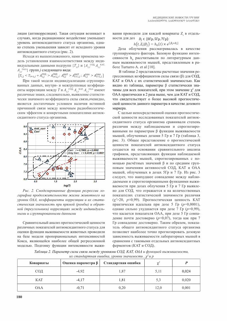
180
МЕДИЦИНСКИЕ НОВОСТИ ГРУЗИИ
CFMFHSDTKJC CFVTLBWBYJ CBF[KTYB
ляция (антикорреляция). Такая ситуация возникает в случаях, когда радиационное воздействие уменьшает уровень антиоксидантного статуса организма, одна-ко степень уменьшения зависит от исходного уровня антиоксидантного статуса (рис. 2).
Исходя из вышеизложенного, нами применена мо-дель установления взаимосоответствия между инди-видуальными данными подгрупп {TIJ} и {AI,J
СОД, AI,JКАТ,
AI,JОАА} групп j следующего вида:
При такой модели индивидуализации сгруппиро-ванных данных, внутри- и межгрупповые коэффици-енты корреляции между T и AI,J
СОД, AI,JКАТ, AI,J
ОАА имеют различные знаки, следовательно, выявление статисти-чески значимого коэффициента силы связи,очевидно, является достаточным условием наличия истинной причинной связи между конечным радиобиологиче-ским эффектом и конкретными показателями антиок-сидантного статуса организма.
Рис. 2. Смоделированные функции регрессии ло-гарифма продолжительности жизни животного на уровни ОАА, коэффициенты корреляции и их стати-стическая значимость при прямой (ромбы) и обрат-ной (треугольники) корреляциях между индивидуаль-ными и сгруппированными данными
Сравнительный анализ прогностической ценности различных показателей антиоксидантного статуса для оценки функции выживаемости животных проводили на базе модели пропорциональных интенсивностей Кокса, являющейся наиболее общей регрессионной моделью. Подгонку функции интенсивности выжи-
вания проводили для каждой ковариаты Zi в отдель-ности для доз
Доза облучения рассматривалась в качестве группирующего фактора. Базовую функцию интен-сивности h0 рассчитывали по литературным дан-ным выживаемости мышей, представленным в ра-боте Turturro A. et al [10].
В таблице 2 представлены расчетные значения ре-грессионных коэффициентов силы связи (β) для СОД, КАТ и ОАА с их статистической значимостью. Как видно из таблицы, параметры β статистически зна-чимы для всех показателей, при этом значение χ2 для ОАА практически в 2 раза выше, чем для КАТ и СОД, что свидетельствует о более высокой прогностиче-ской ценности данного параметра в качестве дозового маркера.
С целью непосредственной оценки прогностиче-ской ценности исследованных показателей антиок-сидантного статуса организма сравнивали степень различия между наблюдаемыми и спрогнозиро-ванными по параметрам β функции выживаемости мышей, облученных дозами 5 Гр и 7 Гр (таблица 3, рис. 3). Общее представление о прогностической ценности показателей антиоксидантного статуса создается на основании сравнительного анализа графиков, представляющих функции наблюдаемой выживаемости мышей, спрогнозированных с по-мощью расчётных значений β и по средним груп-повым значениям активностей СОД, КАТ и ОАА мышей, облученных в дозах 5Гр и 7 Гр. Из рис. 3 следует, что наихудшее совпадение между наблю-даемыми и спрогнозированными функциями выжи-ваемости при дозах облучения 5 Гр и 7 Гр выявле-но для СОД, что отражается и на количественных показателях статистической значимости различия (χ2>20, p≈0,99). Прогностическая ценность КАТ практически идеальна при дозе 5 Гр (p=0,0001), однако сильно ухудшается при дозе 7 Гр (p=0,99), что касается показателя ОАА, при дозе 5 Гр совпа-дение почти достоверно (p=0,07), тогда как при 7 Гр совпадение достоверно. Таким образом, показа-тель общего антиоксидантного статуса организма позволяет наиболее точно прогнозировать дозовую зависимость выживаемости лабораторных мышей в сравнении с таковыми отдельных антиоксидантных ферментов (КАТ и СОД).
Таблица 2. Параметр силы связи между уровнями СОД, КАТ, ОАА и функцией выживаемости, их стандартная ошибка, уровни значимости , χ2 и р
Ковариаты Оценка параметра β Стандартная ошибка χ2 P
СОД -4,92 1,87 5,11 0,024
КАТ -4,17 1,81 5,3 0,020
ОАА -0,71 0,20 12,0 0,001
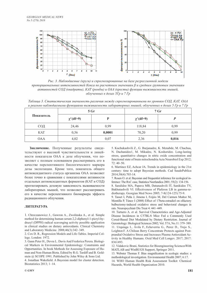
GEORGIAN MEDICAL NEWS No 5 (278) 2018
© GMN 181
Рис. 3. Наблюдаемые (круги) и спрогнозированные на базе регрессионной модели пропорциональных интенсивностей Кокса по расчетным значениям β и средним групповым значениям
активностей СОД (квадраты), КАТ (ромбы) и ОАА (кресты) функции выживаемости мышей, облученных в дозах 5Гр и 7 Гр
Таблица 3. Статистическая значимость различия между спрогнозированными по уровням СОД, КАТ, ОАА и реально наблюдаемыми функциями выживаемости лабораторных мышей, облученных в дозах 5 Гр и 7 Гр
Показатель5 Gr 7 Gr
χ2 (df=9) Р χ2 (df=9) P
СОД 24,46 0,99 118,84 0,99
КАТ 0,56 0,0001 70,20 0,99
ОАА 4,02 0,07 2,36 0,016
Заключение. Полученные результаты свиде-тельствуют о высокой чувствительности и линей-ности показателя ОАА к дозе облучения, что по-зволяет с полным основанием рассматривать его в качестве перспективного биологического маркера дозы экспозиции. Кроме того, показатель общего антиоксидантного статуса организма ОАА позволяет более точно в сравнении с показателями активности отдельных антиоксидантных ферментов (КАТ и СОД) прогнозировать дозовую зависимость выживаемости лабораторных мышей, что позволяет рассматривать его в качестве перспективного биомаркера эффекта радиационного облучения.
ЛИТЕРАТУРА
1. Chrzczanowicz J., Gawron A., Zwolinska A., et al. Simple method for determining human serum 2,2-diphenyl-1-picryl-hy-drazyl (DPPH) radical scavenging activity possible application in clinical studies on dietary antioxidants. Clinical Chemistry and Laboratory Medicine. 2008;46(3):342–349.2. Cox D. R., Regression Models and Life-Tables. Imperial Col-lege. London: 1972.3. Gann Peter H., Devra L. Davis And Frederica Perera. Biologi-cal Markers in Environmental Epidemiology: Constraints and Opportunities. In book Methods for Assessing Exposure of Hu-man and Non-Human Biota. Edited by R.G. Tardiff and B. Gold-stein @ SCOPE 1991. Published by John Wiley & Sons Ltd.4. Jonathan Wakefield. A Bayesian model for cluster detection. Biostatistics 2013; 1–14.
5. Kuchukashvili Z., G. Burjanadze, K. Menabde, M. Chachua, N. Dachanidze1, M. Mikadze, N. Koshoridze. Long-lasting stress, quantitative changes in nitric oxide concentration and functional state of brain mitochondria Acta Neurobiol Exp 2012; 72: 40–50.6. Martinez EZ, Achcar JA. Trends in epidemiology in the 21st century: time to adopt Bayesian methods. Cad SaudePublica 2014;30(4):703-14.7. Rosen O. et al. Bayesian and frequentist inference for ecological in-ference: The RxC case, Statistica Neerlandica 2001; 55(2): 134-156.8. Saralidze MA, Papava MB, Datunashvili IT, Sanikidze TV, Bakhutashvili VI. Effectiveness of Plaferon LB in gamma-ra-diotherapy. Georgian Med News 2005; 7-8(124-125):75-9.9. Tasset I, Peña J, Jimena I, Feijóo M, Del Carmen Muñoz M, Montilla P, Túnez I (2008) Effect of 17beta-estradiol on olfactory bulbectomy-induced oxidative stress and behavioral changes in rats. Neuropsychiatr Dis Treat 4: 441–449.10. Turturro A. et al. Survival Characteristics and Age-Adjusted Disease Incidences in C57BL/6 Mice Fed a Commonly Used Cereal-Based Diet Modulated by Dietary Restriction. Journal of Gerontology: Biological Sciences 2002; Vol. 57A, No.11: 379–389.11. Urquiaga I., Ávila F., Echeverria G., Perez D., Trejo S.,
Leighton F. A Chilean Berry Concentrate Protects against Post-prandial Oxidative Stress and Increases Plasma Antioxidant Ac-tivity in Healthy Humans. Oxid Med Cell Longev. 2017; 2017: 8361493. 12. Vidakovic Brani, Statistics for Bioengineering SciencesWith MATLAB and WinBUGS Support, Springer 2011.13. Webster Thomas F. Bias magnification in ecologic studies: a methodological investigation. Environmental Health 2007; 6:17.14. WHO Human Health Risk Assessment Toolkit: Chemical Hazards, World Health Organization 2010.
182
МЕДИЦИНСКИЕ НОВОСТИ ГРУЗИИ
CFMFHSDTKJC CFVTLBWBYJ CBF[KTYB
SUMMARY
STUDY OF TOTAL ANTIOOCIDANT STATUS OF THE ORGANISM AS A POSSIBLE BIOMARKER OF DOSE AND EFFECT OF RADIATION EXPO-SURE
Gvilava I., Chkhikvishvili I., Sanikidze T., Giorgobiani M., Nana V. Kipiani, Ormotsadze G.
Tbilisi State Medical University; I. Beritashvili Institute of Medical Biotechnology, Georgia
In presented article, by means of a comparative analysis of the relationship between the dose-dependent alterations in the organism’s redox status, measured by the innovative method developed by us and the standard methods used for assessing catalase and superoxide dis-mutase activity, and an end radiobiological effect, was attempted the preliminary assessment of the possibility to apply the parameter of blood plasma total antioxidant activity (TAA) as marker of dose and effect of radiation exposure.
The experiments were carried out on white mice ran-domly divided into groups of irradiated and sham irradi-ated animals. The mice were exposed to a whole body gamma irradiation by source Cesium-137 (137Cs) at dos-es of 5 and 7 Gr, a dose rate of 1.1 Gr / min. After 5 days of beginning of observation in animals’ blood measure-ments of the activity of antioxidant enzymes (superoxide dismutase - SOD and catalase - CAT) and total antioxi-dant activity by spectrophotometric method were evaluat-ed. Parallel monitoring of animal survival was conducted.
At the given stage of the study, applicability of OAA as a marker of dose-dependent alterations in antioxidant status was assessed by the criteria of sensitivity and lin-earity, and as a marker of the effect the strength of the relation between the antioxidant status indicators and the final radiobiological effect, measured by animal life span in post- radiation period (time-effect)
For comparative analysis of the effects of radiation on the levels of antioxidant status indicators (SOD, cata-lase, OAA), ANOVA methods were used, the nature of the causal relationship between levels of antioxidant sta-tus and the life span of laboratory animals was analyzed on the basis of the Cox proportional intensity model with time covariates, preliminary processing of data, basic cal-culations and visualization of the results were carried out using a mathematical package that “ STATISTIC 12”.
The received results testify to the significantly high sensitivity of the total redox-status indicator (OAA) to the dose of irradiation, which makes it possible with full justification to consider it as a promising candidate of the biological exposure dose marker. In addition, the indica-tor of the total antioxidant status (OAA) of the body al-lows predicting the dose dependence of the survival of laboratory mice more accurately than the indices of the
activity of individual antioxidant enzymes (catalase and SOD), which makes it possible to consider it as a promis-ing candidate for the biomarker of the dose and the effect of radiation exposure.
Keywords: antioxidant status, biomarker of radiation exposure, redox-status.
РЕЗЮМЕ
ИССЛЕДОВАНИЕ ОБЩЕГО АНТИОКСИДАНТ-НОГО СТАТУСА ОРГАНИЗМА В КАЧЕСТВЕ ВОЗМОЖНОГО БИОМАРКЕРА ДОЗЫ И ЭФ-ФЕКТА РАДИАЦИОННОГО ОБЛУЧЕНИЯ
Гвилава И., Чхиквишвили И.Д., Саникидзе Т.В., Гиоргобиани М.Т., Нана В. Кипиани, Ормоцадзе Г.Л.
Тбилисский государственный медицинский универси-тет; Центр экспериментальной биомедицины им. И. Бериташвили, Грузия
В статье путем сравнительного анализа характера связи между дозозависимыми изменениями редокс-статуса организма, измеренным разработанным нами методом и стандартными методами оценки каталазы и супероксиддисмутазы и конечным радиобиологи-ческим эффектом, предпринимается попытка предва-рительной оценки возможности применения разрабо-танного нами метода определения антиоксидантной активности (ОАА) плазмы крови в качестве маркера дозы и эффекта радиационного воздействия.
Эксперименты проводились на белых беспород-ных мышах самцах. Животные рандомизированно разделены на группы облученных и ложно облучён-ных животных. Общее гамма облучение животных проводили источником Cesium-137 (137Cs) в дозах 5 и 7 Гр, мошность дозы 1,1 Гр/мин. С целью оценки активности антиоксидантных ферментов (суперок-сиддисмутаза - СОД и каталаза - КАТ) и общей анти-оксидантной активности кровь у животных забирали на 6 день наблюдения. Параллельно вели наблюдение за выживаемостью животных.
На данном этапе исследования применимость ОАА в качестве маркера дозозависимых изменений антиоксидантного статуса оценивали по критериям чувствительности и линейности, а в качестве маркера эффекта воздействия – силой связи между уровнями показателей антиоксидантного статуса и конечным радиолобиологическим эффектом, который опреде-ляли продолжительностью жизни животного в пост-радиационном периоде (время-эффект)
Для сравнительного анализа эффектов радиаци-онного воздействия на уровни показателей антиок-сидантного статуса организма (СОД, КАТ, ОАА) при-меняли методы дисперсионного анализа (ANOVA),
GEORGIAN MEDICAL NEWS No 5 (278) 2018
© GMN 183
характер причинной связи между уровнями по-казателей антиоксидантного статуса и продолжи-тельностью жизни лабораторных животных ана-лизировали на базе модели пропорциональных интенсивностей Кокса, предварительную обработ-ку данных, основные расчеты и визуализацию ре-зультатов проводили с помощью математического пакета “STATISTICA 12”.
Полученные результаты свидетельствуют о высо-кой чувствительности и линейности показателя ОАА к дозе облучения, что позволяет с полным основани-ем рассматривать его в качестве перспективного био-логического маркера дозы экспозиции. Кроме того, показатель общего антиоксидантного статуса орга-низма ОАА позволяет более точно в сравнении с по-казателями активности отдельных антиоксидантных ферментов (КАТ и СОД) прогнозировать дозовую за-висимость выживаемости лабораторных мышей, что позволяет рассматривать его в качестве перспектив-ного биомаркера эффекта радиационного облучения.
reziume
organizmis saerTo antioqsidanturi statu-sis maCvenebeli, rogorc dozisa da radiaci-uli eqspoziciis efeqteis SesaZlo markeri
i. gvilava, i. CxikviSvili, T. sanikiZe, m. giorgobiani, nana yifiani, g. ormocaZe
Tbilisis saxelmwofo samedicino univer-siteti; i. beritaSvilis sax. samedicino bioteqnologiis instituti, saqarTvelo
statiaSi warmodgenilia kvlevis Sede-gebi maionizebeli radiaciiT zemoqmedebis saboloo biologiur efeqtsa da redoqs-sta-tusis (rs) maxasiaTebelTa doza-damokide-bul cvlilebebs Soris kavSiris xasiaTis Sesaxeb. rs-s cvlilebebi Seiswavleboda sisxlis plazmaSi superoqsiddismutazis (sod), katalazis (kat) aqtivobis gansaz-Rvris standartuli meTodebisa da saerTo antioqsidanturi statusis (sas) gansaz-Rvris originaluri damuSavebis meToduri midgomis gamoyenebiT.
kvlevis mizans warmoadgenda saerTo an-tioqsidanturi statusis, rogorc maioni-zebeli radiaciis dozisa da efeqtis biomarkeris Sefaseba. perspeqtiulobis
kriteriumad, gamokvlevaTa mocemul etapze, gamoiyenebuli iyo maxasiaTeblis mgrZno-beloba dasxivebis dozisadmi da dozaze damokidebulebis wrfivobis xarisxi, maxa-siaTeblis doza-damokidebul cvlilebaTa kavSiris simWidrove zemoqmedebis biolo-giur efeqtTan, romlis maxasiaTebladac ga-nixileboda sicocxlis xangrZlioba post-radiaciul periodSi (dro-efeqti).
eqsperimenti tardeboda TeTr, ujiSo mamr Tagvebze. cxovelebi (n=54) randomizirebu-lad iyvnen dayofili dasxivebul da sakon-trolo jgufebad. calkeuli jgufebi dayo-fili iyo or sub-jgufad - antioqsidantur fermentTa aqtivobis gansazRvris (n=6) da sicocxlis xangrZliobaze dakvirvebis (n=12) gaTvaliswinebiT. cxovelTa saerTo gama-dasxiveba Catarda Cesium-137 (137Cs) wya-ros gamoyenebiT, 5 grei da 7 grei dozebiT, dozis simZlavre 1.1 grei/wT. cxovelebis sisxlSi antioqsidanturi fermentebis aqtivobis Se-fasebisaTvis sisxlis aReba xdeboda dasx-ivebidan 5-e dRes speqtrometruli meTodiT. cxovelTa sicocxlis xangrZliobaze dak-virveba grZeldeboda 30 dRis ganmavlobaSi.
Sedegebis analizi tardeboda variaci-uli analizisa (ANOVA) da gadarCenis in-tensivobis koqsis damoukidebel kovariatTa regresiuli modelis Teoriuli aparat-is bazaze. monacemTa winaswari damuSaveba, ZiriTadi gamoTvlebi da Sedegebis vizuali-zacia Catarda “STATISTICA -12” maTemaTikuri paketis gamoyenebiT.
miRebulma Sedegebma gamoavlina sas-is gansazRvris SemoTavazebuli meTodis maRa-li mgrZnobeloba da doza-damokidebul cvl-ilebaTa wrfivobis maRali xarisxi sod da kat-Tan SedarebiT. gamovlinda sxvadasxva dozebiT dasxivebul cxovelTa gadarCenis damzeril mrudeebsa da koqsis regresiuli modelis bazaze sas-is doza-damokidebul cvlilebebiT prognozirebul gadarCenis mrudebs Soris damTxvevis statistikuri sar-wmunoebis maRali xarisxi.
yovelive zemowarmodgenili iZleva re-alur perspeqtivas sas-is Sefasebis Semo-Tavazebuli meTodis bazaze SemuSavdes maionizebeli radiaciis dasxivebis dozisa da efeqtis gansazRvris iafi da saimedo test-meTodi.
184
МЕДИЦИНСКИЕ НОВОСТИ ГРУЗИИ
CFMFHSDTKJC CFVTLBWBYJ CBF[KTYB
Proper research environment is an environment that effectively stimulates research, attracts productive sci-entists and ensure the efficiency, that is, the result in the form of scientific knowledge transferred into practical ap-plication. Establishing the proper environment depends on many factors. The analysis of international experience was done in the research, as well as the study of the as-sociation between various quantitative and qualitative characteristics of the contingent, the amount of attracted research funding of medical universities and effectiveness of research activities according to indicators assessing re-search and innovation activities of medical education or-ganizations and science of Kazakhstan Republic [1].
The aim of the study - to establish the association be-tween different quantitative and qualitative characteristics of the contingents, funding of research activities of medi-cal universities and key research performance indicators.
Material and methods. Data analysis was carried out for 7 medical Universities of Kazakhstan: “Astana Medi-cal University”, Kazakh National Medical University of S. Asfendiyarov, Karaganda state Medical University, South Kazakhstan state pharmaceutical Academy, West Kazakh-stan State medical University and State medical University of Semey. The dataset included data on key indicators from 7 medical universities in the period 2013-2016 and the fol-lowing data: average annual contingent of students, the total number of staff positions, the total number of individuals, number of master students, number of doctoral students, qualitative composition: the number of medical science doc-tors, candidates of medical Sciences, PhD, the total number of staff with scientific degree, the share of staff with scien-tific degrees in the university, the amount of total funding attracted to scientific research in the same period. The result-ing array of data on key performance indicators were sys-tematized and statistically processed in the program SPSS 2017. There was made the database for correlation analysis using Spearman coefficient. Data collection is carried out by the Center for Human resources development and science of Republican Center for health development. To assess the correlation Chaddoc scale has been used.
Results and their discussion. Gained data are pre-sented in Table 1.
Thus, at a level of p value<0.05 revealed the following correlation:1. For the sum of key indicators with:(a) number of staff who have PhD degree (r=0.426, p=0,024);(b) the proportion of people with a scientific degree (r=0.430, p=0,023).The correlations was assessed as low.2. On the indicator “number of publications in valuable
databases (Thomson reuter, Scopus, Springer)” - a signifi-cant correlation was not revealed;3. On the indicator “number of citations over the last 5 years” – a) The low correlation with the total number of individuals, working in organization (r=0.462, p=0,013), with the number of doctoral students (r=-0.461, p=0,014), number of Candidates of medical sciences (CMS) (r=-0.469, p=0.012), total number of staff with scientific de-grees (r=-0.466, p=0.012);b)The moderate correlation with - the total number of staff positions (r=-0.561, p=0.002), the average annual contingent of students (r=-0.504, at p=0,006);4. On the indicator “number of patents and other security documents”- a significant correlation was not detected;5. On the indicator “the number of scientific develop-ments” significant relations were not identified too.
Further, there was studied correlation for years, in dif-ferent years were found changes in the presence or ab-sence of association (Table 2).
Correlation analysis of the data in terms of various years showed the presence ofhigh correlation between the number of doctoral students enrolled in medical universities and with the sum of key performance indicators of the University (in 2013 r=0.774, p=0.041 and 2015 r=0.838, p=0.018) and also with the amount of funding raised in terms of 100 person of staff in 2015 year- r=0.784, p=0.037.
In the context of medical universities of Kazakhstan revealed the existence of very high correlation between the sum of key indicators and the amount of funding for the following Universities ZKGMU (r=0.943, p=0.057) and KazNMU (r=0.993, p=0.007); the presence ofa high negative correlation between the indicators sumand the total number of individuals, working in the organization (r=-1.000, p≤0.001), with the quantity of DMS (r=-0.949, p=0.051) and with the proportion of staff with scientific degree (r=-1.000, p≤0.001) in ZKGMU.
This study examines factors affecting the performance of basic research in South Korea. Creative Research Pro-gram (CRP), which is one of the major basic research programmes, is a study target. The survey of N = 120 researchers from universities and research institutes was conducted in 2010. Basic research performance which is the dependent variable was measured by subjective sur-vey questions. The data were analysed using factor analysis and multiple regression. The result shows that leadership is the most important factor affecting the basic research perfor-mance based on the regression coefficient (β). Knowledge sharing, autonomy, collaboration and creativity in sequence were also statistically valuable factors that impact on the per-formance of basic research in South Korea [2].
MODEL OF CREATING PROPER RESEARCH ENVIRONMENT IN MEDICAL EDUCATION ORGANIZATIONS
1Umbetzhanova A., 1Bekbergenova Zh., 2Koikov V., 1Derbissalina G., 1Tuleshova G.
1Astana Medical University; 2Republican Centre for Health Development, Astana, Republic of Kazakhstan
GEORGIAN MEDICAL NEWS No 5 (278) 2018
© GMN 185
Table 1.Results of correlation analysis (period 2013-2016 years)
Sum of key indicators
Quantity of publications in valuable databases
Quantity of citations (over the 5 past years)
Quantity of patents
Quantity of Scientific
developments
Total number of staff positions r=-0.030, p=0.881
r=-0.098, p=0.620
r=-0.561, p=0.002
r=0.112, p=0.570
r=0.092, p=0.641
Total number of individuals, working in irganization
r=0.041, p=0.836
r=-0.110, p=0.577
r=-0.462, p=0.013
r=0.090, p=0.649
r=0.085, p=0.667
Average annual contingent of students
r=0.173, p=0.379
r=-0.123, p=0.534
r=-0.504, p=0.006
r=0.167, p=0.395
r=0.157, p=0.425
Number of master students r=0.289, p=0.135
r=-0.093, p=0.638
r=0.104, p=0.599
r=0.125, p=0.525
r=0.046, p=0.816
Number of doctoral students r=0.305, p=0.114
r=-0.065, p=0.744
r=-0.461, p=0.014
r=0.104, p=0.600
r=-0.004, p=0.984
Number of Doctor of Medical Science
r=0.219, p=0.263
r=-0.044, p=0.826
r=-0.317, p=0.100
r=0.158, p=0.422
r=0.237, p=0.225
Number of Candidate of Medical Science
r=0.194, p=0.322
r=-0.094, p=0.633
r=-469, p=0.012
r=0.094, p=0.634
r=0.082, p=0.678
Number of PhD r=0.426, p=0.024
r=-0.361, p=0.059
r=-0.118, p=0.550
r=-0.075, p=0.706
r=0.141, p=0.475
Total number of staff with scientific degrees
r=0.176, p=0.371
r=-0.111, p=0.574
r=-0.466, p=0.012
r=0.077, p=0.696
r=0.077, p=0.695
Proportion of staff with scientific degree
r=0.430, p=0.023
r=0.042, p=0.832
r=0.052, p=0.795
r=0.165, p=0.402
r=0.153, p=0.438
Financing up to 100 people r=0.119, p=0.545
r=-0.250, p=0.200
r=-0.642, p≤0.001
r=-0.061, p=0.758
r=-0.020, p=0.921
Table 2. Meanings of correlations by the sum of key indicators in Medical Universities in (2013-2016) yearsSum of key indicators
2013 2014 2015 2016Total number of staff positions r=0.502, p=0.251 r=0.267, p=0.563 r=0.249, p=0.591 r=0.027, p=0.955Total number of individuals, working in organization r=0.338, p=0.458 r=0.276, p=0.549 r=0.137, p=0.769 r=-0.182, p=0.696
The average annual contingent of students r=0.543, p=0.208 r=0.210, p=0.652 r=0.327, p=0.474 r=-0.077, p=0.870
Number of master students r=0.553, p=0.198 r=0.176, p=0.706 r=-0.145, p=0.756 r=-0.042, p=0.928Number of doctoral students r=0.774, p=0.041 r=0.255, p=0.582 r=0.838, p=0.018 r=-0.080, p=0.864Number of DMS (Doctor of medical science) r=0.500, p=0.253 r=0.630, p=0.129 r=0.283, p=0.538 r=-0.306, p=0.505
Number of CMS (Candidate of medical science)
r=0.525, p=0.226 r=0.696, p=0.082 r=0.364, p=0.423 r=-0.035, p=0.940
Number of PhD r=0.337, p=0.460 r=0.018, p=0.969 r=-0.646, p=0.117 r=-0.407, p=0.365Total number of staff with scientific degree r=0.560, p=0.192 r=0.677, p=0.095 r=0.337, p=0.460 r=-0.145, p=0.757
Proportion of staff with the scientific degree r=0.290, p=0.527 r=0.643, p=0.119 r=0.566, p=0.186 r=-0.018, p=0.970
Financing up to 100 people r=0.070, p=0.882 r=-0.307, p=0.502 r=0.784, p=0.037 r=0.096, p=0.837
British researchers from King’s College in London [3] have studied the characteristics of highly productive research
186
МЕДИЦИНСКИЕ НОВОСТИ ГРУЗИИ
CFMFHSDTKJC CFVTLBWBYJ CBF[KTYB
groups and identified the following 8 aspects of the proper research environment:
- In highly productive research teams most of the re-searchers have PhD degrees, professorships, international experience, and usually, their work sponsored from outside;
- Highly productive research teams focus on selecting and retaining top scientists;
- Highly productive research teams provide training and mentoring for staff development, offering rewards for high performance;
- Staff in these groups follows a particular ideal of so-cial and ethical values;
- Usually the leaders (heads) of these highly produc-tive research teams have/had earned some kind of «ac-countable autonomy/independence» of their units in their institution of higher education;
- High-performance research teams have strategies that are real, live and self-developed, and not just a writ-ten document;
- Such groups receive more income per researcher than the average research unit;
- Highly productive research teams seamlessly acti-vate and encourage researchers to initiate cooperation, and do not use the top-down approach.
Koen Frenken, Gaston J., Heimeriks, JarnoHoek-man in his paper analyzes the factors underlying univer-sity research performance as indicated by the number of highly-cited publications, international co-publications, and university-industry co-publications. The three per-formance indicators evaluate three possible university missions, respectively: research excellence, international-ization, and innovation. Using a regression analysis, we assess to what extent a university’s research performance is influenced by structural variables including size, age, city size, location in a capital city, disciplinary orienta-tion, and country location. Our results show that research performance differences among universities mainly stem from size, disciplinary orientation and country location. This suggests that simple global benchmarking can be misleading; rather, benchmarking is most meaningful between universities of a similar size supplemented with contextual information on a university’s specific mission, orientation and national institutions.Highlights: We assess university performance by size, age, city size, specializa-tion, and country; Differences in university performance mainly stem from size and country location; Benchmark-ing is meaningful among universities of similar size and countries; No country excels at impact, internationaliza-tion and innovation simultaneously. [4].
The Sri Lankan medical research output during the last decade is only a small fraction of the global research out-put. There it is a necessity to setup an enabling environment for research, with a proper vision, support, funds and train-ing. In addition, collaborations across the region need to be strengthened to face common regional health challenges [5].
The structure of research system in Germany it is the federal and state levels of research management. The Fed-
eral Ministry of Education and Research (BMBF) has pri-mary responsibility for science and technology policy at the federal level. At the state level, this responsible is shared by the Ministry of Education and Science and the Ministry of Economy. The author emphasizes the role of the National Academy of Sciences «Leopoldina», whose principal objec-tive is to provide advisory services to German policymak-ers and present German science at the international level. Special attention is paid to the wide spectrum of German research agents: public and private research organizations, higher education institutions, R&D departments of industrial companies. The article stresses the research potential of uni-versities that receive funding under the Excellence Initiative and describes the contribution of production in research and development activities, focusing on the top ten German com-panies in terms of R&D expenditure [6].
Medical education research suffers from a significant and persistent lack of funding. Although adequate funding has been shown to improve the quality of research, there are a number of factors that continue to limit it. The competitive environment for medical education research funding makes it essential to understand strategies for improving the search for funding sources and the preparation of proposals. This ar-ticle offers a number of resources, strategies, and suggestions for finding funding. Investigators must be able to frame their research in the context of significant issues and principles in education. They must set their proposed work in the context of prior work and demonstrate its potential for significant new contributions. Because there are few funding sources earmarked for medical education research, researchers much also be creative, flexible, and adaptive as they seek to pres-ent their ideas in ways that are appealing and relevant to the goals of funders. Above all, the search for funding requires persistence and perseverance [7].
The vision of the recently created Canadian Association of Emergency Physicians (CAEP) Academic Section is to promote high-quality emergency patient care by conducting world-leading education and research in emergency medi-cine. The Academic Section plans to achieve this goal by enhancing academic emergency medicine primarily at Ca-nadian medical schools and teaching hospitals. It seeks to foster and develop education, research, and academic lead-ership amongst Canadian emergency physicians, residents, and students. In this light, the Academic Section began in 2013 to hold the annual Academic Symposia to highlight best practices and recommendations for the three core do-mains of governance and leadership, education scholar-ship, and research. Each year, members of three panels are asked to review the literature, survey and interview experts, achieve consensus, and present their recommendations at the Symposium (2013, Education Scholarship; 2014, Research; and 2015, Governance and Funding). Research is essential to medical advancement. As a relatively young specialty, emergency medicine is rapidly evolving to adapt to new diagnostic tools, the challenges of crowding in emergency departments, and the growing needs of emergency patients. There is significant variability in the infrastructure, support,
GEORGIAN MEDICAL NEWS No 5 (278) 2018
© GMN 187
and productivity of emergency medicine research programs across Canada. All Canadians benefit from an investigation of the means to improve research infrastructure, training programs, and funding opportunities. Such an analysis is es-sential to identify areas for improvement, which will support the expansion of emergency medicine research. To this end, physician-scientist leaders were gathered from across Cana-da to develop pragmatic recommendations on the improve-ment of emergency medicine research through a comprehen-sive analysis of current best practices, systematic literature reviews, stakeholder surveys, and expert interviews [8].
The emancipatory nature of education requires re-search as its fundamental base, because physicians can only improve their skills and knowledge through enquiry. The number and quality of scientific publications by Lat-in-American spine surgeons found in the Medline data-base was low between 2000 and 2011. Nevertheless, the research Bank Survey of AOSpine Latin America (AO-SLA) members showed that 96% of responders were very interested and motivated to perform scientific research.
The research officer of AOSLA together with the Country Council and the AOSpine Research Commission established a competency-based curriculum to improve understanding of what is necessary to produce research and the best methods to achieve this goal. The research curriculum was divided into four main components: (1) research educational plan, (2) performing research, (3) technical and professional support and (4) assessment.
The competences, learning outcomes and a syllabus on knowledge in research were developed to enable the partici-pants to understand and perform investigations effectively. The eLearning module was designed to improve the com-petences to access, evaluate and use scientific information available in the main databases efficiently. Research courses were given as an isolated activity four times in Brazil and Mexico and as precourse activities six times in Brazil, Mex-ico and Peru. The result was an increased number of articles published and works presented at congresses [9].
Although the National Institutes of Health (NIH) has made extensive investments in educational programs re-lated to clinical and translational science (CTS), there has been no systematic investigation of the number and char-acteristics of PhD programs providing training to future leaders in CTS. The authors undertook to determine the number of institutions that, having had received NIH-funded Clinical and Translational Science Awards (CT-SAs), currently had or were developing PhD programs in CTS; to examine differences between programs devel-oped before and after CTSA funding; and to provide de-tailed characteristics of new programs.
In 2012, CTS program leaders at the 60 CTSA-funded institutions completed a cross-sectional survey focusing on four key domains related to PhD programs in CTS: program development and oversight; students; curriculum and research; and milestones.
Twenty-two institutions had fully developed PhD pro-grams in CTS, and 268 students were earning PhDs in this
new field; 13 institutions were planning PhD programs. New programs were more likely to have fully developed PhD competencies and more likely to include students in medical school, students working only on their PhD, students working on a first doctoral degree, and students working in T1 translational research. They were less like-ly to include physicians and students working in clinical or T2 research.
Although CTS PhD programs have similarities, they also vary in their characteristics and management of stu-dents. This may be due to diversity in translational sci-ence itself or to the relative infancy of CTS as a discipline.
The project of education in research can be effectively disseminated and applied across regions, across students and across specialties [10].
Conclusion. A brief review of international experi-ence and results of own research allow us to develop own model of effective research environment in the organiza-tions of medical education and science (Fig.).
Fig. Model for appropriate research media of Univer-sities, Science research institutes, Science centers
In our view, this environment should include:- Strategy and development plans to achieve the KPI
on science at the level of the authorized body, and at the level of the organization;
- Sufficient resources (information resources, materi-al-technical base), the efficiency of investments of them is evaluated by KPI on science;
- Qualification requirements for employees, which is also based on KPI science;
- The system of motivation and rewards based on KPI science;
- System of continuous professional development of staff in the attaining and development of research compe-tencies, KPI-based on science;
- Research infrastructure, performance evaluation based on KPI science;
- Educational activities, the performance of which is estimated by the presence on students KPI for science;
- Clinical activities based on the implementation in practice of new scientific achievements (KPI science);
188
МЕДИЦИНСКИЕ НОВОСТИ ГРУЗИИ
CFMFHSDTKJC CFVTLBWBYJ CBF[KTYB
- The system of information support (print journals, web site) and the politics of the Internet presence of the organization and its employees contribute to the achieve-ment of KPI’s for science.
The above listed components of an appropriate re-search media of the medical education and science orga-nizations, which together ensure the achievement of its ef-fectiveness, there is a general increase of competitiveness, which means increasing the quality of the research, the recognition of national medical science on a global level.
REFERENCES
1. Methodology of ranking organizations of medical science and medical education according to the results of scientific and innova-tion activities. Health Ministry of Kazakhstan Republic, July 2013.2. The Factors Affecting Basic Research Performance Funded by Government: ‘Creative Research Program’ Case in South Korea 2016; v.21 issue 3: 349-365. 3. Manville C. et al. Characteristics of high-performing research units. – 2015.4. Koen Frenken, Gaston J., Heimeriks, Jarno Hoekman . What drives university research performance? An analysis using the CWTS //Leiden Ranking data Journal of Informetrics. Volume 11, Issue 3, August 2017, Pages 859-872.5. Ranasinghe P, Jayawardena R, Katulanda P.Sri Lanka in global medical research: a scientific analysis of the Sri Lankan research output during 2000-2009 //BMC Res Notes. 2012 Feb 24;5:121.6. Berezhnaya G. S.Organization of scientific research in Ger-many//Science and Education 2012 P. – 149-157.7. Gruppen L.D. Needles and Haystacks: Finding Funding for Medical Education Research // Acad Med. 2016; 91(4):480-4.8. Stiell I.G, Artz J.D, Perry J., Vaillancourt C., Calder L.Executive summary of the CAEP 2014 Academic Sympo-sium: How to make research succeed in your department // CJEM. 2015; 17(3): 328-33.9. Falavigna A., Martins Filho D.E., Avila J.M., Guyot J.P., Gonzáles A.S., Riew D.K. Strategy to increase research in Latin America: project on education in research by AOSpine Latin America // Eur J Orthop Surg Traumatol. 2015; 25 Suppl 1: S13-20.10. Switzer G.E., Robinson G.F., Rubio D.M., Fowler N.R., Ka-poor W.N. Doctoral programs to train future leaders in clinical and translational science // Acad Med. 2013; 88(9): 1332-9.
SUMMARY
MODEL OF CREATING PROPER RESEARCH EN-VIRONMENT IN MEDICAL EDUCATION ORGA-NIZATIONS
1Umbetzhanova.A., 1Bekbergenova Zh., 2Koikov V., 1Derbissalina G., 1Tuleshova G.
1Astana Medical University; 2Republican Centre for Health Development, Astana, Republic of Kazakhstan
A good research environment is an environment that effectively stimulates research, attracts productive scien-tists and ensures the result in the form of scientific knowl-edge transferred to practical application. The formation of
a good research environment depends on a large number of factors. The aim of the study was to identify factors, which influence on the medical universities research per-formance and on the basis of this forming the model of proper research environment in the medical education or-ganizations. The data used in the study was collected by Republican Center for health development annually from 7 (seven) medical universities of Republic Kazakhstan on the indicators of research and innovative activities. The study analyzes the associations between various quan-titative and qualitative characteristics of the faculty and students, the amount of attracted funding for the scien-tific activities of medical schools and the effectiveness of scientific activity according to the indicators of the assess-ment of scientific and innovative activity of organizations of medical education and science in the Republic of Ka-zakhstan. AuthorsuseSpearmencoefficient. Results of the study revealed the most significant associations between the quality of the teaching staff and meanings of the indi-cators such number of publications and scientific develop-ments. Results of the study allow to build the model of the good research environment, which helps increase the effectiveness of research activities and to transfer results of the studies into the practice.
Key words: medical science, research performance measure, medical education organizations.
РЕЗЮМЕ
МОДЕЛЬ ФОРМИРОВАНИЯ НАДЛЕЖАЩЕЙ ИССЛЕДОВАТЕЛЬСКОЙ СРЕДЫ В ОРГАНИЗА-ЦИЯХ МЕДИЦИНСКОГО ОБРАЗОВАНИЯ
1Умбетжанова A.Т., 1Бекбергенова Ж.Б., 2Койков В.В., 1Дербисалина Г.А., 1Тулешова Г.Т.
1Медицинский университет Астана; 2Республикан-ский центр развития здравоохранения, Астана, Ре-спублика Казахстан
Понятие «надлежащая исследовательская среда» оз-начает эффективное стимулирование проведения иссле-дований, привлечение продуктивных ученых для обе-спечения эффективности исследования в виде научного знания, переданного для практического применения. Формирование надлежащей исследовательской среды зависит от множества факторов.
Целью исследования явилось определение факторов, влияющих на результативность научной деятельности в медицинском ВУЗе, и на основании этого, построение модели надлежащей исследовательской среды.
Материалами исследования послужили данные по индикаторам научной и инновационной деятельности 7 медицинских университетов Республики Казахстан, со-бираемые ежегодно Республиканским центром развития здравоохранения, а также качественные и количествен-
GEORGIAN MEDICAL NEWS No 5 (278) 2018
© GMN 189
ные характеристики контингента учащихся, профессор-ско-преподавательского состава и объема финансирова-ния, выделенного на проведение научных исследований. В ходе исследования проанализирован опыт зарубежных стран построения моделей надлежащей исследователь-ской среды; для определения связи между значениями индикаторов научной и инновационной деятельности ВУЗов и их различными показателями использованы методы корреляционного анализа с применением коэф-фициента Спирмена. Результаты исследования показали наиболее значимые связи между качественным составом профессорско-преподавательского состава и показателями индикаторов, таких как количество публикаций, научных разработок. Результаты исследования позволили построить модель надлежащей исследовательской среды медицин-ского ВУЗа, которая послужит повышению эффективности научной деятельности и внедрению результатов исследова-ний в практику.
reziume
saTanado kvleviTi garemos formireba sa-medicino ganaTlebis organizaciebSi
1a. umbetJanova, 1J. bekbergenova, 2v. koikovi, 1g. derbisalina, 1g. tuleSova
1astanas samedicino universiteti; 2jandac-vis ganviTarebis respublikuri centri, as-tana, yazaxeTi
cneba “saTanado samedicino garemo” gulisx-mobs samecniero kvlevebis Catarebis efeqtur stimulirebas, momqmedi mecnierebis mozidvas kvlevebis efeqturobis uzrunvelsayofad kvlevis Sedegebis SemdgomSi praqtikuli da-nergvisaTvis. saTanado kvleveTi garemos Seqmna damokidebulia mraval faqtorze.
kvlevis mizans warmoadgenda im faq-torebis gamovlena, romlebic,zemoqmedebas axdenen samecniero moRvaweobis Sedegobri-vobaze samedicino umaRlesi saganmanaTleb-lo dawesebulebaSi da miRebuli Sedegebis gaTvaliswinebiT saTanado samecniero gare-mos modelis gamomuSaveba samedicino uni-versitetisaTvis.
kvlevis masalad gamoyenebulia yazaxeTis 7 samedicino universitetis samecniero da inovaciuri moRvaweobis indikatoruli mona-cemebi, romlebic yovelwliurad kumulirde-ba jandacvis centris mier, aseve gaanalize-bulia moswavle da pedagogiuri personalis xarisxobrivi da raodenobrivi maxasiaTeble-bi, samedicino kvlevebis CatarebisaTvis gamo-yofili finansirebis moculoba.
kvlevis mimdinareobaSi ganxiluli da Sefasebuli iyo sazRvargareTis qveynebis saTanado kvleviTi garemos modelebis formirebis gamocdileba. umaRlesi sagan-manaTleblo saswavleblis samecniero da inovaciuri moRvaweobis indikatorebi da maT sxvadasxva maCveneblebs Soris kavSiris gansaz-Rvra ganxor-cielda korelaciuri analizis meTodis meSveobiT, spirmenis koeficientis gamoyenebiT. Catarebuli kvlevis Sedegma ga-moavlina mniSvnelovani kavSiri profesor-maswavlebelTa xarisxobriv Semadgenlobisa da indikatorebis iseT maCveneblebs Soris, rogoricaa gamoqveynebuli Sromebi da samec-niero inovaciebi. Catarebuli Sromis Sede-gebi saSualebas iZleva SemuSavebuli iqnas saTanado samecniero garemos iseTi modeli, romelic uzrunvelyofs umaRles samedicino saswavlo dawesebulebebSi samecniero moR-vaweobis efeqturobis zrdas da misi Sede-gebis praqtikaSi danergvas.
DETERMINATION OF ANTIOXIDANT ACTIVITY OF WINES AND WINE’S MAJOR PHENOLIC COMPOUNDS
BY ELECTRON SPIN RESONANS, USING SPIN-TRAPS METHOD
Chikvaidze E., Gogoladze T., Miminoshvili A.
I. Javakhishvili Tbilisi State Univeristy; Sokhumi State University, Georgia
In normal conditions living organisms have effec-tive tools for combating free radicals – enzyme sys-tems: superoxide dismutase, catalase, glutathione per-oxidase as well as natural antioxidants: a- tocopherol, ascorbic acid, b- carotin, selenium and others. How-ever, as the result of the various pathogenic processes
in the organism, the concentration of free radicals may increase considerably. Free radicals are known to harm proteins, free amino acids, lipids, lipoproteins, nucleic acids as well as cause oxidation of lipids with peroxide, which results in cell damage and leads to destruction of the organism. [2,3,4]
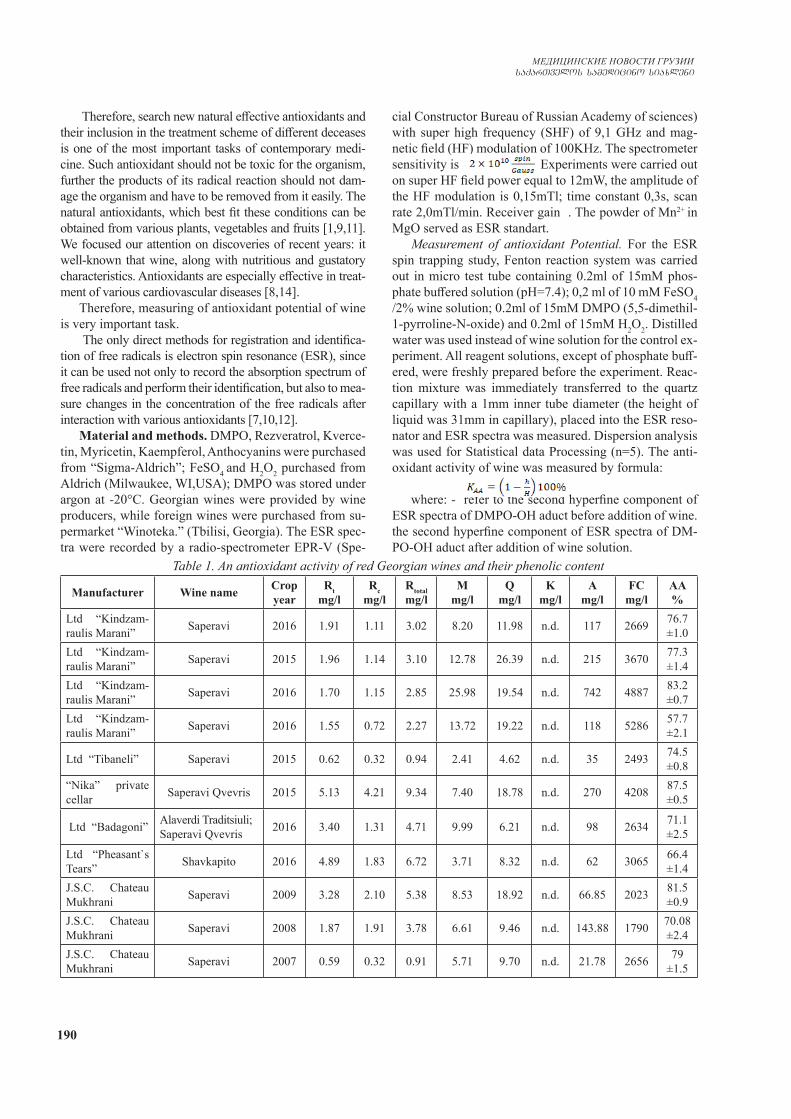
190
МЕДИЦИНСКИЕ НОВОСТИ ГРУЗИИ
CFMFHSDTKJC CFVTLBWBYJ CBF[KTYB
Therefore, search new natural effective antioxidants and their inclusion in the treatment scheme of different deceases is one of the most important tasks of contemporary medi-cine. Such antioxidant should not be toxic for the organism, further the products of its radical reaction should not dam-age the organism and have to be removed from it easily. The natural antioxidants, which best fit these conditions can be obtained from various plants, vegetables and fruits [1,9,11]. We focused our attention on discoveries of recent years: it well-known that wine, along with nutritious and gustatory characteristics. Antioxidants are especially effective in treat-ment of various cardiovascular diseases [8,14].
Therefore, measuring of antioxidant potential of wine is very important task.
The only direct methods for registration and identifica-tion of free radicals is electron spin resonance (ESR), since it can be used not only to record the absorption spectrum of free radicals and perform their identification, but also to mea-sure changes in the concentration of the free radicals after interaction with various antioxidants [7,10,12].
Material and methods. DMPO, Rezveratrol, Kverce-tin, Myricetin, Kaempferol, Anthocyanins were purchased from “Sigma-Aldrich”; FeSO4 and H2O2 purchased from Aldrich (Milwaukee, WI,USA); DMPO was stored under argon at -20°C. Georgian wines were provided by wine producers, while foreign wines were purchased from su-permarket “Winoteka.” (Tbilisi, Georgia). The ESR spec-tra were recorded by a radio-spectrometer EPR-V (Spe-
cial Constructor Bureau of Russian Academy of sciences) with super high frequency (SHF) of 9,1 GHz and mag-netic field (HF) modulation of 100KHz. The spectrometer sensitivity is . Experiments were carried out on super HF field power equal to 12mW, the amplitude of the HF modulation is 0,15mTl; time constant 0,3s, scan rate 2,0mTl/min. Receiver gain . The powder of Mn2+ in MgO served as ESR standart.
Measurement of antioxidant Potential. For the ESR spin trapping study, Fenton reaction system was carried out in micro test tube containing 0.2ml of 15mM phos-phate buffered solution (pH=7.4); 0,2 ml of 10 mM FeSO4 /2% wine solution; 0.2ml of 15mM DMPO (5,5-dimethil-1-pyrroline-N-oxide) and 0.2ml of 15mM H2O2. Distilled water was used instead of wine solution for the control ex-periment. All reagent solutions, except of phosphate buff-ered, were freshly prepared before the experiment. Reac-tion mixture was immediately transferred to the quartz capillary with a 1mm inner tube diameter (the height of liquid was 31mm in capillary), placed into the ESR reso-nator and ESR spectra was measured. Dispersion analysis was used for Statistical data Processing (n=5). The anti-oxidant activity of wine was measured by formula:
where: - refer to the second hyperfine component of
ESR spectra of DMPO-OH aduct before addition of wine. the second hyperfine component of ESR spectra of DM-PO-OH aduct after addition of wine solution.
Table 1. An antioxidant activity of red Georgian wines and their phenolic content
Manufacturer Wine name Crop year
Rtmg/l
Rcmg/l
Rtotalmg/l
Mmg/l
Qmg/l
Kmg/l
Amg/l
FCmg/l
AA%
Ltd “Kindzam-raulis Marani” Saperavi 2016 1.91 1.11 3.02 8.20 11.98 n.d. 117 2669 76.7
±1.0Ltd “Kindzam-raulis Marani” Saperavi 2015 1.96 1.14 3.10 12.78 26.39 n.d. 215 3670 77.3
±1.4Ltd “Kindzam-raulis Marani” Saperavi 2016 1.70 1.15 2.85 25.98 19.54 n.d. 742 4887 83.2
±0.7Ltd “Kindzam-raulis Marani” Saperavi 2016 1.55 0.72 2.27 13.72 19.22 n.d. 118 5286 57.7
±2.1
Ltd “Tibaneli” Saperavi 2015 0.62 0.32 0.94 2.41 4.62 n.d. 35 2493 74.5±0.8
“Nika” private cellar Saperavi Qvevris 2015 5.13 4.21 9.34 7.40 18.78 n.d. 270 4208 87.5
±0.5
Ltd “Badagoni” Alaverdi Traditsiuli; Saperavi Qvevris 2016 3.40 1.31 4.71 9.99 6.21 n.d. 98 2634 71.1
±2.5
Ltd “Pheasant`s Tears” Shavkapito 2016 4.89 1.83 6.72 3.71 8.32 n.d. 62 3065 66.4
±1.4J.S.C. Chateau Mukhrani Saperavi 2009 3.28 2.10 5.38 8.53 18.92 n.d. 66.85 2023 81.5
±0.9J.S.C. Chateau Mukhrani Saperavi 2008 1.87 1.91 3.78 6.61 9.46 n.d. 143.88 1790 70.08
±2.4J.S.C. Chateau Mukhrani Saperavi 2007 0.59 0.32 0.91 5.71 9.70 n.d. 21.78 2656 79
±1.5

GEORGIAN MEDICAL NEWS No 5 (278) 2018
© GMN 191
Investigation of anti oxidative activity of Georgian and foreign red and white wine and major phenol com-ponents of red wine was performed using Electron Para-magnetic Resonance (EPR) spin-trapping method. It was showed up, that red as well as white wines have very high anti oxidative activities. It is important to mention, that anti oxidative activity of red wine depends on the concen-tration of major antioxidants. Chromatographic investiga-tion for defining concentration of antioxidants and anthocyan was performed with highly effective chromatography system “Knauer” (Berlin, Germany) using ChromGate software.
Wave length: 280nm (for resveratrol); 365nm (for quercitin, myrcitin and Kaempferol) under 40°C. Data processing was performed using „Origin 6.0” software. Research took place at OIV (Organisation Internationale de la Vigme et du Vin) laboratory. Total number of phenols have been defined us-ing spectrophotometric method (FolinChiocalteu test using OIV nomenclature). Optimal concentration of the wine was 0.25%. Anti oxidative activity and concentration of major antioxidants have been investigated for different Georgian and foreign red wines (Table 1,2). And antioxidant activity of Georgian and foreign white wines (Table 3,4).
Table 2. An antioxidant activity of red foreign country’s wines and their phenolic content
Manufacturer Wine name Crop year
Rtmg/l
Rcmg/l
Rtotalmg/l
Mmg/l
Qmg/l
Kmg/l
Amg/l
FCmg/l
AA%
Clos Mont-Blanc, Spain Syrah 2016 1.93 0.83 2.76 5.26 5.82 n.d. 2.44 1725 74.5
±0.6Coral Bay, Califor-nia, USA Zinfandel 2015 0.26 0.29 0.55 4.1 6.41 n.d. r.a. 1007 62
±2.5
Valdevista, Chile Cabernet Sauvi-gnon 2016 0.2 0.23 0.41 9.1 10.5 n.d. 8.10 1400 71.0
±2.0Domaine La Flo-rane, France Syrah 2016 2.1 0.6 2.7 5.5 13.6 n.d. r.a. 1683 73
±1.0Chateau Gandoy-Perrinat, France
Bordeaux Clairet 2016 0.57 0.45 1.02 0.95 0.79 n.d. r.a. 1525 71
±1.4Castillo D Mont-blanc
Cava Reserva, Brut 2016 0.19 0.01 0.20 n.d. 0.42 n.d. n.p. 953 64.6
±2.8Abbreviations: Rt - Trans-rezveratrol; FC – Total phenols; Rc - Cis-rezveratroli; n.p. - not presented;
Rtotal Total rezveratrol; r.a. – residual amount; M – Mircetin; AA – Antioxidants activity; Q – Quercetin; K – Kemferol; A – Total anthocianins (Calculated to Malvidin’s glucoside)
Table 3. An antioxidant activity of Georgian white wine Manufacturer Wine name Crop year Antioxidants activity
Tifliski vinii pogreb Mtsvane 2015 69,8±1.1GWS Manavis mtsvane 2015 77.1±0.7Tbilivino Tsinandali 2016 71.6±0.8
Tiflis Marani Tsinandali 2016 73.2±0.7
Ltd “Kindzamrauli” Tsinandali 2016 70.2±1.6 Ltd “Badagoni” Alaverdi Traditsiuli Rkatsiteli Qvevris 2015 76.2 ± 1.5
Table 4. An antioxidant activity of foreign white wine Country Manufacturer Wine name Crop year Antioxidants activity
Spain CASTILLIO D. MONT-BLANC Macabeu chardonnay 2016 63.1±2.1
France ANNE DELAROCHE VIN FIN DE BOURGOGNE CHABLIC 66.7±0.4
France LA VIGNE BLANCHE SANCERRE 2016 59.3±1.2Spain GLOS MONTBLANC CHARGONNAY premium 2016 79.1±1.3
The present studies show that measured antioxidants activity of wines did not always correlate with phenolic content of wines. It is may be due to the diverse concen-tration of major antioxidants in variety wines. For this reason we decided to investigate the weight of antioxidant
activity of each wine’s major antioxidants (resveratrol, quercetin, mircetin, kemferol) in total antioxidant activity of given wine. The correlation between their quantitative content [5,6,13] in wine and the weight of antioxidant ac-tivity was determined. This correlation is determined as
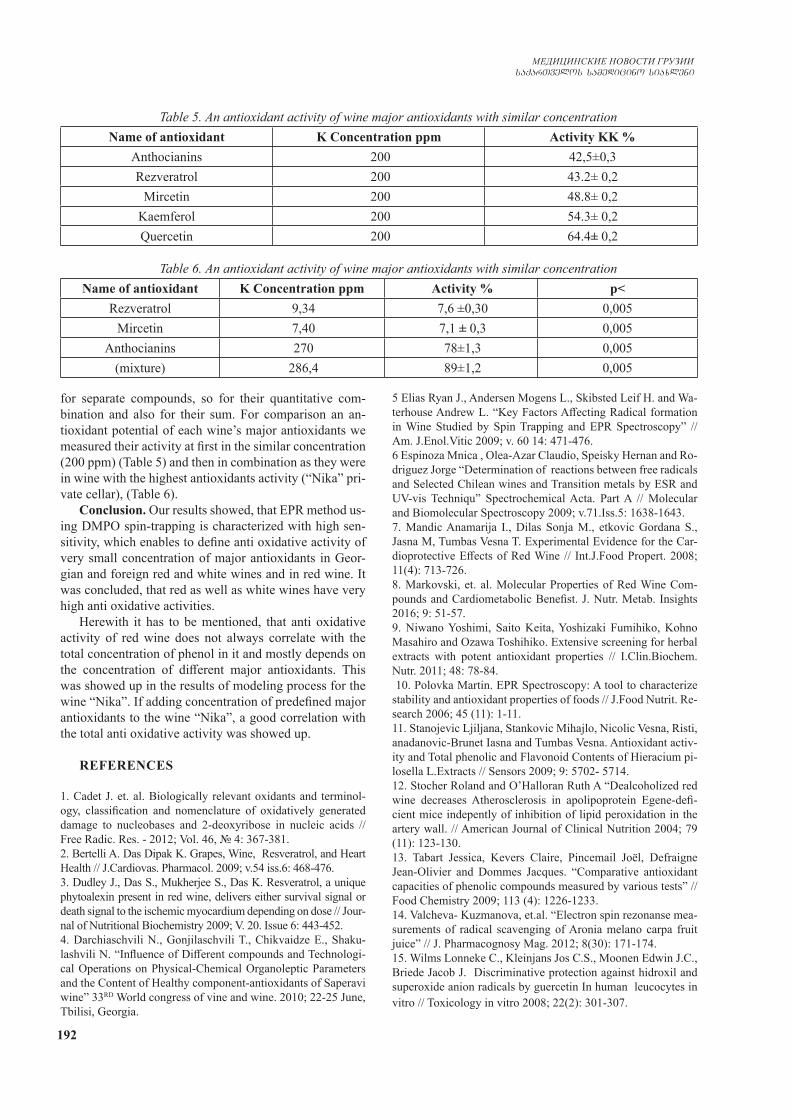
192
МЕДИЦИНСКИЕ НОВОСТИ ГРУЗИИ
CFMFHSDTKJC CFVTLBWBYJ CBF[KTYB
for separate compounds, so for their quantitative com-bination and also for their sum. For comparison an an-tioxidant potential of each wine’s major antioxidants we measured their activity at first in the similar concentration (200 ppm) (Table 5) and then in combination as they were in wine with the highest antioxidants activity (“Nika” pri-vate cellar), (Table 6).
Conclusion. Our results showed, that EPR method us-ing DMPO spin-trapping is characterized with high sen-sitivity, which enables to define anti oxidative activity of very small concentration of major antioxidants in Geor-gian and foreign red and white wines and in red wine. It was concluded, that red as well as white wines have very high anti oxidative activities.
Herewith it has to be mentioned, that anti oxidative activity of red wine does not always correlate with the total concentration of phenol in it and mostly depends on the concentration of different major antioxidants. This was showed up in the results of modeling process for the wine “Nika”. If adding concentration of predefined major antioxidants to the wine “Nika”, a good correlation with the total anti oxidative activity was showed up.
REFERENCES
1. Cadet J. et. al. Biologically relevant oxidants and terminol-ogy, classification and nomenclature of oxidatively generated damage to nucleobases and 2-deoxyribose in nucleic acids // Free Radic. Res. - 2012; Vol. 46, № 4: 367-381. 2. Bertelli A. Das Dipak K. Grapes, Wine, Resveratrol, and Heart Health // J.Cardiovas. Pharmacol. 2009; v.54 iss.6: 468-476.3. Dudley J., Das S., Mukherjee S., Das K. Resveratrol, a unique phytoalexin present in red wine, delivers either survival signal or death signal to the ischemic myocardium depending on dose // Jour-nal of Nutritional Biochemistry 2009; V. 20. Issue 6: 443-452.4. Darchiaschvili N., Gonjilaschvili T., Chikvaidze E., Shaku-lashvili N. “Influence of Different compounds and Technologi-cal Operations on Physical-Chemical Organoleptic Parameters and the Content of Healthy component-antioxidants of Saperavi wine” 33RD World congress of vine and wine. 2010; 22-25 June, Tbilisi, Georgia.
5 Elias Ryan J., Andersen Mogens L., Skibsted Leif H. and Wa-terhouse Andrew L. “Key Factors Affecting Radical formation in Wine Studied by Spin Trapping and EPR Spectroscopy” // Am. J.Enol.Vitic 2009; v. 60 14: 471-476. 6 Espinoza Mnica , Olea-Azar Claudio, Speisky Hernan and Ro-driguez Jorge “Determination of reactions between free radicals and Selected Chilean wines and Transition metals by ESR and UV-vis Techniqu” Spectrochemical Acta. Part A // Molecular and Biomolecular Spectroscopy 2009; v.71.Iss.5: 1638-1643.7. Mandic Anamarija I., Dilas Sonja M., etkovic Gordana S., Jasna M, Tumbas Vesna T. Experimental Evidence for the Car-dioprotective Effects of Red Wine // Int.J.Food Propert. 2008; 11(4): 713-726.8. Markovski, et. al. Molecular Properties of Red Wine Com-pounds and Cardiometabolic Benefist. J. Nutr. Metab. Insights 2016; 9: 51-57.9. Niwano Yoshimi, Saito Keita, Yoshizaki Fumihiko, Kohno Masahiro and Ozawa Toshihiko. Extensive screening for herbal extracts with potent antioxidant properties // I.Clin.Biochem.Nutr. 2011; 48: 78-84. 10. Polovka Martin. EPR Spectroscopy: A tool to characterize stability and antioxidant properties of foods // J.Food Nutrit. Re-search 2006; 45 (11): 1-11.11. Stanojevic Ljiljana, Stankovic Mihajlo, Nicolic Vesna, Risti, anadanovic-Brunet Iasna and Tumbas Vesna. Antioxidant activ-ity and Total phenolic and Flavonoid Contents of Hieracium pi-losella L.Extracts // Sensors 2009; 9: 5702- 5714. 12. Stocher Roland and O’Halloran Ruth A “Dealcoholized red wine decreases Atherosclerosis in apolipoprotein Egene-defi-cient mice indepently of inhibition of lipid peroxidation in the artery wall. // American Journal of Clinical Nutrition 2004; 79 (11): 123-130.13. Tabart Jessica, Kevers Claire, Pincemail Joël, Defraigne Jean-Olivier and Dommes Jacques. “Comparative antioxidant capacities of phenolic compounds measured by various tests” // Food Chemistry 2009; 113 (4): 1226-1233.14. Valcheva- Kuzmanova, et.al. “Electron spin rezonanse mea-surements of radical scavenging of Aronia melano carpa fruit juice” // J. Pharmacognosy Mag. 2012; 8(30): 171-174. 15. Wilms Lonneke C., Kleinjans Jos C.S., Moonen Edwin J.C., Briede Jacob J. Discriminative protection against hidroxil and superoxide anion radicals by guercetin In human leucocytes in vitro // Toxicology in vitro 2008; 22(2): 301-307.
Table 5. An antioxidant activity of wine major antioxidants with similar concentrationName of antioxidant K Concentration ppm Activity KK %
Anthocianins 200 42,5±0,3Rezveratrol 200 43.2± 0,2
Mircetin 200 48.8± 0,2Kaemferol 200 54.3± 0,2Quercetin 200 64.4± 0,2
Table 6. An antioxidant activity of wine major antioxidants with similar concentrationName of antioxidant K Concentration ppm Activity % p<
Rezveratrol 9,34 7,6 ±0,30 0,005Mircetin 7,40 7,1 ± 0,3 0,005
Anthocianins 270 78±1,3 0,005(mixture) 286,4 89±1,2 0,005
GEORGIAN MEDICAL NEWS No 5 (278) 2018
© GMN 193
SUMMARY
DETERMINATION OF ANTIOXIDANT ACTIVITY OF WINES AND WINE’S MAJOR PHENOLIC COMPOUNDS BY ELECTRON SPIN RESONANS, USING SPIN-TRAPS METHOD
Chikvaidze E., Gogoladze T., Miminoshvili A.
I. Javakhishvili Tbilisi State Univeristy; Sokhumi State University, Georgia
Our results showed, that EPR method using DMPO spin-trapping is characterized with high sensitivity, which enables to define anti oxidative activity of very small con-centration of major antioxidants in Georgian and foreign red and white wines and in red wine. It was concluded, that red as well as white wines have very high anti oxidative activities. Herewith it has to be mentioned, that anti oxi-dative activity of red wine does not always correlate with the total concentration of phenol in it and mostly depends
on the concentration of different major antioxidants. This was showed up in the results of modeling process for the wine “Nika”. If adding concentration of predefined major antioxidants to the wine “Nika”, a good correlation with the total anti oxidative activity was showed up.
Keywords: Electron Spin resonans, antioxidant ac-tivity, spin-trap, major antioxidants, Trans-rezveratrol, Total phenols, Cis-rezveratroli, Total rezveratrol, Mirce-tin, Quercetin, Kemferol, Total anthocianins.
РЕЗЮМЕ
ОПРЕДЕЛЕНИЕ АНТИОКСИДАНТНОЙ АКТИВНОСТИ И ОСНОВНЫХ МАЖОРНЫХ ФЕНОЛЬНЫХ КОМПОНЕНТОВ ВИН МЕТОДОМ ЭЛЕКТРОН-СПИН-РЕЗОНАНСА
С ПРИМЕНЕНИЕМ СПИН-ЛОВУШЕК
Чикваидзе Э.Н., Гоголадзе Т.В., Миминошвили А.А.
Тбилисский государственный универсистет им. И. Джавахишвили; Сухумский государственный университет, Тбилиси, Грузия
Методом электрон-спин-резонансной спектроско-пии с применением спин-ловушек изучена антиокси-дантная активность Грузинских и иностранных красных и белых вин, а также отдельных мажорных фенольных соединений, входящих в их состав. Установлена высокая антиоксидантная активность как красных, так и белых вин. Показано, что антиоксидантная активность красных вин, в
основном, зависит от коцентрации отдельных мажорных антиоксидантов. Эти данные подтверждаются на примере модели антиоксидантной активности вина “ Ника“, в ко-тором антиоксидантная активность смеси мажорных фе-нольных соединений в концентрациях, соответствующих таковым в вине, находится в полном соответствии с анти-оксидантной активностью вина.
reziume
Rvinisa da misi ZiriTadi maJoruli fenoluri komponentebis antioqsidanturi aqtivobis gansazRvra eleqtronuli spin-rezonansis meTodiT spin-xafangis gamoyenebiT
e. CikvaiZe, T. gogolaZe, a. miminoSvili
i. javaxiSvilis sax. Tbilisis saxelmwifo universiteti; soxumis saxelmwifo universiteti, Tbilisi, saqarTvelo
eleqtronuli spin-rezonansis speqtro-skopiis meTodiT spin-xafangis gamoyenebiT Seswavlilia rogorc qarTuli, aseve ucxouri wiTeli da TeTri Rvinoebis da wiTel Rvinoeb-Si Semavali zogierTi maJoruli fenoluri komponentebis antioqsidanturi aqtivoba. dadgenilia, rom rogorc wiTel aseve TeTr Rvinoebs gaaCnia maRali antioqsidanturi aq-tivoba. wiTeli Rvinis antioqsidanturi aq-
tivoba ZiriTadaT damokidebulia RvinoSi calkeuli maJoruli anrioqsidantebis kon-centraciaze, rac dasturdeba Rvino „nika“-s modelirebis magaliTze, rodesac RvinoSi gansazRvruli calkeuli maJoruli antioqsi-dantebis Sesabamisi koncentraciiT Serevisas, narevis antioqsidanturi aqtivoba karg Tanxved-raSia Rvino „nika“–s antioqsidantur aqtivo-basTan.
194
МЕДИЦИНСКИЕ НОВОСТИ ГРУЗИИ
CFMFHSDTKJC CFVTLBWBYJ CBF[KTYB
Наиболее актуальной проблемой современно-го здравоохранения является качество оказания первичной медико-санитарной помощи, которая зависит от развития сети амбулаторно-поликлини-ческих учреждений. Первичная медико-санитарная помощь (ПМСП), как известно, составляет неотъ-емлемую часть национальной системы здравоохра-нения, осуществляя ее главную функцию и являясь центральным звеном всей системы организации здравоохранения. Она представляет собой первый этап непрерывного процесса в охране здоровья на-селения. Исходя из вышеизложенного, вопросам первичной медицинской помощи в проводимых на национальном уровне реформах уделяется особое внимание [8,9,12,13].
Государственная программа Республики Ка-захстан «Денсаулық» на 2016-2019 гг. направле-на на обеспечение национальных систем здраво-охранения кадровыми ресурсами в необходимом объеме и с должным уровнем квалификации, на развитие эффективной, устойчивой системы ох-раны здоровья граждан как основы социального благополучия и экономического процветания го-сударства [1,6,7].
Большинство национальных систем здравоох-ранения сталкиваются с глубоко укоренившимися проблемами дисбаланса кадровых ресурсов, усили-вающихся под воздействием демографических, по-литических, социально- экономических, технологи-ческих, эпидемиологических изменений [4]. В этой связи возникает необходимость проведения посто-янного мониторинга и анализа кадровых ресурсов здравоохранения (КРЗ) [14].
На 1 января 2017 года медицинскую помощь населению Республики Казахстан оказывают 840 больничных (670 государственных и 170 частных) и 2718 амбулаторно-поликлинических организаций (1856 государственных и 862 частных). Сельско-му населению республики медицинскую помощь оказывают 143 центральных и районных больниц, 142 сельских участковых и сельских больниц, 198 районных и сельских поликлиник, 1328 врачебных и семейных врачебных амбулаторий, 20 центров ПМСП, 3987 фельдшерско-акушерских и медицин-ских пунктов [14].
Эффективность функционирования всех инсти-тутов системы здравоохранения напрямую зависит от количественных и качественных характеристик
КРЗ. При этом решение проблемы отрасли и от-дельных ее служб путем простого увеличения ко-личества кадров не всегда оправдано: большинство стран «большой десятки» (G10) имеют более низкий показатель обеспеченности врачебными кадрами, однако более положительную динамику медико-де-мографических показателей. В Республике Казах-стан (РК) количество врачей составляет 30,4 на 10 тыс. населения, в США, Великобритании, Японии и Канаде - 26, 23, 21 и 19 на 10 тыс. населения, соот-ветственно [15,18]. Медико-демографический пока-затель по Западному Казахстану - Актюбинской об-ласти - составляет 32,0, Атырауской области - 21,6, в Западно-Казахстанской области (ЗКО) - 24,9 на 10 тыс. населения [14].
Целью настоящего исследования явилось изуче-ние состояния кадрового обеспечения амбулаторно-поликлинических учреждений Западного Казахстана, проводящих медицинские осмотры населения.
Материал и методы. В основе оценки кадровой обеспеченности системы здравоохранения в мире лежат такие показатели как численность населе-ния, половозрастной состав и показатель заболева-емости. Другой путь - это изучение норм нагрузки на специалистов и объем предоставляемых услуг. Состояние кадровой обеспеченности амбулатор-но-поликлинических учреждений по Западному Казахстану, проводящих медицинские осмотры на-селения, в данном случае изучалось посредством анализа деятельности 58 медицинских учреждений за 2013-2016 гг. Исследования проведены в Актю-бинской, Атырауской, Западно-Казахстанской об-ластях.
Проанализирован показатель обеспеченности амбулаторно-поликлинических учреждений врачеб-ными кадрами, непосредствененно участвующими в проведении медицинских осмотров населения. При оценке кадровой обеспеченности изучались также вопросы дальнейшего повышения врачами профессиональнной квалификации и прохожде-ния тематического усовершенствования. Особое внимание уделено переподготовке по специаль-ности «профпатология» и тематическому усовер-шенствованию для имеющих действующий сер-тификат по специальности «профпатология» для участия в проведении медицинсих осмотров за указанный период.
Результаты и их обсуждение. В соответствии с
СОСТОЯНИЕ КАДРОВОГО ОБЕСПЕЧЕНИЯ АМБУЛАТОРНО-ПОЛИКЛИНИЧЕСКИХ УЧРЕЖДЕНИЙ,
ПРОВОДЯЩИХ МЕДИЦИНСКИЕ ОСМОТРЫ НАСЕЛЕНИЯ
Шаймбетов Ж.М., Сатыбалдиева У.А., Мамырбаев А.А., Путкарадзе М., Глонти С.
Западно-Казахстанский государственный медицинский университет им. Марата Оспанова, Актобе, Республика Казахстан
GEORGIAN MEDICAL NEWS No 5 (278) 2018
© GMN 195
пунктом 4 статьи 155 Кодекса Республики Казахстан от 18 сентября 2009 года «О здоровье народа и системе здравоохранения», издан приказ министра националь-ной экономики Республики Казахстан от 28 февраля 2015 года №175, который содержит перечень вредных производственных факторов, профессий, при которых проводятся обязательные медицинские осмотры [2]. В Перечне четко прописано в каких производствах, профессиях, какие специалисты должны проходить обязательные медицинские осмотры, их периодич-ность, участие врачей узких специальностей, прово-димые лабораторные и функциональные исследова-ния, а также медицинские противопоказания к тем или иным видам профессиональной деятельности.
Проведенный анализ выявил, что, по сей день от-мечается дефицит врачебных кадров. Укомплектован-ность медицинскими кадрами амбулаторно-поликли-нических учреждений Атырауской области в 2013 году составляла - 59,2%, в 2014 -54,3%, 2015 - 68,7%, 2016 - 70,5%. Следовательно, приведенные фактиче-ские данные указывают на динамический рост обе-спеченности врачебными кадрами. Однако, определя-ется дефицит врачей различных специальностей.
В таблице 1 показано, что из 16 необходимых специальностей, участвующих в медицинских осмо-трах, на 100% укомплектованы только две - хирур-гия и терапия. По трем специальностям отмечается острая нехватка кадров: обеспеченность онкологами составляет - 4,7-52,0%, гематологами - 4,7-8,0%, ал-лергологами - 9,4-32,0%, однако в динамике отмеча-ется стабильный рост онкологов и аллергологов, за
исключением гематологов. По трем ниже следующим специальностям достоверная динамика отсутствует - дерматовенерология, психиатрия, урология.
Численность врачей, проводящих медицинские осмотры населения ЗКО, согласно проведенному анализу по 2013-2016 гг, самая низкая по Западному региону, Как видно из таблицы 2, укомплектован-ность медицинскими кадрами амбулаторно-поли-клинических учреждений ЗКО в 2013 г. составила 54,2%, в 2014, 2015, 2016 гг. - 54,8%, 56,8%, 58,5%, соответственно. Приведенные данные свидетель-ствуют о дефиците врачей, учавствующих в про-ведении периодических медицинских осмотров населения. Наиболее остро отмечается нехватка ге-матологов - 3,7%, онкологов - 29,6%, аллергологов - 14,8%, кардиологов - 22,2%.
Совершенно иные данные по обеспеченности по-лучены в городе Актобе, где процент врачей в период проведения медицинских осмотров населения в 2013 г. составил 61,2%, в 2014, 2015, 2016 гг. 67,4%, 73,8%, 86,3%, соответственно. В данном регионе в динами-ке отмечается достаточно хорошая обеспеченность медицинскими кадрами, в сравнении с предыдущими областями.
Анализ обеспеченности узкими специалистами, проведенный по г. Актобе в 2016 году (профпатолог, терапевт, оториноларинголог, невропатолог, дерма-товенеролог, аллерголог, офтальмолог, гинеколог, рентгенолог) выявил 100% обеспеченность. Однако определен дефицит психиатров - 32,2% и гематологов – 16,6%.
Таблица 1. Обеспеченность врачебными кадрами амбулаторно-поликлинических учреждений Атырауской области, проводящих медицинские осмотры населения в 2013-2016 гг.
Специальность 2013 г 2014 г 2015 г 2016 гПрофпатолог 70,5 67,2 83,6 88,0Терапевт 100,0 100,0 100,0 100,0Оториноларинголог 89,3 84,0 100,0 100,0Невропатолог 84,6 79,8 100,0 92,0Дерматовенеролог 42,3 37,8 48,4 56,0Аллерголог 9,4 12,6 30,8 32,0Офтальмолог 94,0 88,2 100,0 100,0Онколог 4,7 8,4 49,6 52,0Стоматолог 28,2 29,4 35,2 40,0Гинеколог 94,0 88,2 92,4 92,0Психиатр 28,2 25,2 26,4 32,0Гематолог 4,7 4,2 8,8 8,0Кардиолог 65,8 58,8 88,0 92,0Рентгенолог 89,3 79,8 88,0 92,0Уролог 42,3 37,8 48,4 52,0Хирург 100,0 96,6 100,0 100,0Обеспечнность врачами (%) 59,2 54,3 68,7 70,5
196
МЕДИЦИНСКИЕ НОВОСТИ ГРУЗИИ
CFMFHSDTKJC CFVTLBWBYJ CBF[KTYB
Таблица 2. Обеспеченность врачебными кадрами амбулаторно-поликлинических учреждений по Западно-Казахстанской области, проводящих медицинские осмотры населения в 2013-2016 гг.
Специальность 2013 г 2014 г 2015 г 2016 гПрофпатолог 66,6 62,9 70,3 77,7Терапевт 92,5 96,2 92,5 96,2Оториноларинголог 96,2 96,2 85,1 81,4Невропатолог 88,8 88,8 81,4 92,6Дерматовенеролог 59,2 59,2 62,9 77,7Аллерголог 14,8 14,8 14,8 14,8Офтальмолог 92,5 92,5 88,8 88,8Онколог 29,6 29,6 33,3 29,6Стоматолог 33,3 33,3 40,7 37,0Гинеколог 81,4 81,4 81,4 85,1Психиатр 18,5 18,5 29,6 33,3Гематолог 0,0 0,0 0,0 3,7Кардиолог 18,5 22,2 25,9 22,2Рентгенолог 51,8 55,5 70,3 66,6Уролог 29,6 33,3 37,0 40,7Хирург 92,5 92,5 96,2 88,8Обеспечнность врачами (%) 54,2 54,8 56,8 58,5
Таблица 3. Обеспеченность врачебными кадрами амбулаторно-поликлинических учреждений, проводящих медицинские осмотры населения по г. Актобе в 2013-2016 гг.
Специальность 2013 г 2014 г 2015 г 2016 г
Профпатолог 66,4 66,4 83,0 100Терапевт 83,0 83,0 100 100Оториноларинголог 83,0 83,0 100 100Невропатолог 66,4 83,0 100 100Дерматовенеролог 66,4 83,0 83,0 100Аллерголог 66,4 83,0 83,0 100Офтальмолог 83,0 83,0 100 100Онколог 49,8 49,8 66,4 83,0Стоматолог 49,8 66,4 66,4 66,4Гинеколог 66,4 66,4 83,0 100Психиатр 16,6 16,6 33,2 33,2Гематолог 16,6 16,6 16,6 16,6Кардиолог 49,8 66,4 66,4 83,0Рентгенолог 66,4 66,4 83,0 100Уролог 66,4 83,0 100 100Хирург 83,0 83,0 100 100Обеспечнность врачами (%) 61,2 67,4 73,8 86,3
Согласно действующим приказам министерства здравоохранения РК по проведению предваритель-ного или периодического медицинского осмотра, администрацией той или иной медицинской орга-
низации формируется постоянно действующая вра-чебная комиссия, в состав которой входят: профпа-толог и врачи узких специальностей, прошедшие в установленном порядке повышение квалификации
GEORGIAN MEDICAL NEWS No 5 (278) 2018
© GMN 197
по специальности “профпатология” и имеющие действующий сертификат по специальности “про-фпатология”. Врачебную комиссию возглавляет профпатолог. Обеспеченность профпатологами по Западному Казахстану амбулаторно-поликлиниче-ских учреждений в период с 2013 по 2016 гг. варьи-рует от 71% до 90%; за эти годы отмечена четкая тен-денция увеличения профпатологов на 19%. Наиболее острая их нехватка отмечается в ЗКО - 78,6% в 2016 г. В городе Актобе выявлена 100% обеспеченность про-фпатологами (рис. 1).
Рис. 1. Обеспеченность амбулаторно-поликлини-ческих учреждений профпатологами по Западному Казахстану
Число врачей, прошедших в установленном по-рядке повышение квалификации по специальности «проф-патология» или имеющих действующий сер-тификат по специальности для проведения меди-цинских осмотров в период 2013-2016 гг., по Запад-ному Казахстану в 2013 г. составило 67,7%, в 2014 г. – 75,3%, 2015 г. - 84,6%, 2016 г. - 87,8%. Следователь-но, приведенные фактические данные указывают на динамический рост числа специалистов, прошедших повышение квалификации или имеющих действую-щий сертификат по профпатологии. Наиболее низкий показатель отмечается в ЗКО - 73,4% в 2016 г. В срав-нении с вышеуказанным регионом, в Актобе обеспе-ченность соответствующими кадрами наблюдается на уровне 96,4%, данные представлены на рис. 2.
Рис. 2. Показатели врачей, прошедших в установ-ленном порядке повышение квалификации по специ-альности «профпатология» или имеющих действую-щий сертификат по этой специальности
Согласно прогнозу [6], экономика развитых стран мира с середины 2020-х будет эффективнее развиваться в том числе и благодаря сфере здра-воохранения. По данным ВОЗ, расходы на кадры здравоохранения, включая подготовку 2,4 миллио-на медицинских работников до 2020 года, составят 39 миллиардов долларов США. По подсчетам ВОЗ, в 38 странах Африки - в районе Сахары норматив – 20 врачей на 100.000 населения не выполняется. Анализ кадров в 2012 г. в США показал, что страна нуждается в 100.000 врачах, 1 миллионе медицин-ских сестер, 250.000 профессиональных работни-ков общественного здравоохранения. В Австралии 5% рабочей силы в здравоохранении абсорбируют две трети расходов на здравоохранение, т.е. 9,7% от валового внутреннего продукта [11].
Состояние здоровья населения России весьма не-благоприятное - демографическая ситуация в регио-нах ухудшается; недостаточная эффективность систе-мы здравоохранения требует ресурсных инвестиций и кадровых ресурсов [16]. Несмотря на вышеперечис-ленные проблемы, уровень обеспеченности врачеб-ными кадрами в целом по России за последние годы увеличился, однако по субъектам Российской Федера-ции отмечается двукратная диспропорция. Отмечена несбалансированность подготовки кадров по специ-альностям, характеризующаяся ростом численности врачей-специалистов «узкого» профиля и уменьше-нием числа врачей терапевтического профиля (на 20,4%), педиатров (на 5,3%), санитарных врачей (на 30,7%), врачей скорой помощи (на 8,8%) [16].
В кадровой политике здравоохранения Казах-стана имеется множество проблемных вопросов, многие из них характерны для стран СНГ и требу-ют безотлагательного решения [3,4]. В Казахстане среднемесячная номинальная заработная плата вра-ча составляет около 372 долларов США, что почти в 16 раз меньше, чем в странах, входящих в Органи-зацию экономического сотрудничества и развития, что обуславливает непрестижность профессии и слабую трудовую мотивацию медицинских работ-ников. Следует также отметить слабую зависимость оплаты труда от его интенсивности, результата и квалификации работников [10].
Результаты проведенных исследовании показали, что в анализируемых регионах Западного Казахста-на отмечается существенный дефицит обеспечен-ности врачами узких специальностей. Особо выра-женный дефицит во врачебных кадрах наблюдается в Атырауской, Западно-Казахстанской областях. Обе-спеченность врачами профпатологами по сей день остается низкой. Учитывая, что предварительные и периодические медосмотры направлены, прежде все-го, на определение влияния вредных производствен-ных факторов на здоровье работающего населения, сложившаяся ситуация по кадровому обеспечению
198
МЕДИЦИНСКИЕ НОВОСТИ ГРУЗИИ
CFMFHSDTKJC CFVTLBWBYJ CBF[KTYB
амбулаторно-поликлинических учреждений напря-мую влияет на качество проводимых медицинских осмотров организованных групп населения.
Результаты исследования вопросов кадрового обе-спечения в динамике указывает на благоприятную тенденцию по укомплектованности амбулаторно-по-ликлинических учреждений Западного Казахстана врачебными кадрами узких специальностей.
ЛИТЕРАТУРА
1. Государственная программа развития здравоохранения Республики Казахстан «Денсаулык» на 2016-2019 годы. 2. Приказ Министра национальной экономики Республики Казахстан от 28 февраля 2015 года № 175. Об утверждении Перечня вредных производственных факторов, профессий, при которых проводятся обязательные медицинские осмотры. 3. Методы планирования, прогнозирования кадровых ре-сурсов здравоохранения (Методические рекомендации) Астана: 2015.4. Концепция развития кадровых ресурсов здравоохранения на 2012-2020 гг. https://kaznmu.kz/. 5. Аналитический материал расширенной коллеги Министер-ства Здравоохранения Республики Казахстан, Астана: 2017.6. Бекмухамбетов Е.Ж., О Государственной программе раз-вития здравоохранения Республики Казахстан «Денсаулык» на 2016-2019 годы / Е.Ж. Бекмухамбетов, Р.Е. Нургалиева, И.Б. Кенжебаева // Медицинский журнал Западного Казах-стана. – 2016. - №2 (50). –С. 86-90. 18.7. Мамырбаев А.А., Современные аспекты состояния обще-ственного здоровья (обзор литературы) / Мамырбаев А.А, Умарова Г.А. // Georgian medical news 2016; №5(254): 15.8. Мамырбаев А.А., Современные медико-экологическое аспекты состояния общественного здоровья / Мамырба-ев А.А., Умарова Г.А., // Астана медециналық журналы - 1/2017; 7-13.9. Мамырбаев А.А., Анализ деятельности амбулаторно-по-ликлинических организаций в современных условиях. / Ма-мырбаев А.А., Каститис Шмигелкас, Умарова Г.А., // Вест-ник КазНМУ - №1-2017; 515-519.10. Медицина Казахстана – болевые точки и выбор пути, 2015 http://www.rcrz.kz/old/index.php/ru 11. Роберт Фогель (Нобелевский лауреат по экономике). Подходы ВОЗ И ЕС к оценке кадрового потенциала http://studbooks.net/155733012. Русев И.Т., Организация оказания первичной медико-санитарной помощи в амбулаторных условиях на примере мегаполиса. / Русев И.Т., Карайланов М.Г., Прокин И.Г., Кузьмин С.Г., // Вестник Российской Военно-медициской академии 2016; 3(55): 144-147. 13. Стародубов В.Н., Первичная медицинская помощь: со-стояние и перспективы развития // Медицина 2007; 261.14. Сыздыкова А.А., Егеубаева С.А., Койков В.В., Калиева М.А., Березин С.С., Турумбетова Т.Б., Современные под-ходы к решению проблем кадровой обеспеченности в об-ласти здравоохранения / Сыздыкова А.А., Егеубаева С.А., Койков В.В., Калиева М.А., Березин С.С., Турумбетова Т.Б. // Journal of health development - 2013; №1-2(66-67), С20-28.15. Handbook on monitoring and evaluation of human resources for health, with special applications forlow- and middle-income countries. Geneva, World Health Organization, 2009. Avaliable at URL:http://www.who.int/hrh/resources/handbook/en/
16. Starodubov V.I., Mihaylova Yu.V., Leonov S.A. Human re-sources for health in the Russian Federation: a condition, prob-lems and basic tendencies of development // Federal Public Health Institute, Moscow: 2010.17. The world health report 2006: working together for health. Geneva, World Health Organization, 2006. Avaliable at URL: http://www.who.int/whr/2006. 18. World Health Statistics 2009. Avaliable at URL: http://www.who.int/gho/publications/world_health_statistics/2009/en/in-dex.html.
SUMMARY
STATE OF HUMAN RESOURCING FOR OUTPA-TIENT-AND-POLYCLINIC INSTITUTIONS CON-DUCTING MEDICAL INSPECTIONS OF THE POPULATION
Shaimbetov Zh., Satybaldiyeva U., Mamyrbaev A., Phutkaradze M., Glonti S.
West Kazakhstan Marat Ospanov State Medical Univer-sity, Aktobe, Kazakhstan
The article presents data related to research on the staffing situation of outpatient-and-polyclinic institutions in Western Kazakhstan, conducting medical examinations of the population. The personnel supply and activity of 58 medical institutions of Aktobe, Atyrau, West Kazakhstan regions for 2013-2016 are analyzed. The low availability of outpatient-and-polyclinic institutions by medical per-sonnel of narrow specialties is established. At the same time, there is a tendency to improve the availability of medical personnel for the period under study. The most prosperous situation is in the city of Aktobe.
Keywords: outpatient-and-polyclinic institutions, medical care, Kazakhstan.
РЕЗЮМЕ
СОСТОЯНИЕ КАДРОВОГО ОБЕСПЕЧЕНИЯ АМБУЛАТОРНО-ПОЛИКЛИНИЧЕСКИХ УЧ-РЕЖДЕНИЙ, ПРОВОДЯЩИХ МЕДИЦИНСКИЕ ОСМОТРЫ НАСЕЛЕНИЯ
Шаймбетов Ж.М., Сатыбалдиева У.А., Мамырбаев А.А., Путкарадзе М., Глонти С.
Западно-Казахстанский государственный медицин-ский университет им. Марата Оспанова, Актобе, Республика Казахстан
В статье представлены данные исследования по со-стоянию кадровой обеспеченности амбулаторно-по-ликлинических учреждений Западного Казахстана, проводящих медицинские осмотры населения. Проана-лизирована кадровая обеспеченность и деятельность 58 медицинских учреждений Актюбинской, Атырауской,
GEORGIAN MEDICAL NEWS No 5 (278) 2018
© GMN 199
Западно-Казахстанской областей за 2013-2016 гг. Выяв-лена низкая обеспеченность амбулаторно-поликлиниче-ских учреждений врачебными кадрами узких специаль-ностей. Показана тенденция улучшения обеспеченности
врачебными кадрами за исследуемый промежуток вре-мени. Наиболее благополучная ситуация выявлена в го-роде Актобе.
VOCATIONAL TRAINING AND ACTIVITY OF PHARMACISTS IN GEORGIA
1,2Sulashvili N., 1Beglaryan M., 3Kvijinadze N., 4Matoshvili M.
1Yerevan State Medical University, Department of Social Pharmacy and Pharmaceutical Management, Armenia;
2University of Georgia, School of Health Sciences and Public Health; Tbilisi State Medical University, 3Department of Social and Clinical Pharmacy; 4Department of Dermatology and Venerology, Georgia
Pharmacists’ job satisfaction is a complex phenom-enon. There is a relationship between pharmacists’ job satisfaction and motivation. A positive correlation between job satisfaction, productivity and motivation encourages employee. [5,14,15]. Job satisfaction is described as pleasant or positive emotional state the evaluation of their work or work experience. The re-sults of pharmacist job satisfaction from the perception that performs its task, or allows the execution of its own important values job, provided that to extent that these values are comparable to their needs [9,11,13]. Job satisfaction, affective and emotional response to the various aspects of work. Pharmacist job satisfac-tion or dissatisfaction plays an important role in many aspects of community pharmacy practice [7,8]. Ca-reer satisfaction is a very important factor to human motivation and performance. Many studies related to the work were conducted among pharmacists in dif-ferent countries. In Georgia, very little research has been conducted on the job satisfaction of pharmacists [6,10,12]. Job satisfaction and career satisfaction and both are happily with their working lives, but contrib-ute to this happiness differently. Career satisfaction can be defined as the level of general happiness, em-pirically chosen professions. Job satisfaction is related
to the current situation of and depends on many fac-tors, including market conditions, place of work and other dynamic effects. Job satisfaction is a subjective variable, which defies easy quantification. Pharma-cists experience job satisfaction when they fulfill the needs or considerations, is seen as important in their work. Job satisfaction is positively related to com-munity pharmacists’ career satisfaction [1,2,3,4,16].
Aim and objects was to study and analyze fea-tures of job satisfaction of pharmacists in Georgia.
Material and methods. Research objects are materi-als of sociological research: The study was quantitative investigation and analysis of job satisfaction for phar-macists’ in Georgia by using survey (Questionnaire by Olga Sokolova) [17]. Surveys were for pharmacists, 810 pharmacists were interviewed in Georgia. The survey was conducted on 09.09.2016. - 30.04.2017. We used methods of systematic, sociological (surveying, questioning), com-parative, mathematical-statistical, graphical analysis. The data was processed and analyzed with the SPSS program.
Results and their discussion. On the basis of per-formed of sociological study of pharmacist specialists the following results have been founded:
The vast majority of pharmacist specialists were female (Fig. 1). In my opinion male pharmacists do not have inter-
reziume
ambulatoruli da poliklinikuri dawesebulebebis eqimebiT sakadro uzrunvelyofis mdgomareobis Sefaseba
J. Saimbetovi, u. satibaldieva, a. mamirbaevi, m. fuTkaraZe, s. Rlonti
dasavleT yazaxeTis m. ospanovis sax. saxelmwifo samedicino universiteti, aqtobe, yazaxeTi
statiaSi warmodgenilia monacemebi dasavleT yazaxeTis ambulatoruli da po-liklinikuri qselis da misi sakadro uz-runvelyofis mdgomareobis Sesaxeb. gaanali-zebulia 58 sxvadasxva samxareo samedicino
dawesebulebebis sakadro mdgomareoba da moRvaweoba 2013-2016 ww. periodSi. gamovle-nilia ambulatoruli da poliklimikuri samsaxuris Zalian dabali uzrunvelyofa viwro specoalobebis eqimebiT.
200
МЕДИЦИНСКИЕ НОВОСТИ ГРУЗИИ
CFMFHSDTKJC CFVTLBWBYJ CBF[KTYB
est to work in drug-store. The reason is low income and un-satisfactory working conditions for male pharmacists. It is important that the drug-store (pharmacy) as the work place to become more attractive for the male pharmacists.
Fig. 1. Distribution of respondents by gender
Age of the pharmacists varied from 21 to 70 years. Majority of pharmacist specialists were from 26 to 45 years old. (Fig. 2). Long term work experienced pharma-cist had professional diseases, because pharmacists’ work contain large physical load. It is important that, the elderly pharmacists might be more actively involved in the work of the drugstore in more appropriate and adapted working environment and condition.
By the education, 0.2% of Pharmacists have second-ary professional education, 0.4% of pharmacists have col-lege education, 96.4% of pharmacists have higher edu-cation and 3% of pharmacists have postgraduate studies (PhD studies or Doctoral studies). Majority of respondent pharmacists’ specialists owned pharmacist qualification with 5 years of higher education. About a quarter of re-spondent pharmacists specialists owned Bachelor –Phar-macist qualification (Bachelor‘s Degree in Pharmacy-B Pharma), About one seventh of respondent pharmacists specialists owned Master -Pharmacist qualification (Mas-ter s academic degree in pharmacy). About three quarters of respondent pharmacists specialists owned certified
pharmacist qualification by the Ministry of Health. It is necessary to start pharmacists’ certification. This will im-prove pharmaceutical care in the pharmacy.
By the qualification, 0.6% of respondents have phar-macist’s assistant qualification.69% of respondents have pharmacist qualification with 5 years of higher education, 24.2% of respondents have Bachelor –Pharmacist qualifica-tion (Bachelor‘s Degree in Pharmacy), 13.3% of respondents have Master –Pharmacist qualification (Master ‘s academic degree in pharmacy), Respondents 72% were Certified Phar-macist by the ministry of health, respondents’ 0.6% were PhD in Pharmaceutical Sciences, respondents 0.2% were Academic Doctor in Pharmacy(PhD in Pharmacy). Majority of the respondent pharmacist specialists owned pharmacist qualification in 5 years of higher education. About a quar-ter of them owned bachelor pharmacist qualification (Bach-elor‘s Degree in Pharmacy-B Pharma), about one seventh of them owned Master –Pharmacist qualification (Master‘s academic degree in pharmacy). About three quarters of the respondent pharmacists specialists owned certified phar-macist qualification by the MOH of Georgia (Fig. 3). It is necessary to start pharmacists’ certification to improve the pharmaceutical care.
By the type of job, 94.2% of pharmacists work at drugstore – pharmacy, 1% of pharmacists work at whole-sale pharmaceutical organization (pharmaceutical ware-house), 0.5% of pharmacists work at pharmacy point- drugstore point, 0.1% of pharmacists work at analytical laboratory, 4.2% of pharmacists work at hospital-clinic pharmacy (drugstore). The vast majority type of pharma-ceutical organization jobs of respondent pharmacists was drugstore, being as a major place for pharmacists’ em-ployment (Fig. 4).
Fig. 2. Respondent’s aget
Fig. 3. Qualification of respondents
Fig. 4. Pharmaceutical organization type at job of respondents’

GEORGIAN MEDICAL NEWS No 5 (278) 2018
© GMN 201
On the question what most of all had influence on your profession (occupational) choice (indicate only one answer), the responses was such: pharmacists’ 23% think parents’ advices, pharmacists’ 4% think teachers’ advices , pharmacists’ 3.5% think advice of an expert-specialist of professional orientation ( of career guidance) , pharma-cists’ 13.3% think the desire (ambition) to obtain a profes-sion in compliance of own trends, aspirations, and incli-nations (affections), respondents pharmacists’ 1.9% think there was nowhere to go , pharmacists’ 2.2% consider dis-satisfaction with first education , pharmacists’ 37.8% con-sider personal desire , pharmacists’ 14.4% think specialty love from childhood. In my mind, it is more efficient that the potential pharmacists choose their future profession only according to their wishes, personal desire, awareness based on their own trends, aspirations and inclinations (Fig. 5).
The main underlying motives, while making profes-sional (occupational) choice of respondent pharmacist were: desire to obtain high-quality professional education (training)-44.8%, guarantee to be employed 42.6%, inter-est in a profession-39.4%, the desire to care for the health of people-43%.
On the question-are you satisfied with your profes-sional (occupational) choice? Are you satisfied with your profession? Pharmacist’ 57.7% satisfied with professional (occupational) choice, pharmacist’ 25.3% partially sat-isfied with professional (occupational) choice, pharma-cists’ 4.4% have doubts with professional (occupational) choice, pharmacists’ 6% disappointed with professional (occupational) choice. Pharmacists’ 6.5% not satisfied with professional (occupational) choice. A little bit more than half of respondent pharmacists (pharmacist special-ists) were satisfied of professional choice; about a quar-ter of them were partially satisfied of professional choice (Fig. 6). It is significant, that pharmaceutical companies make study of their own pharmacists’ motivations, as-pirations, tendency, intention, ambition, desire and wish for professional choice. The pharmaceutical companies should make better working condition for pharmacists’, to increase satisfaction of occupational choice.
On the question -are you satisfied with your profes-sional career? Pharmacists’ 30.4 % were satisfied with professional career, pharmacists’ 33.7 % were partially satisfied with professional career, pharmacist’ 35.9 % were not satisfied with professional career. More than one third of respondent pharmacists were not satisfied with pro-fessional career, about one third of them were partially sat-isfied with professional career (Fig.7). It is significant, that pharmaceutical companies make study of their own pharma-cists’ satisfaction with professional career. The pharmaceuti-cal companies should study a combination of all factors that affect the satisfaction with professional career.
Fig. 7. Satisfaction professional career of respondents’
On the question are you satisfied with your work (job)? Pharmacist 33.2% were satisfied with work (job), pharma-cists’ 37.3% were partially satisfied with work (job), phar-macists’ 24.4 % were not satisfied with work (job) and phar-macists’ 5.1% cannot say. About a quarter of respondent pharmacists were not satisfied with work; more than one third of them were partially satisfied with work (Fig. 8). It is significant, that pharmaceutical companies make study of pharmacists work satisfaction. The pharmaceutical compa-nies should determine the combination of factors that affect on the pharmacists’ work satisfaction.
Fig. 8. Satisfaction with work of respondents’
On the question to what extent you have realized your professional capabilities, skills and habits? Pharmacists’ 18.4% answer -to the full extent, pharmacists’ 46.3% answer
Fig. 5. Factors, mostly influenced on respondents’ profession (occupational) choice
Fig. 6. Satisfaction of professional choice of respondents’

202
МЕДИЦИНСКИЕ НОВОСТИ ГРУЗИИ
CFMFHSDTKJC CFVTLBWBYJ CBF[KTYB
-partially, more than 50% of own potential, pharmacists’ 24.7% answer- partially, less than 50% of own potential, pharmacists’ 10.6% answer-cannot say. About a quarter of respondent pharmacists (pharmacist specialists) have real-ized professional capabilities, skills and habits partially; less than 50% of them - of own potential; about half of them have realized professional capabilities, skills and habits partially; more than 50% of them - of own potential (Fig. 9). Phar-maceutical companies should create constructive working conditions for pharmacists to maximally realize their profes-sional capabilities, skills and habits. This will increase the quality of pharmaceutical care in pharmacies.
On the question -In your opinion, at what level it is possi-ble to cease education? Pharmacists’ 4.3% answer -after get-ting specialist diploma (degree), pharmacists’ 11.2% answer- after getting the specialist certificate, pharmacists’ 84.4% answer -education should not be ceased. The respondents (pharmacist specialists) ‘ majority considered that education should not be ceased; the minority of them - that it is possible to cease education after getting specialist diploma (degree) or the specialist certificate (Fig. 10). It is of the crucial neces-sity that all the pharmacists should realize, reconsider and understand the importance of continuous pharmaceutical and medical education in constantly.
On the question-have you used knowledge in the practice, obtained from professional publications? Phar-macists’ 51.4 % answer yes, pharmacists’ 40.7% answer –partially, pharmacists’ 7.9% answer-no. The minorities of respondent pharmacists had not used knowledge in their practice, obtained from professional publications; less than half of them had partially used that knowledge (Fig. 11). It is very important that pharmacists have to use knowledge obtained from the professional publications, journals and the modern pharmaceutical literature in their practice. In my opinion pharmacists’ certifications give more motivation to all pharmacists to read modern phar-maceutical publications permanently.
Fig. 11. Opinion of respondents having used knowl-edge in the practice, obtained from professional publica-tions
Fig. 9. To what extent respondents’ have realized professional capabilities, skills and habits
Fig. 10. Respodents’ opinion, at what level possible to cease education
Fig. 12. Attitude of respondents’ to qualification upgrading (improvement) study courses
On the question-What is your attitude to qualification up-grading (improvement) study courses? Pharmacists’ 55.6% answer I learn with great pleasure, pharmacists’ 38.6% an-swer learning process rise interest to me, pharmacists’ 5.8% answer -I have indifferent attitude toward learning. The
minority of respondents (pharmacist specialists) had indif-ferent attitude toward learning and qualification upgrading (improvement) study courses (Fig. 12). The professional trainings and professional workshops are appear to be very necessary for the further professional advancement.
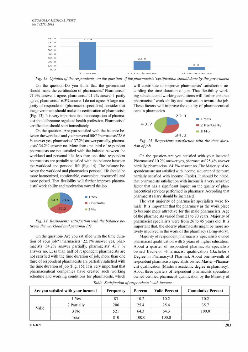
GEORGIAN MEDICAL NEWS No 5 (278) 2018
© GMN 203
On the question-Do you think that the government should make the certification of pharmacists? Pharmacists’ 71.9% answer I agree, pharmacists’21.9% answer I partly agree, pharmacists’ 6.3% answer I do not agree. A large ma-jority of respondents’ (pharmacist specialists) consider that the government should make the certification of pharmacists (Fig. 13). It is very important that the occupation of pharma-cist should become regulated health profession. Pharmacists’ certification should start immediately.
On the question- Are you satisfied with the balance be-tween the workload and your personal life? Pharmacists’ 28.6 % answer yes, pharmacists’ 37.2% answer partially, pharma-cists’ 34.2% answer no. More than one third of respondent pharmacists are not satisfied with the balance between the workload and personal life, less than one third respondent pharmacists are partially satisfied with the balance between the workload and personal life (Fig. 14). The balance be-tween the workload and pharmacists personal life should be more harmonized, comfortable, convenient, resourceful and more poised. That flexibility will further improve pharma-cists’ work ability and motivation toward the job.
Fig. 14. Respodents’ satisfaction with the balance be-tween the workload and personal life
On the question- Are you satisfied with the time dura-tion of your job? Pharmacists’ 22.1% answer yes, phar-macists’ 34.2% answer partially, pharmacists’ 43.7 % answer no. Less than half of respondent pharmacists are not satisfied with the time duration of job, more than one third of respondent pharmacists are partially satisfied with the time duration of job (Fig. 15). It is very important that pharmaceutical companies have created such working schedule and working conditions for pharmacists, which
will contribute to improve pharmacists’ satisfaction ac-cording the time duration of job. That flexibility work-ing schedule and working conditions will further enhance pharmacists’ work ability and motivation toward the job. These factors will improve the quality of pharmaceutical care in pharmacies.
Fig. 15. Respodents satisfaction with the time dura-tion of job
On the question-Are you satisfied with your income? Pharmacists’ 10.2% answer yes, pharmacists’ 25.4% answer partially, pharmacists’ 64.3% answer no. The Majority of re-spondents are not satisfied with income, a quarter of them are partially satisfied with income (Table). It should be noted, that pharmacists satisfaction with income is a very sensitive factor that has a significant impact on the quality of phar-maceutical services performed in pharmacy. According that pharmacist salary should be increased.
The vast majority of pharmacist specialists were fe-male. It is important that the pharmacy as the work place to become more attractive for the male pharmacists. Age of the pharmacists varied from 21 to 70 years. Majority of pharmacist specialists were from 26 to 45 years old. It is important that, the elderly pharmacists might be more ac-tively involved in the work of the pharmacy (Drug-story).
Majority of respondent pharmacists’ specialists owned pharmacist qualification with 5 years of higher education. About a quarter of respondent pharmacists specialists owned Bachelor –Pharmacist qualification (Bachelor‘s Degree in Pharmacy-B Pharma), About one seventh of respondent pharmacists specialists owned Master –Pharma-cist qualification (Master s academic degree in pharmacy). About three quarters of respondent pharmacists specialists owned certified pharmacist qualification by the Ministry of
Fig. 13. Opinion of the respondents, on the question- if the pharmacists’ certification should done by the government
Table. Satisfaction of respondents’ with income
Are you satisfied with your income? Frequency Percent Valid Percent Cumulative Percent
Valid
1 Yes 83 10.2 10.2 10.22 Partially 206 25.4 25.4 35.7
3 No 521 64.3 64.3 100.0Total 810 100.0 100.0
204
МЕДИЦИНСКИЕ НОВОСТИ ГРУЗИИ
CFMFHSDTKJC CFVTLBWBYJ CBF[KTYB
Health. It is necessary to start pharmacists’ certification. This will improve pharmaceutical care in the pharmacy.
Majority of the respondent pharmacist specialists owned pharmacist qualification in 5 years of higher edu-cation. About a quarter of them owned Bbachelor – phar-macist qualification (Bachelor‘s Degree in Pharmacy-B Pharma), about one seventh of them owned Master –Pharmacist qualification (Master‘s academic degree in pharmacy). About three quarters of the respondent phar-macists specialists owned certified pharmacist qualifica-tion by the MOH of Georgia. It is necessary to start phar-macists’ certification to improve the pharmaceutical care.
The vast majority type of pharmaceutical organization jobs of respondent pharmacists was pharmacy, being aas a major place for pharmacists’ employment
The main underlying motives, while making profes-sional (occupational) choice of respondent pharmacist were: desire to obtain high-quality professional education (training)-44.8%, guarantee to be employed 42.6%, inter-est in a profession-39.4%, the desire to care for the health of people-43% .It is more efficient that the potential phar-macists choose their future profession only according to their wishes, personal desire, awareness based on their own trends, aspirations and inclinations.
A little bit more than half of respondent pharmacists (pharmacist specialists) were satisfied of professional choice; about a quarter of them were partially satisfied of professional choice. It is significant, that pharmaceutical companies make study of their own pharmacists’ moti-vations, aspirations, tendency, intention, ambition, desire and wish for professional choice.
More than one third of respondent pharmacists were not satisfied with professional career, about one third of them were partially satisfied with professional career. It is significant, that pharmaceutical companies make study of their own pharmacists’ satisfaction with professional career. The pharmaceutical companies should study a combination of all factors that affect the satisfaction with professional career.
About a quarter of respondent pharmacists were not satisfied with work; more than one third of them were partially satisfied with work. It is significant, that pharma-ceutical companies make study of pharmacists work satis-faction. The pharmaceutical companies should determine the combination of factors that effect on the pharmacists’ work satisfaction.
About a quarter of respondent pharmacists (pharma-cist specialists) have realized professional capabilities, skills and habits partially; less than 50% of them - of own potential; about half of them have realized professional capabilities, skills and habits partially; more than 50% of them - of own potential . Pharmaceutical companies should create constructive working conditions for phar-macists to maximally realize their professional capabili-ties, skills and habits. This will increase the quality of pharmaceutical care in pharmacies.
The respondents (pharmacist specialists) ‘ majority con-
sidered that education should not be ceased; the minority of them - that it is possible to cease education after getting specialist diploma (degree) or the specialist certificate. It is of the crucial necessity that all the pharmacists should real-ize, reconsider and understand the importance of continuous pharmaceutical and medical education in constantly.
The minorities of respondent pharmacists had not used knowledge in their practice, obtained from professional publications; less than half of them had partially used that knowledge. It is very important that pharmacists have to use knowledge obtained from the professional publica-tions, journals and the modern pharmaceutical literature in their practice.
The minority of respondents (pharmacist specialists) had indifferent attitude toward learning and qualification upgrading (improvement) study courses. The professional trainings and professional workshops are appear to be very necessary for the further professional advancement.
A large majority of respondents’ (pharmacist specialists) consider that the government should make the certification of pharmacists. It is very important that the occupation of pharmacist should become regulated health profession. Pharmacists’ certification should start immediately.
More than one third of respondent pharmacists are not satisfied with the balance between the workload and per-sonal life, less than one third respondent pharmacists are partially satisfied with the balance between the workload and personal life. The balance between the workload and pharmacists personal life should be more harmonized, comfortable, convenient, resourceful and more poised. That flexibility will further improve pharmacists’ work ability and motivation toward the job.
Less than half of respondent pharmacists are not satis-fied with the time duration of job, more than one third of respondent pharmacists are partially satisfied with the time duration of job. It is very important that pharmaceutical companies have created such working schedule and working conditions for pharmacists, which will contribute to improve pharmacists’ satisfaction according the time duration of job. That flexibility working schedule and working conditions will further enhance pharmacists’ work ability and motiva-tion toward the job. These factors will improve the quality of pharmaceutical care in pharmacies.
The Majority of respondents are not satisfied with in-come, a quarter of them are partially satisfied with income .It should be noted, that pharmacists satisfaction with income is a very sensitive factor that has a significant impact on the quality of pharmaceutical services performed in pharmacy. According that pharmacist salary should be increased.
Conclusion. The pharmaceutical society should orga-nize preparation and implementation of the registration-certification regulations for the pharmacists’ staff. To raise the professional standards the Government should make the certification of higher pharmaceutical educa-tion pharmacists, which is very essential for pharmacists’ professional perfection, the higher pharmaceutical edu-cation pharmacists’ self-realization and also their career
GEORGIAN MEDICAL NEWS No 5 (278) 2018
© GMN 205
advancement, for the pharmaceutical continuous profes-sional education provision, for pharmacists’ professional growth, their job and career satisfaction. This implemen-tation will ensure the pharmacists’ much higher status among the healthcare specialists, their economic welfare and career advancement, also enable realization of the received knowledge and the professional capabilities and skills in work at the maximal extent, as well as get sat-isfaction with the profession, job and salary, which is in their turn is essential to provide a high correspondence of the pharmacists’ qualification to work and an opportunity to have a private pharmaceutical activity. All that men-tioned above is very important to provide higher quality pharmaceutical care in pharmacy.
REFERENCES
1. Ballantyne PJ. Assessing pharmacists’ impacts in primary health care: are we asking the right questions? Southern Med. Rev. 2011;pp41-42.2. Bowling NA. Is the job satisfaction-Job performance rela-tionship spurious? A meta-analytic examination. J Vocat Behav. 2007; 26-34.3. Faragher EB, Cass M, Cooper CL. The relationship between job satisfaction and health: a meta-analysis. Occup Environ Med 2015;62: 105–112.4. Hassell K, Seston E, Shann P. Measuring job satisfaction of UK pharmacists: a pilot study. Int. J. Pharm. Practice . 2007;56-59.5. Khalidi D.A., Wazaify M. Assessment of pharmacists’ job satisfaction and job related stress in Amman 2013. Int. J. Clin. Pharm. 2013;35:821–828. 6. Koustelios A, Tsigilis N. The relationship between burnout and job satisfaction among physical education teachers: a multi-variate approach. Eur Phys Ed Rev 2005; 17-22.7. Mahoney R, Katona C, Mcparland M, Noble L, Livingston G. Shortage specialties: changes in career intentions from medical student to newly qualified doctor. Med Teach. 2004;12-14.8. Mak VS, March GJ, Clark A, Gilbert AL. Australian intern pharmacists’ perceived preparedness for practice, and their ex-pectations and experiences of the internship year and future ca-reer intentions. Integ Pharm Res Prac. 2013; 13-16.9. McCann L, Hughes C M, Adair C G, Cardwell C. Assessing job satisfaction and stress among pharmacists in Northern Ire-land. Pharm. World Sci. 2009; 188–194. 10. McPhersonGS,DaviesJG, BewickV,Bhudia A. Identification of factors affecting job and career satisfaction in hospital phar-macists. Pharm J 2009 ;21-2511. Mott D.A., Doucette W.R., Gaither C.A., Pedersen C.A., Schommer J.C. Pharmacists’ attitudes toward worklife: Results from a national survey of pharmacists 2004. J. Am. Pharm. As-soc. 2004;11-16.12. Mott DA. Pharmacist job turnover, length of service, and reasons for leaving,. Am J Health Syst Pharm 2000;57: 975–984.13. Schafheutle E.I. Factors influencing pharmacist performance: A review of the peer-reviewed literature. Health Policy. 2011; 15-17.14. Seston E, Hassell K, Ferguson J, Hann M. Exploring the relationship between pharmacists’ job satisfaction, intention to quit profession, and actual quitting. Soc. Pharm. 2009; 8-14.15. Seston E., Hassell K., Ferguson J., Hann M. Exploring the relationship between pharmacists’ job satisfaction, inten-tion to quit the profession, and actual quitting. Res.Soc.Adm.Pharm.2009;25-28.
16. Sweney, P.D. McFarlin, D.B. Organizational Behavior, Solu-tions for Management, McGrawHill/Irwin, New York, 2005. 57-62.17. Соколова О. В. Совершенствование процесса профессионального становления фармацевтиче-ских работников в системе фармацевтической помо-щи: дис. … канд. фармац. наук. – М., 2009. – 217 с.
SUMMARY
VOCATIONAL TRAINING AND ACTIVITY OF PHARMACISTS IN GEORGIA
1,2Sulashvili N., 1Beglaryan M., 3Kvijinadze N., 4Matoshvili M.
1Yerevan State Medical University, Department of Social Pharmacy and Pharmaceutical Management, Armenia;
2University of Georgia, School of Health Sciences and Public Health; Tbilisi State Medical University, 3Depart-ment of Social and Clinical Pharmacy; 4Department of Dermatology and Venerology, Georgia
Aim of the research was to study features of pharmacists’ job satisfaction in Georgia. The study was quantitative in-vestigation by using survey (Questionnaire). Surveys were for pharmacists, 810 pharmacists were interviewed in Geor-gia. We used methods of systematic, sociological (survey-ing, questioning), comparative, segmentation, mathematical-statistical, graphical analysis. The data was processed and analyzed with the SPSS program. On the basis of performed of sociological study of pharmacist specialists have been founded that the government should organize preparation and implementation of the registration-certification regula-tions for the pharmacists’ staff.
Keywords: Features ,pharmacy, employed, pharma-cist, faculty, work, study, satisfaction, job, career, drug-store.
РЕЗЮМЕ
ПРОФЕССИОНАЛЬНАЯ ПОДГОТОВКА И ДЕЯ-ТЕЛЬНОСТЬ ФАРМАЦЕВТОВ В ГРУЗИИ
1,2Сулашвили Н.В., 1Бегларян М.Г., 3Квижинадзе Н.О., 4Матошвили М.Т.
1Ереванский государственный медицинский универ-ситет, кафедра социальной фармации и фармацев-тического менеджмента, Армения; 2Университет Грузии, школа общественного здравоохранения; Тби-лисский государственный медицинский университет, 3департамент социальной и клинической фармации; 4департамент дерматологии и венерологии, Грузия
Целью исследования явилась оценка професси-ональной подготовки и деятельности фармацевтов в
206
МЕДИЦИНСКИЕ НОВОСТИ ГРУЗИИ
CFMFHSDTKJC CFVTLBWBYJ CBF[KTYB
Грузии. Проведено анкетирование фармацевтов с ис-пользованием опросников. Опрошены 810 фармацевтов, работающих в фармацевтических учреждениях Грузии. Использованы социологические, сравнительные, сег-ментационные, математико-статистические и графиче-
ские методы анализа. Полученные данные проанали-зированы и обработаны в программе SPSS. На основе проведенного исследования установлена необходимость организации подготовки и внедрения правил регистрации, сертификации и имплементации для фармацевтов.
reziume
farmacevtTa profesiuli momzadeba da saqmianoba saqarTveloSi
1,2n. sulaSvili, 1m. beglariani, 3n. kviJinaZe, 4m. maToSvili
1erevnis saxelmwifo samedicino universiteti, socialuri farmaciis da farmaciis menejmentis departamenti, somxeTi; 2saqarTvelos universiteti,
janmrTelobis mecnierebisa da sazogadoebrivi jandacvis skola; Tbilisis saxelmwifo samedicino universiteti, 3socialuri da klinikuri
farmaciis departamenti; 4dermatologiis da venerologiis departamenti, saqarTvelo
kvlevis mizans warmoadgenda saqarTveloSi momuSave farmacevtebis profesiuli momza-debis da maTi saqmianobis Seswavla. farma-cevtebisaTvis Sedgenili specialuri kiTx-varis gamoyenebiT Catarda 810 farmacevtis gamokiTxva. kvlevaSi gamoyenebuli iyo socio-logiuri, SedarebiTi, segmentaciuri, maTema-
tikur-statistikuri da grafikuli analizis meTodebi. miRebuli monacemebi damuSavda SPSS programis gamoyenebiT. kvlevis Sedegebze dayrdnobiT avtorebis mier dadgenilia, rom aucilebelia ganxorcieldes farmacevtTa registraciis da sertificirebis reguli-rebis wesebis momzadeba da implementacia.
* * *
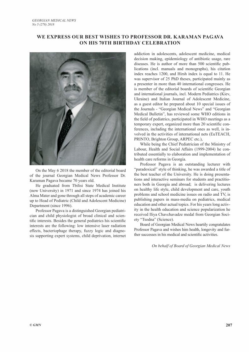
GEORGIAN MEDICAL NEWS No 5 (278) 2018
© GMN 207
WE EXPRESS OUR BEST WISHES TO PROFESSOR DR. KARAMAN PAGAVA ON HIS 70TH BIRTHDAY CELEBRATION
On the May 6 2018 the member of the editorial board of the journal Georgian Medical News Professor Dr. Karaman Pagava became 70 years old.
He graduated from Tbilisi State Medical Institute (now University) in 1971 and since 1974 has joined his Alma Mater and gone through all steps of academic career up to Head of Pediatric (Child and Adolescent Medicine) Department (since 1996).
Professor Pagava is a distinguished Georgian pediatri-cian and child physiologist of broad clinical and scien-tific interests. Besides the general pediatrics his scientific interests are the following: low intensive laser radiation effects, bacteriophage therapy, fuzzy logic and diagno-sis supporting expert systems, child deprivation, internet
addiction in adolescents, adolescent medicine, medical decision making, epidemiology of antibiotic usage, rare diseases. He is author of more than 500 scientific pub-lications (incl. manuals and monographs), his citation index reaches 1200, and Hirsh index is equal to 11. He was supervisor of 25 PhD theses, participated mainly as a presenter in more than 40 international congresses. He is member of the editorial boards of scientific Georgian and international journals, incl. Modern Pediatrics (Kiev, Ukraine) and Italian Journal of Adolescent Medicine, as a guest editor he prepared about 10 special issues of the Journals - “Georgian Medical News” and “Georgian Medical Bulletin”, has reviewed some WHO editions in the field of pediatrics, participated in WHO meetings as a temporary expert, organized more than 20 scientific con-ferences, including the international ones as well, is in-volved in the activities of international nets (EuTEACH, PRINTO, Brighton Group, ARPEC etc.),
While being the Chief Pediatrician of the Ministry of Labour, Health and Social Affairs (1999-2004) he con-tributed essentially to elaboration and implementation of health care reforms in Georgia.
Professor Pagava is an outstanding lecturer with “paradoxical” style of thinking, he was awarded a title of the best teacher of the University. He is doing presenta-tions and interactive seminars for students and practitio-ners both in Georgia and abroad; is delivering lectures on healthy life style, child development and care, youth problems and school medicine issues on radio and TV, is publishing papers in mass-media on pediatrics, medical education and other actual topics. For his years long activ-ity in the health education and science popularization he received Iliya Chavchavadze medal from Georgian Soci-ety “Tsodna” (Science).
Board of Georgian Medical News heartily congratulates Professor Pagava and wishes him health, longevity and fur-ther successes in his medical and scientific activities.
On behalf of Board of Georgian Medical News
Related Documents


