Volume 57(6): 531–541, 2009 Journal of Histochemistry & Cytochemistry http://www.jhc.org ARTICLE Enzyme Activity and Immunohistochemical Localization of Dipeptidyl Peptidase 8 and 9 in Male Reproductive Tissues Véronique Dubois, Chris Van Ginneken, Hilde De Cock, Anne-Marie Lambeir, Pieter Van der Veken, Koen Augustyns, Xin Chen, Simon Scharpé, and Ingrid De Meester Laboratory of Medical Biochemistry (VD,A-ML,SS,IDM), Anatomy/Embryology of Domestic Animals (CVG,HDC), and Laboratory of Medicinal Chemistry (PVDV,KA), University of Antwerp, Wilrijk, Belgium, and Division of Biotechnology and Pharmaceutical Research, National Health Research Institutes, Zhu Nan, Taiwan, China (XC) SUMMARY The mRNA expression pattern of dipeptidyl peptidase (DPP) 8 and DPP9, two DPP4 homologs, was studied previously and showed a broad tissue distribution. In this study, protein expression and activity of DPP8 and DPP9 were investigated in male repro- ductive tissues of different mammals. Based on specific DPP activities and inhibition profiles, the proline-selective DPP activity in the bovine and rat testis could predominantly be attrib- uted to DPP8/9 and not to DPP4. This is in contrast to the epididymis, where most of the ac- tivity was caused by DPP4. Bovine sperm preparations had very low or undetectable DPP8/9 activity. After characterization of polyclonal antibodies specific for DPP8 or DPP9, we could localize both enzymes in seminiferous tubules of the testis. A specific staining for DPP9 was found associated with spermatozoids embedded in the epithelium, just before their release into the lumen, and in spermatids. DPP8 was localized in spermatozoids in an earlier stage of maturation. These findings help to provide insight into the physiological role of DPP4-like enzymes in the male reproductive system. This manuscript contains online supplemental material at http://www.jhc.org. Please visit this article online to view these materials. (J Histochem Cytochem 57:531–541, 2009) KEY WORDS dipeptidyl peptidase DPP IV DPP4 DPP2 DPP8 DPP9 testis epididymis anti-DPP8 antibody anti-DPP9 antibody THE PROLINE-SELECTIVE DIPEPTIDYL PEPTIDASES (DPPs) are a family of serine proteases that regulate diverse biologi- cal processes by the release of N-terminal dipeptides from peptides with proline at the penultimate position (Rosenblum and Kozarich 2003; Van der Veken et al. 2007a). The group comprises DPP4, fibroblast activa- tion protein a (FAP), DPP2, DPP8, and DPP9. DPP4 has been examined most intensively (reviewed in Boonacker and Van Noorden 2003; Lambeir et al. 2003), in con- trast to the other members, which are poorly character- ized (Abbott et al. 2000; Olsen and Wagtmann 2002; Henry et al. 2007; Maes et al. 2007b). The mRNA expression pattern of DPP8 and DPP9 was studied and showed a broad distribution among hu- man tissues. The highest DPP8 mRNA levels are found in testis and placenta. The enzyme is upregulated in ac- tivated T cells and expressed in all B- and T-cell lines examined (Abbott et al. 2000; Qi et al. 2003). The DPP9 mRNA expression levels are high in skeletal mus- cle, heart, liver, and peripheral blood leukocytes (Olsen and Wagtmann 2002; Qi et al. 2003; Ajami et al. 2004). The DPP8 and DPP9 mRNA expression profiles showed a ubiquitous distribution in different skin cell types (Thielitz et al. 2008a,b). In kidney, small intestine, lung, and pancreas of pig and dog, semiquantitative RT-PCR analysis showed different relative abundance of DPP4- like enzymes, with DPP4 and DPP9 having the highest expression, followed by DPP2 and DPP8 (Wagner et al. 2006). In mice, a higher number of DPP8 and DPP9 transcripts compared with DPP4 were present in colon, brain, skin, and thymus (Helmuth et al. 2008). Only very recently, the expression of DPP8 and DPP9 started to be studied at the protein and/or activity level. High expression levels of DPP8/9 were reported in rat and human brain (Frerker et al. 2007; Stremenova et al. 2007; Busek et al. 2008). Also, human leukocytes con- tained DPP8/9 activity (Maes et al. 2007a). DPP8 and DPP9 were upregulated in the bronchi after induction Correspondence to: Ingrid De Meester, Laboratory of Medical Biochemistry, University of Antwerp, Universiteitsplein 1, 2610 Wilrijk, Belgium. E-mail: [email protected] Received for publication September 11, 2008; accepted January 16, 2009 [DOI: 10.1369/jhc.2009.952739]. C The Histochemical Society, Inc. 0022-1554/09/$3.30 531 The Journal of Histochemistry & Cytochemistry

Welcome message from author
This document is posted to help you gain knowledge. Please leave a comment to let me know what you think about it! Share it to your friends and learn new things together.
Transcript
Volume 57(6): 531–541, 2009Journal of Histochemistry & Cytochemistry
http://www.jhc.org
ARTICLE
Enzyme Activity and Immunohistochemical Localization ofDipeptidyl Peptidase 8 and 9 in Male Reproductive Tissues
Véronique Dubois, Chris Van Ginneken, Hilde De Cock, Anne-Marie Lambeir,Pieter Van der Veken, Koen Augustyns, Xin Chen, Simon Scharpé, and Ingrid De Meester
Laboratory of Medical Biochemistry (VD,A-ML,SS,IDM), Anatomy/Embryology of Domestic Animals (CVG,HDC), andLaboratory of Medicinal Chemistry (PVDV,KA), University of Antwerp, Wilrijk, Belgium, and Division of Biotechnology andPharmaceutical Research, National Health Research Institutes, Zhu Nan, Taiwan, China (XC)
SUMMARY The mRNA expression pattern of dipeptidyl peptidase (DPP) 8 and DPP9, twoDPP4 homologs, was studied previously and showed a broad tissue distribution. In thisstudy, protein expression and activity of DPP8 and DPP9 were investigated in male repro-ductive tissues of different mammals. Based on specific DPP activities and inhibition profiles,the proline-selective DPP activity in the bovine and rat testis could predominantly be attrib-uted to DPP8/9 and not to DPP4. This is in contrast to the epididymis, where most of the ac-tivity was caused by DPP4. Bovine sperm preparations had very low or undetectable DPP8/9activity. After characterization of polyclonal antibodies specific for DPP8 or DPP9, we couldlocalize both enzymes in seminiferous tubules of the testis. A specific staining for DPP9 wasfound associated with spermatozoids embedded in the epithelium, just before their releaseinto the lumen, and in spermatids. DPP8 was localized in spermatozoids in an earlier stageof maturation. These findings help to provide insight into the physiological role of DPP4-likeenzymes in the male reproductive system. This manuscript contains online supplementalmaterial at http://www.jhc.org. Please visit this article online to view these materials.
(J Histochem Cytochem 57:531–541, 2009)
KEY WORDS
dipeptidyl peptidase
DPP IV
DPP4
DPP2
DPP8
DPP9
testis
epididymis
anti-DPP8 antibody
anti-DPP9 antibody
THE PROLINE-SELECTIVE DIPEPTIDYL PEPTIDASES (DPPs) are afamily of serine proteases that regulate diverse biologi-cal processes by the release of N-terminal dipeptidesfrom peptides with proline at the penultimate position(Rosenblum and Kozarich 2003; Van der Veken et al.2007a). The group comprises DPP4, fibroblast activa-tion protein a (FAP), DPP2, DPP8, and DPP9. DPP4 hasbeen examined most intensively (reviewed in Boonackerand Van Noorden 2003; Lambeir et al. 2003), in con-trast to the other members, which are poorly character-ized (Abbott et al. 2000; Olsen and Wagtmann 2002;Henry et al. 2007; Maes et al. 2007b).
The mRNA expression pattern of DPP8 and DPP9was studied and showed a broad distribution among hu-man tissues. The highest DPP8 mRNA levels are foundin testis and placenta. The enzyme is upregulated in ac-
tivated T cells and expressed in all B- and T-cell linesexamined (Abbott et al. 2000; Qi et al. 2003). TheDPP9 mRNA expression levels are high in skeletal mus-cle, heart, liver, and peripheral blood leukocytes (Olsenand Wagtmann 2002; Qi et al. 2003; Ajami et al. 2004).The DPP8 and DPP9 mRNA expression profiles showeda ubiquitous distribution in different skin cell types(Thielitz et al. 2008a,b). In kidney, small intestine, lung,and pancreas of pig and dog, semiquantitative RT-PCRanalysis showed different relative abundance of DPP4-like enzymes, with DPP4 and DPP9 having the highestexpression, followed by DPP2 and DPP8 (Wagner et al.2006). In mice, a higher number of DPP8 and DPP9transcripts compared with DPP4 were present in colon,brain, skin, and thymus (Helmuth et al. 2008).
Only very recently, the expression of DPP8 and DPP9started to be studied at the protein and/or activity level.High expression levels of DPP8/9 were reported in ratand human brain (Frerker et al. 2007; Stremenova et al.2007; Busek et al. 2008). Also, human leukocytes con-tained DPP8/9 activity (Maes et al. 2007a). DPP8 andDPP9 were upregulated in the bronchi after induction
Correspondence to: Ingrid De Meester, Laboratory of MedicalBiochemistry, University of Antwerp, Universiteitsplein 1, 2610Wilrijk,Belgium. E-mail: [email protected]
Received for publication September 11, 2008; accepted January 16,2009 [DOI: 10.1369/jhc.2009.952739].
C The Histochemical Society, Inc. 0022-1554/09/$3.30 531
TheJourna
lof
Histoch
emistry&
Cytoc
hemistry
of experimental asthma in the rat (Schade et al. 2008).The precise functions of these enzymes in vivo are stillunknown. There is some evidence that, in leukocytes,they are involved in immunoregulation. Inhibition ofDPP8 and DPP9 suppresses mitogen-stimulated T-cellresponses, whereas selective inhibition of DPP4 andDPP2 does not (Reinhold et al. 2008).
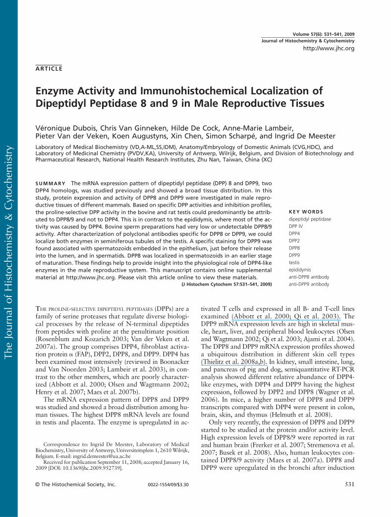
Despite the similar substrate specificity on X-Pro–containing chromogenic and fluorogenic substrates atneutral pH, the DPP4-like enzymes can be discriminatedusing selective inhibitors. The selectiveDPP2 inhibitor N-(4-chlorobenzyl)-4-oxo-4-(1-piperidinyl)-1,3-(S)-butane-diamine dihydrochloride (UAMC00039) (Senten et al.2004), the selective DPP4 inhibitor (3R)-3-amino-1-[3-(trifluoromethyl)-5,6-dihydro-[1,2,4]triazolo[4,3-a]pyrazin-7(8H)-yl]-4-(2,4,5-trifluorophenyl)butan-1-one(sitagliptin) (Kim et al. 2005), and the DPP8/9 inhibitor(2S,3R)-2-amino-1-(isoindolin-2-yl)-3-methylpentan-1-one (allo-Ile-isoindoline) (Lankas et al. 2005) were usedin our study (Figure 1).
Recently, we purified proline-selective DPPs clearlydifferent from DPP2 and DPP4 from bovine testes(Dubois et al. 2008). One of them was identified asDPP9. Here, we studied the distribution of the activityand cell-specific expression of DPP8 andDPP9 in themalereproductive system. Because no antibody preparationsagainst these enzymes have been studied thoroughly be-fore, we included a comparison and characterization here.Based on activity and inhibition profiles and immuno-blotting, we showed the expression of DPP8 and DPP9in the testis, epididymis, and sperm. IHC stains were usedto localize DPP8 and DPP9 in these tissues.
Materials and Methods
Materials
The DPP2 inhibitor UAMC00039 and the DPP8/9 in-hibitor allo-Ile-isoindoline were synthesized as described(Senten et al. 2004; Lankas et al. 2005). The DPP4 in-
hibitor sitagliptin was extracted from Januvia tablets(Merck; Vienna, Austria). Bovine tissues were kindlyprovided by a local slaughterhouse. Tissues of Wistarrats were obtained from the animalarium of the Univer-sity of Antwerp. After the animals were sacrificed, tissueswere excised, either immediately frozen in liquid nitrogenand stored at –80C until use or fixed in 4% paraformal-dehyde in PBS for 24 hr. Bovine sperm was obtainedfrom the “Vlaamse Rundveeverbetering” (Oosterzele,Belgium). DPP2 and DPP4 were purified from humanseminal plasma (De Meester et al. 1996; Maes et al.2005). Recombinant human DPP8 (rDPP8) was ex-pressed and purified as described (Chen et al. 2004).DPP9 was purified from bovine testes (Dubois et al.2008). The rabbit polyclonal anti-DPP9 antibody wasgenerated by Eurogentec as described in Dubois et al.(2008). Rabbit polyclonal antibodies against the spacerregion of DPP4 (ab28340), the aminoterminal end(ab42075), the spacer region (ab42076), and the cata-lytic domain (ab42077) of DPP8; the aminoterminal end(ab42078), the spacer region (ab42079), and the catalyticdomain (ab42080) of DPP9; and the chicken polyclonalantibody against DPP9 (ab26177) were obtained fromAbcam (Cambridge, UK) (Table 1). Ala-Pro-p-nitroanilide(-pNA) and Gly-Pro-pNAwere purchased from Bachem(Budendorf, Switzerland).
Tissue Homogenate and Sperm Preparations
Tissues were homogenized in lysis buffer (LB; 1%octylglucoside, 10 mM EDTA, and 70 mg/ml aprotininin 0.05 M HEPES buffer, pH 7.0; 5 ml/g) (Maes et al.2007a). After incubation for 1 hr, the homogenizedtissue was centrifuged for 10 min at 3000 3 g, andthe resulting supernatant was subsequently centrifugedfor 1 hr at 20,000 3 g at 4C. The final supernatantwas filtered.
Bull sperm samples were pooled. Differential centri-fugation of bovine sperm was performed according toVanhoof et al. (1992). In brief, bovine sperm was cen-
Figure 1 Structures of the dipeptidyl peptidase (DPP) inhibitors used: the DPP2 inhibitor N-(4-chlorobenzyl)-4-oxo-4-(1-piperidinyl)-1,3-(S)-butanediamine dihydrochloride (UAMC00039), the DPP8/9 inhibitor (2S,3R)-2-amino-1-(isoindolin-2-yl)-3-methylpentan-1-one (allo-Ile-isoindoline),and the DPP4 inhibitor (3R)-3-amino-1-[3-(trifluoromethyl)-5,6-dihydro-[1,2,4]triazolo[4,3-a]pyrazin-7(8H)-yl]-4-(2,4,5-trifluorophenyl)butan-1-one (sitagliptin).
532 Dubois, Van Ginneken, De Cock, Lambeir, Van der Veken, Augustyns, Chen, Scharpé, De Meester
TheJourna
lof
Histoch
emistry&
Cytoc
hemistry
trifuged at 900 3 g for 10 min at 4C. Spermatozoawere obtained from the pellet. The supernatant wascentrifuged at 105,000 3 g for 2 hr to precipitate theprostasomes. The final supernatant contained the semi-nal fluid. For clarification, 1% octylglucoside was addedto the seminal fluid. The seminal fluid was centrifuged at13,000 3 g at 4C, and the supernatant was used. Thespermatozoa and prostasomes were lysed as describedin Maes et al. (2007a). Subcellular fractionation of sper-matozoa was performed according to Cooper (2000).Cells were ruptured by a single freeze-thaw cycle, fol-lowed by 3 3 15-sec sonication on ice.
The resulting samples were used to measure enzymeactivities and protein concentration.
For the immunodetection of DPPs in homogenates,tissues were homogenized in 0.02 M HEPES buffer,pH7.0, containing 1%octylglucoside anda complete pro-tease inhibitor cocktail tablet (Roche; Brussels, Belgium).Apart from this, the procedures were as described above.
Enzyme Assays
Enzymatic activities were determined kinetically in afinal volume of 200 ml for 10 min at 37C by measuringthe initial velocities of pNA release (405 nm) from thesubstrate using a Spectramax plus microtiter plate reader(Molecular Devices; Sunnyvale, CA). One unit of enzy-matic activity is the amount of enzyme that catalyzesthe release of 1 mmol pNA from the substrate per minuteunder assay conditions. The substrates Ala-Pro-pNA(1 mM in 0.05 M HEPES buffer, pH 7.0, containing10 mM EDTA, 14 mg/ml aprotinin, and 0.1% Tween 20)and Gly-Pro-pNA (0.5 mM in 0.05MTris buffer, pH 8.3,containing 10 mM EDTA and 14 mg/ml aprotinin) wereused to probe DPP2, DPP4, DPP8/9, and/or FAP activity(Maes et al. 2007a).
Protein concentration was determined according toBradford (1976) with BSA (Sigma; Bornem, Belgium)as the standard.
Inhibition Assays
Tissue homogenates, cell lysates, and pure enzymesamples diluted in LB supplemented with ?10 mg/mlBSA were preincubated for 15 min at 37C with a widerange of inhibitor concentrations. DPP activities weredetermined as described above. The specific contribu-tion of DPP2 activity to the Ala-Pro-pNA cleavage wasdetermined using the DPP2 inhibitor UAMC00039(100 nM). The dual DPP8/9 inhibitor allo-Ile-isoindoline(5–5000 nM) served to determine the contribution ofboth enzymes to the total DPP activity. DPP4 activitywas inhibited by its selective inhibitor sitagliptin (50–50,000 nM).
Immunoaffinity Purification of the Rabbit PolyclonalAnti-DPP9 Antibody
The DPP9 peptide (LKSQGYDWSEPFSPG), synthe-sized by Eurogentec, was immobilized onto cyanogenbromide (CNBr)-activated Sepharose 4B (GE Healthcare;Diegem, Belgium) according to the manufacturer’s in-structions (Supplementary Materials, Dataset SD1). Rab-bit serum (5 ml) diluted 1:2 with PBS was incubated withthe DPP9 peptide-Sepharose 4B (1 ml) for 1 hr at roomtemperature, and the gel was washed successively withPBS, 0.05 M Tris, pH 8.0 and 9.0, and 0.05 M sodiumphosphate, pH 6.3, all containing 0.1% Triton X-100and 0.5 M NaCl. Bound antibodies were eluted with0.1 M citric acid, pH 3.0, and 0.1 M citric acid, pH 2.5.The fractions (1 ml) were neutralized in 250 ml 1 M Tris,pH 9.0, and visualized by dot spots using horseradishperoxidase (HRP)-conjugated goat anti-rabbit IgG (Bio-source; Merelbeke, Belgium) for 1 hr at 37C. Visualiza-tion of the blots occurred by DAB (Roche) diluted inperoxide buffer.
Western Blot Analysis
Western blot was performed as described by Duboiset al. (2008) with slight modifications. For the detec-
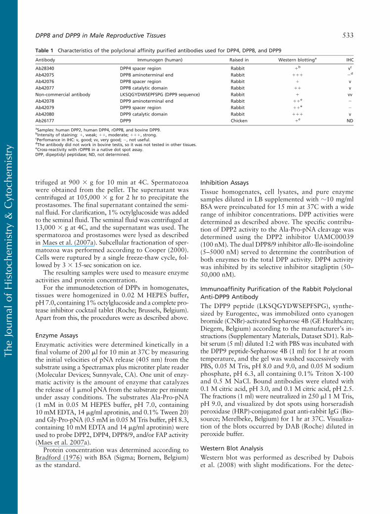
Table 1 Characteristics of the polyclonal affinity purified antibodies used for DPP4, DPP8, and DPP9
Antibody Immunogen (human) Raised in Western blottinga IHC
Ab28340 DPP4 spacer region Rabbit 1b vc
Ab42075 DPP8 aminoterminal end Rabbit 111 2d
Ab42076 DPP8 spacer region Rabbit 1 vAb42077 DPP8 catalytic domain Rabbit 11 vNon-commercial antibody LKSQGYDWSEPFSPG (DPP9 sequence) Rabbit 1 vvAb42078 DPP9 aminoterminal end Rabbit 11e 2
Ab42079 DPP9 spacer region Rabbit 11e 2
Ab42080 DPP9 catalytic domain Rabbit 111 vAb26177 DPP9 Chicken 1e ND
aSamples: human DPP2, human DPP4, rDPP8, and bovine DPP9.bIntensity of staining: 1, weak; 11, moderate; 111, strong.cPerfomance in IHC: v, good; vv, very good; 2, not useful.dThe antibody did not work in bovine testis, so it was not tested in other tissues.eCross-reactivity with rDPP8 in a native dot spot assay.DPP, dipeptidyl peptidase; ND, not determined.
DPP8 and DPP9 in Male Reproductive Tissues 533
TheJourna
lof
Histoch
emistry&
Cytoc
hemistry
tion of DPPs in tissue homogenates, blots were blockedwith 0.05 M Tris buffer containing 0.15 M NaCl,0.1% Tween 20, and 5% BSA for 1 hr at room tem-perature. Blots were incubated with 10% goat serum(Dako; Heverlee, Belgium) for 1 hr at room tempera-ture and with the primary antibodies for 1 hr at roomtemperature and overnight at 4C. HRP-conjugated goatanti-rabbit IgG (1:10,000) was used as a secondary anti-body in a 2-hr incubation at room temperature. Aftereach incubation step the blots were washed with 0.05 MTris buffer containing 0.15MNaCl and 0.1% Tween 20.Visualization of the blots occurred by OptiGo (Isogen-Life Science; Sint-Pieters-Leeuw, Belgium) using thesupersignal west femto maximum sensitivity substrate(Pierce; Erembodegem, Belgium).
Histochemical Activity Assay
The substrate H-Gly-L-Pro-1-hydroxy-4-naphtylamidehydrochloride was synthesized in-house, and the histo-chemical activity assay was performed according toSchade et al. (2008) with slight modifications. Sectionswere preincubated for 5 min at 37C with the inhibitorsUAMC00039 (100 nM), sitagliptin (10 mM), and/orallo-Ile-isoindoline (5 mM) in 0.1 M phosphate buffer,pH 7.8. Subsequently, the sections were incubated for20 hr at 37C in 0.1 M phosphate buffer, pH 7.8, con-taining 0.25 mMH-Gly-L-Pro-1-hydroxy-4-naphtylamidehydrochloride dissolved in DMSO (final: 0.1% DMSOin solution; Acros Organics, Geel, Belgium), 0.25 mMnitro blue tetrazolium (NBT; Sigma), and the inhibitorsat concentrations mentioned above. The sections werewashed (three times) with 0.1 M phosphate buffer,pH 7.8, and fixed in 4% paraformaldehyde. Sectionswere counterstainedwith 0.5%methyl green andmountedin 75% glycerol. Controls consisted of sections incu-bated without substrate.
IHC
After fixation, tissues were washed (three times) withPBS for 30 min, dehydrated, and embedded in paraffin,and 4-mm sections were cut. After deparaffinization andrehydration, antigen retrieval was performed for 15 minin a microwave in citrate buffer (pH 6.0; DPP8) or Tris/EDTA buffer (pH 9.0; DPP4 and DPP9; Dako). The sec-tions were treated for 10 min with 3% H2O2 in TBS toblock endogenous peroxidase activity and incubated sub-sequently with 10%non-immune goat serum tominimizenonspecific antibody binding. Sections were incubatedwith the primary polyclonal antibody diluted in TBScontaining 0.3% Triton X-100 and 1% BSA for 1 hrat room temperature, followed by incubating overnightat 4C. Incubation with biotinylated polyclonal goat anti-rabbit IgG (1:200; Dako) and streptavidin HRP (1:200;Dako) were each carried out for 30 min at room tem-perature. Immunoreactivity was visualized by applying
aminoethylcarbazole (Dako). Sections were counter-stained with hematoxylin and mounted in 75% glycerol.Controls consisted of (a) sections incubated with TBScontaining 0.3% Triton X-100 and 1% BSA instead ofthe specific primary antibody, and (b) sections incubatedwith normal rabbit serum at the same dilution as of thespecific primary antibody. Control slides were processedsimultaneously in the same session to eliminate interex-perimental variations and yielded non-detectable stain-ing, showing specificity of the staining results.
Results
Activity of DPPs in Male Reproductive Tissues andSperm Preparations
The distribution of the different proline-selective DPPactivities in male bovine and rat reproductive tissues andin bovine sperm preparations was determined using Ala-Pro-pNA at pH 7.0 and Gly-Pro-pNA at pH 8.3 (Maeset al. 2007a). Ala-Pro-pNA is cleaved by DPP2, DPP4,DPP8, DPP9, and FAP at neutral pH, whereas the cleav-age of Gly-Pro-pNA at pH 8.3 is catalyzed by the sameenzymes with the exception of DPP2, which is not activeat this alkaline pH. The choice of the selective inhibi-tors (Figure 1) allowed us to determine the contributionof DPP2, DPP4, and DPP8/9 to the total Ala-Pro-pNAcleaving activity. FAP has been reported not to be pres-ent in normal tissues (Wang et al. 2008); therefore, wedid not anticipate interference by FAP in our assays.At concentrations used to inhibit DPP2, DPP8/9, andDPP4 completely, none of the inhibitors used (Figure 1)affected rFAP activity (Kim et al. 2005; Van der Vekenet al. 2008). In the presence of all three inhibitors, hardlyany Ala-Pro-pNa-cleaving activity (#12%) was mea-sured in the male reproductive tissues (SupplementaryMaterials, Table ST1). Therefore, we concluded thatthere was no significant interference from FAP in ourexperimental setup.
The specific activities of individual proline-selectiveDPPs outlined in Figures 2 and 3 were calculated basedon the inhibition profiles using 5 mM allo-Ile-isoindoline(Supplementary Materials, Figures SF1 and SF2).
In the bovine testis and both reproductive tissues ofthe rat, the contribution of DPP2 was two to three timeshigher than in the bovine epididymis (Figure 2A). Ahigh DPP4 activity was found in the epididymis of bothspecies compared with the testis (Figure 2). The bovineepididymis exhibited the highest DPP4 vs DPP2 andDPP8/9 activity. In the bovine and rat testis, DPP8/9exceeded DPP4 activity (Figure 2).
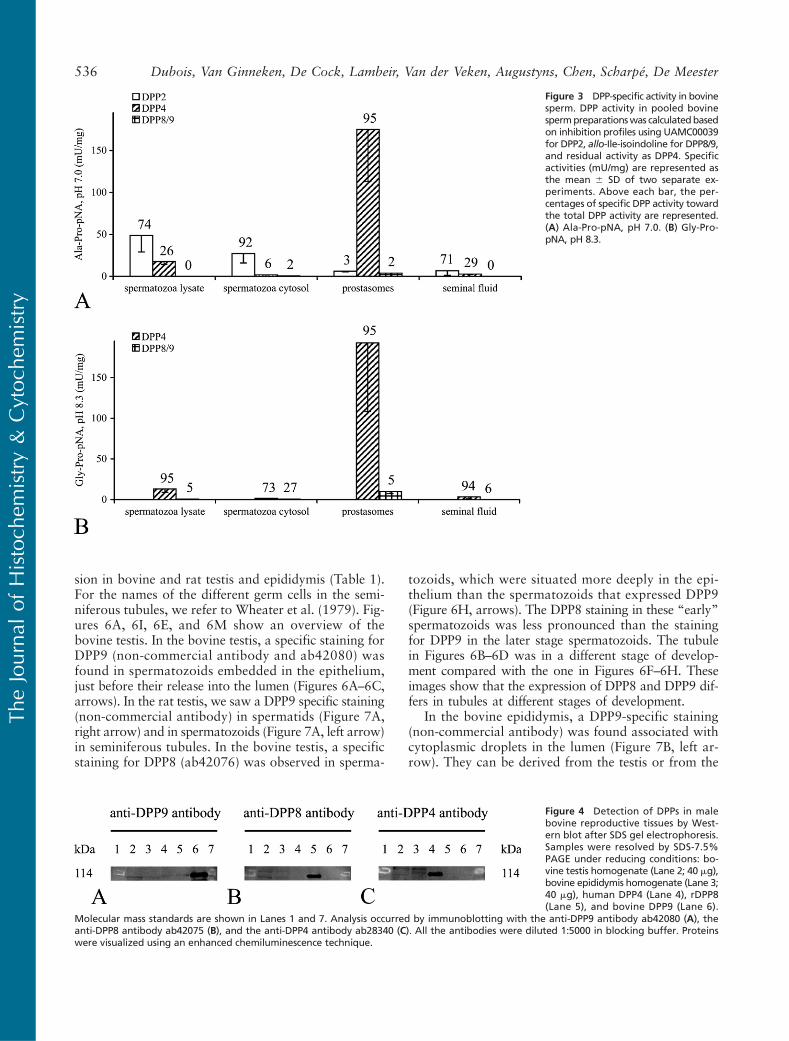
In the cytosol of bovine spermatozoa, almost all ofthe Ala-Pro-pNA-cleaving activity was attributed toDPP2 (Figure 3A). In the spermatozoa lysate and semi-nal fluid, the contribution of DPP2 was ?70–75%. Theprostasomes contained the highest DPP4 vs DPP2 and
534 Dubois, Van Ginneken, De Cock, Lambeir, Van der Veken, Augustyns, Chen, Scharpé, De Meester
TheJourna
lof
Histoch
emistry&
Cytoc
hemistry
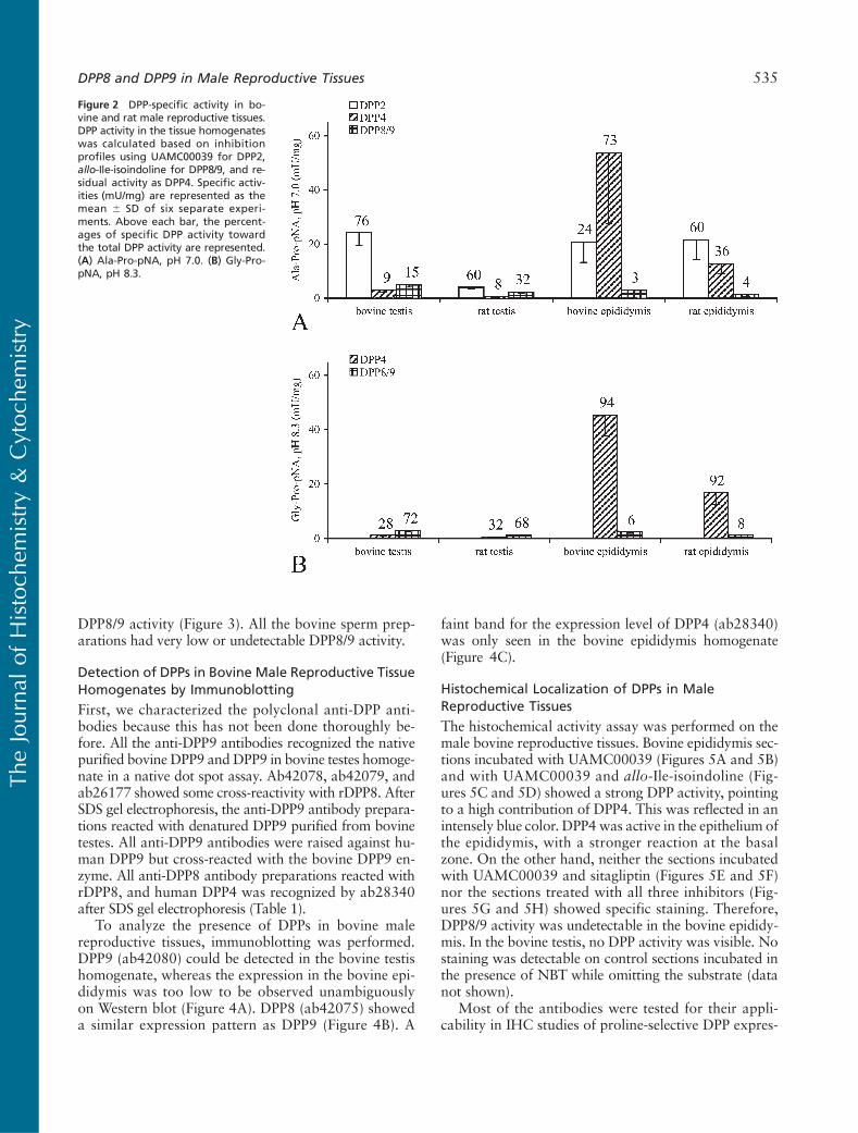
DPP8/9 activity (Figure 3). All the bovine sperm prep-arations had very low or undetectable DPP8/9 activity.
Detection of DPPs in Bovine Male Reproductive TissueHomogenates by Immunoblotting
First, we characterized the polyclonal anti-DPP anti-bodies because this has not been done thoroughly be-fore. All the anti-DPP9 antibodies recognized the nativepurified bovine DPP9 and DPP9 in bovine testes homoge-nate in a native dot spot assay. Ab42078, ab42079, andab26177 showed some cross-reactivity with rDPP8. AfterSDS gel electrophoresis, the anti-DPP9 antibody prepara-tions reacted with denatured DPP9 purified from bovinetestes. All anti-DPP9 antibodies were raised against hu-man DPP9 but cross-reacted with the bovine DPP9 en-zyme. All anti-DPP8 antibody preparations reacted withrDPP8, and human DPP4 was recognized by ab28340after SDS gel electrophoresis (Table 1).
To analyze the presence of DPPs in bovine malereproductive tissues, immunoblotting was performed.DPP9 (ab42080) could be detected in the bovine testishomogenate, whereas the expression in the bovine epi-didymis was too low to be observed unambiguouslyon Western blot (Figure 4A). DPP8 (ab42075) showeda similar expression pattern as DPP9 (Figure 4B). A
faint band for the expression level of DPP4 (ab28340)was only seen in the bovine epididymis homogenate(Figure 4C).
Histochemical Localization of DPPs in MaleReproductive Tissues
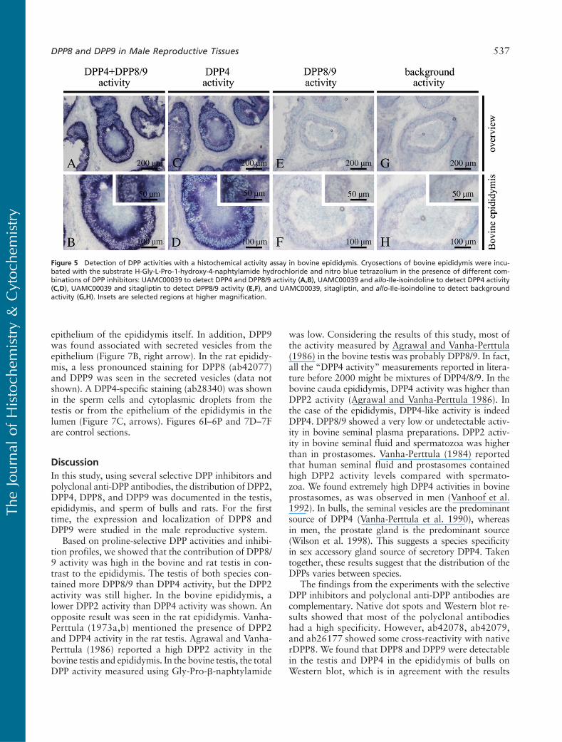
The histochemical activity assay was performed on themale bovine reproductive tissues. Bovine epididymis sec-tions incubated with UAMC00039 (Figures 5A and 5B)and with UAMC00039 and allo-Ile-isoindoline (Fig-ures 5C and 5D) showed a strong DPP activity, pointingto a high contribution of DPP4. This was reflected in anintensely blue color. DPP4 was active in the epithelium ofthe epididymis, with a stronger reaction at the basalzone. On the other hand, neither the sections incubatedwith UAMC00039 and sitagliptin (Figures 5E and 5F)nor the sections treated with all three inhibitors (Fig-ures 5G and 5H) showed specific staining. Therefore,DPP8/9 activity was undetectable in the bovine epididy-mis. In the bovine testis, no DPP activity was visible. Nostaining was detectable on control sections incubated inthe presence of NBT while omitting the substrate (datanot shown).
Most of the antibodies were tested for their appli-cability in IHC studies of proline-selective DPP expres-
Figure 2 DPP-specific activity in bo-vine and rat male reproductive tissues.DPP activity in the tissue homogenateswas calculated based on inhibitionprofiles using UAMC00039 for DPP2,allo-Ile-isoindoline for DPP8/9, and re-sidual activity as DPP4. Specific activ-ities (mU/mg) are represented as themean 6 SD of six separate experi-ments. Above each bar, the percent-ages of specific DPP activity towardthe total DPP activity are represented.(A) Ala-Pro-pNA, pH 7.0. (B) Gly-Pro-pNA, pH 8.3.
DPP8 and DPP9 in Male Reproductive Tissues 535
TheJourna
lof
Histoch
emistry&
Cytoc
hemistry
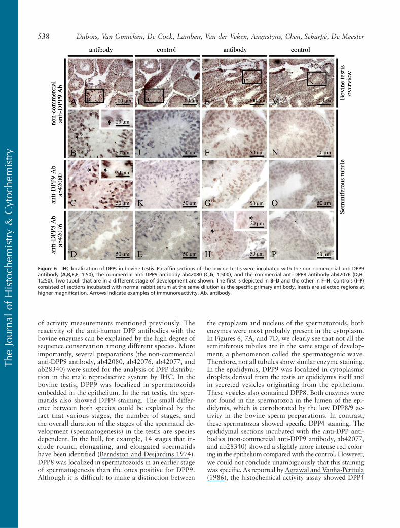
sion in bovine and rat testis and epididymis (Table 1).For the names of the different germ cells in the semi-niferous tubules, we refer to Wheater et al. (1979). Fig-ures 6A, 6I, 6E, and 6M show an overview of thebovine testis. In the bovine testis, a specific staining forDPP9 (non-commercial antibody and ab42080) wasfound in spermatozoids embedded in the epithelium,just before their release into the lumen (Figures 6A–6C,arrows). In the rat testis, we saw a DPP9 specific staining(non-commercial antibody) in spermatids (Figure 7A,right arrow) and in spermatozoids (Figure 7A, left arrow)in seminiferous tubules. In the bovine testis, a specificstaining for DPP8 (ab42076) was observed in sperma-
tozoids, which were situated more deeply in the epi-thelium than the spermatozoids that expressed DPP9(Figure 6H, arrows). The DPP8 staining in these “early”spermatozoids was less pronounced than the stainingfor DPP9 in the later stage spermatozoids. The tubulein Figures 6B–6D was in a different stage of develop-ment compared with the one in Figures 6F–6H. Theseimages show that the expression of DPP8 and DPP9 dif-fers in tubules at different stages of development.
In the bovine epididymis, a DPP9-specific staining(non-commercial antibody) was found associated withcytoplasmic droplets in the lumen (Figure 7B, left ar-row). They can be derived from the testis or from the
Figure 3 DPP-specific activity in bovinesperm. DPP activity in pooled bovinespermpreparationswas calculatedbasedon inhibition profiles using UAMC00039for DPP2, allo-Ile-isoindoline for DPP8/9,and residual activity as DPP4. Specificactivities (mU/mg) are represented asthe mean 6 SD of two separate ex-periments. Above each bar, the per-centages of specific DPP activity towardthe total DPP activity are represented.(A) Ala-Pro-pNA, pH 7.0. (B) Gly-Pro-pNA, pH 8.3.
Figure 4 Detection of DPPs in malebovine reproductive tissues by West-ern blot after SDS gel electrophoresis.Samples were resolved by SDS-7.5%PAGE under reducing conditions: bo-vine testis homogenate (Lane 2; 40 mg),bovine epididymis homogenate (Lane 3;40 mg), human DPP4 (Lane 4), rDPP8(Lane 5), and bovine DPP9 (Lane 6).
Molecular mass standards are shown in Lanes 1 and 7. Analysis occurred by immunoblotting with the anti-DPP9 antibody ab42080 (A), theanti-DPP8 antibody ab42075 (B), and the anti-DPP4 antibody ab28340 (C). All the antibodies were diluted 1:5000 in blocking buffer. Proteinswere visualized using an enhanced chemiluminescence technique.
536 Dubois, Van Ginneken, De Cock, Lambeir, Van der Veken, Augustyns, Chen, Scharpé, De Meester
TheJourna
lof
Histoch
emistry&
Cytoc
hemistry
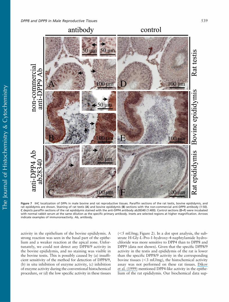
epithelium of the epididymis itself. In addition, DPP9was found associated with secreted vesicles from theepithelium (Figure 7B, right arrow). In the rat epididy-mis, a less pronounced staining for DPP8 (ab42077)and DPP9 was seen in the secreted vesicles (data notshown). A DPP4-specific staining (ab28340) was shownin the sperm cells and cytoplasmic droplets from thetestis or from the epithelium of the epididymis in thelumen (Figure 7C, arrows). Figures 6I–6P and 7D–7Fare control sections.
DiscussionIn this study, using several selective DPP inhibitors andpolyclonal anti-DPP antibodies, the distribution of DPP2,DPP4, DPP8, and DPP9 was documented in the testis,epididymis, and sperm of bulls and rats. For the firsttime, the expression and localization of DPP8 andDPP9 were studied in the male reproductive system.
Based on proline-selective DPP activities and inhibi-tion profiles, we showed that the contribution of DPP8/9 activity was high in the bovine and rat testis in con-trast to the epididymis. The testis of both species con-tained more DPP8/9 than DPP4 activity, but the DPP2activity was still higher. In the bovine epididymis, alower DPP2 activity than DPP4 activity was shown. Anopposite result was seen in the rat epididymis. Vanha-Perttula (1973a,b) mentioned the presence of DPP2and DPP4 activity in the rat testis. Agrawal and Vanha-Perttula (1986) reported a high DPP2 activity in thebovine testis and epididymis. In the bovine testis, the totalDPP activity measured using Gly-Pro-b-naphtylamide
was low. Considering the results of this study, most ofthe activity measured by Agrawal and Vanha-Perttula(1986) in the bovine testis was probably DPP8/9. In fact,all the “DPP4 activity” measurements reported in litera-ture before 2000 might be mixtures of DPP4/8/9. In thebovine cauda epididymis, DPP4 activity was higher thanDPP2 activity (Agrawal and Vanha-Perttula 1986). Inthe case of the epididymis, DPP4-like activity is indeedDPP4. DPP8/9 showed a very low or undetectable activ-ity in bovine seminal plasma preparations. DPP2 activ-ity in bovine seminal fluid and spermatozoa was higherthan in prostasomes. Vanha-Perttula (1984) reportedthat human seminal fluid and prostasomes containedhigh DPP2 activity levels compared with spermato-zoa. We found extremely high DPP4 activities in bovineprostasomes, as was observed in men (Vanhoof et al.1992). In bulls, the seminal vesicles are the predominantsource of DPP4 (Vanha-Perttula et al. 1990), whereasin men, the prostate gland is the predominant source(Wilson et al. 1998). This suggests a species specificityin sex accessory gland source of secretory DPP4. Takentogether, these results suggest that the distribution of theDPPs varies between species.
The findings from the experiments with the selectiveDPP inhibitors and polyclonal anti-DPP antibodies arecomplementary. Native dot spots and Western blot re-sults showed that most of the polyclonal antibodieshad a high specificity. However, ab42078, ab42079,and ab26177 showed some cross-reactivity with nativerDPP8. We found that DPP8 and DPP9 were detectablein the testis and DPP4 in the epididymis of bulls onWestern blot, which is in agreement with the results
Figure 5 Detection of DPP activities with a histochemical activity assay in bovine epididymis. Cryosections of bovine epididymis were incu-bated with the substrate H-Gly-L-Pro-1-hydroxy-4-naphtylamide hydrochloride and nitro blue tetrazolium in the presence of different com-binations of DPP inhibitors: UAMC00039 to detect DPP4 and DPP8/9 activity (A,B), UAMC00039 and allo-Ile-isoindoline to detect DPP4 activity(C,D), UAMC00039 and sitagliptin to detect DPP8/9 activity (E,F), and UAMC00039, sitagliptin, and allo-Ile-isoindoline to detect backgroundactivity (G,H). Insets are selected regions at higher magnification.
DPP8 and DPP9 in Male Reproductive Tissues 537
TheJourna
lof
Histoch
emistry&
Cytoc
hemistry
of activity measurements mentioned previously. Thereactivity of the anti-human DPP antibodies with thebovine enzymes can be explained by the high degree ofsequence conservation among different species. Moreimportantly, several preparations (the non-commercialanti-DPP9 antibody, ab42080, ab42076, ab42077, andab28340) were suited for the analysis of DPP distribu-tion in the male reproductive system by IHC. In thebovine testis, DPP9 was localized in spermatozoidsembedded in the epithelium. In the rat testis, the sper-matids also showed DPP9 staining. The small differ-ence between both species could be explained by thefact that various stages, the number of stages, andthe overall duration of the stages of the spermatid de-velopment (spermatogenesis) in the testis are speciesdependent. In the bull, for example, 14 stages that in-clude round, elongating, and elongated spermatidshave been identified (Berndston and Desjardins 1974).DPP8 was localized in spermatozoids in an earlier stageof spermatogenesis than the ones positive for DPP9.Although it is difficult to make a distinction between
the cytoplasm and nucleus of the spermatozoids, bothenzymes were most probably present in the cytoplasm.In Figures 6, 7A, and 7D, we clearly see that not all theseminiferous tubules are in the same stage of develop-ment, a phenomenon called the spermatogenic wave.Therefore, not all tubules show similar enzyme staining.In the epididymis, DPP9 was localized in cytoplasmicdroplets derived from the testis or epididymis itself andin secreted vesicles originating from the epithelium.These vesicles also contained DPP8. Both enzymes werenot found in the spermatozoa in the lumen of the epi-didymis, which is corroborated by the low DPP8/9 ac-tivity in the bovine sperm preparations. In contrast,these spermatozoa showed specific DPP4 staining. Theepididymal sections incubated with the anti-DPP anti-bodies (non-commercial anti-DPP9 antibody, ab42077,and ab28340) showed a slightly more intense red color-ing in the epithelium compared with the control. However,we could not conclude unambiguously that this stainingwas specific. As reported by Agrawal and Vanha-Perttula(1986), the histochemical activity assay showed DPP4
Figure 6 IHC localization of DPPs in bovine testis. Paraffin sections of the bovine testis were incubated with the non-commercial anti-DPP9antibody (A,B,E,F; 1:50), the commercial anti-DPP9 antibody ab42080 (C,G; 1:500), and the commercial anti-DPP8 antibody ab42076 (D,H;1:250). Two tubuli that are in a different stage of development are shown. The first is depicted in B–D and the other in F–H. Controls (I–P)consisted of sections incubated with normal rabbit serum at the same dilution as the specific primary antibody. Insets are selected regions athigher magnification. Arrows indicate examples of immunoreactivity. Ab, antibody.
538 Dubois, Van Ginneken, De Cock, Lambeir, Van der Veken, Augustyns, Chen, Scharpé, De Meester
TheJourna
lof
Histoch
emistry&
Cytoc
hemistry
activity in the epithelium of the bovine epididymis. Astrong reaction was seen in the basal part of the epithe-lium and a weaker reaction at the apical zone. Unfor-tunately, we could not detect any DPP8/9 activity inthe bovine epididymis, and no staining was visible inthe bovine testis. This is possibly caused by (a) insuffi-cient sensitivity of the method for detection of DPP8/9,(b) in situ inhibition of enzyme activity, (c) inhibitionof enzyme activity during the conventional histochemicalprocedure, or (d) the low specific activity in these tissues
(,5 mU/mg; Figure 2). In a dot spot analysis, the sub-strate H-Gly-L-Pro-1-hydroxy-4-naphtylamide hydro-chloride was more sensitive to DPP4 than to DPP8 andDPP9 (data not shown). Given that the specific DPP8/9activity in the testis and epididymis of the rat is lowerthan the specific DPP8/9 activity in the correspondingbovine tissues (,3 mU/mg), the histochemical activityassay was not performed on these rat tissues. Dikovet al. (1999) mentioned DPP4-like activity in the epithe-lium of the rat epididymis. Our biochemical data sup-
Figure 7 IHC localization of DPPs in male bovine and rat reproductive tissues. Paraffin sections of the rat testis, bovine epididymis, andrat epididymis are shown. Staining of rat testis (A) and bovine epididymis (B) sections with the non-commercial anti-DPP9 antibody (1:50).C depicts paraffin sections of the rat epididymis stained with the anti-DPP4 antibody ab28340 (1:400). Control sections (D–F) were incubatedwith normal rabbit serum at the same dilution as the specific primary antibody. Insets are selected regions at higher magnification. Arrowsindicate examples of immunoreactivity. Ab, antibody.
DPP8 and DPP9 in Male Reproductive Tissues 539
TheJourna
lof
Histoch
emistry&
Cytoc
hemistry
port the conclusion that DPP4 is responsible for thisactivity. In general, we showed that the antibodiesab42080 and ab42075 are suitable for immunoblot-ting experiments by their intense staining and no cross-reactivity. The non-commercial anti-DPP9 antibody isthe best antibody for IHC because it showed a specificstaining in all the tissues used in this study.
Proteases and protease inhibitors are supposed to beinvolved in the events pertinent to germ cell movementin the seminiferous epithelium during spermatogenesis(Fritz et al. 1993; Le Magueresse-Battistoni 2007). Epi-didymal epithelial cells are engaged in absorption ofmaterial derived from the maturing spermatozoa. DPP8and/or DPP9 may be involved in such processes. Thesite-specific activity and expression pattern of DPP4,DPP8, and DPP9 in the male reproductive system suggestdifferential functional roles during late stages of sper-matogenesis. The in vivo substrates and correspondingphysiological roles of the individual proline-selective pep-tidases are currently unknown. Apart from this, the tissuedistribution pattern of DPP8 and DPP9 proteins needsto be studied further. In the near future, the adaptationof existing methods may allow visualization of DPP8/9activity in living cells (Boonacker and Van Noorden2001). Moreover, the ongoing development of specificinhibitors for DPP8 or DPP9 (Van der Veken et al.2007b,2008; Van Goethem et al. 2008) will facilitatefurther physiological studies.
In conclusion, we showed a high expression of DPP8and DPP9 in the bovine and rat testis at both the proteinand activity levels. Both enzymes are localized in specificsperm cells of late stages of spermatogenesis. In contrast,the epididymis and sperm preparations contained lowlevels of DPP8 and DPP9. Our findings suggest that theseproteases may contribute to male gamete development.
Acknowledgments
This study was supported by the Fund for ScientificResearch-Flanders (Belgium; F.W.O.-Vlaanderen) and theUniversity of Antwerp (B.O.F.). We thank Prof. P. Bols forproviding the bovine sperm.
Literature Cited
Abbott CA, Yu DMT, Woollatt E, Sutherland GR, McCaughan GW,Gorrell MD (2000) Cloning, expression and chromosomal locali-zation of a novel human dipeptidyl peptidase (DPP) IV homolog,DPP8. Eur J Biochem 267:6140–6150
Agrawal Y, Vanha-Perttula T (1986) Dipeptidyl peptidases in bovinereproductive organs and secretions. Int J Androl 9:435–452
Ajami K, Abbott CA, McCaughan GW, Gorrell MD (2004) Dipep-tidyl peptidase 9 has two forms, a broad tissue distribution, cyto-plasmic localization and DPIV-like peptidase activity. BiochimBiophys Acta 1679:18–28
Berndston WE, Desjardins C (1974) The cycle of the seminiferousepithelium and spermatogenesis in the bovine testis. Am J Anat140:167–179
Boonacker E, Van Noorden CJF (2001) Enzyme cytochemical tech-niques for metabolic mapping in living cells, with special referenceto proteolysis. J Histochem Cytochem 12:1473–1486
Boonacker E, Van Noorden CJF (2003) The multifunctional ormoonlighting protein CD26/DPPIV. Eur J Cell Biol 82:53–73
Bradford MM (1976) A rapid and sensitive method for the quantita-tion of microgram quantities of protein utilizing the principle ofprotein-dye binding. Anal Biochem 72:248–254
Busek P, Stremenova J, Sedo A (2008) Dipeptidyl peptidase-IV enzy-matic activity bearing molecules in human brain tumors: good orevil? Front Biosci 13:2319–2326
Chen YS, Chien CH, Goparaju CM, Hsu JTA, Liang PH, Chen X(2004) Purification and characterization of human prolyl dipepti-dase DPP8 in Sf9 insect cells. Protein Expr Purif 35:142–146
Cooper GM (2000) The Cell, A Molecular Approach. 2nd ed. Sun-derland, MA, Sinauer Associates
De Meester I, Vanhoof G, Lambeir AM, Scharpé S (1996) Use ofimmobilized adenosine deaminase (EC 3.5.4.4) for the rapid puri-fication of native human CD26/dipeptidyl peptidase IV (EC 3.4.14.5).J Immunol Methods 189:99–105
Dikov A, Dimitrova M, Stoineva I, Halbhuber KJ (1999) New tetra-zolium method for the histochemical localization of dipeptidylpeptidase IV. Cell Mol Biol 45:225–231
Dubois V, Lambeir AM, Van der Veken P, Augustyns K, Creemers J,Chen X, Scharpé S, et al. (2008) Purification and characterizationof a dipeptidyl peptidase 9-like enzyme from bovine testes. FrontBiosci 13:3558–3568
Frerker N, Wagner L, Wolf R, Heiser U, Hoffmann T, Rahfeld JU,Schade J, et al. (2007) Neuropeptide Y (NPY) cleaving enzymes:structural and functional homologues of dipeptidyl peptidase 4.Peptides 28:257–268
Fritz IB, Tung PS, Ailenberg M (1993) Proteases and antiproteasesin the seminiferous tubule. In Russell LD, Griswold MD, eds. TheSertoli Cell. Clearwater, FL, Cache River Press, 217–235
Helmuth M, Reinhold D, Stefin S, Täger M, Ansorge S, Bank U(2008) Tissue distribution of DPIV-like activities in wild type miceand CD26 knock out mice under physiological and pathologicalconditions. Clin Chem Lab Med 46:A26–27
Henry LR, Lee HO, Lee JS, Klein-Szanto A, Watts P, Ross EA, ChenWT, et al. (2007) Clinical implications of fibroblast activation pro-tein in patients with colon cancer. Clin Cancer Res 13:1736–1741
Kim D, Wang L, Beconi M, Eiermann GJ, Fisher MH, He H, HickeyGJ, et al. (2005) (2R)-4-oxo-4-[3-(trifluoromethyl)-5,6-dihydro[1,2,4]triazolo[4,3-a]pyrazin-7(8H)-yl]-1-(2,4,5-trifluorophenyl)butan-2-amine: a potent, orally active dipeptidyl peptidase IV inhibi-tor for the treatment of type 2 diabetes. J Med Chem 48:141–151
Lambeir AM, Durinx C, Scharpé S, De Meester I (2003) Dipeptidyl-peptidase IV from bench to bedside: an update on structural prop-erties, functions, and clinical aspects of the enzyme DPPIV. CritRev Clin Lab Sci 40:209–294
Lankas GR, Leiting B, Roy RS, Eiermann GJ, Beconi MG, Biftu T,Chan CC, et al. (2005) Dipeptidyl peptidase IV inhibition for thetreatment of type 2 diabetes. Potential importance of selectivityover dipeptidyl peptidases 8 and 9. Diabetes 54:2988–2994
Le Magueresse-Battistoni B (2007) Serine proteases and serine pro-tease inhibitors in testicular physiology: the plasminogen activa-tion system. Reproduction 134:721–729
Maes MB, Dubois V, Brandt I, Lambeir AM, Van der Veken P,Augustyns K, Cheng JD, et al. (2007a) Dipeptidyl peptidase 8/9-like activity in human leukocytes. J Leukoc Biol 81:1252–1257
Maes MB, Lambeir AM, Gilany K, Senten K, Van der Veken P,Leiting B, Augustyns K, et al. (2005) Kinetic investigation of hu-man dipeptidyl peptidase II mediated hydrolysis of dipeptide deriv-atives and its identification as quiescent cell proline dipeptidase/dipeptidyl peptidase 7. Biochem J 386:315–324
Maes MB, Scharpé S, De Meester I (2007b) Dipeptidyl peptidase II(DPPII), a review. Clin Chim Acta 380:31–49
Olsen C, Wagtmann N (2002) Identification and characterization ofhuman DPP9, a novel homologue of dipeptidyl peptidase IV. Gene299:185–193
Qi SY, Riviere PJ, Trojnar J, Junien JL, Akinsanya KO (2003)Cloning and characterization of dipeptidyl peptidase 10, a newmember of an emerging subgroup of serine proteases. Biochem J373:179–189
540 Dubois, Van Ginneken, De Cock, Lambeir, Van der Veken, Augustyns, Chen, Scharpé, De Meester
TheJourna
lof
Histoch
emistry&
Cytoc
hemistry
Reinhold D, Goihl A, Kühlmann U, Bank U, Lendeckel U, Faust J,Neubert K, et al. (2008) On the role of dipeptidyl peptidase IV-likeenzymes in T lymphocyte activation. Clin Chem Lab Med 46:A10
Rosenblum JS, Kozarich JW (2003) Prolyl peptidases: a serine pro-tease subfamily with high potential for drug discovery. Curr OpinChem Biol 7:496–504
Schade J, Stephan M, Schmiedl A, Wagner L, Niestroj AJ, DemuthHU, Frerker N, et al. (2008) Regulation of expression and func-tion of dipeptidyl peptidase 4 (DP4), DP8/9, and DP10 in allergicresponses of the lung in rats. J Histochem Cytochem 56:147–155
Senten K, Van der Veken P, De Meester I, Lambeir AM, ScharpéS, Haemers A, Augustyns K (2004) Gamma-amino-substitutedanalogues of 1-[(S)-2,4-diaminobutanoyl]piperidine as highly po-tent and selective dipeptidyl peptidase II inhibitors. J Med Chem47:2906–2916
Stremenova J, Krepela E, Mares V, Trim J, Dbaly V, Marek J,Vanickova Z, et al. (2007) Expression and enzymatic activity ofdipeptidyl peptidase-IV in human astrocytic tumours are asso-ciated with tumour grade. Int J Oncol 31:785–792
Thielitz A, Ansorge S, Bank U, Tager M, Wrenger S, Gollnick H,Reinhold D (2008a) The ectopeptidases dipeptidyl peptidase IV(DP IV) and aminopeptidase N (APN) and their related enzymesas possible targets in the treatment of skin deseases. Front Biosci13:2364–2375
Thielitz A, Vetter RW, Schultze B, Wrenger S, Simeoni L, Ansorge S,Neubert K, et al. (2008b) Inhibitors of dipeptidyl peptidase IV-likeactivity mediate antifibrotic effects in normal and keloid-derivedskin fibroblasts. J Invest Dermatol 128:855–866
Van der Veken P, De Meester I, Dubois V, Soroka A, Van GoethemS, Maes MB, Brandt I, et al. (2008) Inhibitors of dipeptidyl pep-tidase 8 and dipeptidyl peptidase 9. Part 1: identification of dipep-tide derived leads. Bioorg Med Chem Lett 18:4154–4158
Van der Veken P, Haemers A, Augustyns K (2007a) Prolyl peptidasesrelated to dipeptidyl peptidase IV: potential of specific inhibitorsin drug discovery. Curr Top Med Chem 7:621–635
Van der Veken P, Soroka A, Brandt I, Chen YS, Maes MB, LambeirAM, Chen X, et al. (2007b) Irreversible inhibition of dipeptidylpeptidase 8 by dipeptide-derived diaryl phosphonates. JMedChem50:5568–5570
Van Goethem S, Van der Veken P, Dubois V, Soroka A, LambeirAM, Chen X, Haemers A, et al. (2008) Inhibitors of dipeptidylpeptidase 8 and dipeptidyl peptidase 9. Part 2: isoindoline con-taining inhibitors. Bioorg Med Chem Lett 18:4159–4162
Vanha-Perttula T (1973a) Aminopeptidases of rat testis. I. Fraction-ation and characterization. J Reprod Fertil 32:33–44
Vanha-Perttula T (1973b) Aminopeptidases of rat testis. II. Effectsof puberty, cryptorchidism and cadmium chloride treatment. JReprod Fertil 32:45–53
Vanha-Perttula T (1984) Studies on alanine aminopeptidase, dipep-tidyl aminopeptidase I and II of the human seminal fluid andprostasomes. Sel Top Clin Enzym 2:545–564
Vanha-Perttula T, Rönkkö S, Lahtinen R (1990) Hydrolases frombovine seminal vesicle, prostate and Cowper’s gland. Andrologia22:10–24
Vanhoof G, De Meester I, van Sande M, Scharpé S, Yaron A (1992)Distribution of proline-specific aminopeptidases in human tissuesand body fluids. Eur J Clin Chem Clin Biochem 30:333–338
Wagner L, Hoffmann T, Rahfeld JU, Demuth HU (2006) Distribu-tion of dipeptidyl peptidase IV-like activity enzymes in canine andporcine tissue sections by RT-PCR. Adv ExpMed Biol 575:109–116
Wang XM, Yao TW, Nadvi NA, Osborne B, McCaughan GW,Gorrell MD (2008) Fibroblast activation protein and chronic liverdisease. Front Biosci 13:3168–3180
Wheater PR, Burkitt HG, Daniels VG, Deakin PJ (1979) Func-tional Histology. A Text and Colour Atlas. Edinburgh, London,Churchill Livingstone
Wilson MJ, Ruhland AR, Pryor JL, Ercole C, Sinha AA, HensleighH, Kaye KW, et al. (1998) Prostate specific origin of dipeptidyl-peptidase IV (CD-26) in human seminal plasma. J Urol 160:1905–1909
DPP8 and DPP9 in Male Reproductive Tissues 541
TheJourna
lof
Histoch
emistry&
Cytoc
hemistry
Related Documents


