Environmental impact of sunscreen nanomaterials: Ecotoxicity and genotoxicity of altered TiO 2 nanocomposites on Vicia faba Anne-Sophie Foltête a, c, * , Jean-François Masfaraud a, c , Emilie Bigorgne a, c , Johanne Nahmani a, c , Perrine Chaurand b, c , Céline Botta b, c , Jérôme Labille b, c , Jérôme Rose b, c , Jean-François Férard a, c , Sylvie Cotelle a, c a Laboratoire des Interactions Ecotoxicologie, Biodiversité, Ecosystèmes (LIEBE), Université Paul Verlaine-Metz, CNRS UMR 7146, Campus Bridoux, Avenue du Général Delestraint, 57070 Metz, France b Centre Européen de Recherches et d’Enseignement des Géosciences de l’Environnement (CEREGE), UMR 6635 CNRS/Aix-Marseille Université, Europôle de l’Arbois, 13545 Aix-en-Provence, France c iCEINT, International Consortium for the Environmental Implications of Nanotechnology, F-13545 Aix-en-Provence Cedex 04, France 1 article info Article history: Received 8 March 2011 Received in revised form 12 June 2011 Accepted 15 June 2011 Keywords: Rutile Nanoparticles Alteration Titanium dioxide Risk assessment abstract Mineral sunscreen nanocomposites, based on a nano-TiO 2 core, coated with aluminium hydroxide and dimethicone films, were submitted to an artificial ageing process. The resulting Altered TiO 2 Nano- composites (ATN) were then tested in the liquid phase on the plant model Vicia faba, which was exposed 48 h to three nominal concentrations: 5, 25 and 50 mg ATN/L. Plant growth, photosystem II maximum quantum yield, genotoxicity (micronucleus test) and phytochelatins levels showed no change compared to controls. Oxidative stress biomarkers remained unchanged in shoots while in roots, glutathione reductase activity decreased at 50 mg ATN/L and ascorbate peroxidase activity decreased for 5 and 25 mg ATN/L. Nevertheless, despite the weak response of biological endpoints, ICP-MS measurements revealed high Ti and Al concentrations in roots, and X-ray fluorescence micro-spectroscopy revealed titanium internalization in superficial root tissues. Eventual long-term effects on plants may occur. Ó 2011 Elsevier Ltd. All rights reserved. 1. Introduction With the fastly growing number of commercial products incor- porating or made of manufactured nanoparticles (MNPs), the dispersion of MNPs in the environment is not science fiction but already occurring, causing ethical, sociological (Jacobs et al., 2010) and potentially environmental problems, even if those are difficult to assess (Aschberger et al., 2011; Baun et al., 2008; Farré et al., 2011; Navarro et al., 2008; Nowack, 2009; Nowack and Bucheli, 2007). Measurements from Kaegi et al. (2008) represent the first direct evidence of the release of synthetic nanoparticles from MNPs applications into the environment. They showed the leaching of titanium dioxide (TiO 2 ) nanoparticles from house facades treated with paint containing nano-TiO 2 under the influence of atmospheric conditions. A model of the behaviour and expected concentrations of nano-TiO 2 in the different compartments of the environment is proposed by Mueller and Nowack (2008) and by Gottschalk et al. (2010), reviewed by Menard et al. (2011). TiO 2 nanoparticles are more and more included in sunscreen creams for their good UV filter properties (AFSSET, 2010). Like other cosmetic products, sunscreen components are likely to end up in bathing waters, wastewaters and waste disposals, and finally into the whole environment. During sewage treatment, TiO 2 nano- particles seem to show a close affinity for activated sludge, and consequently, the greater part of these nanomaterials are expected to end up in soils through sewage sludge application (Johnson et al., 2011; Kiser et al., 2009). The photocatalytic properties of TiO 2 nanoparticles are really problematic once these particles released into the environment. Indeed, they are powerful reactive oxygen species (ROS) producers when exposed to ultraviolet radiations (Reijnders, 2008). In plants, bare TiO 2 nanoparticles can show either positive or negative effects (Brar et al., 2010; Ma et al., 2010). In sunscreens, nano-TiO 2 particles are surface modified to inhibit ROS generation (Reijnders, 2006; Wakefield et al., 2004) and prevent toxic effects, but the stability of the protective layers and the effects of such altered structures on * Corresponding author. E-mail addresses: [email protected] (A.-S. Foltête), [email protected] (J.-F. Masfaraud), [email protected] (E. Bigorgne), Johanne.nahmani@ univ-metz.fr (J. Nahmani), [email protected] (P. Chaurand), [email protected] (C. Botta), [email protected] (J. Labille), [email protected] (J. Rose), [email protected] (J.-F. Férard), [email protected] (S. Cotelle). 1 http://www.i-ceint.org. Contents lists available at ScienceDirect Environmental Pollution journal homepage: www.elsevier.com/locate/envpol 0269-7491/$ e see front matter Ó 2011 Elsevier Ltd. All rights reserved. doi:10.1016/j.envpol.2011.06.020 Environmental Pollution 159 (2011) 2515e2522

Welcome message from author
This document is posted to help you gain knowledge. Please leave a comment to let me know what you think about it! Share it to your friends and learn new things together.
Transcript
lable at ScienceDirect
Environmental Pollution 159 (2011) 2515e2522
Contents lists avai
Environmental Pollution
journal homepage: www.elsevier .com/locate/envpol
Environmental impact of sunscreen nanomaterials: Ecotoxicityand genotoxicity of altered TiO2 nanocomposites on Vicia faba
Anne-Sophie Foltête a,c,*, Jean-François Masfaraud a,c, Emilie Bigorgne a,c, Johanne Nahmani a,c,Perrine Chaurand b,c, Céline Botta b,c, Jérôme Labille b,c, Jérôme Rose b,c, Jean-François Férard a,c,Sylvie Cotelle a,c
a Laboratoire des Interactions Ecotoxicologie, Biodiversité, Ecosystèmes (LIEBE), Université Paul Verlaine-Metz, CNRS UMR 7146, Campus Bridoux,Avenue du Général Delestraint, 57070 Metz, FrancebCentre Européen de Recherches et d’Enseignement des Géosciences de l’Environnement (CEREGE), UMR 6635 CNRS/Aix-Marseille Université,Europôle de l’Arbois, 13545 Aix-en-Provence, Francec iCEINT, International Consortium for the Environmental Implications of Nanotechnology, F-13545 Aix-en-Provence Cedex 04, France1
a r t i c l e i n f o
Article history:Received 8 March 2011Received in revised form12 June 2011Accepted 15 June 2011
Keywords:RutileNanoparticlesAlterationTitanium dioxideRisk assessment
* Corresponding author.E-mail addresses: [email protected] (A.-S. Foltê
Masfaraud), [email protected] (E. Buniv-metz.fr (J. Nahmani), [email protected] (P. Cha(C. Botta), [email protected] (J. Labille), [email protected](J.-F. Férard), [email protected] (S. Cotelle).
1 http://www.i-ceint.org.
0269-7491/$ e see front matter � 2011 Elsevier Ltd.doi:10.1016/j.envpol.2011.06.020
a b s t r a c t
Mineral sunscreen nanocomposites, based on a nano-TiO2 core, coated with aluminium hydroxide anddimethicone films, were submitted to an artificial ageing process. The resulting Altered TiO2 Nano-composites (ATN) were then tested in the liquid phase on the plant model Vicia faba, which was exposed48 h to three nominal concentrations: 5, 25 and 50 mg ATN/L. Plant growth, photosystem II maximumquantum yield, genotoxicity (micronucleus test) and phytochelatins levels showed no change comparedto controls. Oxidative stress biomarkers remained unchanged in shoots while in roots, glutathionereductase activity decreased at 50 mg ATN/L and ascorbate peroxidase activity decreased for 5 and25 mg ATN/L. Nevertheless, despite the weak response of biological endpoints, ICP-MS measurementsrevealed high Ti and Al concentrations in roots, and X-ray fluorescence micro-spectroscopy revealedtitanium internalization in superficial root tissues. Eventual long-term effects on plants may occur.
� 2011 Elsevier Ltd. All rights reserved.
1. Introduction
With the fastly growing number of commercial products incor-porating or made of manufactured nanoparticles (MNPs), thedispersion of MNPs in the environment is not science fiction butalready occurring, causing ethical, sociological (Jacobs et al., 2010)and potentially environmental problems, even if those are difficultto assess (Aschberger et al., 2011; Baun et al., 2008; Farré et al., 2011;Navarro et al., 2008; Nowack, 2009; Nowack and Bucheli, 2007).Measurements from Kaegi et al. (2008) represent the first directevidence of the release of synthetic nanoparticles from MNPsapplications into the environment. They showed the leaching oftitanium dioxide (TiO2) nanoparticles from house facades treatedwith paint containing nano-TiO2 under the influence of atmospheric
te), [email protected] (J.-F.igorgne), Johanne.nahmani@urand), [email protected](J. Rose), [email protected]
All rights reserved.
conditions. A model of the behaviour and expected concentrationsof nano-TiO2 in the different compartments of the environment isproposed by Mueller and Nowack (2008) and by Gottschalk et al.(2010), reviewed by Menard et al. (2011).
TiO2 nanoparticles are more and more included in sunscreencreams for their good UV filter properties (AFSSET, 2010). Like othercosmetic products, sunscreen components are likely to end up inbathing waters, wastewaters and waste disposals, and finally intothe whole environment. During sewage treatment, TiO2 nano-particles seem to show a close affinity for activated sludge, andconsequently, the greater part of these nanomaterials are expectedto end up in soils through sewage sludge application (Johnson et al.,2011; Kiser et al., 2009).
The photocatalytic properties of TiO2 nanoparticles are reallyproblematic once these particles released into the environment.Indeed, they are powerful reactive oxygen species (ROS) producerswhen exposed to ultraviolet radiations (Reijnders, 2008). In plants,bare TiO2 nanoparticles can show either positive or negative effects(Brar et al., 2010; Ma et al., 2010). In sunscreens, nano-TiO2 particlesare surface modified to inhibit ROS generation (Reijnders, 2006;Wakefield et al., 2004) and prevent toxic effects, but the stabilityof the protective layers and the effects of such altered structures on

A.-S. Foltête et al. / Environmental Pollution 159 (2011) 2515e25222516
ecosystems remain largely unknown. The possibility of the regainof photocatalytic properties by sunscreen nano-TiO2 after use wasproved by Barker and Branch (2008), who noticed an acceleratedweathering (100-fold) of the coating of prepainted steel sheets bynano-TiO2-containing sunscreens used by workers during handlingof the materials.
Auffan et al. (2010) and Labille et al. (2010) have artificiallymimicked the natural expected alteration of T-Lite SF (BASF),a nanocomposite entering in sunscreen formula. T-Lite are based ona nano-TiO2 core coated with aluminium hydroxide Al(OH)3 andpolydimethylsiloxane (PDMS) films. In the continuity of thesestudies, we investigated the biological effects of their Altered TiO2Nanocomposites (ATN) on a plant model, the broad bean Vicia faba.This model was chosen for its well-known sensitivity to pollutants.Indeed it has been used by numerous authors to assess the eco-toxicity, and particularly the genotoxicity of substances, radiations,or liquid or solid polluted matrix (Cotelle et al., 1999; Manier et al.,2009; Minouflet et al., 2005; Radetski et al., 2004; Ünyayar et al.,2006).
After exposing V. faba to ATN suspensions, ecotoxicological andgenotoxicological endpointswere assessed. Titaniumandaluminiumconcentrations were determined in roots, and titanium internaliza-tion was studied by X-ray fluorescence micro-spectroscopy.
2. Material and methods
2.1. Nanocomposites artificial ageing
Mineral sunscreen nanocomposites, based on a TiO2 core (10 nm � 50 nm),coated with aluminium hydroxide and polydimethylsiloxane layers, were submittedto an alteration process detailed by Labille et al. (2010). Briefly, 100 mg nano-composite powder (provided by the manufacturer) were introduced in 250 mLultrapure water and kept agitated (690 rpm) under a white light (400 W Philips�
Master HPI-T Plus) to mimic solar spectrum during 48 h. The pH was not buffered,the volume was constant. After alteration, the resulting mixture was rested for 48 hand the supernatant of stable altered TiO2 nanocomposites (ATN) was used for thetests.
2.2. ATN dilution and size characterization
ATN suspension (120 mg/L) was diluted in a water mix (50% Evian� Frenchmineral water: 50% ultrapure water, v:v) to the following concentrations: 5, 25 and50 mg ATN/L. Evian� is a weakly mineralized water. The predominant ions are Ca2þ
and HCO�3 . The 50% dilution provided a mix with low ionic strength (4.8 mM)
minimizing particle aggregation (Petosa et al., 2010), but sufficiently mineralized toensure an acceptable plant physiological state. At the beginning of the experiment,the pH was adjusted to 6.5 with HNO3. The mediumwas not buffered because of therapid ATN aggregation in every tested buffer.
Laser diffraction was used to measure ATN size distribution in the mixture ofultrapure and mineral waters (50:50, v:v) at 25 mg/L during the first 5 h. Theapparatus used was a Malvern Mastersizer S (Malvern Instruments, Malvern, UK)displaying a measuring range from 50 nm to 900 mm. Two separate particle groupswere specifically studied: particles below 700 nm, considered as stable colloidalsuspension, and particles over 10 mm, considered as aggregates undergoing rapidsettling.
Turbidity measurements were used to determine the limit conditions forcolloidal stability of the ATN dispersion as a function of its concentration. Thesuspension was diluted from 5 to 50 mg/L in the mixture of ultrapure and mineralwaters (50:50, v:v), and left 24 h in the dark to allow settling of particles. Thesupernatant turbidity was then measured using a Hach 2100 AN turbidimeter.
2.3. Plant germination and treatment
Commercial dry seeds of V. faba (var. Aguadulce, Fabre�) were used for theexperiments. In accordance with the French standard NF T90-327 (AFNOR, 2004),seeds were soaked in deionized water for 24 h, allowed to germinate between twolayers of wet cotton at 22 �C for 3 days. After removing the primary root tips, theseedlings were suspended in aerated Hoagland’s solution at 22 �C for 4 days topromote the development of secondary roots.
Each V. faba plantlet was placed in an individual pot (Wheaton� glass) con-taining 100 mL of the diluted suspension. During exposure, plant roots and ATNsuspension were protected from light while leaves were placed under plant growthlights (125 W, Envirolite�, 6400 K). Four individuals were used per concentration,randomly disposed to balance a potentially unequal light distribution. Exposure
lasted 48 h without renewing exposure medium. The pots were briefly agitated 2times a day to resuspend the particles. Experiments were conducted in a glove box,at 20 �C � 1.
The dilution medium was used as a negative control. Maleic hydrazide 10 mM(MH; CAS 123-33-1) was used as a positive control for genotoxicity. CdCl2 0.1 mM(CAS 10108-64-2) was used as a positive control for root elongation and as a refer-ence metal for thiol levels in tissues and oxidative stress biomarkers (Béraud et al.,2007; Lin et al., 2007).
2.4. Plant harvesting
After 48 h of exposure, secondary roots were carefully rinsed twice withultrapure water and elongation of three secondary roots per plant was assessed bythe difference between their length before and after exposure.
For micronucleus test, In accordance with the French standard NF T90-327(AFNOR, 2004), root tips were excised and fixed overnight in Carnoy solution (25%glacial acetic acid: 75% ethanol, v:v) at þ4 �C and then stored in 70% ethanolat þ4 �C.
Photosystem II maximum quantum yield was assessed in leaves through Fv/Fmratio using a portable Handy PEA (Handy-Plant Efficiency Analyser, HansatechInstruments, Norfolk, UK). Fv/Fm ratio is commonly used as an indicator of overallplant physiological status.
Fresh weight of shoots was determined. Shoots and roots were divided into 2groups: the first group was frozen in liquid nitrogen and stored at �80 �C forglutathione, phytochelatins and oxidative stress biomarker measurements, thesecond group was frozen at �20 �C for Ti and Al determinations.
2.5. Metal analyses
Al and Ti concentrations in roots were determined. After drying at 105 �C andmineralization in HFeHClO4, metal concentrations in tissues were measured by ICP-MS.
2.6. Element location in plants
In order to localize the metal (Ti) in the plant tissues, X-Ray Fluorescence micro-spectroscopy (m-XRF) analysis was performed on secondary roots exposed to50 mg ATN/L, fixed in Carnoy solution and kept at þ4 �C in 70% ethanol. To distin-guish between Ti trapped in the mucilage from Ti included in the root tissues, theremainingmucilagewas removed from certain roots following the protocol of Brams(1969) after slight adaptation. Roots conserved in 70% ethanol were rinsed 10 min inultrapure water, and then immersed in 1 M NH4Cl (pH 4.15) for 10 min. After that,they were rinsed 2�10min in ultrapurewater before being put back in 70% ethanol.
Resulting “withmucilage” and “withoutmucilage” roots were then embedded inresin and cross sections were obtained with a diamond wire saw. m-XRF measure-ments were carried out at the lab-scale on a microscope (XGT7000, Horiba JobinYvon) equipped with an X-ray guide tube producing a finely focused beam witha 10 mm spot size (Rh X-ray tube, accelerating voltage of 30 kV, current of 1 mA).Major elements (P, S, and Ca) were also detected by m-XRF and images of their spatialdistribution were obtained. Aluminium mapping was not possible because of thelow m-XRF sensitivity for light element detection.
2.7. Biochemical analyses
Activities of superoxide dismutase (SOD, EC 1.15.1.1), catalase (CAT, EC 1.11.1.6),guaiacol peroxidase (GPX, EC 1.11.1.7), ascorbate peroxidase (APX, EC 1.11.1.11) andglutathione reductase (GR, EC 1.6.4.2) were measured according to Qiu-Fang et al.(2005) for SOD, to Chaoui et al. (1997) for CAT, to Roy et al. (1996) for GPX, and toGarcía-Limones et al. (2002) for APX and GR, each of them modified by Dazy et al.(2008).
Lipid peroxidation was evaluated by the thiobarbituric assay of Aravind andPrasad (2003). Briefly, 900 mL of a 0.5% TBA in 20% TCA (w/v) solution were addedto 100 mL of plant extract. The mixture was incubated at 95 �C for 30 min and thereaction was stopped by transferring tubes on ice. The non-specific absorbance at600 nm was subtracted from the 532 nm absorbance and 3 ¼ 155 mM�1 cm�1 wasused for the calculation of malondialdehyde (MDA) level.
Phytochelatins (PC) 2, 3, 4, 5 and glutathione contents in plant extracts weremeasured by HPLC according to the derivatization method of Courbot et al. (2004).PC standardswere commercially provided (Anaspec, San Jose, USA). Protein contentswere determined according to Bradford (1976) using bovine serum albumin asa standard.
2.8. Micronucleus frequency
As described in the French standard NF T90-327(AFNOR, 2004), the excised roottips were hydrolyzed in 1 N HCl at 60 �C for 6 min, squashed between a slide and itscoverslip and stained with 1% aceto-orcein solution. Micronucleus frequencies werescored from 1000 cells per root tip under a 400� microscope magnification. Twoslides were prepared for each of the 4 replicates and a total of 8000 mitotic cells
y = 226.37x + 3.9752R2 = 0.9978
y = 873.55x + 14.313R2 = 0.9657
0
20
40
60
80
100
120
140
160
0 0.1 0.2 0.3 0.4 0.5 0.6Ti or Al in solution (mmol/L)
Ti o
r A
l in
ro
ots (m
mo
l/kg
d
.w
.)
TiAl
*
*
*
*
*
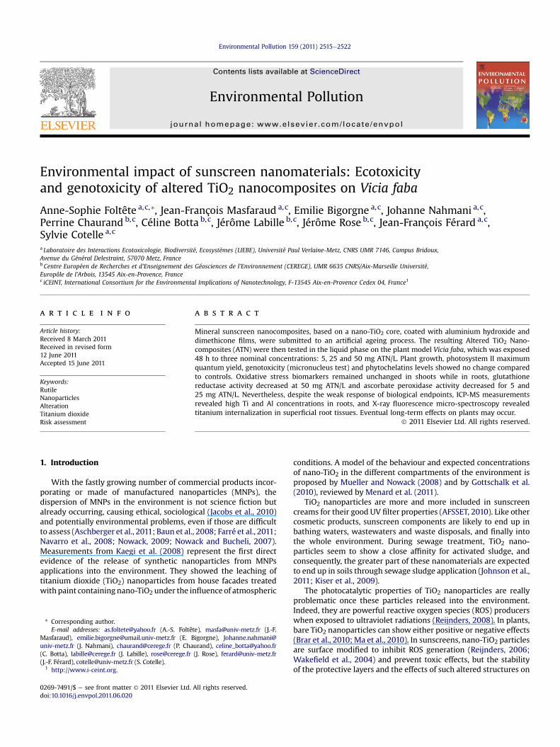
Fig. 2. Ti or Al concentrations in V. faba secondary roots after 48 h exposure withrespect to their concentrations in exposure suspensions. *Statistically significantlydifferent from corresponding controls after logarithmic transformation, Dunnett’st-test, p < 0.05.
Ti/Al ratio in mother solution: [Ti] = 8.5682 [Al]
120
140
160
A.-S. Foltête et al. / Environmental Pollution 159 (2011) 2515e2522 2517
from 8 separate slides per experimental group were observed under blindfoldconditions.
2.9. Statistical analyses
Before comparisons were run, care was taken to check for normality of variabledistributions (ShapiroeWilk test) and homogeneity of variances (Levene test). AOne-way ANOVA was performed between ATN-exposed samples and negativecontrols, followed by Dunnett’s t-test, risk level 5%.
Student t-test was performed between negative controls and MH or CdCl2treated plants.
Root metal concentrations showed no homoscedasticity, that is why a loga-rithmic transformation was performed before testing. Data were analysed usingStatistica 7.0 software (StatSoft).
3. Results and discussion
3.1. ATN physico-chemical properties
Al and Ti total concentrations in 100 mg/L ATN mothersuspension were respectively 3.44 and 52.29 ppm (�5%), asdetermined by ICP-AES after acid digestion. These concentrationscorrespond to 1.09 � 0.055 mmol/L of Ti and 0.127 � 0.006 mmol/Lof Al. Dissolved Al in the mother suspension was below 0.2 mg/L,corresponding to a maximum of 7.4 mM of Al dissolved in thesolution. This rate was not depending to the pH (5e9).
Physico-chemical properties of ATN were detailed by Labilleet al. (2010) and Auffan et al. (2010). These authors display thatthe original nanocomposites are not completely degraded: thePDMS outer coating is significantly altered and desorbed, leavingaluminium hydroxide at the surface of the TiO2 nanoparticlesconstituting the ATN. Moreover, no dissolved aluminium isdetectable in solution. Labille et al. (2010) showed that the ATN areconstituted of aggregates characterized by a size larger than100 nm and an isoelectric point around 7.3 typical for Al(OH)3surface coating. These properties confer to the residues a weaklypositive surface charge at environmental pH and a tendency toaggregate in most common surface waters.
Our results were in accordance with their observations: duringthe first 5 h after dilution (25 mg ATN/L), ATN tended to formaggregates larger than 10 mm in size. The volume fraction ofparticles over 10 mm increased from 17 to 88%, while the volumefraction of particles below 700 nm decreased from 14 to 3% (Fig. 1).We can also see that the relative turbidity of the suspensiondramatically decreased when ATN concentration increased: 35.39,16.47 and 3.73 for 5, 25 and 50 mg ATN/L, respectively. Therefore,ATN suspensions aggregated very rapidly, and the higher ATNconcentration was, the more rapidly aggregation and sedimenta-tion occurred. This tendency of ATN to self aggregation is certainlydue to a pH close to the isoelectric point of the ATN, favouringscreening of the interparticle electrostatic repulsions. Concerningionic strength, Labille et al. (2010) find a critical coagulation
0
20
40
60
80
100
0 1 2 3 4 5Time (Hours)
Vo
lum
e p
erc
en
ta
ge
Fig. 1. Time evolution of particles size in the 5 first hours after dilution of ATN mothersuspension to 25 mg ATN/L in the mixture of ultrapure and mineral waters (50:50, v:v).White triangles: particles below 700 nm; Black squares: particles over 10 mm.
concentration for ATN suspension of 20 mM NaCl or 8 mM MgCl2.Nevertheless, according to these authors, a slow coagulation isinitiated by 5 mM NaCl or 2 mM MgCl2, corresponding to ionicstrengths of 5 and 6 mM, respectively. In comparison, our exposurematrix bore an ionic strength of 4.8 mM. Hence, to our sense, ionicstrength played a minor role in ATN aggregation.
Besides, the kinetics of this aggregation mechanism is alsodetermined by the ATN concentration, which controls their colli-sion frequency, favouring faster aggregation and sedimentation athigher concentration. This effect is evidenced by the supernatantrelative turbidities as a function of the ATN concentration. Theturbidity after 24 h settling indeed decreases in a logarithmic waywhen the particle number increases. The equation describing therelative turbidity (t) as a function of the ATN concentration (c) is thefollowing: t ¼ �13.4ln(c) þ 57.566; the correlation coefficient isR2 ¼ 0.98715. However turbidity measurements gave directevidence of colloids remaining in suspension. This implies differentpotential exposure routes and/or bioaccessibilities of the ATNaccording to their concentration.
0
20
40
60
80
100
0 20 40 60 80 100 120 140 160
Al (mmol/kg)
Ti (m
mo
l/kg
)
5 mg/L ATN
25 mg/L ATN
50 mg/L ATN
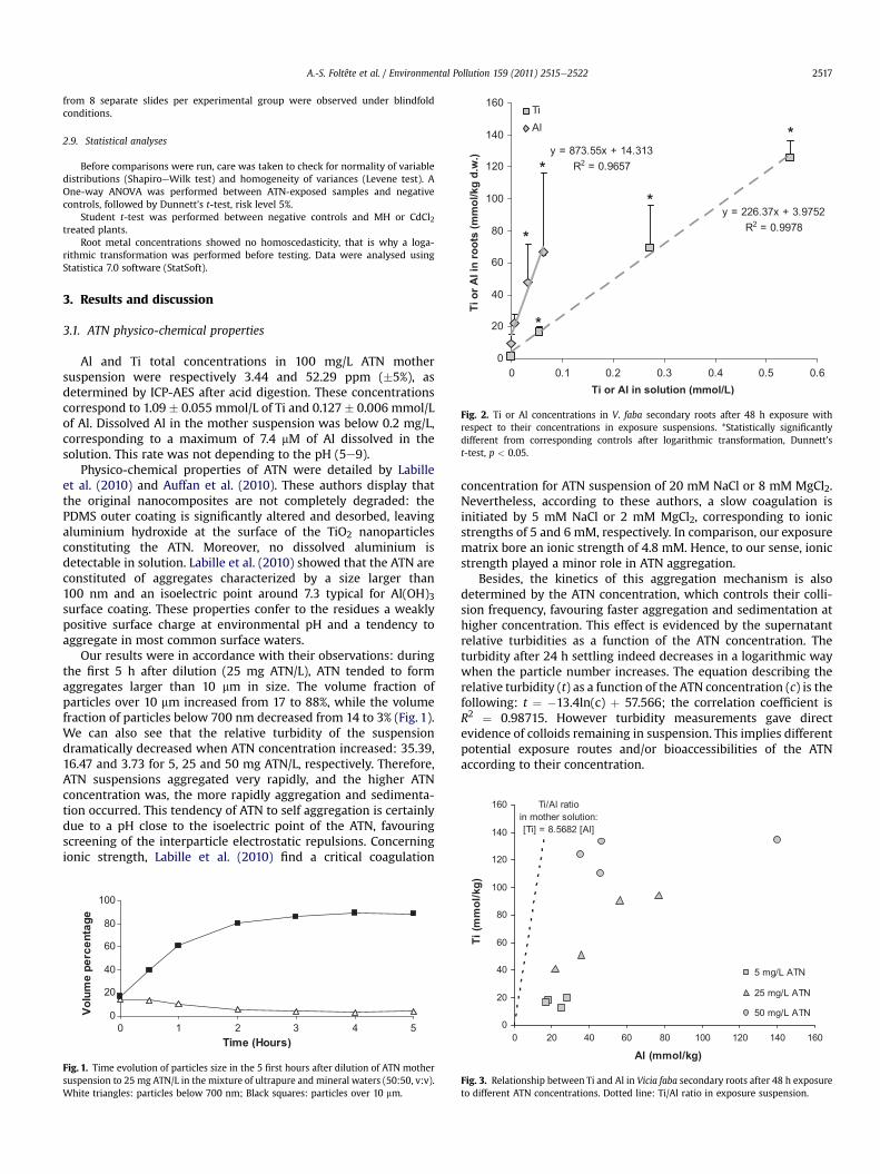
Fig. 3. Relationship between Ti and Al in Vicia faba secondary roots after 48 h exposureto different ATN concentrations. Dotted line: Ti/Al ratio in exposure suspension.
A.-S. Foltête et al. / Environmental Pollution 159 (2011) 2515e25222518
During exposure, root exudates could also have exerted anaction on ATN physico-chemical properties, probably acceleratingATN aggregation.
At the end of the exposure, the pH of the exposure suspensionwas comprised between 6.18 and 6.87 without any clear correlationwith ATN concentration.
3.2. Ti and Al contents in tissues
Titanium and aluminium concentrations in plant roots arerepresented in Fig. 2. Titanium concentrations were highlydifferent from controls regardless of ATN concentration.Aluminium concentrations were significantly different only for 25and 50 mg ATN/L. A strong positive linear correlation exists
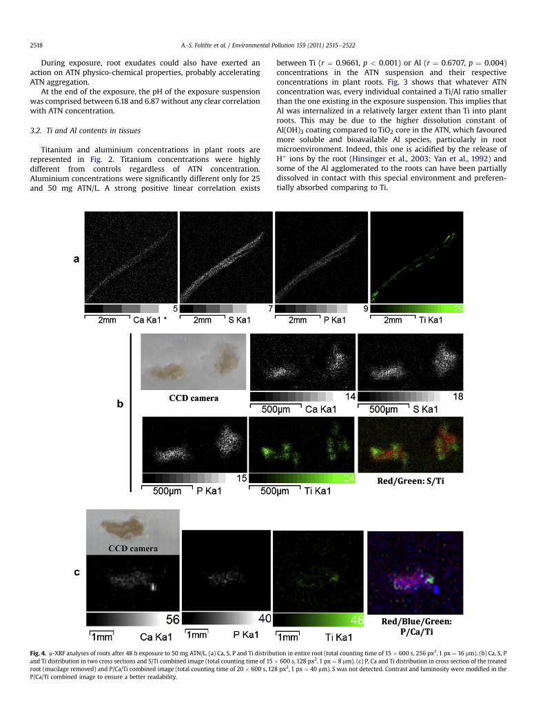
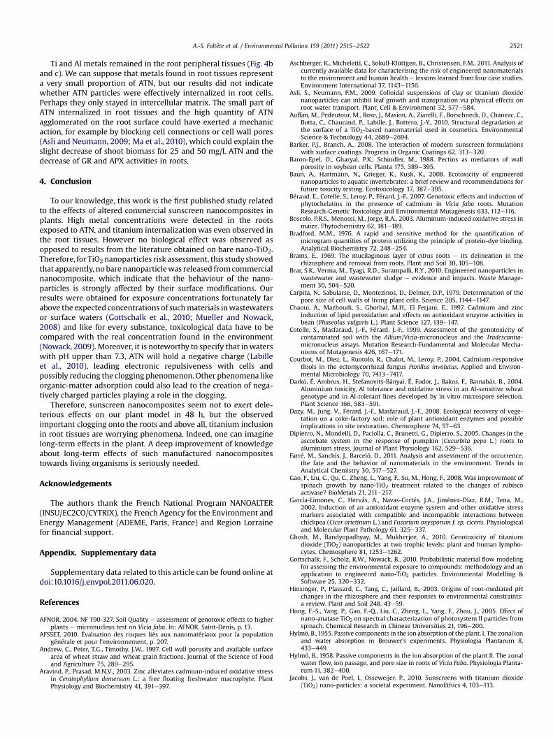
Fig. 4. m-XRF analyses of roots after 48 h exposure to 50 mg ATN/L. (a) Ca, S, P and Ti distribuand Ti distribution in two cross sections and S/Ti combined image (total counting time of 15 �root (mucilage removed) and P/Ca/Ti combined image (total counting time of 20 � 600 s, 12P/Ca/Ti combined image to ensure a better readability.
between Ti (r ¼ 0.9661, p < 0.001) or Al (r ¼ 0.6707, p ¼ 0.004)concentrations in the ATN suspension and their respectiveconcentrations in plant roots. Fig. 3 shows that whatever ATNconcentration was, every individual contained a Ti/Al ratio smallerthan the one existing in the exposure suspension. This implies thatAl was internalized in a relatively larger extent than Ti into plantroots. This may be due to the higher dissolution constant ofAl(OH)3 coating compared to TiO2 core in the ATN, which favouredmore soluble and bioavailable Al species, particularly in rootmicroenvironment. Indeed, this one is acidified by the release ofHþ ions by the root (Hinsinger et al., 2003; Yan et al., 1992) andsome of the Al agglomerated to the roots can have been partiallydissolved in contact with this special environment and preferen-tially absorbed comparing to Ti.
tion in entire root (total counting time of 15 � 600 s, 256 px2, 1 px ¼ 16 mm). (b) Ca, S, P600 s, 128 px2, 1 px ¼ 8 mm). (c) P, Ca and Ti distribution in cross section of the treated
8 px2, 1 px ¼ 40 mm). S was not detected. Contrast and luminosity were modified in the
0.0
0.5
1.0
1.5
2.0
2.5
3.0
3.5
4.0
4.5
0 5 25 50 C dATN (mg/L)
ro
ot e
lo
ng
atio
n (c
m)
*
0.00.2
0.40.6
0.81.0
1.21.4
1.61.8
0 5 25 50ATN (mg/L)
fresh
w
eig
ht (g
)
0.70
0.72
0.74
0.76
0.78
0.80
0.82
0.84
0 5 25 50ATN (mg/L)
Fv/F
m
a
b
c
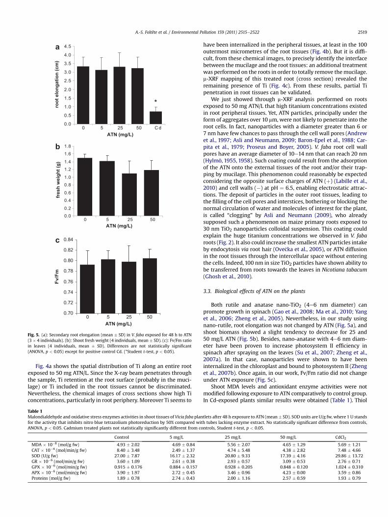
Fig. 5. (a): Secondary root elongation (mean � SD) in V. faba exposed for 48 h to ATN(3 � 4 individuals). (b): Shoot fresh weight (4 individuals, mean � SD). (c): Fv/Fm ratioin leaves (4 individuals, mean � SD). Differences are not statistically significant(ANOVA, p < 0.05) except for positive control Cd. (*Student t-test, p < 0.05).
A.-S. Foltête et al. / Environmental Pollution 159 (2011) 2515e2522 2519
Fig. 4a shows the spatial distribution of Ti along an entire rootexposed to 50 mg ATN/L. Since the X-ray beam penetrates throughthe sample, Ti retention at the root surface (probably in the muci-lage) or Ti included in the root tissues cannot be discriminated.Nevertheless, the chemical images of cross sections show high Ticoncentrations, particularly in root periphery. Moreover Ti seems to
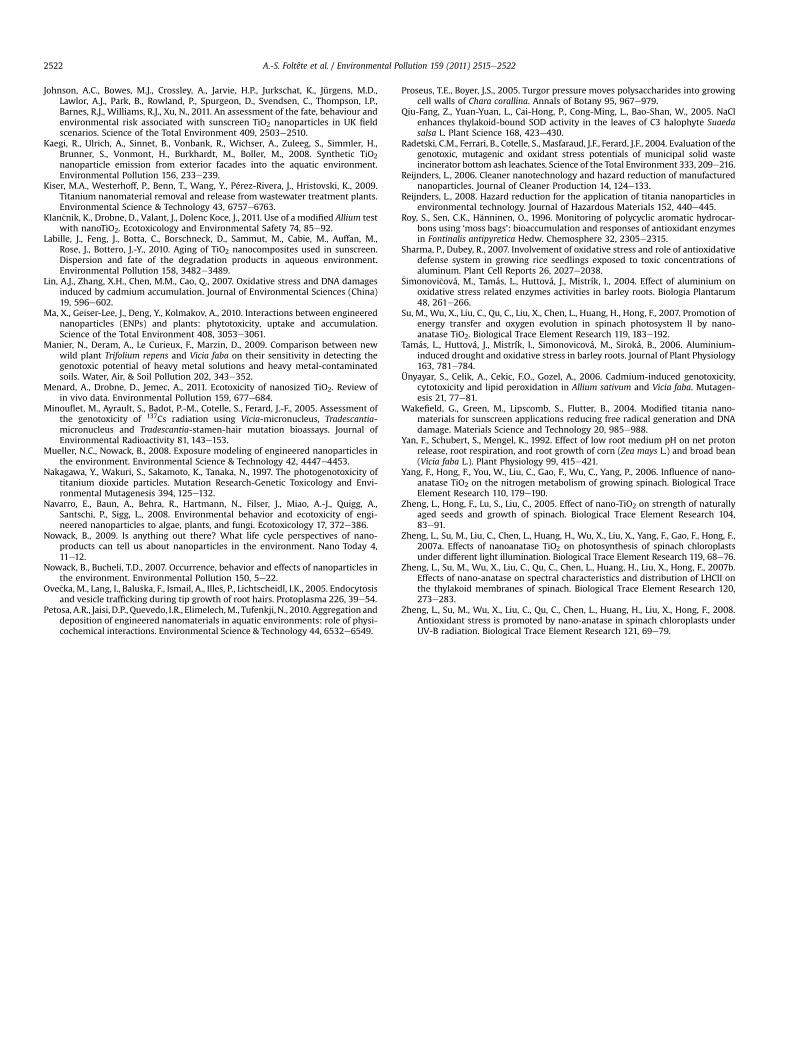
Table 1Malondialdehyde and oxidative stress enzymes activities in shoot tissues of Vicia faba planfor the activity that inhibits nitro blue tetrazolium photoreduction by 50% compared witANOVA, p < 0.05. Cadmium treated plants not statistically significantly different from co
Control 5 mg/L
MDA � 10�8 (mol/g fw) 4.93 � 2.02 4.69 � 0.84CAT � 10�4 (mol/min/g fw) 8.40 � 3.48 2.49 � 1.37SOD (U/g fw) 27.00 � 7.87 16.17 � 2.32GR � 10�6 (mol/min/g fw) 3.60 � 1.09 2.61 � 0.38GPX � 10�6 (mol/min/g fw) 0.915 � 0.176 0.884 � 0.157APX � 10�6 (mol/min/g fw) 3.90 � 1.97 2.72 � 0.45Proteins (mol/g fw) 1.89 � 0.78 2.74 � 0.43
have been internalized in the peripheral tissues, at least in the 100outermost micrometres of the root tissues (Fig. 4b). But it is diffi-cult, from these chemical images, to precisely identify the interfacebetween the mucilage and the root tissues: an additional treatmentwas performed on the roots in order to totally remove themucilage.m-XRF mapping of this treated root (cross section) revealed theremaining presence of Ti (Fig. 4c). From these results, partial Tipenetration in root tissues can be validated.
We just showed through m-XRF analysis performed on rootsexposed to 50 mg ATN/L that high titanium concentrations existedin root peripheral tissues. Yet, ATN particles, principally under theform of aggregates over 10 mm, were not likely to penetrate into theroot cells. In fact, nanoparticles with a diameter greater than 6 or7 nm have few chances to pass through the cell wall pores (Andrewet al., 1997; Asli and Neumann, 2009; Baron-Epel et al., 1988; Car-pita et al., 1979; Proseus and Boyer, 2005). V. faba root cell wallpores have an average diameter of 10e14 nm that can reach 20 nm(Hylmö, 1955, 1958). Such coating could result from the adsorptionof the ATN onto the external tissues of the root and/or their trap-ping by mucilage. This phenomenon could reasonably be expectedconsidering the opposite surface charges of ATN (þ) (Labille et al.,2010) and cell walls (�) at pH ¼ 6.5, enabling electrostatic attrac-tions. The deposit of particles in the outer root tissues, leading tothe filling of the cell pores and interstices, bothering or blocking thenormal circulation of water and molecules of interest for the plant,is called “clogging” by Asli and Neumann (2009), who alreadysupposed such a phenomenon on maize primary roots exposed to30 nm TiO2 nanoparticles colloidal suspension. This coating couldexplain the huge titanium concentrations we observed in V. fabaroots (Fig. 2). It also could increase the smallest ATN particles intakeby endocytosis via root hair (Ove�cka et al., 2005), or ATN diffusionin the root tissues through the intercellular space without enteringthe cells. Indeed,100 nm in size TiO2 particles have shown ability tobe transferred from roots towards the leaves in Nicotiana tabacum(Ghosh et al., 2010).
3.3. Biological effects of ATN on the plants
Both rutile and anatase nano-TiO2 (4e6 nm diameter) canpromote growth in spinach (Gao et al., 2008; Ma et al., 2010; Yanget al., 2006; Zheng et al., 2005). Nevertheless, in our study usingnano-rutile, root elongation was not changed by ATN (Fig. 5a), andshoot biomass showed a slight tendency to decrease for 25 and50 mg/L ATN (Fig. 5b). Besides, nano-anatase with 4e6 nm diam-eter have been proven to increase photosystem II efficiency inspinach after spraying on the leaves (Su et al., 2007; Zheng et al.,2007a). In that case, nanoparticles were shown to have beeninternalized in the chloroplast and bound to photosystem II (Zhenget al., 2007b). Once again, in our work, Fv/Fm ratio did not changeunder ATN exposure (Fig. 5c).
Shoot MDA levels and antioxidant enzyme activities were notmodified following exposure to ATN comparatively to control group.In Cd-exposed plants similar results were obtained (Table 1). Thiol
tlets after 48 h exposure to ATN (mean� SD). SOD units are U/g fw, where 1 U standsh tubes lacking enzyme extract. No statistically significant difference from controls,ntrols, Student t-test, p < 0.05.
25 mg/L 50 mg/L CdCl2
5.56 � 2.07 4.65 � 1.29 5.69 � 1.214.74 � 5.48 4.38 � 2.82 7.48 � 4.66
20.80 � 9.33 17.39 � 4.16 29.86 � 13.722.93 � 0.57 3.09 � 0.53 2.76 � 0.71
0.928 � 0.205 0.848 � 0.120 1.024 � 0.3103.46 � 0.96 4.23 � 0.00 3.59 � 0.862.00 � 1.16 2.57 � 0.59 1.93 � 0.79

Table 2Malondialdehyde and oxidative stress enzyme activities in root tissues of Vicia faba plantlets after 48 h exposure to ATN (mean� SD). SOD units are U/g fw, where 1 U stands forthe activity that inhibits nitro blue tetrazolium photoreduction by 50% compared with tubes lacking enzyme extract. *Statistically significantly different from controls, ANOVAfollowed by Dunnett’s t-test, p < 0.05. #Cadmium treated plants statistically significantly different from controls, Student t-test, p < 0.05.
Control 5 mg/L 25 mg/L 50 mg/L CdCl2
MDA � 10�8 (mol/g fw) 2.26 � 0.27 2.41 � 1.82 3.39 � 1.32 2.85 � 0.51 5.50 � 1.55#CAT � 10�7 (mol/min/g fw) 3.56 � 0.94 3.11 � 0.53 3.44 � 0.95 3.77 � 0.49 5.81 � 1.07#SOD (U/g fw) 13.83 � 2.57 12.84 � 2.73 15.47 � 6.74 13.44 � 2.58 34.38 � 13.91#GR � 10�6 (mol/min/g fw) 1.177 � 0.131 1.157 � 0.131 0.846 � 0.476 0.243 � 0.424* 0.674 � 0.303#GPX � 10�5 (mol/min/g fw) 1.717 � 0.125 1.748 � 0.227 1.681 � 0.561 1.787 � 0.229 3.053 � 0.415#APX � 10�6 (mol/min/g fw) 5.388 � 0.868 3.711 � 0.517* 3.689 � 0.315* 4.062 � 1.294 7.741 � 3.704Proteins (mol/g fw) 0.729 � 0.166 0.606 � 0.225 0.539 � 0.265 0.547 � 0.200 0.340 � 0.120#
50 14
mcn *
A.-S. Foltête et al. / Environmental Pollution 159 (2011) 2515e25222520
concentrations in shoots were neither modified by ATN nor by Cd(data not shown). In roots, lipid peroxidation, MDA levels signifi-cantly increased in Cd-exposed individuals but not in ATN-exposedindividuals (Table 2). CAT, SOD and GPX enzymes responded simi-larly to MDA levels (Table 2). On the contrary, GR enzyme and totalproteins decreased in Cd-exposed roots. In ATN-exposed plants, GRactivity decreased significantly at 50 mg ATN/L (ANOVA p ¼ 0.0037).Nevertheless, APX activity significantly decreased (ANOVAp¼ 0.0433) only for 5 and 25mgATN/L. In other studies, anatase andrutile nano-TiO2 enhanced superoxide dismutase, catalase andperoxidase activities and decreasedmalondialdehyde in chloroplastsexposed to UV (Hong et al., 2005; Zheng et al., 2008). Malondialde-hyde could also be increased in Allium cepa roots (Ghosh et al., 2010).To the contrary, our study showed that in shoots, none of oxidativestress biomarkerwasaffectedbyATN. Evenwhenexposed toCd, anti-oxidative stress biomarkers in shoots remained unchanged, sug-gesting a limited transfer from the root tissue. In roots exposed toATN, none of these biomarkers was either increased, but rootsremained hidden from light during exposure and, to our knowledge,no element in the literature deal with a similar case. Anotherhypothesis could be that the decrease of GR and APX may be due todissolved aluminium, but in the literature, Al at lowconcentrations inhigher plants is mostly responsible for an enhanced oxidant stressenzyme activity and/or expression (Boscolo et al., 2003; Darkó et al.,2004; Dipierro et al., 2005; Sharma and Dubey, 2007; �Simonovi�cováet al., 2004; Tamás et al., 2006). Therefore, this hypothesis is noteither convincing.
Total GSH in roots was not affected in ATN-treated individuals(Fig. 6). GSH/GSSG ratio remained constant (data not shown). PC2,PC3 and PC4 synthesis was stimulated in Cd-treated roots but not inATN-treated ones.
0
0.05
0.1
0.15
0.2
0.25
0.3
0.35
0.4
0 5 25 50 CdATN (mg/L)
Th
io
ls (µ
mo
l / g
fresh
w
eig
ht)
GSHPC2PC3PC4
*
*
*
Fig. 6. Thiol concentrations (GSH and PCs, mean � SD) in roots of V. faba exposed 48 hto ATN (4 individuals). Differences between ATN-treated and control individuals arenot statistically significant, ANOVA, p < 0.05. Cd: reference metal. *Statisticallysignificant from controls (Student t-test, p < 0.05).
Not surprisingly, positive control (MH) induced a 13-fold increaseof micronucleus frequency and a decrease of mitotic index (8.6 and5.5% in negative and positive controls respectively). Exposure ofV. faba roots to ATN did not lead to any genotoxicity (Fig. 7). Celldivision, assessed by mitotic index, was not modified too.
In the literature, photogenotoxicity of nano-TiO2 was proven(Nakagawa et al., 1997), but data are contradictory in plants. In A.cepa, no genotoxicity was found, but only the mitotic index wasmodified in root meristems (Klan�cnik et al., 2011). To the contrary,Ghosh et al. (2010) proved genotoxic effects of 100 nm in size TiO2particles in A. cepa roots and N. tabacum leaves.
Comparing effects in plants of bare TiO2 nanoparticles with theeffects of altered nanocomposites observed in this study leads us tosuggest that no or very few bare nanoparticles were released fromATN during our experiment. To summarize, no biomarker wassignificantly modified by ATN in shoots, and only few ones in roots,without any clear concentration response relationship. Moreover,neither genotoxicity nor phytochelatin synthesis was seen in roots.Despite a high quantity of aluminium and titanium oxides in roots,the quasi-absence of neither effects nor defence reaction noticed inplants would be in accordance with the hypothesis according towhich the metals internalized in the roots were still under a bio-logically inert form. Nevertheless, since Ti/Al ratio was alwayslower in tissues than in exposure suspension (Fig. 3), we can thinkthat ATN structures presented by Auffan et al. (2010) and Labilleet al. (2010) were not under unchanged form in tissues, sincealuminium seemed to have been partly solubilized.
0
5
10
15
20
25
30
35
40
45
0 5 25 50 MH
ATN (mg/L)
mcn
/ 1 000 cells
0
2
4
6
8
10
12
MI (%
)
MI
*
Fig. 7. Micronucleus frequency and mitotic index (mean � SD) in V. faba roots exposed48 h to ATN (2 � 4 individuals). Differences between ATN-treated and control indi-viduals are not statistically significant, ANOVA, p < 0.05. MH: positive control.*Statistically significantly different from controls (Student t-test, p < 0.05).
A.-S. Foltête et al. / Environmental Pollution 159 (2011) 2515e2522 2521
Ti and Al metals remained in the root peripheral tissues (Fig. 4band c). We can suppose that metals found in root tissues representa very small proportion of ATN, but our results did not indicatewhether ATN particles were effectively internalized in root cells.Perhaps they only stayed in intercellular matrix. The small part ofATN internalized in root tissues and the high quantity of ATNagglomerated on the root surface could have exerted a mechanicaction, for example by blocking cell connections or cell wall pores(Asli and Neumann, 2009; Ma et al., 2010), which could explain theslight decrease of shoot biomass for 25 and 50 mg/L ATN and thedecrease of GR and APX activities in roots.
4. Conclusion
To our knowledge, this work is the first published study relatedto the effects of altered commercial sunscreen nanocomposites inplants. High metal concentrations were detected in the rootsexposed to ATN, and titanium internalization was even observed inthe root tissues. However no biological effect was observed asopposed to results from the literature obtained on bare nano-TiO2.Therefore, for TiO2 nanoparticles risk assessment, this study showedthat apparently, no bare nanoparticlewas released fromcommercialnanocomposite, which indicate that the behaviour of the nano-particles is strongly affected by their surface modifications. Ourresults were obtained for exposure concentrations fortunately farabove the expected concentrations of suchmaterials inwastewatersor surface waters (Gottschalk et al., 2010; Mueller and Nowack,2008) and like for every substance, toxicological data have to becompared with the real concentration found in the environment(Nowack, 2009). Moreover, it is noteworthy to specify that inwaterswith pH upper than 7.3, ATN will hold a negative charge (Labilleet al., 2010), leading electronic repulsiveness with cells andpossibly reducing the clogging phenomenon. Other phenomena likeorganic-matter adsorption could also lead to the creation of nega-tively charged particles playing a role in the clogging.
Therefore, sunscreen nanocomposites seem not to exert dele-terious effects on our plant model in 48 h, but the observedimportant clogging onto the roots and above all, titanium inclusionin root tissues are worrying phenomena. Indeed, one can imaginelong-term effects in the plant. A deep improvement of knowledgeabout long-term effects of such manufactured nanocompositestowards living organisms is seriously needed.
Acknowledgements
The authors thank the French National Program NANOALTER(INSU/EC2CO/CYTRIX), the French Agency for the Environment andEnergy Management (ADEME, Paris, France) and Region Lorrainefor financial support.
Appendix. Supplementary data
Supplementary data related to this article can be found online atdoi:10.1016/j.envpol.2011.06.020.
References
AFNOR, 2004. NF T90-327. Soil Quality e assessment of genotoxic effects to higherplants e micronucleus test on Vicia faba. In: AFNOR. Saint-Denis, p. 13.
AFSSET, 2010. Évaluation des risques liés aux nanomatériaux pour la populationgénérale et pour l’environnement, p. 207.
Andrew, C., Peter, T.G., Timothy, J.W., 1997. Cell wall porosity and available surfacearea of wheat straw and wheat grain fractions. Journal of the Science of Foodand Agriculture 75, 289e295.
Aravind, P., Prasad, M.N.V., 2003. Zinc alleviates cadmium-induced oxidative stressin Ceratophyllum demersum L.: a free floating freshwater macrophyte. PlantPhysiology and Biochemistry 41, 391e397.
Aschberger, K., Micheletti, C., Sokull-Klüttgen, B., Christensen, F.M., 2011. Analysis ofcurrently available data for characterising the risk of engineered nanomaterialsto the environment and human health e lessons learned from four case studies.Environment International 37, 1143e1156.
Asli, S., Neumann, P.M., 2009. Colloidal suspensions of clay or titanium dioxidenanoparticles can inhibit leaf growth and transpiration via physical effects onroot water transport. Plant, Cell & Environment 32, 577e584.
Auffan, M., Pedeutour, M., Rose, J., Masion, A., Ziarelli, F., Borschneck, D., Chaneac, C.,Botta, C., Chaurand, P., Labille, J., Bottero, J.-Y., 2010. Structural degradation atthe surface of a TiO2-based nanomaterial used in cosmetics. EnvironmentalScience & Technology 44, 2689e2694.
Barker, P.J., Branch, A., 2008. The interaction of modern sunscreen formulationswith surface coatings. Progress in Organic Coatings 62, 313e320.
Baron-Epel, O., Gharyal, P.K., Schindler, M., 1988. Pectins as mediators of wallporosity in soybean cells. Planta 175, 389e395.
Baun, A., Hartmann, N., Grieger, K., Kusk, K., 2008. Ecotoxicity of engineerednanoparticles to aquatic invertebrates: a brief review and recommendations forfuture toxicity testing. Ecotoxicology 17, 387e395.
Béraud, E., Cotelle, S., Leroy, P., Férard, J.-F., 2007. Genotoxic effects and induction ofphytochelatins in the presence of cadmium in Vicia faba roots. MutationResearch-Genetic Toxicology and Environmental Mutagenesis 633, 112e116.
Boscolo, P.R.S., Menossi, M., Jorge, R.A., 2003. Aluminum-induced oxidative stress inmaize. Phytochemistry 62, 181e189.
Bradford, M.M., 1976. A rapid and sensitive method for the quantification ofmicrogram quantities of protein utilizing the principle of protein-dye binding.Analytical Biochemistry 72, 248e254.
Brams, E., 1969. The mucilaginous layer of citrus roots e its delineation in therhizosphere and removal from roots. Plant and Soil 30, 105e108.
Brar, S.K., Verma, M., Tyagi, R.D., Surampalli, R.Y., 2010. Engineered nanoparticles inwastewater and wastewater sludge e evidence and impacts. Waste Manage-ment 30, 504e520.
Carpita, N., Sabularse, D., Montezinos, D., Delmer, D.P., 1979. Determination of thepore size of cell walls of living plant cells. Science 205, 1144e1147.
Chaoui, A., Mazhoudi, S., Ghorbal, M.H., El Ferjani, E., 1997. Cadmium and zincinduction of lipid peroxidation and effects on antioxidant enzyme activities inbean (Phaseolus vulgaris L.). Plant Science 127, 139e147.
Cotelle, S., Masfaraud, J.-F., Férard, J.-F., 1999. Assessment of the genotoxicity ofcontaminated soil with the Allium/Vicia-micronucleus and the Tradescantia-micronucleus assays. Mutation Research-Fundamental and Molecular Mecha-nisms of Mutagenesis 426, 167e171.
Courbot, M., Diez, L., Ruotolo, R., Chalot, M., Leroy, P., 2004. Cadmium-responsivethiols in the ectomycorrhizal fungus Paxillus involutus. Applied and Environ-mental Microbiology 70, 7413e7417.
Darkó, É, Ambrus, H., Stefanovits-Bányai, É, Fodor, J., Bakos, F., Barnabás, B., 2004.Aluminium toxicity, Al tolerance and oxidative stress in an Al-sensitive wheatgenotype and in Al-tolerant lines developed by in vitro microspore selection.Plant Science 166, 583e591.
Dazy, M., Jung, V., Férard, J.-F., Masfaraud, J.-F., 2008. Ecological recovery of vege-tation on a coke-factory soil: role of plant antioxidant enzymes and possibleimplications in site restoration. Chemosphere 74, 57e63.
Dipierro, N., Mondelli, D., Paciolla, C., Brunetti, G., Dipierro, S., 2005. Changes in theascorbate system in the response of pumpkin (Cucurbita pepo L.) roots toaluminium stress. Journal of Plant Physiology 162, 529e536.
Farré, M., Sanchís, J., Barceló, D., 2011. Analysis and assessment of the occurrence,the fate and the behavior of nanomaterials in the environment. Trends inAnalytical Chemistry 30, 517e527.
Gao, F., Liu, C., Qu, C., Zheng, L., Yang, F., Su, M., Hong, F., 2008. Was improvement ofspinach growth by nano-TiO2 treatment related to the changes of rubiscoactivase? BioMetals 21, 211e217.
García-Limones, C., Hervás, A., Navas-Cortés, J.A., Jiménez-Díaz, R.M., Tena, M.,2002. Induction of an antioxidant enzyme system and other oxidative stressmarkers associated with compatible and incompatible interactions betweenchickpea (Cicer arietinum L.) and Fusarium oxysporum f. sp. ciceris. Physiologicaland Molecular Plant Pathology 61, 325e337.
Ghosh, M., Bandyopadhyay, M., Mukherjee, A., 2010. Genotoxicity of titaniumdioxide (TiO2) nanoparticles at two trophic levels: plant and human lympho-cytes. Chemosphere 81, 1253e1262.
Gottschalk, F., Scholz, R.W., Nowack, B., 2010. Probabilistic material flow modelingfor assessing the environmental exposure to compounds: methodology and anapplication to engineered nano-TiO2 particles. Environmental Modelling &Software 25, 320e332.
Hinsinger, P., Plassard, C., Tang, C., Jaillard, B., 2003. Origins of root-mediated pHchanges in the rhizosphere and their responses to environmental constraints:a review. Plant and Soil 248, 43e59.
Hong, F.-S., Yang, P., Gao, F.-Q., Liu, C., Zheng, L., Yang, F., Zhou, J., 2005. Effect ofnano-anatase TiO2 on spectral characterization of photosystem II particles fromspinach. Chemical Research in Chinese Universities 21, 196e200.
Hylmö, B.,1955. Passive components in the ion absorption of the plant. I. The zonal ionand water absorption in Brouwer’s experiments. Physiologia Plantarum 8,433e449.
Hylmö, B., 1958. Passive components in the ion absorption of the plant II. The zonalwater flow, ion passage, and pore size in roots of Vicia Faba. Physiologia Planta-rum 11, 382e400.
Jacobs, J., van de Poel, I., Osseweijer, P., 2010. Sunscreens with titanium dioxide(TiO2) nano-particles: a societal experiment. NanoEthics 4, 103e113.
A.-S. Foltête et al. / Environmental Pollution 159 (2011) 2515e25222522
Johnson, A.C., Bowes, M.J., Crossley, A., Jarvie, H.P., Jurkschat, K., Jürgens, M.D.,Lawlor, A.J., Park, B., Rowland, P., Spurgeon, D., Svendsen, C., Thompson, I.P.,Barnes, R.J., Williams, R.J., Xu, N., 2011. An assessment of the fate, behaviour andenvironmental risk associated with sunscreen TiO2 nanoparticles in UK fieldscenarios. Science of the Total Environment 409, 2503e2510.
Kaegi, R., Ulrich, A., Sinnet, B., Vonbank, R., Wichser, A., Zuleeg, S., Simmler, H.,Brunner, S., Vonmont, H., Burkhardt, M., Boller, M., 2008. Synthetic TiO2nanoparticle emission from exterior facades into the aquatic environment.Environmental Pollution 156, 233e239.
Kiser, M.A., Westerhoff, P., Benn, T., Wang, Y., Pérez-Rivera, J., Hristovski, K., 2009.Titanium nanomaterial removal and release from wastewater treatment plants.Environmental Science & Technology 43, 6757e6763.
Klan�cnik, K., Drobne, D., Valant, J., Dolenc Koce, J., 2011. Use of a modified Allium testwith nanoTiO2. Ecotoxicology and Environmental Safety 74, 85e92.
Labille, J., Feng, J., Botta, C., Borschneck, D., Sammut, M., Cabie, M., Auffan, M.,Rose, J., Bottero, J.-Y., 2010. Aging of TiO2 nanocomposites used in sunscreen.Dispersion and fate of the degradation products in aqueous environment.Environmental Pollution 158, 3482e3489.
Lin, A.J., Zhang, X.H., Chen, M.M., Cao, Q., 2007. Oxidative stress and DNA damagesinduced by cadmium accumulation. Journal of Environmental Sciences (China)19, 596e602.
Ma, X., Geiser-Lee, J., Deng, Y., Kolmakov, A., 2010. Interactions between engineerednanoparticles (ENPs) and plants: phytotoxicity, uptake and accumulation.Science of the Total Environment 408, 3053e3061.
Manier, N., Deram, A., Le Curieux, F., Marzin, D., 2009. Comparison between newwild plant Trifolium repens and Vicia faba on their sensitivity in detecting thegenotoxic potential of heavy metal solutions and heavy metal-contaminatedsoils. Water, Air, & Soil Pollution 202, 343e352.
Menard, A., Drobne, D., Jemec, A., 2011. Ecotoxicity of nanosized TiO2. Review ofin vivo data. Environmental Pollution 159, 677e684.
Minouflet, M., Ayrault, S., Badot, P.-M., Cotelle, S., Ferard, J.-F., 2005. Assessment ofthe genotoxicity of 137Cs radiation using Vicia-micronucleus, Tradescantia-micronucleus and Tradescantia-stamen-hair mutation bioassays. Journal ofEnvironmental Radioactivity 81, 143e153.
Mueller, N.C., Nowack, B., 2008. Exposure modeling of engineered nanoparticles inthe environment. Environmental Science & Technology 42, 4447e4453.
Nakagawa, Y., Wakuri, S., Sakamoto, K., Tanaka, N., 1997. The photogenotoxicity oftitanium dioxide particles. Mutation Research-Genetic Toxicology and Envi-ronmental Mutagenesis 394, 125e132.
Navarro, E., Baun, A., Behra, R., Hartmann, N., Filser, J., Miao, A.-J., Quigg, A.,Santschi, P., Sigg, L., 2008. Environmental behavior and ecotoxicity of engi-neered nanoparticles to algae, plants, and fungi. Ecotoxicology 17, 372e386.
Nowack, B., 2009. Is anything out there? What life cycle perspectives of nano-products can tell us about nanoparticles in the environment. Nano Today 4,11e12.
Nowack, B., Bucheli, T.D., 2007. Occurrence, behavior and effects of nanoparticles inthe environment. Environmental Pollution 150, 5e22.
Ove�cka, M., Lang, I., Balu�ska, F., Ismail, A., Ille�s, P., Lichtscheidl, I.K., 2005. Endocytosisand vesicle trafficking during tip growth of root hairs. Protoplasma 226, 39e54.
Petosa, A.R., Jaisi, D.P., Quevedo, I.R., Elimelech,M., Tufenkji, N., 2010. Aggregation anddeposition of engineered nanomaterials in aquatic environments: role of physi-cochemical interactions. Environmental Science & Technology 44, 6532e6549.
Proseus, T.E., Boyer, J.S., 2005. Turgor pressure moves polysaccharides into growingcell walls of Chara corallina. Annals of Botany 95, 967e979.
Qiu-Fang, Z., Yuan-Yuan, L., Cai-Hong, P., Cong-Ming, L., Bao-Shan, W., 2005. NaClenhances thylakoid-bound SOD activity in the leaves of C3 halophyte Suaedasalsa L. Plant Science 168, 423e430.
Radetski, C.M., Ferrari, B., Cotelle, S., Masfaraud, J.F., Ferard, J.F., 2004. Evaluation of thegenotoxic, mutagenic and oxidant stress potentials of municipal solid wasteincinerator bottom ash leachates. Science of the Total Environment 333, 209e216.
Reijnders, L., 2006. Cleaner nanotechnology and hazard reduction of manufacturednanoparticles. Journal of Cleaner Production 14, 124e133.
Reijnders, L., 2008. Hazard reduction for the application of titania nanoparticles inenvironmental technology. Journal of Hazardous Materials 152, 440e445.
Roy, S., Sen, C.K., Hänninen, O., 1996. Monitoring of polycyclic aromatic hydrocar-bons using ‘moss bags’: bioaccumulation and responses of antioxidant enzymesin Fontinalis antipyretica Hedw. Chemosphere 32, 2305e2315.
Sharma, P., Dubey, R., 2007. Involvement of oxidative stress and role of antioxidativedefense system in growing rice seedlings exposed to toxic concentrations ofaluminum. Plant Cell Reports 26, 2027e2038.
�Simonovi�cová, M., Tamás, L., Huttová, J., Mistrík, I., 2004. Effect of aluminium onoxidative stress related enzymes activities in barley roots. Biologia Plantarum48, 261e266.
Su, M., Wu, X., Liu, C., Qu, C., Liu, X., Chen, L., Huang, H., Hong, F., 2007. Promotion ofenergy transfer and oxygen evolution in spinach photosystem II by nano-anatase TiO2. Biological Trace Element Research 119, 183e192.
Tamás, L., Huttová, J., Mistrík, I., Simonovicová, M., Siroká, B., 2006. Aluminium-induced drought and oxidative stress in barley roots. Journal of Plant Physiology163, 781e784.
Ünyayar, S., Celik, A., Cekic, F.O., Gozel, A., 2006. Cadmium-induced genotoxicity,cytotoxicity and lipid peroxidation in Allium sativum and Vicia faba. Mutagen-esis 21, 77e81.
Wakefield, G., Green, M., Lipscomb, S., Flutter, B., 2004. Modified titania nano-materials for sunscreen applications reducing free radical generation and DNAdamage. Materials Science and Technology 20, 985e988.
Yan, F., Schubert, S., Mengel, K., 1992. Effect of low root medium pH on net protonrelease, root respiration, and root growth of corn (Zea mays L.) and broad bean(Vicia faba L.). Plant Physiology 99, 415e421.
Yang, F., Hong, F., You, W., Liu, C., Gao, F., Wu, C., Yang, P., 2006. Influence of nano-anatase TiO2 on the nitrogen metabolism of growing spinach. Biological TraceElement Research 110, 179e190.
Zheng, L., Hong, F., Lu, S., Liu, C., 2005. Effect of nano-TiO2 on strength of naturallyaged seeds and growth of spinach. Biological Trace Element Research 104,83e91.
Zheng, L., Su, M., Liu, C., Chen, L., Huang, H., Wu, X., Liu, X., Yang, F., Gao, F., Hong, F.,2007a. Effects of nanoanatase TiO2 on photosynthesis of spinach chloroplastsunder different light illumination. Biological Trace Element Research 119, 68e76.
Zheng, L., Su, M., Wu, X., Liu, C., Qu, C., Chen, L., Huang, H., Liu, X., Hong, F., 2007b.Effects of nano-anatase on spectral characteristics and distribution of LHCII onthe thylakoid membranes of spinach. Biological Trace Element Research 120,273e283.
Zheng, L., Su, M., Wu, X., Liu, C., Qu, C., Chen, L., Huang, H., Liu, X., Hong, F., 2008.Antioxidant stress is promoted by nano-anatase in spinach chloroplasts underUV-B radiation. Biological Trace Element Research 121, 69e79.
Related Documents