Enoxaparin Anticoagulation Monitoring in the Catheterization Laboratory Using a New Bedside Test Johanne Silvain, MD,* Farzin Beygui, MD, PHD,* Annick Ankri, MD,‡ Anne Bellemain-Appaix, MD,* Ana Pena, PHD,* Olivier Barthelemy, MD,* Guillaume Cayla, MD,* Vanessa Gallois, BS,* Sophie Galier, BS,* Dominique Costagliola, PHD,† Jean-Philippe Collet, MD, PHD,* Gilles Montalescot, MD, PHD* Paris, France Objectives This study evaluated the ability of the bedside test Hemochron Jr. Hemonox (International Technidyne Corporation, Edison, New Jersey) to identify patients with insufficient anti-Xa activity level in the catheterization laboratory. Background Inadequate anticoagulation in patients undergoing percutaneous coronary intervention (PCI) is associated with increased periprocedural ischemic events. Methods In 296 unselected patients undergoing catheterization and/or PCI, whole blood Hemonox clotting time (CT) and activated partial thromboplastin time (aPTT) were measured at baseline (T1) and 10 min after the intravenous administration of enoxaparin (T2) in patients receiving additional enoxaparin and compared with plasma chro- mogenic anti-Xa activity level. Results Median values were 0.1 IU/ml (interquartile range [IQR]: 0.1 to 0.1 IU/ml) and 0.87 IU/ml (IQR: 0.74 to 1.03 IU/ml) for anti-Xa; 74 s (IQR: 70 to 81 s) and 143 s (IQR: 114 to 206 s) for Hemonox CT; and 44 s (IQR: 39 to 50 s) and 72 s (IQR: 58 to 93 s) for aPTT at T1 and T2, respectively. When using Hemonox CT to discriminate patients with anti-Xa level 0.5 IU/ml, the area under the receiver operating characteristic curve was 0.95 0.01 (95% confidence inter- val [CI]: 0.93 to 0.97) versus 0.89 0.01 (95% CI: 0.86 to 0.92) for aPTT. The threshold value of 120 s was associ- ated with a 94.9% (95% CI: 91.1% to 97.4%) sensitivity and a 73.3% (95% CI: 67.6% to 78.5%) specificity to detect patients with inadequate anti-Xa level (0.5 IU/ml) and positive predictive and negative predictive values of 73.9% (95% CI: 68.7% to 79.0%) and 94.78% (95% CI: 91.8% to 97.8%), respectively. Conclusions Hemonox CT appears to be a fast and reliable bedside test for detecting patients insufficiently anticoagulated and needing adjustment of anticoagulation therapy with enoxaparin before PCI. (J Am Coll Cardiol 2010;55: 617–25) © 2010 by the American College of Cardiology Foundation In many countries, low molecular weight heparin (LMWH) enoxaparin is the most common form of heparin used in the treatment of acute coronary syndromes (ACS) (1). Current guidelines from the European Society of Cardiology and other international societies strongly recommend enoxaparin in the treatment of ST-segment elevation myocardial infarction (STEMI) in association with fibrinolysis (Class I, Level of Evidence: A), in the treatment of non–ST-segment elevation ACS for invasive strategy (Class IIa, Level of Evidence: B), and for conservative strategy (Class I, Level of Evidence: B) (2). Additionally, enoxaparin is being evaluated for primary percutaneous coronary intervention (PCI) in STEMI patients (NCT00718471). Recently, the safety and efficacy of intrave- nous LMWH anticoagulation therapy for patients undergoing either urgent or elective PCI has been demonstrated in a number of trials (3). Compared with unfractionated heparin (UFH), enoxaparin provides more predictive and more stable anticoagulation therapy. In the STEEPLE (Safety and Effi- cacy of Enoxaparin in Percutaneous Coronary Intervention Patients, an International Randomized Evaluation) trial, target anticoagulation levels were achieved in 86% of patients receiv- ing enoxaparin without monitoring, compared with 20% of From the *Institut de Cardiologie, Institut National de la Santé et de la Recherche Médicale (INSERM) CMR937, †Institut de Cardiologie, INSERM U720, and the ‡Service d’Hématologie Biologique, Pitié-Salpêtrière Hospital (AP-HP), Université Paris 6, Paris, France. This study was supported in part by INSERM and by International Technidyne Corporation (ITC) Edison, New Jersey. Dr. Silvain has received research grants from Bristol-Myers Squibb, Daichi-Sankyo, and Eli Lilly. Dr. Beygui has received lecture fees from Pfizer, Sanofi-Aventis, and Astellas. Dr. Montalescot has received research grants from Bristol-Myers Squibb, Sanofi-Aventis Group, Eli Lilly, Guerbet Medical, Medtronic, Boston Scientific, Cordis, Stago, Centocor, Fondation de France, INSERM, Fédération Française de Cardiologie, and Société Française de Cardiologie; consulting fees from Sanofi-Aventis Group, Eli Lilly, Bristol-Myers Squibb, The Medicines Company, and Schering-Plough; and lecture fees from Sanofi-Aventis, Eli Lilly, Bristol-Myers Squibb, Merck Sharpe & Dohme, Cordis, GlaxoSmithKline, and Schering-Plough. Manuscript received June 23, 2009; revised manuscript received August 27, 2009, accepted August 30, 2009. Journal of the American College of Cardiology Vol. 55, No. 7, 2010 © 2010 by the American College of Cardiology Foundation ISSN 0735-1097/10/$36.00 Published by Elsevier Inc. doi:10.1016/j.jacc.2009.08.077

Welcome message from author
This document is posted to help you gain knowledge. Please leave a comment to let me know what you think about it! Share it to your friends and learn new things together.
Transcript
Ietg
FM‡PIrDMGCSLlD
a
Journal of the American College of Cardiology Vol. 55, No. 7, 2010© 2010 by the American College of Cardiology Foundation ISSN 0735-1097/10/$36.00P
Enoxaparin Anticoagulation Monitoring in theCatheterization Laboratory Using a New Bedside Test
Johanne Silvain, MD,* Farzin Beygui, MD, PHD,* Annick Ankri, MD,‡ Anne Bellemain-Appaix, MD,*Ana Pena, PHD,* Olivier Barthelemy, MD,* Guillaume Cayla, MD,* Vanessa Gallois, BS,*Sophie Galier, BS,* Dominique Costagliola, PHD,† Jean-Philippe Collet, MD, PHD,*Gilles Montalescot, MD, PHD*
Paris, France
Objectives This study evaluated the ability of the bedside test Hemochron Jr. Hemonox (International Technidyne Corporation,Edison, New Jersey) to identify patients with insufficient anti-Xa activity level in the catheterization laboratory.
Background Inadequate anticoagulation in patients undergoing percutaneous coronary intervention (PCI) is associated withincreased periprocedural ischemic events.
Methods In 296 unselected patients undergoing catheterization and/or PCI, whole blood Hemonox clotting time (CT) andactivated partial thromboplastin time (aPTT) were measured at baseline (T1) and 10 min after the intravenousadministration of enoxaparin (T2) in patients receiving additional enoxaparin and compared with plasma chro-mogenic anti-Xa activity level.
Results Median values were 0.1 IU/ml (interquartile range [IQR]: 0.1 to 0.1 IU/ml) and 0.87 IU/ml (IQR: 0.74 to 1.03 IU/ml)for anti-Xa; 74 s (IQR: 70 to 81 s) and 143 s (IQR: 114 to 206 s) for Hemonox CT; and 44 s (IQR: 39 to 50 s) and 72 s(IQR: 58 to 93 s) for aPTT at T1 and T2, respectively. When using Hemonox CT to discriminate patients with anti-Xalevel �0.5 IU/ml, the area under the receiver operating characteristic curve was 0.95 � 0.01 (95% confidence inter-val [CI]: 0.93 to 0.97) versus 0.89 � 0.01 (95% CI: 0.86 to 0.92) for aPTT. The threshold value of 120 s was associ-ated with a 94.9% (95% CI: 91.1% to 97.4%) sensitivity and a 73.3% (95% CI: 67.6% to 78.5%) specificity to detectpatients with inadequate anti-Xa level (�0.5 IU/ml) and positive predictive and negative predictive values of 73.9%(95% CI: 68.7% to 79.0%) and 94.78% (95% CI: 91.8% to 97.8%), respectively.
Conclusions Hemonox CT appears to be a fast and reliable bedside test for detecting patients insufficiently anticoagulatedand needing adjustment of anticoagulation therapy with enoxaparin before PCI. (J Am Coll Cardiol 2010;55:617–25) © 2010 by the American College of Cardiology Foundation
ublished by Elsevier Inc. doi:10.1016/j.jacc.2009.08.077
it(EAa(p(nen(acPa
n many countries, low molecular weight heparin (LMWH)noxaparin is the most common form of heparin used in thereatment of acute coronary syndromes (ACS) (1). Currentuidelines from the European Society of Cardiology and other
rom the *Institut de Cardiologie, Institut National de la Santé et de la Rechercheédicale (INSERM) CMR937, †Institut de Cardiologie, INSERM U720, and the
Service d’Hématologie Biologique, Pitié-Salpêtrière Hospital (AP-HP), Universitéaris 6, Paris, France. This study was supported in part by INSERM and bynternational Technidyne Corporation (ITC) Edison, New Jersey. Dr. Silvain haseceived research grants from Bristol-Myers Squibb, Daichi-Sankyo, and Eli Lilly.r. Beygui has received lecture fees from Pfizer, Sanofi-Aventis, and Astellas. Dr.ontalescot has received research grants from Bristol-Myers Squibb, Sanofi-Aventisroup, Eli Lilly, Guerbet Medical, Medtronic, Boston Scientific, Cordis, Stago,entocor, Fondation de France, INSERM, Fédération Française de Cardiologie, andociété Française de Cardiologie; consulting fees from Sanofi-Aventis Group, Eliilly, Bristol-Myers Squibb, The Medicines Company, and Schering-Plough; and
ecture fees from Sanofi-Aventis, Eli Lilly, Bristol-Myers Squibb, Merck Sharpe &ohme, Cordis, GlaxoSmithKline, and Schering-Plough.
iManuscript received June 23, 2009; revised manuscript received August 27, 2009,
ccepted August 30, 2009.
nternational societies strongly recommend enoxaparin in thereatment of ST-segment elevation myocardial infarctionSTEMI) in association with fibrinolysis (Class I, Level ofvidence: A), in the treatment of non–ST-segment elevationCS for invasive strategy (Class IIa, Level of Evidence: B),
nd for conservative strategy (Class I, Level of Evidence: B)2). Additionally, enoxaparin is being evaluated for primaryercutaneous coronary intervention (PCI) in STEMI patientsNCT00718471). Recently, the safety and efficacy of intrave-ous LMWH anticoagulation therapy for patients undergoingither urgent or elective PCI has been demonstrated in aumber of trials (3). Compared with unfractionated heparinUFH), enoxaparin provides more predictive and more stablenticoagulation therapy. In the STEEPLE (Safety and Effi-acy of Enoxaparin in Percutaneous Coronary Interventionatients, an International Randomized Evaluation) trial, targetnticoagulation levels were achieved in 86% of patients receiv-
ng enoxaparin without monitoring, compared with 20% of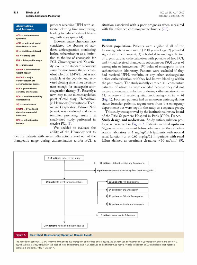
it
sw
M
Pfsoaechbtpr1(sd
oStSirf
618 Silvain et al. JACC Vol. 55, No. 7, 2010Bedside Enoxaparin Monitoring February 16, 2010:617–25
patients receiving UFH with ac-tivated clotting time monitoring,leading to reduced rates of bleed-ing with enoxaparin (4).
However, many physicians haveconsidered the absence of vali-dated anticoagulation monitoringbefore catheterization as a limita-tion to the use of enoxaparin forPCI. Chromogenic anti-Xa activ-ity level is the standard laboratoryassay for monitoring the anticoag-ulant effect of LMWH but is notavailable at the bedside, and acti-vated clotting time is not discrimi-nant enough for enoxaparin anti-coagulation therapy (5). Recently anew, easy to use microcoagulationpoint-of-care assay, HemochronJr. Hemonox (International Tech-nidyne Corporation, Edison, NewJersey), was developed and dem-onstrated promising results in asmall-sized study performed inelective PCI (6).
We decided to evaluate theability of the Hemonox test to
dentify patients with an anti-Xa activity level out of theherapeutic range during catheterization and/or PCI, a
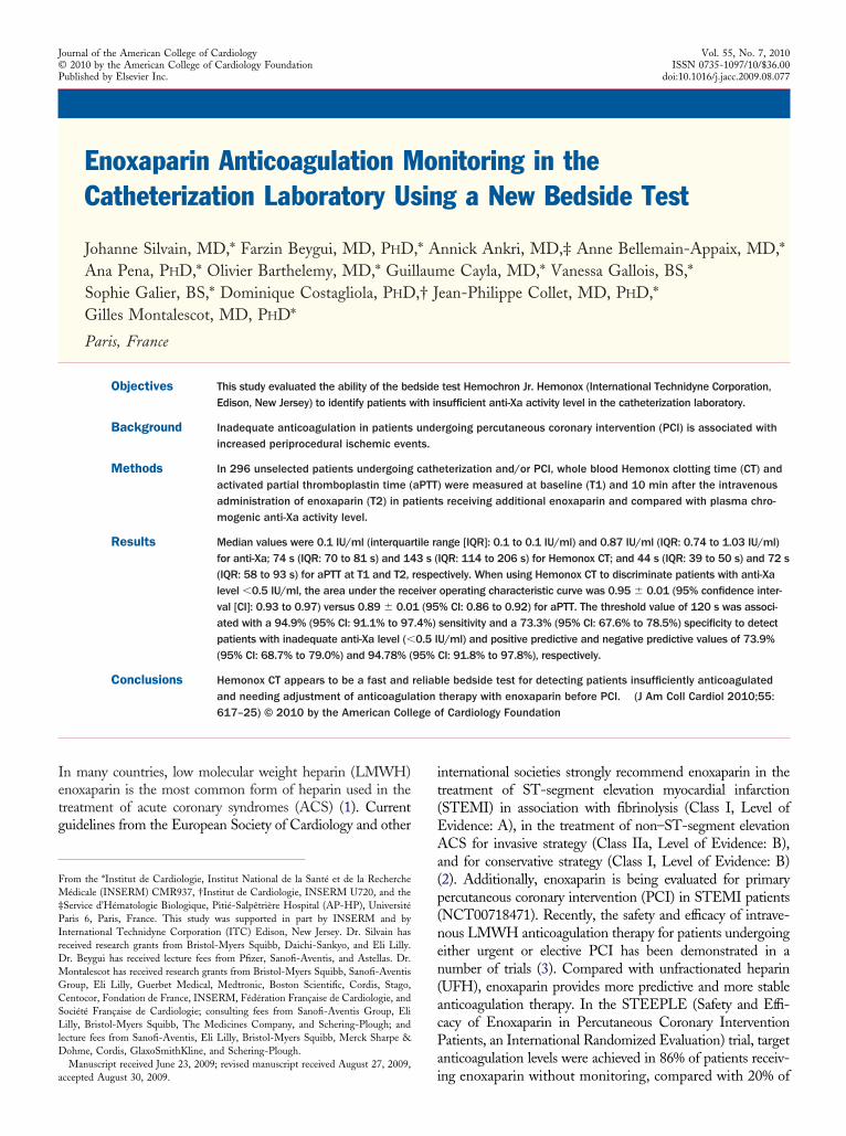
Figure 1 Flow Chart Representing Operation Clinical Events
The majority of patients (71.3%) received intravenous (IV) enoxaparin at the dosemg/kg/12 h (0.65 mg/kg/12 h in the case of renal impairment), and 7.1% receivbetween 8 and 12 h). vit-K � vitamin K.
Abbreviationsand Acronyms
ACS � acute coronarysyndrome
aPTT � activated partialthromboplastin time
CI � confidence interval
CT � clotting time
IQR � interquartile range
IV � intravenous
LMWH � low molecularweight heparin
MACCE � majorcardiovascular andcerebrovascular events
PCI � percutaneouscoronary intervention
ROC � receiver-operatingcharacteristic
SQ � subcutaneous
STEMI � ST-segmentelevation myocardialinfarction
UFH � unfractionatedheparin
ituation associated with a poor prognosis when measuredith the reference chromogenic technique (7,8).
ethods
atient population. Patients were eligible if all of theollowing criteria were met: 1) �18 years of age; 2) providedigned informed consent; 3) scheduled to undergo electiver urgent cardiac catheterization with possible ad hoc PCI;nd 4) had received therapeutic subcutaneous (SQ) doses ofnoxaparin or intravenous (IV) bolus of enoxaparin in theatheterization laboratory. Patients were excluded if theyad received UFH, warfarin, or any other anticoagulantefore catheterization or if they had known bleeding withinhe past month. The study initially enrolled 313 consecutiveatients, of whom 17 were excluded because they did noteceive any enoxaparin before or during catheterization (n �1) or were still receiving vitamin-K antagonist (n � 6)Fig. 1). Fourteen patients had an unknown anticoagulationtatus (transfer patients, urgent cases from the emergencyepartment) but were kept in the study as a separate group.This study was approved by the institutional review board
f the Pitié-Salpêtrière Hospital in Paris (CPP), France.tudy design and medication. Study anticoagulation pro-
ocol is presented in Figure 2. Patients received upstreamQ enoxaparin treatment before admission to the catheter-
zation laboratory at 1 mg/kg/12 h (patients with normalenal function) or at 0.65 mg/kg/12 h (patients with renalailure defined as creatinine clearance �30 ml/min) (9),
mg/kg, 21.6% received subcutaneous (SQ) enoxaparin only at the dose of 1additional 0.25 mg/kg IV dose in addition to SQ enoxaparin (last injection
of 0.5ed an
cmFfitpbihpspIadewBtiaafpj
sea1tMa0mDdaaB2ufaesttwwt
619JACC Vol. 55, No. 7, 2010 Silvain et al.February 16, 2010:617–25 Bedside Enoxaparin Monitoring
orresponding to a previously validated (10) dose adjust-ent for renal failure although slightly different than theood and Drug Administration recommendation. Patients
rom this group received no further anticoagulation therapyf the last SQ dose was given within 8 h of the catheteriza-ion. An additional 0.25 mg/kg IV bolus was given toatients who had their last SQ enoxaparin dose �8 to 12 hefore catheterization. Patients presenting at the catheter-zation laboratory for elective or urgent/primary PCI whoad no prior anticoagulation therapy with enoxaparin oratients with unknown anticoagulation status received aingle IV bolus of enoxaparin (0.5 mg/kg) before therocedure. Flush for catheters contained enoxaparin (5U/ml) and dipyridamole (40 mg/l). Aspirin, clopidogrel,nd glycoprotein IIb/IIIa inhibitor use was left to theiscretion of the physician in charge of the patient. Cath-terization was performed and followed by ad hoc PCIhen needed.lood samples. Patients without prior anticoagulation
herapy and patients presenting 8 to 12 h after the last SQnjection had blood samples drawn from the arterial sheatht 2 different time points for point-of-care (Hemonox andctivated partial thromboplastin time [aPTT] tested onresh whole blood) and chromogenic anti-Xa (tested onlasma) evaluations. The first blood sample (T1) was drawn
Figure 2 Study Design
The first blood sample (T1) was drawn immediately after sheath insertion, and theneeded, according to the anticoagulation protocol. Patients previously treated withbefore percutaneous coronary intervention (PCI), as no additional anticoagulation tactivated partial thromboplastin time; Axa � anti-Xa activity; Cl Creat � renal clea
ust after sheath insertion and before the IV bolus, and the t
econd draw (T2) was obtained 10 min after the IVnoxaparin bolus. Patients previously treated with SQ enox-parin and presenting within 8 h from the last injection haddraw just before PCI (T2), as no additional anticoagula-
ion was administered to these patients.easurements. The Hemonox clotting time (CT) and
PTT were measured immediately after sampling, using.25 ml of blood for each measurement. The remaining 4.5l of the blood was collected in Vacutainer tubes (Bectonickinson, Franklin Lakes, New Jersey) containing triso-
ium citrate 0.129 mol/l for chromogenic measurement ofnti-Xa level. Both Hemonox CT and aPTT were measuredt bedside using the Hemochron Jr. Signature � Wholelood Microcoagulation System with ITC software version.2 (International Technidyne Corporation). The Hemonoxses a proprietary lipidated recombinant rabbit brain tissueactor-based reagent (Pel-Freez Corp., Rogers, Arkansas)nd formulation buffer, which has been optimized forvaluation of the anticoagulation effect of enoxaparin. Aeries of light-emitting diode optical detectors aligned withhe test channel of the disposable Hemonox cuvette detecthe movement of blood in the channel, and its cessationhen clotting occurs, and then the instrument reports thehole blood CT value in seconds. The results obtained from
his study were for research purposes only and were not
d draw (T2) was obtained 10 min after the IV enoxaparin (Enox) bolus, ifoxaparin and presenting within 8 h from the last injection had 1 draw (T2) justwas administered to these patients. ACS � acute coronary syndrome; aPTT �
of creatinine; CT � clotting time; other abbreviations as in Figure 1.
seconSQ enherapyrance
aken into consideration in patient management.
fc1XD
bpn
utwhICtvdrbwSpmtPtWcCpfbSC
R
Tprp
B
D
rbnS
PB
V
620 Silvain et al. JACC Vol. 55, No. 7, 2010Bedside Enoxaparin Monitoring February 16, 2010:617–25
Chromogenic anti-Xa activity measurements were per-ormed on platelet-poor plasma, which was prepared byentrifugation of blood samples at 3,500 g for 20 min at0°C, using the amidolytic assay (CBS 52.44, bovine factora reagents, and STA [simultaneous thermal analyzers],iagnostica Stago, Parsippany, New Jersey).The therapeutic range of anti-Xa levels was considered to
e between 0.5 and 1.8 IU/ml. The lower limit was based onrior studies of target anti-Xa levels in patients with
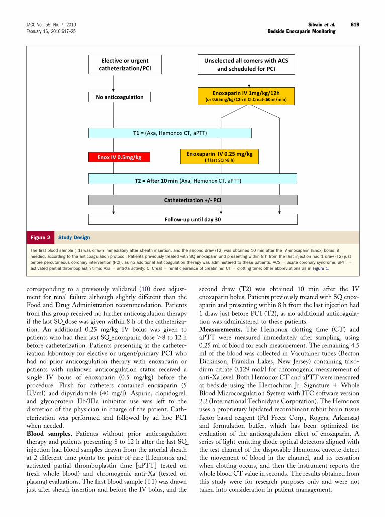
aseline CharacteristicsTable 1 Baseline Characteristics
Study Population(n � 296)
Demographics
Age, yrs 61.5 � 13.7
Elderly, age �75 yrs 46 (15.5)
Sex, male 224 (75.7)
Risk factors
Active smoker 54 (18.2)
Diabetes mellitus 69 (23.3)
Hypertension 167 (56.4)
Dyslipidemia 174 (58.8)
BMI 26.7 � 4.9
Obesity, BMI �30 kg/m2 49 (16.5)
Overweight, BMI �25 kg/m2 118 (39.9)
Creatinine, �M/ml 103.8 � 71
Creatinine Cl, ml/min 84 � 39
Renal insufficiency, Cl �60 ml/min 74 (25)
Renal insufficiency, Cl �30 ml/min 12 (4)
Medical history
ACS 62 (20.9)
Angioplasty 88 (29.7)
Stenting 78 (26.3)
DES 37 (12.5)
CABG 15 (5.1)
Stroke 18 (6.1)
PAD 31 (10.5)
Clinical presentation
Stable angina or asymptomaticpatients
69 (23.3)
Unstable angina 54 (18.2)
NSTEMI, positive troponin 49 (17.4)
STEMI 17 (5.7)
Other 107 (36.1)
Antithrombotic treatment
ASA 248 (83.8)
Clopidogrel 197 (66.5)
GP IIb/IIIa inhibitor 12 (4.0)
Other medications
Beta-blocker 188 (63.5)
ACE inhibitor 130 (43.9)
ARAII 60 (20.3)
Lipid-lowering agent 233 (78.7)
ata presented as mean � SD or n (%).ACE � angiotensin converting enzyme; ACS � acute coronary syndrome; ARAII � angiotensin II
eceptor antagonist; ASA � acetylsalicylic acid; BMI � body mass index; CABG � coronary arteryypass graft surgery; Cl � clearance; DES � drug-eluting stent; GP � glycoprotein; NSTEMI �
on–ST-segment elevation myocardial infarction; PAD � peripheral arterial disease; STEMI �
T-segment elevation myocardial infarction.
on–ST-segment elevation ACS (7,11), and in patients �
ndergoing PCI (12–14). The upper value corresponds tohe 75th percentile of peak anti-Xa values in patients treatedith 1.25 mg/kg every 12 h who did not experience majoremorrhage in the TIMI (Thrombolysis In Myocardialnfarction) 11A study (8).linical follow-up. Thirty-day follow-up was realized
hrough consult and/or telephone interviews. Major cardio-ascular and cerebrovascular events (MACCE), includingeath, stroke, recurrent myocardial infarction, urgentevascularization, and definite and probable stent throm-osis (Academic Research Consortium definition), asell as major and minor bleeding were reported.tatistical analysis. Categorical variables were expressed asercentages, and continuous variables as mean � SD and asedian with interquartile range (IQR) (25th to 75th percen-
iles) for biological values (Hemonox CT, aPTT, and anti-Xa).otential associations between clinical and biological parame-
ers were tested by unvaried procedures using the Mann-hitney U test. Receiver-operating characteristic (ROC)
urves were drawn to determine cutoff values of the HemonoxT as a diagnostic test for monitoring anticoagulation com-ared with anti-Xa activity levels. We used the Spearman testor nonparametric values to calculate correlation betweeniological measurements. All analyses were performed withtatView 5.0 SAS software (SAS Institute, Cary, Northarolina).
esults
he baseline characteristics are shown in Table 1. Details ofrocedure, angiographic results, and biological results areeported in Table 2. Follow-up was achieved in 97% ofatients at 30 days.
rocedure Characteristics andiological Measurement at T1 and T2Table 2 Procedure Characteristics andBiological Measurement at T1 and T2
Study Population(n � 296)
Angiography
Nonsignificant lesion or normal angiogram 142 (47.9)
Patients with at least 1 significant lesion 157 (52.1)
Revascularization
CABG (arterial or saphenous graft) 4 (1.3)
PCI/stenting 165 (55.7)
Anticoagulation and biology, min
Time sheath insertion to T1 6.3 (3.8)
Time T1 to T2 11.5 (7.1)
Biological measurement
Hemonox CT T1, s (n � 229) 74 [70–81] (58–444)
Hemonox CT T2, s (n � 289) 143 [114–206] (57–749)
Anti-Xa activity T1, IU/ml (n � 219) 0.1 [0.1–0.1] (0.1–0.96)
Anti-Xa activity T2, IU/ml (n � 269) 0.87 [0.74–1.03] (0.1–1.9)
aPTT T1, s (n � 225) 44 [39–50] (20–136)
aPTT T2, s (n � 288) 72 [58–93] (20–400)
alues are n (%) or median [interquartile range] (minimum–maximum).
aPTT � activated partial thromboplastin time; CABG � coronary artery bypass graft surgery; CTclotting time; PCI � percutaneous coronary intervention; T1 � time point 1; T2 � time point 2.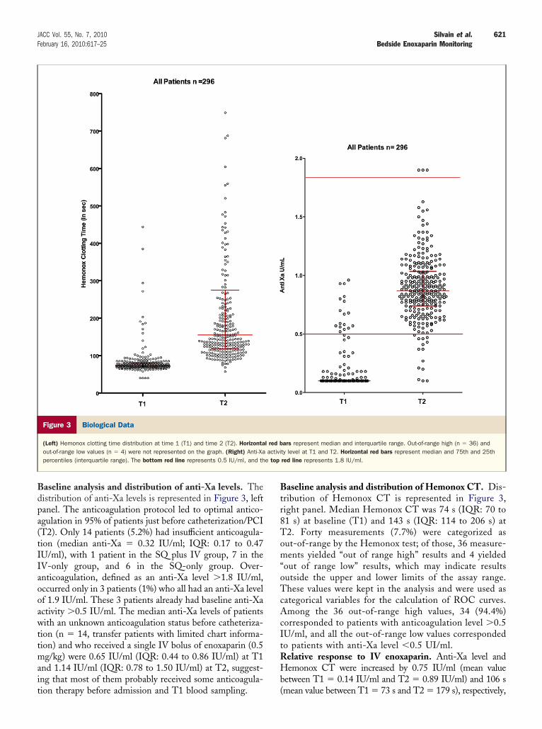
Bdpa(tIIaooawttmait
Btr8Tom“oTcAcItRHb
621JACC Vol. 55, No. 7, 2010 Silvain et al.February 16, 2010:617–25 Bedside Enoxaparin Monitoring
aseline analysis and distribution of anti-Xa levels. Theistribution of anti-Xa levels is represented in Figure 3, leftanel. The anticoagulation protocol led to optimal antico-gulation in 95% of patients just before catheterization/PCIT2). Only 14 patients (5.2%) had insufficient anticoagula-ion (median anti-Xa � 0.32 IU/ml; IQR: 0.17 to 0.47U/ml), with 1 patient in the SQ plus IV group, 7 in theV-only group, and 6 in the SQ-only group. Over-nticoagulation, defined as an anti-Xa level �1.8 IU/ml,ccurred only in 3 patients (1%) who all had an anti-Xa levelf 1.9 IU/ml. These 3 patients already had baseline anti-Xactivity �0.5 IU/ml. The median anti-Xa levels of patientsith an unknown anticoagulation status before catheteriza-
ion (n � 14, transfer patients with limited chart informa-ion) and who received a single IV bolus of enoxaparin (0.5g/kg) were 0.65 IU/ml (IQR: 0.44 to 0.86 IU/ml) at T1
nd 1.14 IU/ml (IQR: 0.78 to 1.50 IU/ml) at T2, suggest-ng that most of them probably received some anticoagula-
Figure 3 Biological Data
(Left) Hemonox clotting time distribution at time 1 (T1) and time 2 (T2). Horizontaout-of-range low values (n � 4) were not represented on the graph. (Right) Anti-Xapercentiles (interquartile range). The bottom red line represents 0.5 IU/ml, and th
ion therapy before admission and T1 blood sampling. (
aseline analysis and distribution of Hemonox CT. Dis-ribution of Hemonox CT is represented in Figure 3,ight panel. Median Hemonox CT was 74 s (IQR: 70 to1 s) at baseline (T1) and 143 s (IQR: 114 to 206 s) at2. Forty measurements (7.7%) were categorized asut-of-range by the Hemonox test; of those, 36 measure-ents yielded “out of range high” results and 4 yielded
out of range low” results, which may indicate resultsutside the upper and lower limits of the assay range.hese values were kept in the analysis and were used as
ategorical variables for the calculation of ROC curves.mong the 36 out-of-range high values, 34 (94.4%)
orresponded to patients with anticoagulation level �0.5U/ml, and all the out-of-range low values correspondedo patients with anti-Xa level �0.5 UI/ml.elative response to IV enoxaparin. Anti-Xa level andemonox CT were increased by 0.75 IU/ml (mean value
etween T1 � 0.14 IU/ml and T2 � 0.89 IU/ml) and 106 s
ars represent median and interquartile range. Out-of-range high (n � 36) andty level at T1 and T2. Horizontal red bars represent median and 75th and 25thred line represents 1.8 IU/ml.
l red bactivi
e top
mean value between T1 � 73 s and T2 � 179 s), respectively,
wp0m�bEacac0(99
�fsdIww0m0&(
S
VoN
622 Silvain et al. JACC Vol. 55, No. 7, 2010Bedside Enoxaparin Monitoring February 16, 2010:617–25
hen a 0.5 mg/kg IV bolus of enoxaparin was given. Inatients who received SQ enoxaparin and needed an additional.25 mg/kg IV bolus of enoxaparin, anti-Xa level and He-onox CT increased by 0.64 UI/ml (mean value between T10.32 IU/ml and T2 � 0.96 IU/ml) and 178 s (mean value
etween T1 � 86 s and T2 � 254 s), respectively.valuation of the biological test. The correlation between
nti-Xa levels and Hemonox CT was stronger than theorrelation between anti-Xa levels and aPTT, whethernalyzed on the global T1 and T2 values (r � 0.81, 95%onfidence interval [CI]: 0.77 to 0.84, p � 0.0001 vs. r �.62, 95% CI: 0.56 to 0.68, p � 0.0001) or separately on T1r � 0.45, 95% CI: 0.33 to 0.55, p � 0.0001 vs. r � 0.17,5% CI: 0.036 to 0.31, p � 0.01) and T2 values (r � 0.39,5% CI: 0.27 to 0.49, p � 0.0001 vs. r � 0.0085, 95% CI:
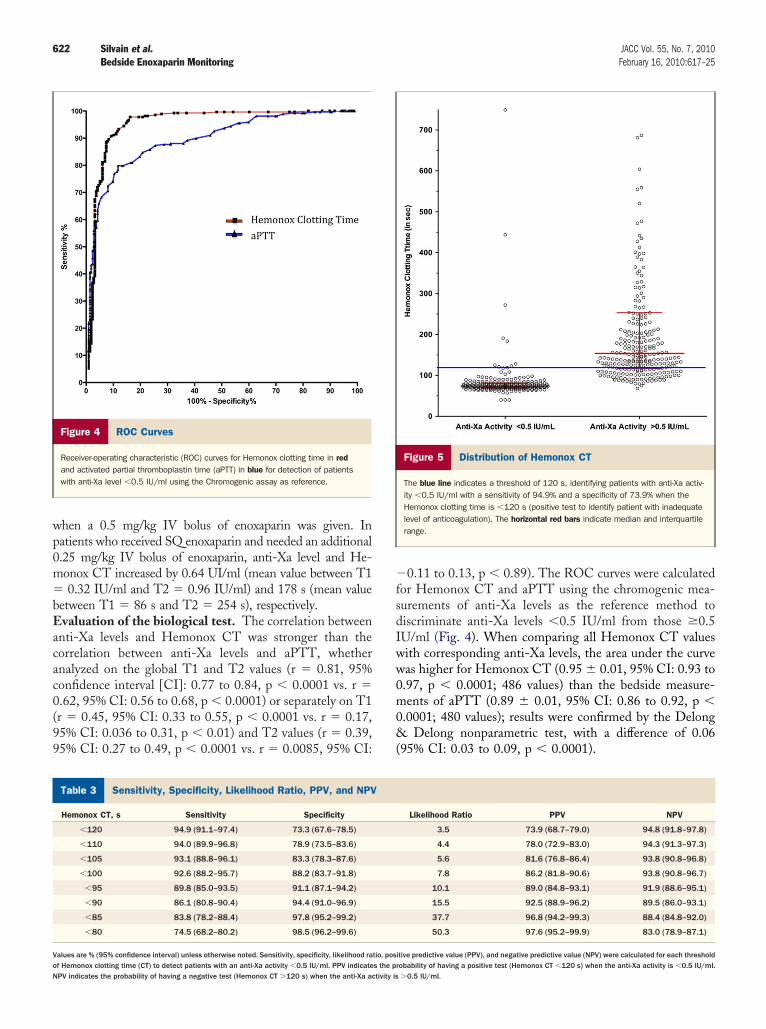
Figure 4 ROC Curves
Receiver-operating characteristic (ROC) curves for Hemonox clotting time in redand activated partial thromboplastin time (aPTT) in blue for detection of patientswith anti-Xa level �0.5 IU/ml using the Chromogenic assay as reference.
ensitivity, Specificity, Likelihood Ratio, PPV, and NPVTable 3 Sensitivity, Specificity, Likelihood Ratio, PPV, and NPV
Hemonox CT, s Sensitivity Specificity
�120 94.9 (91.1–97.4) 73.3 (67.6–78.5)
�110 94.0 (89.9–96.8) 78.9 (73.5–83.6)
�105 93.1 (88.8–96.1) 83.3 (78.3–87.6)
�100 92.6 (88.2–95.7) 88.2 (83.7–91.8)
�95 89.8 (85.0–93.5) 91.1 (87.1–94.2)
�90 86.1 (80.8–90.4) 94.4 (91.0–96.9)
�85 83.8 (78.2–88.4) 97.8 (95.2–99.2)
�80 74.5 (68.2–80.2) 98.5 (96.2–99.6)
alues are % (95% confidence interval) unless otherwise noted. Sensitivity, specificity, likelihood ra
f Hemonox clotting time (CT) to detect patients with an anti-Xa activity �0.5 IU/ml. PPV indicates the prPV indicates the probability of having a negative test (Hemonox CT �120 s) when the anti-Xa activity is0.11 to 0.13, p � 0.89). The ROC curves were calculatedor Hemonox CT and aPTT using the chromogenic mea-urements of anti-Xa levels as the reference method toiscriminate anti-Xa levels �0.5 IU/ml from those �0.5U/ml (Fig. 4). When comparing all Hemonox CT valuesith corresponding anti-Xa levels, the area under the curveas higher for Hemonox CT (0.95 � 0.01, 95% CI: 0.93 to.97, p � 0.0001; 486 values) than the bedside measure-ents of aPTT (0.89 � 0.01, 95% CI: 0.86 to 0.92, p �
.0001; 480 values); results were confirmed by the DelongDelong nonparametric test, with a difference of 0.06
95% CI: 0.03 to 0.09, p � 0.0001).
Figure 5 Distribution of Hemonox CT
The blue line indicates a threshold of 120 s, identifying patients with anti-Xa activ-ity �0.5 IU/ml with a sensitivity of 94.9% and a specificity of 73.9% when theHemonox clotting time is �120 s (positive test to identify patient with inadequatelevel of anticoagulation). The horizontal red bars indicate median and interquartilerange.
Likelihood Ratio PPV NPV
3.5 73.9 (68.7–79.0) 94.8 (91.8–97.8)
4.4 78.0 (72.9–83.0) 94.3 (91.3–97.3)
5.6 81.6 (76.8–86.4) 93.8 (90.8–96.8)
7.8 86.2 (81.8–90.6) 93.8 (90.8–96.7)
10.1 89.0 (84.8–93.1) 91.9 (88.6–95.1)
15.5 92.5 (88.9–96.2) 89.5 (86.0–93.1)
37.7 96.8 (94.2–99.3) 88.4 (84.8–92.0)
50.3 97.6 (95.2–99.9) 83.0 (78.9–87.1)
itive predictive value (PPV), and negative predictive value (NPV) were calculated for each threshold
tio, pos obability of having a positive test (Hemonox CT �120 s) when the anti-Xa activity is �0.5 IU/ml.�0.5 IU/ml.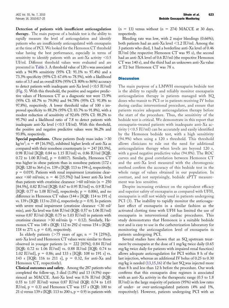
DtrpavsUpw7rt(t(9pm9it9Skc00w[pat[[dvwmvcm1
ao[11HCcr0I2
(r
b3IhCa
D
Tiaddptbetb(aawcamwcm
aePlaestmp
mmalmtcwIo
623JACC Vol. 55, No. 7, 2010 Silvain et al.February 16, 2010:617–25 Bedside Enoxaparin Monitoring
etection of patients with insufficient anticoagulationherapy. The main purpose of a bedside test is the ability toapidly measure the level of anticoagulation and identifyatients who are insufficiently anticoagulated with enoxaparint the time of PCI. We looked for the Hemonox CT thresholdalue having the best performance, especially in terms ofensitivity to identify patients with an anti-Xa activity �0.5I/ml. Different threshold values were evaluated and areresented in Table 3. A threshold value of 120 s was associatedith a 94.9% sensitivity (95% CI: 91.1% to 97.4%) and a3.3% specificity (95% CI: 67.6% to 78.5%), with a likelihoodatio of 3.5 and an overall 83% (95% CI: 80% to 86%) accuracyo detect patients with inadequate anti-Xa level (�0.5 IU/ml)Fig. 5). With this threshold, the positive and negative predic-ive values of Hemonox CT as a diagnostic test were 73.9%95% CI: 68.7% to 79.0%) and 94.78% (95% CI: 91.8% to7.8%), respectively. A lower threshold value of 100 s im-roved specificity to 88.2% (95% CI: 83.7% to 91.8%), with aodest reduction of sensitivity of 92.6% (95% CI: 88.2% to
5.7%) and a likelihood ratio of 7.8 to detect patients withnadequate anti-Xa level (�0.5 UI/ml). With this threshold,he positive and negative predictive values were 86.2% and3.8%, respectively.pecial populations. Obese patients (body mass index �30g/m2; n � 49 [16.5%]), exhibited higher levels of anti-Xa asompared with their nonobese counterparts (n � 247 [83.5%],.98 IU/ml [IQR: 0.84 to 1.15 IU/ml] vs. 0.86 IU/ml [IQR:.72 to 1.00 IU/ml], p � 0.0017). Similarly, Hemonox CTas higher in obese patients than in nonobese patients (172 s
IQR: 120 to 364 s] vs. 139 s [IQR: 113 to 194 s], respectively;� 0.019). Patients with renal impairment (creatinine clear-
nce �60 ml/min; n � 46 [15.5%]) had lower anti-Xa levelhan patients with creatinine clearance �60 ml/min (n � 25084.5%], 0.82 IU/ml [IQR: 0.67 to 0.95 IU/ml] vs. 0.9 IU/mlIQR: 0.77 to 1.08 IU/ml], respectively; p � 0.006), and noifference in Hemonox CT values (144 s [IQR: 114 to 191 s]s. 139 s [IQR: 113 to 210 s], respectively; p � 0.9). In patientsith severe renal impairment (creatinine clearance �30 ml/in), anti-Xa level was 0.66 IU/ml (IQR: 0.57 to 1.06 IU/ml)
ersus 0.87 IU/ml (IQR: 0.75 to 1.03 IU/ml) in patients withreatinine clearance �30 ml/min (p � 0.12). Similarly, He-onox CT was 148 s (IQR: 112 to 292 s) versus 154 s (IQR:
18 to 271 s, p � 0.8), respectively.In elderly patients (�75 years of age; n � 74 [25%]),
nti-Xa level and Hemonox CT values were similar to thosebserved in younger patients (n � 222 [85%]; 0.84 IU/mlIQR: 0.72 to 1.06 IU/ml] vs. 0.88 IU/ml [IQR: 0.74 to.02 IU/ml], p � 0.86; and 133 s [IQR: 108 to 191 s] vs.43 s [IQR: 116 to 211 s], p � 0.32, for anti-Xa andemonox CT, respectively).linical outcomes and safety. Among the 287 patients who
ompleted the follow-up, 3 died (1.0%) and 13 (4.5%) expe-ienced an MACCE. Anti-Xa level was 0.76 IU/ml (IQR:.55 to 1.07 IU/ml) versus 0.87 IU/ml (IQR: 0.74 to 1.03U/ml, p � 0.3) and Hemonox CT was 157 s (IQR: 100 to
1 s) versus 139 s (IQR: 113 to 200 s, p � 0.9) in patients with rn � 13) versus without (n � 274) MACCE at 30 days,espectively.
Bleeding rate was low, with 2 major bleedings (0.66%);oth patients had an anti-Xa level �1.2 IU/ml. Among thepatients who died, 1 had a borderline anti-Xa level of 0.46
U/ml (the respective Hemonox CT was 95 s), the secondad an anti-XA level of 0.6 IU/ml (the respective HemonoxT was 140 s), and the third had an unknown anti-Xa value
t T2 but Hemonox CT was 78 s.
iscussion
he main purpose of a LMWH enoxaparin bedside tests the ability to rapidly and reliably monitor enoxaparinnticoagulation therapy in patients managed with SQoses who transit to PCI or in patients receiving IV bolusuring cardiac interventional procedure, and ensure thatatients receive adequate anticoagulation therapy beforehe start of the procedure. Thus, the sensitivity of theedside test is critical. We demonstrate in this report thatnoxaparin-treated patients with inadequate anti-Xa ac-ivity (�0.5 IU/ml) can be accurately and easily identifiedy the Hemonox bedside test, with a high sensitivity94.9%) when using a 120 s threshold. This thresholdllows clinicians to rule out the need for additionalnticoagulation therapy when levels are beyond 120 s,ith a good negative predictive value (94.8%). The ROC
urves and the good correlation between Hemonox CTnd the anti-Xa level measured with the chromogenicethod confirm the accuracy of this bedside test in thehole range of values obtained in our population. In
ontrast, and not surprisingly, bedside aPTT measure-ent was less sensitive.Despite increasing evidence on the equivalent efficacy
nd superior safety of enoxaparin as compared with UFH,noxaparin is still not widely used in patients undergoingCI (3). The inability to rapidly monitor the anticoagu-
ant effect of enoxaparin in a similar fashion as thectivated clotting time with UFH has limited the use ofnoxaparin in interventional cardiac procedures. Thistudy demonstrates that Hemonox is a suitable bedsideest and is easy to use in the catheterization laboratory foronitoring the anticoagulation level of enoxaparin in
atients undergoing PCI.Several studies have shown that an SQ upstream treat-ent by enoxaparin at the dose of 1 mg/kg twice daily (0.65g/kg twice daily for patients with impaired renal function)
llows adequate anticoagulation for PCI within 8 h of theast injection, whereas an additional IV bolus of 0.25 to 0.30
g/kg is needed (13,15,16) if the last SQ was injected morehan 8 h and less than 12 h before the procedure. Our workonfirms that this enoxaparin dose regimen is associatedith an anti-Xa activity in the therapeutic range (0.5 to 1.8
U/ml) in the large majority of patients (95%) with low ratesf under- or over-anticoagulated patients (4% and 1%,
espectively). However, patients undergoing PCI with an
acsa(Sm(ncm
rspseatctm
lamotmarprlvIIpubt
lmoc1
imwh
caa
daYaSiHtaaat
C
Aatb
ATfP
RB4m
R
624 Silvain et al. JACC Vol. 55, No. 7, 2010Bedside Enoxaparin Monitoring February 16, 2010:617–25
nti-Xa activity �0.5 IU/ml may be at high risk of ischemicomplications. Only a few studies have been conducted withufficient power to assess correlations between targetnti-Xa levels and outcomes of ischemic complications17,18). A recent study involving unstable angina/non-TEMI patients reported a 3-fold increase in 30-dayortality associated with suboptimal anticoagulation
anti-Xa levels �0.5 IU/ml) (7). Such findings suggest theeed for monitoring anti-Xa activity with a rapid point-of-are test to identify the patients who require dose adjust-ent.Our Hemonox results are in agreement with those
eported from previous studies conducted on a small sampleize (5,6). Additionally, the study was conducted in aopulation treated exclusively with enoxaparin and includedpecial patient groups such as patients with renal failure, thelderly, and obese patients. The absence of over-nticoagulation therapy for these specific patient popula-ions shows the effectiveness of the protocol used andonfirms previous results (19). Nevertheless, results fromhis study suggest the utility of the point-of-care test inonitoring enoxaparin in special patient.Because the main objective of the monitoring test is its
evel of sensitivity for defining patients who are under-nticoagulated, the threshold of 120 s may be recom-ended, especially in patients at high risk of thrombosis. In
ther words, if Hemonox CT is �120 s in the catheteriza-ion laboratory, an additional IV enoxaparin bolus of 0.5g/kg could be given to the patient before PCI to ensure
chieving an effective anti-Xa level within the therapeuticange (0.5 to 1.8 IU/ml). Our results indicate that foratients without any prior anticoagulation, an IV enoxapa-in bolus of 0.5 mg/kg yields an average anti-Xa activityevel of 0.73 IU/ml. Thus, patients with a Hemonox CTalue �120 s but with some residual anti-Xa activity (�0.5U/ml) would not be over-anticoagulated with an additionalV bolus (15). Actually, we observed this scenario in a fewatients of our study whose anticoagulation status wasnknown at the time of catheterization; they received an IVolus of 0.5 mg/kg and remained within the recommendedherapeutic range of anti-Xa level.
A more graded approach to Hemonox CT could be used;ower cutoff values (100 s) would increase specificity, which
ay be useful in stable patients and/or patients at high riskf bleeding; additional IV enoxaparin doses �0.5 mg/kgould also be used in patients with Hemonox CT 80 to20 s.All these strategies would have to be tested prospectively
n further studies as well as the value of bedside enoxaparinonitoring in situations such as STEMI patients treatedith fibrinolytic drugs or anticoagulation therapy duringemodialysis.In daily practice, the Hemonox test may be used in the
atheterization laboratory to assess the appropriate level ofnticoagulation therapy in patients undergoing PCI and to
void the use of UFH in addition to enoxaparin, as is still1
one by some interventionists and has been shown to bessociated with worse outcome in the SYNERGY (Superiorield of the New Strategy of Enoxaparin, Revascularizationnd Glycoprotein IIb/IIIa Inhibitors) trial (20).tudy limitations. One important limitation of our study
s that it was not powered to assess the impact of theemonox CT on clinical outcomes. Another limitation is
he limited information obtained on high levels of antico-gulation, as very few patients in our population reachednti-Xa values �1.8 IU/ml. Finally, the cost effectiveness ofmonitoring-guided strategy of enoxaparin anticoagulation
herapy remains to be established.
onclusions
ccording to the results of this study, Hemonox CTppears to be a fast and reliable bedside test that can be usedo monitor enoxaparin anticoagulation therapy immediatelyefore catheterization and PCI.
cknowledgmentshe authors thank Delphine Brugier and Nicolas Vignolles
or their expert technical assistance and Soumaya El Rouby,hD, for reviewing the manuscript.
eprint requests and correspondence: Dr. Gilles Montalescot,ureau 256, Institut de Cardiologie, Pitié-Salpêtrière Hospital,7-83 Boulevard de l’Hôpital, Paris 75013, France. E-mail: [email protected].
EFERENCES
1. Fox KA, Steg PG, Eagle KA, et al. Decline in rates of death and heartfailure in acute coronary syndromes, 1999–2006. JAMA 2007;297:1892–900.
2. Van de Werf F, Bax J, Betriu A, et al. Management of acutemyocardial infarction in patients presenting with persistent ST-segment elevation: the Task Force on the Management of ST-Segment Elevation Acute Myocardial Infarction of the EuropeanSociety of Cardiology. Eur Heart J 2008;29:2909–45.
3. Montalescot G, White HD, Gallo R, et al. Enoxaparin versusunfractionated heparin in elective percutaneous coronary intervention.N Engl J Med 2006;355:1006–17.
4. Montalescot G, Cohen M, Salette G, et al. Impact of anticoagulationlevels on outcomes in patients undergoing elective percutaneouscoronary intervention: insights from the STEEPLE trial. Eur Heart J2008;29:462–71.
5. Marmur JD, Lakhani M, El Rouby S, Cavusoglu E. A novelpoint-of-care assay for the monitoring of low-molecular weight hepa-rins in the cardiac catheterization laboratory. J Invasive Cardiol2008;20:449–54.
6. El Rouby S, Cohen M, Gonzales A, et al. The use of a HEMOCHRONJR. HEMONOX point of care test in monitoring the anticoagulanteffects of enoxaparin during interventional coronary procedures. JThromb Thrombol 2006;21:137–45.
7. Montalescot G, Collet JP, Tanguy ML, et al. Anti-Xa activity relatesto survival and efficacy in unselected acute coronary syndrome patientstreated with enoxaparin. Circulation 2004;110:392–8.
8. The Thrombolysis In Myocardial Infarction (TIMI) 11A Trial Inves-tigators. Dose-ranging trial of enoxaparin for unstable angina: resultsof TIMI 11A. J Am Coll Cardiol 1997;29:1474–82.
9. Cockcroft DW, Gault MH. Prediction of creatinine clearance fromserum creatinine. Nephron 1976;16:31–41.
0. Hulot JS, Montalescot G, Lechat P, Collet JP, Ankri A, Urien S.Dosing strategy in patients with renal failure receiving enoxaparin for
1
1
1
1
1
1
1
1
1
2
K
625JACC Vol. 55, No. 7, 2010 Silvain et al.February 16, 2010:617–25 Bedside Enoxaparin Monitoring
the treatment of non-ST-segment elevation acute coronary syndrome.Clin Pharmacol Ther 2005;77:542–52.
1. Bijsterveld NR, Moons AH, Meijers JC, Levi M, Buller HR, PetersRJ. The impact on coagulation of an intravenous loading dose inaddition to a subcutaneous regimen of low-molecular-weight heparinin the initial treatment of acute coronary syndromes. J Am CollCardiol 2003;42:424–7.
2. Choussat R, Montalescot G, Collet JP, et al. A unique, low dose ofintravenous enoxaparin in elective percutaneous coronary intervention.J Am Coll Cardiol 2002;40:1943–50.
3. Collet JP, Montalescot G, Lison L, et al. Percutaneous coronaryintervention after subcutaneous enoxaparin pretreatment in patientswith unstable angina pectoris. Circulation 2001;103:658–63.
4. Kereiakes DJ, Kleiman NS, Fry E, et al. Dalteparin in combinationwith abciximab during percutaneous coronary intervention. AmHeart J 2001;141:348–52.
5. Martin JL, Fry ET, Sanderink GJ, et al. Reliable anticoagulation withenoxaparin in patients undergoing percutaneous coronary intervention:the Pharmacokinetics of Enoxaparin in PCI (PEPCI) study. CathetCardiovasc Interv 2004;61:163–70.
6. Sanchez-Pena P, Hulot JS, Urien S, et al. Anti-factor Xa kinetics afterintravenous enoxaparin in patients undergoing percutaneous coronaryintervention: a population model analysis. Br J Clin Pharmacol
7. Antman EM, Cohen M, McCabe C, Goodman SG, Murphy SA,Braunwald E. Enoxaparin is superior to unfractionated heparin forpreventing clinical events at 1-year follow-up of TIMI 11B andESSENCE. Eur Heart J 2002;23:308–14.
8. Dumaine R, Borentain M, Bertel O, et al. Intravenous low-molecular-weight heparins compared with unfractionated heparin in percutane-ous coronary intervention: quantitative review of randomized trials.Arch Intern Med 2007;167:2423–30.
9. Spinler SA, Inverso SM, Cohen M, Goodman SG, Stringer KA,Antman EM. Safety and efficacy of unfractionated heparin versusenoxaparin in patients who are obese and patients with severe renalimpairment: analysis from the ESSENCE and TIMI 11B studies. AmHeart J 2003;146:33–41.
0. Ferguson JJ, Califf RM, Antman EM, et al. Enoxaparin vs unfrac-tionated heparin in high-risk patients with non-ST-segment elevationacute coronary syndromes managed with an intended early invasivestrategy: primary results of the SYNERGY randomized trial. JAMA2004;292:45–54.
ey Words: enoxaparin y bedside test y percutaneous coronary
2005;60:364–73. intervention.
Related Documents