Enhancement of Neurite Outgrowth in Neuronal-Like Cells following Boron Nitride Nanotube-Mediated Stimulation Gianni Ciofani,* ,† Serena Danti, ‡ Delfo D’Alessandro, ‡,§ Leonardo Ricotti, Stefania Moscato, § Giovanni Bertoni, ¶ Andrea Falqui, ¶ Stefano Berrettini, ‡ Mario Petrini, Virgilio Mattoli, † and Arianna Menciassi †, † Italian Institute of Technology c/o Scuola Superiore Sant’Anna, Viale Rinaldo Piaggio 34 Pontedera (Pisa), 56025, Italy, ‡ Otology - Cochlear Implant Unit, Department of Neuroscience, University of Pisa, Via Paradisa 2 Pisa, 56124, Italy, § Department of Human Morphology & Applied Biology, University of Pisa, Via Roma 55 Pisa, 56126, Italy, CRIM Lab, Scuola Superiore Sant’Anna, Viale Rinaldo Piaggio 34 Pontedera (Pisa), 56025, Italy, ¶ Italian Institute of Technology, Via Morego 30 Genova, 16163, Italy, and Department of Oncology & Transplants, University of Pisa, Via Roma 55, 56126 Pisa, Italy E lectrical stimulation as an artificial stimulus of neural structures has been widely used in clinical practice and laboratory study. There are multiple strategies reported in the literature and in clinical use that attempt to specifically local- ize the activation of nervous structures. Ef- forts have included both invasive and non- invasive approaches, as well as combined technologies. 1,2 Such systems typically in- volve placement/attachment of an elec- trode to the target organ/tissue (by sur- gery or interventional radiology) and a wired connection to the electronic (battery powered) device, which delivers the appro- priate electric impulses. 3,4 Despite its tech- nological refinements, the basic configura- tion of all such systems for electro- stimulation therapies has two inherent dis- advantages: (i) contact problems at the electrode/tissue interface and (ii) complica- tions due to the implantation of the stimu- lator. In this paper we present a new tech- nology based on indirect, noninvasive electrical stimulation mediated by piezo- electric nanoparticles (boron nitride nano- tubes, BNNTs 5 ) and ultrasounds; in particu- lar, our preliminary results demonstrated that this method could successfully stimu- late neuronal-like cell cultures in vitro. A boron nitride nanotube (BNNT) is the structural analogue of a carbon nanotube: alternating B and N atoms entirely substi- tute for C atoms in a graphitic-like sheet with almost no changes in atomic spacing. In spite of this structural similarity with car- bon nanotubes (CNTs), BNNTs own superior mechanical, chemical, and electrical proper- ties. 6 In the last years several examples of CNT exploitation in biotechnology have been proposed, 7 while the biomedical ap- plications of BNNTs have remained largely unexplored. 8 The first studies about BNNTcell interactions have been per- formed by the authors in refs 9 and 10. Re- cent investigations have confirmed that BNNTs also have excellent piezoelectric properties. Ab initio calculations of the spontaneous polarization and piezoelectric properties of BNNTs have demonstrated that they function as excellent piezoelec- tric systems with response values greater than those of piezoelectric polymers, and comparable to those exhibited by wurtzite semiconductors. 11 In addition, BNNT bend- ing forces have been measured directly in- side high-resolution transmission electron microscopy, and real-time video-recording of their elastic kinking deformation has con- firmed their marked flexibility. 12 Recently, Bai et al. have experimentally verified a deformation-driven electrical transport and the first evidence of a piezoelectric behavior in multiwalled BNNTs. 13 The insulating char- acter of an individual BNNT can be modi- fied by tube squeezing between two gold contacts inside a TEM. A considerable *Address correspondence to [email protected]. Received for review November 17, 2009 and accepted September 27, 2010. Published online October 6, 2010. 10.1021/nn101985a © 2010 American Chemical Society ABSTRACT In this paper, we propose an absolutely innovative technique for the electrical stimulation of cells, based on piezoelectric nanoparticles. Ultrasounds are used to impart mechanical stress to boron nitride nanotubes incubated with neuronal-like PC12 cells. By virtue of their piezoelectric properties, these nanotubes can polarize and convey electrical stimuli to the cells. PC12 stimulated with the present method exhibit neurite sprout 30% greater than the control cultures after 9 days of treatment. KEYWORDS: boron nitride nanotubes · PC12 cells · piezoelectric stimulation · neuronal regeneration ARTICLE www.acsnano.org VOL. 4 ▪ NO. 10 ▪ 6267–6277 ▪ 2010 6267

Welcome message from author
This document is posted to help you gain knowledge. Please leave a comment to let me know what you think about it! Share it to your friends and learn new things together.
Transcript

Enhancement of Neurite Outgrowth inNeuronal-Like Cells following BoronNitride Nanotube-Mediated StimulationGianni Ciofani,*,† Serena Danti,‡ Delfo D’Alessandro,‡,§ Leonardo Ricotti,� Stefania Moscato,§
Giovanni Bertoni,¶ Andrea Falqui,¶ Stefano Berrettini,‡ Mario Petrini,� Virgilio Mattoli,† andArianna Menciassi†,�
†Italian Institute of Technology c/o Scuola Superiore Sant’Anna, Viale Rinaldo Piaggio 34 Pontedera (Pisa), 56025, Italy, ‡Otology - Cochlear Implant Unit, Department ofNeuroscience, University of Pisa, Via Paradisa 2 Pisa, 56124, Italy, §Department of Human Morphology & Applied Biology, University of Pisa, Via Roma 55 Pisa, 56126,Italy, �CRIM Lab, Scuola Superiore Sant’Anna, Viale Rinaldo Piaggio 34 Pontedera (Pisa), 56025, Italy, ¶Italian Institute of Technology, Via Morego 30 Genova, 16163,Italy, and �Department of Oncology & Transplants, University of Pisa, Via Roma 55, 56126 Pisa, Italy
Electrical stimulation as an artificialstimulus of neural structures hasbeen widely used in clinical practice
and laboratory study. There are multiplestrategies reported in the literature and inclinical use that attempt to specifically local-ize the activation of nervous structures. Ef-forts have included both invasive and non-invasive approaches, as well as combinedtechnologies.1,2 Such systems typically in-volve placement/attachment of an elec-trode to the target organ/tissue (by sur-gery or interventional radiology) and awired connection to the electronic (batterypowered) device, which delivers the appro-priate electric impulses.3,4 Despite its tech-nological refinements, the basic configura-tion of all such systems for electro-stimulation therapies has two inherent dis-advantages: (i) contact problems at theelectrode/tissue interface and (ii) complica-tions due to the implantation of the stimu-lator. In this paper we present a new tech-nology based on indirect, noninvasiveelectrical stimulation mediated by piezo-electric nanoparticles (boron nitride nano-tubes, BNNTs5) and ultrasounds; in particu-lar, our preliminary results demonstratedthat this method could successfully stimu-late neuronal-like cell cultures in vitro.
A boron nitride nanotube (BNNT) is thestructural analogue of a carbon nanotube:alternating B and N atoms entirely substi-tute for C atoms in a graphitic-like sheetwith almost no changes in atomic spacing.In spite of this structural similarity with car-bon nanotubes (CNTs), BNNTs own superiormechanical, chemical, and electrical proper-ties.6 In the last years several examples of
CNT exploitation in biotechnology havebeen proposed,7 while the biomedical ap-plications of BNNTs have remained largelyunexplored.8 The first studies aboutBNNT�cell interactions have been per-formed by the authors in refs 9 and 10. Re-cent investigations have confirmed thatBNNTs also have excellent piezoelectricproperties. Ab initio calculations of thespontaneous polarization and piezoelectricproperties of BNNTs have demonstratedthat they function as excellent piezoelec-tric systems with response values greaterthan those of piezoelectric polymers, andcomparable to those exhibited by wurtzitesemiconductors.11 In addition, BNNT bend-ing forces have been measured directly in-side high-resolution transmission electronmicroscopy, and real-time video-recordingof their elastic kinking deformation has con-firmed their marked flexibility.12 Recently,Bai et al. have experimentally verified adeformation-driven electrical transport andthe first evidence of a piezoelectric behaviorin multiwalled BNNTs.13 The insulating char-acter of an individual BNNT can be modi-fied by tube squeezing between two goldcontacts inside a TEM. A considerable
*Address correspondence [email protected].
Received for review November 17, 2009and accepted September 27, 2010.
Published online October 6, 2010.10.1021/nn101985a
© 2010 American Chemical Society
ABSTRACT In this paper, we propose an absolutely innovative technique for the electrical stimulation of
cells, based on piezoelectric nanoparticles. Ultrasounds are used to impart mechanical stress to boron nitride
nanotubes incubated with neuronal-like PC12 cells. By virtue of their piezoelectric properties, these nanotubes
can polarize and convey electrical stimuli to the cells. PC12 stimulated with the present method exhibit neurite
sprout 30% greater than the control cultures after 9 days of treatment.
KEYWORDS: boron nitride nanotubes · PC12 cells · piezoelectricstimulation · neuronal regeneration
ARTIC
LE
www.acsnano.org VOL. 4 ▪ NO. 10 ▪ 6267–6277 ▪ 2010 6267
current of several tens of nA is then able to flowthrough the tube. Such transport has been confirmedto be reversible, and disappears almost completely af-ter tube reloading. These observations underpin thevery high potential of BNNTs as efficient novel nano-scale transducers.
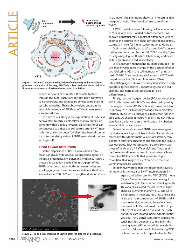
The aim of our study is the exploitation of BNNTs asnanovectors to carry electrical/mechanical signals ondemand within a cellular system. Electrical stimuli canbe conveyed to a tissue or cell culture after BNNT inter-nalization, using an outer “wireless” mechanical source(i.e., ultrasounds) by virtue of BNNT piezoelectric behav-ior (Figure 1).
RESULTS AND DISCUSSIONStable dispersions of BNNTs were obtained by
means of glycol-chitosan (GC) as dispersion agent, onthe basis of noncovalent polymeric wrapping. Figure 2shows a focused ion beam (FIB) micrograph of theBNNTs after preparation: both single nanotubes andsmall aggregates of nanotubes are visible with dimen-sions of about 200�600 nm in length and about 50 nm
in diameter. The inlet figure shows an interesting TEMimage of a typical “bamboo-like” structure of theBNNTs.
A WST-1 viability assay following cell incubation upto 9 days with BNNT-loaded culture medium (CM)showed nonstatistically significant differences with re-spect to the controls with BNNT concentrations up to 50�g/mL (p � 0.05 for higher concentrations, Figure 3).
Optimal cell viability up to 50 �g/mL BNNT concen-tration was confirmed by the LIVE/DEAD viability/cyto-toxicity assay (Figure 4), which labels living and deadcells in green and in red, respectively.
Early apoptotic phenomena could be excluded (Fig-ure 4) by investigating changes in the position of phos-phatidylserine (PS) in the cell membrane,14 using an-nexin V-FITC. The combination of annexin V-FITC withpropidium iodide (PI), a red fluorescent DNA-intercalating agent, allowed necrotic (red stained), earlyapoptotic (green stained), apoptotic (green and redstained), and normal cells (unstained) to bedifferentiated.
Finally, reactive oxygen species (ROS) production inPC12 cells treated with BNNTs was detected by usingthe Image-IT Green ROS Detection kit, based on 5-(and-6)-carboxy-2=,7=-dichlorodihydrofluorescein diacetate(carboxy-H2DCFDA), a fluorogenic marker for ROS in vi-able cells. As shown in Figure 4, BNNTs did not inducesignificant oxidative stress after 9 days of incubation,even at high concentrations.
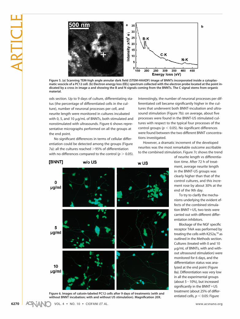
Cellular internalization of BNNTs was investigatedby TEM analysis (Figure 5). Noncellular electron-densematerial with cytoplasmatic vesicle localization, com-patible in terms of appearance and size with the BNNTs,was observed. Such observations are consistent withthose of Yehia et al.,15 Raffa et al.,16 and Tutak et al.17
performed on different types of mammalian cells ex-posed to CNT-loaded CM that presented high-resolution TEM images of electron-dense materialwithin intracellular vacuoles.
To definitively prove that the observed intravesicu-lar material is the result of BNNT internalization, im-
ages acquired in scanning TEM (STEM) mode(Figure 5a) underwent electron energy lossspectroscopy (EELS), as reported in Figure 5b.This analysis allowed the presence of lightchemical elements (namely, B, C, and N) tobe detected in the indicated area. Since B andN are the main components of BNNTs and Bis not naturally present at the cellular level,the result of EELS confirmed that BNNT up-take by PC12 cells did occur and that suchnanotubes are located inside cytoplasmaticvesicles. The C signal stems from organic ma-terial, possibly belonging to the BNNT dis-persing agent (GC) or to cellular metabolismproducts. Stimulation of differentiating PC12cells was carried out as specified in the Meth-
Figure 1. “Wireless” electrical stimulation of cells: every cell internalizingpiezoelectric nanoparticles (e.g., BNNTs) is subject to inner electric stimula-tion as a consequence of external ultrasound irradiation.
Figure 2. FIB and TEM imaging of BNNTs after the dispersion procedure.
ART
ICLE
VOL. 4 ▪ NO. 10 ▪ CIOFANI ET AL. www.acsnano.org6268

Figure 3. GC-BNNT cytocompatibility results on PC12 cells: WST-1 assay after 3, 6, and 9 days of incubation (p � 0.05).
Figure 4. GC-BNNT cytocompatibility results on PC12 cells: live/dead assay, apoptosis, and ROS detection after 9 days of in-cubation. Magnification 4X.
ARTIC
LE
www.acsnano.org VOL. 4 ▪ NO. 10 ▪ 6267–6277 ▪ 2010 6269
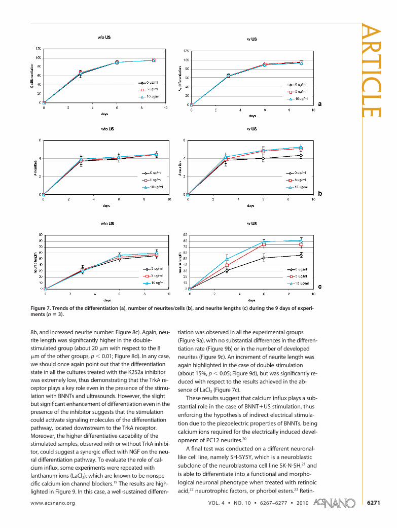
ods section. Up to 9 days of culture, differentiating sta-
tus (the percentage of differentiated cells in the cul-
ture), number of neuronal processes per cell, and
neurite length were monitored in cultures incubated
with 0, 5, and 10 �g/mL of BNNTs, both stimulated and
nonstimulated with ultrasounds. Figure 6 shows repre-
sentative micrographs performed on all the groups at
the end point.
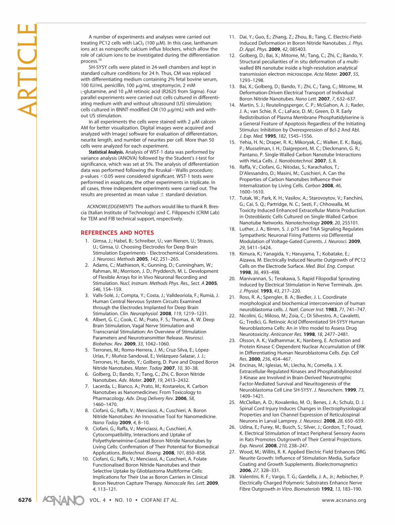
No significant differences in terms of cellular differ-
entiation could be detected among the groups (Figure
7a): all the cultures reached �95% of differentiation
with no differences compared to the control (p � 0.05).
Interestingly, the number of neuronal processes per dif-ferentiated cell became significantly higher in the cul-tures that underwent both BNNT-incubation and ultra-sound stimulation (Figure 7b): on average, about fiveprocesses were found in the BNNT-US stimulated cul-tures with respect to the typical four processes of thecontrol groups (p � 0.05). No significant differenceswere found between the two different BNNT concentra-tions investigated.
However, a dramatic increment of the developedneurites was the most remarkable outcome ascribableto the combined stimulation. Figure 7c shows the trend
of neurite length vs differentia-tion time. After 72 h of treat-ment, average neurite lengthin the BNNT-US groups wasclearly higher than that of thecontrol cultures, and this incre-ment rose by about 30% at theend of the 9th day.
To try to clarify the mecha-nisms underlying the evident ef-fects of the combined stimula-tion BNNT�US, two tests werecarried out with different differ-entiation inhibitors.
Blockage of the NGF specificreceptor TrkA was performed bytreating the cells with K252a,18 asoutlined in the Methods section.Cultures (treated with 0 and 10�g/mL of BNNTs, with and with-out ultrasound stimulation) weremonitored for 6 days, and thedifferentiation status was ana-lyzed at the end point (Figure8a). Differentiation was very lowin all the experimental groups(about 5�10%), but increasedsignificantly in the BNNT�UStreatment (about 25% of differ-entiated cells, p � 0.05: Figure
Figure 5. (a) Scanning TEM-high angle annular dark field (STEM-HAADF) image of BNNTs incorporated inside a cytoplas-matic vescicle of a PC12 cell. (b) Electron energy loss (EEL) spectrum collected with the electron probe located at the point in-dicated by a cross in image a and showing the B and N signals coming from the BNNTs. The C signal stems from organicmaterial.
Figure 6. Images of calcein-labeled PC12 cells after 9 days of treatments (with andwithout BNNT incubation; with and without US stimulation). Magnification 20X.
ART
ICLE
VOL. 4 ▪ NO. 10 ▪ CIOFANI ET AL. www.acsnano.org6270
8b, and increased neurite number: Figure 8c). Again, neu-rite length was significantly higher in the double-stimulated group (about 20 �m with respect to the 8�m of the other groups, p � 0.01; Figure 8d). In any case,we should once again point out that the differentiationstate in all the cultures treated with the K252a inhibitorwas extremely low, thus demonstrating that the TrkA re-ceptor plays a key role even in the presence of the stimu-lation with BNNTs and ultrasounds. However, the slightbut significant enhancement of differentiation even in thepresence of the inhibitor suggests that the stimulationcould activate signaling molecules of the differentiationpathway, located downstream to the TrkA receptor.Moreover, the higher differentiative capability of thestimulated samples, observed with or without TrkA inhibi-tor, could suggest a synergic effect with NGF on the neu-ral differentiation pathway. To evaluate the role of cal-cium influx, some experiments were repeated withlanthanum ions (LaCl3), which are known to be nonspe-cific calcium ion channel blockers.19 The results are high-lighted in Figure 9. In this case, a well-sustained differen-
tiation was observed in all the experimental groups
(Figure 9a), with no substantial differences in the differen-
tiation rate (Figure 9b) or in the number of developed
neurites (Figure 9c). An increment of neurite length was
again highlighted in the case of double stimulation
(about 15%, p � 0.05; Figure 9d), but was significantly re-
duced with respect to the results achieved in the ab-
sence of LaCl3 (Figure 7c).
These results suggest that calcium influx plays a sub-
stantial role in the case of BNNT�US stimulation, thus
enforcing the hypothesis of indirect electrical stimula-
tion due to the piezoelectric properties of BNNTs, being
calcium ions required for the electrically induced devel-
opment of PC12 neurites.20
A final test was conducted on a different neuronal-
like cell line, namely SH-SY5Y, which is a neuroblastic
subclone of the neuroblastoma cell line SK-N-SH,21 and
is able to differentiate into a functional and morpho-
logical neuronal phenotype when treated with retinoic
acid,22 neurotrophic factors, or phorbol esters.23 Retin-
Figure 7. Trends of the differentiation (a), number of neurites/cells (b), and neurite lengths (c) during the 9 days of experi-ments (n � 3).
ARTIC
LE
www.acsnano.org VOL. 4 ▪ NO. 10 ▪ 6267–6277 ▪ 2010 6271

oic acid induces differentiation through a pathway
that does not involve TrkA, thus allowing to further as-
sess the mechanism of the proposed stimulation. The
results are reported in Figure 10. After 6 days of treat-
ment, cultures show a moderate differentiation (about
40% in each experimental group; Figure 10a,b). No dif-
ferences were observed in the number of developed
neurites (Figure 10c) but, once again, a significant in-
crease in neurite length was evidenced on the 6th day
of stimulation with ultrasounds in the presence of 10
�g/mL of BNNTs (about 30%, p � 0.05; Figure 10d).
These data confirm the effects of stimulation on differ-
ent cell lines, with different differentiation pathways:
even if the most important outcomes have been seen
in the case of PC12 cells, where TrkA was demonstrated
to play a major role also in the stimulation-mediated
differentiation, the enhancement of neurite outgrowth
in SH-SY5Y cells demonstrates the efficiency of stimula-
tion also when the TrkA receptor is not involved in the
differentiation pathway.24
All these results clearly demonstrated an efficient in-
direct stimulation, opening exciting perspectives in the
field of neuronal regeneration.
Several in vitro and in vivo studies have shown that
electrical stimulation plays an important role in neurite
extension and in the regeneration of transected nerve
ends.25�27 Electrical charges appear to be focused on
the stimulation of axonal regeneration;28 therefore,
many electrical stimulating materials have been evalu-
ated to determine whether they can be used in the de-
velopment of effective nerve regeneration.29,30 Al-
though the exact mechanisms by which electrical
stimulation favors nerve regeneration have not yet
been clearly understood, it is a well-known fact that
Figure 8. Stimulation experiments in the presence of the TrkA inhibitor K252a: images of calcein-labeled PC12 (a), differen-tiation (b), number of neurites/cells (c), and neurite lengths (d) cells after 6 days of treatments (n � 3, p � 0.05). Magnifica-tion 20X.
ART
ICLE
VOL. 4 ▪ NO. 10 ▪ CIOFANI ET AL. www.acsnano.org6272

electrical stimulation enhances both neurite outgrowth
in vitro31,32 and nerve regeneration in vivo.33�36
Nanotechnology offers several innovative ap-
proaches for neuronal stimulation. Carbon nanotubes,
for example, have been applied in several areas of nerve
tissue engineering to probe and augment cell behav-
ior, to label and track subcellular components, and to
study the growth and organization of neural networks.
Recent reports have shown that nanotubes can sustain
and promote neuronal electrical activity in networks of
cultured cells, but the ways in which they affect cellular
function are still poorly understood. Using single-cell
electrophysiology techniques, electron microscopy
analysis, and theoretical modeling, Cellot et al.37 have
demonstrated that nanotubes improve the responsive-
ness of neurons by forming tight contacts with the cell
membranes. Very interestingly, this report shows that
nanotubes can sustain and promote neuronal electri-
cal activity in networks of cultured cells, by favoring
electrical shortcuts between the proximal and distal
compartments of the neuron.
Recent studies, moreover, have suggested the great
potential of high density, carbon nanotube (CNT) coated
surfaces as interfacing material with neural systems.38�46
A study by Shein et al.47 presents novel carbon nanotube-
based electrode arrays composed of cell-alluring CNT is-
lands. These play the double role of anchoring neurons di-
rectly and only onto the electrode sites (with no need for
chemical treatments) and facilitating high fidelity electri-
cal interfacing-recording and stimulation.
A study by Keefer et al.48 shows that conventional
tungsten and stainless steel wire electrodes can be
coated with carbon nanotubes using electrochemical
techniques, and that this coating enhances both the re-
Figure 9. Stimulation experiments in the presence of the calcium channels inhibitor LaCl3: images of calcein-labeled PC12(a), differentiation (b), number of neurites/cells (c), and neurite lengths (d) cells after 6 days of treatments (n � 3, p � 0.05).Magnification 20X.
ARTIC
LE
www.acsnano.org VOL. 4 ▪ NO. 10 ▪ 6267–6277 ▪ 2010 6273

cording and electrical stimulation of neurons in cul-
ture, rats, and monkeys by decreasing electrode imped-
ance and increasing charge transfer.
Finally, a very recent paper49 has reported that low
concentrations of functionalized CNT, when added with
nerve growth factor (NGF), promote the outgrowth of
neuronal neurites in dorsal root ganglion (DRG) neurons
and in PC12 cells.
Here, we have proposed an innovative solution
based on the combination of noninvasive stimula-
tion with US in the presence of piezoelectric nano-
particles incubated with cells, which can elicit the
same phenomena derived from a “classical” electric
stimulation, i.e., a markedly pronounced outgrowth
of neuronal processes in PC12 cultures, but without
the need for electrodes in the culture. This was
achieved by mechanical stimulation of the BNNTs
which, by virtue of their polarizability, are able to
convey electrical stimuli to the cells. Among the al-
ready mentioned applications in regenerative medi-
cine, this concept could also be used in life science
wherever electrical stimulation is needed, e.g., deep
brain stimulation,50 gastric stimulation for gastroparesis,51
cardiac pacing for various cardiac arrythmias,52 skeletal
muscle stimulation in various neuropathies,53 etc. Of
course, before any realistic in vivo applications, deep in-
vestigations in the BNNT impact on living organisms are
mandatory. Biocompatibility, biodistribution, and degra-
dation of these novel nanoparticles have to be tested, ow-
ing to the total lack of data in the literature.54 Active in
vivo targeting of BNNT toward the site to be treated also
should be achieved.
Although extensive and quantitative investigations
will be needed after these preliminary results, we are
confident in the tremendous impact this technology
could have in biological and clinical practices.
Figure 10. Stimulation experiments carried out on human neuroblastoma SH-SY5Y cells: images of calcein-labeled cultures(a), differentiation (b), number of neurites/cells (c), and neurite lengths (d) cells after 6 days of treatments (n � 3). Magnifi-cation 20X.
ART
ICLE
VOL. 4 ▪ NO. 10 ▪ CIOFANI ET AL. www.acsnano.org6274

METHODSBoron Nitride Nanotubes Preparation. BNNTs (purchased from the
Nano and Ceramic Materials Research Center, Wuhan Instituteof Technology, China) were produced using an annealingmethod from boron containing precursors.55 Details of thesample provided by the supplier include boron nitride �98.5wt % and yield �80%.
Glycol chitosan (G-chitosan 81339 from G7753 Sigma) wasused for the dispersion and stabilization of BNNTs. Dispersionwas prepared with phosphate buffered solution (PBS). BNNTs (5mg) were mixed with 10 mL of a 0.1% G-chitosan solution in apolystyrene tube. The samples were sonicated for 12 h (by aBransonic sonicator 2510), using an output power of 20 W forall the experiments, resulting in a stable G-chitosan-BNNT disper-sion by noncovalent coating of the nanotube walls withG-chitosan. The dispersion was characterized by spectrophoto-metric analysis, using a LIBRA S12 Spectrophotometer UV/vis/NIR(Biochrom). Microphotographs of the final dispersion of BNNTswere obtained with a FEI 200 FIB microscope and Zeiss 902 TEM,dropping a small quantity of BNNT aqueous suspension on acopper grid.
Cell Culture, Viability Assay, and Up-Take Assessment. The studieswere performed on PC12 cells (ATCC CRL-1721), a cell line de-rived from a transplantable rat pheochromocytoma. PC12 cellsrespond reversibly to the administration of the nerve growth fac-tor (NGF) by expressing the neuronal phenotype. As a matter offact, NGF is responsible for phenotype commitment of PC12 cellstoward sympathetic neurons. Additional salient effects of NGFin PC12 cell cultures include inhibition of proliferation, genera-tion of long neurites, acquisition of electrical excitability, hyper-trophy, and other changes associated with the acquisition of aneuronal-like phenotype.56
PC12 were cultured in Dulbecco’s modified Eagle mediumwith 10% horse serum, 5% fetal bovine serum, 100 IU/mL peni-cillin, 100 �g/mL streptomycin, and 2 mM L-glutamine. Cellswere maintained at 37 °C in a saturated humidity atmosphere(i.e., 95% air/5% CO2).
Additional experiments were carried out on human neuro-blastoma SH-SY5Y cells (ATCC CRL-2266). This cell line repre-sents a good model system for studying neuronal properties inculture, as many of their receptor systems have been well char-acterized and widely used as a model system for investigations inneuritogenesis.57 Treatment with retinoic acid has been re-ported to induce differentiation of SH-SY5Y cells, without alter-ing the TrkA expression.58 SH-SY5Y were cultured in Dulbecco’smodified Eagle medium and Ham’s F12 (1:1) with 10% fetal calfserum, 100 IU/mL penicillin, 100 �g/mL streptomycin, and 2 mML-glutamine. Cells were maintained at 37 °C in a saturated humid-ity atmosphere containing 95% air/5% CO2. Compatibility ofBNNTs on SH-SY5Y cells already has been widely documented.59
For viability testing, WST-1 (2-(4-iodophenyl)-3-(4-nitophenyl)-5-(2,4-disulfophenyl)-2H-tetrazoilium monosodiumsalt, provided in a premix electro-coupling solution, BioVision)cell proliferation assays were carried out. After trypsinization andcounting with a hemocytometer, 5000 cells were seeded in 96-well plate chambers. Once adhesion was verified (after about12 h since seeding), cells were incubated with 0, 5, 10, 20, 50,and 100 �g/mL of glycol-chitosan coated BNNTs for 3, 6, and 9days. At the end point of incubation, cell cultures were treatedwith 100 �L of culture medium � 10 �L of the premix solutionfor a further 2 h and, thereafter, absorbance at 450 nm was readwith a microplate reader (Victor3, Perkin-Elmer).
Viability was further investigated with the LIVE/DEAD viabil-ity/cytotoxicity kit (Molecular Probes). The kit contains calceinAM (4 mM in anhydrous DMSO) and ethidium homodimer-1[EthD-1, 2 mM in DMSO/H2O 1:4 (v/v)]. After incubation for 72 hat GC-BNNT concentration of 10 �g/mL, the cells (25 000 in 24-well plate chambers, n � 3) were rinsed with PBS and treated for10 min at 37 °C with 2 �M calcein AM and 4 �M EthD-1 in PBS.The cells were finally observed (after 9 days) using an invertedfluorescent microscope (TE2000U, Nikon) equipped with acooled CCD camera (DS-5MC USB2, Nikon) and by NIS Elementsimaging software, provided with the appropriate filters.
For early apoptosis detection, 25 000 cells were seeded in 24-well plate chambers (n � 3) and treated with 0�100 �g/mL of
glycol-chitosan coated BNNTs for 9 days. The ApoAlert kit(Clonetech Laboratories) was used to evaluate differences be-tween normal and apoptotic cells after treatments. The kit con-tains annexin V-FITC (20 �g/mL in Tris-NaCl), 1X binding buffer,and propidium iodide (50 �g/mL in 1X binding buffer). The cellswere rinsed with 1X binding buffer and then incubated with200 �L of 1X binding buffer containing 5 �L of the annexin V so-lution and 10 �L of the propidium iodide solution. After incuba-tion in the dark at room temperature for 20 min, the cells wereobserved via fluorescence microscopy with the appropriatefilters.
Generation of reactive oxygen species (ROS) is a normalevent for aerobic organisms, which occurs at a controlled ratein healthy cells. Under conditions of oxidative stress, like expo-sure to nanomaterials,60 ROS production is dramatically in-creased, resulting in subsequent alteration of membrane lipids,proteins, and nucleic acids.
ROS production in PC12 cells treated with BNNTs was de-tected with use of the Image-IT Green Reactive Oxygen SpeciesDetection kit (Invitrogen). The assay is based on 5-(and-6)-carboxy-2=,7=-dichlorodihydrofluorescein diacetate (carboxy-H2DCFDA), a fluorogenic marker for ROS in viable cells. The non-fluorescent carboxy-H2DCFDA permeates live cells and isdeacetylated by nonspecific intracellular esterases. In the pres-ence of nonspecific ROS (produced throughout the cell, in par-ticular during oxidative stress) the reduced fluorescein com-pound is oxidized and emits bright green fluorescence.61 Thecells (25 000 per well) were seeded in 24-well plate chambers (n� 3) and treated with 0�100 �g/mL of glycol-chitosan coatedBNNTs for 9 days. They were then incubated for 45 min with a 25�M carboxy-H2DCFDA working solution (in DMSO:PBS at 1:400v/v) and immediately observed via fluorescence microscopy withthe appropriate filters.
For both apoptosis and ROS detection, cell nuclei were coun-terstained with 5 �g/mL of Hoechst 33342.
Internalization was assessed by transmission electron micros-copy (TEM). PC12 cells were seeded at 2 � 106 cells/T25 flask. Af-ter attachment, they were incubated in a GC-BNNT modifiedCM (at a final concentration of 5 �g/mL). After 12 h of incuba-tion, the samples were washed in PBS 0.1 M and then fixed whenstill adherent to flasks with 1% w/v glutaraldehyde�4% w/vparaformaldehyde in PBS 0.1 M pH 7.2 for 2 h at 4 °C. After wash-ing, the cells were detached by scraping, postfixed in 1% w/vOsO4 PBS 0.1 M pH 7.2 for 1 h, washed, and dehydrated withacidified aceton-dimethylacetal (Fluka) for 10 min. The sampleswere mixed in Epon/Durcupan resin in BEEM capsules #00 (Struc-ture Probe) overnight at room temperature and finally embed-ded in resin at 56 °C for 48 h. Ultrathin sections (20�30 nm thick)were obtained with an Ultrotome Nova ultramicrotome (LKB,Bromma, Sweden) equipped with a diamond knife (Diatome,Biel/Bienne, Switzerland).
The sections were placed on 200 square mesh nickel grids,counterstained with saturated aqueous uranyl acetate and leadcitrate solutions, and observed with a Jeol JEM-2200FS micro-scope (Jeol, Japan). To assess the actual presence of BNNTs, B el-emental map was obtained from the B K core-loss edge at 188eV with the three windows method using a 20 eV energy slit. TheEEL spectrum was acquired in STEM mode.
Cell Stimulation Experiments. PC12 cells were plated in 24-wellchambers and kept in standard culture conditions for 24 h. Thus,CM was replaced with differentiating medium containing 2% fe-tal bovine serum, 100 IU/mL penicillin, 100 �g/mL streptomy-cin, 2 mM L-glutamine, and 60 ng/mL of NGF (N1418 fromSigma). Parallel experiments were carried out: cell cultured in dif-ferentiating medium with and without ultrasound (US) stimula-tion; cells cultured in BNNT-modified CM (5 and 10 �g/mL) withand without US stimulation, which was settled at 20 W, 40 kHz,for 5 s, 4 times a day for 9 days, performed with a Bransonic son-icator 2510.
For pharmacological inhibition of TrkA, cultures were treatedwith 200 nM K252a (Calbiochem) and monitored up to 6 days.Four parallel experiments were carried out: cells cultured in dif-ferentiating medium with and without ultrasound (US) stimula-tion; cells cultured in BNNT-modified CM (10 �g/mL) with andwithout US stimulation.
ARTIC
LE
www.acsnano.org VOL. 4 ▪ NO. 10 ▪ 6267–6277 ▪ 2010 6275
A number of experiments and analyses were carried outtreating PC12 cells with LaCl3 (100 �M). In this case, lanthanumions act as nonspecific calcium influx blockers, which allow therole of calcium ions to be investigated during the differentiationprocess.19
SH-SY5Y cells were plated in 24-well chambers and kept instandard culture conditions for 24 h. Thus, CM was replacedwith differentiating medium containing 2% fetal bovine serum,100 IU/mL penicillin, 100 �g/mL streptomycin, 2 mML-glutamine, and 10 �M retinoic acid (R2625 from Sigma). Fourparallel experiments were carried out: cells cultured in differenti-ating medium with and without ultrasound (US) stimulation;cells cultured in BNNT-modified CM (10 �g/mL) with and with-out US stimulation.
In all experiments the cells were stained with 2 �M calceinAM for better visualization. Digital images were acquired andanalyzed with ImageJ software for evaluation of differentiation,neurite length, and number of neurites per cell. More than 50cells were analyzed for each experiment.
Statistical Analysis. Analysis of WST-1 data was performed byvariance analysis (ANOVA) followed by the Student’s t-test forsignificance, which was set at 5%. The analysis of differentiationdata was performed following the Kruskal�Wallis procedure;p-values �0.05 were considered significant. WST-1 tests wereperformed in exaplicate, the other experiments in triplicate. Inall cases, three independent experiments were carried out. Theresults are presented as mean value � standard deviation.
ACKNOWLEDGEMENTS The authors would like to thank R. Bres-cia (Italian Institute of Technology) and C. Filippeschi (CRIM Lab)for TEM and FIB technical support, respectively.
REFERENCES AND NOTES1. Gimsa, J.; Habel, B.; Schreiber, U.; van Rienen, U.; Strauss,
U.; Gimsa, U. Choosing Electrodes for Deep BrainStimulation Experiments - Electrochemical Considerations.J. Neurosci. Methods 2005, 142, 251–265.
2. Adams, C.; Mathieson, K.; Gunning, D.; Cunningham, W.;Rahman, M.; Morrison, J. D.; Prydderch, M. L. Developmentof Flexible Arrays for in Vivo Neuronal Recording andStimulation. Nucl. Instrum. Methods Phys. Res., Sect. A 2005,546, 154–159.
3. Valls-Sole, J.; Compta, Y.; Costa, J.; Valldeoriola, F.; Rumia, J.Human Central Nervous System Circuits Examinedthrough the Electrodes Implanted for Deep BrainStimulation. Clin. Neurophysiol. 2008, 119, 1219–1231.
4. Albert, G. C.; Cook, C. M.; Prato, F. S.; Thomas, A. W. DeepBrain Stimulation, Vagal Nerve Stimulation andTranscranial Stimulation: An Overview of StimulationParameters and Neurotransmitter Release. Neurosci.Biobehav. Rev. 2009, 33, 1042–1060.
5. Terrones, M.; Romo-Herrera, J. M.; Cruz-Silva, E.; Lopez-Urıas, F.; Munoz-Sandoval, E.; Velazquez-Salazar, J. J.;Terrones, H.; Bando, Y.; Golberg, D. Pure and Doped BoronNitride Nanotubes. Mater. Today 2007, 10, 30–38.
6. Golberg, D.; Bando, Y.; Tang, C.; Zhi, C. Boron NitrideNanotubes. Adv. Mater. 2007, 19, 2413–2432.
7. Lacerda, L.; Bianco, A.; Prato, M.; Kostarelos, K. CarbonNanotubes as Nanomedicines: From Toxicology toPharmacology. Adv. Drug Delivery Rev. 2006, 58,1460–1470.
8. Ciofani, G.; Raffa, V.; Menciassi, A.; Cuschieri, A. BoronNitride Nanotubes: An Innovative Tool for Nanomedicine.Nano Today 2009, 4, 8–10.
9. Ciofani, G.; Raffa, V.; Menciassi, A.; Cuschieri, A.Cytocompatibility, Interactions and Uptake ofPolyethyleneimine-Coated Boron Nitride Nanotubes byLiving Cells: Confirmation of Their Potential for BiomedicalApplications. Biotechnol. Bioeng. 2008, 101, 850–858.
10. Ciofani, G.; Raffa, V.; Menciassi, A.; Cuschieri, A. FolateFunctionalised Boron Nitride Nanotubes and theirSelective Uptake by Glioblastoma Multiforme Cells:Implications for Their Use as Boron Carriers in ClinicalBoron Neutron Capture Therapy. Nanoscale Res. Lett. 2009,4, 113–121.
11. Dai, Y.; Guo, E.; Zhang, Z.; Zhou, B.; Tang, C. Electric-Field-Induced Deformation in Boron Nitride Nanotubes. J. Phys.D: Appl. Phys. 2009, 42, 085403.
12. Golberg, D.; Bai, X.; Mitome, M.; Tang, C.; Zhi, C.; Bando, Y.Structural peculiarities of in situ deformation of a multi-walled BN nanotube inside a high-resolution analyticaltransmission electron microscope. Acta Mater. 2007, 55,1293–1298.
13. Bai, X.; Golberg, D.; Bando, Y.; Zhi, C.; Tang, C.; Mitome, M.Deformation-Driven Electrical Transport of IndividualBoron Nitride Nanotubes. Nano Lett. 2007, 7, 632–637.
14. Martin, S. J.; Reutelingsperger, C. P.; McGahon, A. J.; Rader,J. A.; van Schie, R. C.; LaFace, D. M.; Green, D. R. EarlyRedistribution of Plasma Membrane Phosphatidylserine isa General Feature of Apoptosis Regardless of the InitiatingStimulus: Inhibition by Overexpression of Bcl-2 And Abl.J. Exp. Med. 1995, 182, 1545–1556.
15. Yehia, H. N.; Draper, R. K.; Mikoryak, C.; Walker, E. K.; Bajaj,P.; Musselman, I. H.; Daigrepont, M. C.; Dieckmann, G. R.;Pantano, P. Single-Walled Carbon Nanotube Interactionswith HeLa Cells. J. Nanobiotechnol. 2007, 5, 8.
16. Raffa, V.; Ciofani, G.; Nitodas, S.; Karachalios, T.;D’Alessandro, D.; Masini, M.; Cuschieri, A. Can theProperties of Carbon Nanotubes Influence theirInternalization by Living Cells. Carbon 2008, 46,1600–1610.
17. Tutak, W.; Park, K. H.; Vasilov, A.; Starovoytov, V.; Fanchini,G.; Cai, S. Q.; Partridge, N. C.; Sesti, F.; Chhowalla, M.Toxicity Induced Enhanced Extracellular Matrix Productionin Osteoblastic Cells Cultured on Single-Walled CarbonNanotube Networks. Nanotechnology 2009, 20, 255101.
18. Luther, J. A.; Birren, S. J. p75 and TrkA Signaling RegulatesSympathetic Neuronal Firing Patterns via DifferentialModulation of Voltage-Gated Currents. J. Neurosci. 2009,29, 5411–5424.
19. Kimura, K.; Yanagida, Y.; Haruyama, T.; Kobatake, E.;Aizawa, M. Electrically Induced Neurite Outgrowth of PC12Cells on the Electrode Surface. Med. Biol. Eng. Comput.1998, 36, 493–498.
20. Manivannan, S.; Terakawa, S. Rapid Filopodial SproutingInduced by Electrical Stimulation in Nerve Terminals. Jpn.J. Physiol. 1993, 43, 217–220.
21. Ross, R. A.; Spengler, B. A.; Biedler, J. L. Coordinatemorphological and biochemical interconversion of humanneuroblastoma cells. J. Natl. Cancer Inst. 1983, 71, 741–747.
22. Nicolini, G.; Miloso, M.; Zoia, C.; Di Silvestro, A.; Cavaletti,G.; Tredici, G. Retinoic Acid Differentiated SH-SY5Y HumanNeuroblastoma Cells: An in Vitro model to Assess DrugNeurotoxicity. Anticancer Res. 1998, 18, 2477–2481.
23. Olsson, A. K.; Vadhammar, K.; Nanberg, E. Activation andProtein Kinase C-Dependent Nuclear Accumulation of ERKin Differentiating Human Neuroblastoma Cells. Exp. CellRes. 2000, 256, 454–467.
24. Encinas, M.; Iglesias, M.; Llecha, N.; Comella, J. X.Extracellular-Regulated Kinases and Phosphatidylinositol3-Kinase are Involved in Brain-Derived NeurotrophicFactor-Mediated Survival and Neuritogenesis of theNeuroblastoma Cell Line SH-SY5Y. J. Neurochem. 1999, 73,1409–1421.
25. McClellan, A. D.; Kovalenko, M. O.; Benes, J. A.; Schulz, D. J.Spinal Cord Injury Induces Changes in ElectrophysiologicalProperties and Ion Channel Expression of ReticulospinalNeurons in Larval Lamprey. J. Neurosci. 2008, 28, 650–659.
26. Udina, E.; Furey, M.; Busch, S.; Silver, J.; Gordon, T.; Fouad,K. Electrical Stimulation of Intact Peripheral Sensory Axonsin Rats Promotes Outgrowth of Their Central Projections.Exp. Neurol. 2008, 210, 238–247.
27. Wood, M.; Willits, R. K. Applied Electric Field Enhances DRGNeurite Growth: Influence of Stimulation Media, SurfaceCoating and Growth Supplements. Bioelectromagnetics2006, 27, 328–331.
28. Valentini, R. F.; Vargo, T. G.; Gardella, J. A., Jr.; Aebischer, P.Electrically Charged Polymeric Substrates Enhance NerveFibre Outgrowth in Vitro. Biomaterials 1992, 13, 183–190.
ART
ICLE
VOL. 4 ▪ NO. 10 ▪ CIOFANI ET AL. www.acsnano.org6276

29. Gomez, N.; Schmidt, C. E. Nerve Growth Factor-Immobilized Polypyrrole: Bioactive Electrically ConductingPolymer for Enhanced Neurite Extension. J. Biomed. Mater.Res., Part A 2007, 81, 135–149.
30. Chew, S. Y.; Mi, R.; Hoke, A.; Leong, K. W. Aligned Protein-Polymer Composite Fibers Enhance Nerve Regeneration: APotential Tissue-Engineering Platform. Adv. Funct. Mater.2007, 17, 1288–1296.
31. Schmidt, C. E.; Shastri, V. R.; Vacanti, J. P.; Langer, R.Stimulation of Neurite Outgrowth Using an ElectricallyConducting Polymer. Proc. Natl. Acad. Sci. U.S.A. 1997, 94,8948–8953.
32. Valentini, R. F.; Vargo, T. G.; Gardella, J. A., Jr.; Aebischer, P.Patterned Neuronal Attachment and Outgrowth onSurface Modified, Electrically Charged FluoropolymerSubstrates. J. Biomater. Sci., Polym. Ed. 1993, 5, 13–36.
33. Aebischer, P.; Valentini, R. F.; Dario, P.; Domenici, C.;Galletti, P. M. Piezoelectric Guidance Channels EnhanceRegeneration in The Mouse Sciatic Nerve after Axotomy.Brain Res. 1987, 436, 165–168.
34. Sisken, B. F.; Kanje, M.; Lundborg, G.; Herbst, E.; Kurtz, W.Stimulation of Rat Sciatic Nerve Regeneration with PulsedElectromagnetic Fields. Brain Res. 1989, 485, 309–316.
35. Udina, E.; Furey, M.; Busch, S.; Silver, J.; Gordon, T.; Fouad,K. Electrical Stimulation of Intact Peripheral Sensory Axonsin Rats Promotes Outgrowth of their Central Projections.Exp. Neurol. 2008, 210, 238–247.
36. Lyons, K. E.; Pahwa, R. Deep Brain Stimulation InParkinson’s Disease. Curr. Neurol. Neurosci. Rep. 2004, 4,290–295.
37. Cellot, G.; Cilia, E.; Cipollone, S.; Rancic, V.; Sucapane, A.;Giordani, S.; Gambazzi, L.; Markram, H.; Grandolfo, M.;Scaini, D.; et al. Carbon Nanotubes Might ImproveNeuronal Performance by Favouring Electrical Shortcuts.Nat. Nanotechnol. 2009, 4, 126–133.
38. Bekyarova, E.; Ni, Y.; Malarkey, E. B.; Montana, V.;McWilliams, J. L.; Haddon, R. C.; Parpura, V. Applications ofCarbon Nanotubes in Biotechnology and Biomedicine.J. Biomed. Nanotechnol. 2005, 1, 3–17.
39. Gabay, T.; Jakobs, E.; Ben-Jacob, E.; Hanein, Y. EngineeredSelf-Organization of Neural Networks Using CarbonNanotube Clusters. Phys. A 2005, 350, 611–621.
40. Gabay, T.; Ben-David, M.; Kalifa, I.; Sorkin, R.; Abrams, Z. R.;Ben-Jacob, E.; Hanein, Y. Electro-Chemical and BiologicalProperties of Carbon Nanotube Based Multi-ElectrodeArrays. Nanotechnology 2007, 18, 35201.
41. Hu, H.; Ni, Y.; Montana, V.; Haddon, R. C.; Parpura, V.Chemically Functionalized Carbon Nanotubes asSubstrates for Neuronal Growth. Nano Lett. 2004, 4, 507–511.
42. Lovat, V.; Pantarotto, D.; Lagostena, L.; Cacciari, B.;Grandolfo, M.; Righi, M.; Spalluto, G.; Prato, M.; Ballerini, L.Carbon Nanotube Substrates Boost Neuronal ElectricalSignaling. Nano Lett. 2005, 5, 1107–1110.
43. Mattson, M. P.; Haddon, R. C.; Rao, A. M. MolecularFunctionalization of Carbon Nanotubes and Use asSubstrates for Neuronal Growth. J. Mol. Neurosci. 2000, 14,175–182.
44. Mazzatenta, M.; Giugliano, S.; Campidelli, L.; Gambazzi, L.;Businaro, H.; Markram, M.; Prato, M.; Ballerini, L. InterfacingNeurons with Carbon Nanotubes: Electrical Signal Transferand Synaptic Stimulation in Cultured Brain Circuits.J. Neurosci. 2007, 27, 6931–6936.
45. Sorkin, R.; Greenbaum, A.; David-Pur, M.; Anava, S.; Ayali,A.; Ben-Jacob, E.; Hanein, Y. Process Entanglement as aNeuronal Anchorage Mechanism to Rough Surfaces.Nanotechnology 2009, 20, 015101.
46. Zhang, X.; Prasad, S.; Niyogi, S.; Morgan, A.; Ozkan, M.;Ozkan, C. S. Guided Neurite Growth on Patterned CarbonNanotubes. Sens. Actuators, B 2005, 106, 843–850.
47. Shein, M.; Greenbaum, A.; Gabay, T.; Sorkin, R.; David-Pur,M.; Ben-Jacob, E.; Hanein, Y. Engineered Neuronal CircuitsShaped and Interfaced with Carbon NanotubeMicroelectrode Arrays. Biomed. Microdevices 2009, 11,495–501.
48. Keefer, E. W.; Botterman, B. R.; Romero, M. I.; Rossi, A. F.;Gross, G. W. Carbon Nanotube Coating Improves NeuronalRecordings. Nat. Nanotechnol. 2008, 3, 434–439.
49. Matsumoto, K.; Sato, C.; Naka, Y.; Whitby, R.; Shimizu, N.Stimulation of Neuronal Neurite Outgrowth UsingFunctionalized Carbon Nanotubes. Nanotechnology 2010,21, 115101.
50. Della Flora, E.; Perera, C. L.; Cameron, A. L.; Maddern, G. J.Deep Brain Stimulation for Essential Tremor: A systematicreview. Movement Disord. 2010, 25, 1550–1559.
51. Xu, J.; Chen, J. D. Z. Intestinal Electrical StimulationImproves Delayed Gastric Emptying and VomitingInduced by Duodenal Distension in Dogs.Neurogastroenterol. Motil. 2008, 20, 236–242.
52. Ross, K. B.; Dubin, S.; Nigroni, P.; Kepics, F.; Shi, W. Y.; Yan,H. Programmed Stimulation for Simulation of AtrialTachyarrythmias. Biomed. Sci. Instrum. 1997, 33, 25–29.
53. Gordon, T.; Brushart, T. M.; Amirjani, N.; Chan, K. M. ThePotential of Electrical Stimulation to Promote FunctionalRecovery after Peripheral Nerve InjuryO Comparisonsbetween Rats and Humans. Acta Neurochir. 2007, 100, 3–11.
54. Ciofani, G. Potential Applications of Boron NitrideNanotubes as Drug Delivery Systems. Expert Opin. DrugDelivery 2010, 7, 889–893.
55. http://ncm.wit.edu.cn/english.jsp.56. Greene, L. A.; Farinelli, S. E.; Cunningham, M. E.; Park, D. S.
Methodologies for the Culture and Experimental Use ofthe PC12 Rat Pheochromocytoma Cell Line. In CulturingNerve Cells; Banker, F., Goslin, K., Eds.; MIT Press:Cambridge, MA, 1998; Vol. 2.
57. Kaplan, D. R.; Matsumoto, K.; Lucarelli, E.; Theile, C. J.Induction of TrkB by Retinoic Acid Mediates BiologicResponsiveness to BDNF and Differentiation ff HumanNeuroblastoma Cells. Neuron 1993, 11, 321–331.
58. Encinas, M.; Iglesias, M.; Llechas, N.; Comella, J. X.Extracellular-Regulated Kinases and Phosphatidylinositol3-Kinase Are Involved in Brain-Derived NeurotrophicFactor-Mediated Survival and Neuritogenesis of theNeuroblastoma Cell Line SH-SY5Y. J. Neurochem. 1999, 73,1409–1421.
59. Ciofani, G.; Danti, S.; D’Alessandro, D.; Moscato, S.;Menciassi, A. Assessing Cytotoxicity of Boron NitrideNanotubes: Interference with the MTT Assay. Biochem.Biophys. Res. Commun. 2010, 394, 405–411.
60. Pulskamp, K.; Diabate, S.; Krug, H. F. Carbon NanotubesShow no Sign of Acute Toxicity but Induce IntracellularReactive Oxygen Species in Dependence onContaminants. Toxicol. Lett. 2007, 168, 58–74.
61. Park, E. J.; Choi, J.; Park, Y. K.; Park, K. Oxidative StressInduced by Cerium Oxide Nanoparticles in Cultured BEAS-2B Cells. Toxicology 2008, 245, 90–100.
ARTIC
LE
www.acsnano.org VOL. 4 ▪ NO. 10 ▪ 6267–6277 ▪ 2010 6277
Related Documents