Enhanced Expression of Stim, Orai, and TRPC Transcripts and Proteins in Endothelial Progenitor Cells Isolated from Patients with Primary Myelofibrosis Silvia Dragoni 1 , Umberto Laforenza 2 , Elisa Bonetti 3 , Marta Reforgiato 1 , Valentina Poletto 3 , Francesco Lodola 1 , Cinzia Bottino 2 , Daniele Guido 1 , Alessandra Rappa 1 , Sumedha Pareek 1 , Mario Tomasello 1 , Maria Rosa Guarrera 1 , Maria Pia Cinelli 4 , Adele Aronica 3 , Germano Guerra 5 , Giovanni Barosi 3 , Franco Tanzi 1 , Vittorio Rosti 3 * . , Francesco Moccia 1 * . 1 Laboratory of General Physioloy, Department of Biology and Biotechnology ‘‘Lazzaro Spallanzani’’, University of Pavia, Pavia, Italy, 2 Department of Molecular Medicine, University of Pavia, Pavia, Italy, 3 Centre for the Study of Myelofibrosis, Laboratory of Biotechnology, Foundation IRCCS Policlinico San Matteo, Pavia, Italy, 4 Department of Public Health, University of Naples ‘‘Federico II’’, Naples, Italy, 5 Department of Health Sciences, University of Molise, Campobasso, Italy Abstract Background: An increase in the frequency of circulating endothelial colony forming cells (ECFCs), the only subset of endothelial progenitor cells (EPCs) truly belonging to the endothelial phenotype, occurs in patients affected by primary myelofibrosis (PMF). Herein, they might contribute to the enhanced neovascularisation of fibrotic bone marrow and spleen. Store-operated Ca 2+ entry (SOCE) activated by the depletion of the inositol-1,4,5-trisphosphate (InsP 3 )-sensitive Ca 2+ store drives proliferation in ECFCs isolated from both healthy donors (N-ECFCs) and subjects suffering from renal cellular carcinoma (RCC-ECFCs). SOCE is up-regulated in RCC-ECFCs due to the over-expression of its underlying molecular components, namely Stim1, Orai1, and TRPC1. Methodology/Principal Findings: We utilized Ca 2+ imaging, real-time polymerase chain reaction, western blot analysis and functional assays to evaluate molecular structure and the functional role of SOCE in ECFCs derived from PMF patients (PMF- ECFCs). SOCE, induced by either pharmacological (i.e. cyclopiazonic acid or CPA) or physiological (i.e. ATP) stimulation, was significantly higher in PMF-ECFCs. ATP-induced SOCE was inhibited upon blockade of the phospholipase C/InsP 3 signalling pathway with U73111 and 2-APB. The higher amplitude of SOCE was associated to the over-expression of the transcripts encoding for Stim2, Orai2–3, and TRPC1. Conversely, immunoblotting revealed that Stim2 levels remained constant as compared to N-ECFCs, while Stim1, Orai1, Orai3, TRPC1 and TRPC4 proteins were over-expressed in PMF-ECFCs. ATP- induced SOCE was inhibited by BTP-2 and low micromolar La 3+ and Gd 3+ , while CPA-elicited SOCE was insensitive to Gd 3+ . Finally, BTP-2 and La 3+ weakly blocked PMF-ECFC proliferation, while Gd 3+ was ineffective. Conclusions: Two distinct signalling pathways mediate SOCE in PMF-ECFCs; one is activated by passive store depletion and is Gd 3+ -resistant, while the other one is regulated by the InsP 3 -sensitive Ca 2+ pool and is inhibited by Gd 3+ . Unlike N- and RCC-ECFCs, the InsP 3 -dependent SOCE does not drive PMF-ECFC proliferation. Citation: Dragoni S, Laforenza U, Bonetti E, Reforgiato M, Poletto V, et al. (2014) Enhanced Expression of Stim, Orai, and TRPC Transcripts and Proteins in Endothelial Progenitor Cells Isolated from Patients with Primary Myelofibrosis. PLoS ONE 9(3): e91099. doi:10.1371/journal.pone.0091099 Editor: Francesco Bertolini, European Institute of Oncology, Italy Received October 23, 2013; Accepted February 10, 2014; Published March 6, 2014 Copyright: ß 2014 Dragoni et al. This is an open-access article distributed under the terms of the Creative Commons Attribution License, which permits unrestricted use, distribution, and reproduction in any medium, provided the original author and source are credited. Funding: This work was supported by a grant from Associazione Italiana per la Ricerca sul Cancro (AIRC, Milano) Special Program Molecular Clinical Oncology 561000 to AIRC-Gruppo Italiano Malattie Mieloproliferative (AGIMM). A detailed description of the AGIMM project is available at (http://www.progettoagimm.it). The funders had no role in study design, data collection and analysis, decision to publish, or preparation of the manuscript. Competing Interests: The authors have declared that no competing interests exist. * E-mail: [email protected] (FM); [email protected] (VR) . These authors contributed equally to this work. Introduction Primary myelofibrosis (PMF) is a Philadelphia chromosome- negative (Ph-neg) chronic myeloproliferative neoplasm (MPN) characterized by the following hallmarks: bone marrow (BM) fibrosis, myeloid metaplasia, splenomegaly, increased frequency of circulating CD34 + hematopoietic progenitor cells (HPCs), and a V617F mutation of the JAK2 gene in the hematopoietic lineage encountered in 63% of the patients [1,2]. It is characterized by a progressive clinical course and a shortened life expectancy. The only curative therapy for PMF is currently allogenic hematopoietic stem cells transplantation, which is, however, reserved to a minor proportion of patients. Besides the increase in circulating CD34 + HPCs [1], circulating endothelial progenitor cells (EPCs) have been described to be elevated in patients with PMF. These reports, however, suffer from the different methods that were used to isolate EPCs in vitro, generating ambiguity in their identification and enumeration. We have recently showed that patients with PMF have an increased frequency of circulating CD34 + /CD133 + /VEGFR2 + cells as PLOS ONE | www.plosone.org 1 March 2014 | Volume 9 | Issue 3 | e91099

Welcome message from author
This document is posted to help you gain knowledge. Please leave a comment to let me know what you think about it! Share it to your friends and learn new things together.
Transcript
Enhanced Expression of Stim, Orai, and TRPC Transcriptsand Proteins in Endothelial Progenitor Cells Isolatedfrom Patients with Primary MyelofibrosisSilvia Dragoni1, Umberto Laforenza2, Elisa Bonetti3, Marta Reforgiato1, Valentina Poletto3,
Francesco Lodola1, Cinzia Bottino2, Daniele Guido1, Alessandra Rappa1, Sumedha Pareek1,
Mario Tomasello1, Maria Rosa Guarrera1, Maria Pia Cinelli4, Adele Aronica3, Germano Guerra5,
Giovanni Barosi3, Franco Tanzi1, Vittorio Rosti3*., Francesco Moccia1*.
1 Laboratory of General Physioloy, Department of Biology and Biotechnology ‘‘Lazzaro Spallanzani’’, University of Pavia, Pavia, Italy, 2 Department of Molecular Medicine,
University of Pavia, Pavia, Italy, 3 Centre for the Study of Myelofibrosis, Laboratory of Biotechnology, Foundation IRCCS Policlinico San Matteo, Pavia, Italy, 4 Department of
Public Health, University of Naples ‘‘Federico II’’, Naples, Italy, 5 Department of Health Sciences, University of Molise, Campobasso, Italy
Abstract
Background: An increase in the frequency of circulating endothelial colony forming cells (ECFCs), the only subset ofendothelial progenitor cells (EPCs) truly belonging to the endothelial phenotype, occurs in patients affected by primarymyelofibrosis (PMF). Herein, they might contribute to the enhanced neovascularisation of fibrotic bone marrow and spleen.Store-operated Ca2+ entry (SOCE) activated by the depletion of the inositol-1,4,5-trisphosphate (InsP3)-sensitive Ca2+ storedrives proliferation in ECFCs isolated from both healthy donors (N-ECFCs) and subjects suffering from renal cellularcarcinoma (RCC-ECFCs). SOCE is up-regulated in RCC-ECFCs due to the over-expression of its underlying molecularcomponents, namely Stim1, Orai1, and TRPC1.
Methodology/Principal Findings: We utilized Ca2+ imaging, real-time polymerase chain reaction, western blot analysis andfunctional assays to evaluate molecular structure and the functional role of SOCE in ECFCs derived from PMF patients (PMF-ECFCs). SOCE, induced by either pharmacological (i.e. cyclopiazonic acid or CPA) or physiological (i.e. ATP) stimulation, wassignificantly higher in PMF-ECFCs. ATP-induced SOCE was inhibited upon blockade of the phospholipase C/InsP3 signallingpathway with U73111 and 2-APB. The higher amplitude of SOCE was associated to the over-expression of the transcriptsencoding for Stim2, Orai2–3, and TRPC1. Conversely, immunoblotting revealed that Stim2 levels remained constant ascompared to N-ECFCs, while Stim1, Orai1, Orai3, TRPC1 and TRPC4 proteins were over-expressed in PMF-ECFCs. ATP-induced SOCE was inhibited by BTP-2 and low micromolar La3+ and Gd3+, while CPA-elicited SOCE was insensitive to Gd3+.Finally, BTP-2 and La3+ weakly blocked PMF-ECFC proliferation, while Gd3+ was ineffective.
Conclusions: Two distinct signalling pathways mediate SOCE in PMF-ECFCs; one is activated by passive store depletion andis Gd3+-resistant, while the other one is regulated by the InsP3-sensitive Ca2+ pool and is inhibited by Gd3+. Unlike N- andRCC-ECFCs, the InsP3-dependent SOCE does not drive PMF-ECFC proliferation.
Citation: Dragoni S, Laforenza U, Bonetti E, Reforgiato M, Poletto V, et al. (2014) Enhanced Expression of Stim, Orai, and TRPC Transcripts and Proteins inEndothelial Progenitor Cells Isolated from Patients with Primary Myelofibrosis. PLoS ONE 9(3): e91099. doi:10.1371/journal.pone.0091099
Editor: Francesco Bertolini, European Institute of Oncology, Italy
Received October 23, 2013; Accepted February 10, 2014; Published March 6, 2014
Copyright: � 2014 Dragoni et al. This is an open-access article distributed under the terms of the Creative Commons Attribution License, which permitsunrestricted use, distribution, and reproduction in any medium, provided the original author and source are credited.
Funding: This work was supported by a grant from Associazione Italiana per la Ricerca sul Cancro (AIRC, Milano) Special Program Molecular Clinical Oncology561000 to AIRC-Gruppo Italiano Malattie Mieloproliferative (AGIMM). A detailed description of the AGIMM project is available at (http://www.progettoagimm.it).The funders had no role in study design, data collection and analysis, decision to publish, or preparation of the manuscript.
Competing Interests: The authors have declared that no competing interests exist.
* E-mail: [email protected] (FM); [email protected] (VR)
. These authors contributed equally to this work.
Introduction
Primary myelofibrosis (PMF) is a Philadelphia chromosome-
negative (Ph-neg) chronic myeloproliferative neoplasm (MPN)
characterized by the following hallmarks: bone marrow (BM)
fibrosis, myeloid metaplasia, splenomegaly, increased frequency of
circulating CD34+ hematopoietic progenitor cells (HPCs), and a
V617F mutation of the JAK2 gene in the hematopoietic lineage
encountered in 63% of the patients [1,2]. It is characterized by a
progressive clinical course and a shortened life expectancy. The
only curative therapy for PMF is currently allogenic hematopoietic
stem cells transplantation, which is, however, reserved to a minor
proportion of patients.
Besides the increase in circulating CD34+ HPCs [1], circulating
endothelial progenitor cells (EPCs) have been described to be
elevated in patients with PMF. These reports, however, suffer from
the different methods that were used to isolate EPCs in vitro,
generating ambiguity in their identification and enumeration. We
have recently showed that patients with PMF have an increased
frequency of circulating CD34+/CD133+/VEGFR2+ cells as
PLOS ONE | www.plosone.org 1 March 2014 | Volume 9 | Issue 3 | e91099
compared to the patients with Ph-neg chronic MPNs (Polycythe-
mia Vera, PV and Essential Thrombocythemia, ET) and healthy
subjects [3]. Whereas Sozer et al. reported that patients with PMF
have an elevated number of circulating angiogenic monocytes
(AM; aka CFU-ECs, or colony forming unit-endothelial cells) as
compared to patients with PV and to their healthy counterparts
[4]. Therefore, it is clear that CD34+/VEGFR2+/CD133+ cells
are mainly representative of hematopoietic progenitor cells rather
than EPCs [5]; indeed, AM are not bona fide EPCs, since they
derive from the myeloid lineage, share endothelial and hemato-
poietic markers, and harbor the JAK2V617F mutation [4,6,7].
Although AM are not able to directly give rise to new vessels, they
can contribute to angiogenesis in vivo via the paracrine release of
growth factors and cytokines, favouring the recruitment of
endothelial cells required for vessel repair and/or endothelial
homeostasis. More recently, we have demonstrated that patients
with PMF present with an elevated count in the number of
circulating endothelial colony forming cells (ECFCs) [8], the
hitherto only EPC population truly committed to acquire a mature
endothelial phenotype and capable of giving rise to new vessels
and anostomose with host vasculature in vivo [9,10]. At variance
with CFU-ECs, circulating ECFCs from patients with PMF,
carrying the JAK2V617F mutation in their hematopoietic cells, do
not harbour the mutation [7], keeping up with the observation
made in the ECFCs of JAK2V617F mutated PV patients [6].
However, Teofili et al. recently reported that, in a small number of
cases, JAK2V617F positive ECFCs were detectable in patients with
Ph-neg chronic MPNs suffering from thrombotic complications
[11]. ECFCs may be released from both BM and the arterial wall
in response to an ischemic insult to either replace damaged/
senescent endothelial cells or to recapitulate the vascular network
of injured tissues [10,12]. The increased frequency of ECFCs, as
well as of CD34+ HPCs and CFU-ECs, might be directly involved
in the enhanced neovascularisation of both fibrotic BM [13] and
spleen [14] that characterises PMF. Conversely, we could not find
any difference in either their proliferative or in vitro tubulogenic
activities [7].
Recent studies from our group have disclosed the key role
served by Ca2+ signalling in ECFC activation [10,15,16]. We have
found that store-operated Ca2+ entry (SOCE), the most important
Ca2+ entry pathway in mature endothelium [10,17], controls
ECFC proliferation by promoting the nuclear translocation of the
Ca2+-sensitive transcription factor, nuclear factor-kB (NF-kB)
[18,19]. In circulating ECFCs as well as in many other bone
marrow-derived hematopoietic cells [20], SOCE is triggered by a
fall in Ca2+ concentration within the lumen of the endoplasmic
reticulum (ER), the most abundant intracellular Ca2+ pool [21],
which is sensed by Stromal interacting molecule 1 (Stim1). Stim1,
in turn, is a single-pass transmembrane protein endowed with two
Ca2+-sensitive EF-hand motifs within the luminal NH2-tail:
following InsP3-dependent Ca2+ release, Ca2+ dissociates from
the canonical EF-hand domain (cEF), thereby stimulating Stim1 to
oligomerize and translocate towards ER-plasma membrane
junctions, termed puncta. Herein, Stim1 tethers and gates two
Ca2+-permeable channels, namely Canonical Transient Receptor
Potential Channel 1 (TRPC1) and Orai1, which mediate the pro-
angiogenic inflow of Ca2+ [22,23]. Circulating ECFCs also possess
Stim1 and Orai1 paralogues, namely Stim2 and Orai2–3 [18],
which mediate SOCE in heterologous cell systems [20]. The
functions accomplished by these proteins in naıve cells are still
obscure, albeit recent studies have outlined the key role served by
Orai3 and Stim2 in breast [24] and colorectal [25] cancer,
respectively. Stim2 has been proposed as the main regulator of
basal Ca2+ concentration in non-excitable cells, including human
umbilical vein endothelial cells [26]. A series of studies conducted
by our group have disclosed that the Ca2+ signalling machinery in
ECFCs is extremely plastic and rearranges in response to the
environmental conditions. For instance, the amount of Ca2+ stored
within the ER is significantly lower in circulating ECFCs harvested
from patients suffering from renal cell carcinoma (RCC-ECFCs)
relative to their healthy counterparts (N-ECFCs), whereas all the
three known InsP3 receptor (InsP3R1–3) subtypes are dramatically
down-regulated [23]. Conversely, SOCE amplitude is significantly
higher in RCC-ECFCs due to the over-expression of Stim1, Orai1
and TRPC1 [23]. The same membrane signalling pathway is
engaged whatever the stimulus responsible for ER depletion, i.e.
either the pharmacological inhibition of the Sarco-Endoplasmic
Reticulum Ca2+-ATPase (SERCA) or the physiological production
of InsP3, in both N-ECFCs [10] and RCC-ECFCs [23]. An
additional example of the variability in the composition of the
Ca2+ machinery encountered in ECFCs is provided by umbilical
cord derived-cells (UCB-ECFCs): these cells lack InsP3R1 and
express TRPC3, a diacylglycerol (DAG)-gated Ca2+-permeable
channel which is absent in both N- and RCC-ECFCs [18,23,27].
These observations led to the notion that the Ca2+ toolkit
expressed by human ECFCs is sensitive to both local (e.g. tumor
microenvironment) and systemic (e.g. peripheral vs. foetal circu-
lation) influences [28]. It should, however, be pointed out that
InsP3-dependent SOCE controls ECFC proliferation in all the
ECFC populations hitherto analyzed [18,19,23,27]. In the
perspective of the Ca2+ toolkit, it is relevant to assess the
involvement of SOCE in cell proliferation in proliferative diseases,
as cancer cells may divide even in the absence of Ca2+ entry
[29,30].
The present investigation was undertaken with the aim to
analyze the remodelling, if any, in store-dependent Ca2+ inflow in
ECFCs isolated from peripheral blood of patients affected by PMF
(PMF-ECFCs). This was done by exploiting Ca2+ imaging, real-
time reverse transcriptase polymerase chain reaction (qRT-PCR),
western blot analysis, and functional assays. Our results indicate
that PMF-ECFCs undergo a dramatic remodelling of the Ca2+
machinery, which renders them extremely different from any
other ECFC subtype so far investigated. The architecture of Ca2+
signalling should, therefore, not be given for granted, but carefully
investigated under each pathological condition.
Results
Intracellular Ca2+ Release and Store-operated Ca2+ Entryare Abnormal in ECFCs Isolated from PMF Patients
The resting Ca2+ levels measured in PMF-ECFCs and ECFCs
provided by healthy donors (N-ECFCs) were evaluated upon
digital subtraction of the fluorescence background and were not
statistically different (p,0.05), the average values of the Fura-2
ratio being 1.46360.015, n = 353, and 1.47060.022, n = 233,
respectively. Intracellular Ca2+ release and SOCE activation were
monitored by first exposing the cells to ‘‘Ca2+ add-back’’ protocol
[18,23,31]. This procedure entails initial emptying of the ER Ca2+
content in the absence of extracellular Ca2+ (0Ca2+), followed by
repletion of the bathing solution with calcium. The height of the
transient resulting from Ca2+ mobilization reflects the amount of
Ca2+ stored within the intracellular reservoir, whereas the
magnitude of the Ca2+ signal induced by Ca2+ restitution depends
on the extent of SOCE activation. Both N- and PMF-ECFCs were
challenged with agents able to trigger either pharmacological or
physiological depletion of their Ca2+ pool. Cyclopiazonic acid
(CPA) is a widely employed SERCA inhibitor, which prevents the
pump from counterbalancing the passive Ca2+ leak from the stores
Primary Myelofibrosis Remodels SOCE in EPCs
PLOS ONE | www.plosone.org 2 March 2014 | Volume 9 | Issue 3 | e91099
to the cytosol, thereby leading to a massive drop in the ER Ca2+
content which signals the Stim1-mediated gating of store-operated
Ca2+ channels on the plasma membrane. The extent of ER Ca2+
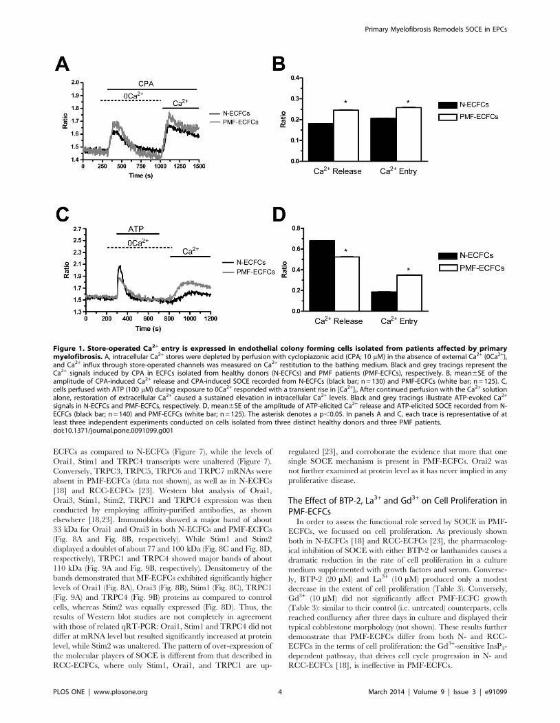
emptying in response to CPA (10 mM) was significantly (p,0.05)
higher in PMF-ECFCs as compared to their control counterparts
(Fig. 1A and Fig. 1B). Likewise, the amplitude of CPA-induced
SOCE was statistically (p,0.05) higher in ECFCs harvested from
PMF patients (Fig. 1A and Fig. 1B). The cells were then probed
with the physiological autacoid ATP (100 mM), which binds to P2Y
receptors to trigger InsP3 synthesis and subsequent InsP3-
dependent Ca2+ mobilization [18]. Unlike CPA, ATP-evoked
Ca2+ release was significantly (p,0.05) lower in PMF-ECFCs
relative to N-ECFCs (Fig. 1C and Fig. 1D), while SOCE was still
higher (Fig. 1C and Fig. 1D). The agonist was removed before
Ca2+ restitution to prevent any contamination from Ca2+ influx
through second messengers-operated channels and P2X receptors
[23]. The onset of a robust increase in [Ca2+]i in the absence of the
extracellular agonist confirms the store-dependent nature of the
Ca2+ entry pathway gated by ATP. Control experiments
conducted by removing and replenishing extracellular Ca2+
without agonist stimulation did not reveal any detectable Ca2+
signal (not shown). Overall, these findings suggest that the Ca2+
toolkit is remodelled in PMF-ECFCs as relative to control cells.
Moreover, the changes in sub-cellular Ca2+ dynamics are different
as compared to ECFCs isolated from patients suffering from solid
cancer, such as RCC, which exhibit a coherent decrease in CPA-
and ATP-induced Ca2+ release [23].
The Higher Amplitude of Store-operated Ca2+ Entry inPMF-ECFCs does not Depend on a more NegativeMembrane Potential
The higher amplitude of SOCE in PMF-ECFCs might depend
on a more negative membrane potential (VM) in these cells as
compared to their control counterparts. A more hyperpolarized
VM would enhance the electrochemical gradient driving Ca2+
entry into the cytosol, thereby resulting in a larger increase in
[Ca2+]i [17]. The VM in ECFCs is mainly set by K+ conductance
[32]. PMF-EPCs were, therefore, exposed to a solution containing
100 mM KCl (high-K+) to clamp both types of cells at the same
VM of about 0 mV, as indicated by the Nernst equation for K+,
EK = (2RT/F)*ln([K+]o/[K+]i). We have previously shown that
such a treatment does not affect SOCE in either N- and RCC-
ECFCs [23]. Store-dependent Ca2+ entry induced by either CPA
(10 mM) (Fig. 2A and Fig. 2B) or ATP (100 mM) (Fig. 2C and
Fig. 2D) was not affected by the elevation in extracellular K+
concentration. It turns out that the higher magnitude of SOCE in
PMF-ECFCs is not due to a larger driving force for Ca2+ entry.
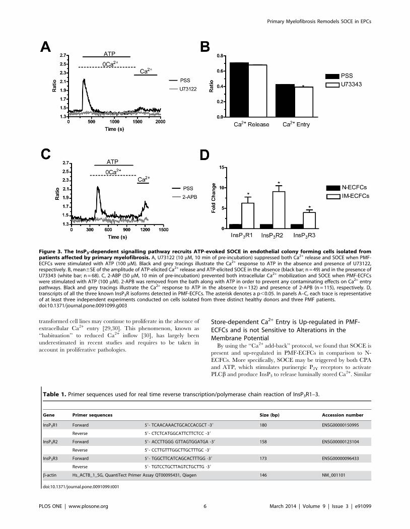
InsP3-dependent Depletion of the ER Ca2+ Store MayActivate Store-operated Ca2+ Entry in PMF-ECFCs
In order to assess the involvement of the PLCb/InsP3 signalling
pathway in the physiological activation of SOCE, PMF-ECFCs
were first pre-incubated for 10 min in the presence of U73122
(10 mM), a widely employed PLC inhibitor [18]. Figure 3A (grey
tracing) shows that neither intracellular Ca2+ release nor SOCE
were activated by ATP (100 mM) in the presence of U73122 in 98
out of 98 cells. Conversely, both phases of the Ca2+ response to
ATP occurred in 76 untreated cells (Fig. 3A, black tracing).
Control experiments were performed by pre-exposing the cells to
U73343 (10 min, 10 mM), which is an inactive structural analogue
of U73122. ATP-induced Ca2+ signals were unaffected by this
manoeuvre (Fig. 3B), thereby confirming the selective effect of
U73122 on PLCb. Furthermore, the acute application of U73122
(10 mM) did not cause any evident increase in [Ca2+]i in PMF-
ECFCs (n = 120, data not shown), which argues against the
reported inhibition of SERCA in other cell types [33]. Finally, the
pharmacological inhibition of InsP3Rs with 2-aminoethoxydiphe-
nyl borate (2-APB; 20 min, 50 mM), prevented both ATP-induced
Ca2+ release and ATP-induced SOCE (Fig. 3C). 2-APB inhibits
both Orai1 and TRPC1 when applied from the extracellular side
[23]. Therefore, it was removed from the bath along with the
agonist 100 sec before Ca2+ re-addition in order to prevent any
direct contaminant effect on plasmalemmal channels. This finding
demonstrates that InsP3-dependent emptying of the ER Ca2+
reservoir is sufficient to induce SOCE activation in PMF-ECFCs,
as well as in N-ECFCs [18] and RCC-ECFCs [23]. Consistently,
qRT-PCR analysis revealed that PMF-ECFCs possess the all
InsP3R transcripts, their pattern of expression being InsP3R2.
InsP3R1.InsP3R3 (Fig. 3D). InsP3R were all up-regulated in
PMF-ECFCs as compared to control cells. The specific primers
described in Table 1 have been utilized to assess the expression
levels of all InsP3R mRNAs. The discrepancy between the lower
amplitude of ATP-induced Ca2+ mobilization and InsP3 over-
expression in PMF-ECFCs deserves further attention and will be
the subject of future investigation.
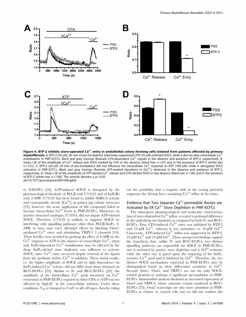
The Pharmacology of Store-operated Ca2+ Entry:Evidence for the Presence of Two Distinct Mechanisms inPMF-ECFCs
Store-dependent Ca2+ inflow in both mature endothelial cells
and more immature committed progenitors is featured by its
sensitivity to a host of rather selective inhibitors, such as BTP-2
and 1–10 mM of the trivalent cations, La3+ and Gd3+
[10,28,34,35]. We have previously found that BTP-2 (20 mM),
La3+ (10 mM), and Gd3+ (10 mM) abrogate SOCE in both N-
ECFCs [18] and RCC-ECFCs [23]. Similarly, BTP-2 (20 min,
20 mM) suppressed SOCE induced by either CPA (10 mM) or
ATP (100 mM) with no significant effect on intracellular Ca2+
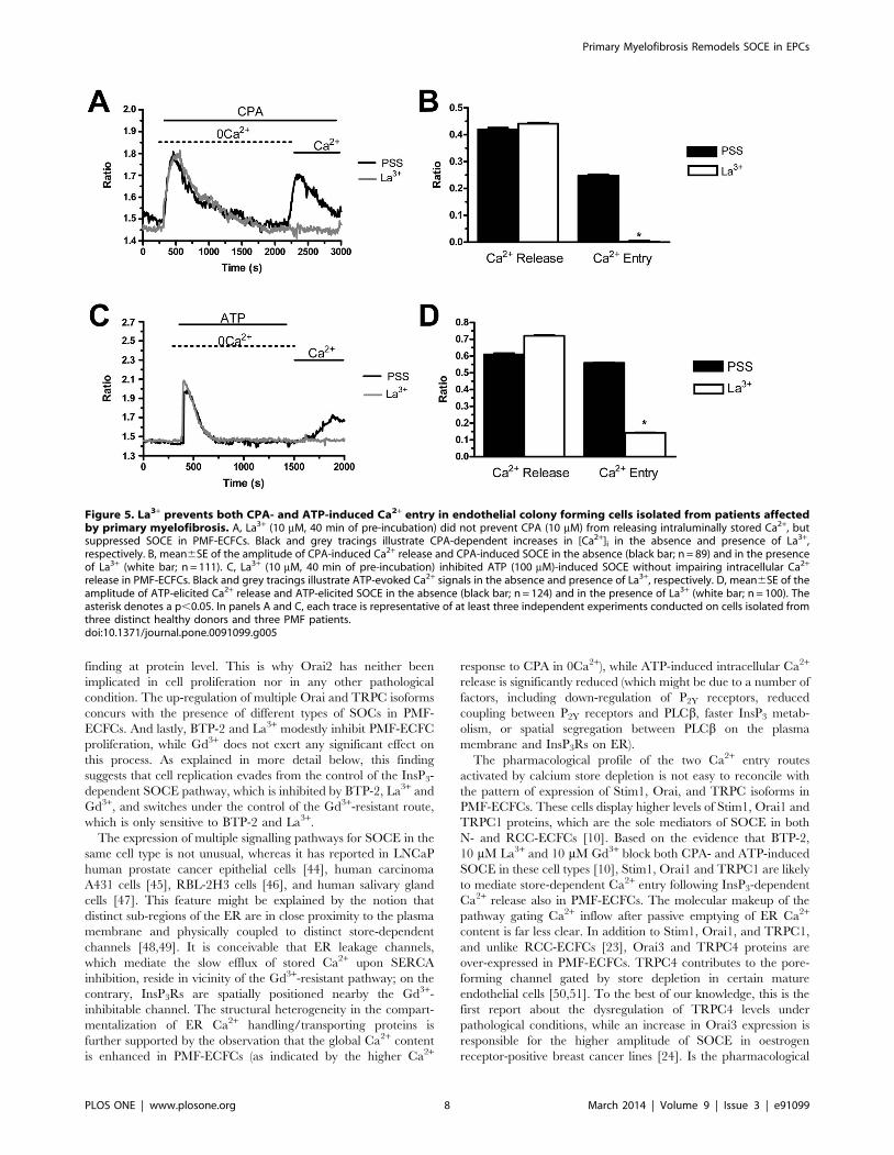
mobilization in both cases (Fig. 4A–4D). Likewise, La3+ (40 min,
10 mM) blocked both CPA- and ATP-evoked Ca2+ inflow without
impairing intracellular Ca2+ release (Fig. 5A–5D). Conversely,
Gd3+ (40 min, 10 mM) significantly (p,0.05) reduced ATP-
induced SOCE (Fig. 6C and Fig. 6D), while it did not affect
CPA-elicited Ca2+ influx (Fig. 6A and Fig. 6B). Only when its
concentration was raised to 100 mM, was Gd3+ able to inhibit
SOCE activated by CPA (n = 124, data not shown). Similar to
BTP-2 and La3+, Gd3+ did not influence the extent of ER Ca2+
pool depletion in the presence of either CPA (Fig. 6A and Fig. 6B)
or ATP (Fig. 6C and Fig. 6D). Overall, these results suggest that
CPA and ATP may utilize two distinct mechanisms to engage
SOCE in PMF-ECFCs, although they stimulate the same pathway
in both N-ECFCs [18,22] and RCC-ECFCs [23].
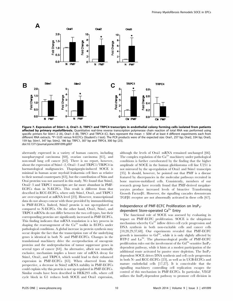
Molecular Players of Store-operated Ca2+ Entry in PMF-ECFCs
The molecular make-up of SOCE in PMF-ECFCs was
elucidated by carrying out a qRT-PCR examination of mRNA
extracts. We focussed on Stim1, Orai1, and TRPC1, which
mediate SOCE in ECFCs isolated from both healthy donors [18]
and RCC patients [23], and on their paralogues, namely Stim2,
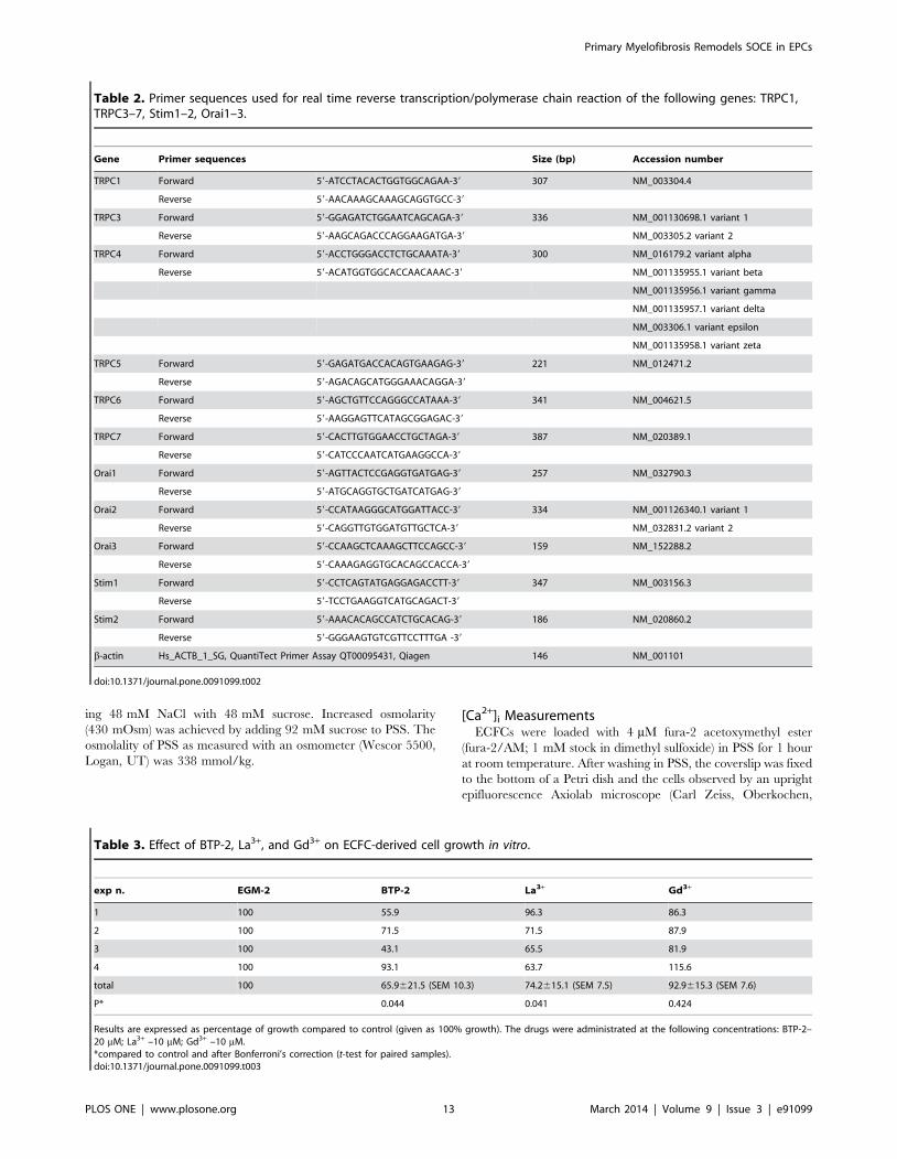
Orai2–3, and TRPC3–6. The specific primers described in Table 2
have been utilized to assess the expression levels of Stim1–2,
Orai1–3, and TRPC1–7 transcripts. Negative controls were
established by removing the reverse transcriptase from the reaction
solution (not shown). We found that the mRNAs encoding for
Stim2, Orai2–3, and TRPC1 were more abundant in PMF-
Primary Myelofibrosis Remodels SOCE in EPCs
PLOS ONE | www.plosone.org 3 March 2014 | Volume 9 | Issue 3 | e91099
ECFCs as compared to N-ECFCs (Figure 7), while the levels of
Orai1, Stim1 and TRPC4 transcripts were unaltered (Figure 7).
Conversely, TRPC3, TRPC5, TRPC6 and TRPC7 mRNAs were
absent in PMF-ECFCs (data not shown), as well as in N-ECFCs
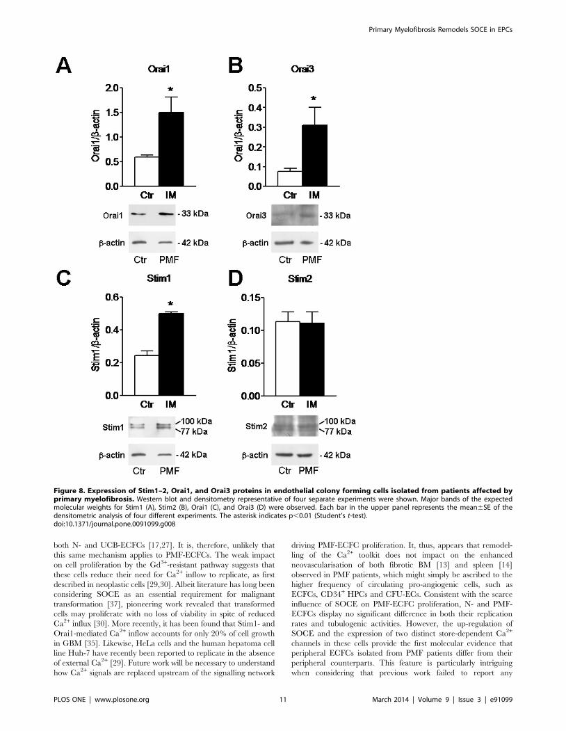
[18] and RCC-ECFCs [23]. Western blot analysis of Orai1,
Orai3, Stim1, Stim2, TRPC1 and TRPC4 expression was then
conducted by employing affinity-purified antibodies, as shown
elsewhere [18,23]. Immunoblots showed a major band of about
33 kDa for Orai1 and Orai3 in both N-ECFCs and PMF-ECFCs
(Fig. 8A and Fig. 8B, respectively). While Stim1 and Stim2
displayed a doublet of about 77 and 100 kDa (Fig. 8C and Fig. 8D,
respectively), TRPC1 and TRPC4 showed major bands of about
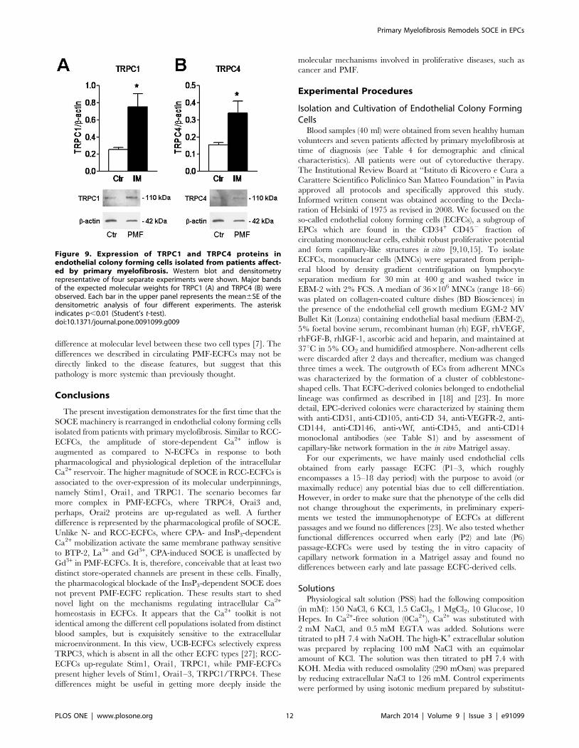
110 kDa (Fig. 9A and Fig. 9B, respectively). Densitometry of the
bands demonstrated that MF-ECFCs exhibited significantly higher
levels of Orai1 (Fig. 8A), Orai3 (Fig. 8B), Stim1 (Fig. 8C), TRPC1
(Fig. 9A) and TRPC4 (Fig. 9B) proteins as compared to control
cells, whereas Stim2 was equally expressed (Fig. 8D). Thus, the
results of Western blot studies are not completely in agreement
with those of related qRT-PCR: Orai1, Stim1 and TRPC4 did not
differ at mRNA level but resulted significantly increased at protein
level, while Stim2 was unaltered. The pattern of over-expression of
the molecular players of SOCE is different from that described in
RCC-ECFCs, where only Stim1, Orai1, and TRPC1 are up-
regulated [23], and corroborate the evidence that more that one
single SOCE mechanism is present in PMF-ECFCs. Orai2 was
not further examined at protein level as it has never implied in any
proliferative disease.
The Effect of BTP-2, La3+ and Gd3+ on Cell Proliferation inPMF-ECFCs
In order to assess the functional role served by SOCE in PMF-
ECFCs, we focussed on cell proliferation. As previously shown
both in N-ECFCs [18] and RCC-ECFCs [23], the pharmacolog-
ical inhibition of SOCE with either BTP-2 or lanthanides causes a
dramatic reduction in the rate of cell proliferation in a culture
medium supplemented with growth factors and serum. Converse-
ly, BTP-2 (20 mM) and La3+ (10 mM) produced only a modest
decrease in the extent of cell proliferation (Table 3). Conversely,
Gd3+ (10 mM) did not significantly affect PMF-ECFC growth
(Table 3): similar to their control (i.e. untreated) counterparts, cells
reached confluency after three days in culture and displayed their
typical cobblestone morphology (not shown). These results further
demonstrate that PMF-ECFCs differ from both N- and RCC-
ECFCs in the terms of cell proliferation: the Gd3+-sensitive InsP3-
dependent pathway, that drives cell cycle progression in N- and
RCC-ECFCs [18], is ineffective in PMF-ECFCs.
Figure 1. Store-operated Ca2+ entry is expressed in endothelial colony forming cells isolated from patients affected by primarymyelofibrosis. A, intracellular Ca2+ stores were depleted by perfusion with cyclopiazonic acid (CPA; 10 mM) in the absence of external Ca2+ (0Ca2+),and Ca2+ influx through store-operated channels was measured on Ca2+ restitution to the bathing medium. Black and grey tracings represent theCa2+ signals induced by CPA in ECFCs isolated from healthy donors (N-ECFCs) and PMF patients (PMF-ECFCs), respectively. B, mean6SE of theamplitude of CPA-induced Ca2+ release and CPA-induced SOCE recorded from N-ECFCs (black bar; n = 130) and PMF-ECFCs (white bar; n = 125). C,cells perfused with ATP (100 mM) during exposure to 0Ca2+ responded with a transient rise in [Ca2+]i. After continued perfusion with the Ca2+ solutionalone, restoration of extracellular Ca2+ caused a sustained elevation in intracellular Ca2+ levels. Black and grey tracings illustrate ATP-evoked Ca2+
signals in N-ECFCs and PMF-ECFCs, respectively. D, mean6SE of the amplitude of ATP-elicited Ca2+ release and ATP-elicited SOCE recorded from N-ECFCs (black bar; n = 140) and PMF-ECFCs (white bar; n = 125). The asterisk denotes a p,0.05. In panels A and C, each trace is representative of atleast three independent experiments conducted on cells isolated from three distinct healthy donors and three PMF patients.doi:10.1371/journal.pone.0091099.g001
Primary Myelofibrosis Remodels SOCE in EPCs
PLOS ONE | www.plosone.org 4 March 2014 | Volume 9 | Issue 3 | e91099
Discussion
Neoplastic transformation is accompanied by a dramatic
remodelling in the Ca2+ machinery of tumor cells [36,37], which
is unlikely to drive malignant initiation, but is instrumental to
confer some of the cancer-specific hallmarks [28,36,38,39]. The
deregulation of the Ca2+ toolkit is not limited to solid tumors,
whereas it has also been observed in haematological malignancies,
such as chronic myeloid leukaemia [40], childhood acute
lymphoblastic leukemia [41], acute promyelocytic leukaemia
[42], and mantle cell lymphoma [43]. PMF is a myeloproliferative
neoplasm featured by an increased neovascularisation of both BM
and spleen as a consequence of the high frequency of circulating
ECFCs [3]. The Ca2+ signalling toolkit expressed by ECFCs is
extremely plastic and may vary depending on both the blood
source (i.e. peripheral vs. cord blood) and donor origin (e.g. healthy
vs. tumor-affected subject) [10,28]. The current investigation lends
further support to the notion that the Ca2+ machinery endowed to
ECFCs is not sculpted in the stone and that each ECFC may
create its own Ca2+ fingerprint, which could be exploited for both
prognostic and therapeutic purposes.
Rationale behind the Examination of Store-operated Ca2+
Entry in PMF-ECFCsAlbeit pro-angiogenic Ca2+ influx may be conducted by DAG-
gated non-selective cation channels, such as TRPC3 and TRPC6
[17], SOCE is by far the most important driver of proliferation in
both mature endothelial cells [17] and more immature endothelial
progenitors [18]. We have previously disclosed that SOCE,
activated upon either passive (e.g. CPA-induced) or InsP3-
dependent depletion of ER Ca2+ reservoir, is up-regulated in
RCC-ECFCs as compared to N-ECFCs due to the over-
expression of Stim1, Orai1 and TRPC1 [23]. The pharmacolog-
ical characterization of SOCE is consistent with the recruitment of
the same signalling pathway by CPA and InsP3: store-dependent
Ca2+ inflow is inhibited by BTP-2, 10 mM La3+ and 10 mM Gd3+
in both N- and RCC-ECFCs [10,23]. Notably, the inhibition of
SOCE with either BTP-2 or lanthanides suppresses cell prolifer-
ation in both N-ECFCs and RCC-ECFCs [18,23], as well as in
UCB-ECFCs [27]. The following observations led us to extend our
analysis on the molecular structure and role of SOCE in PMF-
ECFCs: 1) distinct components of the Ca2+ machinery are altered
in different types of cancer [37], which prompted us to assess
whether and how was SOCE remodelled in ECFCs in the
presence of a myeloproliferative disease; and 2) neoplastic and
Figure 2. The amplitude of store-operated Ca2+ entry is not reduced by a high-K+ extracellular solution in endothelial colonyforming cells isolated from patients affected by primary myelofibrosis. 100 mM NaCl in the extracellular solution was replaced with anequimolar amount of K+ (HighK) to clamp the membrane potential at 0 mV and observe the consequences on the extent of SOCE activation in PMF-ECFCs. A, HighK did not affect either the amplitude or the kinetics of CPA (10 mM)-induced Ca2+ signals in PMF-ECFCs. Black and grey tracingsillustrate CPA-dependent Ca2+ signals in the absence and presence of HighK, respectively. B, mean6SE of the amplitude of CPA-induced Ca2+ releaseand CPA-induced SOCE in the absence (black bar; n = 76) and in the presence of HighK (white bar; n = 77). C, the biphasic Ca2+ response to ATP(100 mM) was not impaired by HighK. Black and grey tracings illustrate ATP-dependent Ca2+ signals in the absence and presence of HighK,respectively. D, mean6SE of the amplitude of ATP-elicited Ca2+ release and ATP-elicited SOCE in the absence (black bar; n = 88) and in the presence ofHighK (white bar; n = 95). In panels A and C, each trace is representative of at least three independent experiments conducted on cells isolated fromthree distinct healthy donors and three PMF patients.doi:10.1371/journal.pone.0091099.g002
Primary Myelofibrosis Remodels SOCE in EPCs
PLOS ONE | www.plosone.org 5 March 2014 | Volume 9 | Issue 3 | e91099
transformed cell lines may continue to proliferate in the absence of
extracellular Ca2+ entry [29,30]. This phenomenon, known as
‘‘habituation’’ to reduced Ca2+ inflow [30], has largely been
underestimated in recent studies and requires to be taken in
account in proliferative pathologies.
Store-dependent Ca2+ Entry is Up-regulated in PMF-ECFCs and is not Sensitive to Alterations in theMembrane Potential
By using the ‘‘Ca2+ add-back’’ protocol, we found that SOCE is
present and up-regulated in PMF-ECFCs in comparison to N-
ECFCs. More specifically, SOCE may be triggered by both CPA
and ATP, which stimulates purinergic P2Y receptors to activate
PLCb and produce InsP3 to release luminally stored Ca2+. Similar
Figure 3. The InsP3-dependent signalling pathway recruits ATP-evoked SOCE in endothelial colony forming cells isolated frompatients affected by primary myelofibrosis. A, U73122 (10 mM, 10 min of pre-incubation) suppressed both Ca2+ release and SOCE when PMF-ECFCs were stimulated with ATP (100 mM). Black and grey tracings illustrate the Ca2+ response to ATP in the absence and presence of U73122,respectively. B, mean6SE of the amplitude of ATP-elicited Ca2+ release and ATP-elicited SOCE in the absence (black bar; n = 49) and in the presence ofU73343 (white bar; n = 68). C, 2-ABP (50 mM, 10 min of pre-incubation) prevented both intracellular Ca2+ mobilization and SOCE when PMF-ECFCswere stimulated with ATP (100 mM). 2-APB was removed from the bath along with ATP in order to prevent any contaminating effects on Ca2+ entrypathways. Black and grey tracings illustrate the Ca2+ response to ATP in the absence (n = 132) and presence of 2-APB (n = 115), respectively. D,transcripts of all the three known InsP3R isoforms detected in PMF-ECFCs. The asterisk denotes a p,0.05. In panels A–C, each trace is representativeof at least three independent experiments conducted on cells isolated from three distinct healthy donors and three PMF patients.doi:10.1371/journal.pone.0091099.g003
Table 1. Primer sequences used for real time reverse transcription/polymerase chain reaction of InsP3R1–3.
Gene Primer sequences Size (bp) Accession number
InsP3R1 Forward 59- TCAACAAACTGCACCACGCT -39 180 ENSG00000150995
Reverse 59- CTCTCATGGCATTCTTCTCC -39
InsP3R2 Forward 59- ACCTTGGG GTTAGTGGATGA -39 158 ENSG00000123104
Reverse 59- CCTTGTTTGGCTTGCTTTGC -39
InsP3R3 Forward 59- TGGCTTCATCAGCACTTTGG -39 173 ENSG00000096433
Reverse 59- TGTCCTGCTTAGTCTGCTTG -39
b-actin Hs_ACTB_1_SG, QuantiTect Primer Assay QT00095431, Qiagen 146 NM_001101
doi:10.1371/journal.pone.0091099.t001
Primary Myelofibrosis Remodels SOCE in EPCs
PLOS ONE | www.plosone.org 6 March 2014 | Volume 9 | Issue 3 | e91099
to N-ECFCs [18], ATP-induced SOCE is abrogated by the
pharmacological blockade of PLCb with U73122 and of InsP3Rs
with 2-APB. U73122 has been found to inhibit SERCA activity
and consequently elevate [Ca2+]i in guinea pig colonic myocytes
[33]; however, the acute application of this compound failed to
increase intracellular Ca2+ levels in PMF-ECFCs. Moreover, its
inactive structural analogue, U73343, did not impair ATP-elicited
SOCE. Therefore, U73122 is unlikely to suppress SOCE by
interfering with signalling pathways other than PLCb/InsP3. 2-
APB, in turn, may exert off-target effects by blocking Orai1-
mediated Ca2+ entry and stimulating TRPV1–3 channels [10].
These hurdles were avoided by probing the effect of 2-APB on the
Ca2+ response to ATP in the absence of extracellular Ca2+, when
only InsP3-dependent Ca2+ mobilization may be affected by the
drug. InsP3-elicited store depletion was sufficient to activate
SOCE, since Ca2+ entry occurred despite removal of the ligand
from the perfusate before Ca2+ re-addition. These initial results,
i.e. the higher amplitude of SOCE and its tight dependence on
InsP3-induced Ca2+ release, did not differ from those obtained on
RCC-ECFCs [23]. Similar to N- and RCC-ECFCs [23], the
amplitude of the intracellular Ca2+ peak measured on Ca2+
restoration to PMF-ECFCs exposed to either CPA or ATP was not
affected by high-K+ in the extracellular solution. Under these
conditions, VM is clamped to 0 mV in all cell types, thereby ruling
out the possibility that a negative shift in the resting potential
augments the driving force sustaining Ca2+ influx in the latter.
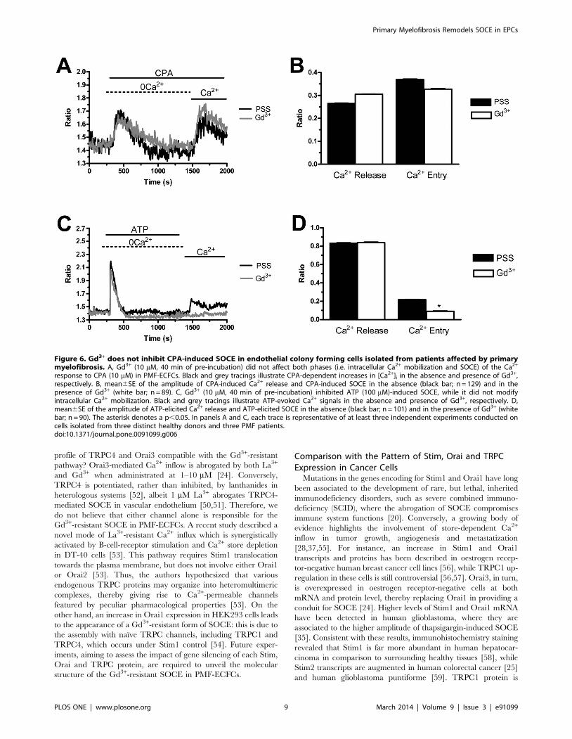
Evidence that Two Separate Ca2+-permeable Routes areActivated by ER Ca2+ Store Depletion in PMF-ECFCs
The subsequent pharmacological and molecular characteriza-
tion of store-dependent Ca2+ inflow revealed a profound difference
in the underlying mechanism(s) as compared to both N- and RCC-
ECFCs. First, CPA-induced Ca2+ entry was inhibited by BTP-2
and 10 mM La3+, whereas it was insensitive to 10 mM Gd3+.
Conversely, ATP-induced Ca2+ influx was suppressed by BTP-2,
10 mM La3+ and 10 mM Gd3+. These unexpected findings support
the hypothesis that, unlike N- and RCC-ECFCs, two distinct
signalling pathways are responsible for SOCE in PMF-ECFCs;
one is activated by passive store depletion and is Gd3+-resistant,
while the other one is gated upon the emptying of the InsP3-
sensitive Ca2+ pool and is inhibited by Gd3+. Therefore, the two
distinct SOCE mechanisms expressed in PMF-ECFCs may be
distinguished based on their differential sensitivity to Gd3+.
Second, Stim1, Orai1, and TRPC1 are not the only SOCE-
related proteins to undergo a significant up-regulation in PMF-
ECFCs. Immunoblot analysis disclosed an increased expression of
Orai3 and TRPC4, whose amounts remain unaltered in RCC-
ECFCs [23]. Orai2 transcripts are also more abundant in PMF-
ECFCs as relative to control cells, but we did not confirm this
Figure 4. BTP-2 inhibits store-operated Ca2+ entry in endothelial colony forming cells isolated from patients affected by primarymyelofibrosis. A, BTP-2 (20 mM, 20 min of pre-incubation) selectively suppressed CPA (10 mM)-solicited SOCE, while it did not alter intracellular Ca2+
mobilization in PMF-ECFCs. Black and grey tracings illustrate CPA-dependent Ca2+ signals in the absence and presence of BTP-2, respectively. B,mean6SE of the amplitude of Ca2+ release and SOCE evoked by CPA in the absence (black bar; n = 67) and in the presence of BTP-2 (white bar;n = 131). C, BTP-2 (20 mM, 20 min of pre-incubation) did not influence the intracellular Ca2+ response to ATP (100 mM), while it abrogated SOCEactivation in PMF-ECFCs. Black and grey tracings illustrate ATP-evoked elevations in [Ca2+]i observed in the absence and presence of BTP-2,respectively. D, mean6SE of the amplitude of ATP-elicited Ca2+ release and CPA-elicited SOCE in the absence (black bar; n = 49) and in the presenceof BTP-2 (white bar; n = 100). The asterisk denotes a p,0.05.doi:10.1371/journal.pone.0091099.g004
Primary Myelofibrosis Remodels SOCE in EPCs
PLOS ONE | www.plosone.org 7 March 2014 | Volume 9 | Issue 3 | e91099
finding at protein level. This is why Orai2 has neither been
implicated in cell proliferation nor in any other pathological
condition. The up-regulation of multiple Orai and TRPC isoforms
concurs with the presence of different types of SOCs in PMF-
ECFCs. And lastly, BTP-2 and La3+ modestly inhibit PMF-ECFC
proliferation, while Gd3+ does not exert any significant effect on
this process. As explained in more detail below, this finding
suggests that cell replication evades from the control of the InsP3-
dependent SOCE pathway, which is inhibited by BTP-2, La3+ and
Gd3+, and switches under the control of the Gd3+-resistant route,
which is only sensitive to BTP-2 and La3+.
The expression of multiple signalling pathways for SOCE in the
same cell type is not unusual, whereas it has reported in LNCaP
human prostate cancer epithelial cells [44], human carcinoma
A431 cells [45], RBL-2H3 cells [46], and human salivary gland
cells [47]. This feature might be explained by the notion that
distinct sub-regions of the ER are in close proximity to the plasma
membrane and physically coupled to distinct store-dependent
channels [48,49]. It is conceivable that ER leakage channels,
which mediate the slow efflux of stored Ca2+ upon SERCA
inhibition, reside in vicinity of the Gd3+-resistant pathway; on the
contrary, InsP3Rs are spatially positioned nearby the Gd3+-
inhibitable channel. The structural heterogeneity in the compart-
mentalization of ER Ca2+ handling/transporting proteins is
further supported by the observation that the global Ca2+ content
is enhanced in PMF-ECFCs (as indicated by the higher Ca2+
response to CPA in 0Ca2+), while ATP-induced intracellular Ca2+
release is significantly reduced (which might be due to a number of
factors, including down-regulation of P2Y receptors, reduced
coupling between P2Y receptors and PLCb, faster InsP3 metab-
olism, or spatial segregation between PLCb on the plasma
membrane and InsP3Rs on ER).
The pharmacological profile of the two Ca2+ entry routes
activated by calcium store depletion is not easy to reconcile with
the pattern of expression of Stim1, Orai, and TRPC isoforms in
PMF-ECFCs. These cells display higher levels of Stim1, Orai1 and
TRPC1 proteins, which are the sole mediators of SOCE in both
N- and RCC-ECFCs [10]. Based on the evidence that BTP-2,
10 mM La3+ and 10 mM Gd3+ block both CPA- and ATP-induced
SOCE in these cell types [10], Stim1, Orai1 and TRPC1 are likely
to mediate store-dependent Ca2+ entry following InsP3-dependent
Ca2+ release also in PMF-ECFCs. The molecular makeup of the
pathway gating Ca2+ inflow after passive emptying of ER Ca2+
content is far less clear. In addition to Stim1, Orai1, and TRPC1,
and unlike RCC-ECFCs [23], Orai3 and TRPC4 proteins are
over-expressed in PMF-ECFCs. TRPC4 contributes to the pore-
forming channel gated by store depletion in certain mature
endothelial cells [50,51]. To the best of our knowledge, this is the
first report about the dysregulation of TRPC4 levels under
pathological conditions, while an increase in Orai3 expression is
responsible for the higher amplitude of SOCE in oestrogen
receptor-positive breast cancer lines [24]. Is the pharmacological
Figure 5. La3+ prevents both CPA- and ATP-induced Ca2+ entry in endothelial colony forming cells isolated from patients affectedby primary myelofibrosis. A, La3+ (10 mM, 40 min of pre-incubation) did not prevent CPA (10 mM) from releasing intraluminally stored Ca2+, butsuppressed SOCE in PMF-ECFCs. Black and grey tracings illustrate CPA-dependent increases in [Ca2+]i in the absence and presence of La3+,respectively. B, mean6SE of the amplitude of CPA-induced Ca2+ release and CPA-induced SOCE in the absence (black bar; n = 89) and in the presenceof La3+ (white bar; n = 111). C, La3+ (10 mM, 40 min of pre-incubation) inhibited ATP (100 mM)-induced SOCE without impairing intracellular Ca2+
release in PMF-ECFCs. Black and grey tracings illustrate ATP-evoked Ca2+ signals in the absence and presence of La3+, respectively. D, mean6SE of theamplitude of ATP-elicited Ca2+ release and ATP-elicited SOCE in the absence (black bar; n = 124) and in the presence of La3+ (white bar; n = 100). Theasterisk denotes a p,0.05. In panels A and C, each trace is representative of at least three independent experiments conducted on cells isolated fromthree distinct healthy donors and three PMF patients.doi:10.1371/journal.pone.0091099.g005
Primary Myelofibrosis Remodels SOCE in EPCs
PLOS ONE | www.plosone.org 8 March 2014 | Volume 9 | Issue 3 | e91099
profile of TRPC4 and Orai3 compatible with the Gd3+-resistant
pathway? Orai3-mediated Ca2+ inflow is abrogated by both La3+
and Gd3+ when administrated at 1–10 mM [24]. Conversely,
TRPC4 is potentiated, rather than inhibited, by lanthanides in
heterologous systems [52], albeit 1 mM La3+ abrogates TRPC4-
mediated SOCE in vascular endothelium [50,51]. Therefore, we
do not believe that either channel alone is responsible for the
Gd3+-resistant SOCE in PMF-ECFCs. A recent study described a
novel mode of La3+-resistant Ca2+ influx which is synergistically
activated by B-cell-receptor stimulation and Ca2+ store depletion
in DT-40 cells [53]. This pathway requires Stim1 translocation
towards the plasma membrane, but does not involve either Orai1
or Orai2 [53]. Thus, the authors hypothesized that various
endogenous TRPC proteins may organize into heteromultimeric
complexes, thereby giving rise to Ca2+-permeable channels
featured by peculiar pharmacological properties [53]. On the
other hand, an increase in Orai1 expression in HEK293 cells leads
to the appearance of a Gd3+-resistant form of SOCE: this is due to
the assembly with naıve TRPC channels, including TRPC1 and
TRPC4, which occurs under Stim1 control [54]. Future exper-
iments, aiming to assess the impact of gene silencing of each Stim,
Orai and TRPC protein, are required to unveil the molecular
structure of the Gd3+-resistant SOCE in PMF-ECFCs.
Comparison with the Pattern of Stim, Orai and TRPCExpression in Cancer Cells
Mutations in the genes encoding for Stim1 and Orai1 have long
been associated to the development of rare, but lethal, inherited
immunodeficiency disorders, such as severe combined immuno-
deficiency (SCID), where the abrogation of SOCE compromises
immune system functions [20]. Conversely, a growing body of
evidence highlights the involvement of store-dependent Ca2+
inflow in tumor growth, angiogenesis and metastatization
[28,37,55]. For instance, an increase in Stim1 and Orai1
transcripts and proteins has been described in oestrogen recep-
tor-negative human breast cancer cell lines [56], while TRPC1 up-
regulation in these cells is still controversial [56,57]. Orai3, in turn,
is overexpressed in oestrogen receptor-negative cells at both
mRNA and protein level, thereby replacing Orai1 in providing a
conduit for SOCE [24]. Higher levels of Stim1 and Orai1 mRNA
have been detected in human glioblastoma, where they are
associated to the higher amplitude of thapsigargin-induced SOCE
[35]. Consistent with these results, immunohistochemistry staining
revealed that Stim1 is far more abundant in human hepatocar-
cinoma in comparison to surrounding healthy tissues [58], while
Stim2 transcripts are augmented in human colorectal cancer [25]
and human glioblastoma puntiforme [59]. TRPC1 protein is
Figure 6. Gd3+ does not inhibit CPA-induced SOCE in endothelial colony forming cells isolated from patients affected by primarymyelofibrosis. A, Gd3+ (10 mM, 40 min of pre-incubation) did not affect both phases (i.e. intracellular Ca2+ mobilization and SOCE) of the Ca2+
response to CPA (10 mM) in PMF-ECFCs. Black and grey tracings illustrate CPA-dependent increases in [Ca2+]i in the absence and presence of Gd3+,respectively. B, mean6SE of the amplitude of CPA-induced Ca2+ release and CPA-induced SOCE in the absence (black bar; n = 129) and in thepresence of Gd3+ (white bar; n = 89). C, Gd3+ (10 mM, 40 min of pre-incubation) inhibited ATP (100 mM)-induced SOCE, while it did not modifyintracellular Ca2+ mobilization. Black and grey tracings illustrate ATP-evoked Ca2+ signals in the absence and presence of Gd3+, respectively. D,mean6SE of the amplitude of ATP-elicited Ca2+ release and ATP-elicited SOCE in the absence (black bar; n = 101) and in the presence of Gd3+ (whitebar; n = 90). The asterisk denotes a p,0.05. In panels A and C, each trace is representative of at least three independent experiments conducted oncells isolated from three distinct healthy donors and three PMF patients.doi:10.1371/journal.pone.0091099.g006
Primary Myelofibrosis Remodels SOCE in EPCs
PLOS ONE | www.plosone.org 9 March 2014 | Volume 9 | Issue 3 | e91099
aberrantly expressed in a variety of human cancers, including
nasopharyngeal carcinoma [60], ovarian carcinoma [61], and
non-small lung cell cancer [62]. There is no report, however,
about the expression of Stim1–2, Orai1–3 and TRPC1/TRPC4 in
haematological malignancies. Thapsigargin-induced SOCE is
minimal in human acute myeloid leukaemia cell lines as relative
to their normal counterparts [63], but the contribution of Stim and
Orai proteins was not assessed in this study. We found that Stim2,
Orai2–3 and TRPC1 transcripts are far more abundant in PMF-
ECFCs than in N-ECFCs. This result is different from that
described in RCC-ECFCs, where only Stim1, Orai1, and TRPC1
are over-expressed at mRNA level [23]. However, transcriptional
data do not always concur with those provided by immunoblotting
in PMF-ECFCs. Indeed, Stim2 protein is not up-regulated as
compared to N-ECFCs. On the other hand, Orai1, Stim1, and
TRPC4 mRNAs do not differ between the two cell types, but their
corresponding proteins are significantly increased in PMF-ECFCs.
This finding indicates that mRNA translation is a key process in
shaping the rearrangement of the Ca2+ toolkit in ECFCs under
pathological conditions. A global increase in protein synthesis may
occur despite the fact that the transcription rate of the underlying
genes is identical in both N- and PMF-ECFCs. Changes in the
translational machinery drive the overproduction of oncogenic
proteins and the underproduction of tumor suppressor genes in
several types of cancer [64]. An alternative, albeit not mutually
exclusive, mechanism implies a lower rate of mRNA decay for
Stim1, Orai1, and TRPC4, which would lead to their enhanced
expression in PMF-ECFCs [65]. When observed from this
perspective, a decrease in Stim2 mRNA stability in PMF-ECFCs
could explain why this protein is not up-regulated in PMF-ECFCs.
Similar results have been described in HEK293 cells, where cell
cycle block in G1 reduces both SOCE and Orai1 expression,
although the levels of Orai1 mRNA remained unchanged [66].
The complex regulation of the Ca2+ machinery under pathological
conditions is further corroborated by the finding that the higher
amplitude of SOCE in the human glioblastoma cell line U251 is
not mirrored by the up-regulation of Orai1 and Stim1 transcripts
[35]. It should, however, be pointed out that PMF is a disease
featured by discrepancies in the molecular pathways recruited in
bone marrow-mobilized cells. Consistently, members of our
research group have recently found that PMF-derived megakar-
yocytes produce increased levels of bioactive Transforming
Growth Factorb1. However, the signalling cascades downstream
TGFb1 receptor are not abnormally activated in these cells [67].
Independence of PMF-ECFC Proliferation on InsP3-dependent Store-operated Ca2+ Entry
The functional role of SOCE was assessed by evaluating its
impact on PMF-ECFC proliferation. SOCE is the ubiquitous
mechanism whereby Ca2+ inflow drives cell cycle progression and
DNA synthesis in both non-excitable cells and cancer cells
[10,28,29,37,68]. Our experiments revealed that PMF-ECFC
growth is insensitive to Gd3+, while it is only slightly affected by
BTP-2 and La3+. The pharmacological profile of PMF-ECFC
proliferation rules out the involvement of the Gd3+-sensitive InsP3-
dependent pathway, while it hints at a modest participation of the
additional route activated by passive store depletion. The InsP3-
dependent SOCE drives DNA synthesis and cell cycle progression
in both N- and RCC-ECFCs [23], as well as in UCB-ECFCs and
mature endothelial cells [17,27]. It is conceivable that the
signalling machinery controlling cell replication escapes the
control of this mechanism in PMF-ECFCs. In particular, VEGF
utilizes the InsP3-dependent pathway to promote cell division in
Figure 7. Expression of Stim1–2, Orai1–3, TRPC1 and TRPC4 transcripts in endothelial colony forming cells isolated from patientsaffected by primary myelofibrosis. Quantitative real-time reverse transcription polymerase chain reaction of total RNA was performed usingspecific primers for Stim1–2 (A), Orai1–3 (B), TRPC1 and TRPC4 (C). Bars represent the mean 6 SEM of at least 4 different experiments each fromdifferent RNA extracts. *P,0.05 versus N-ECFCs (Student’s t test). The PCR products were of the expected size: Orai1, 257 bp; Orai2, 334 bp; Orai3,159 bp; Stim1, 347 bp; Stim2, 186 bp; TRPC1, 307 bp and TRPC4, 300 bp [23].doi:10.1371/journal.pone.0091099.g007
Primary Myelofibrosis Remodels SOCE in EPCs
PLOS ONE | www.plosone.org 10 March 2014 | Volume 9 | Issue 3 | e91099
both N- and UCB-ECFCs [17,27]. It is, therefore, unlikely that
this same mechanism applies to PMF-ECFCs. The weak impact
on cell proliferation by the Gd3+-resistant pathway suggests that
these cells reduce their need for Ca2+ inflow to replicate, as first
described in neoplastic cells [29,30]. Albeit literature has long been
considering SOCE as an essential requirement for malignant
transformation [37], pioneering work revealed that transformed
cells may proliferate with no loss of viability in spite of reduced
Ca2+ influx [30]. More recently, it has been found that Stim1- and
Orai1-mediated Ca2+ inflow accounts for only 20% of cell growth
in GBM [35]. Likewise, HeLa cells and the human hepatoma cell
line Huh-7 have recently been reported to replicate in the absence
of external Ca2+ [29]. Future work will be necessary to understand
how Ca2+ signals are replaced upstream of the signalling network
driving PMF-ECFC proliferation. It, thus, appears that remodel-
ling of the Ca2+ toolkit does not impact on the enhanced
neovascularisation of both fibrotic BM [13] and spleen [14]
observed in PMF patients, which might simply be ascribed to the
higher frequency of circulating pro-angiogenic cells, such as
ECFCs, CD34+ HPCs and CFU-ECs. Consistent with the scarce
influence of SOCE on PMF-ECFC proliferation, N- and PMF-
ECFCs display no significant difference in both their replication
rates and tubulogenic activities. However, the up-regulation of
SOCE and the expression of two distinct store-dependent Ca2+
channels in these cells provide the first molecular evidence that
peripheral ECFCs isolated from PMF patients differ from their
peripheral counterparts. This feature is particularly intriguing
when considering that previous work failed to report any
Figure 8. Expression of Stim1–2, Orai1, and Orai3 proteins in endothelial colony forming cells isolated from patients affected byprimary myelofibrosis. Western blot and densitometry representative of four separate experiments were shown. Major bands of the expectedmolecular weights for Stim1 (A), Stim2 (B), Orai1 (C), and Orai3 (D) were observed. Each bar in the upper panel represents the mean6SE of thedensitometric analysis of four different experiments. The asterisk indicates p,0.01 (Student’s t-test).doi:10.1371/journal.pone.0091099.g008
Primary Myelofibrosis Remodels SOCE in EPCs
PLOS ONE | www.plosone.org 11 March 2014 | Volume 9 | Issue 3 | e91099
difference at molecular level between these two cell types [7]. The
differences we described in circulating PMF-ECFCs may not be
directly linked to the disease features, but suggest that this
pathology is more systemic than previously thought.
Conclusions
The present investigation demonstrates for the first time that the
SOCE machinery is rearranged in endothelial colony forming cells
isolated from patients with primary myelofibrosis. Similar to RCC-
ECFCs, the amplitude of store-dependent Ca2+ inflow is
augmented as compared to N-ECFCs in response to both
pharmacological and physiological depletion of the intracellular
Ca2+ reservoir. The higher magnitude of SOCE in RCC-ECFCs is
associated to the over-expression of its molecular underpinnings,
namely Stim1, Orai1, and TRPC1. The scenario becomes far
more complex in PMF-ECFCs, where TRPC4, Orai3 and,
perhaps, Orai2 proteins are up-regulated as well. A further
difference is represented by the pharmacological profile of SOCE.
Unlike N- and RCC-ECFCs, where CPA- and InsP3-dependent
Ca2+ mobilization activate the same membrane pathway sensitive
to BTP-2, La3+ and Gd3+, CPA-induced SOCE is unaffected by
Gd3+ in PMF-ECFCs. It is, therefore, conceivable that at least two
distinct store-operated channels are present in these cells. Finally,
the pharmacological blockade of the InsP3-dependent SOCE does
not prevent PMF-ECFC replication. These results start to shed
novel light on the mechanisms regulating intracellular Ca2+
homeostasis in ECFCs. It appears that the Ca2+ toolkit is not
identical among the different cell populations isolated from distinct
blood samples, but is exquisitely sensitive to the extracellular
microenvironment. In this view, UCB-ECFCs selectively express
TRPC3, which is absent in all the other ECFC types [27]; RCC-
ECFCs up-regulate Stim1, Orai1, TRPC1, while PMF-ECFCs
present higher levels of Stim1, Orai1–3, TRPC1/TRPC4. These
differences might be useful in getting more deeply inside the
molecular mechanisms involved in proliferative diseases, such as
cancer and PMF.
Experimental Procedures
Isolation and Cultivation of Endothelial Colony FormingCells
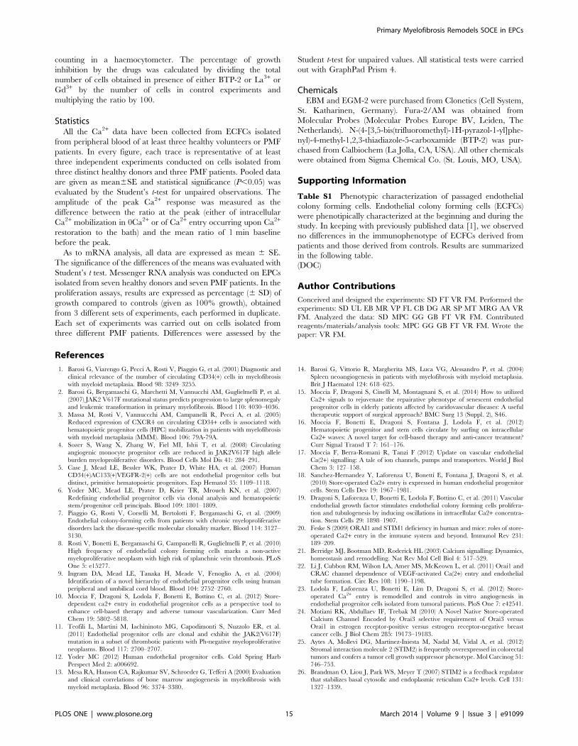
Blood samples (40 ml) were obtained from seven healthy human
volunteers and seven patients affected by primary myelofibrosis at
time of diagnosis (see Table 4 for demographic and clinical
characteristics). All patients were out of cytoreductive therapy.
The Institutional Review Board at ‘‘Istituto di Ricovero e Cura a
Carattere Scientifico Policlinico San Matteo Foundation’’ in Pavia
approved all protocols and specifically approved this study.
Informed written consent was obtained according to the Decla-
ration of Helsinki of 1975 as revised in 2008. We focussed on the
so-called endothelial colony forming cells (ECFCs), a subgroup of
EPCs which are found in the CD34+ CD452 fraction of
circulating mononuclear cells, exhibit robust proliferative potential
and form capillary-like structures in vitro [9,10,15]. To isolate
ECFCs, mononuclear cells (MNCs) were separated from periph-
eral blood by density gradient centrifugation on lymphocyte
separation medium for 30 min at 400 g and washed twice in
EBM-2 with 2% FCS. A median of 366106 MNCs (range 18–66)
was plated on collagen-coated culture dishes (BD Biosciences) in
the presence of the endothelial cell growth medium EGM-2 MV
Bullet Kit (Lonza) containing endothelial basal medium (EBM-2),
5% foetal bovine serum, recombinant human (rh) EGF, rhVEGF,
rhFGF-B, rhIGF-1, ascorbic acid and heparin, and maintained at
37uC in 5% CO2 and humidified atmosphere. Non-adherent cells
were discarded after 2 days and thereafter, medium was changed
three times a week. The outgrowth of ECs from adherent MNCs
was characterized by the formation of a cluster of cobblestone-
shaped cells. That ECFC-derived colonies belonged to endothelial
lineage was confirmed as described in [18] and [23]. In more
detail, EPC-derived colonies were characterized by staining them
with anti-CD31, anti-CD105, anti-CD 34, anti-VEGFR-2, anti-
CD144, anti-CD146, anti-vWf, anti-CD45, and anti-CD14
monoclonal antibodies (see Table S1) and by assessment of
capillary-like network formation in the in vitro Matrigel assay.
For our experiments, we have mainly used endothelial cells
obtained from early passage ECFC (P1–3, which roughly
encompasses a 15–18 day period) with the purpose to avoid (or
maximally reduce) any potential bias due to cell differentiation.
However, in order to make sure that the phenotype of the cells did
not change throughout the experiments, in preliminary experi-
ments we tested the immunophenotype of ECFCs at different
passages and we found no differences [23]. We also tested whether
functional differences occurred when early (P2) and late (P6)
passage-ECFCs were used by testing the in vitro capacity of
capillary network formation in a Matrigel assay and found no
differences between early and late passage ECFC-derived cells.
SolutionsPhysiological salt solution (PSS) had the following composition
(in mM): 150 NaCl, 6 KCl, 1.5 CaCl2, 1 MgCl2, 10 Glucose, 10
Hepes. In Ca2+-free solution (0Ca2+), Ca2+ was substituted with
2 mM NaCl, and 0.5 mM EGTA was added. Solutions were
titrated to pH 7.4 with NaOH. The high-K+ extracellular solution
was prepared by replacing 100 mM NaCl with an equimolar
amount of KCl. The solution was then titrated to pH 7.4 with
KOH. Media with reduced osmolality (290 mOsm) was prepared
by reducing extracellular NaCl to 126 mM. Control experiments
were performed by using isotonic medium prepared by substitut-
Figure 9. Expression of TRPC1 and TRPC4 proteins inendothelial colony forming cells isolated from patients affect-ed by primary myelofibrosis. Western blot and densitometryrepresentative of four separate experiments were shown. Major bandsof the expected molecular weights for TRPC1 (A) and TRPC4 (B) wereobserved. Each bar in the upper panel represents the mean6SE of thedensitometric analysis of four different experiments. The asteriskindicates p,0.01 (Student’s t-test).doi:10.1371/journal.pone.0091099.g009
Primary Myelofibrosis Remodels SOCE in EPCs
PLOS ONE | www.plosone.org 12 March 2014 | Volume 9 | Issue 3 | e91099
ing 48 mM NaCl with 48 mM sucrose. Increased osmolarity
(430 mOsm) was achieved by adding 92 mM sucrose to PSS. The
osmolality of PSS as measured with an osmometer (Wescor 5500,
Logan, UT) was 338 mmol/kg.
[Ca2+]i MeasurementsECFCs were loaded with 4 mM fura-2 acetoxymethyl ester
(fura-2/AM; 1 mM stock in dimethyl sulfoxide) in PSS for 1 hour
at room temperature. After washing in PSS, the coverslip was fixed
to the bottom of a Petri dish and the cells observed by an upright
epifluorescence Axiolab microscope (Carl Zeiss, Oberkochen,
Table 2. Primer sequences used for real time reverse transcription/polymerase chain reaction of the following genes: TRPC1,TRPC3–7, Stim1–2, Orai1–3.
Gene Primer sequences Size (bp) Accession number
TRPC1 Forward 59-ATCCTACACTGGTGGCAGAA-39 307 NM_003304.4
Reverse 59-AACAAAGCAAAGCAGGTGCC-39
TRPC3 Forward 59-GGAGATCTGGAATCAGCAGA-39 336 NM_001130698.1 variant 1
Reverse 59-AAGCAGACCCAGGAAGATGA-39 NM_003305.2 variant 2
TRPC4 Forward 59-ACCTGGGACCTCTGCAAATA-39 300 NM_016179.2 variant alpha
Reverse 59-ACATGGTGGCACCAACAAAC-39 NM_001135955.1 variant beta
NM_001135956.1 variant gamma
NM_001135957.1 variant delta
NM_003306.1 variant epsilon
NM_001135958.1 variant zeta
TRPC5 Forward 59-GAGATGACCACAGTGAAGAG-39 221 NM_012471.2
Reverse 59-AGACAGCATGGGAAACAGGA-39
TRPC6 Forward 59-AGCTGTTCCAGGGCCATAAA-39 341 NM_004621.5
Reverse 59-AAGGAGTTCATAGCGGAGAC-39
TRPC7 Forward 59-CACTTGTGGAACCTGCTAGA-39 387 NM_020389.1
Reverse 59-CATCCCAATCATGAAGGCCA-39
Orai1 Forward 59-AGTTACTCCGAGGTGATGAG-39 257 NM_032790.3
Reverse 59-ATGCAGGTGCTGATCATGAG-39
Orai2 Forward 59-CCATAAGGGCATGGATTACC-39 334 NM_001126340.1 variant 1
Reverse 59-CAGGTTGTGGATGTTGCTCA-39 NM_032831.2 variant 2
Orai3 Forward 59-CCAAGCTCAAAGCTTCCAGCC-39 159 NM_152288.2
Reverse 59-CAAAGAGGTGCACAGCCACCA-39
Stim1 Forward 59-CCTCAGTATGAGGAGACCTT-39 347 NM_003156.3
Reverse 59-TCCTGAAGGTCATGCAGACT-39
Stim2 Forward 59-AAACACAGCCATCTGCACAG-39 186 NM_020860.2
Reverse 59-GGGAAGTGTCGTTCCTTTGA -39
b-actin Hs_ACTB_1_SG, QuantiTect Primer Assay QT00095431, Qiagen 146 NM_001101
doi:10.1371/journal.pone.0091099.t002
Table 3. Effect of BTP-2, La3+, and Gd3+ on ECFC-derived cell growth in vitro.
exp n. EGM-2 BTP-2 La3+ Gd3+
1 100 55.9 96.3 86.3
2 100 71.5 71.5 87.9
3 100 43.1 65.5 81.9
4 100 93.1 63.7 115.6
total 100 65.9621.5 (SEM 10.3) 74.2615.1 (SEM 7.5) 92.9615.3 (SEM 7.6)
P* 0.044 0.041 0.424
Results are expressed as percentage of growth compared to control (given as 100% growth). The drugs were administrated at the following concentrations: BTP-2–20 mM; La3+ –10 mM; Gd3+ –10 mM.*compared to control and after Bonferroni’s correction (t-test for paired samples).doi:10.1371/journal.pone.0091099.t003
Primary Myelofibrosis Remodels SOCE in EPCs
PLOS ONE | www.plosone.org 13 March 2014 | Volume 9 | Issue 3 | e91099
Germany), usually equipped with a Zeiss640 Achroplan objective
(water-immersion, 2.0 mm working distance, 0.9 numerical
aperture). ECFCs were excited alternately at 340 and 380 nm,
and the emitted light was detected at 510 nm. A first neutral
density filter (1 or 0.3 optical density) reduced the overall intensity
of the excitation light and a second neutral density filter (optical
density = 0.3) was coupled to the 380 nm filter to approach the
intensity of the 340 nm light. A round diaphragm was used to
increase the contrast. The excitation filters were mounted on a
filter wheel (Lambda 10, Sutter Instrument, Novato, CA, USA).
Custom software, working in the LINUX environment, was used
to drive the camera (Extended-ISIS Camera, Photonic Science,
Millham, UK) and the filter wheel, and to measure and plot on-
line the fluorescence from 10 up to100 rectangular ‘‘regions of
interest’’ (ROI). Each ROI was identified by a number. Since cell
borders were not clearly identifiable, a ROI may not include the
whole cell or may include part of an adjacent cell. Adjacent ROIs
never superimposed. [Ca2+]i was monitored by measuring, for
each ROI, the ratio of the mean fluorescence emitted at 510 nm
when exciting alternatively at 340 and 380 nm (shortly termed
‘‘ratio’’). An increase in [Ca2+]i causes an increase in the ratio
[18,23]. Ratio measurements were performed and plotted on-line
every 3 s. The experiments were performed at room temperature
(22uC).
RNA Isolation and Real Time RT-PCR (qRT-PCR)Total RNA was extracted from both N- and PMF-ECFCs using
the QIAzol Lysis Reagent (QIAGEN, Italy). Single cDNA was
synthesized from RNA (1 mg) using random hexamers and M-
MLV Reverse Transcriptase (Invitrogen S.R.L., Italy). Reverse
transcription was always performed in the presence or absence
(negative control) of the reverse transcriptase enzyme. qRT-PCR
was performed in triplicate using 1 mg cDNA and specific primers
(intron-spanning primers) for InsP3Rs1–3 (Table 1), Stim1–2
(Table 2), Orai1–3 (Table 2), TRPC1 and TRPC3-7 (Table 2), as
previously described [18,19,23,27]. Briefly, GoTaq qPCR Mas-
termix (Promega, Italy) was used according to the manufacturer
instruction and qRT-PCR performed using Rotor Gene 6000
(Corbett, Concorde, NSW, Australia). The conditions were as
follows: initial denaturation at 95uC for 5 min; 40 cycles of
denaturation at 95uC for 30 sec; annealing at 58uC for 30 sec, and
elongation at 72uC for 40 sec. The qRT-PCR reactions were
normalized using b-actin as the housekeeping gene. Melting curves
were generated to detect the melting temperatures of specific
products immediately after the PCR run. The triplicate threshold
cycles (Ct) values for each sample were averaged resulting in mean
Ct values for both the gene of interest and the housekeeping gene
b-actin. Relative mRNA levels were determined by comparative
quantitation (Corbett) and the results were expressed as fold
change. The sequences of the bands were checked by using the Big
dye terminator cycle sequencing kit (Applied Biosystem, PE, USA).
PCR products were also separated with agarose gel electropho-
resis, stained with ethidium bromide, and image was acquired with
the Image Master VDS (Amersham Biosciences Europe, Italy).
The molecular weight of the PCR products was compared to the
DNA molecular weight marker VIII (Roche Molecular Biochem-
icals, Italy).
Sample Preparation and ImmunoblottingECFCs from normal subjects and MF patients were homoge-
nized by using a Dounce homogenizer in a solution containing:
250 mM Sucrose, 1 mM EDTA, 10 mM Tris-HCl, pH 7.6,
0.1 mg/ml PMSF, 100 mM b-mercaptoethanol and Protease
Inhibitor Cocktail (P8340, Sigma, USA). The homogenates were
solubilized in Laemmli buffer [18] and 30 mg proteins were
separated on 10% SDS-polyacrilamide gel electrophoresis and
transferred to the Hybond-P PVDF Membrane (GE Healthcare,
Italy) by electroelution. After 1 h blocking with Tris buffered saline
(TBS) containing 3% BSA and 0.1% Tween (blocking solution),
the membranes were incubated for 3 h at room temperature with
affinity purified antibodies diluted 1:200 in the TBS and 0.1%
Tween: anti-Stim1 (sc-166840), anti-Orai1 (sc-68895), anti-
TRPC4 (sc-15063) from Santa Cruz Biotechnology (CA, USA),
anti-TRPC1 (T8276), anti-Stim2 (PRS4123), anti-Orai3
(HPA015022) from Sigma-Aldrich (Italy). The membranes were
washed and incubated for 1 h with peroxidase-conjugated mouse,
rabbit or goat IgG (1:100000 in blocking solution), from
Dakocytomation (P0260), Chemicon (AP132P), and Santa Cruz
(sc-2354), respectively. The bands were detected with the ECLTM
Advance western blotting detection system (GE Healthcare
Europe GmbH, Italy). Control experiments were performed as
described in [18]. Prestained molecular weight markers (SDS7B2,
Sigma, Italy) were used to estimate the molecular weight of the
bands. Blots were stripped and re-probed with anti b-actin rabbit
antibody as loading control (Rockland Immunochemicals for
Research, U.S.A.; code, 600-401-886). The antibody was diluted
1:2000 in the TBS and 0.1% Tween. Bands were acquired with
the Image Master VDS (Amersham Biosciences Europe, Italy).
Densitometric analysis of the bands was performed by the Total
Lab V 1.11 computer program (Amersham) and the results were
expressed as a percentage of the gene/b-actin densitometric ratio.
Protein ContentProtein contents of all the samples were determined by the
Bradford’s method using bovine serum albumin (BSA) as standard
[18,23].
Proliferation AssaysAs described elsewhere [18,23], a total of 16105 PMF-ECFCs-
derived cells (first passage) were plated in 30-mm collagen-treated
dishes in EGM-2 MV medium with or without 20 mM BTP-2,
10 mM La3+, or 10 mM Gd3+. Preliminary experiments showed no
unspecific or toxic effect for each agent when used at these
concentrations. Cultures were incubated at 37uC (in 5% CO2 and
humidified atmosphere) and cell growth assessed every day until
confluence was reached in the control cultures (0 mM BTP-2,
0 mM La3+, or 0 mM Gd3+). At this point, cells were recovered by
trypsinization from all dishes and the cell number assessed by
Table 4. Demographic and clinical data of patients and healthy controls enrolled in the study.
sex age JAK2V617F in hematopoiesis Therapy at sampling
Patients (n = 7) 4 M 3 F 40 (31–73) 4/7 acetyl salicilic acid: 2 pts oral anticoagulant: 1 pt
Healthy Controls (n = 12) 6 M 6 F 38 (20–48) 0/12 None
doi:10.1371/journal.pone.0091099.t004
Primary Myelofibrosis Remodels SOCE in EPCs
PLOS ONE | www.plosone.org 14 March 2014 | Volume 9 | Issue 3 | e91099
counting in a haemocytometer. The percentage of growth
inhibition by the drugs was calculated by dividing the total
number of cells obtained in presence of either BTP-2 or La3+ or
Gd3+ by the number of cells in control experiments and
multiplying the ratio by 100.
StatisticsAll the Ca2+ data have been collected from ECFCs isolated
from peripheral blood of at least three healthy volunteers or PMF
patients. In every figure, each trace is representative of at least
three independent experiments conducted on cells isolated from
three distinct healthy donors and three PMF patients. Pooled data
are given as mean6SE and statistical significance (P,0.05) was
evaluated by the Student’s t-test for unpaired observations. The
amplitude of the peak Ca2+ response was measured as the
difference between the ratio at the peak (either of intracellular
Ca2+ mobilization in 0Ca2+ or of Ca2+ entry occurring upon Ca2+
restoration to the bath) and the mean ratio of 1 min baseline
before the peak.
As to mRNA analysis, all data are expressed as mean 6 SE.
The significance of the differences of the means was evaluated with
Student’s t test. Messenger RNA analysis was conducted on EPCs
isolated from seven healthy donors and seven PMF patients. In the
proliferation assays, results are expressed as percentage (6 SD) of
growth compared to controls (given as 100% growth), obtained
from 3 different sets of experiments, each performed in duplicate.
Each set of experiments was carried out on cells isolated from
three different PMF patients. Differences were assessed by the
Student t-test for unpaired values. All statistical tests were carried
out with GraphPad Prism 4.
ChemicalsEBM and EGM-2 were purchased from Clonetics (Cell System,
St. Katharinen, Germany). Fura-2/AM was obtained from
Molecular Probes (Molecular Probes Europe BV, Leiden, The
Netherlands). N-(4-[3,5-bis(trifluoromethyl)-1H-pyrazol-1-yl]phe-
nyl)-4-methyl-1,2,3-thiadiazole-5-carboxamide (BTP-2) was pur-
chased from Calbiochem (La Jolla, CA, USA). All other chemicals
were obtained from Sigma Chemical Co. (St. Louis, MO, USA).
Supporting Information
Table S1 Phenotypic characterization of passaged endothelial
colony forming cells. Endothelial colony forming cells (ECFCs)
were phenotipically characterized at the beginning and during the
study. In keeping with previously published data [1], we observed
no differences in the immunophenotype of ECFCs derived from
patients and those derived from controls. Results are summarized
in the following table.
(DOC)
Author Contributions
Conceived and designed the experiments: SD FT VR FM. Performed the
experiments: SD UL EB MR VP FL CB DG AR SP MT MRG AA VR
FM. Analyzed the data: SD MPC GG GB FT VR FM. Contributed
reagents/materials/analysis tools: MPC GG GB FT VR FM. Wrote the
paper: VR FM.
References
1. Barosi G, Viarengo G, Pecci A, Rosti V, Piaggio G, et al. (2001) Diagnostic and
clinical relevance of the number of circulating CD34(+) cells in myelofibrosis
with myeloid metaplasia. Blood 98: 3249–3255.
2. Barosi G, Bergamaschi G, Marchetti M, Vannucchi AM, Guglielmelli P, et al.
(2007) JAK2 V617F mutational status predicts progression to large splenomegalyand leukemic transformation in primary myelofibrosis. Blood 110: 4030–4036.
3. Massa M, Rosti V, Vannuccchi AM, Campanelli R, Pecci A, et al. (2005)Reduced expression of CXCR4 on circulating CD34+ cells is associated with
hematopoietic progenitor cells (HPC) mobilization in patients with myelofibrosiswith myeloid metaplasia (MMM). Blood 106: 79A-79A.
4. Sozer S, Wang X, Zhang W, Fiel MI, Ishii T, et al. (2008) Circulating
angiogenic monocyte progenitor cells are reduced in JAK2V617F high alleleburden myeloproliferative disorders. Blood Cells Mol Dis 41: 284–291.
5. Case J, Mead LE, Bessler WK, Prater D, White HA, et al. (2007) HumanCD34(+)AC133(+)VEGFR-2(+) cells are not endothelial progenitor cells but
distinct, primitive hematopoietic progenitors. Exp Hematol 35: 1109–1118.
6. Yoder MC, Mead LE, Prater D, Krier TR, Mroueh KN, et al. (2007)
Redefining endothelial progenitor cells via clonal analysis and hematopoieticstem/progenitor cell principals. Blood 109: 1801–1809.
7. Piaggio G, Rosti V, Corselli M, Bertolotti F, Bergamaschi G, et al. (2009)Endothelial colony-forming cells from patients with chronic myeloproliferative
disorders lack the disease-specific molecular clonality marker. Blood 114: 3127–
3130.
8. Rosti V, Bonetti E, Bergamaschi G, Campanelli R, Guglielmelli P, et al. (2010)
High frequency of endothelial colony forming cells marks a non-activemyeloproliferative neoplasm with high risk of splanchnic vein thrombosis. PLoS
One 5: e15277.
9. Ingram DA, Mead LE, Tanaka H, Meade V, Fenoglio A, et al. (2004)
Identification of a novel hierarchy of endothelial progenitor cells using humanperipheral and umbilical cord blood. Blood 104: 2752–2760.
10. Moccia F, Dragoni S, Lodola F, Bonetti E, Bottino C, et al. (2012) Store-dependent ca2+ entry in endothelial progenitor cells as a perspective tool to
enhance cell-based therapy and adverse tumour vascularization. Curr Med
Chem 19: 5802–5818.
11. Teofili L, Martini M, Iachininoto MG, Capodimonti S, Nuzzolo ER, et al.
(2011) Endothelial progenitor cells are clonal and exhibit the JAK2(V617F)mutation in a subset of thrombotic patients with Ph-negative myeloproliferative
neoplasms. Blood 117: 2700–2707.
12. Yoder MC (2012) Human endothelial progenitor cells. Cold Spring Harb
Perspect Med 2: a006692.
13. Mesa RA, Hanson CA, Rajkumar SV, Schroeder G, Tefferi A (2000) Evaluation
and clinical correlations of bone marrow angiogenesis in myelofibrosis with
myeloid metaplasia. Blood 96: 3374–3380.
14. Barosi G, Vittorio R, Margherita MS, Luca VG, Alessandro P, et al. (2004)
Spleen neoangiogenesis in patients with myelofibrosis with myeloid metaplasia.
Brit J Haematol 124: 618–625.
15. Moccia F, Dragoni S, Cinelli M, Montagnani S, et al. (2014) How to utilized
Ca2+ signals to rejuvenate the repairative phenotype of senescent endothelialprogenitor cells in elderly patients affected by caridovascular diseases: A useful
therapeutic support of surgical approach? BMC Surg 13 (Suppl. 2), S46.
16. Moccia F, Bonetti E, Dragoni S, Fontana J, Lodola F, et al. (2012)
Hematopoietic progenitor and stem cells circulate by surfing on intracellularCa2+ waves: A novel target for cell-based therapy and anti-cancer treatment?
Curr Signal Transd T 7: 161–176.
17. Moccia F, Berra-Romani R, Tanzi F (2012) Update on vascular endothelialCa(2+) signalling: A tale of ion channels, pumps and transporters. World J Biol
Chem 3: 127–158.
18. Sanchez-Hernandez Y, Laforenza U, Bonetti E, Fontana J, Dragoni S, et al.
(2010) Store-operated Ca2+ entry is expressed in human endothelial progenitorcells. Stem Cells Dev 19: 1967–1981.
19. Dragoni S, Laforenza U, Bonetti E, Lodola F, Bottino C, et al. (2011) Vascularendothelial growth factor stimulates endothelial colony forming cells prolifera-
tion and tubulogenesis by inducing oscillations in intracellular Ca2+ concentra-
tion. Stem Cells 29: 1898–1907.
20. Feske S (2009) ORAI1 and STIM1 deficiency in human and mice: roles of store-
operated Ca2+ entry in the immune system and beyond. Immunol Rev 231:189–209.
21. Berridge MJ, Bootman MD, Roderick HL (2003) Calcium signalling: Dynamics,homeostasis and remodelling. Nat Rev Mol Cell Biol 4: 517–529.
22. Li J, Cubbon RM, Wilson LA, Amer MS, McKeown L, et al. (2011) Orai1 andCRAC channel dependence of VEGF-activated Ca(2+) entry and endothelial
tube formation. Circ Res 108: 1190–1198.
23. Lodola F, Laforenza U, Bonetti E, Lim D, Dragoni S, et al. (2012) Store-
operated Ca2+ entry is remodelled and controls in vitro angiogenesis inendothelial progenitor cells isolated from tumoral patients. PloS One 7: e42541.
24. Motiani RK, Abdullaev IF, Trebak M (2010) A Novel Native Store-operated
Calcium Channel Encoded by Orai3 selective requirement of Orai3 versusOrai1 in estrogen receptor-positive versus estrogen receptor-negative breast
cancer cells. J Biol Chem 285: 19173–19183.
25. Aytes A, Mollevi DG, Martinez-Iniesta M, Nadal M, Vidal A, et al. (2012)
Stromal interaction molecule 2 (STIM2) is frequently overexpressed in colorectaltumors and confers a tumor cell growth suppressor phenotype. Mol Carcinog 51:
746–753.
26. Brandman O, Liou J, Park WS, Meyer T (2007) STIM2 is a feedback regulator
that stabilizes basal cytosolic and endoplasmic reticulum Ca2+ levels. Cell 131:
1327–1339.
Primary Myelofibrosis Remodels SOCE in EPCs
PLOS ONE | www.plosone.org 15 March 2014 | Volume 9 | Issue 3 | e91099
27. Dragoni S, Laforenza U, Bonetti E, Lodola F, Bottino C, et al. (2013) Canonical
Transient Receptor Potential 3 channel triggers VEGF-induced intracellularCa2+ oscillations in endothelial progenitor cells isolated from umbilical cord
blood. Stem Cells Dev 22: 2561–2580.
28. Moccia F, Dragoni S, Poletto V, Rosti V, Tanzi F, et al. (2014) Orai1 andTransient Receptor Potential channels as novel molecular targets to impair
tumor neovascularisation in renal cell carcinoma and other Malignancies.Anticancer Agents Med Chem 14: 296–312.
29. Capiod T (2013) The need for calcium channels in cell proliferation. Recent Pat
Anticancer Drug Discov 8: 4–17.30. Jaffe LF (2005) A calcium-based theory of carcinogenesis. Adv Cancer Res 94:
231–263.31. Bird GS, DeHaven WI, Smyth JT, Putney JW (2008) Methods for studying
store-operated calcium entry. Methods 46: 204–212.32. Jang S-S, Park J, Hur SW, Hong YH, Hur J, et al. (2011) Endothelial progenitor
cells functionally express inward rectifier potassium channels. Am J Physiol Cell
Physiol 301: C150–C161.33. Hollywood MA, Sergeant GP, Thornbury KD, McHale NG (2010) The PI-PLC
inhibitor U-73122 is a potent inhibitor of the SERCA pump in smooth muscle.Brit J Pharmacol 160: 1293–1294.
34. Abdullaev IF, Bisaillon JM, Potier M, Gonzalez JC, Motiani RK, et al. (2008)
Stim1 and Orai1 mediate CRAC currents and store-operated calcium entryimportant for endothelial cell proliferation. Circ Res 103: 1289–1299.
35. Motiani RK, Hyzinski-Garcia MC, Zhang X, Henkel MM, Abdullaev IF, et al.(2013) STIM1 and Orai1 mediate CRAC channel activity and are essential for
human glioblastoma invasion. Pflugers Arch 465: 1249–1260.36. Monteith GR, McAndrew D, Faddy HM, Roberts-Thomson SJ (2007) Calcium
and cancer: targeting Ca2+ transport. Nat Rev Cancer 7: 519–530.
37. Monteith GR, Davis FM, Roberts-Thomson SJ (2012) Calcium channels andpumps in cancer: Changes and consequences. J Biol Chem 287: 31666–31673.
38. Lehen’kyi Vy, Shapovalov G, Skryma R, Prevarskaya N (2011) Ion channnelsand transporters in cancer. 5. Ion channels in control of cancer and cell
apoptosis. Am J Physiol Cell Physiol 301: C1281–1289.
39. Roderick HL, Cook SJ (2008) Ca2+ signalling checkpoints in cancer:remodelling Ca2+ for cancer cell proliferation and survival. Nat Rev Cancer
8: 361–375.40. Ciarcia R, d’Angelo D, Pacilio C, Pagnini D, Galdiero M, et al. (2010)
Dysregulated calcium homeostasis and oxidative stress in Chronic MyeloidLeukemia (CML) cells. J Cell Physiol 224: 443–453.
41. Han S, Koo HH, Lan Q, Lee K-M, Park AK, et al. (2012) Common variation in
genes related to immune response and risk of childhood leukemia. HumanImmunol 73: 316–319.
42. Papp B, Brouland JP, Gelebart P, Kovacs T, Chomienne C (2004) Endoplasmicreticulum calcium. transport ATPase expression during differentiation of colon
cancer and leukaemia cells. Biochem Biophys Res Commun 322: 1223–1236.
43. Boyd RS, Jukes-Jones R, Walewska R, Brown D, Dyer MJS, et al. (2009) Proteinprofiling of plasma membranes defines aberrant signaling pathways in mantle
cell lymphoma. Mol Cell Proteomics 8: 1501–1515.44. Vanden Abeele F, Lemonnier L, Thebault S, Lepage G, Parys JB, et al. (2004)
Two types of store-operated Ca2+ channels with different activation modes andmolecular origin in LNCaP human prostate cancer epithelial cells. J Biol Chem
279: 30326–30337.
45. Gusev K, Glouchankova L, Zubov A, Kaznacheyeva E, Wang ZN, et al. (2003)The store-operated calcium entry pathways in human carcinoma A431 cells:
Functional properties and activation mechanisms. J Gen Physiol 122: 81–94.46. Zarayskiy V, Monje F, Peter K, Csutora P, Khodorov BI, et al. (2007) Store-
operated Orai1 and IP(3) receptor-operated TRPC1 channel - Separation of the
Siamese twins. Channels 1: 246–252.47. Cheng KT, Liu X, Ong HL, Swaim W, Ambudkar IS (2011) Local Ca2+ entry
via Orai1 regulates plasma membrane recruitment of TRPC1 and controlscytosolic Ca2+ signals required for specific cell functions. PloS Biol 9: e1001025.
48. Barritt GJ, Litjens TL, Castro J, Aromataris E, Rychkov GY (2009) Store-
operated Ca2+ channels and microdomains of Ca2+ in liver cells. Clin ExpPharmacol Physiol 36: 77–83.
49. Parekh AB, Putney JW (2005) Store-operated calcium channels. Physiol Rev 85:
757–810.50. Freichel M, Suh SH, Pfeifer A, Schweig U, Trost C, et al. (2001) Lack of an
endothelial store-operated Ca2+ current impairs agonist-dependent vasorelax-ation in TRP4(–)/(–) mice. Nat Cell Biol 3: 121–127.
51. Tiruppathi C, Freichel M, Vogel SM, Paria BC, Mehta D, et al. (2002)
Impairment of store-operated Ca2+ entry in TRPC4(2/2) mice interferes withincrease in lung microvascular permeability. Circ Res 91: 70–76.
52. Plant TD, Schaefer M (2005) Receptor-operated cation channels formed byTRPC4 and TRPC5. Naunyn Schmiedebergs Arch Pharmacol 371: 266–276.
53. Morita T, Tanimura A, Baba Y, Kurosaki T, Tojyo Y (2009) A Stim1-dependent, noncapacitative Ca2+-entry pathway is activated by B-cell-receptor
stimulation and depletion of Ca2+. J Cell Sci 122: 1220–1228.
54. Liao Y, Plummer NW, George MD, Abramowitz J, Zhu MX, et al. (2009) A rolefor Orai in TRPC-mediated Ca2+ entry suggests that a TRPC:Orai complex
may mediate store and receptor operated Ca2+ entry. Proc Natl Acad Sci U S A106: 3202–3206.
55. Prevarskaya N, Skryma R, Shuba Y (2010) Ion channels and the hallmarks of
cancer. Trends Mol Med 16: 107–121.56. Davis FM, Peters AA, Grice DM, Cabot PJ, Parat M-O, et al. (2012) Non-
stimulated, agonist-stimulated and store-operated Ca2+ influx in MDA-MB-468breast cancer cells and the effect of EGF-induced EMT on calcium entry. PloS
One 7: e36923.57. Ouadid-Ahidouch H, Dhennin-Duthille I, Gautier M, Sevestre H, Ahidouch A
(2013) TRP channels: diagnostic markers and therapeutic targets for breast
cancer? Trends Mol Med 19: 117–124.58. Yang N, Tang Y, Wang F, Zhang H, Xu D, et al. (2013) Blockade of store-
operated Ca2+ entry inhibits hepatocarcinoma cell migration and invasion byregulating focal adhesion turnover. Cancer Lett 330: 163–169.
59. Ruano Y, Mollejo M, Ribalta T, Fiano C, Camacho FI, et al. (2006)
Identification of novel candidate target genes in amplicons of Glioblastomamultiforme tumors detected by expression and CGH microarray profiling. Mol
Cancer 5: 39.60. He B, Liu F, Ruan J, Li A, Chen J, et al. (2012) Silencing TRPC1 expression
inhibits invasion of CNE2 nasopharyngeal tumor cells. Oncol Rep 27: 1548–1554.
61. Zeng B, Yuan C, Yang X, Atkin SL, Xu SZ (2013) TRPC channels and their
splice variants are essential for promoting human ovarian cancer cellproliferation and tumorigenesis. Curr Cancer Drug Targets 13: 103–116.
62. Jiang HN, Zeng B, Zhang Y, Daskoulidou N, Fan H, et al. (2013) Involvementof TRPC channels in lung cancer cell differentiation and the correlation analysis
in human non-small cell lung cancer. PLoS One 8: e67637.
63. Ritchie MF, Zhou Y, Soboloff J (2011) Transcriptional mechanisms regulatingCa(2+) homeostasis. Cell Calcium 49: 314–321.
64. Clemens MJ (2004) Targets and mechanisms for the regulation of translation inmalignant transformation. Oncogene 23: 3180–3188.
65. Misquitta CM, Chen T, Grover AK (2006) Control of protein expressionthrough mRNA stability in calcium signalling. Cell Calcium 40: 329–346.
66. El Boustany C, Bidaux G, Enfissi A, Delcourt P, Prevarskaya N, et al. (2008)
Capacitative calcium entry and transient receptor potential canonical 6expression control human hepatoma cell proliferation. Hepatology 47: 2068–
2077.67. Badalucco S, Di Buduo CA, Campanelli R, Pallotta I, Catarsi P, et al. (2013)
Involvement of TGFb1 in autocrine regulation of proplatelet formation in
healthy subjects and patients with primary myelofibrosis. Haematologica 98:514–517.
68. Courjaret R, Machaca K (2012) STIM and Orai in cellular proliferation anddivision. Front Biosci 4: 331–341.
Primary Myelofibrosis Remodels SOCE in EPCs
PLOS ONE | www.plosone.org 16 March 2014 | Volume 9 | Issue 3 | e91099
Related Documents