Enhanced chemiluminescence of the luminol–KMnO 4 system by CuO nanosheets and its application for determination of meropenem in water and human serum A.R. Khataee a,n , M. Fathinia a , A. Hasanzadeh a , M. Iranifam b , L. Moradkhannejhad a,c a Research Laboratory of Advanced Water and Wastewater Treatment Processes, Department of Applied Chemistry, Faculty of Chemistry, University of Tabriz, Tabriz, Iran b Department of Chemistry, Faculty of Science, University of Maragheh, Maragheh, Iran c Jaber Ebne Hayyan Pharmaceutical Company, Tehran, Iran article info Article history: Received 17 October 2013 Received in revised form 27 December 2013 Accepted 14 January 2014 Available online 22 January 2014 Keywords: CuO nanosheets Sonochemistry Chemiluminescence Flow injection Pharmaceuticals Luminol abstract CuO nanosheets were synthesized by an easy and green sonochemical method. It was found that, CuO nanosheets could significantly catalyze the chemiluminescence (CL) reaction of luminol–KMnO 4 in an alkaline condition. Based on this finding, a new CL system (luminol–KMnO 4 –CuO nanosheets) combined with flow injection analysis has been developed for the determination of meropenem for the first time. Moreover, the CL intensity was enhanced when meropenem was presented in the reaction system. Under the optimum conditions, the enhanced CL intensity was proportional to the concentration of meropenem in the ranges of 0.005–6.00 mg L 1 , with a detection limit (3s) of 0.0036 mg L 1 . The precision of the method was calculated by analyzing samples containing 1.0 mg L 1 meropenem (n ¼11) and the relative standard deviation (RSD) was 1.7%. Also, a total analysis time per sample was 30 s which confirmed the rapidity of the proposed assay. The practicality of the proposed CL system was evaluated by determining meropenem in spiked environmental water samples and human serum. A discussion on the possible CL reaction mechanism was also presented. & 2014 Elsevier B.V. All rights reserved. 1. Introduction Meropenem is a β–lactam antibiotic and belongs to the sub- group of carbapenem. It has a good antibacterial activity against the majority of Gram-positive and Gram-negative bacteria [1]. This antibiotic acts by inhibiting bacterial cell wall synthesis [2]. Previously reported papers demonstrate that the concentration of meropenem in human plasma and at the site of infection is not constant. The standard doses of meropenem lead to under dosing in a considerable number of patients and may result in decreased efficiency. On the other hand, it's overdosing, leads to toxicity without increased efficiency [3]. So, monitoring meropenem in biological fluid is of utmost important to prevent side effects in patients under treatment and to achieve optimum therapeutic concentrations. Moreover, the widespread occurrence of antibio- tics including meropenem as a contaminant in the aquatic envir- onment such as wastewater, surface and ground water resources has increased attention in the last years [4]. This compound can play a role in the maintenance or extension of antibiotic resistance bacteria, finally resulting in hazards to human health [5]. Accord- ing to the above considerations, monitoring meropenem is of importance in environmental water. A variety of methods have been proposed for determination of meropenem in biological samples and environmental water sam- ples, including high-performance liquid chromatography (HPLC) with ultraviolet and mass spectroscopy detection [3,6–11], liquid chromatography–tandem mass spectroscopy (LC–MS–MS) [12,13] spectrophotometry [8,14–16], capillary electrophoresis (CE) [17,18], micellarelectrokinetic capillary chromatography (MEKC) [19] and microbiological procedures [20,21]. However, most of the reported methods for determination of meropenem suffer from some defects such as, high cost instrumentation, difficulties in sample preparation, time-consuming procedure, narrow linear range and the need for large amounts of expensive and toxic solvents [22]. Thus, the development of simple, economical and sensitive techniques for the determination of meropenem in real samples is a valuable analytical task. CL is a highly sensitive analytical technique in many fields of analytical chemistry. These analytical methods have many advan- tages including simplicity, low cost of equipment, reproducibility, rapidity and wide linear dynamic range [23]. Moreover, the Contents lists available at ScienceDirect journal homepage: www.elsevier.com/locate/jlumin Journal of Luminescence 0022-2313/$ - see front matter & 2014 Elsevier B.V. All rights reserved. http://dx.doi.org/10.1016/j.jlumin.2014.01.038 n Corresponding author. Tel.: þ98 411 3393165; fax: þ98 411 3340191. E-mail addresses: [email protected], [email protected] (A.R. Khataee). Journal of Luminescence 149 (2014) 272–279

Welcome message from author
This document is posted to help you gain knowledge. Please leave a comment to let me know what you think about it! Share it to your friends and learn new things together.
Transcript
Enhanced chemiluminescence of the luminol–KMnO4 systemby CuO nanosheets and its application for determinationof meropenem in water and human serum
A.R. Khataee a,n, M. Fathinia a, A. Hasanzadeh a, M. Iranifam b, L. Moradkhannejhad a,c
a Research Laboratory of Advanced Water and Wastewater Treatment Processes, Department of Applied Chemistry, Faculty of Chemistry,University of Tabriz, Tabriz, Iranb Department of Chemistry, Faculty of Science, University of Maragheh, Maragheh, Iranc Jaber Ebne Hayyan Pharmaceutical Company, Tehran, Iran
a r t i c l e i n f o
Article history:Received 17 October 2013Received in revised form27 December 2013Accepted 14 January 2014Available online 22 January 2014
Keywords:CuO nanosheetsSonochemistryChemiluminescenceFlow injectionPharmaceuticalsLuminol
a b s t r a c t
CuO nanosheets were synthesized by an easy and green sonochemical method. It was found that, CuOnanosheets could significantly catalyze the chemiluminescence (CL) reaction of luminol–KMnO4 in analkaline condition. Based on this finding, a new CL system (luminol–KMnO4–CuO nanosheets) combinedwith flow injection analysis has been developed for the determination of meropenem for the first time.Moreover, the CL intensity was enhanced when meropenemwas presented in the reaction system. Underthe optimum conditions, the enhanced CL intensity was proportional to the concentration of meropenemin the ranges of 0.005–6.00 mg L�1, with a detection limit (3s) of 0.0036 mg L�1. The precision of themethod was calculated by analyzing samples containing 1.0 mg L�1 meropenem (n¼11) and the relativestandard deviation (RSD) was 1.7%. Also, a total analysis time per sample was 30 s which confirmed therapidity of the proposed assay. The practicality of the proposed CL system was evaluated by determiningmeropenem in spiked environmental water samples and human serum. A discussion on the possible CLreaction mechanism was also presented.
& 2014 Elsevier B.V. All rights reserved.
1. Introduction
Meropenem is a β–lactam antibiotic and belongs to the sub-group of carbapenem. It has a good antibacterial activity againstthe majority of Gram-positive and Gram-negative bacteria [1]. Thisantibiotic acts by inhibiting bacterial cell wall synthesis [2].Previously reported papers demonstrate that the concentrationof meropenem in human plasma and at the site of infection is notconstant. The standard doses of meropenem lead to under dosingin a considerable number of patients and may result in decreasedefficiency. On the other hand, it's overdosing, leads to toxicitywithout increased efficiency [3]. So, monitoring meropenem inbiological fluid is of utmost important to prevent side effects inpatients under treatment and to achieve optimum therapeuticconcentrations. Moreover, the widespread occurrence of antibio-tics including meropenem as a contaminant in the aquatic envir-onment such as wastewater, surface and ground water resourceshas increased attention in the last years [4]. This compound can
play a role in the maintenance or extension of antibiotic resistancebacteria, finally resulting in hazards to human health [5]. Accord-ing to the above considerations, monitoring meropenem is ofimportance in environmental water.
A variety of methods have been proposed for determination ofmeropenem in biological samples and environmental water sam-ples, including high-performance liquid chromatography (HPLC)with ultraviolet and mass spectroscopy detection [3,6–11], liquidchromatography–tandem mass spectroscopy (LC–MS–MS) [12,13]spectrophotometry [8,14–16], capillary electrophoresis (CE)[17,18], micellarelectrokinetic capillary chromatography (MEKC)[19] and microbiological procedures [20,21]. However, most of thereported methods for determination of meropenem suffer fromsome defects such as, high cost instrumentation, difficulties insample preparation, time-consuming procedure, narrow linearrange and the need for large amounts of expensive and toxicsolvents [22]. Thus, the development of simple, economical andsensitive techniques for the determination of meropenem in realsamples is a valuable analytical task.
CL is a highly sensitive analytical technique in many fields ofanalytical chemistry. These analytical methods have many advan-tages including simplicity, low cost of equipment, reproducibility,rapidity and wide linear dynamic range [23]. Moreover, the
Contents lists available at ScienceDirect
journal homepage: www.elsevier.com/locate/jlumin
Journal of Luminescence
0022-2313/$ - see front matter & 2014 Elsevier B.V. All rights reserved.http://dx.doi.org/10.1016/j.jlumin.2014.01.038
n Corresponding author. Tel.: þ98 411 3393165; fax: þ98 411 3340191.E-mail addresses: [email protected],
[email protected] (A.R. Khataee).
Journal of Luminescence 149 (2014) 272–279
combination of a flow-injection analysis technique with CL detec-tion methods presents high analytical throughput [24]. Due tothese advantages, flow-injection CL methods have been developedas useful and powerful analytical technique in many basic researchand practical applications such as clinical assay, pharmaceuticals,environmental and food analysis [25,26].
Recently, the incorporation of various nanomaterials to CLdetection systems has introduced new approaches [27]. Nanoma-terials with the unique physical and chemical properties haveamplified the CL signal and improved the sensitivity and thestability of the various CL systems. These are an essential factorfor analytical applications [25,28]. As an example, nanomaterials ofgold [29,30], platinum [31], silver [32], CdTe [33], Se [25], Fe3O4
[34] and CuO nanoparticles [35] have been used as CL signalamplifiers. To the best of our knowledge, CuO nanosheets have notbeen so far reported as an enhancer in CL systems. It should bestated that, the catalytic performance of CuO nanostructures ishighly dependent on their morphology and size [36]. In the past,extensive efforts have been focused on controlling the size andshape of CuO nanomaterials. Recently, CuO nanomaterials havebeen synthesized via various techniques such as sonochemistry[37]. Sonochemistry leads to the production of nanomaterials withuniform crystallite size and high surface area, which subsequentlyleads to the high catalytic activity. Also, the ultrasonic irradiationestablishes a green procedure to prepare nanomaterials in a shortreaction time [36]. So, in this work, we have applied the greensonochemical method for preparation of high uniform CuOnanosheets.
CL reaction of luminol has been extensively applied for deter-mination of various substances [24,27,38]. The absence of aneconomical, simple and fast CL method for determination ofmeropenem drew our attention in this context. So, in this workthe proposed flow-injection CL system of luminol–KMnO4–CuOnanosheets was used as a novel and sensitive CL method fordetermination of the meropenem in spiked environmental watersamples and human serum.
2. Experimental details
2.1. Materials and solutions
All the chemicals and reagents used in this work were ofanalytical grade, used without further purification and purchasedfrom Merck Co. (Germany), except for meropenem trihydrate,which was purchased from Jaber Ebne Hayyan pharmaceuticalCo. Tehran, Iran. Doubly distilled water was used in all experi-ments. A stock standard solution of 2�10�2 mol L�1 luminol wasprepared by dissolving 0.354 g luminol in 100 mL of 0.1 mol L�1
NaOH in a brown volumetric flask. A stock solution of0.01 mol L�1potassium permanganate (KMnO4) was prepared bydissolving 0.395 g KMnO4 in 250 mL double distilled water.A 100 mg L�1 stock standard solution of meropenemwas preparedby dissolving 50 mg meropenem trihydrate in 500 mL doublydistilled water and stored at 4 1C in refrigerator and protectedfrom light. All working solutions were prepared by diluting theirrelated stock solutions. Drug-free human serum used in this studywas taken from healthy volunteers and stored in the freezer untilanalysis.
2.2. Green sonochemical synthesis of CuO nanosheets
CuO nanosheets were synthesized by green (without addingtemplate) sonochemical method as follows: 200 mL of 0.02 mol L�1
copper(II) acetate aqueous solution was added to 1 mL of glacialacetic acid. Then, the solution was put into a round-bottomed flask
equipped with a refluxing device. Subsequently, the flask was trans-ferred to the sonicator, heated to boiling under ultrasonic irradia-tion; then about 20 mL of 1 mol L�1NaOH solution was addedgradually into the above boiling solution in ambient air. Thesolution was kept in sonicator for about 1 h. The obtained blackprecipitate was centrifuged and washed thoroughly for severaltimes with ethanol and distilled water to remove residual impu-rities. The resultant product was dried in air at room temperature.
2.3. Apparatus
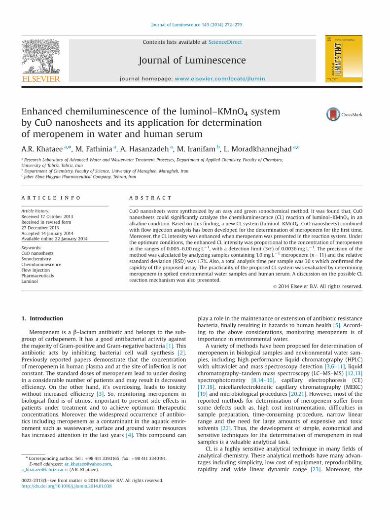
The schematic of the flow-injection CL system for the determi-nation of meropenem is illustrated in Fig. 1. Polytetrafluoroethy-lene (PTFE) tubing with an inner diameter of 1.0 mm was utilizedas a connection material in the flow system. A peristaltic pumpwas used to deliver all respective solutions through the flowsystem at a flow rate of 2.0 mL min�1 for each channel. A six-port valve equipped with a 100 μL sample loop was applied forinjection. The light generation from the CL reaction in the flow cellwas monitored by a FB12 luminometer (Berthold Detection Sys-tems, Germany). Ultraviolet–visible (UV–Vis) spectra were mon-itored by UV–Vis spectrophotometer (WPA lightwave S2000,England) in the range of 200–800 nm. A bath type sonicator(Sonica, 2200 EP S3, Italy) with 50–60 Hz frequency and heatingarrangement was used for sonochemical synthesis of CuO nano-sized samples. To characterize the crystal structure and phasepurity of as-prepared samples, XRD measurements were per-formed at room temperature by using Siemens X-ray diffractionD5000 (California, USA), with CuKα radiation. The acceleratingvoltage of 40 kV and emission current of 30 mAwere utilized. SEM(S-4200, Hitachi, Japan) was used to observe the surface state andthe morphology of the prepared nanostructures using an electronmicroscope. Moreover, the obtained SEM image was analyzedusing Manual Microstructure Distance Measurement software(NahaminPardazan Asia Co., Iran) to determine the diameter andlength size distribution of the obtained samples. The chromato-graphic analysis was performed by an HPLC system (Smartline1000 Knauer, Berlin, Germany) equipped with a C18 column andUV detection.
2.4. Procedures for chemiluminescence assay
As depicted in Fig. 1, the alkaline solution of luminol (a), sampleor standard solution of mixture of meropenem and CuO nano-sheets (b), H2O as the carrier (c) and KMnO4solution (d) werepumped by a peristaltic pump. Solutions of a and b were mixedthrough a mixing tube (silicon tubing, 1.0 mm diameter); then100 μL of mixing solution was injected into the carrier stream andpremixed with KMnO4 stream via a Y–piece; after that, themixture was delivered into the flow cell. The peak height of the
Fig. 1. Schematic diagram of flow-injection CL system; (a): luminol in NaOHsolution; (b): sample or blank solution; (c): H2O as the carrier; (d): KMnO4
solution; (P): peristaltic pump; (M): mixing tube; (V): injection valve; (F): flowcell; (W): waste; (D): detector (luminometer) and (R): recorder (personalcomputer).
A.R. Khataee et al. / Journal of Luminescence 149 (2014) 272–279 273
CL signals was continuously monitored on a computer connectedto the luminometer. Determination of meropenem was performedbased on the increase of CL intensity as ΔI¼ Is–I0, where Is and I0denote CL intensity in the presence and absence of meropenem,respectively.
2.5. Pre-treatment of real samples solution beforechemiluminescence assay
Tap water was analyzed without any pretreatment. Ground andriver water samples were freshly collected and filtered withpolyamide membrane filters of 0.45 mm to remove the suspendedsolid matter and stored in dark at 4 1C in the refrigerator [25]. Theywere used within 1 week. Prior to analysis, 30 mL water sampleswere spiked with 0.005, 0.05, 0.25, 0.5, 1 and 1.5 mL meropenemstandard solutions (100 mg L�1) and diluted to 50 mL with doublydistilled water to prepare solutions of 0.01, 0.1, 0.5, 1, 2 and3 mg L�1, respectively.
For human serum samples, only a deproteinization pretreat-ment step utilizing trichloroacetic acid (CCl3COOH) was carriedout; an extraction process was not needed [39]. To prepare thespiked samples, known amounts of meropenem were spiked into1.0 mL of serum and then, for each sample, 5.0 mL of 10% (w/v)trichloroacetic acid was added. These mixtures were centrifuged at3000 rpm for 15 min. 2.5 mL of the protein-free supernatant wasdiluted to 50 mL with deionized water.
The amount of meropenem in the spiked samples was deter-mined by means of the general procedure using standard additionmethod. Also, a blank value was determined by meropenem-freesamples in the same procedure.
3. Results and discussion
3.1. Characterization of CuO nanosheets
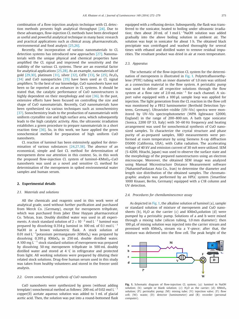
The XRD patterns of the synthesized CuO samples are depictedin Fig. 2a. The XRD diffraction peaks at 2θ of 32.41, 35.51, 38.71,
48.71, 53.21, 57.91, 61.31, 66.11, 68.01, 71.91 and 74.81 can beclassified in to (110), (002), (111), (202), (020), (202), (113), (022),(200), (311) and (222) plane reflections, which are associated withmonoclinic crystal structure CuO according to the standard pow-der diffraction data (JCPDS 05-0661) [40]. No peak for impuritieswas detected, confirming that the applied sonochemical methodin this study was successful in synthesizing the nanosized CuOsamples. Moreover, the sharp diffraction peaks in the XRD spec-trum of the synthesized sample indicated that the synthesizedproduct was high crystalline.
In order to further clarify the size and morphology of thesynthesized product, SEM images were taken at different magni-fications. Fig. 2b shows the SEM microphotographs of the nano-sized samples at different magnifications. The low magnificationSEM image in the left side of Fig. 2b obviously showed that theobtained samples possessed sheet-like morphology. Moreover, thehigh magnification SEM image in the right side of the Fig. 2bconfirmed that the grown nanosheets were randomly grown.
Additionally, the width and thickness size distribution ofnanosheets were calculated by manual microstructure distancemeasurement software. The results showed that the width andthickness size distribution are in the range of 100–150 and 30–40 nm, respectively.
3.2. CuO nanosheets-enhanced chemiluminescence system
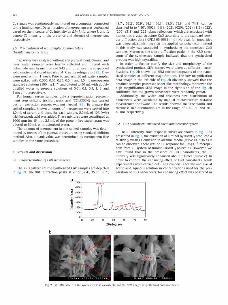
The CL intensity–time response curves are shown in Fig. 3. Aspresented in Fig. 3, the oxidation of luminol by KMnO4 produced arelatively weak CL emission in alkaline media (curve a). Also as itcan be observed, there was no CL response for 1 mg L�1 merope-nem from CL system of luminol–KMnO4 (curve b). However, wehave found that in the presence of CuO nanosheets, the CLintensity was significantly enhanced about 7 times (curve c). Inorder to confirm the enhancing effect of CuO nanosheets, blankexperiments were carried out using copper(II) acetate and glacialacetic acid aqueous solution in concentrations used for the pre-paration of CuO nanosheets. No enhancing effect was observed in
Fig. 2. (a): XRD pattern of the synthesized CuO nanosheets; and (b): SEM images of synthesized CuO nanosheets.
A.R. Khataee et al. / Journal of Luminescence 149 (2014) 272–279274
the presence of glacial acetic acid. Also, an enhancing effect in thepresence of 0.02 mol L�1 copper(II) acetate (curve e) is not muchremarkable in comparison with CuO nanosheets amplified CLsystem. There was no CL response for 1 mg L�1 meropenem inthe above mentioned CL system of luminol–KMnO4–copper(II)acetate. So, CuO nanosheets-enhanced CL system was used forthe determination of meropenem. The results showed that the CLintensity of luminol–KMnO4–CuO nanosheets system significantlyincreased upon the addition of 1.00 mg L�1 meropenem (curve d).In order to deeply investigate the sensitivity of luminol–KMnO4 inthe presence of copper(II) acetate and CuO nanosheets, two CLsystems of luminol–KMnO4–copper(II) acetate and luminol–KMnO4–CuO nanosheets were compared for the determinationof meropenem in the further achieved optimized conditions inSection 3.4. Also, it is worth to mention that, the molar concentra-tion of copper(II) acetate was selected exactly the same as themolar concentration of CuO nanosheets which was achieved inoptimization process (Section 3.3). As can be seen in Fig. 3 (curveg), the addition of 2.00 mg L�1 meropenem increase the intensityof the CL system of luminol–KMnO4–copper(II) acetate.
Furthermore, the effect of different oxidants such as potassiumperiodate, potassium hexacyanoferrate and hydrogen peroxide inluminol–oxidant–CuO nanosheets system was investigated. Therewas no reasonable response in the presence of variable concentra-tions of meropenem. So, in this work, luminol–KMnO4–CuOnanosheets system was selected for determination of meropenem.
3.3. Optimization of the effective parameters
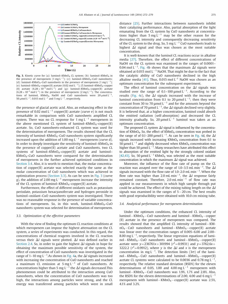
With the view of finding the optimum CL reaction conditions atwhich meropenem can impose the highest attenuation on the CLsystem, a series of experiments was conducted. In this regard, theconcentrations of chemical reagents involved in the CL reactionversus their ΔI signals were plotted. ΔI was defined earlier inSection 2.4. So, in order to gain the highest ΔI signals in hope forobtaining the maximum possible sensitivity of the system, theeffect of concentration of CuO nanosheets was investigated in therange of 1–10 mg L�1.As shown in Fig. 4a, the ΔI signals increasedwith increasing the concentration of CuO nanosheets and reacheda maximum CL emission at 5 mg L�1 of CuO nanosheets. Atconcentrations higher than 5 mg L�1 theΔI signals decreased.Thisphenomenon could be attributed to the interaction among CuOnanosheets, when the concentration of CuO nanosheets was toohigh, the interactions among particles were strong, and the CLenergy was transferred among particles which were in small
distance [25]. Further interactions between nanosheets inhibittheir catalyzing performance. Also, partial absorption of the lightemanating from the CL system by CuO nanosheets at concentra-tions higher than 5 mg L�1 may be the other reason for thedecreasing CL intensity and consequently decreasing sensitivityof the CL system to the drug. So, 5 mg L�1 CuO nanosheets leads tohighest ΔI signal and thus was chosen as the most suitableconcentration.
It is well-known that the luminol CL reactions occur in alkalinemedia [27]. Therefore, the effect of different concentrations ofNaOH on the CL system was examined in the ranges of 0.0001–0.04 mol L�1. Fig. 4b shows that the maximum ΔI signals wereobtained at 0.015 mol L�1 NaOH. That might be due to the fact thatthe catalytic ability of CuO nanosheets declined in the highalkaline media [41]. Thus, 0.015 mol L–1 NaOH was chosen as anoptimum concentration for the subsequent experiment.
The effect of luminol concentration on the ΔI signals wasstudied over the range of 0.1–100 μmol L�1. According to theresults (Fig. 4c), the ΔI signals increased with the increase ofluminol concentration from 0.1 to 20 μmol L�1; then they wereconstant from 30 to 70 μmol L�1 and for the amounts beyond theconcentration of 70 μmol L�1, theΔI signals declined very slightly.It is believed that, at a higher concentration, luminol could absorbthe emitted radiation (self-absorption) and decreased the CLintensity gradually. So, 20 μmol L�1 luminol was taken as anoptimum concentration.
The proposed CL system ΔI signal is influenced by the concentra-tion of KMnO4. So, the effect of KMnO4 concentration was probed inthe range of of 0.1–200 μmol L�1. As can be seen in Fig. 4d, the ΔIsignals increased with increasing KMnO4 concentration from 0.1 to50 μmol L�1 and slightly decreased when KMnO4 concentration washigher than 50 μmol L�1. Many researchers have attributed this effectto absorption of the emitted light by the strongly colored oxidant[42,43]. So, 50 μmol L�1 KMnO4 was selected as the most suitableconcentration in which the maximum ΔI signal was achieved.
Moreover, the influence of the flow rate of pump on the CLreaction was assayed over the range of 1.0–3.5 mL min�1. The ΔIsignals increased with the flow rate of 1.0–2.0 mL min�1. When theflow rate was higher than 2.0 mL min�1, the ΔI response fairlyremained constant. Therefore, 2.0 mL min�1 of flow rate wasapplied in our measurements in which high sample through putcould be achieved. The effect of the mixing tubing length on the ΔIsignals was examined in the ranges of 5�20 cm. The best resultswith good reproducibility were obtained with 10.0 cm mixing tube.
3.4. Analytical performance for meropenem determination
Under the optimum conditions, the enhanced CL intensity ofluminol�KMnO4�CuO nanosheets and luminol�KMnO4�copper(II) acetate in the presence of meropenem was compared. Theresults showed that the amplified CL intensity of luminol�KM-nO4�CuO nanosheets and luminol�KMnO4�copper(II) acetatewas linear over the concentration ranges of 0.005–6.00 and 2.00–8.00 mg L�1, respectively. The linear regression equations of lumi-nol�KMnO4�CuO nanosheets and luminol�KMnO4�copper(II)acetate were y¼236761xþ301994 (r2¼0.9931) and y¼3762.6xþ3222.2 (r2¼0.9912), where y is the ΔI and x is the meropenemconcentration in mg L�1. The detection limits (3s) of the lumi-nol�KMnO4�CuO nanosheets and luminol�KMnO4�copper(II)acetate CL systems were calculated to be 0.0036 and 0.78 mg L�1,respectively. The relative standard deviation (RSD%) for the elevendeterminations of 0.05, 1.00 and 4.00 mg L�1 meropenem withluminol�KMnO4�CuO nanosheets was 1.9%, 1.7% and 2.8%. Also,the RSD% for the eleven determinations of 2.00, 4.00 and 6 mg L�1
meropenem with luminol�KMnO4�copper(II) acetate was 2.5%,4.1% and 3.2%.
Fig. 3. Kinetic curve for (a): luminol–KMnO4 CL system; (b): luminol–KMnO4 inthe presence of meropenem (1 mg L�1); (c): luminol–KMnO4–CuO nanosheets;(d): luminol–KMnO4–CuO nanosheets in the presence of meropenem (1 mg L�1);(e): luminol–KMnO4–copper(II) acetate (0.02 mol L�1); (f) luminol–KMnO4–copper(II) acetate (6.28�10–5 mol L–1) and (g): luminol–KMnO4–copper(II) acetate(6.28�10–5 mol L–1) in the presence of meropenem (2 mg L�1); The concentra-tions of luminol, KMnO4, NaOH and CuO nanosheets were 20 μmol L�1,50 μmol L�1, 0.015 mol L�1 and 5 mg L�1, respectively.
A.R. Khataee et al. / Journal of Luminescence 149 (2014) 272–279 275
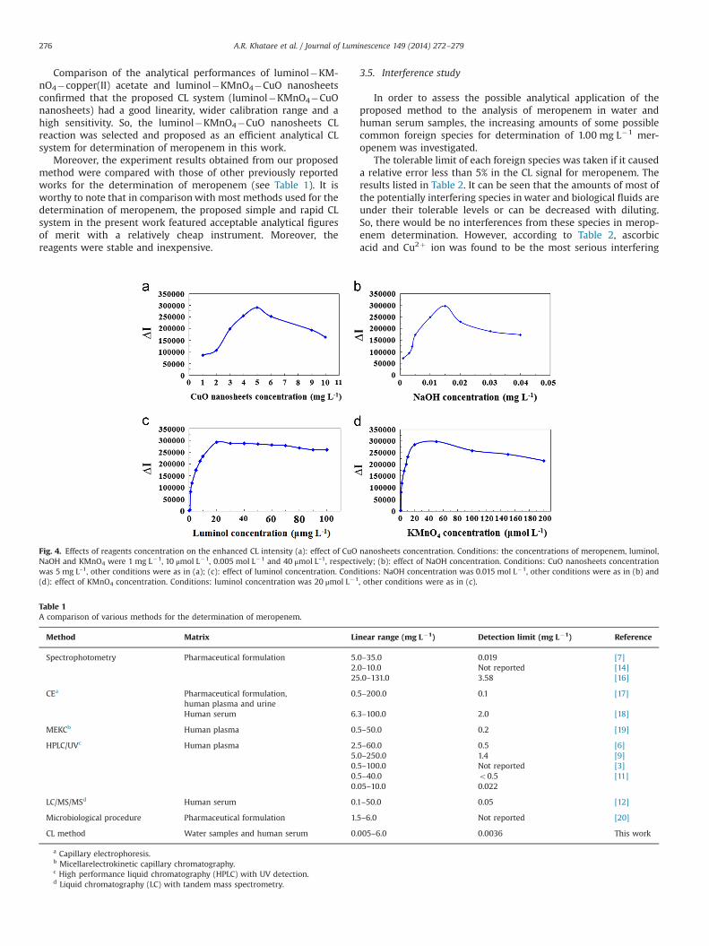
Comparison of the analytical performances of luminol�KM-nO4�copper(II) acetate and luminol�KMnO4�CuO nanosheetsconfirmed that the proposed CL system (luminol�KMnO4�CuOnanosheets) had a good linearity, wider calibration range and ahigh sensitivity. So, the luminol�KMnO4�CuO nanosheets CLreaction was selected and proposed as an efficient analytical CLsystem for determination of meropenem in this work.
Moreover, the experiment results obtained from our proposedmethod were compared with those of other previously reportedworks for the determination of meropenem (see Table 1). It isworthy to note that in comparison with most methods used for thedetermination of meropenem, the proposed simple and rapid CLsystem in the present work featured acceptable analytical figuresof merit with a relatively cheap instrument. Moreover, thereagents were stable and inexpensive.
3.5. Interference study
In order to assess the possible analytical application of theproposed method to the analysis of meropenem in water andhuman serum samples, the increasing amounts of some possiblecommon foreign species for determination of 1.00 mg L�1 mer-openem was investigated.
The tolerable limit of each foreign species was taken if it causeda relative error less than 5% in the CL signal for meropenem. Theresults listed in Table 2. It can be seen that the amounts of most ofthe potentially interfering species in water and biological fluids areunder their tolerable levels or can be decreased with diluting.So, there would be no interferences from these species in merop-enem determination. However, according to Table 2, ascorbicacid and Cu2þ ion was found to be the most serious interfering
Fig. 4. Effects of reagents concentration on the enhanced CL intensity (a): effect of CuO nanosheets concentration. Conditions: the concentrations of meropenem, luminol,NaOH and KMnO4 were 1 mg L�1, 10 μmol L�1, 0.005 mol L�1 and 40 μmol L–1, respectively; (b): effect of NaOH concentration. Conditions: CuO nanosheets concentrationwas 5 mg L–1, other conditions were as in (a); (c): effect of luminol concentration. Conditions: NaOH concentration was 0.015 mol L�1, other conditions were as in (b) and(d): effect of KMnO4 concentration. Conditions: luminol concentration was 20 μmol L�1, other conditions were as in (c).
Table 1A comparison of various methods for the determination of meropenem.
Method Matrix Linear range (mg L�1) Detection limit (mg L�1) Reference
Spectrophotometry Pharmaceutical formulation 5.0–35.0 0.019 [7]2.0–10.0 Not reported [14]25.0–131.0 3.58 [16]
CEa Pharmaceutical formulation,human plasma and urine
0.5–200.0 0.1 [17]
Human serum 6.3–100.0 2.0 [18]
MEKCb Human plasma 0.5–50.0 0.2 [19]
HPLC/UVc Human plasma 2.5–60.0 0.5 [6]5.0–250.0 1.4 [9]0.5–100.0 Not reported [3]0.5–40.0 o0.5 [11]0.05–10.0 0.022
LC/MS/MSd Human serum 0.1–50.0 0.05 [12]
Microbiological procedure Pharmaceutical formulation 1.5–6.0 Not reported [20]
CL method Water samples and human serum 0.005–6.0 0.0036 This work
a Capillary electrophoresis.b Micellarelectrokinetic capillary chromatography.c High performance liquid chromatography (HPLC) with UV detection.d Liquid chromatography (LC) with tandem mass spectrometry.
A.R. Khataee et al. / Journal of Luminescence 149 (2014) 272–279276
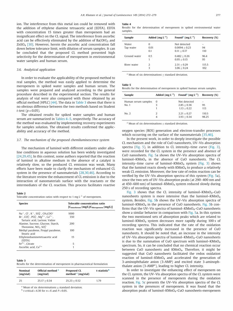
ion. The interference from this metal ion could be removed withthe addition of ethylene diamine tetraacetic acid (EDTA). EDTAwith concentration 15 times greater than meropenem had aninsignificant effect on the CL signal. The interference from ascorbicacid can be effectively eliminated by the addition of Ba(OH)2 andZnSO4 [30]. However, herein the ascorbic acid concentration falldown below tolerance limit, with dilution of serum samples. It canbe concluded that the proposed CL method presented highselectivity for the determination of meropenem in environmentalwater samples and human serum.
3.6. Analytical application
In order to evaluate the applicability of the proposed method toreal samples, the method was easily applied to determine themeropenem in spiked water samples and human serum. Thesamples were prepared and analyzed according to the generalprocedure described in the experimental section. The results foranalysis of vial were also compared with those obtained by anofficial method (HPLC) [44]. The data in Table 3 shows that there isno obvious difference between the two methods based on Studentt-test (p¼0.05).
The obtained results for spiked water samples and humanserum are summarized in Tables 4–5, respectively. The accuracy ofthe method was evaluated by implementing recovery experimentsfor samples solution. The obtained results confirmed the applic-ability and accuracy of the method.
3.7. The mechanism of the proposed chemiluminescence system
The mechanism of luminol with different oxidants under alka-line conditions in aqueous solution has been widely investigated[24,29,45]. In this context, some authors reported that the reactionof luminol in alkaline medium in the absence of a catalyst isrelatively slow, so the produced CL emission was weak. Manyefforts have been made to clarify the mechanism of enhanced CLsystem in the presence of nanomaterials [28,30,46]. According tothe literature review the enhancement of CL emission is due to theinteraction of nanomaterials surface with the reactants or theintermediates of the CL reaction. This process facilitates reactive
oxygen species (ROS) generation and electron-transfer processeswhich occurring on the surface of the nanomaterials [35,46].
In the present work, in order to deeply investigate the possibleCL mechanism and the role of CuO nanosheets, UV–Vis absorptionspectra (Fig. 5), in addition to CL intensity–time curve (Fig. 3),were recorded for the CL system in the presence and absence ofCuO nanosheets. Fig. 5a shows the UV–Vis absorption spectra ofluminol–KMnO4 in the absence of CuO nanosheets. The CLintensity–time curve of luminol–KMnO4 system (Fig. 3) showsthat the luminol reacts slowly with KMnO4 to produce a relativelyweak CL emission. Moreover, the low rate of redox reaction can beverified by the UV–Vis absorption spectra of this system (Fig. 5a),since the two sets of UV–Vis absorption peaks (at 290–400 nm andat 450–600 nm) of luminol–KMnO4 system reduced slowly during250 s of recording spectra.
Fig. 3 shows that the CL intensity of luminol–KMnO4–CuOnanosheets system is more intensive than the luminol–KMnO4
system. Besides, Fig. 5b shows the UV–Vis absorption spectra ofluminol–KMnO4 in the presence of CuO nanosheets. Fig. 5b con-firms that the UV–Vis spectra of luminol–KMnO4–CuO nanosheetsshow a similar behavior in comparison with Fig. 5a. In this systemthe two mentioned sets of absorption peaks which are related toluminol–KMnO4 system decreases more rapidly during 100 s ofrecording spectra. This indicated that the rate of the oxidationreaction was significantly increased in the presence of CuOnanosheets. It should be noted that, an increase in the intensityof UV–Vis absorption spectra of luminol–KMnO4–CuO nanosheetsis due to the summation of CuO spectrum with luminol–KMnO4
spectrum. So, it can be concluded that no chemical reaction occurbetween CuO nanosheets and KMnO4. Therefore, it might besuggested that CuO nanosheets facilitated the redox oxidationreaction of luminol–KMnO4 and accelerated the generation of3-aminophthalate anion (3-AMP) and excited state 3-aminoph-thalate anion (3-AMP*), leading to higher CL intensity.
In order to investigate the enhancing effect of meropenem onthe CL system, the UV–Vis absorption spectra of the CL systemwerescanned in the presence of meropenem during the oxidationreaction. Fig. 5c presents the UV–Vis absorption spectra of the CLsystem in the presences of meropenem. It was found that theabsorption peaks of luminol–KMnO4–CuO nanosheets-meropenem
Table 2Tolerable concentration ratios with respect to 1 mg L–1 of meropenem.
Species Tolerable concentration ratio[Cinterferent (mg/L)/CMeropenem (mg/L)]
Naþ , Cl–, Kþ , SO42–, CH3COO– 1000
Br–, CO32–, PO4
3–, Mg2þ , Ca2þ ,Tartaric acid, Lactose, Valine
350
Alanine, Sucrose, Glucose, Starch,Threonine, NO3
–, SO32–
200
Methyl parabene, Propyl parabene,Stearic acid
100
Ethylenediaminetetraacetic acid(EDTA)
15
Fe2þ , Citrate 5Ascorbic acid, Cu2þ 1
Table 3Results for the determination of meropenem in pharmaceutical formulation
Nominal(mg/vial)
Official method a
(mg/vial)Proposed CLmethod a (mg/vial)
t-statisticb
25 25.1770.34 25.2370.52 1.79
a Mean of six determinations7standard deviation.b t-critical¼4.30 for n¼6 and P¼0.05.
Table 4Results for the determination of meropenem in spiked environmental watersamples.
Sample Added (mg L–1) Founda (mg L–1) Recovery (%)
Water 0 Not detected –
Tap water 0.01 0.009470.23 940.1 0.1170.17 110
Ground water 0.5 0.48270.26 96.41 0.9570.15 95
River water 2 2.3170.29 115.53 3.0670.24 102
a Mean of six determinations7standard deviation.
Table 5Results for the determination of meropenem in spiked human serum samples.
Sample Added (mg L–1) Founda (mg L–1) Recovery (%)
Human serum samples 0 Not detected –
No. 1 3 2.8570.36 951 1.1370.22 113
No. 2 2 2.2170.27 110.54 3.9370.34 98.25
a Mean of six determinations7standard deviation.
A.R. Khataee et al. / Journal of Luminescence 149 (2014) 272–279 277
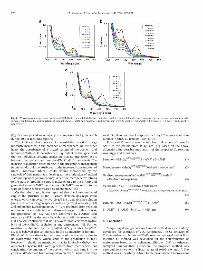
(Fig. 5c) disappeared more rapidly in comparison to Fig. 5a and bduring 60 s of recording spectra.
This indicates that the rate of the oxidation reaction is sig-nificantly increased in the presence of meropenem. On the otherhand, the absorbance of a mixed system of meropenem andluminol–KMnO4–CuO nanosheets is equivalent to the spectra ofthe two individual systems, suggesting that no interaction existsbetween meropenem and luminol–KMnO4–CuO nanosheets. Theincrease of oxidation reaction rate in the presence of meropenemin one hand, could be attributed to the increased consumption ofKMnO4. Moreover, KMnO4 could oxidize meropenem by thecatalysis of CuO nanosheets, leading to the production of excitedstate meropenem (meropenemn). When the meropenemn returnsto the state of ground, it could transfer energy to the 3-AMP, andgenerated more 3-AMPn ion, the more 3–AMPn ions return to thestate of ground with increased CL phenomena [45].
On the other hand, it was reported that the four–memberedring in the structure of the β-lactam skeleton has high strainenergy, which can be easily hydrolyzed in strong alkaline solution[47–49]. Reactive oxygen species such as hydroxyl radicals (dOH)and superoxide radical anions (O2d
�) are produced from reactionof some of these antibiotics with dissolved oxygen. In this context,the production of ROS has been confirmed by electron spinresonance (ESR) in the work by Kubo et al. [48]. However, theirESR analysis confirmed that no ROS were produced from hydro-lysis of penicillin G, cephalothin and aztreonam [48]. Furtheroxidation of luminol by the resulted ROS generates 3�AMPn.So, it is believed that an increase in the CL intensity of luminol–KMnO4–CuO nanosheets in the presence of meropenem is due toits hydrolyzing ability which leads to the production of ROS.However, it should be mentioned that in luminol–KMnO4–mer-openem CL system ROS were generated from meropenem. Butconsidering the amount of meropenem which was 1 mg L–1, theeffect of ROS derived from meropenem on the CL signals was very
weak. So, there was no CL response for 1 mg L–1 meropenem fromluminol–KMnO4 CL reaction (see Fig. 3).
Enhanced CL emission emanates from relaxation of more 3-AMPn to the ground state at 425 nm [47]. Based on the abovediscussion, the possible mechanism of the proposed CL reactionwas suggested as follows:
LuminolþKMnO4 �!CuO nanosheets3�AMPnþ3�AMP ð1Þ
MeropenemþKMnO4 �!CuO nanosheetsOxidized meropenemn ð2Þ
Oxidized meropenemnþ3�AMP �!CuO nanosheets3�AMPn
þOxidized meropenem ð3Þ
MeropenemþNaOH �! Hydrolyzed meropenem
þDissolved oxygen �!CuO nanosheetsHydroxyl and=or superoxide radicals ðROSÞ
ð4Þ
LuminolþROSþNaOH �!CuO nanosheets3�AMPn ð5Þ
3�AMPn-3�AMPþhυ ðλmax ¼ 425 nmÞ ð6Þ
4. Conclusion
Simple, rapid and green sonochemical method was successfullydeveloped for synthesis of CuO nanosheets. The CL behavior ofCuO nanosheets in luminol–KMnO4 reaction was explored. A flowinjection CL method was developed for the determination ofmeropenem based on its enhancing effect on CuO nanosheets–catalyzed luminol–KMnO4 reaction. The proposed method waseasy and sensitive with a linear range of 0.005–6.0 mg L�1. Themethod was successfully utilized for determination of meropenem
Fig. 5. UV–vis absorption spectra of (a): luminol–KMnO4 (b): luminol–KMnO4–CuO nanosheets and (c): luminol–KMnO4–CuO nanosheets in the presence of meropenem CLsystems. Conditions: the concentrations of luminol, KMnO4, NaOH, CuO nanosheets and meropenem were 20 μmol L�1, 50 μmol L�1, 0.015 mol L�1, 5 mg L�1 and 1 mg L–1,respectively.
A.R. Khataee et al. / Journal of Luminescence 149 (2014) 272–279278
in spiked water samples and human serum. A possible CL reactionmechanismwas also proposed by the studies of CL kinetic and UV–Vis absorption spectra.
Acknowledgments
The authors thank the University of Tabriz and Maragheh, Iranfor all of the support provided. We also thank Jaber Ebne Hayyanpharmaceutical Co. Tehran, Iran for providing the pharmaceuticals.
References
[1] J.L Blumer, Int. J. Antimicrob. Ag. 8 (1997) 73–92.[2] G. Zhanel, R. Wiebe, L. Dilay, K. Thomson, E. Rubinstein, D. Hoban, A. Noreddin,
J. Karlowsky, Drugs 67 (2007) 1027–1052.[3] E. Dailly, R. Bouquié, G. Deslandes, P. Jolliet, R. Le Floch, J. Chromatogr. B 879
(2011) 1137–1142.[4] M. Seifrtová, L. Nováková, C. Lino, A. Pena, P. Solich, Anal. Chim. Acta 649
(2009) 158–179.[5] J.M. Cha, S. Yang, K.H. Carlson, J. Chromatogr. A 1115 (2006) 46–57.[6] R. Denooz, C. Charlier, J. Chromatogr. B 864 (2008) 161–167.[7] A.S.L. Mendez, M. Steppe, E.E.S. Schapoval, J. Pharm. Biomed. Anal. 33 (2003)
947–954.[8] L. Venkateswara Rao, G. Ramu, M. Sravan Kumar, C. Rambabu, Int. J. Pharm.
Tech. Res. 4 (2012) 957–962.[9] B.C. McWhinney, S.C. Wallis, T. Hillister, J.A. Roberts, J. Lipman, J.P.J. Ungerer, J.
Chromatogr. B 878 (2010) 2039–2043.[10] Y. Ozkan, L. Kuukguzel, S.A. Ozkan, H.Y. Aboul–Enein, Biomed. Chromatogr. 15
(2001) 263–266.[11] M. Ehrlich, F.D. Daschner, K. Kümmerer, J. Chromatogr. B 751 (2001) 357–363.[12] T. Ohmori, A. Suzuki, T. Niwa, H. Ushikoshi, K. Shirai, S. Yoshida, S. Ogura,
Y. Itoh, J. Chromatogr. B 879 (2011) 1038–1042.[13] M. Carlier, V. Stove, J.A. Roberts, E. Van De Velde, J.J. De Waele, A.G. Verstraete,
Int. J. Antimicrob. Ag. 40 (2012) 416–422.[14] D. Singh, G. Maheshwari, Med. Chem. Res. (2013) 1–5.[15] S.R. Narala, K. Saraswathi, Int. J. Chem. Tech. Res. 3 (2011) 605–609.[16] J. Cielecka–Piontek, M. Paczkowska, K. Lewandowska, B. Barszcz, P. Zalewski,
P. Garbacki, Chem. Cent. J. 7 (2013) 7–98.[17] Y. Mrestani, R. Neubert, F. Nagel, J. Pharm. Biomed. Anal. 20 (1999) 899–903.[18] T. Kitahashi, I. Furuta, J. Chromatogr. Sci. 43 (2005) 430–433.[19] Y.–W. Chou, Y.–H. Yang, J.–H. Chen, C.–C. Kuo, S.–H. Chen, J. Chromatogr. B 856
(2007) 294–301.
[20] A.S.L. Mendez, V. Weisheimer, T.P. Oppe, M. Steppe, E.E.S. Schapoval, J. Pharm.Biomed. Anal. 37 (2005) 649–653.
[21] M.A. Al–Meshal, M.A. Ramadan, K.M. Lotfi, A.M. Shibl, J. Clin. Pharm. Ther. 20(1995) 159–163.
[22] J. Cielecka–Piontek, K. Michalska, P. Zalewski, S. Zasada, Curr. Anal. Chem. 8(2012) 91–115.
[23] M. Iranifam, Flow analysis and chemiluminescence: An update: Advances inflow–chemiluminescence analysis, LAPLambert Academic Publishing,Saarbrücken (2011).
[24] M. Iranifam, Luminescence 23 (2013) 798–820.[25] M. Iranifam, M. Fathinia, T. Sadeghi Rad, Y. Hanifehpour, A.R. Khataee, S.W. Joo,
Talanta 107 (2013) 263–269.[26] F. Zhao, H. Si, J. Lumin. 135 (2013) 259–264.[27] A.R. Khataee, A. Hasanzadeh, M. Iranifam, M. Fathinia, Y. Hanifehpour, S.
W. Joo, Spectrochim. Acta A: Mol. Biomol. Spect. 122 (2014) 737–743.[28] Q. Li, L. Zhang, J. Li, C. Lu, Trends Anal. Chem. 30 (2011) 401–413.[29] Z.F. Zhang, H. Cui, C.Z. Lai, L.J. Liu, Anal. Chem. 77 (2005) 3324–3329.[30] X. Yu, J. Bao, J. Lumin. 129 (2009) 973–978.[31] B. Liu, Y. He, C. Duan, N. Li, H. Cui, J. Photoch. Photobio. A–Chem. 217 (2011)
62–67.[32] W. Liu, J. Kou, X. Jiang, Z. Zhang, H. Qi, J. Lumin. 132 (2012) 1048–1054.[33] S. Kanwal, Z. Traore, C. Zhao, X. Su, J. Lumin. 130 (2010) 1901–1906.[34] M. Iranifam, Trends Anal. Chem 51 (2013) 51–70.[35] W. Chen, L. Hong, A.–L. Liu, J.–Q. Liu, X.–H. Lin, X.–H. Xia, Talanta 99 (2012)
643–648.[36] Z. Ibupoto, K. Khun, V. Beni, X. Liu, M. Willander, Sensors 13 (2013)
7926–7938.[37] S. Anandan, G. J. Lee, J.J. Wu, Ultrason. Sonochem. 19 (2012) 682–686.[38] H. Sun, T. Wang, X. Liu, P. Chen, J. Lumin. 134 (2013) 154–159.[39] G. Hunter, J. Clin. Pathol. 10 (1957) 161–164.[40] C. Deng, H. Hu, X. Ge, C. Han, D. Zhao, G. Shao, Ultrason. Sonochem. 18 (2011)
932–937.[41] B. Haghighi, S. Bozorgzadeh, Microchem. J. 95 (2010) 192–197.[42] H. Liu, Y. Hao, J. Ren, P. He, Y. Fang, Luminescence 22 (2007) 302–308.[43] J.L. Adcock, P.S. Francis, N.W. Barnett, Anal. Chim. Acta 601 (2007) 36–67.[44] The United states Pharmacopeia,34 sted., The National Formulary, 26th ed.,
The United States Pharmacopeial Convention, Rockville, 2011, pp. 3425–3428.[45] R. Su, J.–M. Lin, F. Qu, Z. Chen, Y. Gao, M. Yamada, Anal. Chim. Acta 508 (2004)
11–15.[46] C. Duan, H. Cui, Z. Zhang, B. Liu, J. Guo, W. Wang, J. Phys. Chem. C 111 (2007)
4561–4566.[47] Y. Li, J. Lu, Luminescence 21 (2006) 251–255.[48] H. Kubo, M. Saitoh, S. Murase, T. Inomata, Y. Yoshimura, H. Nakazawa, Anal.
Chim. Acta 389 (1999) 89–94.[49] Y. Li, Y. Tang, H. Yao, J. Fu, Luminescence 18 (2003) 313–317.
A.R. Khataee et al. / Journal of Luminescence 149 (2014) 272–279 279
Related Documents