Enhanced 5-HT1A receptor-dependent feedback control over dorsal raphe serotonin neurons in the SERT knockout mouse Mariano Soiza-Reilly a, b , Nathalie M. Goodfellow c , Evelyn K. Lambe c, d , Kathryn G. Commons a, b, * a Department of Anesthesiology, Perioperative, and Pain Medicine, Boston Children's Hospital, 300 Longwood Ave., Boston, MA 02115, USA b Department of Anaesthesia, Harvard Medical School, Boston, MA 02115, USA c Department of Physiology, University of Toronto, Toronto, ON M5S 1A8, Canada d Department of Obstetrics and Gynaecology, University of Toronto, Toronto, ON M5S 1A8, Canada article info Article history: Received 2 April 2014 Received in revised form 3 September 2014 Accepted 15 September 2014 Available online 28 September 2014 Keywords: Serotonin transporter Feedback inhibition Dorsal raphe nucleus Swim stress Depressive-like behavior Mood disorders abstract 5-HT1A receptors are widely expressed in the brain and play a critical role in feedback inhibition of serotonin (5-HT) neurons through multiple mechanisms. Yet, it remains poorly understood how these feedback mechanisms, particularly those involving long-range projections, adapt in mood disorders. Here, we examined several aspects of 5-HT1A receptor function in the 5-HT transporter knockout mouse (SERT-KO), a model of vulnerability to stress and mood disorders. We found that in comparison to wild- type (WT) mice, SERT-KO mice had more passive coping in response to acute swim stress and this was accompanied by hypo-activation of medial prefrontal cortex (mPFC) Fos expression. Both of these effects were reversed by systemically blocking 5-HT1A receptors. Ex-vivo electrophysiological experiments showed that 5-HT exerted greater 5-HT1A-mediated inhibitory effects in the mPFC of SERT-KO mice compared to WT. Since 5-HT1A receptors in the mPFC provide a key feedback regulation of the dorsal raphe nucleus (DRN), we used a disinhibition strategy to examined endogenous feedback control of 5-HT neurons. Blocking 5-HT1A receptors disinhibited several fold more 5-HT neurons in the DRN of SERT-KO than in WT mice, revealing the presence of enhanced feedback inhibition of 5-HT neurons in the SERT- KO. Taken together our results indicate that increased stress sensitivity in the SERT-KO is associated with the enhanced capacity of 5-HT1A receptors to inhibit neurons in the mPFC as well as to exert feedback inhibition of DRN 5-HT neurons. © 2014 Elsevier Ltd. All rights reserved. 1. Introduction Serotonin (5-HT) neurons in the dorsal raphe nucleus (DRN) are a major source of 5-HT to the forebrain, which influences arousal, mood and motivated behavior (Amat et al., 2005; Nakamura et al., 2008; Takahashi et al., 2010; Bruchas et al., 2011; Warden et al., 2012). The activity of 5-HT neurons in the DRN is influenced by feedback mechanisms involving 5-HT1A receptors. Specifically, 5- HT1A receptors can be found as autoreceptors on 5-HT-producing neurons in the DRN and as heteroreceptors on neurons in other brain regions (Celada et al., 2001; Sharp et al., 2007; Sprouse and Aghajanian, 1987). In particular, 5-HT1A heteroreceptors in the medial prefrontal cortex (mPFC) have been suggested to play a key role in long-range feedback to the DRN (Celada et al., 2001; Sharp et al., 2007; Haj os et al., 1999). Despite the importance of 5-HT1A receptors in the pathophysiology of mood disorders and their treatment (Reviewed by (Savitz et al., 2009)), it is poorly under- stood how these different 5-HT1A-mediated feedback pathways adapt in concert to regulate 5-HT neuron activity in pathological conditions. The 5-HT transporter knockout mouse (SERT-KO) represents a model of vulnerability to stress and mood disorders, suitable for investigating adaptations in 5-HT1A receptor feedback control of the DRN. The SERT-KO mouse is highly stress-sensitive, showing depressive and anxiety-like phenotypes (Reviewed by (Murphy and Lesch, 2008; Haenisch and B€ onisch, 2011)). Of note, 5-HT1A autoreceptors are strongly desensitized and their expression down- regulated in the SERT-KO (Li et al., 1999, 2000; Fabre et al., 2000; Mannoury la Cour et al., 2001). Yet, studies using systemic drug administration show that agonists of 5-HT1A receptors can still inhibit 5-HT neuron firing (Gobbi et al., 2001) and antagonists of 5- * Corresponding author. Department of Anesthesiology, Perioperative, and Pain Medicine, Boston Children's Hospital, 300 Longwood Ave., Boston, MA 02115, USA. E-mail address: [email protected] (K.G. Commons). Contents lists available at ScienceDirect Neuropharmacology journal homepage: www.elsevier.com/locate/neuropharm http://dx.doi.org/10.1016/j.neuropharm.2014.09.017 0028-3908/© 2014 Elsevier Ltd. All rights reserved. Neuropharmacology 89 (2015) 185e192

Welcome message from author
This document is posted to help you gain knowledge. Please leave a comment to let me know what you think about it! Share it to your friends and learn new things together.
Transcript
lable at ScienceDirect
Neuropharmacology 89 (2015) 185e192
Contents lists avai
Neuropharmacology
journal homepage: www.elsevier .com/locate/neuropharm
Enhanced 5-HT1A receptor-dependent feedback control over dorsalraphe serotonin neurons in the SERT knockout mouse
Mariano Soiza-Reilly a, b, Nathalie M. Goodfellow c, Evelyn K. Lambe c, d,Kathryn G. Commons a, b, *
a Department of Anesthesiology, Perioperative, and Pain Medicine, Boston Children's Hospital, 300 Longwood Ave., Boston, MA 02115, USAb Department of Anaesthesia, Harvard Medical School, Boston, MA 02115, USAc Department of Physiology, University of Toronto, Toronto, ON M5S 1A8, Canadad Department of Obstetrics and Gynaecology, University of Toronto, Toronto, ON M5S 1A8, Canada
a r t i c l e i n f o
Article history:Received 2 April 2014Received in revised form3 September 2014Accepted 15 September 2014Available online 28 September 2014
Keywords:Serotonin transporterFeedback inhibitionDorsal raphe nucleusSwim stressDepressive-like behaviorMood disorders
* Corresponding author. Department of AnesthesioMedicine, Boston Children's Hospital, 300 Longwood
E-mail address: [email protected]
http://dx.doi.org/10.1016/j.neuropharm.2014.09.0170028-3908/© 2014 Elsevier Ltd. All rights reserved.
a b s t r a c t
5-HT1A receptors are widely expressed in the brain and play a critical role in feedback inhibition ofserotonin (5-HT) neurons through multiple mechanisms. Yet, it remains poorly understood how thesefeedback mechanisms, particularly those involving long-range projections, adapt in mood disorders.Here, we examined several aspects of 5-HT1A receptor function in the 5-HT transporter knockout mouse(SERT-KO), a model of vulnerability to stress and mood disorders. We found that in comparison to wild-type (WT) mice, SERT-KO mice had more passive coping in response to acute swim stress and this wasaccompanied by hypo-activation of medial prefrontal cortex (mPFC) Fos expression. Both of these effectswere reversed by systemically blocking 5-HT1A receptors. Ex-vivo electrophysiological experimentsshowed that 5-HT exerted greater 5-HT1A-mediated inhibitory effects in the mPFC of SERT-KO micecompared to WT. Since 5-HT1A receptors in the mPFC provide a key feedback regulation of the dorsalraphe nucleus (DRN), we used a disinhibition strategy to examined endogenous feedback control of 5-HTneurons. Blocking 5-HT1A receptors disinhibited several fold more 5-HT neurons in the DRN of SERT-KOthan in WT mice, revealing the presence of enhanced feedback inhibition of 5-HT neurons in the SERT-KO. Taken together our results indicate that increased stress sensitivity in the SERT-KO is associated withthe enhanced capacity of 5-HT1A receptors to inhibit neurons in the mPFC as well as to exert feedbackinhibition of DRN 5-HT neurons.
© 2014 Elsevier Ltd. All rights reserved.
1. Introduction
Serotonin (5-HT) neurons in the dorsal raphe nucleus (DRN) area major source of 5-HT to the forebrain, which influences arousal,mood and motivated behavior (Amat et al., 2005; Nakamura et al.,2008; Takahashi et al., 2010; Bruchas et al., 2011; Warden et al.,2012). The activity of 5-HT neurons in the DRN is influenced byfeedback mechanisms involving 5-HT1A receptors. Specifically, 5-HT1A receptors can be found as autoreceptors on 5-HT-producingneurons in the DRN and as heteroreceptors on neurons in otherbrain regions (Celada et al., 2001; Sharp et al., 2007; Sprouse andAghajanian, 1987). In particular, 5-HT1A heteroreceptors in themedial prefrontal cortex (mPFC) have been suggested to play a key
logy, Perioperative, and PainAve., Boston, MA 02115, USA.ard.edu (K.G. Commons).
role in long-range feedback to the DRN (Celada et al., 2001; Sharpet al., 2007; Haj�os et al., 1999). Despite the importance of 5-HT1Areceptors in the pathophysiology of mood disorders and theirtreatment (Reviewed by (Savitz et al., 2009)), it is poorly under-stood how these different 5-HT1A-mediated feedback pathwaysadapt in concert to regulate 5-HT neuron activity in pathologicalconditions.
The 5-HT transporter knockout mouse (SERT-KO) represents amodel of vulnerability to stress and mood disorders, suitable forinvestigating adaptations in 5-HT1A receptor feedback control ofthe DRN. The SERT-KO mouse is highly stress-sensitive, showingdepressive and anxiety-like phenotypes (Reviewed by (Murphy andLesch, 2008; Haenisch and B€onisch, 2011)). Of note, 5-HT1Aautoreceptors are strongly desensitized and their expression down-regulated in the SERT-KO (Li et al., 1999, 2000; Fabre et al., 2000;Mannoury la Cour et al., 2001). Yet, studies using systemic drugadministration show that agonists of 5-HT1A receptors can stillinhibit 5-HT neuron firing (Gobbi et al., 2001) and antagonists of 5-

M. Soiza-Reilly et al. / Neuropharmacology 89 (2015) 185e192186
HT1A receptors can normalize some of the behavioral deficits of theSERT-KO mouse (Holmes et al., 2003). Together, these findingssuggest a potential adaptation in 5-HT1A receptors in regulatingthe activity of 5-HT neurons in this stress-sensitive mouse model.
In this study, a combination of approaches was used to study theeffects of 5-HT1A receptors and their feedback regulation of 5-HTneurons in WT and SERT-KO mice. Behavioral pharmacology andFos studies were combined with ex-vivo electrophysiology in themPFC, an essential cortical region responsible for multisynaptic or‘long range’ 5-HT1A receptor-mediated feedback inhibition of DRNneurons (Haj�os et al., 1999; Celada et al., 2001; Sharp et al., 2007). Adisinhibition strategy was used to assess the effects of 5-HT1A re-ceptors on 5-HT neurons. That is, 5-HT1A receptors were phar-macologically blocked and Fos expression was assessed, under thepremise that novel activation would provide evidence of prior in-hibition (Commons, 2008).
2. Materials and methods
2.1. Fos expression and behavior
2.1.1. AnimalsAll anatomy and behavioral procedures were reviewed and approved by the
Institutional Care and Use Committee at Boston Children's Hospital, in agreementwith the National Institutes of Health Guide for the Care and Use of LaboratoryAnimals. SERT knockout mice (SERT-KO) (Bengel et al., 1998) and C57BL/6J mice(wild-type, WT) were obtained from The Jackson Laboratory (Bar Harbor, ME). SERT-KO mice were bred from heterozygote females and either heterozyogote or homo-zygote males and group housed. Only homozygous WT or SERT-KO male mice of 8weeks of age (23e30 g) were used in this study. In all experiments, treatments wererandomly assigned and animals were number coded to conduct all measurementsblind to experimental groups.
2.1.2. Swim behaviorSERT-KO and WT mice were injected subcutaneously with either saline (0.9%
NaCl) or WAY-100635 at 0.4 mg/kg (Fletcher et al., 1996; Fornal et al., 1996) using asolution concentration of 0.1 mg/ml in saline. Mice were returned to their homecage, and 5 min after injection, animals were subjected to a modified Porsolt swimtest (Porsolt et al., 1978; Detke et al., 1995; Cryan et al., 2005). In this test, mice wereplaced in a cylindrical glass tank (46 cm high � 20 cm diameter) filled with water(25 ± 1 �C) to a depth of 20 cm for 15 min. Behaviors in the tank were videotaped forsubsequent analysis. Swimming, immobility and climbing behaviors were scored ina cumulative manner, using a 5 s interval sampling method previously described(Cryan et al., 2005). Swimming behavior was defined as movements throughout thetank (typically horizontal), while immobility was scored when minimal movementswere required to keep the head above the water. Climbing behavior was determinedwhen both forepaws were in contact with the tank's wall. Immediately after theswim, mice were removed from the tank, towel dried, and returned to their homecage and rejoined previous cage-mates that were similarly manipulated. Grouphousing as well as handling associated with s.c. injections could influence theappearance of Fos, however these were kept constant between groups. Perfusionstook place after a delay of 120 min after the end of the swim session to allow for theexpression and accumulation of Fos protein.
2.1.3. Locomotor activityIn a different set of SERT-KO mice locomotor activity was automatically
measured in clear plexiglass cages (1000 � 1900 � 800 h) (Photobeam Activity System,San Diego Instruments, San Diego, CA) starting 5 min after administration of eithersaline or WAY-100635. Accumulated beam breaks including ambulatory, fine andrearing movements were registered over twelve bins of 5 min for 60 min.
2.1.4. Tissue preparation and immunohistochemistryMice were anesthetized with an intraperitoneal injection of sodium pentobar-
bital (100 mg/kg) and then perfused through the ascending aorta with 10 ml saline(wash), followed by 40 ml of 4% paraformaldehyde in 0.1 M phosphate buffer (4 �C,pH 7.6). Brains were removed and stored in the same fixative solution overnight(4 �C), and then equilibrated in a solution of 30% sucrose in 0.1 M phosphate buffer.Brains were frozen and sectioned 40 mm thick. Sections were processed whilefloating.
Sections were incubated overnight at room temperature with primary antiseradiluted in 0.1 M phosphate-buffered saline (PBS) with 0.3% Triton X-100, 0.1% bovineserum albumin and 0.01% sodium azide. The 5-HT neurons were detected using anantiserum raised against tryptophan hydroxylase (TPH), the synthetic enzyme for 5-HT, raised in sheep (Millipore, Billerica, MA, #AB1541; 1:2000). This antiserumyields a labeling pattern consistent with the distribution of 5-HT cells (VanderHorstand Ulfhake, 2006). Fos immunoreactivity was detected by incubating sections in
rabbit anti-Fos antisera (EMD Chemicals, Gibbstown, NJ, #PC38; diluted 1:10,000).Sections were then incubated for 90 min with secondary antisera raised in donkeywith minimal cross reactivity to other species and conjugated to CY3 (JacksonImmunoresearch, West Grove, PA) or Alexa 488 (Invitrogen, Carlsbad, CA; diluted1:100). Sections were rinsed, mounted, dried, and cover-slipped with a glycerol-based mounting medium.
2.1.5. Fos analysisTissue was systematically sampled by analyzing every third section through the
entire DRN and forebrain. Sections were imaged and digitally photographed usingan Olympus fluorescence microscope with a 10� objective, a Hamamatsu Orca ERcamera and Slidebook software (3i). Fos-containing cells were counted in themedialregion of the prefrontal cortex including prelimbic and infralimbic regions (from1.98 to 1.70 mm relative to Bregma; Paxinos and Franklin, 2001) using a samplingmask of 450 mm� 450 mm. Nuclei counting in the medial prefrontal cortex was donewith FIJI software using “threshold” and “analyze particles” functions. The thresholdwas set manually to include all nuclei that appeared filled with immunolabelingwith a minimum of noise. In the DRN, cells dually immunolabeled with Fos and TPHwere counted in 7 separate subregions (Sperling and Commons, 2011). Duallylabeled cells in the DRN were manually enumerated by visualization of the indi-vidual and/or merged images of each channel. For the latter analysis, three ros-trocaudal divisions of the DRN were made: rostral was �4.16 to �4.24 mm, middlewas �4.36 to �4.84 mm, and caudal was �4.96 to �5.20 mm relative to Bregma(Paxinos and Franklin, 2001). In each animal, analysis of every third section yieldedat least two or three sections per region. Results from these were averaged togetherto yield a region mean per animal. After that, region means were averaged intogroup means.
2.1.6. StatisticsData were analyzed by multifactorial ANOVA using the IBM SPSS Statistics 19.0
(Chicago, Il). Genotype and treatment were analyzed as between-subject factors forneuroanatomical and swimming data (two levels each). For locomotor measure-ments, treatment and genotypewere analyzed as between-subject factor (two levelseach) while Bin represents the repeated measures factor (twelve levels). Significantinteractions were further analyzed using simple-effect slice ANOVAs at each level.When necessary, multiple comparisons were made using the post-hoc TukeyeK-ramer's test. Significance was established at p < 0.05. Data are shown as meanvalues ± standard error of the mean.
2.2. Electrophysiology
2.2.1. Brain slice preparationThe Faculty of Medicine Animal Care and Use Committee at the University of
Toronto approved the animal procedures. Homozygous WT or SERT-KO male micewere used, originating from heterozygous SERT mice obtained from ColumbiaUniversity (Dr. Jay Gingrich). Male adult brains from WT (n ¼ 4) or SERT-KO group-housed mice (n ¼ 5), bred from heterozygous SERT parents and group-housed, wererapidly cooled with 4 �C oxygenated (95% oxygen and 5% carbon dioxide) sucroseartificial cerebrospinal fluid (ACSF) (254 mM sucrose was substituted for NaCl) and400 mm thick coronal mPFC slices where cut on a Dosaka Linear Slicer (SciMedia).Brain slices were immediately transferred into a pre-chamber with 30 �C oxygen-ated ACSF (128 mM NaCl, 10 mM D-glucose, 24 mM NaHCO3, 2 mM CaCl2, 2 mMMgSO4, 3 mM KCl, 1.25 mM NaH2PO4; pH 7.4) where they were allowed to recoverfor at least 1 h prior to recording.
2.2.2. Whole-cell recordings and pharmacologyBrain slices were placed in a modified superfusion chamber (Warner In-
struments) mounted on the stage of an Olympus BX50WI microscope (Olympus)with 32 �C oxygenated ACSF flowing at a rate of 3e4 ml/min. Using infrared dif-ferential interference contrast (IR-DIC) microscopy, pyramidal neurons in layer V ofthe medial prefrontal cortex (cingulate and prelimbic regions) were patched undervisual control. Patch pipettes (2e3 MU) contained 120 mM potassium gluconate,5 mM KCl, 2 mM MgCl2, 4 mM K2-ATP, 0.4 mM Na2-GTP, 10 mM Na2-phosphocre-atine, and 10mMHEPES buffer (adjusted to pH 7.3with KOH).Whole-cell recordingswere conducted using a Multiclamp 700b (Molecular Devices).
2.2.3. Intrinsic neuronal propertiesIntrinsic neuronal properties were assessed in current clamp. Depolarizing
current steps were used to elicit action potentials in order to calculate spikeamplitude, measured as the voltage difference between the threshold and the peak.Hyperpolarizing current steps were used to obtain the steady state change inmembrane potential needed to calculate input resistance. We observed no differ-ence between the intrinsic properties of neurons from WT and SERT-KO mice (un-paired t test, resting membrane potential, p ¼ 0.2; spike amplitude, p ¼ 0.2; inputresistance, p ¼ 0.5). Combined, the neurons had the following intrinsic properties:resting membrane potential �83.3 ± 3.6 mV; spike amplitude, 83.4 ± 0.8 mV; andinput resistance, 207.3 ± 13.2 UM.

M. Soiza-Reilly et al. / Neuropharmacology 89 (2015) 185e192 187
2.2.4. Outward currents elicited by 5-HTThe currents elicited by 5-HT were recorded using continuous single electrode
voltage-clamp mode at a holding potential of �75 mV, near the equilibrium po-tential for chloride ions. The holding currents did not differ significantly betweengenotypes (WT: 54.2 ± 5.9 pA; KO: 62.5 ± 7.9 pA; p ¼ 0.4). Data were acquired andlow-pass filtered at 3 kHz using pClamp10.2/Digidata1440 software (MolecularDevices). We recorded for a baseline period before adding 5-HT (10 mM; Sigma) tothe bath perfusion for 30 s and then allowed awashout period of at least 5 min. Thisprotocol gave reproducible 5-HT-elicited currents with repeated applications(Supplementary fig. 2A and B). The selective 5-HT1A antagonist WAY-100635(30 nM; 10 min; Tocris) was used to examine the contribution of this receptor tothe 5-HT-elicited outward current. In prefrontal pyramidal neurons of both geno-types, WAY-100635 had negligible effects on the holding current (change in holdingcurrent: WT, �0.03 ± 2.5 pA; SERT-KO, �1.3 ± 3 pA). All compounds were stored instock solutions at�20 �C before being diluted and applied to the slice in oxygenatedACSF.
2.2.5. Analysis of electrophysiological dataThe peak amplitude of the 5-HT current was measured using Clampfit software
(Molecular Devices) by subtracting the mean current at the peak of the 5-HTresponse from the mean holding current at baseline. All data are expressed as themean ± SEM. All electrophysiological datawas tested for Gaussian distribution usingD'Agostino& Pearson omnibus normality test. Comparisons within-subjects (beforeand after WAY-100635 experiments) and between-subjects (genotype) were madeusing paired t test and either ManneWhitney test or unpaired t test, respectively, ata significant level of 0.05. For illustration purposes, electrophysiological recordingswere averaged using Axograph software.
3. Results
3.1. Stress-coping behavior and locomotor activity
We focused on a behavioral manipulation, acute swim stress, toprobe adaptations in 5-HT1A receptor function in the SERT-KObecause this stimulus produces a well-characterized activation of
Fig. 1. Stress-coping behavior in WT and SERT-KO mice after acute blockade of 5-HT1Areceptors. (A) Swimming and immobility events were scored within the 15 min sessionin WT and SERT-KO mice acutely treated with either saline or WAY-100635 5 minbefore swim (WT-SAL N ¼ 8; WT-WAY N ¼ 9; SERT-KO-SAL N ¼ 12; SERT-KO-WAYN ¼ 9). (B) Locomotor activity of the SERT-KO mouse after acute treatment witheither saline or WAY-100635 (SERT-KO-SAL N ¼ 8; SERT-KO-WAY N ¼ 6). Valuesrepresent average number of beam breaks in the cage over 12 bins of 5 min for 60 min.Multiple comparisons were made using the TukeyeKramer's test after ANOVA.Dp < 0.05, *p < 0.01, **p < 0.001.
the 5-HT system (Roche et al., 2003; Commons, 2008). In addition,this acute stress was selected becausemany pharmaceutical agents,particularly serotonergic, have been characterized in this test(reviewed by (Cryan et al., 2005)). Here, we analyzed behaviorduring a single 15 min swim session by scoring climbing, swim-ming and immobility events in SERT-KO or WT mice with andwithout pretreatment with the 5-HT1A antagonist, WAY-100635.ANOVA results of behavior showed a significant interaction be-tween genotype and treatment factors (F1,35 ¼ 26.95, p < 0.001). InWT mice, there was no difference in swimming behavior betweensaline andWAY-100635 pretreatedmice (Fig.1A). In contrast, salinetreated SERT-KO mice swam significantly less than WT mice(F1,19 ¼ 34.07, p < 0.001). Acute administration of WAY-100365 inSERT-KO mice increased swimming events (F1,20 ¼ 69.08,p< 0.001), to even higher levels than inWTmice treatedwithWAY-100635 (F1,16 ¼ 7.35, p < 0.02) (Fig. 1A). Reciprocal changes wereseen in immobility score (Fig. 1A), while climbing events were rareand similar across groups.
We tested whether the effects of acute treatment with WAY-100635 on swimming behavior in SERT-KO mice resulted from anon-specific effect on locomotion. For this, we measured the lo-comotor activity of SERT-KO mice in a novel cage after acute in-jections of either saline or WAY-100635 following the same timecourse used in swimming experiments. These results showed anon-significant interaction between treatment and time. Moreover,WAY-100635 treatment did not affect locomotor activity in the cagewhen compared to saline treated animals (F1,12 ¼ 0.74, p ¼ 0.41)(Fig. 1B) consistent with previous observations (Holmes et al.,2003).
3.2. Fos expression in the mPFC after swim
Since the mPFC has a key role in orchestrating coping behavioras well as in controlling the activity of DRN 5-HT neurons (Celadaet al., 2001; Warden et al., 2012), we next asked whether thereare genotype and treatment differences in neuronal activation inthis cortical region in response to swim stress. We found that SERT-KO mice showed less Fos activation within mPFC after swim stressthan WT mice, but that this genotype difference occurred only inthe absence of 5-HT1A receptors blockade (Fig. 2). Saline-treatedSERT-KO mice showed significantly lower levels of Fos-positivecells when compared to WT mice (F1,18 ¼ 5.72, p < 0.03). WhileFos activation in the mPFC of WT mice was not affected by WAY-100365 (Fig. 2), it was significantly increased by WAY-100635application preceding swim stress in SERT-KO mice (F1,20 ¼ 9.76,p < 0.01), restoring Fos levels to WT values (Fig. 2).
In order to determine if these differences between genotypeswere dependent on the stress condition, we also analyzed Fosexpression in the mPFC of WT and SERT-KOs receiving saline orWAY-100635 injections but did not swim. In the absence of swimthere were similar trends for an enhanced effect of WAY-100635 inthe SERT-KO, although differences did not reach the threshold forsignificance (Supplementary Fig. 1).
3.3. Modulation of mPFC neurons by 5-HT
We hypothesized that altered 5-HT1A heteroreceptor activitymay occur in SERT-KO mice, with consequences for neuronal ac-tivity in the mPFC. Here, we examined mPFC 5-HT1A receptorcurrents in brain slices from an independent cohort of SERT-KO andWT mice. Visually-identified layer V pyramidal neurons wereselected for whole-cell recording (Fig. 3A), since this population ofneurons has been implicated both directly and indirectly in long-range feedback control of the DRN (Haj�os et al., 1999; Celadaet al., 2001; Gabbott et al., 2005; Warden et al., 2012). While brief
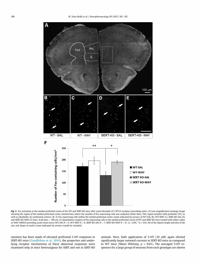
Fig. 2. Fos activation in the medial prefrontal cortex of the WT and SERT-KO mice after acute blockade of 5-HT1A receptors preceding swim. (A) Low magnification montage imageshowing the region of the medial prefrontal cortex (dashed box) where the number of Fos-expressing cells was analyzed (white dots). This region includes both prelimbic (PrL) aswell as infralimbic (IL) prefrontal cortices. (BeE) Fos-expressing cells within the medial prefrontal cortex (some indicated by arrows) of WT-SAL (B), WT-WAY (C), SERT-KO-SAL (D)and SERT-KO-WAY (E) mice. Scale bars ¼ 100 mm. (F) Quantitative analysis of Fos-expressing cells in the medial prefrontal cortex of WT and SERT-KO mice treated with either salineor WAY-100635 preceding acute swim stress (WT-SAL N ¼ 6; WT-WAY N ¼ 6; SERT-KO-SAL N ¼ 7; SERT-KO-WAY N ¼ 6). *p < 0.05, **p < 0.01. All of the objects bright and dim of thesize and shape of nuclei (some indicated by arrows) would be included.
M. Soiza-Reilly et al. / Neuropharmacology 89 (2015) 185e192188
mention has been made of elevated prefrontal 5-HT responses inSERT-KO mice (Goodfellow et al., 2014), the properties and under-lying receptor mechanisms of these abnormal responses wereexamined only in mice heterozygous for SERT and not in SERT-KO
animals. Here, bath application of 5-HT (10 mM) again elicitedsignificantly larger outward currents in SERT-KO mice as comparedto WT mice (Mann Whitney, p < 0.01). The averaged 5-HT re-sponses for a large group of neurons from each genotype are shown
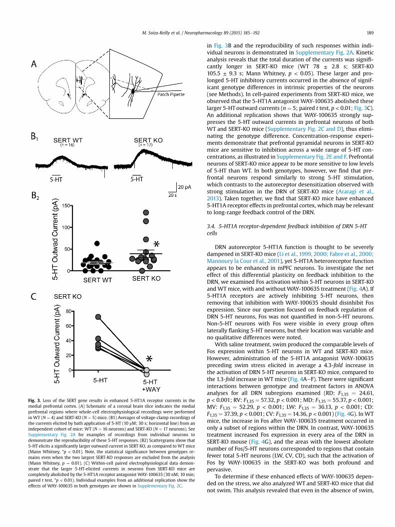
Fig. 3. Loss of the SERT gene results in enhanced 5-HT1A receptor currents in themedial prefrontal cortex. (A) Schematic of a coronal brain slice indicates the medialprefrontal regions where whole-cell electrophysiological recordings were performedin WT (N ¼ 4) and SERT-KO (N ¼ 5) mice. (B1) Averages of voltage-clamp recordings ofthe currents elicited by bath application of 5-HT (10 mM; 30 s; horizontal line) from anindependent cohort of mice: WT (N ¼ 16 neurons) and SERT-KO (N ¼ 17 neurons). SeeSupplementary Fig. 2A for examples of recordings from individual neurons todemonstrate the reproducibility of these 5-HT responses. (B2) Scattergrams show that5-HT elicits a significantly larger outward current in SERT KO, as compared to WT mice(Mann Whitney, *p < 0.01). Note, the statistical significance between genotypes re-mains even when the two largest SERT-KO responses are excluded from the analysis(Mann Whitney, p ¼ 0.01). (C) Within-cell paired electrophysiological data demon-strate that the larger 5-HT-elicited currents in neurons from SERT-KO mice arecompletely abolished by the 5-HT1A receptor antagonist WAY-100635 (30 nM; 10 min;paired t test, *p < 0.01). Individual examples from an additional replication show theeffects of WAY-100635 in both genotypes are shown in Supplementary Fig. 2C.
M. Soiza-Reilly et al. / Neuropharmacology 89 (2015) 185e192 189
in Fig. 3B and the reproducibility of such responses within indi-vidual neurons is demonstrated in Supplementary Fig. 2A. Kineticanalysis reveals that the total duration of the currents was signifi-cantly longer in SERT-KO mice (WT 78 ± 2.8 s; SERT-KO105.5 ± 9.3 s; Mann Whitney, p < 0.05). These larger and pro-longed 5-HT inhibitory currents occurred in the absence of signif-icant genotype differences in intrinsic properties of the neurons(see Methods). In cell-paired experiments from SERT-KO mice, weobserved that the 5-HT1A antagonist WAY-100635 abolished theselarger 5-HT outward currents (n¼ 5; paired t test, p < 0.01; Fig. 3C).An additional replication shows that WAY-100635 strongly sup-presses the 5-HT outward currents in prefrontal neurons of bothWT and SERT-KO mice (Supplementary Fig. 2C and D), thus elimi-nating the genotype difference. Concentration-response experi-ments demonstrate that prefrontal pyramidal neurons in SERT-KOmice are sensitive to inhibition across a wide range of 5-HT con-centrations, as illustrated in Supplementary Fig. 2E and F. Prefrontalneurons of SERT-KO mice appear to be more sensitive to low levelsof 5-HT than WT. In both genotypes, however, we find that pre-frontal neurons respond similarly to strong 5-HT stimulation,which contrasts to the autoreceptor desensitization observed withstrong stimulation in the DRN of SERT-KO mice (Araragi et al.,2013). Taken together, we find that SERT-KO mice have enhanced5-HT1A receptor effects in prefrontal cortex, whichmay be relevantto long-range feedback control of the DRN.
3.4. 5-HT1A receptor-dependent feedback inhibition of DRN 5-HTcells
DRN autoreceptor 5-HT1A function is thought to be severelydampened in SERT-KOmice (Li et al., 1999, 2000; Fabre et al., 2000;Mannoury la Cour et al., 2001), yet 5-HT1A heteroreceptor functionappears to be enhanced in mPFC neurons. To investigate the neteffect of this differential plasticity on feedback inhibition to theDRN, we examined Fos activation within 5-HT neurons in SERT-KOandWTmice, with and withoutWAY-100635 treatment (Fig. 4A). If5-HT1A receptors are actively inhibiting 5-HT neurons, thenremoving that inhibition with WAY-100635 should disinhibit Fosexpression. Since our question focused on feedback regulation ofDRN 5-HT neurons, Fos was not quantified in non-5-HT neurons.Non-5-HT neurons with Fos were visible in every group oftenlaterally flanking 5-HT neurons, but their location was variable andno qualitative differences were noted.
With saline treatment, swim produced the comparable levels ofFos expression within 5-HT neurons in WT and SERT-KO mice.However, administration of the 5-HT1A antagonist WAY-100635preceding swim stress elicited in average a 4.3-fold increase inthe activation of DRN 5-HT neurons in SERT-KO mice, compared tothe 1.3-fold increase in WT mice (Fig. 4AeF). There were significantinteractions between genotype and treatment factors in ANOVAanalyses for all DRN subregions examined (RD: F1,35 ¼ 24.61,p < 0.001; RV: F1,35 ¼ 57.32, p < 0.001; MD: F1,35 ¼ 55.37, p < 0.001;MV: F1,35 ¼ 52.29, p < 0.001; LW: F1,35 ¼ 36.13, p < 0.001; CD:F1,35 ¼ 37.39, p < 0.001; CV: F1,35¼14.36, p < 0.001) (Fig. 4G). InWTmice, the increase in Fos after WAY-100635 treatment occurred inonly a subset of regions within the DRN. In contrast, WAY-100635treatment increased Fos expression in every area of the DRN inSERT-KO mouse (Fig. 4G), and the areas with the lowest absolutenumber of Fos/5-HT neurons corresponded to regions that containfewer total 5-HT neurons (LW, CV, CD), such that the activation ofFos by WAY-100635 in the SERT-KO was both profound andpervasive.
To determine if these enhanced effects of WAY-100635 depen-ded on the stress, we also analyzed WT and SERT-KO mice that didnot swim. This analysis revealed that even in the absence of swim,
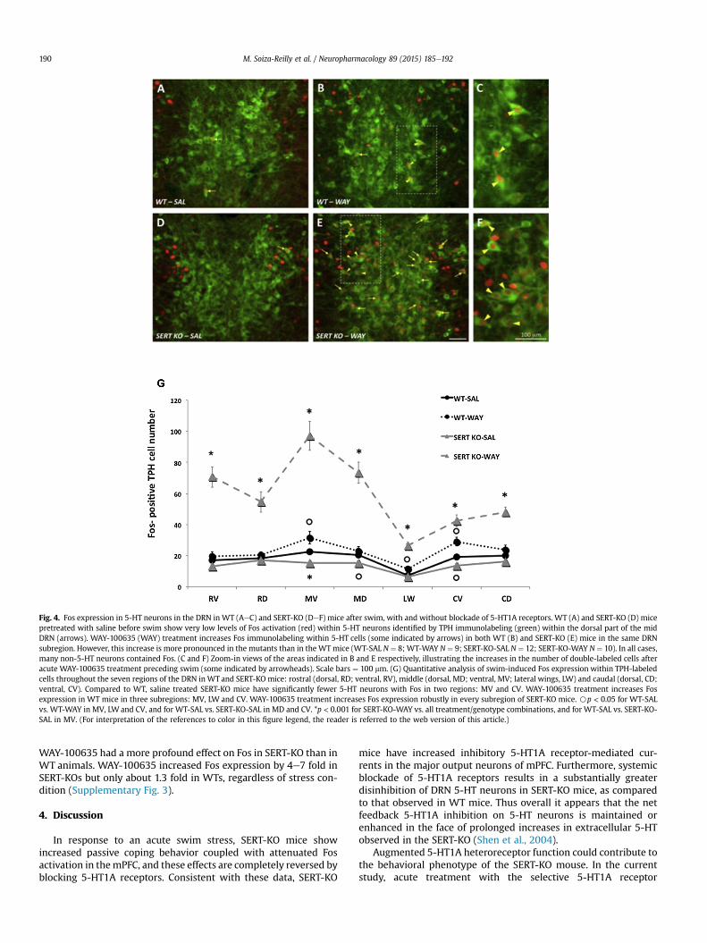
Fig. 4. Fos expression in 5-HT neurons in the DRN in WT (AeC) and SERT-KO (DeF) mice after swim, with and without blockade of 5-HT1A receptors. WT (A) and SERT-KO (D) micepretreated with saline before swim show very low levels of Fos activation (red) within 5-HT neurons identified by TPH immunolabeling (green) within the dorsal part of the midDRN (arrows). WAY-100635 (WAY) treatment increases Fos immunolabeling within 5-HT cells (some indicated by arrows) in both WT (B) and SERT-KO (E) mice in the same DRNsubregion. However, this increase is more pronounced in the mutants than in the WT mice (WT-SAL N ¼ 8; WT-WAY N ¼ 9; SERT-KO-SAL N ¼ 12; SERT-KO-WAY N ¼ 10). In all cases,many non-5-HT neurons contained Fos. (C and F) Zoom-in views of the areas indicated in B and E respectively, illustrating the increases in the number of double-labeled cells afteracute WAY-100635 treatment preceding swim (some indicated by arrowheads). Scale bars ¼ 100 mm. (G) Quantitative analysis of swim-induced Fos expression within TPH-labeledcells throughout the seven regions of the DRN in WT and SERT-KO mice: rostral (dorsal, RD; ventral, RV), middle (dorsal, MD; ventral, MV; lateral wings, LW) and caudal (dorsal, CD;ventral, CV). Compared to WT, saline treated SERT-KO mice have significantly fewer 5-HT neurons with Fos in two regions: MV and CV. WAY-100635 treatment increases Fosexpression in WT mice in three subregions: MV, LW and CV. WAY-100635 treatment increases Fos expression robustly in every subregion of SERT-KO mice. Bp < 0.05 for WT-SALvs. WT-WAY in MV, LW and CV, and for WT-SAL vs. SERT-KO-SAL in MD and CV. *p < 0.001 for SERT-KO-WAY vs. all treatment/genotype combinations, and for WT-SAL vs. SERT-KO-SAL in MV. (For interpretation of the references to color in this figure legend, the reader is referred to the web version of this article.)
M. Soiza-Reilly et al. / Neuropharmacology 89 (2015) 185e192190
WAY-100635 had a more profound effect on Fos in SERT-KO than inWT animals. WAY-100635 increased Fos expression by 4e7 fold inSERT-KOs but only about 1.3 fold in WTs, regardless of stress con-dition (Supplementary Fig. 3).
4. Discussion
In response to an acute swim stress, SERT-KO mice showincreased passive coping behavior coupled with attenuated Fosactivation in themPFC, and these effects are completely reversed byblocking 5-HT1A receptors. Consistent with these data, SERT-KO
mice have increased inhibitory 5-HT1A receptor-mediated cur-rents in the major output neurons of mPFC. Furthermore, systemicblockade of 5-HT1A receptors results in a substantially greaterdisinhibition of DRN 5-HT neurons in SERT-KO mice, as comparedto that observed in WT mice. Thus overall it appears that the netfeedback 5-HT1A inhibition on 5-HT neurons is maintained orenhanced in the face of prolonged increases in extracellular 5-HTobserved in the SERT-KO (Shen et al., 2004).
Augmented 5-HT1A heteroreceptor function could contribute tothe behavioral phenotype of the SERT-KO mouse. In the currentstudy, acute treatment with the selective 5-HT1A receptor
M. Soiza-Reilly et al. / Neuropharmacology 89 (2015) 185e192 191
antagonist WAY-100635 preceding acute swim stress did notmodify stress-coping strategies in WT mice. However in the SERT-KO mouse, WAY-100635 pretreatment markedly increased use ofan active coping strategy. These observations echo previous workshowing that blockade of 5-HT1A receptors normalizes anxiety-likebehavior in the SERT-KO (Holmes et al., 2003). In both cases theeffect of WAY-100635 in SERT KO mice could not be attributed to ageneral increase in locomotor activity.While the location of 5-HT1Areceptors that underlie these effects is unknown, these observa-tions support the behavioral importance of adaptations in 5-HT1Areceptor function for the phenotype of the SERT-KO mouse.
Neuronal activity in the mPFC, as measured by Fos activation,predicted active coping in response to the swim stress. The lowappearance of Fos from our in vivo work in SERT-KO mice fits wellwith the increased inhibitory, 5-HT1A receptor currents wemeasured electrophysiologically inmPFC neurons ex vivo. However,it should be noted that Fosmay be activated as a direct consequenceof 5-HT1A receptor inhibition and/or indirectly as mPFC networkschange state. Since there is abundant 5-HT to stimulate 5-HT1Areceptors in mPFC of SERT-KO mice (Shen et al., 2004), it isconsistent that blocking these receptors would change the balanceof activity in mPFC networks and thus influence coping strategy inthe swim. These observations are consistent with studies that haveimplicated this structure in the selection and implementation ofstress-coping strategies (Amat et al., 2005; Warden et al., 2012).
Blocking 5-HT1A receptors systemically also resulted in amassive disinhibition of Fos within DRN 5-HT neurons in the SERT-KO. This finding indicates that 5-HT1A receptor function in theSERT-KO includes the capacity to exert feedback inhibition of 5-HTneurons in vivo under physiological conditions. That is, blocking 5-HT1A receptors disinhibited Fos expression, implicating the priorpresence of functional feedback inhibition. Consistent with thisobservation, it has previously been noted that systemic adminis-tration of WAY-100635 can increase firing rates of DRN neurons inanesthetized SERT-KOmice (Gobbi et al., 2001). Yet, the locus of therelevant 5-HT1A receptors has not been identified. While the 5-HT1A autoreceptors within the DRN are thought to be down-regulated and desensitized (Li et al., 1999, 2000; Mannoury la Couret al., 2001), they could contribute to this effect. Indeed, a recentstudy using a raphe slice preparation argued that residual 5-HT1Aautoreceptors retain the capacity to provide feedback inhibition of5-HT neurons in the SERT-KO and therefore desensitization doesnot predict their capacity to exert autoinhibition (Araragi et al.,2013). The current data also suggest that 5-HT1A controlled long-feedback loops could be enhanced in SERT-KO mice since mPFCneurons have increased 5-HT1A receptor currents. Thus there couldbe convergent mechanisms contributing to enhanced feedbackcontrol of 5-HT neurons in the SERT-KO.
The DRN and mPFC are reciprocally interconnected and can in-fluence each other bi-directionally (Celada et al., 2001, 2013). There-forewe could speculate that in the SERT-KO,WAY-100635 disinhibitscortical pyramidal neurons, and this results in a shift in descendingcontrol of the DRN from feedback inhibition to direct excitation.However, there aremanypotential circuitdiagrams that couldexplainthe current results. Participation of additional brain regions, otheradaptations in the SERT-KO and the ultimate drivers of activationwithin the DRN and mPFC all remain unknown. Therefore in evalu-ating these scenarios, it is worthwhile to keep inmind that while Fosexperiments give unique insight into adaptations in the SERT-KO,they are hypothesis generating in terms of mechanism.
Our results show that elevated 5-HT levels observed in SERT-KOmice lead to different types of plasticity for the heteroreceptor andautoreceptor subpopulations of 5-HT1A receptors and the nature of5-HT1A-receptor dependent feedback inhibition of the DRN isfundamentally different in the SERT-KO mouse. Future work is
necessary to understand the mechanisms responsible for thesechanges. However, these findings are consistent with the idea thataltered 5-HT1A receptor function could be an etiological factor inmood disorders (Miller et al., 2009). Not only may 5-HT1A re-ceptors be influenced by genetic factors, but they may also bestress-sensitive (Rozeske et al., 2011). Therefore, overall a greaterfocus on interactions between genes and treatment may beessential to understand the neurobiology of stress-relatedpsychopathology.
5. Conclusions
In the current study, SERT-KO mice showed altered behavioralperformance in the swim and parallel changes in mPFC Fosexpression, which were both reversed by blocking 5-HT1A re-ceptors. Enhanced response of mPFC neurons to 5-HT1A-hetero-receptor activation raised the possibility of altered long-rangefeedback to the DRN. Here, we found that net 5-HT1A-receptorinhibition of DRN 5-HT neurons is greatly increased in the SERT-KOmouse. Taken together, these findings suggest that there is greaterdiversity in the plasticity of 5-HT1A-mediated feedback inhibitionin this mouse model of stress sensitivity than previously realized.
Funding and disclosure
This work was supported by NIH grant DA021801 (KGC), as wellas a Discovery Grant from the Natural Science and EngineeringResearch Council (EKL) and an Early Researcher Award from theProvince of Ontario (EKL). EKL holds the Canada Research Chair inDevelopmental Cortical Physiology. NMG was supported by aBanting and Best Canada Graduate Scholarship from the CanadianInstitutes of Health Research.
Acknowledgments
The authors gratefully acknowledge the help of Katherine E.Price, Dr. Guo Yueping and Alayna Silberstein at Boston Children'sHospital and Dr. Derya Sargin of the University of Toronto forinvaluable assistance in completing the experiments.
Appendix A. Supplementary data
Supplementary data related to this article can be found at http://dx.doi.org/10.1016/j.neuropharm.2014.09.017.
References
Amat, J., Baratta, M.V., Paul, E., Bland, S.T., Watkins, L.R., Maier, S.F., 2005. Medialprefrontal cortex determines how stressor controllability affects behavior anddorsal raphe nucleus. Nat. Neurosci. 8, 365e371. http://dx.doi.org/10.1038/nn1399.
Araragi, N., Mlinar, B., Baccini, G., Gutknecht, L., Lesch, K.-P., Corradetti, R., 2013.Conservation of 5-HT1A receptor-mediated autoinhibition of serotonin (5-HT)neurons in mice with altered 5-HT homeostasis. Front. Pharmacol. 4, 97. http://dx.doi.org/10.3389/fphar.2013.00097.
Bengel, D., Murphy, D.L., Andrews, A.M., Wichems, C.H., Feltner, D., Heils, A.,M€ossner, R., Westphal, H., Lesch, K.P., 1998. Altered brain serotonin homeostasisand locomotor insensitivity to 3, 4-methylenedioxymethamphetamine (“Ec-stasy”) in serotonin transporter-deficient mice. Mol. Pharmacol. 53, 649e655.
Bruchas, M.R., Schindler, A.G., Shankar, H., Messinger, D.I., Miyatake, M., Land, B.B.,Lemos, J.C., Hagan, C.E., Neumaier, J.F., Quintana, A., Palmiter, R.D., Chavkin, C.,2011. Selective p38a MAPK deletion in serotonergic neurons produces stressresilience in models of depression and addiction. Neuron 71, 498e511. http://dx.doi.org/10.1016/j.neuron.2011.06.011.
Celada, P., Puig, M.V., Artigas, F., 2013. Serotonin modulation of cortical neurons andnetworks. Front. Integr. Neurosci. 7, 25. http://dx.doi.org/10.3389/fnint.2013.00025.
Celada, P., Puig, M.V., Casanovas, J.M., Guillazo, G., Artigas, F., 2001. Control of dorsalraphe serotonergic neurons by the medial prefrontal cortex: involvement ofserotonin-1A, GABA(A), and glutamate receptors. J. Neurosci. 21, 9917e9929.

M. Soiza-Reilly et al. / Neuropharmacology 89 (2015) 185e192192
Commons, K.G., 2008. Evidence for topographically organized endogenous 5-HT-1Areceptor-dependent feedback inhibition of the ascending serotonin system. Eur.J. Neurosci. 27, 2611e2618. http://dx.doi.org/10.1111/j.1460-9568.2008.06235.x.
Cryan, J.F., Valentino, R.J., Lucki, I., 2005. Assessing substrates underlying thebehavioral effects of antidepressants using the modified rat forced swimmingtest. Neurosci. Biobehav. Rev. 29, 547e569. http://dx.doi.org/10.1016/j.neubiorev.2005.03.008.
Detke, M.J., Rickels, M., Lucki, I., 1995. Active behaviors in the rat forced swimmingtest differentially produced by serotonergic and noradrenergic antidepressants.Psychopharmacol. (Berl.) 121, 66e72.
Fabre, V., Beaufour, C., Evrard, A., Rioux, A., Hanoun, N., Lesch, K.P., Murphy, D.L.,Lanfumey, L., Hamon, M., Martres, M.P., 2000. Altered expression and functionsof serotonin 5-HT1A and 5-HT1B receptors in knock-out mice lacking the 5-HTtransporter. Eur. J. Neurosci. 12, 2299e2310.
Fletcher, A., Forster, E.A., Bill, D.J., Brown, G., Cliffe, I.A., Hartley, J.E., Jones, D.E.,McLenachan, A., Stanhope, K.J., Critchley, D.J., Childs, K.J., Middlefell, V.C.,Lanfumey, L., Corradetti, R., Laporte, A.M., Gozlan, H., Hamon, M., Dourish, C.T.,1996. Electrophysiological, biochemical, neurohormonal and behaviouralstudies with WAY-100635, a potent, selective and silent 5-HT1A receptorantagonist. Behav. Brain Res. 73, 337e353.
Fornal, C.A., Metzler, C.W., Gallegos, R.A., Veasey, S.C., McCreary, A.C., Jacobs, B.L.,1996. WAY-100635, a potent and selective 5-hydroxytryptamine1A antagonist,increases serotonergic neuronal activity in behaving cats: comparison with (S)-WAY-100135. J. Pharmacol. Exp. Ther. 278, 752e762.
Gabbott, P.L.A., Warner, T.A., Jays, P.R.L., Salway, P., Busby, S.J., 2005. Prefrontal cortexin the rat: projections to subcortical autonomic, motor, and limbic centers.J. Comp. Neurol. 492, 145e177. http://dx.doi.org/10.1002/cne.20738.
Gobbi, G., Murphy, D.L., Lesch, K., Blier, P., 2001. Modifications of the serotonergicsystem in mice lacking serotonin transporters: an in vivo electrophysiologicalstudy. J. Pharmacol. Exp. Ther. 296, 987e995.
Goodfellow, N.M., Sargin, D., Ansorge, M.S., Gingrich, J.A., Lambe, E.K., 2014. Micewith compromised 5-HTT function lack phosphotyrosine-mediated inhibitorycontrol over prefrontal 5-HT responses. J. Neurosci. 34, 6107e6111. http://dx.doi.org/10.1523/JNEUROSCI.3762-13.2014.
Haenisch, B., B€onisch, H., 2011. Depression and antidepressants: insights fromknockout of dopamine, serotonin or noradrenaline re-uptake transporters.Pharmacol. Ther. 129, 352e368. http://dx.doi.org/10.1016/j.pharmthera.2010.12.002.
Haj�os, M., Haj�os-Korcsok, E., Sharp, T., 1999. Role of the medial prefrontal cortex in5-HT1A receptor-induced inhibition of 5-HT neuronal activity in the rat. Br. J.Pharmacol. 126, 1741e1750. http://dx.doi.org/10.1038/sj.bjp.0702510.
Holmes, A., Yang, R.J., Lesch, K.-P., Crawley, J.N., Murphy, D.L., 2003. Mice lacking theserotonin transporter exhibit 5-HT(1A) receptor-mediated abnormalities intests for anxiety-like behavior. Neuropsychopharmacology 28, 2077e2088.http://dx.doi.org/10.1038/sj.npp.1300266.
Li, Q., Wichems, C., Heils, A., Lesch, K.P., Murphy, D.L., 2000. Reduction in the densityand expression, but not G-protein coupling, of serotonin receptors (5-HT1A) in5-HT transporter knock-out mice: gender and brain region differences.J. Neurosci. 20, 7888e7895.
Li, Q., Wichems, C., Heils, A., Van De Kar, L.D., Lesch, K.P., Murphy, D.L., 1999.Reduction of 5-hydroxytryptamine (5-HT)(1A)-mediated temperature andneuroendocrine responses and 5-HT(1A) binding sites in 5-HT transporterknockout mice. J. Pharmacol. Exp. Ther. 291, 999e1007.
Mannoury la Cour, C., Boni, C., Hanoun, N., Lesch, K.P., Hamon, M., Lanfumey, L.,2001. Functional consequences of 5-HT transporter gene disruption on 5-HT(1a) receptor-mediated regulation of dorsal raphe and hippocampal cellactivity. J. Neurosci. 21, 2178e2185.
Miller, J.M., Brennan, K.G., Ogden, T.R., Oquendo, M.A., Sullivan, G.M., Mann, J.J.,Parsey, R.V., 2009. Elevated serotonin 1A binding in remitted major depressivedisorder: evidence for a trait biological abnormality. Neuro-psychopharmacology 34, 2275e2284. http://dx.doi.org/10.1038/npp.2009.54.
Murphy, D.L., Lesch, K.-P., 2008. Targeting the murine serotonin transporter: in-sights into human neurobiology. Nat. Rev. Neurosci. 9, 85e96. http://dx.doi.org/10.1038/nrn2284.
Nakamura, K., Matsumoto, M., Hikosaka, O., 2008. Reward-dependent modulationof neuronal activity in the primate dorsal raphe nucleus. J. Neurosci. 28,5331e5343. http://dx.doi.org/10.1523/JNEUROSCI.0021-08.2008.
Porsolt, R.D., Anton, G., Blavet, N., Jalfre, M., 1978. Behavioural despair in rats: a newmodel sensitive to antidepressant treatments. Eur. J. Pharmacol. 47, 379e391.
Roche, M., Commons, K.G., Peoples, A., Valentino, R.J., 2003. Circuitry underlyingregulation of the serotonergic system by swim stress. J. Neurosci. 23, 970e977.
Rozeske, R.R., Evans, A.K., Frank, M.G., Watkins, L.R., Lowry, C.A., Maier, S.F., 2011.Uncontrollable, but not controllable, stress desensitizes 5-HT1A receptors in thedorsal raphe nucleus. J. Neurosci. 31, 14107e14115. http://dx.doi.org/10.1523/JNEUROSCI.3095-11.2011.
Savitz, J., Lucki, I., Drevets, W.C., 2009. 5-HT(1A) receptor function in majordepressive disorder. Prog. Neurobiol. 88, 17e31. http://dx.doi.org/10.1016/j.pneurobio.2009.01.009.
Sharp, T., Boothman, L., Raley, J., Qu�er�ee, P., 2007. Important messages in the “post”:recent discoveries in 5-HT neurone feedback control. Trends Pharmacol. Sci. 28,629e636. http://dx.doi.org/10.1016/j.tips.2007.10.009.
Shen, H.-W., Hagino, Y., Kobayashi, H., Shinohara-Tanaka, K., Ikeda, K.,Yamamoto, H., Yamamoto, T., Lesch, K.-P., Murphy, D.L., Hall, F.S., Uhl, G.R.,Sora, I., 2004. Regional differences in extracellular dopamine and serotoninassessed by in vivo microdialysis in mice lacking dopamine and/or serotonintransporters. Neuropsychopharmacology 29, 1790e1799. http://dx.doi.org/10.1038/sj.npp.1300476.
Sperling, R., Commons, K.G., 2011. Shifting topographic activation and 5-HT1Areceptor-mediated inhibition of dorsal raphe serotonin neurons produced bynicotine exposure and withdrawal. Eur. J. Neurosci. 33, 1866e1875. http://dx.doi.org/10.1111/j.1460-9568.2011.07677.x.
Sprouse, J.S., Aghajanian, G.K., 1987. Electrophysiological responses of serotonin-ergic dorsal raphe neurons to 5-HT1A and 5-HT1B agonists. Synapse 1, 3e9.http://dx.doi.org/10.1002/syn.890010103.
Takahashi, A., Shimamoto, A., Boyson, C.O., DeBold, J.F., Miczek, K.A., 2010. GABA(B)receptor modulation of serotonin neurons in the dorsal raph�e nucleus andescalation of aggression in mice. J. Neurosci. 30, 11771e11780. http://dx.doi.org/10.1523/JNEUROSCI.1814-10.2010.
VanderHorst, V.G.J.M., Ulfhake, B., 2006. The organization of the brainstem andspinal cord of the mouse: relationships between monoaminergic, cholinergic,and spinal projection systems. J. Chem. Neuroanat. 31, 2e36. http://dx.doi.org/10.1016/j.jchemneu.2005.08.003.
Warden, M.R., Selimbeyoglu, A., Mirzabekov, J.J., Lo, M., Thompson, K.R., Kim, S.-Y.,Adhikari, A., Tye, K.M., Frank, L.M., Deisseroth, K., 2012. A prefrontal cortex-brainstem neuronal projection that controls response to behavioural chal-lenge. Nature 492, 428e432. http://dx.doi.org/10.1038/nature11617.
Related Documents