Received: 24 July 2008, Accepted: 24 July 2008, Published online in Wiley InterScience: 2009 Engineered 5S ribosomal RNAs displaying aptamers recognizing vascular endothelial growth factor and malachite green Xing Zhang a , Ajish S. R. Potty b , George W. Jackson c , Victor Stepanov a , Andrew Tang b , Yamei Liu a , Katerina Kourentzi b , Ulrich Strych b , George E. Fox a and Richard C. Willson a,b * In previous work, Vibrio proteolyticus 5S rRNA was shown to stabilize 13–50 nucleotide ‘‘guest’’ RNA sequences for expression in Escherichia coli. The expressed chimeric RNAs accumulated to high levels in E. coli without being incorporated into ribosomes and without obvious effects on the host cells. In this work, we inserted sequences encoding known aptamers recognizing a protein and an organic dye into the 5S rRNA carrier and showed that aptamer function is preserved in the chimeras. A surface plasmon resonance competitive binding assay demonstrated that a vascular endothelial growth factor (VEGF) aptamer/5S rRNA chimera produced in vitro by transcriptional runoff could compete with a DNA aptamer for VEGF, implying binding of the growth factor by the VEGF ‘‘ribosomal RNA aptamer.’’ Separately, a 5S rRNA chimera displaying an aptamer known to increase the fluorescence of malachite green (MG) also enhanced MG fluorescence. Closely related control rRNA molecules showed neither activity. The MG aptamer/5S rRNA chimera, like the original MG aptamer, also increased the fluorescence of other triphenyl methane (TPM) dyes such as crystal violet, methyl violet, and brilliant green, although less effectively than with MG. These results indicate that the molecular recognition properties of aptamers are not lost when they are expressed in the context of a stable 5S rRNA carrier. Inclusion of the aptamer in a carrier may facilitate production of large quantities of RNA aptamers, and may open an approach to screening aptamer libraries in vivo. Copyright ß 2009 John Wiley & Sons, Ltd. Keywords: aptamer/5S rRNA chimera; ribosomal RNA aptamer; vascular endothelial growth factor; malachite green; aptamer; 5S rRNA scaffold; chimeric RNA INTRODUCTION Aptamers—nucleic acid ligands capable of binding a pre- selected molecular target—are normally identified by in vitro selection from a very large pool of 10 15 randomized polynucleotides which is narrowed down to one or a few aptamers by iterative binding selection (Ellington and Szostak, 1990; Tuerk and Gold, 1990). Aptamers have been developed which bind small organic molecules (Hirao et al., 1997; Babendure et al., 2003; Sazani et al., 2004; Chu et al., 2006; Liu and Lu, 2006), proteins (Ruckman et al., 1998; Jayasena, 1999; Srisawat and Engelke, 2001; Lee et al., 2004), heavy metal ions (Chang et al., 2005; Swearingen et al., 2005; Wrzesinski and Ciesiolka, 2005; Stefan et al., 2006; Wernette et al., 2007) and even viruses (Gopinath et al., 2006a,b). Aptamers are best known as ligands to proteins, rivaling antibodies in both affinity and specificity (Jayasena, 1999), and aptamer-based therapeutics are now emerging (Ruckman et al., 1998). Vascular endothelial growth factor (VEGF) is an essential growth factor in the early development of blood vessels. VEGF is generally not required for normal function in the adult except in wound healing, but plays an essential role in tumor growth and age-related macular degeneration (AMD). Pegaptanib sodium (Macugen; Eyetech Pharmaceuticals/Pfizer), the first aptamer drug approved for use in humans, specifically targets VEGF 165 and has proven effective in treating choroidal neovascularization associated with AMD (Ng et al., 2006). Malachite green (MG), a member of the triphenyl methane (TPM) dyes, is used primarily as an antiseptic for the treatment of fungal and bacterial infections of fish in aquaculture, and as a dye in histological staining procedures. MG also has recently drawn attention as a food contaminant (Kraemer, 2008). Compared to (www.interscience.wiley.com) DOI:10.1002/jmr.917 Research Article * Correspondence to: R. C. Willson, Department of Chemical and Biomolecular Engineering University of Houston, 4800 Calhoun Rd., Houston, TX 77204-4004, USA. E-mail: [email protected] a X. Zhang, V. Stepanov, Y. Liu, G. E. Fox, R. C. Willson Department of Biology and Biochemistry, University of Houston, Houston, TX, USA b A. S. R. Potty, A. Tang, K. Kourentzi, U. Strych, R. C. Willson Department of Chemical and Biomolecular Engineering, University of Houston, Houston, TX, USA c G. W. Jackson BioTex, Inc., Houston, TX, USA. Abbreviations used: VEGF, vascular endothelial growth factor; MG, malachite green; CV, crystal violet; CAM, chicken chorioallantoic membrane; AMD, age-related macular degeneration; TPM, triphenyl methane. J. Mol. Recognit. 2009; 22: 154–161 Copyright ß 2009 John Wiley & Sons, Ltd. 154

Welcome message from author
This document is posted to help you gain knowledge. Please leave a comment to let me know what you think about it! Share it to your friends and learn new things together.
Transcript
Research Article
154
Received: 24 July 2008, Accepted: 24 July 2008, Published online in Wiley InterScience: 2009
(www.interscience.wiley.com) DOI:10.1002/jmr.917
Engineered 5S ribosomal RNAs displayingaptamers recognizing vascular endothelialgrowth factor and malachite greenXing Zhanga, Ajish S. R. Pottyb, George W. Jacksonc, Victor Stepanova,Andrew Tangb, Yamei Liua, Katerina Kourentzib, Ulrich Strychb,George E. Foxa and Richard C. Willsona,b*
In previous work, Vibrio proteolyticus 5S rRNA was s
J. Mol. Rec
hown to stabilize 13–50 nucleotide ‘‘guest’’ RNA sequences forexpression in Escherichia coli. The expressed chimeric RNAs accumulated to high levels in E. coli without beingincorporated into ribosomes and without obvious effects on the host cells. In this work, we inserted sequencesencoding known aptamers recognizing a protein and an organic dye into the 5S rRNA carrier and showed thataptamer function is preserved in the chimeras. A surface plasmon resonance competitive binding assay demonstratedthat a vascular endothelial growth factor (VEGF) aptamer/5S rRNA chimera produced in vitro by transcriptional runoffcould compete with a DNA aptamer for VEGF, implying binding of the growth factor by the VEGF ‘‘ribosomal RNAaptamer.’’ Separately, a 5S rRNA chimera displaying an aptamer known to increase the fluorescence ofmalachite green(MG) also enhanced MG fluorescence. Closely related control rRNA molecules showed neither activity. The MGaptamer/5S rRNA chimera, like the original MG aptamer, also increased the fluorescence of other triphenyl methane(TPM) dyes such as crystal violet, methyl violet, and brilliant green, although less effectively than with MG. Theseresults indicate that the molecular recognition properties of aptamers are not lost when they are expressed inthe context of a stable 5S rRNA carrier. Inclusion of the aptamer in a carrier may facilitate production of largequantities of RNA aptamers, andmay open an approach to screening aptamer libraries in vivo. Copyright� 2009 JohnWiley & Sons, Ltd.
Keywords: aptamer/5S rRNA chimera; ribosomal RNA aptamer; vascular endothelial growth factor; malachite green;aptamer; 5S rRNA scaffold; chimeric RNA
* Correspondence to: R. C. Willson, Department of Chemical and Biomolecular
Engineering University of Houston, 4800 Calhoun Rd., Houston, TX
77204-4004, USA.
E-mail: [email protected]
a X. Zhang, V. Stepanov, Y. Liu, G. E. Fox, R. C. Willson
Department of Biology and Biochemistry, University of Houston, Houston, TX,
USA
b A. S. R. Potty, A. Tang, K. Kourentzi, U. Strych, R. C. Willson
Department of Chemical and Biomolecular Engineering, University of
Houston, Houston, TX, USA
c G. W. Jackson
BioTex, Inc., Houston, TX, USA.
Abbreviations used: VEGF, vascular endothelial growth factor; MG, malachite
green; CV, crystal violet; CAM, chicken chorioallantoic membrane; AMD,
age-related macular degeneration; TPM, triphenyl methane.
INTRODUCTION
Aptamers—nucleic acid ligands capable of binding a pre-selected molecular target—are normally identified by in vitroselection from a very large pool of �1015 randomizedpolynucleotides which is narrowed down to one or a fewaptamers by iterative binding selection (Ellington and Szostak,1990; Tuerk and Gold, 1990). Aptamers have been developedwhich bind small organic molecules (Hirao et al., 1997; Babendureet al., 2003; Sazani et al., 2004; Chu et al., 2006; Liu and Lu, 2006),proteins (Ruckman et al., 1998; Jayasena, 1999; Srisawat andEngelke, 2001; Lee et al., 2004), heavy metal ions (Chang et al.,2005; Swearingen et al., 2005; Wrzesinski and Ciesiolka, 2005;Stefan et al., 2006; Wernette et al., 2007) and even viruses(Gopinath et al., 2006a,b). Aptamers are best known as ligands toproteins, rivaling antibodies in both affinity and specificity(Jayasena, 1999), and aptamer-based therapeutics are nowemerging (Ruckman et al., 1998).Vascular endothelial growth factor (VEGF) is an essential
growth factor in the early development of blood vessels. VEGF isgenerally not required for normal function in the adult except inwound healing, but plays an essential role in tumor growth andage-related macular degeneration (AMD). Pegaptanib sodium(Macugen; Eyetech Pharmaceuticals/Pfizer), the first aptamerdrug approved for use in humans, specifically targets VEGF165 and
ognit. 2009; 22: 154–161 Copyright � 20
has proven effective in treating choroidal neovascularizationassociated with AMD (Ng et al., 2006).Malachite green (MG), a member of the triphenyl methane
(TPM) dyes, is used primarily as an antiseptic for the treatment offungal and bacterial infections of fish in aquaculture, and as a dyein histological staining procedures. MG also has recently drawnattention as a food contaminant (Kraemer, 2008). Compared to
09 John Wiley & Sons, Ltd.
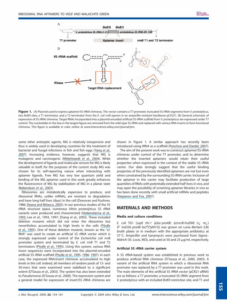
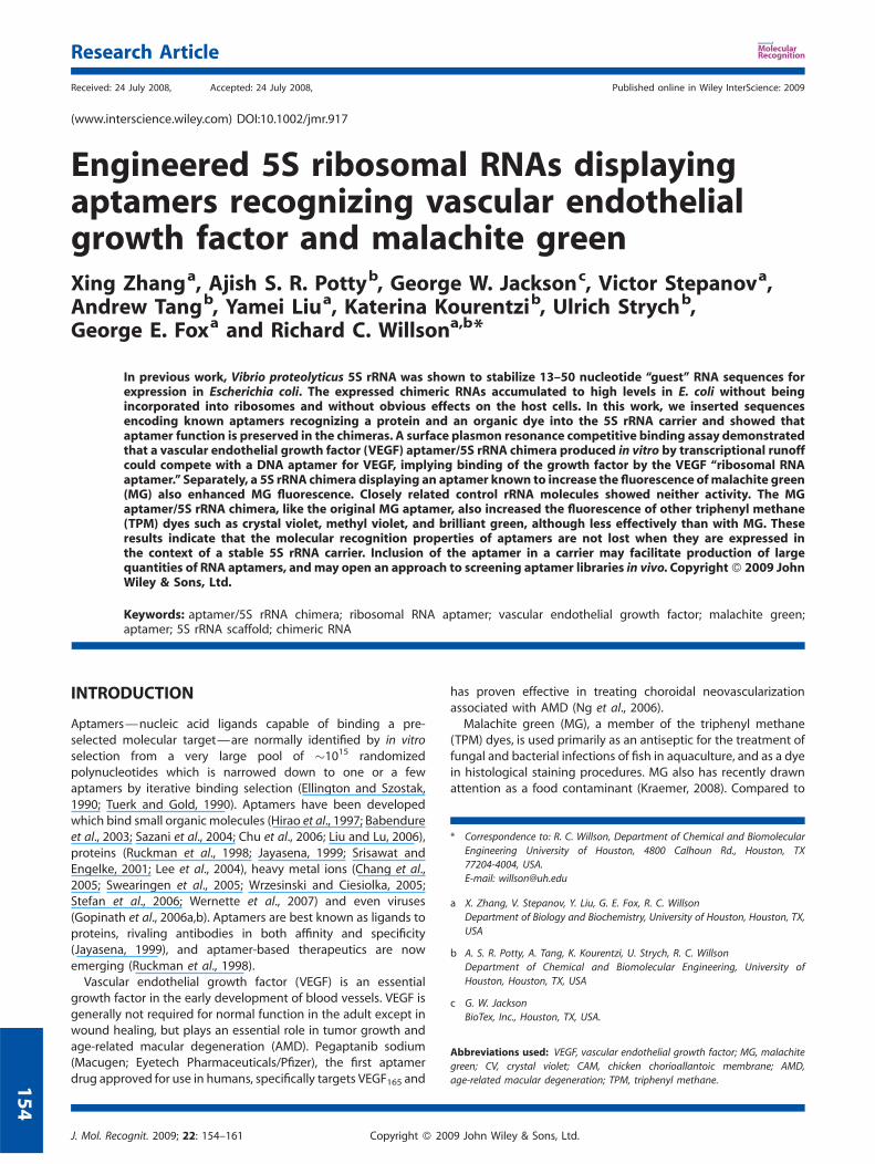
Figure 1. (A) Plasmid used to express aptamer/5S rRNA chimeras. The vector contains a T7 promoter, truncated 5S rRNA segments from V. proteolyticus,
two BstEII sites, a T1 terminator, and a T2 terminator from the E. coli rrnB operon in an ampicillin-resistant backbone pCV251. (B) General schematic of
expression of 5S rRNA chimeras. Target RNAs incorporated into a plasmid-encoded artificial 5S rRNA scaffold from V. proteolyticus are expressed under T7control. The nucleotides in the box in the largest figure are removed from the wild-type 5S rRNA and replaced with various RNA inserts to form functional
chimeras. This figure is available in color online at www.interscience.wiley.com/journal/jmr.
RIBOSOMAL RNA APTAMERS TO VEGF AND MALACHITE GREEN
1
some other antiseptic agents, MG is relatively inexpensive andthus is widely used in developing countries for the treatment ofbacterial and fungal infections in fish and fish eggs (Yang et al.,2007). Increasing evidence, however, suggests that MG ismutagenic and carcinogenic (Mittelstaedt et al., 2004). Whilethe development of ligands andmolecular sensors for MG is likelyvaluable in itself, for the purposes of the current study MG waschosen for its self-reporting nature when interacting withaptamer ligands. Free MG has very low quantum yield andbinding of the MG aptamer used in this work greatly enhancesthe fluorescence of MG by stabilization of MG in a planar state(Babendure et al., 2003).Ribosomes are metabolically expensive to produce, and
ribosomal RNAs, unlike mRNAs, are resistant to degradationand have long half lives (days) in the cell (Donovan and Kushner,1986; Deana and Belasco, 2005). In our previous studies of the 5SrRNA structure space, numerous Vibrio proteolyticus 5S rRNAvariants were produced and characterized (Hedenstierna et al.,1993; Lee et al., 1993, 1997; Zhang et al., 2003). These includeddeletion mutants which did not enter the ribosomes butnevertheless accumulated to high levels in the cells (Pitulleet al., 1995). One of these deletion mutants, known as the ‘‘a1RNA’’ was used to create an artificial 5S rRNA vector which isstrongly expressed under control of the Escherichia coli rrnBpromoter system and terminated by E. coli rrnB T1 and T2terminators (Pitulle et al., 1995). Using this system, various RNAinsert sequences were incorporated into the plasmid-encodedartificial 5S rRNA scaffold (Pitulle et al., 1995, 1996, 1997). In eachcase, the expressed RNA/insert chimeras accumulated to highlevels in the cell. Indeed, all members of random pools of 13- and50-mers that were examined were stabilized to a significantextent (D’Souza et al., 2003). The system has also been extendedto Pseudomonas (D’Souza et al., 2000). The expression system anda general model for expression of insert/5S rRNA chimeras are
J. Mol. Recognit. 2009; 22: 154–161 Copyright � 2009 John Wil
shown in Figure 1. A similar approach has recently beenintroduced using tRNA as a scaffold (Ponchon and Dardel, 2007).The aim of the present work was to construct aptamer/5S rRNA
chimeras under control of the T7 promoter, and to determinewhether the inserted aptamers would retain their usefulproperties when expressed in the context of the stable 5S rRNAcarrier. Our data strongly suggest that the useful bindingproperties of the previously identified aptamers are not lost evenwhen constrained by the surrounding 5S rRNA carrier. Inclusion ofthe aptamer in the carrier may facilitate production of largequantities of RNAs with potentially extended half-lives in vivo, andmay open the possibility of screening aptamer libraries in vivo ashas been done recently with small artificial mRNAs and peptides(Stepanov and Fox, 2007).
MATERIALS AND METHODS
Media and culture conditions
E. coli TG1 [supE thi-1 D(lac-proAB) D(mcrB-hsdSM) (r�K m�K )
(F0 traD36 proAB lacIqZDM15)] was grown on Luria–Bertani (LB)broth plates or in medium with the appropriate antibiotics at378C. Ampicillin and kanamycin were purchased from Sigma–Aldrich (St. Louis, MO), and used at 50 and 25mg/ml, respectively.
Artificial 5S rRNA carrier system
A 5S rRNA-based system was established in previous work toproduce artificial RNA chimeras (D’Souza et al., 2000, 2003). Avariant of the artificial RNA system in which a ribosomal RNApromoter was replaced by a T7 promoter was used in this work.The main elements of the artificial 5S rRNA vector (pCR21-aRNA)are as follows: a T7 promoter, a truncated 5S rRNA segment fromV. proteolyticus with an included BstEII restriction site, and T1 and
ey & Sons, Ltd. www.interscience.wiley.com/journal/jmr
55

Table 1. Aptamer and 5S rRNA chimera sequences
Sequence 50–30
V. proteolyticus 5S rRNA (wild type) UGCCUGGCGACCAUAGCGAUUUGGACCCACCUGAUUCCAUGCCGAACUCAGUAGUGAAACGAAUUAGCGCCGAUGGUAGUGUGGGGUUUCCCCAUGUGAGAGUAGGACAUCGCCAGGCAU
V. proteolyticus 5S rRNA with engineeredBstEII sites and inserted identifier sequence*
UGCCUGGCGACCAUAGCGAUUGGGUAACCGGGUCUAGCGCUGGUUGGGUUUAAUGUGUAGUUGUUUUGCCAUUUGACUGGCUGGUAACCAAUUAGCGCCGAUGGUAGUGUGGGGUUUCCCCAUGUGAGAGUAGGACAUCGCCAGGCAU
Deletion mutant V. proteolyticus 5S rRNA UGCCUGGCGACCAUAGCGAUUGGGUAACCAAUUAGCGCCGAUGGUAGUGUGGGGUUUCCCCAUGUGAGAGUAGGACAUCGCCAGGCAU
MG RNA aptamer GGAUCCCGACUGGCGAGAGCCAGGUAACGAAUGGAUCCMG aptamer/5S rRNA chimera UGCCUGGCGACCAUAGCGAUUGGGUAACCGGAUCCCGACUGGCGAGAGCCAGGU
AACGAAUGGAUCCGGUAACCAAUUAGCGCCGAUGGUAGUGUGGGGUUUCCCCAUGUGAGAGUAGGACAUCGCCAGGCAU
VEGF RNA aptamer GCUCAAUAGUUGGAGGCCUGUCCUCGCCGUAGAGCVEGF aptamer/5S rRNA chimera UGCCUGGCGACCAUAGCGAUUGGGUAACCGCUCAAUAGUUGGAGGCCUGUCCU
CGCCGUAGAGCGGUAACCAAUUAGCGCCGAUGGUAGUGUGGGGUUUCCCCAUGUGAGAGUAGGACAUCGCCAGGCAU
VEGF DNA aptamer CCGTCTTCCAGACAAGAGTGCAGGG
Aptamer sequences inserted into 5S rRNA artificial cassette are underlined, and BstEII sites are in bold.*The 53-nucleotide fragment inserted into the BstEII site of the engineered V. proteolyticus 5S rRNA is a unique identifier sequenceused for non-perturbing labeling of genetically modified organisms (D’Souza et al., 2000).
X. ZHANG ET AL.
156
T2 terminators from the E. coli rrnB operon (Figure 1A). Standardrecombinant DNA techniques were used to insert known VEGF-and MG-aptamer coding sequences into the artificial 5S rRNAscaffold. Agarose gel electrophoresis was used to identifycandidates which appeared to have MG and VEGF aptamercoding sequences were inserted into the BstEII site of the vector,and positive colonies were verified by sequencing. The aptamersequences used in this paper are summarized in Table 1.Molecular modeling of the V. proteolyticus 50S ribosome/aptamerchimera was performed using Pymol (DeLano, 2002) based onPDB structure 2AW4 of the closely related E. coli 50S ribosome(Figure 2).
Figure 2. E. coli 50S ribosome. V. proteolyticus 5S rRNA shares 85%
identity to E. coli 5S rRNA. 5S rRNA is shown in green and the insertion sitefor target RNAs (G21) is shown in red. 23S rRNAs is shown in white, and
ribosomal proteins are shown in yellow.
www.interscience.wiley.com/journal/jmr Copyright � 2009
Recombinant DNA techniques
Restriction endonucleases and DNA-modifying enzymes werepurchased from New England Biolabs (Ipswich, MA). Oligonucleo-tides for construction of VEGF and MG ribosomal RNA aptamerswere purchased from Integrated DNATechnologies (Coralville, IA).DNAwas annealed by denaturation at 958C for 10min followed bycooling to room temperature over 4 h. DNA sequencing wascarried out by Lone Star Laboratories (Houston, TX).
Bacterial expression and purification of VEGF
The human VEGF165 gene from pET-3d-VEGF (kindly provided byDr. Gerhard Siemeister, Tumor Biology Center, Institute ofMolecular Medicine, Freiburg, Germany) was cloned into apET-26b vector (Siemeister et al., 1996). Bacterial cultures weregrown to an OD600 of 1.0, isopropyl-b-D-thiogalactopyranoside(IPTG) was added to a final concentration of 1mM and thecultures were allowed to grow for another 4 h at 378C for VEGFexpression. Bacterial cells were washed in cold phosphatebuffered saline (8mM Na2HPO4; 2mM KH2PO4; 138mM NaCl;2.7mM KCl; pH 7.4), and then resuspended in chilled lysis buffer(50mM Tris–HCl, 10mM 2-mercaptoethanol, 2mM EDTA, 5% gly-cerol, pH 8), and lysed by passage through a French Press at10 000 psi. The mixture was centrifuged at 17 000g and 48C for30 min, and the pellet containing VEGF inclusion bodies wasrecovered. The pellet was resuspended and stirred in solubil-ization buffer (6M urea, 100mM DTT, 50mM MES, pH 5.5) for 1 h.The solubilized mixture containing denatured VEGF was loadedonto a 40mm internal diameter, 30mm long SP Sepharosecation-exchange column (GE Healthcare, Piscataway, NJ). Thebound VEGF eluted between 400 and 500mM NaCl in a gradientof 100mM to 1M NaCl in 6M urea over 10 column volumes.
John Wiley & Sons, Ltd. J. Mol. Recognit. 2009; 22: 154–161

RIBOSOMAL RNA APTAMERS TO VEGF AND MALACHITE GREEN
1
Refolding of denatured VEGF was accomplished by mixing 20mlof 1mg/ml denatured VEGF with 40ml refolding buffer (6M urea,0.5M cystamine, 0.1M glycine, 20mM HEPES, pH 7.4) for 4 h withgentle agitation. The 60ml mixture was then added dropwise at aflow rate of 5ml/min to a 2 L flask containing 1140ml of PBS with50mM glycine buffer, pH 7.4 with gentle stirring until the finalconcentrations of VEGF and urea were 50mg/ml and 0.3M,respectively. The sample was centrifuged at 17 000g and 48C for30 min to remove aggregates. The refolded VEGF dimer wasloaded on a 30mm internal diameter and 30mm long HeparinSepharose 6 Fast Flow heparin-affinity column (GE Healthcare).The refolded VEGF was eluted by 40ml of PBS/1M NaCl, anddialyzed for 24 h against 1 L of PBS at 48C with a Spectra/Portubular membrane with a 6–8000 molecular weight cutoff(Spectrum Laboratories, Rancho Dominguez, CA). The concen-tration of VEGF was determined using the Bradford assay withbovine serum albumin (BSA) as a standard (Bradford, 1976). Thefinal purity of VEGF was determined by SDS–PAGE (Figure 4C).
Characterization of the DNA aptamer binding capacity ofthe purified recombinant VEGF
50-Fluorescein-labeled VEGF DNA aptamer was obtained fromMWG Biotech, Inc. (High Point, NC). The association of thisaptamer with our recombinant VEGF165 was characterized byfluorescence anisotropy using a SPEX 212 instrument (HoribaJobin Yvon, Edison, NJ) in T format. Fluorescein-Labeled DNAaptamer (65 nM) was titrated with VEGF (56–389 nM) in 2.5mlPBS buffer and the change in fluorescence anisotropy wasfollowed.
Chicken chorioallantoic membrane (CAM) angiogenesisassay for VEGF activity
Fertilized, specific-pathogen-free (SPF) chicken eggs werepurchased from Charles River Laboratories, Inc. (Wilmington,MA). On day 1, eggs were sterilized by wiping with 70% ethanoland incubated in a Complete Hatch System incubator obtainedfrom Double R Pet, Farm & Equestrian Supply (Palm Bay, Florida)with humidified air at 378C. To detach the CAM from the shell, ashallow hole was made at the blunt end of a 4-day egg using ahand drill (Dremel #750 MiniMite), and 2ml albumin wasremoved using an 18-gauge hypodermic needle to generate afalse air sac at the top of the egg at the equator. On day 5, chickenembryo development was assessed by candling to observegrowth of blood vessels. On day 6, a window was opened usingfine forceps to pick away the shell over the false air sac. Thewindow was sealed with a 4.4� 4.4 cm2 sterile transparentdressing (TegadermTM, 3M Healthcare, St. Paul, MN). On day 11,20ml of each test sample was placed on an 18� 18mm2
autoclaved glass cover slip (Baxter Healthcare Corporation,Deerfield, IL) and allowed to dry in a Class II Biosafety cabinet for20min. These slips were aseptically flipped over onto the CAMmembranes of the eggs through the opened windows, and thewindows were resealed with sterile transparent dressing. On day14, windows were opened and angiogenesis was assayed bydigital imaging of the formation of blood vessels.
Fluorescence enhancement assay formalachite green binding
To measure the degree of MG fluorescence enhancement by thechimeric MG aptamer/5S rRNA, RNAwas added to a black 96-well
J. Mol. Recognit. 2009; 22: 154–161 Copyright � 2009 John Wil
plate (Corning Life Sciences, Lowell, MA) containing 0.2ml of0.33mMMG to final RNA concentrations ranging from 0.33 nM to3.30mM. All MG fluorescence measurements were performed inan EM microplate spectrofluorometer (Molecular DevicesCorporation, Sunnyvale, CA). The excitation monochromatorwas set to either 430 or 620 nm (see below), and the emissionmonochromator was set to 655 nm. Tominimize scattered light, a630 nm cutoff filter was used on the emission channel. The datawere analyzed using SoftMax Pro software (Version 5.0).
Biacore sensor assay
All surface plasmon resonance experiments were conducted at258C in PBS buffer with 0.005% Tween-20 using a BIAcore 2000instrument (Biacore, Inc., Piscataway, NJ). Carboxymethylateddextran (CM5) chips were purchased from Biacore Life Sciences(Biacore, Inc.). 30-Biotin-modified VEGF DNA aptamer (Table 1) wasordered from Integrated DNA Technologies. For sensor prep-aration, 30-biotin-modified VEGF DNA aptamer was immobilizedon a CM5 chip with avidin–biotin coupling chemistry as describedbelow. Carboxymethylated dextran was activated by flowing afreshly mixed solution of 1-ethyl-3-(3-dimethylaminopropyl)-carbodiimide (EDC 400mM) and N-hydroxysuccinimide (NHS100mM) in a 1:1 ratio over the sensor chip followed by 200ml/minof streptavidin for 7min. The inactivated carboxyl groups werequenched by 1M ethanolamine for another 7min. A flow rate of5ml/min was used throughout the procedure. The 30-biotin-modified VEGF DNA aptamer was then bound to the streptavidin-coated sensor (on selected flow cells) at low loading densities(�20RUs) tominimizemass transport effects. This was achieved bypassing 12.5nM of the 30-biotin-modified VEGF DNA aptamer overthe chip for 2min at 5ml/min. Recognition of VEGF by the aptamer/5S rRNA chimera was studied by performing a competitive bindingexperiment as follows: 66nM VEGF was incubated with 300nMVEGF-aptamer/5S rRNA chimera, or with a control RNA(V. proteolyticus artificial 5S rRNA or a deletion mutantV. proteolyticus 5S rRNA; Table 1) for 30min. The complex wasthen passed over the CM5 sensor chip with immobilizedbiotinylated DNA aptamer to VEGF. Regeneration of the chipwas achieved by treating the surface with a 10 s pulse of 2.5mMNaOH, 25mM NaCl with a flow rate of 40ml/min. Response fromthe reference flow cell (flow cell without the aptamer) wassubtracted to get the actual response. The data were alsodouble-referenced with blank runs of PBS buffer with 0.005%Tween-20. All runs were performed at least in duplicate. Theresulting data were globally fit with parameters kon (associationconstant; M�1 s�1), koff (dissociation constant; s�1), and Rmax (themaximum amount of VEGF in terms of RUs that can bind to thechip; RU) using Scrubber 2.0 (University of Utah, UT).
RESULTS
Predicted secondary structures of VEGF aptamer/5S rRNAand MG aptamer/5S rRNA chimeras
Mfold (Zuker, 2003) was utilized to determine possible secondarystructures of the RNA chimeras expressed from the artificial 5SrRNA vector. We expected those molecules with the lowest freeenergy of folding to be the most likely aptamer structures. TheDG’s of folding at 1M Naþ for the wild-type V. proteolyticus 5S
ey & Sons, Ltd. www.interscience.wiley.com/journal/jmr
57
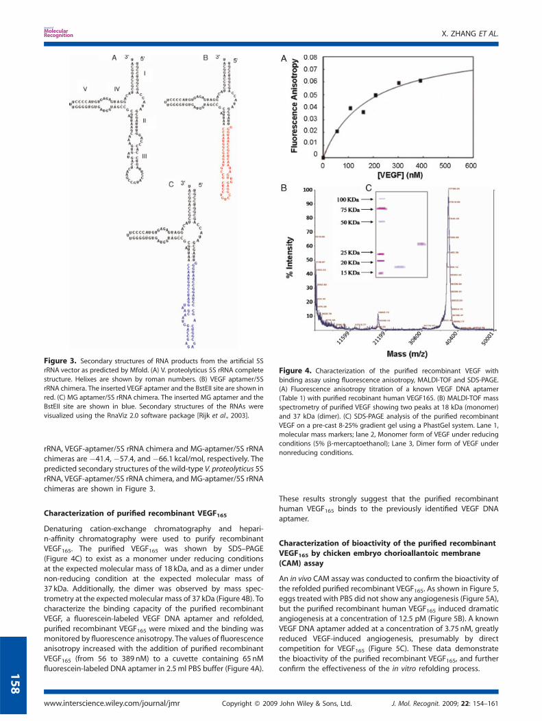
Figure 3. Secondary structures of RNA products from the artificial 5SrRNA vector as predicted by Mfold. (A) V. proteolyticus 5S rRNA complete
structure. Helixes are shown by roman numbers. (B) VEGF aptamer/5S
rRNA chimera. The inserted VEGF aptamer and the BstEII site are shown inred. (C) MG aptamer/5S rRNA chimera. The inserted MG aptamer and the
BstEII site are shown in blue. Secondary structures of the RNAs were
visualized using the RnaViz 2.0 software package [Rijk et al., 2003].
Figure 4. Characterization of the purified recombinant VEGF with
binding assay using fluorescence anisotropy, MALDI-TOF and SDS-PAGE.(A) Fluorescence anisotropy titration of a known VEGF DNA aptamer
(Table 1) with purified recobinant human VEGF165. (B) MALDI-TOF mass
spectrometry of purified VEGF showing two peaks at 18 kDa (monomer)
and 37 kDa (dimer). (C) SDS-PAGE analysis of the purified recombinantVEGF on a pre-cast 8-25% gradient gel using a PhastGel system. Lane 1,
molecular mass markers; lane 2, Monomer form of VEGF under reducing
conditions (5% b-mercaptoethanol); Lane 3, Dimer form of VEGF under
nonreducing conditions.
X. ZHANG ET AL.
158
rRNA, VEGF-aptamer/5S rRNA chimera and MG-aptamer/5S rRNAchimeras are �41.4, �57.4, and �66.1 kcal/mol, respectively. Thepredicted secondary structures of the wild-type V. proteolyticus 5SrRNA, VEGF-aptamer/5S rRNA chimera, and MG-aptamer/5S rRNAchimeras are shown in Figure 3.
Characterization of purified recombinant VEGF165
Denaturing cation-exchange chromatography and hepari-n-affinity chromatography were used to purify recombinantVEGF165. The purified VEGF165 was shown by SDS–PAGE(Figure 4C) to exist as a monomer under reducing conditionsat the expected molecular mass of 18 kDa, and as a dimer undernon-reducing condition at the expected molecular mass of37 kDa. Additionally, the dimer was observed by mass spec-trometry at the expectedmolecular mass of 37 kDa (Figure 4B). Tocharacterize the binding capacity of the purified recombinantVEGF, a fluorescein-labeled VEGF DNA aptamer and refolded,purified recombinant VEGF165 were mixed and the binding wasmonitored by fluorescence anisotropy. The values of fluorescenceanisotropy increased with the addition of purified recombinantVEGF165 (from 56 to 389 nM) to a cuvette containing 65 nMfluorescein-labeled DNA aptamer in 2.5ml PBS buffer (Figure 4A).
www.interscience.wiley.com/journal/jmr Copyright � 2009
These results strongly suggest that the purified recombinanthuman VEGF165 binds to the previously identified VEGF DNAaptamer.
Characterization of bioactivity of the purified recombinantVEGF165 by chicken embryo chorioallantoic membrane(CAM) assay
An in vivo CAM assay was conducted to confirm the bioactivity ofthe refolded purified recombinant VEGF165. As shown in Figure 5,eggs treated with PBS did not show any angiogenesis (Figure 5A),but the purified recombinant human VEGF165 induced dramaticangiogenesis at a concentration of 12.5 pM (Figure 5B). A knownVEGF DNA aptamer added at a concentration of 3.75 nM, greatlyreduced VEGF-induced angiogenesis, presumably by directcompetition for VEGF165 (Figure 5C). These data demonstratethe bioactivity of the purified recombinant VEGF165, and furtherconfirm the effectiveness of the in vitro refolding process.
John Wiley & Sons, Ltd. J. Mol. Recognit. 2009; 22: 154–161
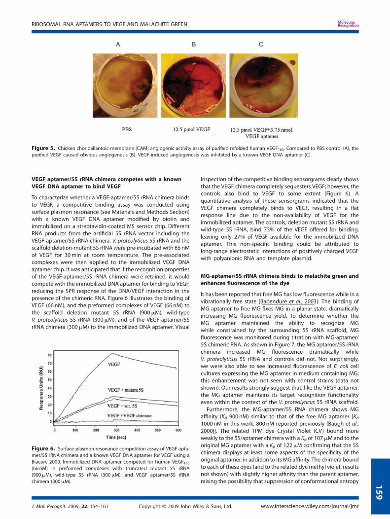
Figure 5. Chicken chorioallantoic membrane (CAM) angiogenic activity assay of purified refolded human VEGF165. Compared to PBS control (A), the
purified VEGF caused obvious angiogenesis (B). VEGF-induced angiogenesis was inhibited by a known VEGF DNA aptamer (C).
RIBOSOMAL RNA APTAMERS TO VEGF AND MALACHITE GREEN
VEGF aptamer/5S rRNA chimera competes with a knownVEGF DNA aptamer to bind VEGF
To characterize whether a VEGF-aptamer/5S rRNA chimera bindsto VEGF, a competitive binding assay was conducted usingsurface plasmon resonance (see Materials and Methods Section)with a known VEGF DNA aptamer modified by biotin andimmobilized on a streptavidin-coated M5 sensor chip. DifferentRNA products from the artificial 5S rRNA vector including theVEGF-aptamer/5S rRNA chimera, V. proteolyticus 5S rRNA and thescaffold deletion-mutant 5S rRNAwere pre-incubated with 65 nMof VEGF for 30min at room temperature. The pre-associatedcomplexes were then applied to the immobilized VEGF DNAaptamer chip. It was anticipated that if the recognition propertiesof the VEGF-aptamer/5S rRNA chimera were retained, it wouldcompete with the immobilized DNA aptamer for binding to VEGF,reducing the SPR response of the DNA/VEGF interaction in thepresence of the chimeric RNA. Figure 6 illustrates the binding ofVEGF (66 nM), and the preformed complexes of VEGF (66 nM) tothe scaffold deletion mutant 5S rRNA (900mM), wild-typeV. proteolyticus 5S rRNA (300mM), and of the VEGF-aptamer/5SrRNA chimera (300mM) to the immobilized DNA aptamer. Visual
Figure 6. Surface plasmon resonance competition assay of VEGF apta-
mer/5S rRNA chimera and a known VEGF DNA aptamer for VEGF using a
Biacore 2000. Immobilized DNA aptamer competed for human VEGF165(66 nM) in preformed complexes with truncated mutant 5S rRNA(900mM), wild-type 5S rRNA (300mM), and VEGF aptamer/5S rRNA
chimera (300mM).
J. Mol. Recognit. 2009; 22: 154–161 Copyright � 2009 John Wil
1
inspection of the competitive binding sensorgrams clearly showsthat the VEGF chimera completely sequesters VEGF; however, thecontrols also bind to VEGF to some extent (Figure 6). Aquantitative analysis of these sensorgrams indicated that theVEGF chimera completely binds to VEGF, resulting in a flatresponse line due to the non-availability of VEGF for theimmobilized aptamer. The controls, deletion mutant 5S rRNA andwild-type 5S rRNA, bind 73% of the VEGF offered for binding,leaving only 27% of VEGF available for the immobilized DNAaptamer. This non-specific binding could be attributed tolong-range electrostatic interactions of positively charged VEGFwith polyanionic RNA and template plasmid.
MG-aptamer/5S rRNA chimera binds to malachite green andenhances fluorescence of the dye
It has been reported that free MG has low fluorescence while in avibrationally free state (Babendure et al., 2003). The binding ofMG aptamer to free MG fixes MG in a planar state, dramaticallyincreasing MG fluorescence yield. To determine whether theMG aptamer maintained the ability to recognize MGwhile constrained by the surrounding 5S rRNA scaffold, MGfluorescence was monitored during titration with MG-aptamer/5S chimeric RNA. As shown in Figure 7, the MG aptamer/5S rRNAchimera increased MG fluorescence dramatically whileV. proteolyticus 5S rRNA and controls did not. Not surprisingly,we were also able to see increased fluorescence of E. coli cellcultures expressing the MG aptamer in medium containing MG;this enhancement was not seen with control strains (data notshown). Our results strongly suggest that, like the VEGF aptamer,the MG aptamer maintains its target recognition functionalityeven within the context of the V. proteolyticus 5S rRNA scaffold.Furthermore, the MG-aptamer/5S RNA chimera shows MG
affinity (Kd 900 nM) similar to that of the free MG aptamer [Kd1000 nM in this work, 800 nM reported previously (Baugh et al.,2000)]. The related TPM dye Crystal Violet (CV) bound moreweakly to the 5S/aptamer chimera with a Kd of 107mM and to theoriginal MG aptamer with a Kd of 122mM confirming that the 5Schimera displays at least some aspects of the specificity of theoriginal aptamer, in addition to its MG affinity. The chimera boundto each of these dyes (and to the related dyemethyl violet, resultsnot shown) with slightly higher affinity than the parent aptamer,raising the possibility that suppression of conformational entropy
ey & Sons, Ltd. www.interscience.wiley.com/journal/jmr
59

Figure 7. MG aptamer/5S rRNA chimera enhances fluorescence of
malachite green and crystal violet. Malachite green and crystal violet
(0.33mM) were incubated with 5S rRNA/MG aptamer chimera, free MG
aptamer or wild-type 5S rRNA (control) for 10 min at room temperature.Fluorescence of the complex was measured by a Gemini EM microplate
spectrofluorometer. Each value represents themean� standard deviation
for three replicates. The best fit was determined using Igor Pro 4.04
(WaveMetrics, Inc., Portland, OR). (A) MG: excitation wavelength 430 nm;emission 655 nm; cutoff 630 nm. (B) CV: excitation 570 nm; emission
650 nm; cutoff 630 nm. This figure is available in color online at www.
interscience.wiley.com/journal/jmr.
X. ZHANG ET AL.
160
of the aptamer increases affinity in the chimera, but thispossibility requires further investigation.
DISCUSSION
In this paper, we proposed a novel expression system for RNAaptamers in E. coli using a 5S rRNA scaffold. Our resultsdemonstrate that both VEGF and MG RNA aptamers retain theiruseful recognition properties even when constrained by the 5SrRNA scaffold. As mentioned above, MG as an aptamer target isinteresting for a variety of reasons but was chosen in this studyprimarily due to its self-reporting fluorescence enhancementupon binding to previously described aptamers. Using thisfeature, it has been shown here that an MG aptamer retainsrecognition for the target dye molecule with sensitivity andspecificity despite being conformationally constrained at boththe 50- and 30-ends and despite any additional steric hindranceresulting from the 5S rRNA derived scaffold. As shown in Figure 7,MG can be readily detected in vitro at concentrations as low as0.33mM with MG aptamer/5S rRNA chimera.
www.interscience.wiley.com/journal/jmr Copyright � 2009
Aside from the sensing application, it is known that MG hasthree different forms including chromatic MG, carbinol base, andleuco MG. Chromatic MG is the most commonly known formwitha characteristic green color and can be converted to carbinol baseand leucoMG through cell metabolism. Among them, leucoMG isbelieved to be most toxic to humans (Mittelstaedt et al., 2004). Amethod to prevent the conversion of chromatic MG to leuco MGmight be important for prophylaxis following MG exposure oringestion. As both MG aptamer and MG aptamer/5S rRNAchimera firmly bind to MG, it is tempting to speculate that theycould be used to sequester MG and block the conversion fromchromatic MG to leucoMG. Further work would be needed to testthis hypothesis.The low yield and high costs of in vitro transcription or
chemical synthesis of RNA at large scales (i.e., for RNA as apharmaceutical) are quite burdensome. In this paper, weconstructed an aptamer/5S rRNA expression system whichmay offer a promising approach to expression and production offunctional RNA aptamers at a large scale by conventionalfermentation of E. coli. While non-natural modifications couldnot be directly introduced via fermentation, we have shownpreviously with non-aptamer RNAs that by ‘‘camouflaging’’ thedesired RNA within a scaffold resembling 5S rRNA, the aptamerproduct(s) should exhibit enhanced stability to nucleasedegradation. In this work in particular, we have clearlydemonstrated that a 5S-MG aptamer RNA retains its ability tobind MG even after prolonged storage of the cells. Of course, inaddition to the large-scale production of chimeric ribosomalaptamers, our approach may be useful for the insertion of anumber of other functional RNAs within an rRNA scaffoldincluding siRNAs, microRNAs, ribozymes, etc. As we havepreviously shown, the 5S rRNA deletion construct can readilyhost RNA inserts up to 120 nt (D’Souza et al., 2003) such thatinserts with dual function may be generated (e.g., an aptamerconjugated to an siRNA). Finally, based on the demonstrationhere that aptamers identified a priori can be inserted in our 5SrRNA-based system without loss of functionality, we haveinitiated studies on in vivo selection of aptamers starting withrandomized inserts and appropriate selection pressure.
Acknowledgements
We thank the members of the Willson and Fox laboratories forhelpful discussions. This work was supported by NIH and bygrants from the Robert A. Welch Foundation (E-1264 and E-1451)as well as NIH STTR grant 1R41ES016478 to GWJ in collaborationwith GEF and RCW.
REFERENCES
Babendure JR, Adams SR, Tsien RY. 2003. Aptamers switch on fluorescenceof triphenylmethane dyes. J. Am. Chem. Soc. 125: 14716–14717.
Baugh C, Grate D, Wilson C. 2000. 2.8 A crystal structure of the malachitegreen aptamer. J. Mol. Biol. 301: 117–128.
Bradford MM. 1976. A rapid and sensitive method for the quantitation ofmicrogram quantities of protein utilizing the principle of protein-dyebinding. Anal. Biochem. 72: 248–254.
Chang I-H, Tulock J, Liu J, Kim W-S, Cannon D Jr, Lu Y, Bohn P, Sweedler J,Cropek D. 2005. Miniaturized lead sensor based on lead-specificDNAzyme in a nanocapillary interconnected microfluidic device.Environ. Sci. Technol. 39: 3756.
Chu TC, Marks 3rd JW, Lavery LA, Faulkner S, RosenblumMG, Ellington AD,Levy M, 2006. Aptamer:toxin conjugates that specifically targetprostate tumor cells. Cancer Res. 66: 5989–5992.
John Wiley & Sons, Ltd. J. Mol. Recognit. 2009; 22: 154–161

RIBOSOMAL RNA APTAMERS TO VEGF AND MALACHITE GREEN
Deana A, Belasco JG. 2005. Lost in translation: The influence of ribosomeson bacterial mRNA decay. Genes Dev. 19: 2526–2533.
D’Souza LM, Willson RC, Fox GE. 2000. Expression of marker RNAs inPseudomonas putida. Curr. Microbiol. 40: 91–95.
D’Souza LM, Larios-Sanz M, Setterquist RA, Willson RC, Fox GE. 2003. SmallRNA sequences are readily stabilized by inclusion in a carrier rRNA.Biotechnol. Prog. 19: 734–738.
DeLano WL. 2002. The PyMOL User’s Manual. DeLano Scientific: Palo Alto,CA, USA.
Donovan WP, Kushner SR. 1986. Polynucleotide phosphorylase andribonuclease II are required for cell viability and mRNA turnoverin Escherichia coli K-12. Proc. Natl. Acad. Sci. USA 83: 120–124.
Ellington AD, Szostak JW. 1990. In vitro selection of RNA molecules thatbind specific ligands. Nature 346: 818–822.
Gopinath SCB, Misono TS, Kawasaki K, Mizuno T, Imai M, Odagiri T, KumarPKR. 2006a. An RNA aptamer that distinguishes between closelyrelated human influenza viruses and inhibits haemagglutinin-mediated membrane fusion. J. Gen. Virol. 87: 479–487.
Gopinath SCB, Sakamaki Y, Kawasaki K, Kumar PKR. 2006b. An efficientRNA aptamer against human influenza B virus hemagglutinin.J. Biochem. 139: 837–846.
Hedenstierna KO, Lee YH, Yang Y, Fox GE. 1993. A prototype stableRNA identification cassette for monitoring plasmids of geneticallyengineered microorganisms. Syst. Appl. Microbiol. 16: 280–286.
Hirao I, Yoshinari S, Yokoyama S, Endo Y, Ellington AD. 1997. In vitroselection of aptamers that bind to ribosome-inactivating toxins.Nucleic Acids Symp. Ser. 37: 283–284.
Jayasena SD. 1999. Aptamers: An emerging class of molecules that rivalantibodies in diagnostics. Clin. Chem. 45: 1628–1650.
Kraemer D. 2008. Hearing on Chinese Seafood: Safety and Trade Issues:‘‘U.S.-China Economic and Security Review Commission’’. FDA: Washing-ton, D.C.; http://www.fda.gov/ola/2008/seafood042408.html.
Lee YH, D’Souza LM, Fox GE. 1993. Experimental investigation of anRNA sequence space. Orig. Life Evol. Biosphere 23: 365–372.
Lee YH, D’Souza LM, Fox GE. 1997. Equally parsimonious pathwaysthrough an RNA sequence space are not equally likely. J. Mol. Evol.45: 278–284.
Lee JF, Hesselberth JR, Meyers LA, Ellington AD. 2004. Aptamer database.Nucleic Acids Res. 32: D95–D100.
Liu J, Lu Y. 2006. Fast colorimetric sensing of adenosine and cocaine basedon a general sensor design involving aptamers and nanoparticles.Angew. Chem. Int. Ed. 117: 90–94.
Mittelstaedt RA, Mei N, Webb PJ, Shaddock JG, Dobrovolsky VN,McGarrity LJ, Morris SM, Chen T, Beland FA, Greenlees KJ,Heflich RH. 2004. Genotoxicity of malachite green and leucomala-chite green in female Big Blue B6C3F1 mice. Mutat. Res. 561: 127–138.
Ng EW, Shima DT, Calias P, Cunningham ET Jr, Guyer DR, Adamis AP. 2006.Pegaptanib, a targeted anti-VEGFaptamer for ocular vascular disease.Nat. Rev. Drug Discov. 5: 123–132.
Pitulle C, Dsouza L, Fox GE. 1997. A low molecular weight artificial RNA ofunique size with multiple probe target regions. Syst. Appl. Microbiol.20: 133–136.
J. Mol. Recognit. 2009; 22: 154–161 Copyright � 2009 John Wil
Pitulle C, Hedenstierna KO, Fox GE. 1995. A novel approach for monitoringgenetically engineered microorganisms by using artificial, stableRNAs. Appl. Environ. Microbiol. 61: 3661–3666.
Pitulle C, Hedenstierna KOF, Fox GE. 1996. Useful properties of restrictionenzymes that recognize interrupted palindromes. Biotechniques 21:619–622.
Ponchon L, Dardel F. 2007. Recombinant RNA technology: The tRNAscaffold. Nat. Methods 4: 571–576.
Rijk PD, Wuyts J, Wachter RD. 2003. RnaViz2: An improved representationof RNA secondary structure. Bioinformatics 19: 299–300.
Ruckman J, Green LS, Beeson J, Waugh S, Gillette WL, Henninger DD,Claesson-Welsh L, Janjic N. 1998. 20-Fluoropyrimidine RNA-based aptamers to the 165-amino acid form of vascular endothelialgrowth factor (VEGF165), inhibition of receptor binding andVegf-induced vascular permeability through interactions requiringthe Exon 7-encoded domain. J. Biol. Chem. 273: 20556–20557.
Sazani PL, Larralde R, Szostak JW. 2004. A small aptamer with strong andspecific recognition of the triphosphate of ATP. J. Am. Chem. Soc. 126:8371.
Siemeister G, Schnurr B, Mohrs K, Schachtele C, Marme D, Martiny-BaronG. 1996. Expression of biologically active isoforms of the tumorangiogenesis factor VEGF in Escherichia coli. Biochem. Biophys. Res.Commun. 222: 249–2455.
Srisawat C, Engelke DR. 2001. Streptavidin aptamers: Affinity tags for thestudy of RNAs and ribonucleoproteins. RNA 7: 632–641.
Stefan LR, Zhang R, Levitan AG, Hendrix DK, Brenner SE, Holbrook SR.2006. MeRNA: A database of metal ion binding sites in RNA structures.Nucleic Acids Res. 34: D131–D134.
Stepanov VG, Fox GE. 2007. Stress-driven in vivo selection of a functionalmini-gene from a randomized DNA library expressing combinatorialpeptides in Escherichia coli. Mol. Biol. Evol. 24: 1480–1491.
Swearingen CB, Wernette DP, Cropek DM, Lu Y, Sweedler JV, Bohn PW.2005. Immobilization of a catalytic DNA molecular beacon on Au forPb(II) detection. Anal. Chem. 77: 442–448.
Tuerk C, Gold L. 1990. Systematic evolution of ligands by exponentialenrichment: RNA ligands to bacteriophage T4 DNA polymerase.Science 249: 505–510.
Wernette DP, Kim H-K, Liu J, Swearingen CB, Yue Z, Zavareh M, Ingram CW,Economy J, Shannon MA, Bohn PW, Lua Y. 2007. New catalytic DNAbiosensors for trace contaminants in water: ‘‘Water CampWS.’’http://www.watercampws.uiuc.edu/media/uploads/research_posters/wernette_et_al_-_dna_biosensors.20070205.45c7a063e298c6.54566625.pdf.
Wrzesinski J, Ciesiolka J. 2005. Characterization of structure andmetal ionsspecificity of Co2þ-binding RNA aptamers. Biochemistry 44:6257–6268.
Yang MC, Fang JM, Kuo TF, Wang DM, Huang YL, Liu LY, Chen PH, ChangTH. 2007. Production of antibodies for selective detection of mala-chite green and the related triphenylmethane dyes in fish andfishpond water. J. Agric. Food Chem. 55: 8851–8856.
Zhang Z, D’Souza LM, Lee YH, Fox GE. 2003. Common 5S rRNA variants arelikely to be accepted in many sequence contexts. J. Mol. Evol. 56:69–76.
Zuker M. 2003. Mfold web server for nucleic acid folding and hybridizationprediction. Nucleic Acids Res. 31: 3406–3415.
ey & Sons, Ltd. www.interscience.wiley.com/journal/jmr
161
Related Documents