ORIGINAL RESEARCH INTERVENTIONAL Endovascular Treatment of Middle Cerebral Artery Aneurysms with Flow Modification with the Use of the Pipeline Embolization Device K. Yavuz, S. Geyik, I. Saatci, and H.S. Cekirge ABSTRACT BACKGROUND AND PURPOSE: The Pipeline Embolization Device was reported to be safe and effective in the treatment of sidewall aneurysms, preserving the patency of the vessels covered by the construct. However, to date, the safety and efficacy of this device in treating bifurcation aneurysms remains unknown. We report our preliminary experience with the use of the Pipeline Embolization Device in the management of MCA aneurysms located at the bifurcations, including mid- and long-term follow-up data. MATERIALS AND METHODS: Wide-neck MCA aneurysms, which give rise to a bifurcating or distal branch in which other endovascular techniques are thought to be unfeasible or more risky, were included. Data including demographics, aneurysm features, antiplatelet therapy, complications, and angiographic follow-up results for up to 30 months were recorded. RESULTS: Twenty-five aneurysms located at the MCA bifurcation (n 21) or distal (n 4) were treated. Of these, 22 were small and 3 were large. A single device was used in all but 2. No deaths occurred in the series. All patients had at least 1 control angiographic study, 21 of which were DSA (3–30 months), which showed that 12 of the rising branches were patent whereas 6 were filling in reduced caliber and 3 were occluded asymptomatically. According to the last angiographic follow-up, complete occlusion was revealed in 21 of 25 aneurysms (84%). CONCLUSIONS: The Pipeline Embolization Device provides a safe and effective treatment alternative for wide-neck MCA aneurysms that give rise to a bifurcating or distal branch when other endovascular techniques are thought to be unfeasible or more risky. ABBREVIATION: PED Pipeline Embolization Device P roviding diseased parent artery reconstruction in addition to exclusion of the aneurysm from the circulation by means of flow disruption, spontaneous aneurysm thrombosis, and endo- thelialization mechanisms, flow diversion is a new but widely ac- cepted endovascular treatment technique for intracranial aneu- rysms. The use of self-expandable neurovascular stents (designed to be used in conjunction with coil embolization) as a mono- therapy was previously reported with the implantation of 1 or more stents. 1-5 A few case series with the use of the sole stent placement technique in the treatment of complex fusiform MCA aneurysms also exist in the literature. 6,7 Additionally, Y-stent flow diversion by use of self-expandable stents with a closed-cell design without endosaccular coiling has also been reported to be effective in a small, selected case series, including 5 MCA bifurcation an- eurysms with complete occlusion in the follow-up. 8 The introduction of the Pipeline Embolization Device (PED) (Covidien/ev3, Irvine, California) as a dedicated flow diverter added a new dimension to this treatment strategy, with its being porous enough to preserve the patency of the branch vessels cov- ered by the construct. 9 The previous experience with the PED in the treatment of saccular aneurysms revealed that when a branch was originating directly from the aneurysm sac, this branch was kept patent when there was a flow demand through it. 10 Al- though, to date, the safety and efficacy of this device in treating bifurcation aneurysms remains unknown. This inspired us to use the PED for treatment of MCA bifurcation or M2 aneurysms in which one of the bifurcating branches or a distal branch orig- inate directly from the aneurysm sac, when other endovascular techniques were deemed unfeasible or more risky. There have been limited numbers of MCA aneurysms reported to be treated with the PED within different case series 11-15 ; however, none included bifurcation aneurysms. We report, to our Received March 4, 2013; accepted after revision May 23. From the Department of Radiology, Hacettepe University Hospitals, Ankara, Turkey. Please address correspondence to Kivilcim Yavuz, MD, Hacettepe University Hospitals, Department of Radiology, 06100 Sihhiye, Ankara, Turkey; e-mail: [email protected] Indicates article with supplemental on-line table. http://dx.doi.org/10.3174/ajnr.A3692 AJNR Am J Neuroradiol ●:● ● 2014 www.ajnr.org 1 Published September 26, 2013 as 10.3174/ajnr.A3692 Copyright 2013 by American Society of Neuroradiology.

Welcome message from author
This document is posted to help you gain knowledge. Please leave a comment to let me know what you think about it! Share it to your friends and learn new things together.
Transcript

ORIGINAL RESEARCHINTERVENTIONAL
Endovascular Treatment ofMiddle Cerebral Artery Aneurysmswith FlowModification with the Use of the Pipeline
Embolization DeviceK. Yavuz, S. Geyik, I. Saatci, and H.S. Cekirge
ABSTRACT
BACKGROUND AND PURPOSE: The Pipeline Embolization Device was reported to be safe and effective in the treatment of sidewallaneurysms, preserving the patency of the vessels covered by the construct. However, to date, the safety and efficacy of this device intreating bifurcation aneurysms remains unknown. We report our preliminary experience with the use of the Pipeline Embolization Devicein the management of MCA aneurysms located at the bifurcations, including mid- and long-term follow-up data.
MATERIALS AND METHODS: Wide-neck MCA aneurysms, which give rise to a bifurcating or distal branch in which other endovasculartechniques are thought to be unfeasible or more risky, were included. Data including demographics, aneurysm features, antiplatelettherapy, complications, and angiographic follow-up results for up to 30 months were recorded.
RESULTS: Twenty-five aneurysms located at the MCA bifurcation (n� 21) or distal (n� 4) were treated. Of these, 22 were small and3 were large. A single device was used in all but 2. No deaths occurred in the series. All patients had at least 1 control angiographicstudy, 21 of which were DSA (3–30 months), which showed that 12 of the rising branches were patent whereas 6 were filling in reducedcaliber and 3 were occluded asymptomatically. According to the last angiographic follow-up, complete occlusion was revealed in 21of 25 aneurysms (84%).
CONCLUSIONS: The Pipeline Embolization Device provides a safe and effective treatment alternative for wide-neck MCA aneurysmsthat give rise to a bifurcating or distal branch when other endovascular techniques are thought to be unfeasible or more risky.
ABBREVIATION: PED� Pipeline Embolization Device
Providing diseased parent artery reconstruction in addition to
exclusion of the aneurysm from the circulation by means of
flow disruption, spontaneous aneurysm thrombosis, and endo-
thelialization mechanisms, flow diversion is a new but widely ac-
cepted endovascular treatment technique for intracranial aneu-
rysms. The use of self-expandable neurovascular stents (designed
to be used in conjunction with coil embolization) as a mono-
therapy was previously reported with the implantation of 1 or
more stents.1-5 A few case series with the use of the sole stent
placement technique in the treatment of complex fusiform MCA
aneurysms also exist in the literature.6,7 Additionally, Y-stent flow
diversion by use of self-expandable stents with a closed-cell design
without endosaccular coiling has also been reported to be effective
in a small, selected case series, including 5 MCA bifurcation an-
eurysms with complete occlusion in the follow-up.8
The introduction of the Pipeline Embolization Device (PED)
(Covidien/ev3, Irvine, California) as a dedicated flow diverter
added a new dimension to this treatment strategy, with its being
porous enough to preserve the patency of the branch vessels cov-
ered by the construct.9 The previous experience with the PED in
the treatment of saccular aneurysms revealed that when a branch
was originating directly from the aneurysm sac, this branch was
kept patent when there was a flow demand through it.10 Al-
though, to date, the safety and efficacy of this device in treating
bifurcation aneurysms remains unknown. This inspired us to use
the PED for treatment of MCA bifurcation or M2 aneurysms in
which one of the bifurcating branches or a distal branch orig-
inate directly from the aneurysm sac, when other endovascular
techniques were deemed unfeasible or more risky. There have
been limited numbers of MCA aneurysms reported to be
treated with the PED within different case series11-15; however,
none included bifurcation aneurysms. We report, to our
Received March 4, 2013; accepted after revision May 23.
From the Department of Radiology, Hacettepe University Hospitals, Ankara,Turkey.
Please address correspondence to Kivilcim Yavuz, MD, Hacettepe UniversityHospitals, Department of Radiology, 06100 Sihhiye, Ankara, Turkey; e-mail:[email protected]
Indicates article with supplemental on-line table.
http://dx.doi.org/10.3174/ajnr.A3692
AJNR Am J Neuroradiol ●:● ● 2014 www.ajnr.org 1
Published September 26, 2013 as 10.3174/ajnr.A3692
Copyright 2013 by American Society of Neuroradiology.

knowledge, the first case series focused on the use of the PED in
the management of MCA aneurysms, in which a bifurcating or
a distal branch emanates directly from the aneurysm sac.
MATERIALS AND METHODSPatient Population and Aneurysm CharacteristicsTwenty-five MCA aneurysms in 21 patients (12 female and 9
male) with an average age of 56 years (range, 34 –74 years) were
treated with the use of the PED (On-line Table). All procedures
were performed with the provision of written informed consent.
This series included MCA aneurysms located at or distal to the
bifurcation in which a bifurcating or distal branch emanated di-
rectly from the aneurysm sac and was treated with PED, 4 of
which were included in a previous report.10 Aneurysms located at
the M1 segment of the MCA were out of the scope of this report.
All patients presented with headache. All aneurysms except for
1 were unruptured. One patient with bilateral MCA aneurysms
had a previous treatment of her ruptured right MCA aneurysm
with balloon-assisted coiling at the acute stage of SAH. This an-
eurysm showed recanalization at follow-up and was treated at the
same session as the unruptured left MCA aneurysm treatment
with the PED. There were 3 other patients who had previous treat-
ments, as indicated in the On-line Table.
Aneurysm sizes were classified as small (�1 cm), large (�1 cm
and �2.5 cm), and giant (�2.5 cm). The aneurysms were referred
to as wide-neck when the dome/neck ratio was �1.5 and/or neck
length was �4 mm.
Treatment and MedicationAll patients were premedicated with double antiplatelet therapy
and diligently investigated for sufficient level of thrombocyte in-
hibition before the treatment, as described previously.10 All pa-
tients received heparin to maintain an activated clotting time level
elevated to 2–3 times the baseline value during the procedure.
Patients who had large aneurysms received dexamethasone, with
an initial dose of 8 mg given during the procedure and continued
4 � 4 mg daily for 1 week; the dose was then tapered within 1 week
and discontinued.
All patients were prescribed clopidogrel/ticlopidine until the
6-month follow-up angiography and discontinued after the pa-
tency of the PED was shown angiographically. Aspirin use (300
mg daily) was prescribed life-long.
All procedures were performed under general anesthesia. Par-
ent artery measurements were obtained by using both 3D and 2D
images at the working projections.
In all patients, through a 6F introducer sheath placed in the
common carotid artery, a 6F guiding catheter was advanced into
the internal carotid artery as distal as possible. A Marksman (Co-
vidien/ev3) microcatheter was then navigated, over different mi-
croguidewires as necessary, to the selected MCA branch. The
technique of PED deployment was performed as described
previously.10
The PED was used as a monotherapy in all but 3 patients with-
out any adjunctive endosaccular coils. In 3 patients who had PED
placement as retreatment, 3 aneurysms had coils from the previ-
ous treatments. Among these, in 1 patient, a self-expandable stent
(Enterprise, Codman & Shurtleff, Raynham, Massachusetts) was
also present from the previous stent-assisted coiling treatment.
Follow-UpAll patients were designated to have a clinical and angiographic
follow-up at 6 months. However, when a patient had ongoing
headache or any new symptoms, 1- to 3-month angiographic con-
trol was obtained either with CTA or DSA. If the 6-month control
angiography revealed incomplete aneurysmal occlusion, an addi-
tional angiographic control was performed during the 12th
month and another at 18 months when necessary. A longer-term
follow-up was planned for 1–2 years after demonstration of com-
plete occlusion.
RESULTSThis series included 25 wide-neck MCA aneurysms located at the
bifurcation in 21 cases and the M2 segment in 4 cases. Of these, 22
were small and 3 were large.
All devices except for 2 were placed properly, without techni-
cal difficulties. In 2 patients with bifurcation aneurysms, the distal
end of the first PED moved backward during the microcatheter
loading maneuver and did not cover the aneurysm neck ade-
quately; therefore, another PED was placed distally, overlapping
its proximal part with the distal portion of the first PED. Other
than these cases, only 1 PED was used in all aneurysms, and a
single device was used to treat 2 aneurysms located at the bifurca-
tion with separate necks in 2 patients.
In this series, there were no deaths. The only procedural com-
plication was SAH of unknown origin revealed by DynaCT (Sie-
mens, Erlangen, Germany) during the procedure. The patient, in
whom 2 overlapping PEDs were used as described above, awak-
ened from the anesthesia without any neurologic deficit; however,
she had ischemic symptoms for several days after the procedure
(rather attributed to vasospasm) and was discharged with mild
right upper-extremity paresis and dysphasia. She was indepen-
dent at 6 months after the treatment (mRS 1), and DSA confirmed
the patency of all bifurcating branches. Other than this patient, all
were discharged without any neurologic deficits. Two patients
had slight left hemiparesthesia 4 weeks after surgery, after the
treatment of their right MCA bifurcation aneurysms. MR imaging
was obtained for both patients. In the first (patient 3), with a small
aneurysm treated by use of 2 overlapping devices, MR imaging,
including DWI/ADC sequences, did not show any abnormalities
and CTA revealed patency of the devices as well as near-complete
occlusion of the aneurysm with minimal residual filling. Addi-
tional low-molecular-weight heparin for 3 days was prescribed. In
the other (patient 15), with a large aneurysm, MR imaging
showed perianeurysmal edema after cessation of dexamethasone.
Steroid therapy was given for an additional 2 weeks and tapered
afterward. Both of these patients became asymptomatic after the
additional treatments.
One patient, who discontinued clopidogrel, presented with
transient right hemiparesis 3 months after her left MCA bifurca-
tion aneurysm had been treated. MR imaging showed a few acute
ischemic lesions in the left frontal lobe. Immediate DSA was per-
formed, in which significant decelerated flow of the left anterior
cerebral artery A1 segment, which had been jailed with the PED
2 Yavuz ● 2014 www.ajnr.org

during the treatment, was noted. This was the only patient in
whom the proximal end of the PED construct extended back in to
the ICA. The level of P2Y12 receptor blockade test was found to
decrease to a value of 10%. Low-molecular-weight heparin
(enoxaparin; 2 � 40 mg, 1 day) and a loading dose of clopidogrel
were administered immediately. This patient did not have a neu-
rologic deficit when she arrived at the hospital and left the hospital
with the same neurologic status.
The angiographic data of the follow-up examinations are
given in the On-line Table. All patients had at least 1 control
angiographic study. Six patients underwent an angiographic con-
trol at 1–3 months (with DSA in 2 patients and CTA in 4 patients)
because of ongoing headache (n � 4) or ischemic symptoms (n �
2). Two of these aneurysms showed complete occlusion at 3
months. Others showed decreased filling.
Six-month follow-up angiograms were obtained in 21 aneu-
rysms, showing complete occlusion in 16 and significantly de-
creased residual filling in 3. In the remaining 2 patients, we ob-
served the so-called “remodeled artery,” which we previously
defined as the “infundibulum-like” appearance resulting from the
branch coming off the parent artery with a bulking origin caused
by the significant shrinkage of the aneurysm due to flow change10
(Figs 1–3). The appearance of an “interruption” between the re-
modeled artery and the bifurcation, which we referred to as the
“healing zone,” was noted in 1 of these patients (Fig 3). The 1-year
angiogram that was obtained for 1 of these remodeled aneurysms
showed no significant change in its appearance. However, this
aneurysm showed complete occlusion at the 18-month angio-
gram. Two aneurysms with residual filling at 6 months showed
complete occlusion in the 18-month DSA. Eight patients with 9
aneurysms underwent a long-term follow-up of more than 1 year;
all showed complete occlusion. According to the last angiographic
follow-up, complete occlusion was revealed in 21 aneurysms
(84%; 21/25); among the remaining 4 aneurysms, the latest con-
trol angiography was performed at 1–3 months in 2 and at 6
months in 2.
Overall 21 aneurysms underwent at least 1 DSA control (3–30
months). Patency of PEDs as well as the branches originating
from the aneurysm sacs was evaluated in all these angiograms. All
PEDs were patent, with no significant intimal changes. According
to the last follow-up, of the branches emanating from the aneu-
rysm sacs, 12 were patent, whereas 6 were filling in reduced caliber
and 3 were occluded asymptomatically. In 1 patient with
6-month, 1-year, and 18-month follow-up angiograms, the bifur-
cating branch was filling in reduced caliber at 6-month and 1-year
controls. Clopidogrel was not discontinued in this patient, and
the branch showed filling in normal caliber at 18 months (patient
5) (Fig 2). In 2 more patients with reduced-sized branches, clopi-
dogrel was continued; further follow-ups are pending.
DISCUSSIONThe PED represents the flow-diverting device designed to exclude
the aneurysm from the circulation by disrupting intra-aneurys-
mal flow, yet allow enough flow through the side branches as well
as small perforators arising along the parent vessel covered by the
construct. In an experimental study in rabbits, Kallmes et al16
demonstrated that the vessels covered by the devices remained
patent at long-term follow-up. On the other hand, in a recent
clinical study,17 evaluating patency of the ophthalmic artery after
treatment of paraclinoid aneurysms, it was demonstrated that
nearly one-quarter of ophthalmic arteries covered by PEDs un-
derwent occlusion; however, none of these patients had visual
FIG 1. Occlusion process of right MCA bifurcation aneurysm. A and B, Preoperative 3D reconstruction and DSA images show the earlybifurcating branch originating from the aneurysm sac.C andD, Six-month control angiogram and 3D image demonstrate the “remodeled artery.”E and F, Eighteen-month control angiogram and 3D image show the complete occlusion of the aneurysm with the bifurcating branch filling inreduced caliber.
AJNR Am J Neuroradiol ●:● ● 2014 www.ajnr.org 3
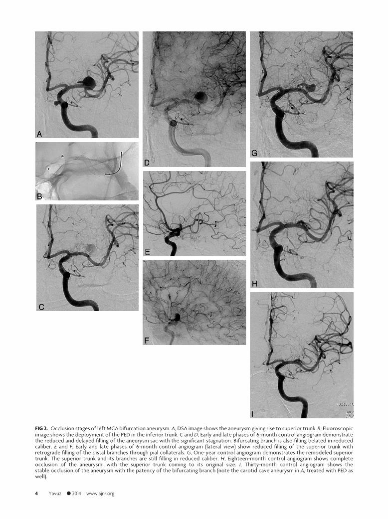
FIG 2. Occlusion stages of left MCA bifurcation aneurysm. A, DSA image shows the aneurysm giving rise to superior trunk. B, Fluoroscopicimage shows the deployment of the PED in the inferior trunk. C and D, Early and late phases of 6-month control angiogram demonstratethe reduced and delayed filling of the aneurysm sac with the significant stagnation. Bifurcating branch is also filling belated in reducedcaliber. E and F, Early and late phases of 6-month control angiogram (lateral view) show reduced filling of the superior trunk withretrograde filling of the distal branches through pial collaterals. G, One-year control angiogram demonstrates the remodeled superiortrunk. The superior trunk and its branches are still filling in reduced caliber. H, Eighteen-month control angiogram shows completeocclusion of the aneurysm, with the superior trunk coming to its original size. I, Thirty-month control angiogram shows thestable occlusion of the aneurysm with the patency of the bifurcating branch (note the carotid cave aneurysm in A, treated with PED aswell).
4 Yavuz ● 2014 www.ajnr.org
loss. This can be explained with the abundant distal collateral
supply of the ophthalmic artery from the external carotid
branches. Correspondingly, in another study including 46 aneu-
rysms in which a branch originated from the sac (excluding the
ophthalmic aneurysms), only in 5 aneurysms was the originating
branch (exclusively posterior communicating artery in all 5) oc-
cluded, with the ipsilateral posterior cerebral artery filling from
the posterior circulation.10 Other branches such as the anterior
choroidal artery stayed open, which led us to postulate that the
branches through which there is a flow demand because of insuf-
ficient distal collateral supply or toward which there is enough
pressure gradient between the high-pressure parent arteries, are
kept patent when covered by a flow diverter. Nevertheless, our
findings in this previous study emerged with the use of a single
device in the treatment of each aneurysm. Limited cases in the
literature reported the use of more than 1 device in the treatment
of aneurysms with the coverage of perforating arteries demon-
strating occlusion of the lenticulostriate branches and infarction
in the related territory.11,18 In the present study, all aneurysms
except for 2 were treated by use of a single PED. One of the 2
patients treated with 2 overlapping devices was admitted to the
hospital in the fourth postoperative week. The MR imaging did
not show any ischemic lesions, and CTA showed patency of the
MCA branches, which was confirmed with 6-month control DSA.
Furthermore, we prescribed low-molecular-weight heparin in ad-
dition to the antiplatelet therapy for 1 week, and the patient did
not have a recurrent event. The remaining patient was the one
with procedural SAH who had ischemic symptoms with unclear
etiology as described above.
In this series, there was 1 PED placed within a pre-existing
intraluminal construct (Enterprise stent), which had been placed
during the previous endosaccular coiling. Lylyk et al19 postulated
in their previous study that endoluminal constructs might repre-
sent potential impediments to the efficacy of the PED. In our
patient, we did not experience any technical difficulty in deploy-
ing the PED. This was the patient in whom we discovered perian-
eurysmal edema 4 weeks after the procedure, which we think was
unrelated to the pre-existing stent and PED combination. Six-
month DSA control revealed complete occlusion of the aneurysm
with patent bifurcating branches.
The main periprocedural and postprocedural complications
of the use of flow diverters in the endovascular treatment of in-
tracranial aneurysms are hemorrhagic and thromboembolic
events. Early and delayed aneurysm ruptures20-23 and distal ipsi-
lateral hemorrhage10,14,24,25 have emerged as the hemorrhagic
complications that are probably related to flow diversion. In this
series, 1 hemorrhagic event occurred as an SAH with unknown
origin during the procedure. No extravasation was observed in
DSA images during the treatment; however, DynaCT revealed
SAH. This bleeding may have been caused by a small, invisible
dissection/wire perforation during catheterization of the MCA
branch.
The only thromboembolic event (presented with transient
ischemic attack 3 months after the procedure) with radiologic
findings in this series occurred in 1 patient as the result of cessa-
tion of the antiplatelet drugs. There was no recurrent ischemic
attack after antiplatelet and anticoagulant medication as de-
scribed above. This result corroborates the importance of rigor-
ously evaluating the thrombocyte inhibition level in response to
clopidogrel or ticlopidine and safety of the PED use with the cov-
erage of perforators and bifurcating branches, at least when a
single device is used. We used whole-blood impedance platelet
aggregation and the rapid platelet function assay VerifyNow
P2Y12 (Accumetrics, San Diego, California) in all patients and
performed the procedure only if there was no resistance to the
drug and the value of Verify Now test was �30%.
In this series, all patients had at least 1 control angiographic
study. According to the last angiographic follow-up, complete
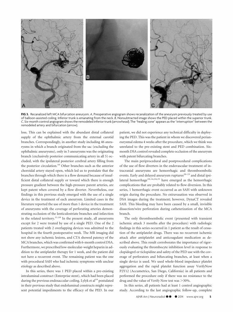
FIG 3. Recanalized left MCA bifurcation aneurysm. A, Preoperative angiogram shows recanalization of the aneurysm previously treated by useof balloon-assisted coiling. Inferior trunk is emanating from the neck. B, Nonsubtracted image shows the PED placed within the superior trunk.C, Six-month control angiogram shows the remodeled inferior trunk (arrowhead). The “healing zone” appears as the “interruption” between theremodeled artery and bifurcation (arrow).
AJNR Am J Neuroradiol ●:● ● 2014 www.ajnr.org 5
occlusion was revealed in 21 aneurysms (84%; 21/25). In our ex-
perience, we have observed that there are generally 3 phases,
though not necessarily, in the occlusion process of aneurysms
bearing a bifurcating branch by using the PED: 1) moderate to
significant decrease in the aneurysm filling (1–3 months), 2) the
infundibulum-like appearance resulting from the branch coming
off the parent artery with a bulking origin caused by significant
shrinkage of the aneurysm due to flow change, the so-called “re-
modeled artery” (3–12 months), and 3) complete occlusion
(6 –18 months) (Figs 1–3). We recommend performing the first
angiographic control at 6 months to evaluate the degree of occlu-
sion as well as the patency of the device and the branch(es) cov-
ered by the device. When the device is patent, with cessation of
clopidogrel or ticlopidine, and continuation of aspirin mono-
therapy, 18-month follow-up can be scheduled. According to the
last follow-up, 12 of the branches originating from the aneurysms
were patent, whereas 6 were filling in reduced caliber and 3 were
occluded asymptomatically. In 1 patient, the bifurcating branch
was filling in reduced caliber at 6-month and 1-year controls.
Clopidogrel was not discontinued in this patient, and the branch
was revealed to fill in normal caliber at 18 months (Fig 2). Our
anticipation is that a branch is kept patent whenever there is a flow
demand through it in the absence of rich distal collateral flow.
Even so, the operator may extend the duration of dual antiplatelet
treatment to 1 year or more.
Endovascular treatment of wide-neck bifurcation aneurysms,
especially when the bifurcating branches emanate directly from
the base of the aneurysm, is still challenging. With the introduc-
tion of the Y-stent placement technique with or without endos-
accular coiling,8,26,27 many of these difficult aneurysms have be-
come amenable to treatment while preserving the parent arteries.
However, in cases in which the branch has an acute angle relative
to the main trunk, intra-aneurysmal maneuvers as well as ex-
change procedures are necessitated to catheterize this branch, in-
creasing the risk of procedural hemorrhagic complications caused
by aneurysmal rupture and wire perforation.
To date, to our knowledge, this series including 21 aneurysms
located at the MCA bifurcations is the first study reporting spe-
cifically the use of the PED in bifurcation aneurysms. The PED,
being technically more simple and straightforward, provides a
safer procedure in cases of existing acutely angled branches. The
difficult branch and the aneurysm sac are not to be catheterized;
therefore, hemorrhagic risks caused by catheterization as well as
endosaccular embolization can be avoided.
We acknowledge the limitations of our study, with the lack of
longer follow-up data as well as the limited number of cases.
Therefore, we do not advocate this treatment alternative for an-
eurysms that can be treated with conventional techniques, includ-
ing clipping. However, the results of our preliminary experience
of PED use in the endovascular management of challenging MCA
aneurysms are encouraging, enabling the treatment of more com-
plex aneurysms with less procedural risk.
CONCLUSIONSThe PED provides a safe and effective solution for wide-neck
MCA aneurysms located at the bifurcation or M2 segment in
which 1 of the bifurcating or distal branches emanates directly
from the sac and when other endovascular techniques are thought
to be unfeasible or more risky. Preliminary results are promising
with low complication rates as well as high angiographic occlu-
sion with the remodeling of the emanating branch. Yet, the safe
use of more than 1 device at these locations remains ambiguous.
Larger series with longer-term follow-up examinations are re-
quired to show the long-term safety and durability of this treat-
ment alternative.
Disclosures: Isil Saatci—RELATED: Consulting Fee or Honorarium: Covidien/ev3.Saruhan Cekirge—RELATED: Consulting Fee or Honorarium: Covidien/ev3;UNRELATED: Consultancy: Covidien/ev3, MicroVention.
REFERENCES1. Doerfler A, Wanke I, Egelhof T, et al. Double-stent method: thera-
peutic alternative for small wide necked aneurysm: technical note.J Neurosurg 2004;100:150 –54
2. Canton G, Levy DI, Lasheras JC, et al. Flow changes caused by thesequential placement of stents across the neck of sidewall cerebralaneurysms. J Neurosurg 2005;103:891–902
3. Zenteno MA, Santos-Franco JA, Freitas-Modenesi JM, et al. Use ofthe sole stenting technique for the management of aneurysms in theposterior circulation in a prospective series of 20 patients. J Neuro-surg 2008;108:1104 –18
4. Fiorella D, Albuquerque FC, Deshmukh VR, et al. Endovascular re-construction with the Neuroform stent as monotherapy for thetreatment of uncoilable intradural pseudoaneurysms. Neurosurgery2006;59:291–300
5. Park SI, Kim BM, Kim DI, et al. Clinical and angiographic follow-upof stent-only therapy for acute intracranial vertebrobasilar dissect-ing aneurysms. AJNR Am J Neuroradiol 2009;30:1351–56
6. Pumar JM, Lete I, Pardo MI, et al. LEO stent monotherapy for theendovascular reconstruction of fusiform aneurysm of the middlecerebral artery. AJNR Am J Neuroradiol 2008;29:1775–76
7. Kim YJ. Sole stenting technique for treatment of complex aneu-rysms. J Korean Neurosurg 2009;46:545–51
8. Cekirge HS, Yavuz K, Geyik S, et al. A novel ‘Y’ stent flow diversiontechnique for the endovascular treatment of bifurcation aneurysmswithout endosaccular coiling. AJNR Am J Neuroradiol2011;32:1262– 68
9. Fiorella D, Woo HH, Albuquerque FC, et al. Definitive reconstruc-tion of circumferential, fusiform intracranial aneurysms with thePipeline embolization device. Neurosurgery 2008;62:1115–21
10. Saatci I, Yavuz K, Ozer C, et al. Treatment of intracranial aneurysmsusing the Pipeline flow-diverter embolization device: a single-cen-ter experience with long-term follow-up results. AJNR Am J Neuro-radiol 2012;33:1436 – 46
11. Nelson PK, Lylyk P, Szikora I, et al. The Pipeline embolization devicefor the intracranial treatment of aneurysms trial. AJNR Am J Neu-roradiol 2011;32:34 – 40
12. Siddiqui AH, Kan P, Abla A, et al. Complications after treatmentwith Pipeline embolization for giant intracranial aneurysms withor without coil embolization. Neurosurgery 2012;71:509 –13
13. Chitale R, Gonzalez LF, Randazzo C, et al. Single center experiencewith Pipeline stent: feasibility, technique, and complications. Neu-rosurgery 2012;71:679 –91
14. O’Kelly CJ, Spears J, Chow M, et al. Canadian experience with thePipeline embolization device for repair of unruptured intracranialaneurysms. AJNR Am J Neuroradiol 2012 Aug 2. [Epub ahead ofprint]
15. Burrows AM, Zipfel G, Lanzino G. Treatment of a pediatric recur-rent fusiform middle cerebral artery (MCA) aneurysm with a flowdiverter. BMJ Case Rep 2012 Nov 15;2012
16. Kallmes DF, Ding YH, Dai D, et al. A new endoluminal, flow-dis-rupting device for treatment of saccular aneurysms. Stroke2007;38:2346 –52
6 Yavuz ● 2014 www.ajnr.org
17. Puffer RC, Kallmes DF, Cloft HJ, et al. Patency of the ophthalmicartery after flow diversion treatment of paraclinoid aneurysms.J Neurosurg 2012;116:892–96
18. van Rooij, Sluzewski M. Perforator infarction after placement of aPipeline flow-diverting stent for an unruptured A1 aneurysm.AJNR Am J Neuroradiol 2010;31:E43– 44
19. Lylyk P, Miranda C, Ceratto R, et al. Curative endovascular recon-struction of cerebral aneurysms with the Pipeline embolizationdevice: the Buenos Aires experience. Neurosurgery 2009;64:632– 43
20. Kulcsar Z, Houdart E, Bonafe A, et al. Intra-aneurysmal thrombosisas a possible cause of delayed aneurysm rupture after flow-diver-sion treatment. AJNR Am J Neuroradiol 2011;32:20 –25
21. Turowski B, Macht S, Kulcsar Z, et al. Early fatal hemorrhage afterendovascular cerebral aneurysm treatment with a flow-diverter(Silk-stent): do we need to rethink our concepts? Neuroradiology2011;53:37– 41
22. Cebral JR, Mut F, Raschi, et al. Aneurysm rupture following treat-ment with flow-diverting stents: computational hemodynamicsanalysis of treatment. AJNR Am J Neuroradiol 2011;32:27–33
23. Hampton T, Walsh D, Tolias C, et al. Mural destabilization afteraneurysm treatment with a flow-diverting device: a report of twocases. J Neurointerv Surg 2011;3:167–71
24. Fischer S, Vajda Z, Perez MA, et al. Pipeline embolization device(PED) for neurovascular reconstruction: initial experience in treat-ment of 101 intracranial aneurysms and dissections. Neuroradiology2012;54:369 – 82
25. Cruz JP, Chow M, O’Kelly C, et al. Delayed ipsilateral parenchy-mal hemorrhage following flow diversion for the treatment ofanterior circulation aneurysms. AJNR Am J Neuroradiol2012;33:603– 08
26. Yavuz K, Geyik S, Cekirge S, et al. Double stent-assisted coil embo-lization treatment for bifurcation aneurysms: immediate treat-ment results and long-term angiographic outcome. AJNR Am JNeuroradiol 2013 Mar 28. [Epub ahead of print]
27. Thorell WE, Chow MM, Woo HH, et al. Y-configured dual intra-cranial stent-assisted coil embolization for the treatment ofwide-necked basilar tip aneurysms. Neurosurgery 2005;56:1035– 40
AJNR Am J Neuroradiol ●:● ● 2014 www.ajnr.org 7
Related Documents